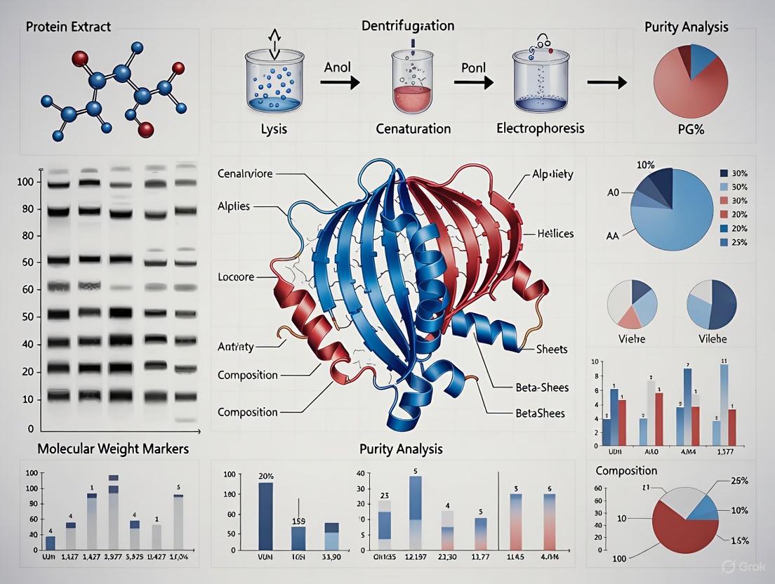
Validating Protein Purity and Composition with SDS-PAGE: A Complete Guide for Biomedical Researchers
This article provides a comprehensive resource for researchers, scientists, and drug development professionals on the application of Sodium Dodecyl Sulfate-Polyacrylamide Gel Electrophoresis (SDS-PAGE) for validating protein purity and composition.
Validating Protein Purity and Composition with SDS-PAGE: A Complete Guide for Biomedical Researchers
Abstract
This article provides a comprehensive resource for researchers, scientists, and drug development professionals on the application of Sodium Dodecyl Sulfate-Polyacrylamide Gel Electrophoresis (SDS-PAGE) for validating protein purity and composition. It covers foundational principles from protein denaturation to molecular sieving, detailed methodological protocols for diverse applications from biopharmaceuticals to food science, advanced troubleshooting for common experimental challenges, and a critical comparison with emerging techniques like Capillary Electrophoresis. The content synthesizes established knowledge with 2025 research trends to offer a complete analytical framework for ensuring protein integrity in research and development.
SDS-PAGE Fundamentals: Principles of Protein Separation and Purity Analysis
For researchers and drug development professionals, validating protein purity and composition is a critical step in ensuring the integrity of biochemical research and biopharmaceutical products. Among the most foundational techniques for this purpose is SDS-PAGE (sodium dodecyl sulfate–polyacrylamide gel electrophoresis), which allows for the separation of proteins based solely on their molecular weight [1] [2]. The reliability of this method hinges on two core functions of the ionic detergent SDS: the complete denaturation of protein structures and the conferment of a uniform negative charge. This guide details the principles and experimental protocols underlying these processes, providing a objective basis for their critical role in protein analysis.
The Dual Role of SDS in Protein Preparation
Protein Denaturation and Linearization
The native three-dimensional structure of a protein, stabilized by hydrogen bonds, hydrophobic interactions, and disulfide bridges, presents a challenge for size-based separation. SDS addresses this by acting as a potent denaturant:
- Disruption of Non-Covalent Bonds: The hydrophobic tail of SDS interacts with and disrupts the hydrophobic core of the protein [3].
- Unfolding: This interaction disrupts the protein's secondary and tertiary structures, leading to its unfolding [4].
- Cleavage of Disulfide Bridges: The denaturation process is typically aided by reducing agents like β-mercaptoethanol or dithiothreitol (DTT), which break disulfide bonds between cysteine residues, ensuring the protein is reduced to its polypeptide subunits [4] [2].
This process results in a fully unfolded, linear polypeptide chain.
Charge Masking and Uniform Charge-to-Mass Ratio
In their native state, proteins possess intrinsic charges based on their amino acid composition, which would cause them to migrate at different speeds in an electric field regardless of size. SDS resolves this issue by:
- Uniform Coating: The linearized polypeptide chain binds SDS along its length in a constant weight ratio of approximately 1.4 g of SDS per 1 g of protein [4] [2]. This translates to roughly one SDS molecule for every two amino acids [2].
- Charge Dominance: Each SDS molecule contributes a strong negative charge from its sulfate group. This overwhelming negative charge effectively masks the protein's intrinsic charge [4].
- Identical Ratio: Consequently, all SDS-coated proteins exhibit an almost identical charge-to-mass ratio [4] [2]. This means that in an electric field, all proteins will experience the same driving force towards the positive anode, eliminating mobility differences based on charge.
The following diagram illustrates the transformative process proteins undergo during SDS treatment before electrophoresis.
Experimental Protocol for SDS-PAGE
A standard protocol for preparing and separating proteins using SDS-PAGE is outlined below. Adherence to this protocol is essential for generating reproducible and reliable results.
Sample Preparation:
- Mix Protein Sample: Combine the protein sample with an SDS-PAGE sample buffer (for composition, see Research Reagent Solutions below) at a recommended ratio of 4:1 (volume of protein solution to volume of 5X sample buffer) [3].
- Denature and Reduce: Heat the mixture at 95–100 °C for 3–5 minutes [2] [3]. This step ensures complete denaturation and, in the presence of a reducing agent, cleavage of disulfide bonds.
- Centrifuge: Briefly centrifuge the sample (e.g., 10,000 rpm for 5 minutes) to pellet any insoluble debris [3].
- Load: Load the supernatant into a well of the polyacrylamide gel.
Electrophoresis:
- Assemble Apparatus: Place the cast gel into the electrophoresis chamber and fill the inner and outer chambers with running buffer (e.g., Tris-Glycine-SDS buffer) [3] [5].
- Apply Voltage: Connect the power supply and apply a constant voltage. A common protocol starts at a lower voltage (e.g., 60–80 V) as the samples move through the stacking gel, then increases to a higher voltage (e.g., 120–200 V) for the separation through the resolving gel [2] [5]. The process typically takes 30 minutes to several hours, depending on the gel size and voltage.
Quantitative Analysis of SDS-Protein Interaction
The table below summarizes key quantitative aspects of the SDS-PAGE process that are critical for experimental design and understanding its limitations.
Table 1: Key Quantitative Parameters in SDS-PAGE
| Parameter | Typical Value or Range | Functional Significance |
|---|---|---|
| SDS Binding Ratio | 1.4 g SDS / 1 g protein [4] [2] | Creates a uniform charge-to-mass ratio, enabling separation by size alone. |
| Critical Micelle Concentration (CMC) | 7–10 mM (monomer-micelle equilibrium) [2] | Ensures sufficient free SDS monomers are available to bind and denature proteins. |
| Effective Protein Denaturation | > 1 mM SDS concentration [2] | Confirms complete unfolding of most proteins at standard buffer concentrations. |
| Molecular Weight Separation Range | 5–250 kDa [2] | Defines the effective scope of standard SDS-PAGE. Techniques like Tris-Tricine SDS-PAGE extend this to 0.5–50 kDa [2]. |
Research Reagent Solutions for SDS-PAGE
The following table details the essential reagents required for a standard SDS-PAGE experiment, along with their specific functions.
Table 2: Essential Reagents for SDS-PAGE Analysis
| Reagent | Function | Typical Composition / Notes |
|---|---|---|
| Sodium Dodecyl Sulfate (SDS) | Denatures proteins and confers uniform negative charge [4] [3]. | Anionic detergent. Used at 0.1-0.5% in gels and buffers, and 1-2% in sample buffer [3]. |
| Reducing Agents (DTT, β-ME) | Breaks disulfide bonds to fully reduce protein to subunits [4] [2]. | Dithiothreitol (DTT) or β-mercaptoethanol. Added to sample buffer. |
| Sample Buffer | Prepares protein for loading by denaturing, charging, and adding density. | Contains SDS, reducing agent, glycerol (for density), Tris buffer, and a tracking dye (e.g., bromophenol blue) [3] [5]. |
| Polyacrylamide Gel | Acts as a molecular sieve; separates proteins by size [4]. | Acrylamide cross-linked with bisacrylamide. Concentration (e.g., 8-15%) determines resolution range [4] [3]. |
| Electrophoresis Buffer | Conducts current and maintains pH during run. | Typically Tris-Glycine-SDS buffer, pH ~8.3-8.8 [4] [3]. |
| Molecular Weight Marker | Provides reference for estimating protein molecular weights. | A mixture of pre-stained or unstained proteins of known sizes [1] [5]. |
Methodological Variations and Technological Evolution
While the core principles of SDS-PAGE remain unchanged, understanding its limitations has spurred the development of complementary and advanced methods.
Native SDS-PAGE (NSDS-PAGE): A modification that uses significantly reduced SDS concentrations and omits heating and reducing agents. This allows for high-resolution separation while retaining native protein functions, such as enzymatic activity and bound metal ions, which are lost in standard SDS-PAGE [6]. For example, one study showed Zn²⁺ retention increased from 26% (standard SDS-PAGE) to 98% (NSDS-PAGE) [6].
Capillary Electrophoresis-SDS (CE-SDS): An advanced, automated technology that replaces slab gels with capillaries. It offers superior advantages for biopharmaceutical development, including higher resolution, automation, superior reproducibility, quantitative precision, and reduced use of toxic reagents compared to traditional SDS-PAGE [7]. This method is widely used in regulatory filings for commercial biotherapeutics [7].
The consistent and predictable behavior of SDS with linearized polypeptides is the cornerstone of a technique that has become a cornerstone of modern molecular biology and biopharmaceutical analysis. Mastery of these principles enables researchers to reliably validate protein purity and composition, forming a solid foundation for downstream applications and quality control.
In the realm of protein research, the validation of protein purity and composition is a cornerstone of reliable scientific findings, particularly in drug development where precise characterization is non-negotiable. Polyacrylamide gel electrophoresis (PAGE) serves as a critical technique for this purpose, functioning primarily through its role as a molecular sieve. This sieving effect is the fundamental principle that enables the separation of proteins based on their size. The gel matrix is a synthetic polymer network formed through the copolymerization of acrylamide with a bifunctional crosslinker, most commonly N,N'-methylenebisacrylamide (Bis). This reaction, catalyzed by ammonium persulfate (APS) and N,N,N',N'-tetramethylethylenediamine (TEMED), creates a intricate, mesh-like structure with uniform pores [8] [9]. The dimensions of these pores determine the size-based separation of proteins, as smaller proteins navigate the pores more readily than larger ones when an electric field is applied [9] [10].
This article will objectively compare the performance of polyacrylamide gels against alternative matrices like agarose, detail how pore size is controlled and optimized, and provide supporting experimental data and protocols. The content is framed within the essential context of validating protein purity and composition, providing researchers and scientists with a clear guide for employing SDS-PAGE in their analytical workflows.
Principles of Molecular Sieving in PAGE
The Mechanism of Size-Based Separation
The core function of the polyacrylamide gel matrix is to act as a molecular sieve. During electrophoresis, an electric field is applied across the gel, causing charged protein molecules to migrate. In the most common form, SDS-PAGE, proteins are denatured and uniformly coated with the anionic detergent sodium dodecyl sulfate (SDS). This SDS coating confers a uniform negative charge density per unit mass, effectively masking the proteins' intrinsic charges and unfolding them into linear chains [9] [10]. Consequently, the separation is based almost exclusively on polypeptide chain length or molecular weight, rather than charge or native structure [11].
The polyacrylamide mesh presents a frictional resistance to the migrating proteins. Smaller proteins experience less hindrance and can move through the pore network more rapidly. Larger proteins, however, encounter greater frictional forces and are impeded by the gel matrix, resulting in slower migration [9] [10]. This differential migration rate results in the physical separation of proteins into distinct bands within the gel, allowing for analysis of protein size, purity, and relative abundance.
Control of Pore Size: T and C Values
The pore size of the gel, and therefore its sieving properties, is not fixed; it can be precisely and reproducibly controlled by adjusting two key parameters during gel fabrication [8]:
- Total Monomer Concentration (T): This is the total percentage concentration (w/v) of acrylamide and bisacrylamide in the gel solution. The relationship between T and pore size is nearly linear: increasing the T value decreases the average pore size. Thus, higher percentage gels (e.g., 15%) with smaller pores are used to separate smaller proteins, while lower percentage gels (e.g., 8%) with larger pores are used for larger proteins [8] [12].
- Crosslinking Ratio (C): This is the percentage of the total monomer (T) that is made up by the crosslinker (bisacrylamide). The relationship between C and pore size is more complex. The minimum pore size is achieved at a C value of about 5% (a 19:1 ratio of acrylamide to bis). Decreasing C results in a more open structure with fewer crosslinks, while increasing C beyond 5% also increases pore size due to the bundling of polymer strands [8].
This reproducible control over pore size, a direct result of the synthetic nature of acrylamide and bisacrylamide, is a key advantage of polyacrylamide gels, leading to minimal batch-to-batch variation and highly consistent results [8].
Comparative Performance: Polyacrylamide vs. Agarose Gels
The choice of a gel matrix for electrophoresis depends heavily on the size and type of the biomolecules being separated. The table below provides a direct comparison between polyacrylamide and agarose gels, the two most common matrices.
Table 1: Objective Comparison of Polyacrylamide and Agarose Gel Matrices
| Feature | Polyacrylamide Gel | Agarose Gel |
|---|---|---|
| Primary Separation Mechanism | Molecular sieving based on size (SDS-PAGE) or size/charge (Native-PAGE) [9] | Molecular sieving based on size [9] |
| Typical Pore Size | Small, controllable pore size [9] | Large pore size [9] |
| Optimal Molecular Size Range | Proteins and small nucleic acids (DNA/RNA) [8] [9] | Large nucleic acids (e.g., genomic DNA, PCR products) and large protein complexes [9] |
| Resolution | Superior high resolution; can separate proteins differing by ~0.1% in size [8] | Lower resolution; suitable for separating larger fragments (e.g., 100 bp vs. 500 bp) [12] |
| Pore Size Control | Highly precise and reproducible via T and C values [8] | Less precise; adjusted by changing agarose percentage [8] |
| Gel Strength & Handling | Strong, durable, and less prone to tearing [12] | Fragile and more easily damaged during handling [12] |
| Common Applications | SDS-PAGE, Native-PAGE, 2D-PAGE, protein analysis, western blotting [9] [10] | DNA gel electrophoresis, plasmid analysis, pulse-field gel electrophoresis [12] |
As the data indicate, polyacrylamide gels are the unequivocal matrix of choice for high-resolution protein separation due to their controllable, small pore size and superior resolving power. Agarose is better suited for the separation of larger nucleic acids where high resolution is not as critical [9] [12].
Optimizing Pore Size for Protein Separation
Gel Percentage Selection Guide
Selecting the correct polyacrylamide gel percentage is paramount for achieving optimal separation. The table below summarizes recommended gel compositions for different molecular size ranges, based on standard crosslinking ratios.
Table 2: Recommended Polyacrylamide Gel Conditions for Separating Various Biomolecules
| Acrylamide:Bis Ratio (C Value) | Gel % (T Value) | Native DNA/RNA (bp) | Denatured DNA/RNA (bp) | Proteins (kDa) |
|---|---|---|---|---|
| 19:1 (C=5%) | 4% | 100-1500 | 70-500 | >100-200 |
| 19:1 (C=5%) | 8% | 40-500 | 20-200 | 20-100 |
| 19:1 (C=5%) | 12% | 20-150 | 10-100 | 8-60 |
| 29:1 (C=3.3%) | 8% | 60-400 | 30-300 | 30-125 |
| 37.5:1 (C=2.6%) | 10% | -- | -- | 25-100 |
| 37.5:1 (C=2.6%) | 12% | -- | -- | 15-80 |
For general protein separation using SDS-PAGE, a C value of 2.6% (37.5:1 ratio) is standard [8]. The optimal T value depends on the target protein's size:
- Low percentage gels (4-8%): Best for separating very large proteins (>100 kDa) [12].
- Medium percentage gels (8-12%): Ideal for separating a broad range of protein sizes (e.g., 15-100 kDa) and are the most commonly used for general protein analysis [12] [10].
- High percentage gels (12-20%): Provide the high resolution needed for separating small proteins and peptides (<30 kDa) [12].
For complex mixtures containing proteins of vastly different sizes, gradient gels (e.g., 4-20%) are highly effective. These gels have a continuously varying acrylamide concentration, creating a pore size gradient that allows both large and small proteins to be resolved sharply within the same gel [9] [10].
Experimental Protocol: SDS-PAGE for Protein Purity Analysis
The following is a detailed protocol for performing SDS-PAGE to validate protein purity and composition, incorporating best practices from the search results [9] [11] [10].
Research Reagent Solutions
| Item/Chemical | Function in the Experiment |
|---|---|
| Acrylamide & Bisacrylamide | monomers that polymerize to form the porous gel matrix [8] |
| Ammonium Persulfate (APS) | initiator that generates free radicals to start the polymerization reaction [9] |
| TEMED | catalyst that accelerates the polymerization reaction by promoting free radical production from APS [8] [9] |
| Tris-HCl Buffer | provides the controlled pH environment necessary for gel polymerization and electrophoresis [9] |
| Sodium Dodecyl Sulfate (SDS) | ionic detergent that denatures proteins and confers a uniform negative charge [9] [10] |
| Glycine | an amino acid that serves as the leading ion in the discontinuous buffer system for efficient stacking [9] |
| Molecular Weight Markers | standardized protein mixture of known sizes, allowing for estimation of sample protein molecular weights [9] [10] |
| Coomassie Brilliant Blue | dye used to stain and visualize protein bands within the polyacrylamide gel after electrophoresis [13] |
Step-by-Step Methodology:
Gel Casting:
- Resolving Gel: Assemble the gel cassette. Mix the acrylamide/bis solution at the desired percentage (T) and crosslinking ratio (C) with Tris-HCl buffer (typically pH 8.8), SDS, APS, and TEMED. Pour the solution into the cassette, leaving space for the stacking gel. Overlay with water or isopropanol to create a flat, smooth interface and prevent inhibition of polymerization by atmospheric oxygen. Allow the gel to polymerize completely (~20-30 minutes) [8] [11].
- Stacking Gel: After polymerization of the resolving gel, remove the overlay. Prepare a lower percentage acrylamide solution (e.g., 4-5%) with Tris-HCl at a lower pH (typically 6.8). Add APS and TEMED, pour over the resolving gel, and immediately insert a comb to create sample wells. Allow to polymerize [9] [10].
Sample Preparation: Mix protein samples with an SDS-PAGE sample loading buffer containing SDS, a reducing agent (e.g., β-mercaptoethanol or DTT) to break disulfide bonds, glycerol to help the sample sink into the well, and a tracking dye (e.g., Bromophenol Blue). Heat the samples at 95-100°C for 3-5 minutes to ensure complete denaturation [11] [10].
Electrophoresis: Assemble the gel cassette in the electrophoresis tank filled with running buffer (e.g., Tris-Glycine-SDS). Load the prepared samples and molecular weight markers into the wells. Apply a constant voltage (100-150V for mini-gels) until the dye front reaches the bottom of the gel [11] [10].
Protein Detection: After electrophoresis, carefully open the cassette and remove the gel. Stain the gel with Coomassie Brilliant Blue to visualize protein bands, followed by destaining to remove background dye. Alternatively, for higher sensitivity, silver staining or western blotting can be performed [13] [10].
Figure 1: SDS-PAGE Experimental Workflow.
Advanced Applications and Data Interpretation
Quantitative and Functional Analysis
Beyond assessing purity, SDS-PAGE can be adapted for quantitative analysis. Densitometry—measuring the optical density of stained protein bands—can be used to determine the relative abundance of proteins in a sample [10]. Furthermore, a study demonstrated the use of SDS-PAGE densitometry for the quantitative estimation of the enzyme papain in pharmaceutical formulations, validating the method for precision, accuracy, and robustness according to ICH guidelines [13].
For experiments requiring the analysis of proteins in their native, functional state, Native PAGE and NSDS-PAGE (Native SDS-PAGE) are valuable alternatives. In Native PAGE, no denaturants are used, allowing proteins to separate based on their native charge, size, and shape, while retaining enzymatic activity and subunit interactions [9] [6]. NSDS-PAGE is a modified technique that uses minimal SDS and omits heating and reducing agents. This approach can achieve high-resolution separation while preserving the functional properties of many proteins, including the retention of bound metal ions and enzymatic activity, as demonstrated for several zinc metalloproteins [6].
Protein Purity and Composition Assessment
In the context of validating protein purity and composition, SDS-PAGE provides critical information:
- Purity Assessment: A pure protein preparation will appear as a single, sharp band on a stained gel. The presence of multiple bands indicates contamination or the existence of different subunits or isoforms [10].
- Molecular Weight Determination: By comparing the migration distance of an unknown protein band to that of proteins in a molecular weight marker lane, the protein's apparent molecular weight can be estimated [9] [10].
- Composite Analysis: For complex samples like cell lysates, the banding pattern provides a composite "fingerprint" of the protein composition, which can be compared between different conditions to observe changes in expression.
Figure 2: Pore Size Controls Protein Separation.
The polyacrylamide gel matrix is an indispensable tool in the scientist's toolkit for protein analysis. Its effectiveness as a molecular sieve, driven by the precise control over pore size through T and C values, provides unparalleled resolution for separating proteins by size. When compared to agarose, polyacrylamide is objectively superior for protein work due to its smaller, reproducible pore structure and the physical strength of the gel. The well-established SDS-PAGE protocol allows researchers to reliably validate protein purity, determine molecular weight, and prepare samples for downstream applications like western blotting and mass spectrometry. Mastery of this technique—including the strategic selection of gel percentage and crosslinking—is fundamental for any research or development professional focused on protein characterization.
Sodium dodecyl sulfate-polyacrylamide gel electrophoresis (SDS-PAGE) is a foundational technique in proteomics research, enabling researchers to separate protein mixtures based on molecular weight. The Laemmli system, introduced in 1970, remains the gold standard method for SDS-PAGE, renowned for its ability to provide clear resolution and accurate molecular weight estimation for proteins from diverse biological sources [14] [15]. This discontinuous buffer system employs distinct stacking and separating gel phases to achieve high-resolution protein separation, a critical first step in validating protein purity and composition in drug development pipelines.
Within the context of protein analysis research, the Laemmli system provides the initial separation necessary for downstream applications including western blotting, mass spectrometry, and protein sequencing. The system's effectiveness hinges on the sophisticated interplay between its two gel regions: the stacking gel, which concentrates protein samples into a sharp starting zone, and the separating gel, which resolves individual protein components based on their molecular sizes [9]. Understanding the biochemical principles underlying these two gel phases is essential for researchers and scientists aiming to optimize protein separation for analytical and preparative purposes.
Fundamental Principles of the Laemmli System
The Role of SDS in Protein Denaturation
In the Laemmli system, the ionic detergent sodium dodecyl sulfate (SDS) plays a fundamental role in denaturing proteins. When a protein sample is heated to between 70-100°C in the presence of SDS and a reducing agent like β-mercaptoethanol or dithiothreitol (DTT), disulfide bonds are cleaved and the protein unfolds completely [9]. SDS molecules bind to the polypeptide backbone in a constant weight ratio of approximately 1.4 g SDS per 1 g of protein, imparting a uniform negative charge to all protein molecules [9]. This SDS coating negates the inherent charges of individual amino acids, ensuring that proteins migrate through the gel matrix strictly according to polypeptide chain length with minimal influence from compositional differences or tertiary structure [16].
The polyacrylamide gel matrix serves as a molecular sieve, with its cross-linked structure creating pores that regulate protein migration. Smaller proteins navigate these pores more easily and migrate rapidly toward the anode, while larger proteins encounter greater frictional resistance and migrate more slowly [9]. This size-dependent migration allows for the separation of protein mixtures into discrete bands that can be visualized with appropriate staining techniques. The pore size is inversely related to the polyacrylamide percentage, with lower percentages (e.g., 7-10%) providing larger pores suitable for separating high molecular weight proteins, and higher percentages (e.g., 12-15%) creating smaller pores optimal for resolving lower molecular weight proteins [9] [16].
The Discontinuous Buffer System
The Laemmli system employs a discontinuous buffer system that utilizes different pH values and ionic compositions in the stacking versus separating gels to achieve exceptional protein resolution. This discontinuity creates a transient state where protein samples become concentrated into an extremely thin zone before entering the separating gel, dramatically improving band sharpness [9].
The stacking gel, cast with a lower acrylamide concentration (typically 4-5%) and buffered at pH 6.8, serves as the initial point of sample entry. The separating gel, with higher acrylamide concentration (ranging from 7.5-15% depending on target protein sizes) and buffered at pH 8.8, provides the molecular sieving matrix where actual protein separation occurs [17]. When current is applied, the key ions in the system—chloride ions from the gel buffer as leading ions, and glycine ions from the running buffer as trailing ions—create an ion gradient that confines protein molecules into a narrow zone during the initial migration phase [14]. This focusing effect ensures that all proteins enter the separating gel simultaneously as a sharp band, which then separates into discrete bands based on molecular weight as they migrate through the pH 8.8 environment where glycine ions become more mobile and overtake the protein-SDS complexes.
The following diagram illustrates the workflow and ionic dynamics of the Laemmli SDS-PAGE system:
Comparative Analysis: Laemmli vs. Alternative Gel Systems
While the Laemmli system remains widely used, several alternative gel systems have been developed to address specific limitations. The table below provides a comprehensive comparison of key performance metrics across different electrophoresis systems:
Table 1: Comparative Performance of SDS-PAGE Gel Systems
| Parameter | Traditional Laemmli | NuPAGE Bis-Tris System | TGX Precast Gels |
|---|---|---|---|
| Operating pH | Highly alkaline (pH ~9.5) [14] | Neutral (pH 7.0) [14] | Modified Laemmli (patented) [15] |
| Shelf Life | 4-6 weeks (handcast) [14] | 8-12 months (precast) [14] | Up to 12 months (precast) [15] |
| Band Resolution | Good, but potential for artifacts at high pH [14] | Excellent, sharper bands due to neutral pH [14] | Superior, tight crisp bands with high linearity (R² > 0.98) [15] |
| Run Time | 30-45 minutes (mini-gel) [15] | Comparable to Laemmli | 15 minutes at 300V (mini-gel) [15] |
| Protein Stability | Risk of deamination, Asp-Pro cleavage [14] | High stability, minimal modifications [14] | High stability, preserves protein integrity [15] |
| Molecular Weight Accuracy | Good for standard proteins | Good for standard proteins | Excellent linearity (R² > 0.98) [15] |
| Typely | Handcast or precast | Precast only | Precast only |
Limitations of the Traditional Laemmli System
The traditional Laemmli system faces several notable limitations that alternative systems aim to address. The highly alkaline operating environment (pH ~9.5) of the separating gel can promote protein deamination and other chemical modifications that may compromise protein integrity [14]. Additionally, the high gel casting pH (~8.7) accelerates polyacrylamide hydrolysis, resulting in a short shelf life of just 4-6 weeks for handcast gels [14]. Another significant concern is the cleavage of Asp-Pro bonds when proteins are heated at 100°C in the Laemmli sample buffer at pH 5.2, potentially generating artifactual bands [14]. These limitations have driven the development of alternative buffer systems that maintain the fundamental principles of discontinuous electrophoresis while addressing these stability issues.
Modern Alternatives: NuPAGE and TGX Systems
The NuPAGE Bis-Tris system represents a significant advancement with its neutral pH electrophoresis environment. Operating at pH 7.0, this system eliminates many of the protein modification issues associated with the alkaline Laemmli system, resulting in sharper band resolution and improved protein stability [14]. The system uses MES or MOPS running buffers instead of Tris-glycine and offers extended shelf life of 8-12 months for precast gels [14]. The neutral pH environment also prevents cleavage of acid-labile bonds like Asp-Pro, providing more accurate representation of protein composition.
Bio-Rad's TGX gels employ a patented modification of the Laemmli system that maintains the standard Tris-glycine running buffers while significantly extending shelf life up to 12 months [15]. These gels demonstrate exceptional performance with increased linearity of separation (R² > 0.98) compared to traditional Laemmli precast gels or NuPAGE Bis-Tris systems (R² = 0.86 for NuPAGE) [15]. The system enables faster run times—15 minutes at 300V compared to 30-45 minutes for traditional systems—without compromising resolution, making it particularly valuable for high-throughput applications in drug development pipelines [15].
Table 2: Transfer Efficiency Comparison for Western Blotting
| Transfer Method | Traditional Laemmli | TGX Precast Gels |
|---|---|---|
| Semi-dry Transfer | 60 minutes at 15V [15] | 15 minutes at 150V [15] |
| Tank Transfer | 60 minutes to overnight [15] | 15 minutes at 150V [15] |
| Transfer Efficiency | Variable, depending on protein size | High efficiency for broad MW range [15] |
Essential Reagents and Research Solutions
Successful protein separation using the Laemmli system requires precise formulation of key reagents. The following table outlines essential components and their functions in the electrophoresis process:
Table 3: Essential Research Reagents for Laemmli SDS-PAGE
| Reagent | Composition/Properties | Function in SDS-PAGE |
|---|---|---|
| Laemmli Sample Buffer (4X) | 250 mM Tris-HCl (pH 6.8), 8% SDS, 40% glycerol, 0.02% bromophenol blue [18] | Denatures proteins, provides tracking dye, and increases sample density for well loading |
| Acrylamide/Bis Solution | 29:1 or 37.5:1 ratio of acrylamide to bisacrylamide | Forms cross-linked polymer matrix that acts as molecular sieve |
| Stacking Gel Buffer | 125 mM Tris-HCl, pH 6.8 [17] | Creates low pH, large-pore environment for protein stacking |
| Separating Gel Buffer | 375 mM Tris-HCl, pH 8.8 [17] | Creates high pH environment for size-based separation |
| Running Buffer | 25 mM Tris, 192 mM glycine, 0.1% SDS, pH ~8.3 [17] | Conducts current and maintains pH during electrophoresis |
| Ammonium Persulfate (APS) | 10% solution in water | Free-radical initiator for acrylamide polymerization |
| TEMED | N,N,N',N'-Tetramethylethylenediamine | Catalyzes acrylamide polymerization by producing free radicals |
| Reducing Agents | DTT or β-mercaptoethanol | Cleaves disulfide bonds for complete protein denaturation |
Proper storage and handling of these reagents is critical for experimental reproducibility. Sample buffer aliquots remain stable for 6 months at -20°C and over one year at -80°C [15]. Ammonium persulfate solutions should be prepared fresh weekly or stored frozen for longer-term use, as the free radicals necessary for polymerization degrade over time. TEMED should be stored tightly sealed at room temperature and protected from light to maintain its catalytic activity.
Experimental Protocols for Protein Separation
Gel Casting Methodology
The following protocol describes the preparation of handcast Laemmli gels for optimal protein separation:
Resolving Gel Preparation: For a standard 10% resolving gel, combine 7.5 mL of 40% acrylamide solution, 3.9 mL of 1% bisacrylamide solution, and 7.5 mL of 1.5 M Tris-HCl (pH 8.8) [9]. Add ultrapure water to a final volume of 30 mL, then add 0.3 mL of 10% SDS, 0.3 mL of 10% ammonium persulfate, and 0.03 mL TEMED to initiate polymerization [9]. Pour the solution immediately between assembled glass plates, leaving space for the stacking gel. Carefully overlay with isopropanol or water to create a flat interface, and allow complete polymerization for 20-30 minutes.
Stacking Gel Preparation: Once the resolving gel has polymerized, prepare the stacking gel by combining appropriate volumes of acrylamide/bis solution with 125 mM Tris-HCl (pH 6.8) [17]. Add 10% APS and TEMED, pour over the resolving gel, and immediately insert a clean comb. Allow 30 minutes for complete polymerization before carefully removing the comb to avoid damaging the wells.
Sample Preparation and Electrophoresis Conditions
Protein Sample Preparation: Dilute protein samples with an appropriate volume of 4X Laemmli sample buffer to achieve 1X final concentration [18]. For reduced conditions, include 50-100 mM DTT or 5% β-mercaptoethanol in the sample buffer. Heat samples at 70-95°C for 3-5 minutes to denature proteins completely [15]. Avoid boiling at 100°C, which can promote protein aggregation, especially for high molecular weight proteins [15]. Centrifuge samples at >10,000 × g for 2 minutes to pellet any insoluble material before loading [15].
Gel Electrophoresis: Assemble the gel cassette in the electrophoresis chamber and fill both inner and outer chambers with running buffer (25 mM Tris, 192 mM glycine, 0.1% SDS) [17]. Load protein samples and appropriate molecular weight markers into wells. For mini-gel formats, run at constant voltage of 150-200V until the bromophenol blue tracking dye reaches the bottom of the gel (typically 30-45 minutes) [15]. For optimal resolution, maintain cool running conditions by using a cooled apparatus or performing electrophoresis in a cold room, particularly when running at higher voltages [19].
Troubleshooting Common Electrophoresis Issues
Even with proper technique, researchers may encounter various challenges during SDS-PAGE separation. The table below outlines common problems, their potential causes, and recommended solutions:
Table 4: Troubleshooting Common SDS-PAGE Issues
| Problem | Possible Causes | Recommended Solutions |
|---|---|---|
| Smeared Bands | Voltage too high [19] [20]; Protein overload [20]; High salt concentration [20] | Run gel at 10-15 V/cm [19]; Reduce protein load [20]; Dialyze sample or desalt [20] |
| Poor Band Resolution | Insufficient run time [19]; Incorrect gel percentage [16]; Old or improperly stored gels [20] | Run until dye front reaches bottom [19]; Use gradient or appropriate % gel [20]; Use fresh gels [20] |
| "Smiling" Bands | Excessive heat generation during run [19] | Run at lower voltage for longer time; Use cooling apparatus or cold room [19] |
| Edge Effect | Empty wells at periphery [19] | Load all wells with sample or buffer [19]; Add 10 µL 1X sample buffer to unused wells [15] |
| Protein Aggregation | Insufficient reducing agent [20]; Sample boiled at too high temperature [15] | Prepare fresh sample buffer with fresh DTT [20]; Heat at 70-95°C instead of 100°C [15] |
| Vertical Streaking | Sample precipitation [20]; Protein overload [20] | Centrifuge samples before loading [15]; Dilute sample or reduce voltage by 25% [20] |
Additional troubleshooting considerations include checking buffer composition and age, as improperly formulated or overused buffers can lead to irregular migration patterns [19] [16]. For persistent issues with band distortion, ensure complete gel polymerization by using fresh ammonium persulfate and TEMED, and casting gels at room temperature [20]. When separating very high or very low molecular weight proteins, consider adjusting the acrylamide concentration or using gradient gels to improve resolution across a broad molecular weight range [16].
The Laemmli system remains a cornerstone technology in protein analysis, providing the fundamental separation principles that support modern proteomics research. While the traditional system faces limitations regarding shelf life and protein stability at alkaline pH, modified systems like NuPAGE Bis-Tris and TGX gels have addressed these challenges while maintaining the core discontinuous buffer approach. For researchers validating protein purity and composition in drug development, understanding the intricacies of stacking and separating gels enables optimal experimental design and troubleshooting. The choice between handcast Laemmli gels, neutral pH systems, or modified Laemmli precast gels ultimately depends on specific research requirements, balancing factors such as resolution needs, throughput considerations, and downstream applications. As protein therapeutic development advances, the principles underlying the Laemmli system continue to provide the foundation for reliable protein separation and analysis.
Sodium Dodecyl Sulfate-Polyacrylamide Gel Electrophoresis (SDS-PAGE) remains a cornerstone technique in biochemical research for analyzing protein purity, molecular weight, and composition. This guide details its experimental protocols, performance data, and comparison with modern alternatives to equip researchers with evidence-based methodology selection.
SDS-PAGE separates proteins based primarily on their molecular weight, enabling critical assessments of protein characteristics essential for research and biopharmaceutical development. The technique employs sodium dodecyl sulfate (SDS), an anionic detergent that denatures proteins by disrupting non-covalent bonds and confers a uniform negative charge proportional to polypeptide chain length. This process masks intrinsic charge differences, ensuring migration during electrophoresis depends solely on molecular weight rather than charge or protein shape [21] [22].
The polyacrylamide gel matrix acts as a molecular sieve, with pore sizes controlled by acrylamide and bis-acrylamide concentrations. Under an electric field, smaller proteins navigate these pores more rapidly than larger ones, effecting separation [21]. The discontinuous buffer system further enhances resolution: a stacking gel (pH ~6.8, low acrylamide) concentrates proteins into sharp bands before they enter the separating gel (pH ~8.8, higher acrylamide) where size-based separation occurs [21] [22]. When combined with reducing agents like dithiothreitol (DTT) or β-mercaptoethanol that break disulfide bonds, SDS-PAGE can dissociate multi-subunit complexes into individual components for detailed structural analysis [23] [22].
Experimental Protocols for Key Applications
Protein Purity Assessment Protocol
Sample Preparation: Dilute protein sample to 0.1-1 mg/mL in buffer containing 1% SDS and 50 mM DTT or β-mercaptoethanol. Heat at 70-95°C for 3-10 minutes to ensure complete denaturation and reduction [24] [22]. Include glycerol (for sample density) and tracking dye (bromophenol blue) in loading buffer.
Gel Electrophoresis: Load 10-50 µL of prepared sample alongside molecular weight markers. For most proteins (10-250 kDa), use 10-12% polyacrylamide gels. Run at constant voltage (100-200V) until dye front reaches gel bottom [1] [22].
Visualization & Analysis: Stain with Coomassie Brilliant Blue (detection limit ~10-100 ng) or more sensitive silver stain (detection limit ~0.1-1 ng) [1] [22]. A pure protein sample displays a single, sharp band at the expected molecular weight. Multiple bands indicate contaminants or degradation products [21] [22]. For quantification, use densitometry software to calculate purity percentage by comparing target band intensity to total lane intensity [25] [1].
Molecular Weight Determination Protocol
Standards Preparation: Use commercial protein ladders covering relevant molecular weight range (e.g., 10-250 kDa). Precision Plus Protein Standards are recommended for accurate calibration [21].
Electrophoresis & Calibration: Run unknown samples alongside standards under identical conditions. Measure migration distances (from well front to band center) for each standard protein after staining [21] [22].
Standard Curve Generation: Plot log₁₀(molecular weight) versus migration distance for standard proteins. The relationship should be linear through the resolving range of the gel concentration used [22]. Estimate unknown protein molecular weight by interpolating from this standard curve [21].
Subunit Composition Analysis Protocol
Sample Preparation Variations: Prepare parallel samples under: (1) Reducing conditions (with DTT/β-mercaptoethanol) to break all disulfide bonds and dissociate subunits; (2) Non-reducing conditions (without reducing agents but with SDS) to maintain interchain disulfide bonds [23].
Comparative Analysis: Compare band patterns between reduced and non-reduced samples. Disappearance of high molecular weight bands and appearance of lower molecular weight bands under reducing conditions indicates subunit dissociation [23] [22]. Identify individual subunits by their molecular weights compared to standards [21].
Table 1: Key Research Reagent Solutions for SDS-PAGE
| Reagent/Material | Function | Key Specifications |
|---|---|---|
| SDS (Sodium Dodecyl Sulfate) | Denatures proteins; confers uniform negative charge | ~1.4g SDS per 1g protein [21] [22] |
| Polyacrylamide Gel | Molecular sieve for size-based separation | 8-15% for most proteins; gradient gels for wide MW ranges [21] |
| Reducing Agents (DTT, β-mercaptoethanol) | Breaks disulfide bonds for complete denaturation | 50-100 mM in sample buffer [22] |
| Molecular Weight Standards | Reference for molecular weight estimation | Pre-stained or unstained protein ladders [21] [22] |
| Coomassie Brilliant Blue | Protein staining for visualization | Detection limit ~10-100 ng [1] [22] |
Performance Data and Comparison with Alternatives
Quantitative Performance Metrics
SDS-PAGE provides robust performance across key analytical parameters when optimized. Studies demonstrate a typical linear range of approximately 2.5 µg to 2.4 ng of protein, with limit of detection (LOD) around 2.4 ng and limit of quantitation (LOQ) of 9.8 ng (quantifiable within ±20% with 95% confidence) [25]. Assay precision shows less than 15% variation in band intensity measurements across replicates when standardized protocols are followed [25].
For purity assessment, the technique reliably detects impurities at levels as low as 5-10% of total protein when using appropriate staining methods [25] [21]. Molecular weight determination accuracy is typically within ±5-10% of true values when appropriate standards and gel concentrations are used [22].
Table 2: SDS-PAGE Performance Comparison with Alternative Techniques
| Parameter | SDS-PAGE | CE-SDS | LC-MS/MS |
|---|---|---|---|
| Purity Assessment | Good resolution; detects major contaminants [24] | Superior resolution and quantitation; higher signal-to-noise [24] | High resolution; identifies specific contaminants [26] |
| Molecular Weight Determination | ±5-10% accuracy [22] | Higher accuracy than SDS-PAGE [24] | Highest accuracy; precise mass measurement [26] |
| Subunit Analysis | Effective under reducing conditions [23] [22] | Effective with similar principles [24] | Can characterize subunits based on peptide sequences [26] |
| Glycoform Separation | Limited resolution [24] | Can detect nonglycosylated IgG easily [24] | Excellent for glycoform characterization [26] |
| Throughput | Medium; batch processing possible | Higher; automated separation [24] | Lower; sequential analysis |
| Cost & Accessibility | Low cost; widely accessible | Higher instrument cost [24] | Highest instrument cost [26] |
| Quantitative Capability | Semi-quantitative with densitometry [1] | Fully quantitative with UV detection [24] | Fully quantitative with appropriate standards [26] |
Application-Specific Comparisons
Purity Analysis: In biopharmaceutical applications comparing normal and heat-stressed IgG, CE-SDS demonstrated significantly higher resolution and signal-to-noise ratio compared to traditional SDS-PAGE. CE-SDS could detect nonglycosylated IgG that was not resolved by SDS-PAGE, a significant advantage since glycosylation critically affects IgG function [24].
Complex Protein Analysis: For wheat glutenin subunit characterization, SDS-PAGE effectively separates high and low molecular weight glutenin subunits but faces limitations in distinguishing certain allelic variants with similar molecular weights [27]. Two-dimensional gel electrophoresis (2-DE) and PCR methods provide better differentiation of specific alleles, while mass spectrometry techniques like MALDI-TOF-MS and LC-MS/MS offer highest resolution for identifying closely related protein variants [27] [26].
Experimental Workflow and Decision Pathways
The following workflow diagrams illustrate key experimental processes and methodological decision points for researchers implementing SDS-PAGE analysis.
SDS-PAGE remains an essential, accessible tool for protein purity assessment, molecular weight determination, and subunit analysis, particularly when cost, accessibility, and throughput are primary considerations. While newer technologies like CE-SDS and LC-MS/MS offer superior resolution, quantitation, and specificity for critical applications [24] [26], SDS-PAGE provides sufficient performance for most routine analyses. Researchers should select methodologies based on their specific resolution requirements, quantitative needs, and available resources, with SDS-PAGE serving as the foundational technique in the protein analytical toolkit.
Historical Context and Enduring Relevance in Modern Biochemistry
For over half a century, Sodium Dodecyl Sulfate-Polyacrylamide Gel Electrophoresis (SDS-PAGE) has served as a cornerstone technique in biochemical research, providing the foundation for protein separation and analysis. First developed in the 1970s through the pioneering work of Ulrich Laemmli, who refined earlier methods from researchers including Baruch Davis and Leonard Ornstein, SDS-PAGE introduced a standardized approach for separating proteins based primarily on molecular weight [10] [7]. This technique revolutionized protein science by allowing researchers to visualize protein mixtures with exceptional resolution using relatively simple equipment. Despite the emergence of sophisticated analytical technologies, SDS-PAGE maintains its position as an indispensable tool in modern laboratories, continuing to evolve through methodological refinements and innovative applications that address contemporary research challenges in biochemistry and biopharmaceutical development.
Historical Development and Technical Evolution
The origins of SDS-PAGE trace back to the 1960s with initial developments in polyacrylamide gel electrophoresis. The groundbreaking innovation came in 1970 when Laemmli incorporated SDS into the discontinuous gel electrophoresis system, creating the standardized protocol that remains widely used today [10] [7]. This system fundamentally changed protein analysis by using SDS to denature proteins and impart a uniform negative charge, allowing separation based primarily on molecular weight rather than intrinsic charge or shape [10].
Early implementations of this technique involved casting polyacrylamide gels in tubes that required mechanical breaking—a laborious process far removed from today's standardized protocols [7]. The subsequent development of slab gels represented a significant advancement, enabling simultaneous analysis of multiple samples and dramatically improving efficiency and comparability [7]. Throughout its history, SDS-PAGE has demonstrated remarkable adaptability, with continuous refinements enhancing its resolution, reproducibility, and application range.
Table: Historical Evolution of SDS-PAGE
| Time Period | Key Development | Primary Innovators | Impact on Protein Research |
|---|---|---|---|
| 1960s | Initial PAGE development | Davis, Ornstein | First protein separation using polyacrylamide gels |
| 1970 | SDS incorporation | Laemmli | Standardized molecular weight-based separation |
| 1970s-1980s | Slab gel system | Multiple groups | Enabled multiple sample comparison |
| 1980s-present | Mini-gel systems | Commercial vendors | Increased throughput, reduced reagent use |
| 1990s-present | Gradient gels | Multiple groups | Enhanced resolution for complex mixtures |
| 2000s-present | Alternative detection methods | Multiple groups | Improved sensitivity and quantification |
The fundamental principle of SDS-PAGE remains consistent: SDS detergent denatures proteins and confers a uniform negative charge, while the polyacrylamide gel matrix acts as a molecular sieve that separates proteins based on size when an electric field is applied [10] [28]. Smaller proteins migrate faster through the gel matrix, while larger proteins encounter greater resistance and migrate more slowly [10]. This elegant simplicity, combined with the technique's reliability, has secured SDS-PAGE's enduring relevance across diverse scientific fields.
Fundamental Principles and Methodological Framework
Core Mechanism of SDS-PAGE
SDS-PAGE operates through a sophisticated biochemical mechanism that standardizes protein behavior during electrophoresis. The anionic detergent SDS plays a dual role: it denatures proteins by breaking non-covalent bonds and disrupting secondary and tertiary structures, and it confers a uniform negative charge along the polypeptide backbone in a consistent ratio (approximately 1.4g SDS per 1g protein) [10] [24]. This charge standardization ensures that proteins migrate toward the anode based solely on molecular size rather than intrinsic charge properties [10] [28].
The electrophoretic separation occurs within a discontinuous gel system comprising two distinct regions: a stacking gel with lower acrylamide concentration and pH where proteins concentrate into a sharp starting zone, and a separating gel with higher acrylamide concentration where size-based separation occurs [10] [28]. The pore size within the polyacrylamide matrix can be tuned by adjusting acrylamide concentration, allowing researchers to optimize separation for specific molecular weight ranges [10] [28].
Table: Optimal Gel Concentrations for Protein Separation
| Acrylamide Concentration (%) | Effective Separation Range (kDa) | Primary Applications |
|---|---|---|
| 5 | 57-212 | Very high molecular weight proteins |
| 7.5 | 36-94 | Standard protein mixtures |
| 10 | 16-68 | Most routine applications |
| 12 | 12-43 | Low molecular weight proteins |
| 15 | <30 | Peptides and small proteins |
Essential Reagents and Equipment
The SDS-PAGE methodology requires specific reagents and equipment, each fulfilling precise functions within the separation process. Understanding these components is essential for proper experimental design and troubleshooting.
Table: Essential Research Reagents for SDS-PAGE Analysis
| Reagent/Equipment | Function | Key Considerations |
|---|---|---|
| Acrylamide/Bis-acrylamide | Forms porous gel matrix | Neurotoxic; requires careful handling |
| SDS (Sodium Dodecyl Sulfate) | Denatures proteins, imparts negative charge | Purity critical for consistent results |
| Tris buffers | Maintains pH during electrophoresis | Different pH for stacking (6.8) and separating (8.8) gels |
| TEMED & Ammonium Persulfate | Catalyzes acrylamide polymerization | Fresh APS solution required for efficient polymerization |
| Molecular weight markers | Reference for size estimation | Pre-stained markers allow tracking during separation |
| Coomassie Brilliant Blue | Protein staining | Detects ~100ng protein; compatible with mass spectrometry |
| Silver stain | High-sensitivity protein detection | Detects 2-5ng protein; may not be quantitative |
The electrophoresis process itself requires specialized equipment including gel casting systems, electrophoresis chambers with buffer tanks, and power supplies capable of providing constant current or voltage [28]. Recent innovations include precast gels that eliminate variability in gel preparation, and advanced staining systems that improve quantification and downstream compatibility [1] [10].
Comparative Analysis: SDS-PAGE Versus Modern Alternatives
SDS-PAGE vs. CE-SDS
Capillary Electrophoresis with SDS (CE-SDS) represents a significant technological advancement in size-based protein separation. This automated approach provides superior resolution, quantification, and reproducibility compared to traditional SDS-PAGE [7] [24].
In direct comparative studies analyzing antibody purity, CE-SDS demonstrated clear advantages in detecting subtle impurities and modifications. Specifically, CE-SDS successfully resolved nonglycosylated IgG variants that conventional SDS-PAGE failed to distinguish—a critical capability given the functional significance of glycosylation patterns in therapeutic antibodies [24]. The quantitative nature of CE-SDS, with UV detection providing direct absorbance measurements, eliminates the subjective band intensity assessments required in gel-based systems [7] [24].
Table: Performance Comparison: SDS-PAGE vs. CE-SDS
| Parameter | SDS-PAGE | CE-SDS |
|---|---|---|
| Resolution | Moderate | High |
| Quantitation | Semi-quantitative (densitometry) | Fully quantitative (UV detection) |
| Reproducibility | Moderate (gel-to-gel variability) | High (minimal run-to-run variation) |
| Sensitivity | ~1-10ng (silver stain) ~100ng (Coomassie) | ~1-10ng (UV detection) |
| Throughput | Moderate (1-2 hours plus staining) | High (multiple automated runs) |
| Automation | Manual | Fully automated |
| Sample Capacity | Multiple samples per gel | Single sample per capillary |
| Impurity Detection | Limited for similar-sized species | Enhanced for variants and fragments |
| Glycoform Resolution | Limited | Excellent |
| Hands-on Time | Significant | Minimal |
SDS-PAGE vs. Native Separation Techniques
The development of Native SDS-PAGE (NSDS-PAGE) addresses a fundamental limitation of conventional SDS-PAGE: the complete denaturation of proteins and consequent loss of functional properties [6]. By modifying buffer conditions—specifically reducing SDS concentration from 0.1% to 0.0375% and eliminating EDTA and heating steps—NSDS-PAGE maintains protein functionality while preserving high-resolution separation [6].
In experimental comparisons, NSDS-PAGE demonstrated remarkable preservation of metalloprotein function, with zinc retention increasing from 26% in standard SDS-PAGE to 98% in NSDS-PAGE conditions [6]. Enzyme activity assays confirmed that seven of nine model enzymes, including four zinc-dependent proteins, retained functionality after NSDS-PAGE separation, whereas all were denatured during conventional SDS-PAGE [6]. This modified approach bridges the gap between the high resolution of denaturing electrophoresis and the functional preservation of native techniques like Blue Native-PAGE (BN-PAGE), which offers superior native state preservation but significantly lower resolution [6].
Recent Technological Innovations
Recent innovations in SDS-PAGE technology have focused on addressing longstanding limitations while maintaining the technique's accessibility and reliability. Horizontal electrophoresis systems with improved electrode designs, such as double-deck flat electrodes that apply electric fields simultaneously from both top and bottom of the gel, demonstrate enhanced resolution by creating more uniform migration fields [29]. When combined with Field Inversion Gel Electrophoresis (FIGE), which applies periodically reversed electric fields, these systems achieve superior band sharpness and separation quality [29].
Practical modifications include the incorporation of colored tracking dyes directly into stacking gels, facilitating easier sample loading and well visualization without compromising separation performance [30]. Such incremental but impactful improvements demonstrate the ongoing optimization of SDS-PAGE methodology to meet evolving research needs.
Applications in Contemporary Research and Biopharmaceutical Development
Protein Purity and Quality Assessment
SDS-PAGE remains indispensable for routine protein purity assessment across diverse research and development contexts [1] [28]. The presence of a single, prominent band at the expected molecular weight provides initial confirmation of sample homogeneity, while multiple bands indicate potential contaminants or degradation products that may require further purification [1]. This application is particularly valuable in biopharmaceutical development, where protein therapeutic purity directly impacts safety and efficacy [24].
In Good Manufacturing Practice (GMP) environments, SDS-PAGE serves as a accessible qualitative tool for lot-to-lot comparison and stability assessment, though quantitative analysis typically requires complementary techniques like CE-SDS for regulatory submissions [24]. The technique's ability to visualize protein integrity through characteristic banding patterns makes it invaluable for monitoring degradation processes, including fragmentation under stress conditions [24].
Molecular Weight Determination and Composition Analysis
The use of SDS-PAGE for molecular weight estimation continues to be a fundamental application, with precision protein standards enabling size determination within 5-10% accuracy [10] [28]. This capability extends to analyzing subunit composition of multi-subunit complexes, particularly when comparing results under reducing and non-reducing conditions [10]. The technique also facilitates detection of post-translational modifications that alter electrophoretic mobility, including phosphorylation and glycosylation, though with lower resolution than specialized methods [10].
Sample Preparation for Downstream Applications
SDS-PAGE serves as a critical sample preparation step for numerous downstream protein characterization techniques. In western blotting, gel separation enables subsequent electrophoretic transfer and antibody-based detection [10]. For mass spectrometric analysis, excised protein bands can be subjected to in-gel digestion and peptide extraction for protein identification [28]. The compatibility of Coomassie-stained proteins with enzymatic digestion and sequencing maintains SDS-PAGE's relevance in proteomic workflows despite the emergence of liquid chromatography-based methods [28].
SDS-PAGE maintains enduring relevance in modern biochemistry through its unique combination of accessibility, robustness, and informational value. While advanced techniques like CE-SDS offer superior quantification and automation for regulated environments, traditional SDS-PAGE provides unparalleled visual characterization of protein samples with minimal infrastructure requirements [7] [24]. The historical evolution of SDS-PAGE demonstrates how foundational methodologies adapt to contemporary research needs through both incremental improvements and paradigm-shifting modifications like NSDS-PAGE [6].
For protein purity assessment and composition analysis, SDS-PAGE remains an essential first-line technique that complements rather than competes with more sophisticated technologies. Its continuing incorporation into innovative methodologies ensures that this decades-old technique will maintain its position in the biochemical toolkit, bridging historical analytical principles with modern research requirements in both academic and industrial settings. As protein therapeutics and precision medicine continue to advance, the visual simplicity and informational richness of SDS-PAGE will ensure its ongoing contribution to biochemical discovery and biopharmaceutical quality assurance.
SDS-PAGE in Practice: Step-by-Step Protocols and Cross-Industry Applications
Validating protein purity and composition is a critical step in biochemical research and drug development. Sodium Dodecyl Sulfate-Polyacrylamide Gel Electrophoresis (SDS-PAGE) serves as a fundamental analytical method for this purpose, allowing researchers to separate protein mixtures based on molecular weight and assess sample composition, purity, and structural characteristics. The reliability of SDS-PAGE analysis depends overwhelmingly on proper sample preparation, particularly the processes of denaturation and reduction, which directly impact protein mobility, resolution, and the accuracy of molecular weight determination. This guide objectively compares key methodologies and buffer conditions central to optimizing protein sample preparation for SDS-PAGE within the framework of protein purity and composition validation.
The Core Principles of SDS-PAGE Sample Preparation
The primary goal of sample preparation for SDS-PAGE is to dismantle the native structure of proteins into linear polypeptides, enabling separation strictly by molecular size rather than charge or shape. This process relies on a carefully formulated sample buffer to achieve three key objectives: protein denaturation, charge uniformity, and sample density for gel loading [31].
The detergent Sodium Dodecyl Sulfate (SDS) plays the most critical role. As an anionic surfactant, SDS binds to hydrophobic regions of proteins, disrupting hydrogen bonds and van der Waals forces that maintain secondary and tertiary structures [32] [31]. This interaction coats the unfolded polypeptides in a uniform negative charge, effectively masking the proteins' intrinsic charge and creating a constant charge-to-mass ratio [9] [31]. Consequently, during electrophoresis, all proteins migrate toward the anode, with their mobility determined solely by size as they sieve through the polyacrylamide matrix [9].
Heating completes the denaturation process. Typically, samples are heated at 95°C for 5 minutes to thoroughly break hydrogen bonds, a step that is especially critical for robust structures like those of membrane proteins [33]. Following denaturation, a brief centrifugation step is recommended to pellet any particulates or aggregates that could interfere with clean separation [33].
Key Components of Sample Buffer: A Comparative Analysis
The sample buffer, often called Laemmli buffer, is a composite of reagents each serving a specific function. The choice of components and their concentrations significantly impacts the outcome of the electrophoretic separation.
Reducing Agents: DTT vs. β-Mercaptoethanol
For proteins stabilized by disulfide bonds, the use of reducing agents is essential to break these covalent linkages and achieve complete unfolding.
Table 1: Comparison of Common Reducing Agents
| Reducing Agent | Mechanism of Action | Key Advantages | Key Disadvantages | Typical Working Concentration |
|---|---|---|---|---|
| Dithiothreitol (DTT) | Cleaves disulfide bonds via thiol-disulfide exchange [33]. | Less odor and greater reducing power than β-ME [33]. | Less stable; breaks down faster, especially in solution [33]. | 50-100 mM |
| β-Mercaptoethanol (BME) | Cleaves disulfide bonds via thiol-disulfide exchange [33]. | More stable in solution; can be freeze-thawed repeatedly [33]. | Strong, unpleasant odor [33]. | 1-5% (v/v) |
The decision to use a reducing agent depends on the experimental goal. Non-reducing SDS-PAGE (omitting DTT/BME) is employed when analyzing native disulfide-cross-linked subunits or molecular weight complexes [33] [23]. For standard purity analysis and subunit molecular weight determination, reducing conditions are the norm [23].
Buffer Composition and Additives
Beyond SDS and reducing agents, other components are vital for sample integrity and gel performance.
Table 2: Key Components of SDS-PAGE Sample Buffer
| Component | Primary Function | Experimental Consideration |
|---|---|---|
| Tris-HCl Buffer | Provides a buffering system, typically at pH 6.8, to maintain stable pH during sample preparation [32]. | The low pH of the stacking gel is matched by the sample buffer. |
| Glycerol | Increases sample density, ensuring it sinks to the bottom of the gel well during loading [32]. | Prevents sample diffusion into the running buffer. |
| Bromophenol Blue | A tracking dye that migrates ahead of the proteins, allowing visual monitoring of electrophoresis progress [32]. | Provides an estimate of when to stop the run before small proteins elute from the gel. |
| EDTA | A chelating agent that binds metal ions, preventing metal-dependent proteolysis [6]. | Often omitted in "native SDS-PAGE" protocols designed to retain metal cofactors [6]. |
Buffer concentration is another practical consideration. While 2X stock is common, a more concentrated stock (5X or 6X) can be beneficial for dilute protein samples, allowing a larger amount of protein to be loaded in a manageable volume [33].
Experimental Protocols for Sample Preparation
Standard Denaturing and Reducing Protocol
This protocol is suitable for most applications requiring complete protein denaturation and reduction for accurate molecular weight estimation [33] [1].
- Sample Dilution: Mix the protein sample with an equal volume of 2X Laemmli sample buffer. For dilute samples, use a 5X or 6X buffer to adjust the final concentration [33]. A typical Laemmli buffer contains:
- 62.5 mM Tris-HCl, pH 6.8
- 2% (w/v) SDS
- 10% (v/v) Glycerol
- 5% (v/v) β-Mercaptoethanol (or 100 mM DTT)
- 0.01% (w/v) Bromophenol Blue
- Denaturation and Reduction: Heat the mixture at 95°C for 5 minutes [33]. This step ensures complete unfolding and reduction of disulfide bonds.
- Clarification: Centrifuge the samples at maximum speed (e.g., 10,000-14,000 x g) for 2-3 minutes to pellet any insoluble material or aggregates [33].
- Gel Loading: Carefully load the supernatant into the wells of the pre-cast or hand-cast polyacrylamide gel. Avoid loading the pellet.
Alternative Method: Native SDS-PAGE for Functional Analysis
A modification of standard SDS-PAGE, known as Native SDS-PAGE (NSDS-PAGE), can be used when the goal is to retain certain functional properties, such as enzymatic activity or bound metal ions, while still achieving good protein resolution [6].
Key modifications from the standard protocol include [6]:
- No Heating: The sample is not heated prior to loading.
- No Reducing Agent: DTT or BME is omitted to preserve disulfide bonds.
- No EDTA: This prevents chelation of metal cofactors.
- Reduced SDS: The running buffer contains a lower concentration of SDS (e.g., 0.0375% instead of 0.1%).
This method represents a hybrid approach, utilizing SDS and the polyacrylamide matrix for separation while minimizing denaturation to preserve aspects of the native state that are lost in fully denaturing conditions [6].
The Scientist's Toolkit: Essential Research Reagents
Successful SDS-PAGE sample preparation requires a set of specific reagents and materials. The following table details these essential components.
Table 3: Essential Reagents for SDS-PAGE Sample Preparation
| Reagent/Material | Function | Application Notes |
|---|---|---|
| SDS (Sodium Dodecyl Sulfate) | Denatures proteins and confers uniform negative charge [32] [31]. | Use high-purity grade. Concentration is critical for consistent results. |
| DTT (Dithiothreitol) | Reduces disulfide bonds [33] [31]. | Preferred for its lower odor; prepare fresh stock solutions frequently. |
| β-Mercaptoethanol (BME) | Reduces disulfide bonds [33] [32]. | A stable alternative to DTT; handle in a fume hood due to toxicity and odor. |
| Tris-HCl Buffer | pH stabilizer in sample buffer [32]. | Standard concentration is 62.5 mM at pH 6.8 for sample buffer. |
| Glycerol | Adds density to sample for easy gel loading [32]. | Prevents samples from diffusing out of wells. |
| Bromophenol Blue | Tracking dye for visual monitoring of electrophoresis progress [32]. | Migrates at the dye front, approximately at the position of a 5 kDa protein. |
| Molecular Weight Markers | Protein standards of known size for estimating molecular weights [33] [1]. | Essential for validating gel separation and estimating protein size. |
| Precast Gels or Acrylamide/Bis-acrylamide | Forms the sieving matrix for protein separation [9]. | Gel percentage must be chosen based on target protein size (e.g., 12% for 30-300 kDa proteins) [34]. |
| Heating Block or Water Bath | Provides controlled heating for sample denaturation [33]. | Set to 95-100°C for effective denaturation. |
Workflow and Decision Pathway
The following diagram illustrates the logical workflow and key decision points for preparing protein samples for SDS-PAGE analysis, integrating the concepts of denaturation, reduction, and buffer conditions.
SDS-PAGE Sample Preparation Decision Pathway
The journey to validating protein purity and composition begins at the sample preparation stage. The critical choices between denaturing and native conditions, reducing and non-reducing environments, and the precise formulation of the sample buffer directly dictate the resolution, accuracy, and interpretability of the final SDS-PAGE analysis. By understanding the function and impact of each buffer component—from SDS and reducing agents like DTT and BME to additives like glycerol and tracking dyes—researchers can objectively select and optimize protocols. This systematic approach to sample preparation ensures that the subsequent electrophoretic separation provides reliable data, forming a solid foundation for downstream applications in research and drug development.
For researchers validating protein purity and composition, sodium dodecyl sulfate-polyacrylamide gel electrophoresis (SDS-PAGE) remains a fundamental analytical technique. The separation resolution essential for accurate analysis depends critically on selecting the appropriate acrylamide percentage, which creates a molecular sieve with specific pore sizes. This guide provides a systematic comparison of gel percentages to empower scientists in making informed decisions that ensure precise molecular weight determination and reliable purity assessment for proteins of interest across various molecular weight ranges.
The Core Principle: Gel Percentage and Protein Separation
In SDS-PAGE, the polyacrylamide gel matrix acts as a molecular sieve. Proteins, coated with the negatively charged detergent SDS, migrate toward the anode when an electric current is applied. Their separation is based almost solely on molecular size because SDS confers a uniform negative charge and denatures the proteins, neutralizing their intrinsic charge and shape differences [35] [2].
The acrylamide percentage directly determines the pore size within the gel polymer network. Higher percentage gels have smaller pores, which better resolve low molecular weight proteins. Conversely, lower percentage gels have larger pores, allowing larger proteins to migrate more effectively and be better separated [36] [37].
Quantitative Gel Selection Guide
The table below summarizes the recommended acrylamide percentages for resolving proteins within specific molecular weight ranges, synthesizing data from multiple laboratory protocols and commercial sources.
Table 1: Protein Molecular Weight Range and Recommended Gel Percentage
| Protein Size (kDa) | Recommended Acrylamide Percentage (%) |
|---|---|
| 4 - 40 | 20% |
| 12 - 45 | 15% |
| 10 - 70 | 12.5% |
| 15 - 100 | 10% |
| 25 - 200 | 8% |
| > 200 | 4 - 6% |
Advanced Separation: Gradient Gels
For complex samples containing proteins with a broad molecular weight range, a single-percentage gel may be insufficient. Polyacrylamide gradient gels, which transition continuously from a low to a high percentage (e.g., 4% to 20%), offer a powerful alternative [37].
Advantages of gradient gels include:
- Broader Resolution Range: A single gel can effectively resolve proteins from very small to very large, maximizing precious samples by eliminating the need for multiple gels [37].
- Sharper Bands: As a protein migrates, its leading edge encounters progressively smaller pores and slows down, while the trailing edge catches up. This "stacking" effect produces sharper, tighter bands [37].
- Improved Separation of Similar-Sized Proteins: The sharpening effect and extended separation distance can resolve proteins of very similar sizes that would appear as a single, fuzzy band on a fixed-percentage gel [37].
Table 2: Gradient Gel Selection Guide
| Range of Protein Sizes (kDa) | Low / High Acrylamide % | Application |
|---|---|---|
| 4 – 250 | 4% / 20% | Discovery work; analyzing complex mixtures |
| 10 – 100 | 8% / 15% | Targeted analysis of a broad range with a single gel |
| 50 – 75 | 10% / 12.5% | High-resolution separation of similarly sized proteins |
Data sourced from [37].
Experimental Protocol: Casting a Discontinuous SDS-PAGE Gel
The following detailed methodology, adapted from established protocols [36], ensures the preparation of high-quality gels for reliable protein separation. The standard discontinuous gel system uses a two-layer structure: a resolving (or separating) gel where size-based separation occurs, and a stacking gel that concentrates all protein samples into a sharp line before they enter the resolving gel.
Materials Required:
- Acrylamide/Bis-acrylamide solution (30%): The monomer and cross-linker for forming the polyacrylamide matrix. A typical ratio is 37.5:1 (acrylamide to bis-acrylamide) [36].
- Tris buffers: 1.5 M, pH 8.8 for the resolving gel; 0.5 M, pH 6.8 for the stacking gel. The different pH values are critical for the discontinuous buffer system [36] [2].
- SDS (10% w/v): Sodium dodecyl sulfate, used to denature proteins and provide uniform charge [36].
- APS (10% w/v): Ammonium persulfate, the radical initiator for polymerization [35].
- TEMED: Tetramethylethylenediamine, the catalyst that initiates polymerization [35].
- Isopropanol: Used to create a flat, oxygen-free interface on top of the resolving gel during polymerization [36].
Step-by-Step Procedure:
- Assemble the Gel Cassette: Clean and assemble glass plates with spacers to form a water-tight cassette [36].
- Prepare and Pour the Resolving Gel: In a beaker, mix the appropriate volumes of 30% acrylamide, 1.5 M Tris-HCl (pH 8.8), 10% SDS, and water for your desired gel percentage (refer to Table 1). Do not add TEMED and APS at this stage [36].
- Initiate Polymerization and Pour: Add APS and TEMED to the resolving gel mixture, mix gently, and immediately pipette the solution into the gel cassette. Leave sufficient space for the stacking gel and comb [36] [35].
- Overlay with Isopropanol: Carefully layer isopropanol on top of the resolving gel to exclude oxygen and create a flat meniscus. Allow 30-45 minutes for complete polymerization [36].
- Prepare and Pour the Stacking Gel: After pouring off the isopropanol and rinsing, prepare the stacking gel solution (typically 4-5% acrylamide) with 0.5 M Tris-HCl (pH 6.8), SDS, and water. Add APS and TEMED, mix, and pour on top of the polymerized resolving gel [36].
- Insert the Comb: Immediately insert a clean sample comb without introducing bubbles. Allow the stacking gel to polymerize for 20-30 minutes [36].
- Store or Use: Once polymerized, gels can be used immediately or wrapped in moist tissue paper and cling film, then stored at 4°C for up to a few weeks [36].
The following diagram illustrates the gel casting workflow and the final gel structure.
Gel Casting and Polymerization Workflow
The Scientist's Toolkit: Essential Reagents for SDS-PAGE
This table details the key reagents required for performing SDS-PAGE analysis, along with their critical functions in the process.
Table 3: Essential Research Reagents for SDS-PAGE Analysis
| Reagent | Function in SDS-PAGE |
|---|---|
| Acrylamide/Bis-acrylamide | Forms the cross-linked polymer matrix (gel) that acts as a molecular sieve [35]. |
| SDS (Sodium Dodecyl Sulfate) | Denatures proteins and confers a uniform negative charge, allowing separation by size alone [35] [2]. |
| Tris-HCl Buffers | Provides the appropriate pH for gel polymerization and electrophoresis (pH 8.8 for resolving gel, pH 6.8 for stacking gel) [36] [2]. |
| TEMED & APS (Ammonium Persulfate) | Catalyze and initiate the free-radical polymerization of acrylamide [35]. |
| Protein Molecular Weight Marker | A set of proteins of known sizes loaded alongside samples to estimate the molecular weight of unknown proteins [39] [40]. |
| 2-Mercaptoethanol (BME) or DTT | Reducing agents that break disulfide bonds in proteins, ensuring complete denaturation [35]. |
| Coomassie Blue Stain | A dye used to visualize protein bands in the gel after electrophoresis [2]. |
Selecting the correct acrylamide percentage is a critical step in experimental design that directly impacts the resolution and accuracy of protein analysis via SDS-PAGE. For targeted analysis of a specific protein, a fixed-percentage gel based on its molecular weight is optimal and cost-effective. For characterizing complex mixtures with a wide mass range or resolving similarly sized proteins, gradient gels provide superior performance. By applying the guidelines and protocols outlined in this guide, researchers and drug development professionals can make informed decisions to rigorously validate protein purity and composition, thereby ensuring the reliability of their downstream results.
For researchers and drug development professionals, sodium dodecyl sulfate-polyacrylamide gel electrophoresis (SDS-PAGE) serves as a fundamental tool for validating protein purity and composition. The technique separates proteins based primarily on molecular weight, enabling critical assessments of sample homogeneity, contaminant detection, and molecular size determination [10] [23]. Achieving clear resolution with sharply defined bands, however, depends significantly on optimizing electrophoresis conditions—particularly voltage and run time. Incorrect parameters can lead to artifacts like smiling bands, smearing, or poor separation that compromise data integrity [41] [42]. This guide examines how different power supply modes and running conditions affect protein separation quality, providing evidence-based protocols for obtaining reliable, publication-ready results.
Principles of SDS-PAGE and Electrophoresis Conditions
In SDS-PAGE, proteins are denatured and coated with the anionic detergent SDS, giving them a uniform negative charge-to-mass ratio [10] [43]. When an electric field is applied, these proteins migrate through a polyacrylamide gel matrix toward the anode, with smaller proteins moving faster through the pores than larger ones [2]. This process forms the basis for molecular weight determination and purity assessment.
The relationship between voltage, current, and power is defined by the equation P = I × V (power equals current multiplied by voltage) [41]. Resistance in the system, governed by the buffer and gel composition, interacts with these parameters according to Ohm's Law (V = I × R) [41]. Understanding these relationships is crucial for selecting appropriate running modes:
- Constant Voltage: Maintains steady electrical potential, causing current to decrease as resistance increases during the run [41]
- Constant Current: Maintains steady current flow, causing voltage to increase as resistance rises [41]
- Constant Power: Maintains steady power output, adjusting both voltage and current as resistance changes [41]
Each mode affects protein migration and heat generation differently, with direct implications for band resolution and gel integrity.
Comparing Electrophoresis Modes and Conditions
Mode-Specific Advantages and Limitations
The choice between constant voltage, current, or power involves trade-offs between run consistency, heat management, and separation quality. The table below summarizes key characteristics of each mode:
| Power Supply Mode | Key Operational Characteristics | Impact on Resolution | Recommended Applications |
|---|---|---|---|
| Constant Voltage | Current decreases as run progresses; produces less heat over time [41] | Migration slows late in run; may require extended run times [41] | Standard analytical separations; heat-sensitive proteins |
| Constant Current | Voltage increases to maintain current; generates increasing heat [41] | Can cause "smiling bands" or warped gels from heat buildup [41] | Applications requiring consistent run duration across multiple gels |
| Constant Power | Adjusts voltage and current to maintain constant power [41] | Limits heat while maintaining more consistent migration [41] | Situations requiring balance between speed and heat management |
Quantitative Voltage and Run Time Guidelines
Optimal voltage settings depend on gel size and system configuration. The following table provides evidence-based recommendations:
| Gel Size / Stage | Recommended Voltage | Approximate Run Time | Expected Outcome |
|---|---|---|---|
| Initial Stacking | 50-60 V [41] | ~30 minutes [41] | Proteins lined up before entering resolving gel |
| Small Gels | ~100 V [41] | 45 minutes - 1.5 hours [41] [42] | Proper separation of standard molecular weight ranges |
| Large Gels | 150-300 V [41] | 1-2 hours [41] | Faster separation while maintaining resolution |
| General Standard | 100-150 V [10] | 40-60 minutes [10] | Balanced separation for most applications |
Heat Management Considerations
Heat production, measured in Joules, is directly proportional to power consumption [41]. While moderate heat aids protein denaturation, excessive heat causes gel expansion leading to smiling bands (upward-curving bands at the edges) or warped gels [41] [42]. Under constant current conditions, heat increases as voltage rises to maintain current against increasing resistance [41]. Effective cooling strategies include:
- Ice Baths: Submerging the electrophoresis apparatus in an ice bath at 4°C [41]
- Cold Rooms: Conducting electrophoresis in a cold room environment [42]
- Reduced Voltage: Running gels at lower voltage for longer durations [42]
Experimental Protocols for Method Comparison
Standard SDS-PAGE Protocol for Purity Analysis
Sample Preparation: Mix protein samples with Laemmli buffer (containing SDS and reducing agents like β-mercaptoethanol or DTT) [43] [2]. Heat at 95°C for 5 minutes to denature proteins [2].
Gel Selection: Choose appropriate acrylamide concentration based on target protein size:
Electrophoresis Setup: Load samples and molecular weight markers into wells. Fill buffer chambers with Tris-glycine-SDS running buffer (pH 8.3-8.8) [43] [2].
Run Conditions:
Visualization: Stain with Coomassie Blue for general detection or silver stain for enhanced sensitivity [1] [10].
Comparative Experimental Design
To objectively compare electrophoresis conditions, researchers can implement this controlled study:
Sample Preparation: Prepare identical aliquots of a reference protein mixture (e.g., purified antibodies or molecular weight markers) [24].
Parallel Electrophoresis: Run identical gels simultaneously using different power supply modes (constant voltage, constant current, constant power) while maintaining equivalent initial power settings [41].
Heat Monitoring: Include temperature probes in buffer chambers to correlate separation quality with heat generation [41].
Resolution Quantification: Capture gel images after staining and analyze band sharpness using densitometry software [10] [24].
Optimization Workflow: A systematic approach to determining optimal electrophoresis conditions.
Troubleshooting Common Voltage-Related Issues
Despite careful optimization, several voltage-related problems can compromise resolution:
| Problem | Probable Cause | Solution | Preventive Measures |
|---|---|---|---|
| Smeared Bands | Voltage too high [42] [20] | Decrease voltage by 25-50% [20] | Run at 10-15 V/cm of gel [42] |
| Smiling Bands | Uneven heat distribution [41] [42] | Improve cooling; reduce voltage [42] | Use cooled apparatus or cold room [41] [20] |
| Poor Resolution | Insufficient run time [42] [10] | Extend electrophoresis time | Run until dye front reaches bottom [42] [10] |
| Fast Migration | Buffer too diluted [42] | Use proper buffer concentration | Prepare running buffer with correct ionic strength [42] |
| Skewed Bands | High salt concentration [20] | Desalt samples | Dialyze samples or use desalting columns [20] |
Advanced Applications in Biopharmaceutical Development
In drug development, SDS-PAGE purity analysis has evolved toward more quantitative automated techniques. Capillary electrophoresis-SDS (CE-SDS) provides enhanced resolution and quantification for antibody analysis [24]. Comparative studies demonstrate CE-SDS's superior ability to detect nonglycosylated IgG variants and degradation products that traditional SDS-PAGE may miss [24]. These advanced applications highlight the importance of optimized separation conditions for rigorous biopharmaceutical characterization.
Essential Research Reagent Solutions
The following reagents are fundamental to successful SDS-PAGE analysis:
| Reagent | Function | Considerations |
|---|---|---|
| SDS (Sodium Dodecyl Sulfate) | Denatures proteins and confers uniform negative charge [10] [43] | Critical for mass-based separation; binds ~1.4g SDS per gram protein [2] |
| Acrylamide/Bis-acrylamide | Forms porous gel matrix for molecular sieving [43] [2] | Concentration determines resolution range; higher % for smaller proteins [10] |
| Tris-Glycine Buffer | Maintains pH and conducts current [43] [2] | Discontinuous system (stacking pH 6.8, resolving pH 8.8) enhances band sharpness [2] |
| TEMED/Ammonium Persulfate | Catalyzes acrylamide polymerization [43] [2] | Fresh preparation ensures consistent gel formation [20] |
| β-Mercaptoethanol/DTT | Reduces disulfide bonds [43] [2] | Essential for complete denaturation of complex proteins [2] |
| Coomassie Blue/Silver Stain | Visualizes separated proteins [1] [10] | Coomassie for standard sensitivity; silver for enhanced detection [10] |
Optimizing electrophoresis conditions is essential for obtaining reliable SDS-PAGE results in protein purity validation. The interplay between voltage, run time, and heat management directly influences band resolution and data interpretation. Constant voltage generally provides more controllable conditions with less heat generation, while constant current ensures consistent run durations across multiple gels. For most applications, a two-stage approach—starting with lower voltage (50-60V) during stacking followed by higher voltage (100-150V) during separation—delivers optimal results. Researchers should adapt these guidelines to their specific experimental systems, using troubleshooting approaches when artifacts occur. As protein analysis evolves toward more quantitative automated platforms like CE-SDS, the fundamental principles of optimized separation remain relevant for accurate protein characterization in both academic research and biopharmaceutical development.
In the pipeline of drug development and biochemical research, validating protein purity and composition is a critical step. Sodium dodecyl sulfate–polyacrylamide gel electrophoresis (SDS-PAGE) serves as a foundational technique for this purpose, but the subsequent steps of protein staining and analysis are what transform the separation into quantifiable data. This guide provides an objective comparison of two principal staining methods—Coomassie Brilliant Blue and Silver Staining—and details the densitometric analysis used for quantification. The selection of an appropriate staining technique directly impacts the sensitivity, dynamic range, and compatibility with downstream analyses, such as mass spectrometry, which are essential for characterizing biopharmaceuticals like recombinant monoclonal antibodies and purified vaccines [44]. Here, we compare their performance based on experimental data and provide standardized protocols to ensure reproducible results in your research.
Following electrophoresis, protein staining is required to visualize the separated bands. The choice of stain involves a trade-off between sensitivity, ease of use, and compatibility with downstream applications like mass spectrometry (MS) or protein sequencing [45] [46].
Coomassie Brilliant Blue Staining is a robust, dye-binding method. In an acidic environment, the Coomassie dye binds to basic and hydrophobic amino acid residues in proteins, changing color from reddish-brown to intense blue [46]. Its key advantages are simplicity, low cost, and excellent compatibility with mass spectrometry because it does not permanently chemically modify the proteins [45] [46]. However, its main limitation is lower sensitivity compared to silver staining.
Silver Staining is a chemical process based on the reduction of silver ions (Ag+) to metallic silver (Ag) at the sites of protein bands. The strongest interactions occur with specific protein functional groups like carboxylic acids (Asp, Glu), imidazoles (His), sulfhydryls (Cys), and amines (Lys) [44] [46]. This method offers exceptionally high sensitivity but is more complex and time-consuming. Traditional protocols that use aldehyde-based fixatives (formaldehyde or glutaraldehyde) cause protein cross-linking, making them incompatible with MS; however, MS-compatible protocols that omit these aldehydes are available, albeit sometimes with a slight sacrifice in sensitivity [44] [45] [47].
Table 1: Core Characteristics of Coomassie and Silver Staining Methods
| Characteristic | Coomassie Brilliant Blue | Silver Staining |
|---|---|---|
| Detection Mechanism | Dye binding to basic/hydrophobic residues [46] | Reduction of silver ions to metallic silver [44] |
| Sensitivity | 5-100 ng per band [45] [46] | 0.1-0.5 ng per band [44] [46] |
| Dynamic Range | ~1 order of magnitude [45] | Narrow (~2-fold) [44] |
| MS Compatibility | Fully compatible [45] [46] | Traditional protocols are not; modified aldehyde-free protocols are compatible [44] [47] |
| Major Advantage | Simple, cost-effective, and MS-compatible [45] | Ultra-high sensitivity [44] |
| Major Challenge | Lower sensitivity [45] | Complex protocol, high background, poor quantification [44] [45] |
Experimental Protocols for Protein Staining
Standardized protocols are essential for achieving consistent and reproducible staining results. The following sections detail step-by-step methodologies for both Coomassie and silver staining.
Coomassie Brilliant Blue Staining Protocol
The classic Coomassie R-250 staining method involves fixation, staining, and destaining to remove background dye [48] [49]. Using heat can significantly accelerate this process.
Materials Needed:
- Coomassie Stain: 0.1% Coomassie R-250, 40% ethanol, 10% acetic acid [49]
- Destain Solution: 10% ethanol, 7.5% acetic acid (or 10% acetic acid, 20% methanol) [48] [49]
- Orbital shaker
- Staining container
- Deionized water
- Microwave oven (optional, for rapid protocol) [49]
Procedure:
- Post-Electrophoresis: Carefully remove the gel from the electrophoresis apparatus and rinse it with deionized water for 5 minutes to remove buffer salts and SDS that can interfere with dye binding [48] [46].
- Staining: Cover the gel with Coomassie stain. For a rapid protocol, heat the container in a microwave until the stain nearly boils (approximately 1 minute), then incubate for 5-10 minutes on a rocking table. Without heating, incubate for at least 1 hour [49].
- Destaining: Pour off the stain. Add destain solution to cover the gel. To accelerate destaining, add knotted Kimwipes or a sponge to absorb excess dye, and heat in a microwave until the solution nearly boils. Incubate on a rocker for 10 minutes to overnight, changing the destain and Kimwipes as needed, until the background is clear and protein bands are distinctly visible [49].
- Storage: Store the destained gel in water or a preserving solution (e.g., 5-10% glycerol) [47].
Silver Staining Protocol
Silver staining is a multi-step process that requires precision in timing and reagent quality to achieve high sensitivity with low background [44] [47]. The following is a standard protocol.
Materials Needed:
- Fixative: 50% methanol, 5-10% acetic acid [47]
- Sensitizer: 0.02% sodium thiosulfate [47]
- Silvering Solution: 0.1% silver nitrate, 0.04% formaldehyde [47]
- Developer: 2% sodium carbonate, 0.04% formaldehyde (must be made fresh) [47]
- Stop Solution: 5% acetic acid [47]
- Clean glass or plastic containers, high-purity water, clean gloves [44] [47]
Procedure:
- Fixation: Fix the gel in 50% methanol, 5% acetic acid for 20-30 minutes to immobilize proteins and remove interfering substances [44] [47]. Wash with 50% methanol for 10 minutes, followed by a water wash for 10 minutes [47].
- Sensitization: Sensitize the gel with 0.02% sodium thiosulfate for 1 minute to enhance staining efficiency and contrast [44] [47]. Rinse quickly with water (2 x 1 minute) [47].
- Silver Impregnation: Submerge the gel in 0.1% silver nitrate solution containing a small amount of formaldehyde (0.04-0.08%) for 20 minutes. This allows silver ions to bind to proteins [47]. Rinse with water for 20 seconds to 1 minute [44] [47].
- Development: Develop the gel in a fresh solution of 2% sodium carbonate and 0.04% formaldehyde. Protein bands will typically appear within 2-10 minutes. If the developer turns yellow, replace it with a fresh solution [47]. Monitor this step closely.
- Stopping: When bands have reached the desired intensity, stop the reaction by immersing the gel in 5% acetic acid for 5-10 minutes [44] [47].
- Washing and Storage: Perform a final wash in water for 5 minutes. For permanent storage, the gel can be incubated in a preserving solution containing glycerol [47].
Important Safety Note: Silver nitrate is corrosive and can stain skin. Formaldehyde is a potential irritant and carcinogen; it should be handled in a fume hood with appropriate personal protective equipment [44].
Protein Quantification by Densitometry
After staining, densitometry is used to quantify protein abundance by measuring the optical density of stained bands, which should be proportional to the amount of protein present [50] [1]. This process involves image capture, analysis with specialized software, and comparison to standards.
Densitometry Workflow Using ImageJ/Fiji
ImageJ is a widely used, open-source software for gel image analysis [50]. The following protocol allows for the quantification of protein bands using a BSA standard curve for normalization.
Materials and Software:
- Stained SDS-PAGE gel
- Flatbed scanner or digital camera
- Computer with ImageJ/Fiji installed
- BSA protein standard for calibration [50]
Procedure:
- Gel Imaging: Scan the stained gel as a grayscale image at 300-600 dpi resolution. Save the image in an uncompressed or lossless format like TIFF to preserve data integrity. Avoid formats like JPEG [50].
- Image Preparation in ImageJ:
- Open the TIFF image in ImageJ.
- If necessary, convert the image to 8-bit (
Image > Type > 8-bit). - Use the "Subtract Background" function (
Process > Subtract Background) with a rolling ball radius of 50-200 pixels to correct for uneven background [50].
- Quantification Using the Gel Analyzer Method:
- Select the "Rectangular" selection tool and draw a box encompassing the first entire lane, from top to bottom.
- Launch the Gel Analyzer (
Analyze > Gels > Select First Lane). - Move the rectangle to each subsequent lane and click
Analyze > Gels > Select Next Laneuntil all lanes are defined. - Click
Analyze > Gels > Plot Lanesto generate lane density profiles. - For each profile, use the "Straight Line" tool to draw a baseline between the peaks. Then, use the "Wand" (tracing) tool to click inside each peak. This will measure the integrated density (area under the curve) for each band [50].
- Creating a Standard Curve and Quantifying Unknowns:
- Load a dilution series of a known protein standard (e.g., BSA) on the same gel.
- In the Results table, note the integrated density values for each known BSA band.
- In spreadsheet software, plot the known protein amounts (ng) against their integrated densities to generate a standard curve and perform linear regression.
- Use the regression equation to calculate the amount of protein in unknown samples based on their measured integrated densities [50].
Table 2: Troubleshooting Common Densitometry and Staining Issues
| Problem | Potential Cause | Solution |
|---|---|---|
| High background in silver stain | Impure reagents, unclean glassware, high room temperature [44] | Use high-purity water and reagents, ensure impeccable cleanliness, maintain temperature below 30°C [44] |
| Saturated bands in image analysis | Overexposure during image capture [50] | Rescan the gel with lower exposure or light intensity [50] |
| Poor linearity in quantification | Saturation of stain (especially silver) [44] | Ensure sample loads are within the dynamic range of the stain; use Coomassie for better linearity [44] [45] |
| Incompatibility with Mass Spectrometry | Use of glutaraldehyde or formaldehyde in silver staining [44] | Use an MS-compatible silver stain kit or protocol that omits aldehydes [44] [47] |
The Researcher's Toolkit: Essential Reagents and Solutions
Successful staining and quantification depend on the quality and appropriate use of key reagents. The following table details essential items for these workflows.
Table 3: Key Research Reagent Solutions for Protein Staining and Quantification
| Reagent / Solution | Function / Purpose | Key Considerations |
|---|---|---|
| Coomassie Brilliant Blue R-250 | Dye that binds proteins for visualization [46] | Prepared in methanol/acetic acid; can be filtered and re-used [49] |
| Silver Nitrate (AgNO₃) | Source of silver ions that bind to protein functional groups [44] | Corrosive; light-sensitive; concentration (typically 0.1%) must be optimized for gel thickness [44] |
| Formaldehyde (HCHO) | Reducing agent in developer for silver staining [44] | Potential irritant and carcinogen; handle in a fume hood [44] |
| Sodium Thiosulfate | Sensitizing agent that improves silver staining efficiency and contrast [44] | Critical for controlling background staining [44] |
| Methanol & Acetic Acid | Fixative components that precipitate and immobilize proteins in the gel [44] | Eliminates SDS and buffers that interfere with staining [44] |
| BSA Protein Standard | Known protein for generating a calibration curve in densitometry [50] | Cheap, well-characterized; should be run on the same gel as unknowns [50] |
The objective comparison between Coomassie and silver staining reveals a clear trade-off. Coomassie Brilliant Blue is the pragmatic choice for routine purity checks, offering simplicity, cost-effectiveness, and robust compatibility with mass spectrometry, albeit with lower sensitivity. In contrast, Silver Staining is an indispensable tool for detecting low-abundance proteins, providing ultra-high sensitivity that is crucial for applications like biomarker discovery, despite its complexity and challenges with quantification.
For researchers validating protein purity and composition, the decision flowchart provides a logical path for method selection. When integrated with a rigorous densitometry workflow using software like ImageJ, both staining methods can yield valuable quantitative data to support drug development and basic research. By understanding the principles, advantages, and limitations of each technique, scientists can optimally apply them to ensure the integrity and reliability of their protein analysis.
The manufacture of monoclonal antibody (mAb) biopharmaceuticals involves critical processes of protein purification, formulation, and stability evaluation, all requiring highly accurate and reproducible analytical results to support decisions made by product developers and manufacturers [24]. Maintaining drug product stability is essential for ensuring safety and efficacy, with aggregation and fragmentation representing two leading degradation pathways that can occur during upstream processing, downstream purification, product formulation, storage, and transport [51]. These physical and chemical instabilities can directly or indirectly affect mAbs' biological activity and potentially increase immunogenicity risk [51]. Forced degradation studies have therefore become integral to the development of recombinant mAb therapeutics, serving objectives from early-stage manufacturability evaluation to supporting comparability assessments both pre- and post-marketing approval [52].
Within this framework, sodium dodecyl sulfate-polyacrylamide gel electrophoresis (SDS-PAGE) has served as a foundational technique for protein characterization, while capillary electrophoresis sodium dodecyl sulfate (CE-SDS) has emerged as an advanced alternative. This guide objectively compares these technologies for monitoring mAb purity and detecting degradation products, providing experimental data and methodologies to support analytical decision-making in biopharmaceutical development.
Technology Comparison: SDS-PAGE versus CE-SDS
Principles and Mechanisms
SDS-PAGE is a denaturing gel electrophoresis technique that separates proteins based primarily on molecular weight [10]. The anionic detergent SDS denatures proteins by disrupting noncovalent bonds and coats them with a uniform negative charge, effectively masking the proteins' intrinsic charge [24] [53]. With an SDS–protein constant-weight binding ratio of approximately 1:1.4, separation depends almost entirely on molecular size as proteins migrate through a polyacrylamide gel matrix under an electric field [24] [10]. Smaller proteins move faster through the gel pores, while larger proteins migrate more slowly, enabling separation by molecular weight [53].
CE-SDS represents an automated, quantitative capillary electrophoresis approach where separation occurs in a SDS-gel filled capillary [24]. Samples are injected into capillary inlets using high voltage, with protein migration through the separation matrix occurring in an anodic direction. Quantitative detection occurs near the distal end of the capillary using UV absorbance detection, eliminating the need for gel staining or destaining required in traditional SDS-PAGE [24].
Performance Comparison and Experimental Data
Direct comparison studies evaluating the same mAb samples in both normal and heat-stressed states reveal significant performance differences between the two technologies:
Table 1: Direct performance comparison of SDS-PAGE and CE-SDS for mAb analysis
| Performance Parameter | SDS-PAGE | CE-SDS | Experimental Context |
|---|---|---|---|
| Resolution | Moderate resolution separation | High-resolution separation allowing easy quantitation of degradation species | Analysis of normal and heat-stressed IgG samples [24] |
| Signal-to-Noise Ratio | Lower signal-to-noise ratios for impurities | Significantly higher signal-to-noise ratio | Analysis of heat-stressed IgG degradation fragments [24] |
| Detection of Nonglycosylated IgG | Not detected | Easily detected | Significant functional implication as glycosylated and nonglycosylated IgG often differ functionally [24] |
| Quantitation Capability | Difficult autointegration for impurity bands | Automated, quantitative analysis with precise peak integration | Comparison of Alpha View software (SDS-PAGE) vs. 32 Karat software (CE-SDS) [24] |
| Reproducibility | Manual processing variables | Excellent overall reproducibility across various fragments | Four consecutive analyses of degraded IgG [24] |
| Throughput | Manual processing, staining, destaining | Automated, no staining or destaining required | System comparison for quality control purposes [24] |
A key study analyzing both normal and heat-stressed IgG samples found CE-SDS technology provided much higher-resolving analytical separation than SDS-PAGE for purity determinations [24]. The significant difference in peak resolution and signal-to-noise ratio between the two methods was readily apparent, with CE-SDS demonstrating superior capability in detecting and quantifying degradation products critical for comprehensive mAb characterization [24].
Experimental Protocols for mAb Purity and Degradation Analysis
SDS-PAGE Methodology for mAb Analysis
Sample Preparation Protocol:
- Dilute antibody samples to 0.2 mg/mL with water [24]
- Further dilute to 0.15 mg/mL with 4× LDS sample buffer [24]
- For reduced conditions, include dithiothreitol (DTT) or beta-mercaptoethanol (BME) to break disulfide bridges [53]
- Heat samples at 70°C for 10 minutes to linearize proteins [6]
- For non-reduced SDS-PAGE, include alkylating agents such as N-ethylmaleimide (NEM) or iodoacetamide (IAM) to prevent disulfide bond scrambling artifacts [54]
Electrophoresis Conditions:
- Use precast 4-12% Bis-Tris gels for optimal separation [24]
- Employ MOPS SDS running buffer [6]
- Run at constant voltage of 200V for approximately 45 minutes [6]
- Include molecular weight markers in parallel lanes [24]
Detection and Analysis:
- Stain with Coomassie Blue, Silver Stain, or fluorescent dyes [10]
- Destain to remove background staining [10]
- Image gel using documentation systems [10]
- Quantify percent area for each band using integration software (e.g., Alpha View) [24]
CE-SDS Methodology for mAb Analysis
Sample Preparation Protocol:
- Dilute antibody samples to 1.0 mg/mL with SDS sample buffer [24]
- For non-reduced samples: heat at 70°C for 3 minutes [24]
- For reduced samples: include reducing agent (BME) at appropriate concentration [55]
- Alkylate with iodoacetamide (IAM) for specific applications [55]
Instrumental Conditions (Based on Validated Method):
- Use bare, fused-silica capillary [24]
- Inject samples at 5 kV for 20 seconds [24]
- Separate proteins in electric field of 500 V/cm for 35 minutes [24]
- Detect using UV absorbance at 220 nm [24]
- Control system and perform quantitations with instrument software (e.g., Beckman Coulter 32 Karat) [24]
Method Validation Parameters (per ICH Q2(R2)):
- Specificity: No interference from formulation buffer or SDS sample buffer [55]
- Linearity: R² = 0.99 for intact IgG and fragments [55]
- Accuracy: 85-128% across target analytes [55]
- Precision: RSD ≤4.5% for repeatability [55]
Forced Degradation Studies for mAb Stability Assessment
Forced degradation studies are essential for understanding mAb degradation pathways and establishing stability-indicating methods [52]. These studies expose mAbs to stressed conditions to generate relevant degradation products within shorter timeframes.
Table 2: Common forced degradation conditions and their primary effects on mAbs
| Stress Condition | Major Degradation Pathways | Experimental Considerations |
|---|---|---|
| High Temperature | Aggregation (soluble/insoluble), fragmentation, deamidation, oxidation, aspartate isomerization | Typically 35°C or higher for weeks; accelerates various pathways [52] |
| Freeze-Thaw | Formation of aggregates (mainly dimers/multimers), precipitation | Highly dependent on pH, buffer concentration, excipients, protein concentration [52] |
| Agitation | Insoluble and soluble aggregates (covalent/non-covalent) | Caused by exposure to hydrophobic interfaces; affected by pH, salts, headspace, surfactants [52] |
| Low/High pH | Fragmentation (especially hinge region), disulfide scrambling, aggregation | pH-dependent fragmentation minimal around pH 6, accelerated at extremes [52] |
| Oxidation | Methionine and tryptophan oxidation, potential structural changes | Use chemical oxidants like hydrogen peroxide or metal-catalyzed systems [52] |
Thermal Stress Protocol for mAbs:
- Incubate mAb samples at physiologically relevant (37°C) and elevated (50°C) temperatures [55]
- Analyze at multiple time points (e.g., 3, 7, and 14 days) [55]
- Monitor fragmentation profiles using non-reduced and reduced CE-SDS [55]
- Assess aggregation by size-exclusion chromatography [55]
- Identify specific modifications (deamidation, oxidation) via LC-MS/MS [55]
Analytical Workflow for mAb Purity Assessment
The following diagram illustrates the comprehensive analytical workflow for assessing mAb purity and degradation products using orthogonal methodologies:
Essential Research Reagent Solutions
Successful mAb purity analysis requires specific reagents and materials to ensure accurate and reproducible results:
Table 3: Essential research reagents for mAb purity analysis by SDS-PAGE and CE-SDS
| Reagent/Material | Function | Application Notes |
|---|---|---|
| SDS (Sodium Dodecyl Sulfate) | Denatures proteins and confers uniform negative charge | Critical for both SDS-PAGE and CE-SDS; disrupts non-covalent bonds [10] [53] |
| DTT or BME (Reducing Agents) | Breaks disulfide bonds for complete denaturation | Essential for reduced conditions; enables separation of heavy and light chains [53] |
| NEM or IAM (Alkylating Agents) | Prevents disulfide bond scrambling artifacts | Particularly important for non-reduced SDS-PAGE of mAbs; NEM shows superior stability at high temperatures [54] |
| Acrylamide/Bis-acrylamide | Forms polyacrylamide gel matrix | Cross-linked polymer creates molecular sieve; concentration determines separation range [53] |
| TEMED and APS | Catalyzes acrylamide polymerization | Initiates free radical polymerization for gel formation [53] |
| Molecular Weight Markers | Reference standards for size determination | Essential for both SDS-PAGE and CE-SDS; enables molecular weight estimation [24] [6] |
| Coomassie/Silver Stains | Protein detection in gels | Coomassie for standard sensitivity; silver for high sensitivity detection [10] |
| Capillary & Running Buffers | CE-SDS separation matrix and electrolyte | Replaceable gel buffer system specific to instrument platform [24] |
SDS-PAGE remains a valuable technique for routine protein separation due to its accessibility, low cost, and simplicity, making it suitable for initial screening and educational applications [10]. However, for regulatory filings and rigorous quality control, CE-SDS demonstrates superior resolution, quantitation, and reproducibility [24] [55]. The automated, quantitative nature of CE-SDS, combined with its higher sensitivity and ability to detect critical quality attributes like nonglycosylated IgG, positions it as the preferred technology for comprehensive mAb characterization in biopharmaceutical development [24] [56].
The choice between these technologies should be guided by specific application requirements, with SDS-PAGE serving well for initial assessments and CE-SDS providing the robust, quantitative data necessary for regulatory submissions and detailed degradation profiling. As the biopharmaceutical industry continues to advance, the integration of these orthogonal methods within a comprehensive analytical strategy ensures thorough characterization of mAb purity and stability throughout the product lifecycle.
In the rigorous field of food science, the validation of protein purity and composition is foundational to ensuring food safety, quality, and authenticity. For decades, Sodium Dodecyl Sulfate-Polyacrylamide Gel Electrophoresis (SDS-PAGE) has served as an indispensable analytical technique for this purpose, providing a robust, cost-effective method for separating proteins based on their molecular weight [23]. Its simplicity and reliability have made it a staple in laboratories worldwide for characterizing complex protein mixtures found in diverse food products [57].
This guide objectively compares SDS-PAGE against other modern analytical techniques within the context of protein profiling, allergen detection, and quality control. As the food industry evolves with new products and stricter regulations, scientists must navigate a growing array of technologies. While innovative methods like Capillary Electrophoresis (CE-SDS) and Mass Spectrometry are gaining prominence, SDS-PAGE remains a critical tool for routine analysis and method validation [24] [58]. This article provides a comparative analysis based on experimental data to help researchers select the most appropriate methodology for their specific needs.
Technical Comparison of Protein Analysis Methods
Choosing the right analytical method is crucial for accurate protein characterization. The table below compares SDS-PAGE with other commonly used techniques across key performance parameters.
Table 1: Technical Comparison of Protein Analysis Methods in Food Science
| Method | Key Principle | Resolution | Quantitation Capability | Throughput | Detection Limit | Key Applications in Food Science |
|---|---|---|---|---|---|---|
| SDS-PAGE | Size-based separation in polyacrylamide gel [23] | Moderate | Semi-quantitative (via densitometry) [25] | Low to Moderate | ~2.4 ng (Coomassie) [25] | Protein profiling, purity checks, adulteration detection [23] |
| CE-SDS | Size-based separation in a capillary [24] | High | Fully quantitative, high signal-to-noise [24] | High | Lower than SDS-PAGE (UV detection) [24] | High-resolution purity analysis, detection of glycosylated variants [24] |
| Liquid Chromatography-Mass Spectrometry (LC-MS/MS) | Mass-to-charge ratio analysis [58] | Very High | Fully quantitative with standards | Moderate | Very Low (pg-fg) [58] | Targeted allergen quantification, peptide mapping, post-translational modification analysis [58] |
| Enzyme-Linked Immunosorbent Assay (ELISA) | Antigen-antibody binding [59] | N/A | Fully quantitative | High | Variable (depends on antibody) [59] | Specific, sensitive detection of known allergens (e.g., milk, peanuts) [59] |
Performance Analysis
- Resolution and Quantitation: While SDS-PAGE provides sufficient resolution for many applications, CE-SDS offers superior resolution and more reliable quantitation due to its fully automated, capillary-based format and higher signal-to-noise ratio, which simplifies the integration and analysis of protein fragments [24]. In one study, CE-SDS could easily detect nonglycosylated IgG, a separation that was not achieved with SDS-PAGE [24].
- Sensitivity and Specificity: For detecting specific, known protein allergens, ELISA is highly sensitive and specific due to its immunological basis [59]. However, LC-MS/MS provides unparalleled specificity and sensitivity for identifying and quantifying multiple allergens simultaneously, even in complex, processed food matrices like hypoallergenic formulas, without requiring specific antibodies [58].
- Cost and Accessibility: SDS-PAGE remains the most accessible and cost-effective option, with minimal equipment requirements compared to the sophisticated, expensive instrumentation needed for CE-SDS and LC-MS [23].
Experimental Comparison: Purity and Allergen Analysis
To illustrate the practical differences between these methods, the following experimental case studies highlight their outputs and performance in real-world food science scenarios.
Case Study 1: Protein Purity and Degradation Analysis
A direct comparison study between SDS-PAGE and CE-SDS for analyzing antibody purity highlights the differences in performance. The same IgG sample, both normal and heat-stressed, was analyzed by both techniques [24].
Table 2: Experimental Results from IgG Purity Analysis
| Analysis Method | Sample Type | Key Findings | Quantitative Ease |
|---|---|---|---|
| SDS-PAGE | Normal IgG | Single major band at 150 kDa; minor band at 130 kDa [24] | Difficult autointegration; low signal-to-noise for impurities [24] |
| SDS-PAGE | Heat-Stressed IgG | Major band at 150 kDa; minor bands at 300, 130, 90, and 25 kDa [24] | Difficult autointegration; low signal-to-noise for impurities [24] |
| CE-SDS | Normal IgG | Impurities easily identified as Light Chain (LC) and 2H1L fragments [24] | High-resolution separation; easy quantitation [24] |
| CE-SDS | Heat-Stressed IgG | Major impurities identified as LC, 2H, 2H1L, and nonglycosylated IgG [24] | High-resolution separation; easy quantitation; high reproducibility [24] |
Experimental Protocol: For SDS-PAGE, samples were diluted to 0.2 mg/mL, mixed with LDS sample buffer, and run on a 4-12% Bis-Tris gel stained with GelCode Blue [24]. For CE-SDS, samples were diluted to 1.0 mg/mL with SDS sample buffer, heated at 70°C for 3 minutes, and injected into a bare, fused-silica capillary with UV detection at 220 nm [24].
Conclusion: The study concluded that CE-SDS is a "much higher-resolving analytical separation option" with a significant advantage in peak resolution, signal-to-noise ratio, and its unique ability to detect and quantify specific variants like nonglycosylated IgG [24].
Case Study 2: Allergen Detection in Hypoallergenic Formulas
The detection of hidden milk allergens demonstrates the power of advanced mass spectrometry. A 2025 study established a method using liquid chromatography coupled with Q-TOF mass spectrometry to simultaneously detect six major cow's milk allergens (α-lactalbumin, β-lactoglobulin, αS1-casein, αS2-casein, β-casein, and κ-casein) in hypoallergenic formulas [58].
Experimental Protocol:
- Protein Extraction: Proteins were extracted from spiked hypoallergenic formula samples.
- Digestion: Extracted proteins were enzymatically digested into peptides.
- LC-MS/MS Analysis: Peptides were separated by liquid chromatography and analyzed by Q-TOF mass spectrometry.
- Data Analysis: Bioinformatics software (PEAKS) was used to screen and select signature peptides for each allergen for reliable quantification [58].
Findings: The method's performance was superior to established VITAL (Voluntary Incidental Trace Allergen Labelling) thresholds, successfully confirming the existence of cow's milk allergens in commercial partially and extensively hydrolyzed formulas [58]. This showcases LC-MS/MS's exceptional specificity and sensitivity for allergen detection in challenging matrices where proteins have been broken down, a task difficult for SDS-PAGE alone.
The Scientist's Toolkit: Essential Reagents and Materials
Successful protein analysis requires a suite of reliable reagents and materials. The following table outlines key solutions for a standard SDS-PAGE workflow.
Table 3: Key Research Reagent Solutions for SDS-PAGE
| Item | Function/Description | Application Note |
|---|---|---|
| Acrylamide-Bis Solution | Forms the cross-linked polyacrylamide gel matrix, which acts as a molecular sieve [23]. | The concentration (%T) determines pore size and resolution for different protein size ranges [23]. |
| SDS (Sodium Dodecyl Sulfate) | Anionic detergent that denatures proteins and confers a uniform negative charge, masking intrinsic charge [24] [23]. | Ensures separation is based primarily on molecular weight rather than protein shape or charge [24]. |
| Reducing Agents (2-ME, DTT) | Breaks disulfide bonds in proteins, fully denaturing them into polypeptide subunits [23]. | Critical for analyzing subunit structure and molecular weight; omitted for non-reducing PAGE [23]. |
| Precast Gels | Commercially prepared polyacrylamide gels of consistent quality and various percentages or gradients [60]. | Enhance reproducibility, save time, and are available in uniform or gradient formulations [60] [25]. |
| Protein Ladder (Marker) | A mixture of proteins of known molecular weights run alongside samples for size estimation [60]. | Essential for calibrating the gel and determining the molecular weight of unknown proteins. |
| Coomassie Stain | A dye that binds non-specifically to proteins, enabling visualization of separated bands [25]. | Common for general purpose detection; colloidal Coomassie offers improved sensitivity [25]. |
| Buffer Systems | Provide the conducting medium and maintain stable pH for electrophoresis (e.g., Tris-Glycine) [23]. | Buffer composition and ionic strength critically impact the efficiency of protein separation [23]. |
Workflow and Pathway Visualization
The following diagram illustrates the logical workflow of a typical SDS-PAGE experiment for protein purity analysis, from sample preparation to data interpretation, highlighting key decision points.
SDS-PAGE Protein Analysis Workflow
SDS-PAGE maintains its status as a fundamental and versatile tool in food science for protein profiling, quality control, and initial allergen screening due to its simplicity, reliability, and low cost [23]. However, this comparison demonstrates that the choice of an analytical method must be driven by the specific requirements of the analysis.
For applications demanding high-resolution separation, precise quantitation, and high throughput, CE-SDS presents a superior, albeit more expensive, alternative to traditional SDS-PAGE [24]. When the task involves identifying or quantifying specific proteins or allergens in complex matrices, especially at trace levels, mass spectrometry-based methods like LC-MS/MS provide unmatched specificity and sensitivity, as evidenced in the detection of hidden milk allergens [58].
Therefore, SDS-PAGE should not be seen as obsolete but as a complementary technique within a modern food science laboratory. It remains excellent for routine checks, educational purposes, and initial screenings. For definitive identification, rigorous quantification, and compliance with stringent regulatory standards, techniques like CE-SDS and LC-MS/MS are indispensable. The ongoing innovation in precast gels, staining techniques, and digital imaging software will ensure that SDS-PAGE continues to be a vital component of the protein scientist's toolkit for the foreseeable future [61].
Troubleshooting SDS-PAGE: Solving Common Problems and Optimizing Resolution
In the rigorous field of protein research, the validation of protein purity and composition is a fundamental requirement for ensuring reliable experimental results. Sodium dodecyl sulfate-polyacrylamide gel electrophoresis (SDS-PAGE) serves as a cornerstone technique for this purpose, enabling researchers to separate proteins by molecular weight and assess sample homogeneity. However, the appearance of band distortion artifacts, commonly known as 'smiling' or 'frowning' bands, can compromise data integrity, leading to inaccurate molecular weight determination and purity assessment. This guide systematically compares the root causes of these phenomena and provides evidence-based solutions, equipping scientists with the knowledge to optimize their electrophoresis protocols for superior protein analysis.
Understanding Band Distortion: 'Smiling' vs. 'Frowning'
Band distortion in SDS-PAGE refers to the non-linear migration of protein bands across the gel, which manifests as two primary visual artifacts:
- 'Smiling' Bands: Characterized by upward-curving bands that migrate faster in the center lanes than at the edges. This pattern indicates uneven heat distribution across the gel, where the center becomes warmer than the periphery, reducing resistance and increasing migration speed in the middle [62] [63].
- 'Frowning' Bands: Present as downward-curving bands that migrate slower in the center. This less common artifact often suggests cooling-related issues or electrical field distortions, potentially from uneven buffer levels or improper gel tank setup [63].
The following diagram illustrates the primary causes and effects leading to band distortion.
Diagram: Primary Causes and Effects of Band Distortion in SDS-PAGE
Comparative Analysis: Root Causes and Corrective Actions
A systematic comparison of the underlying causes for band distortion and their respective solutions is essential for effective troubleshooting. The following table synthesizes experimental data and protocols to provide a clear, actionable framework.
Table 1: Comprehensive Troubleshooting Guide for Band Distortion
| Root Cause | Underlying Mechanism | Impact on Band Morphology | Supported Experimental Solutions |
|---|---|---|---|
| Excessive Joule Heating [62] [63] | High voltage increases electrical resistance, generating disproportionate heat in the gel center. | 'Smiling' bands: center lanes migrate faster than edges [62]. | - Reduce voltage to 10-15 V/cm and extend run time [62].- Use a cooling system: run in a cold room or with ice packs in the apparatus [62] [63].- Utilize a power supply with constant current mode [63]. |
| Edge Effect [62] | Empty peripheral wells alter the electric field path, causing uneven migration in adjacent lanes. | Distorted bands in the outermost lanes (left and right) [62]. | - Avoid empty wells; load protein samples or ladder in all peripheral wells [62].- Load ladders on both ends to act as internal controls for distortion. |
| Improper Gel Apparatus Setup [63] | Crooked electrodes, uneven buffer levels, or a warped gel plate create a non-uniform electric field. | 'Frowning' bands or general uneven migration across the gel [63]. | - Verify the gel is properly seated and electrodes are straight [63].- Ensure buffer levels are equal in both chambers [63] [64].- Check apparatus for leaks that could cause electrical field distortion [64]. |
| High Salt Concentration in Samples [63] | Localized high conductivity in the sample well causes localized heating and field distortion. | Distorted bands starting from the affected well [63]. | - Desalt samples using spin columns or dialysis [63] [64].- Dilute samples to reduce salt concentration before loading [63]. |
| Overloaded Wells [63] | Excessive sample volume or protein mass overwhelms local buffer capacity, creating a conductivity anomaly. | U-shaped or warped bands within a single lane [63]. | - Load a smaller volume or lower mass of protein per well [63] [64].- Ensure well capacity is not exceeded (typically 10-50 µg total protein per lane) [65]. |
Essential Reagents and Research Solutions
Consistent, high-quality SDS-PAGE results depend on the use of reliable, well-prepared reagents. The following table details the essential materials required for troubleshooting and preventing band distortion.
Table 2: Research Reagent Solutions for Optimal SDS-PAGE
| Reagent / Material | Function in Protocol | Role in Preventing Distortion |
|---|---|---|
| Tris-Glycine-SDS Running Buffer [65] | Conducts current and maintains optimal pH for protein separation. | Fresh, correctly concentrated buffer ensures consistent ionic strength and conductivity, preventing erratic migration and heating [62] [63]. |
| Pre-cast Gels or Acrylamide/Bis-Acrylamide [64] [65] | Forms the polyacrylamide matrix that sieves proteins based on size. | Uniform polymerization is critical for an even electric field. Pre-cast gels offer superior consistency, while hand-cast gels require precise mixing and pouring [64]. |
| Molecular Weight Marker (Ladder) | Provides reference for protein size and run progression. | Loading a ladder on both ends of the gel helps monitor and identify edge effects [62]. |
| Cooling System (e.g., Circulating Chiller, Ice Packs) [62] | Actively removes heat from the gel apparatus during electrophoresis. | Mitigates Joule heating, the primary cause of smiling bands, by maintaining a uniform temperature across the gel [62] [63]. |
| Power Supply with Constant Current Mode [63] | Provides stable electrical current for electrophoresis. | Prevents power fluctuations that contribute to uneven heating and band distortion, especially during long runs [63]. |
Advanced Protocol for Distortion-Free SDS-PAGE
Implementing a standardized, optimized workflow is the most effective strategy for preventing band distortion. The following integrated protocol combines sample preparation, gel running, and analysis steps specifically designed to ensure straight bands and high-resolution separation, which is critical for accurate protein purity validation [65].
Sample and Gel Preparation
- Sample Denaturation: Mix protein samples with a reducing Laemmli buffer (e.g., containing β-mercaptoethanol or DTT) [64]. Heat at 95-100°C for 5 minutes to ensure complete denaturation and SDS binding, which prevents smearing and aggregation that can exacerbate distortion [64] [65].
- Gel Selection: Choose an acrylamide percentage appropriate for your target protein's molecular weight (e.g., 8% for 25-200 kDa, 10% for 15-100 kDa) [10]. Using freshly prepared or properly stored pre-cast gels ensures consistent polymerization and sieving properties [64].
- Well Loading: Do not leave peripheral wells empty. If necessary, load control samples, protein ladder, or Laemmli buffer in unused wells to prevent the edge effect [62]. Avoid overloading wells; a general guideline is 10-50 µg of total protein per lane [65].
Electrophoresis and Analysis
- Apparatus Assembly: Confirm the gel cassette is properly sealed in the chamber to prevent buffer leaks. Ensure buffer levels are equal in the inner and outer chambers [63] [64].
- Running Conditions: Avoid excessive voltage. A standard practice is to run the gel at 100-150V [62] [65]. For critical applications or large format gels, lower the voltage (e.g., 80-100V) and extend the run time to minimize heat generation [62].
- Temperature Management: Actively cool the gel during the run. This can be achieved by performing electrophoresis in a cold room, using a built-in cooling module, or placing the entire tank in an ice bath [62] [63].
- Run Termination: Monitor the migration of the dye front (e.g., bromophenol blue). Stop the run when the dye front is approximately 0.5-1 cm from the bottom of the gel to prevent lower molecular weight proteins from running off [62].
Achieving pristine, distortion-free protein separation by SDS-PAGE is a attainable goal through a systematic understanding of underlying principles and rigorous protocol optimization. The pervasive issues of 'smiling' and 'frowning' bands are not mere aesthetic concerns but are symptomatic of correctable physical and technical variables, primarily centered on heat management and electrical field uniformity. By integrating the comparative data and targeted solutions outlined in this guide—including voltage control, active cooling, proper gel handling, and strategic sample loading—researchers can significantly enhance the reliability of their protein purity and composition analyses. Mastering these aspects of SDS-PAGE is foundational for any high-quality drug development or basic research pipeline, ensuring that data integrity supports robust scientific conclusions.
In the rigorous field of biopharmaceutical development and research, the validation of protein purity and composition is a foundational step. SDS-PAGE (Sodium Dodecyl Sulfate-Polyacrylamide Gel Electrophoresis) remains a cornerstone technique for this purpose, providing a direct method to analyze protein size, identity, and sample homogeneity [66] [67]. However, the technique's utility is entirely dependent on the achievement of high-resolution separation, where incomplete or poor resolution can lead to inconclusive or misleading data, ultimately compromising research integrity and decision-making. This guide objectively compares the performance of optimized traditional SDS-PAGE against its modern alternatives, namely capillary electrophoresis-SDS (CE-SDS) and native SDS-PAGE (NSDS-PAGE), providing structured experimental data and protocols to empower scientists in diagnosing and resolving the most common separation challenges.
A Primer on SDS-PAGE Separation Principles
SDS-PAGE separates proteins based primarily on their molecular mass. The mechanism relies on the anionic detergent SDS, which binds to and denatures proteins, imparting a uniform negative charge and masking the protein's inherent charge [68] [67]. This creates a consistent charge-to-mass ratio, ensuring migration through the polyacrylamide gel matrix is a function of size alone. Smaller proteins navigate the porous gel more easily and migrate farther, while larger proteins are impeded [69] [67].
The process uses a discontinuous gel system for superior resolution. A upper, low-density stacking gel concentrates the protein samples into a sharp, unified band before they enter the lower, higher-density resolving gel, where the actual separation by size occurs [69] [68]. Understanding this principle is key to troubleshooting poor resolution, as issues can arise from problems with sample preparation, gel composition, or electrophoretic conditions.
Key Workflow and Critical Control Points
The diagram below outlines the core SDS-PAGE workflow and pinpoints where common resolution issues typically originate, providing a logical framework for troubleshooting.
Troubleshooting Common Causes of Poor Resolution
Diagnosing the root cause of smearing, distorted bands, or poor separation is the first step toward a solution. The following table summarizes the common artifacts, their likely causes, and evidence-based corrective actions.
Table 1: Troubleshooting Guide for Poor SDS-PAGE Resolution
| Observation | Potential Cause | Recommended Solution | Supporting Experimental Evidence |
|---|---|---|---|
| Smearing or Streaking | Protein aggregation or precipitation due to overloading [66]; Insufficient denaturation [70]. | Load ≤20 µg for complex lysates, ≤2 µg for purified protein [66]; Heat at 95°C for 5 min [66]; Add fresh reducing agent (DTT, β-mercaptoethanol) [66]. | Studies show heating is critical for membrane proteins and that reducing agents break disulfide bonds not disrupted by SDS alone [66] [70]. |
| 'Smiling' Bands (curved upwards) | Excessive heat generation during electrophoresis, causing uneven migration [66] [69]. | Maintain gel temperature between 10-20°C [66] [70]; Use a magnetic stirrer in the buffer tank for even heat distribution [66]; Ensure correct buffer composition [69]. | Heat causes outer lanes to migrate slower, a documented artifact prevented by active cooling [66]. |
| Poor Separation of Specific Size Range | Inappropriate gel pore size (acrylamide concentration) [66] [69]. | Use lower % gels for large proteins (e.g., 4-8% for ≥200 kDa) and higher % for small proteins (e.g., 15% for 10-50 kDa) [66] [69]; Employ gradient gels (e.g., 4-20%) for broad size ranges [66]. | Gel percentage directly controls pore size, with established guidelines for protein size ranges [69]. |
| Diffuse or Faint Bands | Sample diffusion from delayed start; Incorrect run time [66] [70]. | Begin electrophoresis immediately after loading [70]; Run at low voltage if pausing is necessary [66]; Follow manufacturer's run-time recommendations (typically ~50 min at 100-150V) [66]. | Running too short causes poor resolution; running too long loses low molecular weight bands [66]. |
Comparative Analysis of Protein Separation Techniques
While optimized SDS-PAGE is highly effective, advanced and alternative methods offer distinct advantages for specific applications, particularly in regulated biopharmaceutical environments. The following table provides a data-driven comparison.
Table 2: Performance Comparison of Protein Separation Techniques
| Technique | Resolution | Reproducibility | Throughput | Quantitative Capability | Key Applications |
|---|---|---|---|---|---|
| Traditional SDS-PAGE | High (dependent on optimization) | Moderate (subject to gel-to-gel variability) [7] | Low to Moderate | Semi-quantitative (band intensity analysis) [7] | Routine protein analysis, western blot sample prep, teaching [66] [68]. |
| CE-SDS (Capillary Electrophoresis) | Very High (minimized band broadening) [7] | Excellent (fully automated) [7] | High (results in 5.5-25 min/sample) [7] | Excellent (accurate peak integration) [7] | Biopharmaceutical QC, product release testing, high-precision purity analysis [7]. |
| Native SDS-PAGE (NSDS-PAGE) | High (comparable to SDS-PAGE) [6] | High (uses standard precast gels) [6] | Low to Moderate | Semi-quantitative | Studying metal-binding proteins, analyzing enzymes requiring non-denatured states [6]. |
Key Insights from Comparative Data
- CE-SDS demonstrates clear technical advantages for quantitative, high-throughput needs. It automates separation, staining, and detection, removing user variability and providing superior reproducibility and quantitative precision, which is why it is increasingly used in regulatory filings for biotherapeutics [7].
- NSDS-PAGE is a modified version of traditional SDS-PAGE where the key innovation is the reduction of SDS in the running buffer (to 0.0375%) and the omission of EDTA and heating from the sample preparation [6]. This method was shown to retain Zn²⁺ in 98% of proteome samples compared to 26% with standard SDS-PAGE, with seven out of nine model enzymes retaining activity post-separation [6]. It fills a unique niche where high resolution must be coupled with the retention of certain native protein functions.
Essential Research Reagent Solutions
The consistency and success of any electrophoretic separation depend heavily on the quality and appropriate use of key reagents. The following toolkit details essential materials and their critical functions.
Table 3: Essential Reagents for SDS-PAGE Experiments
| Reagent / Material | Function / Role in Separation | Optimization Tip |
|---|---|---|
| SDS (Sodium Dodecyl Sulfate) | Denatures proteins and confers a uniform negative charge, enabling separation by size [67]. | Ensure fresh, uncontaminated SDS in buffers for consistent binding. |
| Reducing Agents (DTT, β-mercaptoethanol) | Breaks disulfide bonds to fully unfold proteins, preventing aberrant migration [66] [70]. | DTT is less odorous but less stable; β-mercaptoethanol is more stable for frozen stock solutions [66]. |
| Polyacrylamide Gel | Forms a porous matrix that acts as a molecular sieve [69] [67]. | Choose concentration based on target protein size: 12% is standard for 40-100 kDa proteins [69]. |
| Tris-Glycine Buffer | A common discontinuous buffer system that establishes the pH gradient for stacking and resolving [69]. | Maintain pH ~8.3-8.6 in resolving gel to ensure proteins remain negatively charged [69]. |
| Molecular Weight Markers | Provide size calibration for unknown proteins and control for separation efficiency [66] [69]. | Include prestained markers to monitor run progress and transfer efficiency for western blotting [69]. |
Detailed Experimental Protocols for Key Scenarios
Protocol 1: Optimized Standard SDS-PAGE for Maximum Resolution
This protocol synthesizes best practices from multiple sources to mitigate common resolution issues [66] [70] [69].
Sample Preparation:
- Mix protein sample with 2X or 6X SDS sample buffer (containing Tris-HCl, SDS, glycerol, and a tracking dye). The more concentrated buffer is beneficial for dilute samples to load a higher protein mass in the same volume [66].
- Add a fresh reducing agent (e.g., 50-100 mM DTT or 5% β-mercaptoethanol) to break disulfide bonds [66] [67].
- Heat the mixture at 95°C for 5 minutes to complete denaturation. Avoid longer heating times to prevent aggregation [66] [70].
- Centrifuge at maximum speed for 2-3 minutes to pellet any insoluble aggregates or particulates [66].
Gel Selection and Loading:
- Select the appropriate gel percentage: 8-10% for proteins >70 kDa, 12% for 40-100 kDa, and 15% for proteins <50 kDa [69]. For a broad unknown range, use a 4-20% gradient gel [66].
- Rinse wells with deionized water or running buffer to remove unpolymerized acrylamide and trailing ions [70].
- Load recommended amounts: ≤20 µg for complex mixtures (e.g., cell lysates) and ≤2 µg for purified proteins when using Coomassie stain. Adjust downward for more sensitive stains (silver, fluorescent) or western blot [66].
- Use gel-loading tips for precise dispensing and to avoid cross-contamination between wells [66].
Electrophoresis Conditions:
- Fill the outer chamber with running buffer and consider using a magnetic stirrer for even heat dissipation [66].
- Run the gel immediately after loading to prevent sample diffusion [66] [70].
- Apply a constant voltage of 100-150V for ~40-60 minutes, or as per gel manufacturer's instructions. Monitor the dye front [66].
- Maintain a cool gel temperature (10-20°C) by using a cooling unit or running in a cold room to prevent "smiling" and other heat-related artifacts [66] [70].
Protocol 2: Native SDS-PAGE (NSDS-PAGE) for Metalloprotein Analysis
This protocol, adapted from research literature, allows for high-resolution separation while preserving metal cofactors and some enzymatic activities [6].
- Modified Sample Buffer: Prepare a 4X NSDS sample buffer containing 100 mM Tris HCl, 150 mM Tris base, 10% glycerol, 0.0185% Coomassie G-250, and 0.00625% Phenol Red at pH 8.5. Crucially, omit SDS, EDTA, and reducing agents, and do not heat the sample [6].
- Sample Preparation: Mix 7.5 µL of protein sample with 2.5 µL of the 4X NSDS sample buffer. Do not boil or heat the mixture [6].
- Gel Pre-run and Running Buffer:
- Electrophoresis: Load the prepared samples and run at constant voltage (e.g., 200V) using the modified running buffer. The reduced SDS concentration is key to maintaining partial protein structure and metal binding [6].
Achieving high-resolution protein separation with SDS-PAGE is not a single action but a controlled process requiring attention to detail at every stage, from sample preparation to gel running. For routine analytical and preparative work, the optimized traditional SDS-PAGE protocol remains a robust and accessible choice. However, the comparative data clearly shows that for drug development professionals requiring superior reproducibility, quantitative precision, and high throughput in quality control, CE-SDS represents a technologically superior alternative. Meanwhile, NSDS-PAGE offers a powerful, specialized approach for researchers focused on metalloproteins and functional analyses. By understanding the principles behind the separation and systematically applying the troubleshooting strategies and protocols outlined herein, scientists can confidently validate protein purity and composition, ensuring the integrity of their research and development pipeline.
Troubleshooting Gel Polymerization Issues for Consistent Results
In protein research, the validation of protein purity and composition using SDS-PAGE analysis is a fundamental requirement for generating reliable and reproducible data. The foundation of any successful SDS-PAGE experiment lies in the quality of the polyacrylamide gel, which serves as the molecular sieve for separating proteins by size. However, researchers frequently encounter gel polymerization issues that compromise experimental integrity, leading to inconsistent results, failed experiments, and wasted resources. Proper gel polymerization is not merely a preliminary step but a critical determinant in achieving accurate protein separation, which directly impacts downstream analyses and conclusions about protein purity, molecular weight, and composition. This guide systematically addresses common polymerization challenges, provides evidence-based troubleshooting protocols, and compares solutions to ensure researchers can achieve consistent, publication-quality results in their protein analysis workflows.
Understanding Gel Polymerization: Fundamental Concepts
Polyacrylamide gel formation occurs through a chemical polymerization process fundamentally different from the gellation mechanism of agarose gels. While agarose forms gels through hydrogen bonding and electrostatic interactions when cooled, polyacrylamide gels are created through vinyl addition polymerization catalyzed by free radicals [71]. This distinction is crucial for understanding how to troubleshoot effectively.
The polymerization reaction is initiated by ammonium persulfate (APS), which generates free radicals, while TEMED (N,N,N',N'-Tetramethylethylenediamine) acts as a catalyst that promotes the formation of these radicals [9]. These activated free radicals then transfer electrons to the acrylamide and bisacrylamide monomers, triggering a chain reaction that forms the polyacrylamide polymer network. The bisacrylamide serves as a cross-linking agent that connects the acrylamide chains, creating the porous matrix essential for protein separation [71] [9]. The pore size and consequent separation properties of the gel can be controlled by varying the ratio of acrylamide to bisacrylamide and the total concentration of both components [9].
Understanding this chemical foundation is essential because incomplete or failed polymerization directly impacts the gel's sieving properties, leading to poor protein separation, distorted bands, and compromised data quality in protein purity assessment.
Common Polymerization Problems and Systematic Solutions
Successful troubleshooting requires accurately identifying specific symptoms and their underlying causes. The table below summarizes the most common gel polymerization issues, their probable causes, and evidence-based solutions.
Table 1: Common Polymerization Issues and Evidence-Based Solutions
| Problem Observed | Probable Causes | Recommended Solutions |
|---|---|---|
| Gel does not polymerize [20] | TEMED or APS omitted from gel mixture; reagents degraded; temperature too low [20] | Ensure all components added; use fresh APS/TEMED; polymerize at room temperature [20] |
| Extended polymerization time [20] | Insufficient APS/TEMED; old reagents; low temperature; oxygen inhibition [20] | Increase APS/TEMED concentrations; use fresh reagents; degas acrylamide solution [20] |
| Gel is too soft [20] | Poor quality acrylamide/bisacrylamide; insufficient cross-linker [20] | Use high-quality reagents; increase bisacrylamide amount [20] |
| Gel turns white [20] | Bisacrylamide concentration too high [20] | Recheck and adjust bisacrylamide amount used [20] |
| Gel cracks during polymerization [20] | Excess heat generation [20] | Use cooled reagents; polymerize in cooler environment [20] |
| Non-parallel or distorted bands [72] | Incomplete polymerization; uneven gel interface [72] | Ensure full polymerization; overlay resolving gel with isopropanol/water for even interface [72] |
Advanced Analysis of Band Patterns as Polymerization Indicators
Beyond complete polymerization failure, subtler issues manifest in band patterns after electrophoresis. Non-parallel protein bands indicate improper gel polymerization or an uneven gel interface, which causes samples to migrate at different rates [72]. Similarly, samples leaking from wells during or after loading often result from damaged wells during comb removal or incomplete polymerization of the stacking gel [72]. These issues can be mitigated by ensuring complete polymerization before comb removal and using careful techniques when extracting combs [72].
Poor band resolution and smearing can have multiple causes, including incorrect gel concentration for the target protein size, excessive voltage during running, or protein overloading [20] [16]. Each of these issues requires specific interventions, such as adjusting acrylamide percentage, reducing voltage by 25-50%, or decreasing protein load [20].
Experimental Protocols for Polymerization Optimization
Standardized Gel Polymerization Protocol
Consistent results begin with standardized protocols. The following methodology, adapted from established laboratory protocols, ensures reproducible gel polymerization [73]:
Materials Required:
- 30% Acrylamide/Bis solution (standard ratio 29:1 or 37.5:1)
- Tris-HCl buffers (1.5 M, pH 8.8 for resolving gel; 1.0 M, pH 6.8 for stacking gel)
- 10% Sodium dodecyl sulfate (SDS)
- 10% Ammonium persulfate (APS) - prepare fresh weekly
- TEMED
- Deionized water
- Isopropanol or water for overlay
Resolving Gel Preparation (15% gel, 10 mL volume):
- Combine 2.3 mL deionized water, 5.0 mL 30% Acrylamide/Bis solution, 2.5 mL 1.5 M Tris-HCl (pH 8.8), and 0.1 mL 10% SDS in a beaker. Mix gently without introducing bubbles.
- Add 0.1 mL of fresh 10% APS and 0.004 mL TEMED. Mix thoroughly but gently.
- Immediately pipette the solution between assembled glass plates to the desired height.
- Carefully overlay with absolute ethanol or isopropanol to create a flat interface and prevent oxygen inhibition.
- Allow complete polymerization (typically 15-30 minutes). A distinct schlieren line will be visible at the gel-alcohol interface when polymerization is complete.
Stacking Gel Preparation (5% gel, 3 mL volume):
- After removing the alcohol overlay, prepare stacking gel by combining 2.1 mL deionized water, 0.5 mL 30% Acrylamide/Bis solution, 0.38 mL 1.0 M Tris-HCl (pH 6.8), and 0.03 mL 10% SDS.
- Add 0.03 mL 10% APS and 0.003 mL TEMED. Mix gently.
- Pour over the polymerized resolving gel, immediately insert a clean comb, and allow to polymerize for 20-30 minutes.
- After polymerization, carefully remove the comb and rinse wells with electrophoresis buffer to remove unpolymerized acrylamide.
Polymerization Quality Assessment Protocol
Before proceeding with electrophoresis, verify gel quality through these assessment parameters:
- Visual Inspection: Check for uniformity in appearance without cloudiness, streaks, or bubbles.
- Well Integrity: Examine wells for straight, undamaged walls and uniform base.
- Polymerization Time: Record time from TEMED addition to complete gelation; compare to historical laboratory data (typically 5-30 minutes depending on gel percentage and thickness).
- Benchmark Test: Perform a trial run with molecular weight markers only to assess separation quality before running valuable samples.
Research Reagent Solutions for Consistent Polymerization
The quality and handling of reagents directly impact polymerization success. The following table details essential reagents, their functions, and critical handling considerations for optimal results.
Table 2: Essential Research Reagents for Optimal Gel Polymerization
| Reagent | Function in Polymerization | Critical Handling Considerations |
|---|---|---|
| Acrylamide/Bis-acrylamide [9] | Monomer and cross-linker that forms the porous gel matrix | Neurotoxic until polymerized; use gloves; prepare solution or use commercial premixes; store at 4°C protected from light [73] |
| Ammonium Persulfate (APS) [9] | Free radical initiator that drives polymerization | Prepare 10% solution fresh weekly or store aliquots at -20°C; degraded APS is a common cause of failed polymerization |
| TEMED [9] | Catalyst that promotes free radical formation from APS | Store at room temperature in tight container; avoid extended exposure to air as it absorbs CO₂ which inhibits function |
| Tris-HCl Buffer [73] | Provides optimal pH environment for polymerization | Ensure correct pH (8.8 for resolving gel; 6.8 for stacking gel); filter if contaminated |
| SDS [22] | Imparts uniform negative charge to proteins; included in gel for consistent performance | Use high-purity grade; store at room temperature; concentration in gel typically 0.1% |
Comparative Analysis: Pre-cast Gels vs. Hand-cast Gels
Researchers have two primary options for SDS-PAGE gels: hand-cast gels prepared in the laboratory or commercial pre-cast gels. The following comparative analysis examines the performance characteristics of each system:
Table 3: Performance Comparison of Pre-cast versus Hand-cast Gels
| Parameter | Hand-cast Gels | Pre-cast Gels |
|---|---|---|
| Polymerization Consistency | Variable; depends on technique and reagent quality [20] | High; industrial manufacturing ensures batch-to-batch consistency [16] |
| Experimental Flexibility | High; customizable percentages and formats [73] | Limited to commercial formulations |
| Time Investment | Significant; includes preparation and polymerization time [16] | Minimal; ready to use |
| Cost Considerations | Lower per-gel cost; requires equipment investment | Higher per-gel cost; no additional equipment needed |
| Technical Skill Requirement | High; requires troubleshooting expertise [20] | Low; minimal training needed |
| Shelf Life | Prepared immediately before use | Several months when stored properly [20] |
| Troubleshooting Control | Direct control over all variables | Limited to manufacturer specifications |
For laboratories with high throughput needs or where consistency is paramount, pre-cast gels offer clear advantages despite higher costs [16]. For research requiring specialized formulations or where cost containment is essential, hand-cast gels provide greater flexibility with proper technical expertise.
Integrated Workflow for Polymerization Troubleshooting
The following diagram provides a systematic workflow for diagnosing and resolving gel polymerization issues, integrating the concepts and solutions discussed throughout this guide:
Systematic Troubleshooting Workflow for Gel Polymerization Issues
Reliable protein purity validation through SDS-PAGE depends fundamentally on consistent gel polymerization. The troubleshooting strategies and comparative analyses presented in this guide provide researchers with a comprehensive framework for identifying, addressing, and preventing common polymerization challenges. By implementing standardized protocols, maintaining reagent quality control, and following systematic troubleshooting approaches, laboratories can significantly improve the reproducibility and reliability of their protein analysis data. Whether opting for hand-cast customization or pre-cast convenience, the principles of proper polymerization remain essential for generating defensible scientific conclusions about protein composition and purity in drug development and basic research applications.
In the field of proteomics and protein analysis, validating protein purity and composition is a fundamental requirement for downstream applications in drug development and basic research. While one-dimensional SDS-PAGE provides a straightforward method for separating proteins by molecular weight, its resolution is often insufficient for complex mixtures. To address this limitation, advanced optimization techniques have been developed, primarily gradient gels and two-dimensional electrophoresis (2-DE), which offer significantly enhanced separation capabilities. Gradient gels improve resolution across a broader range of molecular weights by utilizing a polyacrylamide matrix with progressively decreasing pore sizes, enabling better separation of proteins with similar sizes. Meanwhile, 2-DE combines separation based on isoelectric point (pI) in the first dimension with molecular weight separation in the second dimension, creating a high-resolution map where thousands of protein species can be resolved simultaneously. These techniques provide researchers with powerful analytical tools for comprehensive protein characterization, detection of post-translational modifications, and assessment of sample heterogeneity that would remain undetected with conventional methods.
Technical Comparison: Gradient Gels vs. Two-Dimensional Electrophoresis
The selection between gradient gels and two-dimensional electrophoresis depends heavily on the specific analytical goals, sample complexity, and resource availability. Each technique offers distinct advantages and suffers from particular limitations, making them suitable for different experimental scenarios in protein purity and composition validation.
Gradient gels, typically SDS-PAGE systems with a continuously varying acrylamide concentration, provide enhanced resolution over a wide molecular weight range. The decreasing pore size from top to bottom creates a molecular sieve effect that sharpens protein bands during electrophoresis, with smaller proteins encountering progressively greater resistance as they migrate. This mechanism allows for improved separation of proteins with subtle molecular weight differences compared to constant-percentage gels. The technique is particularly valuable for assessing protein purity, identifying degradation products, and characterizing complex protein mixtures where molecular weight distribution is the primary parameter of interest.
In contrast, two-dimensional electrophoresis separates proteins based on two independent physicochemical parameters: isoelectric point (pI) in the first dimension and molecular weight in the second dimension. The most common implementation involves isoelectric focusing (IEF) followed by SDS-PAGE, enabling the resolution of thousands of proteins into discrete spots on a single gel. This orthogonal separation approach is exceptionally powerful for detecting charge variants resulting from post-translational modifications, genetic polymorphisms, or proteolytic processing that would co-migrate in one-dimensional systems. The high resolution of 2-DE makes it indispensable for comprehensive proteomic analyses, biomarker discovery, and detailed characterization of protein composition in complex biological samples.
Table 1: Performance Comparison of Electrophoresis Techniques for Protein Separation
| Parameter | Standard SDS-PAGE | Gradient Gels | Two-Dimensional Electrophoresis (2-DE) |
|---|---|---|---|
| Separation Principle | Molecular weight | Molecular weight | Isoelectric point (pI) & molecular weight |
| Maximum Proteins Resolved | 100-200 bands | 200-300 bands | 1,000-5,000 spots [74] [75] |
| Detection Sensitivity | Moderate | Moderate | High (can detect 10⁻⁴ to 10⁻⁵% of total protein) [74] |
| Ability to Detect Charge Variants | No | No | Yes (single charge difference detectable) [74] |
| Protein Load Capacity | High | Moderate | Lower (sample loss during transfer) [76] [77] |
| Reproducibility | High | High | Moderate (gel-to-gel variations) [78] |
| Throughput | High | High | Low to moderate |
| Technical Complexity | Low | Low | High |
| Equipment Requirements | Basic electrophoresis system | Basic electrophoresis system | Specialized IEF and SDS-PAGE equipment |
Table 2: Quantitative Performance Data from Comparative Studies
| Separation Technique | Protein Identifications from Mitochondrial Extracts | Average Peptides per Protein | Reproducibility for Basic Proteins (pI >7) |
|---|---|---|---|
| 1-D SDS-PAGE | Highest number of identifications [76] | Not specified | Not applicable |
| IEF-IPG (1st dimension of 2-DE) | High number of identifications [76] | Highest [76] | Poor (~50% spots not reproducible) [78] |
| Preparative 1-D SDS-PAGE | Lower than analytical 1-D PAGE [76] | Not specified | Not applicable |
| 2-D PAGE | Complementary identifications [76] | Not specified | Poor |
| NEPHGE-based 2-DE | Not specified | Not specified | Excellent reproducibility [78] |
The comparison reveals that gradient gels and 2-DE offer complementary strengths. Gradient gels provide an excellent balance of resolution, simplicity, and throughput for routine protein purity assessment, particularly when molecular weight distribution is the primary concern. Meanwhile, 2-DE delivers unparalleled resolution for comprehensive composition analysis but requires greater technical expertise and suffers from limitations in protein load capacity and reproducibility, especially for basic proteins. Advanced 2-DE implementations such as NEPHGE (non-equilibrium pH gradient electrophoresis) have shown superior performance for basic protein separation compared to standard IPG (immobilized pH gradient) methods [78]. The choice between these techniques should be guided by the specific analytical requirements, with gradient gels optimal for high-throughput purity assessment and 2-DE preferable for comprehensive characterization of complex protein mixtures.
Experimental Protocols and Methodologies
Gradient Gel Electrophoresis Protocol
Polyacrylamide Gradient Gel Formulation and Preparation Gradient gels are characterized by a continuous change in acrylamide concentration, typically ranging from 4-20%, creating a pore size gradient that optimizes separation across diverse molecular weights. The gradient is established using a gradient mixer connected to the gel casting apparatus, with the low-percentage acrylamide solution (e.g., 4%) and high-percentage solution (e.g., 20%) placed in separate chambers. The polymerization process is catalyzed by ammonium persulfate (APS) and TEMED, which initiate the cross-linking reaction between acrylamide and bisacrylamide monomers [9]. The total acrylamide concentration determines the gel pore size, with lower percentages (e.g., 4-8%) optimal for high molecular weight proteins (>100 kDa) and higher percentages (12-20%) better suited for separating smaller proteins (<30 kDa). A stacking gel with uniform low acrylamide concentration (typically 4-5%) is often poured on top of the polymerized gradient gel to concentrate samples before they enter the separating gradient region.
Electrophoresis Conditions and Parameters Protein samples should be prepared in SDS sample buffer containing β-mercaptoethanol or dithiothreitol (DTT) to reduce disulfide bonds, and heated at 70-100°C for 5-10 minutes to ensure complete denaturation and SDS binding [9]. The electrophoresis is typically performed at constant voltage (100-150V for mini-gels) until the dye front (bromophenol blue) approaches the bottom of the gel. The run time is approximately 60-90 minutes for mini-gel systems, but should be optimized based on the specific gel dimensions and voltage applied. Maintaining consistent temperature during electrophoresis is critical for reproducible migration patterns, as excessive heat can create smiling effects or gradient distortions.
Protein Detection and Purity Assessment Following electrophoresis, proteins are visualized using staining techniques with varying sensitivity. Coomassie Brilliant Blue staining detects approximately 10-100 ng of protein per band, while silver staining offers 10-100 times greater sensitivity but with more complex procedures [1]. For purity assessment, a single prominent band at the expected molecular weight indicates high purity, while multiple bands suggest contaminants or degradation products. Densitometry analysis can quantify purity by comparing the intensity of the target protein band to the total intensity of all bands in the lane [1]. This quantitative approach provides objective purity measurements essential for quality control in protein preparation for pharmaceutical applications.
Two-Dimensional Electrophoresis Protocol
First Dimension: Isoelectric Focusing (IEF) The first dimension of 2-DE separates proteins according to their isoelectric point using isoelectric focusing. The most common method utilizes immobilized pH gradient (IPG) strips, which are commercially available in various pH ranges (broad range pH 3-10 or narrow range such as pH 4-7) and lengths (7-24 cm) [75]. Sample preparation is critical and typically involves lysis buffer containing urea (7-9 M), non-ionic or zwitterionic detergents (CHAPS, Triton X-100), and reducing agents (DTT) to solubilize proteins while maintaining their charge characteristics [74]. Samples are loaded onto rehydrated IPG strips and focused using a programmed voltage gradient, typically reaching 5,000-10,000 V-hr total depending on strip length and pH range. For basic proteins (pI > 8), non-equilibrium pH gradient electrophoresis (NEPHGE) is recommended, where proteins are applied at the anodic end and focusing times are shorter to prevent proteins from reaching their equilibrium positions and precipitating [78] [75].
Second Dimension: SDS-PAGE Following IEF, IPG strips are equilibrated in SDS buffer containing urea, glycerol, and iodoacetamide to alkylate reduced thiol groups and ensure complete SDS binding [75]. The strips are then placed on top of SDS-polyacrylamide gels (either uniform or gradient concentration) and sealed with agarose. The second dimension electrophoresis is performed using standard SDS-PAGE conditions, separating proteins orthogonal to the first dimension based on molecular weight. For enhanced resolution of membrane proteins, Tris-tricine discontinuous SDS-PAGE systems are preferred over conventional Tris-glycine systems, particularly for low molecular weight proteins [77].
Protein Detection and Image Analysis After 2-DE separation, proteins are detected as spots rather than bands using staining methods compatible with subsequent mass spectrometry analysis. SYPRO Ruby and Coomassie Brilliant Blue are common choices, with fluorescence-based stains offering wider dynamic ranges for quantification. Gel images are captured using high-resolution scanners and analyzed with specialized software to detect spots, match patterns across multiple gels, and quantify differential expression. The position of spots provides information about both molecular weight (vertical axis) and isoelectric point (horizontal axis), enabling initial protein characterization and detection of post-translational modifications that alter protein charge.
Workflow Visualization
Electrophoresis Workflow Comparison
Research Reagent Solutions
Successful implementation of advanced electrophoresis techniques requires specific reagents optimized for each separation method. The following table details essential materials and their functions in gradient gel and two-dimensional electrophoresis protocols.
Table 3: Essential Research Reagents for Advanced Electrophoresis
| Reagent Category | Specific Examples | Function and Application |
|---|---|---|
| Denaturing Agents | Urea (7-9 M), Thiourea (2 M) | Protein denaturation and solubilization, especially for 2-DE sample preparation [76] [74] |
| Detergents | SDS, CHAPS, Triton X-100, NP-40 | Solubilize hydrophobic proteins, prevent aggregation during IEF [74] [75] |
| Reducing Agents | DTT, β-mercaptoethanol, TBP | Reduce disulfide bonds, maintain proteins in denatured state [76] [74] |
| Alkylating Agents | Iodoacetamide, Acrylamide | Alkylate thiol groups to prevent reformation of disulfide bonds [76] |
| Ampholytes/IPG Buffers | Carrier Ampholytes, IPG Buffers | Establish and maintain pH gradient during IEF [74] [75] |
| Gel Components | Acrylamide, Bis-acrylamide, APS, TEMED | Form polyacrylamide matrix with specific pore sizes for separation [9] |
| Staining Reagents | Coomassie Brilliant Blue, SYPRO Ruby, Silver Stain | Visualize separated proteins with varying sensitivity [1] |
Gradient gels and two-dimensional electrophoresis represent sophisticated approaches to protein separation that significantly extend the capabilities of standard SDS-PAGE for purity and composition validation. Gradient gels offer enhanced molecular weight-based resolution across diverse protein sizes with minimal additional complexity, making them ideal for routine quality assessment of protein preparations. In contrast, two-dimensional electrophoresis provides unparalleled resolution for complex mixtures by exploiting orthogonal separation principles, enabling comprehensive proteomic analysis and detection of subtle protein modifications. The technical complexity and lower throughput of 2-DE are offset by its exceptional resolving power, which remains unmatched by one-dimensional methods. For researchers engaged in drug development and protein characterization, understanding the complementary strengths and limitations of these techniques enables appropriate method selection based on specific analytical requirements. As proteomic technologies continue to evolve, both gradient gels and 2-DE maintain their relevance as foundational tools in the analytical pipeline, each occupying a distinct niche in the protein scientist's toolkit for rigorous protein purity and composition assessment.
In the critical field of protein analysis, particularly for drug development and biomedical research, the integrity of experimental data is paramount. Protein aggregation and low yield represent two of the most pervasive artifacts that can compromise the validation of protein purity and composition using SDS-PAGE analysis. These artifacts not only skew analytical results but also directly impact the efficacy and safety of biopharmaceutical products, as aggregates can heighten immunogenic risks in therapeutic proteins [79]. The process of SDS-PAGE, while a cornerstone technique for separating proteins by molecular weight, inherently presents scenarios that can promote aggregation or reduce yield, especially during sample preparation [80]. This guide objectively compares standard methodologies against modified and alternative approaches, providing supporting experimental data to help researchers identify and implement optimal strategies for mitigating these detrimental artifacts. By systematically addressing these issues, scientists can ensure more accurate characterization of protein samples, thereby bolstering the reliability of downstream applications in research and development.
Understanding Protein Aggregation & Low Yield
Protein aggregation is a complex process wherein individual protein molecules clump together to form non-native multimers, ranging from small, soluble oligomers to large, insoluble precipitates. In the context of SDS-PAGE, these aggregates can manifest as high-molecular-weight smears at the top of the gel, poorly resolved bands, or even a complete failure to enter the resolving gel [80]. Conversely, low protein yield during sample preparation for SDS-PAGE reduces the detectable signal, leading to faint or absent bands that hinder accurate analysis and quantification.
The factors inducing aggregation are multifaceted. Physicochemical conditions such as high protein concentration, elevated temperatures, pH shifts, and the presence of organic solvents can promote aggregation [81] [79]. At a structural level, proteins possess "aggregation-prone regions" (APRs), and interactions such as 3D domain swapping or the formation of salt bridges between opposite charges on different molecules can drive the aggregation process [79]. Oxidative stress is another significant contributor, where reactive oxygen species can trigger protein oxidation and subsequent aggregation [79].
During standard SDS-PAGE sample preparation, which often involves heating proteins in a buffer containing the anionic detergent SDS (Sodium Dodecyl Sulfate) and a reducing agent, these instigating factors are frequently encountered. For instance, the recommended heating step (typically 70-95°C for 10 minutes), while intended to denature proteins and ensure uniform SDS binding, can instead induce aggregation in susceptible proteins, particularly very hydrophobic or membrane proteins [81] [80]. Furthermore, the use of inappropriate buffers, high salt concentrations, or incorrect detergent levels can also lead to sample loss and aggregation during the preparation phase [80]. Understanding these root causes is the first step in developing effective mitigation strategies, which are explored in the subsequent comparative analysis.
Comparative Analysis of Methodologies
This section provides a direct, data-driven comparison of various methodologies for preventing aggregation and improving yield, framing them as alternatives to the standard SDS-PAGE protocol.
Sample Preparation Buffer Modifications
The composition of the sample buffer is a critical determinant of success. Modifications here can significantly impact protein solubility and stability.
Table 1: Comparison of Sample Preparation Buffer Formulations
| Component | Standard SDS-PAGE Buffer | NSDS-PAGE Buffer [6] | Aggregation Control Additives [79] |
|---|---|---|---|
| SDS | 2-4% LDS/SDS | Not Used / Greatly Reduced | May use sub-micellar concentrations to aid solubility |
| Reducing Agent | DTT or β-mercaptoethanol | Optional (not specified) | DTT/BME to break disulfide bonds |
| Denaturing Conditions | Heating at 70-95°C | No heating step | Heating may be avoided or controlled |
| Stabilizing Additives | Glycerol (for density) | Glycerol, Coomassie G-250 | Urea (4-8M), Arginine, Sugars, Surfactants (PS-80) |
| Primary Mechanism | Full denaturation & charge masking | Partial denaturation & native state preservation | Inhibition of unfolding & shielding hydrophobic patches |
| Impact on Aggregation | High risk for hydrophobic proteins | Reduced risk, preserves some structure | Significantly reduces aggregation |
| Impact on Protein Function | Destroys native function/activity | Retains some enzymatic activity & metal cofactors | Aims to maintain stability, not necessarily activity |
Electrophoresis Condition Modifications
Beyond sample prep, the conditions under which electrophoresis is performed can be tailored to minimize artifacts.
Table 2: Comparison of Electrophoresis Running Conditions
| Parameter | Standard SDS-PAGE [1] | Native SDS-PAGE (NSDS-PAGE) [6] | Blue Native (BN)-PAGE [6] |
|---|---|---|---|
| SDS in Running Buffer | 0.1% SDS | 0.0375% SDS | Not Used |
| Running Buffer Additives | 1mM EDTA | No EDTA | Coomassie G-250 (Cathode Buffer) |
| Gel Matrix | Polyacrylamide (e.g., 12%) | Polyacrylamide (e.g., 12%) | Polyacrylamide (e.g., 4-16% gradient) |
| Key Feature | Full denaturation | Limited denaturation | Native state & protein complexes preserved |
| Reported Zn²⁺ Retention | 26% | 98% | High (method specific) |
| Reported Enzyme Activity Retention | 0/9 model enzymes | 7/9 model enzymes | 9/9 model enzymes |
Quantitative Performance Comparison
The ultimate test of these methodologies is their performance in quantitative outcomes related to aggregation and yield.
Table 3: Quantitative Performance Metrics of Different PAGE Methods
| Method | Protein Purity Assessment | Band Sharpness/Resolution | Metal Cofactor Retention | Enzymatic Activity Post-Electrophoresis |
|---|---|---|---|---|
| Standard SDS-PAGE | High (denatured proteins) | High | Low (26%) [6] | None (0/9 enzymes active) [6] |
| NSDS-PAGE | High (native proteins) | High | High (98%) [6] | High (7/9 enzymes active) [6] |
| BN-PAGE | Moderate (protein complexes) | Lower than SDS-PAGE | High | High (9/9 enzymes active) [6] |
| SDS-PAGE with Additives | High (denatured proteins) | High (if aggregation controlled) | Not Specifically Reported | Not Applicable (proteins denatured) |
The experimental protocol for the impactful NSDS-PAGE method, as derived from the cited research, involves specific steps that diverge from the standard [6]:
- Sample Buffer Preparation: 4X NSDS sample buffer is prepared containing 100 mM Tris HCl, 150 mM Tris base, 10% (v/v) glycerol, 0.0185% (w/v) Coomassie G-250, and 0.00625% (w/v) Phenol Red, with a pH of 8.5.
- Sample Mixing: 7.5 μL of protein sample is mixed with 2.5 μL of the 4X NSDS sample buffer. Crucially, this mixture is not heated.
- Gel Pre-run: A pre-cast NuPAGE Novex 12% Bis-Tris gel is pre-run at 200V for 30 minutes in double-distilled H₂O to remove the storage buffer and any unpolymerized acrylamide.
- Electrophoresis: The gel is run in a modified running buffer containing 50 mM MOPS, 50 mM Tris Base, and only 0.0375% SDS (pH 7.7) at a constant voltage of 200V for approximately 45 minutes.
The Scientist's Toolkit: Essential Research Reagents
Successful mitigation of artifacts relies on a toolkit of specialized reagents, each with a defined function.
Table 4: Key Reagents for Mitigating Aggregation and Low Yield in SDS-PAGE
| Reagent/Solution | Function/Purpose | Example Use Case |
|---|---|---|
| Urea (4-8M) | Chaotropic agent that disrupts hydrogen bonding, solubilizing hydrophobic and aggregated proteins [80]. | Added to lysis or sample buffer for proteins prone to aggregation. |
| DTT or β-Mercaptoethanol | Reducing agents that break disulfide bonds, preventing improper folding and aggregation [80]. | Standard component of denaturing SDS sample buffer. |
| Arginine | Effective stabilizer that reduces surface hydrophobicity via multiple interactions, minimizing aggregation [79]. | Used as an additive in protein storage or sample buffers at low molarities. |
| Sucrose/Trehalose | Sugars that act as osmolytes, enhancing conformational stability and reducing aggregation [79]. | Additives in formulations to stabilize proteins during storage and processing. |
| Non-ionic Surfactants (e.g., PS-80) | Surfactants that reduce protein self-binding and surface adsorption, mitigating aggregation [79]. | Added to samples containing membrane proteins or hydrophobic complexes. |
| Coomassie G-250 | A dye used in NSDS-PAGE and BN-PAGE that provides a charge shift for electrophoresis under native conditions [6]. | Key component of NSDS-PAGE sample and cathode buffers. |
| Glycerol | Increases density of sample for easy well loading and can have a mild stabilizing effect [80]. | Standard component (10-20%) in most sample loading buffers. |
| Protease Inhibitors (e.g., PMSF) | Inhibit proteases released during cell lysis, preventing protein degradation and thus protecting yield [6]. | Added to lysis buffers immediately before cell disruption. |
Decision Workflow for Method Selection
The following diagram maps the logical process for selecting the optimal electrophoresis strategy based on research goals and protein characteristics, integrating the methodologies compared in this guide.
The mitigation of protein aggregation and low yield is not a one-size-fits-all endeavor but a deliberate process of selecting and optimizing methodologies based on the specific protein system and analytical goals. As the comparative data demonstrates, Native SDS-PAGE (NSDS-PAGE) emerges as a powerful hybrid technique, offering a compelling balance between the high resolution of traditional SDS-PAGE and the functional preservation of BN-PAGE, significantly improving metal retention and enzyme activity [6]. For researchers focused purely on denatured protein purity, standard SDS-PAGE augmented with strategic additives like urea and arginine remains a robust approach [80] [79].
The future of artifact mitigation in protein analysis is being shaped by several key trends. The field is moving towards greater automation and miniaturization of electrophoresis systems, which enhances reproducibility and reduces sample handling—a known source of loss and stress [82] [83]. Furthermore, the integration of SDS-PAGE with downstream analytical techniques like mass spectrometry creates powerful workflows for comprehensive protein characterization, making the initial step of clean sample preparation even more critical [83]. Finally, the ongoing development of novel, user-friendly kits and eco-friendly reagents will make advanced mitigation strategies accessible to a broader range of laboratories, ultimately driving higher standards and more reliable data in protein science and drug development [82].
Beyond Traditional SDS-PAGE: Validation with CE-SDS and Emerging Techniques
The validation of protein purity and composition is a critical requirement in biopharmaceutical development and basic research. For decades, sodium dodecyl sulfate-polyacrylamide gel electrophoresis (SDS-PAGE) has served as the foundational method for determining protein molecular weight and assessing sample purity. However, the evolution of analytical technologies has introduced capillary electrophoresis-sodium dodecyl sulfate (CE-SDS) as a potential alternative with distinct operational advantages. This comparison guide objectively evaluates both techniques within the context of protein analysis research, examining their methodological principles, performance characteristics, and practical applications to inform scientific decision-making.
The fundamental principle shared by both techniques involves the proportional binding of SDS to proteins, which masks intrinsic charges and allows separation primarily based on molecular size. Despite this common mechanistic foundation, significant differences in implementation, automation, and data quality necessitate careful consideration when selecting the appropriate method for protein characterization. This analysis synthesizes experimental data from comparative studies to provide researchers with evidence-based guidance for method selection in protein purity and composition validation.
Methodological Principles and Procedures
SDS-PAGE: Traditional Gel-Based Electrophoresis
SDS-PAGE employs a polyacrylamide gel as a sieving matrix through which SDS-denatured proteins migrate under an electric field. The methodology involves multiple manual steps: gel preparation (or commercial gel procurement), sample preparation with SDS-containing buffer (often with reducing agents), loading into wells, electrophoretic separation, and post-separation staining/destaining for protein visualization [24] [84]. Detection typically requires additional steps such as gel imaging and densitometric analysis for semi-quantitation [24]. The entire process is time-consuming, often requiring several hours to complete, with significant hands-on involvement throughout [84] [85].
CE-SDS: Automated Capillary-Based Separation
CE-SDS adapts the separation principle to a capillary format, replacing manual gel handling with automated instrumentation. Proteins are introduced into a capillary filled with a replaceable sieving matrix (typically polymer-based) and separated under high voltage [24] [84]. On-column detection via UV absorbance or laser-induced fluorescence occurs near the distal end of the capillary, generating electropherograms in real-time without staining requirements [24] [86]. This automation significantly reduces manual intervention, with commercial systems offering pre-assembled cartridges and automated cleaning protocols [84]. Analysis times are substantially shorter, with some methods achieving separation in as little as 5-35 minutes depending on the protocol [24] [87].
Figure 1: Comparative workflows of SDS-PAGE and CE-SDS methodologies highlighting differences in manual intervention and processing time.
Critical Performance Comparison
Resolution and Detection Capabilities
Both techniques effectively separate proteins based on molecular size, but demonstrate different resolution characteristics for specific applications. In direct comparisons using the same IgG samples, CE-SDS demonstrated superior resolution for detecting specific impurities and fragments. Notably, CE-SDS successfully resolved nonglycosylated IgG from its glycosylated counterpart, a distinction difficult to achieve with SDS-PAGE [24]. This detection capability is functionally significant since glycosylation affects therapeutic protein efficacy [24]. Additionally, CE-SDS exhibited higher signal-to-noise ratios, facilitating more accurate quantification of low-abundance impurities in degraded antibody samples [24].
Comparative studies of glycoprotein separation revealed substantial differences in electrophoretic mobility between the two techniques. Glycoproteins consistently showed reduced mobility in CE-SDS compared to SDS-PAGE, with reversed migration orders observed between reduced and non-reduced conditions [86]. These findings highlight complex interactions between gel matrices, proteins, and glycans that researchers must consider when interpreting results.
Quantitative Analysis and Reproducibility
A fundamental distinction between the techniques lies in their quantitative capabilities. SDS-PAGE provides semi-quantitative data through post-separation staining intensity, which suffers from limited linear range and reproducibility issues due to variable staining efficiency [84]. In contrast, CE-SDS provides truly quantitative results through direct UV absorbance detection, enabling more accurate purity assessments and impurity quantification [24] [84].
Method reproducibility represents another significant differentiator. SDS-PAGE exhibits higher variability due to multiple manual steps including gel preparation, staining, and destaining [84]. CE-SDS demonstrates excellent run-to-run reproducibility with migration time relative standard deviations (RSD) typically below 0.3% and peak area RSD below 5% [87]. This reproducibility advantage makes CE-SDS particularly valuable for quality control environments where regulatory compliance is essential [84].
Molecular Weight Determination Accuracy
The accuracy of molecular weight determination is crucial for protein characterization. A 2021 comparative study evaluated the trueness of MW determination for both techniques using model proteins with minimal post-translational modifications [88]. The research revealed that the selection of molecular weight markers plays a more significant role in accurate MW determination than the choice of separation platform itself, with marker selection potentially causing deviations exceeding 10% in calculated molecular weights [88].
When using appropriate markers, both techniques demonstrated comparable trueness in MW determination. For CE-SDS, trueness values relative to reference molecular weights ranged between 1.00 and 1.11 across model proteins, while SDS-PAGE values ranged between 0.93 and 1.03 under various experimental conditions [88]. These findings suggest that with proper method optimization and appropriate standard selection, both techniques can provide accurate molecular weight estimates.
Table 1: Performance Comparison of SDS-PAGE and CE-SDS for Protein Analysis
| Performance Parameter | SDS-PAGE | CE-SDS | Experimental Basis |
|---|---|---|---|
| Analysis Time | 1-4 hours (including staining) | 5-35 minutes | [24] [84] [87] |
| Sample Consumption | ~10-20 μL (0.2-0.5 mg/mL) | Minimal (1-10 nL injection) | [24] [89] |
| Quantitation Capability | Semi-quantitative (staining intensity) | Fully quantitative (UV absorbance) | [24] [84] |
| Reproducibility (Migration Time RSD) | Variable (>10% common) | Excellent (<0.3% RSD) | [87] |
| Detection Sensitivity | Moderate (μg range) | High (ng-pg range possible) | [24] [89] |
| Glycoprotein Resolution | Limited separation of glycosylated forms | Superior resolution of glycoforms | [24] [86] |
| Molecular Weight Trueness | 0.93-1.03 (relative to reference) | 1.00-1.11 (relative to reference) | [88] |
Experimental Applications and Data
Case Study: Monoclonal Antibody Purity Analysis
A direct comparison study analyzed normal and heat-stressed IgG samples using both techniques [24]. SDS-PAGE analysis of normal IgG showed a single major band at 150 kDa and a minor band at 130 kDa, while heat-stressed samples revealed additional fragments at 300, 90, and 25 kDa [24]. However, quantitation of these impurities proved challenging due to low signal-to-noise ratios in the gel analysis [24].
Parallel CE-SDS analysis of the same samples provided higher resolution separation, enabling precise quantitation of degradation products [24]. The technique easily identified nonglycosylated IgG, which was not resolved by SDS-PAGE, and demonstrated excellent reproducibility across consecutive analyses [24]. This case study highlights CE-SDS's advantages for biopharmaceutical applications where precise impurity quantification is essential.
Method Validation for Vaccine Antigens
A 2023 study developed and validated a CE-SDS method for purity analysis of acellular pertussis vaccine components, including pertussis toxin (PTx), filamentous haemagglutinin (FHA), and pertactin (PRN) [90]. The validated method showed excellent linearity across concentration ranges of 15.62-1000 μg/mL for PTx, 31.25-1000 μg/mL for FHA, and 3.9-1000 μg/mL for PRN [90].
Method precision for both purity and molecular weight determination demonstrated RSD values below 10% for all three proteins [90]. Comparability studies with SDS-PAGE showed corroborating results, though CE-SDS provided better resolution of PTx subunits and FHA isoforms [90]. The method also demonstrated stability-indicating potential, supporting its application for quality control of vaccine antigens [90].
Practical Implementation Considerations
Research Reagent Solutions
Table 2: Essential Materials and Reagents for SDS-Based Protein Separation
| Item | Function | Examples/Specifications |
|---|---|---|
| SDS Sample Buffer | Denatures proteins and confers negative charge | Invitrogen NuPAGE LDS buffer [24] |
| Reducing Agents | Breaks disulfide bonds for subunit analysis | Dithiothreitol (DTT), β-mercaptoethanol [88] |
| Molecular Weight Markers | Calibration reference for size determination | Critical for accurate MW determination [88] |
| Separation Matrix | Sieving medium for size-based separation | Polyacrylamide gels (SDS-PAGE) [24], dextran or polymer solutions (CE-SDS) [87] [86] |
| Staining Reagents | Protein visualization (SDS-PAGE) | GelCode Blue Stain [24], Coomassie, silver stain |
| Capillary Cartridges | Separation pathway (CE-SDS) | Pre-assembled, various lengths [84] |
Technological Advancements in CE-SDS
Recent innovations address historical limitations of CE-SDS methodologies. SDS-capillary agarose gel electrophoresis (SDS-CAGE) represents a promising development that effectively eliminates baseline disturbances ("humps") commonly observed in traditional CE-SDS separations [87]. This approach uses tetrahydroxyborate cross-linked agarose gels to enable rapid, baseline hump-free analysis of therapeutic proteins across a wide molecular weight range [87].
Throughput improvements continue to enhance CE-SDS applicability in high-demand environments. Next-generation instruments like the BioPhase 8800 system offer 8-fold higher throughput compared to conventional platforms like the PA800+, enabling more comprehensive screening and design-of-experiment studies [91]. Such advancements increasingly position CE-SDS as a viable primary analytical technique rather than merely a complementary approach.
Figure 2: Decision pathway for selecting between SDS-PAGE and CE-SDS based on experimental requirements and constraints.
The comparative analysis of SDS-PAGE and CE-SDS reveals a nuanced landscape for protein purity and composition analysis. SDS-PAGE remains a valuable technique for preliminary screening, educational settings, and laboratories with budget constraints, offering visual simplicity and widespread familiarity. However, CE-SDS demonstrates clear advantages in automation, reproducibility, quantitative capabilities, and resolution for specific applications like glycoprotein analysis.
The selection between these techniques should be guided by experimental objectives, regulatory requirements, available resources, and required data quality. For research environments requiring high-throughput, quantitative data with minimal variability, CE-SDS represents a superior choice despite higher initial investment. As biopharmaceutical analyses increasingly demand robust, reproducible methods for regulatory submissions, CE-SDS continues to gain prominence while SDS-PAGE maintains its position as an accessible, though less quantitative, alternative.
Future methodological developments will likely focus on addressing remaining CE-SDS limitations, including baseline irregularities and expanding applications for complex protein modalities. The ongoing refinement of both techniques will continue to enhance protein characterization capabilities, supporting advances in both basic research and biopharmaceutical development.
For researchers and drug development professionals, analyzing the purity of monoclonal antibodies (mAbs) like Immunoglobulin G (IgG) is a critical step in ensuring product quality, safety, and efficacy. For years, Sodium Dodecyl Sulfate–Polyacrylamide Gel Electrophoresis (SDS-PAGE) has been a foundational technique for this purpose, separating proteins based on their molecular weight under denaturing conditions [1] [57]. However, the limitations of traditional slab gel methods—including poor quantification, manual processing, and limited reproducibility—have driven the adoption of more advanced technologies [92]. Capillary Electrophoresis–Sodium Dodecyl Sulfate (CE-SDS) has emerged as a powerful orthogonal method that directly addresses these shortcomings. This guide provides an objective comparison of CE-SDS versus SDS-PAGE, framing the discussion within the broader context of validating protein purity and composition. By examining supporting experimental data and detailed methodologies, we demonstrate how CE-SDS offers enhanced quantification, superior resolution, and full automation for IgG analysis, making it an indispensable tool in modern biopharmaceutical development.
Technical Comparison: CE-SDS vs. SDS-PAGE
The core difference between the two techniques lies in their format and detection. While SDS-PAGE separates proteins in a cross-linked polyacrylamide gel slab, CE-SDS performs the separation within a capillary filled with a replaceable polymer sieving matrix [92]. This fundamental difference drives the distinct performance characteristics outlined in the table below.
Table 1: A direct comparison of key performance characteristics between CE-SDS and SDS-PAGE.
| Characteristic | CE-SDS | Traditional SDS-PAGE |
|---|---|---|
| Separation Matrix | Replaceable polymer solution [92] | Cross-linked polyacrylamide gel [1] |
| Detection Method | On-capillary UV (220 nm) or Laser-Induced Fluorescence (LIF) [92] | Post-separation staining (e.g., Coomassie, Silver) [1] |
| Quantitation | Fully quantitative, direct UV/LIF detection provides linear data [24] [92] | Semi-quantitative, dependent on dye-binding linearity [92] |
| Resolution | High resolution with superior signal-to-noise ratio [24] | Good resolution, but lower signal-to-noise [24] |
| Automation | Fully automated from injection to data analysis [92] | Manual process: gel running, staining, destaining [1] |
| Analysis Time | ~15-35 minutes per sample [92] | Several hours to a full day [92] |
| Data Reproducibility | Excellent (RSD < 1-2% for migration time) [93] [92] | Moderate; gel-to-gel variability is common [92] |
| Sensitivity | UV: comparable to Coomassie; LIF: comparable to silver stain [92] | Dependent on stain (Coomassie = low, Silver = high) [92] |
| Sample Consumption | Low (nanoliters injected) [92] | Moderate (microliters loaded) |
Experimental Data: A Head-to-Head Comparison
A direct comparative study analyzing a normal and a heat-stressed IgG sample using both techniques provides clear, data-driven evidence of the advantages of CE-SDS [24].
Key Findings from the Comparative Study
- Enhanced Resolution and Quantitation: CE-SDS electropherograms of heat-stressed IgG showed high-resolution separation, allowing for easy identification and quantitation of fragments including heavy chain (HC), light chain (LC), and high molecular weight (HMW) aggregates. In contrast, SDS-PAGE analysis of the same sample showed lower signal-to-noise ratios, making autointegration of impurity bands difficult and less reliable [24].
- Detection of Critical Quality Attributes: A significant functional advantage of CE-SDS is its ability to resolve and quantitate nonglycosylated heavy chain (NGHC), a critical quality attribute that was not detectable by SDS-PAGE in this study. Since glycosylation can profoundly impact IgG effector function, this separation capability is crucial for comprehensive product characterization [24].
- Superior Reproducibility: The study demonstrated excellent reproducibility for CE-SDS across four consecutive analyses of degraded IgG, with minimal variation in the quantitation of various fragments. This level of precision is essential for quality control (QC) in a Good Manufacturing Practice (GMP) environment [24].
Table 2: Quantitative data from a forced degradation study of an anti-VEGF mAb using validated CE-SDS methods. [55]
| Quality Attribute | Method Condition | Key Finding | Implication |
|---|---|---|---|
| Fragmentation | Non-reduced CE-SDS | Time- and temperature-dependent increase in low-molecular-weight (LMW) fragments. | More pronounced degradation at 50°C vs. 37°C. |
| Purity & Impurities | Reduced CE-SDS | Rapid increase in total impurity levels at 50°C, with a decrease in total light and heavy chains. | Confirms product degradation under thermal stress. |
| Aggregation | Size-Exclusion UPLC | Enhanced aggregation under thermal stress, more pronounced at 50°C. | Complementary technique confirming HMW species formation. |
| Post-Translational Modifications | LC-MS/MS Peptide Mapping | Identified specific modifications like asparagine deamidation and N-terminal pyroglutamic acid formation. | Provides molecular-level insight into degradation pathways. |
Experimental Protocols and Workflows
Standard CE-SDS Protocol for IgG Purity Analysis
The following is a generalized methodology for CE-SDS analysis of IgG, as derived from the cited literature [55] [24] [93].
Sample Preparation:
- Non-Reduced Analysis: The IgG sample (e.g., at 1-2 mg/mL) is mixed with an SDS sample buffer and an alkylating agent like iodoacetamide (IAM). Alkylation is critical under non-reducing conditions to prevent disulfide bond scrambling and reduce thermally induced fragmentation during the subsequent heating step [94] [93].
- Reduced Analysis: The IgG sample is mixed with SDS sample buffer and a reducing agent, most commonly β-mercaptoethanol (BME), to break inter-chain disulfide bonds [55] [93].
- Both sample types are then heated at 70°C for 5-10 minutes to ensure complete denaturation and complex formation with SDS [24] [93].
Instrumental Analysis:
- A PA 800 Plus or BioPhase 8800 system (SCIEX) is typically used with a bare-fused silica capillary [24] [95] [93].
- Samples are injected electrokinetically (e.g., at 5 kV for 10-20 seconds) [24].
- Separation occurs in an electric field (e.g., 500 V/cm) using a commercial SDS-MW gel buffer or equivalent polymer sieving matrix at a controlled temperature (e.g., 25°C) [95] [93].
- Detection is performed via UV absorbance at 220 nm [24]. For higher sensitivity, Laser-Induced Fluorescence (LIF) after sample derivatization or Native Fluorescence Detection (NFD) can be employed [93] [92].
Data Analysis:
- The relative abundance of each species (e.g., intact mAb, fragments, aggregates) is reported as a Corrected Peak Area percentage (CPA%), where the area of each peak is corrected by its migration time [93].
Workflow Comparison and Method Development
The following diagrams illustrate the core workflows for both techniques and the systematic AQbD approach to developing a robust CE-SDS method.
For robust, GMP-compliant methods, Analytical Quality by Design (AQbD) is recommended. This framework uses risk assessment and Design of Experiments (DoE) to define a Method Operable Design Region (MODR), ensuring the method remains reliable even with small, intentional parameter variations [94].
The Scientist's Toolkit: Essential Reagents and Solutions
Successful and reproducible CE-SDS analysis relies on specific, high-quality reagents and kits. The following table lists key materials and their functions.
Table 3: Key research reagent solutions for CE-SDS analysis of IgGs. [95] [93]
| Item | Function / Purpose | Example Product / Component |
|---|---|---|
| CE-SDS Analysis Kit | Provides optimized, validated buffers and polymers for reproducible sieving. Includes SDS sample buffer, gel buffer, and internal standard. | SCIEX SDS-MW Analysis Kit, BioPhase CE-SDS Protein Analysis Kit [95] |
| Alkylating Agent | Used in non-reduced sample prep to alkylate free cysteine residues, preventing disulfide bond scrambling. | Iodoacetamide (IAM) [94] [93] |
| Reducing Agent | Used in reduced sample prep to break disulfide bonds, separating heavy and light chains. | β-Mercaptoethanol (BME), Dithiothreitol (DTT) [55] [94] |
| Capillary Cartridge | The consumable containing the fused-silica capillary where separation occurs. | Pre-assembled bare-fused silica capillary (e.g., 30.2 cm length, 50 µm ID) [95] [93] |
| Control Standard | A well-characterized IgG used for system suitability testing and peak identification. | NISTmAb, USP IgG, vendor-provided IgG control [93] |
The experimental data and comparative analysis presented in this guide unequivocally demonstrate that CE-SDS offers significant advantages over traditional SDS-PAGE for the analysis of IgG purity. Its capabilities for fully quantitative analysis, superior resolution of critical variants like nonglycosylated heavy chains, excellent reproducibility, and full automation make it a superior choice for modern biopharmaceutical development and quality control [55] [24] [92]. When developed using an AQbD framework, the CE-SDS method provides a robust, well-understood, and validated procedure that is fit-for-purpose in a GMP environment [94]. For researchers and scientists focused on validating protein purity and composition, the adoption of CE-SDS represents a critical step toward more efficient, reliable, and data-driven characterization of therapeutic antibodies.
Capillary Electrophoresis-Sodium Dodecyl Sulfate (CE-SDS) has emerged as a superior analytical technique for monitoring critical quality attributes (CQAs) of therapeutic proteins, specifically nonglycosylated IgG and fragmentation profiles. This comparison guide objectively evaluates the performance of CE-SDS against traditional SDS-Polyacrylamide Gel Electrophoresis (SDS-PAGE) through experimental data. Evidence demonstrates that CE-SDS provides superior resolution, quantitative accuracy, and detection sensitivity for size variants, enabling robust monitoring of product-related impurities throughout biopharmaceutical development, manufacturing, and stability studies.
Analytical Technique Comparison: CE-SDS vs. SDS-PAGE
The characterization of therapeutic antibodies requires precise monitoring of product-related impurities, such as fragments and nonglycosylated species, which are considered CQAs due to their potential impact on drug efficacy and safety [55] [96]. While SDS-PAGE has been widely used for this purpose, CE-SDS offers significant advancements.
Table 1: Technical Comparison of CE-SDS and SDS-PAGE
| Feature | CE-SDS | Traditional SDS-PAGE |
|---|---|---|
| Separation Mechanism | Electrokinetic migration in capillary filled with sieving gel matrix [24] | Electrophoretic mobility through cross-linked polyacrylamide gel [24] |
| Detection Method | On-capillary UV (220 nm) or Native Fluorescence Detection (NFD) [93] | Gel staining (e.g., Coomassie, Sypro) followed by densitometry [24] |
| Data Output | Digital electropherogram with direct quantitation [24] | Band intensity requiring manual integration [24] |
| Resolution | High-resolution separation of fragments and nonglycosylated IgG [24] | Lower resolution; nonglycosylated IgG often not resolved [24] |
| Quantitation | Excellent reproducibility (%RSD for CPA% < 0.4-4.5%) [55] | Semi-quantitative with higher variability [24] |
| Sensitivity | Enhanced detection of low-level impurities with NFD (Higher S/N ratio) [93] | Lower sensitivity, limited by staining efficiency [24] |
| Automation & Throughput | Full automation with minimal manual intervention [96] | Labor-intensive, multiple manual steps [96] |
A direct comparison study analyzing the same IgG sample in normal and heat-stressed states by both methods clearly demonstrated the superior capabilities of CE-SDS. The CE-SDS electropherogram provided high-resolution separation, allowing for easy quantitation of degradation species due to a high signal-to-noise ratio. Crucially, CE-SDS could readily detect nonglycosylated IgG, a species that SDS-PAGE could not resolve [24]. This is a significant advantage because glycosylated and nonglycosylated IgG often differ functionally, necessitating their accurate separation and quantification for reliable quality control.
Experimental Data and Performance Comparison
Detection of Nonglycosylated IgG and Fragments
Forced degradation studies, particularly under thermal stress, are critical for identifying potential degradation pathways and establishing biosimilarity. A 2025 study compared the degradation profiles of a biosimilar anti-VEGF mAb with its originator counterparts under thermal stress (37°C and 50°C) using validated CE-SDS methods [55].
Table 2: Key Findings from Forced Degradation Study via CE-SDS [55]
| Parameter | Condition | Observation | Implication |
|---|---|---|---|
| Fragmentation (nrCE-SDS) | 50°C for 14 days | Time- and temperature-dependent increase in Low-Molecular-Weight (LMW) fragments; decrease in intact IgG. | More pronounced degradation at higher stress temperatures. |
| Chain Integrity (rCE-SDS) | 50°C for 14 days | Rapid increase in total impurity levels; decrease in total light chain (L) and heavy chain (H) content. | Suggests backbone fragmentation and potential loss of functional binding domains. |
| Aggregation (SE-UPLC) | 50°C for 14 days | Enhanced formation of High-Molecular-Weight (HMW) aggregates. | Risk of increased immunogenicity. |
| Post-Translational Modifications (LC-MS/MS) | 50°C for 14 days | Increased deamidation (e.g., in PENNY peptide) and N-terminal pyroglutamic acid formation. | Potential impact on stability, bioactivity, and immunogenicity. |
The study concluded that the degradation profiles of the biosimilar and originator mAbs were highly comparable, with no significant qualitative differences detected. This underscores the robustness of CE-SDS for comparability assessment [55].
Quantitative Superiority of CE-SDS
The quantitative nature of CE-SDS is a major advantage for quality control. Method validation according to ICH Q2(R2) guidelines shows excellent performance characteristics [55]. For instance, intermediate precision for the corrected peak area percentage (CPA%) of the heavy chain in reduced CE-SDS can achieve %RSD values of less than 0.3% using modern instrumentation with native fluorescence detection (NFD), compared to %RSD of 2.4% for traditional UV-based detection [55] [93]. This reproducibility is crucial for making reliable decisions during product release and stability testing.
Detailed Experimental Protocols
Protocol 1: Non-Reduced CE-SDS for Purity and Fragmentation
This protocol is used to quantify intact antibodies, disulfide-linked LMW species, and covalently bound HMW aggregates under denaturing conditions [55] [96].
- Sample Preparation: The mAb sample is diluted to a target concentration of 1.0 - 1.5 mg/mL using a commercial CE-SDS sample buffer. Iodoacetamide (IAM) is added to a final concentration of 10-20 mM to alkylate free thiols and prevent disulfide bond scrambling during heating. The sample is then heated at 70°C for 3-10 minutes to denature the protein [55] [93] [97].
- Instrumentation and Separation: Analysis is performed using a capillary electrophoresis system (e.g., Beckman Coulter PA 800 plus or SCIEX BioPhase 8800) equipped with a bare fused silica capillary. The sample is injected electrokinetically (e.g., at 5 kV for 10-20 seconds). Separation occurs at a constant voltage (e.g., 500 V/cm) for approximately 30-35 minutes with the capillary temperature maintained at 25°C (or higher, e.g., 40°C, to mitigate artifacts for complex modalities like multispecific antibodies) [24] [97].
- Detection and Analysis: Detection is typically by UV absorbance at 220 nm. Data is processed using instrument software (e.g., 32 Karat) to identify peaks based on migration time relative to an internal standard and report their CPA% [24] [93].
Protocol 2: Reduced CE-SDS for Subunit Analysis
This protocol provides information on free light chains, heavy chains, non-glycosylated heavy chains (NGHC), and non-reducible species by breaking inter-chain disulfide bonds [55].
- Sample Preparation: The mAb is diluted to 1.0 mg/mL in CE-SDS sample buffer. A reducing agent, typically 2-mercaptoethanol (BME) at a final concentration of 1-5% (v/v), is added. The sample is vortexed, briefly centrifuged, and then heat-denatured at 70°C for 10 minutes [55] [93].
- Instrumentation and Separation: The instrument configuration is similar to the non-reduced method. The separation conditions are also comparable, though the migration profile will differ due to the reduced state of the proteins.
- Detection and Analysis: Detection is performed at 220 nm. The software quantifies the percentage of light chains, heavy chains, and any impurities, allowing for the specific quantification of the non-glycosylated heavy chain, a critical quality attribute [55] [24].
CE-SDS Workflow: From Sample to Result
The Scientist's Toolkit: Essential Research Reagents and Materials
A robust CE-SDS analysis relies on specific, high-quality reagents and materials to ensure reproducibility and accuracy.
Table 3: Key Reagent Solutions for CE-SDS Analysis
| Item | Function | Example & Notes |
|---|---|---|
| CE-SDS Sample Buffer | Denatures the protein and provides a uniform negative charge via SDS binding. | Commercial kits (e.g., from Beckman Coulter, SCIEX) ensure consistency [93]. |
| Iodoacetamide (IAM) | Alkylating agent used in non-reduced CE-SDS to block free thiols and prevent disulfide bond scrambling, minimizing artifacts [97]. | Prepared fresh in deionized water; typical final concentration 10-20 mM [93]. |
| 2-Mercaptoethanol (BME) | Reducing agent used in reduced CE-SDS to break inter-chain disulfide bonds for subunit analysis [55]. | Typical final concentration 1-5% (v/v) [55] [93]. |
| Sieving Gel Buffer | Matrix within the capillary that provides molecular sieving for size-based separation. | Part of commercial CE-SDS kits; optimized for resolution and reproducibility [93] [97]. |
| SDS-MW Size Standard | Internal standard for relative migration time (RMT) calculation and molecular weight estimation. | Includes proteins of known molecular weight (e.g., 10-225 kDa) [98]. |
| Bare Fused Silica Capillary | The separation pathway for the analytes. | Standard dimension: 30 cm total length, 50 µm inner diameter [93]. |
Addressing Analytical Artifacts and Challenges
While CE-SDS is a powerful technique, analysts must be aware of potential artifacts. For example, multispecific antibodies or other complex modalities can form noncovalent artificial aggregates during non-reduced CE-SDS analysis, which appear as unexpected peaks and compromise purity assessment [97]. These artifacts can be mitigated by:
- Adding a more hydrophobic surfactant like sodium hexadecyl sulfate (SHS) to the sample and/or gel buffer.
- Reducing the sample loading amount.
- Increasing the capillary separation temperature to above 40°C to disrupt noncovalent interactions [97].
Furthermore, shoulder peaks observed in non-reduced CE-SDS can result from clipping within the CH2 domain (e.g., at Leu306 or Leu309) where the fragments remain linked by an intrachain disulfide bond. Contrary to earlier assumptions, these species can be partially resolved from the intact IgG main peak and require careful investigation [99].
Interpreting CE-SDS Purity Profiles
CE-SDS has firmly established itself as the gold standard for monitoring size-based critical quality attributes, notably nonglycosylated IgG and protein fragments, in therapeutic antibody development. Its superior quantitative capabilities, automation, and resolution compared to SDS-PAGE provide scientists with a highly reliable tool for ensuring product quality, demonstrating biosimilarity, and investigating degradation pathways. As biotherapeutics evolve into more complex formats like bispecific and multispecific antibodies, continued optimization of CE-SDS methods remains essential to address new analytical challenges and maintain the highest standards of drug quality and patient safety.
The validation of protein purity and composition is a cornerstone of biopharmaceutical development and biochemical research. For decades, sodium dodecyl sulfate-polyacrylamide gel electrophoresis (SDS-PAGE) has served as a fundamental, reliable technique for assessing protein homogeneity and molecular weight, forming the backbone of protein analysis protocols worldwide [1] [9]. This method denatures proteins into linear polypeptides coated with negatively charged SDS detergent, allowing separation primarily by molecular mass as molecules migrate through a cross-linked polyacrylamide gel matrix under an electric field [9]. The technique's widespread adoption stems from its simplicity, sensitivity, and ability to provide clear visual evidence of protein integrity through distinct banding patterns [1].
However, traditional SDS-PAGE and its capillary format counterparts face significant limitations that impact analytical accuracy, particularly for complex therapeutic proteins. In capillary formats, conventional separation matrices based on entangled polymer networks or borate-cross-linked dextrans frequently produce baseline disturbances known as "humps" or "waves" [87]. These artifacts severely complicate peak identification and accurate quantification—critical parameters for biopharmaceutical quality control where precise determination of protein aggregates, fragments, and variants is essential. The biopharma industry has long sought solutions to this persistent problem, recognizing that a novel gel matrix composition was necessary to achieve clean, hump-free separations, especially for higher molecular weight biotherapeutics [87].
Conventional Electrophoresis Techniques: Foundations and Limitations
Traditional SDS-PAGE Methodology
The standard SDS-PAGE workflow involves multiple critical steps to ensure proper protein separation. Initially, protein samples are prepared in a buffer containing SDS and reducing agents (such as DTT or β-mercaptoethanol), then heated to denature secondary and tertiary structures and break disulfide bonds [9]. This process ensures proteins unfold and bind SDS in a constant ratio, imparting a uniform negative charge density [9]. The denatured samples are then loaded into wells of a polyacrylamide gel, typically comprising a low-concentration stacking gel (large pores) layered over a higher-concentration resolving gel (smaller pores) that provides molecular sieving [9].
During electrophoresis, applied voltage drives protein migration toward the anode. Smaller polypeptides migrate faster through the gel matrix, while larger ones are retarded, resulting in separation primarily by molecular weight [9]. Following separation, proteins are visualized using stains like Coomassie Brilliant Blue or silver stain, with the latter offering higher sensitivity [1]. A single prominent band at the expected molecular weight indicates high purity, while multiple bands suggest contaminants or degradation products [1]. For quantification, densitometry analysis measures band intensities to calculate purity percentages [1].
Table 1: Key Reagents in Traditional SDS-PAGE
| Reagent | Function | Typical Composition/Concentration |
|---|---|---|
| SDS (Sodium Dodecyl Sulfate) | Denatures proteins and confers uniform negative charge | 1-2% in sample buffer [9] |
| Polyacrylamide | Forms sieving matrix for size-based separation | 4-20% depending on target protein size [9] |
| Bis-acrylamide | Cross-linking agent for gel formation | ~2.6-3% of total acrylamide [9] |
| APS & TEMED | Polymerization initiator and catalyst | 0.1% APS; 0.01-0.1% TEMED [9] |
| Tracking Dye | Visualizes migration progress | Bromophenol blue, xylene cyanol, or orange G [100] |
Capillary Gel Electrophoresis (CGE) Advancements
Capillary gel electrophoresis (CGE) translates slab gel separation principles into an automated, quantitative format with enhanced resolution capabilities. This format employs fused-silica capillaries filled with separation matrices, enabling application of higher electrical field strengths (10-100×) without excessive Joule heating due to efficient heat dissipation [101]. The technique features on-capillary detection, automation, and significantly reduced sample volumes compared to traditional slab gels [101].
In CGE, separation matrices have evolved from traditional cross-linked polyacrylamide to more practical replaceable polymer networks (e.g., linear polyacrylamide or dextran solutions) that can be replenished between runs, improving reproducibility and capillary lifetime [101]. These polymer solutions act as molecular sieves, separating SDS-protein complexes by size during electromigration. Despite these advantages, traditional CE-SDS methods utilizing dextran-based polymer networks remain plagued by baseline humps—consistent upward or downward drifts in the baseline that obscure peak detection and integration, particularly challenging for the analysis of monoclonal antibodies and other complex biologics where accurate quantification of low-abundance species is critical [87].
SDS Capillary Agarose Gel Electrophoresis (SDS-CAGE): A Novel Solution
SDS capillary agarose gel electrophoresis (SDS-CAGE) represents a transformative approach to protein separation that effectively eliminates the baseline disturbances endemic to conventional CE-SDS methods. This innovative technique employs a transiently cross-linked agarose matrix formed through interactions between agarose polymers and tetrahydroxyborate ions [87]. Unlike rigid dextran-based polymer networks, this unique matrix composition provides optimal sieving properties without generating the humps that have challenged the biopharma industry for over a decade [87].
The tetrahydroxyborate-stabilized agarose matrix enables rapid separation of therapeutic proteins across an exceptionally wide molecular weight range while maintaining exceptional resolution and a stable, hump-free baseline [87]. The mechanism involves a dynamic polymer network with appropriate pore size distribution that separates SDS-protein complexes efficiently without the nonspecific interactions believed to cause baseline disturbances in traditional separation matrices. This unique composition allows proteins to migrate according to their molecular weights without the interference phenomena that produce baseline anomalies, thereby revolutionizing capillary SDS gel electrophoresis for monoclonal antibodies and more complex new therapeutic modalities [87].
Experimental Protocol for SDS-CAGE
The methodology for implementing SDS-CAGE involves several critical steps that differ from conventional CE-SDS approaches:
Matrix Preparation: The separation matrix consists of agarose cross-linked with tetrahydroxyborate, prepared at appropriate concentrations to create a transient polymer network with optimal pore sizes for the target protein separation [87].
Capillary Format: The method utilizes standard fused-silica capillaries with effective lengths as short as 10 cm, significantly reducing analysis time compared to traditional formats [87].
Sample Preparation: Protein samples are prepared with SDS to denature and impart charge, similar to conventional methods, but without special additives [87].
Instrument Conditions: Electrophoresis is performed with applied voltage optimized for rapid separation (approximately 5 minutes analysis time), leveraging the high-field strength capabilities of the capillary format [87].
Detection: Standard UV absorbance detection is employed, with the stable baseline enabling precise peak identification and quantification [87].
This streamlined protocol demonstrates the practical advantages of SDS-CAGE, combining simplified matrix preparation with rapid, high-resolution separations across a broad molecular weight spectrum.
Comparative Performance Analysis
Direct Performance Comparison
Quantitative evaluation reveals substantial advantages of SDS-CAGE over traditional CE-SDS methods across multiple performance metrics, particularly for therapeutic protein analysis.
Table 2: Performance Comparison: SDS-CAGE vs. Traditional CE-SDS
| Performance Parameter | SDS-CAGE | Traditional CE-SDS |
|---|---|---|
| Baseline Quality | Hump-free, stable baseline [87] | Frequent baseline humps/waves [87] |
| Analysis Time | ~5 minutes for antibody analysis [87] | Longer separation times [87] |
| Reproducibility (Migration Time RSD) | <0.3% [87] | Typically higher variability |
| Reproducibility (Peak Area RSD) | <5% [87] | Often compromised by baseline issues |
| Resolution Capability | High (RS = 1.65 for non-glycosylated HC and HC fragments) [87] | Variable, often lower resolution |
| Molecular Weight Range | Wide range (up to 660 kDa thyroglobulin) [87] | Limited at higher molecular weights |
Application-Specific Advantages
The implementation of SDS-CAGE technology demonstrates particular benefits for specific analytical challenges in biopharmaceutical characterization:
For monoclonal antibody analysis, SDS-CAGE enables rapid separation of intact antibodies and their subunits (heavy and light chains) with exceptional resolution, effectively distinguishing between closely migrating species such as non-glycosylated heavy chains and heavy chain fragments [87]. This resolution is critical for accurate quantification of antibody fragments and aggregates, essential quality attributes for therapeutic antibodies.
For complex protein modalities, SDS-CAGE successfully analyzes challenging molecules such as the highly glycosylated fusion protein etanercept and very large proteins like thyroglobulin (660 kDa), which often present difficulties for traditional CE-SDS methods [87]. The maintenance of a stable baseline across this wide molecular weight range enables confident detection and quantification of high molecular weight species that might be obscured by baseline disturbances in conventional analyses.
Essential Research Reagent Solutions
Successful implementation of advanced electrophoretic techniques requires specific, high-quality reagents and materials optimized for each methodology.
Table 3: Essential Research Reagents for Electrophoresis Techniques
| Reagent/Material | Function | SDS-CAGE Specifics | Traditional SDS-PAGE |
|---|---|---|---|
| Separation Matrix | Molecular sieving for size-based separation | Tetrahydroxyborate cross-linked agarose [87] | Polyacrylamide/bis-acrylamide (4-20%) [9] |
| Denaturing Agent | Protein denaturation and charge conferment | SDS [87] | SDS (1-2% in sample buffer) [9] |
| Cross-linking Agent | Stabilizes separation matrix | Tetrahydroxyborate [87] | Bis-acrylamide [9] |
| Running Buffer | Conducts current and maintains pH | Optimized for agarose-borate system [87] | Tris-glycine or MOPS with SDS [9] |
| Detection Method | Protein band/peak visualization | UV absorbance (hump-free baseline) [87] | Coomassie, silver stain, or fluorescent dyes [1] |
SDS capillary agarose gel electrophoresis represents a significant advancement in protein analytical technology, effectively addressing the long-standing challenge of baseline disturbances in capillary electrophoretic separations. The novel tetrahydroxyborate-stabilized agarose matrix enables rapid, high-resolution separation of therapeutic proteins across an extensive molecular weight range while maintaining a stable, hump-free baseline essential for accurate quantification [87].
This technology offers the biopharmaceutical industry a robust, efficient platform for characterizing increasingly complex therapeutic modalities, from monoclonal antibodies to highly glycosylated fusion proteins and high molecular weight species [87]. The method's exceptional reproducibility (RSD <0.3% for migration time and <5% for peak area) combined with dramatically reduced analysis times positions SDS-CAGE as a transformative tool for protein therapeutic development and quality control [87].
As the biopharma landscape continues evolving toward more complex therapeutics, including multi-specific antibodies, antibody-drug conjugates, and fusion proteins, techniques like SDS-CAGE that provide enhanced resolution without baseline interference will become increasingly essential for comprehensive characterization. This innovation paves the way for more accurate, reproducible protein analysis, ultimately supporting the development of safer, more effective biopharmaceutical products.
Validating protein purity and composition represents a critical step in biochemical research and biopharmaceutical development. For decades, Sodium Dodecyl Sulfate-Polyacrylamide Gel Electrophoresis (SDS-PAGE) has served as a cornerstone technique for protein analysis, providing researchers with a reliable method for separating complex protein mixtures based on molecular weight [102]. This method denatures proteins with a charged ionic detergent, imparting a uniform negative charge that facilitates separation in an electric field [1]. The polyacrylamide gel matrix acts as a molecular sieve, allowing smaller proteins to migrate faster than larger ones, resulting in high-resolution separation [1].
The technique has evolved from labor-intensive manual workflows to streamlined processes integrating precast gels and automated systems [103]. While the fundamental principles remain unchanged, methodological innovations have continuously expanded its applications across pharmaceutical development, academic research, and clinical diagnostics [60]. As life science research becomes increasingly proteomics-focused, the relevance of SDS-PAGE continues to grow, serving as a foundational tool for applications ranging from biomarker discovery to quality control in biomanufacturing [60].
This guide provides a comprehensive comparison of electrophoretic methods, focusing on their respective advantages, limitations, and appropriate applications within research and quality control contexts. By aligning methodological choices with specific experimental objectives, researchers can optimize their protein characterization workflows for maximum reliability and efficiency.
Comparative Analysis of Electrophoretic Methods
While SDS-PAGE remains the most widely used electrophoretic technique for protein analysis, several alternative methods offer unique advantages for specific applications. The table below provides a systematic comparison of three key electrophoretic approaches.
Table 1: Comparative Analysis of Protein Electrophoresis Methods
| Method | Separation Principle | Protein State | Key Advantages | Key Limitations | Optimal Applications |
|---|---|---|---|---|---|
| SDS-PAGE | Molecular weight (denatured) | Denatured, linearized | High resolution; excellent for molecular weight determination; quantitative analysis possible; highly reproducible [6] [102] | Destroys native structure and functional properties; cannot detect non-covalently bound cofactors [6] | Purity assessment; protein expression analysis; immunoblotting; molecular weight estimation [6] [1] |
| Blue Native (BN)-PAGE | Size, charge, and shape (native) | Native, functional | Retains functional properties; preserves protein-protein interactions; maintains enzymatic activity and metal cofactors [6] | Lower resolution compared to SDS-PAGE; potential ambiguities in molecular weight determination [6] | Protein-protein interaction studies; oligomeric state analysis; functional assays with native proteins [6] |
| Native SDS-PAGE (NSDS-PAGE) | Molecular weight (semi-native) | Partially native, some functions retained | Good resolution with partial function retention (e.g., 7 of 9 model enzymes retained activity); high metal ion retention (98% Zn²⁺) [6] | Not all proteins retain function; requires method optimization [6] | Metalloprotein analysis; when balance between resolution and functional retention is needed [6] |
The selection of an appropriate electrophoretic method must align with the primary research objectives. For routine purity assessment and molecular weight determination, traditional SDS-PAGE offers unparalleled resolution and reproducibility [1]. When investigating protein-protein interactions or enzymatic activities, BN-PAGE is preferable despite its lower resolution, as it preserves functional properties [6]. For specialized applications such as metalloprotein analysis, NSDS-PAGE represents a promising compromise, offering good resolution while retaining most metal ions and some enzymatic activities [6].
Experimental Protocols for Protein Purity Assessment
Standard SDS-PAGE Protocol for Purity Validation
The following detailed protocol enables reliable assessment of protein purity using SDS-PAGE:
Sample Preparation: Suspend protein samples in an appropriate buffer containing SDS to denature proteins and impart a uniform negative charge. The sample buffer should also include a reducing agent (e.g., β-mercaptoethanol or DTT) to break disulfide bonds when analyzing reduced proteins [1]. For membrane proteins, boiling may cause aggregation; as an alternative, incubate at 60°C for 30 minutes or at 37°C for 60 minutes [104].
Gel Selection: Prepare or select an appropriate polyacrylamide gel. For most applications, a 10-12% gel provides optimal separation for proteins in the 10-200 kDa range. Higher percentage gels improve resolution for smaller proteins, while lower percentages are better for larger proteins [1]. Precast gradient gels (e.g., 4-12%) offer flexible separation across a broad molecular weight range without manual casting [60].
Electrophoresis: Load samples into wells alongside molecular weight markers. Apply constant voltage (typically 150-200V) until the dye front approaches the gel bottom (approximately 45-60 minutes) [6]. The SDS-coated proteins migrate toward the positive electrode, separating by size [1].
Detection and Analysis: Stain gels with Coomassie Brilliant Blue for standard sensitivity (detecting ~100 ng/protein) or silver staining for higher sensitivity (detecting ~1 ng/protein) [1] [104]. For pure protein preparations, expect a single prominent band at the expected molecular weight. Multiple bands indicate contaminants or degradation products requiring further purification [1].
Troubleshooting Common Issues
- Atypical Migration of Membrane Proteins: Membrane proteins often migrate anomalously, frequently appearing smaller than expected due to incomplete unfolding or differential SDS binding [104].
- Sample Cleanup: For samples containing interfering detergents or that are too dilute, use an SDS-PAGE Clean-Up kit to precipitate proteins quantitatively while interfering substances remain in solution [104].
- Improved Visualization: Adding acidic dyes (tartrazine, brilliant blue FCF, or new coccine) to the stacking gel facilitates well visualization and sample loading without affecting separation performance [30].
Complementary Techniques for Comprehensive Characterization
While SDS-PAGE excels at assessing purity and molecular weight, comprehensive protein characterization typically requires orthogonal techniques:
Size Homogeneity Analysis by Gel Filtration
Gel filtration chromatography effectively detects protein aggregation under non-denaturing conditions [104]. This is particularly valuable for membrane proteins and protein complexes. A recommended protocol uses Superdex 200 columns with buffers containing detergents at concentrations above their critical micelle concentration (CMC) to maintain protein stability [104]. A symmetric peak indicates homogeneous preparation, while peaks near the void volume suggest aggregation or oligomerization [104].
Charge Homogeneity Characterization
High-resolution anion exchange chromatography provides complementary information about charge heterogeneity [104]. This technique is particularly sensitive for detecting post-translational modifications or degradation products that alter surface charge without affecting molecular weight. A single symmetrical peak indicates charge homogeneity, while shouldered or multiple peaks suggest heterogeneity [104].
Essential Research Reagent Solutions
Successful protein electrophoresis requires several key reagents and materials, each serving specific functions in the separation process:
Table 2: Essential Reagents for Protein Electrophoresis
| Reagent/Material | Function | Key Considerations |
|---|---|---|
| SDS (Sodium Dodecyl Sulfate) | Denatures proteins and confers uniform negative charge | ensures separation based primarily on molecular weight rather than intrinsic charge [102] |
| Polyacrylamide Gel | Forms porous matrix that separates proteins by size | concentration determines resolution range (higher % for smaller proteins) [1] |
| Molecular Weight Markers | Provide reference for estimating protein size | pre-stained or unstained options available; essential for accurate molecular weight determination [1] |
| Coomassie Brilliant Blue | Stains proteins for visualization after electrophoresis | standard sensitivity; compatible with downstream processing like Western blotting [1] |
| Silver Stain | Provides higher sensitivity detection | detects smaller protein quantities; more complex procedure [1] |
| Buffer Systems | Maintain pH and conductivity during electrophoresis | MOPS/Tris systems common; precise formulation affects resolution [6] |
Method Selection Workflow
The following diagram illustrates a systematic approach for selecting the appropriate electrophoretic method based on research objectives:
Electrophoresis Method Selection Workflow
Selecting the appropriate electrophoretic method requires careful consideration of research objectives and protein characteristics. SDS-PAGE remains the gold standard for routine purity assessment and molecular weight determination due to its excellent resolution and reproducibility [1]. When investigating protein-protein interactions or enzymatic activities, BN-PAGE is indispensable despite its lower resolution, as it preserves functional properties [6]. For specialized applications involving metalloproteins, NSDS-PAGE offers a balanced approach with good resolution and metal retention [6].
The evolving landscape of protein electrophoresis continues to introduce innovations in automation, miniaturization, and digital integration, enhancing precision and efficiency in laboratories worldwide [103]. By aligning methodological choices with specific research goals and employing complementary techniques when necessary, scientists can ensure comprehensive protein characterization that meets the rigorous demands of both basic research and quality control applications.
Conclusion
SDS-PAGE remains a cornerstone technique for validating protein purity and composition, offering a unique blend of accessibility, robustness, and informative output. Its foundational principles provide a framework for understanding protein separation, while its methodological versatility supports applications from basic research to stringent industrial quality control. Although newer technologies like CE-SDS offer superior quantification and resolution for specific applications such as biopharmaceutical development, SDS-PAGE continues to evolve. Its integration with advanced imaging, data analytics, and complementary omics technologies ensures its enduring value. For researchers, mastering both traditional SDS-PAGE and understanding its place within the modern analytical toolkit is paramount for driving innovation in drug development, diagnostic applications, and fundamental life science research.
References
- 1. How to Validate Protein Purity Using SDS-PAGE [https://synapse.patsnap.com/article/how-to-validate-protein-purity-using-sds-page]
- 2. SDS-PAGE [https://en.wikipedia.org/wiki/SDS-PAGE]
- 3. SDS-PAGE/聚丙烯酰胺凝胶电泳原理与实验步骤 [https://www.zoonbio.com/protein/sds-page-introduction.html]
- 4. How SDS-PAGE Works: 7 Key Points Every Scientist ... [https://bitesizebio.com/580/how-sds-page-works/]
- 5. 视频: 使用SDS-PAGE 分离蛋白质:程序和应用 [https://www.jove.com/cn/v/5058/separating-protein-with-sds-page-procedure-and-applications]
- 6. Native SDS-PAGE: High Resolution Electrophoretic ... [https://pmc.ncbi.nlm.nih.gov/articles/PMC4517606/]
- 7. The Evolution of SDS-Based Protein Separation [https://www.bio-techne.com/resources/blogs/advantages-of-ce-sds]
- 8. The Polyacrylamide Matrix [https://www.nationaldiagnostics.com/2011/08/12/polyacrylamide-matrix/]
- 9. Overview of Protein Electrophoresis [https://www.thermofisher.com/us/en/home/life-science/protein-biology/protein-biology-learning-center/protein-biology-resource-library/pierce-protein-methods/overview-electrophoresis.html]
- 10. SDS-PAGE guide: Sample preparation to analysis [https://www.abcam.com/en-us/knowledge-center/western-blot/sds-page-guide]
- 11. The principle and method of polyacrylamide gel ... [https://www.mblbio.com/bio/g/support/method/sds-page.html]
- 12. What is Polyacrylamide Gel? [https://rawsource.com/what-is-polyacrylamide-and-polyacrylamide-gel/]
- 13. Utilization of gel electrophoreses for the quantitative ... [https://www.sciencedirect.com/science/article/pii/S1319016416300767]
- 14. One-Dimensional SDS Gel Electrophoresis of Proteins with ... [https://www.thermofisher.com/us/en/home/references/protocols/proteins-expression-isolation-and-analysis/sds-page-protocol/one-dimensional-sds-gel-electrophoresis-of-proteins-pre-cast-gels-.html]
- 15. Reproducible Laemmli PAGE Performance [https://www.genengnews.com/insights/reproducible-laemmli-page-performance/]
- 16. 6 Best Tips for Troubleshooting Poor Band Separation on ... [https://azurebiosystems.com/blog/6-tips-for-troubleshooting-poor-band-separation-on-sds-page-gels/]
- 17. SDS-PAGE: Laemmli System: Solutions | PDF [https://www.scribd.com/doc/233541207/Laemmli-SDS-PAGE-doc]
- 18. Laemmli Sample Buffer(4X), Non-Reducing [https://gendepot.com/products/laemmli-sample-buffer4x-non-reducing]
- 19. Troubleshooting SDS-PAGE Gel Running Issues [https://www.goldbio.com/blogs/articles/troubleshooting-sds-page-gel-running-issues?srsltid=AfmBOoqSSZFXbIXIy1u6tctgYWLc0CesutXzFc1eUUZxO6OFx9oTZTh4]
- 20. Troubleshooting Sodium Dodecyl Sulfate- Polyacrylamide ... [https://www.hycultbiotech.com/sds-page/troubleshooting/]
- 21. SDS-PAGE: A Cornerstone of Protein Analysis in Modern ... [https://www.metwarebio.com/sds-page-protein-analysis/]
- 22. Overview of SDS-PAGE [https://www.creative-proteomics.com/resource/overview-of-sds-page.htm]
- 23. Deciphering food proteins: The applications of SDS-PAGE ... [https://www.sciencedirect.com/science/article/abs/pii/S2212429225003025]
- 24. Comparing SDS-PAGE and CE-SDS for Antibody Purity ... [https://www.bioprocessintl.com/qa-qc/comparing-sds-page-and-ce-sds-for-antibody-purity-analysis]
- 25. Performing a Protein Purity Analysis Comparability Study [https://www.biopharminternational.com/view/performing-protein-purity-analysis-comparability-study]
- 26. Differentiation of wheat high-molecular-weight glutenin ... [https://link.springer.com/article/10.1007/s42976-025-00676-x]
- 27. Comparison of low molecular weight glutenin subunits ... [https://pmc.ncbi.nlm.nih.gov/articles/PMC3017774/]
- 28. Protein analysis SDS PAGE [https://www.qiagen.com/us/knowledge-and-support/knowledge-hub/bench-guide/protein/protein-analysis/protein-analysis-sds-page]
- 29. Improved Separation in Horizontal Protein SDS-PAGE with ... [https://www.mdpi.com/2409-9279/6/6/106]
- 30. Sodium dodecyl sulfate-polyacrylamide gel electrophoresis ... [https://www.sciencedirect.com/science/article/pii/S000326972200207X]
- 31. Protein Electrophoresis Using SDS-PAGE: A Detailed ... [https://www.goldbio.com/blogs/articles/protein-electrophoresis-sds-page-overview?srsltid=AfmBOoqc79HiKH4gPieZOVB_oeUgmsKskAuWWIcbYS5ZT_1MckYY75JR]
- 32. SDS-PAGE: The science behind all those bubbles [https://www.antibodiesinc.com/pages/sds-page-demystified?srsltid=AfmBOorLRaDhudHvEKpUzLjNQdAmqMMsw-9VO_gDYNrO1G1zhAH4nkVj]
- 33. Tips for Optimal SDS-PAGE Separation [https://www.rockland.com/resources/tips-for-optimal-sds-page-separation/?srsltid=AfmBOopwOtnh02LIADWZMxSz947xrGFVIw90NEph8i0VbwSryAZB7H2k]
- 34. SDS-PAGE Optimization for Western Blot [https://www.bosterbio.com/protocol-and-troubleshooting/western-blotting-optimization/sds-page?srsltid=AfmBOorsI9XOgcgd27mUqaiWjZ4miROPENZ0fNeoSeMLR1mG-QzFMaw2]
- 35. Protein Electrophoresis Using SDS-PAGE: A Detailed ... [https://www.goldbio.com/blogs/articles/protein-electrophoresis-sds-page-overview?srsltid=AfmBOoqI3BivXH3i29fJdbj8d7EWy0rKagXhtOEOzgHdPv2sdh1o998m]
- 36. An Easy SDS-PAGE Gel Recipe & 10-Step Protocol [https://bitesizebio.com/59429/sds-page-gel-recipe/]
- 37. A Guide to Gradient Gels: The Why's and How's [https://bitesizebio.com/47184/gradient-gels/]
- 38. Western Blot SDS-PAGE [https://www.novusbio.com/support/support-by-application/western-blot-sds-page?srsltid=AfmBOopei6jVZy4HKRsKsXiTm_SkKZoPfg8LbKq4agBArP93oXnQjcLO]
- 39. Protein Markers [https://www.sigmaaldrich.com/ID/en/products/protein-biology/protein-electrophoresis-and-western-blotting/protein-markers?srsltid=AfmBOooA9oz5jXOEwyHiAVIhCe42HJnsDgaUmpgiv7SIQdejC8s0J2Fq]
- 40. Protein Standards & Ladders | Thermo Fisher Scientific - ID [https://www.thermofisher.com/id/en/home/life-science/protein-biology/protein-gel-electrophoresis/protein-standards-ladders.html]
- 41. How current, voltage, and power settings affect SDS-PAGE [https://www.biossusa.com/blogs/news/how-current-voltage-and-power-settings-affect-sds-page?srsltid=AfmBOop12jPkpswpyciu4_L8Rregv4-Y03ktT2qee_wavm1t2TC54hR1]
- 42. Troubleshooting SDS-PAGE Gel Running Issues [https://www.goldbio.com/blogs/articles/troubleshooting-sds-page-gel-running-issues?srsltid=AfmBOopsZhu5bzElMT4kzEf7g9FJMOIjJtbzuCNOvxEcM1Klv_MuF4RT]
- 43. SDS-PAGE: The science behind all those bubbles [https://www.antibodiesinc.com/pages/sds-page-demystified?srsltid=AfmBOorHRaNYdbWCoQYAoOVaUCMbytAkEMYu7qopFu2e57kVuLjdbD6u]
- 44. Mastering Silver Staining for Protein Gel Analysis [https://www.abcam.com/en-us/knowledge-center/western-blot/silver-staining-protein-gel-detection-guide]
- 45. Protein Staining Methods: An Overview of 3 of the Best [https://bitesizebio.com/9386/protein-staining-methods/]
- 46. Protein Gel Staining Methods [https://www.thermofisher.com/us/en/home/life-science/protein-biology/protein-biology-learning-center/protein-biology-resource-library/pierce-protein-methods/protein-gel-stains.html]
- 47. Protocol for Silver Staining [https://www.rockefeller.edu/proteomics/protocol-silver-staining/]
- 48. Coomassi Blue Staining [https://www.thermofisher.com/us/en/home/references/protocols/proteins-expression-isolation-and-analysis/staining-protein-gels/coomassie-staining.html]
- 49. Coomassie Staining and Destaining - Oslo [https://www.ous-research.no/no/enserink/Protocols/14535]
- 50. Protein quantification in SDS‑PAGE gels by densitometry ... [https://www.protocols.io/view/protein-quantification-in-sds-page-gels-by-densito-g8aubzsex.html]
- 51. Modeling the Degradation of mAb Therapeutics [https://www.biopharminternational.com/view/modeling-degradation-mab-therapeutics]
- 52. Forced degradation of recombinant monoclonal antibodies [https://pmc.ncbi.nlm.nih.gov/articles/PMC5680805/]
- 53. Protein Electrophoresis Using SDS-PAGE: A Detailed ... [https://www.goldbio.com/blogs/articles/protein-electrophoresis-sds-page-overview?srsltid=AfmBOoqquR9nvqZE-XL7fmvKk2E0D1c59bOiun-WAgt7uWp8y5lU6B_g]
- 54. Investigation of monoclonal antibody fragmentation ... [https://www.sciencedirect.com/science/article/abs/pii/S0731708513001842]
- 55. Assessing the Comparability of Degradation Profiles ... [https://pmc.ncbi.nlm.nih.gov/articles/PMC12472202/]
- 56. Advances in monoclonal antibody purity control [https://www.sciencedirect.com/science/article/abs/pii/S1046592825000671]
- 57. What is SDS-PAGE? Uses, How It Works & Top Companies ... [https://www.linkedin.com/pulse/what-sds-page-uses-how-works-top-companies-2025-clearsedge-research-kztwf]
- 58. Quantitative detection of hidden milk allergens in ... [https://www.sciencedirect.com/science/article/pii/S0023643825001306]
- 59. Food Allergenicity Evaluation Methods: Classification, ... [https://pmc.ncbi.nlm.nih.gov/articles/PMC12191692/]
- 60. SDS-PAGE Electrophoresis Market Size & Share 2025-2032 [https://www.360iresearch.com/library/intelligence/sds-page-electrophoresis]
- 61. Revolutionary Advances in SDS-PAGE Technology Drive [https://www.openpr.com/news/4047935/revolutionary-advances-in-sds-page-technology-drive]
- 62. Troubleshooting SDS-PAGE Gel Running Issues [https://www.goldbio.com/blogs/articles/troubleshooting-sds-page-gel-running-issues?srsltid=AfmBOoqBhI11mYWuPDvDvW6PwfvY18MhhVenBSOTcJzYc4CtDmHtISzF]
- 63. Troubleshooting Common Electrophoresis Problems and ... [https://www.labx.com/resources/troubleshooting-common-electrophoresis-problems-and-artifacts/5539]
- 64. SDS-PAGE Protocol & Troubleshooting - Creative Biolabs [https://www.creativebiolabs.net/sds-page.htm]
- 65. SDS-PAGE and Western Blot Protocol [https://www.bostonbioproducts.com/resources/western-blotting-protocol/]
- 66. Tips for Optimal SDS-PAGE Separation [https://www.rockland.com/resources/tips-for-optimal-sds-page-separation/?srsltid=AfmBOoo6wnq4YDw24NywityTHgRIH2BKj5TT6YLED7ziEOoXenHPrv6X]
- 67. Protein Separation Techniques for Accurate Sequencing [https://www.creative-proteomics.com/proteinseq/resource/protein-separation-techniques-for-precise-sequencing.htm]
- 68. How to Master SDS-PAGE Gel & Western Blot [https://www.neobiotechnologies.com/resources/sds-page-gel-western-blot/?srsltid=AfmBOorpBmSAMje8b7T-eC-pjV6Vrqd5jZFVd6h7xGMXsqFxauX1lMhm]
- 69. Western blotting guide: Part 2, Protein separation by SDS- ... [https://www.jacksonimmuno.com/secondary-antibody-resource/technical-tips/western-blotting-guide-part-2/]
- 70. Top tips for optimum SDS-PAGE results [https://stjohnslabs.com/blog/top-tips-for-optimum-sds-page-results?srsltid=AfmBOoqRDtl01D2ADrlY_-sFfetn_SdcGN7SvVm5I6STpw2jjnb8tg7C]
- 71. Agarose Gels Do Not Polymerise! [https://bitesizebio.com/10248/agarose-gels-do-not-polymerise/]
- 72. Troubleshooting Gel Casting/ Polymerization Issues in ... [https://www.goldbio.com/blogs/articles/troubleshooting-gel-casting-polymerization-issues-in-sds-page?srsltid=AfmBOoosDIJPwKCwtvuyS1hRnpULGQY19uY-p9ONzWFkReghTYyp5acR]
- 73. SDS-PAGE Gel Electrophoresis [https://www.protocols.io/view/sds-page-gel-electrophoresis-7hnhj5e.html]
- 74. High Resolution Two-Dimensional Electrophoresis of ... [https://pmc.ncbi.nlm.nih.gov/articles/PMC2874754/]
- 75. UNIT 10.4 Comparing Complex Protein Samples Using Two ... [https://pmc.ncbi.nlm.nih.gov/articles/PMC6579672/]
- 76. Comparison of in-gel protein separation techniques ... [https://pmc.ncbi.nlm.nih.gov/articles/PMC4234072/]
- 77. Effectiveness and limitation of two-dimensional gel ... [https://www.sciencedirect.com/science/article/abs/pii/S1570023204006749]
- 78. of first dimension IPG and NEPHGE Comparison in... techniques [https://proteomesci.biomedcentral.com/articles/10.1186/1477-5956-11-36]
- 79. Factors and Control Strategies for Protein Aggregation [https://www.creative-proteomics.com/pronalyse/resource-factors-and-control-strategies-for-protein-aggregation.html]
- 80. Troubleshooting SDS-PAGE Sample Preparation Issues [https://www.goldbio.com/blogs/articles/troubleshooting-sds-page-sample-preparation-issues?srsltid=AfmBOooRdLqCEMI8kL55UmEYuSoMTtN3nvJUPeZde7B__lNkXn-WNa4Z]
- 81. SDS-resistant aggregation of membrane proteins [https://pmc.ncbi.nlm.nih.gov/articles/PMC1217424/]
- 82. With an anticipated CAGR of 6.2%, the SDS-PAGE ... [https://www.linkedin.com/pulse/anticipated-cagr-62-sds-page-electrophoresis-market-xpopf]
- 83. SDS-PAGE Navigating Dynamics Comprehensive Analysis ... [https://www.archivemarketresearch.com/reports/sds-page-136142]
- 84. Resolve SDS-PAGE Disadvantages with CE-SDS [https://www.bio-techne.com/instruments/ice/advantages-ce-sds-over-sds-page]
- 85. Capillary Gel Electrophoresis: An Alternative to SDS-PAGE? [https://bitesizebio.com/24080/capillary-gel-electrophoresis-an-alternative-to-sds-page/]
- 86. Comparison of glycoprotein separation reveals greater ... [https://www.sciencedirect.com/science/article/abs/pii/S0731708519319922]
- 87. Rapid baseline hump-free analysis of therapeutic proteins ... [https://www.sciencedirect.com/science/article/pii/S0003267025005410]
- 88. A comparative study of CE-SDS, SDS-PAGE, and Simple ... [https://pubmed.ncbi.nlm.nih.gov/33185281/]
- 89. Advantages and Disadvantages of Protein Isoform Analysis ... [https://www.mtoz-biolabs.com/advantages-and-disadvantages-of-protein-isoform-analysis-by-ce-sds.html]
- 90. Development and validation of capillary electrophoresis ... [https://pubmed.ncbi.nlm.nih.gov/37591705/]
- 91. Increasing the Throughput of CE-SDS Experiments for ... [https://chemrxiv.org/engage/chemrxiv/article-details/68127ee6927d1c2e6672faaa]
- 92. Capillary Electrophoresis in the Biopharmaceutical Industry [https://www.chromatographyonline.com/view/capillary-electrophoresis-biopharmaceutical-industry-part-i]
- 93. Intermediate precision study of the CE-SDS assay on ... [https://sciex.com/tech-notes/biopharma/intermediate-precision-study-of-ce-sds-assay-on-biophase8800-with-native-fluorescence-detection]
- 94. Application of Analytical Quality by Design to the ... [https://www.sciencedirect.com/science/article/abs/pii/S0731708524004266]
- 95. CE-SDS Biologics Purity Analysis Kit [https://sciex.com/products/consumables/ce-sds-kit]
- 96. Analysis of Monoclonal Antibodies by Capillary ... [https://www.mdpi.com/2297-8739/8/1/4]
- 97. Investigation and elimination of noncovalent artificial ... [https://www.sciencedirect.com/science/article/abs/pii/S0731708525000147]
- 98. Development and validation of capillary electrophoresis ... [https://www.sciencedirect.com/science/article/abs/pii/S0264410X23009428]
- 99. Identification of a CE-SDS shoulder peak as disulfide ... [https://pmc.ncbi.nlm.nih.gov/articles/PMC8565840/]
- 100. Eight Tips to Improve Gel Electrophoresis Results [https://www.thermofisher.com/us/en/home/brands/thermo-scientific/molecular-biology/molecular-biology-learning-center/molecular-biology-resource-library/spotlight-articles/8-DNA-ladder-tips.html]
- 101. Capillary Gel Electrophoresis - an overview [https://www.sciencedirect.com/topics/engineering/capillary-gel-electrophoresis]
- 102. How to Interpret the Electrophoresis Results of SDS-PAGE ... [https://www.mtoz-biolabs.com/how-to-interpret-the-electrophoresis-results-of-sds-page-for-target-protein-detection.html]
- 103. SDS-PAGE Electrophoresis Market - Global Forecast 2025- ... [https://www.researchandmarkets.com/reports/6140756/sds-page-electrophoresis-market-global?srsltid=AfmBOorn4w0Yfnh8tEdxN8-AONPE-V5S6OBn_ZR4kmfvvedg45u7FbJe]
- 104. Performing a Purity and Homogeneity Check [https://www.sigmaaldrich.com/US/en/technical-documents/technical-article/protein-biology/protein-purification/purity-homogeneity-check?srsltid=AfmBOooma-UGcW04wzd6wwpskgbm-aqYVCNfqEgLOsKAZV9FN6pkwEC8]