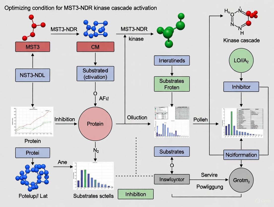
Unlocking the MST3-NDR Kinase Cascade: From Molecular Mechanisms to Therapeutic Optimization
This article provides a comprehensive analysis of the MST3-NDR kinase cascade, a critical signaling axis within the broader Hippo pathway network.
Unlocking the MST3-NDR Kinase Cascade: From Molecular Mechanisms to Therapeutic Optimization
Abstract
This article provides a comprehensive analysis of the MST3-NDR kinase cascade, a critical signaling axis within the broader Hippo pathway network. We explore the foundational biology of MST3 and NDR kinases, detailing their activation mechanisms and physiological roles in processes such as cell cycle regulation and tumor suppression. Methodological approaches for experimental activation and assessment are reviewed, alongside common challenges and optimization strategies for research and therapeutic targeting. The cascade's therapeutic relevance is validated through its implications in cancers, including colorectal and lung cancer, and its interaction with key pathways like p21 and YAP/TAZ. This resource is tailored for researchers and drug development professionals seeking to harness this pathway for biomedical innovation.
Decoding the MST3-NDR Axis: Core Components and Physiological Significance
Core Kinase Functions and Regulatory Mechanisms
The MST3-NDR kinase cascade is a crucial signaling module involved in regulating fundamental cellular processes, including cell cycle progression, apoptosis, and cell morphology. This section introduces the key players and their interrelationships.
- MST3 (Serine/Threonine-Protein Kinase 24, STK24): A mammalian Ste20-like serine/threonine kinase that functions as an upstream activator in the cascade. It is pleiotropic, influencing processes from apoptosis and cell polarization to immune response and metabolism [1].
- NDR1/2 (Nuclear Dbf2-Related 1/2, STK38/STK38L): Serine/threonine kinases belonging to the AGC kinase family. They are highly similar but have distinct, non-redundant physiological functions. They act as key downstream nodes, transmitting signals from upstream kinases like MST3 to control events such as the G1/S cell cycle transition and centrosome biology [2] [3] [4].
The regulatory relationship between these kinases is well-established. MST3 directly phosphorylates NDR1/2 on a critical residue within their hydrophobic motif (Thr444 in NDR1, Thr442 in NDR2), which is a essential step for their full activation [5] [4]. This activation is often supported by the scaffold protein MOB1 [5].
Key Experimental Parameters for the MST3-NDR Axis
The following table summarizes critical quantitative data and regulatory features for these kinases, providing a quick reference for experimental design.
| Kinase / Parameter | Official Name / Symbol | Key Regulatory Phosphorylation Sites | Upstream Regulators & Activators | Primary Downstream Substrates/Effectors |
|---|---|---|---|---|
| MST3 | Serine/threonine-protein kinase 24 (STK24) | Activation Loop: Thr178Other: Ser79, Lys53 [1] | Caspase-3 (cleavage), Cdk5, PKA, MO25 scaffolding protein [1] | NDR1/2, YAP (in Hippo signaling) [6] [5] |
| NDR1 | Serine/threonine-protein kinase 38 (STK38) | Hydrophobic Motif: Thr444Activation Loop: Ser281 [5] [4] | MST3, MST1/2, RASSF1A/MST1 (upon Fas stimulation) [5] [7] | p21 (Ser146), YAP (Ser61, 109, 127, 164), HP1α (Ser95) [2] [4] |
| NDR2 | Serine/threonine-protein kinase 38-like (STK38L) | Hydrophobic Motif: Thr442Activation Loop: Ser282 [5] [4] | MST3, MST1/2 [5] | p21 (Ser146), Rabin8 (Ser272), YAP [2] [4] |
Signaling Pathway and Experimental Workflow
The diagram below illustrates the core MST3-NDR signaling pathway and its connection to key cellular functions, providing a visual overview for troubleshooting experimental outcomes.
Troubleshooting Guide: Frequently Asked Questions
Q1: My co-immunoprecipitation experiments consistently fail to show a robust MST3-NDR interaction. What could be the issue? A1: The MST3-NDR interaction can be transient. Consider these steps:
- Stimulate the Pathway: The interaction may be dependent on specific signals. Treat cells with 1 µM Staurosporine for 4-6 hours to induce apoptosis and activate the caspase-3-MST3 axis [1].
- Phospho-mimetic NDR: Co-express MST3 with a constitutively active NDR mutant. The hydrophobic motif phosphorylation (T444/T442) by MST3 is critical for stable interaction and activation [5].
- Include MOB1: Co-express the scaffold protein MOB1, as it is crucial for facilitating the formation of a stable MST1/2-NDR-MOB1 complex, and this likely applies to MST3 as well [7].
Q2: I observe inconsistent NDR kinase activity in my in vitro assays. How can I achieve full activation? A2: Maximal NDR activation is a multi-step process. Ensure your protocol includes:
- Upstream Kinase Priming: Pre-incubate your NDR kinase with active, purified MST3 and ATP to ensure phosphorylation of the hydrophobic motif (T444/T442) [5].
- MOB1 Co-factor: Add the MOB1A protein to your reaction mixture. Studies show that MST3-mediated phosphorylation combined with MOB1 binding leads to a fully active NDR kinase [5].
- Inhibit Phosphatases: Include phosphatase inhibitors like 1 µM microcystin in your cell lysis and assay buffers to preserve the activating phosphorylation on NDR [5] [4].
Q3: What is a key functional readout to confirm successful MST3-NDR pathway activation in cells? A3: A highly validated downstream effector is the cyclin-dependent kinase inhibitor p21.
- Monitor p21 Phosphorylation: Use a phospho-specific antibody against p21 Ser146. NDR kinases directly phosphorylate p21 at this site, which regulates its protein stability and is a key event in controlling the G1/S cell cycle transition [2] [4].
- Phenotypic Confirmation: Perform a BrdU incorporation assay. Successful activation of the MST3-NDR-p21 axis should result in a measurable decrease in the number of cells entering S-phase, indicating G1/S arrest [2].
Q4: How does the MST3-NDR axis connect to the Hippo-YAP signaling pathway? A4: While the canonical Hippo pathway involves MST1/2 activating LATS1/2 to phosphorylate YAP/TAZ, the MST3-NDR axis represents a parallel, non-canonical route.
- Direct YAP Phosphorylation: NDR1/2 can directly phosphorylate YAP on multiple serine residues (including S61, S109, S127, and S164), leading to its cytoplasmic retention and inactivation, independent of LATS1/2 [4].
- Context-Dependent Signaling: In renal fibrosis models, activated MST3 promotes YAP phosphorylation and nuclear exit, inhibiting cell growth. Conversely, a kinase-dead MST3 mutant leads to YAP nuclear retention and fibrosis, which can be rescued by the AMPK activator metformin [6].
The Scientist's Toolkit: Essential Research Reagents
The following table lists critical reagents for studying the MST3-NDR kinase cascade, based on methodologies cited in the literature.
| Research Reagent / Tool | Key Function / Application | Example & Notes |
|---|---|---|
| Kinase-Dead (KD) Mutants | Serves as a dominant-negative control to inhibit endogenous kinase function. | MST3-K53R: A kinase-dead mutant used to demonstrate MST3's role in fibrosis models [6]. |
| Phospho-Specific Antibodies | Detects the activated, phosphorylated state of kinases and substrates. | Anti-NDR1/2 pT444/pT442: Validates upstream MST3 activity.Anti-p21 pS146: Confirms downstream NDR activity [2] [4]. |
| Pathway Agonists & Antagonists | Chemically modulates pathway activity for functional studies. | Verteporfin: Inhibits YAP-TEAD interaction [6].Metformin: Activates AMPK, can inhibit YAP and rescue fibrotic phenotypes [6]. |
| Scaffold & Binding Proteins | Essential for robust kinase activation in functional assays. | Recombinant MOB1A Protein: Critical for achieving full NDR kinase activation in in vitro kinase assays [5]. |
| siRNA/shRNA Knockdown Systems | Validates the specific role of a kinase in a cellular process. | shRNA against MST3/NDR1/NDR2: Used to establish the requirement of these kinases for G1/S progression and apoptosis [2] [7]. |
Core Signaling Pathway
What is the core molecular mechanism of NDR kinase activation by MST3?
The activation of Nuclear Dbf2-related (NDR) kinases by mammalian Sterile 20-like kinase 3 (MST3) is a precisely regulated process involving specific phosphorylation events within a multi-step pathway.
Mechanism Overview: MST3 functions as an upstream kinase that directly phosphorylates NDR kinases on a critical residue within their C-terminal hydrophobic motif (HM)—specifically Thr444 in NDR1 and Thr442 in NDR2 [5]. This phosphorylation event, coupled with subsequent autophosphorylation and regulatory protein binding, triggers a substantial increase in NDR kinase activity.
Step-by-Step Activation Process:
- HM Phosphorylation: MST3 selectively phosphorylates Thr442 of NDR2, resulting in approximately 10-fold stimulation of NDR kinase activity in vitro [5].
- Co-activator Binding: The MOB1A protein subsequently binds to the phosphorylated NDR, further enhancing its activity and contributing to the formation of a fully active kinase complex [5].
- Activation Loop Phosphorylation: Following HM phosphorylation and MOB1 binding, NDR undergoes autophosphorylation on its activation loop at Ser281 (NDR1) or Ser282 (NDR2), which is essential for achieving full catalytic competence [5] [4].
This multi-step process ensures tight regulatory control over NDR kinase activity, integrating signals from upstream regulators like MST3 with co-activator availability.
How does MST3 fit into the broader Hippo signaling network?
MST3 is part of the mammalian STE20-like protein kinase family and contributes to the complex regulatory network upstream of the Hippo pathway's core kinases [8].
Relationship to Canonical Hippo Signaling: While the canonical Hippo pathway primarily involves MST1/2 kinases activating LATS1/2 kinases, MST3 represents a parallel regulatory input that can activate the related NDR1/2 kinases [4]. The NDR and LATS kinases together form the NDR/LATS subfamily of AGC kinases, both functioning as crucial regulators within the Hippo signaling network [9].
Integrated Pathway View: The following diagram illustrates the position of MST3 within the broader context of NDR kinase regulation and Hippo signaling:
This schematic demonstrates how MST3-mediated phosphorylation initiates a cascade leading to NDR activation and subsequent regulation of downstream effectors like YAP, illustrating the integration point between MST3 signaling and the broader Hippo network [5] [4] [10].
Experimental Analysis & Troubleshooting
What methodologies can confirm MST3-NDR kinase interactions and phosphorylation?
Researchers can employ several well-established experimental approaches to investigate MST3-NDR kinase interactions and phosphorylation events. The table below summarizes key methodologies and their specific applications:
| Method | Specific Application | Key Experimental Details |
|---|---|---|
| In Vitro Kinase Assay | Direct phosphorylation of NDR by MST3 | Recombinant MST3 kinase + NDR substrate; measure Thr442 phosphorylation via phospho-specific antibodies [5]. |
| Co-Immunoprecipitation (Co-IP) | Protein-protein interaction analysis | Co-express tagged MST3 and NDR in cells (e.g., HEK293T); immunoprecipitate one, immunoblot for the other [5]. |
| Western Blot with Phospho-Specific Antibodies | Detection of specific phosphorylation events | Use anti-P-Thr442-NDR2 and anti-P-Ser281/282-NDR antibodies to monitor activation loop and HM phosphorylation [5] [2]. |
| RNA Interference (RNAi) | Functional validation in cellular contexts | Transfert short hairpin RNA (shRNA) targeting MST3; assess resulting decrease in NDR Thr442 phosphorylation [5]. |
| Mass Spectrometry | Identification of novel phosphorylation sites | Immunopurify NDR kinases from cells; analyze tryptic peptides for phosphorylation at Thr444/442 and other sites [5]. |
Detailed Protocol: In Vitro Kinase Assay
- Protein Purification: Express and purify recombinant, active MST3 kinase and NDR2 protein (wild-type and T442A mutant as control) from bacterial or mammalian systems.
- Reaction Setup: Combine MST3 and NDR2 in kinase assay buffer containing Mg²⁺/ATP. Include appropriate controls (e.g., kinase-dead MST3, no MST3).
- Incubation: Conduct the reaction at 30°C for 30 minutes.
- Analysis: Terminate the reaction and analyze phosphorylation by:
- Western Blotting: Using NDR2 phospho-specific antibody (anti-P-Thr442).
- Autoradiography: If using [γ-³²P]ATP, detect incorporated radioactivity.
- Activity Measurement: Assess stimulated NDR2 activity towards a secondary substrate (e.g., myelin basic protein) to confirm functional activation [5].
How can I troubleshoot common problems in studying the MST3-NDR axis?
Several technical challenges may arise when experimentally investigating the MST3-NDR kinase cascade. Below are common issues and recommended solutions:
| Problem | Possible Causes | Troubleshooting Solutions |
|---|---|---|
| Low/No Phosphorylation in Vitro | Non-optimal reaction conditions; insufficient MST3 activity. | - Titrate MST3:NDR ratio (start ~1:10).- Include positive control (known MST3 substrate).- Verify MST3 activity with autophosphorylation assay [5] [8]. |
| High Background in Cellular Phosphorylation | Off-target kinase activity; incomplete pathway inhibition. | - Use kinase-dead MST3 (K53R mutant) as negative control.- Combine genetic knockdown (shMST3) with pharmacological inhibitors [5]. |
| Inconsistent Co-IP Results | Weak/transient interactions; protein degradation. | - Use crosslinkers (e.g., DSP) before lysis to stabilize transient interactions.- Include protease/phosphatase inhibitors in lysis buffer [5] [2]. |
| No Phenotype After MST3 Knockdown | Functional redundancy with other kinases. | - Test simultaneous knockdown of MST3 and related kinases (MST1/2, MAP4Ks).- Extend knockdown duration to account for protein half-life [4] [11]. |
| Cellular Localization Issues | Aberrant nucleocytoplasmic shuttling. | - Treat with okadaic acid (OA) to inhibit PP2A and enhance phosphorylation.- Check for caspase cleavage of MST3 during apoptosis, which alters localization [5] [8]. |
Critical Consideration: Kinase Redundancy A significant challenge in this field is the redundancy among upstream kinases. Multiple Ste20-like kinases, including MST1, MST2, and various MAP4K family members, can potentially phosphorylate NDR kinases under certain conditions [4] [12]. To conclusively demonstrate MST3-specific effects:
- Perform combinatorial knockdown approaches.
- Use selective pharmacological inhibitors where available.
- Employ rescue experiments with wild-type versus kinase-dead MST3 in MST3-deficient cells [5] [11].
Research Reagent Solutions
Successful investigation of the MST3-NDR kinase pathway relies on key reagents and tools. The following table provides an overview of essential research reagents:
| Reagent Category | Specific Examples | Function/Application |
|---|---|---|
| Expression Plasmids | pCMV5-HA-NDR2, pCMV5-HA-MST3, myc-C1-MOB1A [5] | Mammalian expression of tagged proteins for interaction and phosphorylation studies. |
| Mutant Kinases | MST3-KR (kinase-dead K53R), NDR2-T442A (phospho-deficient) [5] [8] | Essential negative controls for phosphorylation experiments and functional studies. |
| Phospho-Specific Antibodies | Anti-P-Thr442-NDR2, Anti-P-Ser281/282-NDR [5] [2] | Critical tools for detecting specific activation-related phosphorylation events. |
| Knockdown Constructs | pTER-shMST3 vectors [5] | Genetic validation of MST3 functions through RNA interference. |
| Chemical Activators/Inhibitors | Okadaic acid (PP2A inhibitor) [5], Staurosporine (apoptosis inducer) [8] | Tools to modulate pathway activity; OA enhances NDR phosphorylation by inhibiting dephosphorylation. |
| Scaffolding Proteins | Recombinant MO25 protein [8] [10] | Activates MST3 in vitro; useful for enhancing MST3 activity in experimental settings. |
Key Experimental Consideration: When using phospho-specific antibodies, always include appropriate controls:
- Phospho-deficient mutants (e.g., NDR2-T442A) to confirm antibody specificity.
- Phosphatase-treated samples to verify phosphorylation-dependent signal.
- Kinase-dead MST3 transfection to demonstrate dependence on MST3 activity [5] [2].
Pathway Visualization & Experimental Workflow
How can I visualize the complete MST3-NDR kinase activation workflow?
The following diagram provides a comprehensive overview of the experimental workflow for analyzing MST3-dependent NDR kinase activation, from cellular stimulation to downstream readouts:
This workflow illustrates the sequential process from initial cellular stimulation to final biological outcomes, with associated analytical techniques shown as dashed red lines. This comprehensive view enables researchers to identify appropriate experimental entry points and validation methods for their specific research questions [5] [2] [8].
Troubleshooting Guide & FAQs
FAQ 1: My experiments consistently show G1 arrest upon MST3/NDR knockdown, but the effect size is variable. What could be the cause? Variability in G1 arrest phenotypes often stems from differences in cell synchronization efficiency. The MST3-NDR axis is specifically active during the G1 phase [2]. Inefficient synchronization results in a heterogeneous cell population, diluting the observable effect. For consistent results, implement a double-thymidine block protocol to achieve robust G1 synchronization before analyzing your knockdown cells.
- Antibody Sensitivity: The phospho-specific antibody may have low affinity. Include a positive control, such as cells overexpressing constitutively active NDR kinase, to validate your reagent.
- Protein Stability: Phosphorylated p21 is targeted for proteasomal degradation, making it short-lived. Treat your cells with a proteasome inhibitor (e.g., MG132 at 10μM) for 4-6 hours before lysis to stabilize the phosphorylated form.
- Kinase Activity: Ensure your NDR kinases are active. Check the activation loop phosphorylation of NDR (e.g., T444 for NDR1) as a proxy for pathway activity.
FAQ 3: What are the best practices for validating the specificity of NDR kinase in the G1/S transition? A robust validation strategy involves a combination of genetic and biochemical rescues [2]:
- Rescue with Wild-Type Kinase: Re-express an RNAi-resistant wild-type NDR2 cDNA in your knockdown cells. This should reverse the G1 arrest phenotype.
- Kinase-Dead Control: Express a kinase-dead mutant of NDR (e.g., NDR1-K118R); this should not rescue the cell cycle defect, confirming that kinase activity is required.
- Phospho-mimetic p21: Expressing a p21 phospho-mimetic mutant (S146D) should partially bypass the need for NDR kinase activity and promote S-phase entry.
FAQ 4: How can I confirm that MST3 is the primary upstream kinase for NDR in the G1 phase? To establish the MST3-NDR functional link in G1 [2]:
- Co-depletion: Perform simultaneous siRNA-mediated knockdown of MST3 and compare the G1 arrest phenotype to single NDR knockdown.
- Phospho-profiling: Analyze the phosphorylation status of the NDR hydrophobic motif (a site directly targeted by MST kinases) in G1-synchronized cells after MST3 knockdown. A significant reduction is expected.
- In Vitro Kinase Assay: Use immunoprecipitated NDR from G1 phase cells as a substrate for recombinant active MST3 in an in vitro kinase assay.
Experimental Protocols & Data
Table 1: Key Quantitative Findings on the MST3-NDR-p21 Axis
| Experimental Manipulation | Observed Phenotype | Key Quantitative Measurement | Proposed Mechanism |
|---|---|---|---|
| shRNA knockdown of NDR1/2 | G1 phase arrest | ~60-70% reduction in S-phase entry (vs. control) [2] | Stabilization of p21, leading to inhibition of Cyclin E-Cdk2 complexes. |
| shRNA knockdown of MST3 | G1 phase arrest | ~50% reduction in S-phase entry [2] | Loss of NDR kinase activation during G1. |
| NDR phosphorylation of p21 (S146) | Reduced p21 protein stability | Direct phosphorylation shown via in vitro kinase assays; phospho-mimetic p21 (S146D) is destabilized [2] | Creates a phosphodegron, promoting ubiquitin-mediated proteasomal degradation of p21. |
| Cycloheximide Chase (NDR knockdown) | Increased p21 half-life | p21 protein half-life increased by ~2-3 fold [2] | NDR activity is required for constitutive turnover of p21 in G1. |
Core Methodology: Analyzing p21 Stability
Protocol: Cycloheximide Chase Assay to Measure p21 Protein Half-life
- Cell Culture & Treatment: Culture U2OS or HeLa cells and transfect with siRNAs targeting NDR1/2 or a non-targeting control.
- Synchronization: Synchronize cells at the G1/S boundary using a double-thymidine block.
- Inhibit Protein Synthesis: Add cycloheximide (CHX) to the culture medium at a final concentration of 50 μg/mL to block new protein synthesis.
- Time-Course Harvest: Harvest cell lysates at regular intervals post-CHX addition (e.g., 0, 30, 60, 90, 120 minutes).
- Western Blotting: Analyze lysates by Western blot using antibodies against p21. Use tubulin or actin loading controls to normalize.
- Quantification: Quantify band intensities and plot the decay of p21 protein over time to calculate its half-life [2].
Protocol: Co-immunoprecipitation for NDR-p21 Interaction
- Lysis: Lyse G1-synchronized cells in a mild, non-denaturing lysis buffer (e.g., NP-40 or RIPA buffer).
- Pre-clear: Pre-clear the lysate with Protein A/G beads.
- Immunoprecipitation: Incubate the lysate with an antibody against NDR1/2 or a control IgG. Capture the immune complexes with Protein A/G beads.
- Washing: Wash the beads extensively with lysis buffer to remove non-specifically bound proteins.
- Elution & Analysis: Elute the bound proteins and analyze by Western blotting. Probe for p21 to confirm interaction and for phospho-S146-p21 to detect the specific phosphorylated form [2].
Signaling Pathway & Workflow Visualization
MST3-NDR-p21 Signaling Pathway
Experimental Workflow for Pathway Analysis
The Scientist's Toolkit
Table 2: Essential Research Reagents for MST3-NDR Research
| Reagent | Function/Application in Research | Example & Notes |
|---|---|---|
| siRNA/shRNA | Targeted knockdown of MST3, NDR1, or NDR2 gene expression to study loss-of-function phenotypes. | Validated siRNA pools (e.g., from Qiagen) are commonly used. Rescue requires RNAi-resistant cDNA constructs [2]. |
| Phospho-Specific Antibodies | Detection of pathway activity and specific phosphorylation events. | Anti-NDR1/2 (pT444): Measures NDR activation. Anti-p21 (pS146): Direct readout of NDR activity on its substrate [2]. |
| Cell Synchronization Agents | To arrest cells in specific cell cycle phases, particularly G1. | Thymidine: Used in a double-block protocol for G1/S synchronization. Nocodazole: For M-phase arrest [2]. |
| Proteasome Inhibitor | To stabilize proteins that are rapidly degraded, such as phosphorylated p21. | MG132: Used at 10 μM for 4-6 hours before cell lysis to accumulate phospho-p21 for detection [2]. |
| Cycloheximide | To inhibit protein synthesis and measure protein half-life in chase assays. | Used at 50 μg/mL to track the decay of p21 over time in knockdown vs. control cells [2]. |
| Active Recombinant Kinases | For in vitro kinase assays to test direct phosphorylation. | Recombinant active MST3 and NDR kinases are used to phosphorylate p21 in a controlled system [2]. |
Troubleshooting Guide: MST3-NDR Kinase Cascade Activation
Q1: My experiments show inconsistent NDR kinase activation even with MST3 overexpression. What could be causing this variability?
A: Inconsistent NDR activation commonly stems from improper phosphorylation coordination. MST3 specifically phosphorylates NDR kinase at the hydrophobic motif (Thr444 in NDR1/Thr442 in NDR2) [5]. However, full NDR activation requires a multi-step process:
- MST3-mediated phosphorylation of the hydrophobic motif (Thr442)
- NDR autophosphorylation of the activation loop (Ser281/Ser282)
- Binding of the regulatory protein MOB1A [5]
Ensure you're measuring complete activation by checking all three requirements. Use phospho-specific antibodies against p-Thr442 for direct MST3 activity assessment [5].
Q2: How can I distinguish MST3-specific effects from other Hippo pathway kinases like MST1/2 in my cellular models?
A: MST3 occupies a distinct position in the broader Hippo network as part of the "atypical" or "non-canonical" Hippo pathway [11]. To isolate MST3-specific functions:
- Utilize kinase-dead MST3 (MST3-KD) as a dominant-negative control [6] [5]
- Employ specific MST3 shRNA knockdown while monitoring MST1/2 activity
- Test AMPK activation (e.g., with metformin) - this inhibits YAP independently of MST3-KD, helping distinguish pathway branches [6]
Q3: What experimental readouts are most reliable for confirming functional MST3-NDR signaling in renal fibrosis models?
A: In renal fibrosis contexts, focus on these key readouts:
- YAP nuclear/cytoplasmic localization (immunofluorescence)
- YAP phosphorylation status (Western blot)
- CTGF expression levels (qPCR/Western) as YAP/TEAD transcriptional target [6]
- Cellular growth patterns in high-density cultures [6]
Experimental Conditions & Data Comparison
Table 1: MST3-NDR Kinase Cascade Components and Functions
| Component | Function in Cascade | Phosphorylation Sites | Key Interacting Partners |
|---|---|---|---|
| MST3 (STK24) | Upstream kinase; phosphorylates NDR hydrophobic motif | Activation loop (autophosphorylation) | NDR1/2, MOB1 [5] |
| NDR1/2 | Downstream effector; regulates cell cycle progression & morphology | Thr444/Thr442 (HM), Ser281/Ser282 (activation loop) | MOB1A/B, S100B [5] |
| MOB1A/B | Scaffold/regulatory protein | Ser/Thr residues | NDR1/2, MST1/2 [5] [12] |
| YAP | Terminal transcriptional co-activator | Ser127 (LATS-mediated) | TEAD1-4, 14-3-3 proteins [6] [13] |
Table 2: Troubleshooting NDR Kinase Activation Assays
| Problem | Potential Causes | Solutions | Validation Methods |
|---|---|---|---|
| Weak NDR phosphorylation | Inadequate MST3 activity, improper cell density, insufficient MOB1 | Co-express MOB1A; optimize cell density; use okadaic acid to enhance detection [5] | Western with p-Thr442-NDR antibodies; in vitro kinase assays [5] |
| YAP nuclear localization despite MST3 expression | MST3-KD contamination, AMPK pathway inhibition, high cell confluence | Verify MST3 construct (WT vs KD); treat with metformin to activate AMPK; control cell density [6] | Immunofluorescence for YAP localization; Western for p-YAP [6] |
| Inconsistent results across cell lines | Cell-type specific expression of Hippo components, varying upstream signals | Standardize cell density; screen multiple cell lines; verify pathway component expression | qPCR for core Hippo components; control for cell density effects [6] [13] |
Detailed Experimental Protocols
Protocol 1: Monitoring MST3-Dependent NDR Phosphorylation
Methodology from Steiger et al. [5]
Cell Culture & Transfection:
- Culture HEK293F or COS-7 cells in Dulbecco's modified Eagle's medium with 10% fetal calf serum
- Transfect with HA-tagged MST3 (HA-MST3) and HA-tagged NDR2 using standard protocols
- Include kinase-dead MST3 (HA-MST3-KR) as negative control
Stimulation & Lysis:
- Stimulate cells with 100-500 nM okadaic acid for 1-2 hours to enhance phosphorylation detection
- Lyse cells in IP buffer: 20 mM Tris-HCl (pH 8.0), 150 mM NaCl, 1% Nonidet P-40, 10% glycerol, plus phosphatase and protease inhibitors
Detection & Analysis:
- Resolve proteins by 10-12% SDS-PAGE, transfer to PVDF membranes
- Probe with anti-P-Thr442 (for NDR2 hydrophobic motif phosphorylation) and anti-P-Ser282 (for activation loop autophosphorylation)
- Use anti-HA antibodies for protein level normalization
Protocol 2: Assessing Functional Outcomes in Fibrosis Models
Methodology from Chan et al. [6]
Cell Culture & Density Conditions:
- Culture MDCK or NIH/3T3 cells under varying density conditions
- Transfect with HA-MST3 or HA-MST3-KD using appropriate reagents
- For high-density conditions, grow to confluence and maintain for 24-48 hours
Pharmacological Interventions:
- Treat MST3-KD cells with 10-50 μM verteporfin (YAP-TEAD inhibitor) for 24 hours
- Alternatively, treat with 1-5 mM metformin to activate AMPK pathway
- Include DMSO-only controls for all treatments
Downstream Analysis:
- Fix cells for immunofluorescence staining of YAP localization
- Extract nuclear and cytoplasmic fractions for YAP distribution analysis by Western blot
- Measure CTGF expression by qPCR or Western blot as YAP transcriptional output
- Assess cell growth patterns and fibrotic markers (e.g., collagen deposition)
The Scientist's Toolkit: Research Reagent Solutions
Table 3: Essential Reagents for MST3-NDR Pathway Research
| Reagent | Specific Function | Application Examples | Key Considerations |
|---|---|---|---|
| HA-MST3 & HA-MST3-KD | Wild-type vs kinase-dead MST3 expression | Gain/loss-of-function studies; control experiments [6] [5] | Verify kinase activity through autophosphorylation assays |
| Phospho-specific antibodies (p-Thr442-NDR) | Detect MST3-mediated NDR phosphorylation | Western blot, monitoring pathway activation [5] | Confirm specificity with kinase-dead MST3 controls |
| MOB1A expression constructs | Enhance NDR activation | Co-expression to maximize NDR kinase activity [5] | Titrate to avoid artificial overexpression effects |
| Verteporfin | Inhibits YAP-TEAD interaction | Determine YAP-dependence of phenotypes [6] | Use light-protected conditions; optimize concentration (typically 1-10 μM) |
| Metformin | Activates AMPK pathway | Test AMPK-mediated YAP regulation independent of MST3 [6] | Dose response recommended (1-10 mM range in cell culture) |
| Okadaic acid | Phosphatase inhibitor | Enhance detection of phosphorylated NDR [5] | Use carefully (100-500 nM); short treatment durations (1-2 hours) |
Pathway Visualization
MST3-NDR Cascade in Hippo Pathway
Experimental Workflow for MST3-NDR Research
Troubleshooting Guides and FAQs
Frequently Asked Questions
Q1: What are the key biological outputs of the activated MST3-NDR kinase cascade? The MST3-NDR kinase cascade regulates critical cellular decisions, primarily influencing cell cycle progression, centrosome duplication, and tumorigenicity. When MST3 phosphorylates NDR at its hydrophobic motif (Thr444/Thr442), it triggers a signaling pathway that controls progression through the cell cycle and proper duplication of centrosomes, which are essential for genomic stability [5] [14]. In diseases like breast cancer, this cascade promotes proliferation and tumorigenicity through downstream effectors like the VAV2/Rac1 signal axis [15].
Q2: Which residues on NDR are critical for activation by MST3, and what is the functional impact? MST3 directly phosphorylates NDR kinase at the hydrophobic motif site Thr444/Thr442 [5]. This phosphorylation is essential for maximal NDR activation and works in concert with autophosphorylation at the activation loop site (Ser281/Ser282) and binding of the MOB1A co-activator protein. This multi-step activation process is crucial for NDR's role in controlling cell cycle progression and morphology [5].
Q3: How does the scaffolding protein MO25 regulate MST3 activity? MO25 functions as a master regulator of STE20 kinases like MST3 by stabilizing the kinase domain in a closed, active conformation. Structural studies reveal that MO25β binds to MST3 through key interface residues (Tyr223 on MO25β and Glu58/Ile71 on MST3), forming an intricate web of interactions that maintain MST3 in an active state even in the absence of ATP [16]. Disrupting these interactions through mutation prevents MST3 activation.
Q4: What is the evidence for MST3's role in cancer progression? MST3 exhibits oncogenic properties in several contexts. It is overexpressed in human breast tumors, and its overexpression predicts poor prognosis in breast cancer patients [15]. MST3 promotes proliferation and tumorigenicity through its interaction with VAV2 to activate Rac1, leading to increased cyclin D1 expression. Knockdown of MST3 inhibits tumor formation in mouse models, confirming its functional importance in cancer pathogenesis [15].
Troubleshooting Common Experimental Issues
Issue: Inconsistent NDR phosphorylation in cascade activation assays
- Potential Cause: Suboptimal buffer conditions or incomplete cascade activation.
- Solution: Systematically optimize buffer conditions. Ensure the presence of all required components for full NDR activation: active MST3 for Thr442 phosphorylation, conditions allowing NDR autophosphorylation at Ser282, and MOB1A protein, which further increases activity to generate a fully active kinase [5]. Adding a small amount of detergent (e.g., 0.05% Tween 20) can prevent aggregation.
Issue: Low signal-to-noise ratio in protein interaction studies
- Potential Cause: Non-specific binding or protein instability.
- Solution: Improve signal-to-noise ratio by empirically testing different buffer conditions. Centrifuge samples (10 minutes at >20,000 x g) and consider adding reducing agents like DTT to maintain protein stability. Using site-specific tagging strategies, such as His-tag labeling, can also reduce background noise [17].
Issue: Difficulty detecting MST3-VAV2 interaction in co-immunoprecipitation
- Potential Cause: The interaction requires a specific proline-rich domain on MST3.
- Solution: Verify the integrity of the proline-rich region (353KDIPKRP359) in your MST3 construct. This region is essential for binding to the SH3 domain of VAV2. Mutation of the two proline residues in this motif significantly attenuates this interaction [15].
Quantitative Data on NDR Kinase Activation
Table 1: Key Parameters of NDR Kinase Activation by MST3
| Parameter | Value/Residue | Functional Significance | Experimental Context |
|---|---|---|---|
| MST3 Phosphorylation Site on NDR | Thr444 (NDR1)Thr442 (NDR2) | Phosphorylation of hydrophobic motif required for maximal activation | In vitro kinase assays [5] |
| NDR Autophosphorylation Site | Ser281 (NDR1)Ser282 (NDR2) | Autophosphorylation in the activation loop stimulates activity | In vitro and in vivo validation [5] |
| Activation Fold by MST3 | ~10-fold | Stimulation of NDR activity after phosphorylation by MST3 | In vitro kinase assay [5] |
| Critical MO25β-MST3 Interface Residues | MO25β: Tyr223MST3: Glu58, Ile71 | Mutation prevents MST3 activation; key for stabilizing active conformation | Structural analysis & mutagenesis [16] |
| MST3-VAV2 Interaction Domain | MST3: ^353^KDIPKRP^359^VAV2: SH3 domain | Proline-rich region essential for oncogenic signaling in breast cancer | Co-immunoprecipitation & domain mapping [15] |
Detailed Experimental Protocols
Protocol 1: Activating the MST3-NDR Kinase Cascade In Vitro
This protocol outlines the steps for reconstituting the MST3-NDR activation cascade, based on research demonstrating MST3-mediated phosphorylation and activation of NDR2 [5].
Recombinant Protein Expression and Purification:
- Express and purify the catalytic domain of human MST3 (residues 19-289) and full-length NDR2 from E. coli BL21(DE3) cells.
- Use affinity chromatography (e.g., Ni-Sepharose for His-tagged proteins) followed by gel filtration (e.g., S200 16/60 column) in a buffer containing 20 mM Tris pH 7.5, 200 mM NaCl, and 0.5 mM TCEP for final purification and complex formation [16].
- Confirm protein identity and purity via SDS-PAGE and mass spectrometry.
Kinase Assay Setup:
- Combine the purified proteins in kinase assay buffer. A typical reaction includes:
- NDR2 (1-2 µg)
- Active MST3 (0.1-0.5 µg)
- MOB1A protein (to achieve full activation) [5]
- ATP (including [γ-³²P]-ATP for radioactive detection or cold ATP for western blot)
- MgCl₂ (as a cofactor)
- Incubate at 30°C for 30 minutes.
- Combine the purified proteins in kinase assay buffer. A typical reaction includes:
Analysis of Phosphorylation and Activation:
- Western Blot: Resolve proteins by SDS-PAGE and transfer to a membrane. Probe with phospho-specific antibodies against NDR2 pThr442 and pSer282 to confirm phosphorylation at both regulatory sites [5].
- Kinase Activity Measurement: Use a substrate like myelin basic protein to measure the resulting kinase activity of NDR2, comparing reactions with and without active MST3 [16] [5].
Protocol 2: Investigating the MST3-VAV2-Rac1 Signaling Axis in Cells
This protocol is adapted from research establishing the pro-tumorigenic MST3-VAV2-Rac1 pathway in breast cancer cells [15].
Cell Culture and Transfection:
- Culture relevant cell lines (e.g., MDA-MB-231 or MDA-MB-468 for breast cancer studies).
- Transfect with constructs for:
- Wild-type MST3 (WT-MST3)
- Proline-rich-deleted MST3 (∆P-MST3), which cannot bind VAV2
- MST3-specific shRNA for knockdown
- Appropriate empty vector controls.
Protein Interaction Analysis (Co-immunoprecipitation):
- Lyse cells in IP buffer (e.g., 20 mM Tris-HCl pH 8.0, 150 mM NaCl, 1% Nonidet P-40, 10% glycerol, plus protease and phosphatase inhibitors) [5].
- Incubate lysates with an antibody against MST3 or VAV2.
- Use Protein A/G beads to pull down the immune complex.
- Wash beads extensively, elute the proteins, and analyze by Western blotting to detect co-precipitated VAV2 or MST3, respectively.
Functional Output Assessment:
- Rac1 Activation: Use a Rac1 G-LISA activation assay or pull-down with PAK-PBD beads to measure levels of GTP-bound Rac1 [15].
- Proliferation: Perform MTT assays or count cells to assess proliferation rates.
- Anchorage-Independent Growth: Conduct soft agar colony formation assays to measure tumorigenic potential in vitro.
- Downstream Signaling: Check expression levels of key effectors like cyclin D1 by Western blot.
Signaling Pathway Diagrams
The Scientist's Toolkit
Research Reagent Solutions
Table 2: Essential Reagents for MST3-NDR Cascade Research
| Reagent / Material | Key Function / Feature | Example Application / Note |
|---|---|---|
| MO25 Protein (α/β isoforms) | Scaffold protein that stabilizes MST3 in its active conformation. | Critical for in vitro activation studies; co-express/purify with MST3 [16]. |
| Phospho-Specific Antibodies | Detect phosphorylation at key activation sites. | Anti-NDR pThr442/pThr444 & anti-NDR pSer281/pSer282 for monitoring activation [5]. |
| MOB1A Protein | Co-activator that binds NDR, leading to full kinase activation. | Add to in vitro kinase assays to achieve maximal NDR activity [5]. |
| Kinase-Dead MST3 (MST3KR) | Acts as a dominant-negative mutant to inhibit endogenous MST3 function. | Control for validating MST3-specific effects in cellular assays [5]. |
| ∆P-MST3 Mutant | MST3 with mutated proline-rich region (disrupted VAV2 binding). | Used to dissect the VAV2/Rac1 oncogenic signaling axis [15]. |
| TRIC-Sensitive Fluorophores | Fluorophores with high Temperature-Related Intensity Change. | Optimal for Microscale Thermophoresis (MST) binding assays between MST3/MO25 or MST3/VAV2 [17]. |
| Rac1 Inhibitor (EHop-016) | Small molecule inhibitor of Rac1 GTPase. | Tool to confirm functional contribution of Rac1 downstream of MST3-VAV2 [15]. |
Experimental Strategies for MST3-NDR Cascade Activation and Analysis
The MST3-NDR kinase cascade is a critical signaling module within the broader STE20-type kinase and Hippo pathway networks, governing fundamental processes such as cell cycle progression, morphogenesis, and proliferation [18] [5]. In this pathway, the upstream kinase, MST3 (STE20-like kinase 3), directly phosphorylates and activates the downstream kinase, NDR (Nuclear Dbf2-related kinase) [5]. Research into this signaling axis has gained substantial momentum due to its documented roles in diseases, including cancer, where MST3 can function as a tumor suppressor, and obesity, where the broader GCKIII kinase family (including STK25 and MST3) regulates lipid homeostasis and ectopic fat deposition [19] [20].
The core experimental objective is to reconstitute this cascade in vitro to quantitatively measure MST3 kinase activity towards its substrate, NDR. The key activating event is the phosphorylation of a specific threonine residue (Thr442 in NDR2) within NDR's hydrophobic motif by MST3 [5]. Successful activation of NDR is a multi-step process, further enhanced by its coactivator protein, MOB1A [5]. This technical guide provides detailed methodologies and troubleshooting support to enable researchers to reliably study this crucial kinase-substrate relationship.
The MST3-NDR Kinase Signaling Pathway
The diagram below illustrates the core molecular events and experimental workflow for measuring MST3 and NDR kinase activity in vitro.
Essential Reagents and Materials
A successful in vitro kinase assay requires high-quality, active proteins and specific detection reagents. The table below catalogs the essential research reagent solutions for studying the MST3-NDR kinase cascade.
Table 1: Key Research Reagent Solutions for MST3-NDR Kinase Assays
| Reagent / Material | Function / Description | Key Details / Specifications |
|---|---|---|
| Recombinant MST3 Kinase | Upstream kinase that phosphorylates NDR. | Catalytic domain (residues 1-303) is often sufficient. Full-length protein can be used. Co-expression with MO25β enhances stability and activity [16]. |
| Recombinant NDR Kinase | Downstream substrate kinase. | Requires phosphorylation at Thr444/Thr442 (human NDR1/NDR2) for full activation. Purified as GST- or HA-tagged fusion protein [5]. |
| MOB1A Protein | Coactivator for NDR kinase. | Binds to NDR, significantly boosting its activity upon phosphorylation by MST3 [5]. |
| Anti-Phospho-NDR Antibodies | Detect specific phosphorylation events. | Anti-pThr442: Measures MST3-mediated hydrophobic motif phosphorylation.Anti-pSer281/282: Measures NDR autophosphorylation [5]. |
| Kinase Buffer Components | Provides optimal reaction environment. | See Table 2 for a detailed breakdown of components and final concentrations. |
| ATP (with [γ-³²P]) | Phosphate donor for the reaction. | Unlabeled ATP for cold reactions; [γ-³²P]-ATP for radiometric detection of phosphorylation [5]. |
| MST3 Inhibitors (e.g., Bosutinib) | Negative controls and functional probes. | Bosutinib is a potent MST3 inhibitor (IC₅₀ = 3 nM). Saracatinib is a weaker inhibitor (IC₅₀ = 11 µM) [21]. |
Core Experimental Protocol
Protein Purification and Preparation
- Expression: Express recombinant MST3 (kinase domain or full-length, preferably with MO25β) and NDR (e.g., GST- or HA-tagged) in an appropriate system like E. coli BL21(DE3) or mammalian HEK293 cells [16] [5].
- Purification: Use affinity chromatography tailored to the fusion tag. For GST-tagged proteins, purify using Glutathione Sepharose 4B resin. For HA- or myc-tagged proteins, use immunoprecipitation with specific antibodies [5].
- Buffer Exchange: Dialyze or desalt the purified proteins into a compatible storage buffer (e.g., 20 mM Tris-HCl pH 7.5, 200 mM NaCl, 0.5 mM TCEP) to remove impurities. Confirm protein concentration and purity via SDS-PAGE [16].
2In VitroKinase Assay Procedure
- Reaction Setup: Assemble the kinase reaction on ice in a final volume of 25-50 µL. The standard reaction mixture should contain the components listed in the table below.
- Incubation: Initiate the reaction by adding ATP and incubate at 30°C for 30 minutes. The reaction time and temperature can be optimized for specific experimental conditions.
- Termination: Stop the reaction by adding SDS-PAGE loading dye and heating at 95°C for 5 minutes.
- Analysis:
- Western Blotting: Resolve proteins by SDS-PAGE, transfer to a membrane, and probe with phospho-specific antibodies (e.g., anti-pThr442-NDR) to detect MST3-dependent phosphorylation [5].
- Radiometric Detection: If using [γ-³²P]-ATP, expose the dried gel to X-ray film or a phosphorimager screen to visualize radioactive phosphate incorporation [5].
Table 2: Standard In Vitro Kinase Reaction Setup
| Component | Final Concentration/Amount | Purpose & Notes |
|---|---|---|
| Kinase Buffer (1X) | 10-20 µL | See recipe below for 10X stock. |
| MST3 Kinase | 10-100 ng | The active upstream kinase. Amount should be titrated. |
| NDR Substrate | 0.2-1.0 µg | The downstream phosphorylation target. |
| MOB1A Protein | 0.1-0.5 µg | Essential for achieving full NDR activation [5]. |
| ATP | 100-200 µM | Phosphate donor. Include [γ-³²P]-ATP for radiometric assays. |
| MgCl₂ / MnCl₂ | 10 mM / 1-5 mM | Divalent cations essential for kinase activity. |
| DTT | 1 mM | Maintaining reducing environment. |
| BSA | 0.1-0.2 mg/mL | Stabilizes proteins in low-concentration reactions. |
| Nuclease-free Water | To final volume |
10X Kinase Buffer Recipe: 200 mM Tris-HCl (pH 7.5), 100 mM MgCl₂, 10 mM MnCl₂, 10 mM DTT. Store in aliquots at -20°C [5].
Troubleshooting Guide
This section addresses common challenges encountered when performing MST3-NDR kinase assays.
Table 3: Troubleshooting Common Experimental Issues
| Problem | Potential Causes | Recommended Solutions |
|---|---|---|
| Low or No NDR Phosphorylation | 1. Inactive MST3 kinase.2. Suboptimal ATP/Mg²⁺ levels.3. NDR protein is degraded or misfolded. | - Verify MST3 activity using a generic substrate (e.g., myelin basic protein).- Ensure ATP and MgCl₂ are fresh and at correct final concentrations.- Check NDR protein integrity by SDS-PAGE and Coomassie staining. |
| High Background Signal | 1. Non-specific antibody binding.2. Contaminating kinase activity. | - Include a no-MST3 control and use phospho-specific antibodies validated for immunoassays.- Include a kinase-dead MST3 mutant (e.g., MST3K53A) as a critical negative control [20]. |
| Poor Reproducibility | 1. Protein instability.2. Inconsistent reaction assembly. | - Aliquot and flash-freeze proteins in a stabilizing buffer after purification.- Prepare a master reaction mix for all samples to minimize pipetting error. |
| Unexpected Kinase Autophosphorylation | 1. Overactive kinase.2. Overlong incubation time. | - Titrate down the amount of MST3 kinase in the reaction.- Perform a time-course experiment to determine the linear range of the reaction. |
Frequently Asked Questions (FAQs)
Q1: What are the key phosphorylation sites to monitor for successful MST3-NDR cascade activation? The most critical site is Thr442 on NDR2 (or the equivalent Thr444 on NDR1), which is directly phosphorylated by MST3. Subsequently, you can monitor Ser281/282 on NDR, which is autophosphorylated following Thr442 phosphorylation and MOB1A binding, indicating full NDR activation [5].
Q2: How can I confirm that the observed phosphorylation is specifically due to MST3? Several control experiments are essential:
- Kinase-dead MST3: Include a reaction with a kinase-inactive mutant of MST3 (e.g., MST3K53A). This should abolish NDR phosphorylation [20].
- Pharmacological Inhibition: Use a specific MST3 inhibitor like Bosutinib (IC₅₀ = 3 nM) in a parallel reaction. Pre-incubate MST3 with the inhibitor before adding NDR and ATP [21].
- Omitting MST3: A reaction containing only NDR and ATP should show no Thr442 phosphorylation.
Q3: My NDR phosphorylation signal is weak even with active MST3. What could be missing? The full activation of NDR is a multi-step process. Ensure you have included its essential coactivator, MOB1A, in your reaction. MOB1A binding to NDR after its initial phosphorylation by MST3 leads to a dramatic (10-fold or more) increase in NDR activity and is required for maximal phosphorylation [5].
Q4: Are there any known scaffolds that enhance MST3 activity itself? Yes, the scaffolding protein MO25 (particularly the MO25β isoform) binds to and stabilizes MST3 in a closed, active conformation. Co-expressing and co-purifying MST3 with MO25β can significantly increase its basal kinase activity and stability in your assays [16].
Q5: What are the optimal expression systems for producing active MST3 and NDR? Both proteins can be expressed in E. coli (e.g., BL21(DE3) strain) for high yield, as demonstrated in structural and biochemical studies [16] [5]. However, for proteins requiring complex eukaryotic post-translational modifications, using a mammalian expression system like HEK293F cells may be more appropriate [5].
Troubleshooting Guides & FAQs
Knockout Technique Troubleshooting
Q: What are the common causes of low knockout efficiency in CRISPR experiments?
Low knockout efficiency often stems from issues with guide RNA design, delivery method, or the cellular environment. The table below summarizes the primary causes and their solutions. [22]
| Cause of Low Efficiency | Description | Recommended Solution |
|---|---|---|
| Suboptimal sgRNA Design | Inefficient binding to target DNA due to poor GC content, secondary structures, or distance from transcription start site. [22] | Use bioinformatics tools (e.g., CRISPR Design Tool, Benchling) to predict optimal sgRNAs. Empirically test 3-5 sgRNAs per gene. [22] [23] |
| Low Transfection Efficiency | Only a small subset of cells receives the CRISPR components (sgRNA and Cas9). [22] | Optimize delivery method. Use electroporation for hard-to-transfect cells or viral delivery. Consider stably expressing Cas9 cell lines. [22] [24] |
| Cell Line Specificity | Some cell lines (e.g., HeLa) have highly efficient DNA repair mechanisms that can fix Cas9-induced double-strand breaks. [22] | Use stably expressing Cas9 cell lines for more consistent and reliable editing outcomes. [22] |
| Inefficient sgRNA:Cas9 Ratio | An incorrect ratio can lead to poor editing efficiency or increased off-target effects. [24] | Fine-tune the gRNA to Cas9 ratio; a typical starting point is 1.2:1. [24] |
Q: My CRISPR knockout was successful at the DNA level, but I still detect protein expression. Why?
This is a common issue, often related to the biology of the target gene rather than the editing itself. [23]
- Alternative Isoforms: Your sgRNA may target an exon that is not present in all protein-coding isoforms of the gene. A truncated or altered protein can still be produced. [23]
- Solution: Redesign your sgRNAs to target an early exon that is common to all prominent isoforms of the transcript. Use genomic databases like Ensembl to analyze isoform structure. [23]
- Alternative Start Sites: Translation may initiate from an alternative start codon, bypassing the introduced frameshift mutation and producing a functional protein fragment. [23]
- Solution: Always validate knockouts at both the genomic and protein levels. Use western blotting to check for the full-length protein and any unexpected shorter bands. [23] [25]
Overexpression Technique Troubleshooting
Q: How can I quickly and accurately quantify the functional output of an overexpression experiment?
Reporter gene assays are a powerful and quantifiable method for this purpose. These assays use a easily measurable reporter protein (e.g., luciferase) whose expression is tied to your biological pathway of interest. [26]
- Protocol Overview: A typical reporter gene assay involves transducing your cells with a plasmid containing a response element (e.g., STAT5) that controls the expression of the luciferase gene. Upon pathway activation (e.g., by a cytokine or an overexpressed kinase), luciferase is produced, and its activity can be measured by adding a substrate and quantifying the luminescent signal. [26]
- Application Example: This method has been successfully used to determine the bioactivity of anti-TSLP monoclonal antibodies and to quantify coronavirus-induced syncytia formation. [26] [27]
- Validation: The assay's performance, including its specificity, linearity, accuracy, and precision, should be validated according to established guidelines like ICH Q2(R2). [26]
General Validation & Best Practices
Q: What is the current gold standard for normalizing protein expression data in western blots?
For quantitative western blotting, Total Protein Normalization (TPN) is now considered the gold standard over the use of Housekeeping Proteins (HKPs). [25]
- Why TPN? HKPs like GAPDH and β-actin can have variable expression under different experimental conditions, tissue types, or disease states, leading to inaccurate normalization. TPN accounts for variability in total protein load in each lane, providing a more robust and reliable reference. [25]
- How to Implement: TPN can be achieved using total protein stains or, more effectively, with fluorescent labeling reagents (e.g., No-Stain Protein Labeling Reagent) that are visualized prior to antibody probing. [25]
- Publication Standards: Major journals, including Nature and Journal of Biological Chemistry, strongly recommend or require TPN for quantitative western blot data. [25]
Q: What are the critical steps for confirming a successful genetic manipulation experiment?
A multi-level validation strategy is crucial for confirming your results.
| Validation Level | Technique | Key Purpose |
|---|---|---|
| Genomic | Sanger Sequencing & ICE Analysis [23] | Confirm intended DNA sequence alteration (indels, insertions). |
| Protein | Quantitative Western Blot [25] | Detect changes in target protein expression or size (knockout: loss of protein; overexpression: increased protein). |
| Functional | Reporter Assays [26], Flow Cytometry [28] | Measure downstream biological consequences of the manipulation (e.g., pathway activity, cytokine production). |
Experimental Workflows
CRISPR Knockout Experimental Workflow
The following diagram outlines the key steps in a successful CRISPR-Cas9 knockout experiment, from design to validation.
Signaling Pathway Context: The Hippo Kinase Cascade
The Hippo pathway is a classic example of a kinase cascade with high relevance to cancer and tissue homeostasis research. Understanding its regulation provides context for optimizing manipulations of other kinase cascades, like MST3-NDR. [29] [12]
The Scientist's Toolkit: Research Reagent Solutions
The table below lists key reagents and materials essential for experiments in genetic manipulation and signaling pathway research.
| Item | Function & Application |
|---|---|
| Stably Expressing Cas9 Cell Lines | Engineered cell lines that provide consistent Cas9 expression, improving knockout reproducibility and efficiency. [22] |
| No-Stain Protein Labeling Reagent | A fluorescent label for total protein normalization (TPN) in western blots, essential for accurate protein quantitation. [25] |
| BD Horizon Brilliant Stain Buffer | A buffer designed to manage fluorescence spillover when using brilliant dyes in multicolor flow cytometry panels. [28] |
| Fixable Viability Stains (FVS) | Dyes used in flow cytometry to distinguish and exclude dead cells, which can cause non-specific staining artifacts. [28] |
| Split Luciferase Systems (e.g., Gluc) | A reporter system where luciferase activity is restored only upon a specific event, such as cell fusion or protein-protein interaction, enabling high-throughput functional quantification. [27] |
| Validated Anti-TSLP mAbs | Critical reagents for developing and validating bioassays, such as reporter gene assays, to determine the bioactivity of therapeutic antibodies. [26] |
Troubleshooting Guides & FAQs
Q1: My constitutively active MST3T178E mutant is not showing the expected hyperactivation of the NDR kinase in the cascade assay. What could be wrong? A1: This issue often stems from incorrect protein expression or purification conditions.
- Cause 1: Low protein expression or instability of the MST3T178E mutant.
- Solution: Use a fresh transfection, optimize transfection reagent-to-DNA ratio, and confirm expression via Western blot with an anti-MST3 or tag-specific antibody. Ensure purification is performed with fresh protease and phosphatase inhibitors.
- Cause 2: The NDR substrate is not being properly recognized or is inactive.
- Solution: Verify the integrity and activity of your NDR kinase. Use a commercial active NDR kinase in a control reaction to ensure your detection method (e.g., phospho-specific antibody) is working.
- Cause 3: The assay buffer conditions are suboptimal.
- Solution: Titrate MgCl₂ and ATP concentrations. A common starting point is 10 mM MgCl₂ and 100 µM ATP. Include a positive control (e.g., wild-type MST3 with a known activator) to benchmark activity.
Q2: The kinase-dead MST3K53A mutant is still showing background phosphorylation in my in vitro kinase assay. How do I reduce this? A2: Background signal can originate from non-specific kinase activity or contaminants.
- Cause 1: Co-purification of endogenous kinases from the expression system (e.g., HEK293 cells).
- Solution: Include a control purification from empty vector-transfected cells. Subject your purified MST3K53A to additional purification steps, such as ion-exchange or size-exclusion chromatography.
- Cause 2: Non-specific phosphorylation or assay artifacts.
- Solution: Run a no-enzyme control and a reaction with a well-characterized kinase inhibitor (e.g., Staurosporine) to confirm the signal is kinase-dependent. Ensure your substrate is specific to the MST3-NDR pathway.
Q3: What is the best way to confirm the successful generation of my MST3 mutants for cell-based assays? A3: A combination of sequencing and functional validation is required.
- Step 1: Sequence the entire MST3 coding region to confirm the T178E or K53A mutation and rule out unintended secondary mutations.
- Step 2: Perform an in vitro autophosphorylation assay. MST3T178E should show high autophosphorylation even without upstream stimuli, while MST3K53A should show negligible activity.
- Step 3: In cell-based assays, co-transfect your mutant with NDR and monitor NDR phosphorylation (e.g., at Ser-281/Ser-295) via Western blot. The MST3T178E should induce strong NDR phosphorylation, while MST3K53A should act as a dominant-negative and suppress pathway activation.
Experimental Protocol: In Vitro MST3-NDR Kinase Cascade Assay
Objective: To quantitatively assess the activity of MST3 mutants (WT, T178E, K53A) on NDR kinase activation.
Materials:
- Purified recombinant proteins: MST3 (WT, T178E, K53A), NDR kinase.
- Kinase Assay Buffer: 25 mM HEPES (pH 7.4), 10 mM MgCl₂, 1 mM DTT.
- ATP solution (1 mM).
- [γ-³²P] ATP or ATP analog for detection.
- Phospho-specific NDR antibody (e.g., anti-pNDR-S281).
- SDS-PAGE and Western blot equipment.
Procedure:
- Primary Kinase Reaction (MST3 autophosphorylation): In a microcentrifuge tube, mix:
- 50 ng of purified MST3 (WT, T178E, or K53A)
- 1x Kinase Assay Buffer
- 100 µM ATP
- Incubate at 30°C for 30 minutes.
- Secondary Kinase Reaction (NDR phosphorylation): Add the following directly to the primary reaction:
- 100 ng of purified NDR kinase.
- Additional 100 µM ATP (including [γ-³²P] ATP for radiometric detection if used).
- Incubate at 30°C for an additional 30 minutes.
- Termination and Detection: Stop the reaction by adding SDS-PAGE loading buffer and boiling for 5 minutes.
- Resolve proteins by SDS-PAGE.
- For radiometric detection: Expose the gel to a phosphorimager screen.
- For antibody-based detection: Transfer to a membrane and probe with anti-pNDR-S281 and total NDR antibodies.
Table 1: Comparative Activity of MST3 Mutants in the NDR Kinase Cascade Assay
| MST3 Variant | Autophosphorylation Level (Relative to WT) | NDR Phosphorylation (pmol/min/µg MST3) | Fold Change vs. WT |
|---|---|---|---|
| Wild-Type (WT) | 1.0 ± 0.2 | 15.3 ± 1.8 | 1.0x |
| T178E (CA) | 3.5 ± 0.4 | 52.1 ± 4.2 | 3.4x |
| K53A (KD) | 0.1 ± 0.05 | 1.2 ± 0.5 | 0.08x |
Data are presented as mean ± SD from three independent experiments. CA: Constitutively Active; KD: Kinase-Dead.
Signaling Pathway & Experimental Workflow
Diagram 1: MST3-NDR Kinase Cascade
MST3-NDR Pathway
Diagram 2: Experimental Workflow for Mutant Validation
Mutant Validation Workflow
The Scientist's Toolkit
Table 2: Essential Research Reagents for MST3-NDR Kinase Studies
| Reagent | Function & Application |
|---|---|
| Anti-MST3 Antibody | Detects total MST3 protein levels in Western blot or immunoprecipitation. |
| Anti-Phospho-NDR (S281) | Specific antibody to monitor NDR kinase activation status in cascade assays. |
| Active NDR Kinase (Recombinant) | Serves as a positive control for phosphorylation-dependent assays and antibody validation. |
| Protein A/G Agarose Beads | Used for immunoprecipitation of MST3 complexes to study interactors or autophosphorylation. |
| Protease & Phosphatase Inhibitor Cocktails | Essential for maintaining protein integrity and phosphorylation states during lysis and purification. |
| ATP (and [γ-³²P] ATP) | The phosphate donor for kinase reactions; radioactive ATP allows for highly sensitive detection. |
| Staurosporine | A broad-spectrum kinase inhibitor used as a negative control to confirm kinase-dependent signals. |
p21 (also known as p21WAF1/CIP1) is a critical cyclin-dependent kinase inhibitor that plays a fundamental role in cell cycle regulation, acting as a mediator of both cell cycle arrest and cell cycle progression. Within the context of kinase cascade research, particularly the MST3-NDR pathway, p21 protein stability—rather than direct phosphorylation—has been identified as a key downstream effect. Research has demonstrated that the MST3-NDR1/2 kinase axis promotes G1/S cell cycle progression by preventing p21 accumulation, indicating a potentially pro-tumorigenic role for this signaling pathway [30]. Proper assessment of p21 stability is therefore essential for researchers investigating cell cycle control mechanisms, tumor biology, and kinase signaling networks.
Key Signaling Pathways Regulating p21
The MST3-NDR Kinase Cascade
The mammalian MST3-NDR kinase cascade represents a crucial signaling pathway that directly impacts p21 protein stability. This pathway involves sequential kinase activation where MST3 phosphorylates and activates NDR1/2 kinases, which in turn regulate the stability of both p21 and the c-myc proto-oncogene [30]. Unlike typical phosphorylation cascades, the MST3-NDR axis controls p21 through protein stability mechanisms rather than direct phosphorylation.
Experimental Evidence: Studies using NDR1/2 deficient cells demonstrate aberrant p21 accumulation, confirming the regulatory role of this kinase cascade. The stabilization of p21 in the absence of functional NDR signaling leads to impaired G1/S transition, highlighting the physiological relevance of this pathway in cell cycle progression [30] [31].
Cross-Talk with Other Kinase Pathways
p21 regulation intersects with multiple signaling networks beyond the MST3-NDR cascade:
- PAK Signaling Pathways: p21-activated kinases (PAKs), particularly Group I PAKs (PAK1, PAK2, PAK3), integrate signals from small GTPases Cdc42 and Rac1. While not directly regulating p21 stability, PAK signaling converges on cell cycle control mechanisms that may indirectly influence p21 function [32].
- MAPK/ERK Pathway: The Ras-Raf-MEK-ERK cascade represents a fundamental signaling pathway controlling proliferation and cell cycle progression [33]. ERK1/2 activation can influence p21 expression and activity through phosphorylation of transcriptional regulators, creating potential cross-talk points with the MST3-NDR pathway.
Troubleshooting Guide: Common Experimental Challenges
Inconsistent p21 Detection by Western Blot
Problem: Researchers report variable p21 protein levels across experimental replicates when assessing MST3-NDR pathway activity.
Solutions:
- Control for Protein Stability: Include cycloheximide chase experiments (10-100µg/mL) to directly measure p21 half-life rather than steady-state levels [30].
- Optimize Lysis Conditions: Use NP-40 lysis buffer (25mM HEPES pH 7.4, 1% Nonidet P-40, 10% glycerol, 50mM sodium fluoride, 10mM sodium pyrophosphate, 137mM NaCl) supplemented with fresh protease inhibitors (1mM PMSF, 10μg/mL aprotinin, 1μg/mL pepstatin, 5μg/mL leupeptin) to prevent p21 degradation during sample preparation [34].
- Verify Antibody Specificity: Include positive controls (e.g., cells treated with DNA damaging agents) and negative controls (p21 knockdown) to confirm antibody specificity.
Unclear MST3-NDR Pathway Activation Status
Problem: Difficulty in determining whether inconsistent p21 stability results from MST3-NDR pathway defects or unrelated mechanisms.
Solutions:
- Implement Phospho-Specific Antibodies: Monitor NDR kinase phosphorylation at critical activation sites (NDR1/2 phosphorylation at Thr-444/Thr-442 respectively) to confirm pathway status [30] [31].
- Utilize Kinase Inhibitors: Employ specific MST family kinase inhibitors to establish causal relationships between pathway activity and p21 stability.
- Genetic Validation: Perform RNAi-mediated knockdown of MST3 or NDR1/2 (48-72 hours post-transfection) to confirm their requirement for p21 regulation [30].
Compensatory Mechanisms Masking Phenotypes
Problem: Minimal changes in p21 stability despite MST3 or NDR perturbation, potentially due to genetic compensation.
Solutions:
- Acute vs. Chronic Knockdown: Utilize inducible knockdown or knockout systems to circumvent compensatory upregulation of related kinases, as observed in STK25/MST3 compensation studies [35].
- Assess Multiple Timepoints: Analyze p21 levels at various timepoints (0-48 hours) after pathway perturbation to capture transient effects that might be masked by compensatory mechanisms.
- Monitor Related Pathways: Check for activation of parallel kinase cascades (PAK, MAPK) that might compensate for MST3-NDR deficiency [35].
Frequently Asked Questions (FAQs)
Q1: Does the MST3-NDR pathway directly phosphorylate p21 to regulate its stability? A: Current evidence suggests that NDR kinases regulate p21 protein stability through indirect mechanisms rather than direct phosphorylation. The exact molecular intermediates linking NDR activation to p21 degradation remain under investigation but may involve regulation of ubiquitin-proteasome pathway components [30].
Q2: What are the best cellular models for studying p21 stability in the context of kinase cascades? A: Primary cells or non-transformed cell lines with intact cell cycle checkpoints typically provide the most physiologically relevant models. Studies validating the MST3-NDR-p21 axis should include appropriate controls for p53 status, as p21 is a key p53 transcriptional target that can confound stability analyses [30].
Q3: How can I distinguish between effects on p21 transcription versus protein stability? A: Employ a combination of approaches:
- mRNA Analysis: Quantify p21 transcript levels by RT-qPCR
- Protein Synthesis Inhibition: Use cycloheximide (10-100µg/mL) to block new protein synthesis and directly measure p21 half-life
- Pulse-Chase Experiments: Metabolic labeling with 35S-methionine/cysteine provides the most direct measurement of protein turnover rates [30]
Q4: What controls should be included when assessing p21 stability in kinase cascade experiments? A: Essential controls include:
- Cells with activated MST3-NDR pathway (positive control for p21 destabilization)
- Cells with inhibited MST3-NDR signaling (positive control for p21 accumulation)
- Untreated cells with normal pathway activity (baseline control)
- Cells treated with proteasome inhibitors (e.g., MG132) to confirm proteasomal dependency of p21 regulation
Experimental Protocols & Methodologies
Protocol 1: Assessing p21 Protein Stability via Cycloheximide Chase
Purpose: To directly measure p21 protein half-life in response to MST3-NDR pathway modulation [30].
Procedure:
- Culture cells in appropriate medium until 70-80% confluent.
- Treat cells with cycloheximide at final concentration of 50µg/mL to inhibit new protein synthesis.
- Harvest cells at timepoints: 0, 30, 60, 120, 240 minutes post-cycloheximide treatment.
- Prepare cell lysates using NP-40 lysis buffer with protease inhibitors [34].
- Perform Western blotting using anti-p21 antibodies.
- Quantify band intensities and plot relative p21 levels versus time to calculate half-life.
Key Considerations: Include proteasome inhibitor (MG132, 10µM) control to confirm proteasomal degradation; normalize to loading controls that are stable during chase period (e.g., tubulin).
Protocol 2: Monitoring MST3-NDR Kinase Cascade Activation
Purpose: To correlate p21 stability changes with MST3-NDR pathway activity [30] [31].
Procedure:
- Stimulate or inhibit MST3-NDR pathway using appropriate agonists/inhibitors.
- Harvest cells at relevant timepoints (0-120 minutes) in kinase lysis buffer.
- Perform Western blotting using phospho-specific antibodies against NDR1 (Thr-444) and NDR2 (Thr-442).
- Reprobe blots for total NDR protein to calculate phosphorylation ratios.
- Parallel samples should be analyzed for p21 protein levels.
- For functional assays, consider kinase immunoprecipitation followed by in vitro kinase assays using appropriate substrates.
Quantitative Data from Key Studies
Table 1: Experimental Parameters for p21 Stability Analysis in Kinase Cascade Research
| Experimental Approach | Key Parameters | Typical Results | Reference |
|---|---|---|---|
| NDR kinase inhibition | p21 protein accumulation | 2-3 fold increase in p21 half-life | [30] |
| MST3-NDR pathway activation | p21 destabilization | 50-70% reduction in p21 levels | [30] |
| Cycloheximide chase assay | p21 half-life measurement | Baseline t1/2: 20-40 min; Extended with NDR inhibition | [30] |
| Proteasome inhibition | Rescue of p21 degradation | Complete stabilization of p21 with MG132 | [30] |
Research Reagent Solutions
Table 2: Essential Reagents for p21 Stability and Kinase Cascade Studies
| Reagent/Category | Specific Examples | Function/Application | Experimental Notes |
|---|---|---|---|
| Kinase Inhibitors | IPA-3 (PAK inhibitor), Mek inhibitors | Pathway perturbation; establish causality | Validate specificity with kinase activity assays [34] [36] |
| Protein Synthesis Inhibitors | Cycloheximide | Measure protein half-life | Use at 10-100µg/mL; optimize for cell type [30] |
| Proteasome Inhibitors | MG132, Lactacystin | Confirm proteasomal degradation | Typically 10-20µM for 4-16 hours [30] |
| Phospho-Specific Antibodies | Anti-pNDR1/2 (Thr444/442) | Monitor pathway activation | Verify specificity with phosphorylation site mutants [30] |
| RNAi Reagents | siRNAs targeting MST3, NDR1/2 | Genetic validation | Include multiple targeting sequences; rescue with wild-type cDNA [30] |
| Lysis Buffers | NP-40 based lysis buffer | Protein extraction with kinase preservation | Include phosphatase and protease inhibitors [34] |
Signaling Pathway Diagrams
MST3-NDR-p21 Signaling Pathway
Experimental Workflow for p21 Stability Assessment
The assessment of p21 phosphorylation and stability within kinase cascade research requires integrated methodological approaches that simultaneously monitor pathway activity and protein turnover. The MST3-NDR kinase cascade represents a clinically relevant pathway for understanding p21 regulation, with implications for cancer biology and therapeutic development. By implementing the troubleshooting strategies, experimental protocols, and analytical frameworks outlined in this technical guide, researchers can enhance the reliability and biological relevance of their investigations into p21-dependent cell cycle regulation.
The Mammalian Sterile 20-like kinase 3 (MST3), a member of the Ste20 serine/threonine protein kinase family, functions as a crucial regulator in the NDR (Nuclear Dbf2-related) kinase signaling pathway. This cascade is evolutionarily conserved and plays fundamental roles in controlling essential cellular processes including cell cycle progression, proliferation, and apoptosis [37] [38]. In pathological contexts, particularly cancer, the MST3-NDR axis demonstrates complex, tissue-specific behaviors, acting as either a tumor suppressor or promoter depending on the cellular environment [37] [20]. This technical guide supports research into this pathway by providing troubleshooting and methodological frameworks for optimizing cascade activation studies.
Key Signaling Pathways and Molecular Interactions
The Core MST3-NDR/LATS Signaling Axis
The diagram below illustrates the core components and regulatory relationships within the MST3-NDR kinase cascade, integrating upstream activation and downstream effects relevant to cancer biology.
Pathway Logic and Regulatory Context: The MST3 kinase transitions to its active state through autophosphorylation at Thr178, a process stabilized by binding to the MO25 scaffolding protein [16] [21]. Active MST3 (pT178) directly phosphorylates the NDR kinase at Thr442, a key step in signal propagation [37] [38]. The pathway diverges to regulate two critical downstream effectors: it can influence the Hippo pathway components LATS and YAP/TAZ, and in breast cancer contexts, it directly activates the VAV2/Rac1/Cyclin D1 axis via a specific proline-rich region interaction [37] [15] [20]. A critical regulatory feedback mechanism involves the phosphatase PGAM5, which is released from mitochondria under stress (e.g., high mtROS) and dephosphorylates/inactivates MST3 [20]. The ultimate cellular outcomes (proliferation, migration, tumorigenicity) are context-dependent, determined by the balance between these signaling arms.
Pathological Feedback Loop in Colorectal Cancer
The following diagram details the specific negative feedback mechanism between PGAM5 and MST3 identified in colorectal cancer models, which can impede cascade activation.
Feedback Loop Explanation: In colorectal cancer (CRC), mitochondrial stress triggers a positive feedback loop that suppresses MST3-NDR/LATS signaling. Elevated mtROS induces the cleavage of PGAM5 from the mitochondrial membrane by PARL [20]. The cytosolic PGAM5 binds to and dephosphorylates active MST3 (pT178), inactivating it [20]. This loss of MST3 activity may contribute to reduced LATS1/2 kinase phosphorylation. Inactive LATS fails to phosphorylate and inhibit YAP, leading to YAP/TAZ activation and nuclear translocation, which drives pro-tumorigenic transcription and CRC progression [20]. This cycle is reinforced as CRC progression can further potentiate mitochondrial dysfunction.
Table 1: Functional Roles of MST3 in Different Cancer Contexts
| Cancer Type | Reported Role of MST3 | Key Interacting Partners/Effectors | Experimental Evidence | Citation |
|---|---|---|---|---|
| Breast Cancer | Oncogenic | VAV2, Rac1, Cyclin D1 | Overexpression in patient tumors; knockdown inhibits proliferation & tumorigenicity in vivo; interacts with VAV2 via proline-rich motif. | [37] [15] |
| Colorectal Cancer | Tumor Suppressor | PGAM5, LATS1/2, YAP | Downregulated in patient tumors; knockout in mouse model increases tumor number/size; inhibits YAP activity. | [20] |
| General Context | Apoptosis Regulator | Caspase-3, MO25 | Cleaved by caspase-3; nuclear translocation during apoptosis; activated by MO25 binding. | [16] [37] |
Table 2: Experimentally Validated MST3 Inhibitors and Key Reagents
| Reagent / Inhibitor | Type / Target | Reported IC₅₀ / Effect | Key Utility / Note | Citation |
|---|---|---|---|---|
| Bosutinib | Kinase Inhibitor | 0.003 µM | Potent MST3 inhibitor; co-crystal structure available. | [21] |
| Saracatinib | Kinase Inhibitor | 11 µM | Weaker MST3 inhibitor; useful for SAR studies. | [21] |
| CDK1/2 Inhibitor III | Kinase Inhibitor | 0.014 µM | Potent MST3 inhibitor; triazole diamine scaffold. | [21] |
| JNJ-7706621 | Kinase Inhibitor | 1.3 µM | Moderate MST3 inhibitor; similar scaffold to CDK1/2 Inhibitor III. | [21] |
| shRNA (TRCN0000000641) | Genetic Knockdown | N/A | Targets MST3 3'UTR; validates phenotype. | [37] [15] |
| MO25β Protein | Scaffold/Activator | N/A | Stabilizes MST3 active conformation for in vitro assays. | [16] |
The Scientist's Toolkit: Essential Research Reagents
Table 3: Key Research Reagent Solutions for MST3-NDR Pathway Studies
| Reagent Category | Specific Example | Function in Experimentation |
|---|---|---|
| Activation State Detection | Anti-pT178-MST3 Antibody | Detects autophosphorylation/activation loop phosphorylation, crucial for assessing MST3 kinase activity status. |
| Genetic Manipulation | MST3 shRNA (e.g., TRCN0000000641) | Validates MST3-specific phenotypes via knockdown; target 3'UTR to allow rescue with wild-type cDNA. |
| Activity Modulation | MST3T178E Mutant | Phosphomimetic mutant used to study constitutive kinase activity; suppresses tumor growth [20]. |
| Activity Modulation | MST3K53A Mutant | Kinase-dead mutant used as a negative control; fails to suppress tumor growth [20]. |
| Complex Stabilization | Recombinant MO25 Protein | Scaffold protein used in in vitro kinase assays to stabilize and enhance MST3 activity [16]. |
| Pathway Output Readout | Anti-pT442-NDR1 Antibody | Direct readout of NDR kinase activation by upstream kinases like MST3 [37] [38]. |
| Interaction Studies | VAV2 SH3 Domain Construct | Used in pull-down/co-IP assays to map and validate interaction with MST3 proline-rich region [37] [15]. |
Detailed Experimental Protocols
Protocol: Co-purification of MST3 and MO25 for Structural & Biochemical Studies
Application: Preparing stable, active MST3:MO25 complex for in vitro kinase assays or crystallization. Background: MO25 binding stabilizes MST3 in a closed, active conformation, dramatically increasing its activity and enabling structural studies [16].
Co-expression:
- Transform E. coli BL21 (DE3) with plasmids for His-tagged MST3 kinase domain (residues 1-303 or 19-289) and untagged MO25β (full-length). Use appropriate antibiotics and supplement with pRARE2 plasmid for codon optimization.
- Grow cultures in LB medium at 37°C to an OD₆₀₀ of ~0.6. Lower temperature to 20°C and induce protein expression with 0.5 mM IPTG. Express proteins overnight.
Cell Lysis and Clarification:
- Resuspend cell pellets in Binding Buffer (e.g., 20 mM Tris pH 7.5, 200 mM NaCl, 0.5 mM TCEP).
- Lyse cells by sonication on ice. Add polyethyleneimine (PEI) to a final concentration of 0.15% to precipitate nucleic acids. Pellet cell debris and precipitated DNA by centrifugation.
Complex Purification:
- Pass the clarified supernatant over a Ni-Sepharose gravity column. Wash with high-salt buffer (e.g., 1 M NaCl) and mid-imidazole buffer (e.g., 60 mM) to remove contaminants and weakly bound proteins.
- Elute the protein complex with Binding Buffer containing 250 mM imidazole.
Size Exclusion Chromatography (SEC):
- Pool the eluted fractions and inject onto an S200 16/60 gel filtration column pre-equilibrated with SEC buffer (e.g., 20 mM Tris pH 7.5, 200 mM NaCl, 0.5 mM TCEP).
- Pool fractions containing the MST3:MO25β complex. Concentrate to ~10 mg/mL for crystallization trials or aliquot for biochemical assays. Confirm protein identity and purity by SDS-PAGE and mass spectrometry [16].
Protocol: Assessing the MST3-VAV2-Rac1 Signaling Axis
Application: Functionally validating the oncogenic role of MST3 in breast cancer models. Background: MST3 promotes proliferation and tumorigenicity in triple-negative breast cancer (TNBC) by interacting with VAV2 via its proline-rich domain, leading to Rac1 activation [37] [15].
Interaction Validation (Co-immunoprecipitation):
- Lyse relevant breast cancer cells (e.g., MDA-MB-231, MDA-MB-468) in a non-denaturing lysis buffer.
- Incubate cell lysates with an antibody against MST3 or VAV2. Use a species-matched non-specific IgG as a control.
- Capture the immune complexes with Protein A/G beads. Wash beads extensively to reduce non-specific binding.
- Elute proteins and analyze by immunoblotting. Probe for VAV2 if MST3 was immunoprecipitated, and vice-versa.
Domain Mapping:
- Generate MST3 constructs with deletions or point mutations in the proline-rich region (e.g., residues 353-359: KDIPKRP). Critical mutants involve changing proline residues to alanine (ΔP-MST3).
- Co-transfect cells with VAV2 and either wild-type MST3 (WT-MST3) or ΔP-MST3. Perform co-IP as in step 1. Loss of interaction with ΔP-MST3 confirms the binding site [37] [15].
Functional Rac1 Activation Assay:
- Use a Rac1 G-LISA activation assay kit or pull-down with the p21-binding domain (PBD) of PAK to selectively isolate GTP-bound Rac1 (GTP-Rac1).
- Downregulate MST3 using shRNA or overexpress WT-MST3 vs. ΔP-MST3 in your cell model.
- Prepare cell lysates and quantify active GTP-Rac1 levels according to the assay protocol. Normalize to total Rac1 and total protein.
Phenotypic Confirmation:
- Perform colony formation assays and anchorage-independent growth assays (soft agar) with cells expressing WT-MST3 vs. ΔP-MST3.
- Expected result: WT-MST3 enhances proliferation and colony formation, while ΔP-MST3 does not, confirming the functional significance of the VAV2 interaction [15].
Troubleshooting Guides & FAQs
Troubleshooting Common Experimental Challenges
| Problem | Potential Cause | Solution |
|---|---|---|
| Low MST3 Kinase Activity In Vitro | 1. Improper folding of recombinant protein.2. Lack of activation loop phosphorylation (pT178).3. Absence of MO25 scaffold. | 1. Co-express with MO25 to stabilize the active conformation [16].2. Ensure MST3 is autophosphorylated; include ATP/Mg²⁺ in purification buffers if needed.3. Use phosphomimetic MST3T178E mutant for consistent activity [20]. |
| Inconsistent MST3-VAV2 Co-IP | 1. Interaction is transient or weak.2. Lysis buffer is too stringent.3. Critical proline-rich region disrupted. | 1. Use gentle, non-ionic detergents (e.g., 1% NP-40) in lysis buffer. Crosslinking may help.2. Ensure MST3 construct contains intact proline-rich region (³⁵³KDIPKRP³⁵⁹). Mutate prolines to validate [37] [15]. |
| Conflicting Phenotypes in Different Cancer Models | MST3 has context-dependent roles (oncogenic vs. tumor suppressor). | 1. Clearly define the pathological context (e.g., breast vs. colorectal cancer).2. Validate baseline MST3 expression and phosphorylation status in your model.3. Analyze the relevant downstream pathway: VAV2/Rac1 in breast cancer vs. YAP in CRC [37] [20]. |
| High Background in Inhibitor Screens | Off-target effects of kinase inhibitors. | 1. Use multiple, structurally distinct inhibitors to confirm phenotype (see Table 2).2. Validate key findings with genetic knockdown (shRNA) as an orthogonal approach [21]. |
Frequently Asked Questions (FAQs)
Q1: Under what conditions does MST3 act as an oncogene versus a tumor suppressor? A: The pathological role of MST3 is highly context-dependent. In breast cancer (particularly triple-negative), MST3 is frequently overexpressed, promotes proliferation via VAV2/Rac1/Cyclin D1, and predicts poor patient prognosis, indicating an oncogenic role [37] [15]. Conversely, in colorectal cancer, MST3 expression is often reduced, and its loss promotes tumor growth and metastasis by dysregulating the PGAM5-YAP axis, indicating a tumor suppressor role [20]. The cellular outcome depends on which downstream signaling partners are engaged and regulated.
Q2: What are the critical controls for experiments involving MST3 kinase activity? A: Essential controls include:
- Kinase-dead mutant (MST3K53A): To distinguish kinase-dependent effects from scaffolding functions [20].
- Phosphomimetic/active mutant (MST3T178E): To study constitutive activity and mimic the phosphorylated, active state [20].
- MO25 co-expression: For in vitro assays to ensure maximal and stable MST3 activation [16].
- Activation-specific phospho-antibody (pT178): To directly monitor the activation state of MST3 in cellular assays or western blotting [21].
Q3: Our data suggests MST3 can regulate YAP, but it's not a core Hippo kinase. What is the proposed mechanism? A: While MST3 is not a core Hippo kinase like MST1/2, recent research in colorectal cancer models reveals an indirect mechanism. Active MST3 is required for the phosphorylation and activation of LATS1/2 kinases. When MST3 is inactivated (e.g., by dephosphorylation from cytosolic PGAM5), LATS1/2 activity is reduced. This leads to decreased phosphorylation of YAP, allowing YAP to translocate to the nucleus and drive pro-tumorigenic transcription programs [20]. Thus, MST3 acts as an upstream regulator of the canonical Hippo effector LATS.
Q4: What is the most reliable method to detect functional activation of the MST3-NDR cascade? A: A multi-pronged approach is most reliable:
- Direct Phosphorylation: Use phospho-specific antibodies to detect MST3 autophosphorylation (pT178) and NDR phosphorylation (pT442) as direct readouts of kinase activity [37] [38].
- Complex Formation: Demonstrate interaction between MST3 and MO25 (activation) or MST3 and VAV2 (in breast cancer contexts) via co-immunoprecipitation [16] [15].
- Functional Downstream Readouts: Measure Rac1-GTP levels for the VAV2 axis or YAP localization/phosphorylation status for the LATS pathway [15] [20]. Correlating multiple data points provides a robust assessment of pathway status.
Overcoming Research Hurdles: Troubleshooting and Enhancing MST3-NDR Signaling
Common Pitfalls in Cascade Activation and Detection
The MST3-NDR kinase cascade represents a crucial signaling pathway involved in fundamental cellular processes including cell cycle progression, morphology, and apoptosis. Mammalian Ste20-like kinase 3 (MST3), a member of the GCK-III kinase family, functions as a key upstream regulator of Nuclear Dbf2-related (NDR) kinases, specifically phosphorylating their hydrophobic motif to initiate downstream signaling events. This pathway has gained significant research attention due to its implications in G1/S cell cycle transition, centrosome duplication, and cellular stress responses. However, researchers frequently encounter technical challenges in properly activating and detecting this cascade, leading to inconsistent experimental outcomes. This technical support guide addresses common pitfalls and provides optimized protocols to ensure reliable and reproducible results in MST3-NDR kinase research.
Frequently Asked Questions (FAQs)
Q: What are the critical phosphorylation sites I need to monitor for MST3-NDR cascade activation? A: The key phosphorylation sites are MST3 at Thr178 (autophosphorylation site) and Thr190, and NDR1/2 at Thr444/Thr442 (hydrophobic motif) and Ser281/Ser282 (activation loop). Proper monitoring of these sites confirms complete cascade activation [5] [8].
Q: Why does my NDR phosphorylation remain low despite MST3 overexpression? A: This common issue typically stems from insufficient MOB1A co-expression, improper cellular context (cell cycle phase dependence), or dominant-negative effects of MST3 constructs. Ensure you include MOB1A in your experiments and verify cell cycle synchronization for G1-phase specific activation [5] [2].
Q: What controls are essential for MST3-NDR kinase experiments? A: Always include kinase-dead MST3 (K53R mutant), non-phosphorylatable NDR (T444A/T442A mutants), and monitor both total and phosphorylated protein levels. For detection antibodies, include peptide competition controls to verify specificity [5] [8].
Q: How does caspase cleavage affect MST3 function in my experiments? A: Caspase-3 cleaves MST3 at AETD313, generating a constitutively active N-terminal fragment that translocates to the nucleus. This significantly alters substrate accessibility and experimental outcomes. Use caspase inhibitors (Z-VAD-FMK) during apoptosis-inducing treatments unless specifically studying cleaved MST3 [8].
Q: Why do I get inconsistent results with different MST3 antibodies? A: MST3 has multiple isoforms and cleavage variants with different subcellular localizations. Validate antibodies for your specific variant of interest and account for molecular weight shifts caused by phosphorylation or cleavage in your western blot analysis [8].
Troubleshooting Guide
Problem 1: Weak or Undetectable NDR Phosphorylation
Potential Causes and Solutions:
- Incomplete Activation Complex: NDR requires both hydrophobic motif phosphorylation (by MST3) AND MOB1A binding for full activation. Overexpress MOB1A alongside MST3 to enhance NDR activity up to 10-fold [5].
- Incorrect Cell Cycle Phase: The MST3-NDR pathway is particularly active during G1 phase. Synchronize cells in G1 phase using serum starvation or CDK4/6 inhibitors (e.g., Palbociclib) before stimulation [2].
- Phosphatase Interference: The STRIPAK complex negatively regulates MST3 through PP2A-mediated dephosphorylation. Use phosphatase inhibitors (okadaic acid, β-glycerol phosphate) in your lysis buffers and during treatments [8].
Experimental Protocol:
- Transfect HEK293T cells with HA-MST3 and myc-MOB1A constructs
- Synchronize in G1 phase via serum starvation for 24 hours
- Stimulate with 100 nM okadaic acid for 30 minutes to inhibit phosphatases
- Lyse using IP buffer containing 1 mM Na3VO4, 20 mM β-glycerol phosphate, 1 μM microcystin, and 50 mM NaF
- Detect phospho-NDR (Thr442/444) and total NDR by western blotting [5] [2]
Problem 2: Non-Specific Antibody Detection
Verification Methodology:
- Peptide Competition: Pre-incubate phospho-specific antibodies with phosphorylated vs. non-phosphorylated peptides (10-fold molar excess, 1 hour at room temperature) before western blotting
- Genetic Validation: Use siRNA knockdown (≥70% efficiency) or CRISPR/Cas9 knockout of target proteins to confirm antibody specificity
- Mutant Controls: Include non-phosphorylatable (Thr→Ala) and phosphomimetic (Thr→Asp/Glu) NDR mutants to verify phospho-antibody specificity [5]
Problem 3: Variable MST3 Autophosphorylation
Optimization Strategies:
- Dimerization Enhancement: MST3 activation requires kinase domain proximity. Co-express with scaffolding proteins like SAV1 or use chemical inducers of dimerization to promote autophosphorylation
- Cellular Localization: Proper subcellular localization is crucial. Use fractionation protocols to verify nuclear-cytoplasmic distribution and account for caspase-cleaved MST3 variants [8] [39]
MST3-NDR Signaling Pathway Visualization
Diagram 1: MST3-NDR kinase cascade activation pathway. MST3 activation occurs through autophosphorylation at Thr178 or caspase-3 cleavage at AETD313. Active MST3 phosphorylates NDR at Thr444/442, while MOB1A binding promotes NDR autophosphorylation at Ser281/282. Activated NDR phosphorylates p21 at Ser146, regulating its stability and influencing G1/S cell cycle progression [5] [2] [8].
Experimental Workflow for Cascade Analysis
Diagram 2: Experimental workflow for MST3-NDR cascade analysis with critical control points. Proper execution requires attention to cell cycle synchronization, appropriate control constructs, MOB1A co-expression, and phosphatase inhibition during lysis to ensure accurate detection of pathway activation [5] [2] [8].
Quantitative Data Reference Table
Table 1: Key phosphorylation sites and activation parameters in the MST3-NDR kinase cascade
| Protein | Phosphorylation Site | Function | Activation Fold Change | Detection Antibodies |
|---|---|---|---|---|
| MST3 | Thr178 | Autophosphorylation, activation | 3-4x (with MO25) | Anti-p-Thr178 (Epitomics) |
| MST3 | Thr190 | Activation loop phosphorylation | Required for activity | Anti-p-Thr190 (Cell Signaling) |
| NDR1 | Thr444 | Hydrophobic motif, MST3 target | ~10x (with MOB1A) | Anti-p-Thr444 [5] |
| NDR2 | Thr442 | Hydrophobic motif, MST3 target | ~10x (with MOB1A) | Anti-p-Thr442 [5] |
| NDR1/2 | Ser281/282 | Activation loop, autophosphorylation | Required for full activity | Anti-p-Ser281/282 [5] |
| p21 | Ser146 | NDR target, regulates stability | Controls G1/S transition | Anti-p-Ser146 (Abgent) [2] |
Research Reagent Solutions
Table 2: Essential reagents for MST3-NDR kinase cascade research
| Reagent | Function/Application | Example Products/Identifiers |
|---|---|---|
| Kinase-dead MST3 (K53R) | Negative control for kinase activity | Plasmid: Addgene #27488 [8] |
| MOB1A expression construct | NDR co-activator, enhances activity | Plasmid: Addgene #17493 [5] |
| Phospho-specific NDR antibodies | Detection of Thr444/Thr442 phosphorylation | Custom produced [5] |
| Okadaic acid | PP2A phosphatase inhibitor, enhances phosphorylation | Sigma O9381; 100 nM treatment [5] |
| Caspase inhibitor Z-VAD-FMK | Prevents MST3 cleavage during apoptosis | Selleckchem S7023; 20 μM [8] |
| MO25 scaffolding protein | MST3 activator, enhances activity 3-4 fold | Sigma SAB4502557 [8] |
Optimizing Cellular Conditions for Robust NDR Phosphorylation
FAQs: Core Concepts of the MST3-NDR Kinase Cascade
Q1: What is the core relationship between MST3 and NDR kinases? MST3 is a direct upstream kinase that phosphorylates and activates NDR kinases. The core reaction involves MST3 phosphorylating a critical threonine residue (Thr442 in NDR2, Thr444 in NDR1) within the hydrophobic motif of the NDR kinase. This phosphorylation, combined with autophosphorylation on a serine residue (Ser282 in NDR2) and binding of the MOB1A protein, leads to full activation of NDR kinase. This activated state is crucial for NDR's role in regulating fundamental cellular processes like cell cycle progression, morphology, and apoptosis [5] [40].
Q2: Why is the phosphorylation of NDR at its hydrophobic motif critical for researchers? Phosphorylation of the hydrophobic motif (e.g., Thr442 in NDR2) is a prerequisite for maximal NDR kinase activity. It represents a key regulatory step that integrates signals from upstream pathways. Monitoring this phosphorylation event with phospho-specific antibodies is, therefore, an essential experimental readout for assessing the activation status of the NDR kinase in your experimental system, providing a direct measure of pathway activity [5] [40].
Q3: What are the primary cellular functions regulated by the MST3-NDR axis? This signaling axis is a multifunctional regulator within the cell. Its key documented roles include:
- Cell Cycle & Division: Controlling progression through key cell cycle checkpoints.
- Cell Morphology & Polarity: Regulating cellular shape and establishing front-rear polarity.
- Apoptosis: Inducing programmed cell death in response to specific cellular stresses.
- Vesicular Trafficking & Autophagy: Managing intracellular transport and degradation pathways.
- Migration & Invasion: Influencing cell movement, a process critically implicated in cancer metastasis [5] [3].
Q4: How can I confirm the specificity of MST3 in phosphorylating NDR in my experiments? A robust strategy involves using a kinase-dead mutant of MST3 (MST3KR or MST3K53A). This mutant acts as a dominant-negative protein. When you overexpress it in cells (e.g., HEK293F), it should potently inhibit the phosphorylation of NDR at Thr442 following a stimulus, such as treatment with the protein phosphatase inhibitor okadaic acid. Conversely, knockdown of MST3 using shRNA constructs has been shown to abolish this specific phosphorylation event, providing genetic evidence for the relationship [5].
Troubleshooting Guide: Experimental Issues and Solutions
| Problem | Potential Cause | Recommended Solution |
|---|---|---|
| Weak or no NDR phosphorylation signal | Low MST3 kinase activity or expression. | Check MST3 activity and expression levels. Use a constitutively active MST3 mutant (MST3T178E) as a positive control [20]. Ensure the integrity of the upstream mtROS/PGAM5 signaling axis that regulates MST3 [20]. |
| High background phosphorylation | Inhibition of key phosphatases. | Avoid unintended inhibition of protein phosphatases like PP2A. Review your protocol for the use of non-specific phosphatase inhibitors [1]. |
| Inconsistent phosphorylation between experiments | Variability in upstream stimuli. | Standardize the concentration and treatment time for activation stimuli like Okadaic Acid. Ensure consistent cell density and serum starvation conditions before treatment [5]. |
| Unexpected cellular localization of phospho-NDR | Disruption of regulatory complexes. | Investigate the expression and localization of scaffolding proteins like MOB1A, which is required for full NDR activation and can influence its cellular distribution [5]. |
| Lack of phenotypic effect despite NDR phosphorylation | Off-target effects or compensatory pathways. | Validate findings with genetic knockdown (shRNA) of NDR itself. Investigate potential crosstalk with parallel pathways like the classical Hippo (MST1/2-LATS1/2) cascade [11] [12]. |
Key Experimental Protocols
Protocol 1: Validating MST3-NDR Signaling In Vitro
This protocol outlines a method for direct kinase assay, establishing a causal link between MST3 and NDR phosphorylation.
- Protein Purification: Express and purify recombinant active MST3 kinase and its substrate, NDR2 protein, from bacterial or eukaryotic expression systems.
- Kinase Reaction Setup: In a kinase buffer, incubate MST3 with NDR2 in the presence of ATP and Mg²⁺.
- Positive Control Enhancement: Include a reaction with both MST3 and the MOB1A protein, which has been shown to further stimulate NDR kinase activity in vitro [5].
- Detection: Terminate the reaction and analyze the products by Western Blot. Use a phospho-specific antibody against NDR2 Thr442 to confirm phosphorylation. A pan-NDR antibody confirms total protein levels.
Protocol 2: Confirming the Functional Link in Cellular Models
This method is used to demonstrate that MST3 is necessary and sufficient for NDR phosphorylation in a live-cell context.
- Stimulation: Treat cells (e.g., HEK293F) with 100-500 nM Okadaic Acid for 30-60 minutes to inhibit PP2A and induce pathway activation [5].
- Gain-of-Function: Transfect cells with wild-type (MST3WT) or constitutively active (MST3T178E) MST3 and monitor NDR phosphorylation [20].
- Loss-of-Function: Transfect cells with a kinase-dead mutant (MST3K53A) or use shRNA to knock down endogenous MST3 expression. Analyze the subsequent inhibition of NDR Thr442 phosphorylation [5] [20].
- Analysis: Perform cell lysis and Western Blotting using phospho-NDR (Thr442) and total NDR antibodies to quantify the activation state.
Signaling Pathway Visualization
The following diagram illustrates the core MST3-NDR kinase cascade and its regulatory inputs.
Diagram 1: The MST3-NDR Kinase Cascade Activation Pathway.
The following diagram outlines a logical experimental workflow for investigating this pathway.
Diagram 2: Experimental Workflow for MST3-NDR Research.
The Scientist's Toolkit: Research Reagent Solutions
| Reagent | Function & Utility in Experimentation | Key Details |
|---|---|---|
| Kinase-Dead MST3 (MST3K53A) | Serves as a dominant-negative tool to inhibit endogenous MST3 function and establish necessity. | Point mutation (Lys53 to Arg) results in deficient kinase activity [1] [20]. |
| Constitutively Active MST3 (MST3T178E) | Used as a positive control to strongly activate the pathway and demonstrate sufficiency. | Phosphomimetic mutation at the autophosphorylation site (Thr178) [20]. |
| Phospho-Specific Antibody (anti-NDR pT442) | Critical for directly measuring NDR kinase activation in Western Blot or immunofluorescence. | Detects phosphorylated Thr442 in NDR2 (or equivalent site in NDR1) [5]. |
| Okadaic Acid | A chemical tool to induce robust phosphorylation of NDR by inhibiting protein phosphatases. | Useful for pathway stimulation in cell-based assays; typical working concentration 100-500 nM [5]. |
| MOB1A Protein | A required co-factor for achieving full NDR kinase activation in both in vitro and in vivo assays. | Binds to NDR and enhances its activity following phosphorylation by MST3 [5]. |
FAQ: Understanding the PGAM5-MST3 Pathway
What is the core function of the PGAM5-MST3 regulatory loop? The PGAM5-MST3 loop is a critical regulatory mechanism that controls cell fate decisions in response to mitochondrial stress. When mitochondrial reactive oxygen species (mtROS) accumulate, phosphatase PGAM5 is cleaved and released into the cytoplasm, where it directly binds to and dephosphorylates MST3 kinase. This prevents STK25-mediated LATS1/2 phosphorylation, leading to YAP activation and promotion of colorectal cancer progression [20].
Is this truly a negative feedback loop? Current evidence describes this relationship as a positive feedback loop. Research indicates that depletion of MST3 reciprocally promotes accumulation of cytosolic PGAM5 by inducing mitochondrial damage, creating a reinforcing cycle that drives cancer progression [20]. In biological systems, positive feedback loops amplify deviations and trigger state changes, moving systems away from equilibrium [41].
How does MST3 kinase activity affect its tumor suppressor function? MST3 suppresses tumor growth and metastasis in a kinase activity-dependent manner. Experiments with MST3 mutants show that constitutive kinase activity (MST3T178E) strongly inhibits tumor growth, while kinase-dead mutants (MST3K53A) lose this protective function [20].
What happens to MST3 expression in colorectal cancer? MST3 consistently shows reduced expression in colon tumors compared to adjacent normal tissues, functioning as a tumor suppressor. This pattern has been observed in both human patient samples and mouse models of colorectal cancer [20].
Troubleshooting Common Experimental Issues
Problem: Inconsistent YAP/TAZ Localization Results Solution Framework:
- Verify mitochondrial stress induction by measuring mtROS levels using MitoSOX Red staining.
- Check PGAM5 cleavage status via western blotting - look for cytosolic fractions.
- Confirm MST3 phosphorylation status using phospho-specific antibodies.
- Use verteporfin as a YAP inhibitor control to validate YAP-dependent phenotypes [6].
Problem: Unclear MST3 Kinase Activity Validation Solution Framework:
- Employ both kinase-dead (MST3K53A) and constitutive active (MST3T178E) mutants as controls.
- Measure phosphorylated MST3 levels; MST3T178E shows upregulated phosphorylation while MST3K53A shows reduced phosphorylation relative to wild-type [20].
- Assess downstream YAP phosphorylation as a functional readout of MST3 activity.
Problem: Difficulty Establishing the PGAM5-MST3 Direct Interaction Solution Framework:
- Perform co-immunoprecipitation assays under mitochondrial stress conditions.
- Use crosslinking agents to capture transient interactions.
- Generate truncated PGAM5 constructs containing only the phosphatase domain released after PARL-mediated cleavage [20].
Quantitative Data Reference Tables
Table 1: MST3 Expression in Colorectal Cancer Models
| Model System | Tissue Type | MST3 Expression | Experimental Method | Citation |
|---|---|---|---|---|
| Human Patients (59 pairs) | Colon adenocarcinoma | Consistent reduction | Protein analysis | [20] |
| Human Patients (10 cases) | Colon adenocarcinoma | Low expression | mRNA assays | [20] |
| AOM-DSS Mouse Model | Mouse colon tumors | Decreased | Protein analysis | [20] |
Table 2: Phenotypic Consequences of MST3 Manipulation
| Experimental Condition | Tumor Growth | Metastatic Potential | YAP Activation | Citation |
|---|---|---|---|---|
| MST3 knockdown | Promoted | Enhanced migration/invasion | Increased | [20] |
| MST3 overexpression | Suppressed | Reduced migration | Decreased | [20] |
| MST3T178E (constitutive active) | Strongly suppressed | Inhibited metastasis | Decreased | [20] |
| MST3K53A (kinase-dead) | No suppression | Promoted metastasis | Increased | [20] |
Detailed Experimental Protocols
Protocol 1: Assessing the PGAM5-MST3-YAP Axis in Cell Culture
Reagents and Equipment:
- HCT116 colorectal cancer cells
- MitoSOX Red Mitochondrial Superoxide Indicator (Thermo Fisher)
- Mitochondrial stress inducers (e.g., antimycin A, rotenone)
- Cytoplasmic and nuclear fractionation kits
- Antibodies: PGAM5, p-MST3, MST3, p-YAP, YAP
Procedure:
- Culture HCT116 cells in appropriate media and induce mitochondrial stress using 1µM antimycin A for 6 hours.
- Measure mtROS production using MitoSOX Red according to manufacturer's protocol.
- Perform subcellular fractionation to separate cytoplasmic and mitochondrial components.
- Analyze PGAM5 localization and cleavage via western blotting across fractions.
- Assess MST3 phosphorylation status using phospho-specific antibodies.
- Evaluate YAP phosphorylation and nuclear localization through western blotting and immunofluorescence.
Expected Results: Mitochondrial stress should induce PGAM5 translocation to cytoplasm, decreased MST3 phosphorylation, and increased nuclear YAP localization.
Protocol 2: In Vivo Validation Using Orthotopic Cecal Injection Model
Reagents and Equipment:
- Luciferase-expressing HCT116 cells
- NOG mice (6-8 weeks old)
- MST3 mutants: MST3WT, MST3T178E, MST3K53A
- In vivo imaging system (IVIS)
- Tissue processing equipment
Procedure:
- Stably transfect HCT116 cells with luciferase and MST3 constructs (wild-type and mutants).
- Inject 1×10^6 cells into terminal cecum of NOG mice (n=8 per group).
- Monitor primary tumor growth weekly via bioluminescent imaging.
- Sacrifice mice at 6 weeks post-injection and examine liver metastases.
- Process tumor tissues for histology and molecular analysis.
Expected Results: MST3T178E should strongly inhibit both primary tumor growth and liver metastasis compared to wild-type and kinase-dead mutants [20].
Pathway Visualization
Diagram 1: PGAM5-MST3 Positive Feedback Loop in Cancer Progression
Diagram 2: Experimental Workflow for Pathway Validation
The Scientist's Toolkit: Research Reagent Solutions
Table 3: Essential Research Reagents for PGAM5-MST3 Studies
| Reagent | Type | Key Function | Example Application |
|---|---|---|---|
| MST3 Mutants | Genetic constructs | Kinase activity validation | MST3T178E (constitutive active) and MST3K53A (kinase-dead) as controls [20] |
| MitoSOX Red | Fluorescent dye | mtROS detection | Quantifying mitochondrial stress induction [20] |
| Phospho-specific MST3 Antibodies | Antibodies | MST3 activity assessment | Detecting dephosphorylation by PGAM5 [20] |
| PARL inhibitors | Chemical inhibitors | Cleavage blockade | Validating PGAM5 processing mechanism [20] |
| Verteporfin | YAP inhibitor | Pathway inhibition control | Confirming YAP-dependent phenotypes [6] |
| MST3-targeting ASO | Antisense oligonucleotide | MST3 knockdown | In vivo validation studies [19] |
| STK25/MST3 inhibitors | Kinase inhibitors | Pathway modulation | Investigating GCKIII kinase relationships [19] |
Frequently Asked Questions (FAQs)
Q1: What are the primary cellular functions that distinguish NDR1 from NDR2? While NDR1 and NDR2 are highly homologous serine/threonine kinases, they regulate distinct cellular processes. NDR1 is critically involved in cell cycle progression, specifically at the G1/S transition, by controlling the stability of the cyclin-dependent kinase inhibitor p21 [2]. In contrast, NDR2 plays a more prominent role in cellular metabolism and immune response; in microglial cells, it regulates mitochondrial respiration, phagocytosis, and migration, particularly under high-glucose stress [42]. Both kinases, however, can be activated by the upstream kinase MST3 [2] [9].
Q2: My kinase assay shows inconsistent NDR activation. What could be the cause? Inconsistent activation of NDR kinases can often be traced to the upstream activator MST3. Ensure that:
- MST3 Kinase Integrity: Verify the activity of your MST3 preparation using a control kinase assay. MST3 is a key activator of NDR kinases in the G1 phase of the cell cycle [2].
- Phosphorylation Status: Check the phosphorylation of the NDR hydrophobic motif (e.g., T444 in NDR1), which is essential for full kinase activity [2]. Use phospho-specific antibodies for detection.
- Cellular Context: Be aware that NDR2 protein levels can be significantly upregulated under specific stress conditions, such as high glucose, which may affect assay outcomes [42].
Q3: How can I specifically inhibit NDR1 without affecting NDR2 in my experiments? Achieving absolute specificity between NDR1 and NDR2 is challenging due to their high sequence similarity. The most reliable approach is genetic knockdown or knockout using:
- siRNA/shRNAs designed to target unique sequences in the 3' UTR of each kinase.
- CRISPR-Cas9 with guide RNAs (sgRNAs) targeting exon-specific regions, as successfully demonstrated for the Ndr2/Stk38l gene in BV-2 microglial cells [42]. Always validate specificity using qPCR and Western blotting with antibodies targeting unique epitopes for each kinase.
Q4: What are the best practices for detecting endogenous NDR1 and NDR2 protein localization? For immunocytochemistry, use antibodies raised against unique terminal regions:
- NDR1/2 antibody (E-2) #sc-271703: Targets the N-terminus (aa 1-100) and can detect both kinases [42].
- NDR2 antibody #STJ94368: Targets the C-terminus (aa 380-460) and is specific for NDR2 [42]. Staining in mouse primary microglial cells shows NDR2 localized predominantly at the cell periphery and at the tips of microglial processes, which can help distinguish its localization pattern from other proteins [42].
Troubleshooting Guides
Problem: Low Phosphorylation of NDR Kinases by MST3
Potential Causes and Solutions:
| # | Problem Cause | Solution | Verification Method |
|---|---|---|---|
| 1 | Inactive MST3 kinase | Co-transfect with constitutively active MST3 (MST3T178E). Ensure kinase buffer contains Mg-ATP and DTT [20]. | Measure autophosphorylation or use a MST3-specific activity assay. |
| 2 | Disrupted upstream feedback | Inhibit the phosphatase PGAM5, which can dephosphorylate and inactivate MST3, particularly under mitochondrial stress [20]. | Monitor MST3 phosphorylation levels via Western blot. |
| 3 | Incorrect cell cycle phase | Synchronize cells in G1 phase, where MST3-NDR signaling is most active [2]. | Analyze cell cycle distribution by FACS after propidium iodide staining. |
Problem: Off-Target Effects in NDR1/2 Knockdown Studies
Potential Causes and Solutions:
| # | Problem Cause | Solution | Verification Method |
|---|---|---|---|
| 1 | Incomplete knockdown | Use a combination of siRNA pools or multiple shRNA constructs. For NDR2, an sgRNA against exon 7 has been successfully employed [42]. | Perform qRT-PCR and Western blot to confirm knockdown at both mRNA and protein levels. |
| 2 | Compensatory upregulation | Create double knockdowns of NDR1 and NDR2. Be aware that NDR2 protein can be upregulated under high-glucose conditions without a change in mRNA [42]. | Monitor expression of both kinases simultaneously. |
| 3 | Phenotype not rescued | Express a silent mutant-resistant version of the target NDR kinase (e.g., NDR2) in knockdown cells to confirm phenotype specificity [2]. | Check if the wild-type phenotype is restored. |
Key Experimental Data and Protocols
Quantitative Comparison of NDR1 and NDR2
Table 1: Functional and Expression Characteristics of NDR1 and NDR2
| Parameter | NDR1 | NDR2 | Experimental Context |
|---|---|---|---|
| Core Function | G1/S cell cycle transition via p21 stability [2] | Metabolic adaptation, phagocytosis, migration [42] | Functional assays in mammalian cell lines |
| Response to High Glucose | No significant change in mRNA or protein level [42] | ~3.3-fold increase in protein after 7h HG; mRNA trend increase [42] | BV-2 microglial cells, Western blot/qPCR |
| Subcellular Localization | Cytoplasmic, peri-nuclear [42] | Cell periphery, tips of processes, cytoplasmic [42] | Immunocytochemistry in microglial cells |
| Upstream Activator | MST3 [2] | MST3 [2] | Kinase assays in G1 phase |
| Association with Disease | Tumor suppression [2] [11] | Diabetic retinopathy progression [42] | Patient samples and disease models |
Essential Protocol: Co-Immunoprecipitation to Assess MST3-NDR Interaction
Methodology:
- Transfection: Co-transfect HEK293T cells with HA-tagged MST3 (HA-MST3) and FLAG-tagged NDR1 or NDR2.
- Lysis: After 48 hours, lyse cells in RIPA buffer (50 mM Tris-HCl pH 7.4, 150 mM NaCl, 1% NP-40, 0.5% sodium deoxycholate) supplemented with protease and phosphatase inhibitors.
- Immunoprecipitation: Incubate cell lysates with anti-FLAG M2 affinity gel for 4 hours at 4°C [2].
- Washing: Wash beads three times with lysis buffer.
- Elution and Analysis: Elute proteins with 2X Laemmli buffer and analyze by Western blotting using anti-HA (1:1000, 12CA5 hybridoma supernatant [2]) and anti-FLAG (1:1000, M2 [2]) antibodies.
Troubleshooting Tip: If interaction is weak, try crosslinking with DSP (dithiobis(succinimidyl propionate)) prior to lysis to stabilize transient kinase-substrate interactions.
Essential Protocol: Kinase Assay for NDR Activity
Methodology:
- Immunoprecipitation: Isolate NDR1 or NDR2 from transfected cell lysates via immunoprecipitation as described above.
- Kinase Reaction: Resuspend beads in 30 µL kinase assay buffer (25 mM HEPES pH 7.4, 25 mM MgCl₂, 25 mM β-glycerophosphate, 0.1 mM Na₃VO₄, 2 mM DTT) containing 100 µM ATP and 1-5 µg of a recombinant substrate (e.g., GST-p21 for NDR1/2 [2]).
- Incubation: Incubate at 30°C for 30 minutes.
- Termination and Analysis: Stop the reaction by adding Laemmli buffer. Analyze phosphorylation of the substrate by Western blot using phospho-specific antibodies (e.g., anti-p21-pS146 [2]).
Detection: Kinase activity can be measured using a variety of microplate reader-based assays, including those detecting ADP-Glo or fluorescence [43].
Research Reagent Solutions
Table 2: Essential Reagents for NDR Kinase Research
| Reagent | Function/Application | Example/Reference |
|---|---|---|
| Anti-NDR1/2 (E-2) #sc-271703 | Immunodetection of both NDR1 and NDR2 via N-terminus [42] | Santa Cruz Biotechnology |
| Anti-NDR2 #STJ94368 | Specific immunodetection of NDR2 via C-terminus (aa 380-460) [42] | St. John's Laboratory |
| Anti-NDR1/2 pT444 | Detection of activated, phosphorylated NDR1/2 [2] | Custom generated [2] |
| MST3T178E mutant | Constitutively active MST3 for robust NDR activation [20] | (Described in [20]) |
| GST-p21 fusion protein | Recombinant substrate for in vitro NDR kinase assays [2] | pGEX2T-GSTp21 construct [2] |
| siRNA against 3' UTR | Specific knockdown of NDR1 or NDR2 without affecting exogenous expression [2] | Commercially available (e.g., Qiagen) |
Signaling Pathway and Experimental Workflow
Welcome to the Technical Support Center for research on the GCKIII kinase subfamily. This resource is designed to assist researchers in navigating the complex functional redundancy and molecular cross-talk between its members—STK25, MST3, and MST4—with a specific focus on optimizing studies related to the MST3-NDR kinase cascade. A key consideration in this field is that these kinases operate via common pathways; simultaneous depletion of all three kinases does not yield stronger phenotypic effects than individual knockdown, indicating a non-additive functional relationship [44]. The guidance below provides targeted troubleshooting and experimental protocols to address common challenges in this context.
FAQs: Core Concepts and Experimental Design
Q1: What is the core functional relationship between STK25, MST3, and MST4? A1: STK25, MST3, and MST4 are STE20-type kinases that form the GCKIII subfamily. They associate with hepatic lipid droplets and critically regulate liver fat homeostasis and susceptibility to metabolic dysfunction–associated steatotic liver disease (MASLD) [44] [45]. The most critical aspect of their relationship is their high degree of functional redundancy. Research shows that single knockdown of any one kinase in human hepatocytes reduces intracellular lipid content and metabolic stress, but simultaneous depletion of all three kinases does not produce any additive or synergistic effects [44]. This suggests they operate through shared, overlapping pathways to control lipid homeostasis.
Q2: Which key binding partners regulate GCKIII kinase activity, and how? A2: A genome-wide yeast two-hybrid screen identified several critical interaction partners that control the stability, activity, and conformation of GCKIII kinases [44]:
- PDCD10: Protects MST3, STK25, and MST4 from degradation, thereby stabilizing the kinases.
- MAP4K4: Regulates GCKIII kinase activity via phosphorylation.
- HSD17B11: Controls the action of GCKIII kinases through a conformational change.
Q3: In an in vivo context, what are the functional consequences of combined STK25 and MST3 inhibition? A3: In vivo studies in obese mouse models reveal that combined inhibition of STK25 and MST3 (via genetic ablation and antisense oligonucleotides) mitigates diet-induced ectopic fat accumulation and associated lipotoxic damage in the liver, kidney, and skeletal muscle to a similar extent as individual kinase inactivation [45]. A striking finding is that dual inhibition, but not single kinase deficiency, leads to reduced body and fat mass gain, accompanied by a marked increase in the abundance of thermogenesis markers (e.g., UCP1) in brown adipose tissue (BAT) [45].
Q4: What are the primary downstream processes regulated by GCKIII kinases? A4: In vitro kinase assays on microfluidic microarrays have identified that GCKIII kinases phosphorylate downstream targets involved in several key metabolic processes [44]:
- Lipid Metabolism: Lipogenesis, lipolysis, and lipid secretion.
- Glucose Metabolism: Glucose uptake, glycolysis, and hexosamine synthesis.
- Other Processes: Ubiquitination.
Troubleshooting Guides
Issue 1: Interpreting Knockdown/Knockout Phenotypes
Problem: A researcher knocks down MST3 in a hepatocyte model and observes a significant reduction in lipid accumulation. When they subsequently knock down both MST3 and STK25, no further reduction in lipid content is observed. They are unsure how to interpret this negative result.
Investigation and Solution:
- Interpretation: This is an expected result based on established GCKIII biology. It strongly indicates that the two kinases function in a linear or shared pathway, and their roles are not additive. The phenotype appears to be "saturated" by the single knockdown.
- Recommended Actions:
- Verify Knockdown Efficiency: Confirm successful protein-level knockdown of both targets using Western blotting.
- Assess Pathway Activity: Instead of just measuring the final phenotype (e.g., lipid content), probe the activity of the shared downstream pathway. Examine the phosphorylation status of common downstream targets you are investigating in your MST3-NDR cascade or measure oxidative/ER stress markers [44].
- Investigate Compensatory Mechanisms: Check if the knockdown of one kinase leads to a compensatory increase in the expression of the other. An absence of such compensation supports the model of functional redundancy within a common pathway [44].
Issue 2: Differentiating Between Functional Redundancy and Unique Roles
Problem: A scientist finds that their in vitro data suggests functional redundancy, but in vivo studies indicate that dual inhibition of STK25 and MST3, but not single inhibition, enhances BAT thermogenesis. This seems to contradict the redundancy model.
Investigation and Solution:
- Interpretation: This is not a true contradiction but rather evidence of context-dependent function. GCKIII kinases may regulate distinct processes in different tissues or under specific physiological stresses.
- Recommended Actions:
- Tissue-Specific Analysis: Compare kinase interaction networks (e.g., binding to PDCD10, MAP4K4) and downstream phospho-targets across different tissues (e.g., liver vs. brown adipose tissue).
- Explore New Interactors: In the tissue where a unique phenotype emerges (e.g., BAT), use techniques like co-immunoprecipitation followed by mass spectrometry to identify tissue-specific binding partners that might modulate kinase function or dictate novel signaling outputs.
- Systemic vs. Cell-Autonomous Effects: Determine if the unique in vivo phenotype (e.g., enhanced thermogenesis) is a direct, cell-autonomous effect in the target tissue or an indirect consequence of improved systemic metabolism.
Summarized Data and Protocols
Key Quantitative Data on GCKIII Kinase Functions
Table 1: Functional Outcomes of GCKIII Kinase Inhibition in Preclinical Models
| Kinase Target | Experimental Model | Key Phenotypic Outcome | Effect on Lipid Accumulation | Unique Phenotypes |
|---|---|---|---|---|
| MST3, STK25, or MST4 (single knockdown) | Immortalized Human Hepatocytes (IHHs) | Reduced lipid content and metabolic stress [44] | Similar reduction for each | None reported |
| MST3, STK25, and MST4 (simultaneous knockdown) | Immortalized Human Hepatocytes (IHHs) | No additive or synergistic effect on lipid reduction [44] | Similar to single knockdown | None reported |
| STK25 or MST3 (single inhibition) | Obese Mouse Model (High-Fat Diet) | Protection from hepatic steatosis, inflammation, and fibrosis [45] | Reduced | Improved systemic glucose tolerance and insulin sensitivity (STK25-/-) [45] |
| STK25 and MST3 (dual inhibition) | Obese Mouse Model (High-Fat Diet) | Protection from multiorgan lipotoxicity (liver, kidney, muscle) [45] | Reduced, similar to single inhibition | Reduced body/fat mass gain; elevated BAT thermogenesis markers (e.g., UCP1) [45] |
| MST4 (knockout) | Obese Mouse Model (High-Fat Diet) | No impact on diet-induced MASLD or insulin resistance [45] | No change | None reported |
Table 2: Key Regulators of GCKIII Kinase Activity
| Regulator | Type | Mechanism of Action on GCKIII Kinases | Experimental Evidence |
|---|---|---|---|
| PDCD10 | Binding Partner | Protects MST3, STK25, and MST4 from degradation [44] | Yeast two-hybrid screen; co-transfection assays |
| MAP4K4 | Upstream Kinase | Regulates activity via phosphorylation [44] | Yeast two-hybrid screen; in vitro kinase assays |
| HSD17B11 | Interaction Partner | Controls action via inducing a conformational change [44] | Yeast two-hybrid screen |
Detailed Experimental Protocol: Assessing Functional Redundancy in Vitro
This protocol outlines how to test for functional redundancy between GCKIII kinases in a cultured hepatocyte model, specifically measuring lipid accumulation as a key endpoint.
Methodology (adapted from [44]):
Cell Culture and Transfection:
- Culture immortalized human hepatocytes (IHHs) under standard conditions. Validate that cells are free of mycoplasma contamination.
- Perform transient transfection using Lipofectamine RNAiMax with the following setup:
- Condition 1: Scrambled siRNA (control).
- Condition 2: MST3-specific siRNA.
- Condition 3: STK25-specific siRNA.
- Condition 4: MST4-specific siRNA.
- Condition 5: Combined MST3, STK25, and MST4 siRNA.
- Incubate for 24 hours.
Lipid Challenge:
- Replace the culture medium with fresh medium supplemented with a lipid cocktail (e.g., 50 µM oleic acid and 400 µM palmitic acid) to induce steatosis.
- Incubate for an additional 48 hours.
Assessment of Lipid Content and Stress:
- Neutral Lipid Staining: Fix cells and stain with Bodipy 493/503 or Oil Red O. Quantify the stained lipid droplet area using fluorescence or bright-field microscopy and image analysis software (e.g., ImageJ).
- Triacylglycerol (TAG) Measurement: Use a commercial Triglyceride Colorimetric Assay Kit on cell lysates.
- Oxidative/ER Stress Markers: Process cells for immunofluorescence using antibodies against markers like 4-HNE (oxidative stress) or CHOP (ER stress). Quantify the fluorescence intensity.
Expected Outcome: If the kinases are functionally redundant, Conditions 2, 3, 4, and 5 will all show a similar, significant reduction in lipid content and stress markers compared to Condition 1, with no significant difference between the knockdown conditions.
Signaling Pathway and Workflow Visualizations
GCKIII Kinase Regulation and Redundancy
Experimental Workflow for Redundancy Testing
The Scientist's Toolkit: Essential Research Reagents
Table 3: Key Reagents for Investigating GCKIII Kinase Cross-Talk
| Reagent / Tool | Function / Application | Example / Note |
|---|---|---|
| siRNA / shRNA | Targeted knockdown of individual (MST3, STK25, MST4) or multiple GCKIII kinases to assess function and redundancy. | Commercially available from suppliers like Thermo Fisher Scientific [44]. |
| Antisense Oligonucleotides (ASOs) | For in vivo inhibition of kinase expression. | Mst3-targeting ASOs used in mouse models [45]. |
| Expression Plasmids | For overexpression studies (kinase-wildtype, constitutive-active, or dominant-negative mutants). | MYC-tagged (MST3, MST4) and FLAG-tagged (STK25) plasmids [44]. |
| Specific Antibodies | Detection of proteins and post-translational modifications via Western Blot, IF, IHC. | Targets: MST3, STK25, MST4, PDCD10, MAP4K4, HSD17B11, oxidative stress markers (4-HNE, 8-oxoG), ER stress markers (CHOP) [44] [45]. |
| Lipid Cocktail (OA/PA) | To induce lipid accumulation and mimic lipotoxic stress in hepatocyte cultures. | 50 µM oleic acid (OA) / 400 µM palmitic acid (PA) [44]. |
| Triglyceride Assay Kit | Quantitative measurement of cellular triacylglycerol content. | Colorimetric assay (e.g., from Cayman Chemical) [44]. |
| Bodipy 493/503 / Oil Red O | Fluorescent (Bodipy) or dye-based (ORO) staining of neutral lipid droplets. | For visualization and quantification of steatosis [44]. |
Validating the Cascade: Disease Relevance and Comparative Pathway Analysis
Mammalian Sterile 20-like kinase 3 (MST3), also known as STK24, is a serine/threonine protein kinase belonging to the germinal center kinase III (GCKIII) subfamily. Recent research has established MST3 as a significant tumor suppressor in colorectal cancer (CRC), with its activity closely linked to the regulation of mitochondrial stress responses, Hippo signaling, and cellular proliferation. This technical support center provides validated experimental protocols and troubleshooting guidance for researchers investigating the MST3-NDR kinase cascade and its role in CRC pathogenesis, facilitating robust and reproducible functional validation studies.
Key Experimental Findings: MST3 in CRC
Table 1: Summary of Key Experimental Evidence Supporting MST3's Tumor Suppressor Role in CRC
| Experimental Approach | Key Finding | Biological Context | Quantitative Data |
|---|---|---|---|
| Patient Tissue Analysis (mRNA) | Consistent reduction of MST3 in colon adenocarcinoma vs. adjacent normal tissues [46]. | 59 pairs of human colon adenocarcinoma and normal tissues [46]. | Significant decrease in tumor tissues [46]. |
| Patient Tissue Analysis (Mouse Model) | Decreased Mst3 expression in mouse colon tumors [46]. | AOM-DSS-induced tumor mouse model [46]. | Significant decrease in tumor tissues [46]. |
| Genetic Knockout (In Vivo) | Increased tumor size and number upon Mst3 deletion [46]. | VillinCre; Mst3fl/fl mice in AOM-DSS model [46]. | Significant increase in tumor number and size [46]. |
| In Vitro Proliferation Assay | Knockdown promoted CRC cell proliferation; overexpression suppressed it [46]. | Stable MST3-knockdown and overexpressing CRC cell lines [46]. | Proliferation rate inversely correlated with MST3 activity [46]. |
| Migration & Invasion Assay | Knockdown promoted migration/invasion; overexpression suppressed it [46]. | Transwell and scratch assays in CRC cells [46]. | Cell migration/invasion potential inversely correlated with MST3 levels [46]. |
| Kinase Activity Dependency | Tumor suppressive effects required functional kinase activity [46]. | Use of kinase-dead (K53A) and constitutively active (T178E) mutants [46]. | MST3T178E most effectively suppressed tumor growth and metastasis [46]. |
| Orthotopic Metastasis Model | MST3 activity inhibited primary and metastatic tumor growth [46]. | Luciferase-expressing HCT116 cells injected into cecum of NOG mice [46]. | MST3T178E strongly inhibited liver metastasis [46]. |
| Mechanistic Investigation | MST3 suppresses YAP signaling in a kinase activity-dependent manner [46]. | Analysis of downstream Hippo pathway effector YAP [46]. | Correlation between MST3 activation and YAP inhibition [46]. |
Researcher's Toolkit: Essential Reagents for MST3 Functional Studies
Table 2: Key Research Reagents for Investigating MST3 Function
| Reagent / Tool | Function / Purpose | Example & Notes |
|---|---|---|
| MST3 Mutants | To study kinase activity-dependent effects. | Kinase-dead (MST3K53A): Contains lysine to alanine point mutation at residue 53, disrupting ATP binding [46] [8]. Constitutively active (MST3T178E): Mimics autophosphorylation at threonine 178, a conserved activation site [46] [8]. |
| Cell Lines | In vitro models for functional assays. | HCT116: Human colorectal carcinoma cell line used for proliferation, migration, and xenograft studies [46] [8]. SW480: Human colorectal adenocarcinoma cell line [46]. |
| Animal Models | In vivo validation of tumorigenesis and metastasis. | VillinCre; Mst3fl/fl (Mst3cKO) Mice: Enables intestinal epithelium-specific deletion of Mst3 [46]. AOM-DSS Model: Chemically-induced model for colitis-associated colon tumorigenesis [46]. Orthotopic Cecal Injection: Model for studying primary CRC growth and liver metastasis [46]. |
| Antibodies | Detection of MST3 and pathway components. | Phospho-specific MST3 antibodies: Critical for assessing activation status (e.g., Thr178) [8]. Anti-YAP/TAZ antibodies: For evaluating downstream pathway activity [46]. |
| Chemical Modulators | To perturb related pathways or processes. | 5-aza-2'-deoxycytidine (AZA): DNA methylation inhibitor used to reactivate epigenetically silenced genes [47]. |
Core Signaling Pathway
Diagram 1: The mtROS-PGAM5-MST3-YAP Signaling Axis in CRC. This pathway illustrates how mitochondrial stress leads to cytosolic release of PGAM5, which dephosphorylates and inactivates MST3, ultimately resulting in YAP activation and cancer progression [46].
Frequently Asked Questions (FAQs)
Q1: What is the most critical control when establishing that an observed phenotype is dependent on MST3 kinase activity?
A: The essential controls are the use of kinase-dead (MST3K53A) and constitutively active (MST3T178E) mutants alongside wild-type MST3 (MST3WT). Research shows that MST3T178E most effectively suppresses tumor growth and metastasis, while MST3K53A loses this tumor-suppressive capability, confirming that the effects are dependent on catalytic activity and not just protein expression [46]. Always verify the phosphorylation status of MST3 and its downstream targets (e.g., YAP) when using these mutants.
Q2: Our data suggests MST3 has context-dependent roles. Could it ever act as an oncogene?
A: While the predominant evidence in CRC casts MST3 as a tumor suppressor, its function can be tissue and context-dependent. The regulatory effects of MST3 are intricately related to its protein activity, post-translational modifications, and subcellular localization [8]. For example, caspase-3 cleavage of MST3 during apoptosis generates a truncated, active N-terminal fragment that translocates to the nucleus and can promote cell death [8]. This highlights the importance of considering cellular context and specific protein isoforms when interpreting experimental results.
Q3: We are unable to detect consistent changes in the classical Hippo kinases (MST1/2, LATS1/2) upon modulating MST3. Is this expected?
A: Yes, this is a key characteristic of the non-classical Hippo signaling pathway. MST3 is a member of the GCK-III kinase family and can regulate the Hippo effector YAP through pathways independent of the classical MST1/2-LATS1/2 kinase cascade [46] [11]. Your findings may align with the model where cytosolic PGAM5 dephosphorylates MST3, which in turn prevents STK25-mediated phosphorylation of LATS1/2, leading to YAP activation without directly altering LATS1/2 expression levels [46].
Q4: What in vivo models are best suited for validating the tumor-suppressive role of MST3 in CRC?
A: The following models have been successfully used and are recommended:
- Genetic Knockout Model: VillinCre; Mst3fl/fl mice, which allow for intestine-specific deletion of Mst3, used in the AOM-DSS-induced colon tumor model [46].
- Orthotopic Metastasis Model: Injection of luciferase-expressing CRC cells (e.g., HCT116) with modulated MST3 expression into the cecal wall of immunodeficient mice (e.g., NOG mice). This model effectively recapitulates primary tumor growth and metastatic spread to the liver, allowing for monitoring of MST3's effect on metastasis [46].
Troubleshooting Guides
Problem 1: Low Efficiency in Generating Stable MST3 Knockdown Cell Lines
Potential Causes and Solutions:
- Cause: Ineffective shRNA/siRNA sequences.
- Solution: Design and test multiple shRNA sequences targeting different regions of the MST3 transcript. Validate knockdown efficiency at both mRNA (qRT-PCR) and protein (Western blot) levels before proceeding with functional assays [46].
- Cause: Low viral titer for lentiviral transduction.
- Solution: Concentrate lentiviral particles and optimize the multiplicity of infection (MOI) using a fluorescent reporter virus. Include polybrene to enhance transduction efficiency.
- Cause: Counter-selection due to growth inhibition.
- Solution: MST3 knockdown may confer a growth advantage, but rapidly dividing cells can overgrow the culture. Use puromycin selection at a pre-titrated lethal concentration and begin experiments shortly after selection is complete to avoid drift in the population.
Problem 2: Failure to Detect MST3 Phosphorylation or Activity-Dependent Effects
Potential Causes and Solutions:
- Cause: Use of inappropriate or low-specificity phospho-antibodies.
- Solution: The autophosphorylation site Thr178 is critical for MST3 kinase activity [8]. Use validated phospho-specific antibodies targeting this site. Always include the kinase-dead mutant (MST3K53A) as a negative control and the constitutively active mutant (MST3T178E) as a positive control in your Western blot experiments [46] [8].
- Cause: Improper cell lysis conditions.
- Solution: Use freshly prepared lysis buffers containing phosphatase and protease inhibitors to preserve post-translational modifications. Keep samples on ice and process immediately.
- Cause: Upstream pathway not adequately stimulated/inhibited.
- Solution: As MST3 can be regulated by mitochondrial stress and reactive oxygen species (mtROS) [46], consider treating cells with relevant stimuli (e.g., H2O2) or inhibitors to modulate the pathway activity before lysis.
Problem 3: High Variability in In Vivo Tumor Growth Assays
Potential Causes and Solutions:
- Cause: Inconsistent genetic background in mouse models.
- Solution: Backcross genetic models like VillinCre; Mst3fl/fl mice for at least 6-8 generations onto a uniform genetic background (e.g., C57BL/6). Use littermate controls for all experiments to minimize genetic variability [46].
- Cause: Inefficient knockout or transgene expression.
- Solution: For knockout models, confirm deletion of the target gene in tumor tissue by genotyping PCR and Western blot at the endpoint. For xenografts, confirm that the expression of your construct (e.g., MST3 mutants) is maintained in the excised tumors.
- Cause: Inconsistent AOM-DSS protocol.
- Solution: Follow a standardized and well-documented AOM-DSS induction protocol meticulously. Use mice of the same age and sex, and ensure consistent DSS batch and concentration across the entire study [46]. Monitor mouse weight and health status closely throughout the experiment.
FAQ: Key Regulatory Mechanisms and Complex Formation
Q1: What are the primary upstream activating partners for MST3 versus MST1/2?
A1: MST3 and MST1/2 are regulated by distinct upstream binding partners that control their activation states. MST3 is primarily activated through direct binding to MO25 scaffolding proteins (MO25α or MO25β). Structural studies reveal that MO25 binds the MST3 kinase domain, stabilizing it in a closed, active conformation even without ATP or inhibitors [16]. This interaction forms a critical regulatory complex that stimulates MST3 catalytic activity.
In contrast, MST1/2 activation depends on homodimerization mediated by their C-terminal SARAH domains [48] [49] [50]. This SARAH-domain-mediated homodimerization orients the two kinase domains for efficient trans-autophosphorylation on their activation loops (Thr183 in MST1, Thr180 in MST2), which is essential for full kinase activity [49] [50]. MST1/2 can also form heterodimers with other SARAH-domain containing proteins like SAV1 and RASSF family members, which further modulates their activity and recruitment into specific signaling complexes [48] [51].
Q2: How do the activation mechanisms differ between these kinase families?
A2: The fundamental activation mechanisms differ significantly due to their distinct structural organizations and regulatory partners:
MST3 Activation: Relies on allosteric activation by MO25 binding. The MO25 protein interacts with the MST3 kinase domain, inducing conformational changes that stabilize the active state [16]. This represents a heterocomplex-driven activation mechanism.
MST1/2 Activation: Depends on dimerization-driven trans-autophosphorylation. The SARAH domains facilitate stable homodimer formation, enabling each kinase domain to phosphorylate its counterpart on the activation loop [49] [50]. Recent structural studies identify a conserved dimerization interface involving the activation loop and αG-helix that is essential for this autophosphorylation [49].
The following diagram illustrates these distinct activation pathways:
Experimental Guide: Analyzing Kinase-NDR Interactions
Protocol 1: Co-Immunoprecipitation to Assess MST-NDR Complex Formation
Purpose: To determine physical interaction between MST kinases (MST3 or MST1/2) and their downstream NDR1/2 targets under different experimental conditions.
Methodology:
- Transfection: Co-transfect HEK293 cells with plasmids encoding your MST kinase (MST3, MST1, or MST2) and NDR1/2. Include appropriate controls (empty vector, kinase-dead mutants).
- Cell Lysis: Harvest cells 24-48 hours post-transfection. Lyse in RIPA buffer (25mM Tris-HCl pH 7.4, 150mM NaCl, 1% NP-40, 1% sodium deoxycholate, 0.1% SDS) supplemented with protease and phosphatase inhibitors.
- Immunoprecipitation: Incubate cell lysates with anti-MST antibody (specific to your kinase of interest) overnight at 4°C with gentle rotation. Add protein A/G agarose beads for 2-4 hours.
- Washing: Pellet beads and wash 3-5 times with lysis buffer.
- Detection: Elute proteins with 2× Laemmli buffer, separate by SDS-PAGE, and transfer to PVDF membrane. Probe with anti-NDR1/2 and anti-MST antibodies to detect co-precipitated proteins [4].
Troubleshooting Tip: If interaction is weak or undetectable, consider co-expressing scaffolding proteins (MO25 for MST3 or SAV1 for MST1/2) to stabilize the complex [16] [48].
Protocol 2: In Vitro Kinase Assay for NDR Phosphorylation
Purpose: To directly measure the ability of MST kinases to phosphorylate NDR1/2 substrates.
Methodology:
- Protein Purification: Express and purify recombinant MST kinases and NDR1/2 proteins from bacterial or mammalian expression systems. For MST3, co-express with MO25 to ensure proper activation [16].
- Kinase Reaction: Set up 25μL reactions containing:
- 1μg MST kinase (MST3, MST1, or MST2)
- 2μg NDR1/2 substrate
- 25mM Tris-HCl (pH 7.5)
- 10mM MgCl₂
- 1mM DTT
- 100μM ATP
- 5μCi [γ-³²P]-ATP (for radioactive detection)
- Incubation: Incubate at 30°C for 30 minutes.
- Termination and Detection: Stop reactions with SDS sample buffer. Separate proteins by SDS-PAGE and visualize phosphorylation by autoradiography (radioactive) or phospho-specific antibodies (non-radioactive) [4].
Critical Control: Include kinase-dead mutants (MST3 K53R, MST1 K59R, MST2 K56R) to confirm phosphorylation specificity.
Quantitative Data Comparison
Table 1: Comparative Features of MST3 and MST1/2 in NDR Kinase Activation
| Feature | MST3 | MST1/2 |
|---|---|---|
| Primary Activator | MO25 scaffolding protein [16] | SARAH-domain-mediated homodimerization [49] [50] |
| Key Phosphorylation Sites | Activation loop (Thr178 in human MST3) [16] | Activation loop (Thr183-MST1, Thr180-MST2) [49] [50] |
| Effect on NDR1/2 | Phosphorylates NDR1/2 on hydrophobic motif (Thr444/Thr442) [4] | Phosphorylates NDR1/2 on hydrophobic motif (Thr444/Thr442) [4] |
| Cellular Functions | Cell polarity, morphogenesis [16] | Apoptosis, immune function, tissue growth [48] [52] |
| Structural Characteristics | Kinase domain + C-terminal regulatory region [16] | Kinase domain + linker + C-terminal SARAH domain [48] [50] |
| Kinase Domain Dimerization | Weak, MO25-enhanced [16] [49] | Strong, SARAH-mediated [49] [50] |
Table 2: NDR1/2 Phosphorylation Sites Targeted by MST Kinases
| Phosphorylation Site | Kinase Responsible | Functional Consequence |
|---|---|---|
| NDR1 Thr444 / NDR2 Thr442 (Hydrophobic motif) | MST1/2 and MST3 [4] | Enhances NDR kinase activity and substrate recognition |
| NDR1 Ser281 / NDR2 Ser282 (T-loop) | Autophosphorylation (MOB1-dependent) [4] | Required for full kinase activation; supported by MOB1 binding |
Core Signaling Pathway Visualization
The following diagram illustrates the complete MST-NDR signaling network, highlighting the distinct upstream regulation of MST3 versus MST1/2 and their convergent phosphorylation of NDR kinases:
Research Reagent Solutions
Table 3: Essential Research Reagents for MST-NDR Signaling Studies
| Reagent | Function/Application | Key Features |
|---|---|---|
| XMU-MP-1 | MST1/2 inhibitor [52] | Used to specifically inhibit MST1/2 kinase activity in cellular studies; validates MST1/2-specific phenotypes |
| MO25α/β plasmids | MST3 activator [16] | Essential for recombinant MST3 expression and activation; stabilizes MST3 in active conformation |
| SAV1/WW45 plasmids | MST1/2 scaffold [48] [51] | Enhances MST1/2 signaling complex formation; required for proper Hippo pathway function |
| Anti-phospho-NDR antibodies | Detection of NDR activation [4] | Specifically recognizes phosphorylated hydrophobic motif (Thr444/Thr442) on NDR1/2 |
| Kinase-dead mutants | Negative controls [48] [50] | MST3 K53R, MST1 K59R, MST2 K56R; essential for establishing phosphorylation specificity |
| Recombinant NDR1/2 proteins | In vitro kinase substrates [4] | Purified NDR1/2 for direct phosphorylation assays by MST kinases |
FAQ: Technical Challenges and Solutions
Q3: My kinase assays show weak NDR phosphorylation. How can I enhance signal detection?
A3: Weak phosphorylation signals can result from several factors:
For MST3 assays: Ensure MO25 co-expression or addition to reaction mixtures. MST3 requires MO25 for optimal activity, and without it, phosphorylation efficiency is substantially reduced [16]. Use a 1:1 molar ratio of MST3:MO25 for maximum activation.
For MST1/2 assays: Promote dimerization conditions by including scaffolding proteins like SAV1. The addition of 150-200mM NaCl in reaction buffers can sometimes enhance homodimer formation and trans-autophosphorylation [49] [50].
General optimization: Extend incubation time to 45-60 minutes and include phosphatase inhibitors (10mM NaF, 1mM Na3VO4) in all buffers to protect phosphorylation events. Consider using phospho-specific antibodies against NDR Thr444/Thr442 for more sensitive detection [4].
Q4: How can I specifically distinguish MST3-mediated versus MST1/2-mediated NDR phosphorylation in cells?
A4: Implement a combination of genetic and pharmacological approaches:
Genetic knockdown: Use siRNA/shRNA specifically targeting MST3 or MST1/2. Combined knockdown of both MST1 and MST2 is often necessary due to functional redundancy [52].
Pharmacological inhibition: Employ XMU-MP-1 to selectively inhibit MST1/2 while preserving MST3 activity [52]. This allows dissection of pathway-specific contributions.
Scaffold protein manipulation: Co-express or knockdown MO25 to specifically modulate MST3 activity without directly affecting MST1/2 [16]. Similarly, manipulate SAV1 to specifically perturb MST1/2 signaling [48] [51].
Monitor downstream readouts: MST1/2 activation typically shows stronger association with Hippo pathway regulation (YAP/TAZ phosphorylation), while MST3 may have more specific roles in cell polarity processes [4] [16].
Frequently Asked Questions (FAQs)
Q1: What is the core functional relationship between MST3 and NDR kinases? MST3 is a direct upstream activator of NDR kinases. It phosphorylates the hydrophobic motif (Thr444/Thr442) of NDR1/2, leading to kinase activation. This MST3-NDR axis regulates fundamental cellular processes including the G1/S cell cycle transition, cell morphology, and apoptosis [5] [2].
Q2: What are the primary consequences of disrupting the MST3-NDR cascade in disease contexts? Disruption of this cascade is implicated in several diseases. In cancer, loss of MST3 function leads to decreased NDR activation, promoting uncontrolled cell proliferation and metastasis, particularly in colorectal cancer [20]. In metabolic diseases, combined inhibition of MST3 and the related kinase STK25 protects against ectopic fat accumulation and lipotoxic damage in obese mice [19]. In renal fibrosis, kinase-dead MST3 mutants promote YAP-driven fibrotic signaling [6].
Q3: What experimental factors are critical for successfully activating the MST3-NDR kinase cascade in vitro? Successful activation depends on several key factors, which are summarized in the table below.
Table 1: Critical Factors for MST3-NDR Cascade Activation In Vitro
| Factor | Requirement | Biological Rationale |
|---|---|---|
| Cell Density | High cell density | Promotes endogenous MST3 activation and subsequent YAP phosphorylation, driving NDR-mediated growth inhibition [6]. |
| Energy Stress | Glucose deprivation or Metformin | Induces AMPK phosphorylation, which can bypass kinase-dead MST3 to promote YAP nuclear exit and inhibit fibrosis [6]. |
| Cell Cycle Phase | G1 phase | NDR kinases are selectively activated by MST3 during the G1 phase to control the G1/S transition [2]. |
| Upstream Stimuli | Okadaic acid (OA) treatment | OA stimulation induces hydrophobic motif phosphorylation of NDR, which is potently inhibited by kinase-dead MST3 (MST3KR) [5]. |
Q4: How can I confirm the specific activity of the MST3-NDR pathway in my experimental models? Specific activity can be confirmed using a combination of phospho-specific antibodies and functional assays:
- Phosphorylation Status: Use antibodies against phosphorylated NDR (T444/T442-P) to directly measure its activation [5] [2].
- Downstream Effectors: Monitor the stability and phosphorylation of the CDK inhibitor p21 (at Ser146), a direct downstream target of NDR kinases [2].
- Pathway Output: Assess the localization of YAP (nuclear vs. cytoplasmic), as it is a key downstream effector whose inhibition can be mediated by MST3 signaling [6] [20].
Q5: My data on MST3's role in cancer seems contradictory. How can it act as both a tumor suppressor and be a target for inhibition? This is a context-dependent paradox. MST3 primarily functions as a tumor suppressor. Its loss or inactivation promotes proliferation, metastasis, and YAP activation in cancers like colorectal cancer [20]. However, in non-cancerous contexts like obesity-induced metabolic disease, inhibiting MST3 activity (alongside STK25) has a protective effect against lipotoxicity in organs like the liver and kidney [19]. Therefore, the therapeutic strategy (activation vs. inhibition) depends entirely on the specific disease context.
Troubleshooting Guides
Problem 1: Inconsistent NDR Kinase Activation
Potential Causes and Solutions:
- Cause: Uncontrolled cell confluence.
- Solution: Standardize and carefully monitor cell density across all experiments, as high cell density is a key activator of the pathway [6].
- Cause: Inadequate assessment of the cell cycle phase.
- Solution: Synchronize cells or account for the cell cycle distribution in your analysis, as NDR activation by MST3 is specific to the G1 phase [2].
- Cause: Off-target effects of pharmacological agents.
Problem 2: Poor Transfection Efficiency in MST3/NDR Overexpression Studies
Potential Causes and Solutions:
- Cause: Cytotoxicity from constitutive overexpression.
- Solution: Use inducible expression systems or titrate DNA amounts to find a balance between expression and cell viability.
- Cause: Low efficiency in your cell model.
Problem 3: High Background in Phospho-NDR Staining
Potential Causes and Solutions:
- Cause: Non-specific antibody binding.
- Solution: Include essential controls: (1) a kinase-dead MST3 mutant (MST3KR) to demonstrate signal dependency, and (2) cells treated with MST3-targeting shRNA to confirm signal specificity [5].
- Cause: Inadequate cell lysis or phosphatase inhibition.
- Solution: Ensure your lysis buffer contains fresh and potent phosphatase inhibitors (e.g., NaF, β-glycerophosphate, microcystin) and process samples quickly on ice [5].
The Scientist's Toolkit: Key Research Reagents
Table 2: Essential Reagents for Investigating the MST3-NDR Axis
| Reagent / Tool | Function / Application | Key Details / Target |
|---|---|---|
| Kinase-Dead MST3 (MST3-KD/K53A) | Negative control to validate the specificity of MST3-dependent phenotypes. | Inhibits Thr442 phosphorylation of NDR2 in response to okadaic acid [5]. Critical for defining kinase-dependent vs. independent functions [20]. |
| Constitutively Active MST3 (MST3-T178E) | Tool for sustained pathway activation without upstream stimulation. | Used to demonstrate kinase-dependent tumor-suppressive effects in CRC models [20]. |
| Phospho-Specific Antibodies | Detect activation status of pathway components. | Anti-P-Thr-442 (NDR), Anti-P-Ser-146 (p21) [5] [2]. |
| Antisense Oligonucleotides (ASOs) | For efficient in vivo knockdown of Mst3. | Used in mouse models to study the combined inhibition of MST3 and STK25 in metabolic disease [19]. |
| Chemical Inhibitors/Activators | Modulate pathway activity. | Verteporfin (YAP-TEAD inhibitor), Metformin (induces energy stress/AMPK) [6]. Okadaic acid (stimulates NDR phosphorylation) [5]. |
Essential Experimental Protocols
Protocol 1: Co-immunoprecipitation to Assess MST3-NDR Interaction
Method:
- Transfection: Co-transfect cells with plasmids encoding HA-tagged MST3 (or MST3-KD) and your NDR construct.
- Lysis: After 24-48 hours, lyse cells in a modified RIPA or IP buffer (e.g., 20 mM Tris-HCl pH 8.0, 150 mM NaCl, 1% Nonidet P-40, 10% glycerol) supplemented with phosphatase and protease inhibitors [5].
- Immunoprecipitation: Incubate the cell lysate with an anti-HA antibody (e.g., 12CA5) bound to protein A/G beads for 2-4 hours at 4°C.
- Washing: Wash the beads 3-5 times with ice-cold lysis buffer.
- Elution and Analysis: Elute proteins by boiling in SDS sample buffer. Analyze the immunoprecipitates and total cell lysates by Western blotting using antibodies against NDR and HA to confirm the interaction.
Protocol 2: Analyzing NDR Kinase Activity via p21 Phosphorylation
Method:
- Knockdown/Overexpression: Modulate NDR or MST3 levels in your cells using siRNA or overexpression vectors [2].
- Stimulation & Lysis: Serum-starve cells to synchronize them in G1, then stimulate with serum or other mitogens. Lyse cells as in Protocol 1.
- Western Blot Analysis:
- Resolve proteins by SDS-PAGE and transfer to a membrane.
- Probe the membrane with an antibody specific for p21 phosphorylated at Serine 146.
- Reprobe with total p21 and NDR/MST3 antibodies to control for expression levels.
- A increase in pS146-p21 signal upon pathway activation indicates successful NDR kinase activity [2].
Signaling Pathway Visualization
Diagram 1: The MST3-NDR signaling network in disease. The canonical tumor suppressor axis (yellow/blue/green) shows MST3 activating NDR to stabilize p21 and inhibit YAP. In cancer, mitochondrial ROS (mtROS) can trigger a feedback loop (red) where PGAM5 dephosphorylates and inactivates MST3, promoting oncogenesis.
Molecular Fundamentals of the MST3-NDR Kinase Cascade
The MST3-NDR kinase axis is a crucial signaling pathway regulating cell cycle progression, with particular importance at the G1/S transition. Mammalian Ste20-like kinase 3 (MST3, also known as STK24) directly activates NDR1/2 kinases by phosphorylating them at a critical residue (Thr442 in NDR2) [2] [37]. This activation initiates a signaling cascade wherein NDR kinases directly phosphorylate the cyclin-dependent kinase inhibitor p21 on Serine 146 [2]. This phosphorylation event reduces p21 protein stability, thereby facilitating the G1/S phase transition—a key checkpoint in cell cycle progression [2]. This newly identified "MST3-NDR-p21 axis" represents a significant regulator of mammalian cell cycle progression [2].
Troubleshooting Guide: Frequently Encountered Experimental Challenges
FAQ 1: What could cause inconsistent NDR kinase activation in my mouse model?
- Potential Cause: Inadequate MST3 kinase activity. MST3 is a primary upstream activator of NDR1/2. Its activity can be influenced by its own phosphorylation status, interaction with regulatory proteins, or subcellular localization [8] [53].
- Solution: Verify MST3 activation by monitoring its autophosphorylation at Thr178, a site essential for its kinase activity [8] [37]. Ensure that conditions which promote MST3 activation, such as binding to the scaffolding protein MO25, are met [8].
FAQ 2: Why am I not observing the expected effect on the G1/S transition after manipulating the MST3-NDR pathway?
- Potential Cause: Disruption of the downstream p21 phosphorylation and stability mechanism. The primary cell cycle function of this pathway is mediated through NDR's direct phosphorylation of p21 at Ser146 [2].
- Solution: Confirm p21 phosphorylation and protein levels. Use phospho-specific antibodies against p21-Ser146 and perform protein stability assays (e.g., cycloheximide chase) to ensure this key regulatory step is functional [2].
FAQ 3: What could lead to conflicting pro-apoptotic versus pro-proliferative results in different cellular contexts?
- Potential Cause: The cellular context and upstream signals dictate the functional outcome of MST3 signaling. MST3 can be cleaved by caspase-3 during apoptosis, leading to nuclear translocation of its active kinase domain and promotion of cell death. In contrast, during cell cycle progression, cytoplasmic MST3 activates the NDR-p21 axis to promote proliferation [8] [37].
- Solution: Carefully characterize the subcellular localization of MST3 (cytoplasmic vs. nuclear) and check for caspase-mediated cleavage, as this is a critical determinant of its function [8].
Experimental Protocols for Key Assays
Table 1: Key Methodologies for Studying the MST3-NDR-p21 Axis
| Assay Objective | Detailed Protocol | Critical Controls & Notes |
|---|---|---|
| Monitoring Protein Stability of p21 | 1. Treat cells with protein synthesis inhibitor (e.g., 50 µg/ml Cycloheximide).2. Harvest cells at various time points (e.g., 0, 30, 60, 120 mins).3. Perform western blotting for p21 protein levels [2]. | - Include a proteasome inhibitor (e.g., 10 µM MG132) as a control to confirm degradation is proteasome-dependent [2].- Normalize p21 levels to a stable loading control (e.g., Actin/Tubulin). |
| Assessing Kinase Cascade Activation | 1. Immunoprecipitate NDR kinase from cell lysates.2. Perform an in vitro kinase assay using purified p21 protein as a substrate.3. Detect phosphorylation of p21 at Ser146 using phospho-specific antibodies [2]. | - Use kinase-dead mutants of NDR (e.g., NDR1-K118R) as a negative control [2].- A phospho-mimetic p21 mutant (S146E) can serve as a positive control for antibody specificity. |
| Validating Functional Outcomes via siRNA Knockdown | 1. Transfect cells with predesigned siRNA targeting MST3 or NDR1/2.2. Confirm knockdown efficiency via western blot after 48-72 hours.3. Analyze functional readouts: Cell cycle profile (by Propidium Iodide staining and FACS), proliferation rates (by BrdU incorporation), or soft agar colony formation [2] [37]. | - Use a non-targeting (scrambled) siRNA as a negative control.- Perform rescue experiments by co-expressing siRNA-resistant wild-type MST3 or NDR to confirm phenotype specificity [2]. |
Signaling Pathway Visualization
MST3-NDR-p21 Signaling Axis in G1/S Transition
The Scientist's Toolkit: Essential Research Reagents
Table 2: Key Reagents for Investigating the MST3-NDR Pathway
| Reagent / Material | Function / Application | Specific Example / Target |
|---|---|---|
| Phospho-Specific Antibodies | Detect activation-specific phosphorylation of pathway components. | Anti-NDR1/2-pT444/T442 [2] [53]; Anti-MST3-pT178 [37]; Anti-p21-pS146 [2]. |
| siRNA/shRNA Plasmids | Knockdown gene expression to determine functional necessity. | Predesigned siRNA for MST3, NDR1, NDR2 [2]; Lentiviral shRNAs for stable knockdown [37]. |
| Kinase-Dead Mutants | Serve as negative controls in kinase assays and to confirm the enzymatic nature of a phenotype. | NDR1 (K118R) [2]; MST3 (K53R/T178A) [8] [37]. |
| Chemical Inhibitors | Pharmacologically inhibit specific steps in the pathway or related processes. | Proteasome inhibitor (MG132) for p21 stability assays [2]; Okadaic Acid (PP2A inhibitor) to modulate NDR activity [2] [53]. |
| Wild-Type & Mutant Expression Constructs | For rescue experiments and structure-function studies. | siRNA-resistant wild-type NDR2/MST3 [2]; p21 phospho-mutant (S146A) [2]; MST3 proline-rich domain mutant (ΔP-MST3) for VAV2 interaction studies [37]. |
Future Diagnostic and Therapeutic Avenues
MST3-NDR Kinase Cascade: Core Signaling Pathway
The MST3-NDR kinase cascade represents a crucial signaling axis within the broader STE20-like kinase network, playing significant roles in regulating cell cycle progression, morphology, and tumorigenesis. [54] [14] Understanding this pathway is fundamental for developing targeted therapeutic strategies.
Experimental Protocols for MST3-NDR Cascade Activation
Protocol 1: Assessing MST3 Kinase ActivityIn Vitro
Objective: To measure MST3 kinase activity through phosphorylation of its substrate NDR2.
Materials Required:
- Purified recombinant MST3 kinase (active)
- Purified recombinant NDR2 substrate protein
- ATP solution (100 µM)
- Kinase reaction buffer (25 mM Tris-HCl pH 7.5, 5 mM β-glycerophosphate, 2 mM DTT, 0.1 mM Na₃VO₄, 10 mM MgCl₂)
- Phospho-specific NDR2 (Thr442) antibody
- SDS-PAGE and western blot equipment
Procedure:
- Prepare reaction mixture containing 50 ng MST3, 200 ng NDR2 in 25 µL kinase buffer
- Initiate reaction by adding ATP to final concentration of 100 µM
- Incubate at 30°C for 30 minutes
- Terminate reaction by adding 6× SDS loading buffer
- Analyze phosphorylation by western blot using phospho-NDR2 (Thr442) antibody
- Include controls: MST3 alone, NDR2 alone, and kinase-dead MST3 (K53R mutant)
Troubleshooting Tip: If background phosphorylation is high, include a phosphatase inhibitor cocktail and reduce reaction time to 15 minutes. [54]
Protocol 2: Monitoring MST3-NDR Interaction in Cellular Systems
Objective: To detect endogenous MST3-NDR complex formation in response to apoptotic stimuli.
Materials Required:
- HEK293F cells or relevant cell line
- Staurosporine (1 µM) or other apoptotic inducer
- Co-immunoprecipitation buffer (20 mM Tris pH 7.5, 150 mM NaCl, 1% Triton X-100, 1 mM EDTA, protease inhibitors)
- MST3 and NDR2 antibodies for immunoprecipitation and western blotting
- Protein A/G agarose beads
Procedure:
- Treat cells with 1 µM staurosporine for 4 hours to induce apoptosis
- Lyse cells in co-IP buffer and clarify by centrifugation (14,000 × g, 15 min)
- Incubate 500 µg lysate with 2 µg MST3 antibody overnight at 4°C
- Add protein A/G beads and incubate 2 hours
- Wash beads 3× with co-IP buffer
- Elute proteins with 2× SDS loading buffer at 95°C for 5 minutes
- Analyze by western blot for NDR2 and phospho-NDR2 (Thr442)
Troubleshooting Tip: For weak interaction signals, try crosslinking with DSS (disuccinimidyl suberate) before lysis to stabilize transient interactions. [54] [1]
Research Reagent Solutions
Table 1: Essential Reagents for MST3-NDR Cascade Research
| Reagent | Function/Application | Example Product/Specification | Key Considerations |
|---|---|---|---|
| MST3 Antibody | Detection, Immunoprecipitation | CST #3723 [55] | Recognizes endogenous total MST3; suitable for WB |
| Phospho-NDR2 (Thr442) Antibody | Activity readout | Custom from published studies [54] | Specific for MST3-phosphorylated NDR2 |
| Kinase-dead MST3 (K53R) | Negative control | Generated by site-directed mutagenesis [1] | Essential for distinguishing specific phosphorylation |
| Active MST3 Kinase | In vitro assays | Recombinant, purified | Verify autophosphorylation at Thr178 for activity |
| MOB1A Protein | NDR co-activator | Recombinant, purified | Enhances NDR activity 3-4 fold in combination with MST3 [54] |
| Caspase-3 | MST3 cleavage studies | Recombinant, active | Cleaves MST3 at AETD313G to generate active fragment [1] |
Frequently Asked Questions & Troubleshooting
MST3 Expression and Activation
Q: My western blot shows multiple bands for MST3. Is this expected? A: Yes, MST3 has multiple isoforms and can be cleaved during apoptosis. The full-length MST3 runs at approximately 50 kDa, while the caspase-cleaved active fragment (MST3/N) runs at about 36 kDa. Ensure your antibody recognizes the appropriate isoforms. [55]
Q: How can I confirm MST3 kinase activity in my cellular model? A: Monitor both autophosphorylation at Thr178 and phosphorylation of downstream substrates like NDR2 at Thr442. Use kinase-dead MST3 (K53R or T178A mutants) as negative controls. [1] [8]
NDR Phosphorylation and Detection
Q: I'm unable to detect NDR2 phosphorylation at Thr442 in my cellular system. What could be wrong? A: Consider these factors:
- Check MST3 expression and activation status in your system
- Ensure adequate stimulus (okadaic acid treatment can enhance phosphorylation)
- Verify antibody specificity using NDR2 Thr442Ala mutant
- Include MOB1A co-expression, which is required for full NDR activation [54]
Q: What are the key regulatory steps for NDR kinase activation? A: NDR activation requires a multi-step process:
- Hydrophobic motif phosphorylation (Thr444/Thr442) by upstream kinase (MST3)
- Autophosphorylation of activation loop (Ser281/Ser282)
- Binding of MOB1A co-activator protein [54] [14]
Pathway Modulation and Experimental Design
Q: How does MST3 subcellular localization affect its function? A: MST3 undergoes dynamic localization:
- Normally cytoplasmic (full-length)
- Nuclear translocation after caspase cleavage during apoptosis
- Nuclear localization sequence (residues 278-292) and nuclear export signal (335-386) regulate shuttling
- Myristoylation can affect localization and activity [1] [8]
Q: What negative regulators should I consider when studying MST3-NDR activation? A: Key negative regulators include:
- STRIPAK complex components (PP2A, FAM40A) - dephosphorylate and inactivate MST3 [1]
- Protein phosphatase 1/2A - can dephosphorylate activation sites
- Kinase-dead mutants (K53R, T178A) for experimental controls
Table 2: Key Quantitative Parameters for MST3-NDR Cascade Components
| Parameter | Value/Range | Experimental Context | Significance |
|---|---|---|---|
| MST3-NDR2 phosphorylation | 10-fold NDR activation [54] | In vitro kinase assay | MST3 significantly enhances NDR2 activity |
| MOB1A enhancement | 3-4 fold increased activity [54] | Combined with MST3 phosphorylation | MOB1A essential for full NDR activation |
| MST3 autophosphorylation | Thr178 critical [1] | Kinase activity requirement | Mutation to Ala eliminates kinase activity |
| MST3 nuclear translocation | Upon caspase cleavage at AETD313G [1] | Apoptotic conditions | Generates active kinase fragment |
| MST3 sequence identity | ~70% with MST4/YSK1; ~40% with MST1/2 [1] | Structural homology | Functional similarities within kinase subgroups |
| NDR2 oncogenic role | Promotes proliferation, migration, vesicle trafficking [3] | Cancer models (particularly lung) | Potential therapeutic target |
MST3 in Disease Contexts and Therapeutic Implications
The MST3-NDR axis demonstrates context-dependent roles in human diseases, particularly in cancer. MST3 exhibits both tumor-promoting and suppressive functions depending on cellular context:
Oncogenic Roles:
- Promotes proliferation and tumorigenicity through VAV2/Rac1/cyclin D1 axis in breast cancer [15]
- Overexpression predicts poor prognosis in breast cancer patients
- Interacts with VAV2 via proline-rich region (³⁵³KDIPKRP³⁵⁹) to activate Rac1 signaling
Tumor Suppressive Potential:
- Triggers apoptosis through caspase-3 activation and mitochondrial pathway [1] [8]
- Promotes anti-tumoral immune response by regulating myeloid-derived suppressor cells and tumor-associated macrophages [1]
This duality highlights the importance of context-specific analysis when targeting the MST3-NDR pathway for therapeutic development. Future diagnostic approaches should consider tissue-specific expression patterns and activation status to stratify patients for targeted interventions.
Conclusion
The MST3-NDR kinase cascade emerges as a pivotal signaling module with fundamental roles in cell cycle control and tumor suppression. Its activation, particularly in the G1 phase, regulates critical downstream effectors like p21, establishing a direct link to cell proliferation decisions. The discovery of context-specific regulators, such as the inhibitory PGAM5-MST3 feedback loop in colorectal cancer, highlights the complexity of targeting this pathway for therapy. Future research must prioritize the development of specific small-molecule activators of MST3, delineate the unique functions of NDR1 versus NDR2, and explore combinatorial treatments that leverage the cascade's interaction with other pathways like Hippo-YAP. Successfully translating these insights offers a promising frontier for novel anticancer strategies and therapies for other proliferation-related diseases.
References
- 1. Molecular mechanisms involved in regulating protein activity ... [https://pmc.ncbi.nlm.nih.gov/articles/PMC10193606/]
- 2. Human NDR Kinases Control G1/S Cell Cycle Transition ... [https://pmc.ncbi.nlm.nih.gov/articles/PMC3135299/]
- 3. NDR2 kinase: A review of its physiological role and ... [https://www.sciencedirect.com/science/article/pii/S0141813025042084]
- 4. The Roles of NDR Protein Kinases in Hippo Signalling - PMC [https://pmc.ncbi.nlm.nih.gov/articles/PMC4880841/]
- 5. Regulation of NDR Protein Kinase by Hydrophobic Motif ... [https://pmc.ncbi.nlm.nih.gov/articles/PMC1316964/]
- 6. MST3 Regulates AMPK and YAP-Hippo Signaling in Cell ... [https://pubmed.ncbi.nlm.nih.gov/40592764/]
- 7. NDR Kinase Is Activated by RASSF1A/MST1 in Response ... [https://www.sciencedirect.com/science/article/pii/S0960982208014309]
- 8. Molecular mechanisms involved in regulating protein activity ... [https://celldiv.biomedcentral.com/articles/10.1186/s13008-023-00090-x]
- 9. The NDR family of kinases: essential regulators of aging [https://www.frontiersin.org/journals/molecular-neuroscience/articles/10.3389/fnmol.2024.1371086/full]
- 10. Structural insights into the activation of MST3 by MO25 [https://www.sciencedirect.com/science/article/pii/S0006291X12024734]
- 11. Atypical Hippo signaling network: uncovering novel ... [https://www.frontiersin.org/journals/cell-and-developmental-biology/articles/10.3389/fcell.2025.1610471/full]
- 12. The Hippo signalling pathway and its implications in ... [https://www.nature.com/articles/s41392-022-01191-9]
- 13. Unraveling the Hippo pathway: YAP/TAZ as central players in ... [https://pmc.ncbi.nlm.nih.gov/articles/PMC12235270/]
- 14. Mammalian NDR protein kinases: From regulation to a role ... [https://www.sciencedirect.com/science/article/abs/pii/S1570963907001793]
- 15. MST3 promotes proliferation and tumorigenicity through ... [https://www.oncotarget.com/article/7542/text/]
- 16. Structural insights into the activation of MST3 by MO25 - PMC [https://pmc.ncbi.nlm.nih.gov/articles/PMC3725419/]
- 17. Top 6 ways to optimize your MST assay [https://nanotempertech.com/blog/top-6-ways-to-optimize-your-mst-assay/]
- 18. The Structure of an NDR/LATS Kinase–Mob Complex Reveals ... [https://journals.plos.org/plosbiology/article?id=10.1371/journal.pbio.1002146]
- 19. Inhibition of GCKIII kinases STK25 and MST3 mitigates ... [https://pmc.ncbi.nlm.nih.gov/articles/PMC12455811/]
- 20. ROS-induced cytosolic release of mitochondrial PGAM5 ... [https://www.nature.com/articles/s41467-025-56444-2]
- 21. Discovery of Diverse Small-Molecule Inhibitors ... [https://pmc.ncbi.nlm.nih.gov/articles/PMC7771544/]
- 22. Troubleshooting Low Knockout Efficiency in CRISPR ... [https://www.biosynsis.com/blog/troubleshooting-low-knockout-efficiency-in-crispr-experiments/]
- 23. Irregular Protein Expression After CRISPR Edits [https://www.synthego.com/blog/crispr-irregular-protein-expression/]
- 24. CRISPR gene knock-out: Ensure efficient genome editing [https://www.idtdna.com/page/support-and-education/decoded-plus/crispr-knock-out-tips-and-tricks/]
- 25. 2024 Guide to Quantitative Western Blot Publication [https://www.thermofisher.com/blog/life-in-the-lab/2023-guide-to-quantitative-western-blot-publication/]
- 26. Development and validation of a reporter gene assay for ... [https://www.sciencedirect.com/science/article/abs/pii/S0731708525004479]
- 27. A novel luciferase-based assay for quantifying coronavirus- ... [https://www.nature.com/articles/s41598-025-02037-4]
- 28. 7 Tips for Optimizing Your Flow Cytometry Experiments [https://www.bdbiosciences.com/en-us/learn/science-thought-leadership/blogs/optimizing-tips-flow-cytometry-experiments]
- 29. The components and regulation of the Hippo pathway and its ... [https://pmc.ncbi.nlm.nih.gov/articles/PMC12366177/]
- 30. of human Downstream : impacting on c-myc and NDR ... kinases p 21 [https://pubmed.ncbi.nlm.nih.gov/21593588/]
- 31. Microsoft Word - Pflichtexemplar-Introduction and general discussion.doc [https://edoc.unibas.ch/1200/1/Pflichtexemplar-Hauke_Cornils.pdf]
- 32. The Role of p21-Activated Kinases in Cancer and Beyond [https://www.frontiersin.org/journals/cell-and-developmental-biology/articles/10.3389/fcell.2021.641381/full]
- 33. Navigating the ERK1/2 MAPK Cascade [https://www.mdpi.com/2218-273X/13/10/1555]
- 34. A p21-activated kinase (PAK1) signaling cascade ... [https://pmc.ncbi.nlm.nih.gov/articles/PMC3578113/]
- 35. STK25 and MST3 Have Overlapping Roles to Regulate ... [https://pmc.ncbi.nlm.nih.gov/articles/PMC8549536/]
- 36. The p21-activated kinase, PAK2, is important in ... [https://www.sciencedirect.com/science/article/pii/S0925443916300345]
- 37. MST3 promotes proliferation and tumorigenicity through the ... [https://pmc.ncbi.nlm.nih.gov/articles/PMC4924737/]
- 38. The NDR/LATS protein kinases in immunology and cancer ... [https://www.sciencedirect.com/science/article/abs/pii/S1044579X17301062]
- 39. Increasing kinase domain proximity promotes MST2 autophosphorylation during Hippo signaling - PMC [https://pmc.ncbi.nlm.nih.gov/articles/PMC7681014/]
- 40. Regulation of NDR2 Protein Kinase by Multi-site ... [https://www.sciencedirect.com/science/article/pii/S002192582066759X]
- 41. Positive feedback in cellular control systems - PMC [https://pmc.ncbi.nlm.nih.gov/articles/PMC2486260/]
- 42. NDR2 Kinase Regulates Microglial Metabolic Adaptation and ... [https://pmc.ncbi.nlm.nih.gov/articles/PMC12609617/]
- 43. Kinase assays [https://www.bmglabtech.com/en/blog/kinase-assays/]
- 44. control hepatocellular lipid homeostasis via shared... GCKIII kinases [https://pmc.ncbi.nlm.nih.gov/articles/PMC11602991/]
- 45. Inhibition of GCKIII and MST3 mitigates organ... kinases STK 25 [https://bmcmedicine.biomedcentral.com/articles/10.1186/s12916-025-04359-6]
- 46. ROS-induced cytosolic release of mitochondrial PGAM5 ... [https://pmc.ncbi.nlm.nih.gov/articles/PMC11802746/]
- 47. Epigenetic silencing of tumor suppressor candidate 3 confers ... [https://pmc.ncbi.nlm.nih.gov/articles/PMC5689568/]
- 48. MST1/MST2 Protein Kinases: Regulation and Physiologic Roles [https://pmc.ncbi.nlm.nih.gov/articles/PMC5479320/]
- 49. Dimerization and autophosphorylation of the MST family of ... [https://pmc.ncbi.nlm.nih.gov/articles/PMC10500444/]
- 50. Activation mechanisms of the Hippo kinase signaling cascade [https://pmc.ncbi.nlm.nih.gov/articles/PMC6131212/]
- 51. Regulation of MST complexes and activity via SARAH domain ... [https://pmc.ncbi.nlm.nih.gov/articles/PMC9540434/]
- 52. The Mst1/2-BNIP3 axis is required for mitophagy induction ... [https://www.nature.com/articles/s12276-024-01198-y]
- 53. The Roles of NDR Protein Kinases in Hippo Signalling [https://www.mdpi.com/2073-4425/7/5/21]
- 54. Regulation of NDR Protein Kinase by Hydrophobic Motif ... [https://pubmed.ncbi.nlm.nih.gov/16314523/]
- 55. MST3 Antibody (#3723) Datasheet Without Images [https://www.cellsignal.com/products/3723/datasheet?images=0&protocol=0&srsltid=AfmBOopeClO5rjfmfDyTkKhZknYWzxHvo6KTEduj_C5YRGnlhUuBcCKo]