Unlocking Immunity: How Cryo-EM Reveals the Structural Secrets of B Cell Receptor-Antigen Complexes
This article explores the transformative role of cryo-electron microscopy (cryo-EM) in characterizing B cell receptor (BCR)-antigen complexes, a cornerstone of adaptive immunity.
Unlocking Immunity: How Cryo-EM Reveals the Structural Secrets of B Cell Receptor-Antigen Complexes
Abstract
This article explores the transformative role of cryo-electron microscopy (cryo-EM) in characterizing B cell receptor (BCR)-antigen complexes, a cornerstone of adaptive immunity. Aimed at researchers, scientists, and drug development professionals, it provides a comprehensive overview from foundational principles to advanced applications. We cover the groundbreaking structural insights cryo-EM has provided into BCR assembly and antigen recognition, detail practical methodologies for complex preparation and analysis, address common challenges in data processing and model building, and validate findings through complementary techniques like molecular dynamics simulations. The synthesis of this information underscores cryo-EM's pivotal role in accelerating the rational design of therapeutics and diagnostics for immune-related diseases and cancers.
Decoding the Blueprint: Cryo-EM Reveals the Architecture of B Cell Receptors
The Asymmetric Assembly of the BCR Complex
The B cell antigen receptor (BCR) is a multi-protein complex expressed on the surface of B lymphocytes, playing a critical role in antigen recognition and the initiation of adaptive immune responses. For decades, immunology textbooks have depicted the BCR as a symmetric complex. However, recent breakthroughs in structural biology, particularly through cryo-electron microscopy (cryo-EM), have fundamentally revised this view, revealing a consistent asymmetric organization across BCR isotypes [1] [2]. This application note details the structural principles of asymmetric BCR assembly and provides standardized protocols for its biochemical and structural characterization, framed within the context of cryo-EM research on BCR-antigen complexes.
Structural Basis of BCR Asymmetry
Stoichiometry and Core Architecture
The canonical BCR complex consists of a membrane-bound immunoglobulin (mIg) for antigen recognition and a heterodimeric signaling module composed of Igα (CD79A) and Igβ (CD79B). Recent cryo-EM structures of mouse and human BCRs have conclusively demonstrated that these components assemble with a 1:1 stoichiometry (one mIg molecule to one Igα/Igβ heterodimer), rather than the previously hypothesized symmetric 1:2 complex [3] [2].
Table 1: Key Structural Features of the Asymmetric BCR Complex
| Structural Region | Key Feature | Functional Implication |
|---|---|---|
| Overall Stoichiometry | 1 mIg : 1 Igα/Igβ heterodimer | Precludes symmetric models; dictates signaling geometry |
| ECD Interaction | Igα predominantly interacts with one heavy chain (μHC) of mIg | Creates inherent asymmetry in antigen-binding complex |
| Transmembrane Domain | Tight four-helix bundle (μHC, μHC', Igα, Igβ) | Prevents recruitment of second Igα/Igβ heterodimer |
| Connecting Peptides | Charge complementarity guides TMD assembly | Critical for complex stability and orientation |
| Igβ ITAM | Nests adjacent to TMD in resting state | Suggests potential autoinhibition mechanism |
In the murine IgM-BCR structure, the Igα/β heterodimer associates predominantly with only one of the two heavy chains (designated μHC) of the mIgM molecule, while the other heavy chain (μHC′) remains largely unbound at the ectodomain [3]. This arrangement creates an inherent asymmetry where the signaling apparatus is positioned on one side of the antigen recognition module.
Structural Determinants of Asymmetry
Three distinct interaction layers mediate and maintain the asymmetric BCR assembly:
Ectodomain Interactions: At the extracellular level, the Igα/β heterodimer primarily uses Igα to associate with the Cμ3 and Cμ4 domains of one heavy chain (μHC), burying approximately 830 Ų of surface area, compared to only ~90 Ų contributed by Igβ [3]. This interaction has a significant electrostatic component, with a negatively charged surface on the Cμ4 dimer engaging a positively charged surface on Igα/β.
Connecting Peptides (CPs): The linkers between the ectodomains and transmembrane domains (termed connecting peptides) play a crucial role in guiding proper assembly. In IgM-BCR, the CP of μHC intervenes between those of Igα and Igβ, creating charge complementarity that orchestrates transmembrane domain organization [3].
Transmembrane Domain (TMD) Assembly: The TMD helices of μHC, μHC′, Igα, and Igβ form a tight four-helix bundle that enforces asymmetry. The specific packing of these helices physically prevents the recruitment of a second Igα/β heterodimer [3]. This architecture is conserved across BCR isotypes, despite differences in their ectodomains and connecting peptides [2].
Figure 1: Structural Architecture of the Asymmetric BCR Complex
Implications for BCR Signaling Mechanisms
The asymmetric organization of the BCR provides a structural framework for reevaluating longstanding models of BCR activation. The classical cross-linking model proposed that BCR triggering required multivalent antigens to cluster multiple BCR complexes. However, the confirmed asymmetry suggests a more nuanced activation mechanism with several implications:
Conformational Change Model: The asymmetric arrangement allows antigen binding to induce conformational changes that are propagated through the single Igα/β heterodimer [1]. Molecular dynamics simulations show that antigen binding increases flexibility in regions distal to the binding site, particularly in the membrane-proximal region and the ectodomains of Igα and Igβ [4].
Dissociation Activation Model: The asymmetric structure supports the possibility that BCRs may exist in autoinhibitory oligomeric states on resting B cells, with antigen binding causing dissociation that exposes phosphorylation sites [1]. The observed positioning of the Igβ ITAM near the membrane in the resting state suggests a structural basis for such autoinhibition [3].
Lateral Interactions and Coreceptor Engagement: The asymmetric TMD arrangement exposes conserved leucine zipper motifs, particularly on CD79b, that may serve as immunoreceptor coupling and organization motifs (ICOMs) [5]. These motifs potentially mediate interactions with coreceptors like CD19, facilitating the formation of signalosomes in activated B cells.
Figure 2: BCR Activation Models Supported by Asymmetric Structure
Experimental Protocols for BCR Structural Characterization
BCR Complex Production and Purification
Objective: To generate stable, monodisperse BCR complexes suitable for structural studies.
Materials:
- Mammalian expression system (HEK293 or J558L cells)
- Plasmid vectors encoding BCR components (mIg heavy chain, light chain, Igα, Igβ)
- Affinity chromatography resin (anti-Flag M2 agarose, Ni-NTA, or protein A/G)
- Size exclusion chromatography column (Superose 6 Increase)
- Detergents for membrane protein extraction (DDM, LMNG)
Procedure:
- Stable Cell Line Generation:
- Engineer mouse myeloma J558L cells or HEK293 cells to stably co-express the mIg heavy chain, light chain, and Igα-Igβ heterodimer [3].
- For affinity purification, incorporate a C-terminal tag (e.g., Flag tag) on Igα or Igβ.
- Validate component co-expression by SDS-PAGE and Western blotting.
Membrane Extraction and Complex Purification:
- Solubilize cells in lysis buffer containing 1% detergent (DDM or LMNG) and protease inhibitors.
- Incubate with anti-Flag M2 affinity resin for 2-4 hours at 4°C.
- Wash resin extensively with wash buffer (0.02% DDM/LMNG).
- Elute with Flag peptide (150-200 μg/mL) in elution buffer.
Size Exclusion Chromatography:
- Concentrate eluate to 0.5-2 mg/mL using centrifugal concentrators.
- Inject onto Superose 6 Increase column pre-equilibrated with SEC buffer (0.005% DDM/LMNG).
- Collect the monodisperse peak corresponding to the intact BCR complex.
- Verify complex integrity and stoichiometry by SDS-PAGE and mass spectrometry.
Cryo-EM Structure Determination
Objective: To determine high-resolution structure of BCR complexes using single-particle cryo-EM.
Materials:
- Vitrification system (Vitrobot Mark IV or equivalent)
- UltrAuFoil R1.2/1.3 300 mesh grids or comparable
- 200 kV or 300 kV cryo-electron microscope with direct electron detector
- Image processing software (cryoSPARC, RELION, or similar)
Procedure:
- Grid Preparation and Vitrification:
- Apply 3-4 μL of purified BCR complex (0.5-1 mg/mL) to freshly plasma-cleaned grids.
- Blot for 2-4 seconds at 100% humidity and plunge-freeze in liquid ethane.
- Screen grids for optimal ice thickness and particle distribution.
Data Collection:
- Collect multi-frame movies at nominal magnification of 165,000x (physical pixel size 0.5-1.0 Å).
- Use defocus range of -0.5 to -2.5 μm.
- Implement energy filtering (slit width 20 eV) if available.
- Target total exposure of 50-60 e⁻/Ų, fractionated over 40-50 frames.
Image Processing and 3D Reconstruction:
- Perform beam-induced motion correction and CTF estimation.
- Use blob picker or template-based picking for particle selection.
- Extract particles and conduct multiple rounds of 2D classification to remove junk particles.
- Generate initial model ab initio or using known structures as reference.
- Perform heterogeneous refinement to separate conformational states.
- Apply non-uniform refinement and local CTF refinement to improve resolution.
- For Fab-deleted constructs (BCRΔFab), focused classification and refinement may improve resolution of core domains [3].
Model Building and Refinement:
- Use AlphaFold (via ColabFold) predictions to assist initial model building [3].
- Dock and manually adjust models in Coot based on cryo-EM density.
- Iteratively refine in Phenix or REFMAC with geometry restraints.
- Validate final model using MolProbity or similar validation tools.
Table 2: Cryo-EM Data Collection and Refinement Statistics for BCR Structures
| Parameter | Mouse IgM-BCR (ΔFab) | Human IgM-BCR | Human IgG-BCR |
|---|---|---|---|
| Resolution (Å) | 3.3 | 4.1 | 3.8 |
| Map Sharpening B-factor (Ų) | -80 to -120 | -90 to -130 | -90 to -130 |
| Particle Images | ~400,000 | ~150,000 | ~150,000 |
| Symmetry Imposed | C1 | C1 | C1 |
| Model Composition | - | - | - |
| Protein Residues | 850 | 920 | 950 |
| Ligands/Ions | NAG, cholesterol | NAG, cholesterol | NAG, cholesterol |
| Refinement Statistics | - | - | - |
| Bonds (Å) | 0.005-0.010 | 0.006-0.012 | 0.006-0.012 |
| Angles (°) | 0.8-1.2 | 0.9-1.3 | 0.9-1.3 |
| MolProbity Score | 1.5-2.0 | 1.6-2.1 | 1.6-2.1 |
Molecular Dynamics Simulations of BCR Activation
Objective: To probe conformational dynamics and allosteric changes upon antigen binding.
Materials:
- High-performance computing cluster
- Molecular dynamics software (GROMACS, NAMD, or AMBER)
- CHARMM36m or Martini 3 force field
- Membrane bilayer model (complex lipid composition)
Procedure:
- System Setup:
- Embed the BCR structure in an asymmetric membrane bilayer mimicking B-cell membrane composition (e.g., 63.2% POPC, 12.6% POPE, 17.4% PSM, 4.2% cholesterol) [4].
- Solvate the system with TIP3P water and add 150 mM NaCl.
Simulation Parameters:
- Use periodic boundary conditions.
- Employ particle mesh Ewald for long-range electrostatics.
- Maintain constant temperature (310 K) and pressure (1 bar) with Nosé-Hoover thermostat and Parrinello-Rahman barostat.
- Apply constraints to bonds involving hydrogen atoms (LINCS algorithm).
Production Simulation and Analysis:
- Run multiple replicas (≥5) of 500 ns-1 μs each for both antigen-bound and unbound BCR.
- Calculate root-mean-square fluctuations (RMSF) to assess flexibility changes.
- Analyze transmembrane helix tilt angles using tools like HELANAL.
- Monitor lipid-protein interactions and local membrane properties.
The Scientist's Toolkit: Essential Research Reagents
Table 3: Key Research Reagent Solutions for BCR Structural Studies
| Reagent/Category | Specific Examples | Function/Application |
|---|---|---|
| Expression Systems | J558L mouse myeloma cells, HEK293 cells | Recombinant BCR complex production with proper folding and post-translational modifications |
| Affinity Tags | C-terminal Flag tag (Igα/Igβ), His tag | Facilitates complex purification under mild detergent conditions |
| Detergents | DDM, LMNG, GDNG | Membrane protein solubilization while maintaining complex integrity |
| Chromatography Media | Anti-Flag M2 agarose, Ni-NTA agarose, Protein A/G | Affinity purification of tagged BCR complexes |
| SEC Columns | Superose 6 Increase 10/300 | Final polishing step to isolate monodisperse BCR complexes |
| Cryo-EM Grids | UltrAuFoil R1.2/1.3, Quantifoil R1.2/1.3 | Support film for vitreous ice formation and high-resolution data collection |
| Structural Biology Software | cryoSPARC, RELION, Coot, Phenix | Image processing, 3D reconstruction, and model building/refinement |
| Molecular Dynamics Tools | GROMACS, CHARMM-GUI, MDAnalysis | Simulation of BCR dynamics and membrane interactions |
The asymmetric assembly of the BCR complex represents a fundamental revision to our understanding of B cell biology with far-reaching implications for immunology research and therapeutic development. The structural insights gained from cryo-EM studies provide a new framework for investigating BCR signaling mechanisms and designing targeted immunotherapies. The experimental protocols outlined herein offer standardized methodologies for producing and characterizing BCR complexes, enabling researchers to build upon these foundational discoveries. As structural biology techniques continue to advance, particularly in studying membrane protein complexes in near-native environments, our understanding of BCR asymmetry and its functional consequences will undoubtedly deepen, opening new avenues for therapeutic intervention in B cell-mediated diseases.
The precise molecular interaction between an antibody's paratope and its cognate antigen's epitope is a cornerstone of adaptive immunity and biologic drug development. [6] This specific binding event initiates B cell receptor (BCR) signaling, leading to B cell activation and differentiation—processes critical for effective vaccine responses and therapeutic antibody efficacy. [7] While traditional methods for characterizing these interactions have been constrained by technical limitations, cryo-electron microscopy (cryo-EM) has emerged as a powerful technique capable of visualizing paratope-epitope interfaces at near-atomic resolution, even for large, flexible complexes that challenge other structural biology methods. [8] This application note details protocols and analytical frameworks for leveraging cryo-EM to characterize these fundamental interactions within the context of BCR-antigen complex research, providing scientists with a structured approach to elucidate immune recognition mechanisms.
Structural Basis of Paratope-Epitope Interactions
The binding interface between an antibody and antigen is characterized by complementary surfaces—the paratope, located on the antibody, and the epitope, on the antigen. Detailed structural studies reveal the complex nature of these interactions, which involve precise geometric and chemical complementarity.
Conformational Dynamics in Receptor Assembly
Studies of the B cell co-receptor complex CD19-CD81, a key regulator of BCR signaling, illustrate the profound conformational rearrangements that can accompany complex formation. Cryo-EM structures show that upon binding CD19, CD81 undergoes a large-scale opening of its ectodomain. [9] Specifically, its extracellular loop (EC2) restructures, with helices A, B, and E swinging approximately 60° relative to the membrane plane, and helices C and D merging and partially unraveling to expose a hydrophobic binding surface. [9] This conformational change is coupled with a reorganization of the transmembrane helices, which move inward to occlude a central cholesterol-binding pocket present in the unbound (apo) state. [9] The primary interaction between CD19 and CD81 is driven by a hydrophobic interface between their ectodomains, burying a total surface area of approximately 700 Ų. [9]
Table 1: Key Structural Changes in CD81 Upon CD19 Binding
| Structural Element | State in Apo-CD81 | State in CD19-Bound CD81 |
|---|---|---|
| EC2 Domain | Closed conformation | Open conformation; 60° swing of A/B/E helices |
| Helices C & D | Structured "top" face | Merged and partially unraveled |
| Transmembrane Helices | Cone-shaped, large central cavity (3300 ų) | Inward movement, cavity virtually eliminated |
| Cholesterol Binding | Accessible pocket | Occluded |
| Small EC1 Loop | Disordered | Ordered and stabilizing |
Classification of Epitopes
Epitopes are broadly categorized to understand the nature of antigen recognition:
- Linear Epitopes: Comprise a continuous sequence of amino acids from the antigen.
- Conformational Epitopes: Formed by discontinuous amino acids brought together by the antigen's three-dimensional folding. [8] Cryo-EM is particularly adept at characterizing conformational epitopes, which are common in native protein antigens.
Cryo-EM Single-Particle Analysis for Epitope Mapping
Cryo-EM single-particle analysis (SPA) provides a robust and versatile method for determining the high-resolution structure of antigen-antibody complexes.
Generic Protocol for Antigen-Antibody Complex Analysis
The following protocol offers a step-by-step workflow for epitope mapping using cryo-EM SPA. [6]
Step 1: Complex Formation and Purification
- Incubate the antigen with a molar excess of the monoclonal antibody or antigen-binding fragment (Fab).
- Purify the formed complex using size-exclusion chromatography (SEC) to isolate monodisperse complexes and remove unbound components. For the CD19-CD81-Fab complex, this yielded a stoichiometric complex with a total molecular weight of approximately 110 kDa. [9]
Step 2: Cryo-Sample Preparation
- Apply 3-4 µL of the purified complex (at ~0.5-1 mg/mL concentration) to a freshly glow-discharged cryo-EM grid.
- Blot excess liquid and vitrify the grid by rapid plunging into liquid ethane cooled by liquid nitrogen.
Step 3: Cryo-EM Data Collection
- Collect micrographs using a modern cryo-electron microscope operating at 200-300 keV.
- Automate data acquisition using software (e.g., SerialEM or EPU) to record hundreds to thousands of micrographs with a defocus range of -0.5 to -2.5 µm.
Step 4: Image Processing and 3D Reconstruction
- Pre-process micrographs: correct for motion, estimate the contrast transfer function (CTF).
- Pick particles automatically from the micrographs.
- Perform 2D classification to select well-defined, homogeneous particles.
- Generate an initial 3D model ab initio or by using a reference.
- Execute 3D classification and refinement to obtain a high-resolution reconstruction. For the CD19-CD81-Fab complex, a final map at 3.8 Å resolution was achieved from ~245,000 particles. [9]
Step 5: Model Building and Validation
- Dock available high-resolution structures of components (e.g., Fab, antigen domains) into the density map.
- Build and refine an atomic model de novo for unresolved regions.
- Validate the model against the map using metrics like cross-correlation and MolProbity.
Advanced Application: Polyclonal Antibody Analysis (Cryo-EMPEM)
A powerful extension of this technique is Cryo-Electron Microscopy Polyclonal Epitope Mapping (Cryo-EMPEM), which characterizes the complex landscape of epitopes targeted by a polyclonal antibody mixture, such as convalescent serum. [10] In this workflow, the antigen is incubated with the polyclonal serum, and the resulting complexes are purified and subjected to single-particle cryo-EM. The resulting 3D reconstructions can resolve multiple, distinct Fabs bound to different epitopes on the same antigen, providing a comprehensive view of the immune response. [10] [8] This method is invaluable for vaccine development and profiling immunogenicity against therapeutic antibodies. [8]
Integrating Cryo-EM with Complementary Methods
Mass Spectrometry for Antibody Sequencing
Mass spectrometry (LC-MS/MS) can sequence antibody-derived peptides from polyclonal mixtures de novo. When combined with cryo-EM, it creates a powerful integrated approach. The cryo-EM reconstruction provides a structural guide, and the MS data supplies precise sequence information. [10] Computational tools like ModelAngelo can derive de novo antibody sequences directly from cryo-EM maps, achieving accuracies of 80-90% for V-gene assignment, which then guides the more accurate assembly of MS-derived peptide sequences. [10]
Computational Prediction of Binding Sites
Geometric deep learning is advancing the computational prediction of paratopes and epitopes. The Geometric Epitope-Paratope (GEP) prediction method compares graph-based and surface-based models, finding that:
- Surface-based models are more efficient for epitope prediction. [11]
- Graph-based models are better for paratope prediction. [11] These models leverage 3D coordinates and spectral descriptors to achieve state-of-the-art performance, useful for initial screening and design. Another framework, MIPE, uses multi-modal contrastive learning on both sequence and structure data to further improve prediction accuracy by leveraging spatial interaction information. [12]
Table 2: Computational Tools for Paratope and Epitope Analysis
| Tool Name | Primary Methodology | Application | Key Feature |
|---|---|---|---|
| GEP [11] | Geometric Deep Learning | Epitope & Paratope Prediction | Compares graph vs. surface representations |
| MIPE [12] | Multi-Modal Contrastive Learning | Paratope & Epitope Prediction | Leverages both sequence and structure data |
| ModelAngelo [10] | Deep Learning | De novo sequence inference from cryo-EM maps | Builds atomic models from cryo-EM density |
The Scientist's Toolkit: Essential Research Reagents and Materials
Successful structural characterization of BCR-antigen complexes relies on a suite of specialized reagents and computational resources.
Table 3: Key Research Reagent Solutions for Cryo-EM Studies
| Reagent / Material | Function and Role in Experimentation |
|---|---|
| Therapeutic/Antigen-Specific Fabs | Serve as well-characterized binding modules; can stabilize complexes and facilitate particle alignment in cryo-EM (e.g., Coltuximab Fab used in CD19-CD81 study [9]). |
| Glycoprotein Antigens | Native-like immunogens (e.g., HIV Env trimers) are crucial for studying biologically relevant epitope-paratope interactions and for vaccine immunogen design. [13] |
| Size-Exclusion Chromatography (SEC) Columns | Critical for purifying monodisperse antigen-antibody complexes immediately before grid preparation, ensuring sample homogeneity. |
| Cryo-EM Grids | Specimen supports (e.g., gold or copper grids with ultra-thin carbon or holey film) for applying and vitrifying the protein complex solution. |
| RELION & cryoSPARC | Software suites for processing cryo-EM data, performing 2D/3D classification, and high-resolution refinement. [6] |
| PyMOL & ChimeraX | Molecular graphics applications for visualization, analysis, and figure generation from atomic models and cryo-EM density maps. |
Concluding Remarks
Cryo-EM has fundamentally transformed our ability to visualize the antigen-binding pocket, providing unprecedented structural insights into the paratope-epitope interactions that underpin B cell function. The protocols outlined here—from sample preparation and data collection to integration with mass spectrometry and computational prediction—offer a comprehensive roadmap for researchers aiming to characterize these complexes. The application of these methods is accelerating the development of therapeutic antibodies and next-generation vaccines by moving the field from empirical discovery toward rational, structure-guided design. As techniques like Cryo-EMPEM and deep-learning-based model building continue to mature, they will further deepen our understanding of immune recognition and enable the precise engineering of biologics.
B cell receptors (BCRs) are fundamental to adaptive immunity, with different isotypes—IgM, IgD, and IgG—playing distinct roles in B cell development and activation. Recent advancements in cryo-electron microscopy (cryo-EM) have revolutionized our understanding of the structural architectures of these BCR isotypes. This application note details the high-resolution structures of IgM, IgD, and IgG BCRs, revealing distinct stoichiometries, transmembrane interactions, and extracellular domain organizations. We provide structured comparisons of quantitative structural data, detailed protocols for cryo-EM analysis of BCR-antigen complexes, and visualization of key signaling pathways. This resource aims to support researchers and drug development professionals in leveraging structural insights for the design of novel therapeutics targeting B cell malignancies and autoimmune diseases.
B cell activation is initiated by the B cell receptor (BCR), a complex composed of a membrane-bound immunoglobulin (mIg) for antigen recognition and a heterodimer of Igα (CD79a) and Igβ (CD79b) for signal transduction [14]. BCRs exist in different isotypes, primarily IgM, IgD, and IgG, which are expressed at various stages of B cell development and confer distinct functional properties [15]. While mature naïve B cells co-express IgM and IgD, memory B cells predominantly express IgG [16]. The structural basis for the diverse signaling capabilities and activation thresholds of these isotypes has long been elusive due to challenges in resolving flexible, membrane-embedded protein complexes.
The advent of cryo-electron microscopy (cryo-EM) has overcome these technical barriers, enabling the determination of high-resolution structures for human BCR isotypes. Seminal cryo-EM studies have confirmed that BCR complexes assemble with a 1:1 stoichiometry between the membrane-bound immunoglobulin and the Igα/Igβ heterodimer, overturning previous symmetric models [4] [17]. This note integrates these recent structural discoveries with experimental protocols and quantitative data to create a comprehensive resource for the study of BCR isotypes in health and disease.
Structural Organization of BCR Isotypes
Recent cryo-EM structures have elucidated the conserved yet distinct architectures of human BCR isotypes. The overall "Y-shaped" complex consists of two antigen-binding Fab domains connected to a membrane-embedded Fc domain, which associates with a single Igα/Igβ heterodimer [4] [17] [14].
Table 1: Comparative Structural Features of Human BCR Isotypes
| Structural Feature | IgM-BCR | IgD-BCR | IgG-BCR |
|---|---|---|---|
| Stoichiometry (mIg:Igα/β) | 1:1 | 1:1 | 1:1 |
| Fab Orientation | Side-by-side interaction with Igα/β | Increased flexibility due to hinge region | Head-to-tail interaction with Igα/β |
| Transmembrane Interactions | Conserved hydrophobic and polar contacts with Igα/β | Not fully characterized | Conserved hydrophobic and polar contacts with Igα/β |
| Connection Peptide Role | Critical for assembly | Not fully characterized | Critical for assembly |
| Mechanical Force Activation Threshold | Multi-threshold (12-56 pN) | Not fully characterized | Low threshold (<12 pN) |
| Signaling Duration | Short-lived | Prolonged | Long-lived |
A key structural difference lies in the connection peptides (CP) and transmembrane domains (TMD). The CP, which links the antibody's constant domains to the transmembrane helix, and the TMD mediate critical interactions that stabilize the BCR complex. The TMD helices of all isotypes share a conserved set of hydrophobic and polar interactions with the Igα/Igβ TM helices [17]. However, the extracellular domains interact differently: the IgG-Cγ3 domain engages in a head-to-tail mode with Igα/β, while the IgM-Cμ4 domain employs a side-by-side interaction mode [17]. IgD possesses a unique, extended hinge region that confers greater Fab flexibility, potentially facilitating binding to polyvalent antigens [15].
Structural Basis for Isotype-Specific Signaling
The distinct structural features of each isotype directly correlate with their signaling properties. IgM-BCRs, typically monomeric with a higher activation threshold, exhibit mechanical force sensitivity across a range of 12-56 pN, demonstrating a multi-threshold effect [16]. In contrast, IgG-BCRs, often found in large clusters on memory B cells, have a low mechanical force threshold (<12 pN) and demonstrate prolonged signaling, contributing to rapid memory responses [15] [16]. The cytoplasmic tail of the IgG heavy chain is both necessary and sufficient for this low mechanical threshold [16].
The following diagram illustrates the structural organization and key differences between IgM and IgG BCR isotypes based on recent cryo-EM findings:
Experimental Protocols for BCR Structural Characterization
Cryo-EM Structure Determination of BCR Complexes
Protocol: Sample Preparation and Data Collection for BCR Cryo-EM
Objective: To determine the high-resolution structure of human BCR isotypes using single-particle cryo-EM.
Materials:
- Purified, full-length BCR complex (e.g., IgM, IgG, or IgD with Igα/Igβ heterodimer)
- Grids: UltraAuFoil 300 mesh R1.2/1.3 or Quantifoil R1.2/1.3
- Vitrification device (e.g., Vitrobot Mark IV)
- Cryo-electron microscope with direct electron detector (e.g., Titan Krios)
- Image processing software (e.g., RELION, cryoSPARC)
Procedure:
- Complex Purification: Express and purify the full-length BCR complex using mammalian cell systems (e.g., HEK293F cells). Maintain the 1:1 stoichiometry of mIg to Igα/Igβ throughout purification [17].
- Grid Preparation: Apply 3-4 μL of BCR sample (0.5-1 mg/mL concentration) to glow-discharged grids. Blot for 3-6 seconds at 100% humidity and plunge-freeze in liquid ethane.
- Data Collection: Collect movies using a cryo-electron microscope equipped with a direct electron detector. Use a defocus range of -1.0 to -2.5 μm and a total dose of 40-60 e⁻/Ų. Collect 2,000-5,000 movies per dataset.
- Image Processing:
- Perform motion correction and CTF estimation.
- Use reference-based picking to select particles.
- Conduct 2D classification to remove junk particles.
- Perform ab initio reconstruction and heterogeneous refinement.
- Apply non-uniform refinement and CTF refinement.
- Model Building:
- Use existing crystal structures of Fab and Fc domains as initial models.
- Dock models into the cryo-EM density map using ChimeraX.
- Iteratively refine the model using real-space refinement in Coot and Phenix.
Troubleshooting Tip: For challenging, flexible regions like the hinge, focus classification methods or the application of deep learning-based denoising algorithms such as Blush regularization can improve resolution [18].
Molecular Dynamics Simulations of BCR Activation
Protocol: All-Atom MD Simulations of BCR-Antigen Complexes
Objective: To probe conformational changes and allosteric mechanisms in BCR complexes upon antigen binding.
Materials:
- High-performance computing cluster with GPU acceleration
- MD simulation software (e.g., GROMACS, NAMD)
- Cryo-EM structure of BCR complex (e.g., PDB: 7XQ8 for IgM-BCR)
- Membrane bilayer composition: 63.2% POPC, 12.6% POPE, 17.4% PSM, 0.5% CER3, 2.2% DAGL, 4.2% CHOL [4]
Procedure:
- System Setup:
- Embed the BCR structure in a complex membrane bilayer mimicking the plasma membrane composition.
- Solvate the system in TIP3P water model and add 150 mM NaCl to mimic physiological conditions.
- Equilibration:
- Minimize energy using steepest descent algorithm.
- Equilibrate with position restraints on protein heavy atoms (NVT and NPT ensembles, 100 ps each).
- Production Simulation:
- Run multiple replicas of 500 ns all-atom MD simulations for both antigen-bound and unbound BCR systems.
- Use a 2-fs time step with periodic boundary conditions.
- Trajectory Analysis:
- Calculate root-mean-square fluctuations (RMSF) to assess flexibility changes.
- Analyze transmembrane helix tilt angles using tools like HELANAL [4].
- Monitor lipid rearrangement around the transmembrane regions.
Application: This protocol revealed that antigen binding increases flexibility in regions distal to the binding site and induces rearrangement of IgM transmembrane helices, supporting the conformation-induced oligomerization model of BCR activation [4].
BCR Signaling Mechanisms and Pathways
BCR activation initiates a complex signaling cascade that determines B cell fate. The following diagram illustrates the core BCR signaling pathway and the points of modulation by different isotypes:
The signaling cascade begins with antigen binding to the BCR, inducing conformational changes that lead to phosphorylation of immunoreceptor tyrosine-based activation motifs (ITAMs) in the Igα/Igβ cytoplasmic domains by Src-family kinases (e.g., Lyn, Fyn) [14]. This is followed by recruitment and activation of spleen tyrosine kinase (Syk), which propagates the signal through multiple pathways including BTK, PI3K, and ultimately NF-κB, leading to B cell survival, proliferation, and differentiation [15] [14].
The structural differences between isotypes directly influence this signaling cascade. IgG-BCRs and IgE-BCRs on memory B cells exhibit lower mechanical force thresholds for activation (<12 pN) compared to IgM-BCRs (12-56 pN), enabling more rapid response upon re-exposure to antigen [16]. Additionally, IgD-BCRs exhibit prolonged signaling activation compared to IgM, potentially due to their distinct spatial organization and reduced association with the CD19 co-receptor [15].
The Scientist's Toolkit: Essential Research Reagents
Table 2: Key Research Reagents for BCR Structural and Functional Studies
| Reagent / Tool | Function / Application | Example / Specification |
|---|---|---|
| Cryo-EM Microscope | High-resolution structure determination | Titan Krios with direct electron detector |
| Direct Electron Detector | Improved signal-to-noise for cryo-EM | Gatan K3 or Falcon 4 |
| Blush Regularization | Deep learning denoising for small complexes | RELION implementation for <40 kDa targets [18] |
| Molecular Dynamics Software | Simulating conformational dynamics | GROMACS, NAMD with membrane models |
| Tension Gauge Tether (TGT) | Measuring mechanical force thresholds | dsDNA-based system (12-56 pN range) [16] |
| IgM-BCR Structure | Reference for structural studies | PDB: 7XQ8 (human IgM-BCR) [4] |
| IgG-BCR Structure | Reference for structural studies | Cryo-EM structure of human IgG-BCR [17] |
| Complex Membrane Model | MD simulations of transmembrane domains | 63.2% POPC, 12.6% POPE, 17.4% PSM, 0.5% CER3, 2.2% DAGL, 4.2% CHOL [4] |
The structural characterization of BCR isotypes through cryo-EM has provided unprecedented insights into their distinct architectures and activation mechanisms. The conserved 1:1 stoichiometry with Igα/Igβ, coupled with isotype-specific variations in extracellular interactions and transmembrane organization, explains their functional diversity in B cell responses. These structural insights create new opportunities for developing targeted therapies against B cell malignancies and autoimmune disorders.
Future research directions should focus on determining the full structure of IgD-BCR, understanding the structural basis of BCR co-receptor interactions (CD19, CD22, CD81), and characterizing the conformational changes induced by different antigen types. The integration of cryo-EM with emerging techniques such as deep learning-based structure prediction and molecular dynamics simulations will further accelerate our understanding of BCR biology and therapeutic targeting.
The Role of the Transmembrane Helix Bundle in Signal Initiation
The B-cell receptor (BCR) is a cornerstone of adaptive immunity, directing B-cell development, activation, and differentiation. While the extracellular antigen-binding domains have been extensively studied, the transmembrane (TM) helix bundle has more recently been recognized as a critical structural element for signal initiation. This application note details protocols for using single-particle cryo-electron microscopy (cryo-EM) to characterize the structure and dynamics of the BCR's TM helix bundle and its role in the earliest events of BCR signaling. The insights gained are foundational for the rational design of therapeutics that modulate BCR signaling in autoimmune diseases and B-cell malignancies.
Structural Platform of the BCR Transmembrane Complex
The BCR complex consists of a membrane-bound immunoglobulin (mIg) for antigen recognition and a heterodimer of Igα (CD79a) and Igβ (CD79b) for signal transduction. Recent cryo-EM structures of the full-length BCR reveal that these subunits assemble into a compact four-helix TM bundle [7] [19].
- Architecture of the Bundle: The TM segments of the mIg heavy chains and the Igα/Igβ heterodimer form a tight, four-helix bundle within the lipid bilayer [7]. This bundle is stabilized by specific, conserved polar interactions and hydrogen bonds between the TM helices [7].
- Stoichiometry and Asymmetry: The complex exhibits a 1:1 stoichiometry, with a single mIg molecule bound to one Igα/Igβ heterodimer. A key feature is the asymmetric assembly, where Igα interacts exclusively with one of the mIg heavy chains [7] [19].
- Stabilizing Regions: The assembly and stability of the TM bundle are reinforced by a membrane-proximal connecting peptide (CP). This region forms a defined, interdigitated topology with the Igα/Igβ heterodimer, creating a braided network of interactions that is believed to be co-folded during complex assembly [7].
- Comparison with TCR: The BCR's four-helix bundle presents a contrast to the T-cell receptor (TCR), which forms an eight-helix bundle with three signaling dimers (CD3γε, CD3δε, and ζ-ζ). This difference correlates with the greater number of immunoreceptor tyrosine-based activation motifs (ITAMs) in the TCR complex, potentially explaining its higher signaling sensitivity [7].
Table 1: Key Features of the BCR Transmembrane Helix Bundle
| Feature | Description | Functional Significance |
|---|---|---|
| Stoichiometry | 1:1 (mIg : Igα/Igβ) | Defines the core signaling unit [7]. |
| Bundle Composition | Four-helix bundle (2 mIg TM, 1 Igα TM, 1 Igβ TM) | Creates a stable, compact core complex [7]. |
| Stabilizing Interactions | Conserved polar residues and hydrogen bonds [7]; Interdigitated connecting peptide (CP) [7]. | Ensures proper assembly and co-folding of the complex; critical for structural integrity. |
| Assembly Symmetry | Asymmetric | Igα interacts with only one mIg heavy chain, potentially defining a specific interface for further interactions [7] [19]. |
Mechanism of Signal Initiation via the TM Helix Bundle
The TM helix bundle is not merely a passive anchor; it is directly involved in the mechanism that converts extracellular antigen binding into intracellular biochemical signals. The prevailing model, supported by recent structural and computational studies, is that antigen binding induces conformational changes and dynamical rearrangements within the TM bundle.
- Antigen-Induced Rigidity and Flexibility: Molecular dynamics (MD) simulations demonstrate that antigen binding to the Fab domains increases flexibility in regions distal to the binding site and induces rearrangement of the IgM TM helices [20]. This includes altering the relative positioning of the Igα/Igβ heterodimer, which carries the intracellular ITAM motifs [20].
- Coupling to the Membrane Lipid Environment: These TM rearrangements lead to changes in the localized lipid composition, particularly promoting association with lipid rafts [20]. This sequestration is a critical step for amplifying the signal by bringing the BCR complex into proximity with key signaling kinases.
- Conformational Change Model: The observed dynamical events support the conformational-change induced model of BCR activation [20]. The mechanical stress from antigen binding is transmitted through the mIg to the TM bundle, perturbing its stable state and triggering the reorganization that facilitates ITAM phosphorylation.
- Role of the CD19-CD81 Co-receptor: The CD19-CD81 co-receptor complex, which amplifies BCR signaling, also exhibits a TM-dependent activation mechanism. The structure of CD81 reveals a large conformational change upon binding CD19, wherein its TM helices move inward to form a five-helix bundle with CD19, simultaneously occluding a cholesterol-binding pocket [9]. This suggests that cholesterol exchange within the membrane could be a regulatory mechanism for co-receptor function and its association with the BCR [9].
Quantitative Analysis of TM Helix Bundle Dynamics
Molecular dynamics simulations provide quantitative metrics to understand the behavior of the TM helix bundle in different states.
Table 2: Molecular Dynamics Parameters of BCR Transmembrane Bundle Activation
| Parameter | Resting State (No Antigen) | Antigen-Bound State | Measurement Technique |
|---|---|---|---|
| TM Helix Rearrangement | Minimal/Stable | Significant reorientation | Root Mean Square Deviation (RMSD) from simulations [20]. |
| Interface Stability | High | Reduced stability at specific subunit interfaces | Interaction energy calculations between TM helices [20]. |
| Membrane Lipid Order | Homogeneous distribution | Increased lipid raft association | Analysis of lipid-protein interactions and diffusion coefficients [20]. |
| Allosteric Communication | Limited | Enhanced from Fab domains to TM region and ITAM tails | Correlation analysis and distance measurements between protein domains [20]. |
Experimental Protocols for cryo-EM of BCR TM Bundle
The following protocol is adapted from recent successful structural determinations of the full-length BCR and its co-receptors [7] [9] [19].
Protocol 1: Sample Preparation and Grid Freezing
Objective: To prepare a stable, monodisperse sample of the full-length BCR complex suitable for high-resolution cryo-EM.
Expression and Purification:
- Express the full-length BCR complex using a mammalian cell system (e.g., HEK293 or Expi293F cells) to ensure proper glycosylation and folding.
- Solubilize the complex from cell membranes using a combination of mild detergents (e.g., digitonin, LMNG) or reconstitute it into a lipid nanodisc system (e.g., MSP1E3D1) to preserve the native membrane environment and TM bundle interactions [21].
- Purify the complex via affinity chromatography (e.g., Strep-Tactin resin if using a Twin-Strep-tag) followed by size-exclusion chromatography (SEC) to isolate monodisperse complexes [9].
Grid Preparation and Vitrification:
- Apply 3-4 µL of the purified BCR complex at a concentration of 0.5-1.0 mg/mL to a glow-discharged cryo-EM grid (e.g., Quantifoil R1.2/1.3 Au 300 mesh).
- Blot the grid for 2-4 seconds at 100% humidity and 4°C, then plunge-freeze it rapidly in liquid ethane using a vitrification device (e.g., Vitrobot Mark IV).
- Critical Note: The presence of detergent or nanodisc lipids requires careful optimization of blotting conditions to achieve a thin, homogeneous ice layer.
Protocol 2: cryo-EM Data Collection and Processing
Objective: To acquire high-resolution data and reconstruct a 3D density map, focusing on resolving the TM helix bundle.
Data Collection:
- Collect multi-frame micrographs on a 300 keV cryo-electron microscope (e.g., Titan Krios) equipped with a high-speed direct electron detector (e.g., Gatan K3 or Falcon 4) and an energy filter (slit width 20 eV).
- Use a defocus range of -0.8 to -2.2 µm and a total electron dose of 40-50 e⁻/Ų. Collect enough micrographs to yield a dataset of 2-5 million particles.
Single-Particle Analysis:
- Pre-processing: Perform beam-induced motion correction and estimate the contrast transfer function (CTF) for each micrograph using software like MotionCor2 and CTFFIND-4.1 or Gctf.
- Particle Picking and Classification: Use reference-free picking (e.g., cryoSPARC Blob Picker) or template-based picking to extract particle images. Subject particles to multiple rounds of 2D and 3D classification in cryoSPARC or RELION to isolate homogeneous subsets with defined features in the TM region.
- High-Resolution Reconstruction: For a stable BCR complex, perform non-uniform refinement to achieve a high-resolution map. For flexible regions like the TM bundle or cytoplasmic tails, apply focused 3D classification with a mask covering the TM region to resolve conformational heterogeneity [19]. This technique was crucial for resolving the flexible TM domains in the recently published BCR structures.
Protocol 3: Model Building and Validation
Objective: To build and validate an atomic model of the BCR, including the TM helix bundle.
- Model Building:
- Dock existing high-resolution structures of the BCR ectodomains as rigid bodies into the cryo-EM map.
- For the TM helix bundle, where side-chain density may be less resolved, build poly-alanine chains into the clear helical density. Use the amino acid sequence to guide the assignment, relying on bulky side chains (e.g., Trp, Phe, Tyr) as markers where density permits [9].
- Validation:
- Refine the model against the cryo-EM map using PHENIX or REFMAC with geometry restraints.
- Validate the final model using MolProbity to ensure proper stereochemistry. Report the local resolution of the TM bundle region, as it is often lower than the global resolution of the map.
The Scientist's Toolkit: Research Reagent Solutions
Table 3: Essential Reagents for BCR Structural Biology
| Reagent / Material | Function / Application | Example & Notes |
|---|---|---|
| Membrane Scaffold Protein (MSP) | Forms lipid nanodiscs to solubilize and stabilize the BCR TM bundle in a native-like lipid environment [21]. | MSP1E3D1 (Sigma-Aldrich, #MSP1E3D1). |
| Twin-Strep-tag | Provides high-affinity, reversible purification for delicate membrane protein complexes, improving sample homogeneity [9]. | IBA Lifesciences, #2-1567. |
| Therapeutic Fab Fragments | Binds and stabilizes specific conformational states of the BCR or its subunits; aids particle alignment in cryo-EM [9]. | Coltuximab (anti-CD19 Fab) [9]. |
| Mild Detergents | Solubilizes the BCR complex from the plasma membrane while preserving protein-protein interactions. | Digitonin (Thermo Fisher, #BN2006); Lauryl Maltose Neopentyl Glycol (LMNG, Anatrace, #NG310). |
| Lipid Raft Isolation Kit | Biochemically validates BCR association with ordered membrane domains upon activation. | Lipid Raft Isolation Kit (Sigma-Aldrich, #LR001). |
Visualizing the BCR Signaling Pathway
The diagram below illustrates the mechanism of signal initiation via the transmembrane helix bundle, from antigen binding to intracellular signaling.
Visualizing the cryo-EM Workflow for TM Bundle Analysis
The following diagram outlines the key experimental and computational steps for characterizing the BCR TM bundle using cryo-EM.
The BCR transmembrane helix bundle serves as a crucial structural and functional module for initiating intracellular signaling. The application of cryo-EM, as detailed in these protocols, allows researchers to visualize this complex directly, providing unprecedented insights into its atomic architecture and the conformational changes that drive B-cell activation. The continued integration of structural data with biochemical and computational analyses will further elucidate the dynamic regulation of this process, opening new avenues for therapeutic intervention in a wide range of immune disorders.
From Sample to Structure: A Practical Guide to Cryo-EM for BCR-Antigen Complexes
The structural characterization of B-cell receptor (BCR) complexes with their antigens is pivotal for advancing our understanding of adaptive immune responses and guiding the development of novel immunotherapeutics and vaccines. Cryo-electron microscopy (cryo-EM) has emerged as a powerful technique for determining high-resolution structures of these complexes, revealing the intricate mechanisms of antigen recognition and subsequent BCR activation [4]. The success of any cryo-EM study is fundamentally dependent on the ability to produce pure, stable, and homogenous samples of the biological complex of interest. This application note provides detailed protocols and methodologies for the purification and stabilization of BCR-antigen complexes, specifically tailored for high-resolution structural analysis using cryo-EM. The procedures are framed within the context of ongoing research aimed at elucidating the antigen-dependent activation mechanisms of BCRs, a process critical to immune function [4].
Scientific Background and Significance
B-cell receptors are multi-protein complexes expressed on the surface of B-cells, responsible for recognizing foreign antigens and initiating humoral immune responses. A typical BCR complex consists of a membrane-bound immunoglobulin (mIg) for antigen binding and a heterodimer of Igα and Igβ (CD79a/CD79b) for signal transduction [4]. Recent cryo-EM structures have revealed that the BCR complex exhibits an asymmetric 1:1 stoichiometry, where the mIg associates with a single Igα/Igβ heterodimer, contrary to the previously hypothesized symmetric model [4].
The activation of BCRs upon antigen binding is a key event in immune response initiation. Several models have been proposed to explain this process, including the cross-linking model, the conformation-induced oligomerization model, and the dissociation activation model [4]. Molecular dynamics simulations have shown that antigen binding induces allosteric changes throughout the BCR complex, increasing flexibility in the Fab and Fc domains and causing rearrangements in the transmembrane helices [4]. These changes ultimately lead to altered interactions with the Igα/Igβ heterodimer, initiating intracellular signaling cascades.
Purifying stable, intact BCR-antigen complexes is therefore essential for capturing these structural transitions and understanding the molecular basis of B-cell activation, with significant implications for vaccine design and therapeutic antibody development.
Methods and Protocols
This section outlines two complementary approaches for preparing BCR-antigen complexes for structural studies: a conventional method and a novel, high-throughput microfluidic approach.
Conventional Purification of BCR-Antigen Complexes
The conventional methodology for preparing BCR-antigen complexes for electron microscopy involves a multi-step purification process that, while reliable, is time-consuming and requires substantial sample volumes.
Procedure:
- Gene Construct Design: Design expression constructs for the BCR components and antigen of interest. For BCRs, this typically includes the variable regions of the heavy and light chains, constant regions, transmembrane domains, and full-length Igα and Igβ signaling subunits. Incorporate affinity tags (e.g., His-tag, Strep-tag) for purification.
- Protein Expression: Express the BCR complex and antigen in a suitable expression system. Mammalian cell lines (e.g., HEK293 or CHO cells) are often preferred for proper folding, assembly, and glycosylation of these complex proteins.
- Complex Formation: Incubate the purified BCR with a slight molar excess of the antigen in a suitable buffer (e.g., PBS or Tris-buffered saline) for 1-2 hours at 4°C to facilitate complex formation.
- Affinity Purification: Purify the formed complexes using immobilized metal affinity chromatography (IMAC) if His-tags are present, or streptavidin affinity chromatography for Strep-tagged proteins.
- Size-Exclusion Chromatography (SEC): Further purify the complexes using SEC (e.g., Superose 6 Increase column) to isolate monodisperse BCR-antigen complexes from excess unbound antigen or aggregated material. Collect the peak fractions corresponding to the complex.
- Concentration and Assessment: Concentrate the pooled SEC fractions to 1-5 mg/mL using a centrifugal concentrator with an appropriate molecular weight cutoff. Assess the sample's purity, monodispersity, and complex integrity using SDS-PAGE, native PAGE, and negative-stain EM [22].
Typical Workflow Duration: 7-10 days.
- Typical Sample Consumption: >0.5 mL of serum or purified antibody [23].
High-Throughput Purification via Microfluidic Electron Microscopy (mEM)
A recently developed microfluidic EM-based polyclonal epitope mapping (mEM) technology enables rapid, high-throughput structural characterization of immune complexes from minimal sample volumes [23].
Procedure:
- Device Preparation: Fabricate polydimethylsiloxane (PDMS) microfluidic flow cells integrated with a gold surface coated with a self-assembled monolayer (SAM) of 16-mercaptohexadecanoic acid (MHDA) at 2.5-5 mM density [23].
- Surface Functionalization: Covalently link the capture protein Strep-TactinXT (an engineered streptavidin) to the SAM surface. This serves as the capture scaffold for tagged glycoproteins.
- Glycoprotein Immobilization: Introduce the Twin-Strep-tagged viral glycoprotein (antigen) into the flow cell at a concentration of ~1 mg/mL, allowing it to bind to the immobilized Strep-TactinXT.
- Blocking: Inject a blocking agent, such as bovine serum albumin (BSA), to prevent non-specific adsorption of antibodies to the surface.
- Antibody Binding: Inject a small volume of patient sera (≤ 4 µL) containing polyclonal antibodies, allowing them to bind to the immobilized antigen and form immune complexes directly on the surface.
- Elution: Elute the formed immune complexes through competitive displacement by injecting a biotin-containing solution.
- Grid Preparation: Immediately prepare cryo-EM or negative-stain EM grids from the eluted complexes using standard vitrification or negative-staining procedures [22] [24].
Typical Workflow Duration: ~90 minutes for sample preparation [23].
- Typical Sample Consumption: < 4 µL of serum [23].
The following workflow diagram illustrates the key steps in the mEM protocol:
Key Data and Technical Specifications
The table below summarizes the quantitative data and performance metrics for the mEM technology based on published results [23].
Table 1: Performance Metrics of mEM for Viral Glycoprotein and Immune Complex Characterization
| Glycoprotein Target | Particle Density (Particles/Micrograph) | Sample Volume | Preparation Time | Achieved Resolution (Cryo-EM) |
|---|---|---|---|---|
| SARS-CoV-2 Spike (S) | 75 - 100 | < 4 µL | ~90 min | 4.6 Å (Closed), 6.9 Å (Open) |
| OC43 Spike (S) | 75 - 155 | < 4 µL | ~90 min | 3.3 Å |
| HKU1 Spike (S) | 75 - 155 | < 4 µL | ~90 min | N/R |
| Influenza B HA | 20 - 45 | < 4 µL | ~90 min | 3.0 Å |
| HIV Env (N332-GT5) | 200 - 270 | < 4 µL | ~90 min | N/R |
N/R: Not explicitly reported in the source material.
The Scientist's Toolkit: Essential Research Reagents and Materials
Successful preparation of BCR-antigen complexes requires specific reagents and materials. The following table details key solutions and their functions.
Table 2: Essential Research Reagent Solutions for BCR-Antigen Complex Preparation
| Reagent / Material | Function / Application | Specification / Notes |
|---|---|---|
| Strep-TactinXT | High-affinity capture protein immobilized on SAM surface for binding Twin-Strep-tagged antigens. | Engineered streptavidin variant with picomolar affinity [23]. |
| Twin-Strep-tag | Affinity tag fused to recombinantly expressed glycoproteins (antigens). | Enables specific, reversible immobilization on Strep-TactinXT surface [23]. |
| 16-Mercaptohexadecanoic Acid (MHDA) | Component of self-assembled monolayer (SAM) on gold surface. | Forms dense scaffolding for protein immobilization; optimal concentration 2.5-5 mM [23]. |
| Polydimethylsiloxane (PDMS) | Polymer used to fabricate microfluidic flow cells. | Biocompatible, reusable, and integrates with functionalized gold surfaces [23]. |
| Holey Carbon Grids (e.g., R1.2/1.3) | Support film for cryo-EM specimens. | Most commonly used grid type for SPA cryo-EM; hole size influences ice thickness [24]. |
| Bovine Serum Albumin (BSA) | Blocking agent in mEM to prevent non-specific antibody adsorption. | Reduces background and improves specificity of complex formation [23]. |
Critical Experimental Considerations and Troubleshooting
Optimizing sample preparation is crucial for high-resolution cryo-EM. Below are key considerations and a troubleshooting guide.
- Sample Homogeneity: The sample should be as homogenous as possible, both compositionally and conformationally. Biochemical purification and ligand-induced stabilization can reduce heterogeneity [24]. For BCR-antigen complexes, this ensures that only one specific complex form is predominant.
- Sample Concentration: Ideal particle density on a cryo-EM grid is high without particle overlap. A starting concentration of 1-5 mg/mL for the target biomolecule is generally recommended for plunge-freezing [24]. The mEM technology achieves particle densities comparable to conventional methods (e.g., 75-270 particles/micrograph) [23].
- Buffer Composition: Additives like glycerol or detergents should be avoided as they increase background noise. If necessary for sample stability, special attention during data analysis is required [24].
- Grid Type and Preparation: 300-mesh gold grids are recommended for single-particle analysis. The carbon film must be rendered hydrophilic via glow discharge to ensure even sample spread [24].
The following diagram outlines a decision-making process for selecting the appropriate preparation method and addressing common issues.
Grid Preparation and Vitrification Strategies for Membrane Proteins
The characterization of B cell receptor-antigen complexes using cryo-electron microscopy (cryo-EM) represents a significant frontier in immunology and drug development. These complexes, often involving membrane-proximal regions, present unique challenges for structural biologists due to their inherent flexibility, heterogeneity, and the presence of hydrophobic domains. Recent advances in cryo-EM have enabled the determination of high-resolution structures of membrane proteins (MPs), with a 174% increase in sub-3.0 Å MP structures between 2020 and 2022 [25]. Successful outcomes depend overwhelmingly on optimizing grid preparation and vitrification—steps that remain the most significant bottlenecks in the cryo-EM workflow [26] [27]. This application note details standardized protocols and strategic considerations for preparing high-quality cryo-EM grids of membrane proteins, with specific emphasis on applications for B cell receptor-antigen complex research.
The Membrane Protein Challenge
Membrane proteins are notoriously difficult to handle in structural studies. Their hydrophobic surfaces, normally embedded in lipid bilayers, require stabilization in aqueous solution for cryo-EM analysis. When removed from their native environment, these surfaces tend to aggregate, denature, or adopt non-native conformations [28] [26]. Furthermore, the air-water interface encountered during grid preparation can cause partial or complete disassembly of complexes and lead to preferred orientation problems, where particles adsorb to the interface in a limited set of orientations, thus compromising 3D reconstruction [26] [27]. For B cell receptor complexes, which may contain flexible domains and glycosylation, these challenges are compounded, requiring specialized strategies to preserve structural integrity and biological relevance.
Table 1: Common Problems and Solutions in Membrane Protein Cryo-EM
| Problem | Potential Causes | Recommended Solutions |
|---|---|---|
| Aggregation | Exposed hydrophobic surfaces; inappropriate detergent; low salt [26] | Switch detergent (e.g., FOM to LMNG); increase salt concentration (e.g., 50-250 mM NaCl) [26] |
| Denaturation/Disassembly | Denaturation at the air-water interface [26] | Add high-CMC secondary detergent (e.g., CHAPS, OG) at 0.05-0.2% [26] |
| Preferred Orientation | Particle adsorption to the air-water interface [27] | Use surfactants, lipid nanodiscs, or amphipols; consider foam film vitrification [28] [27] |
| Heterogeneous Ice | Inconsistent blotting [28] | Optimize blotting parameters; use alternative methods (e.g., Spotiton, foam film) [28] [27] |
| Detergent Artefacts | High detergent concentration [26] | Optimize detergent concentration (0.05-0.4%); use gel filtration post-concentration [26] |
Sample Stabilization Strategies
Before grid preparation, membrane proteins must be extracted from their native membrane and stabilized in solution. The choice of solubilizing agent is critical and depends on the protein's properties and the resolution goal.
Detergents
Detergents are the conventional choice for solubilizing MPs. They mimic the lipid bilayer, covering hydrophobic surfaces with a micellar shield [28]. The most popular detergent for small membrane proteins (< 100 kDa) is DDM (n-dodecyl-β-D-maltoside) [29]. However, the optimal detergent must be determined empirically for each protein. Concentrations should be maintained above the critical micelle concentration (CMC) to prevent protein denaturation but typically between 0.05% and 0.4% for grid preparation to avoid artefacts [26]. Fluorinated octyl-maltoside (FOM) is a mild detergent useful for preserving protein-lipid interactions, while lauryl maltose neopentyl glycol (LMNG) is valuable for preventing aggregation at high concentrations [26].
Alternative Solubilization Environments
Amphipols are amphipathic polymers that wrap around the transmembrane domain of MPs, providing enhanced stability and reducing free surfactant in solution, which can benefit cryo-EM image contrast [28]. Nanodiscs use membrane scaffold proteins (MSPs) to encase a small patch of lipid bilayer containing the protein, providing a more native-like environment that can dramatically improve stability and monodispersity [28] [25]. Styrene-maleic acid copolymers (SMAs) can directly solubilize membranes into SMA-lipid particles (SMALPs), allowing the protein to be studied in its native lipid environment without detergent [28]. For B cell receptor complexes, which require a near-native membrane context for functional studies, nanodiscs or SMALPs often provide the most biologically relevant environment.
Table 2: Comparison of Membrane Protein Solubilization Strategies
| Solubilization Method | Key Features | Advantages for Cryo-EM | Considerations |
|---|---|---|---|
| Detergents | Micelle-forming amphiphiles [28] | Wide commercial availability; well-established protocols [26] | Can destabilize proteins; free micelles add background [28] |
| Amphipols | Amphipathic polymers that coat the transmembrane domain [28] | High stability; minimal free polymer in solution improves contrast [28] | Can be expensive; requires detergent removal after purification [28] |
| Nanodiscs | Lipid bilayer disc encircled by membrane scaffold proteins [28] | Near-native lipid environment; improves particle stability and orientation [28] [25] | Requires optimization of lipid and scaffold protein composition [28] |
| SMALPs | Native membrane patches surrounded by copolymer [28] | No detergent needed; preserves native lipid composition [28] | Limited size of membrane patch; can be heterogeneous [28] |
Grid Preparation and Optimization
Preliminary Quality Control
Sample quality must be rigorously assessed before grid preparation. The protein should be >99% pure, homogenous, and biochemically active [30]. Analytical size-exclusion chromatography (SEC) and dynamic light scattering (DLS) should show a monodisperse peak. Negative stain electron microscopy is an essential gatekeeping technique that allows visualization of sample integrity, particle distribution, and the absence of aggregates or contaminants before proceeding to cryo-EM [30]. This step can save significant time and resources by identifying sample issues early.
Standard Vitrification Protocol
The following protocol describes cryo-EM grid preparation using a Vitrobot Mark IV system, a common laboratory tool. Conditions must be optimized for each protein sample [31].
Materials
- Purified membrane protein sample (≥ 3 mg/mL in optimized buffer)
- Cryo-EM grids (e.g., Quantifoil R1.2/1.3 Au 300 mesh or UltrAufoil R1.2/1.3 Au 300 mesh)
- Liquid ethane
- Vitrobot Mark IV (or equivalent plunger)
- Filter paper (standard Vitrobot grade)
Procedure
- Grid Treatment: Glow-discharge grids immediately before use (e.g., 15-30 seconds at 15-25 mA) to render the surface hydrophilic.
- Vitrobot Setup: Pre-equilibrate the Vitrobot chamber to the desired temperature (typically 4-20°C) and relative humidity (≥ 90%).
- Sample Application: Pipette 3-4 µL of sample onto the grid surface.
- Blotting: Blot for 2-6 seconds with a blot force of 0-5 to remove excess liquid and form a thin film. The optimal time must be determined empirically.
- Plunge-freezing: Immediately plunge the grid into liquid ethane cooled by a liquid nitrogen reservoir.
- Storage: Transfer the vitrified grid under liquid nitrogen to a storage box for long-term preservation.
Advanced and Emerging Vitrification Methods
Traditional blot-based methods face challenges with reproducibility and air-water interface interactions. Newer methods offer improved control:
Blot-Free Systems: Instruments like "Spotiton" use inkjet dispensers to deposit 2-16 nL droplets onto self-blotting grids, improving reproducibility and reducing sample volume requirements [28].
Foam Film Vitrification: This emerging method uses free-standing surfactant-stabilized foam films to generate uniform ice thickness and reduce particle adsorption to carbon foil. The film thickness can be controlled visually before grid application, offering a significant advantage for consistency [27].
Cryo-EM Grid Prep Workflow for Membrane Proteins
Troubleshooting and Optimization
Systematic optimization is required to overcome common issues in membrane protein cryo-EM grid preparation.
Addressing Aggregation
Aggregation is a persistent problem caused by exposed hydrophobic surfaces. To mitigate this:
- Change Detergents: If a protein aggregates in one detergent (e.g., FOM), switch to another (e.g., LMNG) [26].
- Adjust Salt Concentration: Increase NaCl concentration (e.g., from 50 mM to 250 mM) to shield surface charges and improve solubility via "salting in" [26].
- Optimize pH: The protonation states of surface residues are pH-dependent. Test a range of pH values (e.g., 7.0-8.5) while verifying protein activity [26].
Preventing Denaturation at the Air-Water Interface
The air-water interface is highly denaturing for MPs. To protect particles:
- Add a High-CMC Secondary Detergent: Detergents like CHAPS or octylglucoside at 0.05-0.2% form a protective layer at the interface without significantly integrating into the primary detergent micelle [26].
- Use Support Films: Continuous carbon or graphene oxide supports can minimize interface interactions but may increase background noise [26].
Optimizing Ice Thickness and Particle Distribution
Ideal ice thickness should match the particle size. Ice that is too thick increases background noise, while ice that is too thin can disrupt particle structure.
- Adjust Blotting Parameters: Increase blot time for thinner ice; decrease for thicker ice.
- Modify Protein Concentration: Use 0.5-3 mg/mL for most complexes. If particle coverage is variable, increase concentration [26].
- Consider Grid Type: Gold grids (e.g., UltrAufoil) often perform better than copper for MPs due to better thermal conductivity and reduced charging [29] [25].
Table 3: Optimization Parameters for Cryo-EM Grid Preparation
| Parameter | Typical Range | Effect of Increasing Parameter | Optimization Guideline |
|---|---|---|---|
| Protein Concentration | 0.5 - 4 mg/mL [26] [30] | Increased particle density; potential for aggregation | Increase if particle coverage is sparse; decrease if aggregates form |
| Detergent Concentration | 0.05 - 0.4% [26] | Thicker ice; potential detergent artefacts | Use the lowest concentration that maintains protein stability |
| Blot Time | 2 - 6 seconds [31] | Thinner ice | Increase for smaller proteins; decrease for larger complexes |
| Salt Concentration (NaCl) | 50 - 250 mM [26] | Reduced aggregation via "salting in" | Increase if aggregation is observed |
| Secondary Detergent | 0.05 - 0.2% CHAPS/OG [26] | Reduced denaturation at air-water interface | Essential for nanodisc/amphipol samples or with low-CMC detergents |
The Scientist's Toolkit
Table 4: Essential Research Reagent Solutions for Membrane Protein Cryo-EM
| Reagent/Category | Specific Examples | Function/Purpose |
|---|---|---|
| Primary Detergents | DDM, LMNG, FOM [26] [29] | Solubilize and stabilize membrane proteins in aqueous solution |
| Alternative Stabilizers | Amphipols (e.g., A8-35), Nanodiscs (MSPs), SMALPs [28] | Provide a more native environment and enhanced stability for cryo-EM |
| High-CMC Secondary Detergents | CHAPS, Octylglucoside, Fos-choline-8 [26] | Protect proteins from denaturation at the air-water interface during vitrification |
| Grid Supports | Quantifoil (holey carbon), UltrAufoil (gold), Graphene oxide [26] [25] | Provide physical support for vitreous ice film; reduce particle movement |
| Buffers and Additives | Tris, HEPES, MES, NaCl, Glycerol, Imidazole [32] | Maintain pH and ionic strength; provide cryo-protection and optimize solubility |
The successful structural characterization of B cell receptor-antigen complexes and other membrane proteins by cryo-EM hinges on rigorous optimization of grid preparation and vitrification. No single formula guarantees success; rather, a systematic approach involving careful sample stabilization, methodical screening of parameters, and adoption of emerging technologies like foam film vitrification is required. By implementing the protocols and strategies outlined here, researchers can significantly improve the quality and reproducibility of their cryo-EM grids, thereby enabling high-resolution structural insights that can drive fundamental immunological discoveries and targeted therapeutic development.
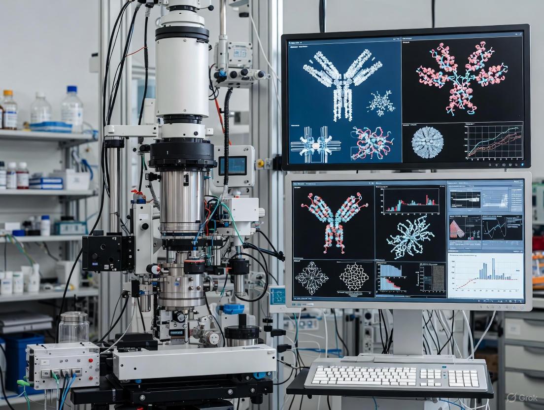
Data Collection Strategies and Single-Particle Analysis Workflow
Cryo-electron microscopy (cryo-EM) has emerged as a revolutionary technique in structural biology, enabling the determination of macromolecular structures at near-atomic to atomic resolution without the need for crystallization [33]. Single-particle analysis (SPA), a key methodology within cryo-EM, has become particularly invaluable for investigating complex biological systems such as membrane proteins, viruses, and large macromolecular complexes [34]. This technique has transformed research in areas ranging from basic molecular mechanisms to structure-based drug design.
In the specific context of B cell receptor (BCR) research, cryo-EM SPA has provided unprecedented insights into the structural basis of BCR assembly and activation mechanisms [4] [17] [7]. The BCR, composed of a membrane-bound immunoglobulin (mIg) and a heterodimeric Igα/Igβ signaling subunit, plays a critical role in adaptive immune responses by recognizing antigens and initiating signaling cascades [7]. Recent cryo-EM structures have revealed that the BCR complex exhibits an asymmetric organization with a 1:1 stoichiometry of mIg to Igα/Igβ, contradicting earlier symmetric models [4] [17]. This structural knowledge is crucial for understanding B cell activation and for rational engineering of therapeutics targeting B cell-mediated diseases [7].
This application note provides a comprehensive framework for data collection strategies and SPA workflow optimization, with specific emphasis on applications in BCR-antigen complex characterization. We detail experimental protocols, quantitative benchmarks, and visualization tools to guide researchers in obtaining high-resolution structures of biologically relevant complexes.
Data Collection Strategies
Specimen Preparation for Cryo-EM
Successful single-particle analysis begins with optimized specimen preparation to ensure structural homogeneity and particle integrity. For BCR complexes, which are membrane-proximal assemblies, careful consideration must be given to purification and vitrification conditions.
Protein Purification and Homogeneity Assessment: Specimen purity and structural homogeneity are paramount for high-resolution reconstruction. Biochemical analyses alone (SDS-PAGE, gel-filtration) are insufficient to assess suitability for EM, as apparently intact complexes may contain compositional or conformational heterogeneity [35]. Negative-stain EM provides a rapid method to evaluate sample quality, as the staining procedure tends to induce proteins to adsorb to carbon film in preferred orientations, facilitating homogeneity assessment [35]. For BCR complexes, which may exhibit conformational flexibility, stabilization through biochemical means is recommended:
- Buffer Optimization: Thermofluor-based screening approaches can identify conditions that stabilize target complexes [35].
- Chemical Cross-linking: Mild cross-linking with glutaraldehyde, including GraFix (glycerol/glutaraldehyde gradient centrifugation) or "on column" cross-linking over size-exclusion columns, can reduce heterogeneity [35]. However, cross-linking may introduce artifacts by stabilizing non-physiological conformations, so native samples should always be analyzed in parallel.
- Conformational Locking: BCR complexes can be locked in defined functional states by adding antigens, inhibitors, or other binding partners [35]. For instance, BCR Fab flexibility can be reduced by antigen binding [4].
Vitrification: Vitrification preserves native structures by rapid plunge-freezing in liquid ethane, embedding particles in amorphous ice [34] [36]. Semi-automated plungers (e.g., Vitrobot, Cryoplunge) enhance reproducibility [35]. Key parameters affecting ice quality include:
- Ice Thickness: Should be sufficient to accommodate particles but not excessively thick, as this reduces contrast and increases defocus spread [35]. Optimal thickness is particularly important for membrane proteins like BCRs, where detergent presence can lower surface tension, complicating thin ice formation [35].
- Particle Distribution: Ideal specimens show high particle density in different orientations without touching [35]. For BCR complexes, which may exhibit preferred orientation, optimizing grid surface properties through glow discharge or plasma cleaning is critical.
Imaging Parameters and Data Acquisition
Modern cryo-EM leverages advanced instrumentation and detection technology to achieve high-resolution reconstruction. Data collection strategies must balance resolution needs with practical constraints of radiation sensitivity and computational resources.
Microscopy Hardware: Direct electron detector devices (DDDs) with superior detective quantum efficiency (DQE) have been instrumental in the "resolution revolution" [35]. These cameras enable dose-fractionated movie collection, allowing computational correction of beam-induced motion [35]. High-end cryo-electron microscopes (e.g., CRYO ARM models) provide the stability and automation required for unattended data collection [36].
Image Acquisition Parameters: Data collection strategies should be optimized based on sample characteristics and resolution targets. The following parameters are particularly critical for BCR complexes:
Table 1: Key Data Collection Parameters for Cryo-EM SPA
| Parameter | Considerations for BCR Complexes | Typical Values/Ranges |
|---|---|---|
| Acceleration Voltage | Balance between contrast and resolution | 200-300 kV |
| Total Electron Dose | Limited by radiation sensitivity; higher doses improve SNR but increase damage | 40-60 e⁻/Ų |
| Defocus Range | Provides phase contrast; must be varied to ensure complete transfer of information | -0.5 to -3.0 μm |
| Pixel Size | Should satisfy Nyquist criterion for target resolution | 0.5-1.5 Å/pixel |
| Number of Micrographs | Depends on particle size, symmetry, and heterogeneity | 500-5000 |
| Particles per Micrograph | Varies with concentration and ice quality | 10-500 |
Automated Data Collection: Software automation enables efficient collection of large datasets necessary for high-resolution reconstruction [35]. Multi-shot acquisition strategies and beam-image shift approaches increase throughput, particularly important for heterogeneous samples like BCR-antigen complexes.
Single-Particle Analysis Workflow
The SPA workflow comprises a series of computational steps that transform raw micrographs into refined 3D reconstructions. The general workflow is depicted below, with specific considerations for BCR complexes highlighted throughout this section.
Image Pre-processing and Particle Picking
Motion Correction and CTF Estimation: Raw movie frames are aligned to correct for beam-induced motion [37]. The contrast transfer function (CTF) is then estimated for each micrograph to characterize microscope optical parameters [37]. Recent advances in real-time CTF estimation and motion correction algorithms have significantly improved image quality and resolution [37].
Particle Picking: Particles are identified and extracted from micrographs. Automated approaches, particularly deep learning-based algorithms, have dramatically improved the accuracy and efficiency of this process [37]. For BCR complexes, which exhibit an asymmetric organization without inherent symmetry [4] [17], template-based or neural network picking approaches are recommended.
2D Classification: Extracted particles are subjected to reference-free alignment and classification to generate 2D class averages representing characteristic views [37] [36]. This step serves as a critical quality control checkpoint, allowing removal of non-particle images (ice, detergent, etc.) and identification of structurally homogeneous subsets. For BCR samples, 2D classification can reveal the characteristic Y-shaped topology of the complex [7].
3D Reconstruction and Refinement
Initial Model Generation: An initial 3D model can be generated through ab initio reconstruction approaches (e.g., stochastic gradient descent) without a starting reference [37]. Alternatively, for BCR complexes, existing low-resolution structures (e.g., PDB: 7XQ8) can serve as initial models [4].
3D Classification and Heterogeneity Analysis: 3D classification identifies structural heterogeneity within the particle stack, separating particles by conformational states or compositional differences [37]. Advanced algorithms like multi-body refinement and 3D variability analysis (3DVA) can resolve complex conformational dynamics [37]. For BCR complexes, this is particularly relevant given the conformational changes induced by antigen binding [4].
3D Refinement: Iterative refinement aligns particles against a reference structure to improve resolution. The following table summarizes key reconstruction parameters and their impact on final map quality:
Table 2: Reconstruction Parameters and Quality Assessment
| Parameter | Impact on Reconstruction | Quality Control Metrics |
|---|---|---|
| Particle Quantity | Affects resolution and statistical reliability; diminishing returns beyond certain point | Resolution vs. particle number curve; 100,000+ often needed for asymmetric complexes |
| Symmetry Application | Greatly improves resolution for symmetric complexes; inappropriate for asymmetric BCR | Directional FSC; local resolution analysis; 2D class inspection |
| Final Particle Count | Determines final resolution; BCR complexes typically require substantial particles | 43,249 particles yielded ~3Å resolution for E2 core [37] |
| Map Resolution | Overall quality indicator; BCR structures now achievable at near-atomic resolution | Gold-standard FSC at 0.143 criterion; local resolution variation |
| Map/Model Validation | Ensures structural accuracy and model quality | MolProbity scores; EMRinger score; geometry statistics |
Symmetry Considerations: While symmetric complexes benefit dramatically from symmetry application during reconstruction (effectively multiplying particle numbers) [37], BCR complexes are fundamentally asymmetric [4] [17] [7]. Applying symmetry to such complexes would introduce severe artifacts and incorrect biological interpretations [37]. Therefore, BCR reconstructions must be performed without symmetry (C1).
Model Building and Validation
Atomic Model Building: Atomic coordinates are built into the cryo-EM density map. For high-resolution maps (<3.5Å), side chains are discernible, enabling accurate model building [34]. Recent automated tools (ModelAngelo, EMbuild, DiffModeler) have streamlined this process, particularly for moderate-resolution maps [37]. For BCR complexes, existing structures (e.g., IgM-BCR, IgG-BCR) provide useful starting points [17] [7].
Validation: Rigorous validation ensures model quality and prevents overinterpretation. Directional Fourier shell correlation (FSC) assesses resolution anisotropy, while model-to-map FSC evaluates fit quality [37]. Geometric statistics (Ramachandran outliers, rotamer outliers) assess model geometry [37]. For BCR complexes, validation should confirm biologically relevant features, such as the compact four-helix transmembrane bundle and the asymmetric interactions between Fc domains and Igα/Igβ [7].
Application to B Cell Receptor Complexes
Structural Insights into BCR Assembly and Activation
Cryo-EM SPA has revolutionized our understanding of BCR structure and function. Recent structures have revealed that human IgM-BCR and IgG-BCR complexes assemble with a 1:1 stoichiometry of mIg to Igα/Igβ, forming an asymmetric complex [17] [7]. Key structural features include:
- Extracellular Domains: The Fc domains interact with Ig-like domains of Igα/Igβ through distinct "side-by-side" (IgM) and "head-by-side" (IgG) modes [7].
- Transmembrane Region: The TM segments of mIg and Igα/Igβ form a compact four-helix bundle stabilized by conserved polar interactions [7].
- Juxtamembrane Region: The membrane-proximal connecting peptide of one mIg chain adopts an interdigitated topology with the Igα/Igβ heterodimer, creating a braided interaction network [7].
- Cytoplasmic Tails: The ITAM-containing tails of Igα/Igβ are typically not resolved, indicating high flexibility in the resting state [7].
Molecular dynamics simulations of BCR complexes have revealed that antigen binding increases flexibility in regions distal to the binding site, particularly in the Fc domains and extracellular domains of Igα/Igβ [4]. Antigen binding also alters the rearrangement of IgM transmembrane helices and modifies the relative interactions with Igα/Igβ, potentially initiating signaling [4]. These observations support conformation-induced models of BCR activation [4].
Specific Workflow Considerations for BCR Complexes
Sample Preparation: BCR complexes require careful extraction and purification from membrane environments while maintaining complex integrity. Detergent selection critically impacts stability, with mild detergents (e.g., DDM, LMNG) often preferred. Incorporating membrane mimetics (nanodiscs, lipodisqs) may enhance stability for full-complex studies.
Grid Preparation: BCR complexes may exhibit preferred orientation due to their membrane-proximal regions and asymmetric charge distribution. Grid surface treatments (different hydrophilicity protocols), additive screening (fluoro-octanol, CHAPSO), and occasionally affinity grid approaches can mitigate this issue.
Data Processing Strategies: The inherent flexibility of BCR complexes, particularly in the Fab regions and cytoplasmic tails, necessitates extensive classification approaches. Focused classification with signal subtraction can resolve flexible regions, while multi-body refinement can characterize dynamic domains relative to more rigid cores.
Experimental Protocols
Protocol: Sample Preparation for BCR Complex Cryo-EM
Materials:
- Purified BCR complex (≥0.5 mg/mL, ≥95% purity)
- Appropriate detergent (e.g., DDM, LMNG) at critical micelle concentration
- Glow-discharged holey carbon grids (Quantifoil, UltrAuFoil)
- Vitrification device (Vitrobot, Cryoplunge)
- Liquid ethane and liquid nitrogen
Procedure:
- Grid Preparation: Glow discharge grids immediately before use (30-60 seconds, medium power).
- Sample Application: Apply 3-5 μL BCR complex to grid. Blot for 2-6 seconds (force: -5 to 20, 100% humidity, 4°C).
- Vitrification: Plunge freeze into liquid ethane cooled by liquid nitrogen.
- Storage: Transfer grids to cryo-box under liquid nitrogen for microscope loading.
Troubleshooting:
- Thick ice: Reduce blot time, increase humidity.
- Preferred orientation: Try different grid types (gold vs. copper), adjust discharge parameters.
- Particle denaturation: Optimize blot force, include detergents/additives.
Protocol: High-Resolution Data Collection
Materials:
- Cryo-electron microscope with direct electron detector
- Automated data collection software (SerialEM, EPU)
Procedure:
- Screening: Assess grid quality at low magnification (100-200x). Identify areas with appropriate ice thickness and particle distribution.
- Atlas Collection: Acquire grid atlas at low magnification for navigation.
- Hole Targeting: Identify suitable holes at intermediate magnification.
- Data Collection Setup: Define collection parameters: dose (40-60 e⁻/Ų), defocus range (-0.5 to -3.0 μm), pixel size (0.5-1.5 Å/pixel).
- Automated Acquisition: Run automated data collection, collecting 500-5000 micrographs based on particle density and heterogeneity.
Quality Control:
- Monitor ice quality during collection.
- Assess motion correction and CTF estimation in real-time if possible.
- Collect dataset subsets for initial processing to confirm quality.
Protocol: Single-Particle Processing for BCR Complexes
Materials:
- Processing software (CryoSPARC, RELION, cisTEM)
- High-performance computing resources
Procedure:
- Pre-processing: Patch motion correction and CTF estimation for all micrographs.
- Particle Picking: Use template-based or neural network picking (e.g., CryoSPARC blob picker followed by 2D classification to generate templates).
- Extraction: Extract particles with box size 1.5-2× maximum dimension, binning as appropriate.
- 2D Classification: Perform multiple rounds of 2D classification to remove junk particles.
- Ab Initio Reconstruction: Generate initial model without reference bias.
- Heterogeneous Refinement: Separate structural populations using 3D classification.
- Homogeneous Refinement: Refine selected particles to high resolution.
- Post-processing: Apply mask and correct for modulation transfer function.
- Model Building: Build atomic model into sharpened map, iteratively refining.
The Scientist's Toolkit
Table 3: Essential Research Reagents and Materials for BCR Complex Cryo-EM
| Item | Function | Examples/Specifications |
|---|---|---|
| Detergents | Solubilize and stabilize membrane proteins | DDM, LMNG, CHAPSO at CMC |
| Grids | Support specimen in thin ice layer | Quantifoil R1.2/1.3, UltrAuFoil, Graphene oxide |
| Negative Stain | Rapid sample assessment | Uranyl acetate, methylamine tungstate |
| Cross-linkers | Stabilize complexes, reduce heterogeneity | Glutaraldehyde (0.1-0.5%), GraFix techniques |
| Vitrification Device | Rapid freezing for ice formation | Vitrobot Mark IV, Cryoplunge 3 |
| Direct Electron Detector | High-resolution image acquisition | Gatan K3, Falcon 4, Selectris X |
| Processing Software | 3D reconstruction from 2D images | CryoSPARC, RELION, EMAN2 |
| Model Building Software | Atomic model construction | Coot, Phenix, ModelAngelo |
| Validation Tools | Assess model and map quality | MolProbity, EMRinger, PHENIX validation |
Atomic Model Building and Refinement into Cryo-EM Density Maps
Atomic model building and refinement are critical steps in cryo-electron microscopy (cryo-EM) structure determination, transforming three-dimensional density maps into detailed atomic coordinates that provide mechanistic insights into biological processes. Within B cell receptor (BCR) research, these techniques enable the visualization of antigen recognition and subsequent activation mechanisms at near-atomic resolution. Recent advances in cryo-EM have revolutionized our understanding of membrane protein complexes like the BCR, revealing asymmetric organization and conformational changes underlying immune activation [4]. This protocol outlines comprehensive methodologies for building and refining atomic models into cryo-EM density maps, with specific applications to BCR-antigen complexes.
The process of atomic model building has been transformed by integration of machine learning approaches with biophysical constraints, enabling more accurate interpretation of medium-resolution density maps where side-chain information remains ambiguous. Concurrently, refinement methodologies have evolved to better balance experimental density fit with proper stereochemical geometry, addressing the unique challenges posed by the cryo-EM data collection process.
Key Software Tools for Atomic Model Building
Table 1: Software Tools for Cryo-EM Atomic Model Building and Refinement
| Software Tool | Primary Methodology | Key Features | Optimal Resolution Range | Applications in BCR Research |
|---|---|---|---|---|
| ModelAngelo [38] | Graph Neural Network (GNN) | Combines cryo-EM density with protein sequence and structural information; automated protein identification | 2-4 Å | Building unknown BCR subunits; complete complex assembly |
| Phenix.maptomodel [39] | Automated tracing and building | Identifies molecular boundaries; applies map sharpening; builds protein/RNA/DNA chains | 4.5 Å or better | Initial BCR model generation; symmetric complex building |
| GROMACS with Maximum Likelihood Refinement [40] | Molecular dynamics with relative entropy potential | Gentle refinement balancing density fit and stereochemistry; adaptive force scaling | 2-4 Å | Refining BCR transmembrane domains; studying conformational changes |
| Graph-based Backbone Threading [41] | Minimum spanning tree algorithm | Segments density into subunits; uses co-evolutionary contact predictions | 3-5 Å | Building BCR extracellular domains when subunit structures unknown |
| SPHIRE [42] | Semi-automated processing pipeline | Integrated processing and model building; graphical user interface | 3.5 Å or better | Processing BCR-antigen complex data sets |
The Scientist's Toolkit: Essential Research Reagents and Materials
Table 2: Essential Research Reagents and Materials for Cryo-EM Model Building Studies
| Reagent/Material | Function/Application | Specifications | Example Use in BCR Research |
|---|---|---|---|
| Membrane Scaffolds | Stabilizing membrane proteins for cryo-EM | Nanodiscs, liposomes, amphipols | Incorporating full-length BCR complexes [21] |
| Cell-Free Protein Synthesis Systems | Producing challenging membrane protein complexes | E. coli lysates supplemented with nanodiscs | Synthesizing full-length β1-adrenergic receptor-G protein complexes [21] |
| B Cell Line Expressing Engineered BCR | FRET-based conformational studies | A20IIA.6 B cells; 293T cells for transient expression | Monitoring antigen-induced conformational changes [43] |
| Site-Specific Labeling Reagents | Incorporating fluorophores for FRET studies | CoA 488 (CoA-conjugated ATTO 488); ReAsH | Labeling ybbR and tetracysteine tags in BCR heavy chain [43] |
| HIV-1 Envelope Antigens | BCR activation and structural studies | gp120 monomer; V51 trimer with foldon domain | Studying BCR-antigen binding interfaces [4] |
| Complex Membrane Lipid Mixtures | Creating native-like membrane environments | POPC, POPE, PSM, CER3, DAGL, cholesterol [4] | Molecular dynamics simulations of BCR membrane interactions |
Experimental Protocols
Automated Model Building with ModelAngelo for BCR Complexes
ModelAngelo represents a transformative machine learning approach that combines information from cryo-EM maps with protein sequence and structural information through a unified graph neural network architecture [38].
Step-by-Step Protocol:
Input Preparation
- Provide a sharpened, locally filtered cryo-EM map in MRC/CCP4 format
- Include all protein and nucleic acid sequences in FASTA format
- For BCR complexes: Include sequences for immunoglobulin heavy and light chains, Igα (CD79A), and Igβ (CD79B)
Graph Neural Network Processing
- The algorithm first identifies Cα and nucleic acid phosphate positions using a convolutional neural network (CNN)
- A graph is constructed where each residue is a node connected to its 20 nearest neighbors
- Three specialized modules process this graph:
- Cryo-EM Module: Incorporates local density information around each residue
- Sequence Module: Performs cross-attention with user-provided sequences embedded via ESM-1b protein language model
- IPA Module: Analyzes geometrical relationships between neighboring residues
Model Generation and Sequence Assignment
- Atomic coordinates are generated from optimized residue positions and orientations
- Amino acid probabilities predicted for each residue are converted to hidden Markov model profiles
- HMMER search identifies and assigns sequences to density regions
- Output includes full atomic model with confidence scores for each residue
Application to BCR Research: ModelAngelo particularly excels at identifying unknown subunits in endogenous BCR complexes, potentially revealing novel interacting proteins in BCR signalosomes. The integration of sequence information allows correct assignment of immunoglobulin domains despite limited side-chain density in 3-4 Å maps [38].
Molecular Dynamics-Based Refinement Using GROMACS
Recent advances enable cryo-EM density-guided molecular dynamics simulations that gently refine models while maintaining proper stereochemistry [40].
Step-by-Step Protocol:
System Setup
- Prepare initial atomic model in PDB format
- Convert cryo-EM map to density potential using the maximum likelihood approach
- Solvate the system in appropriate water model and add ions to physiological concentration
Relative Entropy Potential Setup
- Generate model density from atomic coordinates using cryo-EM-appropriate scattering factors
- Apply relative entropy-based refinement potential: Ufit = -k·S(ρ||ρmodel)
- Implement adaptive force scaling to automatically balance density-derived forces with molecular mechanics force field
Simulation and Refinement
- Run molecular dynamics simulations with density restraints
- Monitor Fourier Shell Correlation (FSC) between model and experimental density
- Continue refinement until convergence of both density correlation and stereochemical quality metrics
- Validate refined model using MolProbity or similar validation tools
Application to BCR Research: This method is particularly valuable for refining BCR transmembrane domains where flexibility and lipid interactions can challenge conventional building approaches. The gentle potential allows natural movements of helices while maintaining agreement with experimental density [40].
FRET-Based Conformational Validation of BCR Models
For validation of structural models, FRET-based approaches can monitor antigen-induced conformational changes in BCR complexes [43].
Step-by-Step Protocol:
Site-Specific Labeling of BCR
- Engineer ybbR tag at N-terminus of BCR heavy chain for CoA-488 labeling
- Insert tetracysteine tag in Cμ2 domain (IgM-BCR) or Cγ2 domain (IgG-BCR) for ReAsH labeling
- Verify labeled BCR function through antigen binding and calcium flux assays
FRET Measurements
- Express dual-labeled BCR in 293T or A20IIA.6 B cells
- Stimulate with antigen presented on planar lipid bilayers
- Perform acceptor photobleaching FRET measurements using TIRFM
- Calculate FRET efficiency from donor recovery after acceptor bleaching: E = 1 - (FDA/FD)
- Alternatively, use FLIM-FRET for concentration-independent measurements
Correlation with Structural Models
- Compare measured FRET distances with distances predicted from atomic models
- Validate antigen-induced conformational changes by FRET efficiency changes
Application to BCR Research: This approach confirmed antigen-induced separation between Fab and Fc domains in IgM-BCR, supporting conformational change models of BCR activation [43].
Workflow Diagrams
Application to B Cell Receptor Research
Cryo-EM model building has provided transformative insights into B cell receptor structure and activation mechanisms. Recent structures reveal the BCR complex exhibits an asymmetric organization with a 1:1 stoichiometry between membrane-bound immunoglobulin and the Igα/Igβ signaling heterodimer, contrary to earlier symmetric models [4]. Molecular dynamics simulations based on cryo-EM densities demonstrate that antigen binding induces increased flexibility in regions distal to the binding site and causes rearrangement of transmembrane helices, altering interactions with localized membrane lipids [4].
FRET-based conformational studies complement cryo-EM structures by capturing dynamic antigen-induced changes. These investigations reveal mechanical force induces conformational changes within the mIg heavy chain and alters spatial relationships between mIg and Igβ, with distinct patterns observed for IgM- versus IgG-BCR [43]. The integration of atomic models with biophysical validation provides a comprehensive framework for understanding how extracellular antigen binding is transduced across the membrane to initiate intracellular signaling.
Advanced model building approaches enable determination of structures containing previously unresolved regions. For instance, the full-length β1-adrenergic receptor structure with intact third intracellular loop (ICL3) revealed enhanced GPCR-G protein interactions, demonstrating how flexible regions can critically influence signaling complex formation [21]. Similar approaches applied to BCR complexes may reveal how cytoplasmic domains orchestrate signal initiation.
The protocols outlined herein provide a comprehensive framework for building and refining atomic models from cryo-EM densities, with specific applications to the structural characterization of B cell receptor complexes. These methodologies enable researchers to transform cryo-EM density maps into accurate atomic coordinates that reveal the molecular mechanisms of immune recognition and activation.
Understanding the precise interaction between an antibody and its target antigen is a cornerstone of modern immunology and biologics discovery. Epitope mapping, the process of identifying the binding site of an antibody on its antigen, is crucial for the rational design of vaccines and therapeutic antibodies [44]. For years, techniques such as X-ray crystallography have been instrumental in this field. However, the emergence of cryo-electron microscopy (cryo-EM) single-particle analysis (SPA) has provided a powerful and complementary technique, capable of studying antibody-antigen complexes that were previously intractable, such as those involving large or flexible antigens and even full-length immunoglobulin G (IgG) molecules [6] [32] [45].
This application note details the methodologies and protocols for employing cryo-EM in epitope mapping, framed within the broader context of B cell receptor (BCR)-antigen complex characterization. We provide a generic cryo-EM protocol, explore advanced hybrid methods that integrate structural and sequencing data, and discuss the direct application of these techniques in vaccine and therapeutic antibody development.
Core Principles of Cryo-EM in Epitope Mapping
Cryo-EM SPA has become a preferred method for structural epitope mapping due to its unique advantages. Unlike crystallography, cryo-EM does not require protein crystallization and is much more tolerant of conformational flexibility and molecular size heterogeneity [45]. This is particularly beneficial for studying antibodies and their complexes, which can be challenging to crystallize. Furthermore, cryo-EM allows for the direct use of full-length IgG in complex with its antigen, bypassing the need to generate Fab fragments, though Fabs are still commonly used to reduce flexibility and improve resolution [32]. The technique can resolve structures to near-atomic resolution, enabling the visualization of side-chain densities in the epitope-paratope interface and providing a direct, visual map of the antibody-binding site [6] [10].
Methods and Experimental Protocols
A Generic Cryo-EM SPA Protocol for Antigen-Antibody Complexes
The following section outlines a standard protocol for determining the structure of an antigen-antibody complex using cryo-EM SPA, compiled from established methodologies [6] [32].
Complex Formation and Purification
The first critical step is to form and purify a stable antigen-antibody complex.
- Complex Preparation: The antigen and antibody (either IgG or Fab fragments) are mixed in a molar ratio, typically with a slight molar excess of the antigen (e.g., 1:1.2 antibody:antigen). The mixture is incubated to allow complex formation. The use of full-length IgG is feasible and can simplify the workflow by avoiding enzymatic digestion steps, though it may introduce flexibility that can limit resolution [32].
- Buffer Exchange: The complex is buffer-exchanged into a final buffer compatible with cryo-EM grid preparation, such as 15 mM Tris-HCl, pH 7.5, and 150 mM NaCl [32].
- Purification: The formed complex is purified from unbound components using size-exclusion chromatography (SEC). A suitable SEC buffer is 15 mM Tris-HCl, pH 8.0, and 150 mM NaCl. Fractions containing the complex are collected and concentrated for grid preparation [32].
Cryo-EM Grid Preparation and Data Collection
- Vitrification: A small volume (e.g., 3-4 µL) of the purified complex is applied to a freshly plasma-cleaned cryo-EM grid. The grid is blotted to remove excess liquid and is subsequently plunge-frozen in a cryogen (typically liquid ethane) cooled by liquid nitrogen. This process vitrifies the sample, preserving its native state in a thin layer of amorphous ice [6].
- Data Collection: The vitrified grid is loaded into a cryo-electron microscope. Data are collected automatically using software that navigates the grid, targets holes with suitable ice thickness, and acquires thousands to millions of movie micrographs at a defined defocus range. These movies record the electron dose as the beam interacts with the sample particles.
Single-Particle Data Processing
Data processing is a multi-step computational process to reconstruct a 3D density map from the 2D particle images. Standard software suites include cryoSPARC and RELION [6] [32].
- Pre-processing: Movie frames are motion-corrected to account for beam-induced movement and dose-weighted to compensate for radiation damage. The contrast transfer function (CTF) of the microscope is estimated for each micrograph.
- Particle Picking and Classification: Particles are automatically picked from the micrographs. Several rounds of 2D classification are performed to separate "good" particles from junk, aggregates, or particles in undesirable orientations.
- Ab-Initio Reconstruction and Heterogeneous Refinement: An initial low-resolution 3D model is generated ab initio. This model is then used in heterogeneous (3D) classification to isolate a homogeneous subset of particles representing the antigen-antibody complex. Focused classification with a mask around the Fab region can be employed to reduce heterogeneity and improve the local resolution of the antibody-antigen interface [46].
- High-Resolution Reconstruction and Refinement: The homogeneous particle set is used for a final, high-resolution 3D reconstruction through iterative refinement, resulting in a density map.
Model Building and Epitope Analysis
- Atomic Model Building: An atomic model of the antigen is fitted into the corresponding density. For the antibody, the sequence of the variable regions must be known. If unavailable, it can be determined from the hybridoma or B cells (see Section 3.2.1). The model is then refined against the cryo-EM map using tools like Coot and PHENIX [32].
- Epitope Identification: The refined atomic model reveals the specific amino acid residues on the antigen (the epitope) that are in contact with the complementary determining regions (CDRs) of the antibody (the paratope). The quality of the map determines if side-chain interactions can be resolved.
The following diagram illustrates this comprehensive workflow from sample preparation to epitope analysis.
Advanced Hybrid Methods: Integrating Structure and Sequence
Innovative methods now combine cryo-EM structural data with next-generation sequencing (NGS) to directly link antibody sequence to antigen specificity and function, bypassing traditional B-cell sorting.
CryoEM-PEM (Polyclonal Epitope Mapping) with NGS
This hybrid approach characterizes the polyclonal antibody response directly from immune serum [46] [10].
- Method Overview: An antigen is incubated with polyclonal serum antibodies (pAbs) to form immune complexes. These complexes are then subjected to single-particle cryo-EM analysis. Through extensive 3D classification, multiple distinct 3D reconstructions are obtained, each representing a specific class of antibodies bound to a particular epitope on the antigen.
- Sequence Identification: To identify the sequences of these pAbs, NGS is performed on the B cell repertoires (e.g., from germinal center B cells or plasma cells) of the same donor. The medium-resolution (~3-4 Å) cryo-EM maps of the Fabs are used as a structural constraint. A hierarchical category assignment for amino acids is manually performed based on the cryo-EM density. A computational search algorithm then matches this structurally-derived sequence profile against the NGS database to identify the best-matching heavy and light chain sequences [46].
- Validation: The identified antibody sequences are synthesized, expressed as monoclonal antibodies (mAbs), and validated for antigen binding and functional activity (e.g., neutralization assays) to confirm they recapitulate the behavior of the polyclonal serum antibodies [46].
LIBRA-seq (Linking B-cell Receptor to Antigen Specificity through Sequencing)
While not a cryo-EM method itself, LIBRA-seq is a powerful complementary technology that can feed directly into structural workflows [47] [48].
- Principle: B cells are incubated with DNA-barcoded antigens, where each antigen is conjugated to a unique oligonucleotide barcode. Antigen-binding B cells internalize their specific B cell receptor (BCR) along with the barcoded antigen. Single-cell BCR sequencing then links the antibody sequence (from the BCR) to the antigen specificity (via the barcode) [47] [48].
- Integration with Ligand Blocking: An advanced application, "LIBRA-seq with ligand blocking," incorporates a functional readout. By including a barcoded ligand (e.g., ACE2 for SARS-CoV-2 spike protein) in the assay, researchers can simultaneously identify B cells that produce antibodies which not only bind the antigen but also block its interaction with the ligand, directly enriching for functionally neutralizing antibodies [48]. The sequences of these prioritized B cells can then be used to produce mAbs for high-resolution structural characterization by cryo-EM.
Application in Vaccine and Therapeutic Design
The methodologies described above have been successfully applied to critical challenges in immunology and drug development.
Case Study: Rapid Response to SARS-CoV-2
The COVID-19 pandemic highlighted the power of these integrated approaches. LIBRA-seq with ligand blocking was used to rapidly isolate potent SARS-CoV-2-neutralizing antibodies from convalescent patients. This method identified B cells producing antibodies that blocked the spike-ACE2 interaction, dramatically increasing the hit rate for neutralizing antibodies and requiring the production and validation of less than a dozen antibodies per experiment [48]. Subsequent cryo-EM structures of these antibodies, such as antibody 5317-4, revealed that it binds the receptor-binding domain (RBD) of the spike protein, with its epitope partially overlapping the ACE2-binding footprint, providing a mechanistic understanding of its neutralization capability [48].
Epitope-Based Vaccine Design
Cryo-EM-based epitope mapping is also instrumental in reverse vaccinology. By analyzing the polyclonal antibody response in protected individuals or animal models, researchers can identify the key neutralizing epitopes on a pathogen that are targeted by the most effective antibodies. This information guides the rational design of epitope-focused vaccines that aim to elicit a targeted, potent, and broad immune response against these specific determinants, a significant advantage over conventional whole-pathogen or protein subunit vaccines [44] [45].
The Scientist's Toolkit
The table below summarizes key reagents, software, and resources essential for executing the cryo-EM epitope mapping protocols described in this note.
Table 1: Key Research Reagent Solutions for Cryo-EM Epitope Mapping
| Category | Item | Function & Application |
|---|---|---|
| Biochemical Reagents | Protein G Binding/Elution Buffers | For purification of IgG antibodies from hybridoma or serum [32]. |
| Size-Exclusion Chromatography (SEC) Buffers | For final purification of antigen-antibody complexes prior to grid preparation [32]. | |
| Cryo-EM Grids (e.g., Quantifoil, UltrAuFoil) | Supports the vitrified sample for imaging in the electron microscope. | |
| Molecular Biology Kits | RNeasy Mini Kit / iScript cDNA Synthesis Kit | For total RNA extraction and cDNA synthesis from hybridoma or B cells for antibody sequencing [32]. |
| Herculase II/Phusion DNA Polymerase | For high-fidelity PCR amplification of antibody variable regions [32]. | |
| Software & Algorithms | cryoSPARC | End-to-end platform for processing cryo-EM data, featuring rapid, unsupervised structure determination [6] [32]. |
| RELION | A widely used, highly flexible software suite for cryo-EM SPA structure determination [6] [32]. | |
| Coot / PHENIX | For model building, visualization, and refinement of atomic models into cryo-EM density maps [32]. | |
| ModelAngelo | A deep-learning tool for building atomic models directly from cryo-EM density maps, useful for de novo antibody sequencing [10]. | |
| NGS & Bioinformatics | LIBRA-seq Barcoded Antigens | Oligonucleotide-barcoded antigens for linking BCR sequence to antigen specificity in high-throughput [48]. |
| NGS Platforms (e.g., Illumina) | For sequencing the BCR repertoires from sorted or unsorted B cell populations [46] [47]. |
Cryo-electron microscopy has fundamentally expanded the toolkit for epitope mapping, moving beyond the characterization of single monoclonal antibodies to enabling the deconvolution of complex polyclonal responses. When integrated with high-throughput B cell receptor sequencing technologies like NGS and LIBRA-seq, it provides an unparalleled, holistic view of the immune response. These integrated approaches are accelerating the discovery and mechanistic understanding of therapeutic antibodies and are guiding the rational design of next-generation vaccines, solidifying their role as indispensable tools in modern biomedical research and drug development.
Navigating Challenges: Optimizing Cryo-EM Workflows for Complex Immune Receptors
Within the broader context of cryo-electron microscopy (cryo-EM) for B-cell receptor (BCR)-antigen complex characterization, resolving dynamic protein regions remains a significant challenge. Flexible Fab (antigen-binding fragment) and hinge regions are inherent to immune receptors like the BCR and antibodies, enabling them to sample multiple conformational states. This flexibility is functionally critical for immune recognition but introduces substantial heterogeneity that can obscure high-resolution structural details. This Application Note details practical strategies and protocols to overcome these challenges, enabling researchers to resolve these dynamic regions for both fundamental biological insight and drug development applications.
Understanding the Flexibility Challenge
Flexibility in Fab and hinge regions manifests as a continuum of conformational states, leading to structural heterogeneity in cryo-EM samples. During single-particle analysis, this heterogeneity results in blurred or missing density for the flexible domains, preventing accurate atomic model building. The immunoglobulin M (IgM) pentamer exemplifies this challenge; its structure reveals antigen-binding domains flexibly attached to an asymmetric and rigid core, with a hinge located at the Cμ3/Cμ2 domain interface allowing Fabs and Cμ2 to pivot as a unit both in-plane and out-of-plane [49]. This motion is distinct from that observed in IgG and IgA, where the two Fab arms swing independently [49].
In the context of BCR complexes, molecular dynamics (MD) simulations demonstrate that antigen binding induces allosteric changes and increases flexibility in regions distal to the binding site itself [4]. This propagated dynamic change underscores that flexibility is not a localized nuisance but a integral functional property that must be captured and understood.
Sample and Grid Preparation Strategies
Optimizing sample preparation is the first critical step to minimize non-biological heterogeneity and preserve native conformations.
Protocol: Stabilization and Vitrification
Objective: To prepare a homogeneous sample of BCR complex with minimized flexible motions without disrupting biological function.
Materials:
- Purified BCR Complex: Stabilized in a suitable detergent (e.g., DDM) for membrane proteins [50].
- Grid Type: Ultrafoil or similar gold grids with perforated support films to reduce background noise and interactions with the air-water interface (AWI) [50].
- Vitrification Device: Thermo Fisher Scientific Vitrobot or equivalent.
- Optimized Buffer: As determined by pre-screening.
Procedure:
- Complex Stabilization: Consider the addition of conformation-specific Fabs or nanobodies to rigidify flexible domains. For example, the therapeutic Fab Coltuximab was used to stabilize the CD19-CD81 complex for cryo-EM [9].
- Grid Optimization: Apply 3-4 µL of sample to a freshly glow-discharged grid. Soak times must be optimized (e.g., 0-60 seconds) to balance particle adhesion with AWI-induced denaturation [50].
- Blot and Plunge-Freeze: Blot for 2-6 seconds under 100% humidity before plunging into liquid ethane. Test multiple blot conditions to find the optimal ice thickness.
Troubleshooting: If particle denaturation or preferential orientation is observed, vary the surfactant concentration (e.g., 0.01-0.1% DDM) or use different grid surface chemistries [50].
Data Processing and Reconstruction Workflows
Advanced computational strategies are essential to disentangle continuous conformational changes.
Quantitative Motion Correction and Particle Recentering
Beam-induced motion and inaccurate particle boxing significantly exacerbate the effects of inherent flexibility. Motion correction software (e.g., MotionCor2, RELION's implementation) must be applied on a per-particle basis to correct local movement [51]. Following initial 2D or 3D classification, a particle re-centring protocol can be implemented to improve the accuracy of particle centring, which subsequently allows for the application of a tighter soft mask during refinement. A benchmark study demonstrated that this recentring procedure improved the resolution of a ~550 kDa complex (V1) from 8.7 Å to 8.0 Å [51].
Protocol: 3D Variability Analysis for Hinge Motion
Objective: To resolve distinct conformations of a flexible Fab-hinge unit.
Software: CryoSPARC or RELION.
Procedure:
- Initial Reconstruction: Perform a standard single-particle processing workflow to obtain an initial, consensus 3D reconstruction of the entire complex.
- Masked 3D Classification: Generate a loose mask encompassing the rigid core of the complex and a second mask encompassing the flexible Fab arms and hinge region. Perform several rounds of 3D classification without alignment to isolate discrete conformational states.
- 3D Variability Analysis (3DVA): In CryoSPARC, run the 3DVA job on the particle stack. Use a mask that encompasses the entire complex or focuses specifically on the Fab-hinge region.
- Conformation Extraction: The 3DVA output will show a trajectory of motion. Use the "Interpolate along mode" function to extract discrete volumes representing major conformational states along this trajectory, as demonstrated for the IgM F(ab')2 [49].
- Focused Refinement: For each extracted conformation, perform a final, high-resolution focused refinement with a tight mask on the now-stabilized Fab-hinge unit.
This workflow successfully resolved five distinct conformations of the IgM F(ab')2, revealing it moves as a rigid body pivoting at the Cμ2/Cμ3 interface [49].
Advanced Deep Learning for Map Enhancement
Deep learning-based post-processing tools can significantly improve the interpretability of cryo-EM maps, especially for flexible regions. EMReady is a framework that uses a Swin-Conv-UNet architecture to simultaneously minimize local smooth L1 distance and maximize non-local structural similarity (SSIM) between experimental and target maps [52]. In evaluations on 110 primary maps, EMReady-processed maps achieved a significantly better average map-model FSC-0.5 (3.57 Å) and Q-score (0.542) compared to deposited maps and those processed by other methods [52]. This enhancement directly aids in the model building of flexible loops and hinges.
Table 1: Benchmarking of Cryo-EM Map Post-Processing Methods
| Method | Type | Average Map-Model FSC-0.5 (Å) | Average Q-score | Dependency on Prior Info |
|---|---|---|---|---|
| EMReady | Deep Learning | 3.57 | 0.542 | No atomic model required [52] |
| DeepEMhancer | Deep Learning | 4.18 | 0.425 | Trained on model-guided maps [52] |
| phenix.auto_sharpen | Global Sharpening | 4.82 | 0.492 | B-factor based [52] |
| Deposited Map | N/A | 4.83 | 0.494 | Varies [52] |
Integrated Analysis of Conformational Dynamics
Correlating Structure with Function via MD Simulations
Molecular dynamics simulations provide a dynamic context for static cryo-EM maps. After obtaining multiple structures of flexible states, all-atom MD simulations can be performed in a solvated lipid bilayer environment to:
- Validate the stability of the resolved conformations.
- Identify the energy barriers and pathways between conformational states.
- Probe allosteric networks linking antigen binding in the Fab to conformational changes in the hinge and transmembrane regions, as observed in BCR simulations [4].
Table 2: Research Reagent Solutions for BCR Complex Cryo-EM
| Reagent / Material | Function / Application | Example & Notes |
|---|---|---|
| Therapeutic Fabs (e.g., Coltuximab) | Binds and stabilizes specific epitopes on target proteins, rigidifying flexible domains. | Used to stabilize CD19 for structure determination of the CD19-CD81 complex [9]. |
| n-Dodecyl-β-Maltoside (DDM) | Mild detergent for solubilizing and purifying membrane protein complexes. | Critical for maintaining stability of BCR and other membrane complexes during grid preparation [50]. |
| UltraFoil Gold Grids | Cryo-EM support grids with a perforated foil to reduce background noise and AWI interactions. | Minimizes particle adhesion to the AWI, preserving native conformation [50]. |
| 3D Variability Analysis (3DVA) | Algorithm to resolve continuous conformational changes from a heterogeneous particle stack. | Essential for visualizing Fab hinge motion in IgM [49]. |
| Deep Learning Enhancer (e.g., EMReady) | Post-processing tool to improve map quality and interpretability, especially in flexible regions. | Improves map-model FSC and Q-scores without requiring an atomic model prior [52]. |
BCR Activation and Signaling Pathway
The following diagram summarizes the conformational changes in the BCR complex upon antigen binding, integrating structural and dynamic data.
(caption: BCR Antigen-Induced Activation Pathway. Antigen binding induces long-range allosteric changes, increasing flexibility in distal domains and culminating in transmembrane helix rearrangements that trigger signaling [4].)
Overcoming the challenges posed by flexible Fab and hinge regions requires an integrated strategy from sample preparation to advanced computation. Key takeaways include:
- Sample Stabilization: The use of fiducial binders like Fabs and optimized grid preparation are crucial to reduce heterogeneity.
- Computational Separation: Techniques like 3D Variability Analysis are powerful for resolving continuous conformational changes.
- Map Enhancement: Deep learning methods like EMReady can significantly improve map quality in flexible regions.
- Dynamic Integration: Combining cryo-EM structures with MD simulations provides a comprehensive view of protein dynamics.
The future of resolving flexibility in cryo-EM lies in the deeper integration of time-resolved methods (trEM) to capture short-lived intermediate states [53], and the continued development of AI-driven tools that can automatically classify, refine, and model continuous heterogeneity. These advances will be indispensable for characterizing the full conformational landscape of dynamic complexes like the BCR, accelerating the structure-based design of novel immunotherapeutics and vaccines.
The structural characterization of endogenous B cell receptor (BCR) complexes is pivotal for understanding the molecular mechanisms of adaptive immunity and for informing targeted drug development. A significant technical hurdle in this field is the low natural abundance of these complexes in native cellular environments, which has historically limited the application of high-resolution techniques like cryo-electron microscopy (cryo-EM) [54]. Traditional cryo-EM workflows often require milligram quantities of purified, recombinant protein, making the study of endogenously expressed complexes, with their inherent low copy numbers and compositional heterogeneity, particularly challenging [55]. This application note details a novel affinity grid-based enrichment strategy designed to overcome this barrier, enabling the structural determination of endogenous BCR complexes and other low-abundance macromolecular assemblies directly from cell lysates.
Technical Innovation: Affinity Grid-Based Enrichment
The core innovation addressing the challenge of low abundance is the development of a graphene-based affinity cryo-EM grid, termed the Graffendor (GFD) grid [54]. This technology transforms the cryo-EM grid from a passive support into an active capture platform.
The GFD-A Grid Principle
The GFD-A grid is functionalized with a genetically modified ALFA nanobody, which serves as a high-affinity capture probe [54]. This design allows for the specific immobilization of target complexes directly from solution onto the grid surface. The process involves a one-step crosslinking batch-production method, ensuring consistent quality and performance across a single batch of 36 grids [54]. The key advantage of this system is its ability to concentrate target complexes directly on the grid, thereby bypassing the need for large-volume culture and multi-step purification that can lead to the loss of transient interactions or structural integrity.
Benchmarking and Validation
The efficacy of the GFD-A grid was rigorously validated. Using a low concentration of a model protein, β-galactosidase-2xALFA, the grid demonstrated efficient capture and enabled the determination of a high-resolution (2.71 Å) cryo-EM structure [54]. More importantly, its application for true endogenous proteins was tested in a biologically relevant context. Researchers engineered yeast cells to express Pop6, a shared component of RNase MRP and RNase P complexes, with a C-terminal tandem affinity tag (3xALFA-Tev-3xFlag: ATF) [54]. Subsequent cryo-EM analysis of samples captured from the cell lysate on GFD-A grids yielded structures of RNase MRP and RNase P at 3.3 Å and 3.0 Å resolution, respectively [54]. Notably, the structures obtained directly from cell lysates preserved additional densities that were absent in structures derived from anti-FLAG eluates, suggesting the GFD-A grid is capable of capturing transient or weakly associated interactors that are lost during conventional purification [54].
Detailed Experimental Protocol
GFD-A Grid Preparation and Sample Application
This protocol outlines the procedure for using GFD-A grids to enrich and vitrify endogenous protein complexes.
Materials:
- GFD-A grids (batch-produced with ALFA nanobody) [54].
- Cell lysate containing the target protein tagged with the ALFA tag.
- Appropriate wash buffer (e.g., 20 mM HEPES, 150 mM KCl, pH 7.4).
- Vitrification device (e.g., Vitrobot).
Procedure:
- Grid Activation: Briefly glow-discharge the GFD-A grid to render the surface hydrophilic.
- Sample Application: Apply 3-5 µL of the prepared cell lysate directly onto the GFD-A grid.
- Incubation: Allow the grid to incubate for 5-10 minutes in a humidified chamber to enable the ALFA-tagged target complexes to bind to the immobilized nanobodies.
- Washing: Gently blot away the excess solution and wash the grid by applying 3-5 µL of wash buffer to remove non-specifically bound cellular contaminants. Repeat this wash step twice.
- Vitrification: After a final blot to achieve an optimal ice thickness, rapidly plunge-freeze the grid in liquid ethane.
BCR Enrichment for Proteomic Studies
For proteomic analyses such as cross-linking mass spectrometry (XL-MS), a bead-based enrichment protocol can be employed.
Materials:
Procedure:
- Cell Lysis: Lyse the harvested B cells using an appropriate lysis buffer to solubilize membrane-bound BCR complexes.
- Antibody Binding: Incubate the cell lysate with a cocktail of intermediary antibodies targeting various BCR subcomponents.
- Pull-Down: Add Protein G-coated agarose beads to the lysate-antibody mixture and incubate to allow the bead-bound Protein G to capture the antibody-BCR complexes.
- Washing: Pellet the beads and wash thoroughly to remove non-specifically bound proteins.
- Elution/Solubilization: Solubilize the enriched BCR complex in an MS-compatible detergent for downstream proteomic analysis [55].
The following workflow diagram illustrates the key steps for the affinity grid-based approach:
The Scientist's Toolkit: Key Research Reagents
Table 1: Essential research reagents for endogenous BCR complex isolation and structural study.
| Reagent / Tool | Function / Description | Application in Protocol |
|---|---|---|
| ALFA Nanobody & Tag | High-affinity peptide-nanobody pair for specific protein capture. | Serves as the affinity probe on the GFD grid and genetic tag on the target protein [54]. |
| GFD-A Affinity Grid | Graphene-based EM grid with immobilized ALFA nanobody. | Active capture platform to concentrate target complexes directly from lysate [54]. |
| Intermediary Antibodies | Antibodies against BCR subunits (e.g., CD79a, CD79b, mIg). | Used in bead-based pull-downs to enrich the entire BCR complex from cell lysates [55]. |
| Protein G Agarose Beads | Beads with recombinant Protein G for antibody binding. | Solid support for immobilizing antibody-BCR complexes during pull-down enrichment [55]. |
| MS-Compatible Detergents | Detergents that do not interfere with mass spectrometry. | Solubilize enriched membrane protein complexes for downstream MS analysis [55]. |
Data Presentation and Analysis
The success of the affinity grid approach is quantified by the resolution of the resulting cryo-EM structures and the identification of novel structural features. The table below summarizes the key quantitative outcomes from the referenced study.
Table 2: Benchmarking performance of the GFD-A grid for cryo-EM structure determination.
| Sample Description | Sample Source | Reported Resolution | Key Observation |
|---|---|---|---|
| β-galactosidase-2xALFA | Low-concentration purified protein | 2.71 Å | Validation of grid efficiency for tagged proteins [54]. |
| RNase MRP | Yeast cell lysate | 3.3 Å | Preservation of additional densities vs. purified sample [54]. |
| RNase P | Yeast cell lysate | 3.0 Å | Preservation of additional densities vs. purified sample [54]. |
| RNase MRP | Anti-FLAG elution | 3.6 Å | Loss of transient densities compared to direct lysate capture [54]. |
| RNase P | Anti-FLAG elution | 3.9 Å | Loss of transient densities compared to direct lysate capture [54]. |
The following diagram summarizes the logical relationship between the low-abundance challenge, the technological solution, and the resulting scientific insights:
The deployment of affinity cryo-EM grids represents a significant advancement in structural biology, directly addressing the critical bottleneck of sample preparation for low-abundance endogenous complexes [54]. This methodology provides a robust and generalizable platform that bypasses the inefficiencies of conventional purification. By enabling the direct capture of complexes from cell lysates, it not only facilitates the determination of high-resolution structures but also uniquely positions researchers to uncover and characterize transient protein-protein interactions that are fundamental to cellular signaling and function, such as those in the native BCR complex [54] [55]. This technical leap is poised to accelerate drug discovery by providing more accurate structural blueprints of pathogenic complexes in their native state.
Improving Resolution for Small and Asymmetric Complexes
Single-particle cryo-electron microscopy (cryo-EM) has revolutionized structural biology but faces inherent challenges with small proteins (<100 kDa) and asymmetric complexes, which produce insufficient signal for high-resolution reconstruction. This application note details validated experimental and computational protocols to overcome these limitations, with a specific focus on applications in B cell receptor (BCR) research. We provide a structured guide covering strategic complex engineering, advanced data processing, and post-processing refinement techniques to achieve atomic-level insights into these critical immunological complexes.
Cryo-EM has become a mainstream structural biology technique, yet its application to proteins smaller than 100 kDa and complexes with low symmetry remains challenging [56] [57]. These samples produce images with a low signal-to-noise ratio (SNR), complicating particle alignment and high-resolution reconstruction [58]. In BCR research, these challenges are paramount as the BCR complex is inherently asymmetric and its components are often difficult to crystallize [4] [43]. This document outlines practical solutions, framing them within the context of a broader thesis on characterizing BCR-antigen interactions.
Strategic Complex Engineering for Size and Symmetry Enhancement
Engineering samples to increase their effective size and symmetry is a powerful strategy to facilitate cryo-EM structure determination.
Scaffolding Systems for Small Proteins
For individual small proteins, attaching to a larger, symmetric scaffolding platform provides the necessary fiducial markers for accurate particle alignment.
Table 1: Comparison of Cryo-EM Scaffolding Approaches
| Scaffold Type | Target Size Demonstrated | Achieved Resolution | Key Features | Modularity |
|---|---|---|---|---|
| DARPin-Cage (DARP14) | 26 kDa (GFP) | 3.8 Å | Cubic symmetry (12 copies); DARPin adaptor | High (DARPin loops can be engineered to bind diverse targets) [57] |
| Covalent Di-Gembodies (DiGb) | 14 - 55 kDa | 2.45 - 3.75 Å | Covalent dimerization of nanobodies; homomeric or heteromeric | High (Bispecific capability; minimal target modification) [58] |
| Megabody | ~50 kDa | ~3.5 Å | Nanobody fused to a rigid protein domain | Medium (Requires fusion engineering) [58] |
Protocol 2.1: Sample Preparation using a DARPin-Cage Scaffold
- Materials: Purified target protein, DARP14 scaffold with engineered DARPin loops (e.g., sequence from 3G124 for GFP [57]), size-exclusion chromatography (SEC) column.
- Procedure:
- Complex Formation: Incubate the DARP14 scaffold with a ~1.2 molar excess of the target protein for 1 hour at 4°C.
- Purification: Load the mixture onto an SEC column pre-equilibrated with a compatible buffer (e.g., 20 mM Tris-HCl pH 7.5, 150 mM NaCl).
- Validation: Analyze the peak fractions by SDS-PAGE and negative-stain EM to confirm complex formation and integrity. Quantitative amino acid analysis can verify binding saturation [57].
- Grid Preparation: Use the purified complex at ~3-5 mg/mL for vitrification using standard cryo-EM protocols.
Engineering Asymmetric Complexes: The BCR Case Study
The human B-cell antigen receptor is an asymmetric "Y"-shaped complex where a membrane-bound immunoglobulin (mIg) associates with a single Igα/Igβ heterodimer [4]. This 1:1 stoichiometry and the resulting asymmetry complicate high-resolution reconstruction. Strategic complex selection and stabilization are critical.
Protocol 2.2: Stabilizing the BCR Complex for Structural Study
- Materials: Gene constructs for BCR components (e.g., VRC01-IgM or VRC-CH31 based [4]), detergent (e.g., DDM), synthetic nanodiscs, Fab fragments (e.g., therapeutic anti-CD19 Coltuximab Fab [9]).
- Procedure:
- Construct Design: Engineer a fusion protein linking the full extracellular and transmembrane domains of the mIg to full-length CD81 via a flexible (GGS)x4 linker. This stabilizes the complex and facilitates purification [9].
- Complex Stabilization: To further increase complex size and stability, bind a therapeutic Fab (e.g., Coltuximab) to the CD19 ectodomain. This increases the total molecular weight and provides additional features for particle alignment [9].
- Mimicking Activated States: Use molecular dynamics (MD) simulations to identify conformational changes upon antigen binding. Simulations can guide the design of complexes trapped in specific functional states for structural analysis [4].
Computational & Data Collection Strategies
Advanced Data Processing Workflows
For scaffolds and asymmetric complexes, advanced 3D classification and refinement are essential to handle flexibility and achieve high resolution.
Protocol 3.1: Processing Data for Scaffolded Small Proteins
- Software: cryoSPARC, RELION.
- Procedure for DARPin-Cage Complexes [57]:
- Initial Reconstruction: Perform ab-initio reconstruction and homogeneous refinement in cryoSPARC with enforced T symmetry to obtain a high-resolution map of the symmetric scaffold core.
- Signal Subtraction: Use RELION to perform symmetry expansion and subtract the density corresponding to all but one copy of the DARPin adaptor and its bound cargo protein.
- Focused Classification: Without alignment, perform 3D classification on the subtracted particles to isolate classes with well-defined density for the DARPin and cargo.
- Multi-body Refinement: Conduct multi-body refinement with a mask focused on a single DARPin and GFP, searching orientations locally while constrained by the symmetric core.
Protocol 3.2: Processing Data for Asymmetric Complexes like BCR
- Software: cryoSPARC, RELION, MDAnalysis (for MD analysis).
- Procedure:
- MD Simulations for Dynamics: Run all-atom or coarse-grained MD simulations of the BCR complex in a realistic membrane model (e.g., containing POPC, POPE, PSM, CHOL) with and without bound antigen (e.g., HIV-1 Env gp120) [4].
- Analysis of Dynamics: Calculate root-mean-square fluctuations (RMSF) and transmembrane helix tilt angles from simulations to understand conformational heterogeneity and identify rigid-body domains for focused refinement [4].
- Focused Refinement: Apply multi-body or focused 3D classification strategies to address flexibility between domains identified as dynamic in MD simulations (e.g., Fab domains, Fc domains).
Post-Processing Map Improvement
Raw cryo-EM maps often require post-processing to enhance interpretability. Deep learning methods have surpassed traditional global sharpening.
Protocol 3.3: Map Sharpening with EMReady
- Software: EMReady.
- Procedure [52]:
- Input: Provide your experimental cryo-EM half-maps or a primary map.
- Processing: The SCUNet architecture in EMReady automatically processes the map, applying simultaneous local (via residual convolution) and non-local (via swin transformer) modeling.
- Output: The output is a sharpened map optimized by minimizing the smooth L1 distance and maximizing the structural similarity (SSIM) to a simulated target map.
- Validation: Quantify improvement using map-model FSC-0.5 and Q-scores. EMReady has been shown to improve the average FSC-0.5 from 4.83 Å to 3.57 Å and the average Q-score from 0.494 to 0.542 on a test set of 110 maps [52].
The Scientist's Toolkit
Table 2: Essential Research Reagents and Tools
| Reagent / Tool | Function / Description | Application Example |
|---|---|---|
| DARPin-Cage (DARP14) | A symmetric protein cage with 12 copies of a engineered DARPin adaptor for rigidly displaying cargo proteins. | Imaging small proteins like the 26 kDa GFP at 3.8 Å resolution [57]. |
| Covalent Di-Gembodies (DiGb) | Disulfide-linked nanobody dimers that provide a constrained, modular fiducial for particle alignment. | Determining structures of small soluble and membrane proteins like RECQL5 (3.18 Å) and SPNS2 (2.79 Å) [58]. |
| Therapeutic Fabs (e.g., Coltuximab) | Fab fragments that bind specific epitopes, increasing complex size and stability. | Stabilizing the CD19-CD81 B cell co-receptor complex for structure determination at 3.8 Å [9]. |
| EMReady | A deep learning tool for post-processing cryo-EM maps using local and non-local modeling. | Improving map quality and interpretability for automatic de novo model building [52]. |
| Molecular Dynamics (MD) Simulations | Computational method to simulate protein movements and conformational changes in a realistic environment. | Probing antigen-induced allosteric changes in the BCR transmembrane domains and local lipid composition [4]. |
Experimental Workflow and Signaling Context
The following diagrams illustrate the core experimental workflow for structure determination and the conformational activation pathway of the B cell receptor, a key system for these methods.
Diagram 1: High-Resolution Structure Determination Workflow. This flowchart outlines the two primary strategic pathways for determining structures of small or asymmetric complexes, culminating in advanced data processing and model building.
Diagram 2: Antigen-Induced BCR Activation Pathway. This diagram summarizes the conformational model of BCR activation, supported by structural and biophysical data, showing the transmission of signal from antigen binding to intracellular signaling initiation [4] [43].
Validating Antibody Variable Region Sequences for Accurate Model Building
Within structural immunology and drug development, characterizing the molecular interaction between B cell receptors (BCRs) or their secreted antibodies and specific antigens is fundamental. Cryo-electron microscopy (cryo-EM) has emerged as a powerful technique for determining the high-resolution structures of these antibody-antigen complexes, providing invaluable insights for rational vaccine design and therapeutic antibody discovery [46] [59]. A critical step in the cryo-EM workflow is the accurate building of atomic models into the reconstructed electron density maps. The validation of antibody variable region sequences used in this model building process is a crucial, yet challenging, prerequisite for ensuring the resulting structural models are biologically accurate and reliable. This application note details integrated experimental and computational protocols for validating these sequences, framed within the broader context of cryo-EM research for BCR-antigen complex characterization.
A Hybrid Structural and Bioinformatics Pipeline for Sequence Assignment
The traditional approach for obtaining antibody sequences relies on isolating single B cells and sequencing their mRNA. However, a transformative hybrid method combines cryo-EM structural data with next-generation sequencing (NGS) to directly identify and validate antibody variable regions from polyclonal mixtures.
Experimental Protocol: cryoEM Polyclonal Epitope Mapping (EMPEM) with NGS Integration
This protocol is designed to identify and validate antibody sequences directly from immune serum, bypassing the need for initial monoclonal antibody isolation [46].
Step 1: Sample Preparation and cryoEM Grid Preparation
- Incubate the purified antigen (e.g., HIV Env trimer) with polyclonal serum or purified polyclonal antibodies from an immunized or infected host.
- Purify the formed immune complexes via size-exclusion chromatography.
- Prepare cryo-EM grids by applying the complex solution to a grid, blotting away excess liquid, and plunging the grid into a cryogen (typically liquid ethane) for vitrification.
Step 2: Single-Particle cryoEM Data Collection and Processing
- Collect a dataset of multiple micrographs of the vitrified samples using a cryo-electron microscope.
- Process the image data through standard single-particle analysis pipelines: perform particle picking, 2D classification, 3D classification, and 3D refinement.
- The outcome is a near-atomic resolution (typically ~3-4 Å) cryo-EM density map of the antigen bound by a mixture of polyclonal Fabs [46].
Step 3: Structure-Based Hierarchical Sequence Assignment
- Manually inspect the Fab-corresponding density in the cryo-EM map using molecular graphics software (e.g., Coot or ChimeraX).
- Assign amino acid categories to each residue position based on the side-chain density. For example:
- Category 1 (e.g., L): Density clearly indicates a large, hydrophobic residue (Leucine, Isoleucine, Phenylalanine, etc.).
- Category 2 (e.g., S): Density indicates a small residue (Serine, Threonine, Alanine, etc.).
- Category 3 (e.g., R): Density indicates a large, positively charged residue (Arginine, Lysine).
- This process generates a sequence "query" for each variable region, comprising a hierarchical list of possible amino acids at every position [46].
Step 4: NGS Database Generation from Antigen-Specific B Cells
- Isolate B cells from the same host and time point from which the serum was derived.
- Use fluorescently labeled antigen to sort antigen-specific B cells via Fluorescence-Activated Cell Sorting (FACS).
- Extract mRNA or genomic DNA from the sorted cells and perform NGS of the BCR repertoires (heavy and light chains). Note: Bulk sequencing loses native heavy-light chain pairing [46].
Step 5: Bioinformatics Search and Sequence Identification
- Develop or use a custom search algorithm to compare the structure-derived sequence queries against the NGS database.
- The algorithm performs a nongapped exhaustive alignment, scoring sequences based on the agreement between the NGS amino acid and the permitted category from the cryo-EM map.
- The output is a ranked list of heavy and light chain sequences from the NGS database that are most consistent with the structural data [46].
Step 6: Validation through Recombinant Expression and Binding Assays
- Synthesize the top-ranking heavy and light chain sequences and co-express them to produce recombinant monoclonal antibodies.
- Validate the binding affinity and specificity of the synthesized mAbs to the original antigen using techniques like Biolayer Interferometry (BLI) and sandwich ELISA.
- Confirm that the binding affinity and epitope specificity are equivalent to the polyclonal response, thereby validating the correctness of the assigned sequences [46].
Workflow Diagram: Hybrid Structural-Bioinformatic Sequencing
The following diagram illustrates the integrated workflow for assigning and validating antibody sequences from polyclonal sera.
Computational Tools for Automated Model Building and Sequence Validation
The rise of machine learning has produced powerful tools that automate model building in cryo-EM maps, offering new avenues for sequence validation.
Protocol: Automated Sequence Identification with ModelAngelo
ModelAngelo is a machine-learning tool that builds atomic models directly from cryo-EM maps and can identify protein sequences without prior knowledge, which is invaluable for validation [38].
Step 1: Input Preparation
- Provide the cryo-EM map in
.mrcformat. - For known components (like the antigen), provide their amino acid sequences in a
.fastafile. Omit the antibody sequence to allow for de novo identification.
- Provide the cryo-EM map in
Step 2: Running ModelAngelo
- Execute the ModelAngelo build command. The tool uses a multimodal graph neural network that integrates:
- Cryo-EM Module: Extracts features from the local density around each residue.
- Sequence Module: Incorporates information from user-provided sequences and a protein language model (ESM-1b).
- IPA Module: Learns the geometric relationships between neighboring residues to ensure proper topology [38].
- Execute the ModelAngelo build command. The tool uses a multimodal graph neural network that integrates:
Step 3: Interpreting Output and Extracting Sequences
- ModelAngelo outputs atomic coordinate files (
.pdbor.cif) and predicted sequences. - The tool generates a Hidden Markov Model (HMM) profile from its predicted amino acid probabilities and uses HMMER to search against the provided sequence database, identifying the most likely protein [38].
- For antibody variable regions, extract the
Fv(VH and VL) sequences from the output model.
- ModelAngelo outputs atomic coordinate files (
Step 4: Validation of Automated Predictions
- Compare the ModelAngelo-derived
Fvsequence against the reference sequence used for model building. - Calculate the per-residue accuracy. Benchmarking studies show ModelAngelo can achieve sequence accuracies of 80-90% for variable domains from high-quality cryo-EM maps [10].
- Manually inspect regions of disagreement in the molecular graphics software, paying close attention to the fit of the side-chain density.
- Compare the ModelAngelo-derived
Quantitative Benchmarks for Automated Sequencing
The table below summarizes the performance of cryo-EM-based antibody sequencing as reported in recent literature.
Table 1: Performance Benchmarks for Antibody Variable Region Sequencing from cryo-EM Maps
| Method | Reported Sequence Accuracy | Key Prerequisites | Primary Use Case |
|---|---|---|---|
| ModelAngelo [10] [38] | Up to 80-90% (variable domains) | High-quality map (≤4 Å resolution) | De novo sequence identification and automated model building |
| Hybrid cryoEM/NGS [46] | High (validated by binding assays) | Paired serum & B cell NGS data | Identifying authentic mAbs from polyclonal sera |
| Manual Hierarchical Assignment [46] | N/A (Provides a probabilistic query) | ~3-4 Å resolution map | Generating sequence restraints for bioinformatic search |
Epitope Mapping as a Functional Validation Tool
Beyond sequence identity, the functional correctness of a built antibody model can be validated by analyzing the epitope it defines on the antigen.
Protocol: In Silico Epitope Prediction with EpiScan
EpiScan is a deep learning framework that predicts antibody-specific epitopes using only antibody sequence information, serving as a computational validation tool [60].
Step 1: Input Generation
- Obtain the amino acid sequences for the variable heavy (VH) and variable light (VL) chains of the validated antibody.
- Obtain the 3D structure of the antigen (e.g., from a PDB file or a homology model).
Step 2: Running EpiScan
- Input the VH and VL sequences and the antigen structure into the EpiScan framework.
- EpiScan uses an attention-based, multi-input network that processes VH, VL, CDRs, and framework regions (FRs) independently before integrating them for a final prediction [60].
Step 3: Analysis and Experimental Correlation
- The output is a prediction of which residues on the antigen surface constitute the epitope for that specific antibody.
- Compare the EpiScan-predicted epitope with the epitope observed in the cryo-EM structure of the antibody-antigen complex.
- A strong correlation between the predicted and experimentally determined epitope provides high-confidence functional validation that the correct antibody variable region sequence was used in model building. EpiScan achieves an AUROC of 0.715 on benchmark datasets, demonstrating its predictive power [60].
Workflow Diagram: Sequence Validation and Epitope Verification
This diagram outlines the logical process for validating a built antibody model using sequence and functional data.
The Scientist's Toolkit: Essential Reagents and Solutions
The following table details key reagents and computational tools essential for implementing the protocols described in this application note.
Table 2: Research Reagent Solutions for cryo-EM Antibody Validation
| Item Name | Function / Application | Critical Specifications / Notes |
|---|---|---|
| Stable Antigen Construct | Target for immune complex formation; must be conformationally intact. | Recombinant protein (e.g., BG505 SOSIP for HIV). Monodisperse, high purity. Purity >95% recommended [46]. |
| Polyclonal Serum / Antibodies | Source of diverse, antigen-specific Fabs for structural analysis. | Sourced from immunized animals or convalescent human donors. Time-point matched to B cell sampling [46]. |
| Fluorescently-Labeled Antigen | Probe for isolating antigen-specific B cells via FACS. | Labeling must not occlude key epitopes. Use of antigen-biotin/streptavidin-fluorophore tetramers increases avidity [47]. |
| NGS Library Prep Kit | Preparation of BCR repertoire sequencing libraries from sorted B cells. | Kits compatible with low input cell numbers are essential. Must target variable regions of heavy and light chains [46] [47]. |
| ModelAngelo Software | Automated atomic model building and protein identification in cryo-EM maps. | Requires cryo-EM map of ≤4 Å resolution for reliable antibody sequencing. Integrates with HMMER for database search [38]. |
| EpiScan Framework | Computational prediction of antibody-specific epitopes from sequence. | Uses antibody VH/VL sequence and antigen structure. AUROC of 0.715 for epitope residue prediction [60]. |
The validation of antibody variable region sequences is a non-negotiable step in generating trustworthy atomic models from cryo-EM studies of BCR-antigen complexes. The integrated pipeline combining hybrid cryoEM/NGS, automated machine learning tools like ModelAngelo, and functional validation through epitope mapping provides a robust framework for researchers. By adhering to these detailed protocols and leveraging the specified toolkit, scientists can significantly enhance the accuracy of their structural models, thereby accelerating the development of effective vaccines and therapeutic antibodies.
Beyond the Map: Validating Cryo-EM BCR Structures with Computational and Biophysical Methods
Cross-Validation with X-ray Crystallography and NMR Data
Structural biology has entered an era of methodological convergence, where hybrid approaches provide more robust insights than any single technique alone. For cryo-electron microscopy (cryo-EM) studies of B-cell receptor (BCR)-antigen complexes, cross-validation with X-ray crystallography and nuclear magnetic resonance (NMR) spectroscopy has become indispensable for verifying structural findings and deriving accurate biological mechanisms. While cryo-EM has revolutionized structural biology by enabling structure determination of large, flexible complexes like the BCR, the technique faces specific challenges including potential overfitting during refinement and map validation issues [61] [62]. Cross-validation with established high-resolution methods provides a critical framework for assessing the reliability of cryo-EM structures, particularly for the flexible regions often present in BCR-antigen complexes.
The complementary nature of these techniques is well-established. X-ray crystallography provides atomic-resolution details but typically requires well-ordered crystals, while NMR reveals dynamic processes in solution but is generally applicable to smaller proteins. Cryo-EM bridges the gap by visualizing large complexes in near-native states, though at typically lower resolution than crystallography [63] [64] [65]. For BCR research, this methodological synergy has proven essential in uncovering the asymmetric organization of the BCR complex, its activation mechanisms, and the structural basis of antigen recognition [4] [3].
Theoretical Foundation of Cross-Validation
The Overfitting Problem in Cryo-EM
The fundamental challenge in cryo-EM structural refinement arises from the significantly lower resolution of typical cryo-EM density maps compared to X-ray crystallography electron density maps. This resolution gap creates a situation where the number of parameters that need to be determined (atomic coordinates) is much larger than the number of experimental observables, making overfitting and misinterpretation of density a serious concern [61]. Without proper validation, researchers risk building models that appear to fit the density but do not represent the true biological structure.
Cross-validation approaches adapted from X-ray crystallography provide a solution to this problem. The core concept involves splitting the dataset into independent work and test sets. The work set is used for refinement, while the test set is reserved for validation. In cryo-EM, special considerations are necessary due to correlations between structure factors in Fourier space, requiring modified approaches such as using continuous high-frequency bands as test sets [61].
Quantitative Measures for Validation
Several quantitative metrics enable rigorous cross-validation between structural techniques:
- Fourier Shell Correlation (FSC): Measures the correlation between maps or models in different resolution shells, with standard thresholds (0.143 or 0.5) used to determine reliable resolution [62]
- Free R-value: Analogous to crystallography, calculates an R-value using only the test set structure factors not used in refinement [61]
- Map-Probability Monitoring: Tracks how map probability evolves over control particle sets during refinement [62]
Table 1: Cross-Validation Metrics and Their Applications
| Validation Metric | Experimental Application | Optimal Value Range | Interpretation |
|---|---|---|---|
| Fourier Shell Correlation (FSC) | Map-to-map or model-to-map comparison | >0.143 at resolution cutoff | Estimates reliable resolution |
| Free R-value (R~free~) | Test set structure factors in refinement | Should track with working R-value | Detects overfitting during refinement |
| Root-mean-square deviation (RMSD) | Atomic model comparison | <2.0 Å for well-determined regions | Measures model precision |
| Bayesian inference probability | Independent particle validation | Increases with iteration and cutoff | Indicates map quality improvement |
Experimental Protocols for Cross-Validation
Cryo-EM Map Validation Using Independent Particle Sets
The use of an independent control set of particles omitted from 3D refinement provides an unbiased approach to validate cryo-EM maps [62]. This methodology is particularly valuable for BCR-antigen complexes where flexibility and heterogeneity can complicate reconstruction.
Protocol Steps:
- Particle Set Division: Split the raw particle dataset into refinement and control sets (typically 90:10 ratio)
- Independent Reconstruction: Generate 3D reconstructions using only the refinement set
- Probability Calculation: Compute the Bayesian inference probability of the reconstructions given the control set
- Iteration Monitoring: Track how the probability evolves as a function of refinement iteration and low-pass frequency cutoff
- Distribution Analysis: Compare probability distributions between half-maps from gold-standard refinement
For high-quality reconstructions, the probability should increase with both higher frequency cutoffs and refinement iterations. This approach successfully discriminates between maps reconstructed from true signal versus those from pure-noise particles [62].
Cross-Validation in Real-Space Refinement
The DireX software package implements cross-validation for real-space refinement against cryo-EM density maps, using deformable elastic network (DEN) restraints to address the low observation-to-parameter ratio at low resolution [61].
Protocol Steps:
- Dataset Splitting: Define work and free bands in Fourier space, typically using a continuous high-frequency band as the test set
- Map Filtering: Compute model density maps using only Fourier components from the work band
- Restraint Optimization: Refine structures with varying DEN parameters (weight factor w~DEN~ and deformability γ)
- Validation: Assess refined models against the free band using free R-value
- Correlation Quantification: Generate perfectly overfitted bead models to quantify correlations between free and work bands
This approach detects overfitting and enables optimization of restraint parameters, balancing flexibility with reliability [61].
Hybrid Model Building and Validation
Integrating atomic models from X-ray crystallography or NMR into cryo-EM maps requires rigorous validation to ensure proper fitting while maintaining stereochemical quality.
Rigid-Body Docking Protocol:
- Domain Identification: Parse high-resolution structures into individual domains
- Density Segmentation: Identify corresponding regions in cryo-EM density
- Independent Docking: Place domains using software such as Situs, EMfit, or UCSF Chimera
- Interface Analysis: Check for logical interfaces and complementarity
- Validation: Assess fit using cross-correlation and clash scores
Flexible Fitting Protocol:
- Initial Placement: Perform rigid-body docking of starting model
- Restraint Application: Apply secondary structure, distance, and DEN restraints
- Molecular Dynamics: Use MDFF, Flex-EM, or Rosetta for flexible fitting
- Iterative Refinement: Cycle between real-space refinement and validation
- Model Validation: Verify using MolProbity, EMRinger, and Q-score metrics
In BCR research, this approach proved successful when the crystal structure of the Ryanodine receptor SPRY2 domain was docked into a 10Å resolution cryo-EM map, achieving an RMSD of only 2.1Å compared to the later high-resolution structure [64].
Application to B-Cell Receptor Research
Structural Characterization of IgM BCR
The structural characterization of the IgM BCR exemplifies the power of cross-validation in elucidating complex biological assemblies. Recent cryo-EM structures revealed the asymmetric organization of the BCR complex, contradicting previous symmetric models [4] [3]. This breakthrough required validation through multiple approaches:
Experimental Design:
- Full-length Complex: Cryo-EM of full-length IgM BCR revealed flexible Fab regions but limited resolution
- Fab-deleted Construct: Cryo-EM of BCRΔFab improved resolution to 3.3Å for core domains
- Cross-validation: The Fab-deleted structure confirmed that Fab removal didn't alter the core BCR organization
- Model Building: AlphaFold predictions assisted model generation, validated against cryo-EM density
- Dynamic Validation: Molecular dynamics simulations tested conformational changes upon antigen binding [4]
This multi-tiered approach established that the BCR forms an asymmetric complex with a 1:1 stoichiometry between membrane-bound immunoglobulin and the Igα/Igβ signaling heterodimer, fundamentally changing our understanding of BCR architecture [3].
Antigen-Induced Activation Mechanisms
Cross-validation has been essential for distinguishing between competing models of BCR activation. Molecular dynamics simulations of the BCR complex with and without bound antigen revealed allosteric changes propagating from antigen-binding sites to transmembrane regions [4].
Key Findings:
- Antigen binding increases flexibility in regions distal to binding sites
- Transmembrane helices become more rigid and less tilted upon antigen binding
- These changes support the conformation-induced oligomerization model over simple cross-linking models
- Rearrangements alter local membrane lipid composition, potentially facilitating signaling
The simulations, validated against cryo-EM structures, identified specific dynamical events associated with antigen-dependent BCR activation, providing mechanistic insights that would be impossible from any single technique [4].
Research Reagent Solutions
Table 2: Essential Research Reagents for BCR Structural Studies
| Reagent/Category | Specific Examples | Research Function | Technical Considerations |
|---|---|---|---|
| Expression Systems | J558L mouse myeloma cell line, E. coli strains | Recombinant production of BCR components | Eukaryotic system required for proper folding and modification |
| Purification Tags | Flag-tag, His-tag, YFP fusions | Affinity purification of complexes | Tags must not interfere with complex assembly or function |
| Isotopic Labeling | ^15^N, ^13^C uniform labeling | NMR studies of dynamics and interactions | Essential for NMR structure determination of domains |
| Detergents/Membrane Mimetics | DDM, LMNG, nanodiscs | Solubilization of membrane proteins | Critical for handling transmembrane regions of BCR |
| Validation Software | Situs, Rosetta, MolProbity, EMRinger | Cross-validation of structural models | Multiple software tools provide complementary metrics |
| Molecular Dynamics | GROMACS, AMBER, CHARMM | Simulating dynamics and conformational changes | Validates structural models against biophysical principles |
Workflow Visualization
Experimental Workflow for Cross-Validation
Cryo-EM Validation with Independent Particles
Cross-validation with X-ray crystallography and NMR data provides an essential framework for ensuring the reliability of cryo-EM structures of BCR-antigen complexes. As these methodologies continue to evolve, the integration of multiple structural techniques will remain crucial for addressing the challenging questions in immunoreceptor biology. The protocols and applications outlined here provide researchers with validated approaches for implementing rigorous cross-validation in their structural studies, ultimately leading to more accurate and biologically relevant models of BCR function and activation.
Using Molecular Dynamics to Simulate Antigen-Induced Conformational Changes
The activation of the B-cell receptor (BCR) is a cornerstone of adaptive immunity. While cryo-electron microscopy (cryo-EM) has recently provided high-resolution structures of the human B-cell antigen receptor, revealing its asymmetric organization, the dynamic mechanisms of antigen-dependent activation remain a subject of intense investigation [66]. Molecular dynamics (MD) simulations serve as a powerful complementary technique to static structures, enabling researchers to probe the conformational transitions and allosteric communications that are fundamental to BCR function [66] [67]. This Application Note details protocols for employing MD simulations to characterize antigen-induced conformational changes in the BCR, framed within a broader research context that integrates cryo-EM structural data.
Key Conformational Changes and Quantitative Insights
Molecular dynamics simulations of the BCR, particularly the IgM isotype, have identified specific conformational events triggered by antigen binding. The table below summarizes the key quantitative changes observed in these simulations.
Table 1: Key Antigen-Induced Conformational Changes in the BCR from MD Simulations
| Affected Region | Type of Change | Quantitative Measure | Proposed Functional Impact |
|---|---|---|---|
| Fab Domains | Increased flexibility in regions distal to binding site [66] | Elevated Root-Mean-Square Fluctuation (RMSF) [66] | May facilitate oligomerization or signal propagation [66] |
| Membrane Proximal Region (MPR) & Fc Domains | Increased overall flexibility [66] | Elevated RMSF in CH3, CH4, and constant domains [66] | Supports conformation-induced oligomerization model [66] |
| Transmembrane Helices (TM1, TM2, Igα, Igβ) | Altered orientation and increased rigidity [66] | Decreased global tilt angles and narrower distributions [66] | Rearranges interactions with Igα/Igβ, initiating intracellular signaling [66] |
| Spatial Relationship between mIg and Igβ | Increased distance at specific sites [43] | Decreased FRET efficiency between labeled sites [43] | Correlated with BCR activation strength; distinct for IgM vs. IgG [43] |
Integrated Experimental-Computational Workflow
The following diagram illustrates a robust workflow that integrates cryo-EM, molecular dynamics, and experimental validation to characterize BCR conformational changes.
Detailed Protocols
Protocol 1: System Setup for BCR-Antigen MD Simulations
This protocol starts from a cryo-EM structure of a BCR-antigen complex to prepare and run all-atom molecular dynamics simulations.
Initial Structure Retrieval and Preparation:
- Obtain the initial BCR coordinates from the Protein Data Bank (e.g., PDB: 7XQ8 for a human IgM-BCR) [66].
- If studying a different antibody specificity, use homology modeling servers like SWISS-MODELER to graft the desired Fab variable domains onto the constant framework [66].
- For the antigen-bound state, dock the antigen (e.g., HIV-1 Env gp120) into the modeled BCR using a docking server like HDOCK [66].
Membrane and Solvent Embedding:
- Embed the prepared BCR structure in a complex asymmetric lipid bilayer that mimics the native B-cell membrane composition. A representative mixture includes 63.2% POPC, 12.6% POPE, 17.4% PSM, 0.5% Ceramide, 2.2% DAG, and 4.2% Cholesterol [66].
- Solvate the entire system in a simulation box filled with explicit water molecules (e.g., TIP3P model) and add physiological concentrations of ions (e.g., 150 mM NaCl) to neutralize the system charge.
Simulation Parameters and Execution:
- Use a molecular dynamics package such as GROMACS, NAMD, or AMBER.
- Employ a force field like CHARMM36 or AMBER ff19SB for proteins, with compatible lipid parameters.
- Energy-minimize the system to remove steric clashes.
- Equilibrate the system in stages, first with positional restraints on the protein and lipid heavy atoms, then with restraints only on the protein backbone, allowing the lipids and side chains to relax.
- Run production simulations without restraints. It is critical to perform multiple independent replicas (e.g., 5x 500 ns) to ensure observed dynamics are reproducible and not artifacts of initial conditions [66].
Protocol 2: Analysis of Trajectories for Conformational Change
This protocol outlines the key analyses to perform on MD simulation trajectories to detect and quantify antigen-induced changes.
Flexibility and Allostery Analysis:
- Calculate the Root-Mean-Square Fluctuation (RMSF) of Cα atoms for each residue across the trajectory. Compare the average RMSF from replicas of the antigen-bound state versus the unbound state [66].
- Identify regions where antigen binding induces increased flexibility or rigidity, particularly in domains distal from the binding site, which is indicative of allosteric propagation [66].
Transmembrane Helix Rearrangement:
- Use analytical tools like the HELANAL module in MDAnalysis to calculate the global tilt angles of transmembrane helices (TM1, TM2, Igα, Igβ) with respect to the membrane normal over the course of the simulation [66].
- Plot histograms of these tilt angles and compare their averages and distributions between bound and unbound states. A shift to narrower, less tilted distributions suggests antigen-induced stabilization [66].
Interaction Persistence and Energy:
- Track persistent intermolecular interactions (salt bridges, hydrogen bonds, hydrophobic contacts) at the antigen-antibody interface and within the BCR complex itself.
- Calculate the interaction energies for these persistent contacts. As demonstrated in antibody-protein complexes, interactions where both residues are stabilized in the bound complex persist longer and contribute more significantly to binding [67].
Protocol 3: Validating Dynamics with FRET
MD-predicted conformational changes require experimental validation. This protocol describes a FRET-based method to monitor distances within the BCR extracellular domain.
Construct Dually Tagged BCR:
- Engineer a BCR (e.g., VRC01-IgM-BCR) with two short peptide tags for site-specific labeling. For example, insert a ybbR tag at the N-terminus of the heavy chain and a tetracysteine tag in the Cμ2 domain [43].
Cell Surface Labeling and Imaging:
- Express the dually tagged BCR in appropriate cells (e.g., 293T or A20IIA.6 B cells).
- Label the tags with donor (e.g., CoA-488) and acceptor (e.g., ReAsH) fluorophores using specific labeling protocols (e.g., Sfp phosphopantetheinyl transferase for ybbR) [43].
- Stimulate the cells with antigen presented on a planar lipid bilayer to mimic physiological conditions.
FRET Efficiency Measurement:
- Quantify conformational changes using acceptor photobleaching FRET imaged by TIRF microscopy, where a decrease in FRET efficiency (donor recovery after bleaching) indicates an increased distance between the two fluorophores [43].
- Confirm results with Fluorescence Lifetime Imaging Microscopy (FLIM-FRET), which measures the donor's fluorescence lifetime and is independent of fluorophore concentration, providing a more robust measurement of distance changes [43].
The Scientist's Toolkit: Research Reagent Solutions
Table 2: Essential Reagents and Computational Tools for BCR Conformational Studies
| Item/Tool | Function/Description | Application in Protocol |
|---|---|---|
| ybbR Tag | A short 11-amino acid peptide tag for site-specific labeling by Sfp phosphopantetheinyl transferase [43]. | FRET-based labeling of BCR N-terminus [43]. |
| Tetracysteine Tag | A 6-amino acid tag (e.g., CCPGCC) that binds biarsenical dyes like ReAsH and FlAsH [43]. | FRET-based labeling of BCR constant domains (e.g., Cμ2) [43]. |
| V51 gp120 Trimer | A trimerized form of HIV-1 gp120 antigen, used for BCR activation [43]. | Physiological antigen stimulation in FRET and signaling assays [43]. |
| CHARMM36 Force Field | A widely used molecular mechanics force field for biomolecular simulations. | Parameterizing proteins and lipids in MD simulations [66]. |
| MDAnalysis Python Library | A Python toolkit to analyze MD simulation trajectories [66]. | Analyzing RMSF, tilt angles, and intermolecular interactions (Protocol 2) [66]. |
| HDOCK Server | A web server for protein-protein docking based on a hybrid algorithm [66]. | Generating initial models of BCR-antigen complexes for simulation [66]. |
| GENESIS MD Software | Molecular dynamics software designed for large-scale biological systems. | Performing advanced sampling simulations like replica-exchange umbrella sampling [68]. |
The B-cell receptor (BCR) stands as a sentinel on the surface of B lymphocytes, responsible for initiating the humoral immune response through the specific recognition of antigens. A comprehensive understanding of the molecular mechanism by which antigen binding is transduced into an intracellular signal remains a central challenge in immunology. Recent breakthroughs in structural biology, particularly through cryo-electron microscopy (cryo-EM), have unveiled the intricate architecture of the BCR complex. These discoveries provide a structural platform to correlate specific molecular features with functional outputs, namely binding affinity and subsequent signal activation [1] [17]. This application note integrates these recent structural insights with functional data to outline standardized protocols for characterizing BCR-antigen interactions and the ensuing activation events, providing a framework for researchers and drug development professionals.
Structural and Mechanistic Insights into the BCR
The Asymmetric Architecture of the BCR Complex
For decades, the textbook model of the BCR depicted a symmetric complex. However, recent high-resolution cryo-EM structures have revolutionized this view, revealing that both IgM-BCR and IgG-BCR form asymmetric 1:1 complexes [4] [17] [19]. In this assembly, a membrane-bound immunoglobulin (mIg) molecule, which is inherently symmetrical, associates with a single heterodimer of the signal-transducing subunits Igα (CD79A) and Igβ (CD79B) [1] [17]. This asymmetry is critical for function, as the orientation of the transmembrane helices prevents the association with a second Igα/Igβ heterodimer and causes the entire complex to tilt within the membrane [4] [1]. This structural arrangement is conserved across different BCR isotypes, though the extracellular domains of Igα/Igβ interact with the constant regions of mIgG (Cγ3) and mIgM (Cμ4) via distinct modes—head-to-tail and side-by-side, respectively [17].
Models of BCR Activation
The structural data provides a physical basis for evaluating long-standing models of BCR activation. The field currently recognizes several non-mutually exclusive models:
- Cross-linking Model: This classical model posits that signaling is initiated by the aggregation of monomeric BCRs by multivalent antigens, leading to the cross-phosphorylation of ITAM motifs by tyrosine kinases [4] [1].
- Conformation-Induced Oligomerization Model: This model proposes that antigen binding induces conformational changes in the BCR's ectodomain, exposing an oligomerization interface that promotes BCR clustering and signaling [4] [1].
- Dissociation Activation Model: This model suggests that resting BCRs exist in auto-inhibitory oligomeric clusters on the B-cell surface. Antigen binding dissociates these clusters, freeing the Igα/Igβ intracellular domains and exposing their ITAMs for phosphorylation [4] [1].
- Conformational Change Model: Emerging evidence supports a model where antigen binding induces conformational changes that are allosterically transmitted through the mIg to alter the spatial relationship between Igα and Igβ, thereby facilitating ITAM phosphorylation without strictly requiring oligomerization [1].
Recent molecular dynamics (MD) simulations probing the conformational changes upon antigen binding provide support for the conformational-change induced models. These studies show that antigen binding increases the flexibility of regions distal to the binding site, including the membrane-proximal region (MPR) and the extracellular domains of Igα/Igβ, and induces rearrangements of the transmembrane helices [4].
Quantitative Data on BCR Structure and Activation
Table 1: Key Structural Parameters of Human BCR Isotypes from Cryo-EM Studies
| Parameter | IgM-BCR | IgG-BCR | Method & Resolution | Citation |
|---|---|---|---|---|
| Stoichiometry (mIg:Igα/β) | 1:1 | 1:1 | Cryo-EM (3.3 Å) | [17] [19] |
| Assembly Interface | Transmembrane helices & extracellular domains | Transmembrane helices & extracellular domains | Cryo-EM | [17] |
| Extracellular Interaction Mode | Side-by-side | Head-to-tail | Cryo-EM | [17] |
| Transmembrane Helix Tilt (without antigen) | Broader distribution | Data not specified | Molecular Dynamics (MD) | [4] |
| Transmembrane Helix Tilt (with antigen) | Shift to narrower, smaller tilt angles | Data not specified | Molecular Dynamics (MD) | [4] |
Table 2: Functional Impact of Antigen and BCR Valence on Signaling
| Experimental Condition | Key Functional Readout | Experimental System | Citation |
|---|---|---|---|
| Monovalent BCR | Strongly impaired signaling and antigen internalization | Engineered Ramos B-cell line | [69] |
| Divalent BCR | Normal signaling and antigen internalization | Engineered Ramos B-cell line | [69] |
| Multivalent Antigen | Robust BCR clustering and signaling | Super-resolution imaging | [69] |
| Monovalent Antigen | Forms only small, non-signaling BCR clusters | Super-resolution imaging | [69] |
| Antigen Binding | Increased flexibility in Fab, Fc, and Igα/β ECDs; Rigidification of transmembrane helices | MD Simulations (CH31 BCR with HIV-1 Env) | [4] |
Experimental Protocols
Protocol 1: Cryo-EM Analysis of BCR-Antigen Complexes
This protocol describes the procedure for determining the structure of a BCR-antigen complex using single-particle cryo-EM.
1. Sample Preparation
- Purification: Express and purify the recombinant BCR complex, typically by generating a stable cell line expressing the full-length mIg and Igα/Igβ heterodimer. Purify the complex using affinity chromatography (e.g., via a tag on Igα or Igβ) followed by size-exclusion chromatography.
- Complex Formation: Incubate the purified BCR with a 1.2-1.5 molar excess of the target antigen for 30-60 minutes on ice.
- Vitrification: Apply 3-4 µL of the sample to a freshly glow-discharged cryo-EM grid. Blot away excess liquid and plunge-freeze the grid in liquid ethane using a vitrification device (e.g., Vitrobot) at ~100% humidity and 4°C.
2. Data Collection
- Use a 300 keV cryo-electron microscope equipped with a direct electron detector.
- Collect a minimum of 3,000-5,000 micrographs in a automated, dose-fractionated mode with a total electron dose of 40-60 e⁻/Ų and a calibrated pixel size corresponding to ~0.8-1.2 Å on the specimen.
3. Image Processing
- Pre-processing: Perform beam-induced motion correction and estimate the contrast transfer function (CTF) for each micrograph.
- Particle Picking: Use template-based or AI-driven (e.g., CrAI [70]) particle picking to extract millions of particle images.
- 2D Classification: Conduct several rounds of 2D classification to remove junk particles and select a homogeneous set of particles for 3D reconstruction.
- 3D Reconstruction: Generate an initial 3D model ab initio or by using a low-resolution model as a reference, followed by high-resolution 3D refinement.
- Map Sharpening: Post-process the final map by applying a B-factor sharpening to enhance high-resolution features.
4. Model Building and Validation
- Docking and Building: Use the cryo-EM map and computational tools like HDOCK [4] to fit existing atomic models of the BCR and antigen. Manually rebuild and refine the model in Coot to fit the density.
- Refinement: Perform iterative rounds of real-space refinement in programs like Phenix.
- Validation: Assess the final model using MolProbity; ensure EMRinger score and map-model FSC meet standard validation criteria.
Protocol 2: Molecular Dynamics (MD) Simulations of BCR Activation
This protocol outlines the steps for performing MD simulations to probe the conformational dynamics of the BCR upon antigen binding, as described in [4].
1. System Setup
- Initial Model: Obtain the starting atomic coordinates from a cryo-EM structure (e.g., PDB: 7XQ8). For antigen-bound states, computationally dock the antigen into the BCR Fab domain using a server like HDOCK [4].
- Membrane Embedding: Embed the BCR model in a complex asymmetric lipid bilayer mimicking the native B-cell membrane composition (e.g., 63.2% POPC, 12.6% POPE, 17.4% PSM, 4.2% Cholesterol, 2.2% DAGL, 0.5% CER3) using a tool like CHARMM-GUI [4] [71].
- Solvation and Ions: Solvate the entire system in a water box (e.g., TIP3P model) and add physiological ions (150 mM NaCl) to neutralize the system charge.
2. Simulation Parameters
- Software: Use a simulation package like GROMACS with the CHARMM36m force field.
- Conditions: Run simulations under constant temperature (310 K) and pressure (1 bar) using a Nosé-Hoover thermostat and Parrinello-Rahman barostat.
- Duration: Perform multiple independent simulation replicas of at least 500 ns each for both the antigen-bound and unbound states to ensure statistical significance [4].
3. Trajectory Analysis
- Flexibility: Calculate the root-mean-square fluctuation (RMSF) of protein Cα atoms to quantify changes in flexibility upon antigen binding.
- Transmembrane Helix Orientation: Use analytical tools (e.g., HELANAL in MDAnalysis [4]) to calculate the global tilt angles of transmembrane helices with respect to the membrane normal over the simulation trajectory.
- Lipid Interactions: Analyze the local lipid density and interaction times around key transmembrane residues to identify lipid-mediated effects.
Protocol 3: Functional Validation of BCR Signaling Using Calcium Flux
This protocol details a method to assess BCR activation functionally by measuring intracellular calcium flux.
1. Cell Preparation
- Use a B-cell line (e.g., Ramos Burkitt's lymphoma B cells) or primary murine/human B cells.
- Load cells with a calcium-sensitive fluorescent dye (e.g., Fluo-4 AM, Indo-1) at 2-5 µM in serum-free media for 30-45 minutes at 37°C.
- Wash the cells and resuspend in a suitable buffer. Equilibrate for 10-15 minutes before running.
2. Stimulation and Data Acquisition
- Setup: Use a spectrofluorometer or a flow cytometer capable of time-based acquisition.
- Baseline: Acquire fluorescence for 30-60 seconds to establish a baseline.
- Stimulation: Add the antigen of interest at varying concentrations or valences. As a positive control, add ionomycin.
- Acquisition: Continuously monitor fluorescence for at least 10-15 minutes post-stimulation.
3. Data Analysis
- Normalize the fluorescence intensity (F) to the baseline value (F₀) for each cell or population.
- Plot the F/F₀ ratio over time.
- Quantify key parameters such as the percentage of responding cells, the peak amplitude of the response, and the area under the curve (AUC) to compare signaling strength under different conditions (e.g., monovalent vs. divalent antigen) [69].
Visualization of Signaling and Workflows
The Scientist's Toolkit: Research Reagent Solutions
Table 3: Essential Reagents and Tools for BCR Structural-Functional Studies
| Reagent / Tool | Category | Function / Application | Example / Citation |
|---|---|---|---|
| Stable BCR-Expressing Cell Lines | Biological Model | Source for purifying endogenous or recombinant BCR complexes for structural studies. | Ramos B-cell line derivatives [69] |
| Recombinant Antigens (Mono/Multivalent) | Biochemical Tool | To probe the role of antigen valence in BCR clustering and signaling activation. | Engineered antigens with defined valency [69] |
| cryo-EM with Direct Electron Detectors | Instrumentation | High-resolution structure determination of membrane protein complexes like the BCR. | [17] [19] |
| AI-Powered Particle Picking Software (CrAI) | Computational Tool | Automates and accelerates the identification of antibody fragments in cryo-EM maps. | CrAI tool [70] |
| Molecular Dynamics (MD) Simulation Software | Computational Tool | Models the dynamics and conformational changes of the BCR in a realistic membrane environment. | GROMACS, CHARMM-GUI [4] [71] |
| Calcium-Sensitive Fluorescent Dyes | Chemical Probe | To monitor early BCR activation events via live-cell calcium flux assays. | Fluo-4 AM, Indo-1 [69] [1] |
| Super-Resolution Microscopy (STED) | Instrumentation | Visualizes nanoscale organization and clustering of BCRs in the plasma membrane. | STED microscopy [69] |
Comparative Analysis with the T Cell Receptor (TCR) Complex
The B cell receptor (BCR) and T cell receptor (TCR) are foundational sentinels of the adaptive immune system, responsible for recognizing a vast array of pathogens and initiating tailored immune responses. While both receptors share a common architectural blueprint involving antigen-binding modules associated with signal-transducing subunits, they exhibit profound differences in ligand recognition, assembly, and activation mechanisms [72] [7]. Recent breakthroughs in cryo-electron microscopy (cryo-EM) have illuminated the molecular architecture of these complexes in unprecedented detail [73] [74] [7]. This application note provides a comparative structural analysis of the BCR and TCR complexes, leveraging high-resolution cryo-EM insights to delineate their unique characteristics. We further present detailed experimental protocols for cryo-EM structure determination of these complexes, empowering researchers in the fields of immunology and drug development.
Structural Organization: A Comparative Analysis
The BCR and TCR complexes are modular transmembrane assemblies that couple extracellular ligand recognition to intracellular signaling.
Table 1: Comparative Stoichiometry and Composition of BCR and TCR Complexes
| Feature | B Cell Receptor (BCR) | T Cell Receptor (TCR) |
|---|---|---|
| Antigen-Binding Module | Membrane-bound Immunoglobulin (mIg) homodimer (e.g., IgM, IgG) [7] | TCRαβ or TCRγδ heterodimer [75] [76] |
| Signaling Module(s) | Igα/Igβ heterodimer (CD79AB) [7] | CD3γε, CD3δε, and CD3ζζ heterodimers [73] [75] |
| Total Subunits | 4 (2 mIg + Igα/Igβ) [7] | 8 (TCRαβ + CD3γε + CD3δε + CD3ζζ) [73] [75] |
| ITAM Motifs | 2 (one each on Igα and Igβ) [7] | 10 (one each on CD3γ, δ, ε; three on each CD3ζ) [7] |
The BCR complex exhibits a 1:1 stoichiometry, with a single antigen-binding mIg homodimer associated with one Igα/Igβ signaling heterodimer [7]. In contrast, the TCR complex is a larger octameric assembly, composed of one antigen-binding TCRαβ (or TCRγδ) heterodimer non-covalently associated with three distinct signaling dimers (CD3γε, CD3δε, and CD3ζζ) [73] [75]. This difference in complexity is reflected in the number of immunoreceptor tyrosine-based activation motifs (ITAMs); the TCR complex contains ten ITAMs compared to two in the BCR complex, which may contribute to the exquisite sensitivity of T cells [7].
Transmembrane Assembly and Stabilizing Interactions
The transmembrane (TM) domains play a critical and evolutionarily conserved role in the assembly and stability of both receptors.
Table 2: Core Structural Interactions in BCR and TCR Transmembrane Domains
| Interaction Type | BCR Complex | TCR Complex |
|---|---|---|
| TM Helix Bundle | 4-helix bundle (2 from mIg, 1 from Igα, 1 from Igβ) [7] | 8-helix bundle (2 from TCR, 6 from CD3 dimers) [75] |
| Key Stabilizing Bonds | Network of interhelical hydrogen bonds involving conserved polar residues [77] [7] | Ionic interactions between basic residues (TCR) and acidic residues (CD3) [75] [7] |
| Core Structure | Conserved mIg TM homodimer interface, analogous to TCRαβ TM interface [77] | Highly conserved TCRαβ TM heterodimer interface forming a rigid core [77] |
Strikingly, despite differences in polypeptide composition, the core TM structure formed by the ligand-binding modules is conserved. The TM domains of the mIg homodimer in the BCR form a core structure that is remarkably similar to that of the TCRαβ heterodimer in the TCR [77]. Both cores are stabilized by a network of hydrogen bonds and are vital for stable assembly with their respective signaling modules in the endoplasmic reticulum [77]. The TCR complex employs a complementary set of ionic interactions between basic residues in the TCRαβ TM domains and acidic residues in the CD3 TM domains to stabilize its eight-helix bundle [75] [7].
Extracellular Domains and Conformational Flexibility
Notable differences exist in the organization and flexibility of the extracellular domains. The BCR's antigen-binding mIg modules adopt a Y-shaped topology, where the Fab fragments are flexible, while the Fc domains pack tightly with the extracellular domains of the Igα/Igβ heterodimer [7]. The TCR's ligand-binding domains, in contrast, were historically viewed as relatively rigid [76]. However, recent structures of a γδ TCR revealed significant conformational heterogeneity in its extracellular domains, which are tethered to the CD3 subunits primarily through their transmembrane regions [76]. This flexibility may represent an adaptation allowing γδ TCRs to engage a more structurally diverse set of ligands compared to αβ TCRs [76].
Antigen Recognition and Signaling Initiation
The fundamental functional distinction between BCRs and TCRs lies in their mode of antigen recognition, which directly shapes their signaling mechanisms.
Diagram 1: Antigen Recognition Pathways
- BCR Antigen Recognition: BCRs directly bind to intact, native antigens in their three-dimensional conformation, such as proteins, polysaccharides, or lipids [72] [78]. This engagement can occur without a requirement for antigen processing.
- TCR Antigen Recognition: TCRs recognize short, processed peptide fragments that are presented by major histocompatibility complex (MHC) molecules on the surface of antigen-presenting cells (APCs) [72] [74] [75]. The TCR specifically engages the composite surface of the peptide and the MHC molecule.
The mechanism by which antigen binding triggers intracellular signaling is an area of active research for both receptors. For the TCR, several models exist, including aggregation, kinetic segregation, mechanosensing, and conformational change [75]. Recent cryo-EM structures of both unliganded and pMHC-bound TCRs indicate that the receptor is largely unchanged by ligand binding, arguing against large-scale spontaneous structural rearrangements as the sole triggering mechanism [73]. Mechanical forces generated during T cell scanning are also thought to play a crucial role in TCR activation [75] [79]. For the BCR, the structural platform provided by the new cryo-EM structures sets the stage for investigating how antigen binding by the flexible Fab domains leads to ITAM phosphorylation and downstream signaling, potentially involving receptor dimerization or oligomerization [7].
Experimental Protocols for Cryo-EM Analysis
The following protocols outline a generalized workflow for the cryo-EM structure determination of full-length BCR or TCR complexes, synthesizing methodologies from recent landmark studies [73] [74] [76].
Protocol 1: Expression and Purification of Immune Receptor Complexes
Objective: To produce a homogeneous, monodisperse sample of fully assembled BCR or TCR complex suitable for high-resolution cryo-EM.
Materials:
- Expression System: HEK293 or CHO cell lines [73] [74] [76].
- Vectors: Lentiviral vectors with polycistronic constructs (subunits separated by viral 2A ribosome-skipping sites) to ensure coordinated expression and proper stoichiometry [73] [76].
- Detergent: Glyco-diosgenin (GDN) or similar for complex solubilization and purification [73].
- Affinity Tags: Tags such as Twin-StrepTag, GFP2, or HALO tag on one subunit (e.g., CD3γ, CD3δ) for facilitated purification [73] [76].
- Chromatography Systems: Immunoaffinity chromatography (e.g., StrepTactin resin), followed by size-exclusion chromatography (SEC) [73].
Procedure:
- Reconstitute Complex: Co-transfect mammalian cells (e.g., CHO or HEK293) with vectors encoding all subunits of the receptor complex. For TCRs, this includes TCRα, TCRβ, CD3γε, CD3δε, and CD3ζζ [73] [74].
- Solubilize Complex: Harvest cells and solubilize membranes in a buffer containing a mild detergent like GDN to extract the assembled complex while preserving native interactions [73].
- Affinity Purify: Pass the solubilized lysate over an affinity column specific to the tagged subunit to isolate the fully assembled complex from partially assembled intermediates or free subunits [73] [76].
- Size-Exclusion Chromatography: Further purify the eluted complex using SEC. A monodisperse, symmetric peak is indicative of a homogeneous sample [73] [74] [76].
- Validate Assembly: Analyze SEC fractions by SDS-PAGE and native PAGE to confirm the presence of all subunits and the integrity of the complex [73].
Protocol 2: Cryo-EM Grid Preparation, Data Collection, and Processing
Objective: To vitrify the purified complex and acquire high-resolution cryo-EM data for 3D reconstruction.
Materials:
- Grids: Holey carbon or all-gold supports covered with a hydrophilized graphene monolayer [73].
- Vitrification Device: plunge freezer (e.g., Vitrobot).
- Fiducial Marker: Fab fragments of antibodies (e.g., UCHT1 anti-CD3ε for TCRs) to improve particle stability and orientation during processing [73] [76].
- Microscope: High-end cryo-electron microscope equipped with a direct electron detector (e.g., Titan Krios) [80].
Procedure:
- Grid Preparation: Apply purified complex (at ~0.5-3 mg/mL concentration) to a freshly plasma-cleaned cryo-EM grid. Blot excess liquid and plunge-freeze in liquid ethane cooled by liquid nitrogen to form a vitreous ice layer [73] [80].
- Data Collection: Collect multi-frame micrographs using a automated data collection software. Use a defocus range (e.g., -1.0 to -2.5 µm) and a total electron dose of ~40-60 e⁻/Ų to balance resolution and beam-induced damage [80].
- Image Processing:
- Pre-processing: Perform beam-induced motion correction and estimate the contrast transfer function (CTF) for each micrograph [80].
- Particle Picking: Use template-based or AI-driven picking (e.g., in cryoSPARC, RELION) to extract particle images [80].
- 2D Classification: Generate 2D class averages to remove junk particles and select well-defined particles for further processing.
- Ab-initio Reconstruction and 3D Refinement: Generate an initial 3D model without a template, followed by heterogeneous refinement to separate conformational or compositional states. Perform multiple rounds of homogeneous refinement and Bayesian polishing to improve resolution [73] [74] [76].
- Model Building: For a high-resolution map (<3.5 Å), build an atomic model de novo or by rigid-body fitting of known domain structures. Iteratively refine the model against the map using real-space refinement tools [73] [74].
The Scientist's Toolkit: Essential Research Reagents
Table 3: Key Reagents for Cryo-EM Studies of Immune Receptors
| Reagent | Function | Application Example |
|---|---|---|
| 2A Peptide System | Ensures coordinated, stoichiometric expression of multiple receptor subunits from a single polycistronic vector [73] [76]. | Expression of full-length TCR-CD3 complex in CHO cells [73]. |
| Glyco-Diosgenin (GDN) | Mild detergent that solubilizes membrane protein complexes while maintaining native protein-protein interactions [73]. | Purification of intact TCR and BCR complexes from cell membranes [73]. |
| UCHT1 Fab Fragment | Anti-CD3ε antibody fragment that binds TCR complexes, acting as a fiducial marker to improve particle alignment and map resolution [73] [76]. | Cryo-EM structure determination of human γδ TCR and αβ TCR complexes [73] [76]. |
| CHS Lipid | Cholesterol hemisuccinate; a cholesterol analog that can incorporate into and stabilize membrane protein complexes in detergent [74]. | Observed within the transmembrane helix bundle of TCR complexes, potentially playing a structural role [74]. |
| Twin-StrepTag | A high-affinity tag used for efficient one-step purification of recombinant complexes under mild conditions [76]. | Affinity purification of the γδ TCR complex via a tag on the CD3γ subunit [76]. |
This application note delineates the architectural parallels and distinctions between the BCR and TCR complexes, underpinned by recent cryo-EM discoveries. The conserved core transmembrane structure highlights an evolutionarily optimized blueprint for immune receptor assembly, while the divergent extracellular organization and ligand recognition strategies underscore their specialized roles in immunity. The detailed protocols and reagent toolkit provided here offer a roadmap for researchers to apply cryo-EM in characterizing these critical receptors and their interactions with antigens. These structural insights are invaluable for the rational design of next-generation immunotherapies, including bispecific T cell engagers and engineered BCR-based therapeutics for cancer and autoimmune diseases.
Conclusion
Cryo-EM has fundamentally advanced our understanding of the B cell receptor by providing the first high-resolution structural blueprints of its full-length, membrane-embedded form. These insights have clarified its asymmetric 1:1 stoichiometry with the Igα/Igβ signaling dimer, the molecular details of antigen recognition, and the conformational dynamics underlying signal activation. The integration of cryo-EM with computational methods like molecular dynamics simulations has been particularly powerful, revealing allosteric changes upon antigen binding and validating structural models. Looking ahead, these detailed structures provide a robust platform for rational drug design, enabling the development of next-generation monoclonal antibodies, bispecific engagers, and small-molecule inhibitors for B cell malignancies and autoimmune diseases. Future directions will focus on capturing intermediate states of BCR activation and oligomerization, further solidifying cryo-EM's role as an indispensable tool in immunology and therapeutic development.
References
- 1. Community series in BCR signaling and B cell activation ... [https://www.frontiersin.org/journals/immunology/articles/10.3389/fimmu.2025.1533646/full]
- 2. Cryo-EM structures of two human B cell receptor isotypes. [https://connect.h1.co/article/742285167]
- 3. Structural principles of B cell antigen receptor assembly [https://pmc.ncbi.nlm.nih.gov/articles/PMC10499536/]
- 4. Dynamics and activation of membrane-bound B cell ... [https://www.nature.com/articles/s42003-025-07478-1]
- 5. Discovering immunoreceptor coupling and organization ... [https://www.frontiersin.org/journals/immunology/articles/10.3389/fimmu.2023.1253412/full]
- 6. Cryo-Electron Microscopy-Based Epitope Mapping [https://pubmed.ncbi.nlm.nih.gov/40593414/]
- 7. A structural platform for B cell receptor signaling [https://www.nature.com/articles/s41422-022-00724-9]
- 8. Antibody Discovery and Epitope Mapping [https://www.thermofisher.com/us/en/home/electron-microscopy/life-sciences/drug-discovery/antibodies.html]
- 9. Cryo-EM Structure of the B Cell Co-receptor CD19 Bound to ... [https://pmc.ncbi.nlm.nih.gov/articles/PMC8111558/]
- 10. Simultaneous polyclonal antibody sequencing and epitope ... [https://elifesciences.org/articles/101322]
- 11. Geometric epitope and paratope prediction - PMC [https://pmc.ncbi.nlm.nih.gov/articles/PMC11245313/]
- 12. Improving Paratope and Epitope Prediction by Multi-Modal ... [https://arxiv.org/abs/2405.20668]
- 13. Optimizing human B cell repertoire analyses to interpret ... [https://www.nature.com/articles/s41541-025-01168-z]
- 14. Combination of High-Resolution Structures for the B Cell ... [https://www.mdpi.com/2072-6694/15/11/2881]
- 15. The importance of B cell receptor isotypes and stereotypes in ... [https://pmc.ncbi.nlm.nih.gov/articles/PMC7182338/]
- 16. The activation of IgM- or isotype-switched IgG- and IgE ... [https://elifesciences.org/articles/06925]
- 17. Cryo-EM structures of two human B cell receptor isotypes [https://pubmed.ncbi.nlm.nih.gov/35981028/]
- 18. Data-driven regularization lowers the size barrier of cryo- ... [https://www.nature.com/articles/s41592-024-02304-8]
- 19. Study reveals the molecular structure of an IgM-type B-cell ... [https://www.drugtargetreview.com/news/105885/new-study-reveals-the-exact-molecular-structure-of-an-igm-type-b-cell-receptor/]
- 20. Dynamics and activation of membrane-bound B cell receptor ... [https://pubmed.ncbi.nlm.nih.gov/39948415/]
- 21. Cryo-EM structure of a cell-free synthesized full-length ... [https://www.sciencedirect.com/science/article/pii/S0969212625002680]
- 22. An introduction to sample preparation and imaging by cryo ... [https://pmc.ncbi.nlm.nih.gov/articles/PMC4854231/]
- 23. Microfluidics combined with electron microscopy for rapid ... [https://www.nature.com/articles/s41551-025-01411-x]
- 24. Cryo-EM sample preparation for high-resolution structure ... [https://pmc.ncbi.nlm.nih.gov/articles/PMC11058511/]
- 25. Overview of Membrane Protein Sample Preparation for ... [https://www.mdpi.com/1422-0067/24/19/14785]
- 26. Cryo-EM grid optimization for membrane proteins - PMC [https://pmc.ncbi.nlm.nih.gov/articles/PMC7900225/]
- 27. Foam film vitrification for cryo-EM [https://www.nature.com/articles/s41467-025-61270-7]
- 28. Cryo-EM Grid Preparation of Membrane Protein Samples ... [https://pmc.ncbi.nlm.nih.gov/articles/PMC6090150/]
- 29. A review of the approaches used to solve sub-100 kDa ... [https://www.sciencedirect.com/science/article/pii/S1047847723000229]
- 30. Cryo-EM Sample Prep: 5 Crucial Considerations [https://bitesizebio.com/62619/cryo-em-sample-prep/]
- 31. 3.3. Prepare cryo-EM grids [https://bio-protocol.org/exchange/minidetail?id=18815442&type=30]
- 32. Direct IgG epitope mapping on bacterial AB toxins by cryo- ... [https://pmc.ncbi.nlm.nih.gov/articles/PMC8496303/]
- 33. Single-Particle Cryo-EM at Crystallographic Resolution [https://www.sciencedirect.com/science/article/pii/S0092867415003694]
- 34. Cryo-EM Techniques | Single Particle Cryo-EM [https://www.thermofisher.com/us/en/home/electron-microscopy/life-sciences/cryo-em/techniques/single-particle.html]
- 35. A Primer to Single-Particle Cryo-Electron Microscopy - PMC [https://pmc.ncbi.nlm.nih.gov/articles/PMC4409659/]
- 36. What is Single Particle Analysis & How Does it Work? [https://www.jeolusa.com/NEWS-EVENTS/Blog/what-is-single-particle-analysis-how-does-it-work]
- 37. Optimizing Single-Particle Analysis Workflow [https://www.mdpi.com/2673-4125/5/3/30]
- 38. Automated model building and protein identification in cryo ... [https://www.nature.com/articles/s41586-024-07215-4]
- 39. Model-building into cryo-EM and low-resolution maps with ... [https://phenix-online.org/documentation/reference/map_to_model.html]
- 40. Gentle and fast all-atom model refinement to cryo-EM ... [https://journals.plos.org/ploscompbiol/article?id=10.1371/journal.pcbi.1011255]
- 41. Building Protein Atomic Models from Cryo-EM Density ... [https://pmc.ncbi.nlm.nih.gov/articles/PMC9496541/]
- 42. High-resolution Single Particle Analysis from Electron Cryo ... [https://pmc.ncbi.nlm.nih.gov/articles/PMC5607996/]
- 43. Conformational change within the extracellular domain of B ... [https://elifesciences.org/articles/42271]
- 44. B-cell epitope mapping for the design of vaccines and ... [https://www.sciencedirect.com/science/article/pii/S1879437816300079]
- 45. Cryo-EM for Antibody Development | Nano Imaging Services [https://www.nanoimagingservices.com/about/blog/antibody-development-structural-characterization-of-antibodies-with-cryo-em]
- 46. From structure to sequence: Antibody discovery using ... [https://pmc.ncbi.nlm.nih.gov/articles/PMC8769551/]
- 47. Advances in antibody discovery from human BCR repertoires [https://www.frontiersin.org/journals/bioinformatics/articles/10.3389/fbinf.2022.1044975/full]
- 48. Efficient discovery of SARS-CoV-2-neutralizing antibodies ... [https://www.nature.com/articles/s41587-022-01232-2]
- 49. Cryomicroscopy reveals the structural basis for a flexible ... [https://www.nature.com/articles/s41467-022-34090-2]
- 50. Untangling the effects of flexibility and the AWI in cryoEM ... [https://www.sciencedirect.com/science/article/pii/S1047847725000413]
- 51. Methods to account for movement and flexibility in cryo-EM ... [https://pmc.ncbi.nlm.nih.gov/articles/PMC4854228/]
- 52. Improvement of cryo-EM maps by simultaneous local and ... [https://www.nature.com/articles/s41467-023-39031-1]
- 53. Analysing Molecular Dynamics with Time-Resolved CryoEM [https://pmc.ncbi.nlm.nih.gov/articles/PMC9825670/]
- 54. The graphene-based affinity cryo-EM grid for the endogenous ... [https://pubmed.ncbi.nlm.nih.gov/40060550/]
- 55. Method for B Cell Receptor Enrichment in Malignant B Cells [https://www.mdpi.com/2072-6694/16/13/2341]
- 56. Cryo-EM: A Unique Tool for the Visualization of ... [https://pmc.ncbi.nlm.nih.gov/articles/PMC4441764/]
- 57. A 3.8 Å resolution cryo-EM structure of a small protein ... [https://www.nature.com/articles/s41467-019-09836-0]
- 58. Covalently constrained 'Di-Gembodies' enable parallel ... [https://pmc.ncbi.nlm.nih.gov/articles/PMC12435805/]
- 59. Cryo-EM reveals conformational variability in the SARS-CoV-2 ... [https://pubs.rsc.org/en/content/articlehtml/2025/ra/d5ra00373c]
- 60. EpiScan: accurate high-throughput mapping of antibody ... [https://www.nature.com/articles/s41540-024-00432-7]
- 61. Cross-validation in cryo-EM–based structural modeling [https://pmc.ncbi.nlm.nih.gov/articles/PMC3670386/]
- 62. Validation tests for cryo-EM maps using an independent ... [https://pmc.ncbi.nlm.nih.gov/articles/PMC7385033/]
- 63. Comparison of X-ray Crystallography, NMR and EM [https://www.creative-biostructure.com/comparison-of-crystallography-nmr-and-em_6.htm?srsltid=AfmBOooQdontpR8lH_rBpYubGaOwxtksexi0etX8c4W7JnaEkE-aLEKB]
- 64. How cryo‐electron microscopy and X‐ray crystallography ... [https://pmc.ncbi.nlm.nih.gov/articles/PMC5192981/]
- 65. Comparison Of Structural Techniques: X-ray, NMR, Cryo-EM [https://peakproteins.com/a-comparison-of-the-structural-techniques-used-at-sygnature-discovery-x-ray-crystallography-nmr-and-cryo-em/]
- 66. Dynamics and Activation of Membrane-Bound B Cell ... [https://pmc.ncbi.nlm.nih.gov/articles/PMC11257630/]
- 67. Using Short Molecular Dynamics Simulations to Determine ... [https://pubmed.ncbi.nlm.nih.gov/39601343/]
- 68. Simulation-Based Analysis of Antibody-Antigen Interaction [https://www.eisai.com/innovation/research/digital/antibody-antigen_interaction/index.html]
- 69. Immunoglobulin divalence promotes B-cell antigen ... [https://www.nature.com/articles/s41423-025-01327-1]
- 70. Finding antibodies in cryo-EM maps with CrAI - PMC [https://pmc.ncbi.nlm.nih.gov/articles/PMC12077295/]
- 71. Dynamics and activation of membrane-bound B cell ... [https://www.osti.gov/pages/biblio/2523984]
- 72. What Are The Differences Between BCR and TCR [https://www.cd-genomics.com/blog/bcr-vs-tcr/]
- 73. Structure of a fully assembled tumor-specific T cell receptor ... [https://pmc.ncbi.nlm.nih.gov/articles/PMC9630439/]
- 74. Structural analysis of cancer-relevant TCR-CD3 and ... [https://www.nature.com/articles/s41467-023-37532-7]
- 75. The structural basis of T-cell receptor (TCR) activation [https://pmc.ncbi.nlm.nih.gov/articles/PMC6983839/]
- 76. Structure of a fully assembled γδ T cell antigen receptor [https://www.nature.com/articles/s41586-024-07920-0]
- 77. T cell and B cell antigen receptors share a conserved core transmembrane structure - PMC [https://pmc.ncbi.nlm.nih.gov/articles/PMC9860311/]
- 78. Comprehensive Analysis of TCR and BCR Repertoires [https://pmc.ncbi.nlm.nih.gov/articles/PMC11853700/]
- 79. Mechanical control of antigen detection and discrimination ... [https://www.sciencedirect.com/science/article/pii/S0006349524003473]
- 80. Challenges and opportunities in cryo-EM single-particle analysis [https://pmc.ncbi.nlm.nih.gov/articles/PMC6442032/]