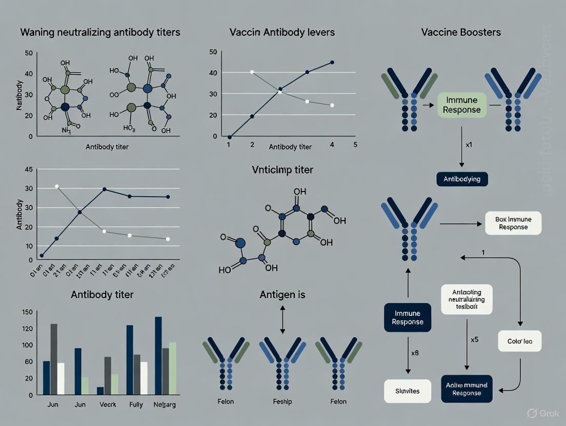
Strategies to Counteract Waning Neutralizing Antibody Titers in COVID-19 Vaccine Boosters
This article synthesizes current research on the durability of neutralizing antibody (nAb) responses elicited by COVID-19 vaccine boosters.
Strategies to Counteract Waning Neutralizing Antibody Titers in COVID-19 Vaccine Boosters
Abstract
This article synthesizes current research on the durability of neutralizing antibody (nAb) responses elicited by COVID-19 vaccine boosters. It explores the foundational mechanisms of antibody waning, methodological approaches for measuring nAb kinetics, and strategies for optimizing booster timing and formulation. By comparing vaccine platforms and validating nAbs as correlates of protection, this review provides a comprehensive framework for researchers and drug development professionals to design next-generation vaccination strategies that sustain robust immunity against evolving SARS-CoV-2 variants.
Understanding the Dynamics of Neutralizing Antibody Waning
Frequently Asked Questions
Q1: What are the typical kinetic patterns of antibody decay after vaccination or infection? Antibody decay typically follows a biphasic pattern: a sharp initial decline immediately after the peak response, followed by a much slower, gradual long-term waning. This pattern is consistent across different antibody isotypes (IgG, IgA, IgM) and vaccination types, though the exact rates differ. For instance, IgG antibodies decay slowly, while IgA shows an initial sharp decline that gradually slows and stabilizes above the seropositivity threshold. IgM drops most rapidly, often to undetectable levels after primary vaccination [1].
Q2: How does disease severity affect neutralizing antibody kinetics? Disease severity significantly impacts both the peak level and decay rate of neutralizing antibodies. Hospitalized individuals with severe COVID-19 develop approximately 10-fold higher peak neutralizing titers compared to those with mild or asymptomatic infections [2]. The decay pattern also differs: severe disease often shows a two-phase decay with rapid initial decline slowing after approximately 80 days, while mild/asymptomatic infections show a nearly flat, stable antibody level over time [2] [3].
Q3: What is the evidence for differential antibody waning between vaccine platforms? Research demonstrates that the Moderna mRNA-1273 booster induces a more durable antibody response compared to the Pfizer/BioNTech BNT162b2 booster, which shows more rapid waning. However, individuals receiving the Pfizer vaccine exhibited a steeper rebound in antibody levels following breakthrough infection [4]. Booster vaccinations consistently reduce the decay rate of antibody levels, helping maintain protective titers for longer periods [5].
Q4: How do antibody responses differ between natural infection and vaccination? Infection leads to the highest antibody peak and slowest decay rate compared to vaccination alone. However, hybrid immunity (infection plus vaccination) provides the most robust and durable response. Three vaccine doses induce higher and more long-lasting anti-spike IgG and IgA levels compared to two doses [1]. For individuals with hybrid immunity, the first vaccine dose after infection produces a robust increase in antibodies, though subsequent doses provide minimal additional boosting [6].
Troubleshooting Guide
Issue: High Variability in Antibody Measurements
Problem: Significant inter-individual variation in antibody levels and decay rates complicates data interpretation and generalization.
Solutions:
- Implement Longitudinal Sampling: Collect samples at consistent time points (e.g., days 0, 28, 180, 360 post-vaccination) to establish individual kinetic profiles [4]
- Use Mixed-Effects Models: Account for both fixed effects (vaccine type, severity) and random individual variations in your statistical analysis [4] [2]
- Standardize Assays: Use consistent assay methodologies (e.g., PRNT for neutralization, standardized ELISA for binding antibodies) across all measurements [3]
Issue: Discrepancies Between Binding and Neutralizing Antibody Decay
Problem: Binding antibody titers (anti-RBD, S2, NP) may show constant decline while neutralizing activity remains stable.
Solutions:
- Measure Multiple Parameters: Track both binding antibodies (IgG, IgA, IgM) and functional neutralizing antibodies in parallel [1] [2]
- Extended Follow-up: Continue monitoring beyond 6 months to capture the complete kinetic profile, as neutralizing antibodies may show delayed stabilization [2]
- Account for Epitope Diversity: Utilize methods that characterize antibody quality and epitope specificity, not just quantity [7]
Issue: Determining Correlates of Protection Against Infection
Problem: Establishing clear thresholds of antibody levels that predict protection against breakthrough infection.
Solutions:
- Incorporate Time-Varying Covariates: Use survival analysis with antibody levels as time-dependent predictors rather than single timepoint measurements [4]
- Establish Functional Thresholds: Based on high attack-rate settings, consider a neutralizing titer of 1:250 as a potential protective cutoff [2]
- Model Protection Durations: Estimate the antibody level needed to sustain specific protection levels (e.g., 20% binding BA.1 IgA for 80% protection over 155 days) [4]
Table 1: Neutralizing Antibody Decay Kinetics by Disease Severity
| Disease Severity | Peak PRNT50 Titer (Estimated) | Early Half-Life (Days) | Late Half-Life (Days) | Time to Detection Limit (Days) |
|---|---|---|---|---|
| Severe | High | 31 [2] | 753 [2] | 372-416 [3] |
| Mild | Moderate | - | - | 416 [3] |
| Asymptomatic | Low | Stable (no significant decay) [2] | - | 133 [3] |
Table 2: Antibody Kinetics After mRNA Booster Vaccination
| Parameter | Pfizer/BNT162b2 | Moderna/mRNA-1273 | Notes |
|---|---|---|---|
| Initial Decay Rate | Faster [4] | Slower [4] | Post-booster (third dose) |
| Post-Infection Response | Steeper rebound [4] | More moderate increase [4] | After breakthrough infection |
| Durability of BA.1 IgA | Shorter [4] | Longer [4] | Associated with reduced infection risk |
Table 3: Comparison of Antibody Isotype Decay Patterns
| Isotype | Decay Pattern | Key Characteristics |
|---|---|---|
| IgG | Slow decay [1] | Most stable isotype; higher levels after 3 doses vs. 2 [1] |
| IgA | Biphasic: sharp initial decline, then stabilization [1] | Important for mucosal immunity; associated with reduced infection risk at day 28 post-booster [4] |
| IgM | Rapid decline to undetectable [1] | Transient initial response |
Experimental Protocols
Protocol 1: Longitudinal Antibody Kinetics Study
Purpose: To map the kinetics of binding and neutralizing antibodies over time following vaccination or infection.
Materials:
- Serum samples collected at defined intervals (e.g., day 0, 28, 180, 360)
- ELISA kits for anti-spike IgG, IgA, and IgM
- Plaque reduction neutralization test (PRNT) or pseudovirus neutralization assay
- -80°C freezer for sample storage
Methodology:
- Collect blood samples at predetermined timepoints [4]
- Process serum and store at -80°C until analysis
- Measure binding antibodies using standardized ELISA against relevant antigens (e.g., WT spike, Omicron BA.1 spike) [4]
- Determine neutralizing antibody titers using PRNT50/PRNT90 or pseudovirus neutralization assays [2] [3]
- Analyze data using nonlinear mixed-effects models to estimate decay rates [4]
Data Analysis:
- Fit biphasic decay models when appropriate (e.g., rapid initial phase followed by slow phase)
- Estimate half-lives for different periods using mixed-effects models
- Calculate hazard ratios for infection risk using time-varying antibody levels in survival analysis [4]
Protocol 2: Hybrid Immunity Response Characterization
Purpose: To evaluate antibody responses in individuals with both natural infection and vaccination.
Materials:
- Serum from cohorts with: natural infection only, vaccination only, and hybrid immunity
- Variant-specific neutralization assays (WT, Delta, Omicron subvariants)
- T cell activation assays (AIM, Fluorospot) [1]
Methodology:
- Stratify participants based on infection and vaccination history [6]
- Measure cross-reactive neutralizing antibodies against multiple variants [6]
- Characterize T cell responses using activation-induced marker (AIM) and IFN-γ/IL-2 Fluorospot assays [1]
- Analyze association between T cell polyfunctionality and subsequent antibody responses [1]
Visualizations
Antibody Decay Kinetics Diagram
Experimental Workflow for Kinetic Studies
The Scientist's Toolkit
Research Reagent Solutions
| Reagent/Assay | Function | Application Notes |
|---|---|---|
| Plaque Reduction Neutralization Test (PRNT) | Gold standard for measuring neutralizing antibody titers [3] | Requires BSL-3 facilities; measures PRNT50/PRNT90 |
| Pseudovirus Neutralization Assay | Safe alternative to live virus neutralization testing [2] | Can be performed at BSL-2; good correlation with PRNT |
| ELISA for Spike RBD/S1/S2 | Quantifies binding antibody levels [4] [5] | Standardized units (BAU) enable cross-study comparisons |
| Elecsys anti-N Assay | Confirms previous infection in vaccinated cohorts [4] | Distinguishes infection-induced from vaccine-induced immunity |
| Activation-Induced Marker (AIM) Assay | Measures antigen-specific T cell responses [1] | Correlates T cell help with antibody durability |
| IFN-γ/IL-2 Fluorospot | Characterizes T cell polyfunctionality [1] | Identifies correlates of sustained antibody responses |
Frequently Asked Questions (FAQs)
Q1: How does a prior SARS-CoV-2 infection alter the antibody response to a vaccine booster? A prior SARS-CoV-2 infection significantly enhances the magnitude and durability of the antibody response following a booster vaccination, a phenomenon known as "hybrid immunity." Individuals with hybrid immunity demonstrate higher baseline antibody levels and a much slower decay rate compared to those who are infection-naïve [8] [9] [10]. One study found that antibody levels in people with hybrid immunity remained above a 75% correlate of protection for 283 days after an immune-boosting event, which is substantially longer than in individuals who only received vaccines [8].
Q2: What is the impact of age on post-booster antibody kinetics? Increasing age is consistently associated with lower baseline antibody levels following vaccination [11] [9]. This is attributed to immunosenescence, the age-related decline in immune system function, which includes reduced production of naïve T cells and a decreased ability of lymphocytes to proliferate [12]. While the initial peak response may be lower, one large study noted that the rate of antibody decline over time was not significantly influenced by age in most models, except for a very slight increase in the decay rate for those aged 80 and above [9].
Q3: Which vaccine platforms elicit the most durable neutralizing antibody response? Research indicates that the platform affects both the magnitude and persistence of the response. mRNA vaccines (BNT162b2 and mRNA-1273) show a robust response, with Moderna's mRNA-1273 potentially demonstrating slower waning in some studies [4]. For individuals primed with inactivated vaccines, a heterologous boost with an adenovirus-vectored vaccine (like Convidecia) has been shown to produce a more robust and durable neutralizing antibody response against variants, including Omicron, compared to a homologous inactivated vaccine booster [13].
Q4: How quickly do neutralizing antibodies wane after a booster dose? Antibody decay is most rapid in the first few months post-vaccination. Studies report a sharp decline in neutralizing antibodies within the first 90 days after a booster, with the decay rate slowing thereafter [13]. The half-life ((t_{1/2})) of antibodies after a two-dose mRNA primary series is approximately 59.8 days, which improves to 99.7 days following a third (booster) dose. Hybrid immunity results in the most durable response, with a half-life of approximately 241 days [8].
Q5: Do gender and lifestyle factors influence the antibody response to boosters? Yes. Females often generate a higher peak antibody response than males, which may be influenced by hormonal differences that affect immune cell function [11]. However, one study suggested that while women start with higher titers, they may also experience a faster rate of decay [11]. Lifestyle factors such as smoking and having an immunosuppressive status are linked to lower baseline post-vaccination antibody levels [9].
Troubleshooting Guides
Issue: Low Neutralizing Antibody Titers Post-Booster in an Older Adult Cohort
- Potential Cause: Immunosenescence, characterized by a shift from lymphoid to myeloid progenitors, thymic atrophy, and telomere erosion in repeatedly stimulated lymphocytes, leading to reduced proliferative capacity [12].
- Recommended Action:
- Consider a heterologous booster regimen, as switching platforms may elicit a stronger immune response [13].
- Evaluate the adjuvant system; novel adjuvants like TLR agonists (e.g., flagellin) may help overcome the high activation threshold in older adults [12].
- Ensure administration of the recommended number of doses; for older adults, a two-dose booster series is often recommended to achieve adequate protection [14].
Issue: Rapid Waning of Protection in Infection-Naïve Individuals
- Potential Cause: The absence of the amplified and broadened immune memory provided by hybrid immunity, leading to a faster decline from peak antibody levels [8] [10].
- Recommended Action:
- Optimize the booster schedule. Data suggests antibody levels in infection-naïve individuals can fall below a protective threshold within 6-8 months; therefore, booster intervals should be planned accordingly [8] [9].
- Monitor antibody kinetics longitudinally to establish a personalized vaccination timeline rather than relying on a fixed schedule.
Issue: Poor Cross-Neutralization Against Variants of Concern (e.g., Omicron)
- Potential Cause: Mutations in the viral spike protein's receptor-binding domain (RBD), which allow escape from neutralizing antibodies induced by the original (wild-type) vaccine strain [15] [13].
- Recommended Action:
- Use updated booster vaccines that match the circulating variants, as these are designed to elicit a more specific antibody response [14].
- Employ a heterologous boosting strategy. Studies have shown that a viral vector booster after a primary series of inactivated vaccines can induce higher and broader neutralizing antibodies against variants compared to homologous regimens [13].
Data Presentation: Antibody Kinetics and Influencing Factors
Table 1: Impact of Key Factors on Post-Booster Antibody Response
| Factor | Impact on Baseline Antibody Level | Impact on Decay Rate / Durability | Key Quantitative Findings |
|---|---|---|---|
| Prior Infection (Hybrid Immunity) | Significant Increase [9] [10] | Slower Decay / Greater Durability [8] [9] | Half-life ((t_{1/2})): 241 days; maintains >75% protection for 283 days [8] |
| Age | Decrease with advancing age [11] [9] | Slightly Faster Decay in Advanced Age [9] | Antibody levels in ≥60 age group can be ~50% lower than in <30 group post-first dose [11] |
| Vaccine Platform (Booster) | Varies by platform and regimen [13] [4] | Varies by platform and regimen [13] [4] | Heterologous adenovirus vector booster after inactivated prime showed higher durable GMTs than homologous [13] |
| Number of Booster Doses | Increase with additional doses [9] | Slower Decay with additional doses [9] | Each additional dose after the 3rd associated with higher baseline and more gradual decline [9] |
| Female Gender | Increase [11] [9] | Inconclusive (Varies by Study) [11] [9] | Post-2nd dose IgG levels can be ~30% higher in females (e.g., 1252 vs 959.6 AU/mL) [11] |
Table 2: Antibody Half-Life Across Different Immune States
| Immune State | Antibody Half-Life ((t_{1/2}), Days) | Notes |
|---|---|---|
| Primary mRNA Series (2 Doses) | 59.8 [8] | Rapid waning, especially in the first 3 months. |
| First Booster Dose (3rd mRNA) | 99.7 [8] | Significantly improved durability over primary series. |
| Hybrid Immunity | 241.8 [8] | Combined effect of infection and vaccination provides the most durable antibody response. |
| Multiple Boosters (4th/5th dose) | No significant change from first booster [8] | Increases magnitude but not necessarily the intrinsic decay rate after the first booster. |
Experimental Protocols
Protocol 1: Multiplexed Bead Array for Neutralization Assay (nC19BA) This protocol provides a high-throughput, cell-free method for simultaneously measuring the neutralizing capacity of antibodies against the RBD of multiple SARS-CoV-2 variants [15].
- Bead Coating: Use a custom cytometric bead array (CBA). Conjugate streptavidin to the beads following the manufacturer's instructions. Incubate the streptavidin-coated beads with biotinylated RBD proteins from the SARS-CoV-2 variants of interest (e.g., Wuhan-1, B.1.351, B.1.1.529) at a concentration of 11 μg/mL. Block any remaining streptavidin sites with an excess of D-biotin [15].
- Sample Incubation: In a LoBind tube, mix ~5000-6000 antigen-coupled beads with 100 μL of diluted serum sample (e.g., 1:5 dilution in PBS) or a control antibody. Incubate for 30 minutes at 4°C, protected from light [15].
- Washing: Centrifuge the tubes at 1000 x g for 5 minutes and carefully remove the supernatant. Wash the bead pellets three times with PBS.
- ACE2 Binding: Resuspend the beads in 100 μL of a solution containing recombinant human ACE2-murine IgG1 Fc fusion protein (0.18 mg/mL). Incubate for 30 minutes at 4°C, protected from light [15].
- Detection: Wash the beads three times with PBS. Incubate with a fluorescently labelled anti-mouse IgG antibody (e.g., BV421-conjugated, 1:200 dilution) for 30 minutes at 4°C in the dark [15].
- Acquisition and Analysis: Perform a final wash, resuspend the beads in PBS, and acquire a minimum of 600 events per bead type on a flow cytometer (e.g., FACSymphony). Calculate the percentage of neutralization using the formula:
%neutralization = (1 - [gMFI_sample - gMFI_max_neutralization] / [gMFI_max_binding - gMFI_max_neutralization]) * 100[15].
Protocol 2: Live Virus Neutralization Assay This is a gold-standard method for quantifying neutralizing antibody titers using live, authentic SARS-CoV-2 virus, requiring a Biosafety Level 3 (BSL-3) laboratory [13].
- Sample Preparation: Collect venous blood and isolate serum. Heat-inactivate the serum at 56°C for 30 minutes to destroy complement activity [13].
- Serial Dilution: Perform two-fold serial dilutions of the inactivated serum in a 96-well microplate (e.g., from 1:2 to 1:1024), in duplicate.
- Virus Neutralization: Add an equal volume of live SARS-CoV-2 virus (100 TCID50, the dose that infects 50% of cultures) to each well containing the diluted serum. Incubate the plate at 37°C for 2 hours to allow antibody-virus interaction [13].
- Cell Infection: Transfer the serum-virus mixture onto a monolayer of Vero E6 cells in a new 96-well plate. Incubate the plate at 37°C for 5 days to allow for cytopathic effect (CPE) development [13].
- Titer Calculation: Observe the cells for CPE. The 50% neutralization titer (NT50) is the reciprocal of the highest serum dilution that protects 50% of the cells from the virus-induced CPE. A positive neutralization is typically defined as an NT50 ≥ 4 [13].
Live Virus Neutralization Assay Workflow
The Scientist's Toolkit: Research Reagent Solutions
Table 3: Essential Reagents for Neutralization & Antibody Assays
| Reagent / Material | Function / Application | Example Specifications |
|---|---|---|
| Biotinylated RBD Proteins | Antigen for coating beads in surrogate neutralization assays; represents the key viral domain for ACE2 receptor binding. | Variant-specific (e.g., Wuhan-1, Omicron BA.1); >95% purity; carrier-free [15]. |
| Recombinant hACE2 Protein | The viral receptor; used in competitive binding assays to measure the blocking activity of neutralizing antibodies. | Fc-tagged (e.g., murine IgG1) for detection; eukaryotic cell-expressed [15]. |
| Live SARS-CoV-2 Virus | Essential for gold-standard virus neutralization assays to measure functional, authentic neutralization. | Wild-type and Variants of Concern (Delta, Omicron); requires BSL-3 containment [13]. |
| Vero E6 Cells | African green monkey kidney epithelial cell line; highly permissive to SARS-CoV-2 infection, used for virus culture and neutralization assays. | ATCC CRL-1586; maintained in DMEM + 10% FBS [13]. |
| Anti-Spike IgG Assays | Automated, high-throughput quantification of anti-Spike or anti-RBD antibody levels for immunogenicity assessment. | e.g., Abbott SARS-CoV-2 IgG II Quant, Roche Elecsys anti-S [10]. |
| Functional Bead Array | Multiplexed, cell-free flow cytometry platform for simultaneous analysis of antibody neutralization against multiple variants. | e.g., Custom BD CBA beads; compatible with standard flow cytometers [15]. |
Surrogate Neutralization Assay Principle
Troubleshooting Guide: Neutralizing Antibody Assays
This guide addresses common challenges researchers face when evaluating neutralizing antibody (NAb) responses against evolving SARS-CoV-2 variants.
Table 1: Troubleshooting Common NAb Assay Challenges
| Problem | Possible Cause | Solution |
|---|---|---|
| Low NAb titers against newer variants (e.g., JN.1) | Antigenic shift and immune escape by the variant [16] [17] [18]. | Use updated pseudovirus neutralization assays with contemporary variant spikes [16] [19]. Include a convalescent serum control from a known variant outbreak for comparison [16]. |
| High variability in NAb measurements | Waning antibody levels over time since vaccination or infection [16] [19] [20]. | Document precise time since last immune exposure (vaccine/infection) for all samples. Use longitudinal sampling to model decay kinetics [20]. |
| Inconsistent results between assay types | Differing sensitivities of ELISA vs. pseudovirus-based assays [16]. | Standardize assays against an international standard serum. For variant comparisons, use a single, validated assay method (e.g., pseudovirus neutralization) across all samples [17]. |
| Unexpectedly low antibody breadth | Immune imprinting from prior exposure to ancestral or earlier variants [18]. | Stratify study participants based on complete vaccination and infection history. Consider prime-boost strategies with variant-adapted vaccines to broaden response [17] [18]. |
Frequently Asked Questions (FAQs)
Q1: What defines an "antigenic shift" in SARS-CoV-2, and how does Omicron exemplify this?
An antigenic shift is a major, abrupt change in a virus's surface antigens, leading to a virus that is significantly different from previously circulating strains. The Omicron variant, with over 30-50 mutations in its spike protein, represents a sharp antigenic divergence from predecessors like Wild-Type, Alpha, Beta, Gamma, and Delta [21]. This is not a gradual drift but a dramatic jump, resulting in substantial evasion of immunity induced by previous infection or vaccination.
Q2: Why do neutralizing antibody titers wane over time, and what is the typical decay rate?
NAb titers naturally decay after the peak immune response. This is a normal characteristic of the humoral immune system. Longitudinal studies show that in older adults (>65 years), NAb titers against variants like KP.3.1.1 can show a 2.5 to 4.6-fold reduction in geometric mean titer (GMT) over six months [19]. The decay rate can be influenced by factors like age, with older individuals often showing a more pronounced decline [16] [19], and initial immune response, where a higher peak response may lead to a slower decay [20].
Q3: What is the correlate of protection for NAbs against SARS-CoV-2 infection?
While a universal threshold is complex, studies have estimated a 50% protective titer. For the XBB variant, research has modeled the protective NAb titer (NT50) to be approximately 1:12.6 [18]. This means a serum dilution of 1:12.6 would neutralize 50% of the virus in vitro. Titers below this level are associated with a higher risk of breakthrough infection.
Q4: How does immune imprinting affect responses to new variants?
Immune imprinting describes how the immune system's first encounter with a virus (via infection or vaccination) shapes its future responses. For example, vaccination with the original (prototype) strain followed by a Delta infection results in immunity that remains heavily focused on the prototype strain [18]. This can limit the potency of the antibody response against antigenically distant variants like Omicron XBB and JN.1, as the immune system preferentially recalls and boosts antibodies against the original imprint.
Quantitative Data on Variant Evasion
Table 2: Neutralizing Antibody Titers Against Key SARS-CoV-2 Variants
Data derived from longitudinal cohort studies showing Geometric Mean Titer (GMT) against various variants, illustrating immune evasion [16] [19] [18].
| Variant | Relative GMT (Post-BA.5 Wave) | Key Immune Evasion Mutations | Notes |
|---|---|---|---|
| Wild-Type (WT) | Baseline (Reference) | N/A | Imprinted response often remains strongest against WT [18]. |
| BA.5 | ~2-4x lower than WT [18] | L452R, F486V, R493Q (reversion) | Dominant during the 2022-2023 wave in China [16]. |
| XBB.1.5 | Significantly lower than BA.5 | F486P, R346T, K444T | Exhibited pronounced immune escape, leading to resurgent waves [16] [18]. |
| EG.5 | Similar to XBB.1.5 [16] | F456L (additional) | Descendant of XBB; maintained immune evasion properties [16]. |
| JN.1 | ~50% lower than XBB.1.5/EG.5 [16] | L455S, R346T, F456L | The L455S mutation is a key driver of its enhanced antibody evasion, contributing to its global dominance [16]. |
Detailed Experimental Protocols
Protocol 1: Pseudovirus Neutralization Assay
Purpose: To quantify the potency of neutralizing antibodies in serum samples against specific SARS-CoV-2 variants.
Workflow Overview:
Materials:
- Serum Samples: Heat-inactivated at 56°C for 30 minutes.
- SARS-CoV-2 Pseudoviruses: Lentiviral or VSV-based particles expressing the spike protein of the target variant (e.g., BA.5, XBB.1.5, JN.1) and a luciferase reporter gene.
- Cell Line: HEK-293T cells stably expressing human ACE2 (hACE2).
- Cell Culture Media: DMEM with 10% FBS and 1% penicillin-streptomycin.
- White Walled 96-well Tissue Culture Plates
- Luciferase Assay System (e.g., Bright-Glo)
- Plate Luminometer
Procedure:
- Serum Dilution: Perform serial 3-fold dilutions of the serum samples in culture media across a 96-well plate.
- Virus-Serum Incubation: Add a standardized amount of pseudovirus (e.g., MOI of 0.1-0.5) to each serum dilution. Incubate the plate at 37°C for 1 hour to allow antibody-virus neutralization.
- Cell Inoculation: Add a suspension of hACE2-293T cells to each well. Incubate the plate at 37°C with 5% CO₂ for 48-72 hours.
- Luciferase Measurement: Following incubation, lyse the cells and add the luciferase substrate according to the manufacturer's instructions. Measure the luminescent signal using a luminometer.
- Data Analysis:
- Normalize luminescence readings:
(Signal from serum well - Signal from cell control) / (Signal from virus control - Signal from cell control) * 100%. - Plot the percentage neutralization against the serum dilution (log10).
- Calculate the NT50 (50% neutralization titer) using a 4-parameter logistic (4PL) regression model.
- Normalize luminescence readings:
Protocol 2: Longitudinal Analysis of NAb Kinetics
Purpose: To model the decay of neutralizing antibodies over time in a cohort of vaccinated and/or convalescent individuals.
Workflow Overview:
Procedure:
- Cohort Establishment: Recruit a cohort with well-documented immune histories. Key stratification factors include: number of vaccine doses, vaccine platform, known infection history, and time since last immune event.
- Serial Blood Collection: Collect serum samples from participants at predefined intervals (e.g., baseline, day 21 post-booster, and then monthly or quarterly for at least 6 months to a year) [16] [19].
- NAb Titer Measurement: Measure NAb titers against relevant variants for all time points using a consistent assay (see Protocol 1).
- Statistical Modeling:
- Linear Mixed Effects (LME) Model: Fit to the decay phase of the NAb response to estimate the average rate of decline. This model can account for between-subject variability [20].
- Nonlinear Mixed Effects (NLME) Model: Use a system of ordinary differential equations to model the entire NAb progression, from initial rise to peak and subsequent decay. This provides more nuanced parameters related to the immune response dynamics [20].
Mechanisms of Immune Evasion: A Visual Guide
This diagram illustrates how key mutations in the spike protein's Receptor-Binding Domain (RBD) contribute to immune evasion by Omicron subvariants.
The Scientist's Toolkit: Research Reagent Solutions
Table 3: Essential Reagents for Studying SARS-CoV-2 Antigenic Shift
| Research Reagent | Function/Application in Research | Key Considerations |
|---|---|---|
| Pseudovirus Systems (Lentivirus, VSV) | Safe, BSL-2 level tool to measure NAbs against specific spike variants [16] [17]. | Ensure the pseudovirus is correctly pseudotyped with the full-length spike of the target variant. Batch-to-batch normalization is critical. |
| hACE2-Expressing Cell Lines (e.g., 293T-ACE2) | Essential host cells for pseudovirus and live virus neutralization assays. | Confirm stable, high-level ACE2 expression. Monitor for loss of expression over prolonged cell culture passages. |
| Reference Sera & International Standards | Calibrate assays across different labs and allow for data comparison [17]. | Source from reputable organizations (e.g., NIBSC, WHO). Include in every assay run as a quality control. |
| Recombinant Spike/RBD Proteins (Multiple Variants) | Used in ELISA to measure binding antibodies and for characterizing monoclonal antibodies. | Source proteins with correct prefusion conformation. Purity and lack of aggregation are key for reliable data. |
| Variants of Concern (VOC) Live Virus Isolates | Gold standard for authentic virus neutralization assays (requires BSL-3). | Necessary for final validation of findings from pseudovirus assays, as they capture all aspects of the viral life cycle. |
Measuring Antibody Kinetics and Predicting Protection
The development and evaluation of vaccine boosters critically depend on accurately measuring functional immune responses. As neutralizing antibody (nAb) titers wane over time, understanding their kinetics is fundamental to determining the timing and composition of subsequent booster doses [22] [23]. Neutralizing antibodies, which directly block viral infection of host cells, serve as a key correlate of protection (CoP) and are a primary metric for assessing vaccine efficacy in research settings [24] [25].
Two pivotal technologies for this purpose are the Microneutralization Test (MNT), a live virus-based assay considered a gold standard, and the Surrogate Virus Neutralization Test (sVNT), a cell-free, high-throughput alternative [26] [27]. The MNT measures the direct functional capacity of antibodies to prevent viral infection in a cell culture system, mimicking the in vivo interaction more comprehensively [28]. In contrast, the sVNT detects antibodies that competitively inhibit the binding of the viral receptor-binding domain (RBD) to its host cell receptor, such as ACE2 for SARS-CoV-2 [24] [29]. This article provides a technical deep-dive into these assays, offering detailed protocols, troubleshooting guidance, and their application in addressing the critical challenge of waning immunity.
Core Principles and Key Differentiators
Microneutralization Test (MNT) is a cell-based assay that uses live, replication-competent virus to measure the ability of serum antibodies to neutralize infection and subsequent viral replication [28] [25]. The readout is a direct measure of biological function—the inhibition of viral cytopathic effect (CPE) or a reduction in viral protein expression [28] [30].
Surrogate Virus Neutralization Test (sVNT) is a competitive immunoassay that does not require live virus or cell culture. It directly measures the ability of antibodies in a sample to block the interaction between the viral protein (like the SARS-CoV-2 RBD) and its cognate cellular receptor (like ACE2) [24] [29] [26].
Table: Comparison of MNT and sVNT Assay Characteristics
| Characteristic | Microneutralization Test (MNT) | Surrogate Virus Neutralization Test (sVNT) |
|---|---|---|
| Principle | Cell-based; inhibition of live virus infection | Cell-free; competitive inhibition of protein-receptor binding |
| Biosafety Level | BSL-3 (for SARS-CoV-2) or BSL-2 (for attenuated strains) [28] [26] | BSL-1 or BSL-2 [29] [26] |
| Throughput | Low to medium (96-well format) [28] | High (96-well ELISA format, automatable) [24] [29] |
| Turnaround Time | 2 to 4 days [29] [25] | ~1 to 2 hours [29] [27] |
| Technical Complexity | High (requires cell culture and virology expertise) | Low (standard ELISA technique) |
| Readout | Cytopathic effect (CPE), viral antigen staining, or HA assay [28] [30] | Colorimetric (OD) measurement [29] |
| Correlation with Protection | High, considered a functional gold standard [25] | Good correlation with MNT, but can be variant-dependent [30] [27] |
The following diagram illustrates the fundamental operational principles and procedural workflows for both MNT and sVNT assays.
Troubleshooting Guides and FAQs
Frequently Asked Questions
Q1: My sVNT shows high background signal. What could be the cause and how can I resolve it? High background in sVNT is often due to non-specific binding or inadequate washing. Ensure sufficient washing steps between incubations to remove unbound reagents. Consider optimizing the composition of your blocking buffer (e.g., using a universal protein blocker) and confirm that the concentration of your primary detection reagent (e.g., HRP-conjugated RBD) is not too high. Titrating the key reagents can help identify the optimal concentration that maximizes signal-to-noise ratio [27] [31].
Q2: Why is there a weak or absent neutralization signal in my MNT, even with known positive controls? A weak MNT signal can result from low viral infectivity titer or insensitive cell lines. First, re-titer your virus stock to ensure you are using the correct infectious dose (e.g., 100 TCID₅₀). Verify that your cell monolayer is healthy and at the correct confluency at the time of infection. Furthermore, ensure that serum samples are properly inactivated (e.g., via heat treatment at 56°C for 30 minutes) to remove non-specific inhibitors before the assay [28] [30].
Q3: How well do sVNT results correlate with the gold standard MNT, especially for new variants? The correlation between sVNT and MNT is generally good to moderate but can be variant-dependent. One study found a moderate correlation (r-values 0.35-0.79) between the two assays, with a higher correlation observed in sera from convalescent patients compared to vaccine recipients [30]. The sVNT might not fully capture the reduction in neutralization against new variants, as it primarily focuses on the RBD-ACE2 interaction and may miss neutralizing antibodies that target other epitopes or neutralization mechanisms present in the live virus MNT [30] [25]. For variant-specific research, using sVNT kits with updated RBD antigens is crucial.
Q4: The neutralizing antibody titers in my longitudinal study are waning rapidly. Is this expected? Yes, waning of nAb titers is a recognized phenomenon. Multiple studies have demonstrated that nAb levels peak shortly after vaccination or infection and then follow a biexponential decay, with a sharp initial drop followed by a stabilization to a lower plateau [22] [23]. The half-life of nAbs after the peak has been reported to range from approximately 29 to 60 days, depending on the vaccine platform and individual immune history [23]. This waning is a primary rationale for booster vaccinations.
Troubleshooting Common Experimental Issues
Table: Troubleshooting Guide for MNT and sVNT Assays
| Problem | Potential Causes | Solutions |
|---|---|---|
| High Background (sVNT) | Insufficient washing; non-specific binding; high reagent concentration. | Increase wash cycles and volume; optimize blocking buffer; titrate detection reagent [27] [31]. |
| Weak/Absent Signal (MNT) | Low virus titer; insensitive cells; over-confluent cell monolayer; serum cytotoxicity. | Re-titer virus stock; use susceptible cell lines (e.g., Vero E6); seed cells at optimal density; filter-sterilize serum [28] [25]. |
| Poor Assay Reproducibility | Inconsistent cell passage number; variable virus storage; inaccurate serum dilution. | Use low-passage cell aliquots; aliquot and titer virus stocks; use precision pipettes and fresh tips for serial dilutions. |
| Low Correlation Between MNT and sVNT | Antigenic differences (e.g., new variants); different antibody targets measured. | Use sVNT with variant-matched RBD antigens; interpret results in the context of the specific variant being studied [30]. |
| High Intra-Assay Variability | Inconsistent cell seeding; uneven reagent distribution; plate edge effects. | Seed cells carefully for uniform monolayers; ensure proper mixing during reagent addition; use inner wells or account for edge effects. |
Detailed Experimental Protocols
Protocol: Microneutralization Test (MNT) for Influenza or SARS-CoV-2
This protocol is adapted from established procedures for assessing nAbs against respiratory viruses like influenza and SARS-CoV-2 [28] [30] [25].
1. Pre-Assay Preparation:
- Cell Culture: Maintain appropriate susceptible cells (e.g., MDCK cells for influenza, Vero E6 for SARS-CoV-2) and seed in 96-well microplates to achieve a confluent monolayer at the time of assay.
- Virus Titering: Determine the titer of your live virus stock using the 50% tissue culture infectious dose (TCID₅₀) method to define the working challenge dose (typically 100 TCID₅₀) [28].
- Serum Inactivation: Heat-inactivate all serum samples at 56°C for 30 minutes to destroy non-specific inhibitors and complement [28].
2. Assay Procedure:
- Serum Dilution: Perform serial dilutions (e.g., two-fold) of the inactivated serum samples in a 96-well plate using cell culture medium.
- Virus Neutralization: Add an equal volume of the challenge virus (100 TCID₅₀) to each serum dilution. Include virus control (virus + medium) and cell control (medium only) wells. Incubate the virus-serum mixture at 37°C for 1-2 hours.
- Cell Infection: Remove growth medium from the cell plate and inoculate each well with the virus-serum mixture.
- Incubation and Development: Incubate the plate for the required period (e.g., 48-72 hours for SARS-CoV-2, time varies by virus) at 37°C with 5% CO₂.
- Readout:
- Cytopathic Effect (CPE): Observe the cell monolayer under a microscope for virus-induced CPE. The neutralization titer (MNT₅₀ or MNT₉₀) is the reciprocal of the highest serum dilution that protects 50% or 90% of the cell monolayer from CPE [28] [30].
- Hemagglutination (HA) Assay (for influenza): Transfer supernatant to a new plate and add red blood cells. The absence of hemagglutination indicates viral neutralization [28].
Protocol: Surrogate Virus Neutralization Test (sVNT) for SARS-CoV-2
This protocol is based on commercial sVNT kits and related research for detecting SARS-CoV-2 nAbs [29] [27].
1. Principle: The assay measures the sample's ability to inhibit the binding of horseradish peroxidase (HRP)-labeled RBD to human ACE2 receptor coated on a plate.
2. Procedure:
- Incubation: Mix the serum/plasma sample with HRP-conjugated RBD and incubate at 37°C for a short period (e.g., 30 minutes).
- Binding Reaction: Transfer the mixture to the ACE2-pre-coated 96-well plate and incubate further (~15-30 minutes at 37°C). Neutralizing antibodies in the sample will bind to HRP-RBD, preventing it from binding to the immobilized ACE2.
- Washing: Wash the plate thoroughly to remove all unbound HRP-RBD and sample components.
- Detection: Add a tetramethylbenzidine (TMB) substrate solution to the wells. Any HRP-RBD that is bound to ACE2 will catalyze the conversion of TMB to a blue product.
- Stop and Read: Add a stop solution (e.g., sulfuric acid) to terminate the reaction, changing the color to yellow. Measure the optical density (OD) at 450 nm.
- Calculation: Calculate the % neutralization using the formula:
% Neutralization = [1 - (OD Sample / OD Negative Control)] × 100%A higher % inhibition indicates a higher concentration of nAbs in the sample [29].
The Scientist's Toolkit: Essential Research Reagents
Successful execution of neutralization assays requires high-quality, well-characterized biological and chemical reagents. The table below lists key materials for setting up these experiments.
Table: Essential Research Reagents for MNT and sVNT
| Reagent / Material | Function / Application | Examples & Notes |
|---|---|---|
| Susceptible Cell Lines | Host for viral replication in MNT. | Vero E6 (SARS-CoV-2), MDCK (Influenza). Use low passage number for consistency [28] [25]. |
| Live Virus or Pseudovirus | Challenge agent for MNT/pVNT. | Live virus (MNT), or SARS-CoV-2 S-pseudotyped lentivirus (pVNT) for safer BSL-2 work [25]. |
| Virus Transport Medium | Diluent for serum and virus. | Cell culture medium (e.g., DMEM) supplemented with antibiotics and a low percentage of serum [28]. |
| Recombinant RBD and ACE2 Proteins | Core components for sVNT. | Purified RBD (often HRP- or biotin-labeled) and hACE2 for coating plates [29] [27]. |
| International Standard Serum | Assay calibration and harmonization. | WHO International Standard for anti-SARS-CoV-2 immunoglobulin enables results in standardized units (BAU/mL) [26]. |
| Detection System | Signal generation and measurement. | HRP-TMB system for sVNT ELISA; microscope for CPE or luciferase reagent for pseudovirus assays [29] [25]. |
Application in Vaccine Booster Research: Analyzing Waning Immunity
Neutralization assays are indispensable for investigating the kinetics of nAbs, a central theme in booster research. Key insights from recent studies include:
- Waning is Biphasic: nAb levels after vaccination or infection typically follow a biexponential decay, characterized by a sharp initial drop (half-life of 29-60 days) followed by a stabilization to a lower plateau [23]. This model helps predict the duration of protection and optimal timing for boosters.
- Variant Impact is Significant: Antigenically shifted variants (e.g., Omicron) can cause a more significant reduction in nAb titers than the waning observed over time against the homologous strain. One analysis found that variant mismatch explained more variability in nAb reduction than time-related waning alone [22].
- Booster Doses Restore Levels: Booster immunizations, whether through additional vaccine doses or breakthrough infections, consistently restore peak nAb levels, including against variants of concern. However, the waning pattern appears to recur after each antigen exposure, indicating a recurring need for immunization to maintain high nAb titers in vulnerable populations [25] [23].
The relationship between nAb titer levels and clinical protection is complex. While higher nAb titers strongly correlate with protection against symptomatic and mild infection, protection against severe disease and death often remains high (>75%) even when nAb titers fall to low or barely detectable levels [22]. This underscores the important role of cellular immunity in preventing severe outcomes and indicates that nAb titers alone may not be a sensitive enough CoP for all disease severities. The following diagram summarizes the kinetics of nAbs and the rationale for booster shots.
Frequently Asked Questions (FAQs)
Q1: Why do my mathematical models fail to capture the rapid waning of antibodies after day 90? Antibody decay is not linear; it often follows a biphasic pattern with an initial rapid decline followed by a slower decay rate or stabilization to a plateau. A single exponential decay model is often insufficient. Using a biexponential decay model or a Log-Log linear model can better fit the observed data, as these account for the sharp initial drop (with a half-life ranging from 29 to 60 days) and the subsequent stabilization phase [32] [13]. Ensure your model structure includes terms for both the initial boosting and the waning phases [4].
Q2: How can I account for the impact of different vaccine platforms on antibody longevity in my analysis? Different vaccine platforms elicit distinct antibody kinetic profiles. Your model should include vaccine type as a covariate. For instance, research shows that the Pfizer/BioNTech BNT162b2 booster led to more rapid waning compared to the Moderna mRNA-1273 booster [4]. Furthermore, heterologous boosting with an adenovirus vector vaccine (like Convidecia) has demonstrated a more robust and durable humoral response compared to homologous inactivated vaccine boosters [13].
Q3: What is the best way to correlate time-varying antibody levels with infection risk? Using antibody levels from a single timepoint (e.g., peak response) provides a limited snapshot. Instead, incorporate longitudinal antibody measurements as time-varying covariates in survival models (e.g., Cox proportional hazards models). This approach dynamically links the fluctuating antibody titer with the instantaneous risk of breakthrough infection, offering a more accurate assessment of protection and its duration [4].
Q4: Our cohort has mixed infection histories. How should we model antibody responses from breakthrough infections? Participants with breakthrough infections post-booster should be analyzed as a distinct group, and their antibody kinetics should include a separate boosting term in the model. Evidence suggests that individuals boosted with the Pfizer vaccine exhibited a steeper rebound in antibody levels after infection. Covariates like age (faster post-infection growth in the elderly) and sex (faster growth in females) can also be included to refine the model [4].
Q5: What is a critical threshold for predicting protection against infection? While thresholds are variant-dependent, one study on Omicron BA.1 suggested that to sustain 80% protection over 155 days post-booster at a medium case incidence, a binding BA.1 IgA response of at least 20% is needed [4]. High antibody levels for wild-type IgG and BA.1 IgA at day 28 post-booster have also been strongly associated with reduced infection risk [4].
Table 1: Antibody Waning Kinetics and Half-Lives from Cohort Studies
| Study / Cohort Description | Antibody Type / Specificity | Key Timepoints | Waning Characteristics | Reported Half-Life / Decay |
|---|---|---|---|---|
| PRIBIVAC Cohort (BNT162b2 or mRNA-1273 booster) [4] | Binding IgA & IgG (WT & Omicron BA.1) | D0, D28, D180, D360 | More rapid waning post-Pfizer vs. Moderna booster | Not specified |
| KING/KING-VAX Cohorts (Various immune states) [32] | Neutralizing Antibodies | Longitudinal sampling over months | Biexponential decay in all groups | 29 to 60 days (initial phase) |
| Fujian Cohort (Inactivated vaccine heterologous/homologous boost) [13] | Neutralizing Antibodies (Live Virus WT, Delta, Omicron) | D0, D14, D28, D90, D180 | Sharpest decline within first 90 days (81-92% reduction vs. D14) | Not specified |
Table 2: Correlates of Protection and Key Hazard Ratios
| Immune Correlate | Timepoint | Associated Outcome | Hazard Ratio (HR) or Finding | Source |
|---|---|---|---|---|
| High Wild-type IgG | Day 28 Post-booster | Reduced Breakthrough Infection Risk | HR: 0.47 (95% CI: 0.22, 0.98) | [4] |
| High Omicron BA.1 IgA | Day 28 Post-booster | Reduced Breakthrough Infection Risk | HR: 0.36 (95% CI: 0.17, 0.78) | [4] |
| Binding BA.1 IgA (≥20%) | To sustain 80% protection for 155 days | Protection against Infection | Required at medium community case incidence | [4] |
| Heterologous Convidecia Booster | Day 180 Post-booster | Higher Neutralizing Antibody Titers | Maintained higher GMTs against Delta & Omicron vs. other boosters | [13] |
Detailed Experimental Protocols
Protocol 1: Longitudinal Cohort Study Design for Antibody Kinetics
Objective: To map the kinetics of binding and neutralizing antibodies over 360 days post-booster vaccination and correlate them with breakthrough infection risk.
Key Materials & Reagents:
- Cohort: Infection-naive adults who have completed a primary vaccination series (e.g., 2-dose BNT162b2), recruited for a booster dose [4].
- Vaccines: Booster vaccines (e.g., BNT162b2, mRNA-1273, or inactivated/viral vector vaccines for heterologous regimens) [4] [13].
- Blood Collection: Tubes for serum/plasma separation (e.g., non-anticoagulant tubes for serum) [13].
- Assay Kits:
- Anti-Spike IgG/IgA: ELISA or multiplex immunoassays (e.g., against WT and Variant spike proteins) [4].
- Anti-Nucleocapsid (N) Antibody Assay: To confirm past infection (e.g., Elecsys anti-N assay) [4].
- Virus Neutralization Test: Live virus or pseudovirus neutralization assay (see Protocol 2) [32] [13].
Workflow:
- Baseline Blood Collection (D0): Collect blood on the day of booster vaccination, prior to administration [4].
- Booster Vaccination: Administer the designated booster vaccine.
- Follow-up Blood Collections: Collect blood at predetermined timepoints (e.g., D28, D90, D180, D360) [4] [13].
- Infection Monitoring: Actively monitor for breakthrough infections via:
- Self-reporting of positive PCR/ART tests.
- Seroconversion confirmed by anti-N antibody testing at each visit [4].
- Sample Processing & Storage:
- Allow blood to clot at room temperature.
- Centrifuge to separate serum/aliquot.
- Cryopreserve serum at -20°C or lower until testing [13].
- Antibody Titer Measurement: Perform binding (IgG/IgA) and neutralizing antibody assays on all serial samples.
Protocol 2: Pseudovirus-Based Neutralization Assay
Objective: To quantify the titer of neutralizing antibodies in plasma/sera against specific SARS-CoV-2 variants.
Key Materials & Reagents:
- Cells:
- Plasmids:
- Reagents: Transfection reagent (e.g., ExpiFectamine 293), cell culture media (DMEM, Expi293 expression medium), DEAE-Dextran, luciferase reagent (e.g., BriteLite Plus) [32].
Workflow:
- Pseudovirus Production:
- Serum/Plasma Preparation:
- Neutralization Reaction:
- Incubate an equal volume of pseudovirus (e.g., 200 TCID50) with the diluted serum/plasma for 1 hour at 37°C [32].
- Infection:
- Detection & Analysis:
The Scientist's Toolkit: Research Reagent Solutions
Table 3: Essential Reagents and Resources for Antibody Kinetics Studies
| Item / Resource | Function / Application | Specific Examples / Notes |
|---|---|---|
| Vaccine Platforms | Homologous/Heterologous Booster Regimens | BNT162b2 (Pfizer), mRNA-1273 (Moderna), inactivated vaccines (CoronaVac, BIBP), adenovirus-vectored (Convidecia) [4] [13]. |
| Anti-Spike IgG/IgA Assays | Quantify binding antibodies against vaccine-homologous and variant strains (e.g., WT, Omicron). | Multiplex immunoassays or ELISA; critical for defining correlates of protection [4]. |
| Anti-Nucleocapsid (N) Assay | Confirm past SARS-CoV-2 infection in study participants; distinguish immune response from vaccination vs. infection. | Elecsys Anti-N Assay [4]. |
| Pseudovirus Neutralization Assay | Safely measure neutralizing antibody titers against specific variants of concern without requiring BSL-3 containment. | HIV-based luciferase-expressing pseudovirus with variant Spike proteins; ID50 calculated as output [32]. |
| Live Virus Neutralization Assay | Gold-standard measurement of neutralizing capacity in a BSL-3 laboratory. | Based on reduction of cytopathic effect (CPE) in Vero E6 cells; NT50 calculated as output [13]. |
| Nonlinear Mixed-Effects Modeling Software | Model complex antibody waning kinetics and account for inter-individual variability. | NONMEM, Monolix, or R/phoenix NLME [4]. |
Modeling Antibody Kinetics: A Conceptual Workflow
Linking Antibody Titers to Breakthrough Infection Risk via Survival Analysis
Frequently Asked Questions
1. What is the relationship between antibody titers and the risk of SARS-CoV-2 breakthrough infection? Higher antibody levels following vaccination are associated with a reduced risk of breakthrough infection. Survival analysis studies have quantified this relationship, showing that individuals with high antibody titers 28 days post-booster have a significantly lower hazard of infection. For instance, high levels of Wild-Type IgG and Omicron BA.1 IgA at day 28 are associated with hazard ratios of 0.47 and 0.36, respectively, compared to low levels [4]. However, antibody levels wane over time, leading to increased infection risk several months after vaccination [33] [34].
2. Which risk factors, beyond antibody titers, influence breakthrough infection risk? Several patient-specific factors influence susceptibility to breakthrough infections. Immunosuppression is a consistently significant risk factor, with one study reporting an adjusted odds ratio of 2.04 [33]. Other high-risk conditions include primary immunodeficiency, organ transplant history, active tumors, and Alzheimer's disease [35]. Demographic factors such as vaccine brand (lower risk with Moderna vs. Pfizer) [35] and time since last vaccination (increased risk after 6 months) [33] also play important roles.
3. What is the appropriate study design for investigating the antibody titer-infection relationship? The test-negative case-control design is a robust approach, where cases are vaccinated individuals with positive SARS-CoV-2 tests and controls are vaccinated individuals with negative tests, matched by factors like age and test date [33]. Longitudinal cohort studies with repeated antibody measurements over time (e.g., at days 0, 28, 180, and 360 post-booster) are also valuable for modeling antibody kinetics and performing time-varying survival analysis [4].
4. How should antibody data be incorporated into survival analysis models? Using antibody levels as time-varying covariates in survival models provides better fit than using single baseline measurements [4]. This approach captures the dynamic nature of immune protection. Nonlinear mixed-effects models can first map individual antibody kinetics, and the resulting predicted trajectories are then incorporated as time-dependent predictors in Cox proportional hazards models to assess infection risk [4].
5. What are common technical challenges in antibody quantification for these studies? Key challenges include distinguishing between total antibody concentration and functional titer, ensuring assay specificity for the antigen of interest (e.g., using irrelevant carrier proteins to discriminate between hapten-specific and carrier-specific antibodies), and selecting appropriate quantification methods (e.g., ELISA, spectrophotometry) based on the required sensitivity and sample purity [36] [37].
Troubleshooting Guides
Issue 1: Inconsistent Association Between Antibody Titers and Infection Risk
Problem: Expected protective relationship between high antibody titers and reduced infection risk is not observed in survival analysis.
Solution:
- Verify antibody specificity: Ensure your assay specifically targets relevant antigens (e.g., spike protein vs. nucleocapsid) and variants (e.g., Omicron-specific rather than just Wild-Type) [4] [34]. Use appropriate negative controls.
- Check timing of measurements: Antibody levels wane rapidly in the first 90 days post-vaccination [38]. Ensure measurements are appropriately timed relative to vaccination and infection events.
- Account for confounders: Adjust for known risk factors in your model, such as immunosuppression, vaccine brand, age, and time since last vaccination [33] [35].
Issue 2: High Rate of Breakthrough Infections in Study Cohort
Problem: Unexpectedly high proportion of vaccinated participants experiencing infection during follow-up.
Solution:
- Verify infection definitions: Use standardized criteria including positive PCR tests, rapid antigen tests, and serological evidence of infection (e.g., anti-nucleocapsid antibodies) [4] [34].
- Stratify by timing: Distinguish between early infections (associated with low peak antibody response) and late infections (associated with antibody waning) [34].
- Monitor variants: Account for circulating variants in your analysis, as some variants (e.g., Omicron) have higher immune evasion potential [4] [34].
Issue 3: Poor Model Fit in Survival Analysis
Problem: Statistical models examining antibody-infection relationship show poor fit or non-proportional hazards.
Solution:
- Use time-varying covariates: Incorporate antibody measurements as time-dependent variables rather than static baseline values [4].
- Consider nonlinear kinetics: Model antibody waning using nonlinear mixed-effects models that account for rapid initial decline followed by slower decay [4] [38].
- Check for interactions: Test for effect modification by factors such as age, vaccine type, and immune status [35] [34].
Experimental Protocols
Protocol 1: Longitudinal Antibody Kinetics and Survival Analysis
Purpose: To model the relationship between time-varying antibody levels and breakthrough infection risk.
Materials:
- Serum samples from vaccinated participants
- ELISA kits for SARS-CoV-2 IgG/IgA quantification
- PCR tests for SARS-CoV-2 infection detection
- Statistical software (R, SAS, or Python)
Procedure:
- Study Design: Recruit vaccinated participants and collect blood samples at predetermined intervals (e.g., days 0, 28, 180, 360 post-booster) [4].
- Infection Monitoring: Actively monitor participants for SARS-CoV-2 infections through self-reported positive tests (PCR or antigen) and confirm with anti-nucleocapsid antibody testing [4].
- Antibody Quantification: Measure binding IgG and IgA antibodies against relevant SARS-CoV-2 variants (e.g., Wild-Type and Omicron BA.1 spike proteins) using standardized ELISA [4].
- Kinetic Modeling: Fit nonlinear mixed-effects models to the longitudinal antibody data to estimate individual-level trajectories [4].
- Survival Analysis: Perform time-to-event analysis with antibody levels as time-varying covariates, adjusting for potential confounders [4].
Protocol 2: Test-Negative Case-Control Study
Purpose: To identify risk factors for breakthrough infections while controlling for healthcare-seeking behavior.
Materials:
- Electronic health records or vaccine registry data
- SARS-CoV-2 testing data
- Covariate data (demographics, comorbidities, vaccine details)
Procedure:
- Case Definition: Identify vaccinated individuals with positive SARS-CoV-2 tests (cases) [33].
- Control Selection: Select vaccinated individuals with negative SARS-CoV-2 tests, matched to cases by age and test date (test-negative design) [33].
- Data Extraction: Extract information on vaccine brand, time since vaccination, demographics, and comorbidities [33] [35].
- Statistical Analysis: Use logistic regression to assess associations between risk factors and breakthrough infection, calculating adjusted odds ratios [33].
Table 1: Hazard Ratios for Breakthrough Infection Associated with Antibody Levels
| Antibody Type | Variant | Timepoint | Hazard Ratio | 95% CI | Reference |
|---|---|---|---|---|---|
| IgG | Wild-Type | Day 28 | 0.47 | [0.22, 0.98] | [4] |
| IgA | BA.1 | Day 28 | 0.36 | [0.17, 0.78] | [4] |
Table 2: Risk Factors for Breakthrough SARS-CoV-2 Infection
| Risk Factor | Effect Size | 95% CI | Effect Measure | Reference |
|---|---|---|---|---|
| Time since last vaccine >6 months | 1.64 | [1.42, 1.88] | aOR | [33] |
| Immunosuppression | 2.04 | [1.58, 2.62] | aOR | [33] |
| Pfizer/BNT162b2 (vs. Moderna) | 1.66 | [1.17, 2.35] | IRR | [35] |
| Male sex | 1.47 | [1.11, 1.94] | IRR | [35] |
| Compromised immune system | 1.48 | [1.09, 2.00] | IRR | [35] |
Table 3: Antibody Waning Patterns Post-Vaccination
| Vaccine Platform | Timeframe | Reduction in nAb GMT | Variants Tested | Reference |
|---|---|---|---|---|
| Inactivated vaccines | 90 days post-booster | 81.24-92.34% | Wild-type, Delta, Omicron | [38] |
| Protein-based XBB vaccines | 6 months post-booster | 2.5-4.6-fold reduction | KP.3.1.1, XEC | [34] |
| Pfizer BNT162b2 | 6 months post-booster | More rapid waning vs. Moderna | Wild-Type, Omicron BA.1 | [4] |
Research Reagent Solutions
Table 4: Essential Reagents for Antibody and Breakthrough Infection Research
| Reagent/Category | Specific Examples | Function/Application | Reference |
|---|---|---|---|
| Antibody Detection Kits | ELISA kits for anti-spike IgG/IgA | Quantification of binding antibodies | [4] |
| Neutralization Assays | Live virus neutralization tests | Functional assessment of neutralizing antibodies | [38] [34] |
| Infection Confirmation | PCR tests, rapid antigen tests, Elecsys anti-N assays | Confirm SARS-CoV-2 infection and distinguish from vaccine response | [4] |
| Isotyping Kits | Commercial antibody isotyping kits (ELISA or membrane format) | Determine antibody class and subclass | [36] |
| Protein Assays | BCA Protein Assay Kit, Coomassie Plus Protein Assay Kit | Measure total antibody concentration | [36] [37] |
Survival Analysis Workflow
Survival Analysis Workflow for Antibody Protection
Key Methodological Details
Antibody Quantification Techniques
Accurate antibody measurement is fundamental to these studies. The enzyme-linked immunosorbent assay (ELISA) is widely used for quantifying antigen-specific antibodies, with different formats available including direct, sandwich, and competition ELISA [36] [37]. Spectrophotometry provides a rapid alternative for determining total antibody concentration via absorbance at 280 nm, though it doesn't distinguish functional from non-functional antibodies [37]. Critical considerations include:
- Distinguishing between antibody concentration (total amount of antibody protein) and titer (functional dilution for a specific assay) [36]
- Using appropriate standards (immunoglobulin standards rather than BSA) for accurate quantification [36]
- Implementing proper controls to distinguish hapten-specific from carrier-specific antibodies when working with conjugated antigens [36]
Statistical Considerations for Time-Varying Analysis
Incorporating antibody levels as time-dependent covariates requires special methodological approaches. The standard Cox proportional hazards model can be extended as follows:
h(t|X(t)) = h₀(t)exp(βX(t) + γZ)
where X(t) represents the time-varying antibody measurements and Z represents fixed covariates [4]. This approach better captures the dynamic relationship between waning immunity and infection risk compared to models using only baseline antibody measurements.
Mathematical Models for Predicting Durations of Protection Post-Booster
Frequently Asked Questions
1. What are the most common mathematical structures used to model post-booster antibody kinetics? The most common structures are compartmental models. A one-compartment model often fails to capture the observed biphasic waning pattern. A two-compartment model is generally preferred as it can accurately describe the initial rapid antibody decay followed by a slower, long-term waning phase, providing a better fit to real-world data [39]. For even more granularity, nonlinear mixed-effects models or Bayesian hierarchical models are used to estimate population-level trends while accounting for individual-level variations in antibody responses [40] [41].
2. Our model fits training data well but fails to predict individual antibody waning. How can we improve its accuracy? Individual variability is a key challenge. To improve accuracy, integrate patient-specific covariates into your model. Research shows that factors like age, sex, and comorbidities (e.g., autoimmune diseases, diabetes, immunosuppression) significantly influence antibody kinetics [40]. Using a Bayesian framework can be particularly effective. It allows you to incorporate prior knowledge and update model predictions for a new individual based on just a single antibody measurement, dramatically improving personalized trajectory forecasts [40].
3. How do I quantitatively account for different vaccine platforms in a single durability model? Model the peak antibody response and the waning rate as platform-dependent parameters. Studies using Bayesian linear regression have found that after primary immunization, peak nAb titers from inactivated and viral vector vaccines were 8.2-fold and 5.6-fold lower, respectively, than those from mRNA vaccines. Furthermore, waning rates can differ; one analysis showed nAb titers from heterologous primary series declined 3.4-fold every 90 days, compared to a 1.4-fold decline for viral vector vaccines in the same period [22].
4. How can we model the duration of protection against infection, not just antibody levels? To move from antibody kinetics to protection, you need to integrate your model with a correlates of protection (CoP) model. This involves two steps: first, use your kinetic model to predict antibody levels over time. Second, apply a pre-established relationship between antibody titer and vaccine effectiveness (VE) against a clinical endpoint like infection. This combined approach allows you to predict how VE declines over time and estimate the point at which protection falls below a desired threshold [41] [4].
5. Neutralizing antibodies wane, but protection against severe disease persists. How do models explain this? This observation suggests that neutralizing antibody titer is not the sole correlate of protection for severe disease. Mathematical models that correlate nAb titers with VE across disease severities show that while VE against mild infection drops significantly as nAbs wane, VE against severe and fatal outcomes often remains high (e.g., >75%) even when nAb titers become very low [22]. This implies that other immune components, such as T-cells and memory B-cells, which are not captured by simple antibody kinetic models, play a crucial role in long-term protection against severe illness.
Troubleshooting Guides
Problem: Model Cannot Capture Biphasic Antibody Waning
- Symptoms: Your model shows a constant exponential decay, but empirical data clearly indicates a rapid initial drop followed by a much slower long-term decline.
- Solution: Implement a two-compartment model instead of a one-compartment model.
- Protocol:
- Model Structure: Structure your model with a central compartment (representing short-lived plasma cells/antibodies) and a peripheral compartment (representing long-lived plasma cells/antibodies) [39].
- Differential Equations:
dA1/dt = k_in - (k12 + k_el) * A1 + k21 * A2dA2/dt = k12 * A1 - k21 * A2(WhereA1andA2are antibody amounts in the central and peripheral compartments,k_inis the zero-order production rate,k_elis the elimination rate, andk12/k21are transfer rates.)
- Parameter Estimation: Use nonlinear mixed-effects modeling software (e.g.,
monolix,NONMEM) or Bayesian frameworks (e.g.,Stanwithbrmsin R) to fit the model to longitudinal antibody data and estimate the rate constants [40] [39]. - Validation: Perform internal validation via k-fold cross-validation and external validation on a separate cohort to ensure predictive accuracy [39].
Problem: Model Predictions Are Inaccurate for New Viral Variants
- Symptoms: Your model, built on data from the ancestral virus, significantly overestimates protection against a new variant like Omicron.
- Solution: Explicitly incorporate antigenic distance into the model.
- Protocol:
- Data Collection: Gather geometric mean neutralizing antibody (nAb) titers against both the homologous (vaccine) strain and the heterologous (new variant) strain from published studies or in-house experiments [22].
- Variant Factor: Calculate a variant-specific fold-reduction factor. For example, models show that the antigenic shift to Omicron BA.1 explains a greater reduction in nAb titers than time-related waning alone [22].
- Model Integration: Adjust the predicted nAb titer against the new variant as:
nAb_variant(t) = nAb_homologous(t) / fold_reduction. - Update CoP: If available, use a variant-specific correlate of protection. If not, apply the homologous CoP curve to the adjusted
nAb_variant(t)with caution, acknowledging that the protective threshold might be higher for immune-evading variants [41] [22].
Problem: Estimating Protection Duration from Sparse Antibody Measurements
- Symptoms: You only have one or two antibody measurements per subject, making it impossible to fit a full kinetic model for each individual.
- Solution: Use a Bayesian hierarchical model to borrow strength from the population.
- Protocol:
- Build a Population Model: First, develop a robust nonlinear model (e.g.,
log(Abt) = a - b * log(t) - c * t) using a rich longitudinal dataset from a training cohort. Define priors for parameters (a, b, c) and hyperpriors for how covariates like age and sex affect them [40]. - Incorporate Sparse Data: For a new individual with a single measurement, use the population model as a prior. Update this prior (a process known as empirical Bayes estimation) with the individual's single data point.
- Generate Prediction: The model will produce a posterior distribution for the individual's full antibody trajectory, providing a personalized estimate of waning and the time until a protective threshold is crossed [40] [39].
- Build a Population Model: First, develop a robust nonlinear model (e.g.,
Quantitative Data on Antibody Waning and Protection
Table 1: Waning Rates of Neutralizing Antibodies After Primary Immunization by Vaccine Platform (over 90 days) [22]
| Vaccine Platform | Fold-Decline (95% CrI) |
|---|---|
| Heterologous Primary | 3.4 (2.5 to 4.7) |
| mRNA | 2.1 (1.8 to 2.4) |
| Inactivated | 1.7 (1.3 to 2.2) |
| Viral Vector | 1.4 (1.1 to 1.9) |
Table 2: Comparative Booster Antibody Kinetics and Protection [4]
| Parameter | Pfizer/BioNTech BNT162b2 | Moderna mRNA-1273 |
|---|---|---|
| Post-Booster Waning | More rapid | Less rapid |
| Post-Infection Rebound | Steeper | Less steep |
| Hazard Ratio (High vs. Low WT IgG at D28) | 0.47 (0.22, 0.98) | 0.47 (0.22, 0.98) |
| Hazard Ratio (High vs. Low BA.1 IgA at D28) | 0.36 (0.17, 0.78) | 0.36 (0.17, 0.78) |
| *Estimated Duration at 80% Protection | ~155 days (for BA.1 IgA ≥20%) | ~155 days (for BA.1 IgA ≥20%) |
| Note: *Estimated at a medium case incidence of 621 cases per million per day. |
Experimental Protocols
Protocol 1: Establishing a Two-Compartment Antibody Kinetic Model
Objective: To develop and validate a mathematical model that describes the biphasic waning of anti-SARS-CoV-2 antibodies after booster vaccination.
Materials:
- Longitudinal serum samples collected at predefined timepoints (e.g., pre-booster, day 28, day 90, day 180, day 360) [4].
- Validated ELISA or multiplex immunoassay for quantifying anti-spike IgG/IgA.
- Statistical software (R, Python) with nonlinear modeling capabilities (e.g.,
nlme,brms,rstan).
Methodology:
- Data Preparation: Log-transform all antibody measurements to normalize variance. Align all data to the date of booster vaccination.
- Model Specification: Code the two-compartment model differential equations into your software. The observed antibody level is typically derived from the central compartment.
- Parameter Estimation: Use a nonlinear mixed-effects approach. Model the system-level parameters (e.g.,
k_el,k12,k21) as fixed effects. Allow key parameters to have random effects to account for inter-individual variability. - Covariate Analysis: Test the influence of covariates (age, sex, vaccine product, comorbidities) on model parameters by adding them as fixed effects and using likelihood ratio tests or information criteria (e.g., WAIC) for selection.
- Model Validation:
- Internal: Perform 10-fold cross-validation on your dataset.
- External: Validate the final model on a completely independent cohort from a different institution [39].
Protocol 2: Integrating Antibody Kinetics with a Correlate of Protection
Objective: To predict the duration of protection against symptomatic infection post-booster.
Materials:
- Output from your antibody kinetic model (predicted titers over time).
- Published data or internal case-control study data linking antibody titers to vaccine effectiveness (VE).
Methodology:
- Define the Correlate: Obtain a logistic regression model that describes the relationship between antibody titer and VE. This is often of the form:
VE = 1 / (1 + exp(-k * (log10(titer) - log10(titer_50)))), wheretiter_50is the titer associated with 50% VE [41] [22]. - Generate Predictions: For a given individual or population, use your kinetic model to forecast antibody trajectories over time.
- Calculate VE Over Time: At each time point
t, input the predicted antibody titerT(t)into the logistic model from step 1 to compute the instantaneous VE(t). - Estimate Protection Duration: Define a protection threshold (e.g., the time until VE drops below 50% or 30%). The model will output the number of days post-booster this threshold is crossed [41] [4].
Model Structure and Workflow Visualizations
Two-Compartment Antibody Kinetics
From Antibody Data to Protection Estimate
The Scientist's Toolkit
Table 3: Essential Research Reagents and Resources
| Item | Function / Application |
|---|---|
| Plaque Reduction Neutralization Test (PRNT) | Gold-standard assay for measuring functional, live-virus neutralizing antibody titers [38] [22]. |
| Anti-Spike/RBD IgG & IgA ELISA | High-throughput quantification of binding antibodies for large longitudinal cohort studies [40] [4]. |
| Pseudovirus Neutralization Assay | Safe and versatile method for assessing nAb against high-consequence variants in BSL-2 labs [22]. |
| Bayesian Statistical Software (e.g., Stan with brms / rstanarm) | For fitting hierarchical kinetic models, handling sparse data, and generating posterior predictions with credible intervals [40] [41]. |
| Nonlinear Mixed-Effects Modeling Software (e.g., monolix, NONMEM) | Industry standard for pharmacokinetic/pharmacodynamic (PK/PD) modeling, directly applicable to antibody kinetics [39]. |
Optimizing Booster Formulations and Administration Strategies
Impact of Dosing Intervals on T-cell Immunity and Reactogenicity
Frequently Asked Questions
Q1: How does the interval between vaccine doses influence T-cell immunity and side effects? Extending the interval between the first and second dose of an mRNA COVID-19 vaccine (e.g., from 3 weeks to 3 months) reshapes the immune response. It reduces the early activation of "unconventional" T cells (like MAIT cells), which are linked to stronger early side effects (reactogenicity). Concurrently, this longer interval promotes the development of a superior long-lived protective "memory" T-cell response, making these T cells better equipped to respond to future booster vaccinations or infections [42].
Q2: Do T-cell responses continue to increase with additional booster doses? No, unlike antibody levels which can increase with each booster, spike-specific T-cell responses typically reach a plateau after the initial vaccination series and remain stable thereafter. Repeated booster doses do not significantly increase the magnitude of these T-cell responses but are crucial for maintaining their stability and long-term presence without signs of functional exhaustion [43] [44].
Q3: What is the role of unconventional T cells in vaccine responses? Unconventional T cells, such as Mucosa-Associated Invariant T (MAIT) cells, act as powerful initial "sensors" or "guard dogs" of the immune system. They are strongly activated within the first day after vaccination, amplifying the overall immune reaction. This powerful early signal is a key contributor to the side effects experienced soon after vaccination and can imprint a long-term impact on the quality of the immune memory generated [42].
Q4: How does the vaccine platform affect the early immune response? Different vaccine platforms trigger these early unconventional T-cell responses at different stages. The Oxford/AstraZeneca (ChAdOx1) vaccine triggers them very strongly after the first dose. In contrast, the Pfizer (BNT162b2) mRNA vaccine induces a stronger unconventional T-cell response after the booster dose [42].
Troubleshooting Guides
Issue: Suboptimal T-Cell Response After Booster
Potential Cause and Investigation:
- Cause: Insufficient interval between doses leading to abbreviated immune maturation.
- Investigation: Verify the timing between all vaccine doses. Evidence suggests that a longer interval (e.g., 3 months vs. 3 weeks) between the first and second dose of an mRNA vaccine is associated with a more robust and high-quality long-term T-cell memory response [42].
- Solution: For future vaccination schedules in research or clinical practice, adhere to or recommend extended dosing intervals where supported by evidence to optimize T-cell immunity.
Issue: High Reactogenicity Interfering with Study Protocols
Potential Cause and Investigation:
- Cause: Over-activation of early innate and unconventional T-cell responses.
- Investigation: Monitor and record participant symptoms closely in the first 24-48 hours. A shorter dosing interval (e.g., 3 weeks for mRNA vaccines) is linked to stronger early immune activation and higher reactogenicity [42].
- Solution: Consider extending the interval to the next dose to potentially mitigate the severity of side effects. The same immune mechanisms that cause reactogenicity also contribute to imprinting the long-term immune response, so balancing these factors is key [42].
Table 1: Impact of Dosing Interval on mRNA Vaccine Response
| Parameter | Short Interval (~3 weeks) | Long Interval (~3 months) |
|---|---|---|
| Early Unconventional T-cell Activation | Higher after booster dose | Reduced |
| Reactogenicity (Side Effects) | Increased | Reduced |
| Long-term T-cell Memory Quality | Standard | Enhanced (Better response to future boosters) |
Source: Adapted from [42]
Table 2: Antibody & T-cell Dynamics Across Multiple Doses
| Immune Parameter | Primary Series (Dose 1 & 2) | First Booster (Dose 3) | Subsequent Boosters (Dose 4+) |
|---|---|---|---|
| Neutralizing Antibody Peak | Lower | High | High, with slower decay post-dose 5 [45] |
| Antibody Waning Half-life | ~29-60 days [32] | ~3-4 months for S-specific antibodies [45] | Slower decay after doses 4 & 5 vs. dose 3 [45] |
| Antigen-specific B-cell Fold-increase | Baseline | 4.2 - 9.0 fold [44] | No notable increase vs. first booster [44] |
| Spike-specific T-cell Response | Establishes response | No significant fold-increase vs. primary series [44] | Stable, high level; no further significant increase [43] [44] |
Detailed Experimental Protocols
Protocol 1: Assessing T-cell Responses via Intracellular Cytokine Staining
This protocol details the method for quantifying SARS-CoV-2-specific T-lymphocytes by measuring cytokine production, as used in the Thai healthcare personnel study [44].
- PBMC Harvesting: Collect heparinized blood and isolate Peripheral Blood Mononuclear Cells (PBMCs) via density gradient centrifugation (e.g., Ficoll-Paque). Cryopreserve PBMCs in liquid nitrogen until batch testing.
- Cell Stimulation: Thaw and rest PBMCs overnight. Stimulate the cells with a Megapool of SARS-CoV-2 spike-specific CD4+ peptides.
- Intracellular Staining: Use brefeldin A to inhibit cytokine secretion. Stain cells with surface marker antibodies (e.g., anti-CD4, anti-CD8), then permeabilize and stain for intracellular cytokines (e.g., IFN-γ and TNF-α).
- Flow Cytometry: Acquire data using a multi-color flow cytometer (e.g., BD FACSCanto II).
- Data Analysis: Analyze using software such as FlowJo. Report results as the percentage of IFN-γ and/or TNF-α-producing CD4+ or CD8+ T-lymphocytes within their total population [44].
Protocol 2: Longitudinal Analysis of Neutralizing Antibody Kinetics
This protocol describes the methodology for modeling the decay kinetics of neutralizing antibodies, based on the longitudinal cohort study [32].
- Sample Collection: Collect longitudinal plasma/serum samples at multiple time points post-vaccination or post-infection.
- Neutralization Assay: Use a pseudovirus-based neutralization assay.
- Generate HIV-based reporter pseudoviruses expressing the desired SARS-CoV-2 spike protein (e.g., ancestral, Omicron subvariants).
- Incubate serial dilutions of heat-inactivated plasma samples with a fixed dose of pseudovirus.
- Add susceptible cells (e.g., HEK293T/hACE2) and incubate.
- Quantification: Measure luciferase activity (Relative Luminescence Units - RLUs) after 48 hours. Calculate the half-maximal inhibitory dilution (ID50), which is the reciprocal plasma dilution required for 50% viral neutralization.
- Kinetic Modeling: Model the decay of ID50 titers over time using appropriate statistical models. Studies often use:
- Log-Log linear mixed-effect models.
- Biexponential decay models (indicating an initial rapid decay phase followed by a slower decay phase or stabilization to a plateau) [32].
The Scientist's Toolkit
Research Reagent Solutions
| Item | Function/Application |
|---|---|
| HEK293T/hACE2 Cells | Target cell line for SARS-CoV-2 pseudovirus neutralization assays, expressing the human ACE2 receptor [32]. |
| SARS-CoV-2 Pseudoviruses | Replication-incompetent viral particles used to safely measure neutralizing antibody titers against specific variants (e.g., WH1, Omicron BA.2, XBB.1.5) [32]. |
| SARS-CoV-2 S1/RBD Proteins | Recombinant antigens used in B-cell ELISpot assays or ELISAs to detect and quantify antigen-specific B-cells and antibodies [44]. |
| SARS-CoV-2 Megapool Peptides | Pre-defined mixtures of peptides spanning viral proteins (e.g., spike) used to stimulate and detect a broad range of antigen-specific T-cells in intracellular cytokine staining assays [44]. |
| Anti-Human IFN-γ & TNF-α Antibodies | Key antibodies for intracellular cytokine staining to identify functional, antigen-specific T-cell responses by flow cytometry [44]. |
| PBMCs (Peripheral Blood Mononuclear Cells) | Primary human cells (including T and B lymphocytes) isolated from blood, used as the starting material for ex vivo cellular immune response assays [44]. |
Frequently Asked Questions (FAQs)
Q1: How does the durability of neutralizing antibodies (nAbs) compare across different vaccine platforms?
The durability of neutralizing antibody responses varies significantly by vaccine platform. Key comparative data is summarized in the table below.
Table 1: Durability and Kinetics of Neutralizing Antibody Responses by Vaccine Platform
| Vaccine Platform | Initial Peak nAb Titer | Antibody Decay Half-life (t₁/₂) | Key Durability Findings |
|---|---|---|---|
| mRNA (e.g., BNT162b2, mRNA-1273) | High [46] | Relatively rapid initial decay [46] [23] | nAbs show a significant decline over 6 months; mRNA-1273 demonstrates higher sustained titers than BNT162b2 [46]. |
| Adenovirus-Vectored (e.g., ChAdOx1-S, Ad26.COV2.S) | Lower than mRNA [47] [46] | More sustained or increasing [46] | Ad26.COV2.S nAbs can increase over 6 months, resulting in titers surpassing BNT162b2 at 6 months [46]. |
| Protein-Based (e.g., SCB-2019, Zifivax) | Variable | Slower waning post-peak [48] | Can exhibit more stable antibody persistence compared to some mRNA vaccines [48]. |
Q2: What is the impact of heterologous boosting (mix-and-match) on the breadth and durability of immunity?
Using a different platform for a booster dose than for the primary series is an effective strategy.
- Enhanced Breadth: Heterologous boosting can improve the breadth of the neutralizing antibody response against variants, though it may still be insufficient against highly immune-evasive strains [17].
- Platform Performance: A study comparing booster platforms in fully primed adults found that a heterologous booster with an adenovirus-vectored vaccine (Convidecia) after inactivated vaccines maintained higher and more durable levels of neutralizing antibodies against Delta and Omicron variants at 180 days compared to homologous or other heterologous protein boosts [49].
- Interchangeability: Real-world studies confirm that using protein, adenovirus-vector, or mRNA vaccines as boosters is generally safe and immunogenic, with similar fold-increases in antibody titers against ancestral and Omicron strains within each platform [48].
Q3: How do updated booster formulations (e.g., XBB.1.5, JN.1) perform against evolving variants?
Updated boosters are crucial for restoring protection, but their efficacy varies against newer variants.
- Performance Against Target Variants: XBB.1.5-specific mRNA boosters significantly increase the neutralization capacity against the XBB.1.5 and JN.1 subvariants [45].
- Limited Cross-Protection: While Omicron-specific boosters enhance neutralization of Omicron variants, they show limited efficacy against newer variants like JN.1, which exhibits the lowest neutralization across vaccine platforms [17].
- Durability in Older Adults: In adults over 65, protein-based XBB-containing boosters (Tri-XBB.1.5, Bi-Omi-XBB, Tetra-XBB.1) elicit nAbs that remain detectable in most participants after 6 months, despite a 2.5-4.6-fold reduction against newer variants like KP.3.1.1 [34].
Q4: What host factors influence the magnitude and durability of neutralizing antibody titers?
Several demographic and health factors predict an individual's nAb response, as shown in the table below.
Table 2: Host Factors Influencing Neutralizing Antibody Response Durability
| Host Factor | Impact on nAb Durability | Vaccine-Specific Notes |
|---|---|---|
| Age | Older age is generally associated with lower nAbs [46]. | Effect is significant for BNT162b2 and Ad26.COV2.S recipients, but not for mRNA-1273 [46]. |
| Sex | Women often show higher and more durable nAbs than men [46]. | This effect is observed irrespective of vaccine platform or time point [46]. |
| Body Mass Index (BMI) | Higher baseline BMI is associated with lower nAbs [46]. | This correlation is significant for Ad26.COV2.S recipients but not consistently for mRNA vaccines [46]. |
| Smoking Status | Non-smokers have significantly higher nAbs compared to smokers [46]. | This effect is observed across platforms [46]. |
Troubleshooting Guides
Issue: Rapid Waning of Neutralizing Antibody Titers in Preclinical Models
Potential Causes and Solutions:
- Cause: Suboptimal vaccine platform or regimen for the target antigen.
- Solution: Consider a heterologous prime-boost regimen. Data indicates that adenovirus-vectored boosters can induce more robust and durable nAb responses compared to homologous inactivated vaccine regimens [49].
- Cause: Insufficient antigen presentation or immune stimulation.
- Solution: Evaluate the inclusion of adjuvants, particularly for protein-based vaccines, to enhance the magnitude and longevity of the humoral response.
- Cause: Mismatch between the vaccine strain and circulating variant.
Issue: Low Neutralizing Antibody Titers Post-Vaccination in Specific Demographics
Potential Causes and Solutions:
- Cause: Advanced age, which is associated with immunosenescence.
- Cause: Host factors such as high BMI or smoking.
- Solution: Account for these variables during study design and data analysis, as they are independent predictors of lower nAb responses [46].
Issue: Inconsistent Neutralization Assay Results
Potential Causes and Solutions:
- Cause: Use of different neutralization assay types (e.g., MNT, PRNT, FRNT).
- Solution: Standardize assays within a study. The plaque reduction neutralization test (PRNT) is a gold standard that enables reliable cross-study comparisons [17]. Always include appropriate controls and reference sera.
- Cause: Testing against outdated viral variants.
- Solution: Regularly update the panel of viral variants used in assays to include recently circulating strains (e.g., JN.1, KP.3) to ensure relevance [34].
Experimental Protocols
Protocol: Live Virus Neutralization Assay to Measure nAb Titers
This protocol is adapted from studies assessing nAb durability against multiple variants [49].
Workflow Diagram:
Title: Live Virus Neutralization Assay Workflow
Materials:
- Research Reagent Solutions:
- Serum Samples: Heat-inactivated at 56°C for 30 minutes.
- Live SARS-CoV-2 Variants: Wild-type, Delta, Omicron, etc. (Handle in BSL-3 containment).
- Vero E6 Cells: Cultured in DMEM with 10% FBS.
- Cell Culture Plates: 96-well plates.
- Virus Diluent: Maintenance medium (e.g., DMEM with 2% FBS).
Procedure:
- Serum Dilution: Perform serial dilutions (e.g., two-fold from 1:2 to 1:1024) of heat-inactivated serum samples in duplicate across a 96-well plate.
- Virus Incubation: Add an equal volume of live virus suspension, normalized to 100 TCID50 (50% tissue culture infectious dose), to each serum dilution well. Include virus and cell controls. Incubate at 37°C for 1-2 hours.
- Cell Infection: Transfer the virus-serum mixture onto a confluent monolayer of Vero E6 cells in a new 96-well plate.
- Incubation and Observation: Incubate the plate at 37°C with 5% CO2 for 5 days. Observe daily for cytopathic effect (CPE).
- Titer Calculation: The nAb titer (NT50) is defined as the reciprocal of the highest serum dilution that protects 50% of the cells from CPE. Calculate using the Karber or Reed-Muench method.
Protocol: Pseudovirus-Based Neutralization Assay
This high-throughput assay is suitable for BSL-2 conditions and is widely used for head-to-head comparisons [46] [23].
Workflow Diagram:
Title: Pseudovirus Neutralization Assay Workflow
Materials:
- Research Reagent Solutions:
- SARS-CoV-2 Pseudovirus: HIV- or VSV-based particles expressing SARS-CoV-2 spike protein and a luciferase reporter gene.
- Target Cells: HEK293T cells overexpressing human ACE2 receptor (HEK293T/hACE2).
- Luciferase Assay Kit: e.g., BriteLite Plus reagent.
- Cell Culture Plates: 96-well white opaque plates for luminescence reading.
Procedure:
- Serum Dilution: Prepare three-fold serial dilutions of heat-inactivated serum samples in a 96-well plate.
- Neutralization: Incubate a fixed dose (e.g., 200 TCID50) of pseudovirus with the serum dilutions for 1 hour at 37°C.
- Infection: Add HEK293T/hACE2 cells to the virus-serum mixture and incubate for 48 hours.
- Luminescence Measurement: Lyse cells and add luciferase substrate. Measure relative luminescence units (RLUs) using a plate reader.
- Data Analysis: Normalize RLUs to control wells (0% and 100% neutralization). The half-maximal inhibitory dilution (ID50) is calculated using non-linear regression (e.g., in GraphPad Prism).
The Scientist's Toolkit: Research Reagent Solutions
Table 3: Essential Reagents for nAb Durability Research
| Research Reagent | Function / Application | Example from Literature |
|---|---|---|
| Live SARS-CoV-2 Variants | Gold standard for measuring nAb potency in vitro; requires BSL-3. | Used to test nAbs against wild-type, Delta AY.23, and Omicron BA.5.2 [49]. |
| Spike-Pseudotyped Viruses | Safe, high-throughput measurement of nAbs under BSL-2 conditions. | HIV-based luciferase reporter pseudoviruses used for head-to-head vaccine comparisons [46] [23]. |
| ACE2-Expressing Cell Lines | Essential target cells for infection in neutralization assays. | HEK293T/hACE2 cells are commonly used in pseudovirus assays [23]. |
| Reference Sera / Controls | Standardization and quality control across assays and batches. | The China National Institutes for Food and Drug Control provides reference serum for neutralization assays [49]. |
| Vero E6 Cells | Cell line highly permissive to SARS-CoV-2 infection, used in live virus assays. | Used for culturing live virus and scoring cytopathic effect (CPE) [49]. |
Geographic and Seasonal Timing for Maximum Protective Efficacy
FAQs: Optimizing Booster Timing and Addressing Waning Immunity
FAQ 1: How does geographic location influence the optimal timing for a COVID-19 booster vaccination?
Geographic location is a critical determinant for optimal booster timing due to localized seasonal infection patterns. Research analyzing spatiotemporal projections of COVID-19 incidence shows that the date offering the highest degree of protection can vary by several months between locations, typically falling in early autumn in the Northern Hemisphere [50].
For instance, the optimal booster date is September 15th for New York City, USA, and September 19th for South Korea. Boosting on these optimal dates can confer a 3- to 5-fold increase in protection against infection compared to the least-effective dates (e.g., late January) in these regions [50]. This location-specific seasonality must be incorporated into the design of vaccination campaigns and individual booster schedules to maximize the reduction in yearly probability of infection.
FAQ 2: What is the impact of a breakthrough infection on the optimal timing for a subsequent booster dose?
A breakthrough infection effectively renews immune protection, similar to a vaccine dose. Therefore, the optimal schedule for the next booster should be adjusted. Current guidance from the U.S. Centers for Disease Control and Prevention (CDC) suggests a delay of up to 3 months after a breakthrough infection [50].
Research leveraging longitudinal antibody and reinfection probabilities supports this guidance, indicating that an infection occurring late in the interval between scheduled boosts substantially alters the optimal boosting date. The immune renewal from the infection warrants delaying the subsequent booster to avoid administering it when antibody levels are already high, thereby extending the period of peak protection [50].
FAQ 3: How does waning humoral immunity affect vaccine effectiveness over time, and what is the observed "gradient" of protection?
Vaccine-induced humoral immunity, particularly neutralizing antibody (IgG) levels, wanes over time. This decline follows a distinct time-dependent gradient, where protection against infection and mild symptoms diminishes faster than protection against severe disease [51].
A national case-control study in Qatar demonstrated that while the effectiveness of two-dose BNT162b2 vaccination against asymptomatic and symptomatic infection declined to below 40% after 180-270 days, effectiveness against severe, critical, and fatal COVID-19 remained consistently high, surpassing 90% [51]. This gradient intensifies as time since the last vaccine dose increases. Booster vaccinations are crucial for restoring neutralizing antibody levels and reinforcing protection across this severity spectrum [52].
FAQ 4: What are the key host factors that predict the magnitude and longevity of COVID-19 vaccine immune responses?
Several host factors significantly influence vaccine effectiveness (VE) and the durability of humoral immune responses [52]:
- Age: Older adults experience accelerated antibody decline.
- Comorbidities: Conditions such as diabetes, cardiovascular disease, chronic kidney disease, and cancer reduce the magnitude and longevity of protection.
- Immunocompromised Status: Individuals with hematologic malignancies, those undergoing chemotherapy, or transplant recipients have diminished humoral responses.
- Hybrid Immunity: Individuals with a history of both vaccination and natural infection often exhibit a more durable and potent immune response.
- Pre-existing Immunity: The number of prior vaccine doses and infections shapes the subsequent immune response to a booster.
Immunocompromised individuals, in particular, derive significant benefit from booster vaccinations, with studies showing VE against hospitalization of 40% in this population [53].
Summarized Quantitative Data
Table 1: Location-Specific Optimal Booster Timing and Efficacy
Table summarizing the findings on the optimal timing for COVID-19 booster vaccination in different geographic locations and the associated protective benefit.
| Geographic Location | Optimal Booster Date | Relative Benefit vs. Least-Effective Date | Key Reference |
|---|---|---|---|
| New York City, USA | September 15 | 3.6-fold increase in protection | [50] |
| South Korea | September 19 | Nearly 5-fold increase in protection | [50] |
| Yamagata, Japan | Late September (slightly later than NYC/S. Korea) | Information Redacted | [50] |
| Northern Hemisphere (General) | Early Autumn | 3- to 4-fold range in protection by date | [50] |
Table 2: COVID-19 Vaccine Effectiveness (VE) Against Various Outcomes
Table summarizing interim VE estimates for the 2024-2025 COVID-19 vaccine and the waning effects observed in earlier studies.
| Vaccine / Study Period | Population | Outcome | VE (%) (95% CI) | Time Since Vaccination | Key Reference |
|---|---|---|---|---|---|
| 2024-2025 Vaccine | Adults ≥18 years | ED/Urgent Care Visit | 33 (28 to 38) | 7-119 days | [53] |
| 2024-2025 Vaccine | Immunocompetent Adults ≥65 years | Hospitalization | 45 (36 to 53) | 7-119 days | [53] |
| 2024-2025 Vaccine | Immunocompromised Adults ≥65 years | Hospitalization | 40 (21 to 54) | 7-119 days | [53] |
| BNT162b2 (Pre-Omicron) | General Population | Fatal COVID-19 | >90 | Shortly after dose | [51] |
| BNT162b2 (Pre-Omicron) | General Population | Symptomatic Infection | <40 | 181-270 days post-dose 2 | [51] |
Experimental Protocols
Protocol: Multi-Scale Modeling of Outbreak Risk and Booster Impact
This protocol details a framework for estimating the time-dependent risk of a large COVID-19 outbreak and optimizing annual booster vaccination campaigns, integrating individual-level antibody dynamics with population-level transmission [54].
1. Model Inputs and Definitions:
- Time-Dependent Transmission ((R_t)): Characterize using a profile of seasonal transmissibility and population susceptibility.
- Dispersion Parameter ((k)): Quantify heterogeneity in individual infectiousness using a gamma distribution. A lower k value indicates greater superspreading potential. Use (k = 0.41) as a baseline based on a COVID-19 meta-analysis [54].
- Generation Time Distribution: Define the serial interval between infections using a discretized distribution for the relevant variant (e.g., Omicron) [54].
2. Individual-Level Antibody Dynamics and Susceptibility:
- Utilize a parameterized model of antibody waning after booster vaccination [54].
- For each calendar day post-vaccination, estimate the relative susceptibility to infection compared to an unvaccinated individual.
- Input: Empirical data on antibody waning and corresponding infection probability, often derived from studies of human-infecting coronaviruses and validated with SARS-CoV-2 data [50] [54].
3. Population-Level Simulation:
- Assume a specific annual vaccine coverage (e.g., 60%) and a distribution window (e.g., October 1 to December 15).
- Calculate the population-averaged susceptibility over time by aggregating the protection of vaccinated and unvaccinated individuals.
- Combine this with the seasonal transmissibility profile to compute the effective (R_t) under the booster campaign.
4. Outbreak Risk Calculation:
- Analytical Approach: Derive and numerically solve a system of equations for the probability that a single introduced infection leads to a large outbreak, given the computed (R_t), dispersion parameter k, and generation time.
- Simulation-Based Approach: Repeatedly simulate a generalized renewal equation model and calculate the outbreak risk as the proportion of simulations where daily incidence exceeds a predefined threshold.
5. Campaign Optimization:
- Calculate the annual maximum outbreak risk for various vaccine distribution start dates and window durations.
- The optimal campaign timing minimizes this annual peak outbreak risk [54].
Protocol: Estimating Vaccine Effectiveness Using a Test-Negative Design
This is a standard methodology used by CDC networks and other researchers to estimate real-world vaccine effectiveness (VE) [53].
1. Study Population and Setting:
- Conduct the study within a defined healthcare network (e.g., hospitals, emergency departments, urgent care centers).
- Include patients presenting with a COVID-19-like illness (e.g., cough, fever, shortness of breath) who receive a molecular (RT-PCR) or antigen test for SARS-CoV-2.
2. Case and Control Definitions:
- Case-Patients: Individuals who test positive for SARS-CoV-2.
- Control-Patients: Individuals who test negative for SARS-CoV-2. (Control patients with positive tests for influenza or RSV may be excluded to avoid confounding).
3. Vaccination Status Ascertainment:
- Determine vaccination history from state immunization information systems, electronic health records, and/or verified self-report.
- Define "vaccinated" individuals as those who received the study vaccine dose (e.g., 2024-2025 COVID-19 vaccine) ≥7 days before the onset of illness or the index medical encounter.
- The "unvaccinated" comparator group consists of individuals who did not receive the study vaccine dose, regardless of prior vaccination or infection history.
4. Statistical Analysis:
- Use a logistic regression model to compare the odds of vaccination among case-patients versus control-patients.
- The model is adjusted for potential confounders such as age, geographic region, calendar time (e.g., week of specimen collection), and comorbidities.
- Calculate Vaccine Effectiveness (VE) as: (VE = (1 - \text{Adjusted Odds Ratio}) \times 100\%).
Research Reagent Solutions
Table 3: Essential Materials for Vaccine Timing and Immunogenicity Research
A list of key reagents, datasets, and tools used in the field of vaccine booster timing and efficacy research.
| Item / Reagent | Function / Application | Example / Specification |
|---|---|---|
| Spatiotemporal Incidence Projections | To model location-specific seasonality and determine optimal booster timing. | Projections based on long-term endemic coronavirus data for Northern Hemisphere locations [50]. |
| Individual-Level Antibody Dynamics Model | To simulate waning of protection and estimate relative susceptibility over time post-vaccination. | Parameterized model using data on IgG antibodies to nucleocapsid and spike proteins [50] [54]. |
| Dispersion Parameter (k) | To quantify individual heterogeneity in infectiousness for accurate outbreak risk modeling. | Gamma distribution of infectiousness; pooled estimate for COVID-19 is (k=0.41) [54]. |
| Generation Time Distribution | To define the serial interval between infection events in transmission models. | Discretized distribution specific to the circulating variant (e.g., Omicron) [54]. |
| National Electronic Health Records & Immunization Registries | To conduct real-world vaccine effectiveness (VE) studies with a test-negative design. | Linked databases for laboratory testing, vaccination, hospitalization, and death records [53]. |
| SARS-CoV-2 IgG Quantitative Assay | To measure humoral immune response magnitude and durability in immunogenicity studies. | Assays targeting IgG against Spike and RBD proteins to correlate levels with protection [52]. |
| High-Dose or Adjuvanted Vaccines | To investigate enhanced immune responses in subgroups with weakened immunity, such as older adults. | Used in studies focusing on optimizing protection in vulnerable populations [55] [56]. |
Workflow and Conceptual Diagrams
Diagram 1: Multi-Scale Modeling Workflow
Diagram 2: Vaccine Effectiveness Gradient & Waning
Strategic Delay of Booster After Breakthrough Infection
Frequently Asked Questions (FAQs)
FAQ 1: What is the scientific rationale for considering a delay in booster vaccination after a breakthrough infection?
A breakthrough infection acts as a potent natural immunological boost. Evidence indicates that a history of Omicron infection is associated with sustained high levels of anti-spike and neutralizing antibodies for over two years, creating a robust and durable immune foundation. Administer a vaccine booster immediately after this natural boost may be less critical for immediate protection. The strategic delay aims to optimize the timing of the next vaccine dose to later, when the naturally-acquired immunity begins to wane, thereby extending the overall duration of protection [41].
FAQ 2: How long do neutralizing antibodies from a breakthrough infection typically last, and how does this inform the delay period?
Neutralizing antibody levels naturally wane over time following both vaccination and infection. Studies modeling antibody decay show that while antibody levels decline after an initial peak, they eventually stabilize at a plateau. Breakthrough infections, particularly with Omicron variants, can significantly boost antibody levels. The current evidence suggests that the protection from a breakthrough infection is substantial, but the exact duration of this protection is an active area of research. The delay period should be informed by ongoing serological monitoring to identify when an individual's antibody titers fall below a protective threshold [32] [57].
FAQ 3: What are the key experimental methods to monitor immune status when researching booster delay strategies?
To effectively monitor immune status, researchers should employ a combination of assays to get a comprehensive picture. Key methodologies are listed in the table below.
Table: Key Assays for Immune Status Monitoring
| Assay Type | Measured Parameter | Utility in Research |
|---|---|---|
| Pseudovirus Neutralization Assay [32] | Half-maximal inhibitory dilution (ID50) | Gold-standard for measuring functional, variant-specific nAbs; models viral entry inhibition. |
| Surrogate Virus Neutralization Test (sVNT) [58] [57] | % Inhibition of RBD-ACE2 interaction | High-throughput ELISA-based method to quantify neutralizing activity; correlates well with live virus assays. |
| Anti-Spike IgG ELISA [5] [41] | Anti-S IgG Titer (BAU/mL) | Tracks total humoral response against the S protein; useful for long-term kinetic modeling. |
| Lateral Flow Immunochromatography (LFIC) [59] | Qualitative Spike Ab detection | Rapid, point-of-care assessment of antibody presence; useful for field studies and quick status checks. |
FAQ 4: What factors should be considered when designing a study on booster timing after breakthrough infection?
Several critical factors must be accounted for in the study design:
- Immune History: Document the number and type of prior vaccine doses, the timing of the last dose, and the variant responsible for the breakthrough infection [41] [57].
- Host Demographics: Participant age is a crucial covariate, as older adults often exhibit lower peak antibody titers and more rapid waning [19] [5].
- Circulating Variants: The antigenic match between the infecting variant, the vaccine strain, and currently circulating viruses significantly impacts the measured protection and antibody cross-reactivity [22] [57].
- Correlates of Protection: Define the study endpoint. While nAb titers strongly correlate with protection against infection, protection against severe disease remains high even after nAbs wane, likely due to T-cell and memory B-cell immunity [22].
Troubleshooting Guides
Issue: High Variability in Neutralizing Antibody Measurements
Potential Causes and Solutions:
- Cause 1: Inconsistent sample timing relative to infection/vaccination.
- Cause 2: Assay selection and protocol drift.
- Cause 3: Unaccounted for prior infections or vaccinations.
- Solution: Use a comprehensive questionnaire and confirm immune history with anti-nucleocapsid (N) protein serology to distinguish infection-induced from vaccine-induced immunity [41].
Issue: Differentiating Between Waning and Viral Escape
Diagnostic Approach: This is a critical distinction. Waning is the natural decline of immune responses over time. Viral escape occurs when a new variant evades existing immunity due to mutations.
- Step 1: Test participant sera against two panels of viruses in parallel: the ancestral strain (e.g., D614G) and the circulating variant of concern (e.g., JN.1).
- Step 2: Analyze the kinetics.
- If nAb titers against both the ancestral strain and the variant decline at a similar rate, the observed reduction in protection is likely due to waning [22].
- If nAb titers against the ancestral strain remain stable while titers against the new variant are significantly lower, the reduction is due to viral escape [22].
Experimental Protocols
Protocol 1: Pseudovirus-Based Neutralization Assay
Objective: To quantitatively measure the potency of neutralizing antibodies in participant sera against specific SARS-CoV-2 variants [32].
Workflow: The following diagram illustrates the key steps of the pseudovirus neutralization assay workflow.
Key Materials:
- Cells: HEK293T/hACE2 (target cells), Expi293F (for virus production) [32].
- Plasmids: pNL4-3.Luc.R-.E- (HIV reporter backbone), SARS-CoV-2.S_CTΔ19 (Spike protein expression) [32].
- Reagents: DMEM/Expi293 expression media, DEAE-Dextran, Luciferase assay reagent [32].
- Equipment: Luminometer plate reader, cell culture incubator [32].
Protocol 2: Longitudinal Antibody Kinetics Modeling
Objective: To model the decay of neutralizing antibodies over time and predict the optimal timing for a booster dose [32] [41].
Procedure:
- Data Collection: Collect serial plasma/serum samples at predetermined intervals (e.g., post-infection peak, then monthly/quarterly). Measure nAb titers (ID50) or anti-S IgG concentrations.
- Model Fitting: Fit the longitudinal antibody titer data to mathematical models to describe the decay kinetics. Common models include:
- Parameter Estimation: Calculate key parameters such as:
- Integration with Correlates of Protection: Combine the modeled antibody waning curve with known correlates of protection (e.g., the nAb titer required for 50% protection against symptomatic infection) to predict the timepoint when protection wanes below a target threshold [22] [41].
Table: Summary of Neutralizing Antibody Waning Kinetics from Key Studies
| Study Population / Immune Context | Peak nAb Titer (ID50 GMT) | Estimated Half-life (Days) | Plateau / Long-term Stability | Source |
|---|---|---|---|---|
| Post-Vaccination (general) | Varies by vaccine platform | 29 - 60 days | Stabilizes to a detectable plateau | [32] |
| Post-Booster (XBB.1.5 vaccine in >65y) | High (variant-specific) | ~2.5 to 4.6-fold reduction in GMT over 6 months | Remained detectable in most participants after 6 months | [19] |
| Omicron Breakthrough Infection | Significant boost, especially for primary series with CoronaVac/ChAdOx1 | Associated with sustained high titers for >2 years | History of Omicron infection led to more sustained titers than bivalent booster alone | [41] [57] |
| Primary Series (2 doses) | mRNA > Viral Vector > Inactivated | Varies by platform | 3-5 doses induced higher peaks & slower waning than 2 doses | [22] [41] |
The Scientist's Toolkit
Table: Essential Research Reagent Solutions
| Research Reagent / Material | Function / Application | Example / Note |
|---|---|---|
| HEK293T/hACE2 Cells | Target cell line for pseudovirus and live virus neutralization assays; constitutively expresses human ACE2 receptor. | Commercially available (e.g., Integral Molecular). Presumably of female origin [32]. |
| HIV-based Luciferase Reporter Pseudovirus | Safe, BSL-2 compatible tool for measuring neutralization; can be engineered with spikes from any variant. | pNL4-3.Luc.R-.E- plasmid available from NIH AIDS Reagent Program [32]. |
| cPass SARS-CoV-2 Neutralization Ab Detection Kit | High-throughput sVNT kit that measures RBD-ACE2 blocking antibodies; correlates well with VNT. | Manufactured by GenScript; FDA-authorized for emergency use [58] [57]. |
| Recombinant SARS-CoV-2 RBD & N Proteins | Antigens for designing in-house ELISAs to distinguish vaccine-induced (anti-S) from infection-induced (anti-N) immunity. | Critical for accurate immune history classification in cohort studies [41]. |
| Standardized Human Convalescent Plasma | Essential positive control for normalizing nAb titers across different assay runs and laboratories. | Should be aliquoted and stored at -80°C to maintain stability over long-term studies [32]. |
Validating Correlates of Protection and Cross-Variant Efficacy
nAbs as a Correlate of Protection Against Mild vs. Severe Disease
FAQs: Understanding nAbs and Disease Severity
FAQ 1: How well do detected neutralizing antibody (nAb) titers correlate with protection against different severities of COVID-19? While a strong correlation exists between nAb titers and protection against mild SARS-CoV-2 infection, this relationship is less sensitive for predicting protection against severe and fatal outcomes. Studies have shown that vaccine effectiveness (VE) against severe disease and death remains high (over 75%) even when nAb titers decline to the detectable limit of standard assays. This suggests that while nAbs are a key correlate of protection, especially against infection, other immune components like memory B and T cells play a more critical role in preventing severe outcomes. [22] [32]
FAQ 2: What has a greater impact on the reduction of nAb titers: time or viral evolution? The emergence of antigenically shifted variants, like Omicron, can lead to greater reductions in nAb titers and VE against mild infections than the waning of immunity observed over a 180-day period. Comparative models have indicated that variant mismatch explains more variability in nAb reduction than time since immunization alone. [22]
FAQ 3: Do nAbs wane at different rates depending on the vaccine platform? Yes, following primary immunization, nAb titers wane at different rates depending on the vaccine platform. For example, after peaking, titers from heterologous primary series declined 3.4-fold every 90 days, whereas titers from viral vector vaccines dropped 1.4-fold over the same period. However, for booster immunization, waning rates do not appear to differ significantly across vaccine platforms. [22]
FAQ 4: How do booster doses impact the kinetics of nAb responses? Booster doses significantly increase nAb levels. However, the waning of nAbs is recurrent, and even after several antigen encounters from boosters or breakthrough infections, antibody levels follow a pattern of an initial peak, followed by a decay that stabilizes to a plateau. The peak level and the plateau value are influenced by factors like vaccine type and infection history. [32] [57] [60]
Troubleshooting Guides for nAb Assays
Issue 1: Inconsistent nAb titer measurements against new variants.
- Potential Cause: The assay is based on the ancestral viral strain (e.g., D614G) and may not accurately reflect protection against newly circulating, antigenically distinct variants.
- Solution: Implement variant-specific neutralization assays. Research shows that homotypic nAb titers (e.g., using an Omicron BA.1 pseudovirus to measure protection against an Omicron BA.1 exposure) are more predictive of protection against symptomatic infection than titers measured against the ancestral strain. [61] Establish a panel of pseudoviruses representing currently circulating variants.
Issue 2: Poor correlation between high anti-RBD IgG levels and functional neutralization.
- Potential Cause: Standard binding antibody assays detect both neutralizing and non-neutralizing antibodies, which do not directly block viral entry.
- Solution:
- Use a surrogate Virus Neutralization Test (sVNT) that specifically measures the ability of antibodies to inhibit the interaction between the RBD and the ACE2 receptor. [57] [60]
- Establish a correlation curve between anti-RBD IgG and nAb levels for your specific patient population and vaccine regimen. Note that this correlation and the predictive IgG threshold can vary with the vaccine type and time since vaccination. [60]
Issue 3: Low-throughput and biosafety concerns with gold-standard assays.
- Potential Cause: Relying solely on live-virus neutralization tests like the Plaque Reduction Neutralization Test (PRNT), which requires Biosafety Level 3 (BSL-3) containment and is labor-intensive. [26]
- Solution: Adopt high-throughput, cell-free neutralization assays that are calibrated to WHO International Standards. These assays show strong correlation with live-virus microneutralization assays, can be performed in BSL-2 labs, and are easily adaptable to standard workflows. [62] Pseudovirus-based neutralization assays are another validated alternative for BSL-2 laboratories. [26] [32]
Quantitative Data on nAb Kinetics and Protection
Table 1: Waning of nAb Titers After Primary Immunization (over 90 days) [22]
| Immunization Type | Fold Decline (Every 90 Days) | Peak Titer Relative to mRNA Vaccine |
|---|---|---|
| Heterologous | 3.4-fold (95% CrI 2.5 to 4.7) | Information not specified |
| Viral Vector | 1.4-fold (95% CrI 1.1 to 1.9) | 5.6-fold lower |
| Inactivated | Information not specified | 8.2-fold lower |
Table 2: Correlates of Protection from Symptomatic InfectionData from a household cohort study exposed to Omicron BA.1/BA.2 [61]
| Pre-exposure Immune Marker | Protection from Symptomatic Infection (per 4-fold titer increase) |
|---|---|
| Homotypic nAb Titer (e.g., BA.1 titer vs. BA.1 exposure) | 28% (95% CI 12–42%) for BA.1; 43% (20–62%) for BA.2 |
| Ancestral (D614G) nAb Titer | Correlated, but required higher average levels than homotypic titers |
Table 3: nAb Half-Life and Peak/Plateau After Immunization [32]
| Parameter | Findings |
|---|---|
| nAb Half-Life | Ranged from 29 to 60 days across various vaccinated groups. |
| Kinetic Model | Followed a biexponential decay: an initial peak followed by a decay that stabilizes to a plateau. |
Experimental Protocols
Protocol 1: Pseudovirus-Based Neutralization Assay
This protocol is adapted for high-throughput screening and can be performed in a BSL-2 laboratory. [26] [32]
Workflow Overview:
Detailed Steps:
Pseudovirus Production:
- Materials: HIV- or VSV-based backbone plasmid (e.g., pNL4-3.Luc.R-.E-), plasmid expressing the SARS-CoV-2 spike protein (e.g., with a Δ19 C-terminal truncation for higher titer). [32]
- Procedure: Co-transfect Expi293F cells (or similar) using a transfection reagent. Harvest the pseudovirus-containing supernatant after 72 hours, filter through a 0.45 µm filter, aliquot, and titrate. [32]
Serum/Plasma Pre-incubation:
- Materials: Heat-inactivated (56°C for 30 min) serum/plasma samples, pseudovirus (e.g., 200 TCID50).
- Procedure: Prepare three-fold serial dilutions of the plasma samples (e.g., from 1:60 to 1:14,580) in a 96-well plate. Incubate with the pseudovirus for 1 hour at 37°C to form immune complexes. [32]
Cell Inoculation:
- Materials: HEK293T cells overexpressing human ACE2 (HEK293T/hACE2).
- Procedure: Add ~1x10^4 HEK293T/hACE2 cells (suspended in medium, optionally treated with DEAE-Dextran) to each well. Incubate the plate for 48 hours at 37°C, 5% CO2. [32]
Luciferase Readout:
- Materials: Luciferase assay reagent (e.g., BriteLite Plus).
- Procedure: Lyse cells and add luciferase substrate. Measure Relative Luminescence Units (RLUs) using a plate reader. [32]
Data Analysis:
- Calculation: Normalize RLUs to virus-only control wells (0% neutralization) and cell-only wells (100% neutralization). Plot the neutralization percentage against the serum dilution and calculate the half-maximal inhibitory dilution (ID50) using a four-parameter nonlinear regression. [32]
Protocol 2: Surrogate Virus Neutralization Test (sVNT)
This is a rapid, cell-free ELISA-based assay that detects antibodies blocking the RBD-ACE2 interaction. [57] [60]
Workflow Overview:
Detailed Steps:
Sample Pre-mix:
- Materials: Serum/plasma samples, HRP-conjugated RBD (RBD-HRP).
- Procedure: Dilute the sample and incubate with RBD-HRP for 30 minutes at 37°C. [57]
ACE2-RBD Binding Incubation:
- Materials: Microplate pre-coated with human ACE2 protein.
- Procedure: Transfer the sample/RBD-HRP mixture to the ACE2-coated plate. During incubation, neutralizing antibodies in the sample will block the RBD from binding to the immobilized ACE2. [60]
Detection:
Data Analysis:
- Calculation: Measure the optical density (OD) at 450nm. Calculate the percent neutralization using the formula:
[1 - (OD_sample / OD_negative_control)] × 100%. A value ≥ 20-30% is typically considered positive for neutralizing activity. [60]
- Calculation: Measure the optical density (OD) at 450nm. Calculate the percent neutralization using the formula:
The Scientist's Toolkit: Research Reagent Solutions
Table 4: Essential Reagents for nAb Research
| Reagent / Material | Function in nAb Assays | Examples & Notes |
|---|---|---|
| Reference Viruses & Standards | Standardization and calibration of assays across laboratories for comparable results. | WHO International Standard for anti-SARS-CoV-2 immunoglobulin; panels of molecularly cloned Env-pseudotyped viruses. [26] [62] [63] |
| Cell Lines | Target cells for virus infection in neutralization assays. | HEK293T/hACE2: Engineered to highly express the human ACE2 receptor. Vero E6: Naturally expresses ACE2 and TMPRSS2, commonly used for live-virus assays. [26] [32] |
| Pseudotyping Systems | Safe production of replication-incompetent viral particles bearing SARS-CoV-2 spike protein for BSL-2 work. | VSV-G pseudotype: High titer. HIV-1 backbone (e.g., pNL4-3.Luc.R-.E-): Commonly used with luciferase reporter. [26] [32] |
| Detection Reagents | Quantification of infection or antibody binding. | Luciferase substrates: For reporter gene assays. HRP-conjugates and TMB substrate: For ELISA/sVNT and other immunoassays. [32] [57] |
Head-to-Head Comparisons of Updated XBB and JN.1 Formulations
Frequently Asked Questions
FAQ 1: What is the fundamental immunological reason for updating COVID-19 boosters from XBB to JN.1 lineages? The update is primarily due to a significant antigenic shift between the XBB and JN.1 lineages. Research demonstrates that these lineages represent distinct serotypes, meaning that immune exposure to one produces antibodies with limited cross-reactivity against the other. In SARS-CoV-2-naive individuals, infection with XBB or JN.1 elicits neutralizing antibodies with no observable cross-lineage reactivity, confirming their antigenic distinctiveness [64].
FAQ 2: Do JN.1-based vaccines offer superior protection against emerging JN.1 subvariants compared to XBB-based vaccines? Yes, evidence indicates that JN.1-based vaccines elicit a more targeted and effective response. Studies show that JN.1 infection or vaccination induces significantly higher neutralizing antibody titers against JN.1 itself and its subvariants (like KP.2 and KP.3) compared to immune responses primed by XBB. On average, JN.1 reinfection induced a 4.8 to 5.9-fold higher neutralization titer against JN.1 subvariants compared to XBB reinfection [64].
FAQ 3: How does immune imprinting affect the response to updated boosters, and how can this be addressed in experimental design? Immune imprinting describes how prior antigen exposures shape subsequent immune responses. Sequential vaccination and infection can create a WT-directed immune dominance, which XBB-targeted vaccination can attenuate in individuals with prior BA.5 infection [65]. This imprinting can inhibit the activation of Omicron-specific naive B cells. For research, this means cohort stratification based on detailed vaccination and infection histories is critical. Antigenic cartography can be used to visualize how different immune backgrounds affect the perceived antigenic distance between variants [64] [65].
FAQ 4: What are the key limitations of current intramuscular mRNA vaccines, and what alternative vaccine strategies are being explored? A key limitation is the induction of robust peripheral, but not mucosal, antibody responses. Both JN.1 and KP.2 mRNA boosters show minimal to no increase in nasal neutralizing antibody titers, which is likely important for preventing viral acquisition [66]. Furthermore, while these boosters increase binding IgG, they do not substantially enhance Spike-specific cellular immune responses [66]. Alternative strategies, such as inactivated whole-virion vaccines (e.g., an XBB.1.5-based inactivated vaccine), are being investigated. These vaccines may offer protection against antigenically distinct strains like JN.1, potentially through mechanisms beyond neutralization, such as antibody-dependent cellular cytotoxicity or T-cell responses directed at viral proteins other than Spike [67].
Experimental Protocols for Comparative Immunogenicity Assessment
Protocol 1: Serum Neutralizing Antibody (NAb) Titer Kinetics
Objective: To quantitatively compare the potency and breadth of neutralizing antibodies induced by different vaccine formulations over time.
Materials:
- Serum samples from vaccinated cohorts (collected at baseline, 3 weeks, and 6 months post-booster).
- Cell lines: Vero E6, Vero E6-TMPRSS2, or IGROV-1.
- Panel of authentic SARS-CoV-2 viruses or pseudoviruses (e.g., XBB.1.5, JN.1, KP.2, KP.3, LP.8.1.1).
- Equipment for microneutralization assays (e.g., focus reduction neutralization test - FRNT).
Method:
- Cohort Enrollment: Recruit participants with well-documented vaccination and infection histories. Stratify groups based on the booster received (e.g., XBB.1.5, JN.1, or KP.2).
- Sample Collection: Collect serial serum samples at predetermined time points (e.g., day 0, day 21, month 6).
- Virus Preparation: Propagate and titrate the panel of SARS-CoV-2 variants.
- Neutralization Assay:
- Serially dilute heat-inactivated serum samples.
- Mix equal volumes of serum dilution with a fixed dose of virus (e.g., 100 TCID50) and incubate.
- Add the serum-virus mixture to susceptible cell monolayers.
- After an incubation period, fix and stain cells to visualize infection foci (for FRNT).
- Data Analysis: Calculate the 50% neutralization titer (NT50) for each serum sample against each variant. Analyze fold-increases from baseline and geometric mean titers (GMTs) to compare immunogenicity and cross-reactivity between vaccine groups [66] [19].
Protocol 2: Antigenic Cartography
Objective: To visualize and quantify the antigenic distances between SARS-CoV-2 variants as perceived by the humoral immune response from different cohorts.
Materials:
- Neutralization titer data (NT50) from Protocol 1 for multiple variants.
- Multidimensional scaling (MDS) algorithm or specialized antigenic cartography software.
Method:
- Data Matrix: Construct a matrix of neutralization titers for each serum sample against all tested variants.
- Distance Calculation: For each sample, calculate the antigenic distance as the fold-change in NT50 between variants (a twofold change typically corresponds to 1 unit of antigenic distance) [65].
- Map Construction: Input the distance matrices into an MDS algorithm to generate a two-dimensional antigenic map.
- Interpretation: The map will position variants and/or serum samples based on their antigenic similarity. Clusters indicate antigenically similar variants, while greater distances represent larger antigenic shifts. This reveals how different immune backgrounds (e.g., XBB-primed vs. JN.1-primed) perceive the relationship between circulating variants [64] [65].
Protocol 3: Memory B Cell (MBC) Repertoire Analysis
Objective: To characterize the specificity and breadth of the B cell receptor repertoire at a single-cell level, providing mechanistic insights into antibody escape.
Materials:
- Peripheral blood mononuclear cells (PBMCs) from vaccinated donors.
- Fluorescently labeled antigen probes (e.g., RBD from different variants).
- Flow cytometer for fluorescence-activated cell sorting (FACS).
- Cell culture materials for single-cell B cell culture and monoclonal antibody (mAb) production.
Method:
- Cell Staining and Sorting: Stain PBMCs with labeled RBD probes. Use FACS to isolate antigen-specific memory B cells.
- Antibody Cloning: Perform single-cell PCR from sorted B cells to amplify and clone immunoglobulin heavy and light chain genes.
- mAb Production: Express the recombinant monoclonal antibodies.
- Epitope Mapping: Characterize the isolated mAbs using deep mutational scanning to identify escape mutations and define their binding epitopes.
- Analysis: Determine the proportion of neutralizing antibodies that are variant-specific versus cross-reactive. Identify which mAbs are evaded by specific mutations in variants like KP.2 and KP.3 [64].
Table 1: Summary of Neutralizing Antibody Responses from Key Comparative Studies
| Study Description | Booster Formulation | Key Comparative Findings (Fold-increase in NAbs or GMT) | Reference |
|---|---|---|---|
| Head-to-head in humans (Germany/USA) | JN.1 mRNA Booster | Robust NAb increase vs JN.1, KP.2, LP.8.1.1, NB.1.8.1; minimal mucosal NAb increase; responses waned but remained detectable at 6 months. | [66] |
| Head-to-head in humans (Germany/USA) | KP.2 mRNA Booster | Robust NAb increase vs JN.1, KP.2, LP.8.1.1, NB.1.8.1; minimal mucosal NAb increase; responses waned but remained detectable at 6 months. | [66] |
| Immune response in human cohorts | JN.1 Infection | Induced 5.9x, 4.9x, and 4.8x higher NT50 against JN.1, KP.2, and KP.3, respectively, compared to XBB reinfection. | [64] |
| Durability in older adults (Protein-based) | XBB.1.5 Vaccine | 2.5–4.6-fold reduction in GMT against KP.3.1.1 and XEC over 6 months; nAbs remained detectable in most participants. | [19] |
| Meta-analysis of real-world VE | XBB.1.5 Vaccine | VE against JN.1 infection: 28.3% (vs 53.7% against XBB). VE against JN.1 hospitalization: 46.2% (vs 67.8% against XBB). | [68] |
| CDC IVY/VISION Networks | 2024-25 (JN.1-lineage) Vaccine | VE against COVID-19 hospitalization in adults ≥65 years: 45%-46% during the first 7-119 days post-vaccination. | [53] |
Table 2: Research Reagent Solutions for Key Assays
| Research Reagent | Specific Function / Application | Experimental Context / Example Use |
|---|---|---|
| Authentic SARS-CoV-2 Viruses | Used in microneutralization assays to measure functional, live-virus neutralization titers. | Isolates of XBB.1, XBB.1.5, JN.1, KP.2, KP.3 for challenge studies or FRNT [64] [69]. |
| Recombinant Spike/RBD Proteins | Probes for sorting antigen-specific B cells; antigens for ELISA to measure binding antibody levels. | XBB.1.5-S and JN.1-S proteins for ELISA to assess cross-reactive IgG [67]. |
| Vero E6-TMPRSS2 Cells | Cell line optimized for efficient propagation of Omicron and subsequent variants. | Used for virus isolation, amplification, and plaque/focus assays [69] [67]. |
| IGROV-1 Cells | A human ovarian cancer cell line reported to be highly sensitive to SARS-CoV-2 infection. | An alternative cell line for virus isolation and replication studies of recent variants [69]. |
| Fluorescently Labeled RBD | Antigen probes for identifying and isolating variant-specific B cells via Fluorescence-Activated Cell Sorting (FACS). | Critical for Protocol 3 (MBC Repertoire Analysis) to isolate RBD-specific memory B cells for mAb discovery [64]. |
Experimental Workflow and Conceptual Diagrams
Experimental Workflow for Vaccine Comparison
Immune Imprinting and Booster Impact
Durability of Cross-Reactive Antibodies Against Antigenically Drifted Variants
Frequently Asked Questions
FAQ 1: What are the primary factors that cause a reduction in cross-reactive neutralizing antibodies over time?
The reduction in cross-reactive neutralizing antibodies (nAbs) is driven by two key factors: time-dependent waning and antigenic drift of viral variants. After vaccination or infection, nAb levels naturally decline over time. One study found that after a primary vaccination series, nAb titers can decline sharply, with one analysis noting a 3.4-fold drop every 90 days for some vaccine platforms [22]. Furthermore, when new, antigenically shifted variants emerge (e.g., the Omicron variant of SARS-CoV-2), the existing antibodies become less effective because they were generated against a previous version of the virus. Research suggests that this variant mismatch can lead to a more significant reduction in neutralizing titers than time-related waning alone [22].
FAQ 2: How can I experimentally test for antibody cross-reactivity against drifted variants?
A common and robust method is the Pseudovirus-Based Neutralization Assay [32]. The workflow is as follows:
- Generate Pseudoviruses: Create replication-incompetent viruses (e.g., using an HIV-based backbone) that express the spike or surface protein (e.g., SARS-CoV-2 Spike or Influenza Hemagglutinin) of the viral variants you wish to test.
- Serum Incubation: Incubate serial dilutions of heat-inactivated serum samples with a standardized dose of the pseudovirus.
- Cell Infection: Add the serum-pseudovirus mixture to cells expressing the viral receptor (e.g., HEK293T/hACE2 for SARS-CoV-2).
- Quantification: After an incubation period, measure the infection rate, often using a luciferase reporter gene. The half-maximal inhibitory dilution (
ID50) is then calculated, representing the serum dilution required to achieve 50% neutralization [32].
FAQ 3: Why might vaccine effectiveness remain high against severe disease even when neutralizing antibody titers have waned significantly?
While nAbs are crucial for blocking infection, protection against severe disease and death is maintained at higher levels even after nAbs wane. This is because the immune system has multiple layers of defense. Cell-mediated immunity, particularly memory T-cells, plays a pivotal role in preventing severe disease by clearing virus-infected cells [32] [22]. Studies have shown that vaccine effectiveness against severe COVID-19 can remain above 75% even when nAb titers have dropped to very low or barely detectable levels [22].
FAQ 4: What is "immune imprinting" and how does it affect responses to drifted variants?
Immune imprinting (sometimes called "original antigenic sin") describes how the immune system's first exposure to a virus shapes its response to future encounters. Upon a second exposure to an antigenically drifted strain, the immune system often preferentially recalls memory B cells from the first exposure rather than generating completely new antibodies. While this can produce antibodies with some cross-reactivity, it can also bias the response towards the original strain and potentially limit the development of de novo responses optimally targeting the new variant [70].
Troubleshooting Guides
Problem: Low or Undetectable Cross-Reactive Neutralizing Antibody Titers in Assays
| Possible Cause | Solution |
|---|---|
| True biological waning: Antibody levels have naturally declined over time. | Analyze serial samples to establish a kinetic profile. Consider that protection against severe disease may still be intact via T-cell immunity [32] [22]. |
| High antigenic distance: The circulating variant is too divergent from the vaccine strain. | Test the sera against a panel of historical and contemporary variants to map the breadth of the response. For influenza, an HA1 sequence identity of <87.5% often indicates significant antigenic drift [70]. |
| Sub-optimal assay sensitivity. | Verify the assay's limit of detection using a known positive control. Ensure the use of appropriate cell lines and viral doses. |
Problem: Inconsistent Results in Hemagglutination Inhibition (HAI) Assays for Influenza
| Possible Cause | Solution |
|---|---|
| Non-specific inhibitors or serum factors interfering in the assay. | Treat sera with receptor-destroying enzyme (RDE) prior to the assay to remove non-specific inhibitors [71]. |
| Antigenic change not detected by HAI. The HAI assay mainly measures antibodies against the hemagglutinin (HA) head. Broader cross-reactivity may be mediated by antibodies against neuraminidase (NA) or other epitopes [72]. | Use additional assays to get a complete picture, such as neuraminidase inhibition (NI) assays to measure anti-NA antibodies [72], or microneutralization (MN) assays. |
| Inter-strain competition in multivalent vaccines. | Evaluate the replicative fitness of vaccine components in human nasal epithelial cells (hNECs), as efficient replication can be a critical factor for efficacy, sometimes even superseding a close antigenic match [71]. |
Table 1: Waning Kinetics of Neutralizing Antibodies After Primary Immunization
This table summarizes the fold-reduction in geometric mean titers (GMT) over time for different vaccine platforms, as modeled from multiple studies [22].
| Vaccine Platform | Fold-Reduction every 90 days (95% CrI) |
|---|---|
| Heterologous Primary Series | 3.4 (2.5 to 4.7) |
| mRNA Vaccine | 2.1 (1.8 to 2.5) |
| Inactivated Vaccine | 2.0 (1.7 to 2.3) |
| Viral Vector Vaccine | 1.4 (1.1 to 1.9) |
Table 2: Impact of Antigenic Drift on Neutralizing Antibody Titers
This table shows the relative drop in nAb titers against the Omicron BA.1/1.1/2 variant compared to the ancestral (Wuhan) strain shortly after booster immunization [22].
| Vaccine Platform (Booster) | Fold-Change in nAb Titer vs. Ancestral Strain (95% CrI) |
|---|---|
| mRNA | 0.09 (0.07 to 0.11) |
| Inactivated | 0.07 (0.05 to 0.10) |
| Viral Vector | 0.09 (0.06 to 0.12) |
| Interpretation: A value of 0.09 indicates a >10-fold reduction in nAbs against Omicron. |
Detailed Experimental Protocols
Protocol 1: Pseudovirus-Based Neutralization Assay [32]
This protocol is widely used to safely measure neutralizing antibodies against different variants without requiring high-containment labs.
Pseudovirus Production:
- Co-transfect Expi293F cells (a suspension HEK293 cell line) with two plasmids:
- A backbone plasmid (e.g., pNL4-3.Luc.R-.E-) encoding a replication-deficient virus with a luciferase reporter gene.
- A plasmid expressing the viral surface glycoprotein of interest (e.g., SARS-CoV-2 Spike, Influenza HA/NA).
- Harvest the cell culture supernatant 72 hours post-transfection, filter it (0.45 µm), aliquot, and freeze at -80°C.
- Titrate the pseudovirus stock on susceptible cells (e.g., HEK293T/hACE2) to determine the tissue culture infectious dose (TCID50).
- Co-transfect Expi293F cells (a suspension HEK293 cell line) with two plasmids:
Neutralization Assay:
- Heat-inactivate test serum or plasma samples at 56°C for 30 minutes.
- Prepare three-fold serial dilutions of the samples in a 96-well plate.
- Add an equal volume of pseudovirus (approximately 200 TCID50) to each well containing the serum dilution. Incubate the virus-serum mixture for 1 hour at 37°C.
- Add susceptible cells (e.g., 1x10^4 HEK293T/hACE2 cells per well, treated with DEAE-Dextran) to the mixture.
- Incubate the plates for 48 hours.
- Measure luciferase activity (Relative Luminescence Units, RLUs) in the cells.
Data Analysis:
- Normalize RLUs to control wells (virus-only control = 0% neutralization; cell-only control = 100% neutralization).
- Plot the percentage of neutralization against the serum dilution and fit a dose-response curve using non-linear regression (e.g., in GraphPad Prism).
- Report the half-maximal inhibitory dilution (ID50), the reciprocal serum dilution that gives 50% neutralization.
Protocol 2: Hemagglutination Inhibition (HAI) Assay for Influenza [71]
The HAI assay is the gold-standard for antigenic characterization of influenza viruses.
- Serum Treatment: Treat ferret or human sera with receptor-destroying enzyme (RDE) to inactivate non-specific inhibitors. Subsequently, inactivate the RDE and absorb the sera with packed red blood cells to remove non-specific agglutinins.
- Standardization: Prepare a working dilution of the influenza virus containing 8 Hemagglutinating Units (HAU) per 25µL.
- Assay Setup:
- Perform two-fold serial dilutions of the treated sera in a V-bottom microtiter plate.
- Add 25µL of the standardized virus (8 HAU) to each serum dilution. Incubate for a set time (e.g., 15-30 minutes) at room temperature.
- Add 50µL of a red blood cell (RBC) suspension (e.g., 0.5% turkey or guinea pig RBCs) to each well.
- Incubate until the RBCs form a distinct button in the negative control wells (virus + RBCs only).
- Reading and Interpretation:
- The HAI titer is the reciprocal of the highest serum dilution that completely inhibits hemagglutination.
- A virus is considered antigenically matched to a reference strain if the antiserum inhibits both with a <4-fold difference in HAI titer [71].
The Scientist's Toolkit
Table 3: Essential Research Reagents and Materials
| Item | Function / Application |
|---|---|
| HEK293T/hACE2 Cells | Target cell line for SARS-CoV-2 pseudovirus neutralization assays; expresses the human ACE2 receptor required for viral entry [32]. |
| Expi293F Cells | A high-density mammalian cell line used for efficient production of pseudoviruses and recombinant viral proteins [32]. |
| Receptor-Destroying Enzyme (RDE) | Enzyme treatment (e.g., from Vibrio cholerae) used to remove non-specific inhibitors from serum samples prior to HAI assays [71]. |
| Reporter Pseudovirus Systems | Replication-incompetent viral particles (e.g., HIV- or VSV-based) engineered to carry a luciferase or GFP gene; safely used to measure neutralization without live virus [32]. |
| Primary Human Nasal Epithelial Cells (hNECs) | Ex vivo model cultured at an air-liquid interface used to evaluate the replicative fitness of live-attenuated influenza vaccines (LAIV), a critical factor for vaccine effectiveness [71]. |
Experimental Workflow and Conceptual Diagrams
Diagram 1: Pseudovirus Neutralization Assay Workflow
Diagram 2: Immune Responses to Antigenically Drifted Variants
Real-World Vaccine Effectiveness (VE) Versus Immunological Assay Data
For researchers and drug development professionals, reconciling real-world vaccine effectiveness (VE) with immunological assay data presents significant challenges. A key focus in modern vaccinology is understanding the relationship between waning neutralizing antibody (nAb) titers following booster vaccinations and the duration of protection against clinical disease. This technical support center addresses the specific experimental and interpretive issues scientists encounter in this domain, providing troubleshooting guidance and methodological clarity for robust research outcomes.
FAQs & Troubleshooting Guides
FAQ 1: How should I interpret a decline in neutralizing antibody titers against a new variant?
Question: My assay data shows a significant drop in neutralizing antibody (nAb) titers against the Omicron variant compared to the wild-type virus post-booster. Does this mean the booster vaccine is ineffective against Omicron in the real world?
Answer: Not necessarily. A pronounced drop in nAb titers against an antigenically shifted variant like Omicron is a common finding, but it does not automatically predict vaccine failure, especially against severe disease.
- Supporting Data: A 2025 meta-analysis published in Nature Communications found that while nAb titers against Omicron BA.1/1.1/2 subvariants dropped substantially, vaccine effectiveness (VE) against severe outcomes and death remained high (>75%) even when nAb titers reached the lower detectable limit of assays [22].
- Key Interpretation: This indicates that detectable nAb titers are not always a sufficiently sensitive correlate of protection (CoP) against severe disease and death. The immune system possesses other layers of protection, such as T-cell immunity and memory B-cell responses, which are not measured by standard nAb assays but contribute significantly to preventing severe outcomes [22]. Your in vitro findings should be contextualized with real-world VE studies for a complete picture.
FAQ 2: What are the primary factors causing reduced neutralizing antibody readings in my assays?
Question: What are the main contributors to the reduction in neutralizing antibody levels I observe in my longitudinal studies?
Answer: Reductions in measured nAb levels over time are typically caused by two major, often confounded, factors:
Table: Key Factors Affecting Neutralizing Antibody Measurements
| Factor | Description | Impact on nAb Titers |
|---|---|---|
| Time-Related Waning | The natural decline of antibody levels in the bloodstream over time after vaccination or infection. | Leads to a gradual reduction in nAb titers against all viral variants, including the ancestral strain [13] [22]. |
| Variant Antigenic Shift | Mutations in the virus's spike protein (e.g., in Omicron) that allow it to partially evade antibodies generated against the original vaccine strain. | Causes a significant and immediate drop in neutralizing capacity against the new variant, independent of time-based waning [22]. |
A 2025 systematic review concluded that for primary immunization, the antigenic shift to Omicron explained more of the variability in nAb reduction (Bayesian R² = 0.76) than time-related waning alone (Bayesian R² = 0.70) [22]. Disentangling these two factors is critical for accurate data interpretation.
FAQ 3: My NAb assay is producing inconsistent results. What are the common pitfalls?
Question: What are the most frequent issues during NAb assay validation and execution that lead to variable or unreliable data?
Answer: Inconsistent NAb assay results often stem from a few common pitfalls in assay design and execution [73]:
- Inconsistent Reagent Quality: Variations in the quality of critical reagents like antibodies, antigens, and viral stocks between batches can directly impact assay sensitivity and specificity.
- Improper Assay Design: Choosing an inappropriate assay format (e.g., cell-based vs. non-cell-based) without proper optimization for your specific research question.
- Inadequate Positive Control Selection: Failing to use a well-characterized and relevant positive control can compromise the entire validation process.
- Matrix Interference: Components in biological samples like serum or plasma can interfere with the assay, leading to false positives or negatives.
- Lack of Standardization: Differences in protocols, equipment, and techniques across laboratories or even between technicians can introduce significant variability.
Troubleshooting Guide: To address these issues, ensure rigorous assay validation, use high-quality, consistent reagents, implement robust positive and negative controls, and standardize protocols across your lab [73].
The following tables consolidate key quantitative findings from recent studies on nAb waning and VE.
Table 1: Waning of Neutralizing Antibody GMTs After Homologous and Heterologous Boosters (180-Day Follow-Up) [13]
| Booster Vaccine Type | Platform | GMT Against Wild-Type (Day 14) | GMT Against Wild-Type (Day 180) | Reduction Rate (Wild-Type, Day 0-90) | GMT Against Omicron (Day 14) vs. Wild-Type |
|---|---|---|---|---|---|
| Convidecia | Adenovirus Vector | Higher than other groups | Higher than other groups | Data Not Specified | 10.78 to 19.88 times lower |
| COVac / BIBP | Inactivated | Baseline | Substantially decreased | 81.24% to 92.34% | 10.78 to 19.88 times lower |
| Zifivax | Recombinant Protein | Baseline | Substantially decreased | 81.24% to 92.34% | 10.78 to 19.88 times lower |
Note: This cohort study involved participants primed with inactivated vaccines. Heterologous boosting with Convidecia maintained more robust and durable nAb responses [13].
Table 2: Comparison of nAb Titer Waning Rates Post-Primary Immunization (by Vaccine Platform) [22]
| Vaccine Platform (Primary Series) | Fold-Reduction in nAb Titers (every 90 days) |
|---|---|
| Heterologous Primary Series | 3.4-fold (95% CrI: 2.5 to 4.7) |
| mRNA Vaccines | 2.8-fold (95% CrI: 2.3 to 3.5) |
| Inactivated Vaccines | 2.0-fold (95% CrI: 1.5 to 2.7) |
| Viral Vector Vaccines | 1.4-fold (95% CrI: 1.1 to 1.9) |
Note: The model was adjusted for immunization type, variant, assay type, and age group. nAb titers from heterologous and mRNA regimens waned faster than those from inactivated or viral vector vaccines post-primary immunization [22].
Experimental Protocols
Protocol 1: Microneutralization Assay (MNT) for Live SARS-CoV-2
This protocol details the measurement of nAb titers using a live virus microneutralization assay, a gold-standard method [13].
Workflow:
Detailed Methodology:
- Sample Preparation: Collect venous blood and separate serum. Heat-inactivate serum at 56°C for 30 minutes to destroy complement activity and other heat-labile non-specific anti-viral factors [13].
- Serum Dilution: Perform two-fold serial dilutions of the inactivated serum sample in a cell culture-compatible medium (e.g., DMEM), typically starting from 1:2 to 1:1024, in a 96-well microplate [13].
- Virus Neutralization: Add an equal volume of live SARS-CoV-2 virus solution, normalized to 100 TCID50 (50% tissue culture infectious dose), to each well containing the diluted serum. Incubate the plate at 37°C for 2 hours to allow antibody-virus neutralization [13].
- Cell Inoculation: After incubation, transfer the serum-virus mixture onto a confluent monolayer of Vero E6 cells in a new 96-well plate. Vero E6 cells are highly permissive to SARS-CoV-2 infection [13].
- Incubation and Observation: Incubate the inoculated cell culture plate at 37°C with 5% CO2 for approximately 5 days. Monitor the cells daily for the appearance of a cytopathic effect (CPE), which indicates virus-induced cell death [13].
- Titer Calculation: The nAb titer (NT50) is defined as the reciprocal of the highest serum dilution that protects 50% of the cell culture wells from CPE. A titer of ≥1:4 is typically considered positive [13].
Protocol 2: Interferon-Gamma Release Assay (IGRA) for T-Cell Response
This protocol measures antigen-specific T-cell responses by quantifying interferon-gamma (IFN-γ) production, providing complementary data to nAb assays [74].
Workflow:
Detailed Methodology:
- Blood Collection and Stimulation: Collect fresh whole blood from vaccinated or convalescent subjects. Incubate the blood in the presence of a SARS-CoV-2 specific antigen (e.g., recombinant S1 or RBD protein), a positive control (e.g., a nonspecific mitogen like SEB), and a negative control (no antigen) for 16-24 hours. This allows antigen-specific T cells to activate and secrete IFN-γ [74].
- Plasma Separation: Centrifuge the incubated blood samples to collect the plasma supernatant, which now contains the released IFN-γ [74].
- IFN-γ Quantification: Use a commercial or validated Enzyme-Linked Immunosorbent Assay (ELISA) kit to quantify the concentration of IFN-γ in the plasma samples. The assay is performed per manufacturer instructions, typically involving adding plasma to an antibody-coated plate, followed by detection antibodies and a colorimetric substrate [74].
- Data Interpretation: Measure the optical density of the ELISA plate and interpolate IFN-γ concentrations from a standard curve. According to typical manufacturer instructions, responses ≥200 mIU/mL are classified as positive, <100 mIU/mL as negative, and values in between as borderline [74]. The positive control validates the functional capacity of the cellular immune response.
The Scientist's Toolkit: Research Reagent Solutions
Table: Essential Materials for Neutralizing Antibody and Immune Response Assays
| Item | Function / Role in the Experiment |
|---|---|
| Vero E6 Cells | A specific cell line derived from African green monkey kidney cells, highly susceptible to SARS-CoV-2 infection, used as the substrate for live virus neutralization assays [13]. |
| Live SARS-CoV-2 Virus (Wild-type & Variants) | The infectious virus is used in plaque-reduction (PRNT), focus-reduction (FRNT), or microneutralization (MNT) assays to directly test the neutralizing capacity of serum antibodies in a BSL-3 laboratory [13] [22]. |
| Recombinant S1/RBD Protein | A purified segment of the viral spike protein used as the target antigen in binding ELISA (to measure total IgG) or in T-cell assays (IGRA) to stimulate immune cells [74]. |
| Positive Control Serum | A well-characterized serum with a known titer of neutralizing antibodies (e.g., from convalescent patients or international reference standards), essential for validating each run of the neutralization assay [73] [13]. |
| Detection Antibody (Anti-human IgG) | An enzyme-conjugated antibody that binds specifically to human IgG. It is used in ELISA and other immunoassays for detection and signal amplification [74]. |
| Streptavidin-PE (SAPE) | In multiplex immunoassays (e.g., Luminex), SAPE binds to biotinylated detection antibodies, providing a fluorescent signal for quantitative measurement of multiple analytes simultaneously [75]. |
| Magnetic Bead-Based Assay Kits (e.g., MILLIPLEX) | Multiplex kits that allow for the simultaneous measurement of multiple cytokines, chemokines, or other biomarkers from a single small volume sample, useful for comprehensive immune profiling [75]. |
Conclusion
The collective evidence underscores that waning neutralizing antibody titers is a predictable yet addressable challenge. Key strategies involve optimizing booster timing based on seasonality and individual infection history, selecting vaccine platforms that confer durable breadth—such as adenovirus vectors in heterologous regimens—and continually updating formulations to match circulating variants. Crucially, while nAb titers are a valuable correlate of protection against infection, they are not the sole determinant of efficacy, as T-cell immunity and memory responses sustain protection against severe disease. Future research must focus on developing pan-coronavirus vaccines, defining precise correlates of protection for new variants, and establishing personalized booster schedules that leverage immune history to maximize long-term population immunity.
References
- 1. High-resolution kinetics and cellular determinants of SARS ... [https://www.sciencedirect.com/science/article/pii/S1286457924001655]
- 2. Stable neutralizing antibody levels 6 months after mild and ... [https://pmc.ncbi.nlm.nih.gov/articles/PMC7847406/]
- 3. Neutralizing antibody titres in SARS-CoV-2 infections [https://www.nature.com/articles/s41467-020-20247-4]
- 4. Modeling Antibody Kinetics Post‐mRNA Booster ... [https://pmc.ncbi.nlm.nih.gov/articles/PMC12327169/]
- 5. Modeling of anti-spike IgG and neutralizing antibody ... [https://www.sciencedirect.com/science/article/pii/S0264410X24008090]
- 6. Persistence and decay of neutralizing antibody responses ... [https://pmc.ncbi.nlm.nih.gov/articles/PMC11960127/]
- 7. A 3D Surface Plot for the Effective Visualization of Specific ... [https://www.mdpi.com/2073-4468/14/3/68]
- 8. Estimating the decay of protective antibodies induced by ... [https://www.nature.com/articles/s44298-025-00156-3]
- 9. Longitudinal antibody dynamics after COVID-19 vaccine ... [https://www.nature.com/articles/s41598-024-55245-9]
- 10. Age-adjusted impact of prior COVID-19 on SARS-CoV-2 ... [https://www.frontiersin.org/journals/immunology/articles/10.3389/fimmu.2023.1087473/full]
- 11. Influence of age, gender, previous SARS-CoV-2 infection, and ... [https://pmc.ncbi.nlm.nih.gov/articles/PMC9998295/]
- 12. The effect of aging of the immune system on vaccination ... [https://pmc.ncbi.nlm.nih.gov/articles/PMC3901832/]
- 13. Waning neutralizing antibodies through 180 days after ... [https://www.frontiersin.org/journals/public-health/articles/10.3389/fpubh.2025.1478627/full]
- 14. Comparing the COVID-19 Vaccines: How Are They Different? [https://www.yalemedicine.org/news/covid-19-vaccine-comparison]
- 15. A flow cytometry-based neutralization assay for simultaneous ... [https://pmc.ncbi.nlm.nih.gov/articles/PMC9730237/]
- 16. Dynamics of neutralizing antibodies against COVID-19 ... [https://www.nature.com/articles/s41392-025-02319-3]
- 17. Neutralizing antibody responses to SARS-CoV-2 variants ... [https://www.sciencedirect.com/science/article/pii/S2590136225000580]
- 18. Immune imprinting toward SARS-CoV-2 XBB [https://www.nature.com/articles/s41392-025-02484-5]
- 19. Neutralizing antibody durability and SARS-CoV-2 infection in ... [https://pmc.ncbi.nlm.nih.gov/articles/PMC12508101/]
- 20. Comparing neutralizing antibody activity over time between ... [https://www.nature.com/articles/s41598-025-18673-9]
- 21. Omicron Variant Could Be an Antigenic Shift of SARS-CoV-2 [https://www.mdpi.com/2673-8112/5/5/73]
- 22. Comparative duration of neutralizing responses and ... [https://www.nature.com/articles/s41467-025-60024-9]
- 23. Recurrent waning of anti-SARS-CoV-2 neutralizing antibodies ... [https://translational-medicine.biomedcentral.com/articles/10.1186/s12967-025-06837-0]
- 24. Advances in Surrogate Neutralization Tests for High ... [https://pmc.ncbi.nlm.nih.gov/articles/PMC11923957/]
- 25. Comparative analysis of the neutralizing activity against ... [https://www.frontiersin.org/journals/immunology/articles/10.3389/fimmu.2022.981693/full]
- 26. Overview of Neutralization Assays and International Standard ... [https://pmc.ncbi.nlm.nih.gov/articles/PMC9322699/]
- 27. Evaluation of a biotin-based surrogate virus neutralization ... [https://www.sciencedirect.com/science/article/pii/S0006291X23000839]
- 28. An In Vitro Microneutralization Assay for Influenza Virus ... [https://pmc.ncbi.nlm.nih.gov/articles/PMC9298957/]
- 29. SARS-CoV-2 Neutralization Antibody Detection Kit (RUO) [https://www.genscript.com/covid-19-detection-svnt.html]
- 30. Higher correlation between neutralizing antibodies and ... [https://journals.plos.org/plosone/article?id=10.1371/journal.pone.0298033]
- 31. Immunohistochemistry Troubleshooting Guide [https://www.enzo.com/note/immunohistochemistry-troubleshooting-guide/]
- 32. Recurrent waning of anti-SARS-CoV-2 neutralizing antibodies ... [https://pmc.ncbi.nlm.nih.gov/articles/PMC12247477/]
- 33. Risk factors of COVID-19 breakthrough infection [https://pmc.ncbi.nlm.nih.gov/articles/PMC12534959/]
- 34. Neutralizing antibody durability and SARS-CoV-2 infection ... [https://www.nature.com/articles/s41392-025-02437-y]
- 35. Risk Factors Associated With SARS-CoV-2 Breakthrough ... [https://publichealth.jmir.org/2022/5/e35311/]
- 36. Antibody Isotyping and Characterization Methods [https://www.thermofisher.com/us/en/home/life-science/antibodies/antibodies-learning-center/antibodies-resource-library/antibody-methods/antibody-isotyping-characterization-methods.html]
- 37. What is the Best Technique for Antibody Quantification? [https://www.denovix.com/what-is-the-best-technique-for-antibody-quantification/]
- 38. Waning neutralizing antibodies through 180 days after ... [https://pubmed.ncbi.nlm.nih.gov/39935878/]
- 39. Estimating immunity with mathematical models for SARS- ... [https://www.nature.com/articles/s41541-023-00626-w]
- 40. Predicting antibody kinetics and duration of protection against ... [https://journals.plos.org/ploscompbiol/article?id=10.1371/journal.pcbi.1013192]
- 41. Predicting long-term waning of anti-spike antibody- ... [https://www.medrxiv.org/content/10.1101/2025.11.14.25340279v1.full]
- 42. New research reveals optimal time intervals for COVID-19 ... [https://www.immunology.ox.ac.uk/news/new-research-reveals-optimal-time-intervals-for-covid-19-vaccine-boosters-1]
- 43. Evolution of SARS-CoV-2 T cell responses as a function ... [https://www.sciencedirect.com/science/article/pii/S2211124725006783]
- 44. The impact of vaccine booster doses on specific B [https://www.nature.com/articles/s41598-025-10400-8]
- 45. Long-term neutralization capacity of vaccine and ... [https://www.sciencedirect.com/science/article/pii/S0264410X25011910]
- 46. Predictors of long-term neutralizing antibody titers following ... [https://www.nature.com/articles/s41598-023-33320-x]
- 47. Comparative B cell and antibody responses induced by ... [https://pmc.ncbi.nlm.nih.gov/articles/PMC10275006/]
- 48. Interchangeability of different COVID-19 vaccine platforms ... [https://www.sciencedirect.com/science/article/pii/S0264410X24005553]
- 49. Waning neutralizing antibodies through 180 days after ... [https://pmc.ncbi.nlm.nih.gov/articles/PMC11811089/]
- 50. Optimal Annual COVID-19 Vaccine Boosting Dates ... [https://pmc.ncbi.nlm.nih.gov/articles/PMC11848277/]
- 51. Effectiveness of two and three doses of COVID-19 mRNA ... [https://www.sciencedirect.com/science/article/pii/S0264410X24004407]
- 52. Factors Predicting COVID-19 Vaccine Effectiveness and ... [https://www.mdpi.com/2076-393X/12/11/1284]
- 53. Interim Estimates of 2024–2025 COVID-19 Vaccine ... [https://www.cdc.gov/mmwr/volumes/74/wr/mm7406a1.htm]
- 54. Effects of individual variation and seasonal vaccination on ... [https://www.nature.com/articles/s41467-025-63375-5]
- 55. Timing Your Flu Shot: The Science Behind When to Get ... [https://historyofvaccines.org/blog/timing-your-flu-shot-science-behind-when-get-vaccinated/]
- 56. Seasonal RTI Vaccinations in Immunocompromised Patients [https://www.idsociety.org/Seasonal-RTI-Vaccinations-in-Immunocompromised-Patients/]
- 57. Neutralizing antibody response to different COVID-19 ... [https://www.frontiersin.org/journals/immunology/articles/10.3389/fimmu.2025.1603612/full]
- 58. Immunogenicity and Breakthrough Outcomes of mRNA ... [https://pmc.ncbi.nlm.nih.gov/articles/PMC12565756/]
- 59. Assessment of SARS-CoV-2 neutralizing antibody persistence ... [https://bmcpublichealth.biomedcentral.com/articles/10.1186/s12889-025-24077-1]
- 60. Neutralizing and binding antibody dynamics following primary ... [https://bmcinfectdis.biomedcentral.com/articles/10.1186/s12879-025-10621-2]
- 61. Variant-specific antibody correlates of protection against ... [https://pmc.ncbi.nlm.nih.gov/articles/PMC12639126/]
- 62. Development and Validation of Novel Cell‐free ... [https://www.sciencedirect.com/science/article/pii/S0166093425000941]
- 63. Standardized Assessments of Neutralizing Antibodies for ... [https://www.hiv.lanl.gov/content/nab-reference-strains/html/home.htm]
- 64. Evolving antibody response to SARS-CoV-2 antigenic shift ... [https://www.nature.com/articles/s41586-024-08315-x]
- 65. Immune imprinting toward SARS-CoV-2 XBB [https://pmc.ncbi.nlm.nih.gov/articles/PMC12615778/]
- 66. Immunogenicity of JN.1 and KP.2 COVID-19 mRNA ... [https://pmc.ncbi.nlm.nih.gov/articles/PMC12643176/]
- 67. An XBB.1.5-based inactivated SARS-CoV-2 vaccine ... [https://www.nature.com/articles/s44298-025-00096-y]
- 68. Effectiveness of the monovalent XBB.1.5 COVID-19 vaccines [https://www.sciencedirect.com/science/article/pii/S1684118225001379]
- 69. Distinct evolution of SARS-CoV-2 Omicron XBB and BA. ... [https://pmc.ncbi.nlm.nih.gov/articles/PMC10938001/]
- 70. Antigenic drift expands influenza viral escape pathways ... [https://www.sciencedirect.com/science/article/abs/pii/S1074761325000743]
- 71. Replication in human cells supersedes serum antigenic ... [https://www.nature.com/articles/s41541-025-01281-z]
- 72. The Persistence of Cross-Reactive Immunity to Influenza B ... [https://pmc.ncbi.nlm.nih.gov/articles/PMC12347243/]
- 73. Evaluating Sensitivity and Specificity in NAb Assays [https://swordbio.com/blog/evaluating-sensitivity-and-specificity-in-nab-assays-what-researchers-need-to-know/]
- 74. Comparison of SARS-CoV-2 immune responses following ... [https://pmc.ncbi.nlm.nih.gov/articles/PMC12310665/]
- 75. Tips & Tricks | Immunoassay Protocols & Troubleshooting [https://www.sigmaaldrich.com/US/en/technical-documents/product-supporting/milliplex/tips-tricks-immunoassay-protocol-troubleshooting-advice?srsltid=AfmBOoqw9moDE-WtLdwnk1qYjk6-ekfoZtxPnDZsqlrC-YT_qBc1OCqe]