Specificity and Selectivity in Analytical Method Validation: A Guide to ICH Q2(R2) Compliance
This article provides a comprehensive guide for researchers, scientists, and drug development professionals on validating the specificity and selectivity of analytical methods.
Specificity and Selectivity in Analytical Method Validation: A Guide to ICH Q2(R2) Compliance
Abstract
This article provides a comprehensive guide for researchers, scientists, and drug development professionals on validating the specificity and selectivity of analytical methods. Covering foundational definitions, practical methodologies, advanced troubleshooting, and regulatory validation, it aligns with the latest ICH Q2(R2) guidelines. Readers will gain a clear understanding of how to demonstrate that a method can accurately measure the analyte of interest without interference from impurities, degradants, or matrix components, ensuring reliable data for pharmaceutical quality assurance and regulatory submissions.
Defining Specificity and Selectivity: Core Concepts and Regulatory Importance
In the field of analytical chemistry, the validation of methods is paramount to ensuring the reliability, accuracy, and regulatory compliance of data. Two cornerstones of method validation are the parameters of specificity and selectivity. While these terms are often used interchangeably, a nuanced and critical distinction exists: specificity operates as an absolute quality, whereas selectivity is a gradable one. This guide delves into this fundamental difference, providing a structured comparison supported by experimental data and protocols. Framing specificity as an absolute attribute and selectivity as a gradable one offers researchers and scientists a more precise framework for developing, validating, and describing analytical methods.
Conceptual Foundations: Absolute vs. Gradable Qualities
To understand the distinction between specificity and selectivity, it is helpful to first grasp the linguistic concepts of "absolute" and "gradable."
- Gradable Qualities can exist in varying degrees or intensities. They can be modified and compared. For example, the adjective "hot" is gradable, as something can be "very hot," "hotter," or the "hottest" [1] [2]. In an analytical context, a gradable parameter can be improved or worsened.
- Absolute (Non-Gradable) Qualities represent a binary state that is either fully present or not. They do not admit degrees of comparison. Descriptors like "dead" or "unique" are absolute; something cannot be "slightly dead" or "very unique" without violating logical precision [3] [4]. An absolute parameter in method validation is a pass/fail criterion.
As we will explore, selectivity is a gradable property of a method, while specificity is its absolute counterpart.
Defining Specificity and Selectivity
Specificity: The Absolute Parameter
According to the ICH Q2(R1) guideline, specificity is defined as "the ability to assess unequivocally the analyte in the presence of components which may be expected to be present" [5] [6].
The key to understanding specificity as an absolute attribute lies in the word "unequivocally." A method is either specific or it is not; there is no middle ground. It is the ideal state where a method responds to one—and only one—analyte [5]. For an identification test, for instance, the method must be specific, providing a definitive positive or negative result without cross-reactivity [5]. You would not typically describe a method as "very specific" in a technical context, just as you would not describe something as "slightly unique." It either achieves the unequivocal assessment or it does not.
Selectivity: The Gradable Parameter
Selectivity, while related, has a broader and more flexible definition. It is the ability of a method to differentiate and quantify multiple analytes within a mixture, distinguishing them from endogenous matrix components or other interferences [5].
Selectivity is a gradable parameter. A method can have high selectivity or poor selectivity. It can be made more selective through optimization of chromatographic conditions, sample preparation, or detection settings. The IUPAC recommends the use of "selectivity" over "specificity" in analytical chemistry precisely because methods often respond to several different analytes to varying degrees, making it a matter of gradation [5]. From this perspective, specificity can be viewed as the ultimate, absolute degree of selectivity.
Conceptual Relationship
The relationship between these two parameters can be visualized as a continuum, where selectivity is the scalable property that can, at its theoretical maximum, achieve the absolute state of specificity.
Comparative Analysis: Specificity vs. Selectivity
The table below provides a consolidated, direct comparison of these two critical validation parameters based on their foundational definitions and characteristics.
| Feature | Specificity | Selectivity |
|---|---|---|
| Core Definition | Ability to assess the analyte unequivocally in the presence of potential interferents [5] [6]. | Ability to differentiate and measure multiple analytes from each other and from matrix components [5]. |
| Nature | Absolute (Binary) | Gradable (Scalable) |
| Analogy | A single key that fits only one lock [5]. | Identifying all keys in a key bunch [5]. |
| Primary Focus | Identity of a single analyte; absence of interference. | Resolution and quantification of all relevant analytes in a mixture. |
| Regulatory Mention | Explicitly defined in ICH Q2(R1) [5]. | Not defined in ICH Q2(R1); more common in other guidelines (e.g., bioanalytical) [5]. |
| Typical Goal | To prove a method is suitable for its intended purpose (e.g., identification, assay). | To demonstrate the method's resolving power can be quantified and optimized. |
Experimental Protocols for Demonstration
Protocol for Demonstrating Specificity
The following methodology is used to provide definitive proof that a method is specific, thereby fulfilling an absolute requirement for validation.
- 1. Objective: To demonstrate that the method produces a response for the target analyte without any interference from other components.
- 2. Materials:
- Analyte of interest (high purity)
- Placebo/excipients (matrix without analyte)
- Known potential interferents (impurities, degradation products, etc.)
- 3. Procedure:
- Prepare and analyze the following samples:
- Sample A: Placebo/excipient mixture.
- Sample B: Analyte of interest (pure standard).
- Sample C: Placebo/excipient mixture spiked with the analyte at the target concentration.
- Sample D: Forced degradation samples (e.g., exposed to acid, base, oxidation, heat, light) of the drug substance or product [5] [7].
- Analyze all samples using the chromatographic or spectroscopic method.
- Prepare and analyze the following samples:
- 4. Data Interpretation: The method is considered specific if:
- Sample A (placebo) shows no peak at the retention time of the analyte.
- The peak for the analyte in Sample C is unaffected (no peak broadening, shifting) compared to Sample B.
- In forced degradation studies (Sample D), the analyte peak is resolved from all degradation product peaks, demonstrating "peak purity" [5] [6].
Protocol for Measuring Selectivity
This protocol is designed to quantify the gradable nature of a method's selectivity, often expressed as resolution in chromatographic systems.
- 1. Objective: To quantify the ability of the method to resolve two or more closely eluting analytes.
- 2. Materials:
- All analytes of interest (e.g., drug compound and its key impurities).
- A sample matrix (e.g., plasma, formulation placebo).
- 3. Procedure:
- Prepare a mixture containing all target analytes at concentrations where they are expected to be present.
- Inject the mixture and record the chromatogram.
- Identify the two components that are the most difficult to separate (critical pair).
- Measure the retention times (tR) and peak widths (W) for this critical pair.
- 4. Data Interpretation:
- Calculate the Resolution (Rs) between the two closest eluting peaks. The formula is: Rs = [2(tR2 - tR1)] / (W1 + W2)
- Grading Selectivity: An Rs value of 1.5 typically represents baseline resolution [5]. The selectivity can be reported as:
- Poor: Rs < 1.0
- Adequate: 1.0 ≤ Rs < 1.5
- Good: Rs ≥ 1.5
- This quantitative result is inherently gradable and can be improved by modifying the method.
Data Presentation and Comparison
The following tables summarize typical experimental outcomes that distinguish a specific method from a selective one.
Specificity Assessment Table
Table 1: Example data from a specificity experiment for a drug assay using HPLC.
| Sample Type | Analyte Peak Retention Time (min) | Peak Purity Index (or Interference %) | Conclusion |
|---|---|---|---|
| Pure Analyte Standard | 5.20 | Pass | Reference peak |
| Drug Product Placebo | No peak | N/A | No interference from excipients |
| Drug Product (Spiked) | 5.21 | Pass | Excipients do not affect analyte |
| Acid Degradation Sample | 5.19 (Analyte), 3.85 (Degradant) | Pass (for analyte peak) | Analyte resolved from degradant |
Selectivity Measurement Table
Table 2: Example data measuring the resolution (Rs) between a drug and its impurities, demonstrating the gradable nature of selectivity.
| Analyte Pair | Retention Time (min) | Resolution (Rs) | Selectivity Grade |
|---|---|---|---|
| Impurity A vs. Impurity B | 4.10, 4.25 | 1.0 | Adequate |
| Impurity B vs. Main Drug | 4.25, 5.20 | 2.5 | Good |
| Main Drug vs. Impurity C | 5.20, 5.45 | 1.8 | Good |
The workflow for establishing these parameters moves from the gradable to the absolute, as shown below.
The Scientist's Toolkit: Essential Reagents and Materials
The experiments to demonstrate specificity and selectivity require precise materials. The following table lists key items and their functions.
| Item | Function in Specificity/Selectivity Testing |
|---|---|
| High-Purity Reference Standards | To generate a pure, unequivocal signal for the target analyte(s) and known impurities for comparison [6]. |
| Placebo/Blank Matrix | To confirm the analytical signal originates from the analyte and not from the sample matrix (excipients, biological components) [5]. |
| Forced Degradation Samples | To intentionally create degradation products and demonstrate the method can resolve the analyte from these potential interferents, proving robustness [5] [7]. |
| Chromatographic Column | The stationary phase is critical for achieving separation (selectivity). Different columns (C18, phenyl, etc.) are screened to find the one that provides the best resolution [7]. |
| Mobile Phase Components | The composition and pH of the mobile phase are key variables fine-tuned to manipulate retention times and improve the resolution (Rs) between analytes [7]. |
The Critical Role in Stability-Indicating Methods and Patient Safety
Stability-indicating methods (SIMs) are validated analytical procedures that stand as a primary defense for patient safety in pharmaceutical development. These methods accurately and precisely measure active pharmaceutical ingredients (APIs) without interference from degradation products, process impurities, or excipients [8]. By ensuring that a drug's quality, safety, and efficacy are maintained throughout its shelf life, SIMs provide the critical data needed to establish reliable expiration dates and appropriate storage conditions, directly preventing the administration of degraded or sub-potent medicines to patients [9] [10].
Regulatory Framework and the Imperative for SIMs
Global regulatory authorities mandate the use of stability-indicating methods. According to FDA guidelines, all assay procedures for stability studies must be stability-indicating [11] [8]. The International Council for Harmonisation (ICH) guidelines Q1A(R2) on stability testing and Q3B on impurities in new drug products further reinforce this requirement, emphasizing that analytical procedures must be validated and suitable for the detection and quantitation of degradation products [8] [12].
The recent 2025 revision of ICH Q1 marks the first major overhaul of global stability testing standards in over two decades. This consolidated guideline supersedes the previous Q1A–Q1F series and Q5C, unifying them into a single comprehensive document. It reinforces a science- and risk-based approach to stability testing, integrating principles from ICH Q8 (Quality by Design), Q9 (Quality Risk Management), and Q10 (Pharmaceutical Quality System) [9]. This evolution underscores the regulatory focus on building product quality and validation directly into the development process, with patient safety as the ultimate goal.
Stability Study Types and Objectives
Stability testing is a multi-faceted process designed to evaluate drug product behavior under various conditions.
| Study Type | Objective | Typical Conditions | Duration |
|---|---|---|---|
| Long-Term [10] [12] | Determine shelf life under proposed storage conditions. | 25°C ± 2°C / 60% RH ± 5% RH | Minimum 12 months |
| Accelerated [10] [12] | Predict long-term stability & identify potential degradation products. | 40°C ± 2°C / 75% RH ± 5% RH | 6 months |
| Intermediate [10] | Refine shelf-life predictions if accelerated studies show significant change. | 30°C ± 2°C / 65% RH ± 5% RH | 6 months |
| Forced Degradation [11] [8] | Identify degradation pathways & validate the stability-indicating power of the method. | Acid, base, oxidant, heat, light | Varies (e.g., 5-20% degradation) |
Core Components of a Stability-Indicating Method
Developing a robust SIM involves a systematic, three-part process: generating degraded samples through forced degradation, developing the analytical method, and rigorously validating it [11] [8].
Forced Degradation and Specificity
Forced degradation (or stress testing) is the foundational step for proving a method is stability-indicating. It involves exposing the drug substance to conditions more severe than accelerated stability tests to generate representative degradation products [11] [8]. The goal is typically to achieve 5-20% degradation of the API, which provides sufficient degradants for method evaluation without creating secondary, irrelevant degradation products [11] [13].
The following diagram illustrates the standard workflow for establishing method specificity through forced degradation.
Table: Common Forced Degradation Conditions [11] [8] [13]
| Stress Condition | Typical Parameters | Purpose |
|---|---|---|
| Acidic Hydrolysis | 0.1-1.0 M HCl, heated (e.g., 55-80°C), 30-60 min | To identify acid-labile degradation products. |
| Basic Hydrolysis | 0.1-1.0 M NaOH, heated (e.g., 55-80°C), 15-30 min | To identify base-labile degradation products. |
| Oxidative Stress | 3-30% H₂O₂, room temperature, up to 48 hours | To identify oxidative degradation products. |
| Thermal Stress (Solid) | Dry heat (e.g., 80°C) for 24 hours | To assess stability in the solid state. |
| Thermal Stress (Solution) | Heated solution (e.g., 80°C) for 48 hours | To assess stability in solution. |
| Photostability | Exposure to UV/Visible light per ICH Q1B | To identify photolytic degradation products. |
Analytical Method Development: HPLC as the Gold Standard
High-Performance Liquid Chromatography (HPLC) with UV detection is the most widely used technique for SIMs due to its superior resolving power, high precision, and broad applicability [14] [15]. The development process focuses on achieving baseline separation of the API from all potential impurities and degradants.
Key Steps in HPLC Method Development [11] [15]:
- Column and Mobile Phase Scouting: Initial runs use a broad gradient on a C18 column with acidified aqueous and organic solvents (e.g., 0.1% formic acid in water and acetonitrile) to determine the hydrophobicity of the API and its related substances.
- Selectivity Tuning: The primary strategy for optimizing separation involves manipulating selectivity by adjusting the mobile phase pH, the type of organic modifier (acetonitrile vs. methanol), and the column temperature. Operating at pH extremes can be particularly effective for ionizable compounds, offering significant selectivity differences and enhanced method robustness [11].
- Detection and Peak Purity: Using a Photodiode Array (PDA) detector is essential for demonstrating specificity. PDA technology collects full UV spectra across a peak and uses software algorithms to confirm peak purity, ensuring the API peak is not co-eluting with a degradant [11]. Mass spectrometry (MS) provides an even higher level of confidence for peak identification.
Method Validation: Proving Method Reliability
Once developed, the SIM must be validated to prove it is fit for its purpose. The validation parameters, as defined by ICH Q2(R1), provide a standardized framework for assessing method performance [11] [13].
Table: Key Validation Parameters for Stability-Indicating Methods [11] [8] [13]
| Validation Parameter | Experimental Approach | Acceptance Criteria Example |
|---|---|---|
| Specificity | Inject blank, placebo, forced degradation samples, and standard. | No interference at the retention time of the API. Peak purity of API confirmed by PDA. |
| Linearity | Prepare and analyze API at a minimum of 5 concentrations. | Correlation coefficient (r) > 0.999. |
| Accuracy (Recovery) | Analyze samples spiked with known amounts of API at multiple levels (e.g., 80%, 100%, 120%). | Mean recovery between 98.0% - 102.0%. |
| Precision | Repeatability: Multiple injections of a homogeneous sample by one analyst. Intermediate Precision: Same procedure on a different day, with a different analyst/instrument. | Relative Standard Deviation (RSD) ≤ 1.0% for assay. |
| Range | Established from the linearity and precision data. | Confirms that the method provides accurate and precise results within the specified limits (e.g., 50-150% of test concentration). |
| Robustness | Deliberately vary method parameters (e.g., flow rate ±0.1 mL/min, temperature ±2°C, pH ±0.1). | The method remains unaffected by small, deliberate variations. |
The Scientist's Toolkit: Essential Reagents and Materials for SIM Development
A successful SIM development project relies on a set of core materials and reagents.
Table: Essential Research Reagent Solutions for SIM Development
| Item Category | Specific Examples | Critical Function in SIM Development |
|---|---|---|
| Chromatographic Columns | C18 (e.g., Phenomenex HyperClone, Waters ACQUITY UPLC BEH), polar-embedded, AQ-type [15] [13] | The stationary phase is the heart of the separation, providing the primary mechanism for resolving the API from degradants. |
| Mobile Phase Reagents | HPLC-Grade Acetonitrile and Methanol; High-Purity Water; Buffer Salts (e.g., Potassium Phosphate), pH Modifiers (e.g., Formic Acid, o-Phosphoric Acid) [15] [13] | The liquid phase carries the sample through the column. Its composition (pH, ionic strength, organic modifier) is the primary tool for manipulating selectivity and retention. |
| Stress Testing Reagents | Hydrochloric Acid (HCl), Sodium Hydroxide (NaOH), Hydrogen Peroxide (H₂O₂) [11] [13] | Used in forced degradation studies to intentionally generate degradation products and challenge the method's specificity. |
| Reference Standards | Certified Active Pharmaceutical Ingredient (API) Reference Standard | Provides an authentic benchmark for confirming the identity, potency, and retention time of the main drug component. |
Advanced Approaches and Future Directions
The field of stability testing is evolving to incorporate more predictive and efficient scientific approaches.
- Quality by Design (QbD): The revised ICH Q1 advocates for a QbD paradigm, where stability-indicating critical quality attributes (CQAs) are selected based on risk assessment and mechanistic understanding. This shifts SIM development from a regulatory formality to a science-driven process [9].
- Stability Modeling: Annex 2 of the new ICH Q1 draft provides a framework for using mathematical models to predict stability outcomes, supplementing traditional real-time studies. Techniques like accelerated stability assessment programs (ASAP) and kinetic modeling can help forecast shelf-life more rapidly [9].
- Advanced Therapy Medicinal Products (ATMPs): The consolidation of ICH Q5C into the new Q1 guideline and the inclusion of a dedicated annex for ATMPs (cell and gene therapies) address the unique stability challenges of these complex products, such as extremely short shelf-lives and the need for functional potency assays [9].
Stability-indicating methods are a non-negotiable pillar of modern pharmaceutical quality control, serving as an indispensable safeguard for patient safety. By providing accurate, specific, and validated data on drug stability, they form the scientific basis for every expiration date on a medicine label. The ongoing harmonization and modernization of ICH guidelines underscore a global commitment to a more scientific, risk-based, and proactive approach to stability science. For researchers and drug development professionals, mastering the development and validation of these sophisticated methods is not just a regulatory requirement—it is a fundamental professional responsibility in the mission to deliver safe and effective medicines to patients.
The validation of analytical procedures is a cornerstone of pharmaceutical development and quality control, ensuring that the data generated are reliable and suitable for their intended purpose. This process is governed by a framework of key regulatory guidelines and pharmacopeial standards, primarily the International Council for Harmonisation (ICH) Q2(R2), the United States Pharmacopeia (USP) General Chapter <1225>, and the expectations set forth by the U.S. Food and Drug Administration (FDA). A thorough understanding of these documents is critical for researchers, scientists, and drug development professionals to maintain regulatory compliance and uphold product quality.
The evolution of these guidelines reflects a shift towards a more holistic, life-cycle approach to analytical procedures. While ICH Q2(R1) has long been the international benchmark, the recent move to ICH Q2(R2) and the concurrent revision of USP <1225> signify an important step towards harmonization and enhanced scientific rigor. Simultaneously, USP <1220> has formally introduced the Analytical Procedure Life Cycle (APLC) concept, which encompasses stages from initial procedure design and development through to ongoing performance verification [16]. This guide provides a comparative analysis of these pivotal documents, with a specific focus on their application in validating the critical parameters of specificity and selectivity.
The following table summarizes the core attributes, scope, and current status of the primary guidelines governing analytical method validation.
Table 1: Key Regulatory Guidelines for Analytical Validation at a Glance
| Guideline | Full Title & Origin | Primary Scope & Focus | Current Status & Relation to Other Documents |
|---|---|---|---|
| ICH Q2(R2) | Validation of Analytical Procedures (International) | Provides validation methodology and definitions for analytical procedures used in the registration of pharmaceuticals [17]. | Active (Final)Revised version (Q2(R2)) modernizes and aligns with the lifecycle approach [18] [16]. |
| USP <1225> | Validation of Compendial Procedures (United States Pharmacopeia) | Provides criteria for validating methods to show they are suitable for their intended analytical application [19]. | Under RevisionProposed revision aligns with ICH Q2(R2) principles and integrates into the APLC described in USP <1220> [18]. |
| FDA Expectations | CGMP Regulations & Associated Guidance (U.S. Regulatory Agency) | The foundational requirement is that "The suitability of all testing methods used shall be verified under actual conditions of use" (21 CFR 211.194(a)) [16]. | Ongoing EnforcementExpects sound science and risk-based approaches; references ICH and USP standards in guidance documents. |
Deep Dive into Specificity and Selectivity
Within the framework of analytical validation, specificity and selectivity are paramount parameters that ensure the reliability of an analytical procedure. The terms are often used interchangeably, but a nuanced distinction exists.
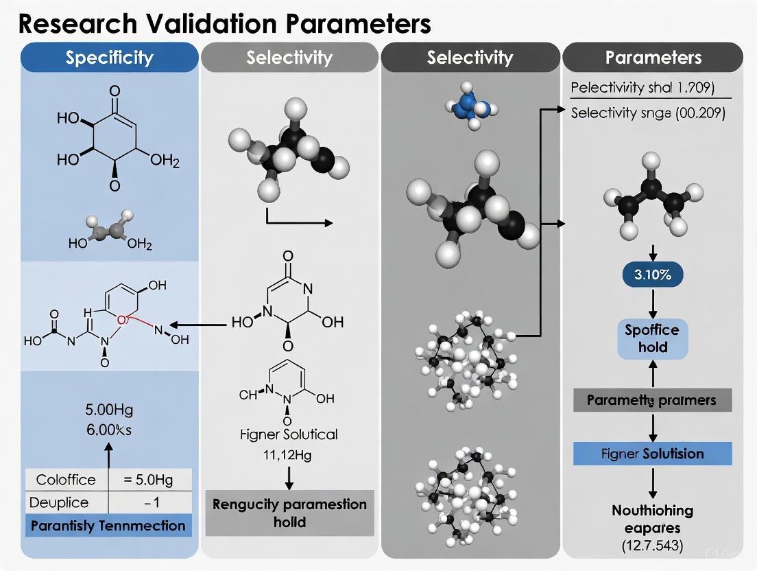
Specificity is the official term used in ICH Q2(R1) and is defined as "the ability to assess unequivocally the analyte in the presence of components which may be expected to be present" [5]. It is often considered the ultimate guarantee of method reliability. A specific method can accurately measure a single analyte in a complex mixture without interference from other components like excipients, impurities, or degradation products. For an identification test, specificity is an absolute requirement to prevent false-positive or false-negative results [5].
Selectivity, while not explicitly defined in ICH Q2(R1), is a term used in other guidelines, such as those for bioanalytical method validation. It describes the ability of a method to differentiate and quantify multiple analytes in the presence of interferences [5]. In practical terms, selectivity requires the identification of all relevant components in a mixture, not just the primary analyte. According to IUPAC recommendations, "selectivity" is the preferred term in analytical chemistry, with specificity representing the ideal case of 100% selectivity [5].
For chromatographic methods, both parameters are demonstrated by achieving a clear resolution between the peaks of interest, particularly for the two components that elute closest to each other [5].
Experimental Protocols for Demonstrating Specificity/Selectivity
The experimental approach to demonstrating specificity varies based on the type of analytical procedure (e.g., identification, assay, impurity test). The following workflow outlines a general strategy, with specific methodologies detailed thereafter.
Diagram 1: Experimental Workflow for Specificity/Selectivity
Protocol 1: Specificity for an Assay Method (e.g., HPLC-UV)
1. Objective: To demonstrate that the method is accurate for the analyte of interest in the presence of sample matrix, impurities, and degradation products.
2. Materials:
- Analytical Standard: High-purity reference standard of the active pharmaceutical ingredient (API).
- Placebo: A mixture of all formulation excipients without the API.
- Test Solution: The finished pharmaceutical product (e.g., tablet powder or injection solution).
- Stressed Samples: Samples of the API and product subjected to forced degradation (e.g., heat, light, acid, base, oxidation).
3. Methodology: 1. Preparation: Prepare and analyze the following solutions: - Blank/Placebo: To identify signals from the matrix. - Standard Solution: To identify the retention time and response of the pure analyte. - Placebo spiked with Analyte: To confirm the accuracy of measurement in the matrix. - Test Solution: The actual product. - Forced Degradation Samples: To generate and separate degradation products. 2. Chromatographic Analysis: Inject all solutions under the validated chromatographic conditions (e.g., HPLC with UV detection). 3. Data Evaluation: - Compare the chromatogram of the placebo with that of the standard to ensure no interfering peaks co-elute with the analyte peak. - In the test solution and stressed samples, ensure that the analyte peak is pure and baseline-resolved from any other peaks. Peak purity can be assessed using a diode array detector (DAD) to confirm spectral homogeneity. - For the spiked placebo, calculate the recovery to confirm accuracy is within acceptance criteria (e.g., 98-102%) [19].
Protocol 2: Selectivity for a Related Substances Method (Impurity Profiling)
1. Objective: To ensure the method can separate, detect, and quantify all known and unknown impurities and degradation products from each other and from the main analyte.
2. Materials: Similar to Protocol 1, with an emphasis on having available reference standards for known impurities.
3. Methodology: 1. Preparation: Prepare a system suitability mixture containing the API and all available impurity standards at appropriate levels (e.g., at the specification limit). 2. Chromatographic Analysis: Inject the mixture and the stressed sample solutions. 3. Data Evaluation: - Calculate the resolution between the main analyte and the closest eluting impurity. The resolution should typically be greater than a predefined limit (e.g., R > 1.5 or 2.0) [5]. - Verify that the detection and quantitation of each impurity is not affected by the others or by the main peak.
The Scientist's Toolkit: Essential Research Reagents and Materials
Successful validation of specificity and selectivity relies on carefully selected, high-quality materials. The following table details key reagents and their critical functions in the experimental process.
Table 2: Essential Research Reagents for Specificity/Selectivity Validation
| Reagent / Material | Function & Role in Validation |
|---|---|
| High-Purity Analytical Reference Standards | Serves as the benchmark for identifying the analyte's retention time, spectral properties, and response factor. Essential for confirming the identity of the target peak [20]. |
| Well-Characterized Placebo/Blank Matrix | Allows for the identification of signals originating from the sample matrix (excipients) rather than the analyte. Critical for demonstrating a lack of interference and confirming the method's accuracy in the matrix [20]. |
| Certified Impurity Standards | Used to identify and confirm the retention times of known impurities. Vital for developing and validating selective impurity methods and for establishing resolution between the API and its impurities. |
| Chemical Stress Agents (e.g., HCl, NaOH, H₂O₂) | Used in forced degradation studies to intentionally generate degradation products. This process helps establish the method's stability-indicating properties by proving it can separate the analyte from its degradation products [5]. |
| Matrix-Blank Spiked Solutions | Solutions where the placebo is spiked with a known concentration of the analyte. Used to calculate analyte recovery, which directly demonstrates the method's accuracy and freedom from matrix interference [20]. |
The landscape of analytical method validation is evolving towards a more integrated, scientific, and risk-based lifecycle approach. ICH Q2(R2) and the revised USP <1225> are converging in their principles, emphasizing fitness for purpose and the control of uncertainty in the reportable result [18]. For the practicing scientist, this means that validation is no longer a one-time exercise but an ongoing process rooted in a deep understanding of the procedure, as defined initially by the Analytical Target Profile (ATP).
While the core experimental protocols for parameters like specificity and selectivity remain fundamentally important, their design and evaluation are now more explicitly linked to the overall goal of ensuring confidence in decision-making for batch release and patient safety. Staying current with these harmonized guidelines and adopting the lifecycle mindset is imperative for drug development professionals to ensure robust, reliable, and regulatory-compliant analytical procedures.
Differentiating Analytical Method Validation from Clinical Biomarker Qualification
In the structured environment of pharmaceutical development and regulatory science, the terms "analytical method validation" and "clinical biomarker qualification" represent distinct but interconnected processes. Analytical method validation is the procedure of performing numerous tests designed to verify that an analytical test system is suitable for its intended purpose and capable of generating reliable analytical data [21]. It focuses primarily on assessing the performance characteristics of the assay itself. In contrast, clinical biomarker qualification is the evidentiary process of linking a biomarker with biological processes and clinical endpoints [22] [23]. This distinction is crucial—validation ensures the test measures correctly, while qualification ensures what the test measures matters clinically.
The terminology has evolved to avoid confusion. As noted in biomarker literature, "the term 'validation' is reserved for analytical methods, and 'qualification' for biomarker clinical evaluation to determine surrogate endpoint candidacy" [22]. This semantic precision helps stakeholders across drug development communicate effectively about the specific evidence required for each purpose. Understanding this fundamental difference—between assay performance and clinical relevance—forms the foundation for appropriate application in research and development.
Conceptual Framework and Key Distinctions
Core Definitions and Relationships
At the most fundamental level, analytical method validation and clinical biomarker qualification address different questions. Analytical method validation asks: "Does this test measure accurately and reliably?" whereas clinical biomarker qualification asks: "Does this measurement meaningfully predict biological or clinical outcomes?" [24] [25].
A biomarker is formally defined as "a defined characteristic that is measured as an indicator of normal biological processes, pathogenic processes, or responses to an exposure or intervention" [25]. The BEST resource further categorizes biomarkers into seven types: susceptibility/risk, diagnostic, monitoring, prognostic, predictive, pharmacodynamic/response, and safety [25]. The context of use is a critical concept that determines the specific application of a biomarker in drug development and directly influences both the validation and qualification requirements [24].
Table 1: Fundamental Distinctions Between Analytical Validation and Clinical Qualification
| Aspect | Analytical Method Validation | Clinical Biomarker Qualification |
|---|---|---|
| Primary Focus | Analytical test system performance [21] | Clinical/biological significance of results [22] |
| Central Question | "Can we measure it correctly?" | "Does it mean what we think it means?" |
| Regulatory Framework | ICH Q2(R2) guidelines [17] [26] | FDA Biomarker Qualification Program [25] |
| Evidence Generated | Technical reliability and reproducibility [21] | Association with biological processes or clinical endpoints [23] |
| Typical Output | Validated method with performance characteristics | Qualified biomarker with defined context of use |
Figure 1: Relationship Between Analytical Validation and Clinical Qualification Processes
Regulatory Frameworks and Guidelines
The regulatory landscapes governing analytical validation and biomarker qualification differ significantly in structure and purpose. Analytical method validation follows well-established technical guidelines, primarily the International Council for Harmonisation (ICH) Q2(R2) guideline titled "Validation of Analytical Procedures" [17] [26]. This guideline provides a harmonized international approach to assessing method performance characteristics and applies to procedures used for release and stability testing of commercial drug substances and products [26]. The recently adopted ICH Q14 guideline further complements this by providing a structured approach to analytical procedure development [27].
In contrast, clinical biomarker qualification follows a more complex, evidence-based regulatory pathway. The FDA's Biomarker Qualification Program operates under a collaborative, multi-stage process defined by the 21st Century Cures Act [25] [28]. This process involves three formal stages: Letter of Intent, Qualification Plan, and Full Qualification Package submission [25]. The European Medicines Agency has developed a similar qualification process for novel methodologies [28]. Unlike analytical validation, which focuses on technical performance, biomarker qualification evaluates the totality of evidence linking a biomarker to specific biological processes or clinical outcomes within a defined context of use.
Analytical Method Validation: Parameters and Protocols
Core Validation Parameters
Analytical method validation systematically assesses multiple performance characteristics to ensure generated data meets quality standards. The ICH Q2(R2) guideline identifies key parameters that collectively demonstrate a method is fit for its intended purpose [17] [26].
Specificity and Selectivity are particularly crucial parameters, though these terms are often confused. According to ICH guidelines, specificity refers to "the ability to assess unequivocally the analyte in the presence of components which may be expected to be present" [5]. It describes a method's capacity to measure solely the intended analyte without interference from other substances in the sample matrix. In practical terms, "specificity tells us about the degree of interference by other substances also present in the sample while analysing the analyte" [5]. Selectivity, while sometimes used interchangeably, carries a nuanced meaning: "Selectivity is like specificity except that the identification of all components in a mixture is mandatory" [5]. For chromatographic techniques, specificity/selectivity is demonstrated by the resolution between the closest eluting components [5].
Accuracy represents the closeness of agreement between the measured value and the true value, typically expressed as percent recovery [21] [26]. Precision, comprising repeatability (same conditions) and intermediate precision (different days, analysts, instruments), measures the degree of scatter among multiple measurements and is expressed as relative standard deviation (%RSD) [21] [26]. The Horwitz equation provides empirical guidance for expected precision values based on analyte concentration, with modified values for repeatability ranging from 1.34% RSD at 100% concentration to 3.30% RSD at 0.25% concentration [21].
Table 2: Core Analytical Method Validation Parameters and Acceptance Criteria
| Parameter | Definition | Typical Assessment Method | Common Acceptance Criteria |
|---|---|---|---|
| Specificity | Ability to measure analyte without interference [5] | Resolution from closest eluting peak [5] | Baseline separation (R ≥ 2.0) [5] |
| Accuracy | Closeness to true value [21] | Spiked recovery with known standards [21] | 98-102% recovery for drug substance [26] |
| Precision | Agreement between repeated measurements [21] | Multiple injections of homogeneous sample [21] | %RSD ≤ 2% for assay methods [26] |
| Linearity | Proportionality of response to concentration [21] | Series of standards at 5+ concentrations [21] | R² ≥ 0.998 [26] |
| Range | Interval where linearity, accuracy, precision are acceptable [21] | Verified through accuracy and precision at range limits [21] | Typically 80-120% of test concentration [26] |
| LOD/LOQ | Lowest detectable/quantifiable amount [21] | Signal-to-noise ratio (3:1 for LOD, 10:1 for LOQ) [21] | Appropriate for intended use [26] |
Experimental Protocols for Key Validation Parameters
Specificity Testing Protocol:
- Sample Preparation: Prepare blank sample (matrix without analyte), standard solution (pure analyte), and spiked sample (matrix with known analyte concentration) [5].
- Forced Degradation: Subject samples to stress conditions (acid, base, oxidation, heat, light) to generate potential degradants [5].
- Chromatographic Analysis: Inject all samples using the proposed method and record chromatograms [5].
- Resolution Assessment: Measure resolution between analyte peak and closest eluting potential interferent; acceptance criterion is typically R ≥ 2.0 [5].
- Peak Purity: For stability-indicating methods, use diode array detection or mass spectrometry to demonstrate analyte peak purity in stressed samples [26].
Linearity and Range Determination Protocol:
- Standard Preparation: Prepare a minimum of five standard solutions covering the proposed range (typically 50-150% of target concentration) [21].
- Analysis: Inject each standard solution in replicate (minimum duplicate) using the proposed method [21].
- Data Analysis: Plot response against concentration and calculate regression parameters [21].
- Statistical Evaluation: Determine correlation coefficient (R), y-intercept, slope, and residual sum of squares [21].
- Acceptance Criteria: Typically R² ≥ 0.998, y-intercept not significantly different from zero, and residuals randomly distributed [26].
Clinical Biomarker Qualification: Process and Evidence Generation
The Qualification Pathway
Clinical biomarker qualification follows a rigorous, stage-gated process that requires generation of substantial evidence linking the biomarker to clinical or biological outcomes. The FDA's Biomarker Qualification Program outlines a formal three-stage pathway [25]:
Stage 1: Letter of Intent - The qualification process begins with submission of a Letter of Intent that describes the biomarker, its proposed context of use, the drug development need it addresses, and preliminary information on how it will be measured [25]. The FDA reviews this submission to assess potential value and feasibility before permitting advancement to the next stage.
Stage 2: Qualification Plan - This detailed proposal describes the complete biomarker development plan, including existing supporting evidence, identified knowledge gaps, and specific studies designed to address these gaps [25]. The Qualification Plan must include comprehensive information about the analytical method and its performance characteristics, linking back to the analytical validation data [25].
Stage 3: Full Qualification Package - The final submission represents a comprehensive compilation of all supporting evidence, organized to inform the FDA's qualification decision [25]. This includes analytical validation data, clinical validation studies, statistical analyses, and a thorough justification for the proposed context of use [25].
Throughout this process, the fit-for-purpose approach is fundamental—"the verification level of a drug development tool needs to be sufficient to support its context of use" [24]. The degree of evidence required scales with the intended application, with biomarkers supporting critical regulatory decisions requiring more extensive qualification [24].
Biomarker Categories and Evidentiary Standards
Biomarkers are categorized based on their level of validation and acceptance. Exploratory biomarkers represent the initial discovery phase and lay groundwork for further development [22]. Probable valid biomarkers are "measured in an analytical test system with well-established performance characteristics and for which there is an established scientific framework or body of evidence that elucidates the physiologic, toxicologic, pharmacologic, or clinical significance of the test results" [22]. Known valid biomarkers achieve widespread acceptance in the scientific community about their clinical or physiological significance [22].
The evidentiary standards for biomarker qualification depend heavily on the proposed context of use. For a biomarker to serve as a surrogate endpoint—substituting for a clinical endpoint—it must meet particularly rigorous standards. According to Fleming and DeMets criteria, a surrogate endpoint must both be correlated with the true clinical outcome and fully capture the net effect of treatment on that clinical outcome [23]. This represents the highest standard of biomarker qualification.
Figure 2: FDA Biomarker Qualification Process Stages
The Scientist's Toolkit: Essential Research Reagents and Materials
Successful biomarker development requires specialized reagents and materials that ensure both analytical reliability and biological relevance. The selection of appropriate tools is critical for generating data suitable for regulatory submission.
Table 3: Essential Research Reagents and Materials for Biomarker Studies
| Reagent/Material | Function | Critical Considerations |
|---|---|---|
| Reference Standards | Quantitation and method calibration [24] | Often recombinant proteins; may differ structurally from endogenous biomarkers [24] |
| Characterized Biologic Samples | Method development and validation [24] | Should represent study population characteristics; well-documented collection/processing [28] |
| Selective Binding Reagents | Target capture and detection (antibodies, aptamers) [24] | Must demonstrate specificity for intended target; cross-reactivity profiling essential [5] |
| Matrix-matched Controls | Accounting for matrix effects [24] | Blank biological matrices often difficult to obtain; may require alternative substrates [24] |
| Stability Materials | Establishing pre-analytical conditions [21] | Includes additives, storage containers, temperature monitoring systems [21] |
Comparative Analysis: Interdependence and Distinct Requirements
While analytical validation and clinical qualification serve different purposes, they are interdependent in the biomarker development pipeline. A clinically qualified biomarker requires an analytically validated measurement method, but analytical validation alone cannot confer clinical utility [22] [24].
The fit-for-purpose approach governs the relationship between these processes. The level of analytical validation should be appropriate for the biomarker's context of use and stage of development [24]. For exploratory biomarkers in early drug development, limited validation may suffice, whereas biomarkers supporting critical regulatory decisions require complete validation [24]. This principle acknowledges that resource allocation should match the evidentiary needs for specific applications.
The fundamental distinction remains: "It is important to note that a biomarker is qualified, and not the biomarker measurement method" [25]. This distinction clarifies the separate but complementary roles of these processes—analytical validation establishes that we can measure the biomarker reliably, while clinical qualification establishes that the measurement meaningfully informs drug development or clinical use.
Understanding the distinction between analytical method validation and clinical biomarker qualification is essential for efficient drug development. Analytical validation provides the foundation of reliable measurement, while clinical qualification establishes meaningful application. These processes operate within different regulatory frameworks, generate distinct types of evidence, and answer fundamentally different questions about biomarker utility.
As biomarker science evolves, the fit-for-purpose approach continues to guide appropriate resource allocation throughout development stages. Researchers must recognize both the separation and interdependence of these processes to successfully advance biomarkers from exploratory tools to qualified drug development tools with defined clinical utility.
In the highly regulated pharmaceutical industry, the reliability of analytical methods is non-negotiable for ensuring product safety and efficacy. Analytical Method Validation provides formal, systematic proof that analytical tests deliver consistent and useful data [29]. A significant paradigm shift is underway, moving from a fixed validation model to a dynamic lifecycle approach that incorporates risk-based thinking and validation tied to intended use [30]. This guide explores this evolution, with a specific focus on how validation parameters for specificity and selectivity develop throughout the drug development process.
Understanding the Analytical Procedure Lifecycle
The traditional view of analytical method validation emphasized a rapid development phase followed by a fixed, comprehensive validation. The modern lifecycle approach, as outlined in emerging guidance like ICH Q2(R2) and USP <1220>, presents a more structured, three-stage model [31].
The Three Stages of the Lifecycle
The lifecycle of an analytical procedure consists of three interconnected stages:
- Procedure Design and Development: This stage is derived from an Analytical Target Profile (ATP), a predefined objective that outlines the procedure's requirements for its intended use [31].
- Procedure Performance Qualification: This is the formal method validation stage, where the procedure's performance characteristics are documented to show it is suitable for its intended use [31].
- Procedure Performance Verification: This involves the ongoing monitoring of the procedure's performance during routine use to ensure it remains in a state of control [31].
The following diagram illustrates the workflow and continuous feedback loops within this lifecycle.
Phase-Appropriate Validation: A Strategic Framework
A core principle of the lifecycle approach is phase-appropriateness. This strategy aligns the depth and rigor of validation activities with the stage of drug development, wisely allocating resources while building knowledge progressively [32]. The following table compares the focus of method validation across clinical development phases.
Table 1: Phase-Appropriate Analytical Method Validation Focus
| Development Phase | Analytical Procedure Status | Primary Validation Focus |
|---|---|---|
| Discovery & Phase I | Simple procedure; limited knowledge of drug product [30]. | Basic parameters: Precision, Linearity, Limited Robustness [30]. |
| Phase II | Procedure develops with better understanding of impurity profile [30]. | Expanded parameters: Specificity, Accuracy, Detection Limit (DL) [30]. |
| Phase III & Commercial | Procedure optimized for long-term commercial use [30]. | Full validation: Specificity, Precision, Intermediate Precision, Linearity, Accuracy, DL, QL, Robustness [30]. |
Core Validation Parameters: Specificity and Selectivity in Focus
Throughout the validation lifecycle, specific parameters are evaluated to ensure method reliability. Specificity and selectivity are critical for confirming that a method accurately measures the intended analyte.
Definitions and Distinctions
- Specificity is defined as the ability to assess unequivocally the analyte in the presence of components that may be expected to be present, such as impurities, degradants, or matrix components [5]. It refers to the method's ability to respond to one single analyte.
- Selectivity is the ability to differentiate the analyte(s) of interest from endogenous components in the matrix or other sample components [5]. It refers to the method's ability to respond to several different analytes in a mixture.
For chromatographic methods, specificity/selectivity is typically demonstrated by achieving baseline resolution between the analyte and the closest eluting potential interferent [5].
Experimental Protocols for Specificity and Selectivity
A robust specificity/selectivity study involves multiple experiments designed to challenge the method's ability to distinguish the analyte.
Table 2: Experimental Protocols for Demonstrating Specificity/Selectivity
| Experiment Type | Protocol Description | Acceptance Criteria |
|---|---|---|
| Analysis of Blank Matrix | Analyze the sample matrix without the analyte (e.g., placebo formulation or biological matrix) [5]. | No response (peak) at the retention time of the analyte [33]. |
| Analysis of Spiked Samples | Analyze the sample matrix spiked with the analyte of interest at the target concentration [5]. | A clear, positive response for the analyte with no co-eluting peaks. |
| Forced Degradation Studies | Stress the drug substance/product (e.g., with heat, light, acid, base, oxidation) and analyze the degraded sample [29]. | The analyte peak is pure and resolved from degradation products (assessed via diode array or mass spectrometry). |
| Interference Testing | Analyze samples spiked with potential interferents (e.g., precursors, known impurities, metabolites) [33]. | No interference at the retention time of the analyte. All peaks of interest are resolved. |
The workflow for a comprehensive specificity and selectivity assessment is methodical and layered, as shown below.
The Scientist's Toolkit: Essential Reagents and Materials
Successful method validation relies on high-quality, well-characterized materials. The following table details key reagents used in validation experiments, particularly for specificity/selectivity.
Table 3: Key Research Reagent Solutions for Validation Studies
| Reagent/Material | Function in Validation | Critical Quality Attributes |
|---|---|---|
| Drug Substance (API) | Primary analyte for quantification and specificity studies. | High purity, well-characterized structure and properties [30]. |
| Placebo/Blank Matrix | Used to demonstrate absence of interference from non-active components [5]. | Representative of final product composition without the active ingredient. |
| Known Impurities | Used to challenge the method's ability to separate and quantify closely related substances [29]. | Certified reference standards with known identity and purity. |
| Forced Degradation Reagents | Used to generate stress samples (acid, base, oxidant, etc.) for specificity studies [29]. | Analytical grade purity to ensure generated degradants are from the analyte. |
| Characterized Reference Materials | Essential for accurate quantitation of drug product and known impurities during method development [30]. | Documented purity and stability, traceable to a primary standard. |
Comparative Performance: Lifecycle vs. Traditional Approach
Adopting a lifecycle approach fundamentally changes how methods are developed, validated, and maintained. The table below compares the outcomes of this modern approach against traditional practices.
Table 4: Performance Comparison of Validation Approaches
| Aspect | Traditional "One-Time" Validation | Lifecycle Approach (with ATP) |
|---|---|---|
| Development Foundation | Often empirical; limited systematic understanding of method robustness [31]. | Science- and risk-based; built on a foundation of systematic knowledge [31]. |
| Method Robustness | May be unknown or poor, leading to "problematic methods" with variable chromatography and SST failures [30]. | Understood and controlled through systematic development, leading to fewer operational failures [31]. |
| Regulatory Perception | A method unchanged for 5–10 years may be a "red flag" [30]. | Welcomes continuous improvement with proper documentation, seen as proactive quality management [30]. |
| Specificity/Specificity Understanding | Typically confirmed only at validation; knowledge of true capability may be limited. | Deeply investigated during Stage 1; method conditions are proven to control critical factors affecting separation. |
The evolution from a fixed, one-time validation model to a dynamic, phase-appropriate lifecycle approach represents a significant advancement in pharmaceutical analytical science. This framework, built around a predefined Analytical Target Profile, ensures that methods are not only validated but are inherently more robust and reliable. For critical parameters like specificity and selectivity, the lifecycle model facilitates a deeper, more scientific understanding of the method's capabilities and limitations. This leads to fewer analytical failures, more reliable data for critical decisions, and ultimately, a more efficient path to delivering safe and effective drug products to patients.
Proven Techniques to Demonstrate Specificity and Selectivity in Practice
Designing and Executing Forced Degradation Studies
Forced degradation studies, also known as stress testing, are an essential developmental activity in pharmaceutical research and development. These studies involve intentionally degrading drug substances and products under exaggerated environmental conditions to identify potential degradation products, elucidate degradation pathways, and establish the intrinsic stability of molecules [34] [35]. Within the broader context of analytical method validation, forced degradation provides the foundational evidence required to demonstrate method specificity—the ability of an analytical procedure to accurately measure the analyte in the presence of components that may be expected to be present, such as impurities, degradation products, or matrix components [35]. The data generated from these studies directly supports regulatory submissions by proving that stability-indicating methods can detect changes in product quality attributes over time [36] [37].
Unlike formal stability studies which aim to establish shelf-life, forced degradation operates under more severe conditions to rapidly generate relevant degradation products. This comparative analysis examines the strategic design, execution, and interpretation of forced degradation studies, providing researchers with a framework for selecting appropriate stress conditions, analytical techniques, and acceptance criteria to meet both scientific and regulatory requirements [34] [35].
Comparative Analysis of Stress Conditions and Methodologies
Strategic Approach to Stress Condition Selection
The design of forced degradation studies requires careful consideration of multiple stress factors to comprehensively challenge the drug molecule. The International Council for Harmonisation (ICH) guidelines Q1A(R2) recommend including a minimal set of stress conditions that typically include hydrolytic degradation (acid and base), oxidative degradation, thermal degradation, photolytic degradation, and humidity stress [36] [34] [35]. The selection of specific parameters within these categories should be scientifically justified based on the drug's chemical structure, intended formulation, and potential exposure conditions.
A comparative analysis of stress methodologies reveals distinct advantages and limitations for each approach. The table below summarizes the typical conditions and strategic considerations for major stress types:
Table 1: Comparative Analysis of Stress Conditions in Forced Degradation Studies
| Stress Type | Typical Conditions | Optimal Degradation Target | Key Strategic Considerations | Common Degradation Mechanisms |
|---|---|---|---|---|
| Acid Hydrolysis | 0.1N–1N HCl at 40–70°C for several hours to days [36] [38] | 5–20% degradation [36] [34] | Use reflux condenser to prevent evaporation; neutralization before analysis [38] | Hydrolysis of ester and amide bonds; dehydration; rearrangement [34] |
| Base Hydrolysis | 0.1N–1N NaOH at 40–70°C for several hours to days [36] [38] | 5–20% degradation [36] [34] | Shorter exposure times often needed compared to acid; neutralization critical [38] | Hydrolysis of esters; β-elimination; racemization [34] |
| Oxidative Stress | 1–3% H₂O₂ at room temperature for up to 24 hours [36] [38] | 5–20% degradation [36] [34] | Perform in dark; avoid elevated temperatures that reduce oxygen solubility [38] | N- and S-oxidation; aromatic hydroxylation; dehydrogenation [34] |
| Thermal Stress | 60–80°C for solid state; 40–70°C for solutions [36] [34] | 5–20% degradation [36] [34] | For APIs with melting point <150°C, stress at 70°C; >150°C at 105°C [38] | Pyrolysis; decomposition; intermolecular reactions [35] |
| Photolytic Stress | Exposed to 1.2 million lux-hr visible and 200 W-hr/m² UV [38] | Evidence of degradation or justification of stability [34] | Follow ICH Q1B guidelines; include dark control; consider container transparency [34] | Ring rearrangement; dimerization; cleavage of side chains [35] |
| Humidity Stress | 75–90% relative humidity for up to 1 week [36] [38] | 5–20% degradation [36] | Often combined with thermal stress; demonstrates sensitivity to moisture [38] | Hydrolysis; hydration; deliquescence [35] |
Targeted Degradation and Analytical Interpretation
The primary objective in forced degradation study design is to achieve controlled degradation within the optimal range of 5-20% of the active pharmaceutical ingredient (API) [36] [34]. This range ensures sufficient degradation products are formed to properly challenge the analytical method's specificity without generating secondary degradants that would not typically form under relevant storage conditions. Studies should include appropriate controls including unstressed API, stressed placebo (for drug products), and stressed solution blanks to properly attribute observed degradation products [38].
The strategic approach to stress condition optimization should follow a systematic workflow:
Diagram 1: Stress Condition Optimization Workflow
When degradation exceeds 20%, there is risk of generating secondary degradation products that may not form under normal storage conditions, potentially leading to unnecessary method complexity. Conversely, under-stressing (<5% degradation) may fail to reveal critical degradation pathways, resulting in analytical methods that lack appropriate specificity [35]. For molecules demonstrating exceptional stability where minimal degradation occurs despite harsh conditions, the study can be terminated with appropriate scientific justification that the molecule is stable under the tested conditions [34].
Experimental Protocols and Methodologies
Detailed Hydrolytic Degradation Protocol
Objective: To evaluate the susceptibility of the drug substance to hydrolysis under acidic, basic, and neutral conditions.
Materials and Equipment:
- Drug substance (API)
- 0.1N hydrochloric acid (HCl)
- 0.1N sodium hydroxide (NaOH)
- pH 7.0 buffer solution
- Water bath with temperature control (±2°C) or reflux apparatus
- HPLC vials and compatible HPLC system
- Optional co-solvents: acetonitrile (preferred) or methanol [38]
Procedure:
- Prepare separate solutions of the drug substance in 0.1N HCl, 0.1N NaOH, and neutral solution (water or buffer) at a concentration of approximately 1 mg/mL [34]. If the drug has poor aqueous solubility, use minimal co-solvent (typically not exceeding 50% v/v) with acetonitrile preferred due to its relative inertness compared to methanol [38].
- Transfer the solutions to suitable containers and maintain at 60-70°C using a water bath or reflux apparatus. For reflux, install a condenser to prevent solvent evaporation and add glass beads or porcelain pieces to prevent bumping [38].
- Remove aliquots at predetermined time points (e.g., 6, 12, 24, 48 hours) and immediately neutralize acidic and basic samples (for HPLC compatibility) [38].
- Analyze samples using the proposed stability-indicating method, typically HPLC with UV detection.
- Include appropriate controls: unstressed drug substance, stressed placebo (for drug products), and stressed solution blanks.
Data Interpretation: Monitor for the appearance of new peaks in the chromatogram and decrease in the main peak area. Calculate the percentage degradation relative to the unstressed control. Optimal degradation for method validation is 5-20% [36] [34].
Oxidative Degradation Protocol
Objective: To evaluate the susceptibility of the drug substance to oxidative degradation.
Materials and Equipment:
- Drug substance (API)
- 3% hydrogen peroxide (H₂O₂) solution
- Amber glass containers to protect from light
- Magnetic stirrer
- HPLC system with compatible vials
Procedure:
- Prepare a solution of the drug substance in 3% H₂O₂ at a concentration of approximately 1 mg/mL.
- Maintain the solution at room temperature (25±2°C) in the dark with constant stirring [38]. Note: Elevated temperatures are generally not recommended for oxidative stress as increased temperature can reduce oxygen solubility in the solvent [38].
- Remove aliquots at predetermined time points (e.g., 6, 12, 24 hours).
- Analyze samples using the proposed stability-indicating method.
- Include appropriate controls: drug substance in water without H₂O₂, stressed placebo, and reagent blanks.
Data Interpretation: Monitor for the appearance of new peaks in the chromatogram and decrease in the main peak. Oxidation often produces polar degradants that may elute earlier in reversed-phase HPLC. For drug products, oxidation may occur through free radical mechanisms, which might require alternative oxidizing agents such as azobisisobutyronitrile (AIBN) or metal ions in some cases [34].
Photolytic Degradation Protocol
Objective: To evaluate the susceptibility of the drug substance to photodegradation.
Materials and Equipment:
- Drug substance (solid and solution if applicable)
- Photostability chamber meeting ICH Q1B requirements [34]
- Lux meter and UV radiometer for energy calibration
- Opaque containers (e.g., aluminum foil wrapping) for dark controls
Procedure:
- Expose the solid drug substance and drug product in their immediate containers to overall illumination of not less than 1.2 million lux hours and an integrated near ultraviolet energy of not less than 200 watt hours/square meter [38].
- Include dark controls wrapped in aluminum foil with the same samples placed in the photostability chamber alongside the exposed samples.
- For solution photostability, prepare drug solutions in transparent containers and expose using the same conditions.
- Analyze samples after exposure using the proposed stability-indicating method.
Data Interpretation: Compare chromatograms of exposed samples versus dark controls for appearance of new peaks and decrease in main peak area. Photodegradation products may include isomers, dimers, and cleavage products. If no significant degradation is observed, this demonstrates photostability which should be documented with justification [34].
Analytical Techniques for Degradation Monitoring
Comparison of Analytical Methods
The selection of analytical techniques for monitoring forced degradation studies is critical for comprehensive profiling of degradation products. A stability-indicating method must be capable of separating, detecting, and quantifying the active pharmaceutical ingredient and its degradation products. The following table compares the primary analytical techniques employed in forced degradation studies:
Table 2: Comparison of Analytical Techniques for Forced Degradation Studies
| Analytical Technique | Primary Applications in Forced Degradation | Key Advantages | Detection Limitations | Suitability for Specificity Demonstration |
|---|---|---|---|---|
| HPLC/UPLC with UV/PDA | Quantitative separation and monitoring of degradants [36] | High resolution; robust quantification; compatible with most pharmaceuticals | Limited to UV-absorbing compounds; may miss non-chromophoric degradants | Excellent for demonstrating separation of known degradants |
| LC-MS | Structural identification of degradants; molecular weight determination [36] | Provides structural information; high sensitivity | Matrix effects; may require method optimization | Superior for peak identification and impurity tracking |
| GC-MS | Volatile degradants; residual solvents; small molecule analysis [38] | High resolution for volatile compounds; excellent detection sensitivity | Limited to volatile and thermally stable compounds | Good for specific compound classes |
| CE (Capillary Electrophoresis) | Charged molecules; biological therapeutics [37] | High efficiency separation; minimal sample volume | Lower precision compared to HPLC; more specialized | Useful for large molecules and charged species |
| IC | Ionic degradants; counterion analysis | Selective for ionic species | Limited to ionizable compounds | Complementary technique for specific impurities |
Assessment of Method Specificity
The fundamental role of forced degradation studies in demonstrating analytical method specificity cannot be overstated. A method is considered stability-indicating when it can accurately quantify the API without interference from degradation products, process impurities, excipients, or other matrix components [35]. The assessment of specificity should include:
- Peak Purity Assessment: Using photodiode array (PDA) detection to demonstrate that the API peak is homogeneous and not contaminated with co-eluting degradants [38]. For methods using detectors without spectral capability (e.g., RI, ELSD), peak homogeneity should be established through orthogonal techniques such as LC-MS [38].
- Resolution: Critical peak pairs should have sufficient resolution (typically >1.5) to ensure accurate quantification [38].
- Mass Balance: The process of adding together the assay value and levels of degradation products to see how closely these add up to 100% of the initial value [38]. Mass balance should ideally achieve at least 95%; significant shortfalls may indicate undetected degradants (non-UV absorbing, volatile, or irreversibly adsorbed to columns) [38].
The relationship between forced degradation and analytical validation can be visualized as follows:
Diagram 2: Forced Degradation in Method Validation
The Scientist's Toolkit: Essential Research Reagents and Materials
Successful execution of forced degradation studies requires careful selection of reagents, solvents, and analytical tools. The following toolkit outlines essential materials and their specific functions in stress testing protocols:
Table 3: Essential Research Reagents and Materials for Forced Degradation Studies
| Category | Specific Items | Function in Forced Degradation | Usage Considerations |
|---|---|---|---|
| Stress Reagents | 0.1N–1N HCl; 0.1N–1N NaOH; 1–3% H₂O₂; various pH buffers [36] [38] | Induce hydrolytic and oxidative degradation | Use high-purity reagents; prepare fresh solutions especially for oxidation studies |
| Solvents | Acetonitrile (HPLC grade); Methanol (HPLC grade); Purified water [38] | Solubilize drug substances; prepare stress solutions | Acetonitrile preferred over methanol for hydrolytic studies due to better chemical inertness [38] |
| Analytical Instruments | HPLC/UPLC with PDA detector; LC-MS; GC-MS; stability chambers [36] | Separate, detect, and identify degradation products | Ensure instrument qualification before study initiation; PDA essential for peak purity assessment [38] |
| Sample Preparation | Volumetric flasks; pipettes; HPLC vials; syringe filters | Accurate sample preparation and introduction to analytical systems | Use inert materials compatible with solvents; filter samples to protect HPLC columns |
| Stress Chambers | Photostability chambers; stability chambers with humidity control; water baths [34] | Provide controlled stress conditions | Calibrate and monitor temperature and humidity; verify light output in photostability chambers |
Regulatory Framework and Compliance Considerations
Forced degradation studies are governed by several ICH guidelines, primarily Q1A(R2) which defines stress testing as studies undertaken to elucidate the intrinsic stability of drug substances [35]. These studies form the scientific basis for demonstrating specificity as required by ICH Q2(R1) for analytical method validation [35]. While regulatory guidance provides general principles, specific experimental parameters are left to the applicant's justification based on scientific understanding of the molecule [37].
Regulatory expectations include:
- Studies should be conducted on at least one batch of drug substance and drug product [34]
- Stress conditions should be more severe than accelerated conditions [34]
- The target degradation should be sufficient to generate relevant degradants (typically 5-20%) [36] [34]
- Analytical methods should be challenged with stressed samples to demonstrate stability-indicating capability [35]
- Degradation products observed in formal stability studies should be correlated with those formed during forced degradation [34]
Documentation of forced degradation studies should include detailed protocols, stress conditions, results, and scientific justification for the selected approach. These documents are typically included in the stability section of regulatory submissions [36].
Forced degradation studies represent a critical comparative tool in pharmaceutical development, providing essential data to demonstrate analytical method specificity and understand drug stability behavior. Through the strategic application of hydrolytic, oxidative, thermal, and photolytic stress conditions, researchers can generate relevant degradation profiles that challenge analytical methods and reveal potential stability issues before product commercialization.
The optimal design of these studies balances sufficient degradation to generate meaningful data (5-20%) without creating irrelevant secondary degradants. When executed with appropriate scientific rigor and comprehensive analytical monitoring, forced degradation studies provide the foundational evidence required for regulatory approval and ensure that stability-indicating methods can reliably monitor product quality throughout its shelf life. As pharmaceutical complexity continues to evolve with the emergence of biologics, oligonucleotides, and other novel modalities, the principles of forced degradation remain essential while requiring adaptation to address new analytical challenges and degradation pathways.
Utilizing Chromatographic Separation and Peak Purity Assessment (PDA/MS)
In pharmaceutical analysis, demonstrating that an analytical method can accurately measure the intended analyte in the presence of potential interferents is a fundamental regulatory requirement. Specificity is the ability to assess unequivocally the analyte in the presence of components that may be expected to be present, such as impurities, degradants, or matrix components [5]. The related term selectivity refers to the ability of the method to respond to several different analytes in the sample, requiring the identification of all components in a mixture [5]. For chromatographic methods, peak purity assessment serves as the practical tool to demonstrate specificity and selectivity by ensuring that a chromatographic peak represents a single, pure compound and is not attributable to more than one component [39]. This guide provides a comparative analysis of Photodiode Array (PDA) and Mass Spectrometry (MS) detection for peak purity assessment within the context of analytical method validation.
Principles of Chromatographic Separation and Performance Parameters
Reliable chromatographic separations form the foundation for meaningful peak purity assessment. The success of a separation depends on several key performance parameters that must be evaluated during method development and validation.
Essential Chromatographic Parameters
Theoretical Plates (Efficiency): This parameter, borrowed from distillation theory, denotes the efficiency of the column. A high number of theoretical plates is associated with a high elution volume and narrow peaks, indicating high separation efficiency [40]. The Height Equivalent to a Theoretical Plate (HETP) is a related parameter calculated by dividing the column length by the number of theoretical plates (HETP = column length/N), providing an indicator of column performance independent of column length [40].
Resolution: A measure of how well two peaks are separated from each other, taking into account both the distance between peak centers and their widths [40]. Resolution values of < 1.5 indicate poor separation, while values > 2.0 indicate baseline separation [40].
Asymmetry Factor/Tailing Factor: This parameter describes peak symmetry. A perfectly Gaussian peak has an asymmetry factor of 1.0, while fronting yields values < 1 and tailing yields values > 1 [40] [41]. The tailing factor (T) is measured as T = b/a, where 'a' is the width of the front half of the peak and 'b' is the width of the back half, measured at 10% of the peak height [41].
Mobile Phase Optimization for Improved Separations
The mobile phase composition, particularly its pH, dramatically affects chromatographic separations for ionizable analytes. The degree of ionization (pKa) controls the ionization state of an analyte in solution and directly impacts interactions with the stationary phase [42]. When the mobile phase pH is too close to the analyte's pKa, both ionized and unionized species coexist, potentially causing split peaks or shoulders [42]. For method robustness, selecting a mobile phase pH where analytes exist predominantly in one form is crucial [42].
Table 1: Key Chromatographic Performance Parameters and Their Significance
| Parameter | Calculation/Definition | Acceptance Criteria | Impact on Separation |
|---|---|---|---|
| Theoretical Plates (N) | N = 16(tR/W)^2 where tR is retention time and W is peak width | Higher values indicate better column efficiency | Higher plate count produces narrower peaks, improving detection sensitivity |
| Resolution (Rs) | Rs = 2(tR2 - tR1)/(W1 + W2) where tR1 and tR2 are retention times of two adjacent peaks | Rs < 1.5: Poor separation; Rs > 2.0: Baseline separation | Direct measure of separation between adjacent peaks; critical for accurate quantification |
| Asymmetry/Tailing Factor (T) | T = b/a where b and a are the back and front half-widths at 5% or 10% of peak height | T = 1: Symmetric peak; T > 1: Tailing; T < 1: Fronting | Asymmetric peaks affect integration accuracy and detection limits; may indicate secondary interactions |
Peak Purity Assessment Techniques: PDA vs. MS
Photodiode Array (PDA) Detection
PDA detection is the most common tool for evaluating peak purity in HPLC workflows, utilizing ultraviolet (UV) absorbance across a peak to identify spectral variations that may indicate coelution [43]. The technique works by comparing UV spectra at different points across the peak profile (front, apex, and tail) to detect the presence of coeluting compounds with different spectral characteristics [39].
Spectral Contrast Algorithm: Commercial CDS software employs algorithms to determine spectral contrast [39]:
- Spectra are baseline-corrected by subtracting interpolated baseline spectra
- Spectra are converted into vectors in n-dimensional space
- Vector lengths are minimized using least-squares regression
- Vectors are moved into a 2D plane and the angle between them is measured
A chromatographic peak is considered spectrally pure when the purity angle is less than the purity threshold [39]. Different software platforms use varying terminology, with Agilent's OpenLab calculating a similarity factor (1000 × r², where r = cosθ) and Shimadzu's LabSolutions using cosθ values directly [39].
Mass Spectrometry (MS) Detection
LC-MS provides a more definitive assessment of peak purity by detecting coelution based on mass differences rather than UV spectral characteristics [43]. MS detection facilitates peak purity assessment by demonstrating that the same precursor ions, product ions, and/or adducts attributed to the parent compound are present consistently across the entire chromatographic peak in the total ion chromatogram (TIC) or extracted ion chromatogram (EIC/XIC) [39].
Mass Spectral Peak Purity Approaches:
- Single Quadrupole MS: Compares mass spectra extracted at peak front, apex, and tail
- Tandem MS/MS: Monitors specific precursor-product ion transitions across the peak
- High-Resolution MS: Detects coeluting species with minimal mass differences using exact mass measurements
Table 2: Comparative Analysis of PDA and MS for Peak Purity Assessment
| Assessment Feature | PDA/UV Detection | MS Detection |
|---|---|---|
| Principle | UV spectral shape comparison across peak | Mass-to-charge ratio (m/z) consistency across peak |
| Detection Capability | Compounds with different UV spectra | Compounds with different molecular masses |
| Limitations | - Compounds with similar UV spectra- Low UV responding compounds- Impurities eluting near apex | - Isomeric compounds- Compounds with same mass- Ion suppression effects |
| Sensitivity | Typically 0.1-1.0% of parent peak [39] | Can detect <0.1% depending on compound and ionization |
| Quantification | Direct proportionality with UV response | Varies with ionization efficiency |
| Regulatory Acceptance | Well-established, widely accepted | Increasingly accepted, particularly for impurity profiling |
| Resource Requirements | Lower cost, easier operation | Higher cost, specialized expertise needed |
Experimental Protocols for Peak Purity Assessment
Forced Degradation Studies
Forced degradation studies are essential for demonstrating method specificity and the stability-indicating nature of analytical methods [39] [44]. These studies involve intentionally exposing the drug substance to various stress conditions to generate potential degradants.
Standard Protocol:
- Acidic/Basic Hydrolysis: Expose drug substance to 0.1-5N HCl or NaOH at ambient to elevated temperatures (e.g., 5N NaOH at ambient temperature for 2 hours) [44]
- Oxidative Stress: Treat with 0.1-3% hydrogen peroxide at room temperature [44]
- Thermal Degradation: Expose solid drug substance to elevated temperatures (e.g., 50-105°C) [44]
- Photolytic Stress: Subject drug substance to UV/visible light per ICH Q1B guidelines [44]
Acceptance Criteria: Degradation between 5-20% is generally targeted to provide meaningful data without excessive degradation [39].
PDA-Based Peak Purity Assessment Method
Instrumentation: HPLC system with PDA detector capable of continuous spectral acquisition during peak elution [39]
Procedure:
- Data Acquisition: Collect UV spectra continuously across the chromatographic peak (typically 5-20 spectra per second) across an appropriate wavelength range (e.g., 210-400 nm) [43]
- Spectral Normalization: Apply baseline correction to subtract interpolated baseline spectra between peak liftoff and touchdown [39]
- Purity Calculation: Software compares all spectra within the peak to the apex spectrum using vector analysis [39]
- Interpretation: Peak is considered pure when purity angle < purity threshold [39]
Critical Parameters:
- Spectral Range Selection: Avoid extreme wavelengths (<210 nm or >800 nm) where noise interference increases [39]
- Peak Integration: Ensure proper integration to avoid interference from background noise or neighboring peaks [39]
- Signal-to-Noise: Assess purity at concentrations >0.1% to minimize false positives from background noise [39]
MS-Based Peak Purity Assessment Method
Instrumentation: LC-MS system with appropriate ionization source (ESI, APCI) and mass analyzer (single quad, tandem MS, or high-resolution MS) [39]
Procedure:
- Ionization Optimization: Select appropriate ionization mode (positive/negative) and optimize source parameters
- Data Acquisition: Acquire full scan or selected ion monitoring data across the chromatographic peak
- Spectral Comparison: Extract mass spectra at peak front, apex, and tail regions
- Deconvolution: Apply spectral deconvolution algorithms if needed to resolve coeluting species
Data Interpretation:
- Consistent mass spectral profile across the peak indicates purity
- Changing ion ratios or appearance of different masses across the peak suggests coelution
- For high-resolution MS, exact mass measurements can identify potential coelutants
Comparative Experimental Data and Case Studies
Performance Comparison in Pharmaceutical Applications
In controlled studies, PDA and MS detection have demonstrated complementary strengths for peak purity assessment. PDA detection excels at identifying coeluting compounds with distinct UV spectra, while MS provides superior capability for detecting impurities with similar UV characteristics but different molecular weights.
Limitations of PDA-Based Assessment:
- False Negatives: Occur when coeluted impurities have minimal spectral differences, poor UV responses, elute near the apex, or are present at very low concentrations [39]
- False Positives: Can result from significant baseline shifts due to mobile phase gradients, suboptimal data processing settings, interference from background noise, or excipient-related signals [39]
MS Advantages: LC-MS enables more definitive peak purity assessment by detecting coelution based on mass differences, making it particularly valuable for identifying low-level contaminants that might escape PDA detection [43].
Advanced Detection Technologies
Emerging detection technologies are expanding capabilities for peak purity assessment. Vacuum Ultraviolet (VUV) detection for gas chromatography, covering an absorption range from 118 nm to 1050 nm, enables differentiation of structurally similar compounds even without complete chromatographic separation [45]. Similar to how PDA benefits LC, VUV brings comprehensive spectral information to GC analysis, facilitating confident peak purity assessment and deconvolution of coeluting analytes [45].
Table 3: Research Reagent Solutions for Chromatographic Separations
| Reagent/Category | Specific Examples | Function/Purpose | Considerations for Peak Purity |
|---|---|---|---|
| Stationary Phases | C8, C18, phenyl, cyano, HILIC, ion-exchange | Selective interaction with analytes based on chemical properties | Column chemistry affects separation selectivity and peak shape |
| Mobile Phase Buffers | Phosphate, acetate, ammonium formate/acetate, ammonium bicarbonate | Control pH and ionic strength to modulate retention and selectivity | Critical for reproducible retention and controlling ionization state |
| Ion-Pairing Reagents | Alkane sulfonates, tetraalkylammonium salts | Improve retention of ionizable compounds in reversed-phase LC | Can improve separation but may cause contamination and ion suppression in MS |
| Organic Modifiers | Acetonitrile, methanol, isopropanol | Solvent strength adjustment for gradient elution | Affects retention, selectivity, and backpressure; acetonitrile preferred for low-UV detection |
| Derivatization Reagents | Dansyl chloride, FMOC, OPA, TNBS | Enhance detection of low-UV-absorbing or non-chromophoric compounds | Can improve sensitivity but adds complexity; potential for incomplete reactions |
Regulatory Perspectives and Best Practices
Compliance with Regulatory Guidelines
Regulatory guidelines provide framework requirements for demonstrating method specificity but allow flexibility in implementation approaches. The ICH Q2(R2) guideline states that "spectra of different components could be compared to assess the possibility of interference" as an alternative to "suitable discrimination" in the Specificity/Selectivity section without mandating any specific technique for peak purity assessment [39]. While ICH Q2(R1) mentions that "peak purity tests may be useful to show that the analyte chromatographic peak is not attributable to more than one component (diode array, mass spectrometry)," this is not regarded as a universal requirement [39].
Health Authority Expectations: Despite the absence of specific regulatory mandates, PDA-facilitated peak purity assessment has become the de facto expectation for many health authority reviewers, evidenced by consistent requests for software-calculated peak purity data in regulatory submissions [39].
Strategic Implementation in Method Validation
A science-based approach to peak purity assessment should consider the specific analytical challenges and intended method application. Best practices include:
Risk-Based Approach: Reserve comprehensive peak purity assessment for methods where coelution risks are highest, such as stability-indicating methods for drug products with complex degradation profiles [39]
Orthogonal Techniques: Combine PDA and MS assessments when dealing with structurally similar impurities or complex matrices [43] [39]
Forced Degradation Correlation: Correlate peak purity results with mass balance data from forced degradation studies to confirm method specificity [39]
Scientific Rationale: Document the scientific justification for the chosen peak purity assessment approach, including its capabilities and limitations [39]
Critical Consideration: "Peak purity assessment never proves unequivocally that a peak is pure. Rather, it can only be used to conclude that no coeluted compounds were detected" [39]. Combining peak purity assessment with other validation parameters greatly increases confidence in the stability-indicating nature of the analytical method [39].
Chromatographic separation coupled with appropriate peak purity assessment provides the foundation for demonstrating analytical method specificity and selectivity in pharmaceutical development. Both PDA and MS detection offer complementary approaches with distinct advantages and limitations. PDA detection remains the most widely implemented technique for routine peak purity assessment due to its accessibility, regulatory acceptance, and effectiveness for detecting coeluting compounds with differing UV spectra. MS detection provides superior capability for identifying impurities with similar UV characteristics but different molecular weights, making it particularly valuable for method development and challenging separations. A science-based approach to peak purity assessment, incorporating risk evaluation and orthogonal techniques when justified, ensures robust method validation while maintaining regulatory compliance.
Assessing Interference from Placebos, Excipients, and Impurities
In the pharmaceutical industry, ensuring the safety and quality of drug products requires analytical methods that can accurately measure active ingredients without interference from other components present in the sample. Specificity and selectivity are two critical validation parameters that address this fundamental requirement, serving as the foundation for reliable analytical data. According to the International Conference on Harmonisation (ICH) Q2(R1) guideline, specificity is defined as the ability to assess unequivocally the analyte in the presence of components that may be expected to be present, such as impurities, degradation products, or excipients [5]. This means a specific method can correctly identify and quantify the substance of interest even when other similar compounds are present in the mixture.
While the terms are often used interchangeably, a subtle but important distinction exists. Selectivity refers to a method's ability to measure and differentiate between several different analytes in a sample, not just one [5]. As one source explains, specificity is like finding the one correct key that opens a lock from a bunch of keys, while selectivity requires identifying all keys in the bunch, not just the one that opens the lock [5]. For chromatographic techniques, selectivity is demonstrated by achieving clear resolution between different peaks, ensuring that each component can be individually identified and quantified without interference [5]. Both parameters are essential for methods supporting drug identification, impurity testing, and assay determination, as they directly impact the reliability of results that inform critical decisions in drug development and quality control.
Core Principles of Interference Assessment
Interference assessment systematically evaluates whether and how other components in a sample affect the measurement of the target analyte. These potentially interfering substances generally fall into three main categories that must be investigated during method validation:
Placebos and Excipients: These are pharmacologically inactive substances that form the vehicle or medium for the active drug substance. Common excipients include binders, fillers, disintegrants, lubricants, and coloring agents. Their chemical composition and concentration can potentially interfere with analytical measurements, particularly if they co-elute with the analyte of interest or produce a detectable signal at the same wavelength [46].
Process-Related Impurities and Degradation Products: These compounds may originate from the synthesis process of the Active Pharmaceutical Ingredient (API) or form during storage of the drug product due to exposure to various environmental factors such as heat, light, or humidity [47]. Degradation products are particularly concerning as they may form after manufacture and potentially have toxicological implications.
Known and Unknown Impurities: Pharmaceutical products may contain both characterized impurities with established limits and unidentified impurities that require monitoring. Regulatory authorities now require not just purity profiles but also comprehensive impurity profiles to ensure drug safety [48].
The fundamental approach for assessing interference involves comparing analytical responses from samples containing the analyte alone with responses from samples where the analyte is present along with the potential interferents. For drug products, this typically involves testing the blank (diluent), placebo (all excipients without API), analyte standard, and placebo spiked with analyte [46]. The acceptance criterion is generally that no interference should be observed at the retention time of the analyte, ensuring that excipients or other components do not contribute to the measured response intended for the API or its impurities [46].
Experimental Protocols for Interference Assessment
Specificity for Assay Methods
For assay methods, which quantify the active moiety in samples of API or drug product, specificity must be established through a series of deliberate experiments:
Blank/Diluent Interference: The diluent used for sample preparation may contain components that interfere with analyte quantification. For chromatographic techniques such as HPLC or GC, the diluent should be injected, and the resulting chromatogram must show no peaks at the retention time of the analyte [46]. This ensures that the solvent system itself does not contribute to the analytical signal attributed to the drug substance.
Placebo Interference: A placebo solution containing all excipients at their respective concentrations in the final formulation is prepared following the test procedure but omitting the API [46]. When analyzed, the placebo should not produce any peak at the retention time of the analyte. If a peak is observed, it indicates interference from excipients, and the method must be modified to achieve separation.
Impurity Interference: Known impurities are individually prepared and analyzed to determine their retention times. Subsequently, a test solution is spiked with all known impurities at their specification level, and the peak purity of the analyte is assessed using a UV detector [46]. Alternatively, if peak purity cannot be assessed, the assay values with and without impurities present can be compared, with a difference of less than 2% generally considered acceptable [46].
Forced Degradation Studies: Also known as stress testing, these studies involve subjecting the API or drug product to harsh conditions to generate degradation products. Recommended stress conditions include:
- Heat: 105°C for approximately 12 hours
- Humidity: About 90% relative humidity at 25°C for not less than 7 days
- Light: Not less than 200 Watt hours/m² of UV light or 1.2 million lux hours of fluorescent light
- Acid and Base Degradation: Reflux with 0.1N HCl or 0.1N NaOH for 30 minutes at 60°C
- Oxidative Degradation: Reflux with 1% H₂O₂ for 30 minutes at 60°C
- Aqueous Degradation: Reflux for 6 hours at 60°C with water [46]
The goal is to achieve 5-20% degradation of the API, which provides sufficient degradation products to demonstrate method specificity without causing excessive secondary degradation [46]. The peak purity of the analyte in stressed samples should be demonstrated, indicating that the analyte peak is pure and not contaminated with co-eluting degradation products.
Specificity for Related Substances (Impurity) Methods
For methods quantifying impurities, the specificity requirements are more stringent as they must accurately separate and quantify multiple components at potentially very low levels:
Blank and Placebo Interference: Both diluent and placebo must show no interference at the retention times of both the API and all known impurities [46]. This ensures that small impurity peaks can be accurately quantified without background interference.
Impurity Separation: Individual solutions of each impurity are prepared and analyzed to confirm their retention times and separation from one another. A mixture containing the API and all known impurities at their specification level is then analyzed to demonstrate baseline separation between all components [46]. The peak purity of each impurity should be demonstrated.
Forced Degradation and Mass Balance: In addition to the forced degradation studies described for assay methods, impurity methods require mass balance calculations. Mass balance confirms whether all degradation products are eluting during the chromatographic run with suitable response factors [46]. It is calculated using the formula:
Mass balance = [(A + B) / C] × 100
Where:
- A = % assay of stressed sample
- B = % degradation in stressed sample (% total impurities in stressed sample - % total impurities in unstressed sample)
- C = % assay of unstressed sample [46]
Mass balance for all stressed samples should ideally be between 95% to 105%. Values outside this range may indicate missing degradants or differences in response factors between the API and its degradation products [46].
The following workflow diagram illustrates the comprehensive process for assessing specificity in analytical methods:
Figure 1: Specificity Assessment Workflow
Comparative Experimental Data
Case Study: HPLC Method for Acetylsalicylic Acid Impurities
A comprehensive study validating an HPLC method for determining acetylsalicylic acid impurities in a new pharmaceutical product provides valuable experimental data on interference assessment [47]. The method was designed to separate and quantify salicylic acid and individual unknown impurities in tablets containing 75, 100, or 150 mg of acetylsalicylic acid with 40 mg of glycine for each dosage [47].
Table 1: Chromatographic Conditions for Acetylsalicylic Acid Impurity Analysis
| Parameter | Specification |
|---|---|
| Column | Waters Symmetry C18 (4.6 × 250 mm, 5 μm) |
| Mobile Phase | Orthophosphoric acid, acetonitrile, purified water (2:400:600 V/V/V) |
| Flow Rate | 1.0 ml min⁻¹ |
| Detection Wavelength | 237 nm |
| Injection Volume | 10 μl |
| Run Time | 50 minutes |
| Temperature | 25°C |
The method validation included rigorous specificity testing, demonstrating that the method could accurately quantify salicylic acid in the range of 0.005–0.40% with respect to acetylsalicylic acid content without interference from excipients or other potential impurities [47]. System suitability requirements included a minimum resolution of 2.0 between acetylsalicylic acid and salicylic acid peaks, ensuring adequate separation between these structurally similar compounds [47].
Table 2: System Suitability Results for Acetylsalicylic Acid Impurity Method
| Injection | Retention Time (min) | Peak Area | Theoretical Plates | Resolution |
|---|---|---|---|---|
| 1 | Data not specified in source | Data not specified in source | Data not specified in source | Meets requirement (>2.0) |
| 2 | Data not specified in source | Data not specified in source | Data not specified in source | Meets requirement (>2.0) |
| 3 | Data not specified in source | Data not specified in source | Data not specified in source | Meets requirement (>2.0) |
| 4 | Data not specified in source | Data not specified in source | Data not specified in source | Meets requirement (>2.0) |
| 5 | Data not specified in source | Data not specified in source | Data not specified in source | Meets requirement (>2.0) |
| 6 | Data not specified in source | Data not specified in source | Data not specified in source | Meets requirement (>2.0) |
| Requirement | Consistent RSD | RSD ≤ 2.0% | As per pharmacopeia | ≥ 2.0 |
The accuracy of the method was determined by analyzing 12 samples for each dosage, with samples spiked with salicylic acid at concentrations of 0.005%, 0.05%, 0.30%, and 0.40% with respect to acetylsalicylic acid content [47]. The results confirmed that the method was accurate and precise across the specified range, with no interference from the tablet matrix, which included excipients such as talc, potato starch, microcrystalline cellulose, and maize starch depending on the dosage form [47].
Essential Research Reagents and Materials
Successful interference assessment requires specific high-quality materials and reagents. The following table details key research reagent solutions and their functions in specificity studies:
Table 3: Essential Research Reagents for Specificity Assessment
| Reagent/Material | Function in Specificity Assessment |
|---|---|
| Pharmaceutical Secondary Standards (Certified Reference Material) | Provides highly characterized reference substances of API, impurities, and degradation products for method development and validation [47]. |
| Placebo Formulation | Contains all excipients at their respective concentrations in the final formulation without API, used to assess excipient interference [46]. |
| Forced Degradation Reagents | Acids (e.g., 0.1N HCl), bases (e.g., 0.1N NaOH), oxidants (e.g., 1% H₂O₂) used to generate degradation products for specificity demonstration [46]. |
| HPLC-Grade Solvents | High-purity acetonitrile, water, and buffer components for mobile phase preparation to minimize background interference [47]. |
| Chromatographic Columns | Different stationary phases (e.g., C18, phenyl, cyano) for method development to achieve optimal separation of analytes from interferents [47]. |
| Syringe Filters | Nylon filters (0.45 µm) for sample preparation to remove particulate matter that could interfere with analysis [47]. |
The relationship between different validation parameters and their role in establishing method validity can be visualized as follows:
Figure 2: Relationship of Specificity to Other Validation Parameters
Regulatory Framework and Compliance
The validation of analytical methods, including specificity assessment, is mandatory in the pharmaceutical industry and governed by several regulatory guidelines and standards. The International Council for Harmonisation (ICH) provides globally recognized standards through its ICH Q2(R1) guideline, "Validation of Analytical Procedures: Text and Methodology" [49]. This guideline defines the fundamental validation parameters required for different types of analytical procedures, including identification tests, testing for impurities, and assay procedures [49].
Regulatory agencies such as the U.S. Food and Drug Administration (FDA) and the European Medicines Agency (EMA) align with ICH guidelines but also emphasize additional aspects including lifecycle management of analytical procedures, robust documentation practices, data integrity, and audit trails [49]. The FDA requires that all methods supporting New Drug Applications (NDAs) or Abbreviated New Drug Applications (ANDAs) undergo complete validation, with comprehensive data demonstrating specificity against placebo components, known impurities, and degradation products [49].
The Good Manufacturing Practice (GMP) regulations require documented evidence that validation was carried out within established parameter ranges and proceeded properly [47]. This documentation is essential for demonstrating that the pharmaceutical products meet established quality requirements before being released to the market. The concept of method validation is further supported by quality management systems, mainly ISO 9000 standards, which refer to the validation of analytical methods as well as processes and control measures [47].
A revised guideline, ICH Q2(R2) along with ICH Q14 (Analytical Procedure Development), is currently under finalization, aiming to integrate more lifecycle and risk-based approaches to analytical method development and validation [49]. These updates reflect the evolving nature of analytical science and the increasing complexity of pharmaceutical products, particularly in areas such as antiviral drugs where impurity profiling has become increasingly critical for ensuring product safety and efficacy [48].
The assessment of interference from placebos, excipients, and impurities represents a fundamental aspect of analytical method validation in pharmaceutical development. Through rigorous specificity testing, including placebo interference studies, forced degradation experiments, and mass balance calculations, scientists can develop methods that accurately quantify active ingredients and impurities without interference from other sample components. The experimental data and protocols presented provide a framework for conducting these essential assessments, while the regulatory context emphasizes their mandatory nature in drug development and quality control. As pharmaceutical products grow more complex, with increasing emphasis on impurity profiling and characterization, robust interference assessment will continue to play a vital role in ensuring drug safety, efficacy, and quality.
Developing Orthogonal Methods for Confirmatory Analysis
In the field of pharmaceutical analysis, the development and validation of robust analytical methods are paramount for ensuring drug safety, efficacy, and quality. Orthogonal analytical methods employ fundamentally different separation mechanisms or detection principles to analyze the same analyte, providing independent verification of results and enhancing confidence in analytical findings. Within the framework of analytical method validation, establishing specificity and selectivity—the ability to measure accurately and specifically the analyte of interest in the presence of potential interferents—represents a cornerstone of method reliability [50]. As outlined in International Council for Harmonisation (ICH) guidelines, validation parameters including accuracy, precision, specificity, and robustness collectively demonstrate that a method is fit for its intended purpose [51] [50].
The growing complexity of therapeutic molecules, particularly biologics such as engineered antibodies, presents significant analytical challenges that often exceed the capabilities of single-method approaches [52]. These complex modalities may contain numerous product-related variants and impurities that can co-elute or escape detection when using a single analytical technique. Orthogonal method development addresses this limitation by applying complementary techniques that exploit different physicochemical properties of the analyte, thereby providing a more comprehensive characterization and confirming method specificity for confirmatory analysis in regulated environments [52] [53].
Theoretical Foundation: Validation Parameters for Specificity and Selectivity
Core Validation Parameters
Analytical method validation systematically establishes, through laboratory studies, that the performance characteristics of a method meet requirements for its intended application [50]. For specificity and selectivity assessment, several key parameters must be evaluated:
Specificity: The ability to measure accurately and specifically the analyte of interest in the presence of other components that may be expected to be present in the sample, including impurities, degradation products, and matrix components [50]. Specificity ensures that a peak's response is due to a single component without co-elutions.
Accuracy: The closeness of agreement between an accepted reference value and the value found in a sample, established across the method range and measured as percent recovery [50].
Precision: The closeness of agreement among individual test results from repeated analyses, encompassing repeatability (intra-assay precision), intermediate precision (within-laboratory variations), and reproducibility (between-laboratory consistency) [50].
Linearity and Range: The ability to obtain test results proportional to analyte concentration within a specified interval, with ICH guidelines recommending minimum of five concentration levels to demonstrate linearity [50].
Limits of Detection and Quantitation: The lowest concentrations of an analyte that can be detected (LOD) and quantitatively measured (LOQ) with acceptable precision and accuracy, typically determined via signal-to-noise ratios of 3:1 and 10:1, respectively [50].
Robustness: A measure of method capacity to remain unaffected by small, deliberate variations in method parameters, indicating reliability during normal usage [50].
The Orthogonality Principle in Specificity Assessment
Orthogonal methods provide complementary information by exploiting different separation mechanisms or detection principles, thereby offering independent verification of results. This approach is particularly valuable when addressing complex analytical challenges where interference or co-elution may compromise method specificity [52] [53]. The fundamental principle underpinning orthogonality is that techniques with different selectivity profiles will resolve analytes differently, revealing separations that might be obscured when using a single method.
For chromatographic methods, orthogonality can be achieved through different stationary phase chemistries (reversed-phase, hydrophilic interaction, ion-exchange), mobile phase compositions, or detection methods (UV, MS, CAD) [53]. When developing orthogonal methods, the goal is to maximize the differences in separation mechanisms while maintaining appropriate sensitivity and reproducibility for the intended application.
Table 1: Common Orthogonal Technique Combinations in Pharmaceutical Analysis
| Primary Technique | Orthogonal Counterpart | Application Context | Mechanistic Difference |
|---|---|---|---|
| Reversed-Phase HPLC | Hydrophilic Interaction Chromatography (HILIC) | Polar analyte separation [53] | Hydrophobic vs. hydrophilic interactions |
| UV Detection | Mass Spectrometric Detection | Peak purity assessment [50] | Spectral absorption vs. mass-to-charge ratio |
| Size Exclusion Chromatography | Dynamic Light Scattering | Aggregation analysis [52] | Hydrodynamic volume vs. particle size |
| Ion-Exchange Chromatography | Reversed-Phase HPLC | Charge variant analysis | Ionic interactions vs. hydrophobicity |
| Capillary Electrophoresis | Liquid Chromatography | Complementary separation principles | Electrophoretic mobility vs. partitioning |
Experimental Approaches: Orthogonal Method Development and Comparison
Case Study: Stability-Indicating Method for Antiviral Drugs
A recent study developed a stability-indicating high-performance liquid chromatography (HPLC) method for the simultaneous analysis of Molnupiravir (MLP) and Favipiravir (FAV), two antiviral drugs used for COVID-19 treatment [51]. The methodological approach exemplifies systematic orthogonal method development:
Chromatographic Conditions: Separation was achieved using a Phenomenex Gemini 5 μ C18 column (250 mm × 4.6 mm, 5 μm) with a mobile phase consisting of 10 mM ammonium acetate (mobile phase A) and a mixture of acetonitrile and methanol (70:30% v/v) as mobile phase B in a ratio of 15:85% v/v. The flow rate was maintained at 1.0 mL/min with detection at 275 nm and column temperature at 40°C [51].
Specificity Assessment: Forced degradation studies under stress conditions (acidic, basic, oxidative, thermal, and photostability) demonstrated method specificity by resolving degradation products from the main analytes. The method successfully separated MLP and Favipiravir from their degradation products, confirming its stability-indicating properties [51].
Orthogonal Confirmation: Liquid chromatography-mass spectrometry (LC-MS) was employed as an orthogonal technique to characterize forced degradation products, providing structural information that confirmed the specificity of the primary HPLC method [51].
Validation Parameters: The method demonstrated excellent linearity in the range of 5-500 μg/mL for both drugs (R² = 0.9995 and 0.9996 for MLP and FAV, respectively). Precision, expressed as relative standard deviation (RSD), was less than 2%, meeting ICH validation criteria [51].
Comprehensive Characterization of Therapeutic Glycoproteins
A separate study illustrated the power of orthogonal liquid chromatography-mass spectrometry methods for comprehensive characterization of therapeutic glycoproteins [53]. This approach highlights how orthogonal techniques address different aspects of macromolecular complexity:
Multi-Level Analysis: The workflow employed different LC/MS methods at various levels of analysis—released glycans, glycopeptides, subunits, and intact protein—to fully characterize both N- and O-glycosylation patterns without requiring additional techniques like capillary electrophoresis or MALDI-TOF [53].
Orthogonal Separation Mechanisms: The implementation of mixed-mode chromatography provided fast profiling of N-glycan sialylation and served as an orthogonal method to separate N-glycans that co-eluted in hydrophilic interaction chromatography (HILIC) mode [53].
Wide-Pore HILIC/MS: This technique enabled analysis of challenging N/O-glycosylation profiles at both peptide and subunit levels, demonstrating how orthogonal methods address specific analytical challenges presented by complex biologics [53].
Table 2: Orthogonal Method Performance Comparison for Antibody Characterization
| Analytical Technique | Assessed Quality Attribute | Detection Capability | Throughput | Structural Insight |
|---|---|---|---|---|
| Size Exclusion Chromatography (SEC) | Aggregation, fragmentation [52] | High for soluble aggregates | High | Limited |
| Dynamic Light Scattering (DLS) | Polydispersity, aggregation [52] | Size distribution | Medium | Hydrodynamic size |
| nano Differential Scanning Fluorimetry (nanoDSF) | Thermal stability [52] | Thermal unfolding | Medium | Stability parameters |
| Mass Photometry | Oligomeric state, aggregation [52] | Molecular mass distribution | Medium | Mass and stoichiometry |
| Circular Dichroism (CD) | Secondary/tertiary structure [52] | Folding defects | Low | Structural elements |
| Small-Angle X-Ray Scattering (SAXS) | Solution conformation, flexibility [52] | Global structure | Low | Shape parameters |
Orthogonal Assessment of Therapeutic Antibody Constructs
A systematic evaluation of analytical methods for characterizing engineered antibody constructs provides compelling evidence for the necessity of orthogonal approaches [52]. This study compared a panel of biophysical methods applied to various antibody formats, including full-length IgG, bivalent fusion antibodies, bispecific tandem single-chain fragment variables (scFv), and individual scFvs:
Methodological Orthogonality: The research team employed SDS-PAGE, nanoDSF, DLS, SEC, mass photometry, CD, SAXS, and electron microscopy to assess the same set of antibody constructs, enabling direct comparison of method capabilities and limitations [52].
Revealing Structural Differences: While full-length antibodies exhibited high thermal and structural stability, engineered fragments displayed increased aggregation propensity and reduced conformational stability. These differences were detectable through multiple orthogonal methods: higher polydispersity in DLS, early elution peaks in SEC, and altered thermal folding profiles in nanoDSF [52].
Complementary Information: SAXS and CD provided additional structural insights, revealing extended flexible conformations in larger constructs and partial folding deficiencies in smaller fragments that were not apparent from techniques focusing solely on size or thermal stability [52].
The experimental workflow below illustrates how these orthogonal methods integrate to provide comprehensive characterization:
Comparative Data Analysis: Quantitative Method Performance
The systematic evaluation of antibody constructs provides quantitative data demonstrating how orthogonal methods reveal different aspects of protein behavior and stability [52]. The comparative performance across methods highlights their complementary nature:
Table 3: Experimental Results from Orthogonal Assessment of Antibody Constructs
| Construct Format | SEC (% Monomer) | DLS (Polydispersity Index) | nanoDSF (Tm, °C) | Structural Integrity |
|---|---|---|---|---|
| Full-length IgG (Ab1) | >95% [52] | Low [52] | High [52] | High stability, predominantly monomeric |
| Bivalent fusion (Ab1-scFv1) | >95% [52] | Low [52] | High [52] | High thermal and structural stability |
| Bispecific tandem scFv | Reduced [52] | Elevated [52] | Reduced [52] | Increased aggregation propensity |
| Individual scFv variants | Variable [52] | Elevated [52] | Variable [52] | Reduced conformational stability |
The data clearly demonstrate that engineered antibody fragments, particularly bispecific tandem scFv and some individual scFv variants, exhibit compromised stability compared to full-length antibodies across multiple orthogonal assessment methods. This consistency across techniques with different measurement principles strengthens the conclusion regarding structure-stability relationships in engineered antibody constructs.
Implementation Framework: Practical Workflow for Orthogonal Method Development
Implementing orthogonal methods requires a systematic approach to ensure comprehensive analysis while maintaining efficiency. The following workflow provides a logical progression from initial analysis to orthogonal confirmation:
The Scientist's Toolkit: Essential Research Reagents and Materials
Successful implementation of orthogonal analytical methods requires specific reagents, instruments, and materials. The following table details key research solutions used in the studies referenced throughout this guide:
Table 4: Essential Research Reagent Solutions for Orthogonal Analysis
| Item/Reagent | Specification/Type | Function in Analysis | Example Application |
|---|---|---|---|
| Chromatography Column | Phenomenex Gemini 5 μ C18 (250 mm × 4.6 mm, 5 μm) [51] | Stationary phase for separation | Small molecule pharmaceutical analysis |
| Mobile Phase Components | 10 mM ammonium acetate, Acetonitrile, Methanol [51] | Liquid carrier for chromatographic separation | Creating optimal separation conditions |
| Protein G Columns | Cytiva Protein G [52] | Affinity purification of antibodies | Isolation of recombinant antibodies |
| Expi293 Cells | Thermo Fisher Scientific [52] | Mammalian expression system | Transient production of recombinant proteins |
| Size Exclusion Columns | Superdex Increase 10/300 [52] | Size-based separation | Aggregation and oligomeric state analysis |
| LDS Sample Buffer | Life Technologies [52] | Protein denaturation and charging | SDS-PAGE sample preparation |
| Bis-Tris Protein Gels | 4%-12% gradient [52] | Electrophoretic separation | Purity assessment and molecular weight estimation |
| Mass Spectrometry Standards | Compound-specific | Instrument calibration and mass accuracy | LC-MS system qualification |
Orthogonal methods provide an essential framework for confirmatory analysis in pharmaceutical development, particularly as therapeutic molecules increase in complexity. The case studies presented demonstrate that while individual analytical methods provide valuable data, only through orthogonal approaches can comprehensive characterization and confirmation of specificity be achieved. The systematic integration of techniques with different separation mechanisms or detection principles offers enhanced confidence in analytical results, supports regulatory submissions, and ultimately contributes to the development of safer and more effective therapeutics. As the field continues to evolve with increasingly complex biologics and heightened regulatory expectations, orthogonal method development will remain a cornerstone of analytical quality by design in pharmaceutical sciences.
Establishing System Suitability Tests for Ongoing Verification
System Suitability Testing (SST) serves as a critical point-of-use check to verify that an analytical system operates adequately for its intended purpose at the time of analysis [54]. For researchers and drug development professionals, SST represents an essential component of the analytical quality triangle, working in conjunction with Analytical Instrument Qualification (AIQ) and method validation to ensure data integrity [55]. Unlike AIQ, which confirms fundamental instrument performance, SST is method-specific and confirms that the entire analytical system—comprising the instrument, electronics, analytical operations, and samples—functions correctly as an integrated system when the analysis occurs [54] [56]. This verification is particularly crucial for demonstrating ongoing method specificity and selectivity throughout a method's lifecycle.
Regulatory authorities emphasize that SST must be performed each time an analysis is conducted, with predefined acceptance criteria tailored to individual methods [54]. The United States Pharmacopoeia (USP) states that system suitability tests are "an integral part of gas and liquid chromatographic methods" used "to verify that the chromatographic system is adequate for the intended analysis" [56]. Furthermore, regulatory guidance clearly indicates that "no sample analysis is acceptable unless the suitability has been demonstrated," underscoring its mandatory nature in regulated environments [56].
Core SST Parameters and Acceptance Criteria
Chromatographic SST Parameters
For chromatographic methods, SST parameters verify separation quality, detection capability, and measurement precision. These parameters directly support method specificity by ensuring the system can adequately resolve and quantify analytes.
Table 1: Key Chromatographic System Suitability Parameters and Criteria
| Parameter | Description | Typical Acceptance Criteria | Role in Specificity/Selectivity |
|---|---|---|---|
| Resolution (Rs) | Measures separation between two peaks [54] | ≥ 1.5 between critical pairs [54] | Confirms method can separate analytes from interferents |
| Tailing Factor (T) | Measures peak symmetry [54] | Typically ≤ 2.0 [54] | Affects integration accuracy and quantification |
| Theoretical Plates (N) | Indicates column efficiency [54] | Method-dependent minimum | Ensures adequate separation power |
| Precision/Repeatability | Measured from replicate injections [54] | RSD ≤ 2.0% for 5 replicates (unless otherwise specified) [54] | Confirms system precision under same conditions |
| Signal-to-Noise Ratio (S/N) | Measures detection sensitivity [57] | S/N ≥ 10 for quantitation [57] | Verifies detection capability for impurities |
| Relative Retention | Measures relative elution position [54] | Method-specific | Helps identify peaks in complex separations |
The upcoming USP <621> revision effective May 2025 introduces modified requirements for system sensitivity and peak symmetry, particularly for impurity tests and assays [57] [58]. The new system sensitivity requirement applies specifically when a reporting threshold is stated in the individual monograph, while peak symmetry requirements focus on the peak in the standard solution used for quantitation [58].
Comparison of Pharmacopeial SST Requirements
While SST fundamentals are consistent across regulatory frameworks, specific requirements vary between pharmacopeias. Understanding these differences is essential for method development and transfer in global drug development programs.
Table 2: Comparison of Pharmacopeial SST Requirements for Chromatographic Methods
| Parameter | USP Requirements | European Pharmacopoeia Requirements | Notes |
|---|---|---|---|
| Precision (Repeatability) | 5 replicates if RSD ≤ 2.0% required; 6 replicates for RSD > 2.0% [54] | Stricter requirements based on formula considering specification limits; max 1.27% when B=3.0 with 6 replicates [54] | Ph. Eur. imposes stricter requirements for narrow specification limits |
| Injection Repeatability | Defined in USP <621> [54] | Based on formula considering specification upper limit and number of replicates [54] | Ph. Eur. approach particularly useful for narrow specification limits |
| Regulatory Basis | USP <621> Chromatography [57] | Ph. Eur. chapter 2.2.46 [54] | Harmonization efforts ongoing through Pharmacopoeial Discussion Group |
| Gradient Elution Modifications | Allowable without revalidation if system suitability met [57] | Similar concepts through harmonization | Additional verification tests may still be required |
Establishing SST Limits: Methodologies and Protocols
Robustness Testing Approach
The International Conference on Harmonisation (ICH) recommends deriving SST limits from robustness test results [59]. This approach establishes criteria that account for expected method variations during transfer between laboratories, operators, or instruments. The experimental methodology involves:
Factor Selection: Identify critical method parameters expected to vary during routine use, such as mobile phase pH (±0.2 units), flow rate (±10%), column temperature (±5°C), and detection wavelength (±5nm) [59].
Experimental Design: Implement a structured design such as a Plackett-Burman matrix to efficiently evaluate multiple factors with minimal experiments [59].
Response Monitoring: Measure chromatographic responses (resolution, tailing, efficiency, retention) across all experimental conditions.
Limit Derivation: Establish SST limits based on the observed ranges from robustness testing, typically using the minimum or maximum values obtained during the study [59].
For complex samples with variable composition, such as antibiotics of microbial origin, the strategy involves testing multiple representative samples to establish appropriate SST limits that accommodate natural variability [59].
Mass Spectrometry Applications
In mass spectrometry assays, including untargeted clinical metabolomic studies, system suitability samples typically contain a small number of authentic chemical standards (5-10 analytes) distributed across the m/z and retention time ranges [60]. Acceptance criteria commonly include:
- Mass measurement accuracy (≤5 ppm compared to theoretical mass)
- Retention time stability (<2% relative standard deviation)
- Peak area precision (±10% of predefined acceptable area)
- Symmetrical peak shape without splitting [60]
For mass spectrometry imaging (MSI), novel statistical approaches using principal component analysis (PCA) of suitability scores have been developed, incorporating metrics for mass measurement accuracy, spectral accuracy, and isotopic distribution resolution [61].
SST Implementation and Regulatory Framework
The Data Quality Triangle
SST functions within a hierarchical quality framework that ensures analytical data reliability:
This structure emphasizes that AIQ provides the instrument foundation, method validation establishes procedure suitability, SST verifies point-of-use performance, and quality control samples monitor ongoing analysis [55].
Regulatory Expectations and Compliance
Regulatory authorities clearly distinguish SST from instrument qualification and calibration. FDA warning letters have cited failures to conduct adequate instrument qualification, emphasizing that SST cannot substitute for proper AIQ [55]. Key compliance aspects include:
- Timing: SST must be performed "before samples are committed for analysis" [56]
- Specificity: SST criteria must be method-specific with predefined acceptance limits [54]
- Documentation: Complete records must be maintained and reviewed [54]
- Standard Materials: High-purity reference standards qualified against former standards must be used, not originating from the same batch as test samples [54]
The FDA explicitly states that "if an assay fails system suitability, the entire assay is discarded and no results are reported other than that the assay failed" [54].
Essential Research Reagents and Materials
The selection of appropriate reference materials and reagents is fundamental to meaningful SST implementation.
Table 3: Essential Research Reagents for System Suitability Testing
| Reagent/Material | Function in SST | Key Considerations | Application Context |
|---|---|---|---|
| High-Purity Reference Standards | SST sample preparation [54] | Must be qualified against former reference standard; different batch from test samples [54] | All quantitative chromatographic applications |
| System Suitability Test Mixtures | Verify system performance across analytical range [60] | Should contain analytes distributed across m/z and retention time ranges [60] | LC-MS, GC-MS, untargeted metabolomics |
| Chromatographic Columns | Separation component [59] | Column batch/brand specified in method; critical for reproducibility | HPLC, UHPLC applications |
| Mobile Phase Components | Liquid chromatography separation [54] | Consistent quality; filtered and degassed; prepared to specified pH and composition | All liquid chromatography methods |
| MSI QC/SST Mixture | Mass spectrometry imaging suitability [61] | Five analytes (caffeine, emtricitabine, propranolol, fluconazole, fluoxetine) at 15μM in 50% MeOH | Mass spectrometry imaging platforms |
Experimental Workflow for SST Implementation
The complete workflow for establishing and implementing system suitability tests encompasses method development, validation, and routine application phases.
This workflow emphasizes that SST criteria should be established during method development and validation based on robustness testing results, then applied consistently throughout the method's lifecycle [59] [62].
System Suitability Tests represent a mandatory component for ongoing verification of analytical method performance, particularly for demonstrating maintained specificity and selectivity throughout a method's application lifecycle. By establishing scientifically justified SST criteria derived from robustness studies, researchers ensure methods remain fit-for-purpose during transfer and routine use. The upcoming USP <621> revisions effective May 2025 further refine SST requirements for sensitivity and peak symmetry, emphasizing the dynamic nature of regulatory expectations. Proper SST implementation within the complete data quality framework—supported by appropriate reference materials and standardized protocols—provides drug development professionals with confidence in analytical results while maintaining regulatory compliance.
Solving Common Challenges in Specificity and Selectivity Demonstrations
Identifying and Resolving Co-elution and Peak Interference
In the validation of analytical methods, specificity is the paramount parameter that ensures the unequivocal assessment of an analyte in the presence of potential interferents. Co-elution and peak interference represent the most significant challenges to achieving this specificity, as they directly compromise the accuracy, precision, and reliability of quantitative results. Co-elution occurs when two or more compounds possess such similar chromatographic properties that they elute from the column at indistinguishable retention times, appearing as a single chromatographic peak [63] [64]. This phenomenon is not merely a minor nuisance; it invalidates the core purpose of chromatography, which is separation, and can lead to severe inaccuracies in quantifying active pharmaceutical ingredients or critical impurities [64].
The fundamental resolution equation, Rs = 1/4 × (α - 1) × √N × [k/(k+1)], provides the theoretical foundation for understanding and attacking this problem [65]. This equation clearly identifies the three levers a chromatographer can adjust to improve separation: the selectivity factor (α), the column efficiency (N), and the retention factor (k). A systematic approach to resolving co-elution, therefore, involves diagnostic experiments to pinpoint which of these factors is deficient, followed by targeted corrective strategies. This guide objectively compares the performance of various technological and methodological solutions for identifying and resolving co-elution, providing a structured framework for method development and validation.
Detection and Diagnostic Strategies
Before attempting to resolve co-elution, one must first confirm its presence. Relying solely on a single detection method or a seemingly symmetrical peak shape is insufficient, as perfect co-elution can manifest as a single, well-shaped peak [64].
Detector-Based Purity Assessment
Advanced detectors are the first line of defense, providing direct evidence of impurity within a peak.
- Diode Array Detector (DAD/PDA): This detector collects full ultraviolet (UV) spectra continuously across a peak. For a pure compound, the UV spectrum remains constant from the leading edge to the tailing edge of the peak. A significant shift in the spectral profile within a single peak is a definitive indicator of co-elution. Modern software automates this peak purity analysis by comparing hundreds of spectra across the peak and flagging any discrepancies [64].
- Mass Spectrometry (MS) Detector: Mass spectrometry offers an even higher level of specificity for detection. The principle is similar to DAD: multiple mass spectra are acquired across the chromatographic peak. A shift in the mass spectral profile, including changes in the ratio of key fragments or the appearance of new ion masses, confirms the presence of multiple co-eluting compounds [64]. LC-MS is particularly powerful for detecting isobaric interferences—compounds with the same nominal mass but different structures [66].
Analytical Techniques for Peak Deconvolution
When hardware-based detection is inconclusive or unavailable, computational and mathematical techniques can be employed to deconvolve overlapping signals.
- Derivative-Based Signal Analysis: This approach uses mathematical derivatives of the chromatographic signal to identify hidden inflection points that correspond to the apex of a co-eluting peak. The first derivative reveals changes in the slope of the absorbance curve, while the second derivative identifies points where the curvature changes sign. These inflection points are critical for determining the true start, apex, and end of hidden peaks, allowing for a more rational and scientifically grounded integration than visual estimation [63].
- Functional Data Analysis (FPCA) and Clustering: For large datasets, such as those in metabolomics, advanced computational methods like Functional Principal Component Analysis (FPCA) can separate co-eluted peaks by detecting sub-peaks with the greatest variability across many samples. This method provides a multidimensional representation of the peak and has the advantage of highlighting differences between experimental groups, which is crucial for comparative studies [67]. Clustering techniques offer an alternative by grouping similar peak shapes from different chromatograms to isolate individual compounds from a convoluted signal [67].
Table 1: Comparison of Co-elution Detection Techniques
| Technique | Principle of Operation | Key Performance Metrics | Best Use Cases |
|---|---|---|---|
| Diode Array (DAD) | Spectral homogeneity check across a peak [64] | Purity factor; Spectral contrast angle [64] | Routine QC methods; Impurity profiling |
| Mass Spectrometry | Mass spectral profile consistency check [64] | Ion ratio stability; Appearance of new m/z signals [66] [64] | High-sensitivity bioanalysis; Metabolite identification |
| Derivative Analysis | Mathematical identification of slope/curvature changes [63] | Identification of inflection points for integration [63] | Post-acquisition data analysis when hardware options are limited |
| FPCA/Clustering | Statistical separation based on variability across many runs [67] | Ability to resolve n compounds from a peak; Preservation of inter-sample variance [67] | Large-scale -omics studies (metabolomics, proteomics) |
Workflow for Diagnosis
The process of diagnosing co-elution follows a logical decision tree. The following diagram outlines a systematic workflow for identifying and confirming peak interference.
Resolution Strategies and Comparative Performance
Once co-elution is confirmed, a systematic approach to resolution is required. The following strategies are ranked from simplest to most complex.
Primary Strategy: Optimizing Chromatographic Conditions
The most direct and often most successful approach involves modifying the chromatographic method itself, guided by the resolution equation [68] [65] [64].
- Adjusting Retention (k) - The Capacity Factor: If analytes are eluting with the void volume (k' < 1), there is insufficient interaction with the stationary phase. Solution: Weaken the mobile phase. In Reversed-Phase HPLC, this means reducing the percentage of the organic solvent (%B). This increases retention, moving the peaks into the optimal k' range of 1-5, which can provide more opportunity for separation to occur [64].
- Enhancing Selectivity (α) - The Chemistry Game: If retention is adequate but peaks still overlap, the chemical interactions are not sufficiently different. This is a selectivity problem. Solutions are twofold:
- Change Mobile Phase Composition: Switching the organic modifier (e.g., from acetonitrile to methanol or tetrahydrofuran) can dramatically alter selectivity because each solvent interacts differently with analytes and the stationary phase [65].
- Change Stationary Phase Chemistry: Moving beyond standard C18 to alternative phases like C8, phenyl-hexyl, biphenyl, pentafluorophenyl (PFP), or amide columns can introduce new interaction mechanisms (e.g., π-π, dipole-dipole, hydrogen bonding) that better discriminate between the problematic compounds [65] [64].
- Increasing Efficiency (N) - Peak Sharpness: Broad, Gaussian peaks are more likely to overlap. Efficiency is increased by using columns packed with smaller particles (e.g., sub-2μm) or solid-core particles, which provide sharper peaks and higher resolution [68] [65]. Increasing column length can also boost efficiency but at the cost of higher backpressure and longer run times [65].
- Fine-Tuning with Temperature: Elevating the column temperature reduces mobile phase viscosity, enhancing mass transfer and often improving efficiency. It can also selectively alter the retention of ionic or ionizable compounds, providing a useful tool for manipulating selectivity [68] [65].
Advanced Strategy: Sample Preparation and Matrix Effect Mitigation
In complex matrices like biological fluids, interferences arise not only from other analytes but from the matrix itself, causing ion suppression or enhancement in LC-MS.
- Selective Sample Cleanup: Techniques like liquid-liquid extraction (LLE) or solid-phase extraction (SPE) can remove a significant portion of matrix interferents before injection, reducing the burden on the chromatographic system [66].
- Chromatographic Maneuvering: The goal is to elute the analyte in a "quiet" region of the chromatogram. Using post-column infusion experiments, one can visualize zones of ion suppression/enhancement and then adjust the gradient or mobile phase to shift the analyte's retention time away from these zones [66] [69].
- Internal Standardization: The use of a stable isotope-labeled (SIL) internal standard is the gold standard for compensating for matrix effects in quantitative LC-MS because it co-elutes with the analyte and experiences nearly identical ionization suppression [66] [69]. If a SIL-IS is unavailable, a structural analog that co-elutes can be a less ideal substitute [69].
Table 2: Experimental Protocol for Post-Column Infusion to Map Matrix Effects
| Protocol Step | Detailed Procedure | Critical Parameters |
|---|---|---|
| 1. Solution Prep | Prepare a solution of the analyte(s) in a suitable solvent. Connect a syringe pump to post-column effluent via a low-dead-volume T-union. | Analyte concentration should give a steady, strong signal; Flow rate must be compatible with MS source. |
| 2. Blank Injection | Inject a prepared blank matrix extract (e.g., plasma, urine) while infusing the analyte and acquiring MRM or full-scan data. | The blank matrix should be representative of study samples; Injection volume should be consistent with the method. |
| 3. Data Analysis | Observe the signal trace of the infused analyte. A dip indicates ion suppression; a rise indicates enhancement. | Note the retention time window of the suppression/enhancement. |
| 4. Method Adjustment | Modify the chromatographic gradient, buffer concentration, or column to move the analyte's elution time outside of the suppression zone. | Goal: Achieve a stable, flat baseline for the infused analyte during the analyte's elution window. |
Comparison of Resolution Strategies
The choice of resolution strategy depends on the root cause of the interference and the analytical instrumentation available. The following table provides a comparative overview.
Table 3: Performance Comparison of Co-elution Resolution Strategies
| Resolution Strategy | Mechanism of Action | Typical Improvement in Resolution (Rs) | Limitations & Costs |
|---|---|---|---|
| Mobile Phase Weakening | Increases retention (k) [64] | Moderate; Highly dependent on initial conditions | Increases analysis time; May cause excessive retention |
| Organic Modifier Change | Alters selectivity (α) [65] | Can be very high; Most powerful chemical tool | Requires re-optimization of gradient; Solvent strength must be re-calibrated [65] |
| New Stationary Phase | Alters selectivity (α) [65] [64] | Can be very high; Accesses different chemical interactions | Cost of new column; Time-consuming re-development |
| Smaller Particle Column | Increases efficiency (N) [68] [65] | Proportional to 1/√(dp); e.g., ~40% gain going from 5μm to 2.5μm particles [65] | Higher backpressure; May require UHPLC instrumentation |
| Increased Column Temp. | Increases efficiency (N); Can affect (α) [65] | Moderate improvement in N; Variable effect on α | Risk of analyte degradation at high temperatures |
| Stable Isotope IS (LC-MS) | Compensates for matrix effects [66] [69] | Corrects quantitative accuracy, not chromatographic resolution | High cost; Not always commercially available |
The Scientist's Toolkit: Essential Reagents and Materials
Successful resolution of co-elution requires a well-stocked laboratory with access to a variety of column chemistries and high-quality reagents.
Table 4: Research Reagent Solutions for Resolving Co-elution
| Item / Reagent | Function in Co-elution Resolution |
|---|---|
| HPLC/SFC Grade Solvents (Acetonitrile, Methanol, Tetrahydrofuran) | High-purity mobile phase components to minimize baseline noise and artifact peaks; Different modifiers alter selectivity [65]. |
| MS-Compatible Buffers (Ammonium formate, Ammonium acetate) | Control mobile phase pH to manipulate the ionization state of acidic/basic analytes, a powerful tool for changing selectivity [68] [66]. |
| Stable Isotope-Labeled Internal Standard (SIL-IS) | Gold standard for correcting matrix effects and ensuring quantitative accuracy in LC-MS by compensating for ion suppression/enhancement [66] [69]. |
| Stationary Phase Library (C18, C8, PFP, Biphenyl, HILIC, etc.) | A collection of columns with different chemistries is crucial for tackling selectivity (α) problems by exploiting diverse molecular interactions [65] [64]. |
| Solid-Core or Sub-2μm Particle Columns | Provides higher column efficiency (N) for sharper peaks, improving resolution and sensitivity. Essential for difficult separations [68] [65]. |
Resolving co-elution and peak interference is a non-negotiable requirement for developing a specific, robust, and validated analytical method. A systematic approach is paramount: begin by using advanced detectors or mathematical tools to definitively diagnose the problem, then apply the principles of the resolution equation to implement a corrective strategy. While simple adjustments to retention (k) and efficiency (N) can often yield improvements, a change in selectivity (α) through mobile phase or stationary phase chemistry typically provides the most powerful solution. For LC-MS applications, mitigating matrix effects through strategic chromatography and internal standardization is critical. By understanding and comparing the performance of these tools and protocols, scientists can make informed decisions to eliminate interference, thereby ensuring the integrity of data used in drug development and scientific research.
Optimizing Chromatographic Conditions for Complex Matrices
In pharmaceutical analysis, achieving optimal chromatographic separation is fundamentally dependent on selectivity—the ability to distinguish the analyte from other components in the sample. This challenge is particularly pronounced in complex matrices such as biological fluids, tissue homogenates, and natural product extracts, where countless interfering compounds coexist with the target analytes. The presence of these matrix components can significantly alter the chromatographic behavior and detection of analytes, leading to phenomena such as ion suppression or enhancement in mass spectrometry, compromised resolution, and inaccurate quantification [70]. Within the framework of analytical method validation, specificity and selectivity are paramount parameters, requiring that the method can unequivocally assess the analyte in the presence of expected impurities, degradation products, or matrix components [71] [72]. This guide objectively compares contemporary chromatographic strategies and stationary phases, providing experimental data and protocols to guide scientists in selecting the optimal conditions for their specific complex matrix challenges.
Comparative Analysis of Stationary Phases and Selectivity
The choice of stationary phase is the primary determinant of chromatographic selectivity. Modern column chemistry offers a diverse array of options, each leveraging different interaction mechanisms to resolve complex mixtures.
Multimodal Chromatography Resins
Multimodal or mixed-mode chromatography utilizes more than one type of interaction (e.g., ionic, hydrophobic, hydrogen bonding) simultaneously, providing enhanced "pseudo-affinity" selectivity [73]. This approach is particularly valuable for purifying biologics like monoclonal antibodies (mAbs) from complex harvests with variable levels of host cell proteins (HCP) and high molecular weight (HMW) aggregates.
Table 1: Comparison of Mixed-Mode Cation Exchange/Hydrophobic Interaction Resins
| Resin Name | Primary Interactions | Key Characteristic | Optimal mAb Elution [NaCl] at pH 6.0 | Strength in Impurity Clearance |
|---|---|---|---|---|
| Capto MMC [73] | Ionic, Hydrophobic | High ionic strength tolerance | ~1100-1300 mM | Robust HCP reduction across multiple mAbs |
| Eshmuno HCX [73] | Ionic, Hydrophobic | High binding capacity | ~1100-1300 mM | Effective HMW removal |
| Nuvia cPrime [73] | Ionic, Hydrophobic | Significant elution volume shift with pH | ~700-900 mM | Good performance at lower ionic strength |
| Tosoh MX-Trp-650M [73] | Ionic, Hydrophobic | Lowest required elution salt concentration | ~400-600 mM | Variable performance depending on mAb |
Experimental data from high-throughput screening and column chromatography demonstrates that Capto MMC and Eshmuno HCX generally require higher salt concentrations for elution, indicating stronger binding, which can be leveraged for more selective washes to remove impurities [73]. The selectivity of these resins can be profoundly influenced by mobile phase modulators—additives that selectively weaken certain interactions. For instance, arginine weakens hydrophobic interactions, while urea disrupts hydrogen bonding. The incorporation of a modulator wash (e.g., 1.0 M Urea) in a Capto MMC step has been shown to reduce HCP levels by over 60% compared to the baseline process without a modulator [73].
Reversed-Phase and Normal-Phase Selectivity
For small molecule analysis, reversed-phase (RP) chromatography remains the workhorse. Selectivity is optimized by carefully selecting the stationary phase chemistry (e.g., C18, C8, phenyl, pentafluorophenyl) and the mobile phase composition [74]. The use of ultra-high-performance liquid chromatography (UHPLC) with sub-2 µm particles provides high-resolution separations, which are essential for complex natural product extracts [75].
In normal-phase (NP) chromatography, optimization of selectivity can be achieved by varying both the solvent strength (percentage of polar modifier) and the solvent type. Research has shown that replacing medium-polarity solvents like chloroform, acetonitrile, or tetrahydrofuran with dioxane can significantly improve band spacing and elution order for compounds like steroids [76].
Method Development Workflow and Optimization Parameters
A systematic approach to method development is crucial for efficiently tackling complex matrices. The following workflow integrates screening and optimization steps to achieve robust selectivity.
Diagram 1: Method Development Workflow
Key Parameters for HPLC Optimization
- Stationary Phase Selection: The choice of column is the first and most critical step. It depends on the chemical properties of the analytes (polarity, functional groups, molecular size). For complex matrices, columns with different selectivities (e.g., C18 vs. phenyl-hexyl) should be screened [74].
- Mobile Phase Composition: The solvent system must be optimized for pH, buffer concentration, and organic modifier type (e.g., methanol vs. acetonitrile). Additives such as formic acid or ammonium acetate can improve ionization in LC-MS, while ion-pairing reagents can help separate ionic compounds [70] [74].
- Chromatographic Conditions: Parameters including flow rate, column temperature, and gradient profile (in gradient elution) directly impact resolution, analysis time, and peak shape. Temperature optimization can enhance column efficiency and stability [74].
- Detection Parameters: For mass spectrometry, tuning MS parameters is essential to minimize matrix effects. This includes optimizing source temperature, desolvation gas flows, and collision energies [70]. In UV detection, wavelength selection is critical for sensitivity and selectivity.
Advanced Strategies for Overcoming Matrix Effects
Matrix effects (ME) represent a major challenge in the analysis of complex matrices by LC-MS, leading to ion suppression or enhancement and compromising accuracy and precision [70]. The strategy to overcome ME depends on the required sensitivity and the availability of a blank matrix.
Table 2: Strategies to Overcome Matrix Effects in LC-MS
| Strategy | Description | When to Apply |
|---|---|---|
| Minimize ME | Adjust MS parameters, improve chromatographic separation, or optimize sample clean-up. | When sensitivity is crucial and a cleaner sample is needed. |
| Compensate with Blank Matrix | Use isotope-labeled internal standards (IS) or matrix-matched calibration standards. | When a blank matrix is available; provides high accuracy. |
| Compensate without Blank Matrix | Use isotope-labeled IS, background subtraction, or surrogate matrices. | When a blank matrix is not available (e.g., for endogenous compounds). |
Experimental Protocols for Matrix Effect Evaluation
Protocol 1: Post-Column Infusion for Qualitative ME Assessment [70]
- Setup: Inject a blank, processed sample extract onto the LC column.
- Infusion: Using a T-piece, continuously infuse a standard solution of the analyte into the column eluent flowing into the MS.
- Analysis: Monitor the analyte signal. A stable signal indicates no ME. Suppression or enhancement of the signal at specific retention times indicates where co-eluting matrix components interfere.
- Outcome: This method provides a qualitative map of ion suppression/enhancement zones throughout the chromatogram, guiding further method development to shift the analyte's retention time away from these zones.
Protocol 2: Post-Extraction Spike Method for Quantitative ME Assessment [70]
- Preparation: Prepare two sets of samples:
- Set A: Pure standard solutions at known concentrations.
- Set B: Blank matrix samples, extracted and then spiked with the same standard solutions (post-extraction addition).
- Analysis: Analyze both sets using the developed LC-MS method.
- Calculation: Calculate the matrix effect (ME) as:
ME (%) = (Peak Area of Set B / Peak Area of Set A) × 100A value of 100% indicates no ME; <100% indicates suppression; >100% indicates enhancement.
Diagram 2: Post-Extraction Spike Method
The Scientist's Toolkit: Essential Research Reagents and Materials
Table 3: Key Reagent Solutions for Chromatographic Optimization
| Item | Function in Optimization | Example Use Case |
|---|---|---|
| Mixed-Mode Resins | Provide combined ionic/hydrophobic selectivity for challenging separations. | Polishing step in mAb purification to remove HCP and HMW aggregates [73]. |
| Mobile Phase Modulators | Selectively weaken specific interactions (H-bonding, hydrophobic) to enhance peak resolution. | Using 1.0 M Urea or 0.5 M Arginine in a wash buffer to improve purity [73]. |
| Isotope-Labeled Internal Standards | Compensate for matrix effects and losses during sample preparation; essential for quantitative LC-MS. | Using deuterated analogs of analytes in bioanalysis to ensure accurate quantification [70]. |
| Buffers & pH Adjusters | Control the ionization state of analytes and stationary phase, critically affecting retention and selectivity. | Optimizing separation of ionizable compounds by testing buffers at different pH values [74]. |
| Generic Extraction Solvents | Enable multiresidue analysis with minimal selective clean-up. | Using acetonitrile for pesticide screening in food commodities, followed by dispersive SPE [77]. |
Optimizing chromatographic conditions for complex matrices is a multidimensional challenge that requires a strategic combination of stationary phase selection, mobile phase engineering, and thorough understanding of matrix effects. As demonstrated by experimental data, multimodal chromatography offers a powerful tool for achieving the high selectivity required in biopharmaceutical purification, while advanced LC-MS strategies are indispensable for accurate quantification in complex biological and environmental samples. The ultimate goal is to develop a specific, robust, and validated method that reliably delivers accurate results, forming a solid foundation for drug development and regulatory approval. By systematically applying the comparative data and experimental protocols outlined in this guide, scientists can make informed decisions to navigate the complexities of their matrices and achieve optimal chromatographic performance.
Strategies for Handling Unavailable Reference Standards
In the development and validation of analytical methods, reference standards are critical for demonstrating that a procedure is suitable for its intended purpose, directly supporting claims of method specificity and selectivity. These standards provide a benchmark for ensuring the identity, purity, and potency of a drug substance or product. However, for novel modalities, particularly Advanced Therapy Medicinal Products (ATMPs), reference materials are not always available. This absence poses a significant challenge for researchers and drug development professionals who must nonetheless provide evidence of analytical procedure validity. This guide compares strategies for overcoming this challenge, providing a framework for maintaining data integrity and regulatory compliance.
Comparative Analysis of Strategic Approaches
The following table summarizes the core strategies for managing the absence of formal reference standards, comparing their key applications, implementation requirements, and inherent limitations.
| Strategy | Primary Application | Key Implementation Requirements | Associated Limitations |
|---|---|---|---|
| Interim Reference Standards | Provides continuity during early development phases; used for method development and initial qualification [78]. | Representative sample from an early GMP batch; well-characterized via extensive analytical testing [78]. | Requires bridging studies when the manufacturing process changes; may lack full traceability [78]. |
| Analytical Controls | Demonstrates assay consistency and performance; supports representativeness during method lifecycle [78]. | Well-defined preparation protocol; established acceptance criteria for system suitability [78]. | Does not replace a primary reference standard; confirms assay performance but not absolute accuracy [78]. |
| Bridging Studies | Maintains comparability when replacing an interim reference or after a significant process change [78]. | Direct parallel testing of old and new standards/processes across multiple validated methods [78]. | Requires significant quantities of retained samples from previous batches; can be resource-intensive [78]. |
| Leveraging Platform Data | Supports method development and specification setting for novel products (e.g., different serotypes in gene therapy) [78]. | Data from similar molecules or processes; scientific justification for applicability [78]. | Extrapolation of data carries risk; requires demonstration of relevance to the specific product [78]. |
| Enhanced Analytical Procedure Lifecycle | Manages method evolution and change through controlled, documented stages from development to routine use [78]. | Adherence to ICH Q14 principles; robust change management protocol [78]. | Requires extensive documentation and continual monitoring; more complex than a one-time validation [78]. |
Detailed Experimental Protocols
Protocol 1: Establishment and Qualification of an Interim Reference Standard
This protocol outlines the process for creating a qualified interim reference standard from a GMP-manufactured batch.
1. Objective: To select, characterize, and qualify a representative sample as an interim reference standard for use in analytical method development and validation when a formal certified reference standard is unavailable.
2. Materials:
- Source Material: A well-documented batch of Drug Substance (DS) or Drug Product (DP) manufactured under GMP conditions.
- Characterization Methods: A panel of orthogonal analytical techniques (e.g., HPLC/UV, CE-SDS, Mass Spectrometry, Bioassay).
- Storage Container: Qualified vials that are inert and ensure stability (e.g., cryogenic vials for frozen storage).
- Storage Facility: Stability chamber or freezer with validated temperature monitoring.
3. Methodology: * Selection: Choose a batch that is representative of the current manufacturing process and has a comprehensive Certificate of Analysis. * Fractionation: Subdivide the bulk material into aliquots under controlled conditions to ensure homogeneity and prevent contamination. * Characterization: Analyze the aliquots using the available orthogonal methods to establish a comprehensive profile of critical quality attributes (CQAs). This data serves as the baseline for the interim reference. * Stability Assessment: Initiate real-time and accelerated stability studies to establish the storage conditions and expiration date/re-test period for the interim standard. * Documentation: Create a certification document that details the source, preparation, characterization data, assigned values for key attributes, and storage conditions.
Protocol 2: Conducting an Analytical Bridging Study
This protocol describes a comparative study to demonstrate the equivalency of a new reference standard to a previously qualified one.
1. Objective: To provide scientific evidence that a new or updated reference standard is equivalent to the one currently in use, ensuring that historical data remains valid and analytical methods do not require re-validation.
2. Materials:
- Reference Standards: The current (old) and proposed new reference standard.
- Test Samples: A set of retained samples from at least three representative batches.
- Analytical Methods: The validated methods for which the reference standard is used (e.g., potency, purity, identity).
3. Methodology: * Experimental Design: Perform parallel testing of both the old and new reference standards alongside the same set of retained test samples. The study should be conducted using the same analytical procedures, reagents, and equipment, and ideally by multiple analysts on different days to account for variability. * Data Analysis: Use statistical tools (e.g., equivalence testing, analysis of variance (ANOVA)) to compare the results obtained with the new standard against those from the old standard. Pre-defined acceptance criteria for equivalency must be established prior to the study. * Report: Document the study design, raw data, statistical analysis, and conclusion. The report should clearly state whether the new standard is equivalent and can be implemented for routine use.
Strategic Workflow and Material Toolkit
Logical Workflow for Standard Management
The following diagram illustrates the decision-making process and strategic interactions for managing reference standards throughout the product lifecycle.
The Scientist's Toolkit: Essential Research Reagents and Materials
This table details key materials required for implementing the strategies discussed.
| Item | Function & Application |
|---|---|
| Interim Reference Material | Serves as a benchmark for method qualification and routine testing; must be representative of the manufacturing process and stored under controlled conditions [78]. |
| System Suitability Controls | Used to verify that the analytical system is functioning correctly at the time of analysis; critical for ensuring day-to-day assay reproducibility [78]. |
| Retained Batch Samples | Archived samples from key process lots; essential for conducting bridging studies and establishing product and assay comparability over time [78]. |
| Platform Process Data | Historical data from similar molecules or processes; supports risk-based justification for method development and initial specification setting in the absence of product-specific data [78]. |
| Characterization Assay Panel | A suite of orthogonal methods (e.g., SEC, Peptide Mapping, Bioassays) used to fully profile the interim reference standard and confirm its identity, purity, and potency [78]. |
The absence of formal reference standards is a significant, but surmountable, challenge in analytical science, especially for ATMPs. A proactive strategy combining the use of well-characterized interim materials, robust analytical controls, and systematic bridging studies provides a scientifically sound path forward. Framing these activities within the enhanced procedure lifecycle described in ICH Q14, and maintaining early and frequent dialogue with regulatory agencies, ensures that the methods developed are fit-for-purpose. This approach ultimately upholds the principles of specificity and selectivity, guaranteeing the quality, safety, and efficacy of the final drug product.
Addressing Method Robustness and Parameter Variations
Within the comprehensive framework of analytical method validation, parameters such as specificity, selectivity, accuracy, and precision typically dominate the scientific discourse. However, the parameter of robustness serves as a critical, though often less emphasized, cornerstone that ensures method reliability under normal operational variations. Defined officially as a measure of an analytical procedure's capacity to remain unaffected by small but deliberate variations in method parameters, robustness provides an indication of the method's suitability and reliability during routine use [79]. While sometimes investigated during method development rather than formal validation, robustness testing has emerged as an indispensable component of method validation protocols, particularly for chromatographic techniques prevalent in pharmaceutical analysis and drug development [79] [80].
The relationship between robustness and other validation parameters, particularly specificity and selectivity, is both synergistic and hierarchical. A method must first demonstrate specificity (the ability to assess unequivocally the analyte in the presence of components that may be expected to be present) and selectivity (the ability to differentiate and quantify multiple analytes in a mixture) before robustness can be properly evaluated [5] [81]. Without these foundational characteristics, assessing a method's resilience to parameter variations becomes meaningless, as the method would lack the fundamental capability to accurately identify or quantify the analyte(s) of interest even under ideal conditions.
Theoretical Framework and Regulatory Perspectives
Distinguishing Robustness from Related Validation Parameters
The terminology surrounding method validation parameters requires precise understanding, particularly regarding the often-confused concepts of robustness and ruggedness. According to regulatory guidelines, robustness specifically evaluates a method's stability when subjected to deliberate variations in method parameters (internal factors), while ruggedness traditionally refers to the degree of reproducibility of test results under a variety of normal operational conditions such as different laboratories, analysts, or instruments [79]. The International Conference on Harmonisation (ICH) addresses ruggedness concerns under the broader categories of intermediate precision (within-laboratory variations) and reproducibility (between-laboratory variations) in its Q2(R1) guideline [79] [50].
The conceptual relationship between specificity, selectivity, and robustness can be visualized as a hierarchical dependency, where each parameter builds upon the former to establish comprehensive method reliability:
Regulatory Guidelines on Robustness Assessment
Major regulatory bodies, including ICH, USP, and EMA, provide specific guidance on robustness evaluation, though with nuanced differences in emphasis and terminology. The ICH Q2(R1) guideline defines robustness as a measure of "the capacity of a method to remain unaffected by small, deliberate variations in method parameters" but does not explicitly use the term "ruggedness," instead incorporating these concepts within intermediate precision and reproducibility [79]. The United States Pharmacopeia (USP) traditionally defined ruggedness separately but has increasingly harmonized with ICH terminology, while still acknowledging the importance of both concepts in method validation [79] [50].
The European Medicines Agency (EMA) and other international bodies generally align with ICH recommendations but may provide additional specific guidance for certain analytical techniques or product types. What remains consistent across all regulatory frameworks is the expectation that methods transferred between laboratories or used routinely over time must demonstrate consistent performance despite inevitable minor variations in operational parameters [80].
Experimental Designs for Robustness Testing
Systematic Approach to Parameter Variation
A scientifically rigorous robustness evaluation requires a structured experimental design that systematically varies critical method parameters while monitoring their impact on method performance. The parameters selected for investigation should be those most likely to vary during routine use and those anticipated to potentially affect analytical results. For chromatographic methods, these typically include factors related to mobile phase composition, instrumental parameters, and environmental conditions [80].
A well-designed robustness study should employ a risk-based approach to parameter selection, focusing resources on factors with the highest potential impact on method performance. The experimental design should test each parameter at at least two levels (typically nominal value ± variation) while maintaining other factors at their nominal values. This approach allows for the identification of not only individual parameter effects but also potential interactions between parameters [80].
Implementation Workflow for Robustness Evaluation
The following diagram illustrates a systematic workflow for planning, executing, and interpreting robustness studies:
Case Study: Robustness Testing in Chromatographic Methods
Liquid Chromatography (LC) methods are particularly sensitive to variations in operational parameters, making robustness testing essential. A representative study design for an HPLC method might investigate the impact of variations in mobile phase pH (±0.2 units), mobile phase composition (±2-5% absolute in organic modifier), column temperature (±2-5°C), flow rate (±10-20%), and detection wavelength (±2-5 nm) [79] [80]. The acceptance criteria for such a study would typically require that system suitability parameters remain within specified limits and that assay values show minimal deviation (e.g., %RSD not more than 2.0% for assay methods) across all variations [79].
For Gas Chromatography (GC) methods, critical parameters typically include column temperature (±1-5°C), flow rate (±5-10%), injection port temperature (±5-10°C), and detector temperature (±5-10°C) [79] [80]. The stability of retention times, peak symmetry, and resolution between critical peak pairs are commonly monitored response variables.
A published example from Jimidar et al. demonstrates a comprehensive robustness evaluation for a capillary electrophoresis method, examining seven critical parameters with defined variation limits [80]:
Table: Experimental Design for CE Method Robustness Testing
| Factor | Parameter | Unit | Limits | Level (-1) | Level (+1) | Nominal |
|---|---|---|---|---|---|---|
| 1 | Concentration of cyclodextrin | mg/25 mL | ±10 mg | 476 | 496 | 486 |
| 2 | Concentration of buffer | mg/100 mL | ±20 mg | 870 | 910 | 890 |
| 3 | pH of buffer | - | ±0.2 | 2.8 | 3.2 | 3.0 |
| 4 | Injection time | s | ±0.5 s | 2.5 | 3.5 | 3.0 |
| 5 | Column temperature | °C | ±2°C | 18 | 22 | 20 |
| 6 | Rinse time (water) | min | ±0.2 min | 1.8 | 2.2 | 2.0 |
| 7 | Rinse time (buffer) | min | ±0.2 min | 3.8 | 4.2 | 4.0 |
This method was found to be robust across all tested parameter variations and was successfully transferred to operational laboratories in Europe, USA, Japan, and China [80].
Comparative Analysis of Method Performance
Acceptance Criteria Across Method Types
The establishment of appropriate acceptance criteria is fundamental to meaningful robustness evaluation. These criteria should be aligned with the method's intended purpose and the criticality of the tested parameters. The following table summarizes typical acceptance criteria for different analytical method types:
Table: Robustness Acceptance Criteria by Method Type
| Method Type | Performance Metrics | Acceptance Criteria | Regulatory Reference |
|---|---|---|---|
| Assay and Content Uniformity | System suitability, %RSD of results | %RSD ≤ 2.0%, system suitability within specifications | [79] |
| Dissolution | Drug release results | %RSD ≤ 5.0% across variations | [79] |
| Related Substances | Impurity profiles | Difference in % impurity results within protocol-defined limits | [79] |
| Residual Solvents | Solvent content | %RSD ≤ 15.0% | [79] |
| Identification Methods | Retention time/match factor | Consistent identification despite parameter variations | [50] |
Impact of Robustness on Method Transfer and Implementation
The robustness of an analytical method directly influences its success during technology transfer between laboratories and its long-term reliability in quality control environments. Methods that demonstrate superior robustness during validation show significantly higher success rates during inter-laboratory transfer and require fewer investigations and amendments during routine use [80]. A robust method typically transfers with minimal issues, while a method with marginal robustness often requires additional controls, redevelopment, or frequent troubleshooting, leading to increased costs and potential delays in drug development timelines.
The relationship between robustness testing during development and successful method implementation can be quantified through several key performance indicators: reduction in out-of-specification (OOS) results, decreased method-related deviations, improved inter-analyst consistency, and enhanced long-term method stability. Investing resources in comprehensive robustness evaluation during method development and validation typically yields substantial returns throughout the method lifecycle [79] [80].
Essential Reagents and Materials for Robustness Studies
The execution of scientifically sound robustness studies requires careful selection and control of materials and reagents. The following table outlines key research reagent solutions and materials essential for comprehensive robustness evaluation:
Table: Essential Research Reagents and Materials for Robustness Studies
| Category | Specific Examples | Function in Robustness Assessment |
|---|---|---|
| Chromatographic Columns | Different lots from same supplier; Columns from different suppliers; Columns of different ages | Evaluates separation consistency despite column variability |
| Mobile Phase Components | Multiple lots of buffers; Different suppliers of organic modifiers; Reagents with varying purity grades | Assesses method performance despite normal variations in reagent quality |
| Reference Standards | Multiple lots of standards; Standards from different sources | Determines accuracy of quantification under varied standard conditions |
| Sample Preparation Materials | Different filters (types/pore sizes); Various extraction solvents; Multiple solid-phase extraction cartridges | Verifies sample processing consistency with different materials |
| Instrument Components | Different instruments (same model); Various detector types; Multiple autosamplers | Confirms method performance across instrumental variations |
Documentation and Knowledge Management
Robustness Study Reporting Requirements
Comprehensive documentation of robustness studies is essential for regulatory compliance and knowledge management. A properly constructed robustness report should include the experimental design, all graphical representations used for data evaluation, tabulated information including factors evaluated and their levels, and statistical analysis of the responses [79]. The report should clearly identify any parameters determined to be critical, along with established control limits for these parameters [79] [50].
Additionally, robustness study documentation should include precautionary statements for any analytical conditions that must be specifically controlled, particularly those identified as potentially affecting method performance if not properly maintained within established limits. This documentation becomes particularly valuable during method transfer activities, investigation of out-of-specification results, and method lifecycle management [79].
Robustness should not be viewed as an isolated validation parameter but rather as an integral component of the comprehensive validation strategy. The information gained from robustness studies directly informs the establishment of system suitability parameters that ensure the continued validity of the analytical procedure throughout its operational lifecycle [79] [50]. When performed early in the validation process, robustness evaluation can provide critical feedback on parameters that may affect method performance if not properly controlled, thereby enabling proactive method improvement before formal validation is completed [79].
The relationship between robustness and other validation parameters is bidirectional: robustness testing may reveal vulnerabilities that necessitate improvements in specificity or selectivity, while strong specificity and selectivity provide the foundation for demonstrating method robustness. This integrated approach to method validation ensures the development of reliable, reproducible analytical methods capable of withstanding the normal variations encountered in routine analytical practice [79] [80] [50].
Within the broader context of analytical method validation, robustness evaluation represents a critical bridge between method development and routine implementation. By systematically addressing method robustness and parameter variations, scientists and drug development professionals can develop methods that not only demonstrate specificity and selectivity under ideal conditions but maintain these characteristics amid the normal variations encountered in different laboratories, by different analysts, and over the method's operational lifecycle. A comprehensive approach to robustness testing, integrated with other validation parameters and supported by appropriate documentation and control strategies, provides assurance of method reliability and contributes significantly to the overall quality and efficiency of drug development processes.
Leveraging Risk-Based and Analytical Quality by Design (AQbD) Principles
The pharmaceutical industry is undergoing a significant transformation in its approach to analytical development, moving away from traditional, rigid testing models toward a more dynamic, science- and risk-based framework. This shift is encapsulated in the principles of Analytical Quality by Design (AQbD), a systematic process that builds product and method understanding into development, emphasizing risk management and control strategy over the entire lifecycle [82]. Driven by regulatory harmonization, particularly through the new ICH Q14 and ICH Q2(R2) guidelines, AQbD has evolved from a modern best practice into a compliance expectation [83]. This guide objectively compares the traditional "Quality by Testing" (QbT) approach with the enhanced AQbD methodology, focusing on their impact on critical validation parameters like specificity and selectivity. For researchers and drug development professionals, understanding this paradigm shift is crucial for developing robust, flexible, and reliable analytical methods that ensure product quality while facilitating regulatory compliance and continuous improvement.
Traditional vs. AQbD Approach: A Comparative Analysis
The core difference between traditional and AQbD approaches lies in their fundamental philosophy: one is reactive and fixed, while the other is proactive and adaptable. The traditional QbT method relies on empirically developed procedures that are locked in after a one-time validation, making post-approval changes difficult and costly [83]. In contrast, AQbD begins with predefined objectives, uses risk assessment and structured experimentation to build scientific understanding, and establishes a controlled yet flexible design space for the method [82]. This results in profound differences in performance and operational efficiency, as summarized in the table below.
Table 1: Comprehensive Comparison of Traditional QbT and Enhanced AQbD Approaches
| Aspect | Traditional Approach (QbT) | Enhanced AQbD Approach |
|---|---|---|
| Development Philosophy | Empirical, often based on implicit knowledge or trial-and-error (OFAT) [82] | Systematic, science- and risk-based, beginning with predefined objectives [83] [82] |
| Primary Goal | Compliance with regulatory standards; "checking the box" [83] | Method understanding, robustness, and lifecycle management [83] [82] |
| Foundation | Fixed analytical procedure with set operating parameters [82] | Analytical Target Profile (ATP) defining the required quality of the reportable value [83] [82] |
| Risk Management | Informal or not integral to development | Formalized Quality Risk Management (QRM), integral to the entire process [82] |
| Control Strategy | Fixed method parameters with strict adherence | Flexible Method Operable Design Region (MODR) within which changes do not require revalidation [83] [82] |
| Validation Paradigm | Static, one-time event pre-submission [83] | Continuous verification and lifecycle-based assurance of performance [83] |
| Handling of Specificity/Selectivity | Demonstrated once during validation; revalidation needed for changes [84] | Understood through risk assessment and DoE; flexibility within MODR to maintain separation [82] |
| Knowledge Management | Siloed, fragmented, and often informal [83] | Structured, centralized, and traceable, forming the backbone of the method lifecycle [83] |
| Regulatory Strategy | Conservative, compliance-first [83] | Science- and risk-based, aligned with ICH Q14/Q2(R2) [83] |
| Impact on Method Performance | Can be fragile to minor, unexpected changes | Inherently robust and resilient due to deep understanding of parameter effects [82] |
Core Concepts and Regulatory Framework in AQbD
Defining Specificity and Selectivity in the AQbD Context
Within analytical method validation, the terms specificity and selectivity are often used, but their definitions and interpretations can vary. In the context of ICH Q2(R1), specificity is officially defined as "the ability to assess unequivocally the analyte in the presence of components which may be expected to be present" such as impurities, degradants, or matrix components [5]. It is generally considered as an absolute term; a method is either specific or it is not. Selectivity, while sometimes used interchangeably, refers to the ability of the method to distinguish and measure multiple analytes simultaneously in a mixture, requiring the identification of all components of interest [5] [84]. As per IUPAC recommendations, selectivity is the preferred term in analytical chemistry, as it can be graded—a method can be more or less selective—whereas specificity is absolute [81]. In practice, for an assay method, the focus is on specificity (ensuring no interference with the main analyte), while for a related substances method, the focus is on selectivity (ensuring separation between all impurities and the main analyte) [84].
The Pillars of AQbD: ATP and MODR
The AQbD framework is built upon two foundational pillars:
- Analytical Target Profile (ATP): The ATP is a prospective description of the desired performance of an analytical procedure. It is a predefined objective that outlines the required quality of the reportable value, including the target analyte, the matrix, and the required performance characteristics such as accuracy, precision, and range [83] [82]. It serves as the cornerstone for all subsequent development activities.
- Method Operable Design Region (MODR): The MODR is the multidimensional combination and interaction of analytical procedure parameters (e.g., mobile phase pH, column temperature, gradient time) that have been demonstrated to provide assurance of acceptable method performance [83] [82]. Operating within the MODR provides flexibility, as changes within this region do not necessitate regulatory re-approval, thereby enabling continuous improvement and more agile lifecycle management.
The Role of ICH Q14 and Q2(R2)
The recent finalization of ICH Q14 and Q2(R2) marks a regulatory turning point, formally embedding AQbD principles into global guidance. ICH Q14 provides a framework for a structured, science- and risk-based approach to analytical procedure development, explicitly addressing concepts like the ATP and lifecycle management [83]. ICH Q2(R2) modernizes the validation process, moving beyond the static parameters of the original 1994 guideline to accommodate continuous method assurance and a wider range of modern analytical technologies [83]. Together, these guidelines signal to the industry that thoughtful design, risk assessment, and lifecycle control are now expected standards.
Experimental Protocols for AQbD Implementation
Implementing AQbD requires a structured, experimental approach to method development. The following workflow and protocols detail the key stages.
Diagram 1: AQbD Workflow
Protocol 1: Defining the Analytical Target Profile (ATP)
The first and most critical step is to define the ATP. This is a collaborative process that sets the quality target for the entire method lifecycle.
- Objective: To create a clear, measurable statement of the analytical procedure's purpose and required quality.
- Methodology:
- Define the Analyte and Matrix: Clearly identify the substance to be measured (e.g., active pharmaceutical ingredient, specific impurity) and the nature of the sample matrix (e.g., tablet formulation, biological fluid).
- Define the Analytical Technique: Specify the intended technique (e.g., HPLC with UV detection, LC-MS).
- Define Performance Requirements: Quantitatively specify the required performance characteristics for the reportable value. These typically include:
- Accuracy: e.g., 98.0-102.0% of the true value.
- Precision: e.g., %RSD ≤ 2.0%.
- Range: The interval between the upper and lower concentration (including accuracy and precision).
- Specificity/SELECTIVITY: The method must be able to quantify the analyte unequivocally in the presence of other components. For a related substance method, this includes resolution between all critical pairs of peaks, for example, Rs > 2.0 [84].
- Output: A finalized ATP document that will guide all subsequent development and serve as a benchmark for success.
Protocol 2: Risk Assessment and Identifying Critical Parameters
This protocol uses risk assessment to focus experimental efforts on the parameters that matter most.
- Objective: To identify and prioritize analytical procedure parameters that pose a potential risk to the Critical Analytical Procedure Attributes (CAPAs) defined in the ATP.
- Methodology:
- Brainstorm Potential Parameters: Assemble a cross-functional team to list all possible method parameters (e.g., mobile phase pH, buffer concentration, column temperature, gradient slope, detection wavelength) using an Ishikawa (fishbone) diagram.
- Perform Risk Analysis: Use a tool like Failure Mode and Effects Analysis (FMEA) to score each parameter based on its potential severity, occurrence, and detectability. The Traffic Light System is another common risk evaluation test [82].
- Identify CAPPs: Parameters with high-risk scores are designated as Critical Analytical Procedure Parameters (CAPPs). These will be investigated further using structured experimentation.
- Output: A prioritized list of CAPPs to be studied in the screening DoE.
Protocol 3: Screening and Optimization Design of Experiments (DoE)
This protocol uses statistical DoE to understand the relationship between CAPPs and method performance, leading to the definition of the MODR.
- Objective: To model the effects and interactions of CAPPs on CAPAs and to define a region where the method performs satisfactorily (the MODR).
- Methodology:
- Screening DoE: Use a fractional factorial or Plackett-Burman design to screen a larger number of CAPPs and identify the most influential ones. This narrows the field for more detailed optimization.
- Optimization DoE: Use a response surface methodology (RSM) design, such as Central Composite Design (CCD) or Box-Behnken Design, on the most influential CAPPs.
- Data Analysis and MODR Definition:
- Inject the experimental runs and record the responses (e.g., resolution between critical pairs, tailing factor, plate count).
- Use statistical software to generate polynomial equations and 3D surface plots that describe the relationship between the parameters and the responses.
- The MODR is defined as the combination of parameter ranges where all predicted responses meet the ATP criteria (e.g., resolution always remains above 2.0) [82].
- Output: A statistically derived MODR and a predictive model for method behavior.
Protocol 4: Specificity/Selectivity Assessment within MODR
This protocol verifies that the method remains specific and selective across the entire MODR, not just at a single working point.
- Objective: To demonstrate that peak homogeneity and separation are maintained within the MODR boundaries, even when parameters are varied.
- Methodology:
- Prepare Solutions: As per standard specificity protocols [84], prepare:
- Blank (diluent)
- Analyte standard at nominal concentration
- Samples containing all known specified and unspecified impurities at their specification levels (e.g., 0.5%, 0.1%)
- A spiked solution containing the analyte and all impurities
- Forced degradation samples (acid, base, oxidative, thermal, photolytic stress)
- Inject at MODR Boundaries: Instead of only injecting at a single set of conditions, perform injections at the edges and corners of the MODR (e.g., high/low pH, high/low temperature).
- Evaluation:
- Check for peak purity using a PDA or DAD detector, ensuring the analyte peak is homogeneous and shows no co-elution [84].
- Measure resolution between the analyte and all nearest-eluting impurities. The method is considered selective if all resolution values are acceptable (e.g., >1.5 or >2.0) across the tested MODR boundaries.
- Prepare Solutions: As per standard specificity protocols [84], prepare:
- Output: Verified evidence that the method's specificity/selectivity is robust across the MODR.
The Scientist's Toolkit: Essential Research Reagents and Materials
Successful AQbD implementation relies on specific reagents, materials, and software. The following table details key components of the toolkit.
Table 2: Essential Research Reagents and Materials for AQbD Implementation
| Tool/Reagent/Material | Function & Role in AQbD |
|---|---|
| Certified Reference Standards | High-purity analyte and impurity standards are essential for defining the ATP, conducting DoE, and demonstrating specificity/selectivity. They provide the benchmark for identity and quantification. |
| Forced Degradation Samples | Samples subjected to stress conditions (heat, light, acid, base, oxidation) are critical for challenging the method and proving its selectivity as a stability-indicating method [84]. |
| Quality Columns (e.g., HPLC) | Columns from different batches or manufacturers are often used in DoE to understand and control the impact of column variability on critical resolutions, building robustness into the method. |
| Design of Experiments (DoE) Software | Statistical software (e.g., JMP, Design-Expert, Minitab) is indispensable for creating experimental designs, modeling data, visualizing response surfaces, and defining the MODR. |
| Chromatographic Data System (CDS) with QbD Features | A modern CDS helps manage the large volume of DoE data, ensures traceability, and can automate the calculation of key performance responses like resolution and peak purity. |
| Photodiode Array (PDA) / DAD Detector | This detector is crucial for demonstrating specificity and peak purity, a core requirement of the ATP. It helps confirm that the analyte peak is homogeneous and free from co-eluting impurities [84]. |
| Knowledge Management Platform | As emphasized by ICH Q14, structured knowledge management is key. Platforms like QbDVision help manage the ATP, risk assessments, DoE data, and MODR, providing traceability across the method lifecycle [83]. |
The adoption of risk-based AQbD principles represents a fundamental and necessary evolution in pharmaceutical analytical science. The comparative data and experimental protocols detailed in this guide demonstrate conclusively that the AQbD paradigm offers superior outcomes in method robustness, flexibility, and regulatory alignment compared to the traditional QbT approach. By starting with a clear ATP, employing scientific risk assessment, and using structured DoE to define a controllable MODR, researchers can develop methods that are not only validated but truly understood. This deep understanding, in turn, facilitates a more efficient analytical lifecycle, from development and tech transfer to post-approval changes, all while maintaining the highest standards of product quality. As regulatory expectations continue to mature with ICH Q14 and Q2(R2), embracing AQbD is no longer merely a best practice but an essential strategy for any forward-looking drug development organization.
Integrating Parameters for Regulatory Compliance and Method Transfer
Linking Specificity with Accuracy, Precision, and Linearity
In analytical method validation, parameters such as specificity, accuracy, precision, and linearity are not isolated metrics; they are intrinsically linked characteristics that collectively define the reliability of an analytical procedure. Specificity, defined as the ability to assess unequivocally the analyte in the presence of components that may be expected to be present, serves as a foundational property [5]. Without a specific method, the credibility of quantitative performance parameters is compromised. This guide explores the critical interplay between specificity and the core trio of accuracy, precision, and linearity, providing a structured comparison and supporting experimental data framed within the broader context of validation parameter research for drug development professionals.
Defining the Core Parameters and Their Interrelationships
Specificity and Selectivity: The Foundation
The terms specificity and selectivity, though often used interchangeably, have a nuanced distinction in analytical chemistry. Specificity refers to the ability of a method to measure only the target analyte, without interference from other substances in the sample matrix such as excipients, impurities, or degradation products [5] [85]. A common analogy describes a specific method as one that can identify the single correct key from a bunch that opens a particular lock, without needing to identify the other keys [5] [85].
Selectivity, while similar, extends this concept. It is the ability of the method to differentiate and quantify multiple different analytes within the same sample, requiring the identification of all components in the mixture [5] [85]. According to IUPAC recommendations, selectivity is often the preferred term in analytical chemistry, as methods typically need to respond to several analytes [5]. For the purpose of linking to other parameters, we will consider specificity as the special case of absolute selectivity for a single analyte.
Accuracy, Precision, and Linearity
- Accuracy signifies the closeness of agreement between the measured value obtained by the method and the true value (or an accepted reference value) [86]. It is often reported as a percentage recovery of a known, spiked amount of analyte.
- Precision expresses the closeness of agreement (degree of scatter) between a series of measurements obtained from multiple sampling of the same homogeneous sample under prescribed conditions [86]. It is evaluated at three levels: repeatability (same conditions), intermediate precision (different days, analysts, equipment), and reproducibility (between laboratories).
- Linearity is the ability of the method to obtain test results that are directly proportional to the concentration of the analyte within a given range [86]. It is demonstrated by evaluating a mathematical relationship, typically via linear regression, across a series of concentrations.
The Logical Workflow of Parameter Interdependence
The following diagram illustrates the foundational role of specificity and its logical connection to other key validation parameters, guiding the experimental workflow.
Diagram: The Interdependence of Key Validation Parameters. Specificity/Selectivity forms the foundation for unbiased measurement, which is a prerequisite for establishing true linearity and accuracy.
Comparative Analysis of Performance Parameters
The table below provides a consolidated overview of the definitions, experimental approaches, and acceptance criteria for specificity, accuracy, precision, and linearity.
Table 1: Comparative Summary of Key Analytical Validation Parameters
| Parameter | Core Definition | Typical Experimental Methodology | Common Acceptance Criteria |
|---|---|---|---|
| Specificity/ Selectivity | Ability to assess the analyte unequivocally in the presence of potential interferents [5]. | Analysis of blank samples, samples spiked with the analyte, and samples spiked with potential interferents (e.g., impurities, degradation products) [33]. | No interference from blank or potential interferents. For chromatography, resolution of closest eluting peaks should be achieved [5]. |
| Accuracy | Closeness of the test result to the true value [86]. | Analysis of samples with known concentration (via reference material) or comparison with a second, well-defined procedure. For drug products, a known amount is added (spiked) to the placebo matrix [86]. | Recovery within 100 ± 2% for drug substance/drug product assays [33]. |
| Precision | Closeness of agreement between a series of measurements from multiple sampling [86]. | Repeatability: Minimum of 9 determinations (3 concentrations/3 replicates) over a short time [86].Intermediate Precision: Variations within lab (different days, analysts, equipment) [86]. | RSD < 2% for repeatability [33]. |
| Linearity | Ability to obtain results directly proportional to analyte concentration [86]. | Analysis of a minimum of 5 concentration levels across the specified range. Evaluation via linear regression (e.g., least squares) [86]. | Correlation coefficient (R²) > 0.99 [86]. |
Experimental Protocols for Establishing Linkages
Protocol 1: Establishing Specificity as a Prerequisite for Accuracy
This protocol tests the hypothesis that insufficient specificity leads to biased and inaccurate results due to interference.
- Objective: To demonstrate the impact of interferents on the accuracy of an assay for a drug substance.
- Materials: Drug substance (analyte), key impurity/degradation product, blank matrix (e.g., placebo for drug product), internal standard (if used).
- Procedure:
- Prepare a standard solution of the analyte at 100% of the test concentration.
- Prepare a sample solution spiked with a known, significant level (e.g., 1-2%) of the impurity/degradation product.
- Analyze both solutions using the chromatographic (e.g., HPLC) method under validation.
- Quantify the analyte in the spiked sample and calculate the recovery (%) against the known concentration in the standard.
- Data Interpretation: If the method is specific, the impurity peak will be baseline separated from the analyte peak, and the calculated recovery for the analyte will be within the acceptance criteria for accuracy (e.g., 98-102%). A non-specific method will show interference, leading to a recovery value outside the acceptable range, thus demonstrating the direct link between specificity and accuracy [5] [86].
Protocol 2: Investigating the Specificity-Linearity Relationship
This protocol examines how matrix effects, which are a specificity concern, can affect the linearity of an analytical method.
- Objective: To assess the linearity of response for an analyte in both a pure solution and a complex matrix.
- Materials: Stock standard solution of analyte, blank sample matrix (e.g., formulation buffer, placebo), appropriate solvents.
- Procedure:
- Prepare two linearity series (e.g., 50%, 75%, 100%, 125%, 150% of target concentration).
- Series A: Prepare dilutions in a pure solvent.
- Series B: Prepare dilutions in the blank sample matrix to account for matrix influence.
- Analyze all samples and plot the analyte response against concentration for both series.
- Perform linear regression analysis to determine the slope, intercept, and correlation coefficient (R²) for each series.
- Data Interpretation: A specific method will show overlapping linearity curves or very similar regression parameters for both series, indicating no matrix effect. A non-specific method may show a significant difference in the slope or a lower R² for the series prepared in the matrix, indicating that the matrix interferes with the proportional response of the analyte, thus breaking the linkage between specificity and linearity [86].
The Scientist's Toolkit: Essential Research Reagent Solutions
The following table details key materials and reagents critical for conducting the experiments that explore the linkages between specificity, accuracy, precision, and linearity.
Table 2: Essential Reagents and Materials for Validation Experiments
| Item | Function in Validation |
|---|---|
| Certified Reference Standard | Serves as the benchmark for the analyte's true identity and concentration, essential for establishing accuracy and linearity [86]. |
| Forced Degradation Samples | Samples of the drug substance or product stressed under conditions (e.g., heat, light, acid/base) to generate degradation products. Critical for challenging method specificity [5]. |
| Placebo Matrix | The formulation of the drug product without the active ingredient. Used to prepare spiked samples for demonstrating accuracy and assessing selectivity in the presence of excipients [86]. |
| Well-Characterized Impurities | Isolated and identified impurity standards. Used to spike samples and verify that the method can separate and quantify impurities from the main analyte, testing selectivity [86]. |
| Internal Standard (for LC-MS/MS) | A compound added in a constant amount to all samples and standards in an analysis. It corrects for variability in sample preparation and instrument response, thereby improving precision and accuracy [5]. |
The integration of specificity with accuracy, precision, and linearity is not merely a regulatory checkbox but a fundamental scientific necessity for ensuring data integrity in drug development. A highly specific method, free from interference, establishes a clean foundation upon which true accuracy, tight precision, and proportional linearity can be reliably built. The experimental protocols and comparative data provided herein offer researchers a framework to not only validate these parameters individually but to understand and demonstrate their critical synergies, leading to more robust and trustworthy analytical methods.
Setting and Justifying Acceptance Criteria for Validation Protocols
In the pharmaceutical industry, analytical method validation is a critical, documented process that provides evidence a method is suitable for its intended use, ensuring the safety, efficacy, and quality of drug products [87]. Among the various validation parameters, specificity and selectivity are foundational, as they confirm that an analytical procedure can accurately measure the analyte of interest amidst a complex sample matrix [88].
Specificity is defined as the ability to assess unequivocally the analyte in the presence of components that may be expected to be present, such as impurities, degradants, or excipients [5] [88]. It confirms that the signal measured belongs only to the target analyte. Selectivity, while often used interchangeably, carries a nuanced meaning; it refers to the ability of the method to distinguish and measure multiple analytes within the same sample, requiring the identification of all relevant components in a mixture [5] [89]. In essence, specificity ensures you are reporting the concentration of "X" and not a false signal from "Y," while selectivity ensures you can report the concentrations of "X," "Y," and "Z" from the same analysis [88]. For the purposes of this guide, which focuses on single-analyte quantification, the term "specificity" will be used predominantly.
Establishing Experimental Protocols for Specificity
Demonstrating specificity is a prerequisite for validating any identification, assay, or impurity test method. The experimental protocols must be designed to challenge the method with all potential interferents.
Core Experimental Methodology
The following workflow outlines the systematic process for establishing and validating method specificity.
Forced Degradation Studies
Stress the drug substance and product under various conditions to generate degradants. Typical conditions include acid and base hydrolysis (e.g., 0.1M HCl/NaOH at elevated temperatures for several hours), oxidative stress (e.g., 3% H₂O₂), thermal stress (e.g., 105°C), and photolytic stress per ICH Q1B guidelines [5] [87]. The method must demonstrate that the analyte peak is pure and unresolved from any degradation peaks.
Interference from Placebo and Matrix
Analyze a blank sample (e.g., drug product placebo or biological matrix) to demonstrate the absence of interfering signals at the retention time or spectral channel of the analyte [5]. For bioanalytical methods, this involves testing samples from at least six different sources to account for matrix variability [88].
Co-eluting Impurities
Analyze samples spiked with known and potential impurities (e.g., process-related intermediates, isomeric compounds) expected to be present. The method must be able to separate the analyte from all these components [5] [87].
Detection and Data Analysis Techniques
The choice of analytical technique dictates the specific data analysis approach for proving specificity.
- For Chromatographic Methods (HPLC/UPLC): Specificity is primarily demonstrated through chromatographic resolution. The resolution factor (Rs) between the analyte peak and the closest eluting potential interferent should be greater than 1.5 [5] [87]. Additionally, peak purity tests using Diode Array Detectors (DAD) or Mass Spectrometers (MS) are employed to confirm the homogeneity of the analyte peak, showing no co-elution.
- For Spectroscopic Methods (UV, IR): Specificity may be demonstrated by comparing the spectra of the analyte from samples with the spectrum of a reference standard. The method should be able to identify the analyte unequivocally without cross-reaction with other substances [88].
Comparative Experimental Data and Acceptance Criteria
The acceptance criteria for specificity are not arbitrary; they are justified by experimental data that proves the method's resilience to interference. The following table summarizes typical acceptance criteria for a quantitative assay, justified by corresponding experimental evidence.
Table 1: Acceptance Criteria for Specificity in a Quantitative Assay Method
| Validation Parameter | Experimental Demonstration | Justified Acceptance Criterion |
|---|---|---|
| Peak Purity | Analyze samples from forced degradation studies. Compare the analyte peak's spectrum (DAD or MS) at different points (up-slope, apex, down-slope). | Peak purity index ≥ 990 (or equivalent threshold per instrument). No significant spectral differences across the peak. |
| Chromatographic Resolution | Inject a mixture of the analyte and all known impurities/degradants. Measure the resolution between the analyte and the closest eluting peak. | Resolution (Rs) ≥ 1.5 between the analyte and all potential interferents [87]. |
| Interference from Blank | Analyze the blank matrix (placebo, biological fluid). Examine the chromatogram at the retention time of the analyte. | No peak observed in the blank at the analyte's retention time with an area ≥ the Limit of Quantitation (LOQ) [87]. |
The comparison of different analytical technologies reveals a clear hierarchy in their inherent specificity, which directly influences the choice of method and the stringency of acceptance criteria.
Table 2: Specificity Comparison of Different Analytical Techniques for BHT Analysis
| Analytical Technique | Key Specificity Indicators | Relative Specificity Strength | Justification for Use |
|---|---|---|---|
| HPLC with UV Detection | Retention Time (RT), Relative Retention Time (RRT), Peak Purity (via DAD), Wavelength (λ). | Moderate | Suitable for relatively clean matrices where no interferences are expected. Specificity can be enhanced using a Fluorescence Detector (FLD) [88]. |
| GC-MS with SIM | Retention Time (RT), RRT, Target Ion, Qualifier Ions, Ion Ratio. | High | Superior for complex matrices. Mass spectral data and Selected Ion Monitoring (SIM) provide a higher degree of confidence in identifying the correct analyte [88]. |
| LC-MS/MS | RT, Precursor Ion, Product Ion(s), Ion Ratios. | Very High | The "gold standard" for complex biological matrices. The combination of chromatographic separation and two stages of mass filtering provides unequivocal specificity. |
The Scientist's Toolkit: Essential Reagents and Materials
The successful execution of specificity experiments relies on a set of well-characterized reagents and materials.
Table 3: Essential Research Reagent Solutions for Specificity Studies
| Reagent/Material | Functional Role in Specificity Assessment | Critical Quality Attributes |
|---|---|---|
| Analyte Reference Standard | Serves as the benchmark for identity, retention time, and spectral properties. | High Purified and fully characterized (e.g., via NMR, MS) material traceable to a recognized standard body. |
| Forced Degradation Reagents | Used to intentionally generate degradation products and challenge the method's ability to separate the analyte from its degradants. | ACS Grade or higher (e.g., HCl, NaOH, H₂O₂) to avoid introducing extraneous impurities. |
| Known Impurity Standards | Used to spike samples and verify chromatographic resolution from the main analyte. | Certified reference materials with known identity and purity. |
| Placebo Formulation / Blank Matrix | Used to identify interference from inactive ingredients (excipients) or endogenous biological components. | Representative of the final product formulation or sourced from multiple donors (for biological matrices). |
| Chromatographic Column | The primary tool for achieving physical separation of the analyte from interferents. | Specified brand, chemistry (C18, HILIC, etc.), particle size, and dimensions. Batch-to-batch reproducibility is critical for robustness [87]. |
Setting and justifying acceptance criteria for specificity is not a mere regulatory formality but a fundamental scientific exercise. It requires a deliberate experimental design that challenges the method with all potential interferents, including degradants, impurities, and matrix components. As demonstrated by the comparative data, the choice of analytical technology—from HPLC/UV to GC/MS and LC/MS/MS—directly impacts the inherent specificity of the method and the corresponding strength of the evidence generated. The acceptance criteria, whether for peak purity, chromatographic resolution, or blank interference, must be grounded in this experimental data to ensure the method is truly fit-for-purpose. Ultimately, a rigorously validated method, with well-defined and justified specificity criteria, forms the bedrock of reliable analytical data, which is indispensable for ensuring drug quality and patient safety.
Navigating the Updated ICH Q2(R2) Framework for Specificity/Selectivity
The updated ICH Q2(R2) guideline introduces critical refinements to the validation of analytical procedures, with significant advancements in the framework for demonstrating specificity and selectivity. This update marks a substantial evolution from the previous ICH Q2(R1) guideline, moving beyond a rigid, one-size-fits-all approach to a more nuanced, science-based, and risk-informed methodology. A pivotal change is the explicit recognition and clarification of the terms "specificity" and "selectivity" [90] [91]. While the guidelines have historically used these terms somewhat interchangeably, the revised text acknowledges that specificity refers to the ability to assess the analyte unequivocally in the presence of components that may be expected to be present, while selectivity is the ability of a procedure to measure the analyte accurately in the presence of interferences[ citation:6]. This distinction is more than semantic; it provides a clearer conceptual foundation for designing validation studies, especially for complex methods where absolute specificity may be unattainable.
Furthermore, the revised guideline is designed to be complementary to ICH Q14 on analytical procedure development, fostering a more integrated Analytical Procedure Lifecycle Management (APLCM) approach [92]. This synergy encourages the use of knowledge and data generated during method development to support validation, reducing redundant testing and promoting efficiency. The scope of Q2(R2) has also been expanded to include explicit guidance for a wider array of analytical techniques, including multivariate methods and those common to biological products, which were insufficiently addressed in the previous version [93] [94]. These changes collectively represent a modernized framework that aligns regulatory expectations with the current state of scientific and technological advancement in pharmaceutical analysis.
Comparative Analysis: Old vs. New Framework
The following table summarizes the core differences between the ICH Q2(R1) and Q2(R2) approaches to specificity and selectivity.
Table 1: Comparison of Specificity/Selectivity Frameworks in ICH Q2(R1) vs. ICH Q2(R2)
| Aspect | ICH Q2(R1) & Previous Practice | ICH Q2(R2) Updated Approach |
|---|---|---|
| Terminology | Terms "specificity" and "selectivity" were often used interchangeably without clear distinction [91]. | Clarifies the concepts; acknowledges selectivity where specificity may not be fully achievable [90] [91]. |
| Demonstration Strategy | Relied heavily on experimental studies (e.g., spiking, stress conditions) for all procedures [90]. | Introduces "technology-inherent justification", allowing for scientific rationale based on technique principles (e.g., NMR, MS) to reduce experimental burden [90] [91]. |
| Handling Complex Methods | Lack of clear guidance for techniques where specificity is challenging (e.g., for proteins, peptides) [94]. | Explicitly recommends the use of a second, orthogonal procedure to demonstrate specificity for complex analytes [94]. |
| Scope of Techniques | Primarily focused on conventional, linear techniques like HPLC and GC [93]. | Broadened scope to include modern techniques (NIR, Raman, NMR, MS, ICP-MS, ELISA, qPCR) and multivariate procedures [91] [94] [95]. |
| Lifecycle Integration | Validation was often viewed as a one-time event [93]. | Integrated with ICH Q14; promotes a knowledge-rich, lifecycle approach where development data supports validation [92] [91]. |
Experimental Protocols for Demonstrating Specificity/Selectivity
The ICH Q2(R2) guideline outlines multiple, flexible pathways to demonstrate that an analytical procedure is suitable for its intended purpose. The following experimental protocols are considered the primary methodologies, which can be used individually or in combination.
Protocol 1: Demonstration by Absence of Interference
This is the classic and most direct experimental approach for demonstrating that potential interferents do not impact the measurement of the analyte.
- Objective: To prove that other components in the sample matrix (excipients, impurities, degradants) do not co-elute or co-detect with the analyte of interest.
- Procedure:
- Prepare Solutions: Independently prepare a blank solution (matrix without analyte), a placebo solution (formulation with all components except the analyte), and a standard solution of the pure analyte.
- Analyze Solutions: Analyze all solutions using the analytical procedure.
- Evaluate Chromatograms/Data: Compare the resulting chromatograms, spectra, or datasets. The blank and placebo should show no interference (e.g., no peaks, signals) at the retention time or spectral location used for the analyte.
- Application: This is a fundamental requirement for chromatographic methods like HPLC and GC for drug substance and product testing [90].
Protocol 2: Orthogonal Procedure Comparison
This strategy is employed when it is difficult to demonstrate complete specificity using a single procedure, which is common for complex molecules like biologics.
- Objective: To verify the results of the primary analytical procedure by comparing them with those from a scientifically independent (orthogonal) method.
- Procedure:
- Select Orthogonal Method: Choose a second method based on a different separation or detection principle (e.g., CE vs. HPLC, HILIC vs. Reversed-Phase HPLC, or ELISA vs. a cell-based bioassay) [94].
- Analyze Test Samples: Analyze a representative set of samples (e.g., stability samples, samples with known impurities) using both the primary and the orthogonal procedures.
- Statistical Comparison: Compare the results statistically. The results from the two methods should show satisfactory agreement, demonstrating that the primary method is unbiased.
- Application: Critical for the analysis of proteins, peptides, and oligonucleotides, and in cases where reference standards for impurities are unavailable [94].
Protocol 3: Technology-Inherent Justification
This is a significant modernization in Q2(R2), which can reduce or eliminate the need for extensive experimental studies for certain well-understood techniques.
- Objective: To justify specificity based on the fundamental scientific principles of the analytical technology itself.
- Procedure:
- Scientific Rationale: Provide a scientific argument explaining why the technology is inherently specific for the analyte. For example:
- Documentation: Document the technical parameters that ensure specificity (e.g., resolution of isotopes in MS, specific chemical shifts in NMR).
- Application: Highly applicable to techniques like NMR, high-resolution MS, and ICP-MS, where the underlying physics/chemistry provides a high degree of inherent specificity [90] [91].
The decision-making process for selecting the appropriate experimental strategy is visually summarized in the following workflow.
The Scientist's Toolkit: Essential Reagents and Materials
Successfully implementing the ICH Q2(R2) framework requires careful selection of reagents, materials, and reference standards. The following table details key solutions and their functions in specificity/selectivity experiments.
Table 2: Key Research Reagent Solutions for Specificity/Selectivity Studies
| Reagent/Material | Function in Specificity/Selectivity Studies |
|---|---|
| Highly Purified Analyte | Serves as the primary reference standard to establish the fundamental response of the analyte and to prepare solutions for the "absence of interference" protocol [94]. |
| Placebo Matrix | Contains all formulation components except the active analyte; critical for demonstrating the absence of interference from excipients in the final drug product [90]. |
| Forced Degradation Samples | Samples (drug substance or product) subjected to stress conditions (heat, light, acid, base, oxidation); used to demonstrate the method can separate the analyte from its degradants [90] [96]. |
| Specified Impurities | Authentic samples of known impurities; used to spike the analyte to prove resolution and a lack of interference at the levels specified in the control strategy [94]. |
| Stability-Indicating Reference Materials | Well-characterized reference materials, especially for biologics (e.g., aggregated forms); used in orthologous methods like ELISA or qPCR to confirm specificity towards the quality attribute [94]. |
The updated ICH Q2(R2) guideline provides a more robust, flexible, and scientifically rigorous framework for demonstrating the specificity and selectivity of analytical procedures. By clarifying terminology, introducing technology-inherent justification, explicitly recommending orthogonal methods for complex analyses, and integrating with the lifecycle approach of ICH Q14, the new framework better aligns with the needs of modern pharmaceutical development. This allows scientists to move beyond a simple checklist mentality and instead design validation studies that are truly fit-for-purpose, ensuring the reliability of analytical data used to make critical decisions about drug quality, safety, and efficacy.
In the realm of analytical science, particularly within pharmaceutical development and regulatory compliance, the validation of method performance is paramount to ensuring data reliability, product quality, and patient safety. Precision, a critical validation parameter, is not a single entity but a hierarchy of measurements that quantify the random error of an analytical procedure under varying conditions. This guide provides a structured comparison of the three fundamental tiers of precision—repeatability, intermediate precision, and reproducibility—framed within the critical context of method specificity and selectivity research. For scientists and drug development professionals, understanding the distinctions, experimental protocols, and performance expectations for each level is essential for robust method development, transfer, and lifecycle management according to modern regulatory standards like ICH Q2(R2) and ICH Q14 [97]. This analysis synthesizes core definitions, experimental data, and practical protocols to guide the objective comparison of analytical method performance.
Defining the Precision Parameters
Precision describes the closeness of agreement between a series of measurements obtained from multiple sampling of the same homogeneous sample under the prescribed conditions [98]. It is a measure of the random error inherent to the method and should not be confused with trueness, which assesses systematic error. A method can be precise but not true, true but not precise, both, or neither [98].
The three recognized levels of precision form a hierarchy, each encompassing an increasing scope of variables, leading to progressively larger expected variability.
- Repeatability, also called intra-assay precision, expresses the results obtained under repeatability conditions: the same measurement procedure, same operators, same measuring system, same operating conditions, and same location over a short period of time (e.g., one day or one analytical run) [99] [98]. It represents the smallest possible variation a method can achieve.
- Intermediate Precision (occasionally called within-lab reproducibility) assesses the precision within a single laboratory over a longer period (e.g., several months). It incorporates additional variations such as different analysts, different equipment, different calibration dates, and different batches of reagents [99] [98]. Its standard deviation is typically larger than that of repeatability.
- Reproducibility (or between-lab reproducibility) expresses the precision between measurement results obtained in different laboratories. This level includes variations from all intermediate precision factors plus laboratory-specific environmental conditions and equipment from different manufacturers [99] [98]. It is crucial for method standardization and inter-laboratory studies.
The logical relationship and escalating scope of these precision parameters are summarized in the following workflow:
The following tables consolidate the core definitions, experimental variables, and typical performance outcomes for repeatability, intermediate precision, and reproducibility, providing a clear framework for comparison.
Table 1: Core Definitions and Experimental Scopes of Precision Parameters
| Parameter | Core Definition | Key Experimental Variables | Standard Symbol |
|---|---|---|---|
| Repeatability [99] [98] | Precision under the same operating conditions over a short period of time. | Short time period, single run, same operator, same instrument, same reagents. | ( s_r ) |
| Intermediate Precision [99] [98] | Precision within a single laboratory over a longer period. | Different days, different analysts, different instruments, different reagent batches. | ( s_{RW} ) |
| Reproducibility [99] [98] | Precision between measurement results obtained in different laboratories. | Different laboratories, different environments, different equipment models/manufacturers. | ( s_R ) |
Table 2: Typical Experimental Outcomes and Statistical Reporting
| Parameter | Expected Relative Standard Deviation (RSD) | Statistical Reporting | Primary Regulatory Context |
|---|---|---|---|
| Repeatability [98] | Smallest RSD | Standard deviation (sr), Relative Standard Deviation (RSD) | ICH Q2(R1), CLSI EP15 |
| Intermediate Precision [98] | RSD > Repeatability | Standard deviation (sRW), RSD, Confidence Intervals | ICH Q2(R1), Internal Method Validation |
| Reproducibility [99] [98] | Largest RSD | Standard deviation (sR), RSD | Method Transfer, Proficiency Testing (e.g., Ring Tests) |
Experimental Protocols for Determination
A rigorous experimental design is critical for obtaining meaningful and reliable precision data. The following protocols are aligned with regulatory guidance and industry best practices.
General Study Design Principles
A robust precision study requires careful planning. Key considerations include [100]:
- Sample Matrix: Use authentic, homogeneous patient or product samples wherever possible.
- Concentration Range: Select samples to cover the entire clinically or analytically meaningful measurement range.
- Replication: Perform a minimum of 6-9 determinations per condition as per ICH Q2(R1) [98]. Duplicate measurements are advisable to minimize random variation.
- Randomization: Randomize sample sequences to avoid carry-over effects and systematic bias.
- Timeframe: Analyze samples over multiple days (at least 5 days) and multiple runs to adequately capture real-world variability for intermediate precision [100].
Protocol for Repeatability Assessment
- Objective: To determine the smallest random error of the method under optimal, unchanging conditions.
- Procedure: Using a single, homogeneous sample at 100% of the test concentration, perform a minimum of 6 injections/measurements in one sequence by the same analyst, using the same instrument and same batch of reagents, within a short time frame (e.g., one day) [98].
- Data Analysis: Calculate the standard deviation (sr) and Relative Standard Deviation (RSD%) of the results.
Protocol for Intermediate Precision Assessment
- Objective: To evaluate the impact of within-laboratory random events on the method's results.
- Procedure: A common approach is a "matrix design" that systematically incorporates variations. For example, analyze the same homogeneous sample(s) covering multiple concentration levels (e.g., 80%, 100%, 120%) in duplicate over 5-7 different days. Deliberately vary the analysts and instruments (of the same model) across these days [99] [98].
- Data Analysis: Analyze all data collectively (e.g., 3 concentrations x 2 replicates x 6 days = 36 determinations) to calculate the overall intermediate precision standard deviation (sRW) and RSD%.
Protocol for Reproducibility Assessment
- Objective: To assess the method's robustness across different laboratory environments, a critical step for method transfer or standardization.
- Procedure: This is typically executed as a formal method transfer or an inter-laboratory ring test. A minimum of 3-5 laboratories are provided with the same analytical procedure, reference standards, and identical homogeneous test samples. Each laboratory performs the analysis following the same protocol, often including repeatability and intermediate precision elements [99] [98].
- Data Analysis: Collect all results from all participating laboratories. Calculate the overall reproducibility standard deviation (sR) and RSD%. Statistical significance of inter-laboratory differences can be evaluated using ANOVA.
The following diagram illustrates the integrated workflow for a comprehensive precision study, from sample preparation to data analysis and reporting.
The Scientist's Toolkit: Essential Research Reagents and Materials
The execution of a rigorous precision study requires high-quality, well-characterized materials to ensure that the observed variability is attributable to the method itself and not to inconsistencies in reagents or standards.
Table 3: Essential Materials for Precision Studies
| Item | Function & Importance | Critical Quality Attribute |
|---|---|---|
| Certified Reference Standard | Provides the known "true value" for trueness assessment and serves as the primary calibrant. Its purity is fundamental to accuracy. | High Purity (>99%), Certified Purity, Stability. |
| Homogeneous Sample Material | A uniform sample is non-negotiable. Inhomogeneity introduces extraneous variability that invalidates precision measurements. | Homogeneity, Stability throughout the study, Relevance to method's intended use (e.g., drug product matrix). |
| Chromatographic Columns (If applicable) | The stationary phase is critical for separation. Different batches or columns can significantly impact retention time and peak shape. | Specified Lot/Type, Reproducibility between batches. |
| High-Purity Solvents & Reagents | Impurities can cause baseline noise, interference peaks, and degradation of analytes or system components. | HPLC/GC Grade, Low UV Cutoff, Specified Lot. |
| Calibrated Instrumentation | Instruments (balances, pipettes, HPLC systems, etc.) must be qualified and calibrated to ensure data integrity. | Performance Qualification (PQ), Calibration Certificates. |
Critical Analysis and Recommendations
Interpreting Results and Common Pitfalls
When comparing method performance, it is expected that the variability increases from repeatability to intermediate precision to reproducibility (( sr < s{RW} < s_R )) [99]. A key pitfall in method comparison is the misuse of statistical tools. Correlation analysis (e.g., Pearson's r) only measures the strength of a linear relationship, not agreement. A high correlation can exist even with a large, consistent bias between two methods [100]. Similarly, t-tests may fail to detect clinically relevant differences with small sample sizes or flag statistically significant but clinically irrelevant differences with very large samples [100]. Instead, difference plots (e.g., Bland-Altman) and regression analysis (e.g., Deming, Passing-Bablok) are more appropriate for comparing two methods [100].
Strategic Recommendations for Implementation
- Adopt a Lifecycle Approach: Align method validation with ICH Q12 and Q14 guidelines, viewing precision as an parameter monitored throughout the method's life, not just at inception [97].
- Define Acceptance Criteria A Priori: Based on the Milano hierarchy, set RSD acceptance criteria using clinical outcome data, biological variation, or state-of-the-art capabilities before experimentation [100].
- Invest in Robustness Testing: During development, use Quality-by-Design (QbD) principles and Design of Experiments (DoE) to identify critical factors that affect precision, thereby creating a more robust method less susceptible to minor operational variations [97].
- Ensure Data Integrity: Maintain compliance with ALCOA+ principles (Attributable, Legible, Contemporaneous, Original, Accurate) throughout the precision study, using electronic systems with robust audit trails [97].
Protocols for Successful Method Transfer and Revalidation
Within the framework of analytical method validation, the parameters of specificity and selectivity are foundational, ensuring an analytical procedure can accurately measure the analyte of interest without interference from other components present in the sample matrix [5]. While the ICH Q2(R1) guideline defines specificity as the "ability to assess unequivocally the analyte in the presence of components which may be expected to be present," selectivity often is described as the method's ability to differentiate and respond to several different analytes in the sample [5] [101]. These parameters are not merely validated during initial method development but must be rigorously maintained throughout the method's lifecycle, particularly during transfer between laboratories and during revalidation activities. This guide objectively compares the performance and applicability of different method transfer and revalidation protocols, providing experimental data to support scientists in selecting the optimal strategy for their context.
Comparative Analysis of Method Transfer Approaches
The documented process of qualifying a receiving laboratory (RU) to use an analytical test procedure that originated in a transferring laboratory (TU) is critical for maintaining data integrity across sites [102]. The choice of transfer strategy is contingent on factors such as the method's validation status, complexity, and the receiving laboratory's prior experience [102]. The four primary modes of transfer, as defined by organizations like USP, are comparative testing, co-validation, revalidation, and transfer waivers [102].
Table 1: Comparison of Analytical Method Transfer Approaches
| Transfer Approach | Definition | Typical Application Context | Key Performance Indicators | Supporting Experimental Data |
|---|---|---|---|---|
| Comparative Testing | A predetermined number of samples from the same lot are analyzed by both TU and RU, and results are compared against pre-defined acceptance criteria [103] [102]. | Most common approach; used when a validated method is transferred to a new QC laboratory [102]. | Agreement between TU and RU results (e.g., difference in assay mean ≤ 2.0%; difference in impurity results ≤ 25.0%) [103]. | Assay: 2 analysts x 3 test samples in triplicate. Impurities: 2 analysts x 3 test samples in triplicate + spiked samples [103]. |
| Co-validation | The RU participates in the inter-laboratory validation study, often establishing the method's reproducibility during the initial validation [103] [102]. | Ideal when method transfer occurs concurrently with initial validation, often from R&D to QC [102] [104]. | Demonstration of intermediate precision (ruggedness) between laboratories as per ICH Q2(R1) validation parameters [103]. | Full validation data set generated collaboratively, with specific parameters (e.g., intermediate precision) assessed at the RU site [104]. |
| Revalidation | The RU performs a complete or partial validation of the method, often when significant changes are made or the TU is unavailable [103] [105]. | Used when other transfer types are not viable or after significant changes to the method, equipment, or reagent at the RU [102] [105]. | Successful validation of all parameters (for full revalidation) or a subset (for partial validation) as per ICH Q2(R1) [103]. | Scope of validation is justified by the nature of the changes; may range from accuracy/precision to a nearly full validation [105]. |
| Transfer Waiver | A formal transfer is waived, justified by the RU's existing experience and knowledge with the method or similar products [103] [102]. | The RU is already experienced with the method; method is pharmacopeial and unchanged; key personnel move from TU to RU [103] [102]. | Documented justification and, in some cases, verification against historical data (e.g., CoA, stability data) [102]. | Limited to verification or transfer of knowledge without generation of comparative inter-laboratory data [102]. |
The experimental design for a comparative testing protocol, often considered the gold standard, must be meticulously crafted. A typical design for an assay method involves two analysts each testing three different test samples in triplicate, using different instrument/column setups and independent solution preparations [103]. Acceptance criteria for assay comparison often require that the difference between the means of the results obtained at the TU and RU be less than 2.0% [103]. For impurity methods, the same design is common, but with the addition of spiked samples to ensure accuracy in detection and quantification, with acceptance criteria for comparison often set at a difference of less than 25.0% [103].
Protocols for Method Revalidation and Partial Validation
Revalidation, or partial validation, is necessary whenever a change occurs that could impact the performance of a previously validated method. The Global Bioanalytical Consortium (GBC) defines partial validation as the demonstration of assay reliability following a modification of an existing bioanalytical method that has previously been fully validated [105]. The extent of revalidation is determined using a risk-based approach, considering the potential impact of the modification on the method's performance characteristics, including its specificity and selectivity [105].
Table 2: Revalidation Triggers and Recommended Experimental Protocols
| Modification Trigger | Risk Level | Recommended Validation Parameters to Re-assess | Example Experimental Protocol |
|---|---|---|---|
| Change in sample preparation (e.g., from protein precipitation to solid-phase extraction) [105] | High | Accuracy, Precision, Selectivity, LOD/LOQ, Robustness. | A minimum of two sets of accuracy and precision data over 2 days using freshly prepared calibration standards and QCs at LLOQ, ULOQ, and mid-range [105]. |
| Change in mobile phase composition (major change, e.g., different organic modifier or buffer pH) [105] | High | Specificity/Selectivity, Linearity, Precision, Robustness. | Analyze samples spiked with known interferents (degradants, impurities) to demonstrate resolution. Perform precision and linearity studies with the new mobile phase [105]. |
| Transfer to a new laboratory (external, with different systems) [105] | High | Full validation except for long-term stability (if already established) [105]. | Full validation including accuracy, precision, bench-top stability, freeze-thaw stability, and selectivity [105]. |
| Change in analytical range [105] | Medium | Linearity, Range, LLOQ/ULOQ, Precision and Accuracy at new range limits. | A minimum of five concentration levels across the new range, with precision and accuracy at LLOQ and ULOQ. |
| New analyst or equipment within the same lab (minor change) [105] | Low | Intermediate Precision (Ruggedness). | A second analyst performs a minimum of six determinations of a homogenous sample. Results are compared against the original analyst's data for precision. |
A critical application of revalidation is during method transfer when a co-validation strategy is not employed. For an external laboratory transfer where operating systems and philosophies are not shared, a full validation is recommended, including all parameters except long-term stability if it has been sufficiently demonstrated by the initiating laboratory [105]. In contrast, for an internal transfer between laboratories sharing common operating procedures and quality systems, a reduced set of experiments, such as a minimum of two sets of accuracy and precision data over a 2-day period, may be sufficient to demonstrate equivalent performance [105].
Experimental Workflow for Method Transfer and Revalidation
The following workflow diagrams the logical sequence and decision points for managing method transfer and revalidation, ensuring the maintained specificity and selectivity of the analytical procedure.
Diagram 1: Method Transfer and Revalidation Workflow
The Scientist's Toolkit: Essential Reagents and Materials
Successful execution of method transfer and revalidation protocols relies on the availability and qualification of specific critical materials. The following table details key reagent solutions and materials essential for these experiments.
Table 3: Essential Research Reagent Solutions for Transfer/Revalidation Experiments
| Reagent/Material | Function & Importance | Key Considerations for Success |
|---|---|---|
| Representative Test Samples | Used in comparative testing to evaluate method performance on actual product matrix [102]. | A minimum of three batches is recommended to capture product and process variability [103]. Samples must be from identical, homogenous lots for TU and RU comparison [102]. |
| Well-Characterized Reference Standards | Serves as the primary benchmark for quantifying the analyte and determining method accuracy and linearity [21]. | Must be of known purity and identity. Provide Certificate of Analysis (CoA) to the RU. Stability and proper storage conditions are critical [103]. |
| Critical Chromatographic Reagents | Includes specific columns, buffers, organic modifiers, and other mobile phase components that directly impact selectivity [105]. | Method performance is highly sensitive to changes. The protocol should specify equivalent columns/chemistries and reagent suppliers to maintain specificity [103] [105]. |
| Impurity and Degradant Standards | Used to spike samples for specificity/selectivity studies and accuracy determination for impurity methods [103]. | Must be available in sufficient quantity and quality. Forced degradation studies (e.g., oxidation, reduction) can generate these materials if isolated standards are unavailable [104]. |
| System Suitability Test (SST) Solutions | Verifies that the analytical system is performing adequately at the time of the test, ensuring day-to-day validity [106]. | Typically a mixture of the analyte and critical impurities. SST criteria (e.g., resolution, tailing factor) must be met before any transfer/revalidation data is accepted [106]. |
Selecting the appropriate protocol for method transfer or revalidation is a critical decision that directly impacts the integrity of analytical data used in drug development and quality control. As demonstrated through the comparative data and experimental protocols, comparative testing offers a robust, data-driven approach for transferring validated methods, while co-validation provides an efficient pathway for concurrent validation and transfer. The revalidation strategy offers flexibility for adapting to changes, and the waiver can optimize resources when justified by existing knowledge. A risk-based approach, centered on preserving the method's specificity and selectivity throughout its lifecycle, is paramount for ensuring that the receiving laboratory is qualified to generate reliable and reproducible data, thereby safeguarding product quality and patient safety.
Conclusion
The rigorous validation of specificity and selectivity is not merely a regulatory checkbox but a fundamental pillar of reliable analytical science in drug development. By mastering the foundational concepts, applying robust methodologies, proactively troubleshooting, and adhering to a comparative validation framework, scientists can ensure their methods are truly fit-for-purpose. The evolution of guidelines like ICH Q2(R2) reinforces the need for a science-based, lifecycle approach. Future directions will likely see greater integration of advanced technologies like machine learning for peak deconvolution, increased use of mass spectrometry for definitive identification, and a stronger emphasis on method robustness to ensure data integrity and patient safety throughout a product's lifecycle.
References
- 1. Absolute adjectives and gradable adjectives | Promova Grammar | Promova Blog [https://promova.com/english-grammar/gradable-and-non-gradable-adjectives]
- 2. Exploring Gradable Adjectives: Definitions and Examples [https://www.edulyte.com/english/gradable-adjective/]
- 3. What are absolute adjectives? Definition & Examples [https://taaleemonline.com/absolute-adjectives-definition-examples/]
- 4. Absolute Adjectives vs. Non-Gradable Adjectives - Grammar [https://word.tips/grammar/adjectives-gradable-absolute/]
- 5. What is the difference between specificity and selectivity ? [https://mpl.loesungsfabrik.de/en/english-blog/method-validation/specificity-selectivity]
- 6. | PPT analytical method validation [https://www.slideshare.net/slideshow/analytical-method-validation/13907905]
- 7. 9 Most Important Parameter for Analytical method validation [https://www.linkedin.com/pulse/9-most-important-parameter-analytical-method-pritam-surkar]
- 8. Review on stability assay indicating (siams) methods [https://www.pharmatutor.org/articles/review-stability-indicating-assay-methods-siams]
- 9. ICH Revises Q1 Guideline, Advancing Stability Testing Standards [https://www.pharmaceuticalonline.com/doc/ich-revises-q-guideline-advancing-stability-testing-standards-0001]
- 10. The Importance of Stability Studies in CMC Compliance [https://synergbiopharma.com/importance-of-stability-studies-cmc-compliance/]
- 11. Developing and Validating - Stability By Michael... Indicating Methods [https://www.chromatographyonline.com/view/developing-and-validating-stability-indicating-methods-michael-swartz-and-ira-krull]
- 12. ICH Guidelines: Drug Stability Testing Essentials | AMSbiopharma [https://amsbiopharma.com/ich-guidelines-drug-stability-testing/]
- 13. Development and Validation of Stability-Indicating Method for Estimation of Chlorthalidone in Bulk and Tablets with the Use of Experimental Design in Forced Degradation Experiments - PMC [https://pmc.ncbi.nlm.nih.gov/articles/PMC4830733/]
- 14. (PDF) Stability HPLC Indicating Development: A Review Method [https://www.academia.edu/130364181/Stability_Indicating_HPLC_Method_Development_A_Review]
- 15. Development of Stability - Indicating Analytical Procedures by HPLC... [https://www.chromatographyonline.com/view/development-of-stability-indicating-analytical-procedures-by-hplc-an-overview-and-best-practices]
- 16. Quo Vadis Analytical Procedure Development and Validation? | Spectroscopy Online [https://www.spectroscopyonline.com/view/quo-vadis-analytical-procedure-development-and-validation]
- 17. ( ICH ) Validation of analytical procedures - Scientific Q 2 R 2 guideline [https://www.ema.europa.eu/en/ich-q2r2-validation-analytical-procedures-scientific-guideline]
- 18. Proposed Revision of USP < 1225 > Published in the Pharmacopeial... [https://www.gmp-compliance.org/gmp-news/proposed-revision-of-usp-1225-published-in-the-pharmacopeial-forum]
- 19. < USP > Method Validation - BA Sciences 1225 [https://www.basciences.com/services/services-by-category/analytical-chemistry-development-validation/pharmaceutical-method-validation/usp-method-validation/]
- 20. A Guide to Analytical Method Validation | SCION Instruments [https://scioninstruments.com/us/blog/a-guide-to-analytical-method-validation-2/]
- 21. Validation of Analytical Methods | IntechOpen [https://www.intechopen.com/chapters/57909]
- 22. of Validation for Analytical Employed in Drug... Methods Biomarkers [https://pmc.ncbi.nlm.nih.gov/articles/PMC2744124/]
- 23. A Pathway and Approach to Biomarker Validation and Qualification for Osteoarthritis Clinical Trials - PMC [https://pmc.ncbi.nlm.nih.gov/articles/PMC3261486/]
- 24. of Analysis Bioanalysis Biomarker Development and... Method [https://www.linkedin.com/pulse/analysis-biomarker-bioanalysis-method-development-validation-]
- 25. About Biomarkers and Qualification | FDA [https://www.fda.gov/drugs/biomarker-qualification-program/about-biomarkers-and-qualification]
- 26. ICH Guidelines for Analytical Method Validation Explained | AMSbiopharma [https://amsbiopharma.com/ich-guidelines-analytical-method/]
- 27. Navigating ICH Q2(R2) compliance in analytical method validation: A gap analysis toolkit to streamline risk assessment and change management - PubMed [https://pubmed.ncbi.nlm.nih.gov/40154788/]
- 28. Biomarkers as drug development tools: discovery, validation, qualification and use | Nature Reviews Rheumatology [https://www.nature.com/articles/s41584-018-0005-9]
- 29. : Ensuring Quality and Consistency in... Analytical Method Validation [https://www.webofpharma.com/2025/11/analytical-method-validation-ensuring.html]
- 30. Applying a Lifecycle to Your Approach Procedures and... Analytical [https://www.technologynetworks.com/analysis/articles/applying-a-lifecycle-approach-to-your-analytical-procedures-and-method-validation-programs-378350]
- 31. Understanding the Lifecycle for Approach Procedures Analytical [https://www.chromatographyonline.com/view/understanding-lifecycle-approach-analytical-procedures]
- 32. Phase-Appropriate Drug Development – Validation Process - Pharmaceutics International, Inc [https://www.pharm-int.com/2020/08/27/phase-appropriate-drug-development-validation-process/]
- 33. Mastering Method Validation [https://www.numberanalytics.com/blog/mastering-method-validation-in-instrumental-analysis]
- 34. Development of forced degradation and stability indicating studies of drugs—A review - PMC [https://pmc.ncbi.nlm.nih.gov/articles/PMC5761119/]
- 35. as per... - ResolveMass Laboratories Inc. Forced Degradation Study [https://resolvemass.ca/forced-degradation-study-ich-guideline/]
- 36. How to Design Robust Forced for API and Drug... Degradation Studies [https://resolvemass.ca/how-to-design-forced-degradation-studies/]
- 37. Forced Degradation Studies: Regulatory Considerations and Implementation | BioPharm International [https://www.biopharminternational.com/view/forced-degradation-studies-regulatory-considerations-and-implementation]
- 38. 45160177 forced - degradation | PPT [https://www.slideshare.net/slideshow/45160177-forceddegradation/12163341]
- 39. Liquid Chromatographic in Forced... Peak Purity Assessments [https://www.chromatographyonline.com/view/liquid-chromatographic-peak-purity-assessments-in-forced-degradation-studies---an-industry-perspective]
- 40. for Performance columns parameters chromatography [https://www.separations.eu.tosohbioscience.com/technical-support/technical-resources/hplc-and-uhplc-column-care/performance-parameters-for-hplc-columns]
- 41. HPLC Basics – Essential Guide to Chromatography Principles [https://www.knauer.net/blog/blog-9/hplc-basics-97]
- 42. Improve Chromatographic : Consider Mobile... - ACD/Labs Separations [https://www.acdlabs.com/blog/improve-chromatographic-separations-consider-mobile-phase-ph-analyte-pka/]
- 43. in HPLC: Peak , Troubleshooting, and Best Practices Purity Assessment [https://www.sepscience.com/peak-purity-in-hplc-assessment-troubleshooting-and-best-practices-11506]
- 44. A Validated Ultra-High-Pressure Liquid Chromatography Method for... [https://juniperpublishers.com/gjpps/GJPPS.MS.ID.555739.php]
- 45. Advancing Pharmaceutical GC Method Development with Vacuum UV... [https://www.chromatographyonline.com/view/advancing-pharmaceutical-gc-method-development-with-vacuum-uv-detection-enhancing-peak-purity-sensitivity-and-selectivity]
- 46. How to perform specificity during method validation ? [https://www.linkedin.com/pulse/how-perform-specificity-during-method-validation-bhaskar-napte]
- 47. Management of validation of HPLC method for determination of acetylsalicylic acid impurities in a new pharmaceutical product | Scientific Reports [https://www.nature.com/articles/s41598-021-99269-x]
- 48. Significance of Impurity Analysis in Antiviral Drug: A Critical Review - PubMed [https://pubmed.ncbi.nlm.nih.gov/40051358/]
- 49. Introduction to Analytical Method Development and Validation [https://www.labmanager.com/introduction-to-analytical-method-development-and-validation-33922]
- 50. Analytical Method Validation: Back to Basics, Part II | LCGC International [https://www.chromatographyonline.com/view/analytical-method-validation-back-basics-part-ii]
- 51. Stability Indicating High Performance Liquid Chromatography Method Development, Validation, and Characterization of Forced Degradant Product by Liquid Chromatography-Mass Spectroscopy for Molnupiravir and Favipiravir - PubMed [https://pubmed.ncbi.nlm.nih.gov/40152015/]
- 52. Frontiers | Enhancing therapeutic antibody profiling: orthogonal ... [https://www.frontiersin.org/journals/pharmacology/articles/10.3389/fphar.2025.1667210/full]
- 53. Orthogonal liquid chromatography-mass spectrometry methods for the comprehensive characterization of therapeutic glycoproteins, from released glycans to intact protein level - PubMed [https://pubmed.ncbi.nlm.nih.gov/28372839/]
- 54. What are system (SST) of suitability ? tests analytical methods [https://mpl.loesungsfabrik.de/en/english-blog/method-validation/sst]
- 55. Why System Suitability Tests Are Not A Substitute for Analytical Instrument Qualification or Calibration (Part 2) | LCGC International [https://www.chromatographyonline.com/view/why-system-suitability-tests-are-not-substitute-analytical-instrument-qualification-or-calibration-1]
- 56. and System Suitability Analytical Methods [https://www.linkedin.com/pulse/system-suitability-analytical-methods-ed-o-connor-vocve]
- 57. Are You Sure You Understand USP <621>? | LCGC International [https://www.chromatographyonline.com/view/are-you-sure-you-understand-usp-621-]
- 58. Further Modifications to USP Chapter Chromatography Published for Comment - ECA Academy [https://www.gmp-compliance.org/gmp-news/further-modifications-to-usp-chapter-621-chromatography-published-for-comment]
- 59. Derivation of system suitability test limits from a robustness test on an LC assay with complex antibiotic samples - ScienceDirect [https://www.sciencedirect.com/science/article/abs/pii/S0731708502004703]
- 60. Guidelines and considerations for the use of system suitability and quality control samples in mass spectrometry assays applied in untargeted clinical metabolomic studies - PMC [https://pmc.ncbi.nlm.nih.gov/articles/PMC5960010/]
- 61. A Statistical Approach to System Suitability Testing (SST) for Mass Spectrometry Imaging - PMC [https://pmc.ncbi.nlm.nih.gov/articles/PMC10926995/]
- 62. : are your Analytical ... Method Validation analytical methods suitable [https://www.qbdgroup.com/en/blog/analytical-method-validation-are-your-methods-suitable-for-intended-use/]
- 63. Resolving Co - Eluted in HPLC: A Derivative-Based Approach to... Peaks [https://www.linkedin.com/pulse/resolving-co-eluted-peaks-hplc-derivative-based-approach-hossain-7kgye]
- 64. - Co : How to Detect and Fix Overlapping Elution . Peaks [https://axionlabs.com/chromatography-training/co-elution-in-chromatography/]
- 65. Methods for Changing Peak Resolution in HPLC: Advantages and Limitations | LCGC International [https://www.chromatographyonline.com/view/methods-changing-peak-resolution-hplc-advantages-and-limitations]
- 66. Interference Testing and Mitigation in LC-MS/MS Assays | myadlm.org [https://myadlm.org/cln/articles/2017/august/interference-testing-and-mitigation-in-liquid-chromatography-tandem-mass-spectrometry-assays]
- 67. Separation of Chromatographic Co-Eluted Compounds by Clustering and by Functional Data Analysis - PMC [https://pmc.ncbi.nlm.nih.gov/articles/PMC8065729/]
- 68. Real Solutions to Improve Your HPLC Peak - AnalyteGuru Resolution [https://www.thermofisher.com/blog/analyteguru/real-solutions-to-improve-your-hplc-peak-resolution/]
- 69. Strategies for the Detection and Elimination of Matrix Effects in Quantitative LC–MS Analysis | LCGC International [https://www.chromatographyonline.com/view/strategies-detection-and-elimination-matrix-effects-quantitative-lc-ms-analysis]
- 70. Compensate for or Minimize Matrix Effects? Strategies for Overcoming Matrix Effects in Liquid Chromatography-Mass Spectrometry Technique: A Tutorial Review - PMC [https://pmc.ncbi.nlm.nih.gov/articles/PMC7412464/]
- 71. Validation of liquid chromatographic and gas chromatographic methods. Applications to pharmacokinetics - PubMed [https://pubmed.ncbi.nlm.nih.gov/8953186/]
- 72. Analytical Method Validation: Back to Basics, Part I | LCGC International [https://www.chromatographyonline.com/view/analytical-method-validation-back-basics-part-i]
- 73. A Comparison of Multimodal Chromatographic Resin: Protein... [https://www.slideshare.net/slideshow/a-comparison-of-multimodal-chromatographic-resin-protein-binding-selectivity/65355450]
- 74. Key Parameters in HPLC Method Optimization [https://www.linkedin.com/pulse/key-parameters-hplc-method-optimization-sansa-grandir-hxnvf]
- 75. Advanced high-resolution chromatographic strategies for efficient isolation of natural products from complex biological matrices: from metabolite profiling to pure chemical entities - PMC [https://pmc.ncbi.nlm.nih.gov/articles/PMC11576662/]
- 76. Selection of high - performance methods in... liquid chromatographic [https://pubmed.ncbi.nlm.nih.gov/3235608/]
- 77. An Expanding Role: The Use of Ultrahigh Performance LC–TOF-MS, GC–TOF-MS, Accurate Mass, and Isotope Modeling for Screening Complex Mixtures | LCGC International [https://www.chromatographyonline.com/view/expanding-role-use-ultrahigh-performance-lc-tof-ms-gc-tof-ms-accurate-mass-and-isotope-modeling-scre]
- 78. Challenges of Analytical for ATMPs | PDA Method Validation [https://www.pda.org/pda-letter-portal/home/full-article/challenges-of-analytical-method-validation-for-atmps]
- 79. of Robustness analytical method [https://www.linkedin.com/pulse/robustness-analytical-method-jairus-fernandes-ygd2f]
- 80. Explanation of robustness in the context of analytical ... method [https://mpl.loesungsfabrik.de/en/english-blog/method-validation/what-is-robustness]
- 81. 2. Selectivity and identity confirmation – Validation of liquid chromatography mass spectrometry (LC-MS) methods [https://sisu.ut.ee/lcms_method_validation/2-selectivity/]
- 82. Quality by Design ( Analytical ) Approach to the Development of... AQbD [https://pmc.ncbi.nlm.nih.gov/articles/PMC9653622/]
- 83. & the new era of ICH Q14 + Q2(R2) | QbDVision AQbD [https://www.qbdvision.com/aqbd-ich-q14-q2r2-guidelines-cmc/]
- 84. How to perform Specificity in Analytical Method Validation [https://pharmaknowledgeforum.com/how-to-perform-specificity-in-amv-case-study/]
- 85. between Difference & Specificity Selectivity [https://www.linkedin.com/pulse/difference-between-specificity-selectivity-kanchan-soni]
- 86. Performance Characteristics In Analytical Method Validation [https://gmpinsiders.com/performance-characteristics-method-validation/]
- 87. in Pharmaceuticals | Pharmaguideline Analytical Method Validation [https://www.pharmaguideline.com/2010/12/analytical-method-validation.html]
- 88. Is specificity a crucial parameter in a validated , quantitative method ? [https://www.linkedin.com/pulse/specificity-crucial-parameter-validated-quantitative-method-de-smedt-mqcoe]
- 89. Essentials Method Validation [https://www.numberanalytics.com/blog/ultimate-guide-method-validation-analytical-chemistry]
- 90. ( ICH ) — Blueprint for Method‑ Q Excellence 2 R 2 Validation [https://www.linkedin.com/pulse/ichq2r2-blueprint-methodvalidation-excellence-anil-s-rawat-elxzf]
- 91. Advancements and knowledge gaps in ICH Q2(R2) - European Pharmaceutical Review [https://www.europeanpharmaceuticalreview.com/article/228989/advancements-and-knowledge-gaps-in-ich-q2r2/]
- 92. BioPhorum publishes Guidance for ICH ( Q ) and Q14... 2 R 2 [https://www.gmp-compliance.org/gmp-news/biophorum-publishes-guidance-for-ich-q2r2-and-q14-implementation]
- 93. New update of the ICH Q2 (R2) Guideline [https://www.ams-lab.com/en/new-update-for-ich-q2-r2-analytical-process-validation-guidance/]
- 94. of analytical procedures – Validation ( ICH ) Q 2 R 2 [https://www.europeanpharmaceuticalreview.com/article/217349/validation-of-analytical-procedures-ich-q2r2/]
- 95. ICH adopts final Text of Q2(R2) and Q14 Guidelines - ECA Academy [https://www.gmp-compliance.org/gmp-news/ich-adopts-final-text-of-q2r2-and-q14-guidelines]
- 96. Highlights from FDA's Analytical Test Method Validation Guidance [https://www.propharmagroup.com/thought-leadership/fda-analytical-test-method-validation-guidance]
- 97. Trends In Pharmaceutical 2025 Development... Analytical Methods [https://agnopharma.com/blog/trends-in-pharmaceutical-analytical-methods-development-validation/]
- 98. What is precision ? [https://mpl.loesungsfabrik.de/en/english-blog/method-validation/what-is-precision]
- 99. 4.1. Repeatability , intermediate and precision ... reproducibility [https://sisu.ut.ee/lcms_method_validation/41-precision-trueness-accuracy/]
- 100. Statistical analysis in method studies part one comparison [https://acutecaretesting.org/en/articles/statistical-analysis-in-method-comparison-studies-part-one]
- 101. Selectivity or specificity? Validation of analytical methods from the perspective of an analytical chemist in the pharmaceutical industry - ScienceDirect [https://www.sciencedirect.com/science/article/abs/pii/0731708595016791]
- 102. SciencePharma | Analytical methods - SciencePharma transfer [https://www.sciencepharma.com/services/analytical-methods-transfer/]
- 103. Analytical method (module 01) | PPTX transfer [https://www.slideshare.net/slideshow/analytical-method-transfer-module-01/48996157]
- 104. Points to Consider in Quality Control Method Validation and Transfer [https://www.bioprocessintl.com/qa-qc/points-to-consider-in-quality-control-method-validation-and-transfer]
- 105. Method Transfer, Partial Validation, and Cross Validation: Recommendations for Best Practices and Harmonization from the Global Bioanalysis Consortium Harmonization Team - PMC [https://pmc.ncbi.nlm.nih.gov/articles/PMC4389741/]
- 106. Importance of acceptance criteria in analytical method transfer [https://www.gmpsop.com/importance-of-acceptance-criteria-in-analytical-method-transfer/]