Solving Protein Band Diffusion: A Complete Troubleshooting Guide for Sharp SDS-PAGE Results
This article provides a comprehensive guide for researchers and drug development professionals troubleshooting protein band diffusion and smearing in SDS-PAGE electrophoresis.
Solving Protein Band Diffusion: A Complete Troubleshooting Guide for Sharp SDS-PAGE Results
Abstract
This article provides a comprehensive guide for researchers and drug development professionals troubleshooting protein band diffusion and smearing in SDS-PAGE electrophoresis. Covering foundational principles through advanced optimization techniques, it details common causes of diffusion including improper sample preparation, suboptimal electrophoresis conditions, and gel composition issues. The content offers systematic methodological approaches for prevention, step-by-step troubleshooting protocols for resolving existing problems, and validation strategies to confirm solution effectiveness and ensure experimental reproducibility in biomedical research applications.
Understanding Protein Band Diffusion: Causes and Identification in SDS-PAGE
Defining Protein Band Diffusion
Protein Band Diffusion in electrophoresis refers to the undesirable spreading or broadening of protein bands as they migrate through a gel. Instead of appearing as tight, sharp lines, bands look fuzzy, smeared, or poorly resolved, which complicates analysis and interpretation [1] [2]. This diffusional spreading occurs when proteins spread out laterally due to various experimental factors, leading to a loss of resolution between adjacent bands [2].
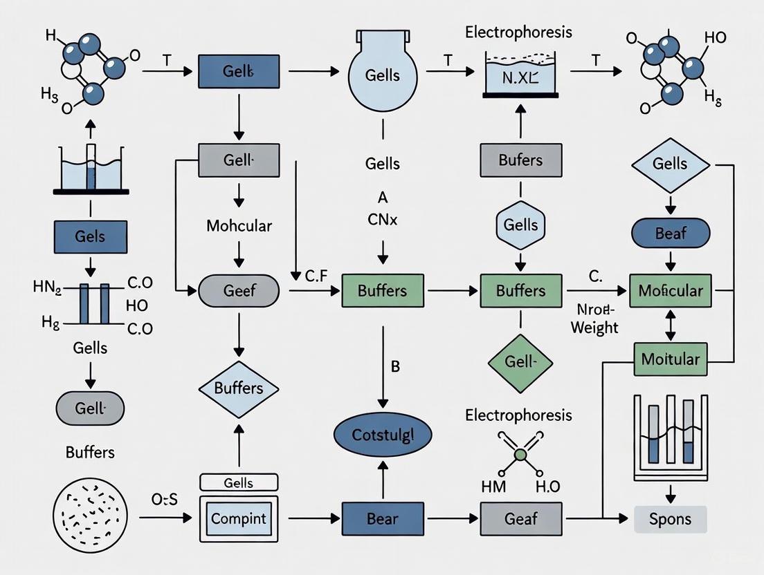
The following diagram illustrates the key decision points for troubleshooting the different manifestations of protein band diffusion.
Troubleshooting Guides & FAQs
Why Are My Protein Bands Smearing or Streaking?
Problem Definition: Smearing appears as continuous, vertical streaks of protein instead of discrete bands [3].
Primary Causes and Solutions:
| Cause | Solution |
|---|---|
| Sample Degradation [4] | Add protease inhibitors; heat samples at 75-95°C immediately after adding sample buffer [4]. |
| Protein Aggregation [5] | Ensure sample buffer has adequate SDS and reducing agents (DTT/BME); heat samples at 95°C for 5 mins [5]. |
| Overloaded Gel [1] | Load less protein; for purified proteins, aim for 0.5–4.0 μg [4]. |
| Improper Transfer [1] | Optimize transfer conditions; avoid excessive current or extended transfer times [1]. |
Why Are My Bands Fuzzy or Abnormally Wide?
Problem Definition: Bands are diffuse, broad, and lack sharpness, sometimes resembling a spread-out blob [1] [5].
Primary Causes and Solutions:
| Cause | Solution |
|---|---|
| Excessive Protein Loading [1] | Optimize and reduce the amount of protein loaded onto the gel [1]. |
| Incorrect Gel Concentration [1] | Use a polyacrylamide gel concentration appropriate for your target protein's molecular weight [1]. |
| Suboptimal Electrophoresis [1] | Adjust voltage and running time; high voltage or prolonged runs can cause overheating and diffusion [1]. |
| Band Diffusion After Running [3] | Image or transfer the gel immediately after electrophoresis to prevent diffusion [3]. |
Why is Band Resolution Poor?
Problem Definition: Bands are closely stacked, poorly separated, and cannot be easily differentiated [3].
Primary Causes and Solutions:
| Cause | Solution |
|---|---|
| Incorrect Gel Percentage [3] | Use a higher percentage gel for better separation of smaller proteins [3]. |
| Insufficient Run Time [3] | Allow the gel to run long enough for sufficient separation, but not so long that bands diffuse [3]. |
| Overloaded Sample [3] | Reduce the amount of protein loaded; overloading leads to fused and warped bands [3]. |
| Incompatible Buffer System [3] | Use fresh running buffer at the correct pH and ensure compatibility between gel and running buffers [5]. |
Research Reagent Solutions
The following table lists essential reagents and materials critical for preventing protein band diffusion.
| Reagent/Material | Function in Preventing Band Diffusion |
|---|---|
| SDS (Sodium Dodecyl Sulfate) [5] | Uniformly coats proteins with a negative charge, ensuring linear migration and preventing aggregation [5]. |
| Reducing Agents (DTT, β-mercaptoethanol) [5] | Breaks disulfide bonds to fully denature proteins, preventing secondary structures that cause smearing [5]. |
| Protease Inhibitor Cocktails [4] | Prevents protein degradation by proteases during sample preparation, which is a common cause of smearing [4]. |
| High-Purity Acrylamide/Bis-acrylamide [1] | Forms a gel with consistent pore size, which is crucial for sharp resolution. Incomplete polymerization leads to poor separation [1] [5]. |
| Fresh Electrophoresis Buffer [5] | Maintains correct ionic strength and pH during a run. Old or incorrect buffers alter migration [5]. |
| Appropriate Gel Concentration [1] | The concentration must be optimized for the target protein's size to provide effective molecular sieving [1]. |
Experimental Workflow for Optimal Results
The diagram below outlines a critical sample preparation workflow to prevent common artifacts that lead to band diffusion.
Frequently Asked Questions (FAQs)
Q: Can my sample buffer itself cause problems? A: Yes. Contaminated sample buffer can introduce keratin, which appears as bands at 55-65 kDa [4]. Furthermore, if a sample buffer with urea is stored improperly, cyanate ions can form and carbamylate proteins, altering their charge and mobility [4]. Always aliquot and store buffers appropriately.
Q: I've heated my sample, but I still see smearing. Why? A: While heating is crucial, excessive heating (e.g., at 100°C for too long) can cleave sensitive peptide bonds, such as Asp-Pro, creating smaller fragments that appear as smearing or extra bands below the main band. If this is suspected, try heating at 75°C for 5 minutes instead [4].
Q: The ladder runs fine, but my protein bands are fuzzy. What does this indicate? A: This typically points to an issue specific to your protein sample, not the gel system itself. The most common causes are overloading your sample with too much protein [1] or issues with the transfer step if you are performing a Western blot (e.g., excessive current or time) [1].
In protein electrophoresis, the quality of your results is directly visible in the bands on your gel. Sharp, well-defined bands are the hallmark of a successful experiment, indicating proper protein separation and integrity. Conversely, diffused or smeared bands often point to issues in sample preparation, gel running, or experimental conditions. This guide provides a detailed visual and analytical framework for troubleshooting protein band diffusion, enabling researchers to diagnose and resolve these common problems effectively.
Visual Identification of Band Patterns
The first step in troubleshooting is to correctly identify the pattern of band distortion. The table below summarizes the key visual indicators and their primary associated causes.
| Visual Pattern | Description of Band Appearance | Primary Associated Causes |
|---|---|---|
| Smearing / Diffused Bands | A continuous, blurry smear running down the lane instead of crisp bands [6]. | Sample degradation by proteases [4]; Improper denaturation [6]; Running gel at excessively high voltage [7]. |
| "Smiling" or "Frowning" Bands | Bands curve upwards ("smiling") or downwards ("frowning") at the edges [6]. | Uneven heat distribution across the gel (Joule heating) [6] [7]. |
| Poor Resolution | Bands are closely stacked, blurry, and overlap, making them difficult to distinguish [6] [7]. | Gel percentage is not optimal for protein size range [6]; Insufficient run time [7]; Overloading of sample [6]. |
| Faint or Absent Bands | Bands are fuzzy, unclear, or completely missing [3]. | Protein concentration too low [4]; Sample leaked from well before run [7]; Gel was over-run, and proteins exited the bottom [3]. |
The following workflow diagram outlines the systematic process for diagnosing these common band issues:
Systematic Troubleshooting of Diffused Bands
Sample Preparation: The Critical First Step
Issues originating at the sample preparation stage are a leading cause of smearing.
- Prevent Protease Degradation: Protein samples added to SDS sample buffer should be heated immediately (at 95-100°C for 5 minutes or at 75°C to avoid cleavage of heat-labile Asp-Pro bonds) to denature and inactivate proteases. Leaving a sample in the buffer at room temperature for extended periods can allow proteases to digest proteins of interest, creating a smear of smaller fragments [4].
- Ensure Complete Denaturation: For SDS-PAGE, proteins must be fully denatured to a linear form. Incomplete denaturation can cause proteins to migrate based on their native shape and charge, not just mass, leading to smearing. Verify that your sample buffer contains sufficient SDS and reducing agent (β-mercaptoethanol or dithiothreitol) [6]. A general recommendation is to maintain a 3:1 ratio of SDS to protein [4].
- Avoid Insoluble Material: After heat treatment, centrifuge your sample briefly (e.g., 2 minutes at 17,000 x g) to remove any insoluble, precipitated material. Loading this precipitate will cause streaking in the gel [4].
- Optimize Protein Load: Overloading a well (>0.5-4.0 µg for a purified protein, depending on well size and stain) can overwhelm the gel's sieving capacity, leading to smeared, warped, or U-shaped bands [4] [6].
Gel Electrophoresis: Optimizing Running Conditions
Even with a perfectly prepared sample, errors during the gel run can cause diffusion.
- Control Voltage and Heat: Running the gel at too high a voltage generates excessive heat (Joule heating), which can denature proteins and cause smearing [6] [7]. A standard practice is to run mini-gels at around 150V. If overheating occurs, run the gel at a lower voltage for a longer duration, or perform the run in a cold room or with a cooling apparatus [7].
- Select the Correct Gel Percentage: The pore size of the polyacrylamide gel determines its resolving power. Use low-percentage gels (e.g., 8%) for large proteins and high-percentage gels (e.g., 15%) for small proteins [8]. An incorrect gel percentage is a primary cause of poor resolution [6].
- Ensure Proper Run Time: Stop the run when the dye front is near the bottom of the gel. Over-running can cause proteins, especially low molecular weight ones, to migrate off the gel, resulting in a blank region or missing bands [3] [7].
The Scientist's Toolkit: Essential Reagent Solutions
The following table lists key reagents and materials critical for preventing band diffusion and ensuring sharp, high-quality results.
| Reagent/Material | Function & Importance in Preventing Band Diffusion |
|---|---|
| SDS (Sodium Dodecyl Sulfate) | An ionic detergent that denatures proteins and confers a uniform negative charge. An excess must be present (recommended 3:1 SDS-to-protein ratio) for linearization and proper migration [8] [4]. |
| Reducing Agents (DTT, β-mercaptoethanol) | Cleave disulfide bonds to fully dissociate protein subunits. This prevents aberrant migration due to incomplete unfolding or aggregation [8] [4]. |
| Protease Inhibitor Cocktails | Added during cell lysis and sample preparation to inhibit endogenous proteases, thereby preventing sample degradation and the smearing it causes [4]. |
| Polyacrylamide Gels | Act as a molecular sieve. The percentage must be matched to the target protein's size for optimal resolution. Gradient gels can resolve a wider size range [8]. |
| Fresh Running Buffer | Conducts current and maintains stable pH. Depleted or incorrect buffer can lead to irregular heating, poor resolution, and smearing [6] [7]. |
| APS & TEMED | Ammonium persulfate (APS) and TEMED are catalysts for polyacrylamide polymerization. Fresh solutions are required to form gels with a uniform matrix for consistent separation [8]. |
Frequently Asked Questions (FAQs)
1. My protein bands are curved ("smiling"). What is the fastest way to fix this? "Smiling" is typically caused by uneven heating across the gel. The fastest solution is to lower the voltage of your run. This reduces overall Joule heating. For a more permanent solution, use a power supply with a constant current mode or ensure your gel apparatus is properly cooled [6] [7].
2. I see a heterogeneous cluster of contaminating bands around 55-65 kDa in my silver-stained gel. What is this? This is likely keratin contamination from skin, hair, or dander. This common artifact can be introduced by touching samples or buffers without gloves, or from dust. To confirm, run a lane with sample buffer alone. To prevent it, practice good laboratory hygiene: wear gloves, use aliquot buffers, and clean surfaces [4].
3. I loaded my samples, but the bands are faint or absent, even though I know the protein is there. What happened? First, check if your protein ladder is visible. If not, the issue is with the electrophoresis setup (e.g., power supply not connected correctly, incorrect buffer). If the ladder is visible, the problem lies with your sample. Potential causes include:
- Diffusion before running: Samples migrated out of the wells because there was a long delay between loading and applying current. Always start the run immediately after loading [7].
- Low concentration: The amount of protein loaded was insufficient for detection. Concentrate your sample or load more volume [4] [6].
- Degradation: Proteins were degraded by proteases before or during preparation [4].
4. My high molecular weight proteins aren't transferring well for western blotting. What can I do? Transfer of high molecular weight proteins is a known challenge. To enhance transfer efficiency, you can:
- Include 0.1% SDS in your transfer buffer.
- Extend the transfer time (e.g., to 16-21 hours).
- Use a two-step transfer protocol or specialized gels with larger pore sizes [9].
Table of Contents
- Core Mechanisms of Band Diffusion
- Troubleshooting Guide: Causes and Solutions
- Experimental Protocols for Optimal Results
- Research Reagent Solutions
- Frequently Asked Questions (FAQs)
Core Mechanisms of Band Diffusion
In protein electrophoresis, sharp, well-defined bands indicate a successful experiment. Band diffusion, smearing, or fuzziness, however, is a common issue that compromises data integrity. This problem primarily stems from three interrelated causes: sample degradation, improper denaturation, and protease activity. Understanding these core mechanisms is the first step in effective troubleshooting.
- Sample Degradation and Protease Activity: Proteins can be degraded by proteases present in the original sample or introduced during isolation. This degradation creates a heterogeneous mixture of protein fragments of various sizes, which manifest as a continuous smear down the lane instead of a sharp band [6]. This activity is exacerbated by mishandling, such as insufficient cooling or repeated freeze-thaw cycles [10].
- Improper Denaturation: For proteins to migrate strictly according to their molecular weight, they must be fully unfolded and uniformly coated with sodium dodecyl sulfate (SDS). Incomplete denaturation, due to insufficient SDS, inadequate reducing agents (DTT or β-mercaptoethanol), or improper heating, leaves proteins with residual secondary or tertiary structure. This leads to abnormal migration patterns, including smearing, aggregation in the wells, or the appearance of multiple bands for a single protein [5] [11].
The diagram below illustrates how these primary causes lead to band diffusion and the corresponding corrective actions.
Troubleshooting Guide: Causes and Solutions
This guide provides a structured approach to diagnosing and resolving the primary causes of band diffusion. The following table summarizes the specific issues related to sample integrity and denaturation, their root causes, and actionable solutions.
| Problem | Primary Cause | Root Cause | Recommended Solution |
|---|---|---|---|
| Sample Degradation | Protease activity | Lysis without protease inhibitors; repeated freeze-thaw cycles; prolonged storage on ice [6] [10]. | Add a broad-spectrum protease inhibitor cocktail to lysis buffer; aliquot samples to minimize freeze-thaw cycles; keep samples on ice during processing [10]. |
| Protein Aggregation | Improper denaturation; hydrophobic proteins | Insufficient heating; old or inactive reducing agents; high salt concentration [12] [11]. | Heat samples at 95-100°C for 5 minutes; use fresh DTT or β-mercaptoethanol; for hydrophobic proteins, add 4-8 M urea to the sample buffer [12] [5] [11]. |
| Incomplete Denaturation | Improper SDS/reducing agent use | Low SDS concentration in sample buffer; insufficient reducing agent to break disulfide bonds [5] [11]. | Ensure sample buffer contains standard 2% SDS; use at least 50 mM DTT or 5% β-mercaptoethanol in sample buffer [5] [10]. |
| General Smearing | Improper electrophoresis conditions | Voltage too high, causing overheating; protein overload [11] [6] [13]. | Run gel at lower voltage (e.g., 100-150V for mini-gels); reduce protein load to 10-20 µg per lane [11] [13]. |
| High Salt Concentration | Improper sample preparation | High salt increases conductivity, distorting migration and causing smearing [11] [6] [10]. | Dialyze samples, precipitate with TCA, or use a desalting column to reduce salt concentration below 100 mM [11] [10]. |
Experimental Protocols for Optimal Results
Protocol 1: Sample Preparation to Minimize Degradation
This protocol is designed to preserve protein integrity from the moment of cell lysis.
- Lysis Buffer Preparation: Prepare a lysis buffer containing 50 mM Tris-HCl (pH 8.0), 150 mM NaCl, 2% SDS, and a broad-spectrum protease inhibitor cocktail. Add fresh 1 mM PMSF (a serine protease inhibitor) for extra protection [11] [10].
- Cell Lysis: Lyse cells directly in the prepared buffer. For bacterial or fungal cells, include a sonication step (3 pulses of 10 seconds each on ice) to ensure complete disruption [12].
- Clarification: Centrifuge the lysate at >12,000 × g for 10 minutes at 4°C to remove insoluble debris and genomic DNA. Transfer the supernatant to a new tube [12].
- Protein Quantification: Use a Bradford or BCA assay to determine protein concentration [14].
- Sample Storage: Aliquot the protein lysate into single-use volumes and flash-freeze in liquid nitrogen before storing at -80°C. Avoid repeated freeze-thaw cycles [11].
Protocol 2: Complete Protein Denaturation
This protocol ensures proteins are fully denatured and reduced for sharp band resolution.
- Sample Buffer Preparation: Prepare a 2X Laemmli sample buffer containing:
- Mixing and Heating: Mix the protein lysate with an equal volume of 2X sample buffer. Vortex thoroughly. Heat the samples at 95-100°C for 5 minutes in a heat block or boiling water bath [5] [10].
- Brief Centrifugation: Briefly spin the tubes in a microcentrifuge to collect condensation and ensure the entire sample is at the bottom of the tube before loading the gel.
Research Reagent Solutions
The following table lists essential reagents for preventing band diffusion, along with their critical functions in sample preparation and electrophoresis.
| Reagent | Function | Technical Specification |
|---|---|---|
| Protease Inhibitor Cocktail | Inhibits a wide range of serine, cysteine, metallo-, and aspartic proteases to prevent sample degradation [10]. | Use a commercial broad-spectrum cocktail as per manufacturer's instructions; add fresh to lysis buffer. |
| SDS (Sodium Dodecyl Sulfate) | Ionic detergent that denatures proteins and confers a uniform negative charge, enabling separation by size [5] [14]. | Final concentration of 1-2% in sample buffer; ensures charge-to-mass ratio is constant. |
| DTT (Dithiothreitol) or β-Mercaptoethanol | Reducing agents that break disulfide bonds within and between protein subunits, facilitating complete unfolding [12] [5]. | Use fresh; final concentration of 50-100 mM DTT or 2-5% β-mercaptoethanol in sample buffer. |
| Urea | Chaotropic agent that disrupts hydrogen bonding, aiding in the solubilization and denaturation of hydrophobic or aggregated proteins [12] [11]. | Add at 4-8 M concentration to sample or lysis buffer for problematic proteins. |
| PMSF (Phenylmethylsulfonyl fluoride) | Serine protease inhibitor that provides additional protection against a common class of proteases [11]. | Add fresh to lysis buffer (0.1-1 mM final concentration); unstable in aqueous solution. |
Frequently Asked Questions (FAQs)
Q1: My bands are fuzzy even though I followed the denaturation protocol. What else could be wrong? Fuzzy bands can also result from issues during the electrophoresis run itself. Running the gel at too high a voltage can generate excessive heat, causing bands to spread and appear fuzzy or smeared [5] [13]. Try reducing the voltage by 25-50% and running the gel for a longer duration. Additionally, ensure your running buffer is fresh and at the correct concentration and pH [11] [6].
Q2: I've added protease inhibitors, but I still see smearing. Could my sample be degraded? Yes, it's possible. Protease inhibitors are not always 100% effective, and degradation can occur very rapidly. Ensure you are using a sufficiently broad cocktail and that you are keeping samples consistently cold during preparation. Also, check that your samples have not undergone multiple freeze-thaw cycles, as this dramatically accelerates degradation [11] [10]. As a test, try preparing a fresh sample from scratch with extra care to cooling and speed.
Q3: How can I tell if my reducing agents (DTT/β-ME) are still active? Old or oxidized reducing agents will fail to reduce disulfide bonds, leading to protein aggregation, horizontal streaking, or multiple bands for a single protein [11]. A simple diagnostic is to prepare a fresh aliquot of DTT or β-ME and compare the banding pattern with your current reagent. Good practice is to make small, single-use aliquots of stock solutions and store them at -20°C to maintain activity.
Q4: My protein of interest is hydrophobic and always smears. What can I do? Hydrophobic proteins are prone to aggregation, even in the presence of SDS. A highly effective solution is to include urea in your sample buffer at a concentration of 4-8 M [12] [11]. This helps to solubilize the protein and prevent aggregation in the well, which can cause severe smearing.
Core Concepts: Gel Percentage and Protein Separation
The polyacrylamide gel matrix acts as a molecular sieve, and its concentration is a primary determinant in the resolution of proteins during SDS-PAGE. The pore size of the gel is inversely proportional to the polyacrylamide concentration; higher percentages create smaller pores, while lower percentages create larger pores [15]. This relationship directly controls which protein sizes can be effectively separated.
Polyacrylamide Gel Percentage Guide
| Gel Percentage (% Acrylamide) | Optimal Protein Size Separation Range | Primary Application |
|---|---|---|
| 4-6% | >200 kDa | Very high molecular weight proteins [16] |
| 8% | 50-200 kDa | High molecular weight proteins [16] |
| 10% | 15-100 kDa | Mid-to-high molecular weight proteins [16] |
| 12.5% | 10-70 kDa | Mid-range molecular weight proteins [16] |
| 15% | 12-45 kDa | Low-to-mid molecular weight proteins [16] |
| Up to 20% | 4-40 kDa | Low molecular weight proteins & peptides [16] |
Using a gel with a pore size inappropriate for your target protein is a common cause of poor band separation. High molecular weight proteins will not migrate efficiently and will stay grouped together near the top in a gel with too high a percentage [15]. Conversely, low molecular weight proteins will migrate too quickly as a group, resulting in poor resolution, in a gel with too low a percentage [15].
Solution: Select a gel percentage appropriate for the size of your protein target. If your proteins of interest span a broad molecular weight range, a gradient gel is highly recommended. Gradient gels provide a continuous range of pore sizes, allowing for the sharp resolution of a wider array of protein sizes on a single gel [16].
Troubleshooting Guide: Poor Band Separation and Diffusion
Band diffusion and poor separation can stem from several factors related to both the gel matrix and experimental conditions. The following table outlines common issues and their solutions.
Troubleshooting Band Diffusion and Separation Issues
| Problem & Symptoms | Possible Cause | Troubleshooting Solution |
|---|---|---|
| Smeared/Diffuse Bands across multiple lanes [17] | Voltage too high; gel overheating [17] | Run gel at lower voltage (e.g., 10-15 V/cm) for a longer time; use a cold room or cooling unit [17] |
| Poor Resolution: Bands are poorly separated or blurry [17] | Gel run time too short; incorrect acrylamide concentration [17] | Run gel until dye front nears bottom; optimize run time for high MW proteins; use correct gel percentage [17] |
| Poor Resolution of all bands | Improperly prepared or overused running buffer [17] [15] | Prepare fresh running buffer with correct ion concentration to ensure proper current flow and pH [17] [15] |
| Bands not separating; single broad band | Protein samples not fully denatured [15] | Ensure sufficient SDS and reducing agent (DTT/β-mercaptoethanol); boil samples 5 min at 98°C, then place on ice [15] |
| Vertical streaking from the well | Sample overloaded; protein precipitation [11] | Load less protein; centrifuge samples before loading to remove insoluble material [11] |
| 'Smiling' bands (curved upwards) | Excessive heat generation during electrophoresis [17] | Decrease voltage; run in a cold room or use an apparatus with a cooling pack to disperse heat evenly [17] |
Experimental Protocol: Optimizing Your SDS-PAGE Run
A. Sample Preparation for Sharp Bands
Proper sample preparation is critical for ensuring proteins are linearized and carry a uniform charge, which allows separation based solely on molecular weight.
- Denaturation: Mix protein sample with an SDS-containing loading buffer (e.g., Laemmli buffer) containing a reducing agent (DTT or β-mercaptoethanol) to break disulfide bonds [15] [18].
- Heating: Boil samples for ~5 minutes at 98°C to fully denature proteins [15].
- Cooling: Immediately after boiling, place samples on ice to prevent renaturation, which can lead to aberrant migration and smearing [15].
- Clarification: Centrifuge samples briefly (e.g., 2 minutes at 17,000 x g) to remove any insoluble debris that could cause streaking [4].
B. Gel Electrophoresis Parameters
- Voltage: A standard practice is to run gels at around 150V. Running at a much higher voltage can cause overheating and smeared bands [17]. For better resolution, run the gel at a lower voltage for a longer duration [17] [15].
- Run Time: Generally, stop the run when the dye front reaches the bottom of the gel. Adjust time accordingly if separating very high or very low molecular weight proteins [17].
The Scientist's Toolkit: Essential Research Reagents
Key Reagents for SDS-PAGE
| Reagent | Function |
|---|---|
| SDS (Sodium Dodecyl Sulfate) | Denatures proteins and confers a uniform negative charge, masking the protein's native charge [15]. |
| DTT or β-Mercaptoethanol | Reducing agents that break disulfide bonds, ensuring complete protein unfolding [15]. |
| Acrylamide/Bis-Acrylamide | Monomers that polymerize to form the porous gel matrix which separates proteins by size [15]. |
| APS & TEMED | Catalyze the polymerization reaction of the polyacrylamide gel. Must be fresh for complete polymerization [15]. |
| Coomassie Brilliant Blue | Dye that binds proteins for visualization. The G-250 variant is often used in sensitive colloidal stains [19]. |
Visualization of Workflow and Concepts
SDS-PAGE Optimization Workflow
Gel Pore Size vs. Protein Migration
Frequently Asked Questions (FAQs)
Q1: My high molecular weight protein (>150 kDa) is not entering the resolving gel. What should I do? A: This indicates the gel pore size is too small. Switch to a lower percentage gel (e.g., 6-8%) to create larger pores that allow large proteins to enter and migrate through the matrix [16] [15].
Q2: I see a cluster of low molecular weight proteins at the bottom of my gel that are not separated. How can I fix this? A: This is a classic sign of using a gel with too low a percentage for small proteins. Use a higher percentage gel (e.g., 15-20%) to create a tighter matrix that will retard the migration of small proteins and improve resolution between them [16] [15].
Q3: What is the advantage of using a gradient gel over a single-percentage gel? A: Gradient gels (e.g., 4-20%) provide a broader effective separation range in a single gel, produce sharper bands as proteins slow down and stack at their pore size limit, and can better separate proteins of similar sizes [16]. They are ideal when analyzing multiple unknown proteins or proteins with a wide mass range.
Q4: I've selected the correct gel percentage, but my bands are still fuzzy. What else should I check? A: Fuzzy bands are often a result of excessive heat. Run the gel at a lower voltage and ensure the apparatus is cool by using a cold room, a cooling unit, or an integrated ice pack [17] [15]. Also, verify that your running buffer is fresh.
Troubleshooting Guides
Guide 1: Addressing Band Smearing and Diffusion
Problem: Protein bands appear as diffuse, fuzzy smears rather than sharp, distinct bands after electrophoresis and transfer.
| Cause Category | Specific Cause | Recommended Solution |
|---|---|---|
| Sample Quality | Protein degradation by proteases [6] [20] | - Use fresh protease and phosphatase inhibitors [20] [18]. - Keep samples on ice during preparation [6] [18]. |
| Incomplete denaturation [6] | - Ensure sample buffer contains fresh SDS and reducing agents (DTT or β-mercaptoethanol) [6] [18]. - Heat denature samples adequately (typically 95°C for 5 minutes) [18]. | |
| Excessive protein load [6] [3] | - Reduce total protein loaded per lane. For whole cell extracts, 20-30 µg is a common starting point [20]. | |
| High salt concentration in sample [6] [3] | - Desalt samples using spin columns or precipitation. - Dilute sample in compatible, low-salt buffer [3]. | |
| Electrophoresis Conditions | Voltage too high [6] [3] | - Run gel at a lower voltage for a longer duration [6] [3]. |
| Incorrect gel concentration [6] [21] | - Use a gel percentage appropriate for your protein's size (see Table 1) [21]. | |
| Gel over-run or under-run [3] | - Optimize run time; monitor dye front migration [3]. | |
| Transfer Conditions | Inefficient transfer [22] [20] | - For high molecular weight proteins (>100 kDa): Add 0.01% SDS to transfer buffer and increase transfer time [22] [20]. - For low molecular weight proteins (<30 kDa): Use 0.2 µm pore membrane and reduce transfer time to prevent "blow-through" [22] [20]. |
| Air bubbles or poor gel-membrane contact [22] | - Roll a glass pipette over the membrane during sandwich assembly to remove air bubbles [22]. |
Guide 2: Resolving Poor Band Resolution
Problem: Bands are poorly separated, too close together, and difficult to distinguish.
| Cause | Solution |
|---|---|
| Suboptimal Gel Concentration [6] [21] | Select a gel percentage optimized for your target protein size range (see Table 1). |
| Overloading Wells [6] [3] | Load a smaller amount of protein per lane [6]. |
| Incorrect Run Time [6] | Run the gel longer for better separation, but avoid excessive run times that cause band diffusion [6]. |
| Voltage Too High [6] | High voltage causes rapid runs but reduces resolution. Use lower voltage for finer separation [6]. |
Guide 3: Fixing Faint or Absent Bands
Problem: Little to no signal is detected for the protein of interest after development.
| Cause Category | Specific Cause | Recommended Solution |
|---|---|---|
| Transfer Issues | Inefficient transfer out of gel [22] [23] | - Confirm power supply was on and connections secure [6]. - Check for air bubbles in transfer sandwich [22] [23]. - Use pre-stained markers to verify transfer efficiency [22]. |
| Over-transfer (blow-through) of small proteins [22] [20] | - For proteins <25-30 kDa, use a 0.2 µm pore membrane and shorten transfer time [22] [20]. | |
| Antibody Issues | Low antibody sensitivity or reactivity [20] | - Use antibodies validated for western blotting. Check species reactivity [20]. |
| Reusing diluted antibodies [20] | - Always use freshly diluted antibodies for optimal results [20]. | |
| Sample & Detection | Insufficient protein concentration [6] [20] | - Increase the amount of protein loaded [6] [20]. - Confirm protein concentration assay is accurate and compatible with your lysis buffer [18]. |
| Low abundance target protein [18] | - Enrich protein prior to electrophoresis using WGA beads (for glycoproteins) or immunoprecipitation [18]. |
Frequently Asked Questions (FAQs)
1. My protein bands are "smiling" (curving upward at the edges). Is this related to voltage or temperature? Yes, this is directly related to temperature. "Smiling" bands are typically caused by uneven heat dissipation across the gel, where the center becomes hotter than the edges, causing samples in the middle to migrate faster. To resolve this, run the gel at a lower voltage to minimize Joule heating, or use a power supply with a constant current mode to maintain a more uniform temperature [6].
2. How does the percentage of methanol in the transfer buffer affect my results? Methanol plays a dual role. It helps remove SDS from protein complexes, improving protein binding to the membrane, but it can also shrink the gel pores, making it harder for large proteins to escape. For most proteins, a concentration of 10-20% is recommended [22]. For high molecular weight proteins (>100 kDa), consider reducing methanol to 5-10% to facilitate transfer [20].
3. I see multiple non-specific bands. Could this be caused by my buffer system? While multiple bands can indicate antibody cross-reactivity or protein isoforms, the buffer system can contribute. Using an incorrect blocking agent or primary antibody dilution buffer can cause high background and non-specific binding. Always use the antibody manufacturer's recommended dilution buffer (e.g., BSA vs. non-fat dry milk) and ensure your washing buffer contains TBS (not PBS) with 0.1% Tween-20 [20].
4. Why did my transfer current run abnormally high? An abnormally high current is most often a buffer issue. If the transfer buffer is too concentrated, it increases conductivity and current. High current can also occur if Tris-HCl is accidentally used instead of Tris base, resulting in low buffer pH and increased conductivity. Remake the transfer buffer according to the correct recipe and avoid adjusting pH with acid/base [22].
Data Presentation
Table 1: Optimal SDS-PAGE Gel Concentration for Protein Separation
Use this table to select the right gel percentage for your target protein, which is critical for preventing smearing and poor resolution [21].
| Protein Molecular Weight Range | Recommended Gel Concentration |
|---|---|
| 100 - 600 kDa | 4% |
| 50 - 500 kDa | 7% |
| 30 - 300 kDa | 10% |
| 10 - 200 kDa | 12% |
| 3 - 100 kDa | 15% |
Experimental Workflow and Diagnostics
The following diagram outlines a logical troubleshooting workflow for diagnosing the root cause of protein band diffusion, integrating checks for voltage, temperature, and buffer systems.
Troubleshooting Band Diffusion
The Scientist's Toolkit: Essential Research Reagent Solutions
| Reagent/Kit | Primary Function | Key Considerations |
|---|---|---|
| Protease Inhibitor Cocktail | Prevents proteolytic degradation of sample proteins by inhibiting a broad spectrum of proteases [20] [18]. | Essential for maintaining sample integrity. Use a commercial 100X cocktail or a combination of PMSF, leupeptin, and aprotinin [20] [18]. |
| Phosphatase Inhibitor Cocktail | Preserves protein phosphorylation states by inhibiting serine/threonine and tyrosine phosphatases [20] [18]. | Critical for detecting post-translational modifications. Include sodium orthovanadate, β-glycerophosphate, and sodium fluoride [20]. |
| RIPA Lysis Buffer | A denaturing buffer for efficient extraction of total, membrane-bound, and nuclear proteins [18]. | Contains ionic (SDS) and non-ionic (Triton) detergents. Disrupts protein-protein interactions. Ideal for whole cell extracts [18]. |
| Laemmli Sample Buffer (2X) | Prepares protein samples for SDS-PAGE by denaturing, reducing disulfide bonds, and adding tracking dye [18]. | Must contain SDS and a reducing agent (DTT or β-mercaptoethanol). Always heat samples after mixing [18]. |
| Pre-cast Protein Gels | Provides consistent, optimized polyacrylamide gels for reproducible protein separation by molecular weight. | Saves time and reduces variability. Available in various percentages and formats (e.g., mini-gels, gradient gels) [21]. |
| PVDF or Nitrocellulose Membrane | Serves as the solid support for immobilizing proteins after gel electrophoresis for antibody probing [22] [20]. | PVDF has higher binding capacity. For proteins <25-30 kDa, use a 0.2 µm pore size to prevent loss [22] [20]. |
| Enhanced Chemiluminescence (ECL) Substrate | Enables sensitive detection of target proteins through an enzyme (HRP)-catalyzed light-emitting reaction. | Choice of substrate (standard vs. high-sensitivity) depends on target protein abundance. Fresh preparation is key [20]. |
Why Band Clarity Matters
In molecular biology research, the clarity of bands on an electrophoresis gel is not merely an aesthetic concern; it is a fundamental prerequisite for generating reliable and reproducible data. Sharp, well-resolved bands are critical for accurate molecular weight determination and for any subsequent downstream analysis, such as protein identification or nucleic acid sequencing. Band diffusion—the blurring or smearing of these bands—directly compromises data integrity by obscuring the true size and quantity of your target molecules, leading to potential misinterpretation of results and hindering scientific progress [3] [19].
This guide provides a systematic approach to troubleshooting protein band diffusion, offering clear solutions to achieve high-resolution results.
Troubleshooting Guide: Resolving Band Diffusion and Smearing
The following table outlines the common causes of poor band clarity and their respective solutions.
| Problem & Symptom | Primary Cause | Recommended Solution |
|---|---|---|
| Faint BandsLow signal, bands unclear or absent [3] | Insufficient sample quantity or degraded sample [3] [24] | Load 0.1–0.2 μg of DNA per mm of well width [3]. Use fresh, nuclease-free reagents and practices to prevent degradation [3] [24]. |
| Smeared BandsDiffuse, blurry bands that lack sharpness [3] [5] | Improper sample preparation (incomplete denaturation, contaminants) [3] [5] | For proteins, ensure samples are boiled with SDS and reducing agents (e.g., DTT) [5]. For nucleic acids, remove proteins and salts via purification [3]. |
| Poorly Separated BandsBands are too close together, poorly resolved [3] | Incorrect gel concentration or type [3] | Use a gel percentage appropriate for your target's size; higher % for smaller molecules [3]. Use denaturing gels for single-stranded nucleic acids [3]. |
| 'Smiling' BandsBands curve upwards at the edges [25] | Uneven heating during electrophoresis, often from high voltage [25] | Run the gel at a lower voltage. Ensure the electrophoresis tank is functioning correctly with secure contacts [25]. |
| Fuzzy Protein Bands (SDS-PAGE)Diffuse protein bands after Western blot [5] | Incomplete gel polymerization or overly long run times [5] | Ensure gels are fully polymerized before use. Follow recommended run times and voltages to prevent overheating and diffusion [5]. |
Step-by-Step Experimental Protocol: Improved Protein Staining
To achieve higher resolution for protein visualization with Coomassie Brilliant Blue (CBB) staining, follow this modified protocol, which adds a crucial fixation step to prevent protein diffusion during washing [19].
Protocol Details:
- Fixation Solution: 40% methanol, 10% acetic acid [19].
- Fixation Time: A minimum of 30 minutes with shaking is essential. This step can be extended overnight for convenience without detriment [19].
- Staining Solution: Prepare a colloidal CBB-G solution containing 0.02% (w/v) CBB G-250, 5% (w/v) aluminium sulfate, 10% (v/v) ethanol, and 2% (v/v) orthophosphoric acid [19].
- Key Modification: The fixation step prior to staining immobilizes the proteins within the gel matrix, preventing them from washing out or diffusing, which results in significantly sharper band resolution [19].
The Scientist's Toolkit: Essential Research Reagent Solutions
The table below lists key reagents and their specific functions in ensuring clear electrophoresis results.
| Reagent / Material | Function & Importance |
|---|---|
| SDS (Sodium Dodecyl Sulfate) | Denatures proteins and confers a uniform negative charge, ensuring separation is based primarily on molecular weight [5]. |
| Reducing Agents (DTT, β-mercaptoethanol) | Breaks disulfide bonds within and between proteins, ensuring complete unfolding and preventing aggregated or multiple bands [5]. |
| Protease Inhibitor Cocktail | Prevents protein degradation during sample preparation, which is a common cause of smearing or multiple lower-weight bands [24]. |
| Ultrapure Agarose / Acrylamide | Provides a consistent, pure matrix for separation. Low sulfate content in agarose minimizes electroendosmosis, which can distort bands [26]. |
| TAE vs. TBE Buffer | TAE: Better for resolving longer DNA fragments (>1 kb). TBE: Preferred for smaller DNA fragments and longer run times due to higher buffering capacity [25]. |
| Appropriate DNA Ladder | A chromatography-purified ladder with bands in your size range of interest is critical for accurate molecular weight determination [25]. |
| Colloidal CBB-G Stain | A highly sensitive staining method. The modified protocol with fixation provides superior band sharpness and is MS-compatible [19]. |
Frequently Asked Questions (FAQs)
Q1: I have confirmed my protein sample is intact, but my bands are still fuzzy after Western blotting. What else should I check? A: Beyond sample integrity, investigate your gel and running conditions. Ensure your polyacrylamide gel has polymerized completely, as incomplete polymerization creates uneven pore sizes and poor resolution [5]. Also, verify that your running buffer is fresh and at the correct pH, and avoid using excessively high voltage, which can generate heat and cause band diffusion [5].
Q2: Why is it critical to load an appropriate amount of sample? A: Both overloading and underloading samples cause problems. Overloading leads to smearing, distorted band shapes, and inaccurate migration, making the fragment appear larger than it is [3] [25]. Underloading results in bands that are too faint to detect reliably [25]. Accurate quantification and loading are essential.
Q3: My nucleic acid bands are smeared. What are the first things to check? A: The most common causes are nuclease contamination or improper sample handling, which degrades the nucleic acids [3] [24]. Ensure all reagents and labware are nuclease-free and use good laboratory practices (e.g., wearing gloves). Also, check that your sample is not in a high-salt buffer, which can interfere with clean migration [3].
Q4: How does gel fixation improve band resolution in CBB staining? A: The fixation step (using methanol and acetic acid) precipitates and immobilizes the proteins within the gel matrix immediately after electrophoresis [19]. This prevents the proteins from diffusing out of the gel or spreading during subsequent washing and staining steps, thereby preserving sharp, high-resolution bands [19].
Optimal SDS-PAGE Protocols: Preventing Band Diffusion Through Proper Technique
Protein band diffusion after electrophoresis presents a significant challenge in biomedical research, often leading to blurred results, poor resolution, and difficulties in accurate data interpretation. This problem frequently originates from suboptimal sample preparation protocols, particularly during the critical stages of denaturation, reduction, and heating. For researchers and drug development professionals, inconsistent or improperly prepared samples can compromise experimental reproducibility, waste precious reagents, and hinder scientific progress. This guide addresses the specific sample preparation factors that contribute to band diffusion and provides targeted troubleshooting methodologies to enhance western blot clarity and reliability, thereby strengthening the overall validity of protein analysis in research settings.
FAQs: Addressing Common Sample Preparation Challenges
1. What causes smeared bands in my western blot, and how can sample preparation fix this?
Smeared bands often result from incomplete denaturation, protein degradation, or aggregation. During sample preparation, ensure you use fresh reducing agents, adequate SDS concentration, and appropriate heating conditions to achieve complete linearization of proteins. Protein degradation can be minimized by adding protease inhibitor cocktails to your lysis buffer and keeping samples on ice during preparation [27] [28]. For proteins prone to aggregation, consider using lower heating temperatures (e.g., 70°C) for longer durations instead of boiling at 95-100°C [28].
2. Why are my protein bands faint or poorly resolved after electrophoresis?
Faint bands typically indicate insufficient protein loading, incomplete transfer, or protein degradation. First, confirm your protein concentration using a reliable assay (Bradford, BCA, or Lowry) [27]. Ensure your sample buffer maintains a proper SDS-to-protein ratio (recommended 3:1 ratio) for complete denaturation [4]. Overly diluted samples or insufficient staining can also cause faint bands—concentrate samples if necessary and verify staining protocols [3].
3. How does improper heating affect my protein samples during preparation?
Heating is crucial for denaturation but can cause multiple issues if improperly applied. Excessive heating (95-100°C for extended periods) can cleave Asp-Pro bonds in proteins [4]. Conversely, insufficient heating fails to completely denature proteases that remain active at room temperature, leading to protein degradation [4]. Heating samples without first mixing with sample buffer causes irreversible aggregation, similar to boiling an egg [29].
4. My small molecular weight proteins disappear from the gel—what's happening?
Small proteins (<15 kDa) may transfer completely through standard 0.45 μm membranes. Use a 0.2 μm pore size membrane to better retain small proteins [30] [28]. Additionally, reduce transfer time for small proteins to prevent over-transfer—for proteins 10-25 kDa, 15 minutes at 25V is often sufficient [30]. During sample preparation, avoid over-heating or excessive sonication that might fragment proteins.
Troubleshooting Guide: Protein Band Diffusion
Problem: Smeared or Diffuse Protein Bands
| Possible Cause | Specific Issue | Solution | Reference |
|---|---|---|---|
| Incomplete Denaturation | Insufficient SDS or reducing agent | Use SDS-to-protein ratio of 3:1; ensure fresh DTT (160 mM) or β-mercaptoethanol | [29] [4] |
| Protein Degradation | Protease activity in sample | Add protease inhibitors; heat samples immediately after adding buffer (75°C for 5 min) | [27] [4] |
| Improper Heating | Protein aggregation at high heat | Heat at 70°C for 5-10 min or 37°C for 30-60 min for sensitive proteins | [27] [28] |
| Sample Overloading | Too much protein per lane | Load 0.1-0.2 μg protein per mm well width; for mini-gels, 30 μg total protein is often optimal | [3] [31] |
| Incorrect Buffer | High salt concentration | Dilute sample in nuclease-free water or desalt before adding loading buffer | [3] |
Quantitative Data for Sample Preparation
Table: Optimal Sample Buffer Components and Concentrations
| Component | Function | Recommended Concentration | Special Notes | |
|---|---|---|---|---|
| SDS (Sodium Dodecyl Sulfate) | Denatures proteins by adding negative charge | 1-4% in loading buffer | Critical for disrupting 2° and 3° structure; binds 1.4:1 mass ratio with protein | [29] |
| DTT or β-mercaptoethanol | Reduces disulfide bonds | 100-160 mM DTT or 5-10% β-mercaptoethanol | DTT preferred due to less odor; essential for reducing covalent bonds | [27] [29] |
| Glycerol | Increases density for well loading | 10-20% | Prevents sample floating out of wells | [27] [29] |
| Tris-HCl | Maintains pH for electrophoresis | 10-125 mM, pH 6.8 | Essential for discontinuous electrophoresis system | [27] [29] |
| Tracking Dye | Visualizes migration | 0.004-0.1% bromophenol blue | Monitors electrophoresis progress | [27] [29] |
Table: Protein Loading Recommendations Based on Application
| Application | Mini-Gel Loading Amount | Optimal Sample Concentration | Well Utilization | |
|---|---|---|---|---|
| Coomassie Staining | 40-60 μg (crude samples) 0.5-4 μg (purified protein) | 1-5 mg/mL | At least 30% of well volume | [4] |
| Western Blot | 20-30 μg total protein | 0.1-5 mg/mL | 10-20 μL per mini-gel well | [27] [31] |
| Silver Staining | 10-100x less than Coomassie | Adjusted accordingly | Similar well utilization | [4] |
Experimental Protocols
Standard Sample Denaturation Protocol for SDS-PAGE
This protocol ensures complete protein denaturation while minimizing artifacts that cause band diffusion.
Materials Needed:
- 2X Laemmli sample buffer (4% SDS, 10% 2-mercaptoethanol, 20% glycerol, 0.004% bromophenol blue, 0.125 M Tris HCl, pH 6.8) [27]
- Heating block or water bath
- Microcentrifuge tubes
- Protein sample (concentration previously determined)
- Protease inhibitor cocktail [28]
Step-by-Step Methodology:
- Determine protein concentration using Bradford, BCA, or Lowry assay with BSA standards [27].
- Dilute protein samples to desired concentration using appropriate lysis buffer. The optimal final concentration for loading is typically 1-5 mg/mL [27].
- Mix sample with equal volume of 2X Laemmli buffer (1:1 ratio) [27]. For a 20 μL final volume, combine 10 μL protein sample with 10 μL 2X buffer.
- Heat samples at 70-75°C for 5-10 minutes [4] [28]. For membrane proteins or aggregation-prone proteins, use 70°C for 10 minutes instead of higher temperatures.
- Briefly centrifuge samples for 1-2 minutes to collect condensation and any insoluble material [29].
- Load appropriate volume onto gel immediately or store at -20°C for future use.
Troubleshooting Notes:
- If using urea in sample buffer, treat with mixed bed resin to remove cyanate ions that cause protein carbamylation [4].
- For viscous samples (high nucleic acid content), add Benzonase Nuclease or briefly sonicate to reduce viscosity [4].
- Remove insoluble material by centrifugation at 17,000 x g for 2 minutes before loading to prevent streaking [4].
Optimization Protocol for Problematic Proteins
Some proteins require specialized handling to prevent band diffusion and artifacts.
For Membrane or Hydrophobic Proteins:
- Add 6-8 M urea or nonionic detergent (Triton X-100) to sample buffer to improve solubility [4].
- Extend heating time at lower temperature (37°C for 30-60 minutes) to prevent aggregation [28].
For Proteins Prone to Degradation:
- Add protease inhibitors immediately upon cell lysis [27] [28].
- Process samples quickly at 4°C and heat immediately after adding sample buffer [4].
- Aliquot sample buffer and store at -80°C to prevent keratin contamination [4].
For Nuclear or DNA-Binding Proteins:
- Sonicate lysates to release proteins from DNA binding [28].
- Treat with Benzonase Nuclease to degrade nucleic acids that cause viscosity [4].
Signaling Pathways and Workflows
Sample Preparation Workflow for Optimal Denaturation
Troubleshooting Decision Pathway for Band Diffusion
The Scientist's Toolkit: Research Reagent Solutions
Table: Essential Reagents for Optimal Sample Preparation
| Reagent | Function | Specific Application Notes |
|---|---|---|
| SDS (Sodium Dodecyl Sulfate) | Denatures proteins by binding to polypeptide chains with constant mass ratio (1.4:1) | Use high-quality grade; old SDS causes indistinct bands and background staining [27] |
| DTT (Dithiothreitol) | Reduces disulfide bonds; preferred over β-mercaptoethanol due to less odor | Prepare fresh solutions; standard concentration 100-160 mM in sample buffer [29] |
| Protease Inhibitor Cocktails | Prevents protein degradation by cellular proteases during sample preparation | Add immediately to lysis buffer; specific inhibitors may be needed for particular proteases [27] |
| Tris-HCl Buffer | Maintains pH at 6.8 for proper stacking in discontinuous electrophoresis | Critical for sample buffer system; ensures proper protein migration [29] |
| Glycerol | Increases density of sample for loading into wells | Prevents sample diffusion from wells before electrophoresis; 10-20% final concentration [27] [29] |
| Urea | Additional denaturant for difficult proteins (membrane, hydrophobic) | Use fresh solutions treated with mixed bed resin to remove cyanate ions [4] |
| Benzonase Nuclease | Degrades DNA/RNA to reduce sample viscosity | Particularly useful for crude cellular extracts; eliminates nucleic acid-induced viscosity [4] |
Correct Gel Percentage Selection Based on Target Protein Molecular Weight
Protein band diffusion, smearing, and poor resolution are common challenges in SDS-PAGE that can compromise experimental data. A primary factor influencing these issues is the selection of an appropriate polyacrylamide gel percentage, which determines the gel's pore size and sieving properties. This guide provides a structured framework to select the correct gel composition based on your target protein's molecular weight, directly addressing a key variable in troubleshooting band diffusion.
► Gel Percentage Selection Guide
The following table summarizes the recommended polyacrylamide gel percentages for optimal separation of proteins based on their molecular weight.
Table 1: Recommended Gel Percentage for Target Protein Size
| Protein Size (kDa) | Recommended Gel Percentage | Primary Application |
|---|---|---|
| >200 kDa | 4-6% [32] | Separation of very high molecular weight proteins [32] |
| 50-200 kDa | 8% [32] [33] | General separation of high molecular weight proteins [33] |
| 15-100 kDa | 10% [32] [33] | Standard broad-range separation [33] |
| 10-70 kDa | 12.5% [32] [33] | Separation of medium molecular weight proteins [33] |
| 12-45 kDa | 15% [32] [33] | Resolution of low molecular weight proteins [33] |
| 4-40 kDa | 15-20% [32] [33] | High-resolution separation of very low molecular weight proteins & peptides [33] |
For proteins with isoforms spanning a wide molecular weight range or when probing for multiple proteins of different sizes, gradient gels (where acrylamide concentration increases from top to bottom) are recommended for optimal separation across a broad size spectrum [32].
► Troubleshooting FAQs
1. My protein bands are smeared or diffused. What could be the cause? Smeared bands can result from several factors related to gel concentration and running conditions:
- Incorrect Gel Percentage: Using a gel with a percentage that is too high for your protein can trap large proteins, while a percentage that is too low can cause small proteins to migrate too quickly and poorly resolve [15]. Consult Table 1 to confirm you are using the correct gel percentage.
- Improper Sample Preparation: Incomplete protein denaturation can cause proteins to migrate in folded states, leading to smearing. Ensure samples are boiled with sufficient SDS and fresh reducing agent (e.g., DTT) [15] [34].
- Excessive Voltage: Running the gel at too high a voltage generates heat, which can cause band diffusion and smiling effects [35] [6]. Troubleshoot by reducing the voltage and increasing the run time.
- Sample Overloading: Loading too much protein per well can overwhelm the gel's capacity, causing proteins to aggregate and smear [15] [34]. Reduce the amount of protein loaded.
2. I see poor separation between bands that are close in size. How can I improve resolution? Poor band resolution is often directly linked to the gel's sieving properties:
- Suboptimal Gel Concentration: The gel percentage is the most critical factor for resolution [6]. For proteins of very similar size, a higher percentage gel will provide better differentiation [15].
- Insufficient Run Time: If the gel is not run long enough, proteins will not have adequate distance to separate from one another. Ensure the dye front is near the bottom before stopping the run [35].
- Gel Polymerization Issues: Incomplete or uneven polymerization creates an inconsistent matrix, hindering sharp resolution. Ensure gel components are fresh and properly mixed [15].
3. My high molecular weight protein is not entering the gel. What should I do? This is a classic sign that the gel pore size is too small. High molecular weight proteins require larger pores for efficient migration.
- Solution: Switch to a low-percentage gel, such as 4-6% or 8%, to create a more open matrix that allows large proteins to enter and migrate [15] [32].
4. My low molecular weight protein ran off the gel. How do I prevent this? This occurs when the gel matrix is too loose, allowing small proteins to migrate virtually unimpeded.
- Solution: Use a higher percentage gel (e.g., 15-20%) to create a tighter mesh that retards the movement of small proteins and provides better resolution [15].
► Experimental Workflow for SDS-PAGE
The following diagram outlines the key decision points and steps in the SDS-PAGE workflow, from sample preparation to analysis, highlighting how to prevent band diffusion.
► The Scientist's Toolkit: Essential Reagents and Materials
Table 2: Key Research Reagent Solutions for SDS-PAGE
| Reagent/Material | Function | Key Considerations |
|---|---|---|
| Acrylamide/Bis-acrylamide | Forms the cross-linked polyacrylamide gel matrix that separates proteins by size. | Standard stock is 30% w/w, typically at a 37.5:1 ratio; concentration determines gel percentage and pore size [33]. |
| SDS (Sodium Dodecyl Sulfate) | Denatures proteins and confers a uniform negative charge, enabling separation by size alone. | Ensures proteins are linearized and masks their native charge; critical for accurate molecular weight estimation [15]. |
| TEMED & APS | Catalyzes (TEMED) and initiates (APS) the free-radical polymerization of acrylamide. | These components must be fresh for complete and uniform gel polymerization [15]. |
| Tris-Glycine Running Buffer | Conducts current and maintains the pH (typically 8.3-8.8) required for protein migration. | Must be fresh and correctly diluted; overused or improper buffer can cause poor resolution and smearing [35] [32]. |
| Reducing Agent (DTT/BME) | Breaks disulfide bonds within and between protein subunits, aiding complete denaturation. | Use fresh aliquots; re-oxidation during run can cause vertical streaking or ghost bands [34] [36]. |
| Protein Molecular Weight Marker | Provides reference bands for estimating the molecular weight of unknown proteins. | Essential for verifying the gel run and transfer efficiency. |
In the context of troubleshooting protein band diffusion in electrophoresis, the preparation and maintenance of buffers are critical foundational steps. Improper pH and ionic strength are frequent, yet often overlooked, culprits behind smearing, poor resolution, and distorted bands. This guide addresses common buffer-related issues to ensure the integrity of your protein separation results.
Troubleshooting Guides
FAQ: How do buffer pH and ionic strength cause protein band smearing and diffusion?
Band smearing and diffusion occur when proteins do not migrate as sharp, discrete zones. Incorrect buffer conditions are a primary cause, as they disrupt the uniform charge and sieving process essential for SDS-PAGE.
- Cause: The buffer's ionic strength is too high, often due to excess salts in the protein sample itself. This creates a local region of high conductivity, leading to uneven heating and distorted migration [34] [11] [6].
- Cause: The buffer's pH is incorrect or unstable. If the pH is not within the optimal range for the buffer system, proteins may not be fully denatured or may not maintain a consistent charge-to-mass ratio, leading to poor stacking and separation [34] [37].
- Cause: The buffer has been over-diluted or is too concentrated. This directly affects the ionic strength, leading to unusually fast or slow migration, poor resolution, and increased heat generation [34] [38].
- Cause: Using old or contaminated running buffer. Buffer depletion or microbial growth can alter pH and ionic strength over time, compromising separation quality [6].
FAQ: What are the best practices for preparing and storing electrophoresis buffers to ensure consistency?
Consistency in buffer preparation is non-negotiable for reproducible electrophoresis results. Small deviations can have pronounced effects on separation quality.
- Precise Recipe Definition: A method described simply as "25 mM phosphate pH 7.0" is ambiguous and irreproducible. The standard operating procedure must specify the exact salt form (e.g., disodium hydrogen phosphate) and the detailed pH adjustment procedure, including the concentration and volume of the acid or base used [37].
- Avoid Diluting pH-Adjusted Stock Solutions: A common error is diluting a concentrated, pH-adjusted stock buffer. This practice can alter the final pH. For example, diluting a 2 M sodium borate stock (pH 9.4) to 500 mM resulted in a pH shift to 9.33. Best practice is to prepare the buffer at its final working concentration and pH [37].
- Proper pH Meter Use and Temperature: Always calibrate the pH meter with fresh buffers and ensure the electrode is clean. Measure the pH of the buffer at the temperature at which it will be used, as pH is temperature-dependent. Allow heated buffer solutions to cool to room temperature before measuring pH [37].
- Fresh Preparation and Storage: Prepare running buffer fresh for optimal results. If storage is necessary, label the buffer with the date of preparation and store it in a sealed container at room temperature for a short period. Avoid re-using running buffer, as its composition changes during electrophoresis [34] [6].
Experimental Protocols
Protocol: Correcting High Salt Concentration in Protein Samples
High salt in samples is a frequent cause of smearing and distorted bands. This protocol outlines a reliable desalting procedure.
Methodology:
- Precipitation with Trichloroacetic Acid (TCA): Precipitate the protein using TCA, then reconstitute the pellet in a low-salt buffer or SDS-PAGE sample buffer [11].
- Dialysis: Transfer the protein sample into a dialysis tubing with a suitable molecular weight cutoff. Dialyze against a large volume of a low-salt buffer (e.g., Tris-HCl) for several hours to overnight at 4°C, with at least one buffer change [34] [11].
- Desalting Column: Use a size-exclusion desalting column (e.g., Sephadex G-25). Equilibrate the column with your desired buffer, load the sample, and elute. The proteins will elute in the void volume, separated from the smaller salt ions [34] [11].
Key Materials:
- Trichloroacetic Acid (TCA)
- Dialysis tubing and clips
- Desalting column (e.g., Sephadex G-25)
- Low-salt buffer (e.g., 50 mM Tris-HCl, pH 6.8)
Protocol: Systematic Preparation of a Tris-Glycine SDS Running Buffer
Tris-glycine is the standard running buffer for many SDS-PAGE systems. Accurate preparation is key.
Methodology:
- Weighing and Dissolving: Accurately weigh 3.03 g of Tris base, 18.8 g of glycine, and 1.0 g of SDS. To ensure the SDS dissolves completely, add the components to approximately 800 mL of deionized water and stir thoroughly [34] [38].
- pH Verification: The pH of the 1X Tris-glycine running buffer should be approximately 8.3. It is good practice to verify the pH with a calibrated meter. Note: No adjustment is typically needed if the reagents are pure and weighed correctly [34].
- Final Volume and Storage: Bring the final volume to 1.0 liter with deionized water. Mix thoroughly. For best results, use the buffer immediately. It can be stored at room temperature for a short period in a sealed bottle.
Key Materials:
- Tris base (MW 121.14)
- Glycine (MW 75.07)
- SDS (Sodium Dodecyl Sulfate)
Key Reagent Solutions
The following table details essential reagents for troubleshooting buffer-related diffusion issues.
Research Reagent Solutions for Buffer Troubleshooting
| Reagent Name | Function in Troubleshooting | Key Considerations |
|---|---|---|
| Desalting Columns (e.g., Sephadex G-25) | Rapidly removes excess salts from protein samples prior to loading. | Fast, spin-column format is convenient for small sample volumes. [34] [11] |
| Dialysis Membrane | Removes salts, detergents, and other small contaminants via slow diffusion. | Requires more time than columns; suitable for larger volumes. [34] [11] |
| Trichloroacetic Acid (TCA) | Precipitates proteins, allowing resuspension in a clean, low-salt buffer. | Can be denaturing; may not be suitable for all downstream applications. [11] |
| Dithiothreitol (DTT) | A reducing agent that breaks disulfide bonds to prevent protein aggregation. | Must be fresh; prepare solutions immediately before use. [39] [34] |
| Urea (4-8 M) | A chaotrope that helps solubilize hydrophobic or aggregated proteins in the sample. | Can cleave peptide bonds at high temperatures; do not heat urea over 37°C. [39] [11] |
Buffer Troubleshooting Workflow
The following diagram outlines the logical relationship between buffer-related problems, their root causes, and the recommended investigative and corrective actions to resolve protein band diffusion.
FAQs and Troubleshooting Guides
Q1: What causes smeared or diffused bands in my protein gel, and how can I fix it?
Smeared bands are a common indicator of protein band diffusion and can stem from several issues related to your electrophoresis conditions and sample preparation.
Possible Cause: Excessive Voltage and Heat Running your gel at too high a voltage generates excessive Joule heating, which can denature proteins and cause smearing [6] [40]. A good practice is to run your gel at 10-15 volts per cm of gel distance [40]. Using a lower voltage for a longer run time allows for better heat dissipation and sharper bands [40].
Possible Cause: Sample Overloading Loading too much protein (>0.1-0.2 μg per mm of well width) can overwhelm the gel's sieving capacity, leading to trailing smears and U-shaped bands [3]. Ensure you load an appropriate amount of protein for your well size.
Possible Cause: Sample Degradation or Impurities Sample degradation by proteases or contamination with high amounts of salt or protein can cause smearing [6] [3]. Handle samples carefully, use fresh, sterile reagents, and purify samples to remove contaminants like salt or interfering proteins [6] [3].
Q2: Why are my protein bands curved ("smiling" or "frowning"), and how do I achieve straight bands?
Non-linear band migration is almost always a result of uneven heat distribution across the gel [6]. The center of the gel often becomes hotter than the edges, causing samples in the middle to migrate faster, creating a "smiling" pattern [6] [25].
- Solution: Improve Heat Dissipation
- Reduce the Voltage: Minimizing voltage reduces Joule heating [6] [25].
- Use Active Cooling: Run the gel in a cold room or place ice packs inside the gel-running apparatus to maintain a uniform temperature [40].
- Ensure Proper Setup: Verify that the gel is properly aligned, the buffer level is consistent, and electrodes are straight to ensure a uniform electric field [6].
Q3: My protein bands are poorly resolved and too close together. How can I improve resolution?
Poor resolution occurs when bands are not sufficiently separated, making them difficult to distinguish.
Primary Cause: Incorrect Gel Concentration The gel concentration is the single most important factor for resolution [6]. A gel with pores that are too large will not separate small fragments well, while pores that are too small will impede the migration of large proteins [6] [3]. Use a gel percentage optimized for the molecular weight range of your target proteins. For higher resolution of low molecular weight proteins, a higher percentage gel is often needed [3].
Other Contributing Factors:
- Run Time: Running the gel for too short a time will not allow for sufficient separation [6] [40]. A standard practice is to run the gel until the dye front is near the bottom, but this may need optimization based on protein size [40].
- Buffer Issues: An incorrect or depleted running buffer can compromise separation by altering pH and ion concentration [6] [40]. Always use fresh buffer prepared at the correct concentration.
Q4: The protein samples diffused out of the wells before I started the run. What happened?
This indicates a procedural error where samples were loaded but the electric current was not applied promptly.
- Explanation and Solution: The electric current ensures streamlined migration of proteins from the wells. If there is a lag between loading and applying power, samples will diffuse haphazardly out of the wells [40]. To prevent this, minimize the time between loading your first sample and starting the electrophoresis run [40]. Load your samples swiftly and start the run immediately after.
The table below summarizes key parameters for optimizing SDS-PAGE conditions to prevent band diffusion.
| Parameter | Recommended Range / Condition | Effect on Experiment & Notes |
|---|---|---|
| Voltage | 10-15 V/cm of gel [40] | Lower voltages minimize Joule heating; higher voltages cause smearing and smiling [6] [40]. |
| Run Time | Until dye front is ~1 cm from bottom of gel [40] | Too short: poor separation [40]. Too long: band diffusion and over-running [3]. |
| Sample Load | 0.1-0.2 μg of protein per mm of well width [3] | Overloading causes smearing, trailing, and poor resolution [6] [3]. |
| Temperature Control | Use active cooling (cold room, ice packs) [40] | Critical for managing Joule heating and preventing smiling bands and sample degradation [6] [40]. |
| Buffer Management | Use fresh, correctly prepared buffer; can be reused 1-2 times [41] | Depleted or incorrect buffer alters conductivity, pH, and leads to poor resolution [6] [40]. |
| Gel Concentration | Optimized for target protein size (e.g., 8-10% for standard separation) [40] | The most critical factor for resolution; higher % for smaller proteins, lower % for larger proteins [6] [3]. |
Experimental Protocol for Reproducible SDS-PAGE
This detailed protocol is designed to minimize protein band diffusion by controlling key variables.
Materials and Reagents
- Research Reagent Solutions:
Reagent Function SDS-PAGE Gel (stacking & resolving) Sieving matrix for size-based separation of proteins. SDS-PAGE Running Buffer (e.g., Tris-Glycine-SDS) Maintains pH and conductivity for electrophoresis. Protein Ladder (Molecular Weight Marker) Essential for estimating protein size and monitoring run progress. 2X SDS-PAGE Loading Buffer Denatures proteins and provides dye to visualize migration. Heat Block or Water Bath For denaturing samples at 95-100°C. Power Supply Provides the electric field for electrophoresis.
Step-by-Step Methodology
Sample Preparation:
- Mix your protein sample with an equal volume of 2X SDS-PAGE loading buffer.
- Denature the samples by heating at 95-100°C for 5 minutes to ensure complete unfolding [6].
- Briefly centrifuge to collect condensation.
Gel Setup:
- Assemble the gel electrophoresis unit and fill the tank with fresh running buffer, ensuring the gel is fully submerged with 3-5 mm of buffer above its surface [25].
Sample Loading:
- Load 10-20 μL of the prepared protein sample (or an amount containing the recommended mass of protein) into the well [41].
- Load a protein ladder into at least one well.
- Critical Step: To prevent the "edge effect," do not leave peripheral wells empty. Load ladder or a control sample in all peripheral wells to ensure a uniform electric field across all lanes of interest [40].
Electrophoresis Run:
- Initial Run: Start the run at a low voltage (~80 V). This allows the proteins to concentrate into sharp bands as they move through the stacking gel [41].
- Main Run: Once the dye front has entered the resolving gel, increase the voltage to ~120-150 V for faster separation [41] [40].
- Temperature Management: If the apparatus feels warm, employ cooling strategies such as running in a cold room or using an ice pack in the tank to manage heat production [40].
Run Completion:
Experimental Workflow and Logical Diagrams
The following diagram illustrates the logical decision-making process for troubleshooting protein band diffusion, linking symptoms to their primary causes and solutions.
Within the context of troubleshooting protein band diffusion, achieving optimal protein loading is a fundamental prerequisite. Both overloading and underloading gels are primary contributors to a range of artifacts that compromise data integrity, including smearing, distorted bands, and poor resolution, which can severely hinder accurate analysis in drug development research [3] [6]. This guide provides specific, actionable protocols and guidelines to help researchers precisely manage sample loading, thereby ensuring reproducible and high-quality results from their SDS-PAGE experiments.
Quantitative Loading Guidelines
Adhering to recommended quantity and volume ranges is the first defense against loading-related artifacts. The following table summarizes the key quantitative parameters for successful protein loading.
Table 1: Quantitative Protein Loading Guidelines for SDS-PAGE
| Parameter | Recommended Guideline | Consequences of Deviation |
|---|---|---|
| Total Protein per Well | Minimum: 0.1 µg (for a sharp single band with Coomassie) [42]Maximum: 40 µg (for a protein mixture) [42] | Underloading: Faint or absent bands [3] [6]Overloading: Smeared, warped, or fused bands [3] [6] |
| Sample Volume per Well | Load an equal volume of 1X loading buffer in any empty wells to prevent edge-effect distortion [41] [43]. | Distorted bands in peripheral lanes due to uneven electrical fields [43] [44]. |
| General Sample Guideline | Load a maximum of 0.1–0.2 µg of material per millimeter of gel well width [3]. | Overloaded wells show trailing smears and poor resolution [3] [6]. |
Troubleshooting Common Loading Issues
This section addresses frequent problems directly linked to protein loading, providing diagnostics and solutions.
Problem: Faint or Absent Bands
Question: "My gel shows faint bands or no bands at all after staining. What went wrong?"
- Potential Cause (Underloading): The most straightforward cause is that the total protein concentration loaded onto the gel was too low for detection by your staining method [6].
- Potential Cause (Sample Degradation/Loss): The protein may have been degraded by proteases or lost during preparation before loading [6].
- Troubleshooting Steps:
- Confirm Sample Integrity: Re-check sample preparation steps, ensure proper handling and storage on ice, and use fresh, sterile reagents [6].
- Increase Sample Concentration: Concentrate your protein sample or increase the volume loaded within the well's capacity [6].
- Use a Positive Control: Always include a known protein ladder or control sample to verify that the electrophoresis and staining processes worked correctly. If the ladder is visible, the problem lies with your specific sample [6].
Problem: Smeared Bands
Question: "My protein bands appear as diffuse smears rather than sharp bands. Is this due to overloading?"
- Potential Cause (Overloading): Loading too much protein is a common cause of smearing. The system becomes overwhelmed, preventing clean separation [3] [6].
- Potential Cause (Sample Preparation): Incomplete denaturation, presence of residual genomic DNA, or high salt concentration can also cause smearing [3] [6] [45].
- Troubleshooting Steps:
- Dilute Your Sample: Load a smaller amount of protein [6].
- Ensure Complete Denaturation: Heat samples at 85°C for 2-5 minutes, not 100°C, to avoid proteolysis while ensuring full denaturation [45]. Verify that your sample buffer contains sufficient SDS and reducing agent [42] [45].
- Reduce Viscosity: Shear genomic DNA in cell lysates by sonication or filtration to reduce viscosity before loading [45].
Problem: Distorted or "Smiling" Bands
Question: "The bands on my gel are curved upwards ('smiling') or distorted. Could my loading technique be a factor?"
- Potential Cause (Edge Effect): Leaving the outermost wells empty can cause an uneven electric field, leading to distorted bands in the peripheral lanes [43].
- Potential Cause (Heat Dissipation): Running the gel at too high a voltage generates excessive heat, causing bands in the warmer center of the gel to migrate faster than those on the edges, creating a smile [6] [43].
- Troubleshooting Steps:
- Load All Wells: If you have empty wells, load them with an equal volume of 1X sample buffer to maintain a uniform current [41] [43].
- Optimize Voltage: Run the gel at a lower voltage (e.g., 10-15 V/cm of gel length) for a longer duration to minimize heat generation [43]. Using a constant current power supply can also help [6].
The following workflow diagram outlines the systematic decision-making process for diagnosing and resolving these common loading issues:
Detailed Experimental Protocol for Sample Preparation
A standardized and meticulous sample preparation protocol is critical to avoiding loading artifacts. The following method is adapted from established laboratory practices [46] [42] [45].
Protocol: Preparing Protein Samples for Reducing SDS-PAGE
Materials (The Scientist's Toolkit):
Table 2: Essential Reagents and Materials for Sample Preparation
| Item | Function & Key Specifications |
|---|---|
| 2X Laemmli Sample Buffer [46] [42] | Contains SDS (denatures proteins), glycerol (adds density), bromophenol blue (tracking dye), and Tris-HCl at pH 6.8. |
| Reducing Agent (e.g., DTT, β-mercaptoethanol) [42] [45] | Breaks disulfide bonds to fully unfold proteins. Final concentration: 50 mM for DTT or 2.5% for BME. |
| Heating Block or Hot Plate [46] | For denaturing samples. Capable of maintaining 85-100°C. |
| Micro-centrifuge Tubes [46] | For sample aliquoting and heating. |
| Prestained Protein Ladder [46] | Essential for monitoring run progress and estimating molecular weight. |
| Pipettes and Tips | For accurate and precise liquid handling. |
Step-by-Step Methodology:
- Calculate and Aliquot Protein: Determine the volume of your protein solution needed to load the desired mass (e.g., 5-40 µg) into a micro-centrifuge tube.
- Add Sample Buffer and Reducing Agent: Mix the protein aliquot with an equal volume of 2X Laemmli Sample Buffer. Ensure the reducing agent is present at the correct final concentration. If using a commercial sample buffer, the reducing agent may already be included [46] [42].
- Denature the Sample: Heat the samples at 85°C for 2-5 minutes [45]. Avoid boiling at 100°C, as this can promote proteolysis and aggregation for some samples.
- Brief Centrifugation: Briefly spin the tubes (1-2 seconds) in a micro-centrifuge to collect condensation and ensure the entire sample is at the bottom of the tube [46].
- Load the Gel Immediately: Load the recommended volume (e.g., 15-30 µL) into the well. Critical Step: Start electrophoresis as soon as possible after loading to prevent samples from diffusing out of the wells [43].
FAQs on Protein Loading
Q1: How can I accurately determine the right amount of protein to load? A1: Use a quantitative assay like the Bradford assay to measure your protein concentration before dilution in sample buffer [46]. For a new sample, perform a loading series (e.g., 5, 10, 20, 40 µg) to identify the optimal amount that gives sharp bands without smearing.
Q2: My sample is in a high-salt buffer. How does this affect loading? A2: High salt increases conductivity, leading to localized heating, distorted bands, and smearing [6] [45]. Before loading, remove salts by dialyzing, desalting, or precipitating and resuspending your protein in a low-salt buffer or nuclease-free water [3] [45].
Q3: Why is it crucial to start the gel run immediately after loading? A3: Without an electric current, proteins will diffuse haphazardly out of the wells, leading to band spreading, cross-contamination between lanes, and loss of material before separation begins [43]. Minimize the lag between loading the first sample and applying voltage.
Q4: What is the maximum protein load for a typical mini-gel well? A4: For a mixture of proteins, the maximum load is about 40 µg per well before significant artifacts like smearing and poor resolution occur [42]. The optimal load for a single, pure protein band can be as low as 0.1 µg for Coomassie staining [42].
Frequently Asked Questions (FAQs)
Q1: What are the visual indicators of incomplete or non-uniform gel polymerization? You may observe several issues during or after your SDS-PAGE run that point to polymerization problems. These include:
- Poorly separated or blurry bands: This can result from an uneven gel matrix that fails to sieve proteins effectively [47] [48].
- Smeared bands: This can be caused by incomplete polymerization, leading to an inconsistent pore structure [34] [48].
- Samples leaking from wells: This often occurs if the wells are torn during comb removal due to poor polymerization around the well areas [47].
- "Smiling" or "barbell" shaped bands: While often heat-related, these distortions can also be caused by a gel lifting from the cassette due to insufficient polymerization [34].
- Vertical streaks or distorted bands in peripheral lanes: Known as the "edge effect," this can be exacerbated by uneven gel interfaces [34] [48].
Q2: Which reagent is most critical for initiating gel polymerization and what affects its efficacy? Tetramethylethylenediamine (TEMED) is the essential catalyst that initiates the polymerization reaction. Its efficacy is highly dependent on freshness and proper storage. Old or degraded TEMED will lead to significantly delayed or incomplete gel polymerization. Ammonium persulfate (APS), the reaction initiator, is equally critical and must also be fresh [15].
Q3: How can I quickly test if my gel has polymerized completely before running my samples? A simple qualitative check is to examine the gel for consistency and firmness after the expected polymerization time. The gel should be solid and not sticky or liquid in any section. For a more definitive test, you can pour a small test gel from the same batch of acrylamide solution. A fully polymerized gel will not weep liquid when lightly pressed and will maintain its shape when removed from the cassette.
Q4: My protein bands are diffuse and smeared even after I verified the polymerization. What else could be wrong? Diffuse or smeared bands can have multiple causes beyond polymerization. Key areas to investigate are:
- Sample Preparation: The sample may be overloaded, contain excess salt, or may not have been denatured properly (e.g., insufficient SDS, old reducing agents) [34] [15] [4].
- Electrophoresis Conditions: Running the gel at too high a voltage can generate excessive heat, causing band distortion and smearing [48].
- Protein Degradation: Proteases acting on your sample before heat denaturation can cause smearing. Always heat samples immediately after adding them to the loading buffer [4].
Troubleshooting Guides
Troubleshooting Common Polymerization-Related Issues
| Observed Problem | Possible Cause | Recommended Solution |
|---|---|---|
| Smeared Bands | Incomplete polymerization creating inconsistent pore sizes [15]. | Ensure TEMED and APS are fresh; allow full polymerization time. |
| Sample overloading causing protein aggregation [34] [15]. | Load less protein per well; validate optimal protein concentration. | |
| Poor Band Separation | Gel percentage inappropriate for target protein size [47] [15]. | Use a lower % gel for high MW proteins; use a higher % gel for low MW proteins. |
| Uneven polymerization leading to a non-uniform gel matrix [48]. | Mix gel solutions thoroughly; pour gels on a level surface. | |
| Samples Leaking from Wells | Wells damaged during comb removal due to poor polymerization [47]. | Remove comb slowly and carefully with the gel submerged in buffer. |
| Comb was pushed too far down, creating a thin or broken well bottom. | Position the comb to leave a 1-2 mm gap between its teeth and the bottom of the cassette. | |
| "Smiling" Bands (curved) | Excessive heat generation during electrophoresis [48]. | Run the gel at a lower voltage or in a cold room/with a cooling unit. |
| Gel lifting from cassette due to degradation or poor polymerization [34]. | Use fresh gels; check polymerization reagents and conditions. | |
| Extra Bands (e.g., ~55-65 kDa) | Keratin contamination from skin or dust [4]. | Wear gloves, clean surfaces, and use aliquoted, uncontaminated buffer. |
Optimizing Gel Percentage for Protein Separation
The percentage of polyacrylamide in the resolving gel determines the pore size and must be matched to the molecular weight (MW) of your target proteins for optimal resolution. The table below provides general guidance [15].
| Gel Percentage (%) | Effective Separation Range (kDa) | Best For |
|---|---|---|
| 8 | 30 - 200 | High molecular weight proteins |
| 10 | 20 - 100 | Standard mixture of proteins |
| 12 | 10 - 60 | Standard mixture of proteins |
| 15 | 5 - 45 | Low molecular weight proteins & peptides |
Quantitative Data for Sample and Buffer Preparation
Adhering to these quantitative guidelines is crucial for successful electrophoresis and avoiding artifacts [4].
| Parameter | Recommended Quantity or Specification | Notes |
|---|---|---|
| Protein Load (Coomassie) | 0.5 - 4.0 µg (pure protein); 40 - 60 µg (crude mix) | Avoid overloading to prevent distorted bands. |
| Protein Load (Silver Stain) | 10-100x less than Coomassie | Adjust based on stain sensitivity. |
| SDS-to-Protein Ratio | 3:1 (mass ratio) | Ensures complete protein denaturation and charging [4]. |
| Sample Heating | 75°C for 5 min OR 95-100°C for 2-5 min | Heating at 75°C avoids Asp-Pro bond cleavage [4]. |
| Final Salt Concentration | < 50-100 mM | Desalt samples if necessary to prevent band distortion [34]. |
Experimental Protocols
Protocol 1: Standard SDS-PAGE Gel Casting with Quality Control Checks
Objective: To reproducibly cast a polyacrylamide gel with complete and uniform polymerization for high-resolution protein separation.
Materials:
- Acrylamide/Bis-acrylamide solution (e.g., 30%/0.8%)
- Resolving gel buffer (e.g., 1.5 M Tris-HCl, pH 8.8)
- Stacking gel buffer (e.g., 0.5 M Tris-HCl, pH 6.8)
- Ammonium Persulfate (APS): 10% (w/v) solution in water, prepared fresh
- Tetramethylethylenediamine (TEMED)
- Sodium Dodecyl Sulfate: 10% (w/v) solution in water
- Water-saturated isobutanol or n-butanol
- Gel cassettes and casting stand
Methodology:
- Prepare the Resolving Gel Mix: In a beaker or flask, combine the following reagents in the order listed for a standard 10% gel (volumes per one mini-gel):
- Nuclease-free Water: 4.0 mL
- 1.5 M Tris-HCl (pH 8.8): 2.5 mL
- 10% SDS: 100 µL
- 30% Acrylamide/Bis mix: 3.3 mL Gently mix without creating bubbles.
- Initiate Polymerization: Add:
- 10% APS: 50 µL
- TEMED: 5 µL Swirl the mixture gently but thoroughly to combine. Note: polymerization begins immediately upon adding TEMED; work promptly.
- Cast the Resolving Gel: Using a pipette, immediately transfer the solution into the assembled gel cassette, leaving space for the stacking gel (approx. 1-2 cm below the comb teeth).
- Top the Gel: Carefully overlay the resolving gel solution with a layer of water-saturated isobutanol or n-butanol. This step is critical for excluding oxygen (which inhibits polymerization) and creating a flat, uniform interface [47].
- Polymerization QC: Allow the gel to polymerize for 20-30 minutes at room temperature. A distinct, sharp refractive interface will appear between the polymerized gel and the overlay solution. This is a key quality control indicator.
- Prepare and Cast the Stacking Gel: After polymerization, pour off the overlay solution and rinse the top of the gel with water. In a new tube, prepare the stacking gel solution (for two gels):
- Water: 3.4 mL
- 0.5 M Tris-HCl (pH 6.8): 1.25 mL
- 10% SDS: 50 µL
- 30% Acrylamide/Bis mix: 750 µL Mix. Then add 10% APS: 25 µL and TEMED: 5 µL. Mix again.
- Insert the Comb: Quickly pour the stacking gel solution onto the polymerized resolving gel. Immediately insert a clean, dry comb at the correct angle, avoiding air bubbles.
- Final Polymerization: Allow the stacking gel to polymerize for 15-20 minutes. Do not disturb.
Protocol 2: Systematic Troubleshooting for Protein Band Diffusion
Objective: To diagnose and resolve the root cause of diffuse, smeared, or poorly resolved protein bands.
Materials:
- Fresh SDS-PAGE running buffer (e.g., 1X Tris-Glycine-SDS)
- Fresh protein samples and loading buffer
- Prestained protein ladder
- Equipment for gel electrophoresis
Methodology: This protocol outlines a logical, stepwise approach to isolate the variable causing band diffusion. The workflow is detailed in the diagram below.
Following the decision tree above, execute the following steps:
Investigate Sample Preparation (P1):
- Prepare a new sample aliquot using fresh loading buffer with newly prepared DTT or β-mercaptoethanol.
- Confirm the protein concentration and load an appropriate amount (refer to Quantitative Data table).
- Heat the sample at 75°C for 5 minutes to denature while minimizing Asp-Pro bond cleavage [4]. Immediately place on ice after heating.
- Run the new sample alongside the old one. If the new sample runs sharply, the issue was sample-related.
Investigate Gel Polymerization (P2):
- Cast a new gel using a new batch of 10% APS and fresh TEMED. Ensure all components are mixed thoroughly and the gel is cast on a level surface.
- Perform the polymerization QC check as described in Protocol 1, ensuring a sharp refractive interface forms.
- Run identical samples on the old and new gels. If the new gel resolves bands better, the issue was polymerization-related.
Investigate Electrophoresis Conditions (P3):
- Prepare fresh running buffer. Old buffer can have altered ion concentration and pH, leading to poor resolution [48].
- Run the gel at a constant voltage of 10-15 Volts per cm of gel length to prevent overheating and "smiling" [48].
- If possible, run the gel in a cold room or use a unit with a built-in cooling apparatus to dissipate heat.
The Scientist's Toolkit: Research Reagent Solutions
| Item | Function | Technical Specification & Notes |
|---|---|---|
| TEMED | Catalyst for gel polymerization; accelerates the formation of free radicals from APS. | Purity: >99%. Storage: Room temp, desiccator. Critical: Degrades upon exposure to air; replace if polymerization is slow. |
| Ammonium Persulfate | Initiator for gel polymerization; provides free radicals to begin the chain reaction. | Preparation: 10% (w/v) solution in water. Storage: Aliquots at -20°C for long-term; 4°C for up to 2 weeks. Must be fresh. |
| High-Purity Acrylamide/Bis | Monomer and cross-linker that forms the polyacrylamide gel matrix. | Standard ratio: 29:1 or 37.5:1 (Acrylamide:Bis). Pre-mixed solutions are recommended for safety and consistency. |
| Molecular Biology Grade Water | Solvent for all gel solutions and buffers. | Must be nuclease-free and of high purity to prevent introduction of contaminants that can inhibit polymerization. |
| Tris-HCl Buffer | Provides the optimal pH environment for the polymerization reaction and electrophoresis. | Resolving Gel: 1.5 M, pH 8.8. Stacking Gel: 0.5 M, pH 6.8. Accuracy in pH is vital for proper stacking and separation. |
| Isobutanol (water-saturated) | Overlay solution for resolving gel to exclude oxygen and create a flat, uniform interface. | Prevents inhibition of polymerization at the gel surface, which is crucial for well-defined wells and band shapes [47]. |
Systematic Troubleshooting: Diagnosing and Fixing Band Diffusion Issues
FAQ: Troubleshooting Protein Band Diffusion
What causes smeared or diffused bands in my SDS-PAGE gel?
Smeared bands have a blurry, fuzzy appearance and are poorly resolved, making results difficult to interpret [3]. Multiple factors can cause this, which our diagnostic framework will help you identify.
My protein bands are diffused. Where should I start troubleshooting?
Begin with our step-by-step diagnostic framework below. Systematically check your sample preparation, gel formulation, and electrophoresis run conditions to identify the root cause [3].
Can running the gel at high voltage cause smearing?
Yes, running your gel at too high a voltage is a common cause of band smearing [49]. This can also cause excessive heat generation, leading to band distortion and diffusion [3].
Step-by-Step Diagnostic Framework
Follow this logical workflow to systematically identify the root cause of protein band diffusion in your electrophoresis experiments.
Step 1: Investigate Sample Preparation Issues
Sample preparation problems are among the most common causes of band diffusion [3].
Diagnostic Questions:
- Could your sample be degraded?
- Have you overloaded the well with too much protein?
- Is your sample in a high-salt buffer?
- Are there proteins interfering with mobility?
Corrective Protocols:
- For suspected degradation: Use molecular biology grade reagents and ensure labware is free of nucleases. Always wear gloves and work in designated nucleic acid handling areas [3].
- For sample overloading: Load 0.1-0.2 μg of sample per millimeter of gel well width. Overloaded gels typically show trailing smears, warped or U-shaped bands [3].
- For high-salt buffers: Dilute the sample in nuclease-free water before adding loading buffer. Alternatively, purify or precipitate the nucleic acid sample and resuspend in nuclease-free water [3].
- For protein interference: Remove contaminating proteins by purifying the sample, or dissociate/denature proteins by preparing samples in loading dye with SDS and heating before loading [3].
Step 2: Examine Gel Formulation and Structure
The physical properties of your gel significantly impact band resolution [3].
Diagnostic Questions:
- Is your gel too thick?
- Are wells properly formed?
- Is the gel percentage appropriate for your target protein size?
- Have you chosen the correct gel type for your application?
Corrective Protocols:
- For gel thickness: Keep horizontal agarose gels to 3-4 mm thickness. Gels thicker than 5 mm often result in band diffusion [3].
- For well formation: Use clean combs and avoid pushing the comb all the way to the bottom of the gel. Allow sufficient time for wells to form before removing comb [3].
- For gel percentage: Use higher percentage gels for smaller molecular fragments. For proteins >1,000 bp, consider polyacrylamide gels for better resolution [3].
- For gel type: Use denaturing gels for single-stranded nucleic acids (like RNA) and non-denaturing gels for double-stranded DNA [3].
Step 3: Check Electrophoresis Run Conditions
Run conditions dramatically affect band sharpness and resolution [3] [49].
Diagnostic Questions:
- Are you using appropriate voltage?
- Is the run time optimal?
- Is your running buffer properly formulated?
- Could bubbles or well damage be affecting migration?
Corrective Protocols:
- For voltage issues: Apply voltage as recommended for your nucleic acid size range and running buffer. Running at 10-15 volts/cm is generally recommended [49]. Very low or high voltage creates suboptimal resolution [3].
- For run time issues: Run the gel long enough to ensure bands are resolved sufficiently, but avoid very long runs that generate excessive heat and cause band diffusion [3]. Stop when the dye front reaches the bottom of the gel [49].
- For buffer issues: Ensure gel preparation and running buffers are compatible and prepared correctly. Use buffers with high buffering capacity for electrophoresis longer than 2 hours [3].
- For bubble/well damage: Avoid introducing air bubbles during sample loading and avoid puncturing wells with pipette tips [3].
Research Reagent Solutions
| Reagent/Equipment | Function in Preventing Diffusion | Key Specifications |
|---|---|---|
| Molecular Biology Grade Reagents | Prevents sample degradation | Nuclease-free, high purity |
| Appropriate Gel Matrix | Provides optimal pore size for separation | Agarose for large fragments, polyacrylamide for small fragments |
| Running Buffer with Proper Ionic Strength | Carries current and maintains pH | Correct salt concentration for optimal current flow |
| Loading Dye with Denaturant | Prevents formation of undesirable duplexes | Contains SDS for proteins, denaturants for RNA |
| Fluorescent Stains | Enables band visualization without diffusion | High sensitivity, appropriate for your nucleic acid type |
Quantitative Troubleshooting Guide
| Problem Indicator | Possible Causes | Quantitative Thresholds | Corrective Actions |
|---|---|---|---|
| Smeared bands | Voltage too high | Run at 10-15 V/cm [49] | Lower voltage, increase run time |
| Sample overloading | Too much protein loaded | Maximum 0.1-0.2 μg per mm well width [3] | Dilute sample or load less volume |
| Gel thickness | Gel too thick | Optimal: 3-4 mm [3] | Use thinner casting trays |
| Poor resolution | Incorrect gel percentage | Higher % for smaller fragments [3] | Adjust acrylamide/agarose concentration |
| Band diffusion | Run time too long | Stop when dye front reaches bottom [49] | Monitor migration and stop promptly |
| Heat distortion | Excessive heat generation | Run in cold room or with ice packs [49] | Reduce voltage, improve cooling |
Advanced Diagnostic Technique: Band-Collision Gel Electrophoresis
For complex diffusion issues, researchers are developing advanced techniques like Band-Collision Gel Electrophoresis (BCGE), which involves fabricating two or more wells in the same lane and loading different reagent species to study collisional reactions between propagating bands [50]. This method can reveal complex interactions between molecular species that may contribute to unusual diffusion patterns.
In protein electrophoresis research, the clarity of your results depends heavily on the quality of your initial sample. Issues like band diffusion, smearing, and poor resolution can often be traced back to problems encountered during sample preparation. This guide addresses three fundamental sample-related challenges—preventing protease degradation, managing high salt concentrations, and optimizing protein loading—to help you achieve sharp, well-defined bands and reproducible data.
Frequently Asked Questions (FAQs)
1. My protein bands appear smeared or diffuse. What are the most common sample-related causes? The most common sample-related causes for smeared bands are protein degradation by proteases, high salt concentrations in the sample, and overloading too much protein on the gel [51] [6] [11]. Degradation creates a mixture of protein fragments, while high salt can distort the electric field, both leading to poor resolution.
2. How can I prevent my target protein from degrading before I even run the gel? To prevent degradation, always work on ice to slow enzymatic activity and include a broad-spectrum protease inhibitor cocktail in your lysis buffer [51]. Freshly prepare your samples and avoid repeated freeze-thaw cycles, which can activate proteases [52].
3. My sample has a high salt content from my purification process. How can I fix this for electrophoresis? High salt content can be effectively reduced using desalting columns, dialysis, or protein precipitation techniques [34] [10] [11]. These methods exchange your sample into an electrophoresis-compatible buffer with a lower ionic strength (typically <100 mM) [10], ensuring normal protein migration.
4. How do I know if I'm loading too much or too little protein? Too much protein can cause smearing, band distortion, and high background [51] [10] [11]. Too little protein may result in a weak or absent signal [51] [52]. The optimal amount, often between 10-20 µg for whole cell lysates, should be determined empirically for your specific protein and detection system [10] [52]. Using a total protein stain can help assess loading uniformity [51].
Troubleshooting Guide
The table below summarizes the primary sample-related issues, their observable effects, and recommended solutions.
Table: Troubleshooting Sample Preparation for Protein Electrophoresis
| Problem Area | Observed Effect on Gel/Western | Recommended Solutions |
|---|---|---|
| Protease Degradation | Missing bands, smearing, or multiple lower molecular weight bands [51] [11]. | • Prepare samples on ice [51].• Add protease inhibitors to lysis buffer [51].• Avoid excessive freeze-thaw cycles [52]. |
| High Salt Concentration | Smiling/frowning bands, smearing, horizontal band spreading, or distorted lanes [34] [10] [6]. | • Use desalting columns (size exclusion) [11] or dialysis [10] [11].• Precipitate protein (e.g., TCA/Acetone) [51] [11].• Ensure final salt concentration is <100 mM [34] [10]. |
| Protein Concentration | Too high: Smearing, high background, non-specific bands, dumbbell-shaped bands [51] [10] [11].Too low: Weak or no signal [51] [52]. | • For high concentration: Dilute sample or load less volume [11].• For low concentration: Precipitate to concentrate [51], load more volume, or use a more sensitive detection method [52]. |
Step-by-Step Experimental Protocols
Protocol 1: Preventing Protease Degradation During Cell Lysate Preparation
This protocol is essential for maintaining protein integrity from the moment you harvest your cells or tissue.
Materials Needed:
- Lysis buffer (e.g., RIPA buffer)
- Broad-spectrum protease inhibitor cocktail
- Ice-cold Phosphate-Buffered Saline (PBS)
- Cell scraper (for adherent cells)
- Centrifuge and pre-cooled microcentrifuge tubes
Procedure:
- Pre-cool Equipment: Pre-cool centrifuge, microcentrifuge tubes, and PBS to 4°C.
- Prepare Lysis Buffer: Add protease inhibitor cocktail to your lysis buffer immediately before use according to the manufacturer's instructions [51].
- Harvest Cells: For adherent cells, place culture dish on ice, aspirate media, and wash with ice-cold PBS. For suspension cells, pellet cells by centrifugation at 4°C.
- Lyse Cells: Add the freshly prepared, ice-cold lysis buffer to the cell pellet or monolayer. For adherent cells, use a cell scraper to harvest.
- Incubate: Incubate the lysate on ice for 15-30 minutes, vortexing briefly every 5-10 minutes.
- Clarify Lysate: Centrifuge the lysate at >12,000 x g for 15 minutes at 4°C to pellet insoluble debris.
- Collect Supernatant: Transfer the clarified supernatant (containing your protein) to a new pre-cooled tube. Proceed with protein quantification or store at -80°C.
Protocol 2: Buffer Exchange and Desalting Using Spin Columns
This rapid protocol is ideal for removing high salts or transferring a protein into a new buffer for electrophoresis.
Materials Needed:
- Commercial desalting spin column (e.g., size exclusion based)
- Electrophoresis-compatible buffer (e.g., Tris-based buffer)
- Microcentrifuge
- Collection tube
Procedure:
- Equilibrate Column: Prepare the spin column according to the manufacturer's instructions. This typically involves equilibrating it with 3-5 column volumes of your desired destination buffer [53].
- Apply Sample: Carefully load your protein sample (up to the maximum volume specified) to the center of the resin bed.
- Desalt: Place the column in a clean collection tube and centrifuge at the recommended speed and time (e.g., 1-2 minutes at 1500 x g). The protein will pass through the column, while small molecules like salt are retained in the resin.
- Collect and Use: The flow-through in the collection tube is your desalted protein sample. It is now in the destination buffer and ready for downstream analysis [53].
Protocol 3: Protein Concentration by TCA/Acetone Precipitation
This method is effective for concentrating dilute protein samples and simultaneously desalting them.
Materials Needed:
- Trichloroacetic Acid (TCA), 100% (w/v) stock
- Ice-cold Acetone
- Ammonium Acetate (optional, for washing)
- Vortex mixer, centrifuge
Procedure:
- Precipitate: Add 1/4 volume of 100% TCA to your protein sample to achieve a final 20% TCA concentration [51]. Vortex to mix and incubate on ice for at least 30 minutes (or overnight at -20°C for maximum yield).
- Pellet Protein: Centrifuge at maximum speed (>12,000 x g) for 15 minutes at 4°C. A protein pellet should be visible at the bottom of the tube.
- Wash: Carefully decant the supernatant. Wash the pellet with 500 µL of ice-cold acetone (with or without 0.1% ammonium acetate) to remove residual TCA. Vortex and centrifuge at maximum speed for 5 minutes. Repeat this wash step once.
- Dry and Resuspend: Air-dry the pellet for 5-10 minutes to evaporate residual acetone. Do not over-dry, as this can make the pellet difficult to resuspend. Finally, resuspend the protein pellet in your desired electrophoresis sample buffer.
Workflow Visualization
The diagram below outlines the logical process for diagnosing and resolving sample-related issues in protein electrophoresis.
Research Reagent Solutions
Table: Essential Reagents for Sample Integrity
| Reagent / Tool | Primary Function | Key Consideration |
|---|---|---|
| Protease Inhibitor Cocktail | Inhibits a wide range of serine, cysteine, and metalloproteases to prevent protein degradation [51]. | Add fresh to lysis buffer immediately before use. |
| Desalting Columns | Rapidly separates proteins from small molecules like salts via size exclusion chromatography [53]. | Ideal for small sample volumes and quick buffer exchange. |
| Dialysis Membranes | Exchanges buffer and removes salts through selective diffusion across a semi-permeable membrane [10] [53]. | Best for large volumes; process is gentle but time-consuming. |
| Trichloroacetic Acid (TCA) | Precipitates proteins out of solution for concentration and purification [51] [11]. | Can denature proteins; pellets may be hard to resuspend if over-dried. |
| Fresh DTT/β-Mercaptoethanol | Reduces disulfide bonds to ensure complete protein denaturation and prevent multimers [51] [34]. | Always use fresh aliquots as reducing agents can oxidize over time. |
In protein electrophoresis research, achieving sharp, well-resolved bands is fundamental to generating reliable, reproducible data. Band diffusion—the frustrating phenomenon where crisp protein bands become smeared, fuzzy, or poorly defined—is a common challenge that directly compromises data integrity. A primary cause of this issue is inefficient management of two interconnected parameters: the voltage applied during the run and the heat it generates. This technical guide provides researchers and scientists with a systematic, evidence-based approach to diagnosing and resolving band diffusion by optimizing these critical factors. Mastering this balance is essential for advancing research and development in proteomics, drug discovery, and diagnostic applications.
Core Principles: Understanding Joule Heating and Its Effects
The relationship between voltage and heat in gel electrophoresis is direct and inescapable. As electrical current passes through the conductive buffer solution, resistance generates heat—a phenomenon known as Joule heating [54]. This heat production is an intrinsic part of the electrophoresis process, but when unmanaged, it has several detrimental consequences that lead to band diffusion:
- Sample Degradation: Excessive heat can denature proteins, breaking them into fragments of various sizes. This creates a continuous spectrum of molecules that appears as a smear rather than a sharp band [6].
- Gel Structure Damage: High temperatures can physically damage the gel matrix, causing uneven pore sizes that distort migration and lead to poor resolution [55].
- Increased Diffusion and Convection: Heat accelerates molecular movement, causing bands to spread laterally and axially through diffusion. It can also create convection currents within the gel and buffer, physically distorting the migration path [6].
- "Smiling" or "Frowning" Bands: Uneven heat distribution across the gel—often warmer in the center than at the edges—causes differential migration speeds, resulting in curved, rather than straight, bands [6] [55].
Troubleshooting Guide: FAQs on Band Diffusion
Why are my protein bands smeared or fuzzy instead of sharp?
Smeared, fuzzy bands are a classic sign of band diffusion, often linked to excessive heat or improper sample preparation.
- Primary Cause: Running the gel at an excessively high voltage is a common culprit, as it generates excessive Joule heating [55] [5]. This heat can denature proteins or cause localized overheating, leading to a continuous smear.
- Other Potential Causes:
- Incomplete Denaturation: Proteins that are not fully denatured and uniformly coated with SDS can form aggregates or migrate irregularly [5].
- Sample Overloading: Loading too much protein can overwhelm the gel's sieving capacity, causing bands to become thick and diffuse [6] [3].
- Gel Quality: A gel with an incorrect acrylamide concentration or one that polymerized incompletely will have uneven pore sizes, impeding clean separation [6] [5].
- Solutions:
- Reduce the voltage and extend the run time. A standard practice is to run the gel at 10-15 Volts/cm of gel length [55].
- Ensure complete sample denaturation by verifying your sample buffer contains adequate SDS and reducing agents (e.g., DTT), and heat samples at 95°C for 5 minutes [5].
- Load a smaller amount of protein per well.
- Use a gel concentration appropriate for your target protein's size and ensure complete polymerization [6].
My bands are curved ("smiling" or "frowning"). How do I fix this?
Curved bands indicate non-uniform migration across the gel, almost always due to uneven temperature distribution.
- Primary Cause: "Smiling" bands, where bands in the middle lanes migrate faster, occur because the center of the gel becomes hotter than the edges. "Frowning" bands have the opposite cause [6].
- Solutions:
- Improve Heat Dissipation: Run the electrophoresis in a cold room, use a gel apparatus with a built-in cooling system, or place ice packs in the tank [55] [54].
- Reduce Voltage: Lowering the voltage directly reduces heat generation [6].
- Use a Different Power Supply Mode: A power supply with constant current mode can help maintain a more uniform temperature [6].
- Ensure Proper Setup: Check that the buffer level is even across the gel and that the gel is properly seated [6].
My protein bands are poorly resolved. What parameters should I adjust?
Poor resolution, where bands are too close to distinguish, stems from suboptimal separation conditions.
- Primary Cause: The gel concentration is the single most important factor for resolution. If the pore size is not optimized for your protein's size range, separation will be poor [6].
- Other Causes: Running the gel for too short a time, using an overly high voltage, or using old or incorrect running buffer [6] [41].
- Solutions:
Quantitative Optimization: Parameter Tables for Clear Results
Table 1: Recommended Voltage and Run Time Guidelines for SDS-PAGE
| Gel Size & Type | Initial Voltage (Stacking) | Final Voltage (Resolving) | Approximate Run Time | Key Rationale |
|---|---|---|---|---|
| Standard Mini-gel | 80 V | 120 - 150 V | 80 - 90 minutes | Low initial voltage allows samples to concentrate into sharp bands before entering the resolving gel [41]. |
| Large Gel | 10 - 15 V/cm of gel length | 10 - 15 V/cm of gel length | Varies by protein size | Using volts/cm standardizes settings across different apparatus sizes and minimizes smiling [55]. |
| High-Percentage Gel (e.g., 15%) | 80 V | 120 V | Slightly longer than 12% gel | Higher density gels generate more heat; a moderate voltage prevents overheating [41]. |
Table 2: Heat Management Solutions and Their Applications
| Method | How It Works | Best For |
|---|---|---|
| Forced Air Cooling | A fan blows air across the gel apparatus to dissipate heat. | Routine, low-to-medium voltage runs [56]. |
| Recirculating Liquid Coolant | Cooled liquid is circulated through a jacket surrounding the gel, providing efficient cooling. | High-voltage runs, capillary electrophoresis, and applications requiring precise temperature control [56] [54]. |
| In-Run Cooling | Running the gel in a cold room or with ice packs in the buffer. | Labs without advanced equipment; effective for reducing overall buffer temperature [55]. |
| Advanced Capillary Cooling | Micro-capillaries tied directly around the analytical capillary provide centrosymmetric cooling. | Applications requiring very high electric fields (>3500 V/cm) and maximum separation efficiency [56]. |
| Buffer Composition | Using a buffer with lower ionic strength reduces current flow and thus heat generation [54]. | All electrophoresis types, as a fundamental parameter to optimize. |
The Scientist's Toolkit: Essential Research Reagent Solutions
Table 3: Key Reagents and Materials for Optimized Electrophoresis
| Item | Function | Optimization Tip |
|---|---|---|
| SDS Sample Buffer | Denatures proteins and provides negative charge for migration. | Always include fresh reducing agents (DTT/β-mercaptoethanol) and heat samples properly to ensure complete denaturation and prevent smearing [5]. |
| Running Buffer | Conducts current and maintains pH during the run. | Prepare fresh or reuse only 1-2 times. Incorrect ionic strength or pH alters migration and causes blurry bands [6] [41]. |
| Polyacrylamide Gel | Acts as a molecular sieve to separate proteins by size. | Choose a percentage optimal for your target protein's size. Ensure complete polymerization for a uniform pore structure [6] [5]. |
| Precision Plus Marker | Provides molecular weight standards for size estimation. | Use a marker that spans the expected size range of your proteins to monitor run progress and resolution. |
| Cooling Apparatus | Actively removes heat from the gel during electrophoresis. | For frequent use or high-resolution needs, invest in a system with a built-in recirculating cooler to maintain stable temperatures [54]. |
Experimental Protocol: A Step-by-Step Workflow for Resolving Band Diffusion
The following workflow provides a systematic method for diagnosing and correcting band diffusion issues in your SDS-PAGE experiments.
Workflow Steps:
Check Sample Preparation: Begin by verifying your sample handling. Ensure proteins are fully denatured by heating at 95°C for 5 minutes in a loading buffer containing fresh SDS and a reducing agent like DTT or β-mercaptoethanol [5]. If overloading is suspected, reduce the amount of protein loaded to the recommended 0.1–0.2 μg per millimeter of well width [3].
Inspect Gel and Buffer: Confirm that the polyacrylamide concentration is appropriate for the molecular weight range of your target proteins [6]. Prepare fresh running buffer to ensure correct ionic strength and pH, as old buffer can lead to poor resolution and smearing [6] [41].
Evaluate Run Conditions: This is the primary focus for heat management. Reduce the applied voltage to the range of 10-15 Volts per cm of gel length to minimize Joule heating [55]. Implement active cooling, such as running the gel in a cold room, using a apparatus with a built-in cooling system, or placing ice packs in the buffer tank [55] [54].
Implement Solution and Iterate: Apply the corrective measure from the step where you identified the most likely issue. After the next run, evaluate whether the band sharpness has improved. If the problem persists, return to the workflow and test the next potential solution.
Troubleshooting Guides
Why are my protein bands smeared or blurry?
Smeared bands are a common issue in SDS-PAGE, often resulting from problems during the gel run or sample preparation.
- Possible Cause 1: Excessively high voltage during electrophoresis.
- Possible Cause 2: Protein sample overload.
- Possible Cause 3: Incorrect gel percentage.
- Possible Cause 4: Incomplete or improper sample denaturation.
- Solution: Ensure proteins are fully denatured by boiling the sample in SDS-loading buffer at 98°C for about 5 minutes. After boiling, immediately place samples on ice to prevent renaturation [15]. Check that your sample buffer contains sufficient SDS and fresh reducing agent (DTT or β-mercaptoethanol).
Why did my gel not polymerize, or why did polymerization take too long?
Polymerization failures are typically related to the reagents used in the gel casting process.
- Possible Cause 1: Key reagents are missing, expired, or degraded.
- Solution: Confirm that both Ammonium Persulfate (APS) and TEMED have been added to the gel solution. These catalysts are essential for polymerization. Use fresh aliquots of APS and TEMED, as they can degrade over time, especially if stored improperly [11].
- Possible Cause 2: The concentration of thiol reagents (e.g., DTT, β-mercaptoethanol) is too high.
- Solution: High concentrations of reducing agents can inhibit the polymerization reaction. Ensure you are using the correct, standardized recipes for your sample buffer and that the sample itself is not added in a volume that overwhelms the gel [11].
- Possible Cause 3: Gel casting temperature is too low.
- Solution: Cast gels at room temperature. Colder temperatures can significantly slow down the polymerization process [11].
- Possible Cause 4: Acrylamide or bis-acrylamide quality is poor.
- Solution: Prepare new gel solutions with fresh, high-quality acrylamide/bis-acrylamide stocks [11]. Degassing the acrylamide solution before adding catalysts can also promote more rapid and uniform polymerization.
Why are my protein bands not separating properly (poor resolution)?
Poor band resolution prevents accurate analysis of protein size and purity.
- Possible Cause 1: Gel run time is insufficient.
- Solution: Run the gel longer. A standard practice is to run the gel until the dye front is about 0.5-1 cm from the bottom. For high molecular weight proteins, a longer run time may be necessary for proper separation [57].
- Possible Cause 2: Incorrect gel percentage for the target protein size.
- Possible Cause 3: Improperly prepared or overused running buffer.
- Possible Cause 4: Incomplete gel polymerization.
- Solution: Ensure the gel has completely polymerized before use. Check that all gel components, especially TEMED and APS, are fresh and added in the correct concentrations [15]. A fully polymerized gel should have a firm, uniform consistency.
Why are my samples leaking from the wells or why are the wells distorted?
Well integrity is crucial for clean sample loading and migration.
- Possible Cause 1: Wells were damaged during comb removal.
- Solution: Always remove the comb slowly and carefully after the gel has been placed in the running chamber and the wells are submerged in running buffer. This provides cushioning and support, preventing tearing [58].
- Possible Cause 2: The comb was pushed too far down into the stacking gel.
- Solution: When casting the gel, do not push the comb all the way to the bottom of the cassette. This can create a thin, fragile base to the wells that is prone to leaking [3].
- Possible Cause 3: The stacking gel resisted comb removal.
- Solution: If the comb is difficult to remove, consider using a stacking gel with a slightly lower acrylamide percentage for future casts [11]. Ensure the stacking gel has polymerized fully (wait at least 30 minutes) before removing the comb.
Frequently Asked Questions (FAQs)
Q1: How do I choose the right polyacrylamide percentage for my protein of interest?
The gel percentage determines the pore size, which acts as a molecular sieve. The table below provides a general guideline.
Table 1: Gel Percentage Guidelines for Optimal Protein Separation
| Target Protein Molecular Weight (kDa) | Recommended Gel Percentage | Purpose |
|---|---|---|
| >100 kDa | 6-10% | Larger pores allow big proteins to migrate |
| 30 - 100 kDa | 10-12% | Standard range for good overall resolution |
| 10 - 30 kDa | 12-15% | Smaller pores to separate small proteins |
| <10 kDa | 15-20% | Very tight mesh to resolve tiny peptides |
For samples with a broad range of protein sizes, a gradient gel (e.g., 4-20%) is often the best choice as it provides a wider range of separation [15] [11].
Q2: What are the common artifacts that can appear on my gel and how can I avoid them?
- Artifact: "Smiling" Bands (bands curve upward at the edges)
- Cause: Excessive heat generation during electrophoresis, causing the gel to expand unevenly.
- Solution: Run the gel at a lower voltage, in a cold room, or use a cooling apparatus or ice pack in the tank [57].
- Artifact: "Edge Effect" (distorted bands in the peripheral lanes)
- Cause: Empty wells on the outer edges of the gel.
- Solution: Load a control protein, ladder, or sample buffer in any unused wells to ensure even current flow across the entire gel [57].
- Artifact: Multiple Bands or Smearing from Protein Degradation
- Artifact: Keratin Contamination (bands at ~55-65 kDa)
- Cause: Contamination from skin, hair, or dust.
- Solution: Wear gloves, clean surfaces and equipment, and aliquot SDS lysis buffer to avoid repeated contact [4].
Q3: My samples migrated out of the wells before I even started the run. What happened?
- Explanation: There was a significant time lag between loading your samples and applying the electric current.
- Solution: The electric current is necessary to "pull" the samples into the gel in a concerted manner. Minimize the time between loading the first sample and starting the electrophoresis run. Load your samples as quickly as possible and start the run immediately after [57].
Experimental Protocols
Detailed Protocol: Casting a Standard SDS-Polyacrylamide Gel
This protocol outlines the steps for preparing a discontinuous SDS-PAGE gel.
Reagents Needed:
- Acrylamide/Bis-acrylamide solution (appropriate concentration for resolving gel)
- Resolving gel buffer (e.g., 1.5 M Tris-HCl, pH 8.8)
- Stacking gel buffer (e.g., 0.5 M Tris-HCl, pH 6.8)
- 10% Sodium Dodecyl Sulfate (SDS)
- 10% Ammonium Persulfate (APS) (freshly prepared or a fresh aliquot)
- TEMED (Tetramethylethylenediamine)
- Isopropanol or water (for overlay)
- Gel casting cassette and apparatus
Methodology:
- Prepare the Resolving Gel Mix: In a beaker or flask, combine the following in order: water, resolving gel buffer, acrylamide/bis solution, and 10% SDS. Mix gently without creating bubbles.
- Initiate Polymerization: Add 10% APS and TEMED to the mixture. Swirl gently to mix. The amounts of APS and TEMED will vary based on the gel percentage but typically range from 0.05-0.1% final concentration.
- Cast the Resolving Gel: Immediately pipette the resolving gel solution into the assembled gel cassette, leaving space for the stacking gel (typically ~2 cm from the top).
- Overlay with Solvent: Carefully top the resolving gel with a layer of isopropanol or water-saturated butanol. This excludes oxygen and creates a sharp, flat interface. Let the gel polymerize completely (usually 20-30 minutes). A clear line will appear between the gel and the overlay.
- Prepare and Cast the Stacking Gel: Pour off the overlay. Prepare the stacking gel mixture (water, stacking gel buffer, acrylamide, SDS, APS, TEMED) and pipette it on top of the polymerized resolving gel.
- Insert the Comb: Immediately insert a clean comb into the stacking gel, avoiding air bubbles. Allow the stacking gel to polymerize for at least 20-30 minutes.
- Store or Use: Once polymerized, the gel can be used immediately or wrapped in a moist towel and stored at 4°C for short-term use.
Detailed Protocol: Optimizing Gel Percentage for a Protein of Unknown Size
When the size of your protein is unknown, a systematic approach is required.
Methodology:
- Initial Run with a Gradient Gel: The most efficient first step is to run your sample on a commercial or homemade 4-20% or 10-20% gradient gel. This will give you a broad idea of the protein's migration distance.
- Analyze the Result: Observe where your protein of interest lands on the gradient gel. If it is in the lower half, it is a higher molecular weight protein. If it is in the upper half, it is a lower molecular weight protein.
- Refine with Single-Percentage Gels: Based on the result from step 2, cast single-percentage gels on either side of the estimated optimal percentage. For example, if the protein ran in the middle of a 4-20% gel, try 8%, 10%, and 12% gels.
- Compare Resolution: Run your samples on these different gels simultaneously, using the same samples and running conditions. Compare the sharpness and separation of the bands to identify the gel percentage that provides the best resolution for your specific protein.
Troubleshooting Protein Band Diffusion: A Logical Workflow
The following diagram outlines a systematic approach to diagnosing and resolving common gel composition issues that lead to poor band definition.
Diagram Title: Troubleshooting Workflow for Band Diffusion
The Scientist's Toolkit: Key Research Reagent Solutions
Table 2: Essential Reagents for SDS-PAGE Troubleshooting
| Reagent | Function | Troubleshooting Tip |
|---|---|---|
| TEMED | Catalyzes the polymerization of acrylamide and bisacrylamide. | Use fresh and store at room temperature as recommended. Degraded TEMED is a primary cause of failed or slow polymerization [11]. |
| Ammonium Persulfate (APS) | Initiates the free-radical polymerization reaction. | Prepare fresh 10% solutions in water weekly and store at 4°C, or use frozen aliquots for longer-term stability [11]. |
| Acrylamide/Bis-acrylamide | Forms the cross-linked polyacrylamide matrix that acts as a molecular sieve. | Use high-quality, fresh stocks. Incorrect or degraded acrylamide can lead to soft gels or poor polymerization [11]. |
| Tris Buffers | Provides the appropriate pH for gel polymerization (pH 8.8 for resolving gel, pH 6.8 for stacking gel) and electrophoresis running buffer. | Ensure accurate pH adjustment and use fresh solutions to maintain proper ion concentration and current flow [57]. |
| SDS (Sodium Dodecyl Sulfate) | Anionic detergent that denatures proteins and confers a uniform negative charge. | Check that your sample buffer contains enough SDS to maintain a ~3:1 ratio of SDS to protein for complete coating [4]. |
| DTT or β-Mercaptoethanol | Reducing agents that break disulfide bonds to fully linearize proteins. | Use fresh reducing agents. Oxidation over time can lead to incomplete reduction, causing artifactual bands or aggregates [15] [59]. |
FAQs: Addressing Common Buffer-Related Issues
FAQ 1: How can my running buffer cause poor band separation or smearing?
Poor band separation or smeared bands can result from several buffer-related issues. Using overused or improperly formulated running buffers hinders proper protein separation [15]. If the ionic strength is too low or the buffer is too diluted, it can lead to unusually fast sample migration, resulting in broad, diffused smears instead of discrete bands [60]. Incorrect pH or the use of a buffer with insufficient buffering capacity can also compromise resolution [60] [37]. Always prepare fresh running buffers with the correct salt concentration and pH before each run, or as frequently as possible [15].
FAQ 2: Why is it critical to use fresh buffers, and what defines "fresh"?
Fresh buffers are essential for reproducible and consistent results because they maintain correct pH and ionic strength [37]. Over time or with reuse, buffers can undergo electrolytic changes and depletion, leading to gradual pH shifts that alter migration times and separation quality [37]. For optimal results, it is good practice to make fresh running and transfer buffers before each electrophoretic run [15]. If large volumes are used regularly, prepare fresh buffers as frequently as possible.
FAQ 3: My protein bands are distorted or "smiling." Could the buffer be involved?
While the "smiling effect" (upward-curving bands at the gel's edges) is often directly caused by uneven heat distribution during electrophoresis, the buffer plays an indirect role [11]. Running the gel at a very high voltage generates excessive heat, which can cause this effect [60]. The buffer's ionic strength directly influences the current and, consequently, the heat generated. To troubleshoot, ensure your buffer concentration is correct and consider running the gel at a lower voltage for a longer duration, potentially in a cold room or with a cooling apparatus [60] [15].
FAQ 4: What buffer compatibility issues can lead to high background or no signal in Western blotting?
The compatibility of your buffer with antibodies is crucial. Using an incorrect primary antibody dilution buffer (e.g., non-fat dry milk when Bovine Serum Albumin (BSA) is recommended, or vice versa) can severely compromise sensitivity and specificity, leading to high background or low signal [61]. The composition of washing buffers is also critical; they should include a buffering agent like Tris-Buffered Saline (TBS) and a detergent such as Tween-20 [61]. Using PBS instead of TBS may weaken the signal intensity for some antibodies [61]. Always consult your antibody's datasheet for recommended buffers.
Troubleshooting Guide: Quantitative Data Tables
Table 1: Troubleshooting Common Buffer-Related Problems in Electrophoresis and Blotting
| Problem | Possible Buffer-Related Cause | Recommended Solution |
|---|---|---|
| Poor band resolution / Smearing | Overused or diluted running buffer; incorrect ionic strength [60] [15] | Prepare fresh running buffer with correct concentration before each run [15]. |
| Fast sample migration | Running buffer too diluted; low ionic strength [60] | Use running buffer with proper salt concentration [60]. |
| "Smiling" bands | High current from buffer leading to excessive heat [60] | Run gel at lower voltage; use cooling system [60] [15]. |
| High background in Western blot | Incompatible antibody dilution buffer [61] | Use antibody manufacturer's recommended buffer (e.g., BSA or milk) [61]. |
| Low or no signal in Western blot | Incorrect washing or transfer buffer composition [61] | Use TBS/0.1% Tween-20 for washing; optimize transfer buffer methanol/SDS [61]. |
| Poor reproducibility & quantitative precision | Vague or incorrect buffer preparation method [37] | Record and follow precise buffer preparation procedures in exquisite detail [37]. |
Table 2: Optimizing Western Blot Transfer Buffer Conditions for Different Protein Sizes
| Protein Size | Methanol Concentration | Transfer Time | Additional Recommendations |
|---|---|---|---|
| Standard / Mixed Sizes | 10-20% [61] | ~2 hours at 70V [61] | Standard wet transfer at 4°C [61]. |
| High Molecular Weight | Decrease to 5-10% [61] | Increase to 3-4 hours at 70V [61] | Lower methanol aids transfer of large proteins [61]. |
| Low Molecular Weight (<25-30 kDa) | 10-20% [61] | Shorter time to prevent "blow-through" [61] | Use 0.2 µm pore size nitrocellulose membrane to retain small proteins [61]. |
Experimental Protocols
Protocol 1: Standard Procedure for Preparing Tris-Glycine-SDS Running Buffer
Purpose: To provide a correctly prepared and compatible running buffer for SDS-PAGE, which maintains protein denaturation, provides correct pH, and allows for proper electrophoretic separation.
Materials:
- Tris Base
- Glycine
- SDS (Sodium Dodecyl Sulfate)
- Deionized Water
Method:
- To prepare 1 liter of 10X stock running buffer, add the following to 800 mL of deionized water:
- 30.3 g Tris Base
- 144.0 g Glycine
- 10.0 g SDS
- Stir until all components are completely dissolved.
- Add deionized water to bring the final volume to 1 liter.
- To use, dilute the 10X stock to 1X with deionized water. For example, add 100 mL of 10X stock to 900 mL of deionized water.
- Critical Note: The pH of the diluted 1X running buffer should be approximately 8.3 without any need for adjustment. Do not adjust the pH of the diluted buffer with acid or base [37].
Protocol 2: Accurate Preparation of a pH-Adjusted Buffer
Purpose: To ensure consistent and reproducible preparation of a pH-adjusted buffer, such as Phosphate-Buffered Saline (PBS) or Tris-Buffered Saline (TBS), avoiding common errors that alter ionic strength.
Materials:
- Buffer salts (e.g., Tris, Phosphates)
- Concentrated acid or base for pH adjustment (e.g., HCl, NaOH)
- pH Meter
- Deionized Water
- Volumetric Flask
Method:
- Prepare the initial buffer solution by dissolving the calculated mass of the primary salt in less than the final volume of deionized water. For example, for "25 mM phosphate pH 7.0," first dissolve disodium hydrogen orthophosphate in about 80% of the final water volume [37].
- Calibrate the pH meter with fresh calibration buffers that span the pH range of interest.
- Under gentle stirring, slowly add the concentrated acid (e.g., phosphoric acid) or base to the solution until the target pH is reached. Avoid overshooting the pH, as adding a base to correct an over-acidified solution (or vice versa) will increase the ionic strength and is not good practice [37].
- Once the target pH is achieved, carefully transfer the solution to a volumetric flask and add deionized water to bring it to the final exact volume.
- Critical Note: Do not prepare a concentrated stock at the correct pH and then dilute it to the working concentration, as the pH will change upon dilution. Always prepare the buffer at its final working concentration [37].
Visualization Diagrams
Buffer Troubleshooting Pathway
Buffer Preparation Workflow
The Scientist's Toolkit: Research Reagent Solutions
Table 3: Essential Reagents for Preventing Buffer-Related Band Diffusion
| Reagent | Function in Troubleshooting | Key Consideration |
|---|---|---|
| High-Purity Buffer Salts (Tris, Glycine, etc.) | Ensures consistent ionic strength and pH, preventing aberrant migration. | Use high-grade reagents; impurities can affect conductivity and pH. |
| Tween-20 (Polysorbate 20) | Reduces non-specific binding in Western blot washing and antibody buffers, lowering background [61]. | Standard concentration is 0.1% in TBS (TBST) for washing and incubation buffers [61]. |
| Bovine Serum Albumin (BSA) or Non-Fat Dry Milk | Used as blocking agents and for diluting antibodies to prevent non-specific binding [61]. | Compatibility is antibody-specific. Consult datasheet; milk can be too stringent for some antibodies [61]. |
| Protease/Phosphatase Inhibitor Cocktails | Prevents sample protein degradation during lysis and storage, which can cause smearing [61]. | Must be added fresh to lysis buffers. Essential for labile proteins and modified targets (e.g., phospho-proteins) [61]. |
| SDS (Sodium Dodecyl Sulfate) | A strong denaturant and charged detergent used in sample buffers and running buffers. | Ensures proteins are linearized and uniformly negatively charged, which is critical for separation by size [15]. |
| Fresh TEMED and APS (Ammonium Persulfate) | Catalyze the polymerization of polyacrylamide gels. | Must be fresh for complete gel polymerization; incomplete polymerization causes poor resolution and smearing [15]. |
Within the framework of a broader thesis on troubleshooting in biomedical research, addressing protein band diffusion after electrophoresis is a critical step towards ensuring data integrity and reproducibility. Band diffusion—manifesting as smeared, fuzzy, or poorly resolved bands—can obscure results, compromise accurate analysis, and hinder progress in research and drug development. This technical support center guide outlines the advanced methodologies of gradient gels and alternative buffer systems to systematically combat these issues, providing researchers with targeted, actionable solutions.
Frequently Asked Questions (FAQs)
1. What are the primary advantages of using a gradient gel over a fixed-concentration gel? Gradient gels, which feature a continuous increase in polyacrylamide concentration (e.g., from 8% to 15%), offer several key advantages. They enable the resolution of a much broader range of protein sizes on a single gel, which is particularly useful when sample quantity is limited [16]. Furthermore, they inherently produce sharper protein bands. As a protein migrates, its leading edge encounters smaller pores and slows down, while its trailing edge continues to move faster in the larger pores, causing the band to "stack" on itself and become more focused [16]. This effect also allows for better separation of proteins with very similar molecular weights.
2. My protein bands are fuzzy and poorly defined after SDS-PAGE and Coomassie staining. What is a simple modification to my protocol to improve resolution? A common cause of fuzzy bands during Coomassie Brilliant Blue G-250 (CBB-G) staining is the diffusion of proteins out of the gel matrix during the washing steps. A simple and effective modification is to incorporate a fixation step prior to staining. Fix the gel in a solution of 40% methanol and 10% acetic acid for 30 minutes (or overnight) before proceeding with the standard colloidal CBB-G staining protocol. This step precipitates the proteins within the gel, preventing their diffusion and resulting in significantly sharper bands [19].
3. When should I consider using an alternative buffer system instead of traditional phosphate or Tris buffers? Alternative buffer systems should be explored when working with sensitive proteins, such as monoclonal antibodies (mAbs) in biopharmaceutical formulations, that show instability (e.g., aggregation) under stress conditions like freeze-thaw cycles or exposure to elevated temperature and light [62]. Studies have shown that buffers like histidine/citrate and arginine/citrate can outperform conventional buffers in stabilizing proteins and preventing soluble aggregate formation under these stresses [62].
4. I suspect my samples are being degraded, leading to smeared bands. How can I investigate this? Protein degradation can occur due to protease activity. To test for this, prepare two identical samples in SDS-PAGE sample buffer. Immediately heat one at 95-100°C for 5 minutes, while leaving the other at room temperature for 2-4 hours before heating. Analyze both on the same gel. If the room-temperature sample shows a smeared pattern or additional lower molecular weight bands compared to the immediately heated sample, protease activity is a likely cause [4]. For heat-sensitive proteins, heating at 75°C for 5 minutes may be sufficient to inactivate proteases while avoiding other heat-induced artifacts like cleavage of Asp-Pro bonds [4].
Troubleshooting Guide: Protein Band Diffusion
The following table summarizes common causes and solutions for diffuse or smeared protein bands.
| Problem Category | Specific Issue | Recommended Solution |
|---|---|---|
| Gel Selection & Design | Gel percentage is inappropriate for the target protein size. | Use a gradient gel (e.g., 4-20%) for a broad size range, or a higher % gel for better resolution of small proteins [8] [16]. |
| Poorly formed wells cause sample leakage. | Ensure the gel comb is clean and not pushed to the very bottom of the cassette. Allow sufficient time for polymerization before removing the comb [3]. | |
| Sample Preparation | Protease degradation. | Heat samples immediately after adding to SDS-PAGE buffer; consider using protease inhibitor cocktails [4]. |
| Protein aggregation. | Include additives like NP-40 and β-Mercaptoethanol in the binding buffer to enhance solubility and prevent aggregation [63]. | |
| Overloading of the sample. | Load 0.5–4.0 µg of purified protein or 40–60 µg of a crude sample for Coomassie staining. Reduce amount for overloading signs [4]. | |
| Insoluble material in the sample. | Centrifuge the heated sample (e.g., 17,000 x g for 2 min) to remove insoluble debris before loading [4]. | |
| Electrophoresis Conditions | Voltage applied is too high or too low. | Follow recommended voltage settings for the gel size and type; very high voltage causes heat-induced denaturation and smearing, while low voltage leads to poor resolution [3]. |
| Incorrect running buffer. | Ensure the running buffer has adequate buffering capacity, especially for long runs. Consider alternative buffer chemistries (e.g., MOPS) for improved resolution [3] [16]. | |
| Post-Electrophoresis | Protein diffusion during staining. | Implement a fixation step (40% methanol, 10% acetic acid) before staining with colloidal Coomassie Blue to lock proteins in place [19]. |
| Delay between electrophoresis and visualization. | Visualize the gel immediately after electrophoresis to prevent band diffusion over time [3]. |
Detailed Experimental Protocols
Protocol 1: Optimized Colloidal Coomassie Staining with Fixation
This protocol enhances band sharpness by preventing protein diffusion during the staining process [19].
- After Electrophoresis, carefully transfer the gel to a plastic container.
- Fixation: Submerge the gel in fixation solution (40% methanol, 10% acetic acid). Shake gently at 80 rpm for 30 minutes. For convenience, this can be extended overnight.
- Rinse: Decant the fixation solution and briefly rinse the gel with ultrapure water.
- Staining: Incubate the gel in CBB-G staining solution (0.02% (w/v) CBB G-250, 5% (w/v) aluminium sulfate, 10% (v/v) ethanol, 2% (v/v) orthophosphoric acid) for 2 hours or overnight with gentle shaking.
- Destaining: Briefly rinse the gel with water. Destain in CBB-G destain solution (10% ethanol, 2% orthophosphoric acid) for 3-5 minutes with shaking.
- Final Wash: Rinse the gel briefly, then wash with ultrapure water for 10 minutes on a shaker to remove all colloidal particles.
- Storage: Store the gel in ultrapure water at 4°C for imaging.
Protocol 2: Investigating IDR-DNA Interactions via Electrophoretic Mobility Shift Assay (EMSA)
This protocol is adapted for detecting interactions involving intrinsically disordered regions (IDRs), which often require high protein-to-DNA ratios [63].
- Prepare DNA Substrate: Use linearized double-stranded DNA (e.g., >2000 bp) at a low final concentration (e.g., 0.2 nM) to facilitate detection of weak interactions.
- Set Up Binding Reactions: In a series of tubes, prepare a 25 µL final reaction volume containing:
- 12.5 µL of 2x EMSA Buffer (see Reagent Table below).
- Increasing concentrations of purified IDR (e.g., 0.01–2.5 µM).
- DNA substrate.
- IDR Suspension Buffer (ISB) to equalize buffer carry-over across all reactions.
- Include control reactions with no protein and with a non-specific protein like BSA.
- Incubation: Incubate the reactions at room temperature for a defined period (e.g., 20-30 minutes).
- Electrophoresis: Load the entire reaction onto a pre-cast agarose gel. Run the gel in 0.5x TAE buffer at a low voltage (e.g., 4-6 V/cm) to stabilize complexes during migration.
- Visualization and Quantification: Stain the gel with a fluorescent nucleic acid stain (e.g., SYBR Gold). Image the gel and quantify the proportion of free DNA versus protein-bound DNA (shifted band).
Research Reagent Solutions
The following table lists key reagents and their functions in optimizing electrophoresis experiments.
| Reagent | Function / Purpose |
|---|---|
| Acrylamide/Bis-acrylamide | Forms the cross-linked polymer matrix (gel) that sieves proteins based on size [8]. |
| Ammonium Persulfate (APS) & TEMED | Catalysts for the polymerization reaction of acrylamide [8]. |
| Gradient Maker | A two-chamber device used to pour linear gradient polyacrylamide gels [16]. |
| Alternative Buffers (e.g., Histidine/Citrate, Arginine/Citrate) | Can provide superior protein stability against aggregation under thermal and freeze-thaw stress compared to conventional buffers [62]. |
| Coomassie Brilliant Blue G-250 (CBB-G) | A triphenylmethane dye that binds basic amino acids, used for staining proteins in gels. The colloidal form reduces background staining [19]. |
| NP-40 Surfactant | A non-ionic detergent used in EMSA buffers to enhance protein solubility and prevent aggregation, crucial for resolving large complexes [63]. |
| β-Mercaptoethanol | A reducing agent that helps break disulfide bonds and maintain protein solubility [63]. |
| SYBR Gold Nucleic Acid Stain | A high-sensitivity fluorescent dye for detecting nucleic acids in gels, useful for EMSA experiments [63]. |
Workflow and Relationship Diagrams
Troubleshooting Band Diffusion
Gradient Gel Preparation
Validation and Quality Control: Ensuring Reproducible and Reliable Results
Protein ladders, also known as molecular weight markers, are indispensable tools in SDS-PAGE and Western blotting experiments. They serve as critical reference points for estimating protein molecular weights, monitoring electrophoresis progress, and confirming efficient transfer to membranes. Consistent and accurate results from your protein ladder are fundamental for reliable quality assessment of your entire experimental workflow. When issues such as band diffusion, smearing, or missing bands occur, they often indicate underlying problems with sample preparation, gel electrophoresis, or transfer conditions that can compromise experimental integrity. This guide addresses common challenges and provides targeted troubleshooting strategies to ensure your protein ladder serves as a reliable benchmark.
Troubleshooting Guide: FAQ Format
My protein ladder bands appear smeared or diffuse. What could be the cause?
Smeared or blurry bands are a common issue that can arise from multiple sources related to sample handling, gel conditions, or the ladder itself.
- Cause 1: Improper Storage or Degradation. Repeated freeze-thaw cycles or storage at incorrect temperatures can degrade the proteins in your ladder, leading to smearing [64].
- Cause 2: Overloading the Gel. Loading too much protein ladder can overwhelm the gel's capacity, causing bands to appear smeary and less distinct [65] [64].
- Solution: Load the recommended volume for your gel size. For a standard mini-gel, this is typically 5 µL per well (for 0.75–1.0 mm thick gels) or 10 µL per well (for 1.5 mm thick gels) [65].
- Cause 3: Issues with Gel Polymerization or Running Conditions. An improperly polymerized gel or running the gel at too high a voltage can cause poor band resolution and smearing [66] [67].
- Solution: Ensure gels are properly cast and polymerized. Run the gel at a lower voltage (e.g., 10-15 V/cm of gel) for a longer duration to improve resolution and prevent overheating [66].
- Cause 4: Oxidation of Reducing Agents (for unstained ladders). Dithiothreitol (DTT) in the storage buffer can oxidize over time, leading to additional bands or smears [68].
- Solution: Add freshly prepared DTT to a final concentration of 100 mM and heat the sample for 5-10 minutes at 95°C before loading. Note that this is not recommended for prestained ladders, as high concentrations of reducing agents can cause destaining [68].
Some bands in my protein ladder are faint or completely missing. How can I fix this?
Missing bands, particularly at the high or low molecular weight range, hinder accurate molecular weight estimation.
- Cause 1: Incomplete Transfer (Western Blotting). Large proteins may not transfer efficiently from the gel to the membrane, while very small proteins may pass through the membrane entirely [65] [69] [28].
- Solution: For large proteins (>200 kDa), increase transfer time or voltage. Pre-equilibrate the gel in transfer buffer containing 0.02–0.04% SDS to improve elution [65]. For small proteins (<15-20 kDa), decrease transfer time and use a membrane with a smaller pore size (0.2 µm instead of 0.45 µm) to prevent pass-through [65] [28].
- Cause 2: Protease Contamination. Contamination can lead to degradation of specific proteins in the ladder, resulting in the loss of bands [68].
- Solution: Use clean equipment and pipette tips. Prepare fresh solutions and avoid working with proteases in the same area where you handle your ladder. If the ladder itself is contaminated, use a new aliquot or tube [68].
- Cause 3: Incorrect Gel Percentage. The gel's acrylamide percentage must be appropriate for the size of the proteins you wish to resolve. Low percentage gels may not resolve small proteins, while high percentage gels can hinder the migration of large proteins [65] [68].
The migration of my pre-stained ladder does not match the expected molecular weights. Why?
Pre-stained ladders are excellent for tracking progress, but their migration can be variable.
- Cause: Apparent Molecular Weight Shifts. The dye covalently bound to proteins in pre-stained ladders alters their mass and charge, causing them to migrate differently than native proteins [65] [68]. This migration is also buffer-system dependent [65].
I see unexpected bands in my ladder lane after Western blot detection. What does this mean?
Extra bands can be confusing and may be mistaken for sample contamination.
- Cause: Non-specific Antibody Binding. The high sensitivity of chemiluminescent detection can sometimes reveal non-specific interactions between the primary or secondary antibodies and the proteins in the ladder [68].
- Solution: Titrate your antibody concentrations to find the optimal dilution that minimizes background and non-specific binding. Using lower antibody concentrations is a general way to handle this problem [68]. Ensure the secondary antibody is specific for the host species of the primary antibody [69].
Quantitative Data for Experimental Setup
Recommended Loading Volumes for Protein Ladders
The amount of ladder to load depends on the size and thickness of your gel. The table below provides general guidance [65].
| Gel Type | Well Thickness | Recommended Ladder Volume |
|---|---|---|
| Mini-gel | 0.75 - 1.0 mm | 5 µL per well |
| Mini-gel | 1.5 mm | 10 µL per well |
| Large Gel | 0.75 - 1.0 mm | 10 µL per well |
| Large Gel | 1.5 mm | 20 µL per well |
Optimizing Western Blot Transfer Conditions
Transfer issues are a major source of problems. Adjust these parameters based on the size of your protein of interest [65] [28].
| Problem | Target Protein Size | Transfer Adjustment | Buffer Modification |
|---|---|---|---|
| Poor Transfer | Large (>200 kDa) | Increase voltage/time; Pre-equilibrate gel with 0.02-0.04% SDS | Add 0.01% SDS to transfer buffer; Ensure methanol is 10-20% |
| Over-Transfer | Small (<15 kDa) | Decrease voltage/time | Increase methanol to 20%; Use 0.2 µm pore membrane |
Experimental Protocol: A Standard Workflow for SDS-PAGE and Western Blotting
This protocol provides a robust methodology for using protein ladders to assess gel electrophoresis and transfer quality.
Sample and Ladder Preparation:
- Thaw your protein ladder and experimental samples on ice.
- Mix the recommended volume of protein ladder with an equal volume of 2X Laemmli sample buffer [67]. Do not boil the protein ladder, as recommended by most manufacturers, to avoid degradation [65].
- For your experimental protein samples, mix with sample buffer and denature by heating at 95°C for 5-10 minutes [67] [28].
- Briefly centrifuge all samples to bring down condensation.
Gel Electrophoresis:
- Assemble the gel electrophoresis unit and fill the tank with 1X Tris-glycine-SDS running buffer [67].
- Load the prepared ladder and samples into the wells. Avoid leaving empty wells between samples to prevent edge effects, which can cause distorted bands in peripheral lanes [66].
- Run the gel at a constant voltage: 80 V through the stacking gel, then 120 V through the resolving gel until the dye front approaches the bottom [67]. Running at too high a voltage can cause smiling bands or smearing; if this occurs, run the gel at a lower voltage or in a cold room [66] [28].
Western Blot Transfer:
- Wet Transfer Method: Soak the gel, membrane, and filter papers in transfer buffer. Assemble the transfer sandwich, ensuring no air bubbles are trapped between the gel and membrane, as these will block transfer and appear as blank circles [69] [28].
- Transfer at 100 V for 60 minutes in a cold room, or as optimized for your protein size (see Table 2).
- After transfer, confirm efficiency by using a reversible stain like Ponceau S on the membrane [69] [28].
Detection and Analysis:
- Block the membrane with 5% non-fat milk or BSA in TBST for 1 hour at room temperature.
- Incubate with primary antibody diluted in blocking buffer, followed by washes.
- Incubate with an enzyme-conjugated secondary antibody (e.g., HRP) [65].
- Develop using a chemiluminescent substrate and image.
- Analyze the band patterns of your protein ladder to confirm successful separation and transfer before interpreting your sample results.
Workflow for Protein Gel and Western Blot Quality Assessment
The Scientist's Toolkit: Research Reagent Solutions
| Reagent | Function & Importance in Quality Assessment |
|---|---|
| Unstained Protein Ladder | Provides accurate molecular weight estimation after Western blot detection; migrates true to size as it lacks bound dye [65] [68]. |
| Pre-stained Protein Ladder | Allows real-time monitoring of electrophoresis and transfer efficiency; provides approximate molecular weight on-gel and on-blot [65] [64]. |
| Ponceau S Stain | A reversible stain used post-transfer to visually confirm uniform protein transfer to the membrane before proceeding with blocking and antibody steps [69] [28]. |
| DTT (Dithiothreitol) | A reducing agent added to unstained ladders to break disulfide bonds and prevent oxidation that causes extra bands or smearing [68]. |
| Transfer Buffer Additives | SDS (0.01-0.04%) aids elution of large proteins; Methanol (10-20%) promotes protein binding to the membrane but can reduce pore size [65]. |
| PVDF/Nitrocellulose Membrane (0.2 µm) | A membrane with smaller pore size is essential for retaining low molecular weight proteins (<15-20 kDa) during transfer and washing steps [65] [28]. |
After electrophoresis, proteins within the gel can diffuse, leading to loss of resolution, fuzzy bands, and decreased sensitivity in detection. Effective fixing strategies are crucial to immobilize these proteins, ensuring sharp, well-defined bands for accurate analysis in both Coomassie staining and western blotting. This guide analyzes various fixing methods to help you select and optimize the best protocol for your specific application.
Comparison of Fixing Methods
The table below summarizes the core characteristics, advantages, and limitations of different fixing strategies.
| Fixing Method | Key Components | Mechanism of Action | Best For | Key Advantages | Potential Limitations |
|---|---|---|---|---|---|
| Organic Solvent & Heat (Western Blot) [70] | Acetone or Methanol, followed by heating (50-100°C) | Organic solvents denature and precipitate proteins; heating further immobilizes them on the membrane. | PVDF or Nitrocellulose membranes for immunoblotting/lectin blotting. | Increases detection intensity 1.6 to 16-fold; greatly reduces protein loss during washing [70]. | Optimization needed for different membranes (e.g., Nitrocellulose dissolves in pure organic solvents). |
| Alcohol-Based Gel Treatment [71] | Ethanol, Methanol, or Isopropanol (≥60% concentration) | Alcohols cause gel shrinkage, dehydration, and opacity, trapping proteins and enhancing band contrast. | SDS-PAGE gels post-staining for improved visualization and storage. | Rapid, single-step (30 min); reversible; increases band contrast up to 534%; facilitates gel drying [71]. | Not for agarose gels; background stain may become more pronounced. |
| Standard Fixation for Coomassie Staining [72] | Acetic Acid, Methanol, Water | Denatures proteins and provides an acidic environment that enhances dye-protein interactions. | Standard CBB R-250 staining in SDS-PAGE gels. | Well-established, standard protocol; effective for routine analysis. | Can be laborious; faint bands may remain undetectable. |
| Fixation-Step in Colloidal CBB-G [19] | Methanol, Acetic Acid | Prevents the diffusion of proteins during the subsequent washing step, sharpening band resolution. | Colloidal Coomassie Brilliant Blue G-250 staining. | Fast, flexible; significantly increases band sharpness and resolution; retains MS compatibility [19]. | Adds one step to the standard colloidal CBB-G protocol. |
| Novel Thermo/Photo-Sensitive Hydrogel [73] | ANP Hydrogel (NIPAM-modified polyacrylamide) | Gel pore size contracts at high temp (55°C) for better separation, expands at low temp (4°C) for better antibody penetration. | Single-cell western blotting (scWB); analysis of low-abundance, small-MW proteins. | Suppresses protein band diffusion during separation; enhances in-gel immunoblotting signal ~16-fold [73]. | Complex gel synthesis; requires temperature control. |
Detailed Experimental Protocols
Optimized Fixation for Western and Lectin Blotting
This protocol is designed for PVDF and nitrocellulose membranes to prevent protein loss during immuno- or lectin blotting [70].
Materials Needed:
- PVDF Membrane Fixation Solution: Acetone, pre-chilled to 0°C.
- Nitrocellulose Membrane Fixation Solution: 50% Methanol in water (v/v), pre-chilled to 0°C.
- Heating oven or block.
Step-by-Step Method:
- Electrophoresis and Transfer: Separate proteins by SDS-PAGE and electroblot onto a PVDF or nitrocellulose membrane using standard procedures.
- Post-Transfer Fixation:
- For PVDF: Immerse the membrane in pre-chilled acetone (0°C) for 30 minutes.
- For Nitrocellulose: Immerse the membrane in pre-chilled 50% methanol (0°C) for 30 minutes.
- Heat-Mediated Immobilization:
- Remove the membrane from the organic solvent and allow it to air-dry briefly.
- Heat the membrane in an oven or on a heating block for 30 minutes.
- For Immunoblotting: Heat at 50°C.
- For Lectin Blotting: Heat at 100°C.
- Proceed with Staining: After the membrane has cooled, rehydrate it with your desired buffer and continue with standard blocking, antibody incubation (for western blotting), or lectin staining protocols.
Single-Step Alcohol Treatment for Enhanced Gel Band Contrast
This protocol is for treating stained SDS-PAGE gels to improve band visibility and enable easy gel drying [71].
Materials Needed:
- Ethanol solution (70-100%)
- Plastic container
Step-by-Step Method:
- Staining and Destaining: Complete your standard Coomassie blue staining and destaining protocol. Equilibrate the gel in water.
- Alcohol Incubation: Transfer the gel to a plastic container and incubate it with a sufficient volume of 70-100% ethanol. For a standard mini-gel, 50-100 mL is adequate.
- Agitate and Observe: Agitate the container gently at room temperature. The gel will begin to shrink, turn opaque white, and the stained protein bands will become more prominent. This process is typically complete within 20-30 minutes for a 10% gel with 100% ethanol.
- Documentation and Storage:
- Documentation: Capture an image of the treated gel. The band contrast will be significantly enhanced.
- Storage: The gel can be stored indefinitely as a dry, flexible sheet.
- Reconstitution (if needed): To recover the gel for further analysis (e.g., mass spectrometry), simply soak the dried gel in water for 20-30 minutes. It will re-swell to near its original size and consistency.
Improved Colloidal Coomassie Staining with Fixation Step
This modification to the standard colloidal Coomassie Brilliant Blue G-250 protocol prevents protein diffusion, yielding sharper bands [19].
Materials Needed:
- Fixation Solution: 40% Methanol, 10% Acetic Acid.
- Staining Solution: 0.02% (w/v) CBB G-250, 5% (w/v) aluminium sulfate, 10% (v/v) ethanol, 2% (v/v) ortho-phosphoric acid.
- Destain Solution: 10% Ethanol, 2% ortho-phosphoric acid.
Step-by-Step Method:
- Electrophoresis: Run SDS-PAGE until the dye front reaches the bottom of the gel.
- Fixation: Transfer the gel to a plastic box containing the fixation solution (40% methanol, 10% acetic acid). Shake at 80 rpm for 30 minutes. Note: This step can be extended overnight for convenience.
- Rinse: Briefly rinse the gel with ultrapure water.
- Staining: Incubate the gel with colloidal CBB-G staining solution for 2 hours or overnight with shaking.
- Destaining: Briefly rinse the gel with water. Destain in the destain solution for 3-5 minutes with shaking.
- Final Wash: Rinse the gel briefly, then wash with ultrapure water for 10 minutes on a shaker to remove all colloidal particles. The gel is now ready for imaging.
Frequently Asked Questions (FAQs)
Q1: My protein bands are fuzzy and lack resolution after Coomassie staining. Which fixing strategy should I try first?
A: The most direct and effective method to improve band sharpness in Coomassie-stained gels is to incorporate a fixation step prior to staining [19]. Using a solution of 40% methanol and 10% acetic acid for 30 minutes before you begin the staining process prevents protein diffusion during subsequent washing steps, resulting in sharper, more distinct bands.
Q2: I am working with low-abundance proteins, and my western blot signals are weak. Can a fixing method help?
A: Yes. Protein loss from the membrane during washing steps is a major cause of weak signals. Applying an organic solvent and heat fixation protocol after transfer can dramatically increase sensitivity. One study demonstrated a 1.8 to 16-fold increase in detection intensity for various proteins by using acetone (for PVDF) or 50% methanol (for nitrocellulose) followed by heating at 50°C before immunostaining [70].
Q3: Are these fixing methods reversible? What if I need to do a second analysis on my gel?
A: It depends on the method. The organic solvent/heat fixation for western blotting is not reversible. However, the alcohol-based treatment for enhancing contrast in stained gels is completely reversible [71]. If you need to perform a downstream analysis like mass spectrometry, you can simply soak the dried, treated gel in water for 20-30 minutes, and it will re-swell, allowing you to excise bands.
Q4: What is the most critical factor for success with alcohol-based gel treatment?
A: The concentration of alcohol is critical. Concentrations below 60% ethanol have no effect. You must use a solution of at least 60% alcohol, with 70-100% being optimal for a rapid and effective reaction [71]. The gel percentage is also a factor, with lower-percentage gels reacting faster than higher-percentage ones.
The Scientist's Toolkit: Essential Research Reagents
| Item | Function/Application |
|---|---|
| Methanol & Acetic Acid | Core components of standard fixation and destaining solutions; denature proteins to prevent diffusion [19] [72]. |
| Ethanol | Key agent in alcohol-based gel treatment and colloidal CBB staining; causes gel dehydration and opacity [71] [19]. |
| Acetone | Organic solvent used for post-transfer fixation of PVDF membranes to immobilize proteins [70]. |
| PVDF/Nitrocellulose Membranes | Solid supports for western and lectin blotting; choice of membrane dictates compatible fixation solvents [70]. |
| Coomassie Brilliant Blue G-250/R-250 | Staining dyes; CBB-G is often used in colloidal (alcoholic-phosphoric acid) protocols for high sensitivity [19]. |
| Aluminium Sulfate | Component of colloidal CBB-G staining solution; helps form the dye colloid for low background [19]. |
| ANP Hydrogel | A novel thermo/photo-dualistic-sensitive hydrogel for single-cell western blotting; reduces protein diffusion during separation [73]. |
Workflow and Decision Diagram
The following diagram outlines a logical workflow to help you select the best fixing strategy based on your application.
Diagram Title: Fixing Strategy Selection Workflow
Troubleshooting Guide: Resolving Protein Band Diffusion
Protein band diffusion, smearing, or poor separation in SDS-PAGE gels compromises data quality and experimental reproducibility. This guide addresses these specific issues through validated troubleshooting methods.
FAQ: Addressing Common Protein Band Issues
Why are my protein bands smeared or diffused instead of sharp?
Smeared bands often result from improper sample preparation or gel conditions. Key causes include [3]:
- Sample Overloading: Excess protein per well causes aggregation and trailing. For mini-gels, do not exceed 150 μg of protein even for complex mixtures [74].
- Incomplete Denaturation: Proteins not fully denatured will not migrate according to molecular weight. Ensure samples are boiled (typically 5 minutes at 98°C) in a loading buffer with appropriate amounts of SDS and DTT, then placed immediately on ice to prevent renaturation [15].
- Gel Thickness: Gels thicker than 5 mm can cause band diffusion during electrophoresis. Aim for 3–4 mm thickness when casting gels [3].
- Old or Improper Buffers: Overused or improperly formulated running buffers hinder separation. Make fresh buffers before each run whenever possible [15].
How can I fix poorly separated bands that are too close together?
Poorly resolved bands indicate issues with the gel matrix or electrophoresis parameters [3] [15].
- Incorrect Gel Percentage: Use a gel percentage appropriate for your protein's size. For low molecular weight proteins, use a higher percentage gel (e.g., 12-15%); for high molecular weight proteins, use a lower percentage (e.g., 8-10%).
- Incomplete Polymerization: Ensure your polyacrylamide gel is fully polymerized by confirming all ingredients (especially TEMED) are fresh and added in correct concentrations.
- Suboptimal Electrophoresis Conditions: Running the gel at too high a voltage generates excessive heat, denatures samples, and causes band diffusion. Run at a lower voltage for a longer time, and use a cooling apparatus if available [3] [15].
My bands are faint or absent. What should I do?
Faint bands typically signal problems with protein quantity, degradation, or detection [3].
- Low Protein Quantity: Load a minimum of 0.1–0.2 μg of protein per millimeter of gel well width. Validate the optimal amount for your protein-antibody pair.
- Protein Degradation: Follow good lab practices: wear gloves, use nuclease-free reagents and labware, and work on ice to prevent protease activity.
- Sample Preparation: Centrifuge all samples at 12,000 g for 2-5 minutes prior to loading to remove any aggregates that could interfere with migration [74].
Quantitative Data for Troubleshooting
Table 1: Recommended Gel Percentage by Protein Molecular Weight [15]
| Protein Size (kDa) | Recommended Gel Percentage |
|---|---|
| >100 | 6-8% |
| 50 - 100 | 8-10% |
| 25 - 50 | 10-12% |
| <25 | 12-15% |
Table 2: Sample Loading Guidelines [3] [74]
| Parameter | Guideline |
|---|---|
| General loading recommendation | 0.1–0.2 μg of protein per millimeter of gel well width. |
| Maximum for complex mixtures (mini-gel) | Do not exceed 150 μg of total protein. |
| Sample volume | Ensure sample volume fills at least 30% of the well to avoid band distortion. |
Experimental Workflow for Troubleshooting Band Diffusion
The following diagram outlines a systematic workflow for diagnosing and resolving protein band diffusion issues.
The Scientist's Toolkit: Essential Research Reagent Solutions
Table 3: Key Reagents for Reproducible SDS-PAGE [74] [15]
| Reagent/Material | Function | Key Consideration for Reproducibility |
|---|---|---|
| High-Quality Acrylamide & Bis | Forms the cross-linked gel matrix that separates proteins by size. | Use high-quality grades; neurotoxic, always wear gloves. |
| Sodium Dodecyl Sulfate (SDS) | Denatures proteins and confers a uniform negative charge. | Use fresh, high-quality SDS. Old SDS can cause stained backgrounds and indistinct bands. |
| Dithiothreitol (DTT) | Reducing agent that breaks disulfide bonds for complete denaturation. | Ensure concentration is appropriate for the sample. |
| TEMED | Catalyst for the polymerization of polyacrylamide gels. | Must be fresh to ensure complete and timely gel polymerization. |
| Trichloroacetic Acid (TCA) | Used to precipitate and concentrate dilute or high-salt samples. | Helps remove contaminants that cause gel artifacts. |
| Fresh Electrophoresis Buffers | Provides the ions necessary for current flow and maintains pH. | Make fresh before each run or as frequently as possible to ensure specific salt concentrations. |
Within the framework of a broader thesis on troubleshooting in biomedical research, this guide addresses a critical bottleneck: protein band diffusion after electrophoresis. This phenomenon complicates the cross-method validation essential for robust protein analysis. When bands are diffuse or smeared in SDS-PAGE, the issue can propagate, leading to failed Western blot transfers or ambiguous mass spectrometry results. This technical support center provides targeted troubleshooting guides and FAQs to help researchers, scientists, and drug development professionals systematically resolve these issues, ensuring that data from SDS-PAGE, Western blot, and mass spectrometry are reliable and mutually reinforcing.
Troubleshooting Guide: Resolving Protein Band Diffusion
The following table summarizes the primary causes of poor band separation and their targeted solutions across the experimental workflow.
| Primary Issue | Root Cause | Solution | Impact on Downstream Validation |
|---|---|---|---|
| Smeared Bands [75] [15] | Incomplete protein denaturation or aggregation. | Increase boiling time to 5-10 minutes at 98°C in the presence of SDS and a reducing agent (e.g., DTT). After boiling, immediately place samples on ice to prevent renaturation [15]. | Ensures proteins are linearized for accurate molecular weight estimation in SDS-PAGE and efficient transfer in Western blot. |
| Poor Band Resolution [75] [15] | Incorrect polyacrylamide percentage for the target protein size. | Use a lower % gel for high molecular weight proteins (>100 kDa) and a higher % gel for low molecular weight proteins (<30 kDa). Consider gradient gels for a broad separation range [76] [15]. | Critical for clear band excision for mass spectrometry and for interpreting Western blot bands. |
| Diffuse Streaks [75] [10] | Overloading of total protein per lane. | Reduce protein load. A maximum of 10-15 μg of cell lysate or 0.5 μg per pure band is recommended for mini-gels [10]. | Preents over-saturation in Western blot and reduces sample complexity for mass spectrometry. |
| "Smiling" or Bent Bands [75] [76] | Excessive heat generation during electrophoresis. | Run the gel at a lower voltage for a longer time or perform electrophoresis in a cold room or using a cooling apparatus [75] [15]. | Heat can degrade proteins, modifying epitopes for Western blot and inducing chemical modifications that interfere with mass spectrometry. |
| Uneven or Distorted Bands [77] [69] | Improper gel polymerization or sample contaminants (e.g., salt, DNA). | Ensure all gel components are fresh and properly mixed. For contaminated samples, dialyze to reduce salt or shear genomic DNA to reduce viscosity [10]. | Impedes accurate protein quantification by densitometry and can clog HPLC columns in mass spectrometry systems. |
Frequently Asked Questions (FAQs)
Q1: My SDS-PAGE shows sharp bands, but I get a smeared or absent signal in Western blot. What could be wrong?
This common validation failure points to issues during or after the transfer step.
- Confirm Transfer Efficiency: Stain your membrane with a reversible stain like Ponceau S after transfer to confirm that all proteins have moved from the gel to the membrane uniformly [78] [69]. Sharp bands on the gel but no Ponceau stain on the membrane indicate a failed transfer.
- Optimize Transfer Conditions: Large proteins (>100 kDa) may transfer inefficiently. Consider adding 0.01-0.05% SDS to your transfer buffer to help elute them from the gel. For small proteins (<20 kDa), adding 20% methanol can prevent them from passing through the membrane, and a shorter transfer time may be needed [10].
- Check Antigen Integrity: Sample preparation conditions that are too harsh can destroy the antigenicity of your protein (e.g., some epitopes are sensitive to reducing agents or excessive boiling). Ensure your sample prep is compatible with your primary antibody [10].
Q2: How can a poorly resolved SDS-PAGE gel negatively impact mass spectrometry analysis?
The quality of the SDS-PAGE separation is paramount for reliable mass spectrometry results, particularly when using GeLC-MS/MS workflows.
- Impure Protein Bands: If an SDS-PAGE band is diffuse or consists of multiple co-migrating proteins, the subsequent in-gel digestion will produce a complex peptide mixture from multiple proteins. This complicates data analysis and can lead to false protein identifications or obscure the true protein of interest [79].
- Inaccurate Quantification: Methods like MS Western, which rely on in-gel codigestion with an isotopically labeled standard, require well-defined, distinct bands to accurately correlate the signal from the standard with the target protein. A smeared band makes this correlation impossible, compromising absolute quantification [79].
Q3: I see multiple bands in my Western blot. How can I determine if this is a true signal versus an artifact?
Multiple bands can stem from biological reality or technical artifacts, and cross-validation is key to distinguishing them.
- Biological Causes: The protein may undergo post-translational modifications (e.g., phosphorylation, glycosylation) that shift its apparent molecular weight. Alternatively, protein degradation or the presence of different splice isoforms can produce multiple bands [69].
- Technical Artifacts: Incomplete reduction of disulfide bonds can cause protein aggregation, leading to high-molecular-weight bands. Using fresh reducing agents (DTT, BME) can resolve this. Non-specific antibody binding is another common culprit. Always run a control without the primary antibody to confirm the specificity of the secondary antibody [69] [10].
Q4: What is the most critical step in sample preparation to ensure correlation between SDS-PAGE, Western blot, and MS?
Consistent and complete protein denaturation is the non-negotiable foundation for all three methods. Inconsistent denaturation leads to proteins migrating based on both size and residual structure, causing inaccurate molecular weight estimation in SDS-PAGE, unpredictable transfer efficiency in Western blot, and inefficient digestion for mass spectrometry. Always use a standardized protocol with fresh SDS and reducing agents [15].
Experimental Workflow for Cross-Method Validation
The following diagram illustrates a robust workflow designed to diagnose and resolve issues with protein band diffusion, ensuring reliable results across SDS-PAGE, Western Blot, and Mass Spectrometry.
The Scientist's Toolkit: Essential Research Reagents
This table details key reagents and materials critical for preventing band diffusion and ensuring successful cross-method validation.
| Item | Function in Experiment | Critical Consideration |
|---|---|---|
| SDS (Sodium Dodecyl Sulfate) [76] [8] | Denatures proteins and confers a uniform negative charge, enabling separation primarily by molecular weight. | Use a high-purity grade. Inconsistent denaturation is a primary cause of smearing and poor resolution. |
| Reducing Agents (DTT, BME) [77] [10] | Breaks disulfide bonds within and between protein subunits, ensuring complete unfolding. | Must be fresh. Incomplete reduction causes protein aggregation, leading to high molecular weight smears. |
| Polyacrylamide Gels [8] [15] | Forms a porous matrix that acts as a molecular sieve. The percentage determines the resolution range. | Match the gel percentage to your target protein's size. Incomplete polymerization causes distorted bands [15]. |
| Transfer Buffer Additives [10] | Methanol aids in protein binding to membranes; SDS helps elute large proteins from the gel. | 20% methanol is crucial for retaining small proteins on the membrane; SDS (0.01-0.05%) is for large protein transfer. |
| Mass Spectrometry Standards (e.g., QconCAT) [79] | Allows for absolute quantification of proteins in complex mixtures when used in GeLC-MS/MS workflows. | Requires well-resolved SDS-PAGE bands for accurate correlation between the standard and the target protein. |
Band diffusion is a common issue in protein electrophoresis that can obscure results and hinder analysis. This guide provides a targeted, step-by-step approach to diagnose and resolve the root causes of smeared, fuzzy, or poorly defined protein bands in your gels, helping you achieve clear, publication-quality data.
Troubleshooting Flowchart: Protein Band Diffusion
The following flowchart provides a systematic method for diagnosing the causes of protein band diffusion. Use it to quickly identify potential issues in your experimental process.
Frequently Asked Questions (FAQs)
What causes smeared protein bands throughout the entire lane?
Smeared bands typically result from sample preparation issues or gel running conditions [3] [80]:
- Sample Degradation: Proteolysis from contaminated reagents or nuclease contamination can degrade proteins. Always use fresh protease inhibitors and work on ice [3].
- Protein Overloading: Loading more than 0.1-0.2 μg of protein per millimeter of well width causes overcrowding and smearing [3].
- High Salt Concentration: Excessive salt in sample buffer alters migration. Desalt samples or dilute in appropriate buffer before loading [3].
- Incomplete Denaturation: Ensure samples are properly heated in loading buffer with SDS to denature proteins completely [3].
Why are my protein bands diffuse or poorly resolved?
Poor resolution often stems from gel composition or electrophoresis conditions [3]:
- Incorrect Gel Percentage: Use higher percentage gels for smaller proteins and lower percentage for larger proteins.
- Suboptimal Voltage: Very low or high voltage creates poor resolution. Follow manufacturer recommendations for your protein size range [3].
- Incorrect Run Time: Running too short doesn't resolve bands; too long causes diffusion and heat denaturation [3].
- Old Running Buffer: Buffers lose pH buffering capacity over time. Always use fresh buffer for runs longer than 2 hours [3].
How can I reduce high background staining?
High background in protein gels has several causes and solutions [81]:
- Incomplete Destaining: Destain longer with multiple changes of destain solution.
- SDS Interference: Wash gel extensively with water before staining to remove SDS [81].
- Low Acrylamide Gels: Gels <10% acrylamide have larger pores that trap stain colloids. Incubate in 25% methanol to clear background [81].
- Overdevelopment (Silver Stain): Reduce development time and use fresh stop solution [81].
Why is my DNA ladder smearing alongside my protein samples?
DNA ladder issues often indicate broader experimental problems [82]:
- Degradation: DNase contamination degrades DNA. Use DNase-free tips and handle samples carefully [82].
- Excessive Loading: Too much DNA causes smearing. Load recommended amount (typically 3-5 μL for commercial ladders) [82].
- Protein Contamination: Proteins in DNA samples alter migration patterns. Use fresh ladders and check for contamination [82].
- Denaturation: Heating DNA ladders or incorrect pH denatures DNA. Keep temperature below 30°C during electrophoresis [82].
Experimental Protocol: Optimized Protein Electrophoresis
Sample Preparation Protocol
- Protein Extraction: Use fresh lysis buffer with protease inhibitors. Keep samples on ice throughout.
- Concentration Measurement: Perform Bradford or BCA assay in duplicate for accuracy.
- Sample Denaturation: Mix protein sample with 4X Laemmli buffer in 3:1 ratio. Heat at 95°C for 5-10 minutes.
- Centrifugation: Spin samples at 12,000 × g for 1 minute to collect condensation.
Gel Electrophoresis Protocol
Gel Selection: Based on your target protein size:
- 8-10% gel: 50-150 kDa proteins
- 12% gel: 15-80 kDa proteins
- 15% gel: <50 kDa proteins
Loading:
- Load molecular weight marker in first lane.
- Load samples not exceeding 0.1-0.2 μg protein per mm of well width.
- Include appropriate positive and negative controls.
Electrophoresis Conditions:
- Stacking gel: 80V constant voltage until dye front enters resolving gel.
- Resolving gel: 120V constant voltage until dye front reaches bottom (approximately 1-1.5 hours).
- Run at 4°C if possible to minimize heat-induced diffusion.
Staining and Destaining Protocol (Coomassie Blue)
- Fixing: Incubate gel in fixing solution (40% methanol, 10% acetic acid) for 30 minutes with gentle agitation.
- Staining: Incubate in Coomassie staining solution (0.1% Coomassie R-250, 40% methanol, 10% acetic acid) for 1 hour with agitation.
- Destaining: Destain with multiple changes of destain solution (40% methanol, 10% acetic acid) until background is clear.
- Storage: Store in 1% acetic acid at 4°C.
Research Reagent Solutions
| Reagent/Equipment | Function | Key Considerations |
|---|---|---|
| Protease Inhibitors | Prevents protein degradation during extraction | Use fresh cocktails; specific inhibitors for serine, cysteine, metalloproteases |
| SDS Loading Buffer | Denatures proteins and adds negative charge | Contains SDS, reducing agent (DTT/β-ME), glycerol, tracking dye |
| Precast Gels | Consistent gel matrix for separation | Various percentages available; check expiration date [81] |
| Protein Ladders | Molecular weight reference | Pre-stained vs. unstained; ready-to-use formulations available [82] |
| Coomassie Stains | Protein visualization | Colloidal formulations offer better sensitivity; destain as needed [81] |
| Silver Stain Kits | High sensitivity detection | Follow timing precisely; use ultra-pure water to prevent background [81] |
| Transfer Buffers | Western blot transfer | Composition affects efficiency; methanol concentration critical |
| ECL Substrates | Chemiluminescent detection | Enhanced sensitivity formulations available; optimize exposure time |
Optimization Data Tables
Appropriate Gel Concentrations for Protein Separation
| Gel Type | Acrylamide Percentage | Optimal Separation Range | Recommended Applications |
|---|---|---|---|
| Tricine Gel | 10-16% | 1-100 kDa | Small peptides, low MW proteins |
| Standard SDS-PAGE | 8-12% | 15-150 kDa | Most routine applications |
| Gradient Gel | 4-20% | 10-300 kDa | Broad range separation |
Troubleshooting Guide: Common Problems and Solutions
| Problem | Possible Causes | Recommended Solutions |
|---|---|---|
| Smeared bands | Sample degradation, overloading, high salt | Use fresh inhibitors, reduce load, desalt sample [3] |
| Diffuse bands | Incorrect gel %, voltage, buffer issues | Optimize gel percentage, adjust voltage, use fresh buffer [3] |
| Faint bands | Low protein, poor transfer, weak detection | Increase load, optimize transfer, enhance detection [81] |
| High background | Incomplete destaining, SDS interference | Extend destaining, increase pre-stain washes [81] |
| No bands | No protein, failed transfer, inactive reagents | Check protein concentration, verify transfer, use fresh reagents [81] |
Frequently Asked Questions (FAQs)
1. My protein bands are diffuse and smeared. What are the most common causes?
Diffuse or smeared protein bands are most commonly caused by issues with the gel electrophoresis process itself or with the sample preparation. The table below summarizes the primary causes and their solutions.
| Cause | Solution |
|---|---|
| Overloading with too much protein [83] | Load less total protein in the gel [81]. |
| Excessive heating during electrophoresis, which can soften the gel [83] | Run the gel at a lower voltage [83]. Ensure the gel apparatus is not placed in an overly warm environment [83]. |
| Incomplete transfer during western blotting [81] | Confirm that your transfer buffer and transfer conditions (e.g., time, field strength) are correct [81]. For large proteins (>300 kDa), consider wet transfer over semi-dry methods for higher efficiency [84]. |
| SDS Interference from insufficient washing prior to staining [81] | Wash the gel more extensively with large volumes of water or a recommended buffer before starting the staining procedure [81]. |
2. How can I tell if my gel run was successful before I even look at my protein bands?
A good initial indicator is to examine your DNA ladder (if used for nucleic acids) or protein molecular weight standard. A well-run ladder will have crisp, distinct bands. If the ladder is smeared, crooked, or shows poor separation, it indicates a problem with the gel run that will also affect your samples [83]. For protein gels, running a pre-stained protein MW marker can help you visually confirm the transfer efficiency and the straightness of the run [84].
3. I see high background staining on my gel or membrane. How can I reduce it?
High background is often related to the blocking or washing steps. The solutions vary depending on the type of stain used.
| Stain Type | Cause | Solution |
|---|---|---|
| Coomassie-based Stains | Gel with low percentage acrylamide (large pores trap stain colloids) [81]. | Incubate the gel in a 25% methanol solution to destain, but monitor closely as this will also remove dye from protein bands [81]. |
| Silver Stains | Overdevelopment, poor water quality, or contaminated equipment [81]. | Reduce development time. Use ultrapure water (>18 megohm/cm resistance) for all solutions. Use clean, dedicated staining trays [81]. |
| Western Blot | Ineffective blocking or insufficient washing [84]. | Empirically test different blocking buffers (e.g., commercial SuperBlock vs. non-fat milk). Ensure wash buffers contain a detergent like 0.05% Tween 20 and perform sufficient wash steps [84]. |
4. My protein bands are faint or absent. What should I check?
This issue can stem from multiple points in the workflow. The table below guides you through the key checks.
| Area to Investigate | Specific Checks |
|---|---|
| Sample | - Confirm protein concentration in the original sample [81].- Load a known amount of purified protein as a control [81]. |
| Gel & Staining | - Load more total protein [81].- For silver stains, check that all solutions were prepared correctly and that no steps were skipped [81]. |
| Western Blot Transfer | - Confirm the transfer apparatus was assembled correctly (gel and membrane orientation) [84].- Check transfer efficiency by staining the membrane post-transfer with a reversible stain like Ponceau S [84]. |
Troubleshooting Guides
Guide 1: Systematic Troubleshooting of Protein Band Diffusion
The following diagram outlines a logical workflow for diagnosing the root cause of smeared or diffuse protein bands. This systematic approach helps in implementing targeted solutions.
Systematic troubleshooting workflow for protein band diffusion.
Guide 2: Protocol for Reliable Gel Quantification and Documentation
Accurate documentation is critical for troubleshooting and continuous improvement. The following workflow, adapted from established protocols, ensures reliable gel analysis [85].
Workflow for reliable gel quantification and documentation.
Research Reagent Solutions
The following table details essential materials and reagents used in electrophoresis and western blotting experiments, along with their primary functions.
| Item | Function / Purpose |
|---|---|
| Agarose & Polyacrylamide Gels | Separation matrix for molecules. Agarose is typically used for DNA, while polyacrylamide (SDS-PAGE) is used for protein separation by mass [84] [86]. |
| SYBR Safe / Ethidium Bromide | Fluorescent dyes that intercalate with DNA strands, allowing visualization under specific light (blue or UV) [87] [86]. SYBR Safe is considered a safer alternative [86]. |
| Coomassie & Silver Stains | Protein-specific stains. Coomassie is a standard colorimetric stain, while silver staining offers higher sensitivity for detecting low protein amounts (1-5 ng) [81]. |
| Nitrocellulose/PVDF Membrane | Porous membrane used in western blotting to which separated proteins are transferred from the gel for antibody probing [84]. |
| Blocking Buffer (e.g., Milk, BSA) | A protein-rich solution (e.g., non-fat milk, bovine serum albumin) used to cover unused binding sites on the membrane, preventing nonspecific antibody attachment and reducing background [84]. |
| Primary & Secondary Antibodies | Key detection reagents in western blotting. The primary antibody binds the target protein; the enzyme- or fluorophore-conjugated secondary antibody binds the primary to generate a signal [84]. |
| Chemiluminescent Substrate | A reagent that produces light when combined with an enzyme (like HRP) conjugated to the secondary antibody. The light signal is captured on film or digitally to detect the protein [84]. |
Conclusion
Protein band diffusion in SDS-PAGE is a multifactorial problem that can be systematically addressed through understanding fundamental principles, implementing rigorous methodological practices, applying structured troubleshooting, and validating solutions. Success requires attention to sample integrity, gel composition, electrophoresis conditions, and buffer quality. By mastering these elements, researchers can achieve consistent, high-resolution protein separation essential for accurate molecular weight determination, quantitative analysis, and reliable downstream applications in drug development and clinical research. Future directions include developing more sensitive detection methods, standardized quality control metrics, and automated troubleshooting systems to further enhance reproducibility and efficiency in protein analysis workflows.
References
- 1. Why Does the WB Band Appear Diffuse, Resembling ... [https://www.mtoz-biolabs.com/why-does-the-wb-band-appear-diffuse-resembling-protein-separation-from-the-loading-control.html]
- 2. What is "DIFFUSIONAL SPREADING" [http://answers.google.com/answers/threadview/id/453199.html]
- 3. Nucleic Acid Gel Electrophoresis Troubleshooting [https://www.thermofisher.com/us/en/home/life-science/cloning/cloning-learning-center/invitrogen-school-of-molecular-biology/na-electrophoresis-education/na-electrophoresis-troubleshooting.html]
- 4. Common artifacts and mistakes made in electrophoresis [https://pmc.ncbi.nlm.nih.gov/articles/PMC7295095/]
- 5. SDS-PAGE Troubleshooting: Why Are My Protein Bands ... [https://synapse.patsnap.com/article/sds-page-troubleshooting-why-are-my-protein-bands-fuzzy]
- 6. Troubleshooting Common Electrophoresis Problems and ... [https://www.labx.com/resources/troubleshooting-common-electrophoresis-problems-and-artifacts/5539]
- 7. Troubleshooting SDS-PAGE Gel Running Issues [https://www.goldbio.com/blogs/articles/troubleshooting-sds-page-gel-running-issues?srsltid=AfmBOorAxKzdW1aKaPyQU9tRbr46wfjgca8QqF1rT_AXFRoolSTZn6SG]
- 8. Overview of Protein Electrophoresis [https://www.thermofisher.com/us/en/home/life-science/protein-biology/protein-biology-learning-center/protein-biology-resource-library/pierce-protein-methods/overview-electrophoresis.html]
- 9. Other Notable Protein Blotting Methods: A Brief Review [https://pmc.ncbi.nlm.nih.gov/articles/PMC7301620/]
- 10. Western Blot Troubleshooting [https://www.thermofisher.com/us/en/home/life-science/protein-biology/protein-biology-learning-center/protein-gel-electrophoresis-information/western-blot-troubleshooting.html]
- 11. Troubleshooting Sodium Dodecyl Sulfate- Polyacrylamide ... [https://www.hycultbiotech.com/sds-page/troubleshooting/]
- 12. Troubleshooting SDS-PAGE Sample Preparation Issues [https://www.goldbio.com/blogs/articles/troubleshooting-sds-page-sample-preparation-issues?srsltid=AfmBOoqUPAaLTqEuW_dkZnKcVARq_IIMrCmsJw2xR-Auznyvm-WrCEs-]
- 13. Troubleshooting SDS-PAGE Gel Running Issues [https://www.goldbio.com/blogs/articles/troubleshooting-sds-page-gel-running-issues?srsltid=AfmBOorSDeIitCdmO4dVVPeQ8rSZrNikHnkGwXTktaQ7a1EAAmLhHL3v]
- 14. - SDS : PAGE Analysis Made Easy Protein [https://www.numberanalytics.com/blog/sds-page-protein-analysis-made-easy]
- 15. 6 Best Tips for Troubleshooting Poor Band Separation on ... [https://azurebiosystems.com/blog/6-tips-for-troubleshooting-poor-band-separation-on-sds-page-gels/]
- 16. A Guide to Gradient Gels: The Why's and How's [https://bitesizebio.com/47184/gradient-gels/]
- 17. Troubleshooting SDS-PAGE Gel Running Issues [https://www.goldbio.com/blogs/articles/troubleshooting-sds-page-gel-running-issues?srsltid=AfmBOopUf9180sZChr7ihjmNmn6aJ0juEUuYjFKLDQEnfj8rGuSuPred]
- 18. Western Blot: The Complete Guide [https://www.antibodies.com/applications/western-blotting]
- 19. Higher resolution protein band visualisation via improvement ... [https://chembioagro.springeropen.com/articles/10.1186/s40538-022-00297-0]
- 20. Western Blotting Troubleshooting Guide [https://www.cellsignal.com/learn-and-support/troubleshooting/western-blot-troubleshooting-guide?srsltid=AfmBOoqMQlfL0LUPmzS0O-CDZbC6PtvUO-KdVq59pYNlHItvNxgN7040]
- 21. SDS-PAGE Optimization for Western Blot [https://www.bosterbio.com/protocol-and-troubleshooting/western-blotting-optimization/sds-page?srsltid=AfmBOoqomV0DJFMgk5s57BAic3NK5WyM3T9RoeM-niqF3FYVgttNCf82]
- 22. Western Blotting Support—Troubleshooting [https://www.thermofisher.com/us/en/home/technical-resources/technical-reference-library/protein-electrophoresis-western-blotting-support-center/western-blotting-support/western-blotting-support-troubleshooting.html]
- 23. Troubleshooting Western Blot: Common Problems and Fixes [https://synapse.patsnap.com/article/troubleshooting-western-blot-common-problems-and-fixes]
- 24. Preparing High-Quality Samples for Electrophoresis: Best ... [https://www.labx.com/resources/preparing-high-quality-samples-for-electrophoresis-best-practices/5550]
- 25. Eight Tips to Improve Gel Electrophoresis Results [https://www.thermofisher.com/us/en/home/brands/thermo-scientific/molecular-biology/molecular-biology-learning-center/molecular-biology-resource-library/spotlight-articles/8-DNA-ladder-tips.html]
- 26. Electrophoresis [https://pubmed.ncbi.nlm.nih.gov/36251838/]
- 27. Sample preparation for western blot [https://www.abcam.com/en-us/technical-resources/guides/western-blot-guide/sample-preparation-for-western-blot]
- 28. Troubleshooting and Optimizing a Western Blot [https://blog.addgene.org/troubleshooting-and-optimizing-a-western-blot]
- 29. Preparing protein samples for sds-page [https://www.ruf.rice.edu/~bioslabs/studies/sds-page/denature.html]
- 30. Comprehensive Optimization of Western Blotting - PMC [https://pmc.ncbi.nlm.nih.gov/articles/PMC10453944/]
- 31. 7 Tips For Optimizing Your Western Blotting Experiments [https://www.ptglab.com/news/blog/7-tips-for-optimizing-your-western-blotting-experiments/?srsltid=AfmBOoo521mkojFmvplFvUFErcqWpeTGZlCxFJo2aSgAptRyqzCz67aR]
- 32. Western Blot SDS-PAGE [https://www.novusbio.com/support/support-by-application/western-blot-sds-page?srsltid=AfmBOoo3DRdaV7IBsV1qvIehjcwPJ5CtIHl5Azs9DbiXmaKz9C1to1cL]
- 33. An Easy SDS-PAGE Gel Recipe & 10-Step Protocol [https://bitesizebio.com/59429/sds-page-gel-recipe/]
- 34. Protein Gel 1D Electrophoresis Support—Troubleshooting [https://www.thermofisher.com/us/en/home/technical-resources/technical-reference-library/protein-electrophoresis-western-blotting-support-center/protein-gel-1d-electrophoresis-support1/protein-gel-1d-electrophoresis-support-troubleshooting.html]
- 35. Troubleshooting SDS-PAGE Gel Running Issues [https://www.goldbio.com/blogs/articles/troubleshooting-sds-page-gel-running-issues?srsltid=AfmBOoq7I1rR1eeAqWh5eT-VbOrcybzY5ztRWNGnd-wy65GNb8GeOYVQ]
- 36. SDS-PAGE Protocol & Troubleshooting - Creative Biolabs [https://www.creativebiolabs.net/sds-page.htm]
- 37. Buffer Preparation — Hints, Tips and Common Errors [https://www.chromatographyonline.com/view/buffer-preparation-hints-tips-and-common-errors]
- 38. Troubleshooting SDS-PAGE Gel Running Issues [https://www.goldbio.com/blogs/articles/troubleshooting-sds-page-gel-running-issues?srsltid=AfmBOoqtJudtgsMaAYLPBpY99e9xSaQsXhuBKC6ANsrQg6v0uzFJVhdw]
- 39. Troubleshooting SDS-PAGE Sample Preparation Issues [https://www.goldbio.com/blogs/articles/troubleshooting-sds-page-sample-preparation-issues?srsltid=AfmBOoqTrp2uWAmqsuwNmMYFfPewe20BFVdgtINJL7fj5NFhZI4L3HDy]
- 40. Troubleshooting SDS-PAGE Gel Running Issues [https://www.goldbio.com/blogs/articles/troubleshooting-sds-page-gel-running-issues?srsltid=AfmBOoqdnc_81i3ZCWPJHfwosw9lSRVxHJrua2PTHtuDtXBPvNYy0IKk]
- 41. Optimizing Gel Electrophoresis: Best Practices for Sample ... [https://www.gelepchina.com/news/optimizing-gel-electrophoresis-best-practices-for-sample-volume-voltage-and-timing/]
- 42. SDS-PAGE - Mullins Lab - [https://mullinslab.ucsf.edu/sds-page/]
- 43. Troubleshooting SDS-PAGE Gel Running Issues [https://www.goldbio.com/blogs/articles/troubleshooting-sds-page-gel-running-issues?srsltid=AfmBOorIziGxTRmoTLSUTZL4sdmgC3ChoySEZUgub4yxe647h_P90S8X]
- 44. News - Protein Electrophoresis Common Issues [https://www.gelepchina.com/news/protein-electrophoresis-common-issues/]
- 45. Gel Electrophoresis Sample Preparation [https://www.thermofisher.com/us/en/home/references/protein-analysis-guide/protein-separation/gel-electrophoresis-sample-preparation.html]
- 46. Protocol for Preparing SDS-PAGE Gels [https://ecampusontario.pressbooks.pub/biochem2l06/chapter/4-1-1-protocol-for-preparing-sds-page-gels/]
- 47. Troubleshooting Gel Casting/ Polymerization Issues in ... [https://www.goldbio.com/blogs/articles/troubleshooting-gel-casting-polymerization-issues-in-sds-page?srsltid=AfmBOoq0gk1hV1LlwZixe0vZMSNmLj1qast15kWF2XGAImsbmQwwlYhc]
- 48. Troubleshooting SDS-PAGE Gel Running Issues [https://www.goldbio.com/blogs/articles/troubleshooting-sds-page-gel-running-issues?srsltid=AfmBOor8iA-UFTUOHTms3NoiLLxsZyvU4tPnev_YiJ6zSIeWU6cS-Jgp]
- 49. Troubleshooting SDS-PAGE Gel Running Issues [https://www.goldbio.com/blogs/articles/troubleshooting-sds-page-gel-running-issues?srsltid=AfmBOoo_6avsW_-4QlQaUZttrXHGRBl0kDNT-QJAdcop5UJsvxRoo9n2]
- 50. Band-collision gel electrophoresis [https://www.nature.com/articles/s41467-019-11438-9]
- 51. Western Blot Troubleshooting [https://www.antibodies.com/applications/western-blotting/western-blot-troubleshooting]
- 52. Western Blot Troubleshooting: 8 Protocol Issues Solved [https://www.stressmarq.com/blog/western-blot-troubleshooting-8-protocol-issues-solved/?srsltid=AfmBOopO8YUd_32UX23WjbT3J3yejg0g775l8qLlJ7_gOwpNiAPcZcpO]
- 53. Overview of Buffer Exchange Techniques [https://www.creative-proteomics.com/resource/overview-buffer-exchange-techniques.htm]
- 54. How to Manage Heat Production During Gel ... [https://eureka.patsnap.com/report-how-to-manage-heat-production-during-gel-electrophoresis]
- 55. Troubleshooting SDS-PAGE Gel Running Issues [https://www.goldbio.com/blogs/articles/troubleshooting-sds-page-gel-running-issues?srsltid=AfmBOoqYuynJJMj4ViCZZdoQzDJaLeGjHvMQfTBvQtRUYXTOsUmiWqbS]
- 56. Thermal Performance When Cooling Capillaries Are Tied ... [https://pmc.ncbi.nlm.nih.gov/articles/PMC12351157/]
- 57. Troubleshooting SDS-PAGE Gel Running Issues [https://www.goldbio.com/blogs/articles/troubleshooting-sds-page-gel-running-issues?srsltid=AfmBOorNUjwpUtbGX_rzxmkV9UFF5itOncAtz6asBRziGRm9xfCxnwly]
- 58. Troubleshooting Gel Casting/ Polymerization Issues in ... [https://www.goldbio.com/blogs/articles/troubleshooting-gel-casting-polymerization-issues-in-sds-page?srsltid=AfmBOorqi905occ4YSLkv63S88Vk7UjHS-bwilrSvmbTDA54eJXc0IAS]
- 59. Unexpected or multiple bands in western blot [https://www.abcam.com/en-us/technical-resources/troubleshooting/unexpected-multiple-bands-western-blot]
- 60. Troubleshooting SDS-PAGE Gel Running Issues [https://www.goldbio.com/blogs/articles/troubleshooting-sds-page-gel-running-issues?srsltid=AfmBOop-bqya1J9X_0SzFMezKnQ4QX3N5oqHYGW_RfLxSa6MXQTfBRCj]
- 61. Western Blotting Troubleshooting Guide [https://www.cellsignal.com/learn-and-support/troubleshooting/western-blot-troubleshooting-guide?srsltid=AfmBOoovfFUN-OCLVAAzblPRce-nPswXHl-HB0ulsACzOstqbdeG6Lam]
- 62. Alternative buffer systems in biopharmaceutical ... [https://pubmed.ncbi.nlm.nih.gov/39279529/]
- 63. Protocol for detecting interactions between intrinsically ... [https://pmc.ncbi.nlm.nih.gov/articles/PMC12305580/]
- 64. Common Issues with Protein Ladders – and How to Avoid Them [https://nusep.us/blogs/protein-ladder-knowledge-hub/protein-ladder-knowledge-hub-troubleshooting-protein-ladders?srsltid=AfmBOoqO530kV0_t9tUhz2jwwDU5_bseEArY4X946TnbaJJ2Ys9sllZ_]
- 65. Protein Standards and Ladders Support — Troubleshooting [https://www.thermofisher.com/us/en/home/technical-resources/technical-reference-library/protein-electrophoresis-western-blotting-support-center/protein-standards-ladders-support/protein-standards-ladders-support-troubleshooting.html]
- 66. Troubleshooting SDS-PAGE Gel Running Issues [https://www.goldbio.com/blogs/articles/troubleshooting-sds-page-gel-running-issues?srsltid=AfmBOooaibilrS9dKS7cOpPOBLY8NAZcYSmDKD9FX-gRCc_jrMyqZLMX]
- 67. How Does SDS-PAGE Work? A Comprehensive Guide [https://www.metwarebio.com/how-sds-page-works-guide/]
- 68. PageRuler™ Unstained High Range Protein Ladder - FAQs [https://www.thermofisher.com/store/v3/products/faqs/26637]
- 69. Western blot troubleshooting guide! [https://www.jacksonimmuno.com/secondary-antibody-resource/technical-tips/western-blot-trouble-shooting/]
- 70. A fixation method for the optimisation of western blotting [https://www.nature.com/articles/s41598-019-43039-3]
- 71. Fast reversible single-step method for enhanced band ... [https://pmc.ncbi.nlm.nih.gov/articles/PMC4672689/]
- 72. The Roles of Acetic Acid and Methanol During Fixing ... [https://pubmed.ncbi.nlm.nih.gov/30097924/]
- 73. Photosensitive Hydrogel with Temperature‐Controlled ... [https://pmc.ncbi.nlm.nih.gov/articles/PMC11109651/]
- 74. Protein Gel Electrophoresis Tips and Troubleshooting Guide [https://aldrin.tripod.com/index-3.html]
- 75. Troubleshooting SDS-PAGE Gel Running Issues [https://www.goldbio.com/blogs/articles/troubleshooting-sds-page-gel-running-issues?srsltid=AfmBOopwp4xeOaxzXa3jjtfq3T-qz1EgHDlcHb6TSRDCSvIkTaWmsipA]
- 76. SDS-PAGE guide: Sample preparation to analysis [https://www.abcam.com/en-us/knowledge-center/western-blot/sds-page-guide]
- 77. Troubleshooting SDS-PAGE Sample Preparation Issues [https://www.goldbio.com/blogs/articles/troubleshooting-sds-page-sample-preparation-issues?srsltid=AfmBOorSzLKK1IPB3SwWuExO7lgt0arsHIy31damfHgzPQc2cNhPgNLG]
- 78. Western Blot Troubleshooting Guide - TotalLab [https://totallab.com/resources/western-blot-troubleshooting-guide/]
- 79. MS Western, a Method of Multiplexed Absolute Protein ... [https://pmc.ncbi.nlm.nih.gov/articles/PMC5795399/]
- 80. Troubleshooting Nucleic Acid Gel Electrophoresis—Solved in ... [https://www.yeasenbio.com/blogs/molecular-biology/troubleshooting-nucleic-acid-gel-electrophoresis-solved-in-one-go]
- 81. Protein Stains Support—Troubleshooting [https://www.thermofisher.com/us/en/home/technical-resources/technical-reference-library/protein-electrophoresis-western-blotting-support-center/protein-stains-support/protein-stains-support-troubleshooting.html]
- 82. Troubleshooting DNA Ladders [https://www.goldbio.com/blogs/articles/troubleshooting-dna-ladders?srsltid=AfmBOooGeBKkq7vayfiS19I_fECGaX_K7bIcx9jyRQvZfFwhgUFTf0NV]
- 83. Interpreting Electrophoresis Gels [https://bento.bio/resources/bento-lab-advice/interpreting-electrophoresis-gels-with-bento-lab/]
- 84. Western Blotting: Products, Protocols, & Applications [https://www.thermofisher.com/us/en/home/life-science/protein-biology/protein-biology-learning-center/protein-biology-resource-library/pierce-protein-methods/overview-western-blotting.html]
- 85. Integrated protocol for reliable and fast quantification and documentation of electrophoresis gels - PubMed [https://pubmed.ncbi.nlm.nih.gov/25514201/]
- 86. Agarose gel electrophoresis - QB3 Berkeley [https://qb3.berkeley.edu/education/lab-fundamentals-bootcamp/manual/cloning/gel-electrophoresis/]
- 87. Sustainable and Cost-Effective Gel Documentation - PMC [https://pmc.ncbi.nlm.nih.gov/articles/PMC10037652/]