Small Molecule vs. Peptidomimetic SH2 Domain Inhibitors: A Strategic Comparison for Targeted Drug Discovery
This article provides a comprehensive comparison for researchers and drug development professionals on two primary strategies for inhibiting SH2 domain-mediated protein-protein interactions: small molecules and peptidomimetics.
Small Molecule vs. Peptidomimetic SH2 Domain Inhibitors: A Strategic Comparison for Targeted Drug Discovery
Abstract
This article provides a comprehensive comparison for researchers and drug development professionals on two primary strategies for inhibiting SH2 domain-mediated protein-protein interactions: small molecules and peptidomimetics. We explore the foundational principles of SH2 domain structure and function, detail modern design and screening methodologies, analyze challenges in achieving potency and selectivity, and present a direct comparative analysis of both approaches. Drawing on recent advances, including the application of molecular dynamics and novel screening platforms, this review synthesizes key considerations for selecting the optimal inhibitor modality based on specific therapeutic objectives, from overcoming bioavailability hurdles to targeting challenging interfaces like STAT3 and SHP2.
SH2 Domains as Therapeutic Targets: Structure, Function, and Inhibition Rationale
Src Homology 2 (SH2) domains are approximately 100 amino acid modular protein domains that serve as critical "readers" of phosphotyrosine (pTyr) signals within cellular signaling networks [1]. The human genome encodes 120 SH2 domains distributed across 110 proteins, forming one of the largest families of specialized protein-protein interaction modules [2] [1]. These domains function within a sophisticated phosphorylation-dependent signaling system where protein tyrosine kinases act as "writers" that install phosphate groups, and protein tyrosine phosphatases serve as "erasers" that remove them [1]. SH2 domains recognize and bind to specific pTyr-containing sequences, thereby enabling the formation of transient signaling complexes that drive fundamental cellular processes including differentiation, proliferation, motility, and apoptosis [1].
Proteins containing SH2 domains exhibit remarkable functional diversity, encompassing enzymes, adaptors, regulators, docking proteins, transcription factors, and cytoskeletal proteins [3]. This functional versatility, combined with precise phosphotyrosine recognition, positions SH2 domains as crucial mediators of signal transduction with significant implications for human disease. Notably, mutations and aberrant expression within SH2-containing proteins are frequently associated with tumorigenesis and other pathological conditions, making these domains attractive therapeutic targets [1] [4].
The Conserved Structural Architecture of SH2 Domains
The Canonical SH2 Fold
Despite substantial sequence variation among family members, all SH2 domains share a highly conserved tertiary structure that forms the structural blueprint for phosphotyrosine recognition. The canonical SH2 fold features a central anti-parallel β-sheet consisting of seven strands (βA-βG) flanked by two α-helices (αA and αB) [1] [3]. This characteristic "sandwich" architecture creates a stable scaffold that supports the specific phosphotyrosine-binding functionality while allowing for sequence diversification at key positions to enable recognition diversity [3].
The N-terminal region of the SH2 domain (from αA to βD) is particularly well-conserved and contains the phosphotyrosine-binding pocket, while the C-terminal region (βD to βG) exhibits greater structural variability and contributes primarily to specificity determination [1]. Structural studies have revealed that the majority of SH2 domain-ligand complexes feature bound pTyr-peptides adopting an extended conformation that binds perpendicular to the central β-strands of the domain [1].
Molecular Determinants of Phosphotyrosine Recognition
The molecular mechanism of phosphotyrosine recognition involves highly conserved structural elements within the SH2 fold:
The pTyr-Binding Pocket: A deep, positively charged pocket formed by residues in the N-terminal region specifically accommodates the phosphorylated tyrosine residue [2] [1]. This pocket contains a critical arginine residue (Arg βB5) located on the βB strand that forms bidentate hydrogen bonds with the phosphate moiety of pTyr [1] [4]. This arginine is part of a conserved FLVR sequence motif (G(S/T)FLVR(E/D)S) found in most SH2 domains [4].
Specificity Pockets: Additional binding pockets that engage residues C-terminal to the phosphotyrosine confer sequence specificity to different SH2 domains [2] [1]. Most SH2 domains contain hydrophobic pockets that recognize specific amino acids at the P+2, P+3, or P+4 positions (the second, third, or fourth residues C-terminal to the pTyr) [2].
Loop-Mediated Specificity Determinants: The loops connecting secondary structure elements, particularly the EF and BG loops, control access to binding pockets and define specificity by either plugging (making inaccessible) or opening (making accessible) these pockets to ligand recognition [2]. This combinatorial use of loops represents a fundamental mechanism for generating specificity diversity across the SH2 domain family while maintaining the conserved structural fold.
Table 1: Key Structural Elements of the SH2 Domain Fold
| Structural Element | Location | Functional Role | Conservation |
|---|---|---|---|
| βB strand | N-terminal region | Contains critical Arg βB5 for pTyr binding | Highly conserved |
| pTyr-binding pocket | N-terminal region | Binds phosphate moiety of pTyr | Highly conserved |
| Specificity pockets | C-terminal region | Recognize residues C-terminal to pTyr | Variable |
| EF and BG loops | Connector regions | Control access to binding pockets | Variable |
| Central β-sheet | Core domain | Structural scaffold | Conserved fold |
| Flanking α-helices | Core domain | Structural stability | Conserved fold |
Diagram 1: The conserved structural architecture of SH2 domains, highlighting core elements, binding regions, and specificity determinants.
Targeting SH2 Domains: Small Molecules vs. Peptidomimetics
The strategic importance of SH2 domains in disease pathology has motivated extensive drug discovery campaigns targeting these domains. Two primary therapeutic strategies have emerged: small molecule inhibitors and peptidomimetic approaches, each with distinct advantages and challenges.
Small Molecule SH2 Domain Inhibitors
Small molecule inhibitors represent a promising therapeutic approach for targeting SH2 domains, with several candidates demonstrating impressive preclinical results:
BTK SH2 Domain Inhibitors: Recludix Pharma has pioneered the development of small molecule inhibitors targeting the SH2 domain of Bruton's Tyrosine Kinase (BTK), demonstrating an alternative to traditional kinase domain inhibition [5]. Their lead compound exhibits exceptional biochemical potency (Kd = 0.055 nM) and remarkable selectivity, showing >8,000-fold selectivity over off-target SH2 domains and minimal cytotoxicity (EC50 > 10,000 nM in Jurkat cells) [5]. Unlike conventional BTK inhibitors that target the kinase domain, the SH2-targeted approach avoids off-target inhibition of TEC kinase, potentially reducing adverse effects such as platelet dysfunction associated with current therapies [5].
SHP2 N-SH2 Domain Inhibitors: Computational drug discovery approaches have identified promising small molecule inhibitors for the N-SH2 domain of SHP2 tyrosine phosphatase. Through virtual screening of repurposing libraries followed by molecular dynamics simulations and MM/PBSA calculations, Irinotecan (CID 60838) emerged as a potential candidate with a favorable binding free energy value of -64.45 kcal/mol and significant interactions with key residues including the critical Arg32 in the phosphotyrosine-binding pocket [4].
Table 2: Small Molecule SH2 Domain Inhibitors in Development
| Target | Compound | Potency | Selectivity | Development Stage | Key Features |
|---|---|---|---|---|---|
| BTK SH2 | Recludix BTK SH2i | Kd = 0.055 nM | >8,000-fold SH2ome selectivity | Preclinical | Avoids TEC kinase inhibition; prodrug delivery |
| SHP2 N-SH2 | Irinotecan (CID 60838) | ΔG = -64.45 kcal/mol | N/A | Computational discovery | Targets Arg32 in pTyr pocket |
| STAT SH2 | Various | Variable | Variable | Early research | Targets STAT dimerization |
Peptidomimetic SH2 Domain Inhibitors
Peptidomimetics offer an alternative strategy that mimics natural peptide ligands while addressing limitations of native peptides:
STAT NTD-Targeting Peptidomimetics: Research has focused on developing β³-peptidomimetic foldamers targeting the N-terminal domain (NTD) of STAT3 and STAT4 proteins to disrupt dimerization [6]. These rationally designed foldamers incorporate β³-amino acids with side chains that mimic residues within interface II of the STAT NTD dimeric structures. Computational studies including molecular docking, molecular dynamics simulations, and binding free energy calculations have demonstrated that these peptidomimetics (Peptide-A, Peptide-B, and Peptide-C) effectively bind to and inhibit the dimerization process of STAT3 and STAT4 NTDs [6]. The use of β-peptides enhances proteolytic stability and bioavailability compared to natural peptides, overcoming significant limitations of peptide-based therapeutics [6].
Comparative Analysis of Therapeutic Approaches
Table 3: Comparison of Small Molecule vs. Peptidomimetic SH2 Domain Inhibitors
| Characteristic | Small Molecule Inhibitors | Peptidomimetic Inhibitors |
|---|---|---|
| Molecular Weight | Typically <500 Da | Larger (peptide-like) |
| Oral Bioavailability | Generally favorable | Often challenging |
| Proteolytic Stability | High | Moderate to high (β-peptides) |
| Target Specificity | Can be exceptional | Potentially high |
| Development Approach | Structure-based design, HTS | Rational design, computational modeling |
| Manufacturing Complexity | Generally lower | Generally higher |
| Cell Permeability | Typically good | Variable |
| Clinical Validation | Emerging (BTK SH2i) | Preclinical stage |
Experimental Approaches in SH2 Domain Research
Methodologies for Inhibitor Development
Small Molecule Discovery Platforms: The development of small molecule SH2 inhibitors employs integrated platforms combining custom DNA-encoded libraries (DELs), SH2-targeted crystallographic structure-guided design, proprietary biochemical screening assays, and prodrug delivery modalities to enhance intracellular exposure [5]. These comprehensive approaches enable the identification of compounds with exceptional potency and selectivity profiles.
Computational Peptidomimetic Design: The development of peptidomimetic inhibitors relies heavily on computational methodologies including molecular docking techniques to assess binding affinity, molecular dynamics simulations to evaluate complex stability and conformational changes, and MM/PBSA calculations to compute binding free energies [6]. These in silico approaches facilitate rational design and optimization before synthetic and experimental validation.
High-Throughput Functional Assays: Advanced cellular assays enable high-throughput assessment of SH2 domain function and inhibition. For BTK research, fitness measurements employ Jurkat T cells or BTK-deficient Ramos B cells, monitoring CD69 upregulation as a functional readout [7]. Cells expressing SH2 domain variants are sorted based on CD69 expression, with RNA sequencing used to measure relative abundances of each variant in selected versus input libraries [7]. Fitness values are calculated as: Fitnessᵢ = log₁₀(SortCountᵢ/InputCountᵢ) - log₁₀(SortCountwildtype/InputCountwildtype) [7].
Diagram 2: Experimental workflow for SH2 domain inhibitor development, showing parallel approaches for small molecule and peptidomimetic strategies.
The Scientist's Toolkit: Essential Research Reagents and Methods
Table 4: Key Research Reagent Solutions for SH2 Domain Studies
| Reagent/Method | Function | Application Examples |
|---|---|---|
| DNA-encoded libraries (DELs) | Large-scale compound screening | BTK SH2 inhibitor discovery [5] |
| SH2-targeted crystallography | Structure-guided inhibitor design | BTK SH2i optimization [5] |
| Custom biochemical assays | Potency and selectivity assessment | Kd determination, kinome profiling [5] |
| Molecular docking software | Binding pose prediction | STAT NTD peptidomimetic design [6] |
| Molecular dynamics simulations | Complex stability assessment | Peptidomimetic-STAT NTD interactions [6] |
| MM/PBSA calculations | Binding free energy estimation | SHP2 N-SH2 inhibitor ranking [4] |
| Cellular fitness assays | Functional activity measurement | BTK SH2 chimera evaluation [7] |
| Phosphospecific antibodies | Pathway activation monitoring | pERK signaling in B cells [5] |
The structural blueprint of SH2 domains—characterized by a conserved fold with variable specificity determinants—continues to inspire innovative therapeutic strategies. Both small molecule and peptidomimetic approaches show significant promise, with small molecules potentially offering superior pharmacokinetic properties while peptidomimetics may provide enhanced specificity through more natural target engagement. The ongoing elucidation of SH2 domain structures and mechanisms, including their roles in autoinhibition [7] and non-canonical functions such as lipid binding [3] and participation in liquid-liquid phase separation [3], continues to reveal new opportunities for therapeutic intervention. As targeting strategies evolve to address the challenges of selectivity and efficacy, SH2 domains remain compelling targets for treating malignancies, inflammatory diseases, and immune disorders driven by aberrant phosphotyrosine signaling.
Src homology 2 (SH2) domains are protein modules approximately 100 amino acids in length that specialize in recognizing and binding phosphorylated tyrosine (pY) motifs [8]. They form a crucial component of the protein-protein interaction network involved in diverse cellular processes, including development, homeostasis, cytoskeletal rearrangement, and immune responses [8]. The primary function of SH2 domains in phosphotyrosine signaling networks is to induce proximity of protein tyrosine kinases (PTKs) and protein tyrosine phosphatases (PTPs) to specific substrates and signaling effectors [8]. The human proteome contains roughly 110 SH2 domain-containing proteins, which can be broadly classified into enzymes, signaling regulators, adapter proteins, docking proteins, transcription factors, and cytoskeleton proteins [8].
Beyond their canonical role in phosphotyrosine recognition, recent research has revealed that nearly 75% of SH2 domains interact with lipid molecules in the membrane, particularly phosphatidylinositol-4,5-bisphosphate (PIP2) and phosphatidylinositol-3,4,5-trisphosphate (PIP3) [8]. These lipid-SH2 domain interactions modulate cell signaling, with examples including the PIP3 binding activity of the TNS2 SH2 domain that regulates phosphorylation of insulin receptor substrate-1 (IRS-1) in insulin signaling pathways [8]. Additionally, proteins with SH2 domains have been linked to the formation of intracellular condensates via protein phase separation, driven by multivalent interactions between modules such as SH2 and SH3 domains [8]. This mechanism enhances signaling capacity, as demonstrated by interactions among GRB2, Gads, and the LAT receptor that contribute to liquid-liquid phase separation formation and enhanced T-cell receptor signaling [8].
Structural Mechanisms of SH2 Domain Function
SH2 Domain Architecture and Ligand Recognition
The structure of SH2 domains has been intensely studied since their discovery in 1986, with approximately 70 SH2 domain structures experimentally solved to date [8]. Despite having some family members with as little as ~15% pairwise sequence identity, all SH2 domains assume nearly identical folds with very little three-dimensional divergence, suggesting these folds have evolved almost exclusively to bind pY-peptide motifs [8]. The conserved structure consists of a "sandwich" comprising a three-stranded antiparallel beta-sheet flanked on each side by an alpha helix, following the pattern αA-βB-βC-βD-αB [8].
The N-terminal region of the SH2 domain contains a deep pocket within the βB strand that binds the phosphate moiety, harboring an invariable arginine at position βB5 that is part of the FLVR motif found in most SH2 domains [8]. This arginine directly binds to the pY residue within peptide ligands through a salt bridge [8]. The C-terminal region contains additional structural elements that contribute to binding specificity, particularly the EF loop (joining β-strands E and F) and the BG loop (joining α-helix B and β-strand G), which control access to ligand specificity pockets [8].
Structurally, SH2 domains can be divided into two major subgroups: the STAT type and SRC type [8]. STAT-type SH2 domains are distinct in that they lack the βE and βF strands as well as the C-terminal adjoining loop, with the αB helix split into two helices—an adaptation that facilitates dimerization, a critical step in STAT-mediated transcriptional regulation [8].
Specificity Determinants and Binding Affinity
SH2 domain binding is characterized by a combination of high specificity toward cognate pY ligands with moderate binding affinity (Kd typically ranging from 0.1–10 μM) [8]. This affinity range allows for specific but short-lived interactions, a defining characteristic of most cell signaling mediator interactions [8]. Approximately 40% of protein interactions involve proteins bound to peptide-like sequences located within unstructured regions of other proteins, facilitating rapid off-rates necessary for dynamic signaling responses [8].
Recent advances have enabled more precise quantification of SH2 domain binding specificities. An integrated experimental-computational framework combining bacterial peptide display, next-generation sequencing, and ProBound statistical learning has allowed researchers to build accurate sequence-to-affinity models that predict binding free energy across the full theoretical ligand sequence space [9]. This approach updates specificity profiling from simple classification to quantitative prediction, enabling researchers to predict novel phosphosite targets or the impact of phosphosite variants on binding [9].
Figure 1: Structural Organization and Functional Determinants of SH2 Domains. SH2 domains feature a conserved core structure with specialized binding pockets that determine phosphotyrosine recognition specificity and additional lipid-binding capabilities.
SH2 Domains in Oncogenesis: SHP2 as a Case Study
SHP2 Structure and Regulatory Mechanisms in Colorectal Cancer
SHP2, encoded by the PTPN11 gene, is the first validated proto-oncogenic phosphatase and represents a compelling case study of SH2 domain involvement in oncogenesis [10]. SHP2 is a non-receptor protein tyrosine phosphatase characterized by three domains: tandem N-terminal SH2 domains (N-SH2 and C-SH2) and a C-terminal catalytic (PTP) domain, along with a regulatory tail containing tyrosine phosphorylation sites (Y542/Y580) [10]. In the resting state, SHP2 exists in an auto-inhibited conformation through interactions between N-SH2 and the PTP domain that suppress catalytic activity [10]. Ligand-induced activation of receptor tyrosine kinases (RTKs) triggers phosphorylation of specific intracellular tyrosine residues, which bind to the SH2 domains, inducing conformational changes that expose the catalytic site of SHP2 [10].
In colorectal cancer (CRC), SHP2 is consistently overexpressed and facilitates oncogenesis by mediating downstream signaling cascades of receptor tyrosine kinases, including the RAS/ERK, JAK/STAT, and PI3K/AKT pathways [10]. Clinicopathological analyses demonstrate that decreased SHP2 expression is significantly associated with poor differentiation, lymph node metastasis, and advanced TNM stage, suggesting its potential as an independent prognostic biomarker in CRC [10].
SHP2 Signaling Pathways in Colorectal Cancer
SHP2 plays a dual regulatory role in the PI3K/AKT pathway in CRC. SHP2 depletion demonstrates potent antitumor effects through dual suppression of cellular proliferation and induction of apoptosis [10]. It is upregulated in oxaliplatin-resistant cells, where it drives chemoresistance via AKT hyperphosphorylation [10]. Additionally, SHP2 plays a critical role in ferroptosis regulatory networks by orchestrating the PI3K/BRD4/TFEB axis to inhibit ferritinophagy, thereby attenuating ROS generation and blocking iron-dependent cell death mechanisms essential for tumor survival [10].
In the JAK/STAT pathway, SHP2 expression exhibits a negative correlation with nuclear STAT3 expression in CRC [10]. Patients with elevated SHP2 expression and diminished nuclear STAT3 levels experience significantly longer disease-specific survival and disease-free survival [10]. SHP2 inhibits CRC cell proliferation by dephosphorylating STAT3 at Tyr705, although mutant p53 can reverse this suppression through competitive binding to STAT3 [10].
Within the RAS/MAPK pathway, SHP2-IL-22R1 binding is essential for IL-22-mediated ERK activation, which drives downstream MAPK signaling to promote cell proliferation [10]. Additionally, MUC1-C interacts with SHP2 to enhance RTK-mediated RAS/ERK signaling, making it a potential therapeutic target in BRAFV600E-mutant CRC [10].
SHP2 in Tumor Microenvironment Remodeling
SHP2 has emerged as a critical regulator of the tumor microenvironment (TME) in CRC. In tumor-associated macrophages (TAMs), targeting SHP2 with PHPS1 attenuates its expression, activating the PI3K/AKT pathway to induce M2-polarized TAMs and exosome secretion that foster CRC metastasis [10]. Genetic ablation of SHP2 in macrophages exacerbates CRC hepatic metastasis through the Tie2-PI3K/AKT/mTOR signaling axis, upregulating pro-metastatic mediators including VEGF, COX-2, and MMP2/MMP9 that collectively foster a metastasis-permissive niche [10].
In T-cells, CD4+ T cell-specific SHP2 deficiency results in reduced tumor volume, accompanied by elevated IFN-γ expression and amplified cytotoxic CD8+ T cell activity [10]. Inhibition of SHP2 using the allosteric inhibitor SHP099 potentiates antitumor immunity, evidenced by STAT1 hyperphosphorylation, an elevated proportion of CD8+IFN-γ+ T cells, and marked reduction in tumor burden [10].
Figure 2: SHP2-Mediated Signaling Networks in Colorectal Cancer Oncogenesis. SHP2 integrates signals from multiple pathways to drive tumor progression, metastasis, and immune evasion through its SH2 domain-mediated protein interactions.
Therapeutic Targeting of SH2 Domains: Small Molecules vs. Peptidomimetics
Allosteric SHP2 Inhibitors as Small Molecule Therapeutics
Significant progress has been made in developing small molecule inhibitors targeting SH2 domains, particularly against SHP2. A recent study published in the Journal of Medicinal Chemistry explored the development of allosteric inhibitors targeting SH2 domain-containing protein tyrosine phosphatase-2 (SHP2) for cancer therapy [11]. These allosteric inhibitors constitute a class of compounds designed to modulate SHP2 activity by interfering with its signaling pathways, potentially thwarting oncogenic functions [11].
Among synthesized compounds, compound 28 exhibited potent inhibitory activity and selectivity against SHP2, warranting further investigation into its therapeutic potential [11]. permeability studies using CacoReady plates revealed varying degrees of cell membrane permeability among the SHP2 inhibitors, highlighting the need for refining chemical properties to balance potency and permeability for optimal therapeutic outcomes [11]. Allosteric SHP2 inhibitors characterized by high oral bioavailability and potent target specificity are currently under evaluation in multicenter phase I/II trials [10].
BTK SH2 Domain Inhibitors: Achieving Exceptional Selectivity
A groundbreaking approach in SH2 domain targeting comes from Recludix Pharma's development of first-in-class Bruton's tyrosine kinase (BTK) SH2 domain inhibitors [12]. Preclinical data demonstrates that BTK inhibition through the SH2 domain results in greater selectivity compared to BTK tyrosine kinase inhibitors (TKIs) or degraders [12]. The company's BTK SH2 inhibitor demonstrated best-in-class selectivity for BTK far greater than even the most selective BTK TKIs, with broader off-target profiling across the kinome confirming no off-target inhibition of TEC, which is associated with platelet dysfunction—a limitation of current BTK TKIs and degraders [12].
In preclinical models, the BTK SH2 inhibitor robustly inhibited proximal SH2-dependent phosphorylation (pERK) signaling and downstream immune cell activation (B cell CD69 expression) [12]. In an in vivo model of chronic spontaneous urticaria (CSU), dose-dependent reduction in skin inflammation was observed following a single dose of the BTK SH2 inhibitor [12]. This highly selective BTK SH2 inhibitor has the potential to provide maximal efficacy through deep and durable inhibition of the BTK pathway while minimizing off-target safety risks associated with kinase-targeted agents [12].
STAT SH2 Domain Targeting and Biosensor Development
STAT proteins represent another important therapeutic target class dependent on SH2 domain function. STAT proteins are structurally composed of six domains, including the N-terminal domain (NTD), coiled-coil domain (CCD), DNA-binding domain (DBD), linker domain (LD), SH2 domain, and C-terminal transactivation domain (TAD) [13]. The SH2 domain plays a critical role in STAT activation, facilitating dimerization through reciprocal phosphotyrosine-SH2 domain interactions between STAT monomers [13].
Recent technological advances have enabled better monitoring of STAT activation, crucial for drug development. Researchers have developed a class of highly sensitive genetically encoded STAT biosensors termed STATeLights, which allow direct and continuous detection of STAT activity in live cells with high spatiotemporal resolution [13]. These biosensors open up unprecedented possibilities for studying STAT biology and druggability in various cellular contexts, facilitating real-time tracking of STAT5 activation in human primary CD4+ T cells and precise selection of compounds targeting the STAT5 signaling pathway [13].
Table 1: Comparison of SH2 Domain-Targeted Therapeutic Approaches
| Therapeutic Approach | Molecular Target | Key Characteristics | Development Stage | Advantages | Limitations |
|---|---|---|---|---|---|
| Allosteric SHP2 Inhibitors [10] [11] | SHP2 (N-SH2 domain) | • Allosteric modulation• High oral bioavailability• Potent target specificity | Phase I/II clinical trials | • Overcomes autoinhibition• Favorable pharmacokinetics | • Acquired resistance• Complex pathway interactions |
| BTK SH2 Inhibitors [12] | BTK SH2 domain | • Exceptional selectivity• No TEC kinase inhibition• Durable pathway inhibition | Preclinical development | • Minimal platelet toxicity• Deep pathway inhibition | • Early development stage• Limited clinical data |
| STAT-Targeting Approaches [13] | STAT SH2 domains | • Blocks dimerization• Transcriptional inhibition• Biosensor-monitored | Research phase | • Direct nuclear signaling blockade• Real-time activity monitoring | • Delivery challenges• Compensatory pathway activation |
| Peptidomimetic Inhibitors [8] | Various SH2 domains | • High specificity• Phosphotyrosine mimicry | Early research | • Direct competition• Rational design | • Poor bioavailability• Metabolic instability |
Experimental Approaches and Research Tools
Methodologies for SH2 Domain Binding Characterization
Advanced methodologies have been developed to characterize SH2 domain binding specificities and affinities. A concerted experimental and computational strategy updates specificity profiling from classification to quantification [9]. Multi-round affinity selection on random phosphopeptide libraries yields next-generation sequencing data suitable for training additive models that accurately predict binding free energy across the full theoretical ligand sequence space [9].
This integrated approach employs bacterial display of genetically-encoded peptide libraries, enzymatic phosphorylation of displayed peptides, affinity-based selection, and NGS, followed by ProBound analysis—a statistical learning method that generates quantitative sequence-to-affinity models covering the full theoretical sequence space independent of library format [9]. For SH2 domains profiled using this method, the sequence-to-affinity model can predict novel phosphosite targets or the impact of phosphosite variants on binding [9].
Biosensor Engineering for Real-Time Monitoring
Innovative biosensor technology has been developed to monitor STAT activation in real-time, providing valuable tools for drug discovery. STATeLights employ fluorescence lifetime imaging microscopy-Förster resonance energy transfer (FLIM-FRET) to directly monitor conformational rearrangement of STAT dimers rather than just phosphorylation events [13]. This approach is not susceptible to potential adverse signals stemming from inactive phosphorylated monomers or truncated STAT variants, allowing specific observation of STAT activation [13].
The biosensor engineering process involved comprehensive screening of various STAT5A constructs tagged with fluorescent proteins, identifying optimal fusion sites that maximize FRET efficiency changes upon activation [13]. The selected biosensor variant exhibited up to 12% FRET efficiency upon IL-2 stimulation, consistent with predicted close distances between SH2 domains in the parallel conformation [13].
Figure 3: Experimental Workflows for SH2 Domain Research. Integrated approaches combining peptide display, sequencing, and computational modeling enable comprehensive binding characterization, while biosensor engineering facilitates real-time monitoring of SH2-dependent signaling.
Table 2: Essential Research Tools for SH2 Domain Investigation
| Research Tool | Application | Key Features | Experimental Use |
|---|---|---|---|
| Bacterial Peptide Display [9] | SH2 binding specificity profiling | • Random peptide libraries• Enzymatic phosphorylation• High diversity (10⁶-10⁷ sequences) | Multi-round affinity selection with NGS readout for comprehensive specificity mapping |
| ProBound Analysis [9] | Binding affinity prediction | • Free energy regression• Additive binding models• Library format independence | Builds sequence-to-affinity models predicting ∆∆G for any ligand sequence |
| STATeLight Biosensors [13] | Real-time STAT activation monitoring | • FLIM-FRET detection• Conformational change sensing• Live cell compatibility | Continuous tracking of STAT activation dynamics in primary cells and drug screening |
| CacoReady Assay Systems [11] | Inhibitor permeability assessment | • In vitro permeability model• Bioavailability prediction• High-throughput capability | Evaluates membrane permeability of SH2 inhibitors during optimization |
| X-ray Crystallography [8] | SH2 structure determination | • High-resolution structures• Ligand binding visualization• Conformational analysis | Solved 70+ SH2 domain structures to inform rational inhibitor design |
Clinical Implications and Future Directions
Therapeutic Challenges and Combination Strategies
Targeting SHP2 in colorectal cancer has entered a transformative phase, though limitations of monotherapy have emerged [10]. Therapeutic challenges include compensatory AKT reactivation through PDGFRβ-PI3K signaling during SHP2 monotherapy, which can be circumvented by combining SHP2 inhibitors with AKT/FAK inhibitors to achieve synergistic pathway blockade [10]. Resistance mechanisms involving WWP1-mediated AKT resilience can be addressed through dual SHP2/WWP1 inhibition using agents like I3C [10].
Emerging therapeutic agents, such as metallocene-curcumin hybrid derivatives (e.g., compound 3f), show dual efficacy by directly suppressing PI3K-AKT signaling while modulating the tumor immune microenvironment, highlighting the multifaceted potential of SHP2-PI3K axis modulation in precision CRC therapeutics [10]. Similarly, HPK1 inhibitors represent another SH2 domain-related target showing promise in cancer immunotherapy, with compound 34 demonstrating 1257-fold selectivity over GLK and significant anti-tumor effects when combined with anti-PD-1 treatment [14].
Targeting SH2 Domain-Lipid Interactions
Emerging research on SH2 domain-lipid interactions opens new therapeutic avenues. Studies have identified cationic regions in SH2 domains close to the pY-binding pocket as lipid-binding sites, usually flanked by aromatic or hydrophobic amino acid side chains [8]. Many disease-causing mutations in SH2 domains are localized within these lipid-binding pockets [8]. Research indicates that targeting lipid binding in SH2 domain-containing kinases may offer promising avenues for new small-molecule drugs [8].
Cologna and colleagues have successfully developed nonlipidic inhibitors of Syk kinase, demonstrating that nonlipidic small molecules are capable of specific and potent inhibition of lipid protein interactions [8]. This approach could produce potent, selective, and resistance-resistant inhibitors for various other kinases possessing the SH2 domain [8].
SH2 domains represent critical hubs in cellular signaling networks, with their dysfunction strongly linked to oncogenesis through multiple mechanisms. The structural conservation of these domains across diverse proteins highlights their fundamental importance in phosphotyrosine-dependent signaling, while variations in specificity determinants enable precise signaling circuit control. Therapeutic targeting of SH2 domains has evolved from basic research to clinical development, with allosteric SHP2 inhibitors and selective BTK SH2 inhibitors demonstrating the feasibility of targeting these domains. Advanced experimental approaches, including comprehensive binding affinity profiling and real-time biosensor monitoring, provide powerful tools for both basic research and drug discovery. As our understanding of non-canonical SH2 functions—including lipid binding and phase separation roles—continues to expand, new therapeutic opportunities will likely emerge, offering potential for more effective and selective interventions in cancer and other diseases driven by aberrant SH2 domain-mediated signaling.
Src Homology 2 (SH2) domains are protein interaction modules, approximately 100 amino acids in length, that specifically recognize and bind to phosphorylated tyrosine (pTyr) motifs on partner proteins [8] [15]. They are fundamental components of intracellular signaling networks, governing critical cellular processes such as proliferation, differentiation, and survival by transducing signals from activated receptor tyrosine kinases [16]. The human proteome encodes approximately 110 proteins containing SH2 domains, making them one of the most prominent families of phosphorylation-dependent signaling domains [8] [15]. Their direct involvement in dysregulated signaling pathways driving diseases like cancer, immune disorders, and developmental syndromes has established them as highly attractive therapeutic targets [15] [17].
However, the very features that define their biological function have also classified them as "undruggable" [18]. The primary challenge lies in targeting their large, polar, and shallow pTyr-binding pocket, which has a high affinity for negatively charged phosphate groups [16] [18]. Developing small molecules that can compete with native phosphopeptide ligands without inheriting poor drug-like properties—such as low cell permeability, rapid proteolytic degradation, and enzymatic lability of the phosphate group—has represented a formidable hurdle for drug developers [19] [18]. This review will objectively compare the two predominant strategies to overcome this challenge: small molecule inhibitors and peptidomimetic approaches, providing a structured analysis of their performance and underlying experimental data.
SH2 Domain Structure and Function: The Basis for Inhibitor Design
Conserved Architecture and Mechanism
All SH2 domains share a highly conserved fold, described as a "sandwich" consisting of a central anti-parallel β-sheet flanked by two α-helices [8] [3]. The key to their function is a deep, positively charged pocket that binds the phosphate moiety of the pTyr residue. This pocket contains a nearly invariant arginine residue (from the FLVR motif) that forms a salt bridge with the phosphate, constituting the primary anchor point for binding [8] [20]. Specificity for distinct physiological targets is determined by additional interactions between residues C-terminal to the pTyr and so-called "specificity-determining" regions of the SH2 domain, which can include variable loops and secondary structural elements [8] [16]. This combination of a universal phosphotyrosine anchor and variable specificity pockets provides the structural blueprint for inhibitor design.
Diverse Biological Roles in Signaling
SH2 domain-containing proteins are functionally diverse, encompassing enzymes, adaptors, docking proteins, and transcription factors [8] [3]. A critical non-canonical function that expands their therapeutic relevance is their interaction with membrane lipids. Recent research indicates nearly 75% of SH2 domains interact with lipid molecules, particularly phosphatidylinositol-4,5-bisphosphate (PIP2) or phosphatidylinositol-3,4,5-trisphosphate (PIP3), which can modulate their signaling activity [8] [3]. Furthermore, SH2 domains contribute to the formation of intracellular condensates via liquid-liquid phase separation (LLPS), a process that enhances signaling efficiency in pathways such as T-cell receptor activation [8] [3]. This functional versatility underscores their broad influence on cellular physiology and the potential impact of their pharmacological inhibition.
Figure 1: SH2 Domain-Mediated Signal Transduction. SH2 domains are recruited to phosphorylated tyrosine motifs on activated receptors, initiating downstream signaling cascades.
Comparative Analysis of Inhibitor Strategies
The pursuit of SH2 domain inhibitors has followed two primary trajectories: the development of non-peptidic small molecules and the rational design of modified peptides, or peptidomimetics. The table below provides a direct, data-driven comparison of these core strategies based on key performance metrics.
Table 1: Strategic Comparison of SH2 Domain Inhibitor Platforms
| Feature | Small Molecule Inhibitors | Peptidomimetic Inhibitors |
|---|---|---|
| Core Design Principle | Target allosteric sites or develop non-charged pTyr mimics; high chemical diversity. | Retain pTyr anchor but incorporate non-natural amino acids (e.g., β-amino acids) to enhance stability. |
| Representative Example | SHP099 (Allosteric SHP2 inhibitor) [17]. | Mixed α,β-phosphopeptides targeting SAP SH2 domain [19]. |
| Binding Affinity | SHP099 IC~50~ = 71 nM [17]. | K~d~ range: ~120 nM to 10 μM [19] [18]. |
| Selectivity Profile | Potentially high for allosteric sites; can be engineered for multi-target (dual) inhibition. | Moderate to high; driven by sequence mimicking native peptide ligand. |
| Cell Permeability | Good; enabled by optimized logP and low molecular weight. | Poor for naked pTyr; requires prodrug strategies (e.g., POM-capping) [18]. |
| Metabolic Stability | Generally high; resistant to proteolysis. | Improved over native peptides, but susceptibility depends on modifications. |
| Primary Challenge | Identifying novel, druggable pockets outside the conserved pTyr site. | Achieving oral bioavailability and balancing affinity with drug-like properties. |
| Therapeutic Validation | Multiple candidates in clinical trials (e.g., SHP2 allosteric inhibitors) [17]. | Largely pre-clinical; valued as high-quality chemical probes. |
Analysis of Comparative Data
The data in Table 1 reveals a strategic trade-off. Small molecule inhibitors targeting allosteric sites, such as SHP099, demonstrate superior drug-like properties, including excellent cell permeability and metabolic stability, which has facilitated their advancement into clinical trials [17]. In contrast, peptidomimetic inhibitors can achieve high affinity and selectivity by closely mimicking the natural ligand but incur significant penalties in permeability and stability, often requiring sophisticated prodrug technologies to become functionally useful in cellular systems [19] [18].
Experimental Protocols for Inhibitor Discovery and Validation
The development of both small molecule and peptidomimetic inhibitors relies on a suite of sophisticated biochemical, biophysical, and computational techniques. The workflows below detail the standard protocols for identifying and characterizing these inhibitors.
Protocol for Small Molecule Inhibitor Discovery
This pathway is exemplified by the discovery of allosteric SHP2 inhibitors [20] [17].
- Target Identification and Validation: A target SH2 domain-containing protein (e.g., SHP2 phosphatase) is selected based on its genetic and functional link to disease. The autoinhibited structure of SHP2 (PDB: 2SHP) provides a blueprint for targeting its closed conformation [17] [21].
- Virtual Screening: Large compound libraries (e.g., Broad Repurposing Hub, ZINC15) are computationally docked against a defined allosteric pocket (e.g., the "tunnel" site at the C-SH2/PTP interface). Docking software like Smina/Autodock Vina is used with a defined grid box to rank compounds by predicted binding score [20].
- Hit Validation and Optimization: Top-ranking hits are subjected to biochemical assays to determine IC~50~ values. Co-crystal structures of protein-ligand complexes are solved to guide medicinal chemistry for structure-activity relationship (SAR) studies and optimization of potency and pharmacokinetics [17].
- Cellular and In Vivo Evaluation: The inhibitory activity of lead compounds is tested in cellular models (e.g., inhibition of SHP2-dependent RAS-ERK signaling) and, subsequently, in animal models of cancer to demonstrate efficacy and acceptable pharmacokinetic profiles [17].
Protocol for Peptidomimetic Inhibitor Discovery
This pathway is exemplified by the development of SAP SH2 domain inhibitors using the Integrated Chemical Biophysics (ICB) platform [19].
- Library Design and Synthesis: Combinatorial one-bead-one-compound (OBOC) libraries of mixed α/β-phosphopeptides are designed. The pTyr residue is often retained as an anchor, while flanking residues are systematically replaced with β-amino acids to enhance metabolic stability and explore new chemical space [19].
- On-Bead Screening (CONA): The library is incubated with a fluorescently labeled SH2 domain (e.g., Cy5-labelled SAP). Beads displaying high-affinity ligands are identified quantitatively using confocal nanoscanning (CONA) [19].
- Hit Confirmation in Solution: Compounds from hit beads are re-synthesized and their binding affinity (K~d~) and kinetics are rigorously characterized in homogeneous solution using techniques like fluorescence polarization (FP) or isothermal titration calorimetry (ITC) [19] [18].
- Binding Mode Rationalization: Computational docking and molecular dynamics (MD) simulations are performed to understand the binding mode of hit compounds and rationalize the structure-activity relationship (SAR) to inform the design of subsequent, optimized libraries [19].
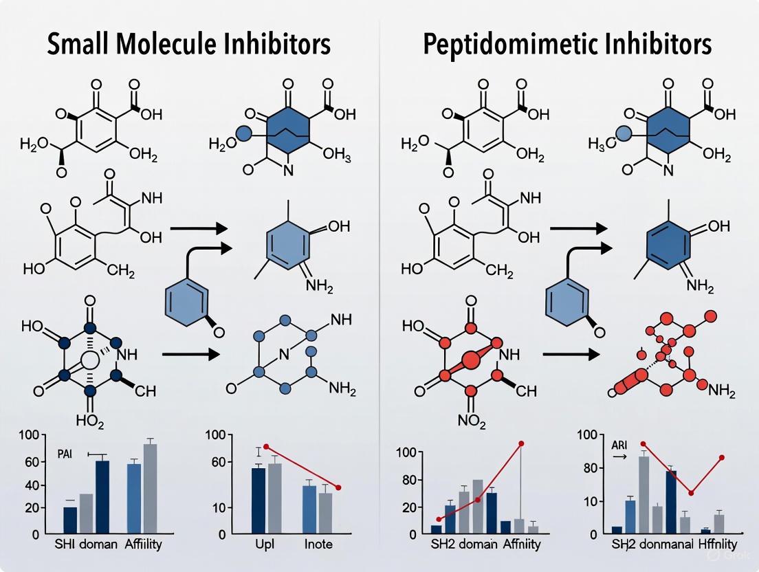
Figure 2: Workflow comparison of small molecule (top) and peptidomimetic (bottom) SH2 inhibitor discovery.
The Scientist's Toolkit: Essential Research Reagents and Solutions
Advancing SH2 domain research and drug discovery requires a specialized set of reagents and tools. The following table catalogs key solutions utilized in the experiments and methodologies cited throughout this review.
Table 2: Key Research Reagent Solutions for SH2 Domain Drug Discovery
| Research Reagent / Tool | Function and Application | Example Use Case |
|---|---|---|
| Recombinant SH2 Domains & Proteins | Purified proteins for biophysical binding assays (SPR, ITC, FP), structural studies, and high-throughput screening. | SHP2 protein (PDB: 2SHP) for crystallography and enzymatic assays [20] [17]. |
| Phosphopeptide Microarrays (Pepspot Chips) | High-density peptide chips to probe the binding specificity and affinity of SH2 domains against thousands of tyrosine phosphopeptides from the human proteome. | Mapping the SH2 domain interaction landscape [22]. |
| Covalent Probe MN551 / Prodrug MN714 | A covalent inhibitor (MN551) targeting Cys111 of SOCS2 and its prodrug (MN714) with a POM group to mask the phosphate charge for cell permeability. | Validating cellular target engagement of the SOCS2 SH2 domain [18]. |
| Allosteric SHP2 Inhibitor SHP099 | A first-in-class, tool compound that stabilizes SHP2 in an autoinhibited conformation by binding the "tunnel" allosteric site. | Proof-of-concept for SHP2 allosteric inhibition and combination therapy studies [17]. |
| Split-NanoLuc-based Assay | A cellular assay system used to demonstrate intracellular target engagement of a drug with its protein target. | Confirming cellular activity of the SOCS2 prodrug MN714 [18]. |
| Molecular Dynamics (MD) Simulation Software (e.g., GROMACS) | Software for simulating the physical movements of atoms and molecules over time to study protein-ligand interactions and stability. | Analyzing the binding stability of hit compounds with the target SH2 domain [20]. |
The druggability challenge of SH2 domains is being systematically overcome through innovative chemical and biological strategies. The comparative data indicates that small molecule allosteric inhibitors currently lead in the transition to clinically viable therapeutics, as evidenced by multiple SHP2 inhibitors in clinical trials [17]. Conversely, peptidomimetic approaches provide powerful tools for validating targets and generating highly selective chemical probes, though their development as drugs remains complex [19] [18].
Future directions are likely to focus on combination therapies to overcome resistance, the development of bifunctional molecules such as PROTACs to degrade SH2-containing proteins, and the continued expansion of E3 ligase ligands for targeted protein degradation, as demonstrated by the recent targeting of SOCS2 [17] [18]. The integration of high-throughput interaction data [22], advanced computational screening [20], and sophisticated prodrug technologies [18] will continue to drive the field forward, transforming SH2 domains from "undruggable" targets into a new frontier for precision medicine.
Src homology 2 (SH2) domains are protein modules approximately 100 amino acids in length that specifically recognize and bind to phosphorylated tyrosine (pTyr) residues, playing a fundamental role in intracellular signal transduction [3]. These domains are found in approximately 110 human proteins and function as critical components in phosphotyrosine-mediated signaling networks immediately downstream of protein tyrosine kinases [23] [3]. By facilitating the recruitment of specific effector proteins to activated receptor complexes, SH2 domains help determine signaling specificity in crucial cellular processes including proliferation, survival, and differentiation [23] [24]. The structured binding mechanism of SH2 domains involves a conserved "two-pronged plug" interaction, where a deep basic pocket binds the phosphotyrosine residue while an adjacent specificity pocket recognizes residues C-terminal to the pTyr, typically at the +3 position [24]. Among SH2 domain-containing proteins, Signal Transducer and Activator of Transcription 3 (STAT3) has emerged as a particularly compelling therapeutic target. STAT3 drives oncogenic progression through constitutive activation in many cancers, regulating the expression of genes involved in cell survival, proliferation, and angiogenesis [25]. This central role in malignancy has made the STAT3 SH2 domain a focus for drug discovery efforts, primarily aiming to disrupt the phosphotyrosine-SH2 interactions necessary for STAT3 dimerization and activation [25] [26]. Two distinct yet complementary therapeutic approaches have emerged: peptidomimetics and small molecules, each with characteristic advantages and challenges that define their application in biomedical research and drug development.
Core Characteristics at a Glance
The table below summarizes the fundamental properties that distinguish peptidomimetic inhibitors from small-molecule inhibitors, with a specific focus on their application against SH2 domains.
Table 1: Core Characteristics of Peptidomimetic and Small-Molecule SH2 Domain Inhibitors
| Characteristic | Peptidomimetics | Small Molecules |
|---|---|---|
| Molecular Origin | Derived from native peptide sequences of SH2 domain ligands [25] [27] | Identified via screening of synthetic chemical libraries [26] |
| Molecular Weight | Moderate to High (e.g., ~500-800 Da) [27] | Low (often <500 Da) [26] |
| Key Structural Features | Incorporates non-hydrolysable pTyr mimetics and conformational constraints [28] [27] | Often neutral, drug-like scaffolds; may lack phosphate groups [26] |
| Binding Affinity | High to very high (e.g., IC₅₀: 150 nM to 162 nM) [25] [27] | Variable, can be highly potent (low µM range) [26] |
| Selectivity | High, due to targeted design from native interaction sequences [25] | Can be challenging; must be engineered [26] |
| Cell Permeability | Often limited, may require cell-penetrating sequences [25] | Generally favorable, a key design advantage [26] |
| Oral Bioavailability | Low, due to peptidic nature and susceptibility to proteolysis [26] | High, a primary objective in their development [29] |
| Primary Development Challenge | Optimizing drug-like properties while maintaining high affinity [27] | Achieving high affinity against a large, flexible protein-protein interface [26] |
Peptidomimetic Inhibitors: Rational Design from Native Ligands
Design Philosophy and Evolution from Peptide Leads
Peptidomimetics are sophisticated compounds designed to retain the high affinity and specificity of native peptide ligands while overcoming their inherent pharmacological limitations. The development journey typically begins with the identification of high-affinity phosphopeptide sequences derived from physiological SH2 domain-binding partners. For the STAT3 SH2 domain, early work identified the native peptide sequence Pro-pTyr-Leu-Lys-Thr-Lys as a starting point, demonstrating that disruption of Stat3 dimerization was a viable therapeutic strategy [25]. A significant breakthrough came from screening receptor docking sites, which identified the hexapeptide Ac-pTyr-Leu-Pro-Gln-Thr-Val-NH₂ from the gp130 receptor as a high-affinity ligand with an IC₅₀ of 150 nM [25] [27]. This peptide provided the essential pharmacophore blueprint for subsequent mimetic development.
Key Structural Engineering Strategies
The transformation of a lead peptide into a potent peptidomimetic involves several strategic modifications aimed at enhancing stability, affinity, and drug-like properties.
Phosphotyrosine Mimetics: Replacement of the labile phosphate ester with non-hydrolysable, isosteric substitutes is crucial for metabolic stability. Examples include 4-phosphoryloxycinnamate (pCinn) and L-O-malonyltyrosine (L-OMT) [28] [27]. This modification challenges the notion that all SH2 domains tolerate the same pTyr mimetics, as some, like phosphonodifluoromethyl phenylalanine (F2Pmp), can abolish binding to certain SH2 domains [28].
Conformational Constraint: Incorporation of rigid structural elements pre-organizes the inhibitor into its bioactive conformation, reducing the entropy penalty upon binding. For instance, replacing the proline at position pY+2 with a rigid (2S,5S)-5-amino-1,2,4,5,6,7-hexahydro-4-oxo-azepino[3,2,1-hi]indole-2-carboxylic acid (Haic) scaffold significantly enhanced affinity, leading to the potent peptidomimetic pCinn-Haic-Gln-NHBn (IC₅₀ = 162 nM) [27].
Terminal Modifications: The C-terminal dipeptide (Thr-Val) can be effectively replaced by a simple benzyl group, reducing peptide character and molecular weight while maintaining key hydrophobic interactions [27].
The diagram below illustrates the logical workflow for developing a peptidomimetic SH2 domain inhibitor, from lead identification to optimized compound.
Small-Molecule Inhibitors: Targeting Druggability and Cellular Permeability
Discovery Methodologies and Design Challenges
In contrast to the rational design of peptidomimetics, small-molecule inhibitors are typically identified through systematic screening of diverse chemical libraries. Structure-based virtual ligand screening (SB-VLS) has emerged as a powerful technique, where compound libraries are computationally docked into the three-dimensional structure of the target SH2 domain [26]. A significant challenge in this approach is the inherent flexibility of the SH2 domain, which can lead to the identification of low-affinity hits. Advanced methods now incorporate molecular dynamics (MD) simulations to account for this flexibility, creating "induced-active site" receptor models that more accurately represent the conformational landscape of the target and improve screening outcomes [26].
A primary objective in small-molecule design is to eliminate charged groups, particularly phosphate mimics, which often impair cell permeability. Successful campaigns have identified neutral, low-molecular-weight compounds that fulfill Lipinski's rule-of-five and possess favorable drug-like properties, making them superior candidates for further development [26].
Experimental Protocols for Validation
Rigorous biochemical and cellular assays are essential to validate and characterize potential inhibitors.
Binding Affinity Measurement (Fluorescence Polarization, FP): This solution-based assay quantifies the direct interaction between the inhibitor and the purified SH2 domain. A fluorescently labeled phosphopeptide probe is incubated with the SH2 domain, causing high polarization due to slow molecular tumbling. Test compounds displace the probe, leading to a decrease in polarization that is quantified to determine IC₅₀ values [27].
Cellular Target Engagement (Electrophoretic Mobility Shift Assay, EMSA): This assay assesses the functional consequence of inhibition by measuring the disruption of STAT3:STAT3-DNA binding in nuclear extracts. Inhibitors that prevent STAT3 dimerization reduce the formation of the protein-DNA complex, visible as a decrease in a shifted band on a gel [25].
Cellular Signaling Inhibition (Western Blot): To confirm on-target activity in cells, the levels of phosphorylated Tyr705-STAT3 are measured via immunoblotting following cytokine stimulation. A reduction in pY-STAT3 signals effective pathway inhibition [26].
The following diagram outlines a representative screening workflow for identifying small-molecule SH2 domain inhibitors.
The Scientist's Toolkit: Essential Research Reagents
The following table catalogues key reagents and their applications in the development and characterization of SH2 domain inhibitors.
Table 2: Essential Research Reagents for SH2 Domain Inhibitor Studies
| Reagent / Material | Function and Application | Reference / Example |
|---|---|---|
| Recombinant SH2 Domains (GST-tagged) | Purified protein for in vitro binding assays (FP, SPR) and structural studies. | Expressed in E. coli; purified via glutathione-Sepharose [23]. |
| Fluorescent Phosphopeptide Probes | Tracer molecules for Fluorescence Polarization (FP) competition assays. | Derived from native sequences (e.g., gp130 pY905LPQTV) [26] [27]. |
| Phosphotyrosine Mimetics (e.g., pCinn, L-OMT) | Stable, non-hydrolysable chemical substitutes for phosphotyrosine in inhibitor design. | pCinn incorporated via solid-phase peptide synthesis [28] [27]. |
| Constrained Scaffolds (e.g., Haic) | Rigid amino acid analogs to pre-organize peptidomimetics into bioactive conformations. | Synthesized as Fmoc-protected building blocks for SPPS [27]. |
| Cell Lines with Constitutive pY-STAT3 | Cellular models for testing inhibitor efficacy (e.g., MDA-MB-231, NIH3T3/v-Src). | Used to assess inhibition of STAT3 phosphorylation and downstream effects [25] [26]. |
| Phospho-Specific Antibodies (e.g., pY705-STAT3) | Essential tools for Western Blot and ELISA to monitor target engagement in cells. | Used to detect levels of activated STAT3 in treated vs. untreated cells [26]. |
The strategic choice between peptidomimetic and small-molecule modalities for targeting SH2 domains involves a fundamental trade-off between affinity/selectivity and druggability/permeability. Peptidomimetics, born from native ligand structures, offer a rational path to high-potency, selective inhibitors but require intensive chemical optimization to overcome poor pharmacokinetic properties. Small molecules, while more amenable to oral administration and favorable physicochemical properties, face the significant hurdle of achieving high affinity against challenging, flexible protein-protein interaction interfaces like the SH2 domain. The future of SH2-directed therapeutics lies in the continued refinement of both approaches: leveraging advanced structural insights and dynamics for smarter small-molecule design, while applying innovative chemistry to further minimize the peptide character of peptidomimetics. This dual-track strategy maximizes the chances of delivering viable clinical candidates for cancer and other diseases driven by aberrant SH2 domain-mediated signaling.
Design and Discovery: Modern Strategies for Developing SH2 Inhibitors
Src Homology 2 (SH2) domains are protein modules comprising approximately 100 amino acids that specifically recognize and bind to phosphorylated tyrosine (pTyr) residues, thereby facilitating critical protein-protein interactions (PPIs) in cellular signaling networks [15] [3]. These domains are found in 111 human proteins, with roles ranging from enzyme activation to adaptor functions, and are fundamental to signaling pathways that regulate cell proliferation, differentiation, and survival [15] [3]. Their central role in signal transduction, coupled with their dysregulation in diseases like cancer and autoimmune disorders, makes them attractive therapeutic targets [15] [30]. However, targeting the large, relatively shallow, and conserved pTyr-binding pocket of SH2 domains presents a significant drug design challenge [31] [3]. This has spurred the development of peptidomimetics—molecules that mimic the structure and function of native peptide ligands but are engineered with improved drug-like properties, such as enhanced metabolic stability, cell permeability, and binding affinity [31].
This guide provides a comparative analysis of two central strategies in peptidomimetic design: (1) the constraint of peptide conformations to pre-organize the binding structure and (2) the specific mimicry of key recognition motifs, with a focus on the Stat3-specific pY-X-X-Q motif. We objectively evaluate these approaches by comparing experimental data on their binding affinity, selectivity, and functional activity, providing researchers with a structured framework for inhibitor selection and design.
Structural Basis of SH2 Domain Recognition
Canonical SH2-pTyr Peptide Binding Mechanism
SH2 domains share a conserved architectural fold featuring a central antiparallel β-sheet flanked by two α-helices [3] [4]. The binding event is anchored by a deep pocket within the βB strand that accommodates the phosphotyrosine residue. A nearly invariant arginine residue (Arg βB5) within the FLVR motif forms a critical salt bridge with the phosphate moiety of pTyr, contributing a substantial portion of the total binding energy [3] [4]. The specificity of the interaction is further determined by residues in the peptide sequence C-terminal to the pTyr, which bind to secondary pockets on the SH2 domain surface labeled as +1, +2, +3, etc. [3] [32]. This two-tiered mechanism—conserved pTyr anchoring and sequence-specific side-chain recognition—provides the blueprint for rational peptidomimetic design.
The Stat3 and pY-X-X-Q Motif: A Case Study in Specificity
The Signal Transducer and Activator of Transcription 3 (Stat3) is an SH2-containing protein constitutively activated in many cancers [32] [30]. Its recruitment and dimerization depend on binding to phosphopeptides containing the signature pY-X-X-Q motif [32]. Structural and mutational studies have elucidated that this high specificity is achieved through a unique β-turn conformation in the peptide backbone, which positions the glutamine residue at the +3 position for optimal interaction [32]. The side chain of Glu-638 on the Stat3 SH2 domain hydrogen-bonds with the amide of the +3 Gln, a interaction crucial for binding [32]. Disrupting this specific interaction, for instance by mutating Glu-638 or substituting Gln with other residues, drastically reduces binding affinity, highlighting the critical nature of this motif [32]. Consequently, accurately mimicking the pY-X-X-Q motif and its associated backbone structure is a primary objective for designing specific Stat3 inhibitors.
The diagram below illustrates the key steps and decision points in the rational design of peptidomimetics targeting SH2 domains.
Figure 1: A generalized workflow for the rational design of SH2 domain-targeting peptidomimetics, beginning with target identification and culminating in experimental validation.
Comparative Analysis of Peptidomimetic Design Strategies
Strategy 1: Constraining Peptide Conformations
A primary limitation of native peptides is their conformational flexibility in solution, which results in a significant entropy penalty upon binding to their target. Constraining peptides aims to pre-organize the ligand into its bioactive conformation, thereby minimizing this entropy cost and improving binding affinity and metabolic stability [31]. This is often achieved through macrocyclization, stapling, or the introduction of rigid scaffolds that lock the peptide into a specific secondary structure, such as a β-turn or α-helix.
Table 1: Experimental Data from the Development of a Constrained C-SH2 Inhibitor Peptide (CSIP) for SHP2
| Parameter | Unmodified ITSM(pTyr) Peptide | Constrained/Optimized CSIP (Peptide 21) | Experimental Method |
|---|---|---|---|
| Sequence | EQTE(pY)ATIVFP-NH₂ | -QTE(pY)ATIKHP-NH₂ | Peptide Synthesis [33] |
| Binding Affinity (K_D) to C-SH2 | 48.9 nM | 88.16 nM | Fluorescence Polarization (FP) [33] |
| Binding Affinity (K_D) to N-SH2 | 164.0 nM | >3000 nM | Fluorescence Polarization (FP) [33] |
| Inhibitory Potency (IC₅₀) | Not Applicable | 1.054 µM | Competitive FP Assay [33] |
| Selectivity (C-SH2 vs. N-SH2) | Low (Binds both domains) | High (>34-fold preference for C-SH2) | Calculated from K_D ratios [33] |
| Cellular Permeability | Presumed Low | Demonstrated as Cell-Permeable | Live-Cell Assay [33] |
| Cytotoxicity | Not Reported | Non-cytotoxic (up to 100 µM) | Cell Viability Assay [33] |
The data in Table 1 demonstrates the success of a rational optimization process. While the constrained CSIP experienced a slight decrease in absolute affinity for the C-SH2 domain compared to the native peptide, this was traded for a dramatic improvement in domain selectivity and the acquisition of favorable drug-like properties, including cell permeability and lack of cytotoxicity [33]. This trade-off is often desirable in drug development, as a highly selective inhibitor can more precisely probe biological function with fewer off-target effects.
Strategy 2: Mimicking the pY-X-X-Q Motif for Stat3 Inhibition
For targets like Stat3, where specificity is governed by a well-defined motif like pY-X-X-Q, the design focus shifts to accurately replicating these key interactions. This involves not only presenting the correct side chains but also mimicking the backbone conformation that optimally orients them. A critical component of this strategy is the use of non-hydrolysable pTyr mimetics to overcome the metabolic instability of phosphotyrosine.
Table 2: Comparison of pTyr Mimetics in Peptide Inhibitors
| pTyr Mimetic | Chemical Structure | Binding Affinity to SHP2 C-SH2 | Key Advantages & Disadvantages |
|---|---|---|---|
| Phosphotyrosine (pTyr) | -PO₃²⁻ | K_D = 48.9 - 58.8 nM (from ITSM peptide) | Native residue; high affinity but poor metabolic stability and cell permeability [33] |
| L-O-malonyltyrosine (l-OMT) | -O-CO-CH₂-COO⁻ | IC₅₀ = 7.8 µM (in peptide sequence) | Non-hydrolysable, stable; confers cell permeability; moderate affinity [33] |
| Phosphonodifluoromethyl phenylalanine (F₂Pmp) | -PO₃H-CF₂ | No binding detected | Widely assumed to be a general pTyr mimetic; can abolish binding to specific SH2 domains like SHP2's C-SH2 [33] |
The data in Table 2 underscores that the choice of pTyr mimetic is not one-size-fits-all. The failure of F₂Pmp to bind the C-SH2 domain of SHP2, in contrast to its success in other contexts, challenges existing notions and highlights the necessity of empirical testing for each specific SH2 target [33]. For Stat3, research has shown that beyond the pTyr mimetic, preserving the β-turn structure that presents the +3 Gln is paramount. Mutagenesis studies confirm that mutation of key Stat3 residues like Lys-591, Arg-609 (which contact the pTyr), or Glu-638 (which hydrogen-bonds with the +3 Gln) leads to a complete loss of binding to pYXXQ-containing peptides [32].
The Scientist's Toolkit: Essential Research Reagents and Protocols
Research Reagent Solutions
Table 3: Key Reagents for Peptidomimetic Research on SH2 Domains
| Reagent / Resource | Function / Application | Example / Source |
|---|---|---|
| Recombinant SH2 Domains | In vitro binding and inhibition assays (FP, ITC, SPR). | Purified N-SH2 and C-SH2 domains of SHP2 [33]; Stat3 SH2 domain [32]. |
| Fluorescent Peptide Probes | Tracer molecules for Fluorescence Polarization (FP) competition assays. | FAM-labeled ITSM(pTyr) peptide (FAM-EQTE(pY)ATIVFP-NH₂) [33]. |
| Non-hydrolysable pTyr Mimetics | Incorporating stability into peptide designs for cellular assays. | l-O-malonyltyrosine (l-OMT), Phosphonodifluoromethyl phenylalanine (F₂Pmp) [33]. |
| Structure-Based Design Software | Visualizing binding pockets, docking, and optimizing designs. | Pymol (visualization); Molecular docking (Smina/AutoDock Vina [4]); MD simulations (GROMACS [4]). |
Detailed Experimental Protocol: Fluorescence Polarization Competition Assay
This protocol is adapted from the methodology used to characterize the C-SH2 inhibitor peptide (CSIP) [33] and is a cornerstone for quantifying inhibitor potency.
- Prepare the Assay Mixture: In a buffer-compatible format (e.g., 384-well plate), create a solution containing a fixed, low concentration (e.g., 10-50 nM) of the fluorescent tracer peptide (e.g., FAM-ITSM(pTyr)) and a fixed concentration of the target SH2 domain at or near its K_D for the tracer to ensure a robust signal window.
- Titrate the Inhibitor: Add the unlabeled inhibitor peptide (or peptidomimetic) across a series of wells in a concentration gradient, typically spanning several orders of magnitude (e.g., 1 nM to 100 µM).
- Incubate and Measure: Allow the plate to incubate in the dark to reach binding equilibrium. Then, measure the fluorescence polarization (mP units) for each well using a plate reader.
- Data Analysis: The decrease in polarization as a function of increasing inhibitor concentration is fit to a sigmoidal dose-response curve. The concentration that displaces 50% of the tracer (IC₅₀) is calculated from this curve and serves as the primary measure of inhibitory potency.
Detailed Experimental Protocol: Molecular Dynamics (MD) Simulation and Binding Free Energy Calculation
This in silico protocol, as employed in the identification of SHP2 N-SH2 inhibitors [4], provides atomic-level insights into binding stability and energetics.
- System Preparation: Obtain the crystal structure of the target SH2 domain (e.g., PDB ID: 2SHP). Prepare it by adding missing hydrogen atoms and residues using a tool like PDBFixer. The ligand topology is generated using a service like LigParGen.
- Simulation Setup: Solvate the protein-ligand complex in an explicit water model (e.g., SPC216) within a defined periodic box. Add ions to neutralize the system's charge.
- MD Simulation Run: Perform the simulation using software like GROMACS [4]. This involves energy minimization, equilibration under constant number, volume, and temperature (NVT) and constant number, pressure, and temperature (NPT) ensembles, followed by a production run (typically 100 ns or more).
- MM/PBSA Calculation: To estimate binding free energy (ΔG_bind), extract multiple snapshots from the stable trajectory. Use a tool like
g_mmpbsato perform Molecular Mechanics/Poisson-Boltzmann Surface Area calculations, which sum the gas-phase energy, solvation energy, and surface area terms [4].
The diagram below maps the key signaling pathways discussed, highlighting the points of therapeutic intervention.
Figure 2: Key cellular signaling pathways regulated by SH2 domains and the points of inhibition by different peptidomimetic strategies. The diagram shows how inhibitors like CSIP, Stat3-targeting mimetics, and allosteric inhibitors like SHP099 intervene at specific points to modulate signaling output.
The objective comparison of experimental data reveals that the choice between constraining peptides and mimicking specific motifs is not mutually exclusive and is often guided by the target biology and desired therapeutic outcome.
- Constraining Peptides is a powerful strategy for enhancing selectivity and improving drug-like properties, as evidenced by the development of CSIP. It is particularly valuable when targeting specific isoforms or domains within a family of highly related proteins (e.g., the C-SH2 vs. N-SH2 domain of SHP2).
- Mimicking the pY-X-X-Q Motif is essential for achieving high specificity against targets like Stat3, where the binding motif is well-defined. The success of this approach hinges on the dual challenge of selecting an appropriate pTyr mimetic and engineering the peptidomimetic scaffold to faithfully reproduce the bioactive conformation of the native peptide.
The future of peptidomimetic design lies in the intelligent integration of these strategies. Advances in structural biology, computational chemistry, and synthetic methodology will enable the creation of next-generation inhibitors that are not only highly potent and selective but also possess optimal pharmacokinetic properties for in vivo efficacy. The continued refinement of these approaches promises to deliver valuable chemical probes and novel therapeutics targeting the biologically rich but challenging landscape of SH2 domain-mediated interactions.
This guide provides an objective comparison of current virtual screening and molecular docking methodologies, contextualized within research focused on discovering inhibitors of Src Homology 2 (SH2) domains—a prominent target in oncology for its role in signal transduction. Performance data and protocols for various software platforms are detailed to inform selection for small molecule and peptidomimetic drug discovery projects.
Biological Context: SH2 Domains as Therapeutic Targets
SH2 domains are protein modules approximately 100 amino acids long that specifically recognize and bind to phosphorylated tyrosine (pY) residues on their partner proteins. [3] [34] This ability makes them critical "readers" in intracellular signaling networks, regulating key cellular processes such as proliferation, survival, and differentiation. [3] [34] The human proteome contains roughly 110 proteins with SH2 domains, which can be enzymes, adaptors, or transcription factors. [3]
Dysregulation of SH2-mediated interactions is strongly implicated in disease, particularly cancer. For example, the transcription factor STAT3 must dimerize via its SH2 domain to translocate to the nucleus and activate genes that promote cell survival and proliferation. [35] Consequently, inhibiting the SH2 domain of STAT3 to prevent its dimerization is a validated strategy for anticancer drug development. [35] [6] Both small molecules and peptidomimetics are being explored to block these critical protein-protein interactions (PPIs). [6]
The following diagram illustrates the central role of SH2 domains in the JAK-STAT signaling pathway and the strategic point for inhibitory intervention.
Comparative Performance of Docking and Virtual Screening Methods
The core task of virtual screening is to accurately predict how a small molecule binds to a target (pose prediction) and to rank compounds by their predicted affinity (virtual screening enrichment). The table below summarizes the performance of leading methods across standardized benchmarks.
Table 1: Performance Comparison of State-of-the-Art Virtual Screening Tools
| Method / Platform | Type | Key Feature | Pose Prediction Success (RMSD ≤ 2 Å) | Virtual Screening Enrichment (Top 1%) | Key Advantage |
|---|---|---|---|---|---|
| Glide SP [36] [37] | Traditional Physics-Based | Robust scoring function & sampling | ~80% (Astex Diverse Set) [36] | Not Specified | High physical validity & reliability [36] |
| Glide WS [37] | Traditional Physics-Based (New) | Explicit water modeling | 92% (Self-docking, 2.5 Å criterion) [37] | Significant improvement over Glide SP [37] | Superior docking accuracy & enrichment [37] |
| RosettaVS [38] | Physics-Based with AI | Models receptor flexibility & entropy | High performance on CASF2016 [38] | EF1~1%~ = 16.72 (Top on CASF2016) [38] | Best-in-class affinity ranking [38] |
| OpenVS [38] | AI-Accelerated Platform | Integrated active learning | Dependent on docking engine (e.g., RosettaVS) | High hit rates (14-44% in prospective studies) [38] | Speed for ultra-large libraries [38] |
| SurfDock [36] | Deep Learning (Generative) | Diffusion model for pose generation | >75% across benchmarks [36] | Not Specified | High pose prediction accuracy [36] |
| CryoXKit + AutoDock [39] | Data-Guided Docking | Uses experimental density maps | Significant improvement in cross-docking [39] | Better discriminatory power [39] | Leverages experimental structural data [39] |
EF~1%~ = Enrichment Factor at the top 1% of the screened library. A higher value indicates better ability to identify true binders early. [38]
Performance Trends: Traditional vs. Deep Learning Methods
A comprehensive 2025 study reveals a clear stratification of docking methods. [36]
- Traditional physics-based methods like Glide SP consistently achieve high physical validity (e.g., correct bond lengths, no steric clashes), with success rates above 94% in generating chemically plausible structures. [36]
- Deep learning generative models (e.g., SurfDock) excel in raw pose prediction accuracy (RMSD ≤ 2 Å), often exceeding 70-90% on standard benchmarks. [36]
- A significant caveat for many deep learning methods is that they can produce physically invalid poses or fail to recover critical molecular interactions despite having a good RMSD, limiting their direct application in lead optimization. [36]
- Hybrid methods that combine traditional conformational sampling with AI-driven scoring offer a balanced approach, though they may not surpass the performance of top-tier traditional methods like Glide. [36]
Experimental Protocols for Virtual Screening
Successful virtual screening campaigns follow a structured workflow. The diagram below outlines the key stages, from target preparation to experimental validation.
Detailed Methodologies for Key Stages
1. Target Preparation
- Structure Selection: For targets with multiple structures, select the receptor from a co-crystal structure with a ligand most similar to the compounds being screened ("dock-close" strategy). This often outperforms using multiple structures. [40]
- Binding Site Definition: Clearly define the coordinates of the binding site. For SH2 domains, this is the conserved pY-binding pocket. [3] [34]
- Receptor Flexibility: Incorporate flexibility where critical. RosettaVS allows for full side-chain and limited backbone flexibility in its high-precision mode. [38] Ensemble docking is an alternative, but can introduce noise. [40]
2. Library Preparation
- Library Curation: Use focused libraries for specific targets. For PPIs like SH2 domain inhibition, libraries rich in peptidomimetics or multicomponent reaction (MCR)-derived compounds are advantageous. [40]
- Compound Preprocessing: Generate realistic, low-energy 3D conformers for each compound. Tools like Open Babel or OMEGA are standard for this step. [40]
3. Virtual Screening Execution
- Docking Protocol: A tiered approach is computationally efficient.
- Active Learning: Platforms like OpenVS integrate active learning, where a target-specific neural network is trained during docking to intelligently select promising compounds for more expensive calculations, dramatically speeding up the screening of billion-member libraries. [38]
4. Post-Docking Analysis
- Consensus Scoring: Rank compounds using multiple scoring functions or more computationally intensive methods like Molecular Mechanics with Generalized Born and Surface Area Solvation (MM/GBSA) to improve hit rates. [40] [6]
- Interaction Analysis: Manually inspect top-ranked poses to ensure they form key interactions with the target. For STAT3 SH2 domain inhibitors, this involves verifying interactions with the critical arginine residue in the pY-binding pocket. [35]
5. Experimental Validation
- Biochemical Assays: Validate computational hits using assays like Surface Plasmon Resonance (SPR) to measure binding affinity.
- Structural Validation: Where possible, confirm predicted binding modes using X-ray crystallography, as was done for a KLHDC2 ligand discovered via the OpenVS platform. [38]
The Scientist's Toolkit: Essential Research Reagents and Solutions
Table 2: Key Reagents and Software for SH2 Domain Inhibitor Screening
| Tool / Reagent | Function / Description | Example Use in SH2 Research |
|---|---|---|
| SH2 Domain Protein | Recombinant protein for binding assays. | Essential for experimental validation of predicted inhibitors (e.g., STAT3 SH2 domain). [35] |
| Phosphopeptide Ligands | Native pY-containing peptides. | Used as positive controls in competition binding assays. [3] [34] |
| Peptidomimetic Libraries | Libraries of compounds mimicking peptide structure. | Source of potential inhibitors with improved stability over native peptides (e.g., β-peptides for STAT NTD). [6] |
| Molecular Dynamics Software | Simulates dynamic behavior of protein-ligand complexes. | Used for post-docking refinement and binding free energy calculation (e.g., via MM/PBSA). [6] |
| Docking Software (Glide, AutoDock Vina, etc.) | Predicts binding pose and affinity. | Core engine for structure-based virtual screening. [40] [36] |
| Virtual Compound Libraries | Commercially available or make-on-demand chemical libraries. | Source of small molecules for screening (e.g., ZINC, Enamine).. [38] [40] |
The choice of a virtual screening platform depends heavily on the project's goals and constraints. For projects requiring high confidence in pose validity and reliability, especially for lead optimization, traditional physics-based methods like Glide WS and RosettaVS are currently the most robust choice, with Glide WS showing top-tier accuracy and RosettaVS excelling in affinity ranking and handling flexibility. [38] [36] [37] For tasks involving the screening of ultra-large libraries (billions of compounds) where speed is critical, AI-accelerated platforms like OpenVS that leverage active learning provide a powerful and prospectively validated solution. [38] While deep learning methods show great promise in raw pose prediction accuracy, researchers should be cautious of their tendency to generate physically invalid structures and should prioritize methods that have been prospectively validated in real-world drug discovery campaigns. [36]
Protein dynamics play a crucial role in governing biological function, yet static structural representations often obscure transient structural features critical for drug discovery. This review comprehensively examines how molecular dynamics (MD) simulations are revolutionizing the identification and characterization of cryptic binding pockets—transient cavities that emerge from protein flexibility—with a specific focus on SH2 domain inhibitors. We objectively compare the performance of small molecule versus peptidomimetic approaches targeting these elusive sites, synthesizing experimental data across multiple therapeutic targets. By integrating recent advances in simulation methodologies, high-throughput screening, and structural biology, this analysis provides researchers and drug development professionals with a framework for leveraging protein dynamics in rational drug design, particularly for challenging targets where conventional structure-based approaches have proven insufficient.
Proteins are inherently dynamic entities that undergo continuous conformational changes essential for biological processes such as enzyme catalysis, protein-protein interactions, and allosteric regulation [41]. The traditional paradigm of structure-based drug design has relied heavily on static snapshots from X-ray crystallography or cryo-EM, which often fail to capture the full conformational landscape of therapeutic targets. This limitation is particularly pronounced for proteins with significant flexibility, where transiently formed binding sites—known as cryptic pockets—represent promising yet elusive targets for therapeutic intervention [42].
The Src homology 2 (SH2) domain represents a compelling case study in the challenges and opportunities presented by protein flexibility. SH2 domains are approximately 100 amino acid modules that specifically recognize phosphotyrosine (pY) motifs, forming crucial components of intracellular signaling networks [8]. Their function in mediating protein-protein interactions through binding to pY-containing sequences makes them attractive targets for modulating pathological signaling in cancer and immune disorders. However, the moderate binding affinity (Kd 0.1–10 µM) and dynamic nature of SH2 domains have complicated drug discovery efforts [8].
Cryptic pockets represent a promising frontier for targeting proteins like SH2 domains that have proven difficult to drug through conventional approaches. These pockets are not typically visible in crystal structures but can be captured through MD simulations, providing new opportunities for allosteric inhibition and selective targeting [43] [42]. This review systematically compares experimental approaches for identifying and targeting cryptic pockets, with particular emphasis on the methodological considerations for SH2 domain-targeted therapeutics.
SH2 Domain Structure and Dynamics: Implications for Inhibitor Design
Structural Fundamentals of SH2 Domains
SH2 domains share a conserved fold consisting of a three-stranded antiparallel beta-sheet flanked by two alpha helices (αA-βB-βC-βD-αB), forming a compact structure that specifically recognizes phosphotyrosine-containing peptides [8]. The N-terminal region contains a deep pocket that binds the phosphate moiety of pY, featuring an invariable arginine residue (βB5) that forms a salt bridge with the phosphorylated tyrosine. The C-terminal region provides additional specificity determinants that recognize residues C-terminal to the pY, particularly positions +1 to +3, enabling discrimination between different pY-containing sequences [8] [44].
Structurally, SH2 domains can be classified into two major subgroups: STAT-type and SRC-type. STAT-type SH2 domains lack the βE and βF strands and have a split αB helix, an adaptation that facilitates the dimerization critical for STAT-mediated transcriptional regulation [8]. This structural variation highlights how evolutionary adaptations of the core SH2 fold have created distinct dynamic properties across different protein families, with direct implications for inhibitor design.
Dynamic Behavior and Cryptic Pocket Formation
Recent research has revealed unexpected flexibility in SH2 domains and their containing proteins. Molecular dynamics simulations of SHP2, which contains two SH2 domains (N-SH2 and C-SH2), have demonstrated that oncogenic mutations like E76K convert the protein to its open state through a substantial 120-degree rotation of the C-SH2 domain [43]. This conformational change represents an unusually large magnitude transition that significantly alters the protein's surface topology and creates new, transient binding interfaces.
Studies employing extensive MD simulations (totaling ~28.6 µs) have shown that while individual domains in wild-type SHP2 exhibit remarkable stability, the C-SH2 domain in the E76K mutant displays significantly higher fluctuations, suggesting heightened flexibility in the activated state [43]. This flexibility manifests as transient pockets at domain interfaces that are not evident in static structures. For instance, simulations of SHP2 have identified an alternative allosteric binding pocket in solution structures that emerges from the dynamic properties of the protein [43].
Table 1: Key Dynamic Properties of SH2 Domain-Containing Proteins Relevant to Cryptic Pocket Formation
| Protein | Dynamic Feature | Functional Consequence | Cryptic Pocket Location |
|---|---|---|---|
| SHP2 | 120° C-SH2 rotation in E76K mutant | Transition to open, active state | Interface between C-SH2 and PTP domains |
| STAT3 | Loop deformation upon inhibitor binding | Novel binding mode formation | Near SH2 domain pY+1 to pY+3 subsites |
| PPM1D | Flap domain flexibility | Cryptic pocket formation at flap-hinge interface | Interface between flap and hinge regions |
| BTK | SH2 domain lipid-binding site | Membrane recruitment and activation | Cationic region near pY-binding pocket |
Comparative Analysis of SH2 Domain Targeting Strategies
Peptidomimetic Inhibitors: Leveraging Native Recognition Motifs
Peptidomimetic inhibitors represent a rational approach to targeting SH2 domains by building upon their natural peptide recognition preferences. Early efforts focused on optimizing native phosphopeptide sequences, such as the pTyr-Glu-Glu-Ile motif recognized by the GRB2 SH2 domain [25]. Structure-activity relationship studies revealed that simple tripeptides like Xxx-pTyr-Leu (e.g., Pro-pTyr-Leu and Ala-pTyr-Leu) could effectively block STAT3 homodimerization with DB50 values of 182-217 μM [25].
Significant advances came from receptor-based peptide optimization. McMurray and colleagues developed hexapeptides containing the pTyr-Xxx-Xxx-Gln-Xxx-Xxx sequence, with Ac-pTyr-Leu-Pro-Gln-Thr-Val-NH2 demonstrating remarkable potency (IC50 = 150 nM) – a 133-fold improvement over early leads [25]. Alanine scanning confirmed the importance of leucine at position pTyr+1, proline at pTyr+2, and glutamine at pTyr+3, highlighting the stringent sequence requirements for high-affinity binding.
To address limitations of peptide agents, particularly poor cell permeability, researchers have developed sophisticated peptidomimetics that replace the N-terminal proline with non-peptidic moieties. Incorporation of 4-cyanobenzoyl (ISS610) or 2,6-dimethoxybenzoyl (ISS637) groups resulted in a fivefold increase in inhibition of STAT3-DNA binding activity compared to the lead tripeptide [25]. These hybrid molecules retain the specific recognition elements of native peptides while improving drug-like properties.
Small Molecule Inhibitors: Targeting Cryptic Pockets for Selectivity
Small molecule inhibitors offer distinct advantages for targeting SH2 domains, including improved bioavailability and the potential for greater selectivity through engagement of cryptic pockets not utilized by native peptides. The development of allosteric inhibitors that stabilize the closed state of SHP2 exemplifies this approach. Compounds like SHP099 function as "molecular glue" inhibitors, targeting an allosteric site formed between the C-SH2 and PTP domains and demonstrating high-affinity binding with antiproliferative effects in cancer cell lines [43].
Recent efforts have identified novel allosteric binding pockets through extensive modeling and simulations. For SHP2, researchers discovered an alternative allosteric pocket in solution structures distinct from previously characterized sites [43]. Multi-tier screening approaches have successfully identified potential binders targeting this cryptic site, contributing to community-wide initiatives developing therapies using multiple allosteric inhibitors to target distinct pockets on SHP2.
A groundbreaking advance in SH2 domain targeting comes from Recludix Pharma's Bruton's tyrosine kinase (BTK) SH2 domain inhibitor. This compound demonstrates exceptional selectivity (>8000-fold over off-target SH2 domains) and potency (BTK Kd = 0.055 nM) by targeting a cryptic pocket within the BTK SH2 domain [5]. Unlike traditional kinase domain inhibitors, this SH2-targeted approach avoids off-target effects on TEC kinase, potentially mitigating adverse effects like platelet dysfunction associated with conventional BTK inhibitors.
Table 2: Performance Comparison of SH2 Domain Inhibitor Classes
| Parameter | Peptidomimetics | Small Molecules | Experimental Methods |
|---|---|---|---|
| Binding Affinity | IC50 150 nM (optimized peptides) [25] | Kd 0.055 nM (BTK SH2i) [5] | Fluorescence polarization, SPR |
| Selectivity | Moderate (sequence-based recognition) | High (>8000-fold for BTK SH2i) [5] | SH2ome profiling, kinome screening |
| Cell Permeability | Limited (requires membrane translocation sequences) [25] | Favorable (prodrug strategies available) [5] | Cellular uptake assays |
| Target Engagement | DB50 182-217 μM (early leads) [25] | Sustained >48 hours in PBMCs [5] | Pharmacodynamic profiling |
| Exploitation of Cryptic Pockets | Limited (binds canonical pY site) | Extensive (novel allosteric sites) [43] [5] | MD simulations, X-ray crystallography |
Methodological Advances in Cryptic Pocket Detection
Molecular Dynamics Simulation Protocols
Molecular dynamics simulations have emerged as powerful tools for capturing protein flexibility and identifying cryptic pockets. Standardized protocols have been developed to ensure reproducibility and comparability across systems. The ATLAS database, for instance, provides a large-scale collection of all-atom MD simulations performed using a consistent methodology [41]. Their protocol involves:
- System Preparation: Proteins are placed in a periodic triclinic box, solvated with TIP3P water molecules, and neutralized with Na+/Cl− ions at 150 mM concentration.
- Energy Minimization: Steepest descent algorithm for 5000 steps to optimize geometry.
- Equilibration: 200 ps in canonical ensemble (NVT) followed by 1 ns in isothermal-isobaric ensemble (NPT) with heavy atom restraints.
- Production Simulation: Three 100 ns replicates with a 2 fs time step, coordinates saved every 10 ps [41].
For enhanced sampling of cryptic pockets, advanced techniques like Markov state models (MSMs) and adaptive sampling algorithms such as FAST (Flopping Amplified Sampling Technique) have been developed. These approaches balance exploration with exploitation to efficiently search conformational space for transient pocket formation [42]. In the case of PPM1D phosphatase, simulations initiated from an AlphaFold-predicted structure revealed a cryptic pocket at the interface between flap and hinge regions that was not evident in the static structure [42].
Integrative Approaches Combining MD with Experimental Data
The integration of MD simulations with experimental structural biology techniques has proven particularly powerful for cryptic pocket identification. Cryo-electron microscopy (cryo-EM) data can provide valuable constraints for simulations, as demonstrated by the RMSF-net approach, which combines cryo-EM density information with atomic models to predict protein flexibility [45]. This deep learning model achieves correlation coefficients of 0.746 ± 0.127 with MD simulations at the voxel level while requiring only seconds per prediction compared to days or weeks for conventional MD [45].
Experimental validation of cryptic pockets identified through MD often employs biophysical techniques such as:
- Site-directed mutagenesis: Residues lining the predicted pocket are mutated to assess impact on inhibitor binding.
- Hydrogen-deuterium exchange mass spectrometry (HDX-MS): Detects changes in protein dynamics and solvent accessibility upon inhibitor binding.
- SAXS (Small-angle X-ray scattering): Provides solution-state validation of conformational changes observed in simulations.
In the case of SHP2, MD simulations have been validated against NMR and SAXS data, confirming the unexpected flexibility within the open state of the E76K mutant [43]. Similarly, for PPM1D, deuterium exchange profiles provided experimental support for the cryptic pocket identified through simulations [42].
Case Studies: Successful Application of MD in SH2 Domain-Targeted Drug Discovery
STAT3 Inhibitors: From Peptidomimetics to Novel Binding Modes
STAT3 represents a paradigmatic example of how MD simulations can guide the evolution of SH2 domain inhibitors. Early peptidomimetic inhibitors were designed based on the native pTyr705 binding motif, with compounds like Ac-pTyr-Leu-Pro-Gln-Thr-Val-NH2 achieving nanomolar affinity (IC50 = 150 nM) [25]. However, these peptides suffered from limited cell permeability, prompting the development of hybrid peptidomimetics with improved drug-like properties.
Computational modeling combining molecular docking and MD simulations revealed a novel binding mode for these peptidomimetics that involves deformation of two loops in the STAT3 SH2 domain [44]. This distorted conformation buries the C-terminal end of stronger inhibitors, creating a binding interface not observed in crystal structures of the native SH2 domain. MD simulations further enabled estimation of binding affinities using MMPB/GBSA-based energies that correlated strongly with experimental values (R = 0.82-0.92), validating the modeled complexes [44].
SHP2 Allosteric Inhibitors: Targeting Dynamic Interfaces
SHP2 phosphatase, a key signaling node in the Ras-MAPK pathway, has emerged as an important cancer target. Allosteric inhibitors like SHP099 stabilize the autoinhibited closed state by binding at the interface of the C-SH2, N-SH2, and PTP domains [43]. Extensive MD simulations (totaling ~28.6 µs) have revealed the dynamic properties of SHP2 across different states, showing that oncogenic mutations like E76K significantly impact allosteric inhibition sites.
These simulations identified an alternative allosteric binding pocket in solution structures, distinct from previously characterized sites [43]. Multi-tier screening targeting this dynamic pocket identified potential binders that could complement existing allosteric inhibitors. This approach represents a promising strategy for combating drug resistance, as activating mutations that destabilize the closed state reduce efficacy of conventional allosteric inhibitors.
BTK SH2 Domain Inhibitors: A New Paradigm in Selectivity
The recent development of BTK SH2 domain inhibitors by Recludix Pharma exemplifies the clinical potential of targeting cryptic pockets. Traditional BTK inhibitors targeting the kinase domain have been limited by off-target effects on TEC kinase and transient target engagement. The SH2-targeted approach achieves exceptional selectivity (>8000-fold over off-target SH2 domains) and sustained pathway inhibition [5].
Preclinical studies demonstrate that BTK SH2 inhibitors provide robust inhibition of SH2-dependent pERK signaling and suppress downstream CD69 expression in B cells [5]. In a mouse model of ovalbumin-induced chronic spontaneous urticaria, a single prophylactic dose of BTK SH2 inhibitor led to significant, dose-dependent reduction in skin inflammation, outperforming conventional kinase domain inhibitors. This success highlights the therapeutic advantage of targeting cryptic pockets in SH2 domains.
Table 3: Experimental Protocols for Cryptic Pocket Identification and Validation
| Method | Key Steps | Applications | References |
|---|---|---|---|
| Standard MD Protocol | System preparation → Energy minimization → NVT/NPT equilibration → Production simulation | Large-scale dynamics analysis (ATLAS database) | [41] |
| Enhanced Sampling (FAST) | Adaptive sampling balancing exploration/exploitation → Markov state model construction | Cryptic pocket identification (PPM1D) | [42] |
| Integrated MD/Cryo-EM (RMSF-net) | Cryo-EM map + PDB model → Dual feature input → 3D CNN prediction | Flexibility prediction across 335 proteins | [45] |
| Binding Affinity Estimation | Molecular docking → MD simulation → MMPB/GBSA energy calculation | Peptidomimetic inhibitor optimization (STAT3) | [44] |
| Experimental Validation | HDX-MS → Site-directed mutagenesis → Cellular assays | Cryptic pocket confirmation (SHP2, PPM1D) | [43] [42] |
The Scientist's Toolkit: Essential Research Reagents and Methodologies
Table 4: Essential Research Reagents and Computational Tools for Studying SH2 Domain Dynamics
| Resource | Type | Function | Access |
|---|---|---|---|
| ATLAS Database | Database | Standardized MD simulations for 1390 proteins | https://www.dsimb.inserm.fr/ATLAS [41] |
| CHARMM36m Force Field | Computational | Balanced sampling of folded/unfolded states | GROMACS/AMBER [41] |
| RMSF-net | Deep Learning Tool | Protein flexibility prediction from cryo-EM maps | Python package [45] |
| FAST | Adaptive Sampling Algorithm | Efficient conformational space sampling | OpenSource [42] |
| Custom DNA-encoded Libraries | Screening Resource | SH2-targeted compound identification | Recludix Platform [5] |
| SHP2 Allosteric Inhibitors | Chemical Probes | Stabilize closed conformation (e.g., SHP099) | Commercial vendors [43] |
| Phosphotyrosine Mimetics | Chemical Reagents | Non-hydrolysable pTyr substitutes (e.g., l-OMT, F2Pmp) | Chemical suppliers [28] |
Visualizing Workflows: From Structure Prediction to Cryptic Pocket Discovery
Molecular Dynamics Workflow for Cryptic Pocket Detection
SH2 Domain Inhibitor Development Strategy
The integration of molecular dynamics simulations into the drug discovery pipeline has fundamentally transformed our approach to targeting challenging proteins like SH2 domains. By moving beyond static structural representations to embrace protein flexibility, researchers can now identify and characterize cryptic pockets that offer new opportunities for therapeutic intervention. The comparative analysis presented here demonstrates that both peptidomimetic and small molecule strategies have distinct advantages, with the optimal approach depending on the specific target and therapeutic context.
Peptidomimetics leverage native recognition mechanisms and benefit from established structure-activity relationship principles, while small molecules offer greater potential for exploiting cryptic pockets and achieving superior drug-like properties. The recent success of BTK SH2 domain inhibitors highlights the clinical potential of targeting these transient sites, achieving unprecedented selectivity and sustained pathway inhibition.
Looking forward, several emerging technologies promise to accelerate cryptic pocket discovery and exploitation. Deep learning approaches like RMSF-net that integrate cryo-EM data with atomic structures can predict flexibility in seconds rather than days, potentially enabling large-scale analysis of entire protein families [45]. The continued expansion of databases like ATLAS, which provides standardized MD simulations for diverse proteins, will facilitate comparative studies of dynamics across structural classes [41]. Additionally, advanced sampling methods and Markov state models will enhance our ability to capture rare conformational transitions that give rise to cryptic pockets.
As these methodologies mature, the systematic incorporation of protein flexibility into drug discovery programs will become increasingly routine, opening new frontiers for targeting proteins previously considered "undruggable." For SH2 domains and other dynamic targets, this paradigm shift promises to deliver next-generation therapeutics with enhanced selectivity and efficacy profiles.
The search for effective inhibitors of SH2 domains, protein modules that are critical in intracellular signaling and are linked to diseases like cancer, represents a major frontier in chemical biology and drug discovery [8]. This pursuit has expanded beyond traditional small molecules to include sophisticated biologics and peptide-based agents. Among the most promising emergent technologies for profiling and targeting these domains are Affimer reagents and bacterial peptide display systems. These platforms enable the rapid generation of high-specificity binding proteins and bioactive peptides, offering distinct advantages and applications. This guide provides an objective comparison of these two technological paradigms, framing them within the broader research context of developing small molecule versus peptidomimetic SH2 domain inhibitors. It summarizes key experimental data, details foundational methodologies, and provides resources to equip researchers in selecting the appropriate tool for their investigative goals.
Affimer reagents are small, engineered proteins that mimic the binding properties of antibodies. Derived from cystatin protein scaffolds, they are highly stable, easily produced in E. coli, and feature two variable loops that can be randomized to bind targets with high affinity and specificity [46] [47]. Their primary application in SH2 research has been as high-specificity intracellular binding reagents to modulate and study domain function.
Bacterial Peptide Display, exemplified by technologies like the Intracellular Release Peptide Display (IRPD), is a platform for the high-throughput discovery of functional linear peptides [48]. In IRPD, a viral protease domain is used to express and liberate linear peptides intracellularly in E. coli, allowing for the functional screening of massive libraries for bioactive sequences, including those that target bacterial membranes or other essential components.
Table 1: Core Characteristics of Profiling Technologies
| Feature | Affimer Reagents | Bacterial Peptide Display (IRPD) |
|---|---|---|
| Molecular Format | Engineered protein scaffold (~12-14 kDa) [46] | Linear peptides (e.g., 12-mer) [48] |
| Primary Application | Target validation, intracellular inhibition, tool development [49] | High-throughput discovery of bioactive peptides [48] |
| Key Advantage | High specificity and affinity; suitability for intracellular expression [49] [47] | Ability to screen millions of sequences for functional activity [48] |
| Throughput | Medium-throughput (e.g., screening of 100+ clones) [49] | Ultra-high-throughput (screening of >2.6 million clones) [48] |
| Typical Output | Nanomolar binders (e.g., KD 270.9 nM) [49] | Functional hit peptides (e.g., MIC data) [48] |
Performance Data in SH2 Domain and Peptide Research
Affimer Reagents in SH2 Domain Profiling
A significant study generated a toolbox of Affimer reagents targeting SH2 domains. The researchers screened Affimers against 41 SH2 domains, identifying binders for 38 of them. Through microarray analysis, they confirmed that 51 Affimers showed high specificity for 22 distinct SH2 domains, with off-target interactions ≤10% of the intended target signal [49]. This demonstrates the platform's capability for generating specific reagents against a large family of highly conserved domains.
In functional intracellular assays, these Affimers proved effective. When screened for their effect on EGFR signaling using a pERK nuclear translocation assay, 18 Affimer hits were identified that significantly reduced nuclear pERK, successfully pinpointing SH2 domains involved in positive EGFR signaling [49]. Furthermore, specific Grb2-binding Affimers derived from this toolbox exhibited potent inhibitory activity, with IC₅₀ values ranging from 270.9 nM to 1.22 µM and the ability to pull down endogenous Grb2 from cell lysates [49].
Bacterial Peptide Display for Antimicrobial Peptide Discovery
While not directly applied to SH2 domains in the provided data, the IRPD technology showcases the power of bacterial display for peptide discovery. A library of over 2.6 million unique 12-mer peptides was screened for sequences that cause bacterial cell envelope permeability and lysis [48]. From this, a lead peptide, P38, was identified. P38 effectively killed Gram-negative pathogens by disrupting the inner membrane, and notably, demonstrated no detectable resistance development in E. coli during experimental evolution assays [48]. This highlights the technology's potential for discovering novel, resistance-resistant peptide therapeutics.
Table 2: Quantitative Experimental Outcomes
| Technology/Experiment | Key Metric | Result |
|---|---|---|
| Affimer: SH2 Domain Toolbox [49] | SH2 domains targeted | 22 out of 41 |
| Specific hit rate | 63% | |
| Affimer: Grb2 Inhibitors [49] | IC₅₀ | 270.9 nM - 1.22 µM |
| Bacterial Display: IRPD Library [48] | Library size | >2.6 million peptides |
| Bacterial Display: Peptide P38 [48] | Resistance development | Not detectable |
Experimental Protocols
Key Protocol: Affimer Selection and Validation for SH2 Domains
This protocol is adapted from studies that generated specific Affimer binders for SH2 domains [49] [47].
- Target Preparation: Express and purify the recombinant SH2 domain of interest, often with a tag (e.g., biotinylation tag) for immobilization.
- Phage Display Panning: Use a phage library displaying the Affimer scaffold. Perform 3-4 rounds of panning against the immobilized SH2 domain. Include competitive elution or counter-screening against other domains to enhance specificity.
- Clone Screening: After panning, isolate 24-48 individual clones. Screen for binding to the target SH2 domain via phage ELISA or direct ELISA.
- Specificity Analysis: Sequence positive clones to identify unique sequences. Test specificity using a microarray assay printed with multiple SH2 domains. Affimers are considered specific if off-target binding is ≤10% of the target signal [49].
- Affinity Measurement: Determine binding affinity (KD) for lead Affimers using techniques like Surface Plasmon Resonance (SPR) or Microscale Thermophoresis (MST).
- Functional Intracellular Validation: Subclone Affimer sequences into a mammalian expression vector (e.g., pCMV6-tGFP). Transfect cells and assess the impact on relevant signaling pathways (e.g., pERK nuclear translocation via high-content imaging) [49].
Key Protocol: Intracellular Release Peptide Display (IRPD)
This protocol details the IRPD method for discovering bioactive peptides [48].
- Library Construction: Clone a gene library encoding the protease domain of the Semliki Forest virus capsid protein (CP) fused to a degenerate oligonucleotide sequence for 12-mer peptides into a plasmid under an inducible promoter (e.g., pBAD).
- Transformation and Expression: Transform the plasmid library into E. coli. Induce expression of the CP-peptide fusion protein with arabinose. The CP domain undergoes co-translational autocatalytic cleavage, liberating the free peptide intracellularly.
- Functional Screening: Screen for peptides that cause bacterial cell envelope disruption. Use a colorimetric assay with chlorophenol red-β-D-galactopyranoside (CPRG); cleavage by cytoplasmic β-galactosidase (LacZ) upon membrane permeabilization causes a color change from yellow to pink [48].
- Hit Identification and Validation: Sequence plasmids from clones showing a positive phenotype (pink color). Recombinantly express and purify the candidate peptides. Validate antimicrobial activity through minimum inhibitory concentration (MIC) assays and membrane depolarization studies.
- Resistance Development Assay: Passage bacteria in sub-MIC concentrations of the hit peptide over multiple days to monitor for the emergence of resistance [48].
The Scientist's Toolkit
Table 3: Essential Research Reagent Solutions
| Reagent / Material | Function in Research |
|---|---|
| Affimer Phage Library [47] | A diverse library of Affimer clones used as the starting point for selecting binders against a target of interest. |
| SH2 Domain Microarray [49] | A slide printed with multiple purified SH2 domains for high-throughput specificity screening of potential binders. |
| pCMV6-tGFP Mammalian Vector [49] | An expression vector for cloning and intracellular expression of Affimer reagents in mammalian cells for functional studies. |
| IRPD Plasmid System (pACYC184-pBAD-CP) [48] | The core plasmid for the IRPD technology, containing the inducible promoter and the CP scaffold for intracellular peptide release. |
| Chlorophenol Red-β-D-galactopyranoside (CPRG) [48] | A chromogenic substrate used in IRPD screens to identify peptides that cause bacterial membrane permeabilization. |
Signaling Pathways and Workflows
The diagram below illustrates the foundational role of SH2 domains in intracellular signaling, which is the primary research context for Affimer applications. It also integrates how Affimer reagents act as inhibitors within this pathway.
The following diagram outlines the core workflow for discovering bioactive peptides using the Bacterial Peptide Display (IRPD) technology, providing a direct visual comparison to the Affimer selection process.
Overcoming Hurdles: Optimization of Potency, Selectivity, and Drug-Like Properties
Tyrosine phosphorylation is a fundamental post-translational modification that regulates critical cellular processes including proliferation, differentiation, and survival through the precise coordination of signal transduction pathways [8]. This regulation depends on specific molecular recognition events, particularly the interaction between phosphorylated tyrosine (pTyr) residues and specialized binding domains such as Src Homology 2 (SH2) domains [8]. The phosphate group of pTyr is essential for binding, contributing significantly to binding energy through ionic interactions with a conserved arginine residue in the SH2 domain binding pocket [4]. However, the inherent chemical and metabolic instability of the phosphate moiety, combined with its high polarity that limits cell permeability, presents substantial challenges for therapeutic development and biochemical studies [50] [51].
This "pTyr mimicry problem" has driven extensive research into developing stable, biologically active phosphate replacements that can faithfully replicate the molecular recognition properties of natural phosphotyrosine. The ideal pTyr mimic would maintain the binding affinity and specificity of the native phosphate while offering enhanced stability against phosphatases and improved pharmacokinetic properties [50]. This guide comprehensively compares the leading strategies addressing this challenge, providing experimental data and methodological details to inform research in inhibitor development and phosphoprotein engineering.
Comparative Analysis of pTyr Mimicry Strategies
The pursuit of effective phosphotyrosine mimics has proceeded along several parallel tracks, each with distinct advantages and limitations. The following comparison encompasses the primary approaches documented in the current literature.
Table 1: Comprehensive Comparison of pTyr Mimicry Strategies
| Strategy | Representative Examples | Key Advantages | Key Limitations | Experimental Binding Affinity (Kd) | Primary Applications |
|---|---|---|---|---|---|
| Phosphonate-based Mimics | Pmp (phosphonomethyl phenylalanine) [50] | • High phosphatase stability• Charge-based mimicry | • Altered pKa (monoanionic at physiological pH)• Reduced binding affinity | ~10 nM (PI-3K N-SH2 with Pmp peptide) [50] | Peptide inhibitors for SH2 domains and PTPs [50] |
| DFMP-based Mimics | F₂Pmp (difluorophosphonomethyl phenylalanine) [51] | • Improved pKa match (dianionic)• Enhanced binding affinity• Maintained phosphatase stability | • Synthetic complexity• Potential metabolic liabilities | Not specified in results | High-affinity probes for structural and functional studies [51] |
| Genetic Code Expansion | pTyr analogue with phosphoramidate protection [52] | • Enables native pTyr incorporation in proteins• High fidelity and yield• Applicable to various protein sizes and functions | • Requires specialized expression systems• Multi-step protein production | N/A (Used for full protein production) | Production of homogeneous phosphoproteins for structural and functional studies [52] |
| Phosphomimetic Amino Acids | Aspartate, Glutamate [53] | • Technical simplicity• Direct genetic encoding• Suitable for cellular studies | • Poor charge and geometry mimicry• Often fails to replicate native phosphorylation effects | N/A (Cellular functional studies) | Preliminary assessment of phosphorylation effects in cellular contexts [53] |
Table 2: Strategic Application Guide for pTyr Mimicry Approaches
| Research Objective | Recommended Strategy | Critical Experimental Considerations |
|---|---|---|
| SH2 Domain Inhibitor Development | F₂Pmp-containing peptides [50] [51] | • Confirm binding mode via crystallography• Assess specificity across SH2 domain family• Evaluate cellular activity and permeability |
| PTP Inhibitor Design | Pmp-based compounds [50] | • Test phosphatase stability in cellular lysates• Determine selectivity against PTP family members• Evaluate effects on downstream signaling |
| Structural Biology of Phosphoproteins | Genetic incorporation with protecting group strategy [52] | • Optimize expression conditions for target protein• Verify complete deprotection via MS• Confirm retained protein function post-modification |
| Cellular Signaling Studies | Phosphomimetic amino acids (Asp/Glu) [53] | • Interpret results with caution due to poor mimicry• Validate key findings with orthogonal approaches• Consider combinatorial mutation strategies |
Beyond the chemical approaches outlined above, computational methods have emerged as powerful tools for identifying novel inhibitors that target pTyr-binding sites. For example, structure-based virtual screening targeting the conserved Arg32 in the N-SH2 domain of SHP2 has identified promising small molecule inhibitors like Irinotecan, which demonstrated a binding free energy of -64.45 kcal/mol in molecular dynamics simulations [4]. This represents an alternative strategy that bypasses direct phosphate mimicry altogether.
Experimental Protocols for Key Methodologies
Genetic Incorporation of Protected pTyr Analogue
The site-specific incorporation of a phosphoramidate-protected pTyr analogue, followed by chemical deprotection to generate native pTyr-containing proteins, represents one of the most advanced methodologies for producing homogeneous phosphoproteins [52].
Detailed Protocol:
- Unnatural Amino Acid Synthesis: Synthesize the phosphoramidate-protected pTyr analogue (Uaa 1) achieving high purity (82% yield) without column chromatography [52].
- Orthogonal tRNA/aaRS Pair Engineering: Evolve the Methanosarcina mazei pyrrolysyl-tRNA synthetase (PylRS) to specifically recognize Uaa 1 through directed evolution of residues 302, 309, 322, 346, 348, 401, 417, and 419, generating the specific mutant MmNpYRS [52].
- Protein Expression: Co-express the target protein containing an amber (TAG) mutation at the desired phosphorylation site with the MmNpYRS and tRNACUAPyl in E. coli in the presence of 1 mM Uaa 1 [52].
- Protein Purification: Purify the full-length protein using standard techniques (e.g., His-tag purification). Yields of approximately 1.0-1.25 mg/L culture have been reported for various proteins [52].
- Acidic Deprotection: Treat the purified protein solution (0.1-0.6 mg/mL) with HCl (final concentration 0.4 M, pH ~1) for 16-48 hours at 4°C. The optimal time depends on protein concentration and structure [52].
- Sample Recovery: Lyophilize to remove acid, then resuspend in water or buffer. Alternatively, for acid-sensitive proteins, readjust to neutral pH with NaOH after treatment [52].
Validation Methods:
- Mass Spectrometry: Confirm incorporation of Uaa 1 and subsequent conversion to pTyr by ESI-MS. For calmodulin, observe mass shift from 18,103.8 Da (Uaa 1-containing) to 18,049.2 Da (pTyr-containing) [52].
- Western Blotting: Detect pTyr formation using pTyr-specific antibodies, noting characteristic band up-shifts in SDS-PAGE due to added negative charge [52].
- Functional Assay: Verify that protein function is retained after incorporation and deprotection (e.g., fluorescence for GFP variants) [52].
Diagram 1: Genetic incorporation and deprotection workflow.
Evaluating SH2 Domain Binding Interactions
Comprehensive characterization of binding interactions is essential for validating any pTyr mimic strategy, particularly for SH2 domain-targeted compounds.
High-Density Peptide Chip Technology [54] [22]:
- Chip Preparation: Design a tyrosine phosphopeptide chip containing 6,202 thirteen-residue phosphopeptides with pTyr in the middle position, printed in triplicates with appropriate controls.
- Binding Assay: Probe the chip with GST-tagged SH2 domains (70+ domains can be screened systematically).
- Detection: Incubate with anti-GST fluorescent antibody and quantify binding signals.
- Data Analysis: Identify positive interactions using Z-score >2 threshold (exceeding average signal by 2 standard deviations). Generate sequence logos from aligned binding peptides to visualize specificity determinants.
- Validation: Assess intra-chip reproducibility (Pearson correlation coefficient >0.95) and inter-chip reproducibility across independent experiments.
Artificial Neural Network Predictors (NetSH2) [54]:
- Training Data: Utilize peptide sequences and normalized log-ratio intensities from pTyr-chip experiments as training sets.
- Model Development: Train individual artificial neural network predictors for each of the 70 profiled SH2 domains. -Performance: Achieve average Pearson correlation coefficient of 0.4 between predicted and experimental binding affinities.
- Application: Predict binding potential for newly discovered phosphopeptides not represented on the original array.
Signaling Pathways and Molecular Context
Understanding the biological systems where pTyr mimicry strategies are applied provides crucial context for their development and optimization.
SH2 Domain Structure and pTyr Recognition
SH2 domains are approximately 100 amino acid modules that adopt a conserved fold consisting of a central three-stranded antiparallel beta-sheet flanked by two alpha helices (αA-βB-βC-βD-αB) [8]. The key feature is a deep binding pocket located within the βB strand that contains a critically important, invariant arginine residue (position βB5) that forms a salt bridge with the phosphate moiety of pTyr [8] [4]. This interaction provides more than half of the total binding energy in many SH2 domain interactions [4]. Additional specificity is determined by residues C-terminal to the pTyr, which engage a secondary binding pocket whose composition varies among different SH2 domains [50].
Diagram 2: SH2 domains in cellular signaling.
Therapeutic Targeting Opportunities
The strategic importance of pTyr mimicry is underscored by the central role of SH2 domain-containing proteins and protein tyrosine phosphatases in human disease:
SHP2 Phosphatase: Gain-of-function mutations in PTPN11 (encoding SHP2) cause approximately 30% of juvenile myelomonocytic leukemia cases and are also found in Noonan syndrome and solid tumors [4]. SHP2 activation requires engagement of its N-SH2 domain with pTyr-containing signaling partners, making this interaction a prime target for pTyr mimetics [4].
STAT3 Transcription Factor: STAT3 dimerization occurs through reciprocal SH2-pTyr interactions following phosphorylation by JAK kinases [50]. Constitutive STAT3 activation promotes tumor growth, angiogenesis, and metastasis through upregulation of target genes including VEGF, survivin, and Bcl-xL [50].
Grb2 and Grb7 Adaptor Proteins: These proteins link activated growth factor receptors (e.g., EGFR, HER2) to Ras activation through their SH2 domains, promoting proliferation and migration in multiple cancer types [50].
The Scientist's Toolkit: Essential Research Reagents
Successful implementation of pTyr mimicry strategies requires access to specialized reagents and computational resources.
Table 3: Essential Research Reagents for pTyr Mimicry Studies
| Reagent/Resource | Specifications | Research Application | Key Suppliers/References |
|---|---|---|---|
| Protected pTyr Analog (Uaa 1) | Phosphoramidate-protected tyrosine analogue [52] | Genetic incorporation for native pTyr protein production | Custom synthesis per [52] |
| Orthogonal tRNA/aaRS Pair | Engineered MmNpYRS and tRNACUAPyl [52] | Site-specific incorporation of Uaa 1 in E. coli | Directed evolution per [52] |
| F₂Pmp Building Blocks | Difluorophosphonomethyl phenylalanine derivatives [51] | Synthesis of high-affinity, phosphatase-stable peptide inhibitors | Custom chemical synthesis |
| SH2 Domain Library | 99 human SH2 domains as GST fusion proteins [54] | Comprehensive binding specificity profiling | cDNA libraries; commercial sources |
| PepSpotDB Database | Publicly accessible database of SH2-peptide interactions [54] | Reference for interaction specificity and network analysis | http://mint.bio.uniroma2.it/PepspotDB/ |
| NetSH2 Predictors | Artificial neural network predictors for 70 SH2 domains [54] | In silico prediction of SH2 binding potential | Integrated in Netphorest resource |
The development of effective phosphotyrosine mimics remains an actively evolving frontier at the intersection of chemical biology and drug discovery. The optimal strategy depends critically on the specific research application: phosphonate-based mimics like F₂Pmp offer excellent stability and affinity for peptide-based inhibitor development; genetic incorporation approaches provide the most authentic mimicry for structural and biophysical studies; while computational screening methods offer promising pathways to small molecule inhibitors that circumvent traditional mimicry challenges. As structural insights into SH2 domain-ligand interactions deepen and chemical biology methods advance, the next generation of pTyr mimics will likely combine elements from multiple strategies to achieve unprecedented specificity and efficacy for both basic research and therapeutic applications.
Achieving Selectivity Among Highly Conserved SH2 Domains
Src Homology 2 (SH2) domains are protein-protein interaction modules that recognize phosphotyrosine (pTyr) motifs, playing a fundamental role in cellular signal transduction. With 121 SH2 domains identified across 111 human proteins, achieving selective inhibition of specific SH2 domains represents a significant pharmacological challenge due to their highly conserved phosphotyrosine-binding pockets [15]. The therapeutic imperative is substantial, as dysregulated SH2-mediated interactions contribute to various pathologies, including cancer and autoimmune disorders [15] [55]. This guide compares two strategic approaches—small molecules and peptidomimetics—for developing selective SH2 domain inhibitors, focusing on their mechanisms, experimental validation, and relative performance.
The conservation among SH2 domains creates a selectivity hurdle. These domains typically recognize pTyr within specific sequence contexts, with binding affinity deriving approximately 50% from the phosphate moiety and 50% from interactions with adjacent residues [56]. This conservation means that inhibitors targeting the pTyr pocket must achieve selectivity through nuanced interactions with less conserved regions outside the primary binding site.
Strategic Approaches: Small Molecules vs. Peptidomimetics
Peptidomimetic Inhibitors
Peptidomimetics evolved from native phosphopeptide ligands by incorporating structural modifications to enhance drug-like properties while maintaining high affinity and selectivity.
- Core Design Strategy: Peptidomimetics retain elements of natural peptide ligands but address key limitations including poor cellular permeability, metabolic instability, and phosphatase sensitivity [55].
- Key Structural Modifications:
- Phosphotyrosine Mimetics: Replacement of phosphate groups with non-hydrolyzable surrogates like malonyloxy (Pmf), phosphonomethyl (Pmp), and phosphonodifluoromethyl (F₂Pmp) groups [55]. Interestingly, l-O-malonyltyrosine (l-OMT) recently demonstrated robust binding to the C-SH2 domain of SHP2, while F₂Pmp abolished binding, challenging the notion that pTyr mimetics are general SH2 binders [28].
- Conformational Constraint: Macrocyclization to reduce entropy penalty upon binding, resulting in dramatically improved affinity (low nM to pM range) [55].
- Amino Acid Substitutions: Replacement of natural amino acids with non-natural counterparts to optimize interactions with subtype-specific regions of SH2 domains.
Table 1: Representative Peptidomimetic SH2 Domain Inhibitors
| Target | Compound | Key Structural Features | Affinity (IC₅₀/Kd) | Cellular Activity |
|---|---|---|---|---|
| STAT3 | pCinn-Haic-Gln-NHBn [57] | 4-phosphoryloxycinnamate, constrained Haic scaffold | 162 nM | Inhibited Stat3-dependent signaling and tumor cell growth |
| Grb2 | CGP78850 [55] | Pmp mimetic, Ac6c at Y+1, naphthylpropyl C-terminus | Low nM | Inhibited Grb2-EGFR association at 100 nM |
| Grb2 | C90 [55] | Pmf mimetic, oxalyl α-amide, indole C-terminus | 70 nM | Inhibited Grb2-erbB2 interaction; antiangiogenic |
| SHP2 (C-SH2) | CSIP [28] | l-OMT pTyr mimetic | N/A | Cell permeable, non-cytotoxic, blocked SHP2 interactions |
Small Molecule Inhibitors
Small molecule inhibitors employ de novo design strategies to create compounds that typically target regions beyond the conserved pTyr-binding pocket.
- Core Design Strategy: Small molecules often exploit unique structural features in secondary binding sites or utilize fragment-based approaches to achieve selectivity [5].
- Key Technical Advances:
- DNA-encoded Libraries (DEL): Enable high-throughput screening of vast chemical diversity against specific SH2 domains [5].
- Structure-Guided Design: X-ray crystallography and computational modeling inform optimization of binding interactions and selectivity profiles [58].
- Prodrug Strategies: Address cellular permeability challenges by incorporating bioreversible moieties that mask charged groups [5].
Table 2: Representative Small Molecule SH2 Domain Inhibitors
| Target | Compound/Platform | Key Structural Features | Affinity (IC₅₀/Kd) | Selectivity Profile |
|---|---|---|---|---|
| BTK | Recludix BTK SH2i [5] | Small molecule prodrug | 0.055 nM | >8,000-fold over off-target SH2 domains; avoids TEC kinase inhibition |
| STAT5a/5b | Fluorescence Polarization Assays [58] | Point mutant proteins to study binding determinants | Variable by compound | Determinants include residues outside SH2 domain proper |
Experimental Approaches for Evaluating Selectivity
Biochemical Binding Assays
Fluorescence Polarization (FP) Assays provide a sensitive method for quantifying inhibitor binding and selectivity:
- Protocol Principle: FP measures changes in molecular rotation by monitoring emission anisotropy of a fluorescent probe, enabling direct quantification of binding events without separation steps [58].
- Key Applications:
- Competitive binding studies to determine inhibitor IC₅₀ values
- Selectivity profiling across multiple SH2 domains
- Binding mechanism studies using point mutant proteins
- Experimental Workflow: Clone SH2 domains → express and purify proteins → label with fluorescent dye → incubate with inhibitors → measure polarization → calculate binding parameters [58].
Diagram 1: Fluorescence Polarization Assay Workflow
Structural and Mutagenesis Approaches
Point Mutation Analysis dissects selectivity determinants by systematically altering residues in the SH2 domain binding site:
- Protocol Details: Site-directed mutagenesis of selected amino acids → protein expression and purification → binding affinity determination with inhibitors [58].
- Key Insights: Studies with STAT5a/5b inhibitors demonstrated that residues outside the canonical SH2 domain can significantly influence binding preferences, enabling transfer of selectivity between closely related domains through targeted mutations [58].
X-ray Crystallography provides atomic-level resolution of inhibitor-SH2 domain interactions:
- Application: Identifies secondary binding sites that contribute to selectivity, such as the phosphorylation-independent interaction between FGFR1 kinase domain and a secondary site on the PLCγ SH2 domain [59].
- Utility: Informs rational design of inhibitors targeting unique structural features rather than conserved pTyr-binding pockets.
Direct Comparative Analysis: Performance Metrics
Table 3: Head-to-Head Comparison of Inhibitor Classes
| Parameter | Peptidomimetics | Small Molecules |
|---|---|---|
| Typical Potency | Low nM to pM range [55] | Sub-nM demonstrated [5] |
| Selectivity Mechanism | Sequence-specific recognition beyond pTyr [15] | Secondary binding sites; unique structural motifs [59] [5] |
| Cellular Permeability | Challenging due to charged groups; requires prodrug strategies [55] | More favorable; enhanced by prodrug approaches [5] |
| Metabolic Stability | Moderate to poor; peptide backbone susceptible to proteolysis | Generally superior |
| In Vivo Efficacy | Demonstrated for STAT3, Grb2 inhibitors [60] [55] | Preclinical demonstration in CSU model [5] |
| Technology Dependencies | Structure-activity relationship studies [57] | DNA-encoded libraries, structural biology [5] |
| Key Limitations | Pharmaceutical properties; formulation challenges | Synthetic complexity; potential for off-target effects |
Signaling Pathways and Therapeutic Applications
SH2 domains function as critical intermediaries in multiple signaling cascades, and their inhibition disrupts specific pathological pathways.
Diagram 2: SH2 Domain Inhibition in Signaling Pathways
Therapeutic Target Applications
- Breast Cancer: Grb2, Grb7, and STAT3 SH2 domains represent validated targets, with inhibitors showing efficacy in preclinical models of triple-negative breast cancer [55].
- Autoimmune Diseases: BTK SH2 inhibition demonstrates exceptional selectivity in chronic spontaneous urticaria models, avoiding platelet dysfunction associated with kinase domain inhibition [5].
- Oncology Beyond Breast Cancer: STAT3 inhibitors show broad potential across cancer types due to STAT3's role in tumor cell survival and proliferation [60] [57].
The Scientist's Toolkit: Essential Research Reagents
Table 4: Key Research Reagents for SH2 Domain Selectivity Studies
| Reagent / Material | Function & Application | Representative Examples |
|---|---|---|
| Phosphotyrosine Mimetics | Replace phosphate in inhibitors; resist phosphatases | Pmf, Pmp, F₂Pmp, l-OMT [28] [55] |
| DNA-encoded Libraries (DEL) | High-throughput screening against SH2 domains | Recludix platform [5] |
| Point Mutant SH2 Domains | Dissect binding determinants and selectivity | STAT5a/5b mutants [58] |
| Fluorescent Probes | FP assays for binding affinity measurements | Tetramethylrhodamine; fluorescein conjugates [58] |
| Prodrug Motifs | Enhance cellular permeability of charged inhibitors | McQuigan phenyl phosphoramidate [55] |
| SH2 Domain Proteins | Direct binding studies; structural biology | Recombinant SH2 domains [15] [58] |
The strategic choice between small molecule and peptidomimetic approaches for targeting SH2 domains involves balancing multiple factors including desired selectivity profile, therapeutic application, and development resources. Peptidomimetics leverage natural binding interactions to achieve high potency and have demonstrated convincing proof-of-concept in cellular and animal models [60] [55]. Small molecules offer potential advantages in pharmaceutical properties and can exploit unique binding mechanisms for exceptional selectivity, as evidenced by the BTK SH2 inhibitor with >8,000-fold selectivity over off-target SH2 domains [5].
The emerging understanding of secondary binding sites and allosteric mechanisms [59] [56], combined with advanced screening technologies [5], suggests that both strategies will continue to evolve. The optimal approach will likely be target-dependent, driven by the specific structural biology of the SH2 domain in question and the therapeutic context. As both classes advance, they provide powerful chemical tools to decipher SH2 domain biology and develop innovative therapeutics for cancer and immune disorders.
Src Homology 2 (SH2) domains are protein modules approximately 100 amino acids in length that specifically recognize and bind to phosphorylated tyrosine (pY) motifs, playing a pivotal role in intracellular signaling networks [3] [34]. These domains are crucial for mediating protein-protein interactions in numerous cellular processes, including development, immune responses, and cytoskeletal rearrangement [3]. As therapeutic targets, SH2 domains present a unique challenge: their binding interfaces are typically large, flat, and polar, making them traditionally "undruggable" with conventional small molecules [61]. Consequently, researchers have developed two primary strategies to inhibit SH2 domain function—peptidomimetics and small molecules—each with distinct pharmacokinetic profiles. This guide provides a comprehensive comparison of these approaches, focusing specifically on their cell permeability and oral bioavailability, with supporting experimental data to inform research and development decisions.
Structural Basis and Inhibition Strategies
SH2 Domain Architecture and Function
The SH2 domain maintains a highly conserved structural fold consisting of a central antiparallel β-sheet flanked by two α-helices, forming a binding pocket that recognizes phosphotyrosine residues [3] [8]. A critically conserved arginine residue (βB5) within the FLVRES motif forms a salt bridge with the phosphate moiety of phosphorylated tyrosine [3] [20] [8]. Beyond this conserved pY binding, specificity is determined by interactions between surface loops of the SH2 domain and residues C-terminal to the pY in the target peptide, typically at the pY+1 to pY+3 positions [8]. This precise recognition mechanism makes SH2 domains attractive targets for therapeutic intervention, as disrupting these interactions can selectively inhibit specific signaling pathways.
Strategic Approaches to SH2 Domain Inhibition
Peptidomimetic Inhibitors are structurally derived from native peptide ligands but incorporate modifications to enhance stability and binding affinity. Early examples include the PY*LKTK peptide (where Y* represents phosphotyrosine), which mimics the STAT3 SH2 domain recognition sequence but suffers from poor pharmacokinetic properties [62]. Advanced peptidomimetics address these limitations through strategic modifications: incorporating non-hydrolysable pTyr mimetics like O-malonyltyrosine (OMT) [28], adding hydrophobic membrane-translocating sequences to improve cellular uptake [62], and employing prodrug strategies with esterase-labile protecting groups to mask negative charges [62].
Small Molecule Inhibitors typically target allosteric sites rather than the conserved pY binding pocket. For example, SHP2 inhibitors such as TK-642 stabilize the auto-inhibited conformation of the protein through interactions at a tunnel-like allosteric site located between the N-SH2 and PTP domains [63]. This approach circumvents the challenge of competing with the high-affinity pY binding interaction and often results in compounds with superior drug-like properties. Fragment-based drug discovery and structure-based design have been crucial for identifying these allosteric small molecule inhibitors [63] [61].
Comparative Pharmacokinetic Profiles
Table 1: Direct Comparison of Peptidomimetic vs. Small Molecule SH2 Domain Inhibitors
| Parameter | Peptidomimetic Inhibitors | Small Molecule Inhibitors |
|---|---|---|
| Cell Permeability | Moderate to poor; requires structural modifications (e.g., membrane-translocating sequences) [62] | Generally high; inherent ability to cross cell membranes [63] |
| Oral Bioavailability | Typically low due to poor intestinal absorption and susceptibility to proteolysis [62] | Generally favorable; TK-642 demonstrates 42.5% oral bioavailability in preclinical models [63] |
| Metabolic Stability | Low; susceptible to proteolytic degradation and phosphatase activity [62] | High; TK-642 shows half-life of 2.47 hours [63] |
| Molecular Weight | Higher (often >500 Da) [62] | Lower (often <500 Da) [63] |
| Structural Modifications | pTyr mimetics (F₂Pmp, OMT), prodrug groups (pivaloyloxymethyl) [62] [28] | Allosteric binding motifs, heterocyclic systems (pyrazolopyrazine in TK-642) [63] |
| Target Engagement | Directly competes with native pY ligands for the SH2 binding pocket [62] | Often allosteric; stabilizes auto-inhibited forms (e.g., SHP2 inhibitors) [63] |
| Potency | Variable; PY*LKTK IC₅₀ = 235 μM [62]; PM-73G IC₅₀ = 100-500 nM [62] | High potency; TK-642 IC₅₀ = 2.7 nM against SHP2 [63] |
Table 2: Experimental Performance Metrics of Representative Inhibitors
| Inhibitor | Class | Target | Permeability Evidence | Bioavailability Evidence | Therapeutic Model |
|---|---|---|---|---|---|
| PY*LKTK-mts | Peptidomimetic | STAT3 SH2 | Membrane-translocating sequence enables cellular activity [62] | Not reported; requires high concentrations (1 mM) [62] | Src-transformed fibroblasts [62] |
| PM-73G | Peptidomimetic (prodrug) | STAT3 SH2 | Esterase-labile groups enhance cellular uptake [62] | Not explicitly reported; shows in vivo efficacy [62] | MDA-MB-468 breast cancer xenografts [62] |
| CSIP | Peptidomimetic | SHP2 C-SH2 | Demonstrated cell permeability [28] | Not explicitly reported [28] | Cellular SHP2 function studies [28] |
| TK-642 | Small molecule | SHP2 (allosteric) | Implied by cellular activity (IC₅₀ = 5.73 μM in KYSE-520) [63] | 42.5% oral bioavailability in mice [63] | KYSE-520 esophageal cancer xenografts [63] |
| Irinotecan (CID 60838) | Small molecule | SHP2 N-SH2 | Predicted by computational studies [20] | Known clinical pharmacokinetics (repurposed drug) [20] | Computational identification for SHP2 inhibition [20] |
Experimental Protocols for Key Assessments
Cell Permeability Assessment
Phosphopeptide Uptake Protocol: For evaluating peptidomimetic permeability, researchers conjugate potential inhibitors to hydrophobic membrane-translocating sequences (e.g., derived from hydrophobic region of signal sequences) and measure intracellular accumulation using fluorescence tagging or immunodetection methods [62]. Cells are incubated with modified peptides, washed extensively with acidic buffer to remove surface-bound compounds, then lysed for quantitative analysis.
Cellular Activity Correlates: For small molecules, permeability is often inferred from functional cellular assays. The protocol involves treating cultured cells (e.g., KYSE-520 esophageal cancer cells) with test compounds, followed by measurement of pathway inhibition (e.g., p-ERK reduction via Western blot) or anti-proliferative effects (MTT assay) [63]. Effective cellular activity at low micromolar concentrations generally indicates sufficient membrane permeability.
Oral Bioavailability Determination
Preclinical Pharmacokinetic Study Design: The standard protocol for assessing oral bioavailability involves administering test compounds to animal models (typically mice or rats) via both intravenous and oral routes [63]. For small molecules like TK-642, researchers collect serial blood samples at predetermined time points, quantify plasma concentrations using LC-MS/MS, and calculate key parameters including maximum concentration (Cmax), time to maximum concentration (Tmax), area under the curve (AUC), and elimination half-life (t1/2). Bioavailability is calculated as (AUCoral × DoseIV)/(AUCIV × Doseoral) × 100% [63].
In Vivo Efficacy Correlation: For both inhibitor classes, ultimate proof of adequate bioavailability comes from demonstration of in vivo efficacy in relevant disease models [62] [63]. The experimental design typically involves oral administration to tumor-bearing mice and measurement of tumor growth inhibition over time, with pharmacokinetic-pharmacodynamic correlations providing evidence for target engagement.
SH2 Domain-Mediated Signaling Pathways
Diagram Title: SH2 Domain Inhibition in Cellular Signaling
Research Reagent Solutions
Table 3: Essential Research Tools for SH2 Inhibitor Development
| Reagent/Category | Specific Examples | Function/Application | Considerations |
|---|---|---|---|
| pTyr Mimetics | F₂Pmp, O-malonyltyrosine (OMT) [62] [28] | Replace phosphate group in peptidomimetics; enhance metabolic stability | OMT shows superior binding to SHP2 C-SH2 vs F₂Pmp [28] |
| Prodrug Groups | Pivaloyloxymethyl esters [62] | Mask negative charges to improve cellular uptake | Cleaved by intracellular esterases to release active compound [62] |
| Allosteric Scaffolds | Pyrazolopyrazine (TK-642) [63] | Target tunnel allosteric site in SHP2; enable high selectivity | Enables oral bioavailability (42.5%) and potency (IC₅₀ = 2.7 nM) [63] |
| Computational Tools | Molecular docking (Smina/Autodock Vina) [20], MD simulations (GROMACS) [20] | Predict binding poses, screen compound libraries, calculate binding free energies | Identified Irinotecan as potential N-SH2 binder [20] |
| Binding Assays | Fluorescence polarization, SPR, ITC [62] | Measure binding affinity and kinetics for SH2 domain interactions | FP used to identify STATTIC inhibitor [62] |
| Cellular Models | KYSE-520 (esophageal cancer) [63], MDA-MB-468 (breast cancer) [62] | Evaluate cellular efficacy and pathway modulation | KYSE-520 shows SHP2 dependency [63] |
The comparative analysis reveals a distinct pharmacokinetic advantage for small molecule SH2 domain inhibitors, particularly exemplified by TK-642's favorable oral bioavailability (42.5%) and metabolic stability (t1/2 = 2.47 hours) [63]. Peptidomimetic strategies, while offering direct target engagement, require extensive structural modification to overcome inherent pharmacokinetic limitations [62] [28]. However, both approaches continue to evolve, with peptidomimetics advancing through improved pTyr mimetics and prodrug strategies, while small molecules benefit from allosteric targeting and structure-based design. The choice between these strategies ultimately depends on the specific SH2 domain target, the therapeutic context, and the ability to implement structural modifications that address the unique pharmacokinetic challenges of inhibiting protein-protein interactions. Future directions include developing bifunctional molecules, PROTAC-based degraders, and combining allosteric with orthosteric targeting to overcome resistance mechanisms [63] [61].
The targeting of Src Homology 2 (SH2) domains represents a promising therapeutic strategy for modulating dysregulated signaling pathways in cancer and inflammatory diseases. However, the development of effective inhibitors faces significant challenges, particularly the emergence of resistance mechanisms that limit long-term efficacy. Within this context, two distinct therapeutic approaches have emerged: traditional peptidomimetic inhibitors designed to compete with phosphotyrosine (pY) peptides at the canonical binding pocket, and innovative allosteric small molecules that target alternative regulatory sites. This comparison guide objectively evaluates the performance characteristics, experimental support, and resistance profiles of these competing strategies, providing researchers with a framework for selecting appropriate chemical modalities based on specific therapeutic goals and resistance concerns.
The evolution of resistance to active-site directed inhibitors has stimulated investigation into alternative targeting strategies. Allosteric inhibitors represent a particularly promising approach for circumventing resistance mechanisms that frequently arise from mutations within the conserved pY-binding pocket. Simultaneously, advances in peptidomimetic chemistry have yielded compounds with improved pharmacological properties that remain viable for specific applications. This analysis synthesizes experimental data and performance metrics to guide strategic decisions in SH2-targeted drug development programs.
Comparative Analysis of Inhibitor Classes
Table 1: Strategic Comparison of SH2 Domain Inhibitor Classes
| Parameter | Peptidomimetic Inhibitors | Allosteric Small Molecules |
|---|---|---|
| Binding Site | Canonical pY-binding pocket [28] | Alternative sites (e.g., C333 on PTP domain, lipid-binding regions) [64] [8] |
| Typical Size | Larger molecular weight (peptide-derived) | Smaller molecular weight (drug-like) [64] |
| Selectivity Challenges | High due to conserved pY-binding pocket across SH2 domains [28] | Potentially higher due to targeting of non-conserved regions [64] |
| Resistance Potential | Higher (mutations in binding pocket disrupt activity) | Lower (targets less conserved regions less prone to mutation) [64] |
| Drug-like Properties | Often poor membrane permeability due to charged pharmacophores [64] | Improved bioavailability without charged pY-mimetics [64] |
| Primary Application | Tool compounds for mechanistic studies; starting points for optimization [28] | Therapeutic development with favorable pharmacokinetics [64] |
| Reported Affinity | Kd values in nanomolar range for optimized peptides (e.g., 150-330 nM for SAP) [65] | IC50 values in micromolar range for early leads (e.g., ~35 μM for compound 12) [64] |
Table 2: Experimentally Demonstrated Inhibitors and Their Properties
| Inhibitor Name/Target | Class | Mechanism | Experimental Evidence | Key Findings |
|---|---|---|---|---|
| CSIP (C-SH2 Inhibitor Peptide) [28] | Peptidomimetic | Competitive binding to C-SH2 domain of SHP2 | Cell permeability, binding affinity assays | Selective for C-SH2 over N-SH2 domain; incorporates non-hydrolysable pTyr mimetic (l-OMT) |
| Compound 12 (SHP2 inhibitor) [64] | Small Molecule | Irreversible covalent binding to allosteric site C333 | Dose-response assays, mutant validation (C333P) | 35 μM IC50; selective for wild-type over C333P mutant; no detergent effect suggests specific binding |
| SHP099 (SHP2 inhibitor) [64] | Small Molecule | Stabilizes autoinhibited conformation | Cellular signaling assays, mutational studies | Attenuated potency against gain-of-function mutations that disrupt autoinhibition |
| Nonlipidic Syk inhibitors [8] | Small Molecule | Targets lipid-protein interaction (LPI) interface | Kinase activity assays, lipid binding competition | Demonstrates targeting of lipid-binding functionality rather than canonical pY-binding pocket |
Detailed Experimental Protocols
Protocol for Assessing Allosteric Inhibition
The identification and validation of allosteric SHP2 inhibitors, as demonstrated by the development of compound 12, follows a rigorous experimental workflow designed to confirm both potency and mechanism of action [64]:
Compound Screening: Screen candidate compounds against both wild-type SHP2 PTP domain and a C333P mutant using enzymatic activity assays. Compounds showing selective inhibition of wild-type over the mutant suggest specific interaction with the allosteric site.
Dose-Response Analysis: Treat wild-type SHP2 with serial dilutions of candidate compound (e.g., 0-150 μM) to determine IC50 values. Include detergent controls to rule out nonspecific aggregation mechanisms.
Selectivity Validation: Test compound against C333P SHP2 mutant at the highest concentration used in dose-response studies. Compounds acting specifically through the allosteric mechanism should show minimal inhibition of the mutant enzyme.
Cellular Activity Assessment: Evaluate cell permeability and target engagement in relevant cell models, monitoring downstream signaling pathways affected by SHP2 inhibition.
Counter-Screening: Test against a panel of related PTPs to confirm selectivity across the phosphatase family, leveraging the non-conserved nature of C333.
This protocol successfully identified compound 12 as an irreversible allosteric inhibitor with an IC50 of approximately 35 μM against wild-type SHP2 and minimal activity against the C333P mutant even at 150 μM concentration [64].
Protocol for Peptidomimetic Inhibitor Development
The development of CSIP, a selective C-SH2 domain inhibitor peptide, illustrates a standardized approach for peptidomimetic inhibitor generation [28]:
Peptide Design: Synthesize peptide sequences based on natural binding motifs, incorporating non-hydrolysable pTyr mimetics such as l-O-malonyltyrosine (l-OMT) or phosphonodifluoromethyl phenylalanine (F2Pmp).
Affinity Measurement: Determine binding constants using surface plasmon resonance (SPR) or isothermal titration calorimetry (ITC) for SH2 domain-inhibitor interactions.
Selectivity Profiling: Test peptides against multiple SH2 domains (e.g., N-SH2 vs C-SH2 of SHP2) to identify domain-selective inhibitors.
Stability Assessment: Incubate peptides in serum-containing media and analyze by HPLC/MS at various time points to determine half-life.
Cellular Validation: Evaluate cell permeability using fluorescently tagged variants and assess effects on downstream signaling pathways.
This approach demonstrated that incorporation of l-OMT maintained robust binding affinity to the C-SH2 domain, while F2Pmp unexpectedly abolished binding - highlighting the importance of empirical testing of pTyr mimetics [28].
Protocol for SH2 Domain Binding Profiling
Advanced methods for comprehensive SH2 domain binding characterization employ the following workflow [9]:
Library Construction: Generate highly diverse random peptide libraries (10^6-10^7 sequences) using bacterial display systems with genetically-encoded peptide sequences.
Affinity Selection: Perform multiple rounds of affinity-based selection against purified SH2 domains under controlled conditions.
Next-Generation Sequencing: Sequence input and selected pools using NGS to obtain quantitative count data across the ligand sequence space.
Data Modeling: Analyze sequencing data with ProBound software to build quantitative sequence-to-affinity models that predict binding free energy.
Model Validation: Validate predictions using orthogonal methods such as ITC or SPR for selected high-affinity and low-affinity sequences.
This integrated experimental-computational approach enables accurate prediction of binding affinity (ΔΔG) across the full theoretical ligand sequence space and can be adapted for both inhibitor design and natural ligand discovery [9].
Signaling Pathways and Experimental Workflows
SH2 Signaling and Inhibition Pathways
High-Throughput Inhibitor Discovery
The Scientist's Toolkit: Research Reagent Solutions
Table 3: Essential Research Reagents for SH2 Domain Studies
| Reagent / Tool | Function | Application Example |
|---|---|---|
| SH2 Family-Wide Selectivity Panel [66] | Profiling compound cross-reactivity across >75% of human SH2 domains | Confirmation of inhibitor selectivity; identification of off-target effects |
| DNA-Encoded Libraries (DELs) [66] | Discovery of novel chemical matter against SH2 domains | Identification of lead compounds with unique binding modes |
| Non-hydrolysable pTyr Mimetics (l-OMT, F2Pmp) [28] | Replacement of phosphotyrosine in peptide inhibitors | Enhancing peptide stability while maintaining binding affinity |
| C333P SHP2 Mutant [64] | Control for allosteric inhibition mechanism | Validation of allosteric vs. canonical binding site engagement |
| ProBound Software [9] | Quantitative sequence-to-affinity modeling | Prediction of binding free energies from NGS selection data |
| Bacterial Peptide Display Libraries [9] | Highly diverse random peptide libraries | Comprehensive profiling of SH2 domain binding specificity |
| Reversible Covalent Inhibitors (cyanoacrylamides) [64] | Transient covalent engagement of cysteine residues | Enhanced potency while maintaining selectivity through targeted covalent binding |
Comparative Performance Analysis
The experimental data reveal distinctive performance profiles for peptidomimetic versus small molecule allosteric inhibitors. Peptidomimetics such as CSIP demonstrate robust binding affinity with dissociation constants in the nanomolar range (150-330 nM for SAP SH2 domain) [28] [65]. However, these compounds frequently face challenges with cell permeability and proteolytic stability due to their peptide-like character and charged pharmacophores [64]. Additionally, their targeting of the highly conserved pY-binding pocket creates inherent selectivity challenges across the SH2 domain family and increased vulnerability to resistance mutations within the binding site [28].
In contrast, allosteric small molecules typically exhibit more moderate binding affinity in the micromolar range (e.g., 35 μM IC50 for compound 12) but offer superior drug-like properties including improved membrane permeability and oral bioavailability [64]. Most significantly, allosteric inhibitors demonstrate enhanced resistance profiles by targeting non-conserved regions less prone to mutation-induced resistance [64]. For example, compound 12 maintains activity against SHP2 variants that render autoinhibition-stabilizing compounds like SHP099 less effective [64].
Emerging strategies that target alternative binding modes show particular promise for addressing resistance. The SAP SH2 domain demonstrates the potential for phosphorylation-independent binding, recognizing tyrosine residues without requiring phosphorylation [65]. Similarly, targeting lipid-binding interfaces of SH2 domains, as demonstrated with nonlipidic Syk inhibitors, represents another innovative approach to circumvent canonical resistance mechanisms [8] [66].
The comparative analysis of peptidomimetic and allosteric small molecule inhibitors reveals a clear trade-off between binding affinity and resistance profile. Peptidomimetic compounds remain valuable as research tools for mechanistic studies and provide starting points for optimization programs focused on high-affinity target engagement. However, their development as therapeutics faces significant hurdles related to pharmacological properties and susceptibility to resistance.
For therapeutic applications where long-term efficacy is paramount, allosteric small molecules offer distinct advantages despite their typically lower binding affinity. Their targeting of non-conserved sites reduces vulnerability to mutation-induced resistance, while their superior drug-like properties enhance developability. The emergence of platform technologies enabling SH2 family-wide selectivity profiling and structure-based design positions allosteric inhibition as the more promising approach for sustainable therapeutic development [66].
Future directions should explore combination strategies leveraging both approaches, with peptidomimetics providing immediate pathway suppression while allosteric inhibitors address resistance mechanisms. Additionally, targeting novel binding modes such as lipid-interaction interfaces and developing compounds that exploit phosphorylation-independent recognition mechanisms may further expand the arsenal against resistance in SH2 domain-targeted therapies.
Head-to-Head Analysis: Evaluating Efficacy, Applications, and Clinical Potential
Direct Comparison of Binding Affinities and Inhibitory Potency (IC50/Ki)
The Src Homology 2 (SH2) domain, a protein module of approximately 100 amino acids, specifically recognizes phosphotyrosine (pY) motifs and mediates critical protein-protein interactions in intracellular signaling cascades [8] [4]. As a key regulatory element in numerous signaling pathways, the SH2 domain represents a significant therapeutic target for various diseases, including cancer, immune disorders, and inflammatory conditions [67] [8] [68]. Inhibitors targeting SH2 domains primarily fall into two categories: peptidomimetics (derived from native peptide sequences but modified for enhanced stability and permeability) and small molecules (typically identified through screening or structure-based design) [67] [68]. This guide provides a direct comparison of the binding affinities and inhibitory potencies of these distinct inhibitor classes, offering objective performance data for research and development applications.
Quantitative Comparison of SH2 Domain Inhibitors
The following tables summarize experimental binding and inhibitory data for various SH2 domain inhibitors, organized by target protein and inhibitor class.
Table 1: Performance Data for STAT Family SH2 Domain Inhibitors
| Target | Inhibitor Name | Inhibitor Class | Binding Affinity (IC50/Ki) | Cellular Potency (EC50) | Cross-Reactivity Notes |
|---|---|---|---|---|---|
| STAT6 | PM-301 | Phosphopeptide | IC50 = 117 nM [67] | Not Specified | High affinity for STAT6 SH2 domain [67] |
| STAT6 | PM-43I | Peptidomimetic (Prodrug) | Not Specified | 1-2 µM (pSTAT6 inhibition) [67] | Significant cross-reactivity with STAT5; slight inhibition of STAT3 at 5 µM [67] |
| STAT6 | PM-86I | Peptidomimetic (Prodrug) | Not Specified | ~100-500 nM (pSTAT6 inhibition) [67] | High specificity; no cross-reactivity with STAT1, STAT3, STAT5, AKT, or FAK at 5 µM [67] |
| STAT3 | CJ-887 | Peptidomimetic | Ki = 15 nM [68] | Not Specified | High affinity for STAT3 SH2 domain [68] |
Table 2: Performance Data for Other SH2 Domain Inhibitors
| Target | Inhibitor Name/ID | Inhibitor Class | Binding Affinity (IC50/Ki) | Cellular/Biological Activity |
|---|---|---|---|---|
| SHP2 (N-SH2) | CID 60838 (Irinotecan) | Small Molecule (Repurposed Drug) | Binding Free Energy = -64.45 kcal/mol (MM/PBSA) [4] | Identified via virtual screening; targets key Arg32 residue [4] |
| SHP2 (C-SH2) | CSIP | Peptide (with l-OMT pTyr mimetic) | Robust binding affinity (specific value not provided) [28] | Cell permeable, non-cytotoxic, blocks protein-protein interactions [28] |
Experimental Protocols for Key Assessments
The quantitative data presented in the comparison tables are derived from standardized experimental methodologies. Below are the core protocols used to generate these critical metrics.
Binding Affinity Measurement (Fluorescence Polarization)
The binding affinity (IC50) for peptidomimetic inhibitors like the PM series is commonly determined using fluorescence polarization (FP) assays [67].
- Procedure: A fluorescently-labelled phosphopeptide probe is incubated with the purified SH2 domain protein. The test inhibitor is titrated into this mixture. The change in polarization value, which inversely correlates with the inhibitor's ability to displace the fluorescent probe, is measured.
- Data Analysis: The concentration of inhibitor that reduces the polarization signal by 50% (i.e., displaces 50% of the bound probe) is reported as the IC50 value [67].
Cellular Inhibitory Potency Assessment (Western Blotting)
The cellular potency (EC50) of prodrug inhibitors is typically evaluated by measuring the reduction of phosphorylated STAT levels in cell-based assays.
- Cell Stimulation: Cells (e.g., Beas-2B bronchial epithelial cells) are stimulated with the relevant cytokine (e.g., IL-4 for STAT6 activation) in the presence or absence of a titrated inhibitor [67].
- Detection: Cells are lysed, and proteins are separated by SDS-PAGE. Phosphorylated STAT (pSTAT) is detected and quantified via Western blotting using phospho-specific antibodies.
- Data Analysis: The concentration of inhibitor that reduces pSTAT levels by 50% compared to the stimulated, untreated control is calculated as the EC50 [67].
Specificity Profiling (Cellular Cross-Reactivity Panel)
Inhibitor specificity is assessed by testing its effect on multiple SH2 domain-containing signaling pathways within the same cellular context.
- Multi-Pathway Activation: Cells (e.g., MDA-MB-468) are treated with the inhibitor and then stimulated with different growth factors or cytokines (e.g., EGF for STAT3/STAT5/AKT/FAK activation; IFN-γ for STAT1 activation) [67].
- Parallel Analysis: The activation states (phosphorylation) of various SH2 domain-dependent proteins (STAT1, STAT3, STAT5, AKT, FAK) are analyzed in parallel, typically by Western blotting [67]. A specific inhibitor will show minimal effect on these alternative pathways at its effective concentration.
SH2 Domain Signaling and Inhibitor Screening Workflows
The diagrams below illustrate the biological context of SH2 domain function and the modern computational approaches used to discover small-molecule inhibitors.
The Scientist's Toolkit: Key Research Reagents
Essential reagents and tools for researching SH2 domain inhibitors include the following.
Table 3: Essential Reagents for SH2 Domain Inhibitor Research
| Reagent / Tool | Function / Application | Example / Specification |
|---|---|---|
| Phosphorylated Peptide Probes | Serves as the binding probe for in vitro affinity assays (e.g., Fluorescence Polarization) to determine IC50 values [67]. | Sequence: Glu-Pro-Gln-pTyr-Glu-Glu-Ile-Pro-Ile-Tyr-Leu, designed based on canonical SH2-binding motifs [69]. |
| pPeps@SiO2 Microspheres | Affinity resin for isolating and enriching SH2 domain-containing proteins from complex biological samples like plasma [69]. | Fibrous SiO2 microspheres functionalized with phosphorylated peptides via Schiff base reaction [69]. |
| Stable Cell Lines | Cellular models for evaluating inhibitor potency (EC50) and specificity in a relevant physiological context. | Beas-2B (airway epithelium), MDA-MB-468 (breast cancer) [67] [68]. |
| Phospho-Specific Antibodies | Critical for detecting inhibition of STAT phosphorylation in cells via Western blotting [67] [68]. | Antibodies specific for pSTAT6, pSTAT3, pSTAT5, etc. |
| Molecular Dynamics Software | Used to simulate protein-ligand interactions, accounting for flexibility to create improved "induced-active site" models for virtual screening [68]. | GROMACS [4]; used to generate conformational ensembles for docking. |
| Virtual Screening Libraries | Databases of small molecules for computational screening to identify novel inhibitor hits [70] [68] [4]. | ZINC15, Broad Institute's Drug Repurposing Hub [70] [4]. |
Src Homology 2 (SH2) domains are protein modules that recognize and bind to phosphorylated tyrosine (pY) residues, facilitating numerous protein-protein interactions within cellular signaling pathways [3] [34]. As crucial "readers" of phosphotyrosine signaling, they are central to the function of many oncogenic proteins, making them attractive therapeutic targets [34]. The development of inhibitors against these domains primarily follows two strategies: small molecules and peptidomimetics. Small molecule inhibitors offer advantages in oral bioavailability and cell permeability, while peptidomimetics are designed to mimic natural peptide ligands to achieve high specificity by targeting the pY-binding pocket [71] [28]. This guide objectively compares the performance of both approaches for three key oncogenic targets—STAT3, SHP2, and Grb2—by synthesizing current experimental data and methodologies.
STAT3 Inhibitors: Targeting the SH2 Domain to Disrupt Dimerization
The Signal Transducer and Activator of Transcription 3 (STAT3) protein is a transcription factor that, upon activation, dimerizes via reciprocal SH2 domain-phosphotyrosine interactions and translocates to the nucleus to drive the expression of genes involved in cell proliferation and survival [72]. Its aberrant activation is prevalent in many cancers, making its SH2 domain a prime target for inhibitory strategies.
Comparative Performance Data for STAT3 Inhibitors
Table 1: Experimental Performance Data for STAT3 SH2 Domain Inhibitors
| Inhibitor Name | Type | Key Experimental Model | Reported Efficacy/Potency | Key Findings |
|---|---|---|---|---|
| FPG Model [73] | Small Molecule (AI-predicted) | Test set of screened molecules | Average AUC: 0.897 | Integrates fingerprint and graph neural network features; outperformed existing models. |
| BBI608 (Napabucasin) [72] | Natural Product/Small Molecule | Phase III Clinical Trials | IC50 in low µM range (cell-based) | Targeted SH2 domain; inhibited STAT3 phosphorylation and transcriptional activity. Failed primary endpoints in several Phase III trials. |
| BP-1-102 [72] | Small Molecule | Human breast and lung tumor xenografts | Kd: 504 nM; Tumor growth inhibition via IV/oral gavage | Orally bioavailable; blocks STAT3-pTyr peptide interactions. |
| STX-0119 [72] | Small Molecule | Cancer cell lines | Selective STAT3 dimerization inhibitor | Suppressed expression of STAT3-regulated oncoproteins like c-myc and survivin. |
| Curcumin [72] | Natural Product/Small Molecule | Cancer cell lines | Inhibited STAT3 phosphorylation at 10 µM | Rapid inhibitor (30 min); also classified as a PAINS/IMPS compound, indicating potential false-positive activity in assays. |
Key Experimental Protocols for STAT3 Inhibition
A. FPG Model Construction and Validation [73]
- Data Source: 1,724 STAT3 inhibitors and 192,974 non-inhibitors from PubChem Bioassay AID 862, balanced via under-sampling.
- Molecular Representation: The Fingerprint-Enhanced Graph (FPG) model combines sequence-based molecular fingerprints (MACCS and Morgan) with structural features encoded by a Graph Attention Network (GAT).
- Model Training: The concatenated feature vector is processed by a Multilayer Perceptron (MLP) for classification. Model performance was evaluated on a separate external validation set from the ChEMBL database.
- Interpretability: SHapley Additive exPlanations (SHAP) algorithms and attention heatmaps were used to interpret model predictions and identify key structural features.
B. General Biological Validation for Small Molecules [72]
- Cellular Assays: Inhibition of STAT3 phosphorylation (Tyr705), nuclear translocation, and downstream target gene expression (e.g., c-myc, survivin) measured by Western blot, immunofluorescence, and RT-qPCR.
- Anti-proliferative Activity: Cell viability assays (e.g., MTT) on STAT3-dependent cancer cell lines to determine IC50 values.
- Apoptosis Assays: Measurement of caspase activation and apoptosis induction via flow cytometry.
STAT3 Signaling and Inhibitor Screening Workflow
The following diagram illustrates the STAT3 activation pathway and the points of intervention for SH2 domain inhibitors, as well as a high-level workflow for computational inhibitor screening.
SHP2 Inhibitors: Disrupting the Auto-inhibited Structure
SHP2 is a protein tyrosine phosphatase encoded by the PTPN11 gene, pivotal in signaling pathways such as RAS-MAPK. It contains two SH2 domains: N-SH2 and C-SH2. In its basal state, the N-SH2 domain binds the catalytic PTP domain, auto-inhibiting the enzyme. Binding of pY ligands to the SH2 domains releases this auto-inhibition [4] [74]. Allosteric inhibitors stabilize the closed form, while SH2-targeted inhibitors aim to block its recruitment to signaling complexes.
Comparative Performance Data for SHP2 Inhibitors
Table 2: Experimental Performance Data for SHP2-Targeting Inhibitors
| Inhibitor Name | Type | Key Experimental Model | Reported Efficacy/Potency | Key Findings |
|---|---|---|---|---|
| CSIP [28] | Peptidomimetic (C-SH2 selective) | In vitro binding & cellular assays | Robust binding affinity | Cell-permeable, non-cytotoxic, and stable. Incorporated l-OMT pTyr mimetic; F2Pmp mimetic abolished binding. |
| Irinotecan (CID 60838) [4] | Small Molecule (Repurposed) | In silico screening & MD simulations | Binding Free Energy: -64.45 kcal/mol | Identified as a potential N-SH2 binder via virtual screening of repurposing libraries. |
| SHP099 [74] | Small Molecule (Allosteric) | Preclinical cancer models | Potent enzyme inhibition | Stabilizes the auto-inhibited conformation of SHP2; represents a clinically advanced allosteric approach. |
Key Experimental Protocols for SHP2 Inhibition
A. Development of the C-SH2 Inhibitor Peptide (CSIP) [28]
- Peptide Design & Synthesis: A peptide sequence incorporating a non-hydrolysable pTyr mimetic, l-O-malonyltyrosine (l-OMT), was developed to target the C-SH2 domain selectively.
- Binding Affinity Measurement: Affinity was determined using techniques like Surface Plasmon Resonance (SPR) or Isothermal Titration Calorimetry (ITC), comparing l-OMT to other mimetics like F2Pmp.
- Cellular Permeability & Toxicity: Cell penetration was assessed using fluorescently labeled peptides, and cytotoxicity was measured via assays like MTT or LDH release.
B. In Silico Discovery of N-SH2 Binders [4]
- Virtual Screening: Molecular docking of compounds from the Broad's Drug Repurposing Hub and ZINC15 libraries into the pTyr-binding pocket of the N-SH2 domain (PDB: 2SHP), targeting key residue Arg32.
- Molecular Dynamics (MD) Simulations: The top hits (e.g., Irinotecan) were subjected to MD simulations (e.g., using GROMACS) to assess complex stability.
- Binding Free Energy Calculation: The MM/PBSA (Molecular Mechanics/Poisson-Boltzmann Surface Area) method was used to calculate binding free energies from the MD trajectories.
SHP2 Activation and Inhibitor Mechanism
The diagram below outlines the transition of SHP2 from an auto-inhibited to an active state, and the distinct mechanisms of allosteric versus SH2-domain-targeted inhibitors.
Grb2 Inhibitors: Blocking the Adaptor Function
Growth factor receptor-bound protein 2 (Grb2) is an adaptor protein with a central SH2 domain flanked by two SH3 domains. Its SH2 domain binds to pY-X-N-X motifs on activated receptors (e.g., EGFR, FAK), linking them to SOS and RAS activation via the SH3 domains, thereby driving proliferative signaling in cancer and cardiac hypertrophy [71] [75].
Comparative Performance Data for Grb2-SH2 Inhibitors
Table 3: Experimental Performance Data for Grb2-SH2 Domain Inhibitors
| Inhibitor Name | Type | Key Experimental Model | Reported Efficacy/Potency | Key Findings |
|---|---|---|---|---|
| DO71_2 [71] | Small Molecule (Heterocyclic) | SPR, Competitive ELISA | KD: 9.4 nM | >50-fold better affinity than phosphorylated peptide substrate; stable binding in MD simulations. |
| CGP78850 / CGP85793 [71] | Small Molecule (Prodrug) | Live cell assays | Nanomolar affinity | Improved cell penetration; prevented anchorage-independent growth. |
| Sulfoxide-cyclized peptidomimetic [71] | Peptidomimetic | SW620 colorectal carcinoma cells | Reduced cell motility | Early example of a peptidomimetic approach. |
| NHD2-15 [71] | Small Molecule | Leukemic proliferation assays | Inhibited proliferation | Novel small molecule antagonist. |
Key Experimental Protocols for Grb2 Inhibition
A. Hit-to-Lead Optimization of Grb2-SH2 Antagonists [71]
- Virtual Screening & ADMET Prediction: 11,12,479 synthesizable analogs were screened using AutoDock Vina. ADMET properties were predicted with SwissADME and pkCSM.
- Molecular Dynamics (MD) Simulations: Simulations were conducted using AMBER. The MM/PBSA method was used to calculate binding energies, and per-residue decomposition identified key interacting residues.
- In Vitro Validation:
- Protein Production: The Grb2-SH2 domain was cloned, expressed as a GST-fusion protein, and purified via affinity chromatography.
- Binding Affinity (SPR): Surface Plasmon Resonance was used to determine dissociation constants (KD).
- Functional Assay (Competitive ELISA): Concentration-dependent inhibition of Grb2-SH2 binding to a immobilized phosphorylated peptide substrate was measured.
B. Studying Allosteric Modulation [75]
- Stopped-Flow Kinetics: The SH2-SH3 tandem construct of Grb2 was mixed with a Gab2-derived peptide. The change in fluorescence of Trp194/195 was monitored to determine binding kinetics (kon, koff, KD).
- Ligand Modulation: Experiments were repeated with the SH2 domain pre-bound to different phosphopeptides (Shp-2, Irs-1) to study their allosteric effect on SH3 binding kinetics.
- Double-Mutant Cycle Analysis: This methodology was applied to quantify the energetic coupling between residues in the SH2 and SH3 domains, providing evidence of allosteric communication.
Grb2 Function and Allosteric Communication
Grb2 acts as a critical adaptor in signal transduction. Recent research reveals allosteric communication between its domains, where ligand binding to the SH2 domain can influence the binding properties of the adjacent SH3 domain [75].
The Scientist's Toolkit: Essential Research Reagents & Materials
Table 4: Key Reagents and Materials for SH2 Domain Inhibitor Research
| Reagent / Material | Function / Application | Example Use Case |
|---|---|---|
| GST-Tagged SH2 Domains | Protein purification and pull-down assays; used in SPR and ELISA. | Purification of Grb2-SH2 domain for in vitro binding studies [71]. |
| Phosphotyrosine (pY) Peptides | Positive controls for binding assays; competitors in inhibition studies. | Mimicking physiological ligands in kinetic and allosteric studies [75]. |
| DNA-Encoded Libraries (DELs) | High-throughput screening of vast chemical space against target domains. | Discovery platform for Recludix's BTK SH2 inhibitor [5]. |
| SPR (Surface Plasmon Resonance) Biosensors | Label-free, real-time measurement of binding kinetics (KD, kon, koff). | Determining nanomolar KD values for Grb2-SH2 inhibitors [71]. |
| Competitive ELISA Kits | High-throughput screening of inhibitor potency in a plate-based format. | Confirming concentration-dependent inhibition of Grb2-SH2 substrate binding [71]. |
| MD Simulation Software (AMBER, GROMACS) | Studying atomistic interactions, binding stability, and free energy. | Validating stable binding and calculating MM/PBSA energies for lead compounds [71] [4]. |
| pTyrosine Mimetics (l-OMT, F2Pmp) | Incorporation into peptidomimetics to enhance stability and binding. | l-OMT was used in CSIP for SHP2 C-SH2 inhibition, while F2Pmp failed [28]. |
Integrated Discussion: Weighing the Modalities Across Targets
The presented case studies reveal a complex landscape where the choice between small molecules and peptidomimetics is target- and context-dependent.
- STAT3: The field is dominated by small molecules, from natural products like BBI608 to synthetically optimized compounds like BP-1-102 and computationally discovered leads. The primary challenge has been translating potent in vitro activity into clinical success, as evidenced by BBI608's Phase III failures. New approaches, like the FPG AI model, aim to improve the predictive power of early-stage screening [73] [72].
- SHP2: This target showcases a clear strategic divergence. The most clinically advanced compounds are allosteric small molecules that stabilize the auto-inhibited form. However, research into direct SH2 binders continues, with peptidomimetics like CSIP providing valuable tool compounds for probing C-SH2 biology, and in silico efforts seeking to find new small molecule scaffolds [28] [4] [74].
- Grb2: This adaptor protein demonstrates the successful application of structure-based design to develop high-affinity, non-peptidic, non-phosphorous small molecule inhibitors (e.g., DO71_2) with nanomolar affinity. The discovery of allosteric communication between its SH2 and SH3 domains adds a new layer of complexity that could be exploited by future inhibitors [71] [75].
A notable trend is the emergence of highly selective SH2 inhibitors for targets beyond the ones discussed here. For instance, Recludix Pharma's BTK SH2 inhibitor demonstrates the potential for this class to achieve unprecedented selectivity (>8000-fold over off-target SH2 domains) and durable pathway inhibition, addressing key limitations of traditional kinase-domain inhibitors [5]. This success underscores the broader therapeutic potential of targeting SH2 domains with small molecules.
In conclusion, while peptidomimetics provide a rational path to high specificity and remain invaluable as research tools, the current momentum in the field is firmly behind the development of drug-like small molecules. Advances in virtual screening, molecular dynamics, and structural biology are steadily overcoming the historical challenges of targeting the shallow, charged pY-binding pocket of SH2 domains.
Assessment of Cellular Activity and Phenotypic Outcomes in Functional Assays
Src Homology 2 (SH2) domains are protein interaction modules, approximately 100 amino acids in length, that specifically recognize and bind to phosphorylated tyrosine (pY) motifs on partner proteins. [3] [34] They are fundamental components of intracellular signaling networks, governing critical processes such as cell proliferation, differentiation, and survival by facilitating the assembly of multi-protein complexes. [3] [4] Their pivotal role in oncogenic signaling pathways, including RAS-MAPK, JAK-STAT, and PI3K-AKT, has established them as attractive targets for anticancer therapeutics. [3] [76] This guide objectively compares two primary strategies for inhibiting SH2 domain function: small molecules and peptidomimetics. We focus on their performance in functional assays, detailing experimental protocols, quantitative data on cellular activity, and resultant phenotypic outcomes, providing a framework for researchers in drug development.
SH2 Domain Biology and Signaling Pathways
The SH2 domain possesses a conserved structure featuring a central antiparallel β-sheet flanked by two α-helices. [3] [34] A key invariant arginine residue within the FLVR motif forms a salt bridge with the phosphate moiety of the phosphotyrosine, providing a significant portion of the binding energy. [3] [4] Specificity for distinct pY motifs is conferred by interactions between variable regions of the SH2 domain and the 3-5 amino acids C-terminal to the phosphotyrosine. [77] Signaling proteins that contain SH2 domains include enzymes (e.g., kinases, phosphatases), adaptor proteins, and transcription factors. [3] The diagram below illustrates a canonical SH2-mediated signaling pathway, such as JAK-STAT or growth factor receptor signaling.
Comparative Analysis of Inhibitor Classes
The following section provides a detailed, data-driven comparison of small molecule and peptidomimetic SH2 domain inhibitors, focusing on their performance in key experimental paradigms.
Table 1: Quantitative Comparison of Cellular Activity and Phenotypic Outcomes
| Inhibitor Class / Example | Molecular Target | Key Functional Assays & Readouts | Potency (IC50 / Ki) | Phenotypic Outcomes (In vitro) | Key Advantages | Key Limitations |
|---|---|---|---|---|---|---|
| Small MoleculeB8 [76] | SHP2 (Allosteric) | - Enzymatic Inhibition: SHP2 phosphatase IC50.- Cell Viability: CyQuant / trypan blue exclusion.- Western Blot: p-ERK reduction in RAS-MAPK pathway. | IC50 = 15 nM [76] | - Synergistic cell death with MCL-1 inhibitor in AML models.- Reduced proliferation. | - High oral bioavailability potential.- Superior cell permeability.- High selectivity for allosteric site. | - Phenotypic efficacy often requires combination therapy. |
| Small MoleculePMM-172 [78] | STAT3 SH2 Domain | - Anti-proliferation: MTT assay in cancer cell lines.- Luciferase Reporter Assay: STAT3 transcriptional activity.- Apoptosis: Annexin V/PI staining & PARP cleavage. | IC50 = 1.98 µM (MDA-MB-231 cells) [78] | - Induced apoptosis in ~63% of cells (8 µM).- Reduced mitochondrial membrane potential.- Suppressed STAT3 nuclear localization. | - Directly targets protein-protein interaction.- Low toxicity to non-tumorigenic cells. | - Moderate micromolar potency. |
| PeptidomimeticSPI [79] [80] | STAT3 SH2 Domain | - Electrophoretic Mobility Shift Assay (EMSA): Disrupted STAT3-DNA binding.- Western Blot: Reduced STAT3 phosphorylation (Y705).- Apoptosis: Annexin V/7-AAD flow cytometry. | N/D (Binds pY peptides with similar affinity to STAT3 SH2 domain) [79] | - Induced apoptosis in breast, pancreatic, and lung cancer cells.- Loss of cell viability. | - High specificity; mimics native SH2 domain.- Proven cell permeability and nuclear localization. | - Peptide-based, potentially poor pharmacokinetics (PK). |
Table 2: Summary of Experimental Protocols for Key Functional Assays
| Assay Name | Protocol Summary | Measured Parameters | Application in Cited Studies |
|---|---|---|---|
| Cell Viability / Proliferation | Cells treated with inhibitors for 24-72 hrs. Viability assessed via dye-based assays (CyQuant, MTT) or direct cell counting with trypan blue exclusion. [76] [79] | IC50 value; percentage of viable cells relative to control. [78] | Used to establish anti-proliferative effects of B8, PMM-172, and SPI. [76] [78] [79] |
| Apoptosis Assay | Cells stained with Annexin V (binds phosphatidylserine) and a viability dye (e.g., PI, 7-AAD). Analyzed by flow cytometry. [78] [79] | Percentage of cells in early (Annexin V+/PI-) and late (Annexin V+/PI+) apoptosis. [78] | PMM-172 (8 µM) induced ~63% apoptosis; SPI also showed significant induction. [78] [79] |
| Luciferase Reporter Gene Assay | Cells co-transfected with a STAT3-responsive luciferase reporter and a control plasmid (e.g., β-galactosidase). Luciferase activity measured after inhibitor treatment. [78] [79] | Normalized luciferase activity (relative light units). | PMM-172 inhibited STAT3 transcriptional activity in a dose-dependent manner. [78] |
| Electrophoretic Mobility Shift Assay (EMSA) | Nuclear extracts from treated/untreated cells incubated with a 32P-labeled DNA probe containing a STAT3-binding site (e.g., hSIE). Complexes resolved on a native gel. [79] | Intensity of the shifted STAT3-DNA complex band. | SPI treatment specifically inhibited constitutive STAT3 DNA-binding activity. [79] |
| Western Blot Analysis | Standard SDS-PAGE and immunoblotting using phospho-specific antibodies (e.g., pY705-STAT3) and total protein antibodies. [78] [79] | Band intensity of target phosphoproteins and total proteins. | Confirmed inhibition of STAT3 phosphorylation by SPI and pathway modulation by small molecules. [76] [78] [79] |
Analysis of Comparative Data
The data reveals a clear trade-off between drug-like properties and mechanistic directness. Small molecule allosteric inhibitors like B8 demonstrate high potency (nanomolar range) and favorable pharmacokinetic profiles, making them attractive for oral dosing. [76] However, their phenotypic efficacy, while promising, may be context-dependent and enhanced by combination therapies. In contrast, peptidomimetics like SPI offer a highly specific mechanism by directly mimicking the native SH2 domain and competitively disrupting protein-protein interactions. [79] [80] This approach has demonstrated compelling phenotypic outcomes, including robust apoptosis induction across multiple cancer cell types. The main challenge for peptidomimetics remains their inherent peptide character, which can lead to poor metabolic stability and pharmacokinetics.
The Scientist's Toolkit: Essential Research Reagents
Table 3: Key Research Reagent Solutions for SH2 Inhibitor Functional Assays
| Research Reagent | Function in Assay | Example Application |
|---|---|---|
| Phospho-Specific Antibodies | Detect activation status of target proteins (e.g., pY705-STAT3, p-ERK) via Western Blot or immunofluorescence. | Monitoring inhibition of STAT3 or MAPK pathway signaling. [78] [79] |
| Luciferase Reporter Constructs | Measure transcriptional activity of a pathway of interest (e.g., STAT3-responsive luciferase plasmid). | Quantifying functional inhibition of STAT3-dependent transcription. [78] |
| Annexin V Conjugates | Label phosphatidylserine exposed on the outer leaflet of the plasma membrane of apoptotic cells for flow cytometry. | Quantifying inhibitor-induced apoptosis. [78] [79] |
| Fluorescent-Labeled Peptides (e.g., 5-carboxyfluorescein-SPI) | Track cellular uptake, subcellular localization, and target engagement of peptidomimetic inhibitors using fluorescence microscopy. | Confirming cell permeability and intracellular distribution. [79] |
| Recombinant SH2 Domains & Kinases | Used for in vitro binding assays (SPR, ITC) or enzymatic activity assays to determine direct biochemical potency. | Validating direct target engagement and measuring IC50 values. [76] [77] |
Experimental Workflow: From Screening to Phenotypic Validation
The typical workflow for evaluating SH2 domain inhibitors integrates multiple techniques, from initial biochemical screening to comprehensive phenotypic analysis in cells. The following diagram outlines this multi-stage process.
The functional assessment of SH2 domain inhibitors reveals that the choice between small molecules and peptidomimetics is not a simple matter of superiority but one of strategic alignment with research and development goals. Small molecule inhibitors targeting allosteric sites, exemplified by B8, lead in developing orally bioavailable drugs with high potency and favorable pharmacokinetic profiles. [76] Their application may be most effective in rational combination therapies. Conversely, peptidomimetics, as demonstrated by SPI, serve as powerful molecular probes that unequivocally validate the therapeutic potential of disrupting specific SH2 domain-mediated interactions. [79] [80] They provide a strong proof-of-concept but face significant development hurdles for direct therapeutic application. Future directions will likely involve leveraging the structural insights gained from peptidomimetics to design novel, non-peptidic small molecules, and exploring targeted protein degradation strategies to fully capitalize on this promising target class in oncology.
Src homology 2 (SH2) domains are protein modules approximately 100 amino acids in length that specialize in recognizing and binding to phosphorylated tyrosine (pY) motifs, thereby forming a crucial component of the protein-protein interaction network in intracellular signaling [8]. These domains function as phosphorylation-dependent molecular switches that control signal transduction in numerous cellular processes, including development, homeostasis, immune responses, and cytoskeletal rearrangement [8]. The human proteome contains approximately 110-121 SH2 domains distributed across 111 different proteins, highlighting their fundamental importance in cellular regulation [8] [20] [81]. When SH2 domain-mediated signaling becomes dysregulated, it can contribute to various diseases, including cancer, inflammatory disorders, and immune-related conditions, making these domains attractive targets for therapeutic intervention [8] [20] [67].
Drug discovery programs targeting SH2 domains face the significant challenge of disrupting specific protein-protein interactions that typically feature large, shallow binding surfaces. Researchers have pursued two primary strategic approaches: developing small molecule inhibitors and designing peptidomimetic compounds. This guide provides a comprehensive comparison of these approaches, analyzing their progression from pre-clinical hits to clinical candidates, with supporting experimental data and methodological details to inform research and development decisions.
Strategic Approaches: Small Molecules versus Peptidomimetics
Fundamental Design Philosophies
Small Molecule Inhibitors are typically synthetic, non-peptidic compounds with molecular weights below 500-600 Da. They aim to target key binding pockets within SH2 domains, particularly the conserved phosphotyrosine (pY) binding site that contains an invariant arginine residue (Arg βB5) critical for phosphate group recognition [8] [20] [81]. These compounds prioritize drug-like properties, including oral bioavailability, metabolic stability, and membrane permeability, often adhering to established guidelines such as Lipinski's Rule of Five [29].
Peptidomimetic Inhibitors are structurally modified peptides that mimic the natural phosphopeptide ligands of SH2 domains. They retain key recognition elements—particularly phosphotyrosine or isosteric replacements—while addressing the inherent limitations of native peptides, such as poor metabolic stability and cell permeability [67] [82]. Strategic modifications include incorporating phosphatase-stable phosphonate groups (e.g., phosphonodifluoromethyl), adding cell-permeability enhancers (e.g., pivaloyloxymethyl/POM prodrug groups), and introducing conformational constraints to improve binding affinity and selectivity [67] [82].
Representative Compounds and Their Status
Table 1: Developmental Status of Representative SH2 Domain Inhibitors
| Target | Approach | Representative Compound | Key Structural Features | Developmental Status | Reported Affinity/Activity |
|---|---|---|---|---|---|
| STAT6 | Peptidomimetic | PM-43I | Phosphopeptidomimetic with prodrug modifications | Preclinical | EC₅₀: 1-2 μM (cellular) [67] |
| STAT6 | Peptidomimetic | PM-242H | Cell-permeable, phosphatase-stable prodrug | Preclinical | Effective in murine asthma model [67] |
| STAT6 | Peptidomimetic | Compound 1 | 4-phosphoryloxycinnamic acid, phosphonodifluoromethyl, POM groups | Preclinical | IC₅₀: <1 μM (biochemical); active at 1-10 μM (cellular) [82] |
| SHP2 (N-SH2) | Small Molecule | CID 60838 (Irinotecan) | Repurposed compound identified via computational screening | Preclinical (in silico) | Binding free energy: -64.45 kcal/mol (calculated) [20] |
| Various SH2 domains | Small Molecule | Nonlipidic inhibitors (e.g., for Syk) | Non-peptidic, non-lipidic scaffolds | Early research | Potent and selective inhibition demonstrated [8] |
Table 2: Advantages and Limitations of Each Strategic Approach
| Aspect | Small Molecule Inhibitors | Peptidomimetic Inhibitors |
|---|---|---|
| Molecular Properties | Lower molecular weight, better drug-like properties | Higher molecular weight, challenging physicochemical properties |
| Permeability | Generally good cell permeability | Often require prodrug strategies for cellular uptake |
| Metabolic Stability | Generally favorable | Susceptible to proteolytic degradation without modifications |
| Target Specificity | Potential for off-target effects due to smaller size | Higher potential for specificity due to mimicking natural ligands |
| Synthetic Feasibility | Straightforward synthesis and optimization | Complex synthetic routes, especially for phosphorylated analogs |
| Clinical Developability | More favorable ADME profiles | Uncertain pharmacokinetics and bioavailability |
Experimental Platforms and Methodologies
Screening and Identification Methods
Virtual Screening and Computational Approaches leverage structure-based drug design to identify potential SH2 domain inhibitors. The typical workflow involves molecular docking studies against the phosphotyrosine binding pocket, followed by molecular dynamics simulations to assess binding stability, and MM/PBSA (Molecular Mechanics/Poisson-Boltzmann Surface Area) calculations to estimate binding free energies [20]. For example, in the identification of SHP2 N-SH2 domain inhibitors, researchers utilized the Broad Institute's Drug Repurposing Hub and ZINC15 databases, with docking performed using Smina (an Autodock Vina variant), and molecular dynamics simulations conducted with GROMACS [20].
Experimental Screening Platforms employ various biochemical and biophysical techniques:
- Peptide Library Screening: "One-bead-one-compound" phosphotyrosine peptide libraries synthesized on TentaGel beads enable systematic assessment of SH2 domain sequence specificity [81]. Positive beads identified through enzyme-linked assays are sequenced via partial Edman degradation/mass spectrometry (PED/MS) [81].
- High-Density Peptide Chip Technology: Allows probing of SH2 domain affinity against large fractions of the human tyrosine phosphopeptide proteome, enabling experimental identification of thousands of putative interactions [22].
- Fluorescence Polarization Assays: Measure direct binding affinities (IC₅₀ values) of inhibitors to SH2 domains, as demonstrated in STAT6 inhibitor development where compounds like PM-43I showed IC₅₀ values in the nanomolar range [67].
Figure 1: SH2 Domain Inhibitor Development Workflow. This diagram illustrates parallel approaches for small molecule and peptidomimetic inhibitor development, from target identification to clinical candidates.
Validation and Characterization Methods
Cellular Activity Assessment typically involves treating relevant cell lines (e.g., Beas-2B bronchial epithelial cells for STAT6 inhibitors) with compounds, followed by stimulation with appropriate cytokines (IL-4/IL-13 for STAT6 pathway), and measurement of phosphorylation inhibition via Western blotting [67] [82]. For example, STAT6 inhibitor PM-43I demonstrated complete inhibition of STAT6 phosphorylation at 1-2 μM concentration in cellular assays [67].
Specificity Profiling evaluates potential cross-reactivity with related SH2 domain-containing proteins. This involves testing compounds against various STAT factors (STAT1, STAT3, STAT5) and other signaling pathways (AKT, FAK) in appropriate cell models [67]. For instance, PM-86I showed high specificity for STAT6 with minimal cross-reactivity, while PM-43I exhibited some cross-reactivity with STAT5 [67].
In Vivo Efficacy Studies utilize disease-relevant animal models to demonstrate therapeutic potential. For STAT6 inhibitors, murine models of allergic airway disease are commonly employed, with parameters including airway hyperresponsiveness (AHR), eosinophil recruitment, mucus production, and STAT6 phosphorylation levels measured in lung tissues [67]. PM-242H demonstrated effectiveness in preventing and reversing allergic lung disease in murine asthma models [67].
The Scientist's Toolkit: Essential Research Reagents and Solutions
Table 3: Key Research Reagents for SH2 Domain Inhibitor Development
| Reagent/Category | Specific Examples | Function/Application | References |
|---|---|---|---|
| SH2 Domain Constructs | Recombinant SHP2 N-SH2, STAT6 SH2, Grb2 SH2 | Structural studies, binding assays, screening | [20] [67] [83] |
| Peptide Libraries | "One-bead-one-compound" pY libraries, High-density peptide chips | Specificity profiling, epitope mapping | [81] [22] |
| Computational Tools | GROMACS, Autodock Vina/Smina, MM/PBSA | Molecular dynamics, docking, binding energy calculations | [20] |
| Cell-Based Assay Systems | Beas-2B (bronchial epithelial), MDA-MB-468 (breast cancer) | Cellular activity, permeability, toxicity assessment | [67] [82] |
| Animal Disease Models | Murine asthma models (OVA-induced) | In vivo efficacy and toxicity evaluation | [67] |
| Analytical Techniques | Fluorescence polarization, NMR spectroscopy, Western blot | Binding affinity, structural analysis, cellular target engagement | [67] [83] |
Signaling Pathways and Molecular Mechanisms
SH2 Domain Function in Cellular Signaling
SH2 domains function as critical mediators in tyrosine kinase signaling pathways. Their fundamental role involves recognizing phosphorylated tyrosine residues on activated receptor tyrosine kinases (RTKs) or adaptor proteins, leading to the formation of multi-protein signaling complexes [8] [83]. The canonical SH2 domain structure consists of a central antiparallel β-sheet flanked by two α-helices, forming a conserved fold that specifically accommodates phosphotyrosine-containing peptides [8] [83]. A key feature is the invariant arginine residue (Arg βB5) located in the βB strand that forms bidentate hydrogen bonds with the phosphate moiety of phosphotyrosine, providing the fundamental binding energy [8] [20] [81].
The regulation of SHP2 phosphatase exemplifies the strategic importance of SH2 domains in controlling signaling pathways. In its basal state, SHP2 adopts an autoinhibited conformation where its N-SH2 domain binds to and blocks the catalytic PTP domain [20] [84]. Upon recognition of bisphosphorylated activating ligands, the N-SH2 domain undergoes a conformational change that releases inhibition and activates the phosphatase [84]. This mechanism demonstrates how SH2 domains can function as allosteric regulators in addition to their role as recruitment modules.
Figure 2: SH2 Domain Molecular Mechanisms and Inhibition Strategies. This diagram illustrates the normal activation pathway of SHP2 phosphatase and the strategic approaches for inhibitor development targeting SH2 domains.
Pathological Signaling in Disease
In pathological conditions, dysregulated SH2 domain-mediated signaling contributes to disease progression. For example, in asthma and allergic inflammation, the IL-4/IL-13-STAT6 signaling pathway plays a central role [67]. Cytokine binding to IL-4Rα recruits Janus kinases (JAK1, JAK3, Tyk2) that phosphorylate Tyr631 on IL-4Rα, creating a docking site for STAT6 via its SH2 domain [67]. Subsequent phosphorylation of STAT6 on Tyr641 enables STAT6 homodimerization through reciprocal SH2-pY interactions, nuclear translocation, and transcription of genes driving asthma pathology [67].
In cancer, gain-of-function mutations in SH2 domain-containing proteins can lead to constitutive signaling activation. For instance, oncogenic mutations in SHP2 phosphatase (encoded by PTPN11) stabilize an open, active conformation and are associated with juvenile myelomonocytic leukemia (JMML), Noonan syndrome, and solid tumors [20]. These mutations highlight the critical importance of precise regulation of SH2 domain function in maintaining normal cellular homeostasis.
The development of SH2 domain inhibitors represents a promising but challenging frontier in therapeutic discovery. Current evidence indicates that both small molecule and peptidomimetic approaches have demonstrated pre-clinical proof-of-concept, with peptidomimetics generally showing more advanced cellular and in vivo validation, particularly for STAT6 inhibition in inflammatory diseases [67] [82]. Small molecule strategies benefit from more favorable drug-like properties but face challenges in achieving sufficient potency and specificity against protein-protein interaction interfaces [20] [29].
Future directions in this field will likely focus on addressing key limitations of both approaches. For peptidomimetics, improving oral bioavailability and metabolic stability through innovative prodrug strategies and structural optimization will be essential for clinical translation [67] [82]. For small molecules, advancing beyond in silico predictions to demonstrated cellular activity and in vivo efficacy remains a critical hurdle [20]. Emerging strategies including targeted protein degradation, allosteric inhibition, and combination therapies may provide alternative pathways to effectively modulate SH2 domain-mediated signaling networks.
The continued integration of structural biology, computational modeling, and sophisticated screening platforms will be essential for advancing both small molecule and peptidomimetic SH2 domain inhibitors toward clinical application. As understanding of SH2 domain biology expands to include non-canonical functions such as liquid-liquid phase separation and lipid interactions, new therapeutic opportunities may emerge for targeting these critical signaling modules in human disease [8].
Conclusion
The strategic choice between small molecule and peptidomimetic SH2 domain inhibitors is not a matter of one superior modality, but rather a balance of distinct advantages tailored to the biological and therapeutic context. Peptidomimetics, derived from native peptide sequences, often provide a strong starting point for achieving high potency and have proven valuable as mechanistic probes and for validating targets like STAT3. Small molecules, while historically more challenging to design for extensive protein-protein interfaces, offer greater potential for oral bioavailability and penetrating intracellular targets, with advances in computational screening incorporating protein flexibility opening new avenues for their discovery. Future directions will likely see a convergence of these strategies, employing insights from peptidomimetics to inform the design of more sophisticated small molecules, alongside the exploration of novel modalities like Affimer proteins. The continued evolution of these inhibitors, particularly those targeting SHP2 and STAT3, holds significant promise for advancing targeted therapies in cancer and other diseases driven by aberrant tyrosine kinase signaling.
References
- 1. Phosphotyrosine recognition domains: the typical, the atypical ... [https://biosignaling.biomedcentral.com/articles/10.1186/1478-811X-10-32]
- 2. Loops Govern SH2 Domain Specificity by Controlling Access ... [https://pmc.ncbi.nlm.nih.gov/articles/PMC6590088/]
- 3. Update on Structure and Function of SH2 Domains [https://pmc.ncbi.nlm.nih.gov/articles/PMC12470777/]
- 4. Determination of promising inhibitors for N-SH2 domain of ... [https://link.springer.com/article/10.1007/s11030-024-10880-2]
- 5. Recludix Unveils First BTK SH2 Domain Inhibitor [https://www.dermatologytimes.com/view/recludix-unveils-first-btk-sh2-domain-inhibitor]
- 6. In-silico assessment of novel peptidomimetics inhibitor ... [https://www.sciencedirect.com/science/article/abs/pii/S0006291X24011203]
- 7. BTK autoinhibition analyzed by high-throughput swaps of ... [https://pmc.ncbi.nlm.nih.gov/articles/PMC12541323/]
- 8. Update on Structure and Function of SH2 Domains [https://www.mdpi.com/1422-0067/26/18/9060]
- 9. Accurate affinity models for SH2 domains from peptide binding ... [https://pmc.ncbi.nlm.nih.gov/articles/PMC12521615/]
- 10. Targeting SHP2: Dual breakthroughs in colorectal cancer ... [https://pmc.ncbi.nlm.nih.gov/articles/PMC12278243/]
- 11. Study unveils novel SHP2 inhibitors in cancer therapy [https://medtechbcn.com/readycell/blog/study-unveils-allosteric-shp2-inhibitors-for-cancer-therapy/]
- 12. Bruton's tyrosine kinase (BTK) SH2 inhibitor reduced skin ... [https://recludixpharma.com/recludix-pharma-presents-preclinical-data-on-first-in-class-btk-sh2-domain-inhibitor-demonstrating-powerful-btk-inhibition-exceptional-selectivity-and-encouraging-efficacy-in-a-model-of-chronic-spon/]
- 13. Real-time visualization of STAT activation in live cells using ... [https://www.nature.com/articles/s41589-025-02012-0]
- 14. Discovery of highly selective and potent HPK1 inhibitors for ... [https://www.sciencedirect.com/science/article/abs/pii/S0223523425010876]
- 15. SH2 Domains: Folding, Binding and Therapeutical Approaches [https://pmc.ncbi.nlm.nih.gov/articles/PMC9783222/]
- 16. Review The SH2 domain: versatile signaling module and ... [https://www.sciencedirect.com/science/article/abs/pii/S1570963904002882]
- 17. Advances in SHP2 tunnel allosteric inhibitors and ... [https://www.sciencedirect.com/science/article/abs/pii/S0223523424004598]
- 18. Structure-based design of a phosphotyrosine-masked ... [https://www.nature.com/articles/s41467-023-41894-3]
- 19. identification of new mixed α,β-peptidomimetic ligands for ... [https://pmc.ncbi.nlm.nih.gov/articles/PMC3345037/]
- 20. Determination of promising inhibitors for N-SH2 domain of ... [https://pmc.ncbi.nlm.nih.gov/articles/PMC11612003/]
- 21. Revealing mechanisms for SH2 domain mediated ... [https://www.sciencedirect.com/science/article/pii/S0969212698000276]
- 22. The SH2 Domain Interaction Landscape [https://www.sciencedirect.com/science/article/pii/S2211124713001083]
- 23. SH2 Domains Recognize Contextual Peptide Sequence ... [https://pmc.ncbi.nlm.nih.gov/articles/PMC2984226/]
- 24. SH2 Domain Binding: Diverse FLVRs of Partnership [https://www.frontiersin.org/journals/endocrinology/articles/10.3389/fendo.2020.575220/full]
- 25. Molecular Approaches towards the Inhibition of the Signal ... [https://pmc.ncbi.nlm.nih.gov/articles/PMC2603136/]
- 26. Novel STAT3 small-molecule inhibitors identified by ... [https://www.sciencedirect.com/science/article/abs/pii/S1043661821002218]
- 27. Conformationally Constrained Peptidomimetic Inhibitors of ... [https://pmc.ncbi.nlm.nih.gov/articles/PMC2735258/]
- 28. Development of a Peptide Inhibitor Targeting the C-SH2 ... [https://pubmed.ncbi.nlm.nih.gov/40318117/]
- 29. Properties of FDA-approved small molecule protein kinase ... [https://www.sciencedirect.com/science/article/pii/S1043661825001483]
- 30. The JAK-STAT pathway: from structural biology to cytokine ... [https://www.nature.com/articles/s41392-024-01934-w]
- 31. Rational design of small-sized peptidomimetic inhibitors ... [https://pubmed.ncbi.nlm.nih.gov/39026653/]
- 32. Structural Requirements for Signal Transducer and ... [https://www.sciencedirect.com/science/article/pii/S0021925819754581]
- 33. Development of a Peptide Inhibitor Targeting the C‐SH2 ... [https://pmc.ncbi.nlm.nih.gov/articles/PMC12118337/]
- 34. SH2 Domains: Folding, Binding and Therapeutical ... [https://www.mdpi.com/1422-0067/23/24/15944]
- 35. Binding modes of peptidomimetics designed to inhibit STAT3 [https://pubmed.ncbi.nlm.nih.gov/23251591/]
- 36. Decoding the limits of deep learning in molecular docking for ... [https://pubs.rsc.org/en/content/articlehtml/2025/sc/d5sc05395a]
- 37. Glide WS: Methodology and Initial Assessment of ... [https://chemrxiv.org/engage/chemrxiv/article-details/68848821fc5f0acb5215026e]
- 38. An artificial intelligence accelerated virtual screening ... [https://www.nature.com/articles/s41467-024-52061-7]
- 39. Docking guidance with experimental ligand structural density ... [https://pubmed.ncbi.nlm.nih.gov/39998966/]
- 40. Improving small molecule virtual screening strategies for ... [https://pmc.ncbi.nlm.nih.gov/articles/PMC6089075/]
- 41. ATLAS: protein flexibility description from atomistic ... [https://pmc.ncbi.nlm.nih.gov/articles/PMC10767941/]
- 42. Discovery of a cryptic pocket in the AI-predicted structure ... [https://www.frontiersin.org/journals/molecular-biosciences/articles/10.3389/fmolb.2023.1171143/full]
- 43. Targeting SHP2 Cryptic Allosteric Sites for Effective Cancer ... [https://pmc.ncbi.nlm.nih.gov/articles/PMC11172685/]
- 44. Binding Modes of Peptidomimetics Designed to Inhibit STAT3 [https://journals.plos.org/plosone/article?id=10.1371/journal.pone.0051603]
- 45. Accurate Prediction of Protein Structural Flexibility by Deep ... [https://www.nature.com/articles/s41467-024-49858-x]
- 46. Affimers [https://www.drugtargetreview.com/topic/affimers/]
- 47. Affimer reagents as tool molecules to modulate platelet GPVI ... [https://pmc.ncbi.nlm.nih.gov/articles/PMC11321386/]
- 48. An intracellular release peptide display technology unveils ... [https://pmc.ncbi.nlm.nih.gov/articles/PMC12164208/]
- 49. Generating and validating renewable affimer protein ... [https://www.nature.com/articles/s41598-024-79357-4]
- 50. Phosphotyrosine Isosteres: Past, Present and Future - PMC [https://pmc.ncbi.nlm.nih.gov/articles/PMC7233463/]
- 51. Synthetic Approaches to Protein Phosphorylation - PMC [https://pmc.ncbi.nlm.nih.gov/articles/PMC4624483/]
- 52. Site-specific incorporation of phosphotyrosine using an ... [https://pmc.ncbi.nlm.nih.gov/articles/PMC5577362/]
- 53. Phosphomimetics [https://en.wikipedia.org/wiki/Phosphomimetics]
- 54. The SH2 domain interaction landscape - PMC - PubMed Central [https://pmc.ncbi.nlm.nih.gov/articles/PMC4110347/]
- 55. Targeting SH2 domains in breast cancer - PMC [https://pmc.ncbi.nlm.nih.gov/articles/PMC4339284/]
- 56. SH2 domains: modulators of nonreceptor tyrosine kinase ... [https://pmc.ncbi.nlm.nih.gov/articles/PMC2791838/]
- 57. Conformationally constrained peptidomimetic inhibitors of signal transducer and activator of transcription. 3: Evaluation and molecular modeling - PubMed [https://pubmed.ncbi.nlm.nih.gov/19334714/]
- 58. Dissecting Selectivity Determinants of Small-Molecule ... [https://pubmed.ncbi.nlm.nih.gov/37668977/]
- 59. The Selectivity of Receptor Tyrosine Kinase Signaling Is ... [https://www.sciencedirect.com/science/article/pii/S009286740900631X]
- 60. Novel peptidomimetic inhibitors of signal transducer and activator of transcription 3 dimerization and biological activity - PubMed [https://pubmed.ncbi.nlm.nih.gov/15026546/]
- 61. Recent advances in targeting the “undruggable” proteins [https://www.nature.com/articles/s41392-023-01589-z]
- 62. SH2 Domain - an overview [https://www.sciencedirect.com/topics/neuroscience/sh2-domain]
- 63. Discovery of TK-642 as a highly potent, selective, orally ... [https://pmc.ncbi.nlm.nih.gov/articles/PMC11372460/]
- 64. The Allosteric Site on SHP2’s Protein Tyrosine Phosphatase Domain is Targetable with Druglike Small Molecules - PMC [https://pmc.ncbi.nlm.nih.gov/articles/PMC6275946/]
- 65. Novel mode of ligand binding by the SH2 domain ... [https://www.sciencedirect.com/science/article/pii/S0960982200800809]
- 66. Recludix's Platform [https://recludixpharma.com/platform-science/platform/]
- 67. Small molecule targeting of the STAT5/6 Src homology 2 (SH2 ... [https://pmc.ncbi.nlm.nih.gov/articles/PMC6028980/]
- 68. Novel STAT3 Small-Molecule Inhibitors Identified by Structure ... [https://pmc.ncbi.nlm.nih.gov/articles/PMC8217378/]
- 69. Efficient Isolation and Enrichment of SH2 Domain Protein ... [https://pmc.ncbi.nlm.nih.gov/articles/PMC12658793/]
- 70. Identifying p56lck SH2 Domain Inhibitors Using Molecular ... [https://pmc.ncbi.nlm.nih.gov/articles/PMC11906178/]
- 71. Novel potent heterocyclic Grb2–SH2 domain antagonists ... [https://www.sciencedirect.com/science/article/abs/pii/S0006291X25003948]
- 72. Natural STAT3 inhibitors: A mini perspective [https://www.sciencedirect.com/science/article/abs/pii/S0045206821005460]
- 73. A Multi-input Deep Learning Architecture for STAT3 ... [https://pmc.ncbi.nlm.nih.gov/articles/PMC12489654/]
- 74. SHP2 happens, just sail with it: the role of the protein ... [https://www.sciencedirect.com/science/article/pii/S2468867325000215]
- 75. Allosteric modulation of Grb2 drives ligand-dependent signal ... [https://biologydirect.biomedcentral.com/articles/10.1186/s13062-025-00656-5]
- 76. Structure-Guided Expansion Strategy Unveils Potent ... [https://www.sciencedirect.com/science/article/abs/pii/S0223523425007536]
- 77. High-throughput profiling of sequence recognition by ... [https://elifesciences.org/articles/82345]
- 78. Identification of New Shikonin Derivatives as Antitumor ... [https://www.nature.com/articles/s41598-017-02671-7]
- 79. A Cell-permeable Stat3 SH2 Domain Mimetic Inhibits Stat3 ... [https://pmc.ncbi.nlm.nih.gov/articles/PMC2975209/]
- 80. A Cell-permeable Stat3 SH2 Domain Mimetic Inhibits ... [https://www.sciencedirect.com/science/article/pii/S002192582046925X]
- 81. Defining SH2 domain and PTP specificity by screening ... [https://pmc.ncbi.nlm.nih.gov/articles/PMC2041848/]
- 82. Synthesis and in Vitro Evaluation of a Peptidomimetic ... [https://pmc.ncbi.nlm.nih.gov/articles/PMC4027771/]
- 83. Structure and Dynamics of Drk-SH2 Domain and Its Site- ... [https://www.mdpi.com/1422-0067/25/12/6386]
- 84. Article Crystal Structure of the Tyrosine Phosphatase SHP-2 [https://www.sciencedirect.com/science/article/pii/S0092867400809381]