Priming HIV bnAb Precursors: mRNA-LNP Platforms for VRC01-Class B Cell Recruitment and Maturation
This article comprehensively reviews the strategic use of mRNA-Lipid Nanoparticle (LNP) platforms to prime and expand VRC01-class broadly neutralizing antibody (bnAb) precursors, a critical goal in HIV vaccine development.
Priming HIV bnAb Precursors: mRNA-LNP Platforms for VRC01-Class B Cell Recruitment and Maturation
Abstract
This article comprehensively reviews the strategic use of mRNA-Lipid Nanoparticle (LNP) platforms to prime and expand VRC01-class broadly neutralizing antibody (bnAb) precursors, a critical goal in HIV vaccine development. It explores the foundational rationale for germline-targeting immunogens, details the optimization of mRNA-LNP delivery systems for enhanced immunogenicity, and analyzes preclinical data validating this approach. Aimed at researchers, scientists, and drug development professionals, the content synthesizes current methodologies, troubleshooting strategies for lineage guidance, and comparative efficacy of vaccine regimens, providing a roadmap for next-generation immunogen design and clinical translation.
The VRC01-Class bnAb Challenge: Foundations of Germline-Targeting Vaccine Design
VRC01-class antibodies are a genetically restricted group of broadly neutralizing antibodies (bNAbs) that target the CD4-binding site (CD4-BS) on the HIV-1 envelope glycoprotein [1] [2]. They are capable of potently neutralizing diverse strains of HIV-1 by partially mimicking the interaction of the primary virus receptor, CD4, with gp120 [2] [3]. These antibodies have been isolated from multiple HIV-1-infected individuals and share distinctive genetic origins: their heavy chains are derived from the VH1-2*02 allele and are paired with light chains expressing rare 5-amino-acid-long CDRL3 regions [1] [4] [5]. When passively delivered, VRC01-class antibodies confer protection from experimental HIV infection, making their elicitation through vaccination a key goal in HIV vaccine development [6].
A significant challenge in eliciting these antibodies through vaccination is that unmutated VRC01 precursors fail to recognize recombinant HIV-1 Envelope proteins, necessitating the development of specialized germline-targeting immunogens [1] [4]. Additionally, mature VRC01-class bNAbs require extensive somatic hypermutation (up to 30-40% amino acid difference from germline) and must overcome steric restrictions posed by glycans, particularly at the conserved N276 position on Env [1] [7]. Recent advances in mRNA vaccine platforms have shown promise in priming VRC01-class B cell precursors and guiding their maturation through sequential immunization strategies [8] [6] [9].
Genetic and Structural Basis of VRC01-Class Antibodies
Genetic Restrictions and Key Features
VRC01-class antibodies exhibit remarkable genetic homogeneity despite being isolated from different donors. The table below summarizes their key genetic and structural characteristics:
Table 1: Genetic and Structural Features of VRC01-Class Antibodies
| Feature | Description | Functional Significance |
|---|---|---|
| Heavy Chain Origin | VH1-2*02 allele [1] [5] | Essential for proper epitope recognition and CD4 mimicry |
| Light Chain CDRL3 | Rare 5-amino-acid length [1] [4] | Enables correct binding orientation to CD4-BS |
| Somatic Hypermutation | Up to 30-40% from germline [1] [4] | Required for neutralization breadth and potency |
| Conserved Residues | Trp50, Asn58, Arg71 in heavy chain [5] | Critical for Env binding and neutralization capacity |
| Glycan Accommodation | CDRL1 modifications (shortening or glycine insertion) [1] [7] | Allows bypassing of steric hindrance from N276 glycan |
Structural Mechanism of Neutralization
The neutralizing mechanism of VRC01-class antibodies involves partial mimicry of CD4 binding to gp120. Structural analyses reveal that VRC01 interacts with gp120 through its heavy chain in a manner similar to CD4, with a 43° rotation and 6Å shift from the CD4-defined orientation [2]. This unique binding mode allows VRC01 to focus its interaction on the conformationally invariant outer domain of gp120, covering approximately 2500 Ų of interactive surface [2] [3].
Unlike CD4, which makes 33% of its contacts with the flexible bridging sheet, VRC01 reduces bridging sheet interactions to only 13% of its contact surface, enabling it to recognize both CD4-bound and non-CD4-bound conformations of gp120 [2]. This structural adaptation allows VRC01 to avoid the conformational masking that diminishes the neutralization potency of most CD4-binding-site antibodies [2] [3].
Figure 1: Mechanism of VRC01-class antibody neutralization through partial CD4 receptor mimicry
Experimental Models and Methodologies for VRC01 Research
Key Research Models
Several specialized experimental models have been developed to study VRC01-class antibody development and maturation:
Table 2: Experimental Models for VRC01-Class Antibody Research
| Model System | Key Features | Applications | Limitations |
|---|---|---|---|
| Knockin Mice | Heterozygous for glVRC01 heavy chain with endogenous mouse light chains [1] | Evaluation of germline-targeting immunogens; B cell activation studies | Limited light chain repertoire; non-human context |
| Adoptive Transfer Models | iGL-VRC01 B cells transferred into wild-type mice at physiological levels [4] [5] | Study of B cell competition and germinal center responses | Requires cell transfer procedures |
| Non-Human Primates | Rhesus macaques with human-sequence B-cell receptors [8] | Preclinical evaluation of immunization regimens | High cost; limited availability |
The Scientist's Toolkit: Essential Research Reagents
Table 3: Key Research Reagents for VRC01-Class Antibody Studies
| Reagent Category | Specific Examples | Function/Application |
|---|---|---|
| Germline-Targeting Immunogens | 426c Core, eOD-GT8 60-mer, BG505 SOSIP GT1.2 [1] [7] [9] | Prime naive VRC01-class precursor B cells |
| Adjuvants | Poly(I:C), GLA-LSQ, SMNP (saponin/MPLA nanoparticle) [1] [4] | Enhance immunogen-specific B cell responses |
| Detection Reagents | Anti-idiotypic mAbs (iv4, iv8, iv9) [4] [5] | Sort and detect VRC01-class B cells |
| Env Proteins | Wild-type gp120, engineered core proteins with modified glycans [1] [3] | Assess antibody binding and maturation |
Protocols for Evaluating VRC01-Class Antibody Responses
Protocol: B Cell Adoptive Transfer and Immunization
This protocol outlines the procedure for evaluating VRC01-class B cell responses using an adoptive transfer model [4] [5]:
- B Cell Preparation: Isolate 500,000 CD45.2+ iGL-VRC01 B cells from donor mice [4].
- Cell Transfer: Intravenously inject prepared B cells into wild-type CD45.1+ recipient mice one day prior to immunization [4].
- Immunization Formulation: Formulate germline-targeting immunogens (e.g., iv4/iv9 bispecific ai-mAb or 426c.Core) with appropriate adjuvant (SMNP recommended over SAS for enhanced responses) [4].
- Administration: Administer via intramuscular injection in quadriceps muscle (50µg immunogen in 50µL volume) [4].
- Response Monitoring: At day 14 post-immunization, assess VRC01-class serum titers by ELISA and analyze CD45.2+ B cell populations in spleen and lymph nodes by flow cytometry [4].
Protocol: Neutralization Assay for VRC01-Class Antibodies
This protocol describes the standard method for evaluating neutralization breadth and potency of VRC01-class antibodies [3]:
- Virus Preparation: Generate Env pseudoviruses through transfection of 293T cells with gp160 plasmid DNA and luciferase reporter plasmid [3].
- Antibody Serial Dilution: Prepare 3-fold serial dilutions of test antibodies in culture medium.
- Virus-Antibody Incubation: Mix pseudovirus with antibody dilutions and incubate at 37°C for 1 hour.
- Infection Step: Add TZM-bl target cells (express CD4 and CCR5/CXCR4) and incubate for 48 hours.
- Detection: Measure luciferase activity as indicator of viral entry.
- Analysis: Calculate IC50 and IC80 values using non-linear regression curve fitting.
Figure 2: Sequential immunization strategy for VRC01-class antibody maturation
Protocol: Single-Cell Sorting and BCR Sequencing of VRC01-Class B Cells
This protocol details the procedure for isolating and sequencing VRC01-class B cells from immunized animals [7]:
- Cell Preparation: Harvest spleen and lymph nodes from immunized mice at peak response (typically day 10-14 post-boost).
- Cell Staining: Stain single-cell suspensions with fluorescently-labeled anti-B cell markers (B220, CD19) and antigen-specific probes (e.g., biotinylated eOD-GT8 followed by streptavidin-fluorophore).
- Flow Cytometry Sorting: Sort single antigen-specific B cells into 96-well plates containing lysis buffer.
- Reverse Transcription and PCR: Amplify heavy and light chain variable regions using nested PCR with V-gene specific primers.
- Sequence Analysis: Clone and sequence PCR products or submit for next-generation sequencing to analyze somatic mutations and lineage development.
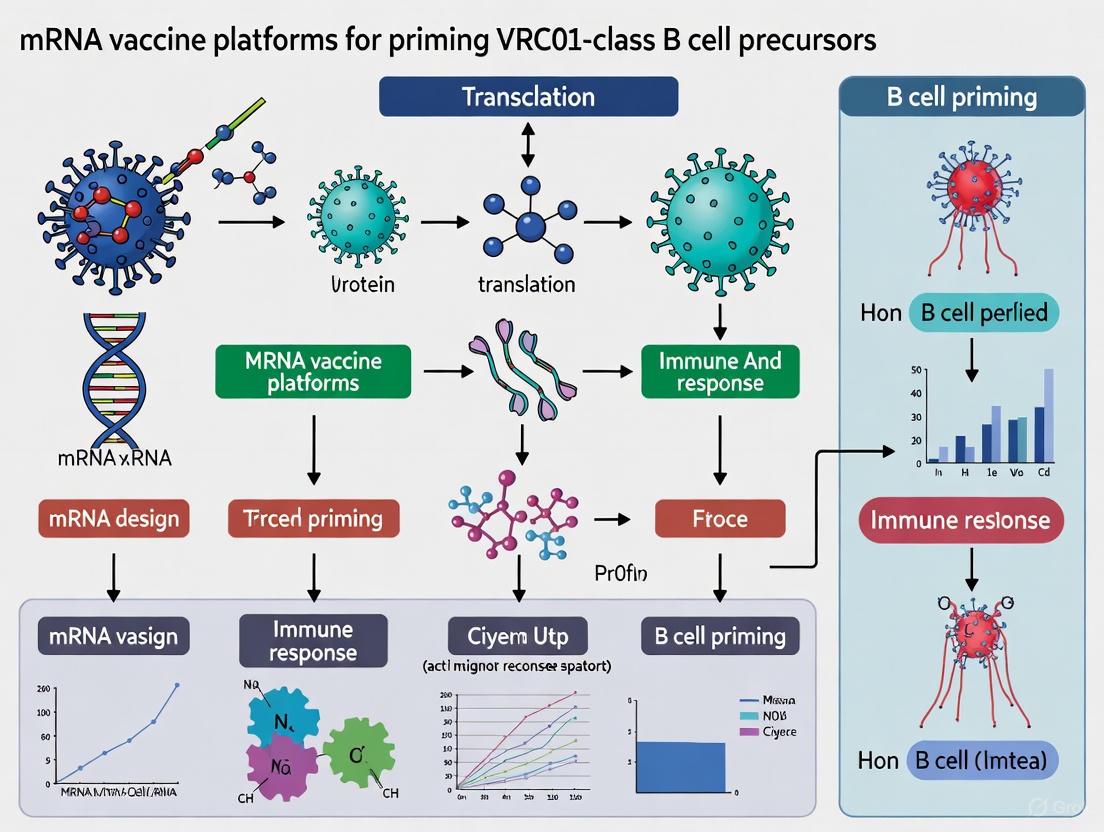
mRNA Vaccine Platforms for VRC01 Precursor Priming
mRNA-LNP Vaccine Formulation and Administration
The development of mRNA-LNP vaccines for HIV represents a significant advancement in germline-targeting strategies [8] [6] [9]. The following protocol outlines key considerations for mRNA vaccine design and evaluation:
- Immunogen Design: Engineer mRNA sequences encoding germline-targeting immunogens (e.g., eOD-GT8, 426c.Mod.Core) with optimized codons for enhanced expression [6] [9].
- LNP Formulation: Encapsulate mRNA in lipid nanoparticles composed of ionizable lipid, phospholipid, cholesterol, and PEG-lipid [6].
- Vaccination Regimen:
- Prime with mRNA-LNP encoding germline-targeting immunogen (50-100µg)
- Boost at 8-16 week intervals with heterologous immunogens to drive affinity maturation [6]
- Response Evaluation:
Recent clinical trials (IAVI G002 and G003) have demonstrated that mRNA-LNP vaccines can effectively prime VRC01-class B cell precursors in humans, with 97% of participants showing detectable responses after two immunizations [6] [9]. The mRNA platform offers advantages in rapid production and the ability to co-deliver multiple immunogens to target different bnAb lineages simultaneously [8].
Quantitative Data from Clinical Trials
Table 4: Summary of Clinical Trial Results for VRC01-Class Targeted Vaccines
| Trial Identifier | Vaccine Platform | Population | Response Rate | Key Findings |
|---|---|---|---|---|
| IAVI G001 [9] | Protein nanoparticle (eOD-GT8 60-mer) | 36 participants | 97% (35/36) | Established proof-of-concept for germline targeting |
| IAVI G002 [6] | mRNA-LNP | 60 participants | 100% with prime-boost (17/17) | Heterologous boost drove "elite" responses with multiple beneficial mutations |
| IAVI G003 [6] | mRNA-LNP | 18 participants (East Africa) | 94% (17/18) | Demonstrated feasibility in African populations |
| HVTN 301 [9] | Protein nanoparticle (426c.Mod.Core) | 48 participants | Data pending | Evaluating dose-sparing regimens |
VRC01-class antibodies represent a promising target for HIV vaccine development due to their exceptional breadth and potency against diverse viral strains. Their unique genetic restrictions—requiring pairing of VH1-2*02 heavy chains with light chains containing rare 5-amino-acid CDRL3s—present both a challenge and an opportunity for precise vaccine targeting. The development of germline-targeting immunogens and sequential immunization strategies has enabled significant progress in guiding the maturation of these antibodies in animal models and humans.
Recent advances in mRNA vaccine platforms have demonstrated particular promise, showing efficient priming of VRC01-class precursor B cells in clinical trials. The successful application of these technologies, combined with a deeper understanding of the structural requirements for bypassing steric restrictions such as the N276 glycan, brings us closer to an effective HIV vaccine strategy. Future work will need to focus on optimizing sequential immunization regimens, understanding and mitigating inter-clonal B cell competition, and validating these approaches in diverse populations.
A significant impediment to developing an effective HIV-1 vaccine is the inherent low affinity between naive B cell receptors (BCRs) of broadly neutralizing antibody (bnAb) lineages and the native HIV envelope glycoprotein (Env). Precursors to VRC01-class bnAbs, which target the highly conserved CD4 binding site (CD4bs), often exhibit weak or undetectable binding to wild-type Env [10] [11]. This low affinity fails to effectively initiate the critical processes of B cell activation, germinal center (GC) recruitment, and the clonal expansion necessary for affinity maturation [4]. Consequently, conventional Env immunogens typically elicit strain-specific or non-neutralizing antibodies, leaving the conserved bnAb epitopes subdominant. Overcoming this initial activation barrier is therefore a primary focus of modern HIV vaccine research, particularly with the advent of germline-targeting strategies and novel delivery platforms like mRNA-LNPs.
Core Concepts and Key Research Findings
The Permissive Selection Model and Affinity Requirements
Recent evidence suggests that B cell selection in germinal centers is more permissive than previously thought, allowing the survival and expansion of lower-affinity clones. This creates a "natural window" for B cells targeting structurally constrained epitopes like the HIV CD4bs [12] [10]. Studies in transgenic mice with human-like antibody diversity show that immunization can focus B cell memory on the conserved CD4bs through both conventional affinity maturation and the reproducible expansion of low-affinity BCR clones. Intriguingly, in some cases, somatic hypermutation (SHM) appears to facilitate target acquisition by decreasing binding strength, suggesting a complex selection process that enables epitope discovery [10].
Quantitative Affinity and Kinetic Parameters of BCR Activation
The activation of B cells, especially those expressing IgM BCRs, depends not only on affinity (equilibrium dissociation constant, KD) but also on the kinetic rates of antigen binding. Research using Ramos B cell lines expressing IgM BCRs with specificity for CD4bs bnAbs reveals that B cells discriminate antigens based on the association rate (kon) and a threshold antigen-BCR half-life, rather than on the overall KD alone [13]. A minimal antigen valency (n ≥ 3) is required for effective BCR signaling and internalization, highlighting the importance of multivalent immunogen design for activating precursor B cells [13].
Table 1: Key Biophysical Parameters for Naive BCR Activation
| Parameter | Description | Impact on B Cell Activation |
|---|---|---|
| Affinity (KD) | Equilibrium dissociation constant | Low affinity of naive VRC01-class BCRs for native Env is a major hurdle for initial activation [10] [11]. |
| Association Rate (kon) | Rate of antigen-BCR binding | A critical sensed parameter; must be sufficiently high for BCR signaling and internalization [13]. |
| Dissociation Rate (koff) | Rate of antigen-BCR complex dissociation | Must be sufficiently low to allow a threshold antigen-BCR half-life for downstream signaling [13]. |
| Antigen Valency | Number of binding sites per immunogen molecule | A minimum valency (n=3) is required for both BCR signaling and down-modulation [13]. |
The mRNA-LNP Vaccine Platform for Priming Precursors
The mRNA-Lipid Nanoparticle (LNP) platform presents a promising solution for priming VRC01-class precursor B cells. Preclinical studies in knock-in mouse models demonstrate that mRNA-LNP encoding the germline-targeting immunogen eOD-GT8 as a soluble self-assembling 60mer nanoparticle can successfully prime multiple VRC01-precursor B cell lineages simultaneously, even when these precursors are present at low, physiological frequencies [14]. Compared to protein immunization, the mRNA-LNP format can induce more robust and sustained germinal center responses, drive higher levels of somatic hypermutation, and promote better class-switching to IgG memory B cells, which is crucial for the development of long-lived immunity [14].
Experimental Protocols and Applications
Protocol: Evaluating mRNA-LNP Priming of VRC01-Class Precursors in Humanized Mouse Models
This protocol outlines the use of the mRNA-LNP platform to prime and expand VRC01-class B cell precursors in adoptive transfer mouse models, based on methodologies validated in [14].
1. Key Research Reagent Solutions Table 2: Essential Reagents for mRNA-LNP Priming Studies
| Reagent | Function/Description | Key Feature |
|---|---|---|
| eOD-GT8 60mer mRNA-LNP | Germline-targeting immunogen; encodes self-assembling nanoparticle. | Soluble nanoparticle presentation; prolonged antigen expression [14]. |
| CLK Series Knock-In Mice | Mouse models bearing genuine human VRC01-precursor BCRs (e.g., CLK19, CLK09, CLK21). | Express authentic, non-inferred human BCRs at controllable frequencies [14]. |
| Fluorescently-Labeled Antigen Probes | e.g., eOD-GT8 and eOD-GT8KO tetramers. | Used for tracking antigen-specific B cells via flow cytometry; KO probe identifies on-target cells [11]. |
| Adjuvant Controls | e.g., SMNP (saponin/MPLA nanoparticle). | Benchmark for assessing mRNA-LNP adjuvanting effects [4]. |
2. Procedure
- Step 1: Precursor Cell Engraftment. Adoptively transfer naive B cells from donor CLK mice (e.g., CD45.2+) into recipient wild-type mice (e.g., CD45.1+) to achieve a physiological precursor frequency (e.g., ~1 in a million B cells) [14].
- Step 2: Prime Immunization. Immunize engrafted mice via intramuscular injection with 0.6 - 10 µg of eOD-GT8 60mer mRNA-LNP. Include control groups immunized with eOD-GT8 60mer protein formulated with a standard adjuvant like SAS or SMNP [14].
- Step 3: Longitudinal Immune Monitoring. At defined time points (e.g., days 14, 28, 42, 72) post-immunization, analyze immune responses in spleen and lymph nodes:
- Flow Cytometry: Identify and sort antigen-specific B cells using multi-color eOD-GT8 and eOD-GT8KO tetramer staining. Key populations: GC B cells (B220+CD38loGL7+Fas+), class-switched memory B cells (B220+IgM-IgD-CD38+) [14] [11].
- Serology: Measure antigen-specific serum IgG titers by ELISA.
- BCR Sequencing: Perform single-cell BCR sequencing (e.g., 10x Genomics platform) on sorted antigen-specific B cells to track clonal expansion, somatic hypermutation, and lineage development [14] [11].
- Step 4: Boosting and Maturation. Boost immunized mice with mRNA-LNP encoding native-like Env immunogens (e.g., 426c.Core) to drive further affinity maturation and selection for bnAb traits [14].
3. Data Analysis
- Calculate the frequency and absolute number of engrafted (CD45.2+) B cells in GCs.
- Analyze SHM patterns in recovered BCR sequences, focusing on known VRC01-class signature mutations.
- Correlate the magnitude of GC responses and SHM load with the dose of mRNA-LNP and the starting precursor frequency.
Protocol: Measuring BCR Activation Kinetics Using Ramos B Cell Lines
This protocol details the use of Ramos B cell lines to dissect the role of binding kinetics in BCR activation by HIV Env proteins, as described in [13].
1. Key Reagents
- Ramos B Cell Lines: Stably express IgM BCRs of defined HIV-1 neutralizing antibodies.
- Panel of Engineered Env Proteins: Variants with similar KD values but different association (kon) and dissociation (koff) rates. Fluorescently labeled for detection.
- Calcium-Sensitive Dyes: e.g., Fluo-4 AM, for measuring intracellular calcium flux.
- Phospho-Specific Antibodies: For detecting proximal BCR signaling (e.g., phospho-Syk, phospho-BLNK).
2. Procedure
- Step 1: Antigen-BCR Binding Characterization. Determine the KD, kon, and koff for the panel of Env proteins binding to the Ramos BCRs using surface plasmon resonance (SPR) or bio-layer interferometry (BLI).
- Step 2: Early BCR Signaling Assays.
- Calcium Flux: Load Ramos cells with a calcium-sensitive dye. Stimulate with multimeric forms (≥ trimeric) of Env proteins and monitor real-time calcium mobilization using a flow cytometer or fluorometer.
- Phospho-Signaling: Stimulate cells with Env proteins for short durations (e.g., 0, 2, 5, 10 min). Fix and permeabilize cells, then stain with phospho-specific antibodies for analysis by flow cytometry.
- Step 3: Late BCR Activation Assays.
- BCR Internalization: Incubate Ramos cells with fluorescently labeled Env proteins at 37°C. At various time points, quantify the loss of surface BCR fluorescence using flow cytometry.
- Antigen Internalization: Track the uptake of labeled Env into the cells over time by flow cytometry or confocal microscopy.
3. Data Analysis
- Correlate the kinetic parameters (kon, koff, half-life) of each Env protein with the amplitude and kinetics of calcium flux, phospho-signaling, and internalization.
- Establish the minimum kon and half-life thresholds required for activating the specific BCR of interest.
Visualizing the Scientific Workflow and Concepts
Diagram: Overcoming the Low-Affinity Hurdle with mRNA Vaccines
Diagram: Experimental Workflow for mRNA-LNP Priming
The low affinity of naive VRC01-class BCRs for native HIV Env remains a formidable but surmountable hurdle. The integration of germline-targeting immunogen design with the mRNA-LNP delivery platform represents a potent strategy for initiating bnAb responses. The platform's ability to drive robust and sustained germinal center reactions, promote necessary somatic hypermutation, and prime multiple precursor lineages simultaneously positions it as a leading candidate for HIV vaccine development. Future work should focus on optimizing sequential mRNA booster regimens featuring progressively more native-like Env immunogens to guide B cell maturation toward broad neutralization, ultimately clearing the path for an effective HIV-1 vaccine.
Core Concept and Rationale
The germline-targeting strategy is a rational vaccine approach designed to initiate an immune response by engaging and activating rare B lymphocytes that possess the intrinsic potential to develop into broadly neutralizing antibodies (bnAbs). This is particularly critical for pathogens like HIV-1, where conventional vaccine strategies have failed. The VRC01-class of bnAbs, which target the highly conserved CD4 binding site (CD4bs) on the HIV-1 envelope (Env) protein, is a primary focus of these efforts. These antibodies are characterized by their use of the VH1-2*02 gene segment and light chains with a short 5-amino acid CDRL3 [4] [15].
A significant challenge is that the unmutated, germline precursors of these potent bnAbs typically do not recognize native HIV-1 Env proteins. Therefore, the initial priming immunogen must be specially engineered to bind and activate these rare B cell receptors (BCRs). The success of this first step was demonstrated in the IAVI G001 phase 1 clinical trial, where a germline-targeting immunogen successfully activated naive B cells with VRC01-class features in 97% of vaccine recipients [16]. This priming event is the essential first step in a guided process, often described as "shepherding" the immune system, where subsequent booster immunizations are intended to drive the affinity maturation and somatic hypermutation of these precursors toward bnAbs with increased breadth and potency [17].
Table: Key Characteristics of VRC01-class Broadly Neutralizing Antibodies
| Feature | Description | Significance for Vaccine Design |
|---|---|---|
| Target Epitope | CD4 binding site (CD4bs) on HIV-1 Env [15] | Target is conserved across many HIV-1 strains, making it ideal for a broad vaccine. |
| Heavy Chain Gene | IGHV1-2*02 [15] | Priming immunogens must be designed to specifically engage BCRs using this gene. |
| Light Chain Feature | 5-amino acid CDRL3 [4] | A key recognition feature for specific germline-targeting immunogens. |
| Maturation Requirements | High somatic hypermutation (up to ~40%), sometimes requiring rare insertions/deletions [15] | A simple immunogen is insufficient; sequential immunization is needed to guide maturation. |
Current Experimental Approaches and Data
Recent research has validated several platform technologies and immunization regimens for priming VRC01-class precursor B cells. The data below highlight the progression from protein-based to mRNA-LNP delivered immunogens and the critical importance of the immunogen's antigenic context.
mRNA-LNP as a Potent Delivery Platform
The use of mRNA-LNP (lipid nanoparticle) technology has shown significant promise in preclinical models for priming and boosting VRC01-class responses. This platform offers potential advantages over protein-based vaccines, including sustained antigen production that can lead to larger and more durable Germinal Center (GC) reactions.
In a key study, an mRNA-LNP encoding the self-assembling eOD-GT8 60mer nanoparticle was used to prime B cell responses in mice with physiological frequencies of human VRC01-precursor BCRs [14]. The results demonstrated that:
- mRNA-LNP priming recruited precursor B cells to germinal centers as effectively as eOD-GT8 60mer protein.
- A high dose (10 µg) of mRNA-LNP was more effective at recruiting precursor B cells to splenic GCs at lower, more physiologically relevant starting frequencies.
- The mRNA-LNP platform induced a significantly higher percentage of class-switched IgG memory B cells (MBCs) and sustained GC responses until day 42, indicating the promotion of long-lived immunity [14].
Furthermore, this platform enabled the simultaneous priming of multiple VRC01-precursor lineages (CLK19, CLK09, CLK21) within a single host without exclusionary competition, a crucial finding for designing vaccines that can elicit a diverse and robust antibody response [14].
The Critical Role of Immunogen Design and Sequence
The choice of the priming immunogen is paramount, as its design dictates the specificity and quality of the initial B cell response. Research directly comparing immunogens has yielded critical insights:
- Superiority of Env-based Priming: A head-to-head comparison in a murine adoptive transfer model revealed that priming with a germline-targeting HIV-1 Env protein (426c.Mod.Core) was superior to priming with a non-envelope anti-idiotypic antibody (ai-mAb). The Env-Env prime-boost regimen led to the greatest expansion of on-target VRC01 B cells, larger GC responses, and higher antibody titers. Priming with the non-Env ai-mAb drove "off-track" somatic mutations that disfavored subsequent boosting with native-like Env immunogens [4].
- Positive Feedback from Off-Target Antibodies: The same study uncovered a positive feedback mechanism. Passive transfer of vaccine-elicited, Env-specific, non-VRC01 IgG was able to rescue and promote the expansion of VRC01-class B cells following an Env boost in ai-mAb primed mice. This suggests that the initial diversity of the B cell response, including so-called "off-target" responses, can be beneficial for focusing and amplifying the maturation of the desired bnAb precursors [4].
Table: Summary of Preclinical Immunization Regimens and Outcomes
| Prime Immunogen | Boost Immunogen | Model System | Key Outcome | Reference |
|---|---|---|---|---|
| eOD-GT8 60mer mRNA-LNP | eOD-GT8 60mer mRNA-LNP (Homologous) | CLK KI mice (multiple VRC01 precursors) | Simultaneous priming of multiple lineages; sustained GCs; high MBC formation. | [14] |
| 426c.Mod.Core (Env) | 426c.Mod.Core (Env) | Murine adoptive transfer | Greatest expansion of VRC01 B cells and GC responses. | [4] |
| ai-mAb (iv4/iv9) | 426c.Mod.Core (Env) | Murine adoptive transfer | Disfavored boosting; drove off-track mutations. | [4] |
| BG505 SOSIP GT1.2 | Shaping/polishing immunogens | gl-CH31 KI mice | Selected for VRC01-class mutations including critical insertions and deletions. | [15] |
Detailed Experimental Protocols
Protocol: Evaluating Germline-Targeting Primers in a Murine Adoptive Transfer Model
This protocol is adapted from procedures used to compare Env vs. non-Env priming immunogens [4].
I. Objective To assess the efficacy of different germline-targeting immunogens in activating, expanding, and directing the maturation of rare VRC01-precursor B cells in vivo.
II. Materials
- Animal Model: Wild-type mice (e.g., C57BL/6) expressing the CD45.1 allele.
- B Cells: Naive B cells from donor mice expressing the CD45.2 allele and bearing the VRC01-class precursor BCR (e.g., iGL-VRC01 B cells).
- Immunogens: Purified germline-targeting proteins (e.g., 426c.Mod.Core, ai-mAb iv4/iv9) or mRNA-LNP formulations.
- Adjuvants: SMNP (saponin/MPLA nanoparticle) or SAS (sigma adjuvant system).
- Flow Cytometry Antibodies: Anti-CD45.1, Anti-CD45.2, Anti-B220, Anti-GL7, Anti-CD95.
III. Procedure
- B Cell Isolation and Transfer:
- Isolate naive B cells from the spleens of donor CD45.2+ iGL-VRC01 mice using a negative selection B cell isolation kit.
- Resuspend 500,000 CD45.2+ iGL-VRC01 B cells in sterile PBS.
- Intravenously inject the cell suspension into CD45.1+ recipient mice via the tail vein. This establishes a physiological frequency of target precursor B cells.
Immunization:
- On day 0, prime the recipient mice via intramuscular (I.M.) injection in the quadriceps.
- Formulate immunogens (e.g., 10 µg of iv4/iv9 or 426c.Mod.Core) with an adjuvant like SMNP.
- Include control groups immunized with adjuvant alone.
Sample Collection and Analysis (Day 14 Post-Prime):
- Serum Collection: Collect blood via retro-orbital bleed or submandibular puncture. Isolate serum and store at -20°C for ELISA.
- Tissue Harvest: Euthanize mice and harvest spleens and draining lymph nodes.
- Single-Cell Suspension: Process tissues into single-cell suspensions and lyse red blood cells (spleen).
- Flow Cytometry Staining: Stain cells with antibodies for CD45.1, CD45.2, B220, GL7, and CD95 to identify transferred (CD45.2+) VRC01-precursor B cells and their entry into germinal centers (B220+GL7+CD95+).
Boosting and Longitudinal Analysis (Optional):
- On day 21 or 28, boost primed mice with a shaping immunogen (e.g., a native-like Env trimer).
- Repeat sample collection and analysis 7-14 days post-boost to evaluate memory recall and further maturation.
IV. Analysis
- ELISA: Measure serum antibody titers against the priming immunogen and control antigens to determine the specificity of the response.
- Flow Cytometry: Quantify the frequency and absolute number of transferred CD45.2+ B cells in GCs compared to control groups.
- B Cell Receptor Sequencing: Single-cell sort GC B cells (CD45.2+B220+GL7+CD95+) and sequence their VH and VL genes to analyze somatic hypermutation and track lineage development.
Protocol: B Cell Receptor Sequencing and Analysis of VRC01-class Lineages
This protocol details the process for analyzing the B cell response at the clonal level [4] [15].
I. Objective To obtain and analyze the variable region sequences of antigen-specific B cells to evaluate somatic hypermutation, clonal diversity, and lineage development.
II. Materials
- Single-Cell Sorter: Fluorescence-activated cell sorter (FACS).
- Lysis Buffer: For reverse transcription (RT).
- Single-Cell RNA Sequencing Kit: e.g., 10x Genomics Chromium Single Cell 5' Kit.
- Nested PCR Primers: Specific for mouse/human Ig variable regions.
III. Procedure (Manual Single-Cell Sorting and RT-PCR)
- Single-Cell Sorting:
- Prepare a single-cell suspension from harvested lymphoid tissues.
- Stain cells with antibodies to identify antigen-specific GC B cells (e.g., for tetramer staining, use fluorescently labeled eOD-GT8 or Env probes).
- Use FACS to single-cell sort 100-500 probe+ GC B cells (B220+GL7+CD95+Antigen+) into a 96-well PCR plate containing lysis buffer. Sort one cell per well.
Reverse Transcription and PCR Amplification:
- Lyse cells and perform reverse transcription using gene-specific primers for Ig constant regions or switch to a template-switching oligo-based method.
- Perform nested PCR to amplify the heavy and light chain variable regions separately. The first PCR uses V-gene family-specific forward primers and a constant region reverse primer. The second PCR uses the first PCR product as a template with nested primers.
Sequence Analysis:
- Purify PCR products and Sanger sequence them.
- Use antibody analysis tools (e.g., IMGT/V-QUEST, IgBLAST) to align sequences, identify V(D)J gene usage, and calculate mutation frequencies.
- Analyze sequences for the acquisition of critical mutations, such as CDRL1 deletions/glycine substitutions to accommodate the N276 glycan or insertions in CDRH1/FWR3, which are hallmarks of mature VRC01-class bnAbs [15].
The Scientist's Toolkit: Research Reagent Solutions
Table: Essential Reagents for VRC01-class Germline-Targeting Research
| Reagent / Solution | Function / Description | Example Use Case |
|---|---|---|
| Germline-Targeting Immunogens | Engineered proteins/mRNA designed to bind unmutated VRC01-precursor BCRs. | eOD-GT8 60mer, 426c.Mod.Core, BG505 SOSIP.GT1.2; used for priming [14] [15]. |
| Knock-in (KI) Mouse Models | Mice engineered with human VRC01-precursor BCRs (e.g., CLK19, CLK09, CLK21, gl-CH31). | Provide a source of rare, genetically defined precursor B cells for in vivo studies [14] [15]. |
| Adjuvant Systems | Immune potentiators that enhance the magnitude and quality of the response to co-administered immunogens. | SMNP (saponin/MPLA nanoparticle) shown to be effective for priming with ai-mAb immunogens [4]. |
| Fluorescent Antigen Probes | Labeled immunogens (e.g., eOD-GT8-AF647) for detecting antigen-specific B cells via flow cytometry. | Used to identify and sort VRC01-class B cells from mouse spleen or human PBMCs for downstream analysis [16]. |
| mRNA-LNP Formulations | Lipid nanoparticles encapsulating mRNA that encodes the immunogen of interest. | Platform for delivering germline-targeting immunogens like eOD-GT8 60mer; enables sustained antigen expression [14] [18]. |
Visualization of the Germline-Targeting Strategy Workflow
The following diagram illustrates the multi-step process of the germline-targeting strategy, from immunogen design to the analysis of the mature B cell response.
The development of a prophylactic HIV vaccine represents one of the most significant challenges in modern immunology. A key goal is inducing broadly neutralizing antibodies (bnAbs) that can protect against diverse HIV strains. The germline-targeting vaccine strategy addresses this challenge through precisely engineered priming immunogens designed to activate rare B cell precursors with the genetic potential to develop into bnAbs [19] [20]. Two leading approaches have emerged: the eOD-GT8 60mer nanoparticle for priming VRC01-class bnAbs targeting the CD4-binding site, and epitope scaffold nanoparticles for priming bnAbs against diverse epitopes including the membrane-proximal external region (MPER) and V2 apex [21] [22] [23].
The scientific rationale for these immunogens stems from the observation that unmutated ancestors of most bnAbs typically show no detectable affinity for wild-type HIV envelope (Env) proteins [19]. Germline-targeting immunogens are therefore engineered with specific modifications to engage these rare precursor B cells, initiating the process that, through sequential boosting with increasingly native-like immunogens, can guide their maturation toward broadly neutralizing activity [24]. This application note details the technical specifications, experimental protocols, and research reagents for working with these cutting-edge vaccine components.
Quantitative Profile of Key Priming Immunogens
Table 1: Comparative Analysis of Leading Priming Immunogens
| Immunogen | Target Epitope | Platform | Precursor Frequency | Affinity (KD) | Response Rate |
|---|---|---|---|---|---|
| eOD-GT8 60mer | CD4-binding site (VRC01-class) | Protein nanoparticle + AS01B | 1 in 1.78 million (human VRC01-classVK1-33) [19] | 3.4 μM (human naive VRC01-class) [19] | 97% (35/36) in IAVI G001 [25] |
| eOD-GT8 60mer (mRNA-LNP) | CD4-binding site (VRC01-class) | mRNA-encoded nanoparticle | Similar frequency to protein platform [20] | Comparable to protein platform [20] | At least as effective as protein in IAVI G002 [20] |
| Core-g28v2 60mer | CD4-binding site (first boost) | mRNA-encoded nanoparticle | Designed to engage eOD-GT8-primed B cells [19] | Selects for key mutations [19] | Supported maturation in mouse models [19] |
| 10E8-GT Epitope Scaffolds | MPER (10E8-class) | Protein nanoparticle + SMNP | 0.7-0.8% of naïve B cells bound [23] | ≤10 μM for 10E8 UCA & NGS precursors [21] | Elicited precursors in mice & macaques [21] |
| ApexGT6 Trimer | V2 Apex (PCT64-class) | Soluble protein or mRNA-encoded | 1,288 per million (human PCT64-like) [22] | Enhanced affinity for PCT64 & PG9 precursors [22] | Primed PCT64-like responses in mouse models [22] |
Table 2: In Vivo Immunogenicity Profiles Across Models
| Immunogen | Animal Model | Dose | Schedule | GC Responses | bnAb Precursors |
|---|---|---|---|---|---|
| eOD-GT8 60mer + AS01B | Human (IAVI G001) | 20 μg or 100 μg | Weeks 0 & 8 [25] | Robust circulating Tfh detected [25] | VRC01-class in 97% [25] |
| eOD-GT8 60mer mRNA | Human (IAVI G002) | mRNA-1644 | Prime-boost [26] | Under evaluation | VRC01-class with more mutations than protein [20] |
| 10E8-GT10.2 12mer | Rhesus macaques | Dose escalation | Day 0 & 14 [23] | Robust CD71+CD38- GC B cells [21] | Detectable 10E8-class HCDR3s in GCs & memory B cells [21] |
| ApexGT6 (congly & L14) | Rhesus macaques | Adjuvanted protein or mRNA-LNP | Prime-boost [22] | Antigen-specific GC B cells | PCT64-like & DDY-motif HCDR3s [22] |
| 10E8-GT12 24mer | hD3-3/JH6 mice | Protein + SMNP or mRNA-LNP | Single immunization [21] | Germinal center formation | 10E8- and LN01-class HCDR3s [21] |
Experimental Protocols
Protocol: Assessing Vaccine-Induced B Cell Responses Using Flow Cytometry
Purpose: To identify and characterize antigen-specific B cells following immunization with germline-targeting immunogens.
Materials:
- Single-cell suspensions from spleen or lymph nodes
- Fluorescently-labeled antigen probes (e.g., eOD-GT8, 10E8-GT)
- Anti-mouse CD16/32 (FC block)
- Antibody panel: Anti-B220, Anti-CD19, Anti-CD38, Anti-CD95, Anti-GL7, Anti-IgD
- Flow cytometry buffer (PBS + 2% FBS)
- FACs sorter or analyzer
Procedure:
- Prepare single-cell suspensions from lymphoid tissues 7-14 days post-immunization.
- Count cells and aliquot 1-2×10^6 cells per staining condition.
- Incubate with FC block (1:100) for 10 minutes at 4°C to reduce nonspecific binding.
- Stain cells with fluorescently-labeled antigen probes (1-5 μg/mL) for 30 minutes at 4°C.
- Add surface antibody cocktail and incubate for 20 minutes at 4°C protected from light.
- Wash cells twice with flow cytometry buffer and resuspend in buffer for analysis.
- Identify antigen-binding B cells as live, singlet, CD19+B220+, antigen-probe+ cells.
- Characterize germinal center B cells as CD38loCD95+GL7+IgD- population.
Notes: Antigen probes should be extensively validated for specificity using knockout controls. For rare populations, pre-enrichment strategies may be necessary [19] [21].
Protocol: Single-Cell BCR Sequencing of Antigen-Specific B Cells
Purpose: To obtain paired heavy- and light-chain variable region sequences from antigen-specific B cells for analysis of mutation frequency and lineage development.
Materials:
- FACS-sorted antigen-specific B cells (100-10,000 cells)
- Single-cell RNA/DNA sequencing kit (10x Genomics)
- Barcode-conjugated antibodies for hashing
- Reverse transcription reagents
- PCR purification kits
- Next-generation sequencer
Procedure:
- Sort antigen-specific B cells into 96-well plates containing lysis buffer or prepare suspensions for 10x Genomics.
- For 10x Genomics: Partition single cells with barcoded beads using the Chromium system.
- Perform reverse transcription to add cell barcodes and unique molecular identifiers (UMIs).
- Amplify V(D)J regions using locus-specific primers.
- Construct sequencing libraries following manufacturer's protocols.
- Sequence libraries on Illumina platform (minimum 2×150 bp).
- Process data using Cell Ranger V(D)J or similar pipeline.
- Analyze sequences for V/D/J usage, mutation frequency, and CDR3 features.
Notes: This approach was used to determine VRC01-class precursor frequency in SE09 mice (1 in 13,600) and humans (1 in 228,000) [19].
Protocol: mRNA-LNP Formulation and Immunization
Purpose: To deliver mRNA-encoded immunogens via lipid nanoparticles for in vivo expression.
Materials:
- mRNA encoding immunogen (e.g., mRNA-1644 for eOD-GT8)
- Lipid mixture (ionizable lipid, DSPC, cholesterol, PEG-lipid)
- Microfluidic device or T-tube mixer
- PBS, pH 7.4
- Dialysis cassettes (100 kDa MWCO)
- 0.22 μm sterile filters
Procedure:
- Prepare aqueous phase: dilute mRNA in 10 mM citrate buffer, pH 3.0.
- Prepare lipid phase: dissolve lipids in ethanol at precise molar ratios.
- Mix aqueous and lipid phases using microfluidic device at 1:3 ratio (aqueous:ethanol).
- Dialyze against PBS, pH 7.4, for 18-24 hours to remove ethanol and raise pH.
- Concentrate LNPs using centrifugal filters if needed.
- Filter-sterilize through 0.22 μm filter.
- Characterize LNP size (60-100 nm), PDI (<0.2), and mRNA encapsulation efficiency (>80%).
- Administer via intramuscular injection (5-50 μg mRNA dose in mice).
Notes: mRNA-LNP delivery of eOD-GT8 in IAVI G002 trial induced VRC01-class precursors with greater mutations than protein vaccination [20] [26].
Conceptual Framework and Signaling Pathways
Diagram 1: Germline-Targeting Vaccine Strategy Logic
Diagram 2: Immunogen Processing and Immune Activation Pathways
The Scientist's Toolkit: Essential Research Reagents
Table 3: Key Research Reagents for Germline-Targeting HIV Vaccine Research
| Reagent/Category | Specific Examples | Function/Application | Validation Notes |
|---|---|---|---|
| Priming Immunogens | eOD-GT8 60mer, 426c.Mod.Core, ApexGT6, 10E8-GT scaffolds [19] [21] [22] | Activate rare bnAb precursor B cells | Validate binding to inferred germline antibodies & human naive B cells |
| Delivery Platforms | mRNA-LNP, Protein + AS01B, Protein + SMNP [19] [21] [25] | Enhance immunogenicity & specific B cell targeting | mRNA enables rapid iteration; adjuvants enhance magnitude & quality |
| Animal Models | SE09 (VH1-2/VK1-33), hD3-3/JH6, Rhesus macaques [19] [21] [22] | Preclinical evaluation with humanized Ig loci | SE09: VRC01-class precursors; hD3-3: 10E8-class; Macaques: translational |
| Analytical Tools | Antigen-specific B cell sorting, Single-cell BCR sequencing, SPR/BLI [19] [21] [20] | Characterize frequency, specificity & affinity of responses | Use knockout antigens to confirm epitope specificity |
| Adjuvant Systems | AS01B, SMNP, Aluminum hydroxide [25] [4] [23] | Enhance immunogenicity & promote GC responses | AS01B strong for CD4 T cells; SMNP for nanoparticle vaccines |
The development of eOD-GT8 60mer and epitope scaffold nanoparticles represents a transformative advance in rational vaccine design. These immunogens demonstrate that precise engineering of vaccine antigens can successfully engage rare bnAb precursor B cells—a critical first step in the path toward an effective HIV vaccine. The integration of mRNA platform technology further accelerates this approach by enabling rapid iteration and enhanced immunogenicity. As these strategies progress through clinical trials, they offer not only promise for HIV prevention but also a blueprint for targeting other pathogens with high antigenic diversity.
Precursor Frequency and Diversity in the Human Repertoire
The following tables consolidate key quantitative findings on VRC01-class B cell precursor frequencies and the impact of genetic diversity, which are critical for germline-targeting vaccine design.
Table 1: IGHV1-2 Allelic Variation and Naive Repertoire Frequency [27]
| IGHV1-2 Allele | VRC01-class Binding Capability | Mean mRNA Expression in Naive Repertoire (Per-Allele, Homozygous) | Mean mRNA Expression in Naive Repertoire (Per-Allele, Heterozygous) | Approximate Frequency of Unique HCDR3s (vs. *02) |
|---|---|---|---|---|
| *02 | Required (with 5-aa LC) | 3.1% (95% CI: 2.7–3.6%) | 3.3% (95% CI: 2.9–3.8%) | Baseline (Highest) |
| *04 | Required (with 5-aa LC) | 0.9% (95% CI: 0.7–1.1%) | 0.7% (95% CI: 0.6–0.8%) | ~4-fold lower than *02 |
| *05 | Not capable | 0.09% (95% CI: 0.03–0.14%) | Information missing in source | Not applicable |
| *06 | Not capable | 2.4% (95% CI: 1.9–3.0%) | Information missing in source | Not applicable |
Table 2: Response to eOD-GT8 60mer Immunization in Clinical and Preclinical Models [14] [27]
| Model / Trial | Immunogen / Regimen | Key Finding on VRC01-class B Cells | Quantitative Outcome |
|---|---|---|---|
| IAVI G001 (Clinical Trial) | eOD-GT8 60mer protein + AS01B, 2 doses | Higher precursor frequency in individuals with IGHV1-2*02 vs. *04 allele | Median frequency in memory B cells: 0.13% (high dose) vs. 0.09% (low dose); difference linked to genotype imbalance. |
| mRNA-LNP Mouse Model (CLK19) | eOD-GT8 60mer mRNA-LNP, single prime | Efficient priming and memory formation at physiological precursor frequency | High dose (10 µg) mRNA-LNP recruited more B cells to splenic GCs than protein at low starting frequency; induced significantly higher percentage of class-switched IgG Memory B Cells. |
| mRNA-LNP Mouse Model (Multi-lineage) | eOD-GT8 60mer mRNA-LNP prime/boost | Simultaneous priming and affinity maturation of multiple VRC01-precursor lineages | Three distinct precursor lineages (CLK19, CLK09, CLK21) were simultaneously primed, participated in GCs, and accumulated somatic hypermutations after boosting. |
Detailed Experimental Protocols
Protocol 1: Assessing Human IGHV Repertoire and Allelic Impact on Vaccine Priming
This protocol outlines the methodology for genotyping and quantifying IGHV1-2 allele usage to evaluate its impact on germline-targeting vaccine responses, as performed in the IAVI G001 clinical trial analysis [27].
Materials
- Biological Sample: Peripheral blood mononuclear cells (PBMCs) from clinical trial participants.
- RNA Extraction Kit: Standard kit for high-quality total RNA isolation.
- Reverse Transcription Kit: Includes primers for IgM constant region and unique molecular identifiers (UMIs).
- PCR Reagents: For amplifying immunoglobulin variable regions.
- High-Throughput Sequencer: Illumina platform or equivalent.
- Bioinformatics Tools: IgDiscover germline allele inference tool and custom scripts for UMI counting and HCDR3 analysis [27].
Procedure
Sample Collection and Library Preparation:
- Collect PBMCs from participants pre- and post-vaccination.
- Isolate total RNA from PBMCs.
- Synthesize IgM cDNA using reverse transcription primers containing UMIs.
- Perform PCR to amplify IGHV regions using V-gene-specific primers.
High-Throughput Sequencing and Genotyping:
- Sequence the amplified libraries on a high-throughput platform.
- Use the IgDiscover tool to perform personalized, nucleotide-level genotyping of IGHV1-2 and other alleles for each participant [27].
- Classify alleles as *02, *04, *05, *06, or other variants.
Quantification of Allele Usage and Precursor Frequency:
- Process sequencing data to count UMIs for each IGHV1-2 allele. The UMI count is proportional to the mRNA expression level of that allele in the naive repertoire.
- Extract and count unique HCDR3 sequences associated with each IGHV1-2 allele. This serves as a proxy for the number of unique B cells expressing that allele.
- Calculate the frequency of each allele by determining its proportion relative to the total IGHV UMI count or total HCDR3 count.
Correlation with Vaccine Response:
- Statistically model the relationship between IGHV1-2 genotype (e.g., 02/02 vs. 04/04), allele-specific precursor frequency, and the frequency of vaccine-induced VRC01-class IgG memory B cells measured via flow cytometry.
Protocol 2: Evaluating mRNA-LNP Priming and Boosting of bnAb Precursors in Humanized Mouse Models
This protocol describes the use of mRNA-LNP vaccines to prime and boost multiple VRC01-class precursor lineages in adoptive transfer mouse models, validating the platform for precursor engagement and maturation [14].
Materials
- Mouse Models: Wild-type recipient mice (e.g., expressing CD45.1 allele) for adoptive transfer.
- Donor B Cells: Naive B cells from knock-in mouse models (e.g., CLK19, CLK09, CLK21) expressing genuine human VRC01-class BCRs with known affinities for eOD-GT8. These B cells are isogenic for the BCR and express the CD45.2 allele for tracking.
- Immunogens:
- Prime and Boost: LNP-encapsulated nucleoside-modified mRNA encoding eOD-GT8 self-assembling 60mer nanoparticle.
- Control: eOD-GT8 60mer protein formulated with adjuvant.
- Adjuvant: SMNP (saponin and monophosphoryl Lipid A nanoparticle adjuvant) for protein immunizations [4].
- Flow Cytometry Reagents: Antibodies against CD45.1, CD45.2, B220, CD38, GL7, IgG, and fluorescently labeled eOD-GT8 and native-like Env probes.
Procedure
Adoptive B Cell Transfer:
- Isolate naive CD45.2+ B cells from donor CLK mice.
- Adoptively transfer these cells intravenously into CD45.1+ recipient mice. To model physiological conditions, use a low number of transferred cells (e.g., resulting in a precursor frequency of ~1 in 300,000 total B cells) [14].
Immunization:
- Prime mice via intramuscular injection with eOD-GT8 mRNA-LNP (e.g., 10 µg or 0.6 µg) or eOD-GT8 60mer protein with adjuvant at day 0.
- Boost mice at week 8 or later with the same or a more native-like mRNA-LNP immunogen.
Immune Response Monitoring:
- At various time points (e.g., days 14, 42, 72), analyze immune responses in spleen, lymph nodes, and serum.
- Flow Cytometry: Identify and sort donor-derived (CD45.2+) B cells. Analyze GC B cells (B220+ CD38lo GL7+) and memory B cells (B220+ CD38+ IgG+). Use antigen-specific probes to identify eOD-GT8- and Env-binding B cells.
- Serology: Measure antigen-specific serum antibody titers by ELISA.
B Cell Receptor Analysis:
- Single-cell sort donor-derived antigen-binding GC B cells and memory B cells.
- Amplify and sequence Ig heavy and light chain variable regions.
- Analyze sequences for somatic hypermutation (SHM) and mutation hotspots, comparing to known VRC01-class bnAb mutation patterns.
Figure 1: Experimental workflow for evaluating mRNA-LNP priming and boosting in mouse models.
The Scientist's Toolkit: Research Reagent Solutions
Table 3: Essential Research Reagents for Precursor Frequency and Priming Studies [4] [14] [27]
| Reagent / Tool | Function / Application | Specific Examples / Notes |
|---|---|---|
| Germline-Targeting Immunogens | Prime rare bnAb-precursor B cells by engaging their unmutated BCRs. | eOD-GT8 60mer nanoparticle [14]; 426c.Mod.Core-C4b [4]; BG505 SOSIP.GT1.1 gp140 [4]. |
| mRNA-LNP Platform | Deliver encoded immunogens for in vivo expression; promotes strong GC responses and MBC formation. | Nucleoside-modified mRNA encoding soluble self-assembling nanoparticles [14]. |
| Adoptive Transfer Mouse Models | Preclinically test immunogens using humanized BCRs at controllable, physiological frequencies. | CLK series mice (e.g., CLK19, CLK09, CLK21) bearing genuine human VRC01-class BCRs [14]. |
| Adjuvants | Enhance the magnitude and quality of the immune response to protein immunogens. | SMNP (saponin/MPLA nanoparticle) [4]; AS01B (used in clinical trials) [27]. |
| Antigen-Specific B Cell Probes | Identify and sort antigen-specific B cells via flow cytometry. | Recombinant proteins: eOD-GT8, native-like Env (SOSIP), and knockout (KO) mutants to confirm specificity [4] [14]. |
| IGHV Genotyping & Repertoire Sequencing | Determine individual's immunoglobulin genotype and quantify allele expression/precursor frequency. | High-throughput sequencing of IgM libraries; bioinformatics tools (IgDiscover) for personalized genotyping [27]. |
Figure 2: The logical relationship between genetic makeup, precursor frequency, and the outcome of germline-targeting vaccination.
Engineering mRNA-LNP Immunogens for Precursor Priming and Expansion
Within modern vaccinology, messenger RNA encapsulated in lipid nanoparticles (mRNA-LNP) has emerged as a transformative platform, offering distinct advantages in speed and efficacy. These benefits are particularly salient for advanced vaccine concepts, such as the precise priming of VRC01-class B cell precursors, a cornerstone of next-generation HIV vaccine research [6] [20]. The "rapid design" of mRNA vaccines allows for the swift testing of novel immunogen sequences, while the "sustained antigen expression" enabled by LNPs promotes the extended germinal center reactions necessary for guiding naïve B cells toward the development of broadly neutralizing antibodies (bNAbs) [20]. This application note details how these platform advantages are leveraged in the context of priming VRC01-class B cell responses, providing key quantitative data, standardized protocols, and visual frameworks to support research and development efforts.
Platform Advantages and Quantitative Evidence
The mRNA-LNP platform provides two foundational advantages for complex vaccine development: a rapid and flexible production cycle and the ability to induce potent, high-quality immune responses through sustained antigen expression. Table 1 summarizes key quantitative data from recent studies that substantiate these claims.
Table 1: Quantitative Evidence of mRNA-LNP Platform Advantages in Recent HIV Vaccine Trials
| Trial / Study Identifier | Platform & Immunogen | Key Quantitative Immunogenicity Results | Implication for Platform Advantage |
|---|---|---|---|
| IAVI G002 (NCT05001373) [6] | mRNA-LNP encoding eOD-GT8 60-mer | 100% (17/17) of participants receiving prime + heterologous boost developed VRC01-class responses. >80% showed "elite" responses with multiple helpful mutations. | Demonstrates high efficiency in initiating and advancing desired B cell lineages. |
| IAVI G001 (NCT03547245) [20] | Protein eOD-GT8 60-mer with AS01B adjuvant | 97% (35/36) response rate for priming VRC01-class B cell precursors after two immunizations. | Serves as a benchmark for the priming step; mRNA platform shown to be at least as effective [20]. |
| L@Mn-mRNA Platform [28] | Novel LNP with metal-ion enriched mRNA core | ~2x increase in mRNA loading capacity and cellular uptake efficiency compared to conventional LNP-mRNA. | Enhances delivery efficiency and could enable dose-sparing, improving the safety profile. |
The speed of the platform stems from its fully in vitro transcription process, which allows for the rapid production of new immunogen sequences, such as the eOD-GT8 60-mer, without the need for cell-based systems or complex protein purification [29]. Once delivered, the LNP protects the mRNA, facilitates cellular uptake, and enables endosomal escape into the cytoplasm for translation [30]. The subsequent sustained production of native-like antigenic protein by host cells, such as muscle or immune cells, is critical for driving the affinity maturation of B cells. This prolonged antigen exposure is essential for guiding rare B cell precursors, like those for VRC01-class bNAbs, through the complex sequence of somatic hypermutations required to achieve breadth and potency [6] [20].
Experimental Protocols for mRNA-LNP Vaccine Assessment
The following protocols outline key methodologies for evaluating the performance of mRNA-LNP vaccines in preclinical and clinical settings, with a focus on B cell priming.
Protocol: In Vivo Immunogenicity Assessment of mRNA-LNP Vaccines
Objective: To evaluate the immunogenicity of an mRNA-LNP vaccine candidate, specifically its ability to prime and expand VRC01-class B cell precursors in a murine model.
Materials:
- mRNA-LNP formulation (e.g., encoding eOD-GT8 60-mer)
- Control (e.g., PBS or irrelevant mRNA-LNP)
- C57BL/6 mice (6-8 weeks old)
- Flow cytometer with cell sorter
- Fluorochrome-labeled antibodies (anti-mouse B220, CD19, IgG)
- Antigen-specific probes (e.g., biotinylated eOD-GT8 for staining precursor B cells)
- ELISA plates and reagents
Procedure:
- Immunization: Administer a prime intramuscular (i.m.) injection of 10 µg mRNA-LNP in a 50 µL volume to the mouse hind leg. Perform a boost immunization with the same dose and route at week 3.
- Sample Collection: At defined endpoints (e.g., day 7 post-prime and day 10 post-boost), euthanize mice and collect spleens and sera.
- B Cell Analysis by Flow Cytometry:
- Prepare a single-cell suspension from spleens.
- Stain 1-2 x 10^6 cells with a panel containing B220, CD19, and a fluorescently labeled eOD-GT8 probe to identify antigen-specific B cells.
- Include a viability dye to exclude dead cells.
- Analyze cells on a flow cytometer. The population of interest is B220+CD19+eOD-GT8+.
- For advanced analysis, sort single eOD-GT8+ B cells into 96-well plates for subsequent monoclonal antibody sequencing and functional analysis.
- Serum Antibody Analysis by ELISA:
- Coat a 96-well ELISA plate with 2 µg/mL of eOD-GT8 protein overnight at 4°C.
- Block the plate with a protein-based blocking buffer for 1-2 hours at room temperature (RT).
- Add serial dilutions of mouse serum and incubate for 2 hours at RT.
- Detect bound IgG using an enzyme-conjugated anti-mouse IgG antibody and a colorimetric substrate.
- Read the absorbance and calculate endpoint titers.
Protocol: Preparation of High-Loading-Capacity L@Mn-mRNA Nanoparticles
Objective: To synthesize a novel LNP formulation with enhanced mRNA loading capacity and cellular uptake, based on a recently published metal-ion enrichment strategy [28].
Materials:
- Purified mRNA (e.g., encoding antigen of interest)
- Manganese(II) chloride (MnCl2)
- Lipid mixture (ionizable lipid, DSPC, cholesterol, PEG-lipid)
- Microfluidic device or T-tube apparatus for mixing
- Heating block
- Dialysis cassettes or tangential flow filtration system
Procedure:
- Synthesis of Mn-mRNA Core Nanoparticles:
- Mix mRNA (e.g., 100 µg) with MnCl2 in a molar ratio of Mn2+ to mRNA bases of 5:1 in nuclease-free water.
- Incubate the mixture at 65°C for 5 minutes to form the Mn-mRNA nanoparticles.
- Allow the complex to cool to room temperature. The formation of nanoparticles can be confirmed by dynamic light scattering (DLS) and transmission electron microscopy (TEM).
- Lipid Coating to Form L@Mn-mRNA:
- Prepare an ethanolic solution of lipids at a defined molar ratio (e.g., ionizable lipid:DSPC:cholesterol:PEG-lipid = 50:10:38.5:1.5).
- Rapidly mix the ethanolic lipid solution with the aqueous Mn-mRNA nanoparticle dispersion using a microfluidic device at a typical flow rate ratio of 3:1 (aqueous:organic) and a total flow rate of 12 mL/min.
- Immediately after mixing, dialyze the resulting L@Mn-mRNA formulation against a phosphate buffer (pH 7.4) for 4-6 hours at 4°C to remove ethanol and unencapsulated components.
- Sterile-filter the final formulation through a 0.22 µm filter. The encapsulation efficiency can be determined using a Ribogreen assay.
Visualizing the Pathway and Workflow
The following diagrams illustrate the key immunological mechanism and a standard experimental workflow for evaluating mRNA-LNP vaccines.
Diagram: mRNA-LNP Platform for B Cell Priming
(Diagram 1: The mRNA-LNP platform enables sustained antigen expression that drives the germinal center reactions necessary for priming rare, target B cell precursors.)
Diagram: Workflow for Vaccine Immunogenicity Assessment
(Diagram 2: A standard workflow for assessing the immunogenicity of an mRNA-LNP vaccine, from immunization to deep B cell and antibody analysis.)
The Scientist's Toolkit
Table 2: Essential Research Reagents for mRNA-LNP HIV Vaccine Research
| Reagent / Material | Function & Application | Example Use Case |
|---|---|---|
| Ionizable Cationic Lipid | Core component of LNPs; enables mRNA encapsulation and endosomal escape via proton sponge effect. | A key functional lipid in LNP formulations like SM-102 (Moderna) or ALC-0315 (Pfizer-BioNTech) [30]. |
| CleanCap AG Reagent | Co-transcriptional capping reagent for IVT mRNA; produces Cap 1 structure for high translation and low immunogenicity. | Used during mRNA synthesis to generate functional mRNA with >94% capping efficiency for vaccines [29]. |
| N1-Methylpseudouridine | Modified nucleoside; incorporated into IVT mRNA to reduce innate immune activation and enhance translational efficiency. | Replaces uridine in the mRNA sequence to improve protein yield and vaccine tolerability [30]. |
| Fluorescent Antigen Probe | Custom-made multimeric protein for detecting antigen-specific B cells via flow cytometry. | eOD-GT8 protein coupled to fluorophores (e.g., PE) to identify and sort rare VRC01-class precursor B cells from immunized mice or human samples [20]. |
| Polyethylene Glycol (PEG)-lipid | LNP component that modulates nanoparticle size, stability, and pharmacokinetics; can influence anti-PEG immunity. | Included at 1.5-2% molar ratio in LNP formulations; potential target for accelerated blood clearance [30] [28]. |
| Microfluidic Device | Instrument for controlled, rapid mixing of aqueous and ethanolic phases to form uniform, stable LNPs. | Used in the standardized preparation of mRNA-LNP formulations with high encapsulation efficiency and batch-to-batch consistency (e.g., NanoAssemblr, PDMS chips) [30]. |
Within the strategic framework of mRNA vaccine development for priming VRC01-class B cell precursors against HIV, soluble self-assembling nanoparticles represent a transformative platform for antigen presentation. The primary challenge in eliciting broadly neutralizing antibodies (bNAbs) is the initial activation of rare B cell precursors with the correct genetic characteristics, a process that requires high-density, repetitive antigen display to effectively engage the immune system [20] [21]. Nanoparticle platforms address this challenge through multivalent antigen presentation that mimics natural viral surfaces, significantly enhancing B cell receptor cross-linking and activation compared to soluble antigens [31] [32].
These engineered nanoparticles serve as precise immunological tools, particularly for germline-targeting strategies aimed at priming specific B cell lineages. For VRC01-class bnAb precursors, which target the CD4-binding site on HIV Env, nanoparticle display provides the necessary signal strength to activate these rare populations [20]. The integration of this antigen presentation technology with mRNA delivery platforms creates a powerful synergy: mRNA encodes the complex immunogen, while the self-assembling nanoparticle structure ensures proper immunological recognition, overcoming the traditional trade-off between immunogen complexity and manufacturability [30] [28].
Key Platform Technologies and Design Principles
Ferritin-Based Nanoparticle Engineering
The human ferritin heavy chain (FTH1) provides a robust structural scaffold for 24-mer self-assembling nanoparticles with exceptional stability and biocompatibility. Recent advances have overcome previous challenges with particle heterogeneity through structure-guided design. Research on the natural ferritinophagy complex between FTH1 and nuclear receptor coactivator 4 (NCOA4) has enabled the engineering of a novel 16-amino acid Fagy tag peptide that binds FTH1 with high affinity (34 nM) [31].
Table 1: Comparison of Self-Assembling Nanoparticle Platforms
| Platform | Structural Basis | Assembly Mechanism | Antigen Coupling | Key Advantages |
|---|---|---|---|---|
| Ferritin-Fagy Tag | Human FTH1 24-mer | Spontaneous oligomerization | Non-covalent peptide binding | High homogeneity, natural human protein, no chemical conjugation |
| Germline-Targeting Epitope Scaffolds | Engineered protein scaffolds | Nanoparticle display | Genetic fusion or covalent linkage | Precise epitope focusing, B cell precursor specificity |
| Metal Ion mRNA Core (L@Mn-mRNA) | Mn²⁺-mRNA coordination | Ion-mediated condensation + lipid coating | mRNA encoded | 2x higher mRNA loading, enhanced cellular uptake |
mRNA Nanoparticle Engineering with Enhanced Loading Capacity
Conventional lipid nanoparticles (LNPs) suffer from limited mRNA loading capacity (typically <5% by weight), necessitating high lipid doses that can cause undesirable side effects [28]. An innovative metal ion-mediated approach addresses this limitation by pre-condensing mRNA with Mn²⁺ ions before lipid coating. This L@Mn-mRNA platform achieves nearly twice the mRNA loading capacity (95.6% mRNA by weight in the core) compared to conventional LNPs, while simultaneously improving cellular uptake by 2-fold due to the enhanced stiffness of the Mn-mRNA core [28]. This advancement is particularly valuable for complex immunogen delivery, as it reduces the lipid dose required for effective vaccination while maintaining potent immunogenicity.
Experimental Protocols
Protocol 1: Fagy Tag Antigen Conjugation to Ferritin Nanoparticles
Principle: This protocol describes the non-covalent conjugation of Fagy-tagged antigens to ferritin nanoparticles, enabling high-density antigen presentation while maintaining particle homogeneity [31].
Materials:
- Purified FTH1 ferritin subunit
- Antigen gene construct with C-terminal Fagy tag (16-amino acid sequence)
- E. coli BL21(DE3) expression system
- Ni-NTA affinity chromatography resin
- Size exclusion chromatography (Superose 6 Increase) column
- Tris-buffered saline (TBS), pH 7.4
- Analytical ultracentrifugation equipment
Procedure:
- Co-expression and Assembly: Co-express FTH1 and Fagy-tagged antigen (e.g., RABV GDIII) in E. coli BL21(DE3) using standard protein expression protocols.
- Complex Purification: Purify the assembled nanoparticle using Ni-NTA affinity chromatography followed by size exclusion chromatography with TBS, pH 7.4.
- Quality Control: Verify assembly homogeneity and stability using:
- Analytical ultracentrifugation (sedimentation velocity)
- Dynamic light scattering (PDI < 0.1)
- Negative stain electron microscopy
- Binding Affinity Validation: Confirm Fagy tag binding to FTH1 using surface plasmon resonance (SPR) or biolayer interferometry (BLI), expecting K_D ≈ 34 nM.
Critical Considerations: Maintain strict control over buffer composition and purification timing to ensure proper nanoparticle assembly. Verify antigen orientation and accessibility after conjugation using antibody binding assays.
Protocol 2: mRNA-Encoded Nanoparticle Immunogen Evaluation
Principle: This protocol outlines the evaluation of mRNA-LNP formulations encoding self-assembling nanoparticle immunogens, specifically for assessing VRC01-class B cell precursor priming [20] [21].
Materials:
- mRNA encoding self-assembling immunogen (e.g., eOD-GT8 60mer or 10E8-class epitope scaffold)
- LNP formulation components (ionizable lipid, DSPC, cholesterol, PEG-lipid)
- C57BL/6 mice or appropriate transgenic models
- Flow cytometry with antigen-specific B cell probes
- Biolayer interferometry (BLI) for antibody affinity measurement
Procedure:
- mRNA-LNP Formulation: Prepare LNP formulations using microfluidic mixing with 0.2-0.3 mg/mL mRNA concentration and 30-35% encapsulation efficiency target.
- Immunization: Administer mRNA-LNP intramuscularly to mice (e.g., 5-50 μg dose) with prime-boost intervals of 4-8 weeks.
- B Cell Analysis: At 7-10 days post-immunization, analyze antigen-specific B cells using:
- Fluorescently labeled germline-targeting immunogen probes
- Surface staining for B cell markers (B220⁺, IgD⁺, IgM⁺, CD19⁺)
- Memory B cell phenotyping (CD80⁺, PD-L2⁺)
- Antibody Characterization: Isolate monoclonal antibodies from single sorted B cells and characterize using:
- BLI for binding affinity to immunogen and native Env
- In vitro neutralization assays against HIV pseudoviruses
- HCDR3 sequencing for VRC01-class features
Critical Considerations: Use appropriate germline-targeting immunogen probes that specifically recognize B cell precursors without activating mature B cells. Include controls for non-specific immune activation.
Data Presentation and Analysis
Quantitative Assessment of Nanoparticle Immunogenicity
Table 2: Efficacy Metrics for Self-Assembling Nanoparticle Vaccines
| Platform/Immunogen | Animal Model | Dose & Schedule | Immune Readout | Key Results |
|---|---|---|---|---|
| eOD-GT8 60mer mRNA-LNP | Human (IAVI G001) | 2 immunizations, 4-week interval | VRC01-class B cell precursor frequency | 97% response rate (35/36 participants) [20] |
| Germline-targeting 10E8-class nanoparticles | Rhesus macaques | Protein nanoparticle, prime-boost | bnAb-precursor B cell activation | Specific precursor expansion with predefined HCDR3 features [21] |
| L@Mn-mRNA | Mice | Single 5 μg dose | Antigen-specific IgG titers | 2-fold increase vs. conventional LNP-mRNA [28] |
| GDIII-Ferritin (Fagy tag) | Mouse challenge model | Single or two doses | Protection against RABV | 100% protection after intracerebral challenge [31] |
Research Reagent Solutions
Table 3: Essential Research Reagents for Nanoparticle Vaccine Development
| Reagent/Category | Specific Examples | Function/Application | Key Characteristics |
|---|---|---|---|
| Nanoparticle Scaffolds | Human FTH1 ferritin, Lumazine synthase, I53-50 | Antigen display platform | Thermodynamically stable self-assembly, precise geometry |
| Antigen Coupling Systems | Fagy tag, SpyTag/SpyCatcher, Coiled-coil domains | Antigen attachment to scaffold | Covalent or high-affinity non-covalent conjugation |
| mRNA Formulation Components | Ionizable lipids (DLin-MC3-DMA), PEG-lipids, cholesterol | mRNA encapsulation and delivery | Efficient in vivo delivery, endosomal escape capability |
| Analysis Tools | Antigen-specific B cell probes, BLI, Cryo-EM | Immune response characterization | Precise detection of rare B cell populations, structural verification |
Visualizing Experimental Workflows and Signaling Pathways
Diagram 1: mRNA nanoparticle vaccine workflow for B cell precursor priming.
Diagram 2: Platform selection for complex immunogen delivery.
The development of effective mRNA vaccine platforms for priming VRC01-class B cell precursors represents a frontier in HIV-1 immunization research. The success of this approach critically depends on precise mRNA sequence optimization to ensure robust, sustained, and controlled expression of germline-targeting immunogens. Such optimization encompasses nucleoside modification, untranslated region (UTR) engineering, and poly(A) tail design, which collectively influence mRNA stability, translational efficiency, and immunogenicity [33] [34]. This document provides detailed application notes and protocols for optimizing mRNA sequences, framed within the context of priming VRC01-class B cell responses, to support researchers and drug development professionals in advancing this promising vaccine strategy.
Key Optimization Parameters and Quantitative Data
The optimization of mRNA vaccines is a multi-parametric process. The table below summarizes the core structural elements, their optimization goals, and key quantitative findings from recent literature, particularly relevant to the expression of VRC01-class immunogens.
Table 1: Key mRNA Structural Elements and Optimization Strategies
| Structural Element | Primary Optimization Goal | Key Optimization Strategies | Reported Impact/Data |
|---|---|---|---|
| Nucleoside Modification | Reduce immunogenicity, enhance translation efficiency, and improve stability [34]. | Replacement of uridine with pseudouridine (Ψ) or N1-methylpseudouridine (m1Ψ) [35] [34]. | • m1Ψ modification significantly increased translation and made mRNA non-immunogenic [35] [36].• A single 1.4 mg kg⁻¹ dose of m1Ψ-modified VRC01 mRNA in mice yielded ~170 μg ml⁻¹ antibody levels in plasma after 24 hours [35]. |
| 5' Cap | Promote translation initiation and protect from 5' to 3' exoribonuclease degradation [33]. | Use of synthetic cap analogs (e.g., CleanCap), anti-reverse cap analogs (ARCA), and modified bridge structures [33]. | • Cap analogs like m7GpCH2ppG resist Dcp1/2 degradation [33].• A cap with two m7G groups linked by tetraphosphate (m7Gppppm7G) showed a 2-fold higher affinity for eIF4E [33]. |
| 5' and 3' UTRs | Regulate mRNA stability, decay, and translation efficiency by interacting with RNA-binding proteins [33] [37]. | Selection of naturally stable UTRs from highly expressed genes (e.g., α- and β-globin) or de novo design using AI algorithms [37] [36]. | • UTRs can be generated using transformer-based models (e.g., RNA-Bart) to produce sequences that enhance translation efficiency [37].• Optimized UTRs are critical for balancing translation and mRNA decay [33]. |
| Open Reading Frame (ORF) | Maximize translation efficiency and protein yield while preserving the authentic amino acid sequence [38] [37]. | Codon optimization for the target species (e.g., human), considering codon adaptation index (CAI) and tRNA adaptation index (tAI) [38]. | • Deep learning models (e.g., RNop) enable ORF optimization with high fidelity (no amino acid changes), optimizing CAI, tAI, and mRNA secondary structure simultaneously [38]. |
| Poly(A) Tail | Protect mRNA from 3' to 5' degradation and enhance translation by interacting with the 5' cap [33]. | Engineering a defined tail length, typically 100-150 nucleotides, added enzymatically or encoded in the DNA template [33] [36]. | • The length of the poly(A) tail is a primary determinant of mRNA stability; longer tails generally correlate with longer half-lives and increased protein expression [33]. |
Experimental Protocols for mRNA Evaluation
Protocol: In Vivo Evaluation of Optimized mRNA-LNPs for Antibody Expression
This protocol is adapted from studies demonstrating the successful in vivo expression of VRC01-class antibodies using nucleoside-modified mRNA [35].
Application Note: This protocol is designed to quantify the expression kinetics and concentration of a functional antibody, such as VRC01, produced in vivo after administering its mRNA-LNP formulation.
mRNA-LNP Formulation:
- Template Design: Clone the heavy and light chain sequences of the VRC01 antibody into a suitable IVT vector containing optimized 5' and 3' UTRs and a poly(A) tail [36].
- IVT and Capping: Synthesize mRNA using an in vitro transcription kit, incorporating m1Ψ in place of uridine. Use a capping enzyme to add a Cap 1 structure post-transcriptionally. Purify the mRNA using FPLC to remove dsRNA contaminants [35] [34].
- Encapsulation: Encapsulate the purified mRNA in lipid nanoparticles (LNPs) using a microfluidic mixer. Standardize the N/P ratio and particle size (e.g., 80-100 nm) [35].
Animal Administration and Sampling:
- Subjects: Use appropriate mouse models (e.g., BALB/c or NSG for human antibody expression).
- Dosing: Administer a single intravenous dose (e.g., 1.4 mg kg⁻¹) of the VRC01 mRNA-LNP formulation. Include a control group receiving an mRNA encoding an irrelevant protein (e.g., firefly luciferase) [35].
- Bleeding: Collect blood plasma from the mice at predetermined time points (e.g., 6h, 24h, and every other day up to 11 days post-injection).
Antibody Quantification:
- ELISA: Coat ELISA plates with an antigen specific for the expressed antibody (e.g., HIV-1 gp120 for VRC01). Use serial dilutions of mouse plasma and a species-specific anti-IgG detection antibody conjugated to HRP to determine antibody concentrations [35].
- Standard Curve: Generate a standard curve using a known concentration of recombinant VRC01 antibody for accurate quantification.
Protocol: Assessing Germinal Center Recruitment of VRC01-Precursor B Cells
This protocol is based on prime-boost studies using mRNA-LNPs encoding germline-targeting immunogens like eOD-GT8 60mer to activate and mature VRC01-precursor B cells [14].
Application Note: This method evaluates the efficacy of an mRNA-encoded immunogen to prime specific B cell lineages and recruit them into germinal centers, a critical step for affinity maturation.
Animal Model Preparation:
Immunization:
- Prime Immunization: Immunize mice intramuscularly with eOD-GT8 60mer mRNA-LNP (e.g., 10 μg dose) [14].
- Boost Immunization: Administer a boost immunization with the same or a more native-like immunogen (e.g., 426c.Mod.Core) 4-6 weeks after the prime, again formulated in mRNA-LNP.
Analysis of Germinal Center Response:
- Tissue Collection: Harvest spleens and draining lymph nodes from mice 7-14 days post-immunization.
- Flow Cytometry: Create a single-cell suspension and stain with fluorescently labeled antibodies. Identify antigen-specific GC B cells by gating on live, singlet, CD19⁺, IgG⁺, GL7⁺, CD95⁺, and CD45.2⁺ (donor-derived) populations [14].
- Somatic Hypermutation (SHM) Analysis: Sort single GC B cells and perform RT-PCR to amplify their VH and VL genes. Sequence the amplified products and analyze for the accumulation of mutations, particularly in key VRC01-class positions [14].
Visualization of Workflows and Pathways
mRNA Optimization and In Vivo Expression Workflow
The following diagram illustrates the integrated workflow from mRNA sequence design and optimization to in vivo evaluation of the immune response, crucial for VRC01 precursor priming.
Diagram 1: mRNA Vaccine Workflow
mRNA Translation and Decay Pathway
This diagram outlines the core cellular mechanisms of mRNA translation initiation and the major pathways of mRNA decay, which are directly influenced by optimization strategies.
Diagram 2: mRNA Translation and Decay
The Scientist's Toolkit: Research Reagent Solutions
The following table lists essential reagents and tools for developing and evaluating mRNA-based immunogens for VRC01-class B cell priming.
Table 2: Key Research Reagents for mRNA Vaccine Development
| Reagent / Tool | Function / Application | Specific Example / Note |
|---|---|---|
| Modified Nucleotides | Reduces immunogenicity and enhances translation efficiency of IVT mRNA [34]. | N1-methylpseudouridine (m1Ψ) triphosphate. Source: Commercially available from chemical suppliers. |
| Cap Analog | Ensures proper translation initiation and enhances mRNA stability [33]. | CleanCap AG (3' OMe) or anti-reverse cap analogs (ARCA). Source: Commercially available from mRNA synthesis kit providers. |
| LNP Formulation System | Protects mRNA and enables efficient in vivo delivery via systemic or intramuscular injection [35] [14]. | Microfluidic mixer for reproducible nanoparticle formation. Note: Compositions are often proprietary but typically include ionizable lipid, phospholipid, cholesterol, and PEG-lipid. |
| VRC01-Precursor Mouse Models | Preclinical in vivo model to study the priming and maturation of specific B cell lineages [14] [4]. | CLK series knock-in mice (e.g., CLK19, CLK09, CLK21) bearing genuine human VRC01-precursor BCRs. Application: Adoptive transfer into wild-type recipients. |
| Germline-Targeting Immunogen | The antigen encoded by the mRNA to specifically engage and prime VRC01-class precursor B cells [14]. | eOD-GT8 60mer nanoparticle. Note: Can be delivered as protein or encoded by mRNA-LNPs for in vivo expression. |
| AI-Based Optimization Platform | Computationally optimizes mRNA sequences for high fidelity and protein expression [38] [37]. | RNop framework (deep learning) or similar services. Function: Integrates optimization of ORF (via CAILoss, tAILoss), secondary structure (MFELoss), and ensures synonymous sequence preservation (GPLoss). |
Lipid Nanoparticles (LNPs) have emerged as the leading non-viral delivery system for mRNA-based vaccines and therapeutics. Their formulation is critical for efficient mRNA delivery, particularly in applications requiring precise immune priming, such as the development of VRC01-class broadly neutralizing antibodies against HIV. The composition and design of LNPs directly influence key biological processes, including lymph node trafficking, cellular uptake, and endosomal escape, which collectively determine the efficacy of the encoded immunogen. This document details the core components of LNPs, their functions, and provides standardized protocols for formulating and evaluating LNPs optimized for delivery efficiency and lymph node targeting within the context of VRC01-class B cell precursor priming research.
Core LNP Components and Their Functional Roles
The efficacy of mRNA-LNP vaccines hinges on a multi-component system where each lipid plays a distinct and critical role. The table below summarizes the key components, their molecular functions, and their impact on vaccine efficacy.
Table 1: Core Components of mRNA-LNPs and Their Functional Roles
| Component | Chemical Family Examples | Primary Function | Impact on Vaccine Efficacy |
|---|---|---|---|
| Ionizable Lipid | SM-102, ALC-0315, AMG1541 [39] | - Enters a cationic state at acidic pH to enable mRNA complexation.- Mediates endosomal escape via the proton sponge effect. | - Primary determinant of potency [40].- Improved ionizable lipids can enhance endosomal escape, boosting protein expression and allowing for dose-sparing effects (up to 100-fold) [39]. |
| Helper Phospholipid | DSPC, DOPE | - Stabilizes the LNP bilayer structure.- May synergistically enhance endosomal escape. | - Contributes to particle stability and integrity, preventing premature mRNA release. |
| Cholesterol | Animal-derived, Plant-derived | - Modulates membrane fluidity and integrity.- Enhances LNP stability and cellular uptake. | - Critical for maintaining LNP structure in vivo and facilitating fusion with endosomal membranes. |
| PEGylated Lipid | DMG-PEG2000, ALC-0159 | - Shields LNP surface, reduces aggregation, and extends circulation half-life.- Modulates particle size and immunogenicity. | - Smaller particles (aided by PEG-lipid) show improved draining lymph node (dLN) trafficking [41].- Can induce anti-PEG antibodies, accelerating blood clearance upon repeated dosing [28]. |
Quantitative Data on LNP Performance and Trafficking
Performance data from recent studies highlights how innovations in LNP formulation, particularly the ionizable lipid, directly translate to enhanced biological outcomes. The following table quantifies the performance of advanced LNP systems.
Table 2: Quantitative Performance of Advanced LNP Formulations
| LNP Formulation / Strategy | Key Performance Metric | Reported Outcome | Implication for Vaccine Design |
|---|---|---|---|
| AMG1541 (Novel Ionizable Lipid) [39] | mRNA Vaccine Dose Requirement | Generated equivalent antibody response in mice with a 100-fold lower dose compared to SM-102 LNPs. | Enables significant dose-sparing, reducing costs and potential side effects. |
| Metal-Ion Enrichment (L@Mn-mRNA) [28] | mRNA Loading Capacity & Cellular Uptake | - ~2x higher mRNA loading- ~2x increase in cellular uptake efficiency | Higher antigen load per particle can enhance immune responses and reduce lipid-related toxicity. |
| PET-Tracer Labeled LNP [41] | Lymph Node Trafficking (Non-Human Primate) | Rapid trafficking to multiple draining lymph nodes (dLNs) post-intramuscular injection. | Confirms dLNs as critical sites for early antigen presentation and immune activation. |
Visualizing LNP Trafficking and Immune Activation
The journey of an LNP from injection site to antigen presentation is a coordinated process. The following diagram illustrates the key pathways and cell types involved in LNP trafficking and immune activation, specifically in the context of priming VRC01-class precursors.
Diagram Title: LNP Trafficking and VRC01 B Cell Priming Pathway
The diagram shows the critical pathway from intramuscular injection to the generation of broadly neutralizing antibodies. A key early step is the rapid trafficking of LNPs to the draining lymph nodes, where antigen-presenting cells (APCs) internalize them [41]. These APCs then translate the mRNA and present the immunogen, which is crucial for activating rare VRC01-class precursor B cells and recruiting them into the germinal center reaction [14]. Subsequent boosting with mRNA-LNP immunogens drives the necessary somatic hypermutation and affinity maturation toward a broadly neutralizing antibody state [14].
Key Experimental Protocols
Protocol: Formulating LNPs via Microfluidic Mixing
This protocol describes the standard method for preparing mRNA-LNPs using a microfluidic device, which allows for reproducible and scalable production of nanoparticles with a narrow size distribution.
- Objective: To prepare sterile, monodisperse mRNA-LNPs suitable for in vivo immunization studies.
- Materials:
- Lipid Stock Solutions: Ionizable lipid, DSPC, Cholesterol, DMG-PEG2000 dissolved in ethanol.
- mRNA Solution: Purified mRNA (e.g., encoding eOD-GT8) in citrate buffer (pH 4.0).
- Equipment: NanoAssemblr or similar microfluidic mixer, peristaltic pumps, tubing.
- Procedure:
- Prepare Solutions: Dilute the mRNA in aqueous citrate buffer (pH 4.0) to a final concentration of 0.2 mg/mL. Combine the lipids in ethanol at a molar ratio of 50:10:38.5:1.5 (ionizable lipid:DSPC:Cholesterol:DMG-PEG2000).
- Mixing: Set the total flow rate (TFR) to 12 mL/min and an aqueous-to-organic flow rate ratio (FRR) of 3:1. Simultaneously pump the aqueous mRNA and organic lipid solutions through the microfluidic device.
- Dialyze: Immediately collect the effluent and dialyze against a large volume of PBS (pH 7.4) for 4-6 hours at 4°C to remove ethanol and establish a neutral pH.
- Concentrate & Sterilize: Concentrate the LNPs using centrifugal filter units (100 kDa MWCO). Sterilize by passing through a 0.22 µm filter.
- Characterize: Measure particle size and PDI by DLS, mRNA encapsulation efficiency using a RiboGreen assay, and endotoxin levels.
Protocol: Evaluating Lymph Node Trafficking Using PET-CT
This protocol outlines a method for quantitatively visualizing LNP trafficking in live animals, which is essential for validating vaccine biodistribution.
- Objective: To track and quantify the real-time biodistribution of administered LNPs in mice or non-human primates.
- Materials:
- Tracer LNPs: LNPs synthesized with a metal chelator-lipid conjugate (e.g., DOTA-lipid) and radiolabeled with Zirconium-89 (89Zr) or a similar PET isotope [41].
- Equipment: PET-CT scanner, flow cytometer, tissue homogenization equipment.
- Procedure:
- Administration: Administer 89Zr-labeled LNPs via intramuscular injection into the deltoid or quadriceps muscle of anesthetized animals.
- Imaging: Acquire PET-CT images at multiple time points post-injection (e.g., 2, 24, 48 hours).
- Analysis: Quantify the radioactive signal in the injection site, draining lymph nodes (axillary, brachial, inguinal), and distal organs using region-of-interest (ROI) analysis.
- Ex Vivo Validation: Euthanize animals post-imaging. Harvest and weigh tissues. Quantify radioactivity using a gamma counter and correlate with PET data. Analyze cell populations in dLNs by flow cytometry to identify LNP-positive antigen-presenting cells.
The Scientist's Toolkit: Essential Research Reagents
The following table lists key reagents and tools required for investigating LNP-mediated VRC01-class B cell priming.
Table 3: Essential Research Reagents for LNP and B Cell Priming Studies
| Reagent / Tool | Function / Application | Example & Notes |
|---|---|---|
| Ionizable Lipid Libraries | Screening for improved delivery efficiency and endosomal escape. | Novel lipids like AMG1541 [39]; AI-designed lipids. |
| mRNA Encoding Immunogen | Antigen source for in vivo expression and immune response elicitation. | eOD-GT8 60mer mRNA for VRC01-class precursor priming [14]. |
| Metal Chelator-Lipid Conjugate | Enables radiolabeling of LNPs for quantitative biodistribution studies (PET imaging) [41]. | DOTA-lipid conjugated, labeled with 89Zr. |
| Humanized B Cell Mouse Models | Preclinical models for studying human B cell responses. | CLK series mice (e.g., CLK19, CLK09) with genuine human VRC01-precursor BCRs [14]. |
| ApoE & Corona Analysis Tools | To study the impact of the protein corona on LNP tropism and function. | Antibodies for ApoE, Vitronectin; proteomics workflows for corona analysis [42]. |
The induction of broadly neutralizing antibodies (bnAbs) represents a central goal in the development of effective vaccines against rapidly evolving pathogens such as HIV-1. A significant hurdle in this process is the need to guide rare B-cell precursors through extensive affinity maturation, a process involving somatic hypermutation (SHM) and germinal center (GC) selection, to generate antibodies capable of neutralizing diverse viral variants. The germline-targeting strategy has emerged as a promising approach to address this challenge, utilizing specifically engineered immunogens to prime and expand rare B-cell precursors, followed by booster immunogens to steer their maturation toward bnAbs [43].
This Application Note details experimental protocols and data supporting the use of mRNA-LNP prime-boost regimens to drive the affinity maturation of VRC01-class bnAb precursors, a specialized class of antibodies capable of neutralizing over 90% of diverse HIV-1 strains by targeting the CD4-binding site [44] [43]. We provide standardized methodologies for evaluating these regimens in preclinical models, with a focus on quantifying GC reactions, SHM accumulation, and the development of neutralizing breadth.
Experimental Approaches
This section outlines the core protocols for establishing prime-boost regimens in murine models and analyzing the resulting B-cell responses.
mRNA-LNP Prime-Boost Immunization Protocol
This procedure describes the vaccination schedule and methods for priming and boosting humanized mouse models to drive VRC01-class B cell responses [44].
Materials:
- Immunogens: mRNA-LNP encoding eOD-GT8 60mer (priming immunogen); Boosting immunogens (e.g., mRNA-LNP encoding native-like HIV Env trimers).
- Animals: Humanized mouse models reconstituted with human B-cell precursors (e.g., mice bearing VRC01-precursor B cell receptors at physiological frequencies).
- Adjuvants: None required for mRNA-LNP; protein immunogens may require Sigma Adjuvant System (SAS) or SMNP (saponin/MPLA nanoparticles) [4].
- Equipment: Syringes (0.5-1 mL), needle-free injection device (e.g., Biojector) optional, flow cytometer.
Procedure:
- Priming Immunization (Day 0): Administer a primary intramuscular (I.M.) injection of mRNA-LNP (e.g., 30-50 µg) encoding the germline-targeting immunogen eOD-GT8 into the quadriceps of mice.
- Boosting Immunization (Day 28-84): Administer a booster I.M. injection of mRNA-LNP encoding a native-like Env trimer. The interval between prime and boost is critical; extended intervals (e.g., 16 weeks) have been shown to enhance B-cell maturity in other vaccine models [45].
- Sample Collection: At defined endpoints (e.g., 7, 14, and 21 days post-boost), collect serum for antibody analysis and lymphoid tissues (spleen, lymph nodes) for B-cell analysis by flow cytometry and sequencing.
Germinal Center and B-Cell Analysis Workflow
This protocol details the isolation and characterization of antigen-specific B cells from immunized animals to assess GC participation and affinity maturation [44] [4].
Materials:
- Staining Buffers: FACS buffer (PBS + 2% FBS).
- Flow Cytometry Antibodies: Anti-mouse/human CD19, CD20, CD38, CD95 (Fas), GL7, IgG.
- Antigen Probes: Fluorescently labeled recombinant RBD or Env proteins (e.g., eOD-GT8, core-426c) to identify antigen-specific B cells.
- Cell Culture Media: RPMI-1640 supplemented with 10% FBS, L-glutamine, and penicillin-streptomycin.
Procedure:
- Tissue Processing: Harvest spleens and draining lymph nodes. Generate single-cell suspensions by mechanical disruption through a 70-µm cell strainer.
- Cell Surface Staining:
- Incubate ~1x10^7 cells with fluorescently labeled antigen probes (e.g., RBD1 and RBD2) for 30 minutes on ice to identify antigen-specific B cells [45].
- Wash cells and subsequently stain with antibodies against B-cell surface markers (CD19, CD20) and GC markers (CD38, CD95, GL7) for 20 minutes on ice.
- After washing, resuspend cells in FACS buffer for analysis.
- Flow Cytometry Analysis:
- Identify antigen-specific B cells as CD19+CD20+RBD1+RBD2+.
- Quantify GC B cells among the antigen-specific population as CD19+CD20+CD38loCD95+GL7+.
- B-Cell Sorting and Sequencing:
- Sort single antigen-specific GC B cells into 96-well plates containing lysis buffer.
- Perform reverse transcription-PCR to amplify immunoglobulin heavy and light chain variable region genes.
- Subject PCR products to Sanger or next-generation sequencing to analyze SHM frequencies and lineage trees.
Data Analysis:
- SHM Analysis: Align sequenced variable genes to germline sequences and calculate mutation frequency.
- Clonal Continuity: Track B-cell clones across different time points (e.g., post-prime vs. post-boost) to assess continuous GC participation [46].
The following diagram illustrates the logical workflow and key analysis endpoints for the germinal center and B-cell analysis procedure.
Key Findings and Data
Preclinical Validation of mRNA-LNP Prime-Boost Regimens
Recent preclinical studies have demonstrated the efficacy of mRNA-LNP regimens in priming and expanding VRC01-class precursor B cells. The following table summarizes quantitative outcomes from a key study evaluating an eOD-GT8 mRNA-LNP prime boost in humanized mouse models [44].
Table 1: B-Cell Response Outcomes in Preclinical mRNA-LNP Prime-Boost Studies
| Experimental Parameter | Post-Prime Results | Post-Boost Results | Measurement Technique |
|---|---|---|---|
| Precursor B Cell Priming | Successful priming of multiple VRC01-precursor lineages simultaneously | Sustained participation in germinal centers | Flow cytometry, BCR sequencing |
| Somatic Hypermutation (SHM) | Low mutation frequency | Significant accumulation of SHMs, including in key VRC01-class positions | Ig gene sequencing from sorted GC B cells |
| Affinity Maturation | Binding to priming immunogen (eOD-GT8) | Increased affinity to boost immunogen and native-like Env antigens | Antigen-binding assays, surface plasmon resonance |
| Lineage Competition | No exclusionary competition between different precursor lineages | Diversification and affinity maturation in 2 of 3 precursor lineages | Longitudinal BCR repertoire sequencing |
Impact of Prime-Boost Interval on B-Cell Immunity
The interval between prime and boost immunizations is a critical determinant of immune quality. Data from SARS-CoV-2 vaccine studies provide general principles applicable to other systems, showing that a 16-week interval between doses, compared to a standard 3–4 week interval, results in a higher magnitude and a more mature phenotype of antigen-specific B cells [45]. Key observations include:
- A strong positive correlation between post-prime and post-boost RBD-specific B cell magnitudes in the long-interval cohort.
- A significantly higher proportion of class-switched IgG+ RBD-specific B cells in the long-interval (16-week) cohort compared to the short-interval cohort (∼3-week) at both post-boost and memory time points.
The Scientist's Toolkit
This section catalogs essential reagents and tools for implementing the described protocols.
Table 2: Key Research Reagent Solutions for Prime-Boost Studies
| Reagent/Tool | Function in Protocol | Example/Description |
|---|---|---|
| mRNA-LNP (eOD-GT8) | Germline-targeting priming immunogen | Encodes a self-assembling nanoparticle that engages VRC01-class precursor B cells [44]. |
| mRNA-LNP (Env Trimer) | Boosting immunogen | Encodes a native-like HIV-1 Envelope trimer to drive maturation of primed B cells [44] [43]. |
| Fluorescent Antigen Probes | Identification of antigen-specific B cells | Labeled recombinant proteins (e.g., RBD, eOD-GT8); dual-probe staining enhances specificity [45]. |
| SMNP Adjuvant | Potentiating immune responses for protein immunogens | Saponin and MPLA nanoparticle adjuvant that elicits robust GC and antibody responses [4]. |
| Adoptive Transfer Model | B-cell kinetics studies | Mouse model (e.g., CD45.1+ host) engrafted with defined BCR precursors (e.g., iGL-VRC01, CD45.2+) to track lineage fate [4]. |
The protocols detailed in this application note provide a robust framework for preclinical evaluation of prime-boost vaccination regimens designed to elicit bnAbs. The data demonstrate that mRNA-LNP technology is highly effective for both priming rare B-cell precursors and sequentially boosting them toward bnAb development through controlled germinal center reactions. Critical factors for success include the use of germline-targeting immunogens for priming, the selection of native-like Env proteins for boosting, and optimization of the prime-boost interval to favor mature, class-switched B-cell responses. These standardized methodologies support the ongoing development and refinement of next-generation vaccine strategies against HIV and other challenging pathogens.
Overcoming Hurdles: Lineage Guidance, Competition, and Immune Optimization
Managing B Cell Competition and Immunodominance in Germinal Centers
The development of an effective HIV vaccine necessitates the elicitation of broadly neutralizing antibodies (bnAbs), such as those of the VRC01-class that target the highly conserved CD4 binding site (CD4bs) on the HIV envelope (Env) protein. A significant hurdle in this process is managing B cell competition and immunodominance within germinal centers (GCs). Precursors to bnAbs are often rare in the repertoire and may possess lower initial affinities for Env compared to off-target B cells, rendering them susceptible to being outcompeted during immune responses. The emergence of mRNA-LNP (lipid nanoparticle) vaccine platforms offers a promising technological advance to address these challenges. This document details application notes and protocols for utilizing mRNA-LNP-based immunogens to prime VRC01-class B cell precursors, drawing on recent preclinical evidence that demonstrates their efficacy in initiating and guiding multi-lineage B cell responses along the path to bnAb development.
Key Principles and Scientific Context
The germinal center (GC) reaction is a competitive microenvironment where antigen-activated B cells undergo somatic hypermutation (SHM) and are selected based on affinity for their cognate antigen. For HIV bnAb development, this natural process presents several interconnected challenges:
- Rarity of Precursors: Naive B cells bearing B cell receptors (BCRs) with the potential to develop into VRC01-class bnAbs are exceptionally rare in the human repertoire, with an estimated frequency of approximately 1 in 300,000 B cells [47].
- Subdominant Affinity: Unmutated VRC01-class precursors often exhibit lower initial affinity for native HIV Env compared to B cells targeting immunodominant, non-neutralizing epitopes [4] [47].
- Immunodominance: B cells with higher affinity for off-target epitopes can dominate the GC response, effectively suppressing the expansion and maturation of the desired bnAb-precursor lineages [14] [4].
The mRNA-LNP Platform as a Strategic Solution
Recent research indicates that the mRNA-LNP platform possesses inherent properties that can help overcome these hurdles in the context of a germline-targeting vaccine strategy:
- Sustained Antigen Expression: mRNA-LNP vaccines can lead to the production of immunogen for up to 10 days post-administration, potentially prolonging GC reactions and allowing bnAb precursors more time to undergo critical SHM and affinity maturation [14].
- Efficient Priming at Physiological Frequencies: The eOD-GT8 60mer immunogen, when delivered via mRNA-LNP, can prime authentic human VRC01-class BCRs even when they are present at stringent, physiological precursor frequencies [14] [47].
- Activation of Multiple Lineages: mRNA-LNP-encoded immunogens can simultaneously prime multiple VRC01-class precursor lineages within a single host without exclusionary competition, a crucial step for elicitating a robust and diverse bnAb response [14] [48].
Table 1: Key Advantages of mRNA-LNP Platform for bnAb Precursor Priming
| Feature | Traditional Protein Immunogen | mRNA-LNP Immunogen | Impact on B Cell Competition |
|---|---|---|---|
| Antigen Persistence | Short-lived | ~10 days of production [14] | Prolongs GC lifetime, allowing more cycles of SHM for lower-affinity precursors. |
| Response at Low Frequency | Effective | Enhanced, especially at high dose [14] | Improves chances of recruiting rare bnAb precursors. |
| Memory B Cell Induction | Effective | Significantly higher class-switched IgG MBCs [14] | Creates a larger pool of precursor-derived cells for boosting. |
| Multi-Lineage Priming | Not Reported | Demonstrated simultaneous priming of three distinct lineages [14] [48] | Prevents a single lineage from dominating, fostering breadth. |
Application Notes: Critical Experimental Findings
mRNA-LNP Drives Affinity Maturation in Multiple Lineages
A cornerstone finding is that an mRNA-LNP prime-boost regimen can shepherd multiple VRC01-class precursors toward bnAb development. In a key study, three mouse models (CLK19, CLK09, CLK21) bearing distinct, authentic human VRC01-precursor BCRs were immunized with mRNA-LNP encoding eOD-GT8 60mer [14].
- Diversification: All three precursor lineages were simultaneously primed and underwent diversification in the same host.
- Affinity Maturation: Boosting drove precursor B cells to participate in GCs, accumulate SHMs (including mutations in key VRC01-class positions), and mature in affinity toward native-like HIV Env antigens in two of the three lineages [14] [48].
- Sustained GC Responses: The mRNA-LNP platform not only primed but also sustained GC responses until day 42, indicating its ability to maintain the GC reaction over an extended period [14].
Antigen Affinity Dictates Memory B Cell Recall
The affinity of the boost immunogen is a critical parameter for effectively engaging memory B cells (MBCs). Studies using HIV antibody knockin mice with fate-mapping genes revealed [49]:
- High-Affinity Boost: Recruits a greater number of MBCs back into secondary GCs.
- Low-Affinity Boost: Results in fewer MBCs entering secondary GCs, but those that do have a higher average level of somatic mutations, indicating a more stringent selection for affinity-matured clones.
This finding underscores the importance of carefully selecting the affinity of sequential boost immunogens to either maximize the recall of precursor-derived MBCs or to selectively apply pressure for further maturation.
Pitfalls of Non-Envelope Priming Immunogens
The choice of the priming immunogen is paramount. Research comparing an anti-idiotypic monoclonal antibody (ai-mAb) prime to an Env prime, followed by an Env boost, demonstrated that antigenic similarity between prime and boost is critical [4].
- Env Prime/Env Boost: This regimen led to the greatest expansion of on-target VRC01-class B cells and larger GC responses.
- ai-mAb Prime/Env Boost: Priming with the antigenically distinct ai-mAb drove B cell somatic mutations away from Env recognition, hampering the subsequent response to the Env boost. This highlights that non-Env immunogens may be suboptimal for priming when the boost immunogens are Env-based [4].
Detailed Experimental Protocols
Protocol: Tri-Lineage B Cell Priming and Boosting with mRNA-LNP
This protocol validates the simultaneous priming of multiple VRC01-class precursor lineages using mRNA-LNP in an adoptive transfer model [14].
I. Materials
- Knock-in Mouse Models: CLK19 (Vκ1-33, eOD-GT8 KD: 1.8 μM), CLK09 (Vκ1-33, eOD-GT8 KD: 350 nM), CLK21 (Vκ1-5, eOD-GT8 KD: 440 nM) [14].
- Immunogen: mRNA-LNP encoding eOD-GT8 as a soluble self-assembling 60mer nanoparticle.
- Control: eOD-GT8-knockout (KO) 60mer mRNA-LNP.
- Adjuvant: Not required for mRNA-LNP immunizations.
- Flow Cytometry Antibodies: Anti-mouse CD45.1, CD45.2, B220, GL7, CD95, IgG.
II. Methods
- Adoptive B Cell Transfer:
- Harvest naive B cells from donor CLK19, CLK09, and CLK21 mice.
- Pool the B cells and adoptively transfer them intravenously into congenic recipient mice (e.g., expressing CD45.1) to achieve a physiological precursor frequency.
- mRNA-LNP Immunization:
- Prime: At day 0, immunize mice intramuscularly with 10 μg or 0.6 μg of eOD-GT8 60mer mRNA-LNP. Control groups receive eOD-GT8-KO mRNA-LNP.
- Boost: Administer one or more booster immunizations at week 6 or later using mRNA-LNP encoding eOD-GT8 or a more native-like boost immunogen.
- Tissue Collection and Analysis (Day 14 Post-Prime):
- Harvest spleens and lymph nodes.
- Prepare single-cell suspensions and stain for flow cytometry to identify GC B cells (B220+GL7+CD95+) and, specifically, donor-derived VRC01-class precursor B cells (CD45.2+).
- Endpoint Analyses:
- Serology: Measure antigen-specific serum antibody titers by ELISA against eOD-GT8 and eOD-GT8-KO.
- GC B Cell Sorting: Sort single CD45.2+ GC B cells.
- BCR Sequencing: Isolate RNA from sorted cells and perform reverse transcription-PCR to amplify Ig VH and VL genes. Sequence the products to analyze SHM.
III. Data Analysis
- Calculate the frequency and absolute number of donor-derived B cells in GCs.
- Quantify the level of SHM in the heavy and light chain variable regions and track the acquisition of key VRC01-class mutations.
- Compare antibody titers and neutralization capacity against a panel of HIV pseudoviruses.
The following workflow diagram illustrates the key stages of this protocol:
Protocol: Evaluating the Impact of Boost Affinity on Recall Responses
This protocol assesses how the affinity of the boost immunogen influences the recall of memory B cells, a critical consideration for sequential vaccination [49].
I. Materials
- Mouse Model: HIV antibody knockin mice with fate-mapping genes.
- Immunogens: A series of boost immunogens with varying affinities for the bnAb precursor BCR (e.g., high-affinity and low-affinity versions).
- Flow Cytometry Antibodies: Anti-mouse B220, GL7, CD95, and a fate-mapping reporter (e.g., Confetti).
II. Methods
- Primary Immunization:
- Immunize fate-mapping knockin mice with a priming immunogen to establish a primary GC response and generate MBCs.
- Secondary Immunization (Boosting):
- After a suitable interval (e.g., 4-8 weeks), boost mice with either a high-affinity or a low-affinity version of the immunogen.
- Analysis of Recall Response:
- At day 7-10 post-boost, harvest lymphoid tissues.
- Use flow cytometry to quantify the number of MBCs that have entered secondary GCs (fate-mapped, B220+GL7+CD95+).
- Sort MBC-derived secondary GC B cells and perform Ig gene sequencing to determine the level of SHM.
III. Data Analysis
- Compare the total number of memory-derived B cells in secondary GCs between the high-affinity and low-affinity boost groups.
- Compare the average SHM in the memory-derived B cells between the two groups.
Table 2: Quantitative Outcomes of Boost Affinity on Recall Responses
| Parameter Measured | High-Affinity Boost | Low-Affinity Boost | Interpretation |
|---|---|---|---|
| Number of MBCs in Secondary GCs | Increased [49] | Decreased [49] | High-affinity boosts recruit more MBCs back into the GC reaction. |
| Average Somatic Hypermutation (SHM) | Lower [49] | Higher [49] | Low-affinity boosts selectively recruit MBCs that have undergone more extensive maturation. |
| Experimental Implication | Useful for expanding a broad pool of precursor-derived MBCs. | Useful for applying selective pressure to drive further affinity maturation. |
The Scientist's Toolkit: Research Reagent Solutions
Table 3: Essential Reagents for mRNA-LNP VRC01-Class B Cell Research
| Reagent / Tool | Function / Description | Example Use in Context |
|---|---|---|
| Authentic BCR Knock-in Mice (e.g., CLK09/19/21) | Mouse models expressing genuine human naive VRC01-class BCRs isolated from healthy donors. | Provides a physiologically relevant model to study human B cell responses to immunogens, including competition at realistic precursor frequencies [14] [47]. |
| mRNA-LNP encoding eOD-GT8 60mer | Lipid nanoparticle-formulated mRNA that directs in vivo production of the self-assembling germline-targeting nanoparticle immunogen. | Serves as both prime and boost immunogen in sequential vaccination regimens to activate and diversify VRC01-class precursors [14] [48] [50]. |
| Sequence-Native Boost Immunogens | A series of Env-derived immunogens (e.g., native-like trimers) with increasing similarity to native HIV Env. | Used after priming to guide B cell lineages toward bnAbs by selecting for key mutations and affinity for the native epitope [14] [9]. |
| Congenic Marker System (CD45.1/CD45.2) | Cell surface proteins used to distinguish host-derived and donor-derived immune cells in adoptive transfer experiments. | Enables precise tracking of the fate of transferred bnAb-precursor B cells in a competitive environment [14] [4]. |
| Adjuvants for Protein Immunogens (SMNP, SAS) | Immune potentiators used with protein-based immunizations to enhance the immune response (e.g., SMNP: saponin/MPLA nanoparticle). | Serves as a control or comparison for evaluating the efficacy of mRNA-LNP platforms and for protein-based prime/boost studies [4]. |
Addressing Off-Target Responses and Driving On-Track Somatic Mutations
The development of an effective HIV vaccine hinges on the ability to elicit broadly neutralizing antibodies (bNAbs), such as those of the VRC01-class, which target the conserved CD4-binding site (CD4bs) on the HIV envelope (Env) [4] [9]. A significant challenge in this process is managing the immune system's tendency to generate off-target responses—antibodies directed against non-neutralizing epitopes—which can dominate the reaction to a vaccine and hinder the development of the desired bNAbs [4] [9]. Furthermore, VRC01-class bNAbs require extensive and specific somatic hypermutation (SHM) to achieve breadth and potency, a process that must be carefully guided by the vaccine regimen [4] [51]. The mRNA vaccine platform offers a promising tool to address these challenges due to its flexibility and ability to elicit robust germinal center (GC) responses [6] [14]. This application note details protocols and analytical methods for using mRNA vaccines to prime VRC01-class B cell precursors while mitigating off-target responses and promoting on-track somatic mutations.
Table 1: Comparative Outcomes of Prime-Boost Immunization Regimens in Preclinical and Clinical Studies
| Study Model / Trial | Priming Immunogen | Boosting Immunogen | On-Target VRC01-Class B Cell Response | Key Mutations or Serum Titers | Off-Target Env-Specific Responses |
|---|---|---|---|---|---|
| Murine Adoptive Transfer [4] | Anti-idiotypic mAb (iv4/iv9) + SMNP | Germline-targeting Env (426c) | Lower expansion and GC response | Low serum binding titers | Not quantified, but immunogen drove "off-track" SHM |
| Murine Adoptive Transfer [4] | Env (426c) | Germline-targeting Env (426c) | Greatest expansion and largest GC responses | Higher serum antibody titers | Substantial, but provided positive feedback |
| IAVI G001 (Phase 1) [51] [6] | eOD-GT8 60mer protein + AS01B | - | 97% participant response rate | Antibodies with >90% germline identity; key glycan accommodation | - |
| IAVI G002 (Phase 1) [6] | eOD-GT8 60mer mRNA-LNP | Heterologous Env mRNA-LNP | 100% of boosted participants developed VRC01-class responses | >80% showed "elite" responses with multiple key SHMs | - |
Table 2: Impact of mRNA-LNP Platform on B Cell Responses in Preclinical Models
| Parameter Assessed | eOD-GT8 60mer Protein Immunization | eOD-GT8 60mer mRNA-LNP Immunization | Significance / Context |
|---|---|---|---|
| GC B Cell Recruitment (at physiological precursor frequency) [14] | Standard recruitment | Significantly higher recruitment in spleen | mRNA-LNP enhances response at low precursor frequencies |
| Germinal Center Longevity [14] | Not sustained past 14 days in some models | Sustained GC responses until day 42 | Prolonged antigen expression supports extended GC reactions |
| Memory B Cell (IgG+) Formation [14] | Standard CSM formation | Significantly higher percentage of IgG+ CSM | mRNA-LNP promotes stronger B cell memory |
| Serum Antibody Titers (Day 72) [14] | Lower titers at physiological frequency | Significantly higher titers | Enhanced long-term humoral immunity |
Experimental Protocols
Protocol: Evaluating Prime-Boost Regimens in a Murine Adoptive Transfer Model
This protocol is designed to compare the efficacy of different priming immunogens and assess on-target versus off-target B cell responses [4].
Materials & Equipment:
- Wild-type C57BL/6 mice (e.g., CD45.1+ allele)
- Naive B cells from donor mice expressing the iGL-VRC01 BCR (CD45.2+)
- Purified priming immunogens: e.g., bispecific ai-mAb (iv4/iv9) or germline-targeting Env (426c.Mod.Core)
- Boosting immunogen: e.g., germline-targeting Env (426c.Mod.Core)
- Adjuvants: SMNP (saponin/MPLA nanoparticle) or AS01B
- Flow cytometry antibodies: anti-CD45.1, anti-CD45.2, anti-B220, anti-GL7, anti-CD95
- ELISA plates and reagents: e.g., eOD-GT8 and eOD-GT8 KO antigens
Procedure:
- B Cell Transfer: On day -1, intravenously transfer 500,000 CD45.2+ iGL-VRC01 B cells into each wild-type (CD45.1+) recipient mouse.
- Priming Immunization: On day 0, immunize mice intramuscularly (I.M.) with one of the following, formulated with SMNP adjuvant:
- Experimental Group 1: 10-50 µg of ai-mAb (iv4/iv9)
- Experimental Group 2: 10-50 µg of Env immunogen (426c.Mod.Core)
- Control Group: PBS or adjuvant alone.
- Boosting Immunization: On day 21, boost all mice from Groups 1 and 2 I.M. with 10-50 µg of the Env immunogen (426c.Mod.Core) formulated with SMNP.
- Sample Collection: On day 35 (14 days post-boost), collect blood via retro-orbital bleeding for serum analysis. Euthanize mice and harvest spleens and draining lymph nodes for cell isolation.
- Flow Cytometric Analysis:
- Create a single-cell suspension from spleens and lymph nodes.
- Stain cells with fluorescently labeled antibodies against CD45.1, CD45.2, B220, GL7, and CD95.
- Analyze by flow cytometry to identify transferred (CD45.2+) B cells and their entry into germinal centers (B220+ GL7+ CD95+).
- Calculate the frequency and absolute number of on-target VRC01-class B cells in GCs.
- Serological Analysis:
- Use standard ELISA to measure serum antibody endpoint titers against eOD-GT8 and the knockout variant (eOD-GT8 KO).
- The difference in titers indicates the proportion of antibodies specifically targeting the VRC01-class epitope.
Protocol: B Cell Receptor Sequencing and Analysis of Somatic Mutations
This protocol outlines the steps for isolating antigen-specific B cells and sequencing their BCRs to track somatic mutation patterns [4] [51].
Materials & Equipment:
- Fluorescently labeled recombinant antigens (e.g., eOD-GT8, full-length Env)
- Fluorescence-activated cell sorter (FACS)
- Single-cell RNA/DNA sequencing platform (e.g., 10x Genomics)
- RT-PCR and nested PCR reagents for immunoglobulin gene amplification
- Bioinformatics pipelines for BCR analysis (e.g., IgBLAST, Change-O)
Procedure:
- Cell Staining and Sorting:
- Stain single-cell suspensions from immunized mice (or human trial samples) with labeled antigens (e.g., eOD-GT8) and B cell surface markers (e.g., anti-CD19, anti-CD20).
- Include a viability dye to exclude dead cells.
- Using FACS, sort single antigen-binding (e.g., eOD-GT8+) B cells into 96-well plates containing lysis buffer, or bulk sort antigen-specific memory or GC B cell populations for bulk BCR repertoire sequencing.
- BCR Amplification and Sequencing:
- For single cells, perform reverse transcription followed by nested PCR to amplify heavy- and light-chain variable regions.
- For bulk populations, extract total RNA and prepare libraries for high-throughput sequencing targeting the immunoglobulin loci.
- Bioinformatic Analysis:
- Process raw sequencing data to identify V(D)J segments and extract complementarity-determining region 3 (CDR3) sequences.
- Align the obtained variable region sequences to the corresponding germline genes (e.g., IGHV1-2*02 for VRC01-class heavy chains) to calculate the level of somatic hypermutation.
- Analyze mutation patterns for key residues known to be critical for VRC01-class maturation, such as those in CDRL1 that facilitate N276 glycan accommodation [51].
- Compare the mutation patterns in B cells from different immunization groups (e.g., ai-mAb prime vs. Env prime) to identify "off-track" mutations that do not contribute to Env binding.
Protocol: Assessing the Impact of Off-Target IgG via Passive Transfer
This protocol tests the hypothesis that off-target antibodies can provide positive feedback for on-target B cell responses [4].
Materials & Equipment:
- Serum or purified IgG from mice immunized with Env immunogens
- Naive recipient mice (adoptive transfer model as in Protocol 3.1)
- iGL-VRC01 B cells for transfer
Procedure:
- IgG Preparation: Purify total IgG from the serum of donor mice that were immunized with an Env immunogen and have developed substantial off-target Env-specific responses.
- Passive Transfer and Immunization:
- Transfer 500,000 CD45.2+ iGL-VRC01 B cells into recipient mice on day -1.
- On day 0, prime these mice with the ai-mAb (iv4/iv9) immunogen.
- On day 7, intravenously inject the purified off-target IgG (e.g., 200-500 µg per mouse) into the recipient mice.
- On day 21, boost the mice with the Env immunogen.
- Analysis: On day 35, analyze the GC and serum responses as described in Protocol 3.1. Compare the results to control groups that did not receive the passive IgG to determine the effect on the expansion and GC entry of on-target iGL-VRC01 B cells.
Visualizing the Experimental Workflow and Key Findings
The following diagram illustrates the core experimental workflow and the critical finding regarding the impact of non-Env priming.
Immunization Workflow and Key Finding
The diagram above outlines the parallel group study design used to compare priming strategies and highlights the central conclusion that Env priming is superior for driving robust on-target responses after an Env boost.
The following diagram illustrates the mechanism discovered to enhance on-target responses and the strategy to guide somatic mutations.
Mechanisms of Response Enhancement and Mutation Guidance
The Scientist's Toolkit: Essential Research Reagents
Table 3: Key Reagents for mRNA Vaccine Research on VRC01-Class B Cells
| Reagent / Material | Function and Application in Research | Example from Search Results |
|---|---|---|
| Germline-Targeting Immunogens | Engineered antigens designed to specifically activate rare, naive B cells expressing BCRs with VRC01-class characteristics. | eOD-GT8 60mer nanoparticle [51] [6], 426c.Mod.Core [4] [9], BG505 SOSIP GT1.1 [9] |
| mRNA-LNP Platform | A delivery system for in vivo expression of encoded immunogens. Elicits strong GC responses and can lower the activation threshold for low-affinity precursors. | mRNA-encoded eOD-GT8 60mer [6] [14], membrane-bound ApexGT immunogens [52] |
| Adoptive Transfer Murine Models | Models where B cells expressing a specific BCR (e.g., iGL-VRC01) are transferred into recipient mice to study their response to immunization at physiological frequencies. | Mice receiving CD45.2+ iGL-VRC01 B cells [4] [14], CLK-series knock-in mice [14] |
| Adjuvants | Substances that enhance the immune response to co-administered antigens. Critical for shaping the quality and magnitude of the B cell response. | SMNP (saponin/MPLA nanoparticle) [4], AS01B [51] [6] |
| Fluorescently Labeled Antigens | Recombinant proteins (e.g., eOD-GT8, native Env trimers) used to identify and sort antigen-specific B cells by flow cytometry. | eOD-GT8 and eOD-GT8 KO for detecting CD4bs-specific B cells [4] |
| B Cell Receptor Sequencing | Techniques (single-cell or bulk) to sequence the variable regions of BCRs from sorted B cells, allowing tracking of clonal lineages and somatic hypermutation. | Analysis of SHM in VRC01-class precursors from clinical trials [51] and mouse models [4] |
AI and Computational Frameworks for LNP Design and Immune Response Prediction
The development of mRNA vaccines for initiating specific B cell responses, such as those of the VRC01-class against HIV-1, necessitates precision in both the immunogen design and its delivery system. This protocol details the integration of artificial intelligence (AI) and computational immune modeling to guide the rational design of lipid nanoparticles (LNPs) for the targeted priming of VRC01-class B cell precursors. We provide application notes for an AI-driven framework that optimizes LNP formulations for enhanced delivery efficiency and specificity, coupled with in silico methods for predicting and profiling immune responses. These methodologies support the broader objective of creating effective mRNA vaccine platforms capable of eliciting broadly neutralizing antibodies.
The quest for an HIV-1 vaccine capable of eliciting VRC01-class broadly neutralizing antibodies (bNAbs) presents a unique challenge: initiating and guiding the maturation of B cell precursors with specific genetic traits. mRNA vaccines encapsulated in lipid nanoparticles (LNPs) offer a versatile platform for this purpose, as they can encode germline-targeting immunogens and be engineered for precise delivery. The traditional LNP development process, however, is often slow, empirical, and poorly suited for predicting complex immune outcomes. The integration of artificial intelligence (AI) and computational immunology creates a paradigm shift, enabling the rapid, data-driven design of LNPs and the in silico prediction of their immunogenicity, thereby accelerating the development of targeted vaccine regimens for VRC01-class B cell priming.
AI-Guided LNP Design and Screening Protocols
LNPs are complex delivery systems typically composed of four key lipidic components, each serving a distinct function crucial for the stability, delivery, and expression of mRNA [53] [30].
- Ionizable Lipid: Critical for encapsulating mRNA and facilitating endosomal escape. Its charge is neutral at physiological pH but positive in acidic endosomal environments.
- Phospholipid: Acts as a structural "helper" lipid, contributing to the stability and bilayer structure of the nanoparticle.
- Cholesterol: Enhances the stability and integrity of the LNP by filling gaps between other lipid molecules and modulating membrane fluidity.
- PEGylated Lipid: Shields the LNP surface, reducing nonspecific interactions, improving colloidal stability, and modulating pharmacokinetics.
A primary challenge in LNP design is the vast combinatorial space of these components. Traditional screening is time-consuming and costly, often requiring the synthesis and testing of thousands of formulations [54].
Machine Learning for Rapid Screening of Ionizable Lipids
The "LipidAI" framework provides a protocol for the rapid in silico screening of novel ionizable lipids with high predicted mRNA delivery efficiency [54].
Protocol: Rapid Screening with LipidAI
- Objective: To identify novel ionizable lipid structures optimized for high mRNA expression in vivo.
- Input Data: A library of known ionizable lipids with associated in vivo mRNA expression data (e.g., luciferase activity).
- Step 1 - Data Augmentation:
- Implement the Methyl Tail Augmentation (MTA) strategy. This computationally generates new lipid variants by precisely adding or removing methyl groups to the hydrophobic tail chains of existing lipids in the library, effectively tripling the dataset size and enhancing model robustness.
- Step 2 - Model Training:
- Employ an Ensemble Stacking Learning (ESL) algorithm. Integrate multiple base machine learning algorithms (e.g., Random Forest, Gradient Boosting) to create a meta-learner that surpasses the predictive accuracy of any single model.
- The model learns the complex, non-linear relationships between the chemical structure of the ionizable lipid and the resulting in vivo mRNA expression levels.
- Step 3 - Prediction and Validation:
- Use the trained LipidAI model to predict the efficacy of newly designed or virtual lipid structures.
- Synthesize the top-ranking predicted lipids and formulate them into LNPs.
- Validate the predictions by testing the novel LNPs in vivo by measuring the expression of a reporter mRNA (e.g., Luc-mRNA). High concordance between predicted and actual expression confirms the model's accuracy.
AI for Multi-Component LNP Optimization
The COMET (Compositional Multi-component Evaluation Transformer) model addresses the challenge of optimizing all LNP components simultaneously [55].
Protocol: Multi-Parameter Optimization with COMET
- Objective: To discover optimal LNP formulations from a vast combinatorial space of four or more components for specific applications (e.g., targeting specific cell types).
- Step 1 - Library Creation and Testing:
- Generate a diverse library of approximately 3,000 LNP formulations with varying ratios of cholesterol, helper lipid, ionizable lipid, and PEG-lipid.
- Test each formulation in vitro for a key performance indicator, such as delivery efficiency of fluorescent protein-encoding mRNA to a target cell line.
- Step 2 - Model Training:
- Train the COMET model, which is based on a transformer architecture, on the generated library. The model learns how the combination of different chemical ingredients influences the LNP's functional properties.
- Step 3 - Targeted Formulation Discovery:
- Query the trained model for formulations optimized for specific goals. For VRC01 precursor research, this could include:
- Cell-Type Specificity: "Predict formulations for high mRNA delivery to Caco-2 cells or other relevant cell lines."
- Novel Material Incorporation: "Predict formulations that effectively incorporate a fifth component, such as branched Poly(beta-amino esters) (PBAEs), to enhance performance."
- Stability: "Predict formulations that maintain high efficacy post-lyophilization (freeze-drying)."
- Query the trained model for formulations optimized for specific goals. For VRC01 precursor research, this could include:
- Step 4 - Experimental Validation:
- Synthesize and test the AI-predicted formulations. The COMET model has been shown to identify LNPs that outperform commercially available formulations.
Table 1: Key AI Models for LNP Development
| Model Name | Primary Function | Key Innovation | Application in VRC01 Research |
|---|---|---|---|
| LipidAI [54] | Rapid screening of ionizable lipids | Ensemble Stacking Learning & Methyl Tail Augmentation for data expansion | Identify lipids that maximize mRNA expression in antigen-presenting cells. |
| COMET [55] | Optimization of multi-component LNP formulations | Transformer architecture to understand component interactions | Design LNPs that target specific tissues (e.g., lymph nodes, spleen) for optimal B cell priming. |
| Random Forest Model [56] | Predicts immune activation risk from LNP parameters | Embedded in a genetic algorithm for iterative design optimization | Screen out LNP designs with a high predicted risk of off-target immune activation. |
| Generative Adversarial Networks (GANs) [57] | De novo design of novel ionizable lipids | Generates novel, optimized lipid structures beyond known chemical space | Create proprietary lipids with tailored properties for HIV immunogen delivery. |
Diagram 1: AI-driven LNP design and screening workflow.
Computational Framework for Immune Response Prediction
In SilicoModeling of Vaccine-Induced Immune Responses
A critical step in rational vaccine design is predicting the immunological outcome of an LNP-mRNA formulation before moving to in vivo studies. The following protocol outlines a computational framework for this purpose [56].
Protocol: In Silico Immune Response Profiling
- Objective: To construct a risk index of off-target immune activation and predict the immunogenicity of mRNA vaccine candidates.
- Step 1 - Synthetic Transcriptomics Generation:
- Use biologically informed computational models to generate synthetic RNA-sequencing (RNA-seq) datasets. These datasets emulate the gene expression profiles in key immune-related tissues (e.g., spleen, lymph nodes) following vaccination.
- Step 2 - Differential Gene Expression Analysis:
- Analyze the synthetic transcriptomic data to identify compartment-specific transcriptional responses. This involves identifying immune-related genes that are significantly upregulated or downregulated post-vaccination.
- Step 3 - Immune Activation Risk Index:
- Construct a risk index based on two primary factors:
- The predicted level of general immune activation.
- The number of significantly upregulated immune markers (e.g., cytokines, interferon-stimulated genes).
- A higher risk index indicates a greater likelihood of undesirable, off-target immune reactions, which could detract from the desired VRC01-precursor response.
- Construct a risk index based on two primary factors:
- Step 4 - AI-Guided De-Risking:
- Integrate this risk index with the AI-driven LNP design process. A Random Forest regression model can be trained to predict the immune activation value based on LNP design parameters (size, charge, PEG content).
- Embed this model into a genetic algorithm that actively selects for LNP designs that minimize the predicted immune activation risk while maintaining high delivery efficiency.
Integrating Findings from Prime-Boost Immunization Studies
Computational frameworks must be informed by relevant experimental immunology. Research on priming VRC01-precursor B cells provides critical insights for model parameterization [4].
Key Experimental Finding: Priming with a non-envelope anti-idiotypic immunogen (ai-mAb), followed by boosting with HIV-1 Envelope protein (Env), was less effective at expanding VRC01-class B cells than an Env prime/Env boost regimen. The ai-mAb prime drove somatic hypermutation "off-track" from Env recognition, and the lack of off-target, Env-specific IgG antibodies diminished a positive feedback loop necessary for robust germinal center responses [4].
Modeling Implication: An accurate in silico immune model for VRC01-targeting vaccines must account for:
- The antigenic distance between prime and boost immunogens.
- The role of off-target, helper B cells and their antibodies in providing a positive feedback mechanism for on-target B cell expansion.
Diagram 2: Computational workflow for predicting and minimizing immune activation risk.
The Scientist's Toolkit: Research Reagent Solutions
Table 2: Essential Research Reagents for AI-Guided LNP Vaccine Development
| Reagent / Material | Function in Protocol | Specific Example / Note |
|---|---|---|
| Ionizable Lipid Library | Core component for mRNA encapsulation and endosomal escape. Basis for AI model training. | Includes historical lipids (e.g., DLin-MC3-DMA [58]) and novel AI-predicted structures (e.g., from LipidAI [54]). |
| LNP Component Stocks | Helper lipid, Cholesterol, and PEG-lipid for formulating complete LNPs. | DSPC (helper lipid) [30], commercially available cholesterol, DMG-PEG 2000 [30]. |
| Microfluidic Mixer | Enables reproducible, scalable preparation of LNPs with precise size control. | NanoAssemblr or similar chip-based devices [30]. |
| mRNA Constructs | The payload; encodes the target antigen for vaccination. | Codon-optimized mRNA with 5' cap and 3' poly(A) tail, encoding immunogens like eOD-GT8 or 426c.Core for VRC01 priming [4]. |
| Adjuvants | To enhance the immune response when required by the experimental design. | SMNP (saponin/MPLA nanoparticle) shown to elicit larger GC responses than SAS [4]. |
| Cell Lines for In Vitro Testing | For initial validation of LNP delivery efficiency and cell-type specificity. | Caco-2 cells (for gut targeting), dendritic cells, or other antigen-presenting cell lines [55]. |
| Adoptive Transfer Model | In vivo model to study specific B cell responses under physiological conditions. | Mouse model engrafted with defined numbers of CD45.2+ iGL-VRC01 B cells [4]. |
Application Notes for VRC01-Class B Cell Priming Research
The integration of these AI and computational protocols is particularly pertinent for the challenging goal of priming VRC01-class B cell precursors. The following points should be considered:
- Defining Success Metrics for AI: The objective of LNP optimization in this context is not merely to maximize mRNA expression, but to achieve expression in the specific lymphoid compartments and antigen-presenting cells that most efficiently initiate the rare VRC01-precursor B cell response. AI training datasets must be built with this in mind.
- Modeling the Germinal Center: A future frontier is the development of in silico models of the germinal center reaction that can simulate the competition between VRC01-precursors and off-target B cells. This would allow for the virtual testing of different prime-boost regimens, predicting which sequences are most likely to drive on-target somatic mutation toward breadth and potency.
- Informing Immunogen Design: The finding that antigenically dissimilar prime-boost regimens can be detrimental [4] should be incorporated as a constraint in computational vaccine design. AI models can be used to score candidate immunogens not just on their ability to bind precursor B cells, but also on their antigenic similarity to subsequent boost immunogens in a planned sequence.
By leveraging these AI-guided and computational frameworks, researchers can transition from an empirical, trial-and-error approach to a rational, predictive, and accelerated pipeline for developing next-generation mRNA vaccines against intractable targets like HIV-1.
The Role of Adjuvants and Formulation in Enhancing GC Responses
Germinal centers (GCs) are specialized microanatomical sites where B cells undergo clonal expansion, affinity maturation, and differentiation into memory B cells or long-lived plasma cells. For mRNA vaccine platforms targeting the induction of VRC01-class broadly neutralizing antibodies (bnAbs) against HIV, the strategic enhancement of GC responses is paramount. VRC01-class antibodies target the CD4 binding site of the HIV envelope and require extensive somatic hypermutation to achieve breadth and potency. Adjuvants and formulation systems serve as critical tools to overcome the inherent challenges of recruiting rare bnAb-precursor B cells and guiding their maturation along the desired pathway. The emergence of lipid nanoparticles (LNPs) as delivery vehicles for mRNA vaccines has created new opportunities for adjuvant design, allowing for coordinated control over antigen expression and immune activation. This application note details the current experimental approaches and protocols for leveraging adjuvants and formulations to enhance GC responses, with specific application to VRC01-class B cell precursor priming research.
Comparative Analysis of Adjuvant Systems for GC Response Enhancement
Table 1: Quantitative Comparison of Adjuvant Systems in Preclinical Models
| Adjuvant/Formulation | Vaccine Platform | GC B-cell Increase | Tfh Cell Response | Antibody Titers | Model System | Reference |
|---|---|---|---|---|---|---|
| SMNP (saponin/MPLA nanoparticle) | Protein (ai-mAb) | ~90-fold higher antigen-specific titers vs. SAS | Not specified | Significant enhancement of eOD-GT8 binding titers | Murine adoptive transfer | [59] |
| mRNA-LNP (eOD-GT8 60mer) | mRNA | Sustained GC responses until day 42 | Robust | Higher and more durable vs. protein format | CLK mouse models | [14] |
| BDX100/BDX301 (TLR2/4 agonists) | Intranasal protein | Increased Tfh and GC B-cell percentages in NALT | Enhanced | Elevated IgA and IgG in respiratory tract | Murine influenza challenge | [60] |
| Water-in-oil emulsions (ISA720) | Recombinant protein (SARS-CoV-2 spike) | Robust T-cell responses in spleen and lung | Potent IFN-γ production | Potent humoral immunity with cross-neutralization | Mouse models | [61] |
| Genetic adjuvants (IL-12) | DNA/mRNA vaccines | Enhanced GC recruitment and SHM | Th1 polarization | Increased antigen-specific antibodies | Multiple preclinical models | [62] |
Table 2: Impact of mRNA Modifications and LNP Composition on Vaccine Performance
| Parameter | Unmodified mRNA (UNR) | N1-methylpseudouridine (M1Ψ) | Ionizable Lipid Dependence | Reference |
|---|---|---|---|---|
| Protein Expression | Moderate | Significantly enhanced in multiple cell types | Varies by lipid and cell type (OF-02, cKK-E10 > SM-102) | [63] |
| Innate Immune Activation | Strong antiviral signatures | Attenuated but not abolished | Differential gene expression based on lipid composition | [63] |
| Global Translation | ~58% inhibition at low doses | 40-46% higher than UNR | OF-02 showed greater repression than cKK-E10 | [63] |
| Functional Antibodies | Moderate titers | Moderately enhanced | Significant variation across LNP formulations | [63] |
Experimental Protocols for Evaluating Adjuvant Effects on GC Responses
Protocol: Assessing Germinal Center Responses in Adoptive Transfer Models
Purpose: To evaluate the capacity of adjuvant formulations to recruit and retain rare VRC01-class precursor B cells in germinal centers.
Materials:
- CLK knock-in mouse models (e.g., CLK19, CLK09, CLK21) expressing genuine human VRC01-class BCRs
- Test adjuvants (SMNP, SAS, mRNA-LNP formulations)
- eOD-GT8 60mer antigen (protein or mRNA encoded)
- Flow cytometry antibodies: B220, CD95, GL7, CD45.1/CD45.2, IgG
Procedure:
- Adoptive Transfer: Harvest naïve B cells from donor CD45.2+ CLK mice and transfer 500,000 cells into congenic CD45.1+ recipient mice via intravenous injection [59] [14].
- Immunization: At day 0, immunize mice intramuscularly with:
- Experimental: eOD-GT8 60mer formulated with test adjuvant (e.g., 0.6-10 μg mRNA-LNP or protein with SMNP/SAS)
- Control: Unadjuvanted antigen or irrelevant antigen
- Tissue Collection: At day 14 post-immunization (or multiple timepoints for kinetics), harvest spleens and lymph nodes.
- GC B-cell Analysis:
- Prepare single-cell suspensions from lymphoid tissues
- Stain with fluorescently-labeled antibodies: B220 (B cells), CD95, GL7 (GC markers), CD45.2 (transferred cells)
- Include staining for T-follicular helper cells (CXCR5, PD-1, BCL-6)
- Analyze by flow cytometry, gating on CD45.2+ B220+ cells to identify donor-derived GC B cells (CD95+ GL7+)
- SHM Analysis: Sort GC B cells and perform BCR sequencing to quantify somatic hypermutation in VH1-2 heavy chains and assess acquisition of VRC01-class signature mutations [14].
Interpretation: Effective adjuvants will demonstrate significantly increased percentages of donor-derived B cells in GCs, sustained GC persistence beyond 28 days, and accumulation of VRC01-class characteristic mutations.
Protocol: Evaluating Prime-Boost Regimens with mRNA-LNP Formulations
Purpose: To determine the capacity of mRNA-LNP prime-boost regimens to drive affinity maturation of VRC01-class precursors toward bnAb development.
Materials:
- mRNA-LNPs encoding eOD-GT8 60mer and boost immunogens (e.g., Core-ΔV2-60mer, Core-ΔV2-IGT-60mer)
- CLK mouse models with physiological precursor frequencies
- ELISA plates coated with eOD-GT8, boost immunogens, and native-like Env proteins
Procedure:
- Priming Immunization: Administer 0.6-10 μg of eOD-GT8 60mer mRNA-LNP intramuscularly to CLK mice at day 0 [14].
- Boosting Immunization: At week 8-12, administer boost immunizations with mRNA-LNP encoding more native-like Env immunogens.
- Longitudinal Monitoring:
- Collect serum at 2-week intervals to monitor antigen-specific antibody titers by ELISA
- Assess affinity maturation by measuring antibody binding to boost immunogens and native-like Env proteins
- GC Dynamics: Analyze GC B cell frequencies and phenotypes at multiple timepoints post-boost to assess memory B cell recruitment and secondary GC formation.
- BCR Evolution: Sequence BCRs from sorted GC B cells after each boost to track lineage development and acquisition of critical mutations for bnAb activity.
Interpretation: Successful regimens will show progressive increases in affinity for native-like Env proteins, recruitment of primed B cells back into GCs after boosting, and accumulation of VRC01-class signature mutations associated with neutralization breadth.
Signaling Pathways in Adjuvant-Mediated GC Enhancement
Diagram Title: Adjuvant Mechanisms in Germinal Center Formation
mRNA Vaccine Prime-Boost Experimental Workflow
Diagram Title: mRNA Vaccine GC Response Evaluation Workflow
Research Reagent Solutions for GC Response Studies
Table 3: Essential Research Reagents for Adjuvant and GC Response Studies
| Reagent Category | Specific Examples | Function/Application | Key Characteristics | Reference |
|---|---|---|---|---|
| Mouse Models | CLK series (CLK19, CLK09, CLK21) | Study human VRC01-class BCR responses | Genuine human precursors at physiological frequencies | [14] |
| Germline-Targeting Immunogens | eOD-GT8 60mer, 426c.Mod.Core | Prime rare bnAb precursors | Engineered affinity for unmutated VRC01-class BCRs | [59] [21] |
| Nanoparticle Adjuvants | SMNP, SAS | Enhance GC recruitment and SHM | Saponin/MPLA nanoparticles induce strong GC responses | [59] |
| Delivery Systems | mRNA-LNP with ionizable lipids | Intracellular mRNA delivery | OF-02, cKK-E10, SM-102 lipids with varying immunogenicity | [63] [14] |
| Molecular Adjuvants | IL-12, GM-CSF | Direction of immune polarization | Encoded as genetic adjuvants in DNA/mRNA vaccines | [62] |
| TLR Agonist Adjuvants | BDX100, BDX301 | Mucosal vaccine enhancement | TLR2/TLR4 agonists for intranasal vaccination | [60] |
The strategic application of adjuvants and formulation technologies represents a critical frontier in the development of mRNA vaccines capable of eliciting VRC01-class bnAbs against HIV. Current research demonstrates that lipid nanoparticles, molecular adjuvants, and specifically designed immunization regimens can significantly enhance germinal center responses, driving the recruitment and maturation of rare bnAb-precursor B cells. The protocols and data presented herein provide a framework for systematic evaluation of adjuvant effects on GC responses, with particular relevance to HIV vaccine development. As the field advances, the integration of structural biology insights with rational adjuvant design will enable increasingly precise control over the humoral immune response, potentially unlocking the long-sought goal of an effective HIV vaccine.
Optimizing Affinity Gradients for Maturation toward Neutralizing Breadth
The development of a broadly effective HIV vaccine remains a paramount challenge in global health. A key strategy involves the design of sequential immunization regimens that can initiate and guide the maturation of rare B-cell precursors toward the production of broadly neutralizing antibodies (bNAbs) [20]. This Application Note details protocols for evaluating and optimizing mRNA-based vaccine candidates, with a specific focus on priming VRC01-class B cell precursors. These antibodies target the highly conserved CD4-binding site on the HIV envelope glycoprotein and represent a major pathway for vaccine development [20]. The methods herein are designed for researchers to characterize early B-cell responses and affinity gradients, which are critical for informing the design of subsequent booster immunogens.
Key Quantitative Data from Preclinical and Clinical Studies
The table below summarizes key quantitative findings from recent studies utilizing germline-targeting immunogens to prime VRC01-class B cell responses.
Table 1: Quantitative Outcomes of VRC01-class B Cell Precursor Priming Strategies
| Immunogen / Platform | Trial / Model | Key Quantitative Metrics | Reference |
|---|---|---|---|
| eOD-GT8 60-mer (Protein) | IAVI G001 (Human) | 97% response rate (35/36 participants); induction of VRC01-class BCR precursors [20]. | |
| eOD-GT8 60-mer (mRNA) | IAVI G002 (Human) | Priming at least as effective as protein platform; higher SHM in IGHV1-2-using mAbs [20]. | |
| 426 c.Mod.Core (Nanoparticle) | HVTN 301 (Human) | 38 mAbs isolated; characterization showing VRC01-like reactivity [20]. | |
| BG505 SOSIP GT1.1 (Trimer) | Infant Macaque Model | Expanded VRC01-class B cells accumulated mutations associated with bNAbs [20]. | |
| RiboDecode-Optimized HA mRNA | Mouse Model | ~10x stronger neutralizing antibody responses against influenza virus [64]. | |
| TA mRNA Consensus Spike | Mouse Model | Neutralizing antibody levels comparable to conventional mRNA with 40x less antigen mRNA; >10-fold reduction in lung viral titers post-Omicron challenge [65]. |
Experimental Protocols
Protocol: Isolation and Characterization of Vaccine-Induced B Cells and Monoclonal Antibodies
This protocol is adapted from methodologies used in clinical trials (e.g., HVTN 301, IAVI G001/G002) for deep immune profiling [20].
I. Key Research Reagent Solutions
Table 2: Essential Reagents for B Cell Repertoire Analysis
| Reagent / Solution | Function / Application | Example / Note |
|---|---|---|
| Germline-Targeting Immunogens | Prime naive B cells expressing BCRs with bNAb potential. | eOD-GT8 60-mer, 426 c.Mod.Core [20]. |
| Adjuvants | Enhance magnitude and quality of immune response. | 3M-052-AF combined with aluminum hydroxide [20]. |
| FACS Staining Panel | Identify and sort antigen-specific B cells. | Includes labeled immunogen, B cell markers (e.g., CD19, CD20), and memory/activation markers. |
| BLI (Bio-Layer Interferometry) | Measure binding kinetics and affinity of isolated mAbs. | Quantifies on/off rates for immunogen and Env proteins [20]. |
| In Vitro Neutralization Assays | Assess functional breadth and potency of antibody responses. | Pseudovirus or live virus assays against HIV panels [20]. |
| Cryo-EM | Structural analysis of antibody-antigen complexes. | Reveals binding mode and somatic maturation status [20]. |
II. Step-by-Step Procedure
- Immunization: Administer the prime immunogen (e.g., eOD-GT8 60-mer or 426 c.Mod.Core) via intramuscular injection. For mRNA platforms, use lipid nanoparticles (LNPs) for delivery. Note the dose and formulation, as fractional versus full bolus doses can impact responses [20].
- Sample Collection: Collect peripheral blood mononuclear cells (PBMCs) from vaccinated individuals or animal models at predefined time points (e.g., pre-vaccination, and 1-2 weeks post-each immunization).
- Flow Cytometry and Cell Sorting: a. Stain PBMCs with a fluorescently labeled version of the immunogen. b. Include antibodies for B cell lineage markers (e.g., CD19, CD20), IgD, CD27, CD38 to distinguish memory and plasmablast populations. c. Use fluorescence-activated cell sorting (FACS) to isolate single immunogen-specific B cells.
- Monoclonal Antibody Generation: a. Perform single-cell RT-PCR from sorted B cells to amplify immunoglobulin heavy and light chain variable region genes. b. Clone these sequences into antibody expression vectors. c. Co-transfect HEK-293 or ExpiCHO cells to produce recombinant monoclonal antibodies (mAbs) for downstream analysis.
- Antibody Characterization: a. Binding Analysis: Use BLI to test mAb binding to the priming immunogen, homologous and heterologous Env proteins. This profiles the affinity gradient. b. Functional Assessment: Test mAbs in in vitro neutralization assays against a tiered panel of HIV pseudoviruses to assess breadth and potency. c. Structural Studies: For lead mAbs, perform Cryo-EM to visualize complex structures with the Env trimer, identifying key contacts and maturation features.
Protocol: In Silico mRNA Codon Optimization for Enhanced In Vivo Expression
This protocol utilizes deep learning frameworks like RiboDecode to optimize mRNA sequences for improved translation, a key factor in immunogen presentation and immune potency [64].
I. Key Research Reagent Solutions
Table 3: Essential Tools for mRNA Sequence Optimization
| Tool / Resource | Function / Application | Example / Note |
|---|---|---|
| RiboDecode | Deep learning framework for mRNA codon optimization. | Learns from ribosome profiling (Ribo-seq) data; jointly optimizes translation and stability [64]. |
| Ribo-seq & RNA-seq Data | Training data for predictive models of translation. | Public datasets (e.g., from 24 human tissues/cell lines) [64]. |
| LinearDesign | Algorithm for joint optimization of translation and mRNA stability. | Increases CAI, reduces MFE [64]. |
| In Vitro Transcription Kit | Generate optimized mRNA for testing. | Includes capping and nucleoside modification (e.g., m1Ψ) capabilities. |
II. Step-by-Step Procedure
- Input Sequence: Provide the native amino acid sequence of the target immunogen (e.g., the VRC01-class germline-targeting antigen).
- Model Configuration:
a. Use a framework such as RiboDecode, which integrates a translation prediction model (trained on Ribo-seq data) and an MFE prediction model.
b. Set the optimization parameter
w(where w=0 optimizes for translation only, w=1 for MFE only, and 0 - Sequence Generation: Run the optimizer, which uses gradient ascent to explore the synonymous codon space and generate candidate mRNA sequences that maximize the predicted fitness score (translation level and/or stability).
- In Silico Validation: Predict the performance of the optimized sequence(s), including the CAI, GC content, and MFE, and compare them to the native sequence.
- In Vitro/In Vivo Validation: a. Synthesize the top candidate mRNA sequences using IVT with clean cap and m1Ψ modification. b. Formulate the mRNA into LNPs. c. Transfert cell lines (e.g., HEK-293) and measure immunogen protein expression via Western blot or ELISA to confirm enhanced yield. d. Proceed to animal immunization studies to evaluate the potency of the immune response, as evidenced by higher neutralizing antibody titers [64].
Application to Vaccine Design Strategy
The data generated from these protocols feeds directly into a rational vaccine design cycle. The ability of a prime immunogen to engage the desired precursors and initiate affinity maturation is assessed by tracking the accumulation of somatic hypermutations (SHMs) and the development of binding breadth in isolated mAbs [20]. For example, the observation that mRNA-primed recipients (IAVI G002) showed a greater number of mutations in VRC01-class antibodies than protein-primed recipients (IAVI G001) provides a quantitative measure of the platform's ability to drive affinity maturation [20]. This information is critical for selecting the optimal sequence of booster immunogens—often heterologous Env trimers—designed to selectively expand B cell lineages that are on a productive path toward broad neutralization, thereby overcoming one of the central obstacles in HIV vaccine development.
Preclinical and Comparative Validation of mRNA-LNP Vaccination Strategies
Application Notes & Protocols: Preclinical Models for bnAb Precursor Priming
The development of a protective HIV vaccine is contingent on the successful elicitation of broadly neutralizing antibodies (bnAbs), such as those of the VRC01-class that target the highly conserved CD4-binding site on the HIV envelope [20]. A significant barrier is that the naïve B cell precursors capable of evolving into such bnAbs are exceptionally rare in the human repertoire and require a carefully guided maturation process involving substantial somatic hypermutation (SHM) [20] [21]. The germline-targeting vaccine strategy is designed to overcome this hurdle. This approach uses engineered immunogens to specifically engage and expand these rare bnAb-precursor B cells with an initial "prime" immunization. Subsequent "boost" immunizations, often with increasingly native-like antigens, are then used to steer the affinity maturation of these lineages toward broad neutralization breadth [6] [21].
The emergence of mRNA-LNP (lipid nanoparticle) vaccine platforms has introduced a powerful tool for this endeavor. This platform enables robust in vivo expression of complex antigen designs, such as self-assembling nanoparticles, and can potentially enhance the magnitude and durability of germinal center (GC) reactions compared to traditional protein immunogens [48] [14]. These Application Notes detail the standardized protocols and analytical methods for utilizing preclinical models, specifically humanized mouse models and non-human primates (NHPs), to evaluate the efficacy of mRNA-based germline-targeting immunogens in priming VRC01-class bnAb precursors.
Key Preclinical Models & Workflows
Preclinical studies for HIV bnAb vaccines rely on sophisticated animal models that incorporate elements of the human immune system to evaluate immunogen performance.
Humanized Mouse Models
These models are engineered to express human B cell receptors (BCRs), allowing for the direct assessment of human bnAb-precursor activation and maturation.
- Model Generation (CLK Series): The CLK mouse models are generated using a CRISPR/Cas9 method to create knock-in (KI) strains that bear genuine, pre-rearranged human BCRs derived from VRC01-class precursors (e.g., CLK19, CLK09 using Vκ1-33; CLK21 using Vκ1-5) [14].
- Adoptive Transfer Protocol: For studies requiring physiological precursor frequencies, a common workflow involves the adoptive transfer of naïve B cells from a donor KI mouse into a wild-type or congenically distinct recipient mouse [14] [4]. This allows researchers to control the initial frequency of target precursors and track their fate after immunization.
Non-Human Primate (NHP) Models
NHPs, such as rhesus macaques, provide a translationally critical model due to their physiological and immunological similarity to humans. They are used to validate immunogen efficacy in a complex, outbred immune system and to assess the priming of endogenous, rare bnAb precursors [21].
Table 1: Summary of Key Preclinical Models for bnAb Priming Research
| Model Name/Type | Key Characteristics | Primary Application in bnAb Priming | Example Immunogen & Platform |
|---|---|---|---|
| CLK KI Mice [14] | Bear genuine human VRC01-precursor BCRs (e.g., CLK19, CLK09, CLK21). | Quantifying precursor activation, GC recruitment, and SHM in a controlled system. | eOD-GT8 60mer, delivered as mRNA-LNP or protein [14]. |
| Adoptive Transfer Models [4] | Wild-type mice engrafted with defined numbers of B cells from KI donors. | Studying B cell competition and maturation at physiological precursor frequencies. | Anti-idiotypic mAbs (e.g., iv4/iv9) and Env proteins (e.g., 426c.Mod.Core) [4]. |
| Rhesus Macaques (NHP) [21] | Complex, outbred immune system with endogenous bnAb precursors. | Preclinical validation of immunogen's ability to prime rare, endogenous bnAb-precursor B cells. | 10E8-class germline-targeting epitope scaffold nanoparticles [21]. |
The following workflow diagram illustrates a standard experimental pipeline for evaluating an mRNA-LNP immunogen in a humanized mouse model.
Key Experimental Readouts & Analysis
The efficacy of a priming immunogen is evaluated through a multi-parametric analysis of the B cell response.
- Serological Analysis: ELISA is used to quantify serum antibody titers against the immunogen (e.g., eOD-GT8) and control proteins to confirm on-target, CD4-binding site-specific responses [4].
- Cellular Immunophenotyping: Flow Cytometry is critical for tracking the expansion and differentiation of antigen-specific B cells. Key populations analyzed include:
- Germinal Center (GC) B cells (Bcl-6+, GL-7+, CD95+)
- Antigen-specific B cells (using labeled antigen probes)
- Class-switched Memory B cells (CD80+, PD-L2+) [14]
- B Cell Receptor Repertoire Analysis: Next-generation sequencing (NGS) of sorted B cells is used to quantify the accumulation of somatic hypermutations (SHMs) and track the phylogenetic development of B cell lineages. This is the definitive method for confirming that mutations are consistent with the VRC01-class bnAb pathway (e.g., mutations in key VRC01-class positions) [48] [14] [20].
Table 2: Quantitative Data from Preclinical mRNA-LNP Immunization Studies
| Model & Immunogen | Priming Efficiency | Key Maturation Metrics | Reference |
|---|---|---|---|
| CLK Mice / eOD-GT8 mRNA-LNP [14] | Effective GC recruitment of CLK19, CLK09, and CLK21 precursors. Induced significantly higher % of class-switched IgG memory B cells than protein immunogen. | mRNA-LNP drove accumulation of SHMs, including in key VRC01-class positions. Affinity maturation to native-like antigens observed in two of three precursor lineages. | [14] |
| Humanized Mice / 10E8-class mRNA Nanoparticle [21] | Primed bnAb-precursor B cells with predefined long HCDR3 and specific binding motif. | N/A | [21] |
| Rhesus Macaques / 10E8-class Protein Nanoparticle [21] | Induced bnAb-precursor responses in a stringent model with a complex, endogenous immune repertoire. | N/A | [21] |
Detailed Experimental Protocol: mRNA-LNP Immunization in Humanized Mice
This protocol outlines the key steps for evaluating a germline-targeting mRNA-LNP immunogen in a humanized B cell mouse model.
Materials and Reagents
Table 3: The Scientist's Toolkit: Essential Research Reagents
| Reagent / Solution | Function / Description | Example / Note |
|---|---|---|
| mRNA-LNP Immunogen | Encodes the germline-targeting antigen (e.g., eOD-GT8 60mer). Drives in vivo antigen expression. | LNP-encapsulated nucleoside-modified mRNA. Store at ≤ -60°C [48] [14]. |
| Humanized B Cell Mice | Preclinical model expressing human VRC01-class precursor BCRs. | CLK knock-in (KI) models (e.g., CLK19, CLK09, CLK21) [14]. |
| Adjuvant | Enhances the immune response to co-administered protein immunogens. | SMNP (saponin/MPLA nanoparticle) shown to elicit larger GC responses than SAS [4]. |
| Flow Cytometry Antibodies | For immunophenotyping of B cell populations (GC, memory). | Anti-mouse/human CD45, CD19, B220, GL-7, CD95, IgG, CD80, PD-L2 [14] [4]. |
| Antigen Probes | Fluorescently labeled antigens for detecting antigen-specific B cells. | eOD-GT8 and knockout (KO) mutants to confirm on-target binding [4]. |
Step-by-Step Procedure
Animal Model Preparation:
- Utilize CLK KI mice or perform adoptive transfer. For adoptive transfer, isolate naïve B cells from the spleen of a donor KI mouse (e.g., CD45.2+) and intravenously inject a defined number (e.g., 500,000 cells) into a congenic recipient mouse (e.g., CD45.1+) one day prior to immunization [4].
Immunization:
- Prepare the mRNA-LNP immunogen according to manufacturer specifications. Dilute in sterile PBS to the desired working concentration (e.g., 0.6 µg or 10 µg doses used in studies) [14].
- Administer the prime immunization via intramuscular (IM) injection into the quadriceps muscle on Day 0.
- For prime-boost regimens, administer the heterologous boost immunogen (e.g., a different mRNA-LNP or protein nanoparticle) via the same route at the predetermined interval (e.g., 4-8 weeks post-prime).
Sample Collection & Timeline:
- Serum: Collect blood samples at predefined intervals (e.g., pre-immune, Day 14, Day 28, etc.) to monitor the development of antigen-specific antibody titers via ELISA.
- Tissues: Euthanize cohorts of mice at specific time points (e.g., Day 14 post-each immunization) to harvest spleens and draining lymph nodes (inguinal, popliteal, axial) for cellular analysis.
Tissue Processing for Flow Cytometry:
- Create a single-cell suspension from spleens and lymph nodes by mechanical disruption.
- Treat with red blood cell lysis buffer.
- Pass cells through a cell strainer (70 µm) and resuspend in FACS buffer (PBS with 2% FBS).
Flow Cytometry Staining & Analysis:
- Aliquot cells into staining tubes.
- Block Fc receptors using an anti-CD16/32 antibody to reduce non-specific binding.
- Stain the cells with a cocktail of fluorescently labeled antibodies for surface markers (see Table 3) and viability dye. Include labeled antigen probes (e.g., eOD-GT8) to identify antigen-specific B cells.
- Incubate for 20-30 minutes at 4°C in the dark, then wash twice with FACS buffer.
- Analyze cells on a flow cytometer. Gate on live, single cells, then identify donor-derived B cells (CD45.2+) and analyze their differentiation into GC (B220+GL-7+CD95+) and memory phenotypes.
B Cell Sorting and Sequencing:
- Using a fluorescence-activated cell sorter (FACS), sort single antigen-specific, donor-derived GC B cells into 96-well plates containing lysis buffer.
- Perform reverse transcription and PCR to amplify immunoglobulin heavy- and light-chain variable genes.
- Subject the amplicons to NGS for high-throughput sequencing of the BCR repertoire.
- Analyze the sequence data for SHM frequency and phylogenetic relationships to confirm maturation along the VRC01-class pathway.
Critical Pathway & Regulatory Network
The goal of germline-targeting immunization is to initiate a specific signaling cascade that leads to the activation and clonal expansion of rare bnAb-precursor B cells. The following diagram summarizes the key mechanistic pathway triggered by a successful mRNA-LNP prime.
Concluding Remarks
Preclinical models, particularly advanced humanized mouse systems and NHPs, are indispensable for de-risking and advancing mRNA-based HIV vaccine candidates. The standardized protocols outlined here—focusing on mRNA-LNP immunization, detailed cellular phenotyping, and high-resolution BCR repertoire analysis—provide a robust framework for quantitatively assessing the critical first step in the bnAb-elicitation pathway: the priming of rare and specific precursor B cells. The consistent success of immunogens like eOD-GT8 60mer and other germline-targeting nanoparticles in these models provides a strong mechanistic foundation for their ongoing evaluation in clinical trials [6] [14] [20].
Application Notes
This document provides a detailed comparison of mRNA-Lipid Nanoparticle (mRNA-LNP) and protein subunit vaccine platforms, with a specific focus on their efficacy in recruiting and sustaining B cell responses, particularly in the context of priming VRC01-class broadly neutralizing antibody (bnAb) precursors for HIV vaccine development. The data summarized herein support the thesis that mRNA vaccine platforms offer distinct advantages for next-generation immunization strategies aimed at guiding complex B cell maturation pathways.
The fundamental difference between the platforms lies in antigen production. mRNA-LNP vaccines deliver nucleotide sequences that instruct host cells to produce the target antigen internally, leading to endogenous antigen synthesis and presentation on both MHC I and MHC II molecules [66]. In contrast, protein subunit vaccines deliver pre-formed, exogenous antigen, typically resulting in presentation primarily via the MHC II pathway [67]. This key distinction influences the breadth and type of adaptive immune response elicited.
Comparative Immunogenicity and Efficacy
Direct comparisons in preclinical models reveal platform-specific immune profiles. The table below summarizes key quantitative findings from head-to-head studies.
Table 1: Comparative Immune Responses Elicited by mRNA-LNP and Protein Subunit Vaccines
| Immune Parameter | mRNA-LNP Platform | Protein Subunit Platform | Experimental Context |
|---|---|---|---|
| Antigen Expression Kinetics | High levels, rapid onset (peak at ~6h), short duration (<24h) [67]. | Lower levels, administered directly, no synthesis required [67]. | Luciferase reporter vaccines in mice [67]. |
| Germinal Center (GC) B Cell Response | Robust and sustained GC B cell and T follicular helper (Tfh) cell responses [68] [14]. | More modest GC and Tfh responses compared to LNP-adjuvanted formulations [68]. | eOD-GT8 60mer immunization in knock-in mouse models [14]. |
| VRC01-Class Precursor Priming | Effective priming of multiple VRC01-class precursor lineages simultaneously; induces greater somatic hypermutation (SHM) in some models [14] [9]. | Effective priming of VRC01-class precursors; SHM accumulation may be lower compared to mRNA-LNP in direct comparisons [9]. | eOD-GT8 60mer immunization in humanized mouse models and clinical trials (IAVI G001/G002) [14] [9]. |
| Magnitude of Humoral Response | Induces high, durable antibody titers; can outperform adjuvanted protein vaccines [68] [69]. | Requires potent adjuvants (e.g., AddaVax, LNP) to induce strong antibody responses [68] [69]. | Influenza and SARS-CoV-2 vaccines in mice [68] [69]. |
| Durability of Antibody Response | Can induce sustained antibody responses; may wane faster than some traditional platforms for certain antigens [70] [71]. | With strong adjuvants, can induce durable responses (e.g., SARS-CoV-2 protein vaccine with LNP adjuvant) [69]. | SARS-CoV-2 and rabies vaccine studies [69] [70] [71]. |
| Overcoming Maternal Antibody Interference | Robust de novo antibody response even in the presence of specific maternal antibodies [69]. | Able to circumvent inhibition only when co-administered with adjuvants like LNP or AddaVax [69]. | SARS-CoV-2 maternal antibody mouse model [69]. |
The following diagram illustrates the mechanistic basis for the enhanced GC and Tfh responses observed with the mRNA-LNP platform, highlighting the critical role of the LNP itself as an adjuvant.
Diagram 1: Immune activation pathway of mRNA-LNP vaccines.
Critical Protocol: Evaluating B Cell Priming in VRC01-Precursor Models
A key application for these platforms is germline-targeting vaccination. The following protocol details the methodology for directly comparing mRNA-LNP and protein subunit immunogens in preclinical models reconstituted with VRC01-class B cell precursors.
Table 2: Key Research Reagent Solutions for Precursor B Cell Priming Studies
| Reagent / Material | Function/Description | Example from Literature |
|---|---|---|
| Knock-in Mouse Models | Mouse models engineered with B cells expressing genuine human VRC01-class precursor BCRs (e.g., CLK19, CLK09, CLK21). | CLK19 (N6 subclass, Vκ1-33), CLK09 (N6 subclass, Vκ1-33), CLK21 (PCIN63 subclass, Vκ1-5) [14]. |
| Germline-Targeting Immunogen | Engineered antigen designed to bind and activate rare bnAb-precursor B cells. | eOD-GT8 60mer self-assembling nanoparticle [14] [9]. |
| mRNA-LNP Construct | Nucleoside-modified mRNA encoding the immunogen, encapsulated in lipid nanoparticles. | eOD-GT8 60mer mRNA-LNP [14]. |
| Protein Immunogen + Adjuvant | Recombinant protein immunogen formulated with a potent adjuvant. | eOD-GT8 60mer protein mixed with AS01B adjuvant or empty LNP (eLNP) [68] [9]. |
| Flow Cytometry Panel for GC | Antibodies to identify GC B cells and Tfh cells. | Anti-B220, GL7, CD95 (GC B cells); Anti-CD4, CXCR5, PD-1 (Tfh cells) [68] [14]. |
Experimental Workflow:
Diagram 2: Workflow for comparing vaccine platforms in precursor models.
Detailed Protocol Steps:
B Cell Transfer and Immunization:
- Isolate naïve B cells from donor spleens of VRC01-precursor knock-in mice (e.g., CLK19).
- Adoptively transfer these B cells into congenic, wild-type recipient mice via intravenous injection. To model physiological rarity, use a low precursor frequency (e.g., ~1-10 transferred precursor cells per million host B cells) [14].
- After allowing cells to home (e.g., 1 day), immunize mice intramuscularly with the test articles. Key groups include:
- Experimental Group 1: mRNA-LNP encoding the germline-targeting immunogen (e.g., 0.1 - 10 µg dose) [14] [70].
- Experimental Group 2: Recombinant protein immunogen (e.g., 5-10 µg dose) formulated with a potent adjuvant. Empty LNP (eLNP) can serve as an adjuvant control, as it has been shown to have intrinsic adjuvant activity comparable to mRNA-LNP [68] [69].
- Control Group: Placebo (e.g., PBS) or irrelevant immunogen.
Longitudinal Immune Monitoring:
- Collect serum and tissues (spleen, lymph nodes) at defined timepoints post-prime (e.g., days 14, 28, 42) and post-boost.
- Serology: Measure antigen-specific IgG and IgM titers by ELISA. For HIV models, assess binding to the priming immunogen (e.g., eOD-GT8) and native-like Env trimers [14] [9].
- Flow Cytometry: Process lymphoid tissues to single-cell suspensions. Stain for GC B cells (B220(^+)GL7(^+)CD95(^+)) and T follicular helper cells (CD4(^+)CXCR5(^+)PD-1(^+)) [68] [14]. Use congenic markers (e.g., CD45.2) to track the transferred precursor B cell population specifically.
Endpoint Analysis:
- B Cell Repertoire Sequencing: Sort single antigen-specific GC B cells or memory B cells derived from the transferred precursors. Perform V(D)J sequencing of immunoglobulin genes to quantify SHM and track clonal lineages [14] [9].
- Monoclonal Antibody Isolation and Characterization: Generate recombinant monoclonal antibodies from sorted B cells. Characterize them using:
The collective data demonstrate that the mRNA-LNP platform is highly effective, and often superior to adjuvanted protein subunits, at recruiting B cells into germinal centers and sustaining their maturation. The intrinsic adjuvant activity of LNPs, driven by the ionizable lipid component and IL-6 induction, promotes a robust Tfh-GC B cell axis that is critical for guiding the complex maturation pathway of VRC01-class B cell precursors [68] [14]. For research aimed at priming bnAb precursors, the mRNA-LNP platform offers a versatile and potent tool, enabling prolonged antigen expression and enhanced recruitment of multiple B cell lineages into the immune response, a key requirement for successful germline-targeting strategies [14] [9].
Simultaneous Priming of Multiple VRC01 Precursor Lineages
The development of a preventive HIV-1 vaccine represents one of the most significant challenges in modern immunology. A promising approach focuses on eliciting VRC01-class broadly neutralizing antibodies (bnAbs), which target the highly conserved CD4-binding site on the HIV envelope (Env) protein [4] [9]. These antibodies are capable of neutralizing a wide range of HIV variants, but their elicitation through vaccination has proven difficult due to the rarity of precursor B cells and the extensive somatic hypermutation required for neutralization breadth [72].
Recent advances in germline-targeting vaccine design and the application of mRNA platforms have created new pathways for initiating and guiding the development of these antibodies. This application note details a validated protocol for the simultaneous priming of multiple VRC01-class precursor B cell lineages using an mRNA-LNP delivered immunogen, a critical first step in a sequential vaccination strategy aimed at eliciting protective bnAb responses against HIV-1 [44].
Quantitative Outcomes of Multiple Lineage Priming
Research by Wang et al. (2024) demonstrated that a prime-boost regimen using mRNA-LNP encoding eOD-GT8 60mer successfully primed and diversified three distinct humanized B cell lineages with VRC01-precursor B cell receptors (BCRs) in vivo [44]. The following table summarizes the key quantitative outcomes from this study.
Table 1: Summary of Key Experimental Outcomes for Simultaneous Priming of Multiple VRC01-Precursor Lineages
| Experimental Outcome | Finding | Significance |
|---|---|---|
| Priming of Multiple Lineages | Three distinct humanized BCR lineages with similar affinities for eOD-GT8 were simultaneously primed [44]. | Demonstrates the regimen's ability to engage a diverse set of precursors without exclusionary competition. |
| B Cell Diversification & SHMs | Primed B cells underwent diversification and accumulated somatic hypermutations (SHMs) in key VRC01-class positions [44]. | Indicates progression along the bnAb maturation pathway. |
| Germinal Center Participation | Boosting drove precursor B cell participation in germinal centers [44]. | Confirms the initiation of affinity maturation processes critical for bnAb development. |
| Affinity Maturation | Affinity maturation to boost and native-like antigens was observed in two of the three precursor lineages [44]. | Shows functional improvement in B cell receptor binding capability. |
Clinical Trial Validation of the Germline-Targeting Approach
Supporting the preclinical data, recent clinical trials (IAVI G002 and G003) have validated the germline-targeting approach in humans. The table below consolidates the critical immunogenicity and response data from these trials.
Table 2: Immunogenicity and Response Data from Clinical Trials of VRC01-Class Targeting Vaccines
| Parameter | IAVI G002 Trial (North America) | IAVI G003 Trial (Africa) |
|---|---|---|
| Vaccine Platform | mRNA [6] | mRNA [6] |
| Priming Immunogen | eOD-GT8 60mer [6] | eOD-GT8 60mer [6] |
| Boosting Immunogen | Heterologous boost (Core-g28v2 60mer) [6] | Not applicable (Prime only) [6] |
| VRC01-Class Response Rate | 100% (17/17) of participants receiving prime and boost [6] | 94% of participants [6] |
| "Elite" Response | >80% of boosted participants showed multiple helpful mutations [6] | Similarly high levels of antibody mutation and diversity as G002 [6] |
Experimental Protocols
mRNA-LNP Vaccine Inoculation in Humanized Mouse Models
Objective: To prime and affinity-mature multiple VRC01-precursor B cell lineages in vivo.
- Animal Model: Humanized mouse models engrafted with B cells expressing three distinct lineages of VRC01-precursor BCRs with similar affinities for the eOD-GT8 immunogen [44].
- mRNA-LNP Immunogen: Lipid nanoparticle-encapsulated nucleoside-modified mRNA (mRNA-LNP) encoding eOD-GT8 60mer, a soluble self-assembling nanoparticle [44].
- Immunization Protocol:
- Priming Immunization: Administer a single dose of mRNA-LNP encoding eOD-GT8 60mer via intramuscular or subcutaneous injection [44].
- Boosting Immunization: Administer one or more booster shots of the same or a related immunogen after a suitable interval (e.g., 4-8 weeks) to drive germinal center reactions and affinity maturation [44].
- Analysis Timepoints: Animals are typically sacrificed 1-2 weeks after the final immunization for analysis of B cell responses [44].
Analysis of Antigen-Specific B Cell Responses
Objective: To isolate and characterize vaccine-induced, antigen-specific B cells.
- Cell Preparation: Harvest lymphoid tissues (spleen, lymph nodes) and prepare single-cell suspensions [72].
- Flow Cytometry & Cell Sorting:
- Use fluorophore-labeled antigen probes (e.g., eOD-GT8) to identify antigen-specific B cells [72].
- To isolate truly specific B cells, employ a dual-tetramer staining strategy. Cells are stained with the same antigen (eOD-GT8) conjugated to two different fluorophores, plus a knockout (KO) mutant probe (eOD-GT8KO) that does not bind VRC01-class BCRs. Specific B cells are identified as eOD-GT8 double-positive (eOD-GT8++) and KO-negative (eOD-GT8KOneg) [72].
- Sort live, antigen-specific B cells for downstream analysis [72].
- Single-Cell BCR Sequencing:
- Utilize high-throughput droplet-based single-cell BCR sequencing (e.g., 10x Genomics Chromium platform) on sorted cells to obtain paired heavy- and light-chain sequences [72].
- Bioinformatic Analysis: Process sequencing data to identify V(D)J genes, CDR3 sequences, and somatic hypermutations (SHMs). Key parameters include usage of IGHV1-2 heavy chain, light chain with 5-amino acid CDRL3, and mutations in key VRC01-class positions [44] [72].
Serum Antibody Analysis
Objective: To evaluate the binding and functional characteristics of serum antibodies elicited by vaccination.
- Enzyme-Linked Immunosorbent Assay (ELISA):
- Coat plates with the priming immunogen (e.g., eOD-GT8) and a knockout control (eOD-GT8 KO) [4].
- Incubate with serial dilutions of mouse serum.
- Detect binding using enzyme-conjugated anti-mouse IgG antibodies.
- Calculate endpoint titers to determine the concentration of antigen-specific antibodies. Specificity is confirmed by low or absent binding to the KO control [4].
- Neutralization Assays: To assess functionality, serum antibodies can be tested in in vitro neutralization assays against a panel of HIV pseudoviruses to determine the presence and potency of autologous tier 2 neutralizing antibodies [73].
Workflow and Pathway Visualization
The following diagram illustrates the core experimental workflow for evaluating simultaneous lineage priming, from immunization to data analysis.
Diagram 1: Experimental workflow for evaluating simultaneous priming of VRC01-precursor lineages, from immunization in humanized mouse models to integrated data analysis.
The germinal center reaction is a critical process for B cell affinity maturation. The diagram below details the key steps and cellular interactions involved.
Diagram 2: Key stages of the germinal center reaction, where primed B cells undergo somatic hypermutation and affinity selection with Tfh cell help, leading to the production of matured B cells.
The Scientist's Toolkit: Research Reagent Solutions
The following table provides a curated list of essential reagents and tools for implementing the protocols described in this application note.
Table 3: Key Research Reagents for VRC01-Class Precursor Priming Studies
| Reagent / Material | Function / Application | Example / Key Feature |
|---|---|---|
| eOD-GT8 60mer Immunogen | Germline-targeting prime immunogen [6] [44]. | Engineered to activate naive VRC01-class B cells [72]. |
| mRNA-LNP Platform | Delivery vehicle for immunogen-encoding mRNA [6] [44]. | Enables rapid production and in vivo expression of encoded antigen [26]. |
| Fluorophore-Labled eOD-GT8 & KO Probes | Detection and sorting of antigen-specific B cells via flow cytometry [72]. | Dual-tetramer staining (eOD-GT8++/KO-) ensures specificity [72]. |
| SMNP Adjuvant | Saponin/MPL-A nanoparticle adjuvant [4] [74]. | Potent driver of germinal center responses [4]. |
| Droplet-Based scRNA-Seq Platform | High-throughput paired BCR sequencing [72]. | Enables recovery of heavy and light chain pairs from single cells (e.g., 10x Genomics) [72]. |
| Humanized Mouse Models | In vivo model for studying human B cell responses [44]. | Engrafted with human B cells expressing VRC01-precursor BCRs [44]. |
Comparative Analysis of Env vs. Non-Env Priming Immunogens
This application note provides a comparative analysis of HIV-1 Envelope (Env) versus non-Env immunogens for priming VRC01-class B cell precursors, with specific consideration for mRNA vaccine platform development. VRC01-class antibodies are a genetically restricted class of antibodies capable of potently neutralizing diverse strains of HIV-1, making their elicitation a crucial goal of HIV-1 vaccine development [75] [59]. The findings presented herein are derived from recent murine and non-human primate studies investigating prime-boost regimens, which demonstrate that sequential immunization with Env-based immunogens yields superior outcomes for VRC01-precursor B cell expansion and maturation compared to antigenically disparate non-Env priming strategies [75] [4] [59].
Table 1: Key Comparative Findings Between Env and Non-Env Priming Immunogens
| Parameter | Env-Prime/Env-Boost Regimen | Non-Env-Prime/Env-Boost Regimen |
|---|---|---|
| On-target VRC01 B Cell Expansion | Greatest expansion observed [75] [4] | Reduced expansion compared to Env-Env [75] [4] |
| Germinal Center (GC) Responses | Larger VRC01-class GC responses [75] [4] | Diminished GC responses post-Env boost [75] [4] |
| Circulating Antibody Titers | Higher titers elicited [75] [4] | Lower titers post-Env boost [75] [4] |
| Somatic Hypermutation (SHM) Guidance | Favorable for driving on-target mutations [75] [59] | Prone to "off-track" mutations away from Env recognition [75] [59] |
| Off-target B Cell Activation | Substantial off-target Env-specific responses [75] [4] | Limited initial off-target activation [75] [4] |
| Influence of Antibody Feedback | Off-target antibodies can potentiate on-target responses [75] [59] | Limited initial feedback; can be supplemented [75] [59] |
The development of an effective HIV-1 vaccine capable of eliciting broadly neutralizing antibodies (bNAbs) remains a formidable challenge. VRC01-class bNAbs target the CD4 binding site (CD4-BS) on the HIV-1 Envelope (Env) glycoprotein and have demonstrated protective efficacy in passive transfer studies [75] [59]. A significant hurdle is that the unmutated, germline precursors of these antibodies fail to bind recombinant HIV-1 Env proteins [75] [4] [59]. This necessitates a vaccination strategy that begins with "germline-targeting" immunogens specifically designed to engage and activate these rare precursor B cells.
A central question in vaccine design has been whether the initial priming immunogen should be derived from native Env or from antigenically distinct molecules, such as anti-idiotypic monoclonal antibodies (ai-mAbs), engineered to selectively engage VRC01-class B cell receptors (BCRs) without activating off-target B cells [75] [59]. This note synthesizes recent evidence comparing these approaches, providing data and protocols to guide preclinical decision-making, particularly within modern mRNA vaccine platforms.
Comparative Data Analysis
Quantitative Outcomes of Prime-Boost Regimens
Recent investigations directly compared an ai-mAb prime/Env boost regimen against an Env-prime/Env boost regimen in murine adoptive transfer models. The data indicate a clear advantage for the Env-priming approach in expanding on-target B cell lineages.
Table 2: Detailed Quantitative Data from Prime-Boost Immunization Studies
| Study Readout | Immunization Regimen | Experimental Model | Quantitative Result | Citation |
|---|---|---|---|---|
| Serum Antibody Titers | iv4/iv9 ai-mAb (SMNP) prime | Murine adoptive transfer | ~90-fold increase over adjuvant control [4] [59] | [4] [59] |
| Serum Antibody Titers | iv4/iv9 ai-mAb (SAS) prime | Murine adoptive transfer | No statistical difference from adjuvant control [4] [59] | [4] [59] |
| Neutralizing Antibody GMT (ID₅₀) | Protein Env-NP prime (x4) | Rhesus macaques | 16,893 (against CH848 10.17DT) [76] | [76] |
| Neutralizing Antibody GMT (ID₅₀) | mRNA Env-NP prime (x4) | Rhesus macaques | 615 (against CH848 10.17DT) [76] | [76] |
| B Cell Cross-Reactivity | Multiclade gp120 cocktail | Rhesus macaques | 81% (591/734) of B-cells cross-reacted with multiple Envs [77] | [77] |
| B Cell Cross-Reactivity | Sequential gp140 → SOSIP | Rhesus macaques | Pre-SOSIP: 27% cross-reactive with SOSIP; Post-SOSIP: 82% non-cross-reactive with prior immunogens [77] | [77] |
Impact of Immunogen Valency and Format
Beyond the source of the antigen, the physical presentation of the immunogen is a critical parameter. Studies demonstrate that increased valency improves the maturation of vaccine-elicited VRC01-like antibodies by preferentially selecting for somatic mutations that are characteristic of broadly neutralizing members of this class [78]. Furthermore, the choice of immunogen format (e.g., membrane-bound gp160 vs. ferritin nanoparticles) significantly impacts in vivo protein expression and consequent immunogenicity, especially in mRNA-LNP platforms [76].
Key Experimental Protocols
Protocol: Murine Adoptive Transfer Model for Evaluating Priming Immunogens
This protocol is designed to assess the efficacy of germline-targeting immunogens in activating and expanding rare VRC01-precursor B cells in vivo [75] [4] [59].
I. Materials
- Wild-type C57BL/6 mice (e.g., expressing the CD45.1+ allele)
- Naive B cells from transgenic mice expressing the inferred germline (iGL) VRC01 BCR (CD45.2+)
- Test immunogens: e.g., germline-targeting Env (426c.Mod.Core), bispecific ai-mAb (iv4/iv9)
- Adjuvants: SMNP (saponin/MPL nanoparticle) or Sigma Adjuvant System (SAS)
- Flow cytometry antibodies: Anti-CD45.1, Anti-CD45.2, Anti-B220, Anti-GL7, Anti-CD95
II. Procedure
- B Cell Isolation and Transfer (Day -1):
- Isolate naive B cells from the spleens of iGL-VRC01 (CD45.2+) donor mice using a negative selection B cell isolation kit.
- Resuspend 500,000 CD45.2+ iGL-VRC01 B cells in sterile PBS.
- Intravenously inject the cell suspension into recipient CD45.1+ wild-type mice via the tail vein.
Prime Immunization (Day 0):
- Formulate the priming immunogen (e.g., 10-20 µg of 426c.Mod.Core or iv4/iv9) with an appropriate adjuvant (e.g., SMNP).
- Administer the immunization via intramuscular (I.M.) injection into the quadriceps muscle.
Boost Immunization (Day 21-28):
- Boost mice with a heterologous germline-targeting Env protein (e.g., HxB2.WT.Core) formulated with the same adjuvant via I.M. injection.
Sample Collection and Analysis (Day 14 Post-prime & Post-boost):
- Serum Collection: Collect blood via retro-orbital bleed or submandibular puncture. Isolate serum for ELISA to measure antigen-specific antibody titers.
- Tissue Harvest: Euthanize mice and harvest spleens and draining lymph nodes (inguinal and popliteal).
- Single-Cell Suspension: Prepare single-cell suspensions from tissues by mechanical disruption and filtering through a 70-µm cell strainer.
- Flow Cytometric Analysis: a. Stain cells with antibodies against CD45.1, CD45.2, B220, GL7, and CD95. b. Analyze on a flow cytometer. c. Identify transferred, on-target B cells as CD45.2+ B220+. d. Identify germinal center B cells as CD45.2+ B220+ GL7+ CD95+.
Downstream Applications:
- Single-Cell Sorting: Sort single CD45.2+ GL7+ CD95+ B cells into 96-well plates for BCR sequencing to analyze somatic hypermutation patterns [75] [59].
- IgG Transfer Experiments: Purify IgG from the serum of mice immunized with Env. Passively transfer this IgG into naive mice or ai-mAb-primed mice prior to Env boosting to evaluate antibody feedback effects [75] [59].
Protocol: In Vivo Evaluation of Immunogen Valency
This protocol assesses how immunogen valency influences the affinity maturation pathway of VRC01-class antibodies [78].
I. Materials
- Animal Model: Suitable mouse model (e.g., VRC01 precursor knock-in) or non-human primates.
- Immunogens: Monomeric vs. multimeric (e.g., nanoparticle) forms of the same germline-targeting Env antigen.
- Adjuvant: e.g., SMNP.
II. Procedure
- Immunization Groups: Divide animals into groups to receive either low-valency (monomeric) or high-valency (multimeric) immunogens.
- Prime and Boost: Administer immunizations following a predetermined schedule (e.g., prime at week 0, boosts at weeks 4 and 8) via I.M. injection.
- Analysis: Isolate monoclonal antibodies from antigen-specific memory B cells or plasma cells post-immunization.
- Characterization: Test the isolated antibodies for: a. Binding breadth: ELISA against a panel of heterologous Env proteins. b. Neutralizing potency and breadth: TZM-bl neutralization assay against a global panel of HIV-1 pseudoviruses. c. Somatic hypermutation (SHM): Perform heavy and light chain variable region sequencing and compare to germline sequences.
The Scientist's Toolkit
Table 3: Essential Research Reagents for VRC01-Precursor Priming Studies
| Reagent / Material | Function / Application | Example & Notes |
|---|---|---|
| Germline-Targeting Immunogens | Engineered to engage unmutated VRC01-class BCRs for initial priming. | 426c.Mod.Core Env [75] [59], eOD-GT8 [4] [59], CH848 10.17DTe Env-NP [76] |
| Anti-Idiotypic Antibodies (ai-mAb) | Non-Env alternative to selectively prime VRC01-precursor B cells. | Bispecific iv4/iv9 ai-mAb [75] [59] |
| Heterologous Boost Immunogens | Drive affinity maturation of primed B cells; should differ from prime. | HxB2.WT.Core [79], SOSIP-stabilized trimers (e.g., BG505 SOSIP.664, CH505.w100 SOSIP) [80] [77] |
| Specialized Adjuvants | Modulate innate immunity and enhance GC responses; critical for priming rare B cells. | SMNP (saponin/MPL nanoparticle) [4] [59] [81], GLA-LSQ, Poly(I:C) [79] |
| Animal Models | In vivo systems to study B cell recruitment and lineage maturation. | Adoptive transfer models (WT mice + iGL-VRC01 B cells) [75] [59], Precursor knock-in mice [82], Non-human primates [81] [77] |
| mRNA-LNP Platform | Vaccine platform for in vivo expression of encoded immunogens. | Requires optimized mRNA design (e.g., for membrane-bound gp160 showed superior immunogenicity to Env-NP at low doses) [76] |
Visualized Workflows and Signaling
The following diagrams illustrate the core experimental workflow and the critical biological mechanism of antibody feedback identified in these studies.
Title: Workflow for Comparing Priming Immunogens
Title: Antibody Feedback Enhances On-Target Responses
Application Notes: Advancing VRC01-Class B Cell Priming via mRNA Platforms
The successful deployment of mRNA vaccines during the COVID-19 pandemic has fundamentally accelerated the development of a long-sought preventive HIV vaccine. This platform offers distinct advantages for priming VRC01-class B cell precursors, a critical step in eliciting broadly neutralizing antibodies (bNAbs). Key translational lessons are emerging from early-stage clinical trials.
Platform Speed and Flexibility: The mRNA platform significantly reduces manufacturing timelines compared to traditional recombinant protein vaccines, enabling rapid iterative testing of novel immunogen designs [73]. This is particularly valuable for complex sequential immunization regimens required to guide B cell maturation toward bnAb development [6].
Antigen Presentation: A critical advancement involves using mRNA to deliver HIV envelope (Env) trimers in a membrane-anchored form (gp151), rather than as soluble proteins (gp140). This approach conceals the highly immunogenic, non-neutralizing epitopes at the trimer base, which typically distract the immune system. Clinical data (HVTN 302 trial) demonstrate that this design shift dramatically increases the rate of autologous tier 2 neutralizing antibody responses, from 4% with soluble trimers to 80% with membrane-anchored trimers [83] [73].
Priming and Heterologous Boosting: The IAVI G002 trial demonstrated the successful implementation of a germline-targeting strategy in humans. An mRNA-priming dose was used to activate rare, naïve B cells with VRC01-class features. Subsequently, a heterologous boost with a distinct immunogen guided these precursor B cells toward further maturation. This regimen elicited VRC01-class responses in all recipients, with over 80% showing "elite" responses with multiple helpful mutations [6].
Safety Profile: The mRNA HIV vaccines have generally been well-tolerated, with reactogenicity profiles similar to COVID-19 mRNA vaccines. However, an important safety signal has been identified: a subset of participants (6.5-18% across trials) developed urticaria (hives), with some cases becoming chronic [83] [73] [6]. These events were often manageable with antihistamines, but they underscore a need for further investigation into mRNA-LNP-related hypersensitivity in the context of HIV immunogens.
Table 1: Summary of Key Clinical Trial Outcomes for mRNA-Based HIV Vaccine Candidates
| Trial Identifier / Reference | Vaccine Design | Key Immunological Findings | Safety Observations |
|---|---|---|---|
| HVTN 302 [73] | mRNA-encoded membrane-anchored Env trimer (gp151) | 80% of participants developed autologous tier 2 neutralizing antibodies | 6.5% of participants developed urticaria; generally well-tolerated otherwise |
| IAVI G002 [6] | Germline-targeting prime + heterologous boost | 100% of participants developed VRC01-class responses; >80% showed "elite" maturation | 18% experienced skin reactions (e.g., urticaria); 10% developed chronic urticaria |
| IAVI G003 [6] | Germline-targeting prime in African populations | 94% of participants activated target naïve B cells | No cases of urticaria; 11% experienced mild, short-lived itching |
Experimental Protocols
Protocol: Immunization Regimen for B Cell Precursor Priming and Heterologous Boosting
This protocol outlines the sequential immunization strategy to prime and mature VRC01-class B cell precursors, as validated in the IAVI G002 clinical trial [6].
Objective: To activate naïve B cells bearing VRC01-class B cell receptors and guide their somatic hypermutation toward broadly neutralizing antibodies.
Materials:
- Priming Immunogen: mRNA-LNP encoding a germline-targeting HIV Env trimer (e.g., eOD-GT8 60mer).
- Boosting Immunogen: mRNA-LNP encoding a distinct, native-like HIV Env trimer (e.g., BG505 SOSIP.664 or a membrane-anchored variant).
- Control: Placebo (e.g., 0.9% saline) or non-relevant mRNA-LNP.
- Subjects: Healthy, HIV-seronegative adult volunteers.
Procedure:
- Prime Immunization (Day 0): Administer a single intramuscular (deltoid) injection of the priming immunogen (e.g., 100 μg mRNA). Note: The IAVI G002 trial found one prime more effective than two prior to boosting [6].
- Immune Monitoring (Week 8): Collect peripheral blood mononuclear cells (PBMCs) and serum.
- Analyze activation and expansion of VRC01-class precursor B cells via flow cytometry using labeled germline-targeting immunogen probes.
- Assess serum antibody titers against the priming immunogen by ELISA.
- Heterologous Boost (Week 24): Administer a single intramuscular injection of the boosting immunogen.
- Post-Boost Analysis (Week 32): Collect PBMCs and serum.
- Quantify the frequency and phenotype of antigen-specific memory B cells and plasma cells.
- Isolate monoclonal antibodies from single sorted B cells to characterize somatic hypermutation, binding breadth, and neutralizing activity.
- Monitor for the development of "elite" responses, defined by the acquisition of multiple critical mutations associated with bnAb development [6].
Protocol: Evaluating Immunogenicity of Membrane-Anchored vs. Soluble Env Trimmers
This protocol details the comparative assessment of membrane-anchored Env trimers, a key design innovation enabled by the mRNA platform [73].
Objective: To compare the immunogenicity and specificity of antibody responses elicited by mRNA-encoded membrane-anchored and soluble HIV Env trimers.
Materials:
- Test Vaccine 1: mRNA-LNP encoding a membrane-anchored Env trimer (e.g., BG505 MD39.3 gp151).
- Test Vaccine 2: mRNA-LNP encoding a soluble Env trimer (e.g., BG505 MD39.3 gp140).
- Adjuvant/Formulation: The LNP formulation is constant across groups.
- Animal Model: Preclinical models (e.g., mice or non-human primates) or human clinical trial cohorts.
Procedure:
- Study Design: Randomize subjects into groups to receive either the membrane-anchored or soluble trimer vaccine at defined doses (e.g., 100 μg or 250 μg). Include a placebo control group.
- Immunization Schedule: Administer immunizations via intramuscular injection at weeks 0, 4, and 8.
- Serum Collection: Collect serum samples pre-vaccination and 2 weeks after each immunization.
- Antibody Specificity Analysis:
- ELISA for Base-Binding Antibodies: Coat plates with recombinant Env trimer. Detect total trimer-binding IgG. To specifically quantify non-neutralizing, base-directed antibodies, use a competitive ELISA with a known base-binding monoclonal antibody or probe [73].
- Neutralization Assay: Perform a TZM-bl or similar assay to test serum for autologous tier 2 neutralization against the vaccine-matched strain (e.g., BG505). Calculate the neutralization titer (ID50).
- Data Interpretation: The membrane-anchored trimer group is expected to show a significant reduction in serum antibodies binding to the trimer base and a higher frequency of participants developing tier 2 neutralizing antibodies compared to the soluble trimer group [73].
Visualization of Strategic Workflows
HIV mRNA Vaccine Strategy
Germline-Targeting Prime/Boost Workflow
The Scientist's Toolkit: Research Reagent Solutions
Table 2: Essential Reagents for mRNA HIV Vaccine Research
| Research Reagent | Function & Application | Example / Key Feature |
|---|---|---|
| Stabilized Env Trimer Immunogens | Serve as the antigenic target for eliciting neutralizing antibodies. | BG505 SOSIP.664: A stable, native-like cleaved Env trimer; basis for many vaccine candidates [84]. |
| mRNA-LNP Formulations | Deliver genetic instructions for in vivo antigen production, enhancing speed and flexibility. | Lipid Nanoparticles (LNPs): The delivery vehicle for mRNA, as used in COVID-19 and current HIV trials [6]. |
| Germline-Targeting Primes | Engineered immunogens designed to specifically activate rare, naïve B cell precursors of bNAbs. | eOD-GT8: A nanoparticle immunogen designed to engage unmutated VRC01-class BCRs [6]. |
| Membrane-Anchored Trimer Constructs | mRNA-encoded immunogens that present Env trimers in a membrane-bound context to hide non-neutralizing epitopes. | BG505 MD39.3 gp151: Includes a transmembrane domain to anchor the trimer, improving antibody response quality [73]. |
| B Cell Probes | Fluorescently labeled recombinant proteins for identifying and sorting antigen-specific B cells via flow cytometry. | Env Trimer Probes: Used to detect and isolate B cells that bind to the target HIV Env antigen [6]. |
| Adjuvants for Preclinical Studies | Enhance the immune response to protein-based immunogens in preclinical models. | SMNP Nanoparticle Adjuvant: Derived from saponin and MPLA; elicits strong GC and antibody responses [59]. |
Conclusion
The integration of mRNA-LNP technology with germline-targeting immunogen design represents a transformative strategy for initiating VRC01-class bnAb responses. Preclinical studies robustly validate that mRNA-encoded nanoparticles can prime multiple bnAb-precursor lineages, drive them into germinal centers, and guide their affinity maturation. Future success hinges on refining boost immunogens to shepherd these lineages toward broad neutralization, leveraging AI for smarter LNP design, and translating these advanced regimens into clinical trials. This platform, proven for speed and efficacy during the COVID-19 pandemic, is poised to become a cornerstone in the pursuit of a protective HIV vaccine and for tackling other antigenically diverse pathogens.
References
- 1. Overcoming Steric Restrictions of VRC01 HIV-1 ... [https://pmc.ncbi.nlm.nih.gov/articles/PMC6936959/]
- 2. Structural Basis for Broad and Potent Neutralization of HIV-1 ... [https://pmc.ncbi.nlm.nih.gov/articles/PMC2981354/]
- 3. Mechanism of Neutralization by the Broadly ... [https://pmc.ncbi.nlm.nih.gov/articles/PMC3165784/]
- 4. Priming VRC01-precursor B cells with non-envelope ... [https://www.nature.com/articles/s41541-025-01235-5]
- 5. Development of a VRC01-class germline targeting ... [https://pmc.ncbi.nlm.nih.gov/articles/PMC8127986/]
- 6. Two HIV vaccine trials show proof of concept for pathway to ... [https://www.iavi.org/press-release/two-hiv-vaccine-trials-show-proof-of-concept-for-pathway-to-broadly-neutralizing-antibodies/]
- 7. Germline-targeting HIV-1 Env vaccination induces VRC01- ... [https://www.sciencedirect.com/science/article/pii/S2666379123001131]
- 8. Combined HIV vaccines can act in concert to achieve ... [https://www.eatg.org/hiv-news/combined-hiv-vaccines-can-act-in-concert-to-achieve-diverse-antibody-priming/]
- 9. Optimizing human B cell repertoire analyses to interpret ... [https://www.nature.com/articles/s41541-025-01168-z]
- 10. Engaging an HIV vaccine target through the acquisition of low B cell affinity | Nature Communications [https://www.nature.com/articles/s41467-023-40918-2]
- 11. Vaccine genetics of IGHV1-2 VRC01-class broadly neutralizing antibody precursor naïve human B cells - PMC [https://pmc.ncbi.nlm.nih.gov/articles/PMC8421370/]
- 12. Engaging an HIV vaccine target through the acquisition of low B cell affinity - PubMed [https://pubmed.ncbi.nlm.nih.gov/37640732/]
- 13. B cells expressing IgM B cell receptors of HIV-1 ... [https://www.sciencedirect.com/science/article/pii/S2211124722008105]
- 14. mRNA-LNP prime–boost evolves precursors toward ... [https://pmc.ncbi.nlm.nih.gov/articles/PMC11488661/]
- 15. Germline-targeting HIV-1 Env vaccination induces VRC01 ... [https://pmc.ncbi.nlm.nih.gov/articles/PMC10140475/]
- 16. One step closer to a successful bnAb-based HIV vaccine [https://pmc.ncbi.nlm.nih.gov/articles/PMC9898727/]
- 17. A step in the right direction [http://www.iavi.org/iavi-report/a-step-in-the-right-direction/]
- 18. mRNA vaccines for infectious diseases: principles, delivery ... [https://www.nature.com/articles/s41573-021-00283-5]
- 19. Heterologous prime-boost vaccination drives early ... [https://pmc.ncbi.nlm.nih.gov/articles/PMC11233128/]
- 20. Optimizing human B cell repertoire analyses to interpret ... [https://pmc.ncbi.nlm.nih.gov/articles/PMC12271457/]
- 21. Vaccination induces broadly neutralizing antibody ... [https://www.nature.com/articles/s41590-024-01833-w]
- 22. HIV broadly neutralizing antibody precursors to the Apex ... [https://pmc.ncbi.nlm.nih.gov/articles/PMC12453070/]
- 23. New strategy targets rare B cells for effective HIV vaccine ... [https://www.news-medical.net/news/20240603/New-strategy-targets-rare-B-cells-for-effective-HIV-vaccine-development.aspx]
- 24. HIV-1 Vaccines Based on Antibody Identification, B Cell ... [https://www.sciencedirect.com/science/article/pii/S1074761318302000]
- 25. A first-in-human germline-targeting HIV nanoparticle vaccine ... [https://pmc.ncbi.nlm.nih.gov/articles/PMC11036875/]
- 26. IAVI and Moderna launch trial of HIV vaccine antigens ... [https://www.fredhutch.org/en/news/releases/2022/01/mRNA-hiv-vaccine-trial.html]
- 27. Human immunoglobulin gene allelic variation impacts germline-targeting vaccine priming | npj Vaccines [https://www.nature.com/articles/s41541-024-00811-5]
- 28. Engineering of mRNA vaccine platform with reduced lipids ... [https://www.nature.com/articles/s41467-025-63965-3]
- 29. a comprehensive review of mRNA vaccine technology and ... [https://virologyj.biomedcentral.com/articles/10.1186/s12985-025-02645-6]
- 30. Design Strategies for Novel Lipid Nanoparticle for mRNA ... [https://pmc.ncbi.nlm.nih.gov/articles/PMC12497691/]
- 31. Self-assembling nanoparticle engineered from the ... [https://www.nature.com/articles/s41467-024-52908-z]
- 32. Self-assembled nanoparticles: A new platform for ... [https://www.frontiersin.org/journals/immunology/articles/10.3389/fimmu.2023.1125253/full]
- 33. mRNA-based therapeutics: powerful and versatile tools to ... [https://www.nature.com/articles/s41392-022-01007-w]
- 34. mRNA medicine: Recent progresses in chemical ... [https://pmc.ncbi.nlm.nih.gov/articles/PMC12311907/]
- 35. Administration of nucleoside-modified mRNA encoding ... [https://pmc.ncbi.nlm.nih.gov/articles/PMC5337964/]
- 36. How mRNA therapeutics are entering the monoclonal ... [https://translational-medicine.biomedcentral.com/articles/10.1186/s12967-019-1804-8]
- 37. mRNA Sequence Optimization - Creative Biogene [https://www.creative-biogene.com/ai/mrna-sequence-optimization.html]
- 38. A New Deep-learning-Based Approach For mRNA Optimization: High Fidelity, Computation Efficiency, and Multiple Optimization Factors [https://arxiv.org/html/2505.23862]
- 39. Particles that enhance mRNA delivery could reduce ... [https://news.mit.edu/2025/particles-enhance-mrna-delivery-could-reduce-vaccine-dosage-costs-1107]
- 40. Rational design of lipid nanoparticles for enabling gene ... [https://www.sciencedirect.com/science/article/pii/S2329050125001135]
- 41. Visualizing lipid nanoparticle trafficking for mRNA vaccine ... [https://www.sciencedirect.com/science/article/pii/S1525001625000127]
- 42. Protein corona formed on lipid nanoparticles compromises ... [https://www.nature.com/articles/s41467-025-63726-2]
- 43. Advancing toward a preventative HIV vaccine [https://www.iavi.org/press-release/advancing-toward-a-preventative-hiv-vaccine/]
- 44. mRNA-LNP prime boost evolves precursors toward VRC01 ... [https://pubmed.ncbi.nlm.nih.gov/38753808/]
- 45. An extended SARS-CoV-2 mRNA vaccine prime-boost ... [https://pmc.ncbi.nlm.nih.gov/articles/PMC9797215/]
- 46. mRNA prime-boost vaccination promotes clonal continuity ... [https://www.sciencedirect.com/science/article/pii/S1525001625006380]
- 47. B cells expressing authentic naive human VRC01-class BCRs ... [https://pmc.ncbi.nlm.nih.gov/articles/PMC7502816/]
- 48. mRNA-LNP prime boost evolves precursors toward VRC01 ... [https://pubmed.ncbi.nlm.nih.gov/pubmed/38753808/]
- 49. Antigen affinity and site of immunization dictate B cell recall ... [https://www.sciencedirect.com/science/article/pii/S2211124724015729]
- 50. Immunogenicity of RNA Replicons Encoding HIV Env ... [https://www.sciencedirect.com/science/article/pii/S1525001619303673]
- 51. Structural insights into VRC01-class bnAb precursors with ... [https://pmc.ncbi.nlm.nih.gov/articles/PMC12377726/]
- 52. Membrane-bound mRNA immunogens lower the threshold ... [https://www.sciencedirect.com/science/article/pii/S1074761322004976]
- 53. mRNA vaccine platforms: linking infectious disease ... [https://pmc.ncbi.nlm.nih.gov/articles/PMC11937095/]
- 54. A novel machine learning framework-based rapid ... [https://www.sciencedirect.com/science/article/pii/S2211383525007221]
- 55. How AI could speed the development of RNA vaccines and ... [https://news.mit.edu/2025/how-ai-could-speed-development-rna-vaccines-and-other-rna-therapies-0815]
- 56. A Computational Framework for Optimizing mRNA Vaccine ... [https://www.frontiersin.org/journals/immunology/articles/10.3389/fimmu.2025.1628583/full]
- 57. Artificial Intelligence-Driven Strategies for Targeted Delivery ... [https://pmc.ncbi.nlm.nih.gov/articles/PMC12389219/]
- 58. Lipid nanoparticles for mRNA delivery [https://www.nature.com/articles/s41578-021-00358-0]
- 59. Priming VRC01-precursor B cells with non-envelope ... [https://pmc.ncbi.nlm.nih.gov/articles/PMC12325944/]
- 60. Immunogenicity and protective efficacy of an intranasal ... [https://www.nature.com/articles/s41541-025-01209-7]
- 61. Adjuvant-dependent protection of SARS-CoV-2 spike vaccines [https://pmc.ncbi.nlm.nih.gov/articles/PMC12548409/]
- 62. Advances in molecular adjuvants for nucleic acid vaccines [https://www.frontiersin.org/journals/immunology/articles/10.3389/fimmu.2025.1646800/full]
- 63. Synergistic effect of nucleoside modification and ionizable ... [https://www.nature.com/articles/s41541-025-01263-1]
- 64. Deep generative optimization of mRNA codon sequences ... [https://www.nature.com/articles/s41467-025-64894-x]
- 65. Trans amplifying mRNA vaccine expressing consensus ... [https://www.nature.com/articles/s41541-025-01166-1]
- 66. mRNA Vaccines: Current Applications and Future Directions [https://pmc.ncbi.nlm.nih.gov/articles/PMC12572956/]
- 67. Comparative analysis of adenovirus, mRNA, and protein ... [https://insight.jci.org/articles/view/198069]
- 68. Lipid nanoparticles enhance the efficacy of mRNA and protein ... [https://pmc.ncbi.nlm.nih.gov/articles/PMC8566475/]
- 69. Evaluation of mRNA-LNP and adjuvanted protein SARS ... [https://www.nature.com/articles/s41541-024-00901-4]
- 70. Durability of next-generation self-replicating RNA vaccine ... [https://www.nature.com/articles/s43856-025-01147-4]
- 71. Longitudinal immune kinetics of COVID-19 booster versus ... [https://www.sciencedirect.com/science/article/pii/S2405844024032420]
- 72. Vaccine genetics of IGHV1-2 VRC01-class broadly neutralizing antibody precursor naïve human B cells | npj Vaccines [https://www.nature.com/articles/s41541-021-00376-7]
- 73. Vaccination with mRNA-encoded membrane-anchored HIV ... [https://pmc.ncbi.nlm.nih.gov/articles/PMC12478711/]
- 74. Diverse priming outcomes under conditions of very rare ... [https://pmc.ncbi.nlm.nih.gov/articles/PMC12060733/]
- 75. - Priming with VRC -envelope 01 ... precursor B cells non immunogens [https://pubmed.ncbi.nlm.nih.gov/40764319/]
- 76. Comparison of the immunogenicity of mRNA-encoded and ... [https://pmc.ncbi.nlm.nih.gov/articles/PMC11406964/]
- 77. Convergence and divergence of B cell responses in two ... [https://www.nature.com/articles/s43856-025-00899-3]
- 78. Increased immunogen valency improves the maturation of ... [https://pubmed.ncbi.nlm.nih.gov/40440315/]
- 79. Article Adjuvants influence the maturation of VRC01-like ... [https://www.sciencedirect.com/science/article/pii/S258900422201745X]
- 80. Frontiers | Oriented display of HIV-1 Env trimers by a novel coupling... [https://www.frontiersin.org/journals/immunology/articles/10.3389/fimmu.2024.1344346/full]
- 81. Diverse priming outcomes under conditions of very rare ... [https://www.sciencedirect.com/science/article/pii/S1074761325001219]
- 82. (PDF) Specifically modified Env activate immunogens - B ... cell [https://www.academia.edu/74756154/Specifically_modified_Env_immunogens_activate_B_cell_precursors_of_broadly_neutralizing_HIV_1_antibodies_in_transgenic_mice]
- 83. In long quest for HIV vaccine, two studies offer ... - STAT News [https://www.statnews.com/2025/07/30/mrna-hiv-vaccine-study-science-translational-medicine/]
- 84. The Quest for an HIV Vaccine | Newsroom [https://news.weill.cornell.edu/news/2025/08/the-quest-for-an-hiv-vaccine]