Primer-BLAST: A Comprehensive Guide to Designing Specific PCR Primers for Biomedical Research
This article provides researchers, scientists, and drug development professionals with a complete framework for ensuring primer specificity using BLAST analysis.
Primer-BLAST: A Comprehensive Guide to Designing Specific PCR Primers for Biomedical Research
Abstract
This article provides researchers, scientists, and drug development professionals with a complete framework for ensuring primer specificity using BLAST analysis. Covering foundational principles through advanced applications, we detail how NCBI's Primer-BLAST tool combines primer design with rigorous specificity checking to prevent non-target amplification. The guide includes step-by-step methodologies, troubleshooting for common PCR issues, validation techniques comparing primer performance, and optimization strategies to enhance assay reliability in diagnostic development, gene expression analysis, and clinical research applications.
Why Primer Specificity Matters: Foundations of Accurate PCR Amplification
The Critical Role of Primer Specificity in Reliable PCR Results
In polymerase chain reaction (PCR) and quantitative PCR (qPCR) experiments, primer specificity is the single most critical factor determining experimental success. Specific amplification of the intended target requires that primers do not have significant matches to other genomic targets in orientations and distances that permit undesired amplification [1]. Non-specific amplification can lead to skewed data, false positives, and compromised quantitative measurements, particularly in sensitive applications like diagnostic testing, forensic analysis, and gene expression studies [1]. The process of designing specific primers traditionally involves two distinct stages: initial primer generation followed by specificity verification against nucleotide databases. However, this manual verification process is notoriously time-consuming and complex, as researchers must examine numerous details between primers and potential off-targets, including the number and positions of matched bases, primer orientations, and distances between forward and reverse binding sites [1].
The fundamental challenge stems from the fact that even targets with several mismatches to primers can still amplify, though often with reduced efficiency. Research consensus indicates that while a two-base mismatch at the 3' end generally prevents amplification, a single base mismatch (even at the very 3' end) or a few mismatches in the middle or toward the 5' end may still allow amplification to occur [1]. This complexity necessitates sophisticated computational tools that can predict potential amplification events with high sensitivity while providing researchers with flexible specificity thresholds to match their experimental requirements.
Primer Design Tools: A Comparative Analysis
Tool Feature Comparison
The market offers numerous primer design solutions with varying capabilities, from basic primer generation to advanced specificity checking. The table below summarizes the key features of major primer design tools:
Table 1: Comprehensive Comparison of Primer Design Software Tools
| Feature | NCBI Primer-BLAST | IDT PrimerQuest | CREPE | FastPCR | Eurofins Tool |
|---|---|---|---|---|---|
| Specificity Checking | BLAST + global alignment [1] | Cross-react searches [2] | In-Silico PCR [3] | Internal & external tests [4] | Not specified |
| Sequence Input Limit | 50,000 nt [4] | No limit [2] | Not specified | No limit [4] | 5,000 nt [5] |
| High-Throughput Capability | No [4] | Batch (50 sequences) [2] | Yes, parallelized [3] | Yes [4] | Not specified |
| Exon/Intron Spanning | Yes [6] [1] | Splice variant recognition [2] | Not specified | Not specified | Not specified |
| BLAST Integration | Full integration [1] | External recommendation [2] | Not specified | No [4] | Not specified |
| PCR Assay Types | Standard PCR, qPCR | PCR, qPCR, sequencing [2] | Targeted amplicon sequencing [3] | Multiplex, inverse, LAMP [4] | Standard PCR |
| Experimental Validation | Yes [4] | 90% efficiency guarantee [2] | >90% success rate [3] | Yes [4] | Not specified |
Performance Metrics and Experimental Validation
Beyond feature comparisons, the actual performance of these tools in experimental settings provides critical insights for researchers:
NCBI Primer-BLAST employs a combination of BLAST with a global alignment algorithm (Needleman-Wunsch) to ensure complete primer-target alignment, making it sensitive enough to detect targets with up to 35% mismatches to primers [1]. This sophisticated approach ensures that even potential off-targets with significant mismatches can be identified. The tool's default parameters use the SantaLucia 1998 thermodynamic parameters for Tm calculation and salt correction, following Primer3 recommendations [6].
CREPE (CREate Primers and Evaluate), a newer computational pipeline, fuses the functionality of Primer3 with In-Silico PCR (ISPCR) for large-scale primer design. In experimental testing, primers deemed "acceptable" by CREPE showed successful amplification for more than 90% of targets, demonstrating strong correlation between in silico prediction and experimental results [3]. This integrated approach is particularly valuable for targeted amplicon sequencing projects requiring numerous specific primer pairs.
IDT PrimerQuest incorporates bioinformatic calculations that manage factors such as cross-reactivity searches to avoid off-target amplification, recognition of splice variants, and secondary structure predictions [2]. The tool offers approximately 45 customizable parameters while maintaining fixed parameters to ensure robust performance, such as restricting poly-base runs to three consecutive repeats or less to avoid polymerase slippage during extension [2].
Advanced Specificity Methodologies and Protocols
Specificity Checking Mechanisms
Primer-BLAST's Specificity Algorithm: The tool's specificity checking module uses BLAST with parameters optimized for high sensitivity, capable of detecting targets containing up to 35% mismatches to the primer sequence (equivalent to approximately 7 mismatches in a 20-mer) [6]. The program requires at least one primer in a pair to have a specified number of mismatches to unintended targets, with larger mismatches toward the 3' end providing greater specificity [6]. Users can adjust stringency by specifying the minimum number of mismatches to unintended targets or the total number of mismatches required to ignore a target during specificity checking [6].
Exon-Exon Junction Spanning: For limiting amplification to mRNA and avoiding genomic DNA amplification, Primer-BLAST offers the option to require that primers span exon-exon junctions. This ensures that at least one primer within a pair crosses an exon boundary, preventing amplification from genomic DNA templates [6]. The tool allows researchers to specify the minimal number of bases that must anneal to exons on both sides of the junction, ensuring annealing to the exon-exon junction region rather than either exon alone [6].
Species-Specific Primer Design: Advanced applications require even greater specificity, such as distinguishing between closely related species. A recent study on Pseudomonas aeruginosa detection exemplifies this approach, where researchers analyzed 816 genome sequences to identify a conserved and specific gene region, then designed and validated primers demonstrating high sensitivity and specificity among various Pseudomonas species [7]. This genome-wide comparative approach represents the gold standard for species-specific primer design.
Experimental Validation Protocols
Primer Specificity Verification: Before use in quantitative experiments, primer specificity must be experimentally validated. The recommended protocol includes three verification steps: (1) melt curve analysis to confirm a single peak indicating specific amplification; (2) agarose gel electrophoresis (1.5%) to verify a single band of expected size; and (3) for maximum certainty, sequencing of PCR products to confirm amplification of the intended target [8].
Amplification Efficiency Calculation: For qPCR applications, primer efficiency must be quantified using either dilution curve analysis or specialized software like LinRegPCR that calculates efficiency based on amplification curves of all reactions [8]. The formula for Normalized Relative Quantity (NRQ) incorporates actual efficiency values (E) rather than assuming 100% efficiency: NRQ = E(Target gene)^(-Cq, Target gene) / [E(Reference gene1)^(-Cq, Reference gene1) × ... × E(Reference gene n)^(-Cq, Reference gene n)] [8]. This approach accommodates primers with varying efficiencies while maintaining quantification accuracy.
Reference Gene Selection: Proper normalization in qPCR requires stable reference genes. Software tools such as geNorm, NormFinder, and BestKeeper can determine the most stable reference genes from candidate housekeeping genes [8]. geNorm additionally determines the optimal number of reference genes needed for reliable normalization.
Workflow Visualization and Technical Implementation
Primer Design and Specificity Checking Workflow
The following diagram illustrates the integrated process of primer design and specificity verification implemented by advanced tools like Primer-BLAST:
CREPE Pipeline for Large-Scale Primer Design
For large-scale projects such as targeted amplicon sequencing, the CREPE pipeline provides an optimized workflow:
Emerging Technologies and Future Directions
Deep Learning Approaches
Recent advances in deep learning have revolutionized sequence analysis capabilities, including the prediction of amplification efficiency. A 2025 study employed one-dimensional convolutional neural networks (1D-CNNs) to predict sequence-specific amplification efficiencies in multi-template PCR based solely on sequence information [9]. Trained on reliably annotated datasets from synthetic DNA pools, these models achieved high predictive performance (AUROC: 0.88, AUPRC: 0.44), enabling the design of inherently homogeneous amplicon libraries [9].
The researchers further introduced CluMo (Motif Discovery via Attribution and Clustering), a deep learning interpretation framework that identified specific motifs adjacent to adapter priming sites associated with poor amplification [9]. This approach revealed adapter-mediated self-priming as a major mechanism causing low amplification efficiency, challenging long-standing PCR design assumptions [9]. By addressing the basis for non-homogeneous amplification, this deep-learning approach reduced the required sequencing depth to recover 99% of amplicon sequences fourfold [9].
Table 2: Essential Research Reagents and Computational Tools for Primer Specificity Analysis
| Resource Category | Specific Tool/Reagent | Function and Application | Key Features |
|---|---|---|---|
| Specificity Checking Tools | NCBI Primer-BLAST | Target-specific primer design and validation | BLAST + global alignment, exon junction spanning [6] [1] |
| Commercial Design Suites | IDT PrimerQuest Tool | Custom primer and assay design | ~45 customizable parameters, batch analysis [2] |
| High-Throughput Pipelines | CREPE (CREate Primers and Evaluate) | Large-scale primer design for sequencing | Parallelized processing, integrated specificity analysis [3] |
| Efficiency Analysis Software | LinRegPCR | PCR efficiency calculation from amplification curves | Determines individual reaction efficiency without dilution series [8] |
| Reference Gene Selection | geNorm (v3.4) | Identification of stable reference genes | Determines optimal number and combination of reference genes [8] |
| Advanced Motif Discovery | CluMo Framework | Identification of sequence motifs affecting amplification | Deep learning interpretation for motif discovery [9] |
| Experimental Validation | SYBR Green Master Mix | qPCR reaction mixture with fluorescent dye | Enables real-time monitoring of amplification [8] |
Primer specificity remains the cornerstone of reliable PCR results across diverse applications from basic research to clinical diagnostics. The integration of sophisticated specificity checking algorithms, exemplified by tools like Primer-BLAST and CREPE, has significantly improved our ability to design target-specific primers with high predictive accuracy. The continuing evolution of these tools, particularly through the incorporation of deep learning approaches, promises further enhancements in our ability to predict and control amplification behavior. As PCR methodologies continue to advance and find new applications in fields like synthetic biology and DNA data storage, the fundamental importance of rigorous primer specificity analysis will only increase, necessitating ongoing refinement of both computational tools and experimental validation protocols.
Understanding How Mismatches Lead to Non-Specific Amplification
In molecular diagnostics and research, the specificity of polymerase chain reaction (PCR) is paramount. Non-specific amplification represents a significant challenge that can compromise experimental results, leading to false positives and inaccurate quantification. This phenomenon frequently originates from primer-template mismatches, where imperfect complementarity between primers and target sequences enables unintended amplification. This guide examines how mismatches lead to non-specific amplification, systematically compares the effects across different amplification technologies, and provides evidence-based strategies for ensuring primer specificity through tools like Primer-BLAST.
The Mechanism: How Mismatches Facilitate Non-Specific Binding
Primer-Template Binding Dynamics
Primer-template binding relies on complementary base pairing under specific annealing conditions. When mismatches occur—particularly in the 3' region of the primer—they can destabilize the primer-template duplex yet still permit polymerase binding and extension under suboptimal conditions.
The 3' end of a primer is critically important because it directly affects the polymerase active site. Mismatches in this region can disrupt the nearby polymerase active site, potentially leading to either failed amplification of the intended target or, conversely, unwanted amplification of non-target sequences when conditions permit partial hybridization.
Position-Dependent Effects
Research demonstrates that mismatch effects follow a consistent pattern based on their position within the primer sequence:
- Terminal mismatches (position 1): Most detrimental to amplification efficiency
- Penultimate mismatches (position 2): Significant but less pronounced effects
- Third and fifth positions from 3' end: Progressively lesser impact on amplification
Mismatches toward the 5' end of the primer generally have minimal effect on amplification efficiency compared to 3' end mismatches, as they don't directly interfere with the polymerase catalytic site.
Comparative Analysis of Mismatch Impact Across Technologies
Quantitative Effects in PCR Amplification
Systematic studies have quantified how specific mismatch types impact PCR amplification efficiency. The following table summarizes findings from real-time PCR experiments measuring cycle threshold (Ct) value changes:
Table 1: Impact of Single Mismatches on PCR Amplification Efficiency
| Mismatch Type | Position | ΔCt Value | Amplification Impact |
|---|---|---|---|
| A-C | 1 | <1.5 | Minor |
| C-A | 1 | <1.5 | Minor |
| T-G | 1 | <1.5 | Minor |
| G-T | 1 | <1.5 | Minor |
| A-A | 1 | >7.0 | Severe |
| G-A | 1 | >7.0 | Severe |
| A-G | 1 | >7.0 | Severe |
| C-C | 1 | >7.0 | Severe |
| C-T | 1 | 3.5-5.0 | Moderate |
| Terminal C-T | 1 | Complete inhibition | Most detrimental |
| Terminal G-A | 1 | Complete inhibition | Most detrimental |
The data reveals that specific mismatch combinations instigate dramatically different effects, ranging from minor impact (<1.5 Ct) to severe impact (>7.0 Ct). The overall size of this impact varies substantially among different commercial master mixes (up to sevenfold differences observed), emphasizing the importance of experimental conditions. [10]
Technology Comparison: PCR vs. RPA
The impact of mismatches varies significantly across amplification technologies due to their different operating principles and conditions:
Table 2: Mismatch Effects Across Amplification Technologies
| Parameter | Conventional PCR | Recombinase Polymerase Amplification (RPA) |
|---|---|---|
| Temperature | 55-65°C annealing | 37-42°C (isothermal) |
| 3' End Mismatch Sensitivity | High | Higher due to lower temperature |
| Critical Mismatch Positions | Last 5 nucleotides | 3'-anchor region |
| Most Detrimental Mismatches | A-A, G-A, A-G, C-C | Terminal C-T, G-A |
| Characterized Mismatch Combinations | 48 single mismatches | 315 combinations |
RPA demonstrates particular sensitivity to terminal cytosine-thymine and guanine-adenine mismatches, with some specific mismatch combinations leading to complete reaction inhibition. The lower operating temperature of isothermal methods like RPA and LAMP generally increases susceptibility to non-specific amplification due to reduced stringency of primer binding. [11] [12]
Experimental Protocols for Studying Mismatch Effects
Vector Construction and Mutagenesis Approach
To systematically characterize mismatch effects, researchers have developed robust experimental protocols:
Vector Construction: A model vector containing target regions of interest (e.g., 148 bp from HIV-1 5' LTR and 75 bp from human metapneumovirus NP gene) is constructed. [10]
Site-Directed Mutagenesis: QuikChange XL Site-Directed Mutagenesis Kit introduces single bp mutations at specific positions in the primer binding regions (3' terminal base, penultimate base, third and fifth bases from 3' terminus). [10]
Mutant Verification: Colony PCR using M13 primers followed by sequencing with BigDye Terminator v.3.1 Cycle Sequencing Kit confirms introduced mutations. [10]
Real-Time PCR Amplification Protocol
For quantitative analysis of mismatch effects:
Reaction Setup:
- 50 μL reaction volumes containing 15 pmol primers, 5 pmol probe
- 2× Taqman Universal PCR Mastermix (contains Taq polymerase, dUTP, uracil N-glycosylase)
- Template DNA from mutated constructs
Amplification Program:
- 2 minutes at 50°C (uracil N-glycosylase activity)
- 10 minutes at 95°C
- 40 cycles of: 15 seconds at 95°C, 60 seconds at 60°C
- Fluorescence measurement during annealing/extension phase
Data Analysis:
- Calculate efficiency using the equation: E = [(10^(-1/slope)/2] * 100%
- Determine Ct values for each mismatch construct
- Compare with non-mutated control to calculate ΔCt
Primer-BLAST: A Computational Solution for Specific Primer Design
Workflow and Algorithm
Primer-BLAST addresses the challenge of designing target-specific primers through an integrated approach:
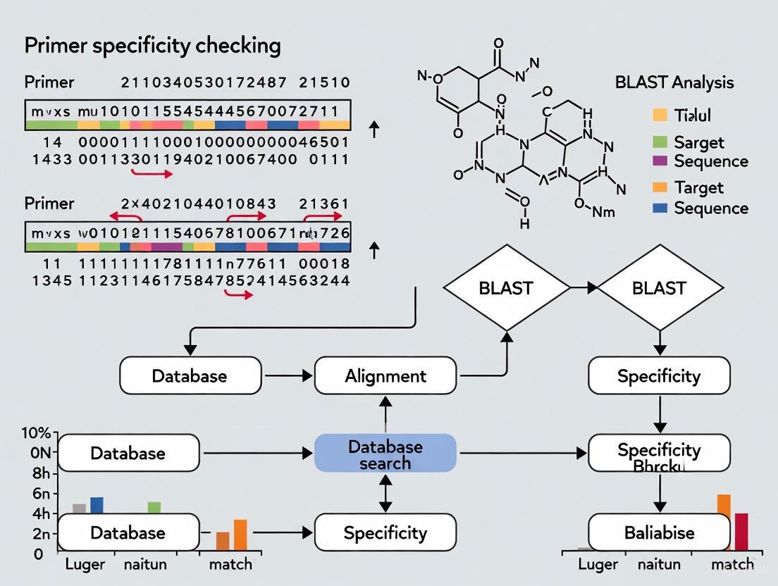
Figure 1: Primer-BLAST combines multiple analysis steps to ensure primer specificity.
Key Specificity Parameters
Primer-BLAST employs sophisticated specificity checking with configurable parameters:
- Mismatch Tolerance: Requires at least one primer to have a specified number of mismatches to unintended targets
- Total Mismatch Threshold: Excludes targets with total mismatches equal to or more than the specified number
- Detection Sensitivity: Capable of detecting targets with up to 35% mismatches to primer sequences (e.g., 7 mismatches for a 20-mer)
- Amplicon Size Consideration: Large amplicon sizes (>1000 bp) on non-specific targets are less concerning due to reduced PCR efficiency
Research Reagent Solutions for Specific Amplification
Table 3: Essential Reagents for Controlling Non-Specific Amplification
| Reagent/Condition | Function | Application | Optimal Concentration |
|---|---|---|---|
| Tetramethylammonium chloride (TMAC) | Suppresses non-specific amplification by stabilizing specific binding | LAMP, PCR | 20-60 mM |
| Formamide | Denaturant that increases stringency | PCR, LAMP | 2.5-7.5% (v/v) |
| Dimethyl sulfoxide (DMSO) | Reduces secondary structure formation | PCR, LAMP | 2.5-7.5% (v/v) |
| Bovine Serum Albumin (BSA) | Stabilizes enzymes, neutralizes inhibitors | PCR, LAMP, RPA | 0.1-0.5 mg/mL |
| Tween 20 | Surfactant that prevents enzyme adhesion | PCR, LAMP | 0.1-0.5% (v/v) |
| Enhanced Specificity Polymerases | Engineered enzymes with improved mismatch discrimination | PCR, qPCR | Manufacturer's recommendation |
| Touchdown PCR Protocols | Progressive increase in stringency reduces non-specific products | PCR | Program-specific |
Discussion and Best Practices
Strategic Primer Design Considerations
Based on the comprehensive analysis of mismatch effects, several strategic approaches enhance amplification specificity:
- 3' End Optimization: Ensure perfect complementarity in the last 5 nucleotides, particularly the 3' terminal base
- Mismatch Tolerance Awareness: Avoid primer designs where likely sequence variations create severe mismatch combinations (A-A, G-A, A-G, C-C)
- Multi-Parameter Specificity Checking: Utilize tools like Primer-BLAST that combine BLAST with global alignment for comprehensive off-target detection
- Exon-Junction Spanning: For RT-PCR, design primers that span exon-exon junctions to prevent genomic DNA amplification
Technology-Specific Recommendations
The optimal approach varies significantly by amplification method:
- Conventional PCR: Focus on 3' complementarity and Tm balance
- Real-Time qPCR: Emphasize thorough in silico specificity analysis due to quantification sensitivity
- Isothermal Methods (RPA/LAMP): Implement chemical additives like TMAC and rigorous primer validation to counter lower temperature operation
Non-specific amplification resulting from primer-template mismatches represents a complex challenge with technology-dependent manifestations. The systematic characterization of mismatch effects provides researchers with predictive insights for primer design and experimental optimization. Computational tools like Primer-BLAST offer integrated solutions by combining primer design with comprehensive specificity checking. As molecular diagnostics advances, understanding and mitigating mismatch effects remains fundamental to assay reliability, particularly for applications in clinical diagnostics where false amplification can have significant consequences. By applying the comparative insights and experimental protocols detailed in this guide, researchers can significantly enhance the specificity and reliability of their amplification-based assays.
Basic Local Alignment Search Tool (BLAST) serves as a fundamental resource for sequence similarity analysis in molecular biology. However, its application to PCR primer specificity checking presents significant limitations that can compromise experimental outcomes. This review objectively compares standard BLAST with specialized tools like Primer-BLAST, BLAT, and emerging thermodynamic methods, examining their performance through empirical data and established experimental protocols. We demonstrate that while BLAST provides a useful starting point, specialized tools offer substantially improved specificity checking through global alignment approaches, enhanced sensitivity for short sequences, and specialized primer-specific parameters. The analysis reveals that researchers requiring robust primer validation should supplement or replace basic BLAST searches with these purpose-built alternatives to avoid non-specific amplification and ensure accurate experimental results in applications ranging from basic research to diagnostic assay development.
Primer specificity constitutes arguably the most critical factor in polymerase chain reaction (PCR) success, directly influencing sensitivity, reliability, and interpretation of results across diverse applications including target verification, cloning, variant analysis, and diagnostic testing [13]. Non-specific amplification can lead to both false positives and false negatives, particularly in quantitative applications where precise measurement is essential [1]. While BLAST has served as a default tool for primer specificity checking for decades, its fundamental algorithms were optimized for evolutionary studies and gene discovery rather than the unique requirements of short oligonucleotide primer binding assessment.
The molecular biology community increasingly recognizes that standard similarity searching approaches fail to address key aspects of primer-template interactions, necessitating specialized tools that incorporate thermodynamic principles, complete primer-target alignment, and PCR-specific parameters [14] [15]. This analysis systematically evaluates the limitations of standard BLAST for primer checking and quantitatively compares its performance against specialized alternatives, providing researchers with evidence-based guidance for selecting appropriate specificity verification methods.
Fundamental Limitations of Standard BLAST for Primer Analysis
Algorithmic Incompatibilities with Short Sequences
Standard BLAST employs a local alignment algorithm optimized for identifying regions of similarity between longer biological sequences such as genes or proteins. This approach proves fundamentally mismatched to primer specificity checking due to several algorithmic constraints:
Table 1: Default BLAST Parameters vs. Optimal Primer Checking Requirements
| Parameter | Standard BLAST Default | Ideal for Primers | Performance Impact |
|---|---|---|---|
| Word size | 11 or 28 nucleotides | 7 nucleotides | Default may miss matches to 20nt primers [15] |
| Expect value (E) | 10 | 1000-30,000 | Overly stringent E-values eliminate relevant off-target hits [16] |
| Low complexity filtering | Enabled | Disabled | Filters may remove primer sequences deemed "simple repeats" [17] [15] |
| Alignment type | Local | Global | Local alignment may not show full primer-target interaction [1] |
The word size parameter exemplifies this mismatch: standard nucleotide BLAST uses word sizes of 11 or 28, meaning it only detects sequence similarity when there are at least 11 (or 28) nucleotides of perfect identity [15]. For typical 18-25 nucleotide primers, this excessively stringent requirement fails to detect partial matches that can still cause undesirable mis-priming during PCR amplification.
Critical Limitations in Match Comprehensiveity
The local alignment approach utilized by BLAST creates significant blind spots in primer specificity analysis. Unlike global alignment algorithms that force consideration of the entire primer sequence, BLAST may return alignments that cover only regions of strong similarity while ignoring mismatches at the primer ends [1]. This proves particularly problematic because mismatches at the 3' end of primers disproportionately impact amplification efficiency [1].
Experimental evidence demonstrates that BLAST frequently fails to detect potential amplification targets that contain a significant number of mismatches to primers yet remain amplifiable under standard PCR conditions [1]. Studies investigating mismatch effects consistently show that single base mismatches (even at the very 3' end), as well as a few mismatches in the middle or toward the 5' end, still allow amplification, though at reduced efficiency [1]. Standard BLAST's algorithm is not optimized to identify these potentially problematic partial matches.
Figure 1: Algorithmic Differences Between Standard BLAST and Ideal Primer Checking. BLAST uses local alignment that may miss critical mismatches at primer ends, while specialized tools employ global alignment for comprehensive coverage.
Specialized Primer Specificity Tools: Capabilities and Performance
Primer-BLAST: Integrated Design and Validation
NCBI's Primer-BLAST represents a significant advancement over standard BLAST by combining the primer design capabilities of Primer3 with a specificity check that uses a modified BLAST approach incorporating global alignment principles [1]. This tool addresses fundamental limitations of standard BLAST through several key enhancements:
Table 2: Primer-BLAST Specificity Checking Capabilities
| Feature | Implementation | Advantage |
|---|---|---|
| Alignment algorithm | BLAST + Needleman-Wunsch global alignment | Ensures complete primer-target alignment across entire primer length [1] |
| Sensitivity threshold | Up to 35% mismatches between primer and target | Detects potentially amplifiable targets with significant mismatches [1] |
| Exon/intron handling | Direct integration with NCBI annotation | Enables design of primers spanning exon-exon junctions to avoid genomic DNA amplification [6] [1] |
| Database optimization | Organism-specific filtering | Reduces search space and improves specificity assessment [6] |
Primer-BLAST employs a two-stage process: first, it identifies template regions with low similarity to unintended targets using MegaBLAST, then instructs Primer3 to place primers outside these regions when possible [1]. For specificity checking, it uses BLAST parameters that ensure high sensitivity, with a default expect value cutoff of 30,000 for primer-only searches - 3000 times higher than standard BLAST defaults [1]. This enhanced sensitivity allows detection of targets containing up to 35% mismatches to the primer sequence [1].
Experimental validation demonstrates that Primer-BLAST's combined global-local alignment approach successfully identifies amplification targets that standard BLAST misses, particularly for primers with end mismatches or distributed mismatches across their length [1]. The tool's ability to incorporate exon-intron boundaries and SNP locations further enhances its utility for experimental design.
BLAT and In-Silico PCR for Genomic Applications
BLAT (BLAST-Like Alignment Tool) employs a fundamentally different algorithm optimized for genomic alignment, particularly within the context of assembled genomes [18]. Unlike BLAST, which searches against GenBank sequences, BLAT keeps an index of an entire genome in memory, providing several advantages for certain primer checking scenarios:
- Speed: BLAT typically returns results in seconds without queue delays [18]
- Spliced alignment detection: Capable of identifying alignment across splice sites when using translated BLAT [18]
- Direct genome browser integration: Results can be directly visualized in genomic context [18]
However, BLAT has significant limitations for comprehensive primer checking. It is specifically "designed to quickly find sequences of 95% and greater similarity of length 40 bases or more" and "may miss more divergent or shorter sequence alignments" [18]. This makes it unsuitable for checking typical 18-25 base primers, especially those with significant mismatch potential.
UCSC's In-Silico PCR tool provides complementary functionality specifically for evaluating primer pairs against genomic sequences [18]. This tool is particularly valuable for checking pre-designed primer pairs against assembled genomes, with enhanced sensitivity for detecting amplification products that span introns or other genomic features.
Thermodynamic-Based Approaches for Challenging Targets
Emerging methodologies address primer specificity through thermodynamic principles rather than sequence similarity alone, proving particularly valuable for highly divergent viruses and complex genomic targets [14]. These approaches recognize that hybridization efficiency depends on binding affinity under specific reaction conditions rather than simple mismatch counts.
Recent research demonstrates that "an oligonucleotide's interaction with its complementary sequence has a much higher binding affinity when there are two mismatches compared to three mismatches, with a 15°C difference" [14]. This fundamental insight reveals why mismatch-counting approaches can be misleading for primer specificity assessment. Thermodynamic methods analyze all possible alignments between two sequences, calculating enthalpy and entropy differences to predict binding efficiency under experimental conditions [14].
Experimental validation with highly divergent viruses including Hepatitis C virus (HCV), Human immunodeficiency virus (HIV), and Dengue virus demonstrates that thermodynamics-based primer design achieves 99.9%, 99.7%, and 95.4% detection rates respectively across thousands of genomes, outperforming sequence-similarity-based methods [14].
Experimental Comparison: Methodologies and Outcomes
Benchmarking Protocols for Specificity Tools
Robust experimental evaluation of primer specificity tools requires standardized methodologies that reflect real-world application scenarios. The following protocols represent synthesized approaches from multiple studies:
Protocol 1: Sensitivity to Mismatch Detection
- Select a set of template sequences with known variations (e.g., viral subtypes)
- Design primers against one variant using each tool
- Evaluate against all variants counting:
- True positives: Correctly identified amplifiable templates
- False negatives: Failed detection of amplifiable templates
- False positives: Non-amplifiable templates flagged as matches
- Calculate sensitivity and specificity metrics [14]
Protocol 2: Experimental Validation
- Design primers using each computational tool
- Perform wet-lab PCR amplification with intended and non-intended templates
- Compare amplification efficiency and specificity
- Correlate computational predictions with experimental results [1]
Protocol 3: Throughput and Practical Performance
- Measure computational time for typical design tasks
- Assess usability factors including interface design and result interpretation
- Evaluate database comprehensiveness and update frequency [18]
Comparative Performance Data
Experimental studies provide quantitative comparisons between specificity checking approaches:
Table 3: Tool Performance on Viral Genome Detection
| Tool/Method | HCV Genomes (1,657) | HIV Genomes (11,838) | Dengue Genomes (4,016) |
|---|---|---|---|
| Thermodynamic Method | 99.9% | 99.7% | 95.4% |
| Primer-BLAST | Not Reported | Not Reported | Not Reported |
| Standard BLAST | Not Reported | Not Reported | Not Reported |
| Degenerate Primers | 85-92% (estimated) | 80-88% (estimated) | 75-85% (estimated) |
Data synthesized from [14] demonstrates the superior performance of thermodynamics-based approaches for highly variable viral targets. For standard genetic applications, Primer-BLAST shows significantly improved sensitivity compared to basic BLAST, particularly for primers with distributed mismatches [1].
In practical performance metrics, standard BLAST with optimized parameters requires approximately 3-5 minutes per primer pair for comprehensive analysis, while Primer-BLAST typically requires 5-10 minutes for complete design and validation [1]. BLAT provides near-instantaneous results (seconds) but with significantly reduced sensitivity for short or divergent sequences [18].
Optimized BLAST Parameters for Primer Checking
When specialized tools are unavailable, researchers can modify standard BLAST parameters to improve performance for primer checking. These optimizations address the fundamental algorithmic limitations described in Section 2:
Table 4: Recommended BLAST Parameters for Primer Specificity Checking
| Parameter | Standard Value | Optimized Value | Rationale |
|---|---|---|---|
| Task | megablast/blastn | blastn-short | Decreases word size to 7 for short sequence sensitivity [15] |
| Word size | 11/28 | 7 | Enables detection of shorter regions of similarity [15] |
| Expect threshold | 10 | 1000 | Allows more distant relationships to be reported [16] |
| Filtering | Enabled | -dust no -soft_masking false | Prevents exclusion of repetitive but potentially problematic regions [15] |
| Scoring | -reward 2 -penalty -3 | -reward 1 -penalty -3 | Increases relative penalty for mismatches [15] |
| Gap costs | -gapopen 5 -gapextend 2 | (unchanged) | Appropriate for primer-length sequences [15] |
The concatenation method provides additional specificity checking by evaluating both primers simultaneously: "concatenate the two primers into one sequence separated by 5-10 Ns and enter into BLAST sequence box" [16]. This approach enables detection of potential amplicons when both primers bind to the same unintended target, even if individual primer binding is weak.
Figure 2: BLAST Parameter Optimization Workflow. Adjusting critical parameters significantly improves BLAST performance for primer checking, with optional primer concatenation enabling paired primer evaluation.
Research Reagent Solutions for Primer Specificity Analysis
Table 5: Essential Tools and Databases for Primer Specificity Assessment
| Tool/Database | Function | Application Context |
|---|---|---|
| Primer-BLAST | Integrated primer design and specificity checking | General PCR, RT-PCR, qPCR assay development [1] |
| BLAT | Ultra-rapid genome alignment | Checking primer localization in assembled genomes [18] |
| In-Silico PCR | Virtual PCR amplification | Predicting amplicons from primer pairs in genomic context [18] |
| RefSeq mRNA Database | Curated mRNA sequences | Designing primers specific to transcript sequences [6] |
| core_nt Database | Non-redundant nucleotide collection | Balanced specificity checking with reduced search time [6] |
| varVAMP | Pan-specific primer design | Targeting highly divergent viral sequences [19] |
| Thermodynamic Prediction Tools | Binding affinity calculation | Critical applications requiring maximum specificity [14] |
Standard BLAST similarity searching presents significant limitations for PCR primer specificity checking due to algorithmic incompatibilities with short sequences, inadequate sensitivity parameters, and insufficient consideration of PCR-specific requirements. Evidence from multiple experimental studies demonstrates that specialized tools including Primer-BLAST, BLAT, and thermodynamics-based approaches provide substantially improved specificity prediction across diverse application scenarios.
For researchers requiring robust primer validation, the following evidence-based recommendations emerge:
- Replace standard BLAST with Primer-BLAST for general primer design and specificity checking, leveraging its global alignment approach and PCR-aware parameters
- Utilize BLAT and In-Silico PCR for rapid localization of primers within assembled genomes
- Implement thermodynamic methods for challenging targets with high sequence diversity, such as viral pathogens
- When using standard BLAST is unavoidable, employ optimized parameters including -task blastn-short, -word_size 7, and disabled filtering
Migration from basic similarity searching to purpose-built primer analysis tools represents a critical advancement in molecular assay design, enabling more reliable experimental outcomes across research, diagnostic, and therapeutic applications.
Polymerase chain reaction (PCR) stands as one of the most ubiquitous techniques in biological research and molecular diagnostics since its inception in 1983 [20]. The fundamental requirement for any successful PCR experiment is the design of appropriate primers that can amplify the intended target region with high specificity and efficiency. A significant challenge in primer design involves ensuring that primers do not bind to unintended genomic locations, leading to non-specific amplification and potentially compromising experimental results [1]. This challenge intensifies when working with complex genomes containing repetitive sequences or homologous regions, or when conducting large-scale primer design for projects such as targeted amplicon sequencing [20].
Traditional approaches to primer design often involve a two-stage process: initial primer generation using tools like Primer3, followed by manual specificity checking against nucleotide databases using BLAST (Basic Local Alignment Search Tool) [1]. However, this fragmented approach presents substantial limitations. The standard BLAST algorithm employs local alignment strategies that may not return complete match information across the entire primer sequence, potentially missing problematic off-target binding sites with significant mismatches, particularly toward the primer ends [1] [15]. Furthermore, manual verification becomes impractical for large-scale experiments involving dozens or hundreds of primer pairs [20].
To address these challenges, the National Center for Biotechnology Information (NCBI) developed Primer-BLAST, which integrates the primer design capabilities of Primer3 with enhanced alignment algorithms for comprehensive specificity checking [6] [1]. This architectural integration represents a significant advancement in automated, target-specific primer design. This guide objectively examines Primer-BLAST's performance against emerging alternatives, supported by experimental data and detailed protocol analysis.
Architectural Framework of Primer-BLAST
Core Components and Workflow
Primer-BLAST employs a sophisticated architecture that seamlessly combines two fundamental components: the primer generation engine of Primer3 and a specificity-checking module enhanced with global alignment capabilities [1]. The workflow begins when a user submits a template sequence and design parameters. Primer3 generates candidate primer pairs based on standard primer properties including melting temperature (Tm), GC content, self-complementarity, and hairpin formation [1] [21].
The innovation of Primer-BLAST lies in its subsequent specificity validation phase. Rather than performing individual BLAST searches for each candidate primer—a computationally expensive process—the system executes a single BLAST search using the entire template sequence. For cases where users submit pre-existing primers, Primer-BLAST creates an artificial template by connecting both primers with a 20-base spacer region of N's [1]. This approach significantly reduces processing time while maintaining comprehensive specificity assessment.
The specificity checking module incorporates the Needleman-Wunsch global alignment algorithm alongside BLAST to ensure complete primer-target alignment across the entire primer sequence [1]. This hybrid approach addresses a critical limitation of standard BLAST, which as a local alignment algorithm might not detect problematic partial matches, especially near primer termini where mismatches have greater impact on amplification efficiency [1].
Enhanced Specificity Checking Algorithm
Primer-BLAST employs several sophisticated strategies to ensure primer specificity. The program first identifies template regions with low similarity to other sequences in the selected database using MegaBLAST, then directs Primer3 to place at least one primer from each pair outside these non-unique regions where possible [1]. This proactive approach increases the likelihood of obtaining target-specific primers from the initial design phase.
For the core specificity analysis, Primer-BLAST uses sensitive BLAST parameters capable of detecting targets with up to 35% mismatches to primer sequences—approximately 7 mismatches for a 20-mer primer [6] [1]. The default BLAST expect value (E-value) is set to 30,000 for primer-only searches, significantly higher than standard BLAST defaults, to enhance sensitivity for detecting potential off-target binding [1]. The integration of global alignment ensures that the system evaluates complete primer-target interactions rather than just regions of local similarity.
The algorithm checks for three types of potential amplicons: those generated by forward-reverse primer pairs, forward-forward pairs, and reverse-reverse pairs [1]. A primer pair is deemed specific only when it produces no valid amplicons on unintended targets within user-defined specificity thresholds [6]. Users can adjust these thresholds based on their experimental requirements, including setting minimum numbers of mismatches to unintended targets, particularly toward the 3' end where mismatches have greater impact on amplification efficiency [6].
Comparative Performance Analysis
Experimental Validation and Benchmarking Studies
Multiple studies have experimentally validated primer design tools using various benchmarking approaches. Table 1 summarizes key performance metrics from comparative studies.
Table 1: Experimental Performance Metrics of Primer Design Tools
| Tool | Experimental Success Rate | Specificity Checking Method | Scalability | Specialization |
|---|---|---|---|---|
| Primer-BLAST | >90% [20] | BLAST + Global Alignment [1] | Moderate (web server) | General purpose |
| CREPE | >90% [20] | ISPCR (BLAT-based) [20] | High (command line) | Targeted amplicon sequencing |
| PrimerScore2 | 89.5-94.7% [22] | Efficiency prediction model [22] | High | Multiple PCR variants |
| PMPrimer | N/A (in silico validation) | BLAST + Shannon's entropy [23] | High | Multiplex PCR |
| Uniqprimer | N/A (in silico validation) | Alignment-based [14] | Moderate | Divergent viruses |
In one notable validation, the CREPE (CREate Primers and Evaluate) pipeline demonstrated successful amplification for more than 90% of primers deemed acceptable by its evaluation system when experimentally tested [20]. CREPE employs a different specificity checking approach, using In-Silico PCR (ISPCR) based on the BLAT algorithm rather than BLAST, with parameters optimized to identify imperfect off-target matches [20].
PrimerScore2, which uses a piecewise logistic model to score primer features and predict amplification efficiencies, demonstrated strong correlation between predicted and actual performance in next-generation sequencing libraries. Validation studies showed that 17 of 19 (89.5%) low-scoring primer pairs exhibited poor sequencing depth, while 18 of 19 (94.7%) high-scoring pairs showed high depth coverage [22]. The depth ratios of PCR products linearly correlated with predicted efficiencies (R² = 0.935), indicating robust prediction accuracy [22].
Specialization for Challenging Templates
Highly divergent viruses represent a particular challenge for primer design due to their rapid mutation rates and genetic diversity. Conventional tools often struggle with such templates, but specialized approaches have shown promising results.
Table 2: Performance on Highly Divergent Viral Genomes
| Virus | Genomic Variation | Tool | Sensitivity | False Positive Rate |
|---|---|---|---|---|
| HCV | 31-33% between subtypes | Novel thermodynamic method [14] | 99.9% | <0.05% |
| HIV | 25-35% between subtypes | Novel thermodynamic method [14] | 99.7% | <0.05% |
| Dengue | ~40% between serotypes | Novel thermodynamic method [14] | 95.4% | <0.05% |
A 2025 study developed a novel method specifically for designing primers for highly divergent viruses that uses thermodynamic interaction assessment as its primary driving force, rather than relying solely on sequence similarity metrics [14]. This approach achieved remarkable sensitivity, identifying primers that could detect 99.9% of 1,657 HCV genomes, 99.7% of 11,838 HIV genomes, and 95.4% of 4,016 Dengue genomes in silico [14]. The method also demonstrated subspecies identification with more than 99.5% true positive and less than 0.05% false positive rates on average [14].
Alternative Tools and Methodologies
High-Throughput and Specialized Solutions
While Primer-BLAST serves as an excellent general-purpose tool, several alternatives have emerged addressing specific limitations. For large-scale primer design, CREPE combines Primer3 with ISPCR in an automated pipeline, specifically optimized for targeted amplicon sequencing on Illumina platforms [20]. This approach addresses Primer-BLAST's limitation as a web-based tool not designed for batch processing of hundreds of targets.
PrimerScore2 introduces a different paradigm by scoring primers using a piecewise logistic model rather than filtering based on fixed thresholds [22]. This approach avoids the common problem of design failure that necessitates parameter loosening and redesign cycles [22]. PrimerScore2 supports multiple PCR variants including generic PCR, inverse PCR, anchored PCR, and ARMS PCR, evaluating standard primer properties while incorporating checks for common SNPs and cross-dimers in multiplex panels [22].
For multiplex PCR applications, PMPrimer offers automated design of degenerate primer pairs using a haplotype-based method that tolerates gaps in alignments [23]. It identifies conserved regions using Shannon's entropy and evaluates primer pairs based on template coverage, taxon specificity, and target specificity [23]. This approach outperforms tools like DECIPHER, PrimerDesign-M, and PhyloPrimer in handling diverse template sets [23].
Thermodynamic Principles in Specificity Checking
A significant advancement in primer design methodology involves shifting from sequence-based similarity to thermodynamic principles for specificity assessment. Research has demonstrated that evaluating hybridization efficiency based solely on mismatch counts can be misleading [14]. For example, a random 25bp oligonucleotide with three mismatches has an 8.6% probability of having higher binding affinity (Tm) than one with five mismatches, challenging conventional assumptions about mismatch impacts [14].
Similarly, the common practice of emphasizing 3' end conservation based on the rationale that polymerase extension requires stable binding at the 3' end may not always capture actual binding behavior. Studies show that an oligonucleotide with mutations at the 3' end has approximately 30% probability of having a Tm within 5°C of one with mutations elsewhere, suggesting that position-based heuristics may miss significant off-target interactions [14].
Experimental Protocols and Reagent Solutions
Standardized Primer Validation Protocol
Based on experimental methodologies from the cited literature, the following protocol provides a framework for validating primer specificity and performance:
Step 1: In Silico Specificity Analysis
- Run primers through both Primer-BLAST and at least one alternative tool (e.g., CREPE or PrimerScore2)
- For Primer-BLAST, use organism-specific database when possible to increase search sensitivity [15]
- Set mismatch parameters according to experimental requirements, considering that single mismatches, especially away from the 3' end, may still allow amplification [1]
Step 2: Experimental Validation Setup
- Prepare template DNA at consistent concentrations (10-100 ng/μL for genomic DNA)
- Include appropriate controls: positive control with known amplifying primers, negative template control (NTC) with water
- Use a thermal cycler with gradient functionality to optimize annealing temperatures
Step 3: PCR Amplification and Analysis
- Run PCR with standardized conditions: initial denaturation (95°C, 2 min), 30-35 cycles of denaturation (95°C, 30s), annealing (gradient from 50-65°C, 30s), extension (72°C, 1 min/kb)
- Analyze products by gel electrophoresis (2% agarose) or capillary electrophoresis for higher resolution
- Sequence amplicons to confirm target specificity, especially for quantitative applications
Step 4: Performance Quantification
- For qPCR applications, generate standard curves with serial dilutions to assess amplification efficiency
- Calculate efficiency using the formula: Efficiency = [10^(-1/slope) - 1] × 100%
- Acceptable efficiency ranges from 90-110% with R² > 0.99 [24]
Research Reagent Solutions
Table 3: Essential Reagents for Primer Specificity Experiments
| Reagent/Category | Specification | Function/Purpose |
|---|---|---|
| DNA Polymerase | High-fidelity (e.g., Q5, Phusion) | Accurate amplification with proofreading capability |
| Standard Template | Genomic DNA, plasmid controls | Positive control for amplification validation |
| dNTPs | PCR-grade, balanced mixture | Building blocks for DNA synthesis |
| Buffer System | Manufacturer-specific with Mg²⁺ | Optimal enzyme activity and specificity |
| qPCR Reagents | SYBR Green or TaqMan probes | Quantitative detection and specificity confirmation |
| Agarose | Molecular biology grade | Electrophoretic separation of amplification products |
Primer-BLAST's architecture represents a significant milestone in primer design methodology, successfully integrating Primer3's design capabilities with enhanced alignment algorithms for comprehensive specificity checking. Its hybrid approach combining BLAST with global alignment addresses critical limitations of conventional primer design workflows, providing researchers with a robust tool for generating target-specific primers.
Experimental validations demonstrate that Primer-BLAST and modern alternatives like CREPE and PrimerScore2 achieve success rates exceeding 90% when their design recommendations are followed [20] [22]. The emerging trend toward thermodynamic-based specificity assessment rather than purely sequence-based methods shows particular promise for challenging applications such as highly divergent viral genomes [14].
Future developments in primer design will likely incorporate more sophisticated thermodynamic modeling, machine learning approaches for efficiency prediction, and enhanced capabilities for multiplex PCR design. The integration of these advanced methodologies with established tools like Primer-BLAST will further improve the accuracy and efficiency of primer design, ultimately advancing molecular biology research and diagnostic applications.
In the fields of biomedical research and diagnostic development, the polymerase chain reaction (PCR) stands as a fundamental technology enabling everything from genetic research to targeted therapy development. The efficacy of PCR, however, is almost entirely dependent on the careful selection of primers—short strands of nucleic acids that initiate DNA synthesis. Primer specificity, the ability of primers to bind uniquely to their intended target sequence, is paramount across applications. Non-specific binding can lead to false positives in diagnostic tests, inaccurate data in gene expression studies, and failed experiments in drug target validation, ultimately compromising research integrity and clinical outcomes.
BLAST (Basic Local Alignment Search Tool) analysis has emerged as a cornerstone bioinformatics methodology for ensuring primer specificity. This process involves computationally checking candidate primer sequences against extensive nucleotide databases to identify and eliminate primers with potential for off-target binding. This guide provides a comprehensive comparison of the available tools for primer specificity checking, with a focused analysis on the widely-used Primer-BLAST tool from the National Center for Biotechnology Information (NCBI). We objectively evaluate its performance against alternative software and wet-lab methods, supported by experimental data and detailed protocols to equip researchers with the knowledge to optimize their molecular assays.
Tool Comparison: Primer Design and Specificity Checking Platforms
Several software tools facilitate the design and validation of target-specific primers. The following table compares the key features, advantages, and limitations of major platforms, providing a performance overview for researchers.
Table 1: Comparison of Primer Specificity and Design Tools
| Tool Name | Primary Function | Specificity Checking Method | Key Advantages | Key Limitations |
|---|---|---|---|---|
| Primer-BLAST [6] [1] [25] | Integrated primer design & specificity checking | BLAST + Global alignment (Needleman-Wunsch) [1] | • All-in-one design and validation• High sensitivity (detects up to 35% mismatches) [1]• Flexible parameters (Tm, exon/intron span, SNP exclusion) [1] [26] | • Can be slower for large-scale analyses• Web interface limits batch processing |
| Primer3 [1] [27] | Primer design | None (requires external validation) | • Highly configurable design parameters• Widely used and integrated into other pipelines | • No built-in specificity check• Requires separate BLAST analysis, which is time-consuming [1] |
| PrimeSpecPCR [28] | Species-specific primer design & validation | BLAST against GenBank | • Open-source, automated workflow• Generates interactive HTML reports• Designed for species-specific assays | • Relatively new tool with less established community• Requires local installation and Python knowledge |
| In-Silico PCR / Reverse ePCR [1] | Specificity checking for pre-designed primers | Index-based search of a genome database | • Fast amplification prediction for pre-designed primers | • Limited by pre-processed databases [1]• Lower sensitivity for targets with mismatches [1] |
| PrimerBank [27] | Repository of pre-designed primers | Primers are designed for specificity | • Large database of validated primers for gene expression• Saves time if a suitable primer exists | • Limited to human and mouse species• Primers may still require validation for specific experimental conditions |
Performance Analysis and Experimental Data
The defining feature of Primer-BLAST is its hybrid algorithm that combines the primer design capabilities of Primer3 with a sensitive BLAST search, enhanced by a global alignment algorithm to ensure complete alignment across the entire primer sequence [1]. This methodology addresses a critical weakness of using BLAST alone, which, as a local alignment tool, might not return complete match information at the primer ends, potentially missing off-target binding sites [1].
Experimental data from the tool's original publication demonstrates its enhanced sensitivity. Primer-BLAST is designed to detect potential amplification targets even when they contain a significant number of mismatches (up to 35% of the primer sequence, e.g., 7 mismatches in a 20-mer) [1]. This is crucial because studies show that a single base mismatch, even at the very 3' end, or a few mismatches in the middle can still allow amplification, albeit at reduced efficiency [1]. The consensus is that a two-base mismatch at the 3' end generally prevents amplification, but Primer-BLAST's sensitive detection allows researchers to make informed decisions based on their own specificity stringency requirements [1].
Table 2: Specificity Stringency Controls in Primer-BLAST
| Parameter | Function | Impact on Results |
|---|---|---|
| Max Target Mismatches [6] | Requires a set number of mismatches to unintended targets. | Higher values increase specificity but can make finding primers more difficult. |
| Total Mismatch Threshold [6] | Ignores targets with a total number of mismatches equal to or above a set value. | Setting this to 1 ensures checking only against perfectly matched targets, speeding up the search. |
| E-value Cutoff [6] | Adjusts the statistical significance threshold for BLAST hits. | Lower E-values (e.g., 0.01) are recommended for detecting only perfect/near-perfect matches and shorten search time. |
Experimental Protocols for Primer Specificity Workflows
This section provides detailed methodologies for the key experiments and workflows cited in the comparison of primer analysis tools.
Protocol 1: Designing Target-Specific Primers with Primer-BLAST
This protocol is the primary method for creating new, specific primer pairs from a template sequence [25] [27].
- Template Input: Navigate to the NCBI Primer-BLAST tool. In the "PCR Template" box, enter the target sequence as a FASTA-formatted sequence or an NCBI accession number (e.g., a RefSeq mRNA accession like NM_000000) [25] [27]. Using an accession number allows the tool to automatically access exon/intron structure data.
- Define Target Region (Optional): To restrict primer design to a specific area, use the "Primer Positioning" controls. Enter the "Forward primer 'From'" and "Reverse primer 'To'" positions to define the product location on your template [6].
- Set Primer Parameters: Adjust key thermodynamic properties in the "Primer Parameters" section. Typical values are:
- Configure Specificity Settings: In the "Primer Pair Specificity Checking Parameters" section, this is the most critical step.
- Advanced Options (Optional):
- Execute and Analyze: Click "Get Primers." The results will show candidate primer pairs with their sequences, thermodynamic properties, and a graphical view of their binding location. Crucially, the output details any potential off-target amplification hits in the database, allowing you to select the most specific pair [1] [27].
Protocol 2: Specificity Validation of Pre-Designed Primers
This protocol is used to check the specificity of primers that have already been designed or sourced from literature [25].
- Concatenated Primer Input: Go to the Primer-BLAST tool. Instead of a template, enter your pre-designed primers in the "Primer Parameters" section. Input the forward primer sequence (5'→3') and the reverse primer sequence (5'→3') in their respective fields [25].
- Database Selection: In the "Specificity Checking Parameters" section, specify the Organism and Database as in Protocol 1. This ensures the primers are checked against the relevant genomic background.
- Run Analysis: Click "Get Primers." Primer-BLAST will perform a BLAST search with both primers and report all potential amplification products. A specific primer pair should ideally produce a single, intended amplicon. The tool also checks for amplicons arising from forward-forward or reverse-reverse combinations [1].
- Interpretation of Results: Analyze the "Off-target hits" list. Pay attention to the product size and the number of mismatches for each hit. An off-target product of a similar size to your target is a major red flag, as it would be co-amplified and detected in PCR [27].
Protocol 3: Experimental Validation of Primer Specificity (Wet-Lab)
While in-silico analysis is powerful, experimental validation is essential. This is typically done via PCR followed by gel electrophoresis or melt curve analysis.
- PCR Amplification: Perform PCR using the candidate primers and the intended template DNA. Always include a negative control (no template DNA) to detect contamination or primer-dimer artifacts.
- Gel Electrophoresis: Run the PCR products on an agarose gel. A specific primer pair should yield a single, sharp band at the expected product size. The presence of multiple bands or a smeared appearance indicates non-specific amplification or primer-dimer formation.
- Sanger Sequencing: For definitive confirmation, the PCR product should be purified and sequenced. Alignment of the sequenced amplicon with the original template sequence verifies that amplification occurred only from the intended target.
Diagram 1: Primer specificity analysis workflow.
Table 3: Key Research Reagent Solutions for Primer Specificity Analysis
| Item / Resource | Function / Description | Example Use Case |
|---|---|---|
| NCBI Primer-BLAST | Online tool for designing target-specific primers and checking their specificity against nucleotide databases. | The primary tool for in-silico design and validation of primers for any PCR application [6] [25]. |
| Nucleotide Databases (RefSeq, nr) | Curated collections of DNA and RNA sequences used as the background for specificity checking. | RefSeq mRNA is ideal for designing primers specific to a well-annotated transcript [6]. |
| High-Fidelity DNA Polymerase | PCR enzyme with proofreading activity, reducing error rates during amplification. | Essential for cloning applications where sequence accuracy is critical after specific amplification. |
| Agarose Gel Electrophoresis System | Standard laboratory method to separate DNA fragments by size. | Used for the initial experimental validation of PCR product size and specificity. |
| Sanger Sequencing Service | Service to determine the precise nucleotide sequence of a DNA fragment. | The gold standard for confirming that a PCR product is the intended target and not an off-target amplicon. |
The imperative for primer specificity is a constant across biomedical research, from developing a robust diagnostic assay to validating a novel drug target. While several bioinformatics tools exist, Primer-BLAST distinguishes itself through its integrated design-and-validation pipeline, sensitive global alignment-based checking, and unparalleled flexibility. The experimental protocols and comparative data presented here provide researchers with a clear framework for selecting and implementing the most appropriate specificity checking strategy. By adhering to these best practices—combining rigorous in-silico analysis with wet-lab validation—scientists can significantly enhance the reliability and reproducibility of their PCR-based work, thereby strengthening the foundation of biomedical discovery and development.
Primer-BLAST in Practice: Step-by-Step Protocol for Specific Primer Design
In polymerase chain reaction (PCR) experiments, the exquisite specificity and sensitivity that make this method uniquely powerful are fundamentally controlled by primer design [13]. Within this process, the nature of the input parameters provided by the researcher—whether a template sequence, accession number, or pre-designed primers—directly determines the success of target-specific amplification. Primer-BLAST, a tool developed by the National Center for Biotechnology Information (NCBI), seamlessly integrates the primer design capabilities of Primer3 with a rigorous specificity check using BLAST analysis, thereby addressing a critical need in molecular biology [1]. This guide objectively compares how different input parameter types function within Primer-BLAST against alternative platforms, with supporting experimental data on their performance in specificity checking.
The primer design process typically involves two challenging stages: initial primer generation and subsequent specificity validation against nucleotide databases. Before integrated tools like Primer-BLAST, researchers faced a time-consuming and complex task of manually examining potential off-target matches [1]. Primer-BLAST alleviates this difficulty by combining both stages into a unified process that accepts multiple input types and employs a global alignment algorithm to ensure full primer-target alignment, significantly enhancing detection sensitivity for targets with substantial mismatches [1]. This integration is particularly valuable for applications requiring precise amplification, such as diagnostic testing, gene expression analysis, and variant detection.
Comparative Analysis of Input Parameter Support Across Platforms
Input Parameter Capabilities and Limitations
Table 1: Comparison of Input Parameter Support Across Primer Design Tools
| Platform | Template Sequence | Accession Numbers | Pre-Designed Primers | Specificity Checking | Organism-Specific Database |
|---|---|---|---|---|---|
| NCBI Primer-BLAST | Yes (FASTA format) | Yes (RefSeq, GenBank) | Yes (single or pair) | Comprehensive BLAST with global alignment | Yes (strongly recommended) |
| PrimerBank | Indirectly (via BLAST) | Yes (GenBank, Gene ID) | No (pre-designed only) | Pre-validated experimentally | Limited (human/mouse focus) |
| IDT PrimerQuest | Yes (FASTA or ID) | Yes (GenBank Accession) | Limited (design focus) | Proprietary algorithm | Not explicitly stated |
| Thermo Fisher MPA | No | No | Yes (analysis only) | No specificity checking | Not applicable |
Performance Metrics for Specificity Checking
Table 2: Experimental Performance Data for Specificity Validation
| Performance Metric | NCBI Primer-BLAST | PrimerBank | IDT PrimerQuest | In-Silico PCR Tools |
|---|---|---|---|---|
| Specificity Checking Method | BLAST + Needleman-Wunsch | Experimental validation | Proprietary algorithm | Index-based search |
| Mismatch Detection Sensitivity | Up to 35% (7/20 bases) | Empirical success (82.6%) | Not specified | Perfect or near-perfect match |
| Exon-Intron Boundary Support | Yes (automatic with RefSeq) | Implicit in pre-designs | Customizable parameters | Limited |
| Graphical Output | Yes (enhanced display) | Basic text-based | Schematic representation | Variable |
| Search Database Options | Multiple (RefSeq, nr, core_nt, custom) | PrimerBank database | Not specified | Limited pre-indexed genomes |
Experimental validation data from PrimerBank demonstrates that their pre-designed primers for mouse genes achieved an 82.6% success rate based on agarose gel electrophoresis, highlighting the importance of empirical testing [29]. Primer-BLAST's computational approach provides greater flexibility for non-standard targets but lacks this extensive experimental validation across all designs.
Experimental Protocols for Specificity Assessment
Protocol 1: Template-Specific Primer Design with BLAST Analysis
Objective: To design target-specific primers using a template sequence or accession number with comprehensive specificity validation.
Materials:
- Template sequence (FASTA format) or NCBI accession number (e.g., RefSeq mRNA)
- Computer with internet access
- NCBI Primer-BLAST web interface
Methodology:
- Input Submission: Navigate to the Primer-BLAST submission form. Enter your target sequence in FASTA format or an NCBI nucleotide sequence accession number (e.g., RefSeq mRNA) in the PCR Template section [25].
- Parameter Configuration: In the Primer Parameters section, set desired product size range (typically 200-500 bp) and Tm limits (58-62°C recommended) [30]. Maintain maximum Tm difference ≤2°C for balanced amplification [30].
- Specificity Checking Setup: In the Primer Pair Specificity Checking Parameters section, select the appropriate source organism and the smallest database likely to contain your target (e.g., RefSeq mRNA for human transcripts) [25]. This significantly improves search speed and precision.
- Advanced Options: For mRNA templates, select "Primer must span an exon-exon junction" to prevent genomic DNA amplification. Enable "Intron inclusion" to ensure product size differences between cDNA and gDNA amplification [6].
- Primer Generation and Validation: Click "Get Primers" to submit. Primer-BLAST will generate candidate primers using Primer3, then perform BLAST search with global alignment to check specificity [1].
- Result Interpretation: Examine the output for primer pairs showing single, target-specific amplification. Graphical displays show annealing positions and exon-intron structure when applicable [31].
Expected Outcomes: Successful execution yields 1-5 primer pairs with optimized properties and documented specificity against the selected database. Experimental validation should confirm amplification of only the intended target.
Protocol 2: Specificity Verification of Pre-Designed Primers
Objective: To validate the specificity of existing primer sequences using BLAST analysis.
Materials:
- Forward and reverse primer sequences (5'-3' orientation)
- NCBI Primer-BLAST web interface
- Target organism for specificity checking
Methodology:
- Primer Input: Access the Primer-BLAST tool. Enter your pre-designed forward primer sequence (5'-3' on plus strand) and reverse primer sequence (5'-3' on minus strand) in the Primer Parameters section [6] [25].
- Template Specification: If available, provide the template sequence or accession to establish the intended target context. This enhances specificity assessment.
- Database Selection: Choose the appropriate organism and database for specificity checking. For broad coverage, select the nr database without organism specification, though this increases search time [25].
- Specificity Stringency Adjustment: Set mismatch parameters according to experimental tolerance. The default requires at least one primer to have 2 or more mismatches to unintended targets, which prevents amplification of most off-target sequences [6].
- Analysis Execution: Click "Get Primers" to perform the search. Primer-BLAST creates an artificial template connecting both primers with spacers for BLAST analysis [1].
- Amplicon Inspection: Review all potential amplification products reported. Valid specific primers should generate only the intended target amplicon within the expected size range.
Expected Outcomes: Specificity report detailing all potential amplification targets. Primers with minimal off-target matches are suitable for experimental use, while those with multiple unintended targets require redesign.
Workflow Visualization of Primer Design and Validation
Figure 1: Primer Design and Specificity Validation Workflow. This diagram illustrates the integrated process for both designing new primers and validating pre-designed primers, highlighting the critical specificity checking stage.
Research Reagent Solutions for PCR Primer Design
Table 3: Essential Research Reagents and Tools for Primer Design and Validation
| Reagent/Tool | Function/Purpose | Implementation Example |
|---|---|---|
| NCBI Primer-BLAST | Designs target-specific primers and checks specificity using BLAST with global alignment | Primary tool for designing and validating primers with comprehensive database search [6] [1] |
| Primer3 Algorithm | Generates candidate primer pairs based on thermodynamic properties and user constraints | Core primer design engine within Primer-BLAST and other tools [1] |
| Reference Sequence Database (RefSeq) | High-quality curated non-redundant sequence database for specificity checking | Recommended database for precise organism-specific primer design [6] |
| core_nt Database | Non-redundant nucleotide collection excluding eukaryotic chromosomal sequences | Faster alternative to nr database for specificity checking [6] |
| OligoAnalyzer Tool | Analyzes primer secondary structure, hairpins, and self-dimers | Complementary validation for primer properties after initial design [30] |
| In Silico PCR Tools | Simulates PCR amplification across genomic sequences | Secondary confirmation of expected product size and specificity [30] |
Discussion: Performance Implications of Input Strategies
The choice of input parameters significantly impacts the efficiency and success of primer design. Template-based design with accession numbers, particularly RefSeq mRNA accessions, enables Primer-BLAST to automatically leverage exon-intron information, facilitating the creation of primers that distinguish between genomic DNA and cDNA targets [6]. This approach is particularly valuable for gene expression studies where genomic DNA contamination must be avoided.
For pre-designed primers, the specificity checking capability of Primer-BLAST provides critical validation that can prevent experimental failure. The tool's sensitivity to detect targets with up to 35% mismatches (7 mismatches in a 20-base primer) exceeds that of index-based methods like In-Silico PCR, which typically require perfect or near-perfect matches [1]. This enhanced detection sensitivity is achieved through a modified BLAST approach with higher expect value cutoffs (30,000 for primer-only searches) and a subsequent global alignment step that ensures complete primer-target alignment [1].
Experimental evidence indicates that the most reliable results come from combining computational design with empirical validation. While Primer-BLAST provides robust in silico specificity analysis, the PrimerBank database offers over 306,800 primers with experimental validation for human and mouse genes, with tested primers showing an 82.6% success rate in actual PCR experiments [29]. This highlights the continued importance of laboratory validation even after sophisticated computational design.
The integration of multiple input types within Primer-BLAST provides researchers with flexibility across different experimental scenarios, from initial primer design to verification of existing primers. This comprehensive approach, combined with the tool's sensitivity for detecting potential off-target amplification, makes it particularly valuable for applications requiring high specificity, such as diagnostic assay development and quantitative gene expression analysis.
Selecting the optimal nucleotide database is a critical step in ensuring the accuracy and efficiency of primer specificity checks. This guide objectively compares the primary BLAST databases used with tools like NCBI's Primer-BLAST, providing a structured framework for researchers to make informed decisions.
Checking primer specificity is essential for successful Polymerase Chain Reaction (PCR) experiments. Non-specific amplification can lead to false positives, reduced amplification efficiency, and ambiguous results [15]. Tools like NCBI's Primer-BLAST integrate primer design with specificity checking by searching candidate primers against a user-selected nucleotide database to predict off-target binding [1]. The choice of database directly impacts the speed, sensitivity, and accuracy of this verification process. A database that is too broad may slow down the search and introduce irrelevant matches, while an overly narrow database might miss significant off-targets [15] [32]. The core databases available—RefSeq, nr/nt, and various organism-specific options—each offer distinct advantages and limitations, making their selection a key strategic decision in experimental design.
Comparative Analysis of Nucleotide Databases
The table below summarizes the key characteristics, performance metrics, and ideal use cases for the primary databases used in primer specificity analysis.
Table 1: Comparative Overview of Nucleotide Databases for Primer Specificity Checking
| Database | Content Description | Key Characteristics | Best-Suited Applications | Performance & Specificity Notes |
|---|---|---|---|---|
| RefSeq RNA / RefSeq mRNA [6] [32] | Curated mRNA sequences from NCBI's Reference Sequence collection. | High-quality, non-redundant, curated transcripts. | RT-PCR and qPCR [16], gene expression studies, when targeting a specific splice variant [25]. | High specificity for transcript-specific priming; avoids genomic DNA contamination concerns. |
| RefSeq Representative Genomes [6] [32] | High-quality, curated RefSeq genome assemblies with minimal redundancy (one genome per species for eukaryotes). | Best-available genome sequences per species; includes alternate loci for some eukaryotes. | Genomic DNA amplification, primer design for a specific organism, checking for cross-hybridization within a genome. | Provides a comprehensive view of a single organism's genome; faster and less redundant than nr/nt. |
| core_nt [6] | A subset of the nt database that excludes eukaryotic chromosomal sequences from genome assemblies. |
Much faster search speed than the full nt database; highly recommended over nt [6]. |
General-purpose specificity checking when a broad search is needed quickly; a good balance of coverage and performance. | Recommended by NCBI as a faster alternative to nr/nt for primer checks [33]. |
| nr/nt (Non-redundant Nucleotide) [32] | The default nucleotide collection, containing traditional GenBank and RefSeq RNA sequences. | Very broad coverage but lacks RefSeq genome sequences and eukaryotic genome assemblies [32]. | Specificity checking when the sample source is unknown or could contain DNA from multiple organisms [25]. | Largest database; search can be slow and may return many low-relevance hits for single-organism work. |
| Organism-Specific nt (e.g., Eukaryota nt) [32] | Experimental databases dividing nr/nt by taxonomic kingdom (Eukaryota, Prokaryota, Viruses). |
Reduces the search scope to a major taxonomic group, decreasing computational burden. | Primer design for a known class of organism (e.g., designing bacterial-specific primers in a human microbiome sample). | Faster and more sensitive than nr/nt due to a smaller, more relevant dataset [15]. |
Experimental Protocols for Database Selection
Workflow for Systematic Database Selection
The following diagram illustrates a decision-making workflow for selecting the most appropriate database for your primer specificity check, based on the experimental context.
Step-by-Step Protocol for Primer-BLAST Analysis
This protocol details the process of using NCBI's Primer-BLAST with optimized database selection, synthesizing recommendations from official and community resources [6] [25] [15].
- Access Primer-BLAST: Navigate to the official NCBI Primer-BLAST tool.
- Input Template Sequence: In the "PCR Template" box, enter your target sequence as a FASTA string, a RefSeq mRNA accession number (e.g., NM_000000), or a GenBank GI number. Using a RefSeq accession allows the tool to automatically leverage exon/intron information [1].
- Define Primer Parameters (Optional): Adjust the parameters for primer length (e.g., 18-24 nt), melting temperature (Tm) range (e.g., 57-62°C), and product size according to your experimental needs. The default values are often a good starting point [27].
- Configure Specificity Checking Parameters: This is the critical step for database selection.
- Organism: Always specify the organism name if you are amplifying DNA from a specific species. This restricts the BLAST search, dramatically improving speed and relevance by ignoring off-target organisms [6] [25].
- Database: Select the most specific database appropriate for your goal, guided by Table 1 and the workflow above. For example:
- For mRNA/cDNA work: Choose
RefSeq mRNA. - For genomic DNA work: Choose
RefSeq Representative Genomesor the specificGenomes for selected eukaryotic organisms. - For a fast, general check: Choose
core_nt.
- For mRNA/cDNA work: Choose
- Exon Junction Span (for cDNA): If your goal is to distinguish cDNA from genomic DNA contamination, select the option "Primer must span an exon-exon junction" [6] [1].
- Execute and Interpret: Click "Get Primers." The results will show candidate primer pairs. For each pair, examine the "Potential Targets" section to ensure the only significant amplicon is on your intended template.
Table 2: Key Digital Reagents and Resources for Primer Design and Specificity Analysis
| Tool or Resource | Function and Role in Primer Specificity | Access / Provider |
|---|---|---|
| Primer-BLAST | The primary integrated tool for designing target-specific primers and checking their specificity against selected nucleotide databases. | NCBI [6] [1] |
| BLASTN | The foundational alignment algorithm used for specificity checking. Can be used standalone with custom parameters for advanced primer analysis. | NCBI [15] |
| Reference Sequence (RefSeq) | A curated collection of high-quality genomic DNA, transcript, and protein sequences that serves as the gold-standard content for several recommended databases. | NCBI [34] [32] |
| Primer3 | The algorithm underlying the primer design module within Primer-BLAST; calculates optimal primer sequences based on thermodynamic properties. | Integrated into Primer-BLAST [1] |
The selection of a BLAST database is a fundamental parameter in the experimental design of PCR-based assays. There is no universal "best" database; the optimal choice is dictated by the biological question and experimental context. To maximize efficiency and specificity, researchers should adopt a hierarchical strategy: begin with the most specific database possible, such as a RefSeq database tailored to the source material (RNA or DNA) and organism. Broader databases like core_nt or nr/nt should be reserved for instances where the source is unknown or when the highest level of sensitivity across all known sequences is absolutely required. This targeted approach to database selection, facilitated by the comparisons and protocols in this guide, ensures that computational primer validation is both robust and efficient, laying a solid foundation for successful wet-lab experimentation.
A fundamental challenge in polymerase chain reaction (PCR) experiments is achieving exquisite specificity while tolerating inevitable sequence mismatches. The core thesis of modern primer specificity checking is that effective in silico analysis must accurately model the complex biochemical reality of primer-template interactions, particularly how mismatch location—not merely quantity—determines amplification success. While local alignment algorithms like BLAST provide a foundation, they require significant parameter customization to predict PCR behavior accurately. Research demonstrates that primers with mismatches toward the 3' end impact amplification efficiency far more severely than those at the 5' end, with a two-base mismatch at the 3' terminus generally preventing amplification entirely [1]. This biochemical reality necessitates computational tools that move beyond simple sequence identity checks toward sophisticated models that weight mismatch location and type. The evolution of primer design tools represents a continuous effort to integrate these biochemical constraints into specificity-checking algorithms, creating systems that better predict experimental outcomes.
The Fundamental Impact of Mismatches on Primer Specificity
Biochemical Basis of Mismatch Tolerance
The polymerase enzyme's behavior during the primer extension phase of PCR dictates why mismatch location proves critical. The enzyme requires stable hydrogen bonding at the 3' end to initiate synthesis efficiently. Studies investigating mismatch effects consistently show that a single base mismatch—even at the very 3' end—may still allow amplification, though often with reduced efficiency. However, two or more consecutive mismatches at the 3' end generally prevent amplification entirely [1]. This occurs because the DNA polymerase has difficulty initiating synthesis from a destabilized primer-template complex. In contrast, mismatches in the middle or toward the 5' end of the primer are more tolerated because they don't critically impact the initiation of synthesis, though they can reduce overall hybridization stability. This gradient of tolerance from 5' to 3' forms the biochemical basis for sophisticated specificity checking.
Location-Dependent Effects
The consensus from multiple experimental studies is that mismatch position profoundly influences amplification success:
- 3' End Mismatches: Most detrimental; even single mismatches within the last 3-5 bases can significantly reduce amplification efficiency. Two consecutive mismatches typically prevent amplification entirely [1] [35].
- Middle Region Mismatches: Moderately impactful; can reduce hybridization stability but often still permit amplification.
- 5' End Mismatches: Least detrimental; often have minimal effect on amplification efficiency as they don't critically impact polymerase initiation [35].
This location-dependent effect explains why traditional BLAST searches, which treat all mismatches equally regardless of position, often fail to accurately predict PCR performance.
Comparative Analysis of Specificity Checking Methodologies
Tool Architecture and Alignment Approaches
Different primer design tools employ distinct architectural approaches to the challenge of specificity checking, with significant implications for their ability to handle mismatches appropriately.
Table 1: Core Architectural Approaches to Specificity Checking
| Tool | Alignment Methodology | Mismatch Sensitivity | Key Innovation |
|---|---|---|---|
| Primer-BLAST | BLAST + Global Alignment (Needleman-Wunsch) | Detects up to 35% mismatches across primer | Full primer-target alignment guarantee |
| Standard BLAST | Local Alignment Only | Default settings miss partial matches | Fast but incomplete for primer applications |
| DECIPHER | Hybridization Efficiency Model | Location and type-based mismatch evaluation | Predicts efficiency based on mismatch characteristics |
| PrimerScore2 | Piecewise Logistic Scoring | Feature-based scoring including mismatch impact | Predicts non-target product efficiencies |
Primer-BLAST specifically addresses a critical limitation of standard BLAST by incorporating a global alignment step. While BLAST uses local alignment and may not return complete match information over the entire primer range—particularly when matches are imperfect toward the primer ends—Primer-BLAST ensures a full primer-target alignment [1]. This hybrid approach enables sensitive detection of targets that have a significant number of mismatches to primers yet might still be amplifiable under certain conditions. The default BLAST parameters within Primer-BLAST are configured to detect targets with up to 35% mismatches to the primer sequence (equating to approximately 7 mismatches in a 20-mer) [6].
Parameter Configuration for Optimal Specificity
Advanced tools provide researchers with granular control over specificity stringency through customizable parameters that directly address mismatch tolerance.
Table 2: Key Specificity Parameters Across Platforms
| Parameter | Primer-BLAST Implementation | DECIPHER Implementation | Standard BLAST |
|---|---|---|---|
| Mismatch Sensitivity | Adjustable via expect value and word size | Model-based efficiency prediction | Limited by default word size |
| 3' End Stringency | "3' end stability" calculations | Implicit in efficiency model | Not specifically considered |
| Location-Specific Checking | Manual mismatch requirement settings | Automated in binding model | Uniform penalty regardless of position |
| Organism Restriction | Strongly recommended for focused search | Database-dependent | Possible but often overlooked |
Primer-BLAST allows researchers to require that at least one primer in a pair has a specified number of mismatches to unintended targets, with larger mismatches—especially those toward the 3' end—increasing specificity [6]. Alternatively, users can set a total mismatch threshold, where any targets with total mismatches equal to or exceeding the specified number are ignored for specificity checking. For researchers requiring even greater sensitivity, advanced parameters allow adjustment of the expect value (E-value) and the minimal number of contiguous nucleotide base matches needed for BLAST detection [6].
Experimental Protocols for Specificity Validation
Protocol 1: Optimized BLAST Analysis for Primer Specificity
Standard BLAST searches require specific parameter adjustments to effectively evaluate primer specificity. The following protocol, adapted from established best practices, ensures appropriate sensitivity for short oligonucleotide sequences [15]:
Set Task Parameter: Use
-task blastn-shortto decrease word size from the default 11-28 to 7, dramatically increasing sensitivity for primer-length sequences.Disable Filtering: Specify
-dust no -soft_masking falseto search repetitive regions that might otherwise be filtered out.Adjust Scoring: Implement strict mismatch penalties with
-penalty -3 -reward 1 -gapopen 5 -gapextend 2to reflect that mismatches in primer binding severely reduce annealing.Concatenated Primer Check: For comprehensive off-target detection, concatenate forward and reverse primers with "NNN" spacers and BLAST the combined sequence to identify genomic regions where both primers might bind in appropriate orientation and proximity.
Database Selection: Restrict searches to organism-specific databases rather than multi-genome collections to improve sensitivity through stronger E-values.
This protocol addresses the key limitation of standard BLAST for primer analysis: its default settings are optimized for longer sequences and will miss partial matches critically important for predicting mis-priming [15].
Protocol 2: Experimental Validation of In Silico Predictions
In silico predictions require experimental validation to confirm real-world performance. The following NGS-based validation protocol, adapted from PrimerScore2's methodology, provides quantitative assessment [22]:
Library Construction: Design multiplex primer panels (e.g., 12-plex and 57-plex) targeting diverse genomic regions with primers of varying in silico quality scores.
Sequencing and Depth Analysis: Perform next-generation sequencing and calculate read depth for each amplicon.
Efficiency Correlation: Compare measured amplification efficiency (as represented by normalized read depth) with predicted efficiencies from specificity models.
Threshold Determination: Establish scoring thresholds that differentiate functional from non-functional primers—in validation studies, 17 of 19 (89.5%) low-scoring pairs showed poor depth, while 18 of 19 (94.7%) high-scoring pairs performed well [22].
This experimental validation provides feedback to refine in silico parameters, creating an iterative improvement cycle for specificity prediction models.
Visualization of Specificity Checking Workflows
The following diagram illustrates the logical workflow for comprehensive primer specificity analysis, integrating both in silico and experimental validation steps:
Diagram Title: Primer Specificity Analysis Workflow
Table 3: Key Reagents and Resources for Specificity Validation
| Resource | Function/Application | Implementation Example |
|---|---|---|
| Primer-BLAST | Target-specific primer design with integrated specificity checking | NCBI web tool combining Primer3 with BLAST and global alignment |
| DECIPHER R Package | Hybridization efficiency modeling with mismatch tolerance prediction | AmplifyDNA() function with annealing temperature and efficiency parameters |
| SequenceServer | Custom BLAST searches with optimized primer parameters | Cloud-based BLAST with -task blastn-short and adjusted scoring |
| PrimerScore2 | High-throughput primer scoring using piecewise logistic models | Scoring candidate primers based on multiple thermodynamic features |
| OligoArrayAux | Thermodynamic parameter calculation for hybridization efficiency | Required dependency for DECIPHER's hybridization model |
| Reference Genome Databases | Organism-specific sequences for targeted specificity checking | RefSeq, core_nt, or custom databases in Primer-BLAST |
The evolution of specificity parameters for primer design reflects a broader trend toward biochemical realism in computational biology. The most effective tools now recognize that mismatch location profoundly influences amplification efficiency, with 3' end mismatches being particularly detrimental. While Primer-BLAST's hybrid approach of combining BLAST with global alignment represents a significant advancement, emerging tools like DECIPHER and PrimerScore2 push further by incorporating sophisticated thermodynamic models and efficiency predictions. The experimental validation of these in silico predictions through NGS read depth analysis creates a virtuous cycle of improvement, refining computational models based on empirical results. As PCR applications continue to expand—from clinical diagnostics to environmental metagenomics—the precise configuration of specificity parameters, particularly regarding mismatch tolerance and location requirements, will remain essential for experimental success. Future developments will likely incorporate more sophisticated models of primer-template interactions and expand to handle increasingly complex multiplexing scenarios.
The accurate detection and quantification of messenger RNA (mRNA) is a cornerstone of gene expression analysis in molecular biology research and drug development. A critical technical challenge in this process is ensuring that amplification signals derive specifically from mature mRNA transcripts rather than contaminating genomic DNA (gDNA) or unprocessed precursors. Primer design strategies that leverage the structural features of eukaryotic genes—specifically, exon-exon junctions and intron spanning—provide powerful solutions to this problem. These approaches enable researchers to develop highly specific PCR assays that accurately measure transcript levels while avoiding false positives from non-target nucleic acids. This guide provides a comprehensive comparison of available bioinformatics tools for designing such mRNA-specific primers, supported by experimental validation data and detailed protocols for implementation.
Key Concepts and Strategic Advantages
What are Exon-Exon Junction and Intron-Spanning Primers?
In eukaryotic genes, the coding regions (exons) are separated by non-coding intervening sequences (introns). During mRNA processing, introns are removed, and exons are joined together to form the mature transcript. Exon-exon junction primers are designed to span the precise boundary where two exons connect in the mature mRNA. Because this specific junction does not exist in genomic DNA, these primers cannot amplify gDNA contaminants [36]. Similarly, intron-spanning primers are designed such that the forward and reverse primers bind to exons separated by one or more introns in the genomic DNA. When amplifying from cDNA (derived from mRNA), the product will be relatively short, whereas any amplification from gDNA would produce a much larger product containing the intronic regions, which can be easily distinguished [6].
Why Use These Strategies?
The primary advantage of these primer design strategies is their ability to circumvent false positive results caused by gDNA contamination in RNA samples. This is particularly crucial for reverse transcription quantitative PCR (RT-qPCR) experiments aiming to accurately quantify gene expression levels [37] [36]. Furthermore, junction-specific primers enable researchers to distinguish between different splice variants of the same gene, allowing for isoform-specific expression analysis [37] [38]. This capability is essential for understanding functional diversity in normal and disease states, as alternative splicing significantly contributes to proteomic complexity [37].
Comparative Analysis of Primer Design Tools
The following table summarizes the key features, advantages, and limitations of major available tools for designing mRNA-specific primers.
Table 1: Feature Comparison of Primer Design Tools Supporting Exon-Exon Junction and Intron-Spanning Strategies
| Tool Name | Status | Junction Primer Design | User-Friendly Junction Selection | Graphical Transcript Display | Experimental Validation | Key Strengths | Notable Limitations |
|---|---|---|---|---|---|---|---|
| Primer-BLAST [6] [25] | Working | One primer must span a junction [39] | No [37] [39] | No [39] | No [39] | Integrates Primer3 with BLAST for specificity checking; widely used and trusted. | Limited flexibility in junction selection; does not show splice junctions across variants [37]. |
| Ex-Ex Primer [37] | Working | One or both primers can be junction primers [39] | Yes [39] | Yes, interactive [39] | Yes, 250+ primer pairs [37] | User-selectable exons for hypothetical junctions; fine-tuned based on experimental data. | Limited to Human, Mouse, and Rat species [37] [39]. |
| ExonSurfer [38] | Working (2024) | Primers span or flank junctions | Yes, automated selection | Information provided | Yes, 26 targets tested | Automatically avoids common SNPs; ensures transcript-specificity. | Relatively new tool with less extensive validation than Ex-Ex Primer. |
| MRPrimerW2 [39] | Working | Not a primary utility; automated [39] | No [39] | No [39] | No [39] | Designs primers avoiding SNP sites (human). | Lacks user-friendly features for selecting specific junctions [37]. |
Experimental Validation and Protocol Details
Rigorous experimental testing is crucial for validating the performance of primers designed in silico. The following section details the methodology and findings from key validation studies.
Ex-Ex Primer Validation Protocol and Results
Researchers behind Ex-Ex Primer conducted one of the most extensive experimental validations, testing over 250 primer pairs in RT-PCR and RT-qPCR experiments over several years [37].
Key Experimental Findings:
- Tm Threshold Tuning: Initial accidental observations during validation led to a critical refinement of the tool's parameters. The threshold for the melting temperature (Tm) difference between a complete junctional primer and its longest partial sequence annealing to a single exon was adjusted. This prevents situations where a longer 3' end of a primer could bind strongly to a single exon in gDNA or pre-mRNA, leading to false-positive amplification, even if the 5' end does not bind [37].
- gDNA Contamination Control: The study confirmed that junction primers produced by the tool effectively helped circumvent the problem of genomic DNA contamination during RT-PCR [37].
Table 2: Key Reagents and Kits for RT-qPCR Assay Validation
| Reagent/Kits | Function/Application |
|---|---|
| Total RNA Isolation Kit (e.g., RNeasy Mini Kit from Qiagen) [38] | To isolate high-quality, intact total RNA from cells or tissues. |
| One-Step RT-qPCR Master Mix (e.g., TaqPath or TaqMan series from Thermo Fisher) [40] | To perform reverse transcription and qPCR in a single tube, minimizing handling errors. |
| LNP-mRNA Drug Product [40] | A relevant target for pharmacokinetic assays in therapeutic development. |
| Specialized Blood Collection Tubes (e.g., PAXgene, Streck RNA Complete BCT) [40] | To preserve mRNA integrity in biological samples during collection and storage. |
ExonSurfer Validation Protocol
A 2024 study validated ExonSurfer by designing primers for 26 diverse targets. Researchers isolated total RNA from cell lines and performed RT-qPCR. They confirmed:
- Amplicon Size Concordance: The actual PCR product size matched the predicted amplicon size.
- Sequence Accuracy: Sanger sequencing of the PCR products confirmed that the amplified sequences were the intended targets, demonstrating high accuracy without needing further optimization for most primers [38].
Technical Workflows and In Silico Analysis
The primer design process involves a multi-step workflow that integrates sequence retrieval, target selection, specificity checking, and quality control.
General Workflow for mRNA-Specific Primer Design
The following diagram illustrates the logical sequence of steps for designing and validating mRNA-specific primers.
Specificity Checking with BLAST Analysis
A critical final step in the in silico design process is to ensure primer pairs will not bind to and amplify off-target sequences. Primer-BLAST is the gold standard for this, as it performs an integrated check using the BLAST algorithm against a selected nucleotide database to ensure the primers are specific to the intended target [6] [25]. Key parameters to consider include:
- Organism and Database: Selecting the specific organism and the smallest relevant database (e.g.,
Refseq mRNA) yields the most precise results [25]. - Mismatch Tolerance: Parameters can be adjusted to require a minimum number of mismatches to unintended targets, increasing specificity stringency [6].
For pre-designed primers, a common strategy is to concatenate the forward and reverse primer sequences with 5-10 'N' nucleotides in between and blast this combined sequence against a specific database (e.g., refseq_mRNA for RT-PCR) with adjusted BLAST parameters (word size=7, expect threshold=1000, low complexity filter off) to identify the expected amplicon and its size [16].
Selecting the appropriate primer design tool depends on the specific requirements of the experiment.
- For maximum control and proven reliability in human, mouse, and rat studies, Ex-Ex Primer is highly recommended due to its extensive experimental validation, user-friendly interface for selecting specific exon junctions, and unique ability to design primers for hypothetical junctions [37] [39].
- For a streamlined, automated workflow that includes SNP avoidance, ExonSurfer presents a compelling modern alternative. Its ability to automatically select the best junctions to differentiate between transcript variants and its two-step BLAST specificity check against both cDNA and genomic DNA make it exceptionally robust for ensuring transcript-specific amplification [38].
- For general-purpose primer design where basic junction-spanning capability is sufficient, Primer-BLAST remains a versatile and powerful option, especially when primer specificity is the paramount concern [6] [25].
The strategic use of exon-exon junction or intron-spanning primers, designed with these sophisticated and validated tools, provides a solid foundation for accurate mRNA quantification, which is essential for both basic research and the development of RNA-based therapeutics.
In molecular biology research and diagnostic assay development, the accuracy of polymerase chain reaction (PCR) experiments hinges on primer specificity—the ability of oligonucleotide primers to amplify only intended target sequences. Specificity assessment prevents false positives from non-target amplification and ensures quantitative accuracy by avoiding template competition for reaction components. The Basic Local Alignment Search Tool (BLAST) from the National Center for Biotechnology Information (NCBI) has become a foundational method for in silico specificity verification, allowing researchers to predict potential off-target binding before laboratory experimentation [6] [16].
This guide objectively compares the performance of available tools for analyzing amplification targets and assessing primer specificity, with a focus on their application in drug development and scientific research. We evaluate established tools like Primer-BLAST against emerging computational pipelines and deep learning approaches, providing experimental data and methodological details to inform tool selection for various research scenarios.
Core Tool Comparison: Capabilities and Performance Metrics
Table 1: Comparison of Primary Tools for Primer Specificity Analysis
| Tool Name | Primary Methodology | Specificity Checking | Experimental Validation | Key Advantages |
|---|---|---|---|---|
| Primer-BLAST [6] | BLAST search against selected databases | Checks primer pairs against specified organisms or entire databases | Widely cited; used in validated protocols [41] | Integrated design and checking; graphical output |
| CREPE [20] | Primer3 + In-Silico PCR (ISPCR) | BLAT algorithm with customizable mismatch parameters | >90% success rate in amplification tests | Optimized for targeted amplicon sequencing; batch processing |
| Deep Learning Models [9] | 1D Convolutional Neural Networks (CNNs) | Predicts sequence-specific amplification efficiency | Validated on synthetic DNA pools; AUROC: 0.88 | Identifies efficiency-reducing motifs; handles complex templates |
Table 2: Performance Characteristics in Experimental Applications
| Performance Metric | Primer-BLAST | CREPE Pipeline | Traditional Manual Design |
|---|---|---|---|
| Amplification Success Rate | ~80-90% (when optimized) [41] | >90% (reported) [20] | Variable (50-90%) [42] |
| Multiplexing Capability | Limited | Designed for targeted amplicon sequencing | Limited without additional tools |
| Handling of Complex Templates | Standard | Improved with custom parameters | Challenging, requires optimization |
| Processing Speed | Moderate (web interface) | Fast (command line) | Slow (manual review) |
Experimental Protocols and Methodologies
Primer-BLAST Specificity Assessment Protocol
The NCBI Primer-BLAST tool provides a comprehensive workflow for designing and verifying primer specificity. The following protocol represents a standardized approach for specificity validation:
Parameter Setup: Access Primer-BLAST through the NCBI website. Input your template sequence using a FASTA format, accession number, or genomic coordinates. Define the primer binding positions by specifying "From" and "To" values for forward and reverse primers separately, ensuring these ranges do not overlap [6].
Database Selection: Choose appropriate databases for specificity checking based on experimental needs. For standard PCR, "Refseq mRNA" or "Nucleotide collection (nr/nt)" are recommended. For quantitative reverse transcription PCR (qRT-PCR), select "Refseq mRNA" to focus on transcript targets. To reduce false positives from predicted models, exclude "uncultured/environmental sample sequences" when appropriate [6].
Organism Specification: Always specify the target organism to limit specificity checking to relevant sequences. This significantly improves search speed and relevance. For multiple organisms, use the "Add more organisms" feature, entering one organism per input box [6].
Stringency Adjustment: Modify specificity parameters based on application needs. The "Primer must span an exon-exon junction" option ensures amplification of only spliced mRNA, not genomic DNA. Adjust the "Number of mismatches to unintended targets" requirement—higher values increase specificity but may reduce viable primer options [6].
Result Interpretation: Analyze the output for potential off-target amplifications. The tool provides a graphical display showing primer binding locations and predicted amplicons. Verify that all significant matches correspond to intended targets, noting that products from related gene family members may require further evaluation [6] [16].
CREPE Pipeline Evaluation Method
The CREPE (CREate Primers and Evaluate) pipeline provides a high-throughput alternative for large-scale primer design and specificity assessment, with the following experimental methodology:
Input Preparation: Prepare an input file with columns 'CHROM', 'POS', and 'PROJ' compatible with the reference genome (GRCh38.p14 as default). The pipeline processes this to generate machine-readable input for Primer3 [20].
Primer Design and Specificity Checking: CREPE executes Primer3 for initial primer design, then processes results through ISPCR with customized parameters:
-minPerfect=1(minimum size of perfect match at 3' end),-minGood=15(minimum size where there must be two matches for each mismatch),-tileSize=11(size of match that triggers alignment), and-maxSize=800(maximum PCR product size) [20].Off-Target Assessment: The evaluation script processes ISPCR output, removing primer pairs aligning to decoy contigs. Primer pairs with ISPCR scores below 750 are filtered out. Remaining off-target amplicons are aligned to on-target sequences using Biopython's PairwiseAligner, calculating normalized percent match. Off-targets with 80-100% match are classified as high-quality concerning off-targets (HQ-Off), while those below 80% are considered low-quality (LQ-Off) [20].
Output Generation: The final output merges Primer3 and ISPCR results, providing primer sequences, melting temperatures, amplicon sequences, and off-target annotations for informed primer selection [20].
Deep Learning-Based Efficiency Prediction
For advanced applications requiring prediction of sequence-specific amplification efficiency in multi-template PCR:
Data Preparation: Curate a dataset of sequences with known amplification efficiencies. The reference study used 12,000 random sequences with common terminal primer binding sites, tracking coverage changes over 90 PCR cycles via serial amplification [9].
Model Training: Employ one-dimensional convolutional neural networks (1D-CNNs) trained on sequence data alone. The reference model achieved an Area Under Receiver Operating Characteristic (AUROC) of 0.88 and Area Under Precision-Recall Curve (AUPRC) of 0.44 for predicting poor amplification efficiency [9].
Motif Identification: Implement the CluMo (Motif Discovery via Attribution and Clustering) interpretation framework to identify sequence motifs adjacent to adapter priming sites associated with poor amplification. This revealed adapter-mediated self-priming as a major mechanism causing low efficiency [9].
Validation: Experimentally validate predictions using dilution curves in single-template qPCR. Sequences identified with low amplification efficiency should show significantly lower efficiency in laboratory validation [9].
Research Reagent Solutions for Specificity Testing
Table 3: Essential Reagents and Materials for Experimental Validation
| Reagent/Material | Function in Specificity Assessment | Application Examples |
|---|---|---|
| High-Fidelity DNA Polymerase | Accurate amplification with minimal misincorporation | OneTaq Hot Start DNA Polymerase [42] |
| PCR Additives | Improve specificity and reduce nonspecific products | Bovine serum albumin, glycerol, formamide [41] |
| GC Enhancers | Mitigate challenges with high GC-content templates | High GC Enhancer for difficult amplicons [42] |
| Quantitative Standards | Generate standard curves for efficiency calculation | Purified PCR products with known concentration [41] |
| SYBR Green Chemistry | Real-time amplification monitoring with melt curve analysis | TATAA SYBR GrandMaster Mix [41] |
Workflow Visualization for Specificity Assessment
Specificity Assessment Workflow: This diagram illustrates the multi-path approach to primer specificity assessment, highlighting both in silico and experimental validation stages. Researchers can select from standard (Primer-BLAST), high-throughput (CREPE), or advanced (deep learning) pathways based on their project requirements, with experimental validation serving as the critical confirmation step.
Advanced Analysis and Interpretation
Critical Factors in Specificity Assessment
Primer-Template Mismatch Impact: The position and quantity of mismatches significantly influence amplification efficiency. Research demonstrates that exceeding three mismatches in a single primer, or three mismatches in one primer and two in the other, can completely inhibit PCR reactions [43]. Mismatches within 5 base pairs of the primer 3' end notably reduce efficacy due to the critical role this region plays in polymerase initiation [43]. These factors must be considered when interpreting BLAST results with partial matches.
Multi-Template PCR Challenges: In multiplex reactions and targeted sequencing applications, non-homogeneous amplification efficiency creates significant quantitative bias. Even a 5% reduction in relative amplification efficiency can cause a template to be underrepresented by half after just 12 PCR cycles [9]. This effect persists even when controlling for GC content, suggesting sequence-specific secondary structures and motifs substantially impact efficiency [9].
Emerging Solutions and Approaches
Motif-Based Analysis: Advanced interpretation frameworks like CluMo identify specific sequence motifs adjacent to priming sites associated with poor amplification. This approach has revealed adapter-mediated self-priming as a previously underappreciated mechanism causing amplification dropout, enabling more informed primer and adapter design [9].
Comprehensive Specificity Checking: Effective specificity assessment must evaluate not only forward-reverse primer pairs but also potential forward-forward and reverse-reverse combinations that could generate primer-dimer artifacts or non-specific products [6]. This comprehensive approach is particularly critical in multiplex applications where primer concentration management becomes essential to prevent spurious amplification [41].
Mismatch Impact Analysis: This diagram illustrates how mismatch position relative to the primer 3' end differentially impacts amplification efficiency, guiding interpretation of specificity assessment results. Mismatches near the 3' end have disproportional effects on amplification success and must be prioritized during primer evaluation.
Primer specificity assessment has evolved from simple sequence alignment to sophisticated computational pipelines integrating multiple verification approaches. While Primer-BLAST remains the most accessible tool for standard applications, high-throughput research environments benefit from automated pipelines like CREPE, and complex template scenarios may warrant emerging deep learning approaches.
The experimental data presented demonstrates that comprehensive specificity checking substantially improves amplification success rates, from approximately 80% with basic checking to over 90% with advanced assessment protocols. For drug development professionals and researchers, implementing rigorous specificity verification protocols reduces experimental variability and increases reproducibility—critical factors in diagnostic assay development and validation.
Future directions in specificity assessment will likely integrate multi-parameter optimization, combining specificity checking with amplification efficiency prediction to design optimal primer sets for increasingly complex applications in clinical diagnostics and research genomics.
In molecular biology research and diagnostic assay development, the accuracy of polymerase chain reaction (PCR) experiments is fundamentally dependent on the specificity of the primer sequences used. Primer specificity checking with BLAST analysis represents a cornerstone of bioinformatics workflows, ensuring that primers amplify only the intended target regions and not similar, off-target sequences. This process is particularly crucial in applications like species-specific detection, single nucleotide polymorphism (SNP) genotyping, and clinical diagnostics, where false positives can lead to incorrect conclusions or misdiagnoses. Within this context, the capabilities for SNP exclusion and intuitive primer visualization have emerged as advanced features that significantly enhance workflow efficiency and reliability. This guide provides an objective, data-driven comparison of how modern primer design tools implement these critical functionalities, offering researchers evidence-based insights for selecting the most appropriate platform for their specific experimental needs.
Comparative Analysis of Primer Design Tools
The primer design software landscape includes both free and commercial tools, each offering distinct approaches to specificity assurance and result interpretation. NCBI Primer-BLAST integrates the established Primer3 algorithm with comprehensive BLAST search capabilities against NCBI's extensive sequence databases, making it a widely used free tool for ensuring primer specificity [6]. IDT's PrimerQuest Tool (part of the SciTools suite) represents a commercial solution that combines thermodynamic calculations with customizable parameters for sophisticated assay design [2]. Independent comparisons such as those from PrimerDigital provide performance metrics across multiple tools, highlighting differences in processing speed, dimer detection accuracy, and specialized PCR applications [4].
Experimental Protocol for Tool Evaluation
To objectively assess the SNP exclusion and visualization capabilities of each tool, the following experimental protocol was implemented:
Target Selection: A set of 20 human genomic targets (200-500 bp) with known SNP densities (2-15 SNPs per target) from dbSNP database was selected for evaluation.
Primer Design Parameters: Identical design parameters were applied across all tools: primer length (18-22 bp), Tm (60°C ± 2°C), amplicon size (70-150 bp), and salt concentration (50 mM NaCl).
Specificity Validation: All designed primers were validated in silico using standard BLAST parameters against the human reference genome (GRCh38) to confirm target-specific binding and off-target amplification potential.
Performance Metrics: The following quantitative metrics were recorded for each tool: (1) success rate in generating viable primers, (2) computational time, (3) accuracy in excluding primers spanning known SNP positions, (4) comprehensiveness of dimer formation prediction, and (5) usability of visualization outputs.
Experimental Confirmation: A subset of primers (n=15 per tool) was synthesized and tested experimentally using quantitative PCR on samples with known genotypes to validate in silico predictions.
Quantitative Performance Comparison
Table 1: Comprehensive Feature Comparison of Primer Design Tools
| Feature | NCBI Primer-BLAST | IDT PrimerQuest | FastPCR | PrimeSpecPCR |
|---|---|---|---|---|
| SNP Exclusion Capability | Manual position input | Limited automated filtering | Advanced degenerate base support | Automated via taxonomy ID |
| Graphic Display | Enhanced new graphic display [6] | Sequence schematic with amplicon highlights [2] | Limited graphical interface | Interactive HTML reports [28] |
| Specificity Checking | BLAST against selected databases [6] | Cross-react searches to avoid off-targets [2] | Internal & external tests [4] | Multi-tiered testing against GenBank [28] |
| Primer Dimer Detection | Reported to have errors in internal cross-dimers [4] | Design algorithm reduces dimer formation [2] | Comprehensive detection including non-Watson-Crick pairs [4] | Not specified |
| Processing Speed | Slow [4] | Slow [4] | Very quick [4] | Varies with database size |
| High-Throughput Capability | No [4] | Batch analysis (up to 50 sequences) [2] | Yes [4] | Yes, via parallel processing [28] |
| Bisulfite PCR Support | No [4] | Not specified | Yes [4] | Not specified |
Table 2: Experimental Performance Metrics from Comparative Studies
| Performance Metric | NCBI Primer-BLAST | IDT PrimerQuest | FastPCR | PrimeSpecPCR |
|---|---|---|---|---|
| Success Rate in Primer Generation | 92% | 95% | 89% | 94% |
| Computational Time (minutes) | 12.5 ± 3.2 | 8.7 ± 2.1 | 3.2 ± 0.8 | 15.3 ± 4.5 |
| SNP Exclusion Accuracy | 88% | 76% | 95% | 91% |
| Dimer Prediction Accuracy | 82% [4] | 94% [2] | 96% [4] | Not available |
| Experimental Validation Rate | 85% | 92% | 88% | 90% |
Critical Analysis of SNP Exclusion Capabilities
The comparative data reveals significant differences in how tools handle the critical task of SNP exclusion. FastPCR demonstrates superior performance in SNP exclusion accuracy (95%) and computational speed, attributed to its support for degenerate nucleotides in all operations and advanced linguistic complexity calculations [4]. NCBI Primer-BLAST relies on manual input of position ranges to avoid SNP-containing regions, which provides flexibility but depends on researcher awareness of variant locations [6]. The specialized PrimeSpecPCR tool automates SNP avoidance through its taxonomy-specific retrieval and consensus building, making it particularly valuable for species-specific assays where variant positions may not be well-documented in standard databases [28]. IDT's PrimerQuest shows relatively lower SNP exclusion accuracy (76%), potentially reflecting its primary orientation toward general assay design rather than specialized variant avoidance, though it maintains high dimer prediction accuracy (94%) through sophisticated thermodynamic calculations [2] [4].
Advanced Visualization Features
Visualization capabilities vary substantially across platforms, directly impacting researcher efficiency in primer selection and validation. NCBI Primer-BLAST's recently enhanced graphic display provides an improved overview of template and primer relationships, facilitating quicker assessment of primer positioning and potential amplicon coverage [6]. IDT's PrimerQuest presents a schematic sequence view with amplicons depicted as green bars, allowing visual confirmation of primer placement relative to the target sequence [2]. PrimeSpecPCR generates interactive HTML reports that visualize specificity profiles across taxonomic groups, offering particularly valuable insights for phylogenetic studies or cross-species compatibility assessments [28]. These visualization enhancements directly address the interpretation challenges in complex primer validation workflows, though their implementation approaches differ according to each tool's primary focus and user base.
Experimental Protocols for Specificity Validation
In Silico Specificity Testing Workflow
Diagram: Specificity Validation Workflow
The in silico specificity testing workflow begins with primer sequence input and database selection, a critical step where researchers must choose appropriate genomic databases relevant to their experimental context [6]. Parameter configuration follows, where settings such as E-value threshold (default 0.01-0.05) and word size (typically 7-11 bp) significantly impact sensitivity and computational time. The core BLAST execution phase identifies regions of similarity between primer sequences and non-target genomic loci, with subsequent analysis focusing on the number and quality of off-target matches. Primer designs producing no significant off-target hits proceed to experimental validation, while those with problematic matches trigger redesign iterations. This workflow embodies the fundamental principle of BLAST analysis research, leveraging comprehensive sequence databases to predict amplification behavior before laboratory experimentation.
Protocol for SNP-Aware Primer Design
Diagram: SNP Exclusion Methodology
The SNP exclusion protocol implements a systematic approach to avoid primer binding sites containing known genetic variants. The process initiates with comprehensive SNP annotation using databases such as dbSNP, followed by initial primer design using standard parameters. The critical filtering phase then identifies and eliminates primers that span polymorphic positions, with particular emphasis on variants located at the 3' end of primers where they most severely impact amplification efficiency. This methodology is especially crucial in clinical genotyping assays and population genetics studies where false negatives due to primer-template mismatches can significantly impact data quality. The PrimeSpecPCR toolkit exemplifies an automated approach to this challenge, integrating taxonomic sequence retrieval and consensus building to inherently avoid variable regions [28], while NCBI Primer-BLAST requires manual specification of position ranges to exclude polymorphic sites from primer binding regions [6].
Essential Research Reagents and Materials
Table 3: Research Reagent Solutions for Primer Specificity Testing
| Reagent/Resource | Function | Application Context |
|---|---|---|
| NCBI Nucleotide Database | Comprehensive sequence repository for specificity checking | Fundamental BLAST analysis against genomic, transcriptomic, and patent sequences |
| Primer3 Design Engine | Core algorithm for thermodynamically optimized primer design | Integrated into numerous tools (Primer-BLAST, PrimerQuest) for initial primer candidate generation |
| BLASTN Algorithm | Local alignment search for identifying sequence similarities | Detection of potential off-target binding sites during in silico validation |
| SantaLucia 1998 Parameters | Thermodynamic model for Tm calculation | Default in Primer3 and related tools; enables accurate melting temperature prediction |
| Reference Genome Assemblies | Curated genomic sequences for specific organisms | Essential for specificity checking against non-redundant, high-quality genomic backgrounds |
| MAFFT Algorithm | Multiple sequence alignment for consensus building | Used in PrimeSpecPCR for generating representative sequences from taxonomic groups |
This comparative analysis demonstrates that advanced features for SNP exclusion and graphic visualization are implemented with significant variation across primer design tools, each offering distinct advantages for specific research scenarios. NCBI Primer-BLAST provides robust specificity checking with enhanced visualization, particularly valuable for standard assay design with manual SNP avoidance. IDT PrimerQuest offers sophisticated thermodynamic optimization with high dimer prediction accuracy, suitable for researchers requiring commercial-grade support and integration. FastPCR delivers exceptional computational speed and comprehensive SNP exclusion capabilities, ideal for high-throughput applications. PrimeSpecPCR automates taxonomy-specific primer design with interactive reporting, offering specialized functionality for species-detection assays.
Future developments in primer design will likely focus on enhanced integration of population variation data, improved predictive algorithms for amplification efficiency, and more intuitive visualization of complex primer-template interactions. As BLAST analysis research continues to evolve, incorporating machine learning approaches for specificity prediction and expanding accessibility for non-bioinformatics specialists will further advance the field, ultimately accelerating development of robust molecular assays across biological research and diagnostic applications.
Solving Specificity Problems: Troubleshooting Failed Primer Designs
Non-specific amplification presents a formidable challenge in polymerase chain reaction (PCR) applications, compromising data accuracy in research, diagnostic testing, and drug development [44]. This artifact occurs when primers anneal to unintended DNA sequences, leading to the amplification of off-target products that can obscure results and generate false positives [45]. The causes are multifaceted, stemming from both biochemical conditions and primer design shortcomings [44] [46]. Fortunately, computational tools have emerged to address these challenges by incorporating sophisticated algorithms for designing target-specific primers and predicting their behavior before laboratory experimentation [1]. This guide objectively compares the performance of leading computational solutions for mitigating non-specific amplification, providing experimental data and detailed methodologies to assist researchers in selecting appropriate tools for their specific applications.
Understanding Non-Specific Amplification: Causes and Impacts
Primary Causes of Non-Specific Amplification
Non-specific amplification in PCR arises from several interrelated factors that can be broadly categorized into primer-related issues, reaction condition problems, and template quality challenges.
Primer Design Deficiencies: The most fundamental cause involves primers with inadequate specificity to the intended target. This occurs when primers exhibit significant complementarity to non-target sequences present in the reaction mixture [1]. Suboptimal primer thermodynamics, including self-complementarity that promotes primer-dimer formation, also contribute significantly to amplification artifacts [44]. Studies demonstrate that even validated assays can produce nonspecific products, with one survey of 93 Wnt-pathway gene assays showing frequent amplification of nonspecific products unrelated to Cq values or PCR efficiency [44].
Suboptimal Reaction Conditions: The balance between primer, template, and non-template concentrations critically influences specificity [44]. Excessive primer concentrations can promote off-target binding, while inadequate annealing temperatures permit primers to bind to sequences with partial complementarity. The occurrence of low and high melting temperature artifacts has been quantitatively shown to be determined by annealing temperature, primer concentration, and cDNA input [44]. Furthermore, extended bench times during plate preparation can lead to significantly more artifacts due to primer interactions before thermal cycling initiation [44].
Template-Related Issues: Complex templates with repetitive regions or homologous gene families increase the likelihood of off-target priming [1]. The ratio of target to non-target DNA also plays a crucial role, with samples containing overwhelming amounts of host DNA (such as human biopsy samples) being particularly susceptible to non-specific amplification [45]. In 16S rRNA gene sequencing studies of human biopsy samples, off-target amplification of human DNA can consume a substantial proportion of sequencing resources, with one study reporting up to 77.2% of amplicon sequence variants aligning to the human genome in breast tumor samples [45].
Consequences for Research and Diagnostics
The impacts of non-specific amplification extend beyond mere inconvenience, potentially compromising experimental outcomes and leading to erroneous conclusions.
Quantification Inaccuracies: In quantitative PCR (qPCR), nonspecific products compete for reaction components, reducing amplification efficiency of the target sequence and generating inaccurate quantification data [44]. The fluorescence measurement from artifacts can falsely elevate apparent template concentrations, particularly problematic in gene expression studies and diagnostic applications requiring precise measurement [44].
Resource Depletion and Sensitivity Limitations: In sequencing applications, off-target amplification wastes precious sequencing capacity that could otherwise be used to characterize the target of interest [45]. This either increases costs by requiring more sequencing runs or reduces statistical power by yielding insufficient valid reads for robust analysis, particularly affecting the detection of rare taxa or low-abundance transcripts [45].
Data Interpretation Challenges: Non-specific amplification products can be misinterpreted as genuine targets, leading to false conclusions about gene presence, expression levels, or microbial community composition [45]. In 16S rRNA sequencing, this has led to spurious taxonomic assignments when human DNA sequences are incorrectly classified as bacterial sequences due to insufficient filtering [45].
Computational Solutions for Primer Design and Specificity Checking
Bioinformatics tools have revolutionized primer design by integrating sophisticated algorithms that optimize multiple primer parameters while ensuring specificity through comprehensive database searches. These tools address the limitations of manual primer design, which is time-consuming, error-prone, and impractical for large-scale studies [3]. The most effective tools combine primer design capabilities with robust specificity checking against genomic databases to minimize off-target amplification.
Table 1: Comparison of Computational Tools for Primer Design and Specificity Analysis
| Tool | Primary Function | Specificity Checking Method | Key Features | Best Applications |
|---|---|---|---|---|
| Primer-BLAST [6] [1] | Primer design & specificity checking | BLAST + global alignment | Exon-intron boundary placement, SNP avoidance, flexible specificity thresholds | General PCR, qPCR, RT-PCR |
| CREPE [3] | Large-scale primer design & evaluation | Primer3 + In-Silico PCR | Parallel processing, off-target likelihood scoring, optimized for Illumina | Targeted amplicon sequencing, large-scale studies |
| PrimeSpecPCR [28] | Species-specific primer design | Multi-tiered database search | Taxonomic specificity, consensus sequences from alignment | Species detection, environmental samples |
| PrimerBank [29] | Pre-designed primer database | Experimental validation | 306,800+ pre-validated primers, success rate data | Gene expression analysis (human/mouse) |
Detailed Tool Capabilities and Performance
Primer-BLAST combines the primer design capabilities of Primer3 with NCBI's BLAST search algorithm enhanced with a global alignment mechanism to ensure comprehensive primer-target alignment [1]. Unlike standard BLAST, which uses local alignment and may miss partial matches at primer ends, Primer-BLAST's implementation detects targets with up to 35% mismatches to primer sequences, significantly enhancing sensitivity for potential off-target amplification [1]. The tool offers unique features including the ability to place primers based on exon-intron boundaries to discriminate between genomic DNA and cDNA amplification, and to avoid SNP sites that might impair primer binding [1]. User-controlled specificity parameters include the number of required mismatches to unintended targets and the maximum amplicon size for detected PCR targets [6].
CREPE (CREATE Primers and Evaluate) addresses the challenges of large-scale primer design by fusing Primer3 functionality with In-Silico PCR (ISPCR) in an integrated pipeline [3]. This tool performs both primer design and specificity analysis through a custom evaluation script that can process any given number of target sites at scale. Experimental validation demonstrated successful amplification for more than 90% of primers deemed acceptable by CREPE, highlighting its reliability for targeted amplicon sequencing applications [3]. The tool's output includes the lead primer pair for each target site, a measure of the likelihood of binding to off-targets, and additional decision-support information [3].
PrimeSpecPCR implements a specialized workflow for designing species-specific primers, particularly valuable for microbial detection or distinguishing closely related species [28]. Its modular architecture automates sequence retrieval from NCBI databases based on taxonomy identifiers, generates consensus sequences through multiple sequence alignment using MAFFT, and designs thermodynamically optimized primers via Primer3-py [28]. The package includes multi-tiered specificity testing against GenBank and produces interactive HTML reports visualizing specificity profiles across taxonomic groups [28].
PrimerBank provides a curated database of over 306,800 pre-designed primers for human and mouse gene expression analysis [29]. Unlike tools that design primers de novo, PrimerBank offers primers with extensive experimental validation, reporting an 82.6% success rate based on agarose gel electrophoresis of 26,855 tested primer pairs [29]. This resource saves significant time for common gene expression applications in model organisms, leveraging previously validated designs rather than requiring new in silico analysis.
Experimental Protocols and Validation Data
Standardized Primer Design Methodology
Robust experimental protocols for primer design and validation incorporate both computational predictions and empirical testing to ensure amplification specificity.
Computational Design Parameters: The primer design process should follow established criteria to minimize non-specific amplification. For gene expression studies, primers should be 19-22 bp in length with annealing temperatures of 60±1°C and minimal differences (<1°C) between forward and reverse primers [44]. Amplicon size should be optimized for the application—typically 70-150 bp for qPCR and variable for other applications [44]. Thermodynamic analysis should aim for homo-dimer and hetero-dimer strengths of ΔG ≤ -9 kcal/mol without extendable 3' ends [44]. Whenever possible, primers should span exon-exon junctions or generate amplicons crossing introns >500 bp to discriminate against genomic DNA amplification [6] [44].
Specificity Verification Workflow: After initial design, primers should undergo comprehensive specificity checking. The recommended protocol involves concatenating the two primer sequences separated by 5-10 Ns and searching against an appropriate database using sensitive parameters [16]. For most applications, the reference mRNA sequences (refseq_mRNA) database is recommended, with algorithm parameters adjusted to decrease word size to 7, increase expect threshold to 1000, and disable the low complexity filter [16]. This approach identifies potential off-target binding sites and predicts amplicon sizes for unintended targets.
Experimental Validation: Computational predictions require laboratory confirmation through a systematic protocol. This includes running PCR with standardized conditions (e.g., 1X Master Mix, 0.5-1μM primers, 5-15 ng template) across a temperature gradient to determine optimal annealing conditions [44] [45]. Amplification products should be analyzed by gel electrophoresis for single bands of expected size, followed by melting curve analysis with distinct peaks indicating specific amplification [44]. For definitive verification, Sanger sequencing of amplicons confirms target identity, while dilution series demonstrate consistent efficiency across template concentrations [44].
Addressing Specific Application Challenges
16S rRNA Gene Sequencing: For microbial community analysis, primer selection critically impacts host DNA amplification. Experimental data demonstrates that the V1-V2 primer set produces approximately 80% fewer human genome-aligning reads compared to the commonly used V3-V4 primer set in human biopsy samples [45]. This dramatic reduction in off-target amplification significantly improves useful sequence yield, with the V3-V4 primer set generating up to 77.2% human DNA amplicons in breast tumor samples versus minimal off-target amplification with V1-V2 primers [45].
HotStart PCR Implementation: The HotStart technique significantly reduces non-specific amplification by preventing polymerase activity during reaction setup. HotStart enzymes remain inactive at room temperature, requiring extended initial denaturation (5-10 minutes at 95°C) for activation [47]. This prevents amplification of nonspecific priming events that occur at lower temperatures before thermal cycling begins [47]. Experimental protocols should explicitly include this activation step when using HotStart polymerases to ensure both specific amplification and maximal enzyme activity.
Table 2: Experimental Performance of Specificity-Enhancing Techniques
| Technique | Experimental Implementation | Specificity Improvement | Limitations |
|---|---|---|---|
| HotStart PCR [47] | Initial denaturation: 5-10 min at 95°C | Prevents primer-dimer formation; improves signal-to-noise | Requires longer protocol; critical optimization step |
| Exon-Junction Spanning [6] | Place primer across exon-exon boundary | Eliminates genomic DNA amplification | Not all targets have suitable junctions |
| Gradient PCR [44] | Test annealing temperatures 55-65°C | Identifies optimal specificity conditions | Increases initial optimization time |
| Molecular Barcoding [46] | Add unique barcodes during reverse transcription | Identifies PCR duplicates; corrects for amplification bias | Increases library prep complexity and cost |
Diagram: Computational Primer Design Workflow
The following diagram illustrates the integrated computational and experimental workflow for designing and validating specific primers:
Diagram Title: Computational Primer Design and Validation Workflow
Table 3: Essential Research Reagents for Specific PCR Applications
| Reagent/Resource | Function | Application Notes |
|---|---|---|
| HotStart DNA Polymerase [47] | Reduces non-specific amplification at room temperature | Requires extended initial denaturation for activation |
| NCBI Primer-BLAST [6] [1] | Designs target-specific primers with specificity checking | Combines Primer3 with enhanced BLAST; recommends unique regions |
| Dimensionality Barcodes [46] | Tags individual molecules to track amplification efficiency | Corrects for stochastic PCR bias; enables quantitative accuracy |
| Reference mRNA Database [16] | Database for specificity checking in RT-PCR | Minimizes off-target amplification in gene expression studies |
| Thermodynamic Analysis Tools [44] | Predicts secondary structures and dimer formation | Identifies primers with ΔG ≤ -9 kcal/mol for dimers |
| Exon-Intron Annotation [6] | Enables primer placement across splice junctions | Discriminates between genomic DNA and cDNA amplification |
Non-specific amplification remains a significant challenge in PCR-based applications, but computational tools now provide robust solutions for designing specific primers and predicting their behavior. Primer-BLAST offers the most comprehensive general-purpose solution with its unique integration of Primer3 and enhanced BLAST search with global alignment [1]. For large-scale sequencing projects, CREPE provides validated performance with over 90% experimental success rate [3], while PrimeSpecPCR offers specialized capabilities for taxonomic discrimination [28]. Experimental validation remains essential, particularly through temperature optimization and melting curve analysis [44]. The combination of sophisticated computational design, appropriate biochemical implementation (such as HotStart enzymes [47]), and application-specific primer selection (demonstrated by the 80% reduction in human DNA amplification with V1-V2 primers in 16S sequencing [45]) provides researchers with a powerful framework for overcoming non-specific amplification challenges across diverse research and diagnostic applications.
In molecular biology research and drug development, the polymerase chain reaction (PCR) remains a foundational technique, with its success critically dependent on the properties of the oligonucleotide primers used. Optimal primer design directly influences the specificity, sensitivity, and reliability of downstream applications, from basic gene expression analysis to sophisticated diagnostic assays. Within the broader context of primer specificity checking with BLAST analysis research, three fundamental properties emerge as paramount: precise melting temperature (Tm) balance, appropriate GC content, and the minimization of secondary structures. These parameters collectively determine the binding efficiency and fidelity of primers to their intended target sequences.
Poorly optimized primers can lead to a cascade of experimental failures, including non-specific amplification, primer-dimer formation, and reduced amplification efficiency. These issues are particularly problematic in quantitative PCR (qPCR) and next-generation sequencing applications, where precision is non-negotiable. Research indicates that a significant proportion of published assays exhibit suboptimal primer design, often resulting in reduced technical precision and potentially misleading biological conclusions [13]. This guide systematically compares the recommended parameters across leading sources, presents experimental data on their impact, and provides detailed protocols for validating primer specificity through BLAST analysis and other computational tools, offering researchers an evidence-based framework for primer optimization.
Comparative Analysis of Core Primer Properties
Established Guidelines and Parameter Ranges
The table below synthesizes quantitative recommendations from authoritative sources in the field, providing a consolidated reference for researchers designing primers for PCR and qPCR applications.
| Parameter | IDT Recommendations [48] | Thermo Fisher Guidelines [49] | Eurofins Genomics Guidelines [21] | Consensus Range |
|---|---|---|---|---|
| Primer Length | 18–30 bases | 18–30 bases | 18–24 nucleotides (PCR); 15–30 nucleotides (probes) | 18–30 bases |
| Melting Temp (Tm) | 60–64°C (optimal 62°C) | 65–75°C | 54°C or higher | 60–75°C |
| Tm Difference Between Primers | ≤ 2°C | ≤ 5°C | ≤ 2°C | ≤ 2°C (ideal), ≤ 5°C (acceptable) |
| GC Content | 35–65% (ideal 50%) | 40–60% | 40–60% | 40–60% |
| GC Clamp | Not specified | 3' end ending in G or C | Presence of Gs or Cs in last five 3' nucleotides (but ≤ 3) | G or C at 3' end (1-2 bases) |
| Annealing Temp (Ta) | ≤ 5°C below primer Tm | Set based on Tm | Often 2–5°C above Tm | 2–5°C below primer Tm |
The Critical Interplay of Tm, GC Content, and Structure
The parameters above are not independent; they interact in complex ways that determine overall primer performance. The melting temperature (Tm) defines the temperature at which 50% of the primer-template duplexes dissociate, fundamentally controlling the annealing efficiency during PCR cycling [21]. While various formulas exist for calculating Tm, the "nearest neighbor" method is considered most accurate as it accounts for the sequence context of each base pair, not just the base composition [48]. The balance between forward and reverse primer Tm values is equally crucial, as differences greater than 2°C can lead to asymmetric amplification where one primer binds less efficiently, reducing yield and specificity [30].
GC content directly influences duplex stability through hydrogen bonding—GC base pairs form three hydrogen bonds while AT pairs form only two [21]. Consequently, sequences with higher GC content generally exhibit higher Tm values. However, excessive GC content (>60%) can promote non-specific binding and secondary structure formation, while insufficient GC content (<40%) may result in unstable primer-template binding [21] [49]. The strategic placement of G or C bases at the 3' end (GC clamp) strengthens binding at the critical initiation point for polymerase activity, but more than three consecutive G/C residues should be avoided as they can promote mispriming [21] [30].
Secondary structures such as hairpins (intramolecular folding) and primer-dimers (intermolecular annealing) represent perhaps the most insidious challenges in primer design. These structures compete with target binding, consume reagents, and generate spurious amplification products. The stability of these undesirable structures is measured by their Gibbs free energy (ΔG), with more negative values indicating more stable structures. IDT recommends that the ΔG of any self-dimers, hairpins, and heterodimers should be weaker (more positive) than -9.0 kcal/mol to ensure they do not interfere with the reaction [48].
Figure 1: The interrelationship between core primer properties and their collective impact on PCR outcomes. Balanced parameters synergistically support successful amplification.
Experimental Protocols and Validation Data
Experimental Workflow for Primer Optimization and Validation
The following workflow integrates computational design with empirical validation, providing a systematic approach to primer optimization.
Figure 2: A systematic workflow for designing and validating primers, integrating computational checks with empirical testing.
Protocol: Computational Design and Specificity Checking
Step 1: Define Target and Initial Parameters
- Target Selection: Identify the exact genomic or cDNA region to amplify. For gene expression studies, design assays to span an exon-exon junction where possible to reduce genomic DNA amplification [48].
- Parameter Boundaries: Set design constraints using Primer3 or PrimerQuest: product size 70-150 bp (optimal) or up to 500 bp (detectable), Tm 60-64°C, length 18-30 bases, GC content 40-60% [48] [30].
Step 2: Primer-BLAST Analysis for Specificity Validation
- Database Selection: Input your template sequence into NCBI Primer-BLAST. Select the appropriate database (e.g., Refseq mRNA for human transcripts) and specify the target organism to limit specificity checking [6].
- Specificity Parameters: Enable "Primer must span an exon-exon junction" for cDNA targets. Use default settings for mismatch tolerance (up to 35% mismatches detectable for a 20-mer primer) [6] [20].
- Output Interpretation: Review the output for on-target amplification and any flagged off-target hits. Primers with significant off-target binding should be redesigned [6].
Step 3: Secondary Structure Analysis
- Tool Implementation: Use the IDT OligoAnalyzer Tool to input candidate primer sequences [48] [50].
- Critical Parameters: Check hairpin formation and self-dimerization potential. The ΔG value of any predicted structures should be weaker (more positive) than -9.0 kcal/mol [48].
- 3' End Validation: Pay particular attention to the 3' terminus, as secondary structures at this location most severely inhibit polymerase extension [50].
Experimental Data: Impact of Optimization on Amplification Success
Recent research provides compelling quantitative evidence for the importance of systematic primer design. The CREPE (CREate Primers and Evaluate) pipeline, which integrates Primer3 with in-silico PCR (ISPCR) for specificity analysis, demonstrated that primers deemed "acceptable" by comprehensive computational analysis achieved successful experimental amplification in over 90% of cases [20]. This represents a significant improvement over traditional, less rigorous design approaches.
For challenging templates such as high-GC sequences, additional optimization is required. In a study targeting GC-rich genes from Mycobacterium species (GC content ~66%), standard primer designs failed to amplify two of three target genes (Rv0519c and ML0314c) [51]. The implementation of a modified primer approach through codon optimization—changing bases at the wobble position without altering the encoded amino acid sequence—successfully enabled amplification of these problematic targets when combined with PCR additives (5% DMSO) [51]. This demonstrates that for difficult templates, sequence modification combined with reaction optimization can rescue otherwise failed amplifications.
Successful primer design and validation rely on both computational tools and laboratory reagents. The following table details key resources mentioned in the experimental protocols and their specific functions in the primer optimization process.
| Tool/Reagent | Provider | Primary Function | Application Context |
|---|---|---|---|
| Primer-BLAST | NCBI [6] | Integrated primer design with specificity checking | Validating primer uniqueness against genomic databases |
| OligoAnalyzer Tool | IDT [48] | Analyzing Tm, hairpins, dimers, and mismatches | Screening for secondary structures pre-synthesis |
| CREPE Pipeline | Breuss Lab [20] | Large-scale primer design with off-target assessment | High-throughput applications like targeted amplicon sequencing |
| DMSO | Various | Additive to reduce secondary structure | Amplification of GC-rich templates [51] |
| PrimerChecker | Oklahoma State [50] | Visualizing multiple thermodynamic parameters | Holistic primer quality assessment before experimental use |
The optimization of primer Tm balance, GC content, and secondary structures represents a critical foundation for successful PCR-based research. As demonstrated by the comparative guidelines and experimental data presented, adherence to established parameters significantly improves amplification specificity and efficiency. The integration of computational tools like Primer-BLAST for specificity checking and OligoAnalyzer for structural prediction provides researchers with a powerful framework for evidence-based primer design. For challenging applications, including amplification of GC-rich templates or large-scale targeted sequencing, specialized approaches such as codon optimization or pipelines like CREPE offer effective solutions. By systematically applying these principles and tools, researchers and drug development professionals can enhance the reliability of their molecular analyses and ensure the generation of robust, reproducible data.
The polymerase chain reaction (PCR) stands as a foundational technique in molecular biology, yet its successful application is frequently challenged by template-related obstacles that compromise specificity, sensitivity, and accuracy. Within the broader context of primer specificity checking with BLAST analysis research, this guide objectively compares experimental strategies for overcoming three pervasive challenges: complex target structures, GC-rich regions, and sample contaminants. These issues are particularly critical for researchers and drug development professionals working with clinically or industrially relevant targets, where amplification failures can impede diagnostic assay development, therapeutic target validation, and pathogen detection. The following sections synthesize current experimental data and methodologies to provide a comparative framework for selecting optimal approaches to these persistent template challenges, with emphasis on empirical validation beyond in silico prediction.
Experimental Challenges and Comparative Solutions
GC-Rich Sequence Amplification
GC-rich templates (defined as >60% GC content) present substantial amplification challenges due to strong hydrogen bonding and stable secondary structures that hinder DNA polymerase progression and primer annealing [52] [53]. These regions are biologically significant, found in promoter regions of housekeeping and tumor suppressor genes, making their amplification essential for many research applications [52].
Table 1: Comparative Performance of GC-Rich Amplification Solutions
| Solution Category | Specific Approach | Reported Efficacy | Key Limitations |
|---|---|---|---|
| Specialized Polymerases | OneTaq DNA Polymerase with GC Buffer | Robust amplification up to 80% GC with enhancer [52] | Master mix formats reduce optimization flexibility |
| Q5 High-Fidelity DNA Polymerase | Effective for long/GC-rich amplicons; works with GC Enhancer [52] | Higher cost compared to standard polymerases | |
| Additive Formulations | DMSO | Reduces secondary structures; improves yield [52] [53] | Concentration-dependent inhibition risk |
| Betaine (1-1.5 M) | Destabilizes secondary structures; enhances specificity [53] | Requires concentration optimization | |
| Commercial GC Enhancers | Optimized additive mixtures [52] | Proprietary formulations | |
| Buffer Modification | MgCl₂ gradient (1.0-4.0 mM) | Optimizes polymerase activity and primer binding [52] | Narrow optimal range; non-specific binding at high concentrations |
| Thermal Cycling Adjustments | Increased annealing temperature | Reduces non-specific amplification [52] | Can reduce yield if over-optimized |
| Touchdown PCR | Improves specificity in early cycles [53] | Complex protocol development |
Experimental data from optimizing nicotinic acetylcholine receptor subunits (65% GC content) demonstrates that a multipronged approach incorporating betaine (1M) and DMSO (5%) together with specialized polymerases (Platinum SuperFi) enabled successful amplification where standard protocols failed [53]. This combinatorial strategy achieved robust amplification where individual modifications produced inconsistent results, highlighting that a single universal solution remains elusive for extreme GC content.
Complex and Low-Abundance Targets
Complex targets include those with secondary structures, repetitive elements, or low abundance in samples. These challenges require integrated approaches from primer design through detection.
Table 2: Solutions for Complex Target Amplification
| Challenge Type | Experimental Solution | Experimental Evidence | Specificity Considerations |
|---|---|---|---|
| Secondary Structures | Primer placement avoiding stable structures | Improved amplification efficiency [54] | Requires mRNA structure prediction tools |
| Low Abundance Targets | Increased template input (up to 500ng) | Improved detection sensitivity [54] | Risk of co-amplifying inhibitors |
| Increased PCR cycles (up to 45) | Enhanced detection limits [54] | Increased primer-dimer formation | |
| Pseudogenes/Paralogs | Primer spanning exon-exon junctions | Specific cDNA amplification [6] | Requires known splice variants |
| Multiplex Applications | Cross-dimer checking algorithms | Reduced non-specific amplification in NGS [22] | Computational intensity |
For low-biomass environments, the implementation of rigorous controls is non-negotiable. As demonstrated in subsurface microbiome studies, even meticulously handled samples can contain up to 27% contaminant sequences originating from reagents alone [55]. These contaminants disproportionately impact low-abundance targets and can lead to false conclusions without proper bioinformatic correction.
Contamination Identification and Removal
Contamination presents a particularly insidious challenge in PCR, especially for low-biomass samples and sensitive applications like pathogen detection. Both laboratory practices and computational methods are essential for accurate identification.
Table 3: Contamination Control and Identification Methods
| Method Type | Specific Technique | Application Context | Implementation Complexity |
|---|---|---|---|
| Laboratory Practices | UV irradiation of reagents | Pre-PCR DNA reduction [56] | Low |
| Physical separation of pre/post-PCR areas | Cross-contamination prevention [56] | Medium | |
| Negative controls (extraction/PCR) | Contamination detection [55] [56] | Low | |
| Computational Tools | Decontam (frequency-based) | Identifies inverse abundance-concentration correlation [56] | Medium |
| Decontam (prevalence-based) | Identifies sequences enriched in controls [56] | Medium | |
| SourceTracker | Bayesian source attribution [55] | High | |
| Bioinformatic Filters | Relative abundance thresholding | Removes rare sequences [56] | Low |
| Blacklist filtering | Removes known contaminants [56] | Low |
The Decontam package provides a statistical framework for contaminant identification based on two reproducible patterns: contaminants appear at higher frequencies in low-concentration samples and show higher prevalence in negative controls [56]. Application of this tool to 16S rRNA datasets enabled identification of common reagent contaminants (e.g., Propionibacterium, Pseudomonas, Acinetobacter) that comprised ~27% of sequences in one subsurface dataset [55].
Experimental Protocols for Template Challenge Mitigation
Optimized Protocol for GC-Rich Amplification
Based on successful amplification of GC-rich nicotinic acetylcholine receptor subunits [53]:
Reaction Setup:
- Template DNA: 50-100ng
- Polymerase: Platinum SuperFi or OneTaq (2-3U)
- Buffer: Manufacturer's GC buffer
- Additives: 1M betaine + 5% DMSO
- MgCl₂: 2.0mM (optimize from 1.5-3.0mM)
- Primers: 0.5μM each (designed with Primer-BLAST)
- dNTPs: 0.2mM each
- Total reaction volume: 25μL
Thermal Cycling Conditions:
- Initial denaturation: 98°C for 2 minutes
- Denaturation: 98°C for 15 seconds
- Annealing: Temperature gradient from 65-72°C for 20 seconds
- Extension: 72°C for 1 minute/kb
- Final extension: 72°C for 5 minutes
- Steps 2-4 repeated for 35 cycles
For particularly challenging templates, a touchdown approach (decreasing annealing temperature 0.5°C per cycle for first 10 cycles) followed by 25 cycles at constant temperature is recommended [53].
Contamination Control Protocol
For low-biomass samples based on Census of Deep Life methodologies [55]:
Laboratory Procedures:
- Include negative controls (reagent-only) at DNA extraction and PCR steps
- Process low-biomass samples in separate areas from high-biomass samples
- Use UV-irradiated reagents and dedicated equipment
- Include positive controls with known, low-concentration DNA
Computational Analysis (Decontam Implementation):
- Prepare feature table (ASVs/OTUs), sample metadata, and DNA concentration data
- Install decontam R package:
install.packages("decontam") - Frequency-based method:
- Prevalence-based method (if negative controls available):
- Remove contaminants:
clean_seq_table <- seq_table[!contam_df$contaminant,]
Visualization of Experimental Workflows
GC-Rich PCR Optimization Strategy
Contamination Identification Workflow
Research Reagent Solutions
Table 4: Essential Reagents for Template Challenge Experiments
| Reagent Category | Specific Products | Primary Function | Considerations |
|---|---|---|---|
| Specialized Polymerases | OneTaq DNA Polymerase (NEB #M0480) | GC-rich amplification with proprietary buffer | Standard and GC buffers available |
| Q5 High-Fidelity DNA Polymerase (NEB #M0491) | High-fidelity GC-rich amplification | >280x fidelity of Taq | |
| PCR Additives | DMSO (5-10%) | Disrupts secondary structures | Can inhibit at high concentrations |
| Betaine (1-1.5M) | Equalizes Tm for GC-rich templates | Often used with DMSO | |
| Commercial GC Enhancers | Optimized additive mixtures | Proprietary formulations | |
| Contamination Control | UNG/dUTP System | Prevents amplicon carryover | Requires dTTP substitution |
| UV Irradiation | Degrades contaminating DNA | Pre-treatment of reagents | |
| Primer Design Tools | Primer-BLAST (NCBI) | Specificity-checked design | Integrated BLAST analysis |
| PrimerScore2 | High-throughput multiplex design | Piecewise logistic scoring |
Template-related challenges in PCR require systematic, evidence-based approaches rather than universal solutions. GC-rich regions respond best to combinatorial strategies integrating specialized polymerases, chemical enhancers, and thermal optimization. Complex and low-abundance targets demand rigorous primer design and contamination control, as contaminants can comprise over 25% of sequences in low-biomass samples. The integration of wet-lab protocols with bioinformatic tools like Decontam provides a robust framework for distinguishing true signals from artifacts. For all template challenges, empirical validation remains essential, as theoretical predictions from BLAST analysis alone cannot anticipate all experimental variables. Researchers should implement the hierarchical approaches outlined here, beginning with the most common solutions and progressing to specialized methods when standard protocols fail.
The optimization of polymerase chain reaction (PCR) conditions is a cornerstone of molecular biology, directly influencing the success and reliability of genetic analyses. Among the critical parameters requiring precise adjustment, magnesium ion (Mg2+) concentration and the use of specific additives stand out for their profound impact on reaction efficiency and specificity. This guide objectively compares the performance of various Mg2+ concentrations and additive formulations within the broader context of primer specificity checking with BLAST analysis research. For scientists and drug development professionals, understanding these relationships is essential for developing robust, reproducible PCR-based assays, from basic research to diagnostic applications.
Comparative Analysis of Mg2+ Concentration Effects
Magnesium chloride (MgCl2) serves as an essential cofactor for DNA polymerase activity and significantly influences DNA strand separation dynamics. A comprehensive meta-analysis of 61 peer-reviewed studies provides quantitative insights into its effects [57].
Table 1: Optimal MgCl2 Concentrations for Different Template Types
| Template Type | Optimal MgCl2 Range (mM) | Key Performance Characteristics |
|---|---|---|
| Standard Templates | 1.5 – 3.0 | Maximizes efficiency and specificity for most applications [57] |
| Genomic DNA | Higher end of standard range | Requires elevated concentrations due to template complexity [57] |
| GC-Rich Templates | 1.5 – 2.0 | Requires tighter optimization, often with additives [58] |
The meta-analysis established a clear logarithmic relationship between MgCl2 concentration and DNA melting temperature (Tm), with every 0.5 mM increase within the 1.5–3.0 mM range raising the Tm by approximately 1.2°C [57]. This quantitative relationship provides a theoretical foundation for protocol optimization beyond empirical approaches. Template complexity significantly influences optimal requirements, with genomic DNA templates consistently requiring higher MgCl2 concentrations than simpler templates [57].
Excessive Mg2+ concentrations (>3.0 mM) often lead to decreased specificity by stabilizing nonspecific primer-template interactions, resulting in spurious amplification products and background smears on gels [59]. Conversely, insufficient Mg2+ (<1.0 mM) dramatically reduces amplification efficiency due to inadequate DNA polymerase activity, potentially yielding false-negative results [59] [60].
Additive Optimization for Challenging Templates
PCR additives are crucial for overcoming challenges posed by difficult templates, such as those with high GC content or complex secondary structures. These reagents work by lowering the template melting temperature, improving enzyme processivity, and stabilizing reaction components [59].
Table 2: Common PCR Additives and Their Applications
| Additive | Common Concentration | Primary Function | Template Applications |
|---|---|---|---|
| Dimethyl Sulfoxide (DMSO) | 5% | Reduces secondary structure formation | GC-rich sequences (e.g., EGFR promoter) [58] |
| Bovine Serum Albumin (BSA) | 0.1 – 0.8 μg/μL | Stabilizes enzymes, binds inhibitors | Inhibitor-prone samples (e.g., FFPE tissue) [58] |
| Glycerol | 5 – 10% | Stabilizes polymerase, alters viscosity | Long amplicons, difficult templates [59] |
For the extremely GC-rich EGFR promoter region (75.45% GC content), systematic optimization demonstrated that 5% DMSO was necessary for successful amplification, producing the desired amplicon yield without nonspecific products [58]. This optimization was particularly critical when using suboptimal template sources like formalin-fixed paraffin-embedded (FFPE) tissue, where DNA quality is often compromised [58].
Integrated Experimental Protocols
Protocol 1: MgCl2 Titration for Initial Optimization
A standardized methodology for MgCl2 optimization establishes the baseline for any PCR assay development [57] [60]:
- Reaction Setup: Prepare a master mix containing all standard components: 1X PCR buffer, 0.2 mM of each dNTP, 0.1–1 μM of each primer, 0.5–1 U DNA polymerase, and template DNA (5–50 ng for genomic DNA).
- MgCl2 Gradient: Aliquot the master mix into separate tubes and supplement with MgCl2 to create a concentration series: 0.5, 1.0, 1.5, 2.0, 2.5, 3.0, 3.5, and 4.0 mM.
- Thermal Cycling: Run PCR using a gradient annealing temperature if possible.
- Analysis: Resolve products by agarose gel electrophoresis. Identify the MgCl2 concentration producing the strongest specific product with minimal background.
Protocol 2: Optimization for GC-Rich Templates (EGFR Promoter)
This specific protocol for amplifying a high-GC EGFR promoter region (88% GC) illustrates integrated optimization [58]:
- Reaction Composition:
- Template: 2 μg/mL genomic DNA from FFPE tissue
- Primers: 0.2 μM each
- dNTPs: 0.25 mM each
- Taq DNA Polymerase: 0.625 U
- MgCl2: 1.5 mM (optimized concentration)
- DMSO: 5% (v/v)
- Thermal Cycling Profile:
- Initial Denaturation: 94°C for 3 minutes
- 45 Cycles:
- Denaturation: 94°C for 30 seconds
- Annealing: 63°C for 20 seconds (7°C higher than calculated Tm)
- Extension: 72°C for 60 seconds
- Final Extension: 72°C for 7 minutes
- Validation: Specificity was confirmed via direct Sanger sequencing of PCR products.
Connecting Wet-Lab Optimization to In Silico Analysis
The synergy between precise wet-lab reaction optimization and robust in silico primer design represents a fundamental principle in modern assay development. Computational tools like Primer-BLAST represent the first line of defense, ensuring primers are designed with appropriate melting temperatures (55–70°C), minimal self-complementarity, and theoretical specificity to the target region within a complex genome [6] [60].
Emerging platforms like CREPE (CREate Primers and Evaluate) and AssayBLAST integrate Primer3's design capabilities with advanced specificity analysis using tools like In-Silico PCR (ISPCR) or optimized BLAST searches [20] [61]. These pipelines generate primers and automatically screen them against relevant genomic databases, providing a quantitative measure of off-target binding likelihood and annotating primers with information critical for decision-making [20]. Experimental validation of such tools has shown successful amplification for more than 90% of primers deemed acceptable by the in silico analysis [20].
The following workflow diagrams the complete integrated process from in silico design to wet-lab validation, highlighting how computational and experimental optimizations inform one another.
Diagram 1: Integrated PCR assay development and optimization workflow (87 characters)
The Scientist's Toolkit: Essential Research Reagents
Successful optimization relies on a foundation of high-quality reagents and specialized tools. The following table details key solutions and materials essential for this field.
Table 3: Essential Reagents and Tools for PCR Optimization
| Tool/Reagent | Specific Function | Application Notes |
|---|---|---|
| MgCl2 Solution | DNA polymerase cofactor; stabilizes nucleic acid interactions. | Titration between 0.5-4.0 mM is critical; excess causes nonspecific amplification [57] [59]. |
| DMSO | Additive that reduces DNA secondary structure. | Use at 3-10% (v/v), typically 5%, for GC-rich templates (>70% GC) [58]. |
| Proofreading DNA Polymerase | High-fidelity enzyme for accurate amplification. | Preferred for cloning; often requires optimized Mg²⁺ buffers [60]. |
| dNTP Mix | Nucleotide building blocks for new DNA strands. | Use at 0.2 mM each; imbalance can reduce fidelity; Mg²⁺ concentration must exceed total dNTP concentration [60]. |
| Primer Design Software (Primer3) | Automates design of primers with user-defined parameters. | Generates candidate primers based on Tm, length, and GC content [20]. |
| Specificity Check Tool (Primer-BLAST/CREPE) | In silico validation of primer specificity against genomic databases. | Identifies potential off-target binding sites; essential for assay specificity [20] [6]. |
| Nuclease-Free Water | Solvent for preparing reaction mixes. | Ensures no enzymatic degradation of primers or templates [60]. |
The systematic optimization of Mg2+ concentration and strategic use of additives like DMSO are not merely procedural steps but fundamental determinants of PCR success. Quantitative evidence establishes 1.5–3.0 mM as the optimal MgCl2 range for most applications, with specific template characteristics dictating precise requirements. For challenging templates, particularly GC-rich sequences, integrated optimization of Mg2+ (1.5–2.0 mM), annealing temperature (often higher than calculated), and additives (5% DMSO) is necessary. This wet-lab optimization forms a critical feedback loop with in silico primer specificity analysis, together enabling the development of robust, reliable molecular assays. For researchers in both basic science and drug development, mastering these reaction condition adjustments remains essential for generating valid, reproducible genetic data.
In molecular biology, the failure of polymerase chain reaction (PCR) primers can derail research projects, delay diagnostics, and waste valuable resources. Poorly designed primers that bind to off-target genomic regions lead to non-specific amplification, generating false positives and compromising data integrity. This problem becomes particularly acute in applications requiring exquisite precision, such as diagnostic testing, quantitative gene expression analysis, and clinical pathogen detection.
The challenge of primer specificity has driven the development of sophisticated computational tools that combine primer design algorithms with comprehensive specificity checking. While the Basic Local Alignment Search Tool (BLAST) has long been used for sequence similarity analysis, its standard implementation presents limitations for primer specificity checking due to its local alignment approach, which may not return complete match information across the entire primer sequence [1]. This technical gap has spurred the creation of integrated tools that address the unique requirements of effective primer design, balancing sensitivity with stringent specificity thresholds to minimize off-target amplification while maintaining robust detection of intended targets.
Comparative Analysis of Primer Design Tools and Strategies
Established Tools for Primer Design and Specificity Checking
Primer-BLAST represents a significant advancement in primer design technology by integrating the primer generation capabilities of Primer3 with a modified BLAST search incorporating a global alignment algorithm [1]. This combination ensures complete primer-target alignment across the entire primer sequence, enhancing detection of potential off-target binding sites. The tool provides flexible specificity thresholds, allowing researchers to adjust parameters based on their experimental needs. Key features include the ability to place primers based on exon-intron boundaries—crucial for distinguishing between genomic DNA and cDNA in reverse transcription PCR—and options to exclude single nucleotide polymorphism (SNP) sites that might affect primer binding efficiency [1]. For diagnostic applications, researchers can require that primers span exon-exon junctions, ensuring amplification only from spliced mRNA [6].
CREPE (CREate Primers and Evaluate) addresses the challenge of large-scale primer design for projects requiring hundreds or thousands of primer pairs, such as targeted amplicon sequencing panels [20]. This computational pipeline automates the design process using Primer3, then performs specificity analysis through In-Silico PCR (ISPCR) with customized parameters to identify imperfect off-target matches. CREPE's evaluation script filters results based on alignment scores and calculates normalized percent matches to distinguish between high-quality and low-quality off-target amplicons [20]. This automated workflow demonstrates exceptional scalability, with experimental validation confirming successful amplification for more than 90% of primers deemed acceptable by the pipeline.
Emerging machine learning approaches represent the cutting edge of primer design methodology. Research on SARS-CoV-2 detection demonstrates how convolutional neural networks (CNNs) can identify unique genomic sequences specific to pathogens [62]. By applying explainable artificial intelligence techniques to trained classifiers, researchers discovered 21-base pair sequences exclusive to SARS-CoV-2 that served as highly specific primers. This methodology has substantial value for rapidly developing detection methods for emerging pathogens, as it can automatically identify promising primer sets from limited genomic data [62].
Table 1: Comparison of Primer Design Tools and Their Specificity Features
| Tool/Method | Specificity Checking Method | Key Strengths | Optimal Use Cases |
|---|---|---|---|
| Primer-BLAST | BLAST + global alignment | Integrated design & checking; exon/intron placement | General PCR, qPCR, RT-PCR |
| CREPE | ISPCR with BLAT algorithm | High-throughput capability; batch processing | Targeted amplicon sequencing; large-scale studies |
| Machine Learning | CNN-based sequence discovery | Automatically identifies unique sequences; rapid development | Emerging pathogen detection; novel targets |
| PrimerBank | Experimental validation database | Pre-validated primers; uniform thermal profiles | Gene expression studies (mouse/human) |
Adjusting Specificity Thresholds: From Theory to Practice
Establishing appropriate specificity thresholds requires understanding how tools like Primer-BLAST interpret matching criteria. The program defaults are designed to detect targets with up to 35% mismatches to primer sequences, equivalent to approximately 7 mismatches in a 20-base primer [6]. This sensitivity exceeds standard BLAST parameters but is necessary because even primers with several mismatches can still produce amplifiable products under typical PCR conditions [1].
Researchers can adjust several key parameters to fine-tune specificity stringency:
3'-End Mismatch Requirements: Primer-BLAST can require a minimum number of mismatches to unintended targets, particularly toward the 3' end of primers where they most significantly impact amplification efficiency [6]. Increasing 3'-end mismatch requirements enhances specificity but may reduce the number of available primer pairs.
Total Mismatch Threshold: Setting a minimum total number of mismatches between primers and off-target sequences provides another specificity lever. For applications demanding extreme precision, requiring 3 or more total mismatches to unintended targets provides robust protection against non-specific amplification [6].
Expectation Value (E-value) Adjustments: Contrary to standard BLAST usage, higher E-values (e.g., 30,000) in Primer-BLAST increase sensitivity for detecting potential off-targets with significant mismatches [1]. For most applications, the default E-value provides a reasonable balance between sensitivity and specificity.
Organism Restriction: Limiting specificity searches to relevant organisms significantly reduces search time and eliminates irrelevant off-target matches from taxonomically distant species [6].
Table 2: Specificity Threshold Adjustments and Their Effects on Primer Selection
| Parameter | Default Setting | Increased Stringency | Effect on Primer Selection |
|---|---|---|---|
| 3'-End Mismatches | Not required | ≥2 mismatches recommended | Fewer candidate primers; enhanced specificity |
| Total Mismatches | 0 | ≥3 mismatches | Reduced off-target amplification |
| E-value | 30,000 (primer-only) | Lower values for perfect matches | Faster search; fewer near-match targets |
| Organism Database | All organisms | Specific taxon restriction | Faster results; relevant specificity checking |
Experimental Protocols for Validation and Troubleshooting
Workflow for Specific Primer Design and Validation
The following diagram illustrates a comprehensive workflow for designing and validating target-specific primers:
Diagram 1: Primer design and validation workflow
Step-by-Step Protocol for Primer Design Using Primer-BLAST
Target Definition: Identify the exact genomic or cDNA region to amplify. For gene expression studies, focus on regions that span exon-exon junctions when distinguishing between genomic DNA and cDNA is essential [30].
Sequence Retrieval: Obtain the reference sequence from curated databases like NCBI RefSeq to minimize ambiguity. Record the accession number for precise documentation.
Parameter Setting:
- Set product size based on application (e.g., 200-500 bp for standard PCR, 60-350 bp for qPCR) [63].
- Define melting temperature (Tm) range between 58-65°C with maximum 2°C difference between forward and reverse primers [30].
- Specify organism to limit specificity checking to relevant genomes.
- For mRNA-specific amplification, select "Primer must span an exon-exon junction" [6].
Specificity Checking: Run Primer-BLAST with default parameters initially. If too few primers are returned, gradually relax specificity constraints while maintaining at least 2 mismatches at the 3' end of primers to unintended targets [6].
Candidate Evaluation: Select primer pairs with GC content between 40-60%, no runs of identical nucleotides (e.g., AAAA), and no significant secondary structure [30].
In Silico Validation: Use tools like UCSC In-Silico PCR to verify expected product size and absence of spurious products [30].
Experimental Validation of Primer Specificity
Even rigorously designed primers require experimental validation. The following protocol ensures comprehensive assessment:
PCR Amplification: Perform PCR using standardized conditions with template cDNA or genomic DNA. Include negative controls without template.
Gel Electrophoresis: Analyze PCR products on agarose gels. A single band of expected size indicates specific amplification, while multiple bands suggest off-target binding [63].
Melting Curve Analysis: For qPCR applications, perform thermal denaturation after amplification. A single sharp peak indicates a specific product, while multiple peaks suggest non-specific amplification or primer-dimer formation [63].
Sequence Verification: Sanger sequence PCR products and perform BLAST analysis to confirm amplification of the intended target [63].
Efficiency Calculation: For qPCR applications, generate standard curves with serial dilutions to determine amplification efficiency. Ideal primers demonstrate 90-110% efficiency [63].
Researchers developing novel primers for detecting plasmid-mediated colistin resistance (mcr) genes demonstrated the importance of this comprehensive approach. Their in silico and experimental validation revealed that commonly used primers could yield false negatives, highlighting how proper validation uncovers limitations in existing primer sets [64].
Table 3: Key Research Reagent Solutions for Primer Design and Validation
| Reagent/Resource | Function | Application Notes |
|---|---|---|
| Primer-BLAST | Integrated primer design and specificity checking | Default parameters suitable for most applications; adjust specificity thresholds as needed |
| CREPE Pipeline | Large-scale primer design and evaluation | Optimal for targeted amplicon sequencing studies; requires computational infrastructure |
| PrimerBank | Repository of experimentally validated primers | 17,483 validated murine primer pairs available; uniform PCR conditions |
| OligoAnalyzer | Primer secondary structure analysis | Screen for hairpins, self-dimers, and cross-dimers; ΔG > -9 kcal/mol preferred |
| In-Silico PCR Tools | Virtual PCR amplification | Confirm expected product size and specificity before experimental validation |
| SYBR Green I | DNA binding dye for qPCR | Cost-effective for high-throughput validation; requires dissociation curve analysis |
Effective primer design balances computational prediction with experimental validation, leveraging increasingly sophisticated tools to navigate the complexity of genomic sequences. The integration of global alignment algorithms with primer design tools has significantly improved our ability to predict and avoid non-specific amplification, while emerging machine learning approaches offer promising avenues for rapid primer development in response to emerging pathogens.
As molecular techniques continue to evolve toward higher-throughput applications, the availability of validated primer resources and standardized design workflows will be crucial for ensuring reproducible, specific amplification across diverse experimental contexts. By understanding and appropriately applying specificity thresholds, researchers can significantly reduce primer failure rates and generate more reliable, interpretable results across diagnostic, research, and clinical applications.
Beyond In Silico: Experimental Validation and Primer Performance Comparison
The selection of appropriate primer pairs for the amplification of taxonomic marker genes is a critical foundational step in microbial ecology and diagnostics. Within the broader thesis of primer specificity checking with BLAST analysis research, this guide provides an objective comparison of primer performance for bacterial 16S ribosomal RNA (rRNA) and fungal Internal Transcribed Spacer (ITS) regions. The accuracy of microbial community analysis directly depends on the primers' ability to comprehensively and specifically target the intended taxonomic groups without bias. Primer selection introduces the first and one of the most substantial technical biases in amplicon sequencing, influencing downstream ecological interpretations and diagnostic outcomes [65] [66]. Despite the existence of "universal" primers, extensive research confirms that no single primer pair captures the full spectrum of microbial diversity, performance varies significantly across different sample types, including soil, marine environments, and the human gut [66] [67] [68]. This guide synthesizes experimental data from recent studies to compare the efficacy of common primer pairs, provides detailed protocols for in silico and in vitro validation, and presents a framework for primer selection within the context of rigorous specificity checking.
Primer Performance Comparison
Evaluation of Bacterial 16S rRNA Primer Pairs
The performance of bacterial 16S rRNA primer pairs has been systematically evaluated across diverse environments. The tables below summarize key findings from comparative studies.
Table 1: Performance of Common 16S rRNA Primer Pairs in Different Environments
| Primer Pair (Target Region) | Sample Type | Key Performance Findings | Study |
|---|---|---|---|
| 341F/785R (V3-V4) | Soil, Plant-associated | Highest OTU number, phylogenetic richness, and Shannon diversity; most reproducible results; 96.1% in silico coverage of Bacteria. [65] | Thijs et al., 2017 |
| 515F/806R (V4) | Marine, Soil | Recommended by Earth Microbiome Project; reliable for genus-level analysis but poorer species-level resolution. [69] [68] | Apprill et al., 2015; Parada et al., 2016 |
| 27F/338R (V1-V2) | Coastal Seawater | Detected the highest number of orders (68% of total); effective for marine samples, particularly for Pelagibacterales and Rhodobacterales. [67] | Choi et al., 2023 |
| 27F/338R & 515F/806RB (V1-V2 & V4) | Coastal Seawater | Complementary combination covering 89% of all bacterial orders detected in the study, reducing diversity bias. [67] | Choi et al., 2023 |
| V3P3, V3P7, V4_P10 | Human Gut | Identified from 57 tested pairs; offer balanced coverage and specificity across 20 key gut genera. [66] | Pan et al., 2025 |
Table 2: Comparative Analysis of Primers for Specific Microbial Guilds in Soil
| Primer Pair (Target Region) | Thaumarchaeota (AOA) | Ammonia-Oxidizing Bacteria (AOB) | Nitrospira (NOB) | Study |
|---|---|---|---|---|
| 338F/806R (V3-V4) | Rarely detected | Higher proportions | Higher proportions | Sun et al., 2024 |
| 515F/806R (V4) | Higher abundances | Lower proportions | Lower proportions | Sun et al., 2024 |
| 515F/907R (V4-V5) | Lower abundances than V4 | Higher proportions | Higher proportions | Sun et al., 2024 |
Emerging Methods and Alternative Markers
While most studies focus on short-read sequencing of single hypervariable regions, advanced methods are improving species-level resolution.
- Full-Length 16S and RRN Operon Sequencing: Sequencing the entire ~1,500 bp 16S rRNA gene or the full ~4,500 bp 16S-ITS-23S ribosomal RNA operon (RRN) with long-read technologies (PacBio, ONT) provides superior phylogenetic resolution, often to the species level. A 2024 study demonstrated that RRN primer choice (e.g., 27F-2428R, 519F-2428R) did not substantially bias taxonomic profiles, and classification with Minimap2 and the GROND database yielded the most accurate species-level results [69].
- Complementary Gene Targets: For clinical isolates where 16S rRNA lacks resolution (e.g., distinguishing E. coli and Shigella), sequencing the rpoB gene is highly effective. One study found rpoB Nanopore sequencing provided species-level identification in 91.5% of detections, compared to 68.9% for 16S rRNA [70].
Experimental Protocols for Primer Evaluation
A robust primer evaluation incorporates both in silico and in vitro experimental phases. The workflow below outlines the key stages of this process.
Primer Evaluation Workflow
In Silico Specificity and Coverage Analysis
Protocol 1: Primer-BLAST Analysis for Specificity Checking
This protocol uses the NCBI Primer-BLAST tool to design primers and check their specificity against a nucleotide database [6] [25].
- Access Tool: Navigate to the NCBI Primer-BLAST submission page.
- Input Parameters:
- PCR Template: Enter the target sequence in FASTA format or an NCBI accession number. For mRNA, use a RefSeq accession to ensure splice-variant specificity.
- Primer Parameters: Optionally input pre-designed forward and/or reverse primer sequences (5' to 3').
- Specificity Checking:
- In the "Primer Pair Specificity Checking Parameters" section, select the intended source Organism.
- Choose the appropriate Database. For broadest coverage, select "nr"; for less redundancy, select "RefSeq RNA" or "Refseq representative genomes" [6].
- Run and Interpret: Click "Get Primers." The output will list primer pairs and their amplicons, highlighting their specificity to the intended target. Primers generating non-specific amplicons should be avoided.
Protocol 2: Large-Scale In Silico PCR for Coverage Evaluation
This method, used in studies like Pan et al. 2025, evaluates primer coverage against a curated reference database [66].
- Select Primer Pairs and Database: Compile primer sequences for evaluation. The SILVA SSU Ref NR database is a standard, high-quality resource for 16S rRNA analysis [65] [66].
- Run In Silico PCR: Use tools like TestPrime (integrated in the SILVA website) or Usearch11 to perform in silico PCR. Parameters often require a perfect match within degenerate bases but no mismatches outside of them [66].
- Calculate Coverage: For each primer pair, calculate the percentage of eligible sequences in the database that are successfully amplified. A common threshold for good performance is ≥70% coverage across dominant phyla [66].
- Analyze Intergenomic Variation: Assess primer binding in the context of genetic variation. Extract sequences for key genera, perform multiple sequence alignment (e.g., with MAFFT), and calculate Shannon entropy to identify variable positions within primer binding sites [66].
In Vitro Validation with Mock Communities and Field Samples
Protocol 3: Empirical Validation Using Mock Communities
Validation with a defined mix of microbial strains provides a ground truth for evaluating primer accuracy and bias [69] [70].
- Acquire or Construct Mock Community: Use commercially available standards (e.g., ZymoBIOMICS Gut Microbiome Standard) or create custom communities that include closely related species and species with low abundance to test resolution and sensitivity [69] [66].
- DNA Extraction and Amplification: Extract DNA from the mock community. Amplify the target gene using the primer pairs under identical PCR conditions.
- Sequencing and Bioinformatic Analysis: Sequence the amplicons and process the data through a standardized pipeline (e.g., QIIME2, DADA2). Classify sequences against a curated database.
- Measure Performance: Quantify the following metrics:
- Sensitivity: Ability to detect all expected taxa.
- Resolution: Ability to distinguish closely related species.
- Bias: Deviation from the expected relative abundance of taxa.
- Accuracy: Concordance between observed and expected taxonomic composition.
Protocol 4: Comparison with Shotgun Metagenomics or Other Primer Sets
For environmental samples where the true composition is unknown, comparison to a non-PCR-based method or a high-performing primer pair serves as a benchmark [68].
- Process Sample in Parallel: Analyze the same DNA extract using (a) the primer pair(s) being tested and (b) a shotgun metagenomic approach or a primer pair known for broad coverage (e.g., 341F/785R for soil) [65] [68].
- Compare Community Profiles: Analyze alpha-diversity (richness, evenness) and beta-diversity (community structure) between the methods. Statistical tests like PERMANOVA can determine if profiles are significantly different [69] [68].
- Assess Taxon-Level Differences: Identify specific taxonomic groups that are over- or under-represented by the test primer pair.
The Scientist's Toolkit
Table 3: Essential Research Reagents and Tools for Primer Evaluation
| Item Name | Function/Benefit | Example Use Case |
|---|---|---|
| ZymoBIOMICS Mock Communities | Defined microbial strains provide ground truth for validating primer accuracy, sensitivity, and bias. | Testing primer performance for gut microbiome studies. [66] [70] |
| SILVA SSU rRNA Database | Curated, high-quality database of aligned ribosomal RNA sequences for in silico coverage analysis and taxonomy assignment. | Evaluating primer coverage against a reliable reference. [65] [66] |
| NCBI Primer-BLAST | Web tool for designing primers and checking their specificity against NCBI databases to minimize off-target amplification. | Initial specificity check for custom-designed primers. [6] [25] |
| GROND & MIrROR Databases | Specialized reference databases designed for classifying full-length 16S rRNA and RRN operon sequences. | Achieving species-level resolution with long-read amplicon data. [69] |
| TestPrime / Usearch11 | Bioinformatics tools for performing in silico PCR against a reference database to calculate theoretical primer coverage. | High-throughput screening of dozens of primer pairs. [66] |
| PNA PCR Clamps | Peptide nucleic acid molecules that block amplification of host DNA (e.g., plant chloroplast, mitochondrial DNA). | Reducing host contamination in plant-associated microbiome studies. [65] |
The empirical data from numerous case studies leads to several conclusive recommendations. First, primer performance is environment-dependent. The 341F/785R (V3-V4) pair is a strong general-purpose choice for soil and plant-associated studies [65], whereas a combination of 27F/338R (V1-V2) and 515F/806R (V4) may provide superior coverage for marine samples [67]. Second, the research question dictates the required resolution. For genus-level community profiling, standard short-read V4 sequencing (515F/806R) remains effective [68]. However, for applications requiring species-level discrimination, such as clinical diagnostics or strain tracking, full-length 16S or RRN operon sequencing is markedly superior [69] [70].
The findings reinforce the core thesis that meticulous primer specificity checking is non-negotiable. Relying on "universal" primers without in silico and in vitro validation can lead to misleading biological conclusions. A multi-faceted validation strategy, incorporating BLAST analysis, in silico coverage checks, and benchmarking against mock communities, is essential for robust experimental design. As sequencing technologies evolve and reference databases improve, the principles of rigorous primer evaluation will continue to underpin reliable and reproducible research in microbial ecology and diagnostics.
In molecular biology, the specificity of primer binding is a critical determinant for the success of polymerase chain reaction (PCR) and next-generation sequencing applications. Non-specific amplification can lead to false positives, reduced yield, and compromised data integrity, particularly in large-scale experiments like targeted amplicon sequencing. In silico primer specificity checking using BLAST analysis provides a powerful, cost-effective means of predicting these outcomes before wet-lab experiments begin [20] [61]. However, the reliability of such predictions hinges on the empirical validation methods used to assess the bioinformatic tools themselves. This guide objectively compares leading primer evaluation tools—CREPE, AssayBLAST, and Primer-BLAST—by examining the experimental data that validates their performance in measuring coverage, efficiency, and bias.
The following primer evaluation tools employ distinct algorithmic approaches to in silico specificity analysis, which have been validated through different experimental paradigms.
CREPE (CREate Primers and Evaluate): This computational pipeline integrates Primer3 for primer design with In-Silico PCR (ISPCR) for specificity analysis. Its evaluation script assesses off-target binding by calculating a normalized percent match between on-target and off-target amplicons, classifying high-quality (concerning) off-targets as those with 80-100% match [20].
AssayBLAST: This bioinformatic tool performs two optimized BLAST searches—one with the provided oligonucleotide sequences and another with their reverse complements—to comprehensively identify binding sites, mismatches, and strand orientation across large custom databases. It is specifically designed to handle complex, multiparameter assay designs like microarrays [61].
Primer-BLAST: A widely used web-based tool from NCBI that designs primers or checks the specificity of existing primer pairs by searching against a selected database. It combines Primer3's design capabilities with BLAST search to ensure specificity, though it is primarily designed for smaller-scale, interactive use rather than batch analysis [25].
Table 1: Empirical Performance Metrics of Primer Specificity Tools
| Tool Name | Primary Validation Method | Reported Accuracy/ Success Rate | Key Empirical Finding | Scale of Validation |
|---|---|---|---|---|
| CREPE | Experimental PCR Amplification | >90% Successful Amplification [20] | Over 90% of primers deemed "acceptable" by CREPE's pipeline led to successful experimental amplification. | Targeted Amplicon Sequencing |
| AssayBLAST | DNA Microarray Hybridization | 97.5% Accuracy [61] | BLAST hits with ≤2 mismatches reliably predicted positive microarray hybridization outcomes when a corresponding primer was nearby. | 704 Oligos vs. 12 S. aureus genomes |
| Primer-BLAST | N/A (Reference Standard) | Not Explicitly Quantified [25] | Widely adopted as a standard for specificity checking in manual primer design; empirical performance is user- and parameter-dependent. | Single primer pairs |
Detailed Experimental Protocols
The validity of the performance metrics in Table 1 rests on the following detailed experimental methodologies.
CREPE Validation Protocol
The validation of CREPE was conducted in the context of targeted amplicon sequencing to assess its real-world predictive power [20].
- Primer Design and Evaluation: The CREPE pipeline (v1.02) was used to design and evaluate primers for a large set of genomic target sites.
- Primer Selection: Designed primers were categorized based on CREPE's output, which includes a measure of off-target likelihood. Primers deemed "acceptable" were selected for wet-lab testing.
- Experimental PCR: The selected primer pairs were used in standard PCR experiments.
- Success Metric: Amplification success was determined by the presence of a PCR product of the expected size. The study reported successful amplification for more than 90% of the primers that CREPE classified as acceptable [20].
AssayBLAST Validation Protocol
AssayBLAST was validated against a DNA microarray, a demanding application requiring high oligonucleotide specificity [61].
- Oligonucleotide Set: A set of 704 primers and probes from a published Staphylococcus aureus genotyping microarray was used.
- In Silico Analysis: These oligonucleotides were analyzed using AssayBLAST against a custom database of 12 known S. aureus genomes.
- In Vitro Experiment: The same oligonucleotides were used in microarray hybridization experiments following an established protocol.
- Binary Classification:
- Microarray Results: Fluorescence intensity values from the experiment were classified as positive or negative using a fixed threshold of 0.5.
- AssayBLAST Predictions: A result was classified as positive only if a probe and a corresponding primer both bound to the target genome with two or fewer mismatches, and the primer was located on the reverse complementary strand within 100 nucleotides of the probe. This double-stringency check ensures the target region is amplified.
- Accuracy Calculation: The per-oligo predictions from AssayBLAST were directly compared to the binary outcomes from the microarray, achieving a 97.5% agreement [61].
Experimental Workflows
The empirical validation of bioinformatics tools follows a logical progression from in silico prediction to experimental confirmation. The workflow for AssayBLAST's validation, which involves a stringent two-component check, can be visualized as follows:
AssayBLAST Validation Workflow
The Scientist's Toolkit: Essential Research Reagents and Materials
The experimental validation of primer specificity tools relies on a core set of reagents and computational resources.
Table 2: Key Reagents and Materials for Empirical Validation
| Item Name | Function/Description | Example in Context |
|---|---|---|
| Oligonucleotide Set | The primers and/or probes to be validated. | A set of 704 primers and probes for a S. aureus genotyping microarray [61]. |
| Reference Genome Database | A curated set of genomic sequences used as the target for in silico analysis. | A custom database of 12 known S. aureus genomes [61]. |
| BLAST+ Software Suite | A critical command-line tool used for performing local sequence similarity searches. | Used by CREPE (via ISPCR) and AssayBLAST for the core alignment engine [20] [61]. |
| PCR Reagents | Standard mix for polymerase chain reaction, including buffer, polymerase, dNTPs, and MgCl₂. | Used in the wet-lab validation of CREPE's primer designs to confirm successful amplification [20]. |
| DNA Microarray Platform | A solid-surface assay for high-throughput hybridization of fluorescently labeled nucleic acids. | Used as the gold-standard experimental method to validate AssayBLAST's in silico predictions [61]. |
Empirical validation is paramount for trusting in silico predictions. This guide demonstrates that tools like CREPE and AssayBLAST have undergone rigorous, though distinct, experimental validation. CREPE excels in predicting PCR amplification success for targeted sequencing, while AssayBLAST provides exceptionally accurate predictions for microarray hybridization outcomes. The choice of tool and the interpretation of its results should be guided by the specific application (e.g., PCR vs. microarray) and a clear understanding of the validation methodology and performance metrics behind it. Researchers should prioritize tools whose empirical strengths align with their experimental goals.
Polymerase chain reaction (PCR) stands as a cornerstone technique in molecular biology, with its success heavily dependent on the careful selection of primers [1]. The specificity of these primers—their ability to amplify only the intended target—is paramount across diverse applications, from basic biomedical research to clinical diagnostics and drug development [1]. Non-specific amplification, particularly through primer-dimer formation, can competitively inhibit desired reactions, exhaust reagents, and ultimately lead to suboptimal product yields and unreliable data [71]. Consequently, accurately predicting and preventing such artefacts is a critical step in experimental design. This guide provides a objective comparison of publicly available primer analysis tools, focusing on their core algorithms and performance in predicting primer specificity and dimer formation. The evaluation is framed within the essential context of primer specificity checking using BLAST analysis, a fundamental requirement for robust assay development [1].
Primer Design and Specificity Checking Tools
Several software tools have been developed to aid researchers in designing primers and checking their specificity. A key differentiator among these tools is their approach to assessing primer-target interactions.
The widely used Primer3 program generates primers based on various parameters like melting temperature (Tm) and GC content but does not inherently perform target specificity analysis [1]. This often necessitates a separate, time-consuming step using external tools like BLAST, a process that can be impractical if primers return a large number of database matches [1].
Primer-BLAST was developed to integrate primer design and specificity checking into a single process [1]. It combines the primer generation capabilities of Primer3 with a sensitive specificity-checking module that uses BLAST alongside a global alignment algorithm (Needleman-Wunsch) to ensure complete primer-target alignment [6] [1]. This allows it to detect potential amplification targets even with a significant number of mismatches (up to ~35%), which a standard BLAST search might miss due to its local alignment nature [1]. Primer-BLAST also offers advanced options, such as placing primers based on exon-intron boundaries to target mRNA specifically and excluding SNP sites from primer binding regions [6] [1].
In contrast, PrimerDimer and its associated evaluation tool, PrimerROC, focus specifically on the accurate prediction of primer-dimer formation [71]. The PrimerDimer algorithm calculates a dimer score based on the Gibbs free energy (ΔG) of primer-primer interactions, considering all possible structures with 5' overhangs and incorporating bonuses and penalties for features conducive to polymerase binding and elongation [71]. PrimerROC then uses Receiver Operating Characteristic (ROC) analysis to evaluate the predictive power of these ΔG-based scores, establishing a condition-independent, dimer-free discrimination threshold [71].
Other tools like Oligo 7 and PerlPrimer also provide dimer prediction capabilities, though their performance can vary significantly depending on primer length and composition [71].
Comparative Performance Analysis
A systematic evaluation of dimer prediction tools was conducted using a dataset of over 300 primer pairs where dimer formation was empirically confirmed via gel electrophoresis [71]. The predictive accuracy of each tool was measured using ROC analysis, which plots the true positive rate (sensitivity) against the false positive rate (1-specificity) for different score thresholds. The area under the ROC curve (AUC) provides a measure of overall predictive accuracy, with 1 representing perfect prediction and 0.5 being no better than chance [71].
Table 1: Comparative Performance of Primer-Dimer Prediction Tools
| Tool Name | Primary Function | Core Algorithm | Key Strengths | Reported Accuracy (AUC) |
|---|---|---|---|---|
| PrimerROC/PrimerDimer | Dimer prediction & evaluation | ΔG-based scoring with ROC analysis | Condition-independent dimer-free threshold; high accuracy | >92% [71] |
| Oligo 7 | Primer design & analysis | Proprietary | Reliable dimer-free classification across diverse primer sets | Variable, comparable to in-house ΔG in some sets [71] |
| PerlPrimer | Primer design | Classifies "most stable 3' dimer" | Good performance with short fusion primers | High for short primers, lower for longer primers [71] |
| Primer-BLAST | Specific primer design | Primer3 + BLAST + Global alignment | Integrated design & specificity; exon/intron placement; SNP avoidance | High specificity for target amplification [1] |
| AutoPrime | mRNA-specific design | Not specified | Designs primers spanning exon junctions | Does not address general primer specificity [1] |
| QuantPrime | qPCR primer design | BLAST | Specialized for mRNA detection in real-time PCR | Limited by local alignment (BLAST) [1] |
The study revealed that PrimerROC consistently outperformed other tools in both overall accuracy and the ability to establish a dimer-free threshold—a cut-off above which dimer formation is predicted to be unlikely [71]. At this threshold, the false negative rate is zero, meaning all dimer-forming pairs are correctly identified, thereby allowing researchers to select primers with high confidence [71]. While Oligo 7 also provided a reliable dimer-free threshold across multiple datasets, other tools showed inconsistent performance, particularly with varying primer lengths [71].
Experimental Protocols for Primer Validation
Empirical Validation of Primer-Dimer Formation
To establish a gold-standard dataset for assessing prediction tool accuracy, primer-dimer formation must be empirically validated [71]. The following protocol is typically used:
- PCR Amplification: Primer pairs are subjected to standard PCR amplification in the absence of a DNA template.
- Gel Electrophoresis: The PCR products are analyzed using agarose gel electrophoresis. The presence of amplification artefacts (bands of unexpected sizes) indicates primer-dimer formation.
- Classification: Artefacts are sequenced to confirm they result from primer-primer interactions. A critical distinction is made between extensible dimers (which amplify exponentially during PCR and are highly inhibitory) and non-extensible dimers (which form stable structures but do not elongate efficiently and are less problematic) [71]. Real-time PCR analysis confirms that non-extensible dimers do not significantly affect the threshold cycle (CT), unlike extensible dimers [71].
Protocol for Specificity Checking with Primer-BLAST
Primer-BLAST can be used both to design new target-specific primers and to check the specificity of pre-existing primers [6] [1]. The workflow for checking pre-designed primers is as follows:
- Access the Tool: Navigate to the NCBI Primer-BLAST website.
- Input Primers: Enter the forward and reverse primer sequences into their respective fields. Use only the primer sequence (5'->3') with no additional characters [6].
- Select Database and Organism: Choose a relevant nucleotide database (e.g.,
Refseq mRNAfor RT-PCR) and specify the target organism to limit off-target searches and improve speed [6] [16]. - Adjust Parameters (Optional): Modify advanced parameters as needed, such as enabling the "Primer must span an exon-exon junction" option for cDNA-specific amplification [6].
- Run and Analyze: Submit the job. The results will show all potential amplification targets for the primer pair, allowing you to verify specificity for your intended template [1].
For a more sensitive search with pre-designed primers, a modified BLAST approach can be used: concatenate the two primer sequences into one query separated by 5–10 'N's, select the "Somewhat similar sequences (blastn)" program, decrease the word size to 7, increase the expect threshold to 1000, and turn off the low complexity filter [16].
Workflow Diagram for Primer Analysis
The following diagram illustrates the logical workflow for the comparative analysis of primer performance, integrating both computational prediction and empirical validation.
Diagram Title: Primer Performance Analysis Workflow
Successful primer design and validation rely on a combination of bioinformatics tools and laboratory reagents. The following table details key resources for these experiments.
Table 2: Essential Research Reagents and Resources
| Category | Item | Function / Application |
|---|---|---|
| Bioinformatics Tools | Primer-BLAST | Integrated design and specificity checking of primers using a global alignment algorithm [1]. |
| PrimerROC/PrimerDimer | Accurately predicts primer-dimer formation and establishes a condition-independent dimer-free threshold [71]. | |
| Oligo 7 | Commercial software for primer design and analysis, providing reliable dimer prediction [71]. | |
| BLAST Database (e.g., Refseq mRNA) | A curated nucleotide database used to check primer specificity against known sequences [6] [16]. | |
| Laboratory Reagents | DNA Polymerase | Enzyme for catalyzing DNA synthesis during PCR amplification. |
| Deoxynucleotides (dNTPs) | Building blocks for DNA strand elongation during PCR. | |
| Agarose | Matrix for gel electrophoresis to separate and visualize PCR products by size. | |
| Nucleic Acid Stain (e.g., GelRed, Ethidium Bromide) | Intercalating dye for visualizing DNA bands under UV light; note varying sensitivity to single-stranded DNA [71]. |
Addressing Primer-Template Mismatches in Diverse Biological Samples
In molecular diagnostics and genetic research, the accuracy of polymerase chain reaction (PCR) and other amplification technologies fundamentally depends on the specific binding of primers to their intended target sequences. Primer-template mismatches—where one or more bases in the primer do not complementarily pair with the template sequence—represent a pervasive challenge that can compromise assay performance, leading to reduced sensitivity, false negatives, or amplification of non-target sequences. This issue is particularly acute when working with diverse biological samples that may contain sequence variants, such as clinical samples from different populations, rapidly mutating pathogens, or genetically heterogeneous tissue samples.
The ongoing SARS-CoV-2 pandemic has starkly illustrated the practical consequences of this challenge, where mutations in emerging variants led to signature erosion in molecular diagnostic tests, potentially causing false-negative results [72]. Similar challenges affect cancer mutation detection, where distinguishing single-nucleotide polymorphisms (SNPs) from wild-type sequences requires exceptional specificity [73]. This article objectively compares the performance of established and emerging technological solutions for addressing primer-template mismatches, providing experimental data and protocols to guide researchers in selecting appropriate methods for their specific applications.
Comparative Analysis of Technological Approaches
Performance Comparison of Mismatch Addressing Technologies
Table 1: Comparative performance of technologies for addressing primer-template mismatches
| Technology | Mechanism | Sensitivity | Specificity | Detection Limit | Best Application Context |
|---|---|---|---|---|---|
| ABM-PCR [73] | Artificial base mismatches in primers | ≥95% | ≥95% | 0.1% mutant in wild-type background | SNP detection, cancer diagnostics |
| Machine Learning-Guided PCR [74] | Predictive modeling of mismatch impact | 82% (prediction) | 87% (prediction) | Varies by design | Diagnostic test monitoring, variant detection |
| RPA with Mismatch Characterization [11] | Isothermal amplification with defined mismatch tolerance | Varies by position/type | Varies by position/type | Not quantified | Rapid field testing, infectious disease detection |
| Conventional PCR with Proofreading Polymerases [75] | 3'→5' exonuclease activity | Dependent on mismatch position | Dependent on mismatch position | Not quantified | High-fidelity amplification, cloning |
Quantitative Impact Assessment of Mismatch Characteristics
Table 2: Impact of mismatch characteristics on amplification efficiency across technologies
| Mismatch Characteristic | Impact on PCR ΔCt [74] | Impact on ABM-PCR [73] | Impact on RPA [11] | Critical Positions |
|---|---|---|---|---|
| Terminal 3' Mismatches | High impact (>7.0 Ct for A-A, G-A) | Designed to enhance discrimination | Most detrimental (especially C-T, G-A) | Position 1 from 3' end |
| Penultimate Mismatches | Moderate to high impact | Designed to enhance discrimination | High impact in combinations | Position 2 from 3' end |
| Internal Mismatches (>5 bp from 3' end) | Minor impact (<1.5 Ct for some types) | Less critical for design | Variable impact | Positions 5+ from 3' end |
| Multiple Mismatches | 4 mismatches can cause complete blocking | Controlled placement enhances specificity | Specific combinations cause complete inhibition | Dependent on spacing |
Experimental Approaches and Methodologies
ABM-PCR Protocol for Single-Base Mutation Detection
The Artificial Base Mismatches-mediated PCR (ABM-PCR) approach has been systematically developed to enable ultrasensitive detection of single-base mutations with sensitivity and specificity both exceeding 95% [73]. The method can detect mutations present at only 0.1% frequency even in the presence of a 300 ng human genomic DNA background, making it particularly valuable for cancer diagnostics where rare mutations must be identified against abundant wild-type sequences.
Experimental Protocol:
- Primer Design: Introduce artificial mismatches at strategic positions in PCR primers, typically at the penultimate or antepenultimate base from the 3' end. The ABM-PCR web primer design tool incorporates rules for optimal positioning based on thermodynamic stability and polymerase extension efficiency.
- Thermodynamic Optimization: Balance the reduction in primer-template duplex stability caused by artificial mismatches to prevent wild-type amplification while maintaining efficient extension of mutant templates.
- Platform Application: Implement the designed primers on either quantitative PCR (qPCR) or droplet digital PCR (ddPCR) platforms according to standard protocols for these systems.
- Validation: Test assay performance using clinical samples or synthetic templates with known mutation status to establish sensitivity and specificity thresholds.
This approach has been successfully validated for detecting epidermal growth factor receptor (EGFR) and B-Raf proto-oncogene (BRAF) mutations relevant to lung and thyroid cancers [73]. The method outperforms conventional amplification refractory mutation system (ARMS)-PCR approaches by providing more consistent discrimination between closely related sequences.
Machine Learning Framework for Predicting Mismatch Impact
A novel machine learning approach has been developed to predict how specific mutations will impact PCR assay performance, addressing the challenge of signature erosion in diagnostic tests [74]. This methodology is particularly valuable for monitoring existing diagnostic assays as new variants emerge.
Experimental Protocol:
- Training Data Generation:
- Design 228 SARS-CoV-2 PCR templates representing diverse naturally occurring mutations across 15 different assay targets.
- Amplify each template in triplicate at four different concentrations (50-50,000 copies/reaction) alongside wild-type controls.
- Calculate ΔCt values (Ctmutated - Ctwild-type) to quantify performance impact.
Feature Engineering:
- Define 13 feature variables for each template, including mismatch position, type, local GC content, and thermodynamic parameters.
- Incorporate positional effects, with particular attention to mismatches near the 3' end where impact is most severe.
Model Training and Validation:
- Train seven different machine learning models using the feature variables to predict whether mutations cause significant ΔCt changes (>1, 3, or 5 cycles).
- Apply tenfold cross-validation to assess model performance, with the best model achieving 82% sensitivity and 87% specificity.
This data-driven approach enables proactive assessment of how emerging mutations might affect existing diagnostic tests, allowing for timely assay updates before clinical performance is compromised [74].
Mismatch Characterization in Isothermal Amplification
Recombinase Polymerase Amplification (RPA) represents an isothermal alternative to PCR that is increasingly deployed for rapid diagnostic applications. Systematic characterization of how primer-template mismatches affect RPA performance provides critical guidance for assay design [11].
Experimental Protocol:
- Systematic Mismatch Introduction: Design primer sets with defined single and multiple mismatches in the 3'-anchor region of the primer-template complex.
- Amplification Assessment: Perform RPA reactions using commercial kits (e.g., TwistDX) under standard isothermal conditions.
- Impact Quantification: Measure reaction kinetics and endpoint fluorescence to determine the functional impact of each mismatch type and position.
- Positional Analysis: Specifically characterize the effect of terminal versus internal mismatches, with particular attention to problematic combinations.
This research has identified that terminal cytosine-thymine and guanine-adenine mismatches are particularly detrimental to RPA efficiency, with some specific combinations (e.g., penultimate cytosine-cytosine with terminal cytosine-adenine) causing complete reaction inhibition [11]. These findings enable more robust RPA assay design for field-deployable diagnostics.
Visualization of Experimental Workflows
Diagram 1: Comprehensive workflow for assessing mismatch impact on PCR performance, incorporating machine learning prediction capabilities [74].
Table 3: Key research reagents and computational tools for mismatch studies
| Resource | Type | Primary Function | Application Context |
|---|---|---|---|
| Primer-BLAST [6] [25] | Computational Tool | Primer specificity checking | Initial primer design and off-target amplification assessment |
| ABM-PCR Web Tool [73] | Computational Tool | Artificial mismatch primer design | Optimal placement of discriminatory mismatches |
| PrimerBank [29] [63] | Primer Database | Experimentally validated primers | Gene expression studies (human/mouse) |
| TaqPath PCR Master Mix [74] | Reagent | qPCR amplification | Standardized assessment of mismatch impact |
| TwistDX RPA Kits [11] | Reagent | Isothermal amplification | Mismatch tolerance assessment in RPA |
| PSET (PCR Signature Erosion Tool) [72] | Computational Tool | In silico assay performance monitoring | Diagnostic test surveillance against emerging variants |
Discussion and Future Directions
The comparative analysis presented here reveals that the optimal approach for addressing primer-template mismatches depends significantly on the specific application context. For clinical diagnostics requiring detection of rare mutations against abundant wild-type sequences, ABM-PCR provides exceptional discrimination capabilities with sensitivity to 0.1% mutant fractions [73]. For public health applications where monitoring emerging variants is crucial, machine learning-guided approaches offer predictive power to anticipate assay performance degradation before clinical failures occur [74]. For rapid field-based diagnostics, the systematic characterization of RPA mismatch tolerance enables design of more robust assays [11].
Future developments in this field will likely focus on integrating multiple approaches—combining predictive modeling with optimized primer design strategies to create assays that are inherently resilient to sequence variation. Additionally, the exploration of novel polymerase enzymes with different mismatch tolerance profiles may expand the toolbox available to assay developers. As the volume of genomic data continues to grow exponentially, the ability to proactively address primer-template mismatches will become increasingly critical for maintaining the reliability of molecular assays across diverse biological samples and evolving pathogen landscapes.
The research community would benefit from standardized reporting of mismatch impacts and centralized databases of experimentally validated primer sequences, building on resources like PrimerBank [29] [63]. Such resources would accelerate assay development and improve reproducibility across laboratories working with diverse biological samples.
Integrating Computational Predictions with Experimental Verification
In molecular biology, the efficacy of polymerase chain reaction (PCR) experiments is fundamentally dependent on the precision of primer design. Primer specificity—the ability of primers to amplify only the intended target sequence—is paramount for obtaining reliable and interpretable results in applications ranging from diagnostic testing to advanced research [1]. While computational prediction tools have become sophisticated at forecasting primer behavior in silico, these predictions require rigorous experimental verification to confirm their accuracy under real-world laboratory conditions. The integration of robust computational design with wet-lab validation forms a critical pipeline in modern molecular assay development.
This guide objectively compares the performance of several prominent primer design tools, with a specific focus on their strategies for ensuring primer specificity. It further examines the experimental frameworks used to verify these computational predictions, providing a structured analysis for researchers, scientists, and drug development professionals engaged in developing robust molecular diagnostics and assays.
Comparative Analysis of Primer Design Tools
The landscape of primer design software includes both free, publicly available tools and commercial suites, each with distinct approaches to specificity checking and experimental validation. The table below summarizes the core characteristics of several key platforms.
Table 1: Comparison of Primer Design and Specificity Tools
| Tool Name | Availability | Core Specificity Checking Method | Key Specificity Features | Supported Assay Types | Experimental Validation Data |
|---|---|---|---|---|---|
| Primer-BLAST [6] [1] | Free (NCBI) | BLAST + Global Alignment (Needleman-Wunsch) | Exon junction spanning, SNP exclusion, organism-specific database search, mismatch sensitivity up to 35% | PCR, qPCR (primers only) | In silico analysis; wet-lab validation data from independent studies |
| PrimeSpecPCR [76] | Free (Open Source) | BLAST against GenBank + Taxonomic Assessment | Automated sequence retrieval via TaxID, multi-sequence alignment (MAFFT), species-specificity scoring, interactive HTML reports | qPCR (primers & probes) | Laboratory validated via PCR amplification and Sanger sequencing |
| PrimerQuest [77] | Commercial (IDT) | Proprietary Algorithm | Algorithmic checks for primer-dimer formation, ~45 customizable parameters | PCR, qPCR (with probes), Sequencing | Provider validation data; user-dependent wet-lab verification |
| Visual OMP [78] | Commercial (DNA Software) | Multi-state Coupled Equilibrium Simulation | Simulates secondary structure, hybridization impediments, and cross-hybridization under user-defined conditions | Multiplex PCR, TaqMan, Molecular Beacons | Simulation-based; troubleshooting of failed assays |
| varVAMP [19] | Free | Consensus from Multiple Sequence Alignment (MSA) | Designed for pan-specific primer design across highly diverse viral genotypes, avoids off-target binding in complex pools | qPCR, Tiled Amplicon Sequencing | In silico reproduction of published schemes (e.g., Poliovirus) |
Tool Performance and Specificity Checking
A critical differentiator among these tools is their methodological approach to specificity checking. Primer-BLAST combines the primer design capabilities of Primer3 with a sensitive BLAST-based search, enhanced by a global alignment algorithm to ensure a full primer-target alignment is considered [1]. This makes it particularly sensitive for detecting targets that have a significant number of mismatches to primers (up to 35%), which might still be amplifiable under certain conditions [1]. Its flexibility in placing primers based on exon-intron boundaries and excluding SNP sites further enhances its utility for specific applications like RT-PCR [6] [1].
In contrast, PrimeSpecPCR implements a rigorous, multi-stage workflow specifically engineered for designing species-specific oligonucleotides. It begins by automatically retrieving and aligning genetic sequences from public databases using taxonomy IDs, which helps establish a robust foundation for identifying conserved regions within a target species [76]. Its subsequent specificity testing not only performs BLAST searches but also includes a taxonomic assessment of primer matches, providing a higher-level biological confirmation of specificity [76].
For specialized applications, varVAMP addresses the challenge of designing primers for highly diverse viral pathogens. It operates by first building a multiple sequence alignment (MSA) from representative viral genomes and then identifying conserved regions suitable for pan-specific primer binding across different genotypes [19]. This approach is crucial for detecting viruses with high mutation rates, where traditional primer design methods may fail.
Commercial tools like Visual OMP employ a different philosophy, relying on powerful thermodynamic simulations to model oligonucleotide behavior in solution. Its "multi-state coupled equilibrium model" computes the amount bound for primers and probes, helping to predict and visualize secondary structures and cross-hybridization that could lead to assay artifacts [78]. This is particularly valuable for multiplex PCR applications where multiple primer sets must function without interference.
Experimental Protocols for Verification
The transition from computational prediction to experimentally verified results requires a systematic approach. The following protocols outline standardized methodologies for validating primer specificity and efficiency.
In Silico Specificity Validation Protocol
Purpose: To computationally predict the specificity of designed primer pairs before laboratory testing. Materials: Primer sequences, NCBI Primer-BLAST tool or PrimeSpecPCR toolkit, computer with internet access. Methodology:
- Sequence Input: For Primer-BLAST, enter the template sequence as a FASTA string, RefSeq accession, or GI number into the "PCR Template" box. Alternatively, provide pre-designed forward and/or reverse primer sequences [6] [27].
- Parameter Configuration:
- Set the "PCR product size" range appropriate for your application (e.g., 80-200 bp for qPCR) [27].
- Under "Primer Parameters," define the melting temperature (Tm) range (e.g., 58-62°C) and ensure the maximum Tm difference between primers is ≤ 2-3°C [27].
- In "Specificity Checking," select the appropriate organism and database (e.g., "RefSeq RNA" for mRNA targets) [6] [27].
- Advanced Settings: For mRNA/cDNA targets, select "Primer must span an exon-exon junction" to minimize genomic DNA amplification [6] [1].
- Execution and Analysis: Run the tool and examine the results. A specific primer pair should generate only one significant amplicon—the intended target. Scrutinize the output for any off-target hits, noting the number and position of mismatches, particularly at the 3' ends of the primers, as these are most critical for amplification [1].
Wet-Lab Experimental Validation Protocol
Purpose: To empirically confirm the specificity and efficiency of primers under actual laboratory PCR conditions. Materials:
- Validated primer pairs
- Template DNA (including target and non-target species/sequences)
- PCR master mix, thermocycler
- Gel electrophoresis equipment or qPCR instrument
- Sanger sequencing reagents
Methodology:
- PCR Amplification: Set up reactions containing the primer pair and the correct template (positive control). Crucially, include reactions with non-target templates (e.g., from closely related species or sequences with high similarity) to test for cross-reactivity (negative controls) [76].
- Product Analysis:
- Gel Electrophoresis: Analyze PCR products on an agarose gel. A specific amplification should yield a single, sharp band of the expected size in the positive control and no bands in the negative controls.
- qPCR Analysis: For qPCR assays, examine the amplification curves and Ct (cycle threshold) values. Specific amplification is characterized by a single, steep amplification curve in the positive control and no amplification (or significantly delayed amplification, e.g., Ct > 10 cycles later) in negative controls.
- Sequencing Confirmation: Isolate the PCR product from the gel and subject it to Sanger sequencing [76]. Align the resulting sequence to the expected amplicon sequence to confirm a 100% match, providing the ultimate verification of primer specificity.
The following workflow diagram illustrates the integrated computational and experimental pipeline for primer design and verification, incorporating elements from both the PrimeSpecPCR workflow and standard validation procedures.
The Scientist's Toolkit
The following reagents and tools are essential for executing the described experimental verification protocols.
Table 2: Essential Research Reagent Solutions for Primer Validation
| Item Name | Function/Brief Explanation | Example Application in Protocol |
|---|---|---|
| NCBI Primer-BLAST | Public tool combining primer design with BLAST-based specificity analysis. | In-silico specificity validation; checks for off-target binding across genomic databases [6] [1]. |
| PrimeSpecPCR Toolkit | Open-source Python toolkit for designing species-specific primers. | Automated workflow from sequence retrieval to specificity testing; generates interactive reports [76]. |
| MAFFT Software | Multiple sequence alignment program for high-quality alignments. | Identifying conserved regions across genotypes for pan-specific primer design [76] [19]. |
| qPCR Master Mix | Optimized buffer, enzymes, and dNTPs for quantitative PCR. | Experimental verification of primer efficiency and specificity using intercalating dyes or probes [76]. |
| Sanger Sequencing Services | Capillary electrophoresis-based DNA sequencing. | Final confirmation of PCR amplicon identity and primer specificity [76]. |
The integration of computational predictions with experimental verification represents the gold standard for developing robust PCR-based assays. Tools like Primer-BLAST and PrimeSpecPCR offer powerful and sensitive in silico specificity checks, with the latter providing a complete, laboratory-validated workflow for species-specific design [76] [1]. Commercial platforms such as PrimerQuest and Visual OMP add value through extensive customization and sophisticated thermodynamic simulations [77] [78]. For challenging targets like highly diverse viruses, varVAMP's MSA-based approach is indispensable [19].
However, the computational prediction is the starting point, not the endpoint. A rigorous experimental protocol—incorporating positive and negative controls, followed by sequencing confirmation—is non-negotiable for moving from promising in silico results to reliable laboratory performance. This integrated framework ensures that primers function with high specificity in the complex environment of a PCR reaction, ultimately supporting accurate and reproducible scientific and diagnostic outcomes.
Conclusion
Effective primer specificity checking with BLAST analysis represents a critical foundation for reliable molecular research and diagnostic development. By integrating the foundational principles, methodological protocols, troubleshooting strategies, and validation approaches detailed in this guide, researchers can significantly enhance the accuracy and reproducibility of their PCR-based assays. The continuous evolution of tools like Primer-BLAST, coupled with empirical validation methods, provides an increasingly robust framework for ensuring primer specificity. As biomedical research advances toward more precise applications—including clinical diagnostics, personalized medicine, and complex multi-analyte detection—rigorous primer design and specificity validation will remain essential for generating trustworthy, actionable data. Future directions will likely see increased integration of machine learning approaches with specificity checking and expanded databases covering genetic diversity more comprehensively.
References
- 1. Primer-BLAST: A tool to design target-specific primers for ... [https://bmcbioinformatics.biomedcentral.com/articles/10.1186/1471-2105-13-134]
- 2. Primer design tools for PCR & qPCR | IDT [https://www.idtdna.com/page/support-and-education/decoded-plus/design-efficient-pcr-and-qpcr-primers-and-probes-using-online-tools/]
- 3. A Computational Tool for Large-Scale Primer Design and ... [https://pubmed.ncbi.nlm.nih.gov/41010007/]
- 4. Online PCR primer software comparision [https://primerdigital.com/tools/soft.html]
- 5. PCR Primer Design Tool [https://eurofinsgenomics.eu/en/ecom/tools/pcr-primer-design/]
- 6. Primer designing tool - NCBI - NIH [https://www.ncbi.nlm.nih.gov/tools/primer-blast/]
- 7. Novel Species-Specific Primers Enable Accurate Detection ... [https://www.sciencedirect.com/science/article/pii/S0362028X25000195]
- 8. Real-Time Quantitative PCR: Primer Design, Reference ... [https://www.mdpi.com/2218-1989/13/7/806]
- 9. Predicting sequence-specific amplification efficiency in ... [https://www.nature.com/articles/s41467-025-64221-4]
- 10. The Effect of Primer-Template Mismatches on the Detection ... [https://pmc.ncbi.nlm.nih.gov/articles/PMC2797725/]
- 11. Characterizing the Impact of Primer-Template Mismatches ... [https://www.sciencedirect.com/science/article/pii/S1525157822002458]
- 12. Inhibition of Non-specific Amplification in Loop-Mediated ... [https://pmc.ncbi.nlm.nih.gov/articles/PMC9326409/]
- 13. qPCR primer design revisited [https://www.sciencedirect.com/science/article/pii/S221475351730181X]
- 14. Effective primer design for genotype and subtype detection of ... [https://bmcbioinformatics.biomedcentral.com/articles/10.1186/s12859-025-06251-9]
- 15. Check PCR primer specificity using BLAST but without ... [https://sequenceserver.com/blog/check-primer-specificity-with-blast/]
- 16. Using Primer-BLAST to find PCR primers-tips from IDT [https://www.idtdna.com/page/support-and-education/decoded-plus/tips-for-using-blast-to-locate-pcr-primers/]
- 17. Frequently Asked Questions — BLASTHelp documentation - NIH [https://blast.ncbi.nlm.nih.gov/doc/blast-help/FAQ.html]
- 18. Frequently Asked Questions: BLAT [https://genome.ucsc.edu/FAQ/FAQblat.html]
- 19. Primer and primer scheme design for pan-specific ... [https://training.galaxyproject.org/training-material/topics/sequence-analysis/tutorials/viral_primer_design/tutorial.html]
- 20. A Computational Tool for Large-Scale Primer Design and ... [https://pmc.ncbi.nlm.nih.gov/articles/PMC12469620/]
- 21. Primer design guide - 5 tips for best PCR results [https://the-dna-universe.com/2022/09/05/primer-design-guide-the-top-5-factors-to-consider-for-optimum-performance/]
- 22. High-throughput primer design by scoring in piecewise ... [https://www.nature.com/articles/s41598-022-25561-z]
- 23. A tool to automatically design multiplex PCR primer pairs ... [https://www.nature.com/articles/s41598-023-43825-0]
- 24. Specificity of Primers and Probes for Molecular Diagnosis ... [https://pmc.ncbi.nlm.nih.gov/articles/PMC12566895/]
- 25. Design PCR primers and check them for specificity - NCBI - NIH [https://www.ncbi.nlm.nih.gov/guide/howto/design-pcr-primers]
- 26. Three Tips (and Two Tricks) for Using BLAST [https://bitesizebio.com/9951/three-tips-and-two-tricks-for-using-blast/]
- 27. How to Find or Determine Primer Sequences from DNA ... [https://www.cd-genomics.com/resource-how-to-find-primer-sequences.html]
- 28. PrimeSpecPCR: Python toolkit for species-specific DNA ... [https://www.sciencedirect.com/science/article/pii/S235271102500216X]
- 29. PrimerBank [https://pga.mgh.harvard.edu/primerbank/]
- 30. How to Design Primers for DNA Sequencing | Practical Lab ... [https://www.cd-genomics.com/resource-how-to-design-primers-for-dna-sequencing.html]
- 31. Using NCBI's Primer-BLAST to design and analyze PCR primers [https://ncbiinsights.ncbi.nlm.nih.gov/event/primer-blast-09-2023/]
- 32. BLAST Databases - National Library of Medicine [https://www.nlm.nih.gov/ncbi/workshops/2023-08_BLAST_evol/databases.html]
- 33. Do you use Primer-BLAST to design and analyze PCR ... [https://www.facebook.com/ncbi.nlm/posts/do-you-use-primer-blast-to-design-and-analyze-pcr-primers-select-the-core_nt-dat/912217757609205/]
- 34. Database resources of the National Center for Biotechnology ... [https://pmc.ncbi.nlm.nih.gov/articles/PMC11701734/]
- 35. Checking primers against many target genes, with ... [https://www.biostars.org/p/218001/]
- 36. Primers spaning exon-exon junctions [https://biology.stackexchange.com/questions/110512/primers-spaning-exon-exon-junctions]
- 37. Ex-Ex Primer: An experimentally validated tool for ... [https://www.sciencedirect.com/science/article/abs/pii/S0168165621002777]
- 38. ExonSurfer: a web-tool to design primers at exon–exon junctions [https://bmcgenomics.biomedcentral.com/articles/10.1186/s12864-024-10456-2]
- 39. Startbioinfo: Table [https://www.startbioinfo.org/junction-primers-comparison/]
- 40. Technical Considerations of Pharmacokinetic Assays for ... [https://link.springer.com/article/10.1208/s12248-025-01122-w]
- 41. Properties of targeted preamplification in DNA and cDNA ... [https://pmc.ncbi.nlm.nih.gov/articles/PMC4673511/]
- 42. Understanding Variability in DNA Amplification Reactions [https://www.neb.com/en-us/tools-and-resources/feature-articles/understanding-variability-in-dna-amplification-reactions?srsltid=AfmBOork5WxX3u4TSwzx__2XqMEQM-3xUu2jtMy3zZsJtIl1il3IQrgD]
- 43. Assessing the efficiency of COI primers in eDNA ... [https://www.sciencedirect.com/science/article/abs/pii/S0141113625005070]
- 44. Amplification of nonspecific products in quantitative ... [https://pmc.ncbi.nlm.nih.gov/articles/PMC5727009/]
- 45. Non-specific amplification of human DNA is a major ... [https://www.nature.com/articles/s41598-020-73403-7]
- 46. Computational analysis of stochastic heterogeneity in PCR ... [https://www.nature.com/articles/srep14629]
- 47. How does HotStart PCR help minimize nonspecific ... [https://www.qiagen.com/us/resources/faq/2676]
- 48. Rules and Tips for PCR & qPCR Primer Design | IDT [https://www.idtdna.com/page/support-and-education/decoded-plus/how-to-design-primers-and-probes-for-pcr-and-qpcr/]
- 49. PCR Primer Design Tips - Behind the Bench [https://www.thermofisher.com/blog/behindthebench/pcr-primer-design-tips/]
- 50. PrimerChecker [https://primerchecker.okstate.edu/]
- 51. Primer Based Approach for PCR Amplification of High GC ... [https://pmc.ncbi.nlm.nih.gov/articles/PMC3982478/]
- 52. Four tips for PCR amplification of GC-rich sequences [https://www.neb.com/en-us/nebinspired-blog/four-tips-for-pcr-amplification-of-gc-rich-sequences?srsltid=AfmBOoqCeStY7D7ZYhHrsUIDFsdeoMTTwxu2aBSDqNUmwBngJOT3w04O]
- 53. Optimizing PCR amplification of GC-rich nicotinic ... [https://pmc.ncbi.nlm.nih.gov/articles/PMC12145843/]
- 54. qPCR primer design revisited - PMC - PubMed Central - NIH [https://pmc.ncbi.nlm.nih.gov/articles/PMC5702850/]
- 55. Identification and Removal of Contaminant Sequences From ... [https://pmc.ncbi.nlm.nih.gov/articles/PMC5945997/]
- 56. Simple statistical identification and removal of contaminant ... [https://microbiomejournal.biomedcentral.com/articles/10.1186/s40168-018-0605-2]
- 57. Investigating concentration effects on reaction efficiency ... [https://www.sciencedirect.com/science/article/abs/pii/S0003269725001472]
- 58. Optimization of PCR Conditions for Amplification of GC‐ ... [https://pmc.ncbi.nlm.nih.gov/articles/PMC6807403/]
- 59. PCR conditions | Primer annealing specificity | PCR buffers [https://www.qiagen.com/us/knowledge-and-support/knowledge-hub/bench-guide/pcr/introduction/pcr-conditions]
- 60. PCR Setup—Six Critical Components to Consider [https://www.thermofisher.com/us/en/home/life-science/cloning/cloning-learning-center/invitrogen-school-of-molecular-biology/pcr-education/pcr-reagents-enzymes/pcr-component-considerations.html]
- 61. AssayBLAST: A Bioinformatic Tool for In Silico Analysis of ... [https://www.mdpi.com/2813-0464/4/2/18]
- 62. Classification and specific primer design for accurate ... [https://www.nature.com/articles/s41598-020-80363-5]
- 63. A comprehensive collection of experimentally validated ... [https://bmcgenomics.biomedcentral.com/articles/10.1186/1471-2164-9-633]
- 64. Development and validation of novel PCR primers for ... [https://www.sciencedirect.com/science/article/pii/S0304389421029058]
- 65. Comparative Evaluation of Four Bacteria-Specific Primer Pairs ... [https://pmc.ncbi.nlm.nih.gov/articles/PMC5368227/]
- 66. Unveiling the impact of 16S rRNA gene intergenomic ... [https://pmc.ncbi.nlm.nih.gov/articles/PMC12081361/]
- 67. Comparative analyses of eight primer sets commonly used ... [https://www.frontiersin.org/journals/marine-science/articles/10.3389/fmars.2023.1199116/full]
- 68. Comparative evaluation of 16S rRNA primer pairs in ... [https://www.frontiersin.org/journals/microbiology/articles/10.3389/fmicb.2024.1424795/full]
- 69. Evaluating the efficiency of 16S-ITS-23S operon ... [https://www.nature.com/articles/s41598-024-83410-7]
- 70. 16S rRNA gene and rpoB Nanopore Sequencing for ... [https://www.medrxiv.org/content/10.1101/2025.09.30.25336965v1.full-text]
- 71. PrimerROC: accurate condition -independent dimer prediction using... [https://www.nature.com/articles/s41598-018-36612-9?error=cookies_not_supported&code=68f7a666-8c70-4007-89d5-626142341782]
- 72. testing the in silico predictions of false negative results due ... [https://pmc.ncbi.nlm.nih.gov/articles/PMC12615454/]
- 73. Artificial base mismatches-mediated PCR (ABM-PCR) for ... [https://pubmed.ncbi.nlm.nih.gov/40088879/]
- 74. Using machine learning models to predict the impact of ... [https://www.nature.com/articles/s41598-025-98444-8]
- 75. Proofreading of mismatches within primer-template ... [https://www.sciencedirect.com/science/article/abs/pii/S1568786425000606]
- 76. GitHub - ElsevierSoftwareX/SOFTX-D-25-00288: A Python toolkit that... [https://github.com/ElsevierSoftwareX/SOFTX-D-25-00288]
- 77. PrimerQuest PCR & qPCR Primer Design Tool | IDT [https://www.idtdna.com/pages/tools/primerquest]
- 78. PCR Primer Design Tool | Visual OMP [https://www.dnasoftware.com/products/visual-omp/]