Primer Melting Temperature (Tm) Calculation: A 2025 Guide for Accurate PCR Optimization
This article provides a comprehensive guide for researchers and drug development professionals on calculating and applying primer melting temperature (Tm) for optimal PCR and qPCR results.
Primer Melting Temperature (Tm) Calculation: A 2025 Guide for Accurate PCR Optimization
Abstract
This article provides a comprehensive guide for researchers and drug development professionals on calculating and applying primer melting temperature (Tm) for optimal PCR and qPCR results. It covers foundational thermodynamic principles, modern calculation methodologies including the SantaLucia nearest-neighbor method, practical troubleshooting for common experimental challenges, and advanced validation techniques for ensuring primer specificity and accuracy in biomedical applications.
Understanding Tm: The Fundamental Principles of DNA Melting Temperature
What is Tm? Defining the Melting Temperature in Molecular Biology
In molecular biology, the melting temperature (Tm) is a fundamental thermodynamic property defined as the temperature at which 50% of DNA duplexes are in a double-stranded state and 50% are dissociated into single strands [1] [2] [3]. This parameter serves as a critical predictor for the stability of nucleic acid hybrids, enabling researchers to optimize the specificity and efficiency of experimental techniques reliant on hybridization, such as PCR, quantitative PCR (qPCR), Southern blotting, and next-generation sequencing (NGS) [1] [4].
Understanding Tm is not merely an academic exercise; it is a practical necessity for successful experimental design. The Tm of an oligonucleotide dictates the conditions under which it will bind to its complementary sequence. An inaccurate Tm prediction can lead to experimental failures, including non-specific amplification, primer-dimer formation, or complete absence of product in PCR [5] [6]. As noted by Dr. Richard Owczarzy, a principal scientist and nucleic acid thermodynamics expert at Integrated DNA Technologies (IDT), "Tm is not a constant value, but is dependent on the conditions of the experiment" [1]. This underscores the importance of using sophisticated calculation methods that account for the full spectrum of variables influencing duplex stability.
Fundamental Principles of Tm Calculation
The transition from double-stranded DNA (dsDNA) to single-stranded DNA (ssDNA) is a cooperative process that can be measured experimentally by monitoring UV absorbance at 260 nm. As the temperature increases, the hydrogen bonds and base-stacking interactions that stabilize the double helix are disrupted, leading to a sharp increase in UV absorbance as the bases unstack—a phenomenon known as hyperchromicity [4]. The Tm is identified as the midpoint of the transition curve between the double-stranded and single-stranded states (Figure 1).
Key Calculation Methods
Two primary mathematical approaches are used to predict Tm: the basic method (or Wallace rule) and the more sophisticated nearest-neighbor method.
Basic Method (Wallace Rule): This is a simple, length-dependent formula often used for short oligonucleotides (approximately 14-20 nucleotides) [4] [5]. It provides a quick estimate but lacks accuracy as it does not account for sequence context or solution conditions. The standard formula is: Tm = 2 °C × (A + T) + 4 °C × (G + C) [5]. For example, for a primer with 6 A, 6 T, 3 G, and 3 C bases: Tm = 2°C × (6+6) + 4°C × (3+3) = 24 + 24 = 52°C [5].
Nearest-Neighbor Method: This is the gold standard for accurate Tm prediction, especially for longer oligonucleotides (up to 120 bases) [1] [4]. It is a thermodynamic model that considers the sequence dependence of duplex stability by summing the free energy contributions (ΔH, enthalpy; ΔS, entropy) of all adjacent base pairs (nearest neighbors) in the sequence [4]. This method can explicitly incorporate experimental conditions like salt and oligonucleotide concentration into its calculations. The complex formula is: Tm = [ΔH / (A + ΔS + R ln(C))] - 273.15 [4] Where:
- ΔH = total enthalpy change (kcal/mol)
- ΔS = total entropy change (kcal/K·mol)
- A = helix initiation constant (-0.0108 kcal/K·mol)
- R = gas constant (0.00199 kcal/K·mol)
- C = molar concentration of the oligonucleotide [4]
Table 1: Comparison of DNA Melting Temperature (Tm) Calculation Methods
| Feature | Basic Method (Wallace Rule) | Nearest-Neighbor Method |
|---|---|---|
| Typical Use Case | Quick estimates for short primers (14-20 nt) | High-accuracy predictions for assay design |
| Formula | Tm = 2°C(A+T) + 4°C(G+C) | Tm = [ΔH / (A + ΔS + R ln(C))] - 273.15 |
| Sequence Context | Not considered | Accounted for via nearest-neighbor thermodynamics |
| Solution Conditions | Not considered | Explicitly accounts for salt, oligo concentration, etc. |
| Accuracy | Lower | Higher |
Online Tm Calculation Tools
Given the complexity of the nearest-neighbor calculations, researchers typically rely on web-based calculators. Major suppliers provide these tools, which use advanced algorithms and are regularly updated.
- IDT OligoAnalyzer Tool: Part of the SciTools suite, this tool uses sophisticated models that account for sodium and magnesium ion concentrations [1].
- Thermo Fisher Tm Calculator: Calculates Tm and suggests annealing temperatures for specific DNA polymerases like Platinum SuperFi and Phusion [7].
- Tm Tool (University of Utah): A web-based application that predicts Tm using the nearest-neighbor method with adjustments for laboratory conditions [3].
Critical Factors Influencing Tm
The Tm of an oligonucleotide is not an intrinsic property; it is highly dependent on several experimental variables. Optimizing an assay requires a clear understanding of how these factors influence hybridization stability.
Oligonucleotide Sequence Characteristics:
- GC Content: Guanine-cytosine (GC) base pairs, stabilized by three hydrogen bonds, confer greater stability to a duplex than adenine-thymine (AT) pairs, which have only two. Consequently, a higher GC content results in a higher Tm [5]. primers should generally have a GC content between 40–60% for optimal performance [6].
- Length: Longer oligonucleotides have more stabilizing base-pair interactions and therefore a higher Tm [5].
- Sequence Context: The identity of adjacent base pairs (e.g., stacking a G-C pair next to an A-T pair) affects stability, a factor considered in the nearest-neighbor model [4].
Experimental Conditions:
- Oligo Concentration: Tm varies with the total concentration of the interacting strands. In PCR, where the primer is in excess, the primer concentration determines the Tm. Concentration alone can cause Tm to vary by about ±10°C [1].
- Salt and Ion Concentration: Cations in solution neutralize the negative charges on the DNA phosphate backbone, stabilizing the duplex. Monovalent ions (e.g., Na⁺, K⁺) and divalent ions (e.g., Mg²⁺) both raise the Tm, with divalent cations like Mg²⁺ having a more profound effect. A shift from 20-30 mM Na⁺ to 1 M Na⁺ can increase Tm by as much as 20°C [1] [8]. It is crucial to note that it is the concentration of free Mg²⁺ that is relevant, as it can be bound by dNTPs and other solution components [1].
- Mismatches and SNPs: A single base mismatch between a primer and its template can reduce the Tm by anywhere from 1°C to 18°C, depending on the identity of the mismatch, its sequence context, and the solution composition. For instance, A-A and A-C mismatches are among the least stable, while G-T mismatches are more stable [1].
- Chemical Additives: Reagents like dimethyl sulfoxide (DMSO) and urea destabilize duplexes and lower the Tm. It has been reported that 10% DMSO decreases the Tm by 5.5–6.0°C [9].
Table 2: Factors Affecting DNA Melting Temperature (Tm)
| Factor | Effect on Tm | Practical Consideration |
|---|---|---|
| GC Content | Higher GC content increases Tm. | Aim for 40-60% GC in primer design [6]. |
| Length | Longer sequences increase Tm. | Design primers 18-25 nucleotides long [6]. |
| Oligo Concentration | Higher concentration increases Tm. | In PCR, the primer in excess dictates Tm [1]. |
| Divalent Cations (Mg²⁺) | Increase Tm significantly. | Optimize Mg²⁺ concentration; 1.5-2.0 mM is typical for Taq [6]. |
| Monovalent Cations (Na⁺/K⁺) | Increase Tm. | Standard PCR buffers often contain 50 mM KCl [8]. |
| Mismatches | Decrease Tm. | Check for SNPs in primer binding sites [1]. |
| DMSO | Decreases Tm. | Lower annealing temperature if using DMSO for GC-rich templates [9]. |
Application Notes and Protocols
Protocol 1: Calculating Tm and Annealing Temperature for PCR
This protocol outlines the steps for determining the Tm of PCR primers and selecting an optimal annealing temperature using online tools, a critical step for assay specificity and yield [7] [9].
Research Reagent Solutions:
- DNA Polymerase: Select a polymerase (e.g., Platinum SuperFi, Phusion, Taq) based on fidelity, speed, and template requirements [7] [6].
- Primers: Forward and reverse oligonucleotides, typically 18-25 nucleotides in length, with balanced GC content (40-60%) and closely matched Tms [6].
- dNTPs: Deoxynucleoside triphosphates (dATP, dCTP, dGTP, dTTP), typically used at 200 µM each. Higher concentrations can reduce fidelity [6].
- MgCl₂ or MgSO₄: A required cofactor for DNA polymerases. The free concentration must be optimized, as it is chelated by dNTPs and the DNA template [8] [6].
- Reaction Buffer: Provides optimal pH, salt (e.g., 50 mM KCl), and stabilizers for the polymerase [6].
Procedure:
- Design Primers: Follow standard design rules: length of 18-25 nt, GC content of 40-60%, and avoidance of self-complementarity or primer-dimer formation [6].
- Calculate Tm: Use an online calculator like the IDT OligoAnalyzer or Thermo Fisher Tm Calculator [1] [7].
- Select Annealing Temperature (Ta): The annealing temperature is typically set 3–5°C below the calculated Tm of the lower-melting primer for standard polymerases like Taq [6]. For high-fidelity polymerases like Phusion or Phire, follow specific guidelines:
- For primers ≤20 nt, use the lower Tm given by the calculator.
- For primers >20 nt, use an annealing temperature 3°C higher than the lower Tm [9].
- Empirical Optimization: If amplification fails or is non-specific, perform a temperature gradient PCR. Test a range of annealing temperatures from 5–10°C below to 2–5°C above the calculated Ta to empirically determine the optimal condition [7] [9].
Protocol 2: Designing Probes for Mismatch Discrimination
The ability to distinguish a single nucleotide difference (e.g., a SNP) is crucial for genotyping, mutation detection, and diagnostic assays. This is achieved by designing probes where the mismatch has a maximal destabilizing effect [1].
Procedure:
- Identify the Target SNP: Verify the SNP and its context using databases like NCBI's dbSNP [1].
- Design Shorter Probes: Shorter probes are more sensitive to the destabilizing effect of a mismatch, providing better discrimination. However, this also lowers the overall Tm [1].
- Check Both Strands: The type of mismatch formed depends on which DNA strand (sense or antisense) the probe binds. Calculate the Tm for both scenarios using an online tool that allows for mismatch input, such as the IDT OligoAnalyzer Tool [1].
- Position the Mismatch Centrally: The destabilizing effect of a mismatch is greatest when it is located in the central region of the probe, rather than near the ends [1].
- Consider Stabilizing Modifications: If shorter probes result in a Tm that is too low for the assay, incorporate modified bases such as Locked Nucleic Acids (LNAs, e.g., Affinity Plus bases). These modifications increase the probe's Tm and stability, allowing for the use of shorter, more discriminatory sequences without sacrificing hybridization energy [1].
The Scientist's Toolkit: Essential Reagents for Tm-Based Experiments
Table 3: Research Reagent Solutions for Nucleic Acid Hybridization Experiments
| Reagent / Tool | Function / Description | Example Use Case |
|---|---|---|
| High-Fidelity DNA Polymerase | Enzymes with proofreading activity for high-accuracy amplification. | Phusion or PrimeSTAR polymerases for cloning and sequencing applications [9] [8]. |
| Hot Start DNA Polymerase | Polymerase activated only at high temperatures, reducing non-specific amplification. | Phire Hot Start DNA Polymerase for improved PCR specificity [9]. |
| dNTP Mix | Equimolar mixture of dATP, dCTP, dGTP, and dTTP; the building blocks for DNA synthesis. | Standard PCR amplification; use 200 µM of each dNTP for balance of yield and fidelity [6]. |
| MgCl₂ Solution | Source of Mg²⁺ ions, an essential cofactor for DNA polymerase activity. | Optimization of PCR; titrate from 1.5 to 4.0 mM for specific primer-template systems [6]. |
| PCR Buffer with KCl | Provides optimal ionic strength and pH for polymerase activity and primer-template hybridization. | Standard PCR with Taq polymerase; typically contains 50 mM KCl [8] [6]. |
| DMSO (Dimethyl Sulfoxide) | Additive that destabilizes DNA duplexes, aiding in the amplification of GC-rich templates. | Add at 2.5-5% to improve amplification of difficult, GC-rich templates [8]. |
| Online Tm Calculator | Web tool for predicting oligonucleotide Tm using thermodynamic models. | IDT OligoAnalyzer or Thermo Fisher Tm Calculator for primer and probe design [1] [7]. |
| Locked Nucleic Acids (LNA) | Modified nucleotide analogs that increase duplex stability and Tm. | Incorporating into short probes to enhance mismatch discrimination while maintaining a workable Tm [1]. |
The melting temperature (Tm) is a cornerstone parameter in molecular biology that bridges the gap between theoretical oligonucleotide design and successful experimental execution. A deep understanding of its definition, the factors that influence it, and the methods for its accurate calculation is non-negotiable for researchers aiming to develop robust and specific hybridization-based assays. While simple formulas offer a starting point, the use of sophisticated online calculators based on the nearest-neighbor thermodynamic method is strongly recommended for critical applications. By systematically applying the principles and protocols outlined in this article—from calculating Tm and selecting annealing temperatures to designing probes for SNP discrimination—scientists can significantly enhance the reliability and efficiency of their work in PCR, diagnostics, and genetic research.
The Melting Temperature (Tm) of a primer is a fundamental physicochemical property defined as the temperature at which 50% of the DNA duplex remains hybridized with its complementary sequence and 50% dissociates into single strands [3]. In the context of the Polymerase Chain Reaction (PCR), precise knowledge and calculation of primer Tm is not merely a theoretical exercise but a critical prerequisite for experimental success. It serves as the primary determinant for setting the annealing temperature (Ta), which directly controls the stringency of primer-binding to the template DNA [10] [11]. An accurately optimized Ta, derived from a correctly calculated Tm, ensures that primers bind specifically to their intended target sequences, thereby maximizing amplification yield and specificity while minimizing non-specific products such as primer-dimers and spurious amplicons [11].
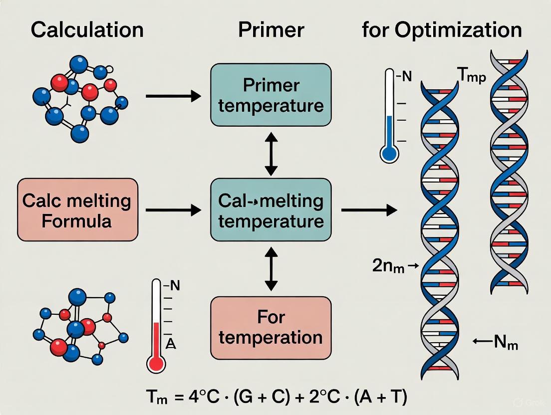
The critical relationship between Tm and the overall PCR process is outlined below, showing how Tm calculation influences experimental setup and outcomes.
Calculating Primer Melting Temperature
Fundamental Calculation Methods
Several mathematical approaches exist for calculating Tm, ranging from simple empirical formulas to complex thermodynamic models. The choice of method depends on primer length, sequence composition, and the required level of precision [12] [10].
Basic Rule-of-Thumb Method: For a quick estimate, the simplest formula is
Tm = 4(G + C) + 2(A + T), where G, C, A, and T represent the number of respective nucleotides in the primer sequence [10]. This method provides a rough approximation but does not account for critical factors like salt concentration, which can significantly alter actual Tm values.Salt-Adjusted Calculation: For greater accuracy, a formula that incorporates salt concentration is recommended:
Tm = 81.5 + 16.6(log[Na+]) + 0.41(%GC) - 675/primer length[10]. This calculation accounts for the stabilizing effect of monovalent cations on DNA duplex formation.Nearest-Neighbor Method: Recognized as the most accurate approach, this thermodynamic model considers the stability of every adjacent dinucleotide pair in the oligo sequence, along with precise concentrations of salts and primers [10] [7]. The modified Allawi & SantaLucia's thermodynamics method, used in sophisticated online calculators, applies this principle with parameters specifically adjusted to maximize PCR specificity and yield [7].
Comparison of Tm Calculation Methods
Table 1: Key methods for calculating primer melting temperature (Tm).
| Method | Formula | Best For | Key Assumptions/Limitations |
|---|---|---|---|
| Basic Rule-of-Thumb | Tm = 4(G+C) + 2(A+T) |
Quick estimates, initial design | Assumes standard conditions; less accurate [12] [10]. |
| Salt-Adjusted | Tm = 81.5 + 16.6(log[Na+]) + 0.41(%GC) - 675/length |
General laboratory use | Accounts for [Na+]; assumes pH ~7 [10]. |
| Nearest-Neighbor | Complex; based on ΔG of dinucleotide pairs | High-fidelity applications, critical assays | Requires software; accounts for sequence, [salts], and [primer] [7] [10]. |
The following diagram illustrates the logical decision process for selecting the appropriate Tm calculation method based on experimental requirements.
Experimental Protocols for Tm-Dependent PCR Optimization
Protocol 1: Gradient PCR for Empirical Annealing Temperature Determination
This protocol provides a systematic method for determining the optimal annealing temperature (Ta) for a primer pair, which is typically 3–5°C below the calculated Tm [10].
Materials:
- Thermostable DNA polymerase and its compatible reaction buffer
- dNTP mix
- Template DNA
- Forward and reverse primers
- Thermal cycler with gradient functionality
Procedure:
- Calculate Tm: Determine the Tm for both forward and reverse primers using the nearest-neighbor method via a reliable online calculator [7].
- Prepare Master Mix: Create a PCR master mix containing all reaction components except template DNA, according to the manufacturer's instructions.
- Aliquot and Add Template: Distribute the master mix into individual PCR tubes, then add template DNA to each.
- Set Gradient: Program the thermal cycler with a gradient across the block that spans a temperature range from 5°C below the lowest primer Tm to 5°C above it [10].
- Run PCR: Initiate the cycling protocol, which typically includes: initial denaturation (94–98°C for 1–3 min); 25–35 cycles of denaturation (94–98°C for 15–30 s), gradient annealing (30 s), and extension (68–72°C for 1 min/kb); final extension (72°C for 5–10 min) [10].
- Analyze Results: Separate PCR products by agarose gel electrophoresis. The optimal Ta produces a single, intense band of the expected size with minimal to no non-specific amplification.
Protocol 2: MgCl₂ Concentration Optimization Based on Tm and Primer Properties
Mg²⁺ is an essential cofactor for DNA polymerase, and its optimal concentration is influenced by primer characteristics and Tm. Recent research has enabled predictive modeling of MgCl₂ requirements with high accuracy (R² = 0.9942) [13].
Materials:
- MgCl₂ stock solution (typically 25–50 mM)
- PCR components as listed in Protocol 1
Procedure:
- Prepare Mg²⁺ Titration Series: Create a series of PCR master mixes with MgCl₂ concentrations ranging from 1.0 to 4.0 mM in 0.5 mM increments.
- Utilize Predictive Modeling (Optional): For a theoretically informed starting point, apply the predictive equation from recent research:
(MgCl₂) ≈ 1.5625 + (-0.0073 × Tm) + (-0.0629 × GC) + (0.0273 × L) + (0.0013 × dNTP) + (-0.0120 × Primers) + (0.0007 × Polymerase) + (0.0012 × log(L)) + (0.0016 × Tm_GC) + (0.0639 × dNTP_Primers) + (0.0056 × pH_Polymerase)[13]. - Run PCR: Amplify using the established Ta from Protocol 1.
- Analyze Results: Identify the MgCl₂ concentration that yields the highest quantity of specific product with minimal background.
Table 2: Relative importance of variables in predicting optimal MgCl₂ concentration [13].
| Variable | Relative Importance (%) |
|---|---|
| dNTP_Primers Interaction | 28.5% |
| GC Content | 22.1% |
| Amplicon Length (L) | 15.7% |
| Primer Tm | 12.3% |
| Primer Concentration | 8.9% |
| pH_Polymerase Interaction | 5.6% |
| Tm_GC Interaction | 3.2% |
| log(Amplicon Length) | 2.1% |
| dNTP Concentration | 1.1% |
| Polymerase Concentration | 0.5% |
The Scientist's Toolkit: Essential Reagents for PCR Optimization
Table 3: Key research reagent solutions for enhancing PCR specificity and efficiency.
| Reagent / Solution | Function / Purpose | Optimal Concentration | Considerations |
|---|---|---|---|
| TMA Oxalate | Novel enhancer that increases specificity and yield of specific PCR products [14]. | 2 mM | Maximum specificity (1.0) and efficiency (2.2) observed at this concentration [14]. |
| DMSO (Dimethyl Sulfoxide) | Disrupts secondary structures; lowers effective Tm particularly for GC-rich templates (>65%) [11]. | 2–10% | 10% DMSO decreases annealing temperature by 5.5–6.0°C [10]. |
| Betaine | Homogenizes thermodynamic stability of DNA; reduces the dependence of duplex stability on base composition [11]. | 1–2 M | Especially useful for long-range PCR and GC-rich templates [11]. |
| Formamide | Increases specificity of amplification; can improve efficiency [14]. | 0.5 M | Maximal efficiency of 1.4 observed at this concentration [14]. |
| MgCl₂ | Essential cofactor for DNA polymerase; stabilizes primer-template duplex [11]. | 1.5–2.5 mM (typical) | Concentration must be optimized; significantly affects fidelity and specificity [13] [11]. |
| High-Fidelity Polymerase Blends | Enzyme mixtures providing proofreading (3'→5' exonuclease) activity for accurate DNA replication [11]. | Manufacturer specified | Reduces error rates from ~1 x 10⁻⁵ (Taq) to as low as ~3 x 10⁻⁶ [11]. |
Advanced Considerations and Troubleshooting
Impact of PCR Additives on Tm Calculation
The presence of enhancers and cosolvents in PCR reactions necessitates adjustments to calculated Tm values. For instance, the addition of 10% DMSO typically lowers the effective annealing temperature by 5.5–6.0°C [10]. Similarly, substitution of dGTP with 7-deaza-dGTP also decreases Tm. When using these additives, the annealing temperature should be adjusted empirically, starting with a temperature 3–5°C lower than the calculated Tm and optimizing from there. This is particularly crucial when amplifying challenging templates such as those with high GC content (>65%), where additives like DMSO or betaine are commonly employed to improve yield [11].
Universal Annealing Temperature Systems
Recent advancements in PCR buffer formulation have led to the development of specialized systems that enable a universal annealing temperature (e.g., 60°C) regardless of primer Tm variations [7]. These systems incorporate isostabilizing components that increase the stability of primer-template duplexes during the annealing step [10]. For DNA polymerases such as Platinum II Taq, Platinum SuperFi II, and Phusion Plus, specially formulated buffers eliminate the need for Tm calculation and individual annealing temperature optimization, streamlining experimental workflow [7]. These developments are particularly valuable in high-throughput settings where multiple primer pairs with different Tm values are used simultaneously.
Troubleshooting Common Tm-Related PCR Issues
- No/Low Amplification: This often results from an annealing temperature that is too high. Lower the Ta in increments of 2–3°C, ensuring it does not fall more than 5°C below the primer Tm [10]. Also verify Mg²⁺ concentration and primer design.
- Non-Specific Bands/Multiple Bands: Typically caused by an annealing temperature that is too low. Increase Ta in increments of 2–3°C up to the extension temperature [10] [11]. Alternatively, use a hot-start polymerase or optimize Mg²⁺ concentration.
- PCR Efficiency in qPCR: For accurate quantification in real-time PCR, assays should be designed to achieve 100% geometric efficiency, where the amount of product doubles each cycle [15]. Assays with efficiencies significantly different from 100% complicate data analysis, particularly when using the ΔΔCt method for quantification [15] [16].
The accurate prediction of primer melting temperature (Tm) is a cornerstone of successful molecular biology research, directly impacting the specificity and efficiency of techniques such as PCR, qPCR, and next-generation sequencing. This application note details the core biochemical factors governing Tm—sequence composition, length, and GC content—and provides validated protocols for their precise calculation and application. By integrating thermodynamic models and modern computational tools, researchers can standardize assay design, thereby enhancing reproducibility and reliability in diagnostic and therapeutic development pipelines.
In polymerase chain reaction (PCR) and its derivatives, the melting temperature (Tm) of an oligonucleotide is the temperature at which 50% of the DNA duplex dissociates into single strands. This parameter fundamentally controls the annealing step, where primers bind to the template DNA. An inaccurately predicted Tm can lead to assay failure through non-specific amplification, primer-dimer formation, or complete absence of product, compromising data integrity in fields from basic research to clinical diagnostics [17] [18]. The shift from simple, rule-based Tm calculations to sophisticated thermodynamic models has been driven by the demanding precision required in modern applications, including single-cell sequencing and CRISPR-based editing, where even minor Tm inconsistencies can cause significant sequence dropouts or biased results [19]. This document delineates the key factors influencing Tm and provides a rigorous framework for its determination.
Core Factors Influencing Melting Temperature
The stability of a DNA duplex, and consequently its Tm, is governed by the sum of the energetic contributions of its constituent nucleotides and their interactions with the reaction environment.
Sequence Composition and Nearest-Neighbor Thermodynamics
The sequence composition's impact on Tm is most accurately captured by the nearest-neighbor model, the current gold standard in Tm prediction. Unlike simplistic methods that consider only base composition, this model accounts for the sequence context by quantifying the stacking energy between adjacent base pairs.
- Thermodynamic Basis: The model sums the enthalpy (ΔH°) and entropy (ΔS°) changes for the ten possible dinucleotide pairs. For instance, a 5'-GC-3' pair, with its three hydrogen bonds, contributes more stability than a 5'-AT-3' pair, which has only two [19].
- Accuracy: This method achieves precision within 1-2°C of experimental values, a significant improvement over the ±5-10°C error of basic GC% formulas [20] [19].
Calculation Foundation: The fundamental Tm calculation formula for a monovalent salt solution is:
Tm = (ΔH° / (ΔS° + R × ln(Ct/4))) - 273.15 + 16.6 × log₁₀[Na⁺]
Where ΔH° and ΔS° are the sum of the nearest-neighbor values, R is the gas constant, and Ct is the total oligonucleotide concentration [19].
Oligonucleotide Length
The length of the oligonucleotide directly influences the total stabilizing energy of the duplex. Longer primers have more dinucleotide stacking interactions, leading to a higher Tm.
- Optimal Range: For most PCR applications, an optimal primer length is between 18 and 24 nucleotides [11]. This provides sufficient sequence for specific binding while avoiding overly high Tm that could reduce annealing efficiency.
- Diminishing Returns: The relationship between length and Tm is not linear. The incremental stability added by each base pair decreases with increasing length, causing Tm to plateau.
GC Content
GC content is a primary, though incomplete, indicator of duplex stability. The proportion of guanine (G) and cytosine (C) bases in a sequence is critical because GC base pairs form three hydrogen bonds, making them more thermally stable than AT base pairs, which form only two.
- Optimal Range: The ideal GC content for primers is between 40% and 60% [19] [11].
- Low GC Content (<30%): Results in unstable priming and low reaction yield due to weak binding [19].
- High GC Content (>70%): Promotes the formation of stable secondary structures (e.g., hairpins) and increases the risk of non-specific binding, challenging assay specificity [19] [11].
Table 1: Summary of Core Factors Influencing Tm
| Factor | Mechanism of Influence | Optimal Range | Impact on Tm |
|---|---|---|---|
| Sequence Composition | Sum of dinucleotide stacking energies (nearest-neighbor model) | N/A | High predictive accuracy (±1-2°C); core to modern calculation |
| Oligonucleotide Length | Total number of stabilizing base-pair interactions | 18-24 nucleotides | Longer sequences have higher Tm; effect plateaus |
| GC Content | Proportion of strong 3-hydrogen-bond GC pairs | 40-60% | Higher GC content significantly increases Tm and stability |
Experimental Protocols for Tm Determination and Verification
Protocol: In-silico Tm Calculation Using the Nearest-Neighbor Method
This protocol outlines the steps for obtaining a theoretically accurate Tm using a SantaLucia-based online calculator, suitable for designing individual primers or small sets.
1. Access the Tool: Navigate to a publicly available Tm calculator that implements the SantaLucia nearest-neighbor method with Owczarzy salt corrections, such as the OligoPool tool [20].
2. Enter Sequence: Paste the oligonucleotide sequence (DNA or RNA) into the input field. The sequence can include spaces and line breaks; the tool will automatically sanitize the input. Example: ATCGATCGATCGATCGATCG [20].
3. Set Reaction Conditions: Input the specific chemical conditions of your assay [20] [19]:
- Na⁺ Concentration: 50 mM for standard PCR.
- Mg²⁺ Concentration: 1.5-2.5 mM for standard PCR. *Note: Mg²⁺ has a stronger stabilizing effect than Na⁺ and must be accounted for using specific correction formulas [19].
- Oligonucleotide Concentration: 0.25 µM is a standard default for primers.
- DMSO: If used, input the percentage (e.g., 5%). DMSO reduces Tm by ~0.5-0.7°C per 1% [20] [19]. 4. Calculate and Interpret: Execute the calculation. The tool will return the Tm, thermodynamic parameters (ΔH, ΔS), GC%, and length. For PCR, the annealing temperature (Ta) is typically set 3-5°C below the calculated Tm [20].
Protocol: Empirical Validation of Tm via Gradient PCR
Theoretical Tm values should be empirically verified, especially for critical assays. Gradient PCR is the standard method for this.
1. Primer-Template System: Prepare a standard PCR mixture containing your target template and the designed primer pair. 2. Thermal Cycler Setup: Program the thermal cycler with a gradient across the annealing step, spanning a range of at least 5°C above and below the predicted Tm. 3. Amplification and Analysis: Run the PCR and analyze the products using gel electrophoresis. 4. Determine Optimal Ta: Identify the annealing temperature that produces the highest yield of the specific target product with minimal non-specific amplification or primer-dimer. This empirically determined Ta validates the accuracy of the theoretical Tm and refines the assay conditions [11].
Table 2: Comparison of Tm Calculation Software
| Software Tool | Calculation Method | Reported Accuracy | Key Features | Best For |
|---|---|---|---|---|
| OligoPool Calculator | SantaLucia Nearest-Neighbor | ±1-2°C [20] | Owczarzy Mg²⁺ correction, batch processing | General PCR, high-accuracy research |
| Primer3 Plus | Nearest-Neighbor | Best-in-class per study [17] | Integrated primer design & analysis | Holistic PCR assay design |
| Primer-BLAST | Nearest-Neighbor | Best-in-class per study [17] | Combines design with specificity check | Ensuring primer specificity |
| NEB Tm Calculator | Proprietary Nearest-Neighbor | ±2-3°C [20] | Optimized for NEB polymerases | Using NEB enzyme/buffer systems |
| IDT OligoAnalyzer | Nearest-Neighbor | ±2-3°C [20] | User-friendly, additional analysis | Quick checks, secondary structure |
The Scientist's Toolkit: Research Reagent Solutions
Table 3: Essential Reagents for PCR and Tm-Dependent Assays
| Reagent / Material | Function / Description | Application Note |
|---|---|---|
| High-Fidelity Polymerase (e.g., Pfu, KOD) | DNA polymerase with 3'→5' exonuclease (proofreading) activity. | Reduces error rate to ~1 x 10⁻⁶ for cloning and sequencing [11]. |
| Hot-Start Taq Polymerase | Enzyme engineered for inactivity at room temperature. | Prevents non-specific amplification and primer-dimer formation prior to thermal cycling [11]. |
| DMSO (Dimethyl Sulfoxide) | A chemical additive that disrupts secondary structures. | Use at 2-10% to lower Tm and improve amplification of GC-rich templates (>65% GC) [19] [11]. |
| Betaine | A chemical additive that homogenizes DNA duplex stability. | Use at 1-2 M to improve the amplification of long or complex templates [11]. |
| MgCl₂ Solution | Source of Mg²⁺, an essential cofactor for polymerase activity. | Concentration must be optimized (typically 1.5-2.5 mM); significantly affects Tm, fidelity, and yield [19] [11]. |
| Synthetic Oligo Pool | Defined mixtures of oligonucleotides synthesized in parallel. | Essential for screening applications (e.g., CRISPR); require uniform Tm (±5°C) for consistent performance [21] [19]. |
Workflow and Relationship Visualization
The following diagram illustrates the logical workflow for designing and validating primers, integrating the key factors influencing Tm and the experimental protocols for verification.
Tm Determination and Primer Validation Workflow
Precision in Tm prediction is not merely a theoretical exercise but a practical necessity for robust and reproducible molecular assays. By understanding and applying the principles of sequence composition, length, and GC content through the framework of nearest-neighbor thermodynamics, researchers can systematically eliminate a major source of experimental variability. The integration of reliable in-silico tools with empirical validation, as outlined in these protocols, provides a clear path to optimizing PCR conditions, ultimately enhancing the fidelity of results in genomics, diagnostics, and drug development.
The Evolution from Simple Formulas to Advanced Thermodynamic Models
The accurate prediction of primer melting temperature (Tm) is a cornerstone of molecular biology, directly influencing the success of polymerase chain reaction (PCR) and related techniques. The evolution from simple, rule-based formulas to sophisticated thermodynamic models represents a significant advancement in the field of assay design and optimization. This evolution has been driven by the growing demand for precision in diagnostic applications, high-throughput genomic studies, and genetic engineering. Early methods provided rough approximations sufficient for basic applications, while modern approaches incorporate comprehensive thermodynamic parameters that more accurately reflect the complex biochemical reality of nucleic acid hybridization. This application note traces this technological journey, detailing the underlying principles, practical methodologies, and reagent solutions that empower researchers to achieve superior experimental outcomes through thermodynamically-informed primer design.
From Rule-Based Calculations to Thermodynamic Predictions
Historical Foundations and Simple Formulas
The initial methods for Tm calculation relied on simple formulas derived from empirical observations. The most iconic among these is the Wallace rule, which states that Tm ≈ 2°C × (A+T) + 4°C × (G+C) for primers shorter than 20 nucleotides. Similarly, the %GC method estimated Tm as the sum of the product of GC percentage and a constant factor, often Tm = 81.5 + 0.41(%GC) - (675/length). These heuristic approaches provided a starting point but suffered from significant limitations. They largely ignored the sequence context of the nucleotides, treating all base pairs as contributing independently to duplex stability, which fails to capture the nuanced energetics of DNA hybridization. Consequently, these methods often resulted in discrepancies between predicted and experimental Tm values, leading to suboptimal PCR conditions and failed experiments [17].
The Advent of the Nearest-Neighbor Model
The development of the nearest-neighbor model marked a paradigm shift in Tm prediction. This model is grounded in the understanding that the stability of a DNA duplex depends not only on its base composition but also critically on the sequence context—specifically, how each base pair stacks upon its neighboring base pairs. The model assigns thermodynamic parameters (ΔH°: enthalpy change; ΔS°: entropy change) for all ten possible combinations of neighboring base pairs (AA/TT, AT/TA, TA/AT, CA/GT, GT/CA, CT/GA, GA/CT, CG/GC, GC/CG, GG/CC). The total free energy change (ΔG°) for duplex formation is calculated by summing these individual nearest-neighbor contributions along with initiation and symmetry terms. The Tm is then derived from the relationship Tm = ΔH°/(ΔS° + R ln(Ct)) - 273.15 + 16.6 log10([Na+]), where R is the gas constant and Ct is the total strand concentration [17].
This approach was significantly refined through the extensive experimental work of SantaLucia and colleagues, who provided a unified set of thermodynamic parameters that have become the standard for accurate Tm calculation [17] [22]. The superiority of the nearest-neighbor model lies in its ability to account for the fact that the stability of a duplex can vary dramatically depending on the order of its nucleotides, even for sequences with identical length and GC content.
Integration of Reaction Conditions and Modern Computational Tools
Modern Tm prediction has evolved further by integrating the nearest-neighbor model with detailed considerations of the reaction environment. Key factors such as monovalent (e.g., Na+) and divalent (e.g., Mg2+) cation concentrations, which stabilize the duplex by shielding the negatively charged phosphate backbone, are now routinely incorporated into calculations [13]. The critical influence of MgCl2 concentration on amplification specificity and primer annealing has been formally modeled using multivariate Taylor series expansions and thermodynamic functions, achieving remarkable predictive accuracy (R² = 0.9942) [13].
This advanced understanding is embedded in contemporary primer design software. A comparative study of 22 Tm calculator tools demonstrated that programs implementing the full nearest-neighbor model with salt correction, notably Primer3 Plus and Primer-BLAST, provide Tm predictions closest to experimentally determined values [17]. These tools have become indispensable for researchers, moving beyond simple Tm calculation to evaluate primer specificity, secondary structure formation, and potential off-target binding, thereby ensuring robust assay design [22].
Table 1: Comparison of Tm Calculation Methods
| Method | Underlying Principle | Key Input Parameters | Accuracy & Limitations |
|---|---|---|---|
| Wallace Rule (Simple Formula) | Empirical; linear combination of A-T and G-C counts. | Primer length, base count. | Low accuracy; only suitable for very short primers. |
| %GC Method (Simple Formula) | Linear relationship with GC content. | Primer length, GC percentage. | Low accuracy; ignores sequence context and reaction conditions. |
| Nearest-Neighbor Model | Thermodynamic sum of base-pair stacking energies. | Primer sequence, oligonucleotide concentration, salt concentration. | High accuracy; accounts for sequence context; basis for modern software. |
Advanced Thermodynamic Integration in Primer Design
The progression of thermodynamic models has enabled the development of sophisticated primer design tools that transcend mere Tm calculation. These tools leverage state-of-the-art DNA binding affinity computations and chemical reaction equilibrium analysis to predict overall PCR efficiency and specificity.
The Pythia Algorithm: A Thermodynamic Framework
The Pythia primer design method exemplifies this advanced approach. It directly integrates DNA binding affinity and folding stability computations into the design process. Pythia employs statistical mechanical models to calculate the binding affinity between all relevant DNA dimers (e.g., primer-template, primer-primer) and the folding energy of individual nucleic acid molecules to predict structures like hairpins. These energetic calculations are then fed into a chemical reaction equilibrium analysis [23].
This analysis considers a system of 11 competing simultaneous reactions that can occur in a PCR mix, including primer folding, primer dimerization, primers binding to off-target template regions, and the desired priming reaction. By minimizing the Gibbs energy of the system, the model determines the equilibrium concentrations of all chemical species. The primer pair quality is then characterized by the minimum of the fractions of left and right primers binding to their correct binding sites under idealized late-stage PCR conditions. This provides a physically meaningful, conservative measure of PCR efficiency derived from first principles, moving beyond arbitrary weighted scoring metrics [23].
Thermodynamic Specificity Assessment
Beyond efficiency, thermodynamic principles are crucial for assessing primer specificity. Following the heuristic proposed by Miura et al. and used in Pythia, the shortest suffix (3'-end sequence) of a primer that would bind stably at equilibrium is determined. This sequence is then used to search for exact matches within a precomputed genomic index. This method leverages the thermodynamic stability of the 3'-terminal region, which is critical for polymerase extension, to predict and minimize off-target amplification [23].
Experimental Protocols for Validation and Optimization
While computational design has become highly sophisticated, empirical validation and optimization remain essential. The following protocols provide a systematic framework for this process.
Protocol 1: In Silico Primer Design with Thermodynamic Parameters
This protocol leverages modern software to design primers based on advanced thermodynamic models.
- Sequence Acquisition and Alignment: Obtain the complete target sequence. If working with a gene family or in a species with a sequenced genome, retrieve all homologous sequences and perform a multiple sequence alignment to identify unique single-nucleotide polymorphisms (SNPs) for designing sequence-specific primers [24].
- Software Selection and Parameter Setting: Use a tool with robust thermodynamic prediction, such as Primer3 Plus or Primer-BLAST [17]. For Primer-BLAST, the default thermodynamic parameters are "SantaLucia 1998" for both the thermodynamic table and salt correction [22].
- Constraint Definition:
- Set the product size range (e.g., 85-125 bp for qPCR).
- Define the primer length (typically 18-25 bases).
- Set the Tm range for both forward and reverse primers to be within 5°C of each other, ideally between 50-72°C [25].
- Set the GC content to 40-60%.
- Avoid repeats and stable secondary structures at the 3'-end.
- Specificity Check: In Primer-BLAST, select the appropriate organism database (e.g., "Refseq mRNA" or "Refseq representative genomes") to ensure primers are specific to the intended target and do not amplify unintended genomic regions [22].
- Output Analysis: Select candidate primer pairs from the output. Manually verify the absence of stable hairpins or significant self-complementarity, especially at the 3'-ends, which can lead to primer-dimer artifacts.
Protocol 2: Stepwise Optimization of Real-Time PCR Assays
Computational design must be followed by wet-lab optimization to account for the complex biological reality [24] [25].
- Primer Preparation: Resuspend desalted or HPLC-purified primers in nuclease-free water to a stock concentration (e.g., 100 µM). Determine the exact concentration spectrophotometrically and dilute to a working concentration (e.g., 10 µM). Aliquot to prevent degradation from freeze-thaw cycles [25].
- Annealing Temperature (Ta) Optimization: Perform a temperature gradient PCR. Center the gradient around the predicted Tm (e.g., Tm ± 5°C). Analyze the PCR products using gel electrophoresis. The optimal Ta is the highest temperature that yields a single, strong band of the expected size.
- Primer Concentration Optimization: Using the optimal Ta, test a range of final primer concentrations (e.g., 0.05 - 1.0 µM) in a standard qPCR reaction [25]. The ideal concentration produces the lowest Cq value without increasing non-specific background.
- Efficiency and Standard Curve Validation: Prepare a serial dilution (at least 5 points) of the target cDNA or DNA template. Run qPCR with the optimized conditions. Generate a standard curve by plotting the Cq values against the log of the template concentration.
- The amplification efficiency (E) is calculated as E = (10^(-1/slope) - 1) × 100%.
- The ideal efficiency is 100% (slope = -3.32), with an acceptable range of 90-110% (R² ≥ 0.99) [24].
- Final Validation: Use the optimized protocol to run experimental samples and confirm specificity through melt curve analysis, showing a single, sharp peak.
Table 2: Essential Research Reagent Solutions for Thermodynamic Optimization
| Reagent/Material | Function in Optimization | Key Considerations |
|---|---|---|
| High-Fidelity DNA Polymerase | Catalyzes DNA synthesis from primers. | Proofreading activity reduces errors; different polymerases may have varying buffer formulations. |
| MgCl₂ Solution | Essential co-factor for polymerase activity; influences Tm and specificity. | Concentration is critical; optimal level is often determined empirically (e.g., 1.5-3.5 mM) [13]. |
| dNTP Mix | Building blocks for new DNA strands. | Balanced concentration is necessary to prevent misincorporation; competes with primers for Mg²⁺. |
| Thermostable Buffer | Provides optimal chemical environment (pH, salts) for polymerization. | Buffer composition (e.g., KCl, (NH₄)₂SO₄) affects primer Tm and should match calculator assumptions. |
| Template DNA/cDNA | The target sequence to be amplified. | Quality and quantity must be high; serial dilutions for standard curves must be accurate. |
Workflow Visualization
The following diagram illustrates the integrated computational and experimental workflow for advanced, thermodynamically-driven primer design and validation.
The evolution of primer melting temperature prediction from simple formulas to advanced thermodynamic models has fundamentally transformed molecular assay design. This journey reflects a broader shift in biology towards quantitative, physics-based approaches that offer greater predictive power and experimental robustness. Modern tools, grounded in the nearest-neighbor model and integrated with chemical equilibrium analysis, allow researchers to design primers with high efficiency and specificity, even in challenging genomic contexts. However, the most successful protocols combine these powerful in silico predictions with rigorous empirical validation, creating a feedback loop that further refines our theoretical understanding. As thermodynamic models continue to incorporate more variables and machine-learning approaches enhance their accuracy, the future promises even more precise and reliable design capabilities, accelerating discovery and diagnostics across the life sciences.
DNA melting temperature (Tm) is a fundamental thermodynamic property critical for optimizing molecular biology techniques such as PCR, hybridization probes, and genotyping assays. This application note details experimental protocols for visualizing and quantifying the DNA melting process, framed within the context of primer Tm calculation for optimization research. We provide methodologies for both ultraviolet (UV) absorbance and fluorescence-based melting analysis, enabling researchers to accurately determine Tm values essential for experimental design. The protocols include detailed workflows, reagent specifications, and data analysis procedures to ensure reproducible results across applications from basic research to drug development.
DNA melting, or denaturation, describes the temperature-dependent separation of double-stranded DNA (dsDNA) into single-stranded DNA (ssDNA). This process is central to many molecular techniques, particularly polymerase chain reaction (PCR), where the accuracy of primer melting temperature (Tm) calculation directly impacts amplification specificity, sensitivity, and efficiency [17] [26]. The Tm is formally defined as the temperature at which half of the DNA duplexes are in a single-stranded state [27].
The thermal stability of DNA duplexes depends on several sequence-specific and environmental factors. GC content significantly influences Tm due to the three hydrogen bonds in G≡C base pairs compared to two in A=T pairs, making GC-rich sequences more stable [27] [28]. DNA length affects stability, with longer fragments generally exhibiting higher Tm values [28]. Sequence composition beyond simple GC content, including nearest-neighbor interactions, plays a crucial role in duplex stability [29] [27]. Environmental factors such as salt concentration stabilize DNA duplexes by neutralizing the negatively charged phosphate backbone, while pH extremes can destabilize the double helix [13] [28].
Accurate Tm prediction enables researchers to optimize annealing temperatures in PCR, design specific hybridization probes, and detect genetic variations through melt curve analysis [26] [28]. This note provides standardized protocols for experimental Tm determination, bridging theoretical calculations with empirical validation for robust experimental design in research and diagnostic applications.
Theoretical Foundations of DNA Melting
Thermodynamic Principles
DNA melting follows a two-state model transitioning from duplex to random coil configurations. The thermodynamics are governed by the Gibbs free energy equation:
ΔG = ΔH - TΔS
Where ΔG is the change in free energy, ΔH is the enthalpy change (primarily from hydrogen bonding and base stacking), ΔS is the entropy change, and T is the temperature in Kelvin [27] [13]. At Tm, ΔG = 0, leading to the relationship:
Tm = ΔH/ΔS
For short oligonucleotides, Tm can be calculated using the nearest-neighbor model, which considers the sequence dependence of stability by summing the free energy contributions of adjacent base pairs [27]. The free energy of forming a nucleic acid duplex is expressed as:
ΔG°₃₇(total) = ΔG°₃₇(initiation) + ΣnᵢΔG°₃₇(i)
Where nᵢ is the number of occurrences of nearest-neighbor i, and ΔG°₃₇(i) is its free energy contribution [27].
Calculation Methods and Tools
Several computational approaches exist for Tm prediction, with varying levels of accuracy:
Table 1: Comparison of DNA Melting Temperature Prediction Tools
| Method/Software | Basis of Calculation | Sequence Length Applicability | Reported Accuracy |
|---|---|---|---|
| Primer3 Plus [17] | Nearest-neighbor model | Oligonucleotides (typically 15-30 bp) | Best prediction performance in comparative studies |
| Primer-BLAST [17] | Nearest-neighbor model | Oligonucleotides (typically 15-30 bp) | Best prediction performance in comparative studies |
| Nearest-neighbor model [27] | Sum of dimer free energies | Short oligonucleotides | High accuracy for short sequences |
| Empirical formula (GC 40-60%) [30] | Tm = ΔH/ΔS–0.27GC%–(150+2n)/n–273.15 | PCR products | Average error <1°C within specified GC range |
| Empirical formula (GC <40%) [30] | Tm = ΔH/ΔS–GC%/3–(150+2n)/n–273.15 | PCR products | Average error <1°C within specified GC range |
| Linear regression model [13] | Multivariate Taylor series expansion | Various lengths | R² = 0.9942 for MgCl₂; 0.9600 for Tm |
Comparative studies evaluating 22 different primer design tools revealed significant variation in Tm prediction accuracy, with Primer3 Plus and Primer-BLAST demonstrating the best performance when benchmarked against experimentally determined Tm values [17]. Recent advances incorporate machine learning and high-throughput experimental data to improve prediction accuracy for diverse structural motifs including mismatches, bulges, and hairpin loops [29] [13].
Experimental Visualization Methodologies
UV-Visible Spectroscopy-Based Melting Analysis
Ultraviolet (UV) absorbance monitoring at 260 nm represents the classical approach for DNA melting analysis, leveraging the hyperchromic effect where single-stranded DNA exhibits approximately 30-40% greater absorbance than double-stranded DNA [26] [28].
Protocol: UV-Vis DNA Melting Analysis
Sample Preparation
- Prepare DNA sample in appropriate buffer (typically 10-50 μM DNA concentration in duplex)
- Common buffers: 10 mM Tris-HCl, pH 7.5-8.0, with 50-100 mM NaCl
- Use quartz cuvette with 1 cm path length
Instrument Setup
- Set spectrophotometer to monitor absorbance at 260 nm
- Program temperature controller for gradual heating (0.5-1.0°C per minute)
- Set temperature range to encompass complete transition (typically 20-95°C)
- Equilibrate at each temperature point for 30-60 seconds before measurement
Data Collection
- Record absorbance values at each temperature increment
- Ensure sufficient data points (≥1 per °C) for accurate curve fitting
Data Analysis
This method requires relatively pure DNA samples without significant contaminating absorbance at 260 nm and is ideal for fundamental thermodynamic studies [28].
Fluorescence-Based Melting Analysis
Fluorescence monitoring using DNA-intercalating dyes offers enhanced sensitivity and compatibility with complex samples, making it particularly suitable for post-PCR melting analysis [26] [31].
Protocol: Fluorescence-Based HRM Analysis
Reaction Setup
- Prepare PCR reaction mix containing:
- DNA template (genomic DNA, plasmid, or PCR product)
- Primers (0.1-0.5 μM each)
- DNA-binding dye (SYBR Green I, EvaGreen, or LCGreen)
- Reaction buffer with Mg²⁺
- dNTPs
- DNA polymerase
- Prepare PCR reaction mix containing:
Amplification and Melting
- Perform PCR amplification if analyzing amplicons:
- Initial denaturation: 95°C for 2 minutes
- 35-45 cycles: 95°C for 15s, annealing for 20s, 72°C for 20s
- Execute high-resolution melting protocol:
- Temperature range: 5-10°C below expected Tm to 10-15°C above
- Temperature increment: 0.1-0.3°C per step
- Equilibration time: 2-15 seconds per step depending on instrument
- Perform PCR amplification if analyzing amplicons:
Data Processing
Variant Detection
- Compare melting curve shapes and Tm values between samples
- Normalize curves to identical temperature scales for shape comparison
- Use difference plots to enhance detection of subtle variants [26]
Fluorescence-based methods enable analysis of complex mixtures and are compatible with high-throughput formats, including digital HRM in picoliter-scale reactions for absolute quantification of sequence variants [32].
Research Reagent Solutions
Table 2: Essential Reagents for DNA Melting Analysis
| Reagent/Category | Specific Examples | Function & Application Notes |
|---|---|---|
| DNA-Binding Dyes | SYBR Green I, EvaGreen, LCGreen | Fluorescent intercalation for real-time monitoring; saturating dyes preferred for HRM [26] |
| Buffer Components | Tris-HCl, NaCl, MgCl₂ | Stabilize pH and ionic strength; Mg²⁺ critical for polymerase activity in PCR [13] |
| Enzymes | Thermostable DNA polymerases | Catalyze DNA amplification; choice affects fidelity and processivity |
| Universal Primers | 16S rRNA gene primers | Enable broad detection of bacterial sequences in digital HRM applications [32] |
| Quantification Standards | Known Tm control DNA | Validate instrument calibration and enable cross-run comparisons [31] |
| Microfluidic Chips | Digitizing chips (20,000 reactions) | Enable massively parallel digital HRM for absolute quantification [32] |
Advanced Applications and Methodologies
High-Resolution Melting (HRM) for Genotyping
High-resolution melting analysis enables sensitive detection of single nucleotide variants (SNVs), small insertions/deletions, and methylation status without sequencing [26]. The precise monitoring of fluorescence loss during DNA melting can distinguish sequences differing by as little as a single base pair.
Protocol: HRM for SNV Detection
Amplicon Design
- Design PCR amplicons of 50-300 bp encompassing the variant of interest
- Avoid repetitive sequences and extreme GC content (>80% or <20%)
PCR Amplification
- Use saturating DNA dyes such as LCGreen or EvaGreen for enhanced resolution
- Optimize primer concentrations to minimize primer-dimer formation
- Include control samples with known genotypes in each run
High-Resolution Melting
- Use instrument capable of temperature precision ≤0.1°C
- Set temperature ramp rate to 0.1-0.3°C per second for optimal resolution
- Ensure sufficient data acquisition (10-25 acquisitions/°C)
Data Analysis
Digital High-Resolution Melting (dHRM)
Digital HRM combines limiting dilution digital PCR with high-resolution melting to achieve absolute quantification of multiple sequence variants in complex samples [32].
Protocol: Digital HRM for Quantitative Profiling
Sample Digitization
- Dilute sample to achieve approximately 0.1-0.01 molecules per reaction
- Partition across 20,000 picoliter-scale reactions using microfluidic chip
Universal Amplification
- Use conserved primers targeting genetic regions of interest (e.g., bacterial 16S gene)
- Perform amplification with intercalating dye (e.g., 2.5X EvaGreen)
Massively Parallel Melting
- Implement precise temperature control with thermoelectric heater/cooler
- Acquire fluorescence images at each temperature point across all reactions
- Use automated image analysis to extract raw fluorescence data
Machine Learning Classification
- Train Support Vector Machine (SVM) algorithm with known melt curve signatures
- Classify individual reaction curves to identify sequence variants
- Quantify variant abundances based on Poisson statistics of positive reactions [32]
This approach reduces reaction volumes by 99.995% and achieves a greater than 200-fold increase in dynamic range compared to traditional PCR-HRM methods [32].
Data Analysis and Interpretation
Melting Curve Processing
Proper data analysis is essential for accurate Tm determination and sequence variant detection:
Normalization Procedures
- For UV data: Normalize absorbance between minimum (double-stranded) and maximum (single-stranded) values
- For fluorescence data: Apply similar normalization, adjusting for dye-specific fluorescence loss independent of melting
Derivative Plot Generation
- Calculate first derivative of normalized melting curve
- Apply smoothing algorithms to reduce noise while preserving signal
- Identify Tm as temperature at derivative peak maximum
Multicomponent Analysis
Quality Control Parameters
Implement rigorous quality control to ensure reliable melting data:
- Replicate Consistency: Tm values should vary by <0.5°C between technical replicates
- Curve Shape: Smooth, sigmoidal melting curves indicate homogeneous samples
- Peak Sharpness: Sharp derivative peaks suggest cooperative melting of uniform duplexes
- Baseline Stability: Stable pre- and post-melt baselines ensure accurate normalization [31]
Advanced analysis tools like web-based LinRegPCR provide automated processing of amplification and melting data, highlighting deviant reactions for exclusion from analysis [31].
Experimental visualization of the DNA melting process provides critical validation for computational Tm predictions, enabling robust optimization of molecular biology applications. The protocols detailed in this application note—spanning traditional UV spectroscopy to advanced digital HRM—offer researchers comprehensive methodologies for accurate Tm determination. By integrating these experimental approaches with theoretical calculations, scientists can enhance the precision of PCR optimization, probe design, and genotyping assays, ultimately advancing research and diagnostic applications in drug development and biomedical science.
Practical Tm Calculation: Methods, Tools, and Laboratory Application
In molecular biology techniques, the melting temperature (Tm) is a fundamental thermodynamic property defined as the temperature at which 50% of DNA duplexes dissociate into single strands and 50% remain hybridized [33]. Accurate Tm prediction is critical for experimental success across numerous applications including PCR, quantitative PCR, site-directed mutagenesis, hybridization-based techniques, and next-generation sequencing library preparation [33] [17]. Errors in Tm estimation directly impact annealing temperature selection, which can lead to failed amplification, reduced product yield, or nonspecific products that compromise experimental results [17].
The selection of an appropriate Tm calculation method depends on multiple factors including oligonucleotide length, sequence composition, and experimental conditions. This application note provides a comprehensive comparison of three predominant calculation methods: the SantaLucia nearest-neighbor method, basic nearest-neighbor approach, and simple GC percentage formula. Understanding the strengths, limitations, and appropriate applications of each method enables researchers to optimize molecular biology assays for improved specificity and efficiency.
Theoretical Foundations of TmCalculation Methods
SantaLucia Nearest-Neighbor Method (1998)
The SantaLucia nearest-neighbor method represents the current gold standard for Tm prediction, incorporating comprehensive thermodynamic parameters derived from experimental data [34] [35]. This method calculates duplex stability by considering the sequence-dependent stacking interactions between adjacent nucleotide pairs, accounting for the fact that DNA stability depends not only on base composition but also on sequence context [35]. The model uses the following thermodynamic equation:
Tm = ΔH° / (ΔS° + R ln(Ct)) - 273.15
Where ΔH° represents the enthalpy change, ΔS° represents the entropy change, R is the gas constant, and Ct is the total oligonucleotide concentration [33]. The SantaLucia method incorporates salt concentration corrections and provides specific thermodynamic parameters for all ten possible Watson-Crick nearest-neighbor pairs, enabling highly accurate predictions across diverse sequence contexts and experimental conditions [34] [35]. This method is particularly valuable for designing critical experiments where precision is paramount, such as quantitative PCR assays or multiplex PCR systems [17].
Basic Nearest-Neighbor Method (1986)
The basic nearest-neighbor method represents an earlier thermodynamic approach that also considers base stacking interactions but with less comprehensive parameterization than the SantaLucia method [33] [35]. Developed based on work by Breslauer et al. (1986) and Freier et al. (1986), this method uses a similar mathematical framework but with different thermodynamic values for the nearest-neighbor pairs [33]. The basic formula is:
Tm = ΔH / (ΔS + R ln(C)) + 16.6 log[Na+] - 272.3
Where ΔH is the sum of enthalpy changes for all nearest-neighbor pairs, ΔS is the sum of entropy changes, R is the gas constant, C is the oligo concentration, and [Na+] is the sodium ion concentration [33]. While this method represents a significant improvement over simplistic formulas, studies have demonstrated that it provides less accurate predictions compared to the updated SantaLucia parameters, particularly for sequences with unusual stacking interactions or complex secondary structures [35].
Simple GC Percentage Method
The simple GC percentage method provides a rudimentary approach to Tm estimation based primarily on base composition rather than sequence arrangement. This method uses the formula:
Tm = 4(G + C) + 2(A + T)
Where G, C, A, and T represent the counts of respective nucleotides in the sequence [36]. This calculation assumes standard conditions of 50 nM primer concentration and 50 mM sodium ion concentration at neutral pH [36]. A common alternative formulation for slightly longer oligonucleotides is:
Tm = 64.9 + 41(G + C - 16.4)/N
Where N represents the total number of bases in the oligonucleotide [36]. The primary limitation of this approach is its failure to account for sequence context, stacking interactions, or precise environmental conditions, potentially leading to errors exceeding 15°C in some cases [37]. Despite its limitations, this method remains in use for quick estimations and educational purposes due to its computational simplicity.
Comparative Analysis of TmCalculation Methods
Table 1: Comparison of Key Features of Tm Calculation Methods
| Feature | SantaLucia (1998) | Basic Nearest-Neighbor | Simple GC% |
|---|---|---|---|
| Theoretical Basis | Nearest-neighbor thermodynamics with updated parameters | Nearest-neighbor thermodynamics with 1986 parameters | Base counting without sequence context |
| Key Parameters | ΔH, ΔS, oligonucleotide concentration, salt correction | ΔH, ΔS, oligonucleotide concentration, salt correction | G+C count, total length |
| Sequence Consideration | Full sequence context including all stacking interactions | Stacking interactions with less accurate parameters | Only base composition |
| Accuracy | Highest (89% predictions within 5°C of experimental Tm) [35] | Moderate | Low (errors can exceed 15°C) [37] |
| Optimal Length Range | 15-120 bases [33] | 15-120 bases | <14 bases: 2(A+T)+4(G+C) [36] |
| Salt Correction | Comprehensive including [Na+] and [Mg2+] | [Na+] correction | No correction |
| Computational Complexity | High | Moderate | Low |
Table 2: Typical Calculation Deviations from Experimental Tm Values
| Method | Average Deviation | Recommended Application Context |
|---|---|---|
| SantaLucia (1998) | 1.6-5.0°C [35] | Critical applications (qPCR, multiplex PCR), high-throughput studies |
| Basic Nearest-Neighbor | 5.0-10.0°C [35] | Routine PCR when SantaLucia unavailable, educational purposes |
| Simple GC% | 10.0-15.0°C+ [37] | Initial estimations, educational demonstrations |
The comparative analysis reveals significant differences in the theoretical foundations and predictive accuracy of these three methods. The SantaLucia method provides superior accuracy due to its comprehensive parameter set derived from extensive experimental data [35]. Studies comparing calculated versus experimental Tm values across hundreds of oligonucleotides demonstrate that the SantaLucia method consistently outperforms both earlier nearest-neighbor approaches and simplistic formulas [35]. The basic nearest-neighbor method, while representing a substantial improvement over GC-based calculations, shows significantly higher deviations from experimental values, particularly for sequences with complex stacking interactions or unusual distributions of nucleotides [35]. The simple GC percentage method shows the highest variability and largest average errors, limiting its utility in precision applications [37].
Decision Framework for Method Selection
The following workflow diagram illustrates the systematic process for selecting the appropriate Tm calculation method based on experimental requirements:
Figure 1: Decision workflow for selecting appropriate Tm calculation method. The SantaLucia method is recommended for most research applications where accuracy is critical, particularly for oligonucleotides longer than 14 bases.
Experimental Protocols for TmDetermination and Validation
Protocol 1: Computational TmDetermination Using SantaLucia Method
Purpose: To accurately determine primer Tm using the SantaLucia nearest-neighbor method for PCR optimization.
Materials:
- Oligonucleotide sequences (forward and reverse primers)
- Computer with internet access
- Tm calculation software (Primer3Plus, Primer-BLAST, or ThermoFisher Tm Calculator)
Procedure:
- Input Preparation: Obtain purified oligonucleotide sequences in 5' to 3' orientation. Verify sequence accuracy and purity.
- Parameter Specification:
- Set oligonucleotide concentration to 50-500 nM (typical range for PCR)
- Specify monovalent ion concentration (typically 50 mM for Na+)
- For buffers containing Mg2+, input concentration (typically 1.5-3.0 mM)
- Select the appropriate DNA polymerase (influences buffer composition)
- Calculation: Input sequences into preferred software. For manual calculation using thermodynamic parameters, sum ΔH and ΔS values for all nearest-neighbor pairs and apply the thermodynamic formula.
- Validation: Cross-verify Tm using multiple tools (e.g., Primer3Plus and OligoCalc) to ensure consistency.
- Annealing Temperature Determination: Calculate optimal annealing temperature (Ta) as Tm - 5°C for initial testing.
Notes: Empirical validation through temperature gradient PCR is recommended for critical applications. Studies indicate Primer3Plus and Primer-BLAST implement the most accurate Tm prediction models based on the SantaLucia method [17].
Protocol 2: Empirical TmValidation Using Temperature Gradient PCR
Purpose: To experimentally validate calculated Tm values and optimize annealing conditions.
Materials:
- Purified DNA template
- Forward and reverse primers with calculated Tm
- PCR master mix with DNA polymerase
- Thermal cycler with temperature gradient capability
Procedure:
- Reaction Setup: Prepare PCR reactions according to manufacturer recommendations with final primer concentration of 0.1-0.5 µM.
- Gradient Programming: Set annealing temperature gradient spanning ±10°C of calculated Tm.
- PCR Amplification: Run amplification protocol with 30-35 cycles.
- Product Analysis: Separate PCR products by agarose gel electrophoresis and visualize.
- Optimal Ta Determination: Identify the highest annealing temperature that produces strong, specific amplification.
Notes: The optimal annealing temperature can be more accurately calculated as Ta = 0.3 × Tm,primer + 0.7 × Tm,product - 14.9, where Tm,primer is the Tm of the less stable primer-template pair [37].
Research Reagent Solutions for TmDetermination
Table 3: Essential Research Reagents for Melting Temperature Studies
| Reagent/Resource | Function | Example Products |
|---|---|---|
| DNA Polymerases | PCR amplification with specific buffer systems | Platinum SuperFi, Phusion, Phire DNA polymerases [7] |
| Tm Calculation Software | Computational Tm prediction | Primer3 Plus, Primer-BLAST, ThermoFisher Tm Calculator [7] [17] |
| Online Tm Tools | Web-based Tm calculation | OligoCalc, Tm Tool, OLV Tm Calculator [38] [3] |
| Site-Directed Mutagenesis Kits | Specialized primer design applications | QuikChange Site-Directed Mutagenesis Kit [39] |
| Buffer Systems | Control ionic environment affecting Tm | Various salt solutions (Na+, K+, Mg2+) |
The selection of an appropriate Tm calculation method significantly impacts the success of molecular biology experiments. The SantaLucia nearest-neighbor method (1998) provides the highest accuracy and is recommended for critical applications such as quantitative PCR, multiplex PCR, and next-generation sequencing library preparation. The basic nearest-neighbor method offers moderate accuracy and may be suitable for routine applications when SantaLucia parameters are unavailable. The simple GC percentage method, while computationally straightforward, demonstrates significant limitations in predictive accuracy and should be reserved for initial estimations or educational purposes. For optimal results, researchers should combine computational Tm predictions with empirical validation through temperature gradient PCR, particularly when working with novel sequences or non-standard reaction conditions. As molecular techniques continue to evolve, accurate Tm prediction remains fundamental to experimental design across diverse applications in biomedical research, diagnostics, and biotechnology.
Step-by-Step Guide to Using Online Tm Calculators (OligoPool, IDT, NEB, Thermo Fisher)
This application note provides a detailed protocol for researchers and scientists on the use of four major online melting temperature (Tm) calculators. Accurate Tm calculation is foundational to the success of polymerase chain reaction (PCR) and other molecular biology techniques, directly impacting the specificity and yield of experiments in drug development and diagnostic research.
Calculator Selection and Key Parameters
The choice of a Tm calculator can influence the accuracy of your predictions. The table below compares the core features of four widely used tools.
Table 1: Comparison of Online Tm Calculators
| Calculator Provider | Primary Calculation Method | Key Salt & Buffer Corrections | Special Features | Best Suited For |
|---|---|---|---|---|
| OligoPool [20] [40] | SantaLucia nearest-neighbor | Na⁺, Mg²⁺, dNTPs, DMSO | Batch processing (up to 1000 sequences); transparent thermodynamic data | High-throughput primer design; general PCR optimization |
| IDT OligoAnalyzer [41] [1] | Nearest-neighbor thermodynamic | Na⁺, Mg²⁺ | Integrated secondary structure analysis (hairpin, dimer); BLAST functionality | Detailed oligonucleotide analysis, especially for qPCR probes |
| Thermo Fisher [7] [9] | Modified Breslauer's/SantaLucia's method | Polymerase-specific buffer formulations | Annealing temperature calculation for specific Thermo Scientific polymerases | Users of Phusion, Phire, or Platinum SuperFi DNA polymerases |
| NEB Tm Calculator [42] | Nearest-neighbor | Proprietary buffer component adjustments | Optimized for NEB's polymerases, particularly high-fidelity enzymes like Q5 | Users of New England Biolabs' DNA polymerases |
Several physical and chemical parameters significantly impact Tm calculations and must be accounted for [1]:
- Oligo Concentration: Tm can vary by up to ±10°C depending on which nucleic acid strand is in excess [1].
- Salt Ions: Divalent cations (Mg²⁺) have a more profound effect than monovalent cations (Na⁺, K⁺). The free Mg²⁺ concentration is critical, as it can be chelated by dNTPs [1] [42].
- Additives: DMSO reduces Tm by approximately 0.5–0.6°C per 1% concentration [20] [9].
Step-by-Step Protocols
OligoPool Tm Calculator
The OligoPool calculator is designed for high accuracy and batch processing, utilizing the SantaLucia nearest-neighbor method [20] [40].
Protocol:
- Access the Tool: Navigate to the OligoPool Tm Calculator [40].
- Enter Sequence: Input your DNA or RNA sequence (10–500 nt) in the input field. The tool automatically handles spaces and case variations [40].
- Set Reaction Conditions:
- Salt Conditions: Input the concentrations of Na⁺ (typically 50 mM for standard PCR) and Mg²⁺ (typically 1.5–3 mM). Input dNTP concentration if applicable, as it chelates Mg²⁺ [20] [40].
- Oligo Concentration: Set the total oligonucleotide concentration (typically 250–500 nM for PCR primers) [20].
- DMSO: Enter the percentage if used (e.g., 5% DMSO reduces Tm by ~2.5–3°C) [20].
- Calculate: Click "Calculate Tm." The results will display the salt-corrected Tm, thermodynamic parameters (ΔH, ΔS), GC content, and a recommended annealing temperature (Tm - 5°C) [40].
- (Optional) Batch Processing: Switch to "Batch Processing" mode to paste and analyze up to 1000 sequences simultaneously using the same set parameters [40].
IDT OligoAnalyzer Tool
The IDT OligoAnalyzer offers comprehensive oligonucleotide analysis, including Tm calculation and secondary structure prediction [41].
Protocol:
- Access the Tool: Go to the IDT OligoAnalyzer Tool [41].
- Enter Sequence & Adjust Parameters:
- Select Function: For standard analysis, ensure "Analyze" is selected. This provides Tm, GC%, molecular weight, and extinction coefficient [41].
- Analyze Secondary Structures: Use the "Hairpin," "Self-Dimer," and "Hetero-Dimer" functions to screen for potential structures that could interfere with PCR efficiency [41].
- Interpret Results: The calculated Tm is displayed prominently. Use the "Tm Mismatch" function to evaluate how specific sequence variations might impact the hybridization temperature [1].
Thermo Fisher Tm Calculator
This calculator is optimized for use with specific Thermo Scientific DNA polymerases and their proprietary buffers [7] [9].
Protocol:
- Access the Tool: Navigate to the Thermo Fisher Tm Calculator [7].
- Select DNA Polymerase: Choose your specific polymerase (e.g., Phusion, Phire, Platinum SuperFi) from the dropdown menu. This automatically applies the appropriate buffer conditions [7].
- Enter Primer Sequences: Input the forward and reverse primer sequences.
- Set Primer Concentration: Enter the final primer concentration in the reaction (e.g., 500 nM for Phusion polymerase) [9].
- Calculate: The tool automatically generates the Tm for each primer and suggests an annealing temperature.
- Follow Polymerase-Specific Guidelines:
- For Phusion and Phire DNA Polymerases: If primers are ≤20 nt, use the lower Tm given by the calculator as the annealing temperature. If primers are >20 nt, use an annealing temperature 3°C higher than the lower Tm [9].
- Note: This calculator is not required for polymerases like Platinum SuperFi II, which are designed for a universal annealing temperature of 60°C [7].
NEB Tm Calculator
The NEB Tm Calculator is calibrated using empirical data to provide optimal annealing temperatures for NEB's polymerases [42].
Protocol:
- Access the Tool: Go to the NEB Tm Calculator [42].
- Select Polymerase and Buffer: Choose your NEB polymerase and the corresponding buffer from the dropdown menus.
- Enter Primer Sequences: Input the sequences of your primer pair.
- Set Primer Concentration: Specify the primer concentration in the reaction.
- Calculate: The calculator provides the Tm values and a recommended annealing temperature. The results account for the specific buffer components that influence melting temperature [42].
- Special Consideration for Q5 Polymerase: The calculator is particularly recommended for Q5 Polymerase, as other online tools may underestimate the optimal annealing temperature for this high-fidelity enzyme [42].
Experimental Validation and Optimization Workflow
Even the most accurate calculators provide a theoretical starting point. Experimental validation is crucial for robust assay development. The workflow below outlines the process from calculation to confirmed conditions.
Troubleshooting Common PCR Issues
- No PCR Product: Verify primer Tms are within 2–3°C of each other. Recalculate Tm with actual buffer conditions and test a gradient PCR starting from (Tm - 10°C). Consider redesigning primers with a higher Tm (55–65°C optimal range) if no product is observed [40].
- Multiple Non-Specific Bands: Increase the annealing temperature closer to the calculated Tm. Check primer specificity using BLAST. Use a touchdown PCR protocol, starting at the Tm and decreasing 1°C per cycle for 5–8 cycles [40].
- Weak or Variable Yield: Recalculate Tm accounting for DMSO if present. Optimize Mg²⁺ concentration (1.5–3.0 mM range). For GC-rich templates (>60% GC), additives like 2–5% DMSO or betaine can improve yield [40] [9].
Research Reagent Solutions
Table 2: Essential Reagents and Materials for Tm-Dependent Experiments
| Item | Function/Description | Considerations for Tm |
|---|---|---|
| DNA Polymerases | Enzymes that synthesize DNA strands. | Buffer composition (salt, Mg²⁺) varies by vendor and polymerase type, directly impacting calculated and empirical Tm [7] [42]. |
| dNTP Mix | Nucleotide building blocks for DNA synthesis. | dNTPs chelate Mg²⁺ ions. The input dNTP concentration is required for accurate calculation of free Mg²⁺, a critical factor for Tm [1]. |
| PCR Buffers | Provide the ionic and pH environment for the reaction. | The concentrations of monovalent (K⁺) and divalent (Mg²⁺) cations in the buffer are the most critical parameters for input into any Tm calculator [20] [1]. |
| Additives (DMSO/Betaine) | Reduce secondary structure in DNA templates. | DMSO consistently lowers the Tm by ~0.5–0.6°C per 1%. This must be factored into annealing temperature calculations [20] [9]. |
| Oligonucleotide Primers | Short DNA sequences designed to bind to the target template. | The primer's sequence, length, and concentration are the primary determinants of its Tm. For primer pairs, Tms should be within 2–3°C of each other [40] [43]. |
The strategic use of online Tm calculators, followed by systematic experimental validation, is a critical component of robust molecular assay design. By understanding the specific algorithms and optimizations built into tools from OligoPool, IDT, Thermo Fisher, and NEB, researchers can make informed decisions to accelerate PCR optimization, enhance specificity, and ensure reproducible results in drug development and basic research.
Accurate calculation of primer melting temperature (Tm) is a cornerstone of molecular biology experimentation, forming the critical foundation for successful PCR, qPCR, and various hybridization techniques. While sequence composition (length, GC content) is universally recognized as fundamental to Tm determination, the profound influence of ionic strength, particularly from physiologically relevant cations such as Na+, K+, and Mg2+, is frequently underestimated or improperly calibrated. The melting temperature represents the point at which half of the DNA duplex dissociates into single strands, a equilibrium heavily influenced by the electrostatic environment surrounding the nucleic acids [1].
Cations in solution shield the negatively charged phosphate backbone of DNA, thereby stabilizing the duplex structure and elevating the observed Tm. The extent of this stabilization varies dramatically between ion types; divalent cations like Mg2+ exert a significantly more potent effect—often by orders of magnitude—compared to monovalent ions like Na+ and K+ [1]. Consequently, a precise understanding and systematic correction for these ions is not merely beneficial but essential for achieving robust, reproducible, and specific assay results. This application note provides a structured framework for quantifying and integrating salt corrections into primer Tm calculations, specifically addressing Na+, K+, and Mg2+ ions, to enhance the reliability of optimization research.
Theoretical Background: Ion-Specific Effects on Duplex Stability
The stability of a DNA duplex in solution is governed by a complex balance of hydrogen bonding, base-stacking interactions, and electrostatic repulsion. The negatively charged phosphate groups along the DNA backbone create mutual repulsion that destabilizes the duplex. Cations in the solution counteract this by forming an ionic atmosphere that neutralizes these negative charges, effectively making it easier for the two strands to remain hybridized [1].
Differential Impact of Monovalent and Divalent Cations
The efficiency of this charge screening is highly dependent on the cation's valency.
- Monovalent Cations (Na+, K+): These ions stabilize DNA duplexes, with increases in concentration up to 1–2 M leading to higher Tm. However, the relationship is not linear across all concentrations. A critical consideration is that a shift from a low ionic strength (e.g., 20-30 mM Na+) to 1 M Na+ can increase the Tm by as much as 20°C [1]. This highlights the necessity of accurately accounting for even routine buffer components like salt in PCR protocols.
- Divalent Cations (Mg2+): Mg2+ ions are far more effective at stabilizing nucleic acid duplexes than monovalent ions. Changes in Mg2+ concentration in the millimolar range can produce significant shifts in Tm [1]. This is particularly relevant for PCR and qPCR, which rely on Mg2+ as a cofactor for DNA polymerase. The binding of Mg2+ to the DNA backbone is so effective that its impact must be modeled with specific formulae that account for its divalency.
A key experimental insight from enzymology corroborates this; studies on the Na+,K+-ATPase have demonstrated that Mg2+ binds with high affinity, with a dissociation constant (Kd) in the micromolar range (e.g., ~0.069 mM for the E1ATP(Na+)3 state), underscoring its potent interaction with biological molecules involving phosphate groups [44].
The Critical Role of Free Ion Concentration
A common pitfall in experimental design is the failure to distinguish between total and free ion concentration. In a typical PCR mixture, Mg2+ ions bind to several components, including dNTPs, primers, the DNA template, and the released pyrophosphate [1]. Therefore, the concentration of free Mg2+ available to stabilize the DNA duplex is substantially lower than the total Mg2+ added to the master mix. This distinction is critical for accurate Tm prediction. Similar principles apply to Na+ and K+, though their binding affinities for common buffer components are generally weaker.
Quantitative Data on Ion-Specific Tm Corrections
The following tables consolidate key quantitative relationships for the effects of Na+, K+, and Mg2+ on DNA melting temperature, providing a reference for researchers to estimate Tm shifts under various conditions.
Table 1: Impact of Monovalent Cation Concentration on Tm This table summarizes the approximate change in Tm for DNA duplexes with increasing concentrations of monovalent salts like NaCl or KCl.
| Na+ or K+ Concentration | Estimated Effect on Tm | Notes |
|---|---|---|
| 20-30 mM | Baseline | Typical for low-salt buffers. |
| 50 mM | Increase of ~2-5°C | Common starting point for many PCR protocols. |
| 100 mM | Increase of ~5-10°C | |
| 500 mM | Increase of ~12-17°C | |
| 1 M | Increase of up to 20°C | Compared to the 20-30 mM baseline [1]. |
Table 2: Impact of Divalent Mg2+ Concentration on Tm This table outlines the profound stabilizing effect of Mg2+ ions, where small changes in concentration cause significant Tm shifts.
| Mg2+ Concentration | Estimated Effect on Tm | Notes |
|---|---|---|
| 0.5 mM | Baseline (varies with sequence) | |
| 1.0 mM | Noticeable increase | Critical range for fine-tuning PCR specificity. |
| 1.5 mM | Significant increase | A change of a few mM can cause a large Tm shift [1]. |
| 3.0 mM | Very significant increase | Typical working concentration for many PCR polymerases. |
| 5.0 mM | Maximum stabilization | Higher concentrations may inhibit some DNA polymerases. |
Table 3: Comparative Stabilizing Power of Cations This table provides a qualitative comparison of the relative ability of different cations to stabilize DNA duplexes and elevate Tm.
| Ion Type | Relative Stabilizing Effect | Practical Consideration |
|---|---|---|
| K+, Na+ (Monovalent) | Low | Effect is significant at high (M) concentrations. |
| NH4+ (Monovalent) | Moderate | Often used in hybridization buffers for improved stringency. |
| Mg2+ (Divalent) | Very High | Dominant factor; small (mM) changes have large effects [1]. |
| Mn2+ (Divalent) | Very High | Not commonly used due to potential mutagenicity. |
Experimental Protocols for Salt Correction
Protocol 1: Empirical Determination of Tm Shift via Thermal Denaturation
This protocol describes a direct method to measure the Tm of a DNA duplex under specific salt conditions using a UV-Vis spectrophotometer.
1. Research Reagent Solutions
- Oligonucleotide Stock Solution: Purified primer or probe, resuspended in nuclease-free water or TE buffer.
- 10X Salt Buffer: A solution containing the final desired concentration of all ions (e.g., Na+, K+, Mg2+). For example, a buffer for PCR might contain 500 mM KCl and 15-100 mM MgCl2.
- Reference Buffer: A low-ionic-strength buffer like TE (10 mM Tris, 0.1 mM EDTA, pH 8.0) for baseline measurement.
2. Procedure 1. Sample Preparation: Prepare two identical samples containing a fixed concentration (e.g., 0.5-1.0 µM) of the DNA duplex in a total volume of 1 mL. Dilute the 10X Salt Buffer to 1X in the "test" sample. Use the Reference Buffer for the "control" sample. 2. Denaturation/Renaturation: Heat both samples to 95°C for 5 minutes and then allow them to cool slowly to room temperature to ensure proper duplex formation. 3. Thermal Denaturation Curve: Using a UV-Vis spectrophotometer equipped with a temperature controller and thermoprobe, monitor the absorbance at 260 nm (A260) while heating the samples from 20°C to 95°C at a slow, constant rate (e.g., 0.5-1.0°C per minute). 4. Data Analysis: Plot A260 against temperature. The Tm is the temperature corresponding to the midpoint of the transition curve (where 50% of the duplex is denatured). The difference in Tm between the test sample (with salts) and the control sample (low salt) is the empirical Tm shift (ΔTm).
Protocol 2: Incorporating Salt Corrections into in silico Tm Calculation
This protocol outlines the steps for using established algorithms and online tools to compute Tm while accounting for salt corrections, which is the most practical approach for most laboratory settings.
1. Research Reagent Solutions
- DNA Sequence(s): The nucleotide sequences of the forward and reverse primers.
- Final Reaction Conditions: The known or estimated final concentrations of all monovalent cations (K+, Na+), divalent cations (Mg2+), and the primers/probes themselves in the reaction mix.
2. Procedure 1. Gather Input Parameters: * Determine the final primer concentration in the reaction (nM or µM). * Determine the final concentration of monovalent ions. Note that K+ from KCl is a major contributor in many PCR buffers. * Determine the final concentration of free Mg2+. If unknown, start with the total Mg2+ concentration, but be aware that it will be an approximation as dNTPs and other components chelate Mg2+ [1]. 2. Select a Calculation Tool: Utilize a sophisticated online Tm calculator that incorporates ion corrections. Recommended tools include: * IDT OligoAnalyzer Tool: Employs sophisticated algorithms developed by Dr. Richard Owczarzy that specifically model the effects of Na+, K+, and Mg2+ on Tm [1]. * ThermoFisher Tm Calculator: Provides Tm calculations for various polymerases and allows input of salt concentrations [7]. * NEB Tm Calculator: Another reliable tool for calculating melting temperatures [45]. 3. Input Data and Calculate: Enter the primer sequence, concentration, and the specific salt concentrations into the calculator. The tool will output a predicted Tm that incorporates the salt corrections. 4. Verification and Optimization: Use the calculated Tm as a starting point for setting the annealing temperature in PCR. Perform a temperature gradient PCR to empirically verify the optimal annealing temperature for the specific primer-template system.
The Scientist's Toolkit: Research Reagent Solutions
Table 4: Essential Reagents and Tools for Tm Determination and Salt Correction
| Item | Function/Description | Example Use Case |
|---|---|---|
| MgCl2 Solution | Source of divalent Mg2+ ions. A critical cofactor for DNA polymerases and a major stabilizer of DNA duplexes. | Titrated in 0.5 mM increments to optimize PCR specificity and yield [1]. |
| KCl Solution | Source of monovalent K+ ions. Used to adjust the overall ionic strength of the reaction buffer. | Standard component of 10X PCR buffers, typically at a final concentration of 50 mM. |
| UV-Vis Spectrophotometer with Peltier | Instrument for measuring thermal denaturation curves of DNA to determine Tm empirically. | Used in Protocol 1 to directly measure the Tm shift caused by a specific salt buffer. |
| Online Tm Calculator (e.g., IDT OligoAnalyzer) | Software tool that computes Tm using advanced algorithms accounting for sequence, concentration, and ion effects. | The most efficient method for predicting primer Tm under specific reaction conditions during assay design [1]. |
| dNTP Mix | Deoxynucleoside triphosphates. Crucial to account for because they chelate Mg2+, reducing the free [Mg2+] available [1]. | A standard 0.2 mM dNTP mix in a PCR will bind a significant portion of the Mg2+, altering the effective Tm. |
Workflow and Relationship Diagrams
The following diagram illustrates the logical workflow for determining the correct melting temperature, integrating both computational predictions and empirical validation while highlighting the central role of salt corrections.
Tm Determination and Optimization Workflow
Melting temperature (Tm) is a fundamental concept in molecular biology, defined as the temperature at which 50% of double-stranded DNA dissociates into single strands. Accurate Tm prediction is critical for the success of techniques such as PCR, qPCR, and hybridization assays. While sequence length and composition form the basis of Tm calculation, the presence of common laboratory additives can significantly alter this parameter, leading to experimental failure if not properly accounted for. This application note details the quantitative effects of DMSO, formamide, and SYBR Green I on DNA Tm, providing researchers with structured data and optimized protocols to enhance experimental reproducibility and precision. Understanding these effects is particularly vital in drug development, where robust and standardized genetic assays are essential for screening and validation.
Quantitative Effects of Common Additives on Tm
The stability of DNA duplexes is highly dependent on the chemical environment. Common additives can interfere with hydrogen bonding, base stacking, and electrostatic interactions, thereby shifting the observed Tm from its predicted value. The table below summarizes the directional impact and magnitude of effect for key additives.
Table 1: Quantitative Effects of Common Additives on DNA Melting Temperature
| Additive | Common Use Concentration | Effect on Tm | Magnitude of Tm Shift | Primary Mechanism of Action |
|---|---|---|---|---|
| DMSO | 5-10% | Decrease | -0.5 to -0.6°C per 1% [20] | Disrupts base stacking and reduces dielectric constant |
| Formamide | 1-10% | Decrease | -0.6 to -0.7°C per 1% [20] | Competes for hydrogen bonding, destabilizing duplex |
| SYBR Green I | e.g., 0.98 µM (1:20,000 dilution) | Variable | Concentration-dependent shift [1] | Intercalates into dsDNA; can stabilize or inhibit based on context |
| Tetraalkylammonium (TAA) Salts | 10-16 mM (e.g., TPrA-Cl) | Context-dependent | Stabilizes dye; indirect effect on assay[CITATION] | Reduces dye binding to primers, enhancing specificity[CITATION] |
Detailed Methodologies for Tm Adjustment
Calculating Tm in the Presence of DMSO and Formamide
DMSO and formamide are crucial for amplifying GC-rich templates or reducing secondary structures, but they require precise Tm correction[CITATION]. The following protocol ensures accurate Tm estimation.
Protocol 1: Tm Correction for DMSO/Formamide
- Calculate Baseline Tm: First, determine the theoretical Tm of your oligonucleotide under standard conditions (e.g., 50 mM Na⁺, 1.5 mM Mg²⁺, 0.25 µM oligo) using a nearest-neighbor method algorithm like SantaLucia's[CITATION]. Our internal Tm Calculator is optimized for this purpose.
- Apply Additive Correction: Subtract the additive-specific correction from the baseline Tm.
- For DMSO: Subtract 0.55°C for every 1% of DMSO in the final reaction volume. For example, a reaction with 5% DMSO requires a Tm reduction of approximately 2.75°C[CITATION].
- For Formamide: Subtract 0.65°C for every 1% of formamide in the final reaction volume[CITATION].
- Set Annealing Temperature: Use the corrected Tm to set the PCR annealing temperature, typically 3–5°C below the adjusted Tm value[CITATION].
Accounting for SYBR Green I in qPCR
SYBR Green I, a ubiquitous dsDNA-binding dye in qPCR, can inhibit PCR amplification and alter the observed Tm of the amplicon[CITATION]. The following method, adapted from published studies, details how to optimize a reaction to account for this.
Protocol 2: Optimizing qPCR with SYBR Green I
Primer and Amplicon Design:
- Design primers with a Tm between 58–62°C.
- Ensure primer pairs have Tms within 5°C of each other[CITATION].
- Keep amplicon length short (e.g., 75–172 bp is typical for SYBR Green assays) to maximize amplification efficiency and facilitate clear melt curve analysis[CITATION].
Reaction Setup with Additives:
- Use a commercial SYBR Green Master Mix or prepare a reaction containing:
- 1X Reaction Buffer (e.g., 10 mM Tris-HCl, pH 8.0, 50 mM KCl)
- 1.5–3.0 mM MgCl₂ (Note: Mg²⁺ concentration significantly affects Tm and should be optimized) [1]
- 200 µM of each dNTP
- 0.1–0.5 µM of each primer
- SYBR Green I dye (e.g., 0.98 µM, or a 1:20,000 dilution from a commercial stock) [46]
- 0.5–1.0 U/µL thermostable DNA polymerase
- Template DNA (e.g., 1–10 ng genomic DNA or 1 µL cDNA)
- Inclusion of Enhancers: To counteract the inhibitory effects of SYBR Green, include 5–6% DMSO or 10–16 mM Tetrapropylammonium chloride (TPrA-Cl) in the master mix. These additives enhance specificity and dye stability[CITATION].
- Use a commercial SYBR Green Master Mix or prepare a reaction containing:
Thermal Cycling and Melt Curve Analysis:
- Initial Denaturation: 95°C for 2 min.
- Amplification (40 cycles):
- Denaturation: 94°C for 15 sec
- Annealing: Use the DMSO/formamide-corrected Tm to set the temperature, typically 3–5°C below the adjusted value. Run for 15 sec.
- Extension: 72°C for 20–60 sec (adjust based on amplicon length).
- Melt Curve Analysis: After amplification, run a melt curve from 70°C to 95°C with 0.2°C increments[CITATION]. A single, sharp peak indicates specific amplification. The observed Tm of the product may differ from the primer Tm due to dye intercalation and should be used as a fingerprint for product identity[CITATION].
The following workflow diagram illustrates the key decision points and procedural steps for accounting for additives in PCR and qPCR experiment design.
The Scientist's Toolkit: Essential Reagents for Tm-Sensitive Assays
Table 2: Key Research Reagent Solutions for Managing Additive Effects
| Reagent / Tool | Function / Description | Example Use Case |
|---|---|---|
| Thermostable DNA Polymerase | Enzyme for PCR; many available with proprietary buffers. | Phusion or Phire polymerases for high-fidelity PCR; Platinum series for universal annealing[CITATION]. |
| SYBR Green I Master Mix | Optimized pre-mixed solution containing buffer, dNTPs, polymerase, and dye. | Bio-Rad iQ SYBR Green Supermix or Qiagen QuantiTect SYBR Green PCR kit for robust qPCR[CITATION]. |
| DMSO (Dimethyl Sulfoxide) | Additive to reduce secondary structure and lower Tm. | Amplification of GC-rich templates (>60% GC)[CITATION]. |
| Tetraalkylammonium Salts | Additives that enhance specificity in SYBR Green qPCR. | Use Tetrapropylammonium chloride (TPrA-Cl) at 10-16 mM to reduce dye-primer binding[CITATION]. |
| Online Tm Calculators | Web tools for predicting Tm using advanced thermodynamic models. | IDT OligoAnalyzer or Thermo Fisher Tm Calculator for accurate, condition-adjusted Tm values[CITATION] [7]. |
The accurate calculation of melting temperature is a cornerstone of reliable molecular biology. As demonstrated, common additives like DMSO, formamide, and SYBR Green I have profound and quantifiable effects on DNA duplex stability. By integrating the correction factors and optimized protocols outlined in this document—specifically, applying precise Tm adjustments for DMSO and formamide, and employing enhancers like TPrA-Cl in SYBR Green qPCR—researchers can significantly improve the specificity and efficiency of their assays. Adherence to these detailed methodologies will ensure robust experimental outcomes, which is paramount in critical applications such as drug development and diagnostic assay validation.
The annealing temperature (Ta) is a critical parameter in the polymerase chain reaction (PCR) that directly influences the specificity and yield of the amplification process. This temperature dictates how firmly the primers bind to their complementary target sequences. An excessively low Ta can promote non-specific binding and spurious amplification, while an excessively high Ta can prevent primer annealing altogether, leading to reaction failure [47]. Determining the optimal Ta begins with accurately calculating the melting temperature (Tm) of the primers, which is the temperature at which half of the DNA duplex molecules are dissociated into single strands [1]. This application note details the principles and practical protocols for transitioning from theoretical Tm calculation to empirical Ta determination, providing a standardized framework for researchers and drug development professionals engaged in PCR optimization.
Theoretical Foundation: From Primer Tm to Reaction Ta
The Core Principle of Melting Temperature (Tm)
The Tm of an oligonucleotide is not an intrinsic constant but is profoundly influenced by the reaction environment. Key factors include:
- Oligonucleotide Sequence and Length: Longer sequences and a higher GC content generally increase Tm due to the greater number of hydrogen bonds and more stable base stacking [1].
- Oligo and Target Concentration: Tm is concentration-dependent; higher concentrations of the oligonucleotide in excess can raise the observed Tm [1].
- Salt and Ion Concentration: Divalent cations, such as Mg²⁺, have a dramatic stabilizing effect on DNA duplexes. Changes in Mg²⁺ concentration in the millimolar range are significant, while monovalent cation concentration also plays a role [1] [48]. The presence of dNTPs, which chelate Mg²⁺, must be accounted for when estimating the free Mg²⁺ available [1].
- Chemical Additives: Agents like DMSO decrease the Tm of primers, necessitating a compensatory lowering of the Ta. A 10% DMSO concentration can lower the Tm by 5.5–6.0°C [9].
Calculating Annealing Temperature (Ta)
The annealing temperature is systematically derived from the primer Tm. The following table summarizes the primary calculation methods and their applications.
Table 1: Common Methods for Calculating PCR Annealing Temperature from Primer Tm
| Calculation Method | Formula / Guideline | Use Case & Notes |
|---|---|---|
| Standard Rule of Thumb | ( Ta = Tm_{lower} - (2-5°C) ) | A general starting point for Taq DNA Polymerase and similar enzymes [48] [47]. |
| Rychlik's Optimization Formula | ( Ta{Opt} = 0.3 \times Tm{primer} + 0.7 \times Tm_{product} - 14.9 ) | Provides a more refined Ta by incorporating the Tm of the PCR product, often yielding higher specificity [49] [47]. |
| Polymerase-Specific Calculators | N/A | Online tools (e.g., NEB Tm Calculator, Thermo Fisher Tm Calculator) use sophisticated algorithms that factor in the specific buffer and salt conditions of the polymerase, providing the most accurate recommendations [7] [50]. |
| Universal Annealing | ~60°C | Some advanced polymerases (e.g., Phusion Plus, Platinum II Taq) are formulated with buffers that allow for a universal annealing temperature around 60°C, simplifying protocol design [7]. |
Experimental Protocols & Workflow
A Standardized Workflow for Ta Determination
The following diagram illustrates the logical pathway from primer design to the final determination of the optimal annealing temperature.
Protocol 1: Calculating Primer Tm and Initial Ta
This protocol utilizes online calculators for the most accurate results, as they account for actual reaction conditions [7] [50].
- Gather Primer Information: Obtain the nucleotide sequences (5' to 3') for both the forward and reverse primers.
- Select a Tm Calculator: Access a reputable online tool such as the NEB Tm Calculator [50] or IDT's OligoAnalyzer [1].
- Input Reaction Parameters:
- Record Results: The calculator will output the Tm for each primer and a recommended initial annealing temperature (Ta). For enzymes like Q5 High-Fidelity DNA Polymerase, this recommended Ta will be higher than for Taq-based polymerses to enhance specificity [50].
Protocol 2: Empirical Optimization Using Temperature Gradient PCR
Even with a calculated Ta, empirical optimization is the gold standard for critical applications [48] [9].
Table 2: Reagent Setup for a Standard 50 µL PCR Optimization Reaction
| Reagent | Final Concentration/Amount | Function & Notes |
|---|---|---|
| Nuclease-free Water | To 50 µL | Solvent for the reaction. |
| 10X Reaction Buffer | 1X | Provides pH, salt, and often Mg²⁺. Use the buffer supplied with your polymerase. |
| MgCl₂ Solution (if needed) | 1.5–2.0 mM (typical) | Cofactor for DNA polymerase. Optimize in 0.5 mM increments if non-specific products or failure occurs [51] [48]. |
| dNTP Mix | 200 µM each | Building blocks for DNA synthesis. Excess can chelate Mg²⁺ [51] [48]. |
| Forward Primer | 0.1–0.5 µM | Binds to the anti-sense template strand. Higher concentrations can cause non-specific binding [48]. |
| Reverse Primer | 0.1–0.5 µM | Binds to the sense template strand. |
| DNA Template | 1–25 ng genomic DNA / 0.001–1 ng plasmid DNA | The target to be amplified. High quality, purified DNA is recommended [51] [48]. |
| DNA Polymerase | 0.5–2.0 units (varies by enzyme) | Enzyme that synthesizes new DNA strands. Add last. |
Experimental Procedure:
- Reaction Setup: Prepare a master mix containing all reagents except the DNA template to minimize pipetting error and variation. Aliquot the master mix into individual PCR tubes or a gradient thermal cycler block.
- Add Template: Add the DNA template to each reaction.
- Thermal Cycling with Gradient:
- Initial Denaturation: 95°C for 2 minutes [48].
- Amplification Cycles (25–35 cycles):
- Denaturation: 95°C for 15–30 seconds.
- Annealing: Set a temperature gradient across the block. The range should start approximately 5°C below the calculated Ta and extend to 5°C above it, or up to the extension temperature for a two-step PCR [9].
- Extension: 68°C (for Taq) for 1 minute per kb of amplicon [48].
- Final Extension: 68°C for 5 minutes.
- Product Analysis: Analyze the PCR products by agarose gel electrophoresis.
- The optimal Ta is the highest temperature that produces a strong, specific band of the correct size and the absence of non-specific products or primer-dimers [49].
The Scientist's Toolkit: Essential Research Reagents
Table 3: Key Research Reagent Solutions for PCR Annealing Temperature Optimization
| Tool / Reagent | Supplier Examples | Function in Ta Determination |
|---|---|---|
| Tm Calculator | NEB, IDT, Thermo Fisher | Calculates primer Tm and suggests an initial Ta based on primer sequences and reaction conditions [7] [50] [1]. |
| High-Fidelity DNA Polymerase (e.g., Q5, Phusion) | NEB, Thermo Fisher | Enzymes that benefit from higher Ta, offering superior specificity and lower error rates for complex templates [50] [9]. |
| Standard Taq DNA Polymerase | NEB, Thermo Fisher, Roche | A robust, well-characterized enzyme suitable for routine PCR; follows the standard Tm - 5°C rule for Ta [48]. |
| Hot Start DNA Polymerase | Various | Reduces non-specific amplification at lower temperatures during reaction setup, providing greater stringency and often higher yield [51] [48]. |
| Gradient Thermal Cycler | Bio-Rad, Thermo Fisher, Eppendorf | Allows a single PCR run to test a range of annealing temperatures, drastically accelerating the optimization process. |
| Agarose Gel Electrophoresis System | Various | Standard method for visualizing PCR products to assess specificity and yield across the tested temperature gradient. |
In polymerase chain reaction (PCR) and quantitative PCR (qPCR) experiments, the melting temperature (Tm) is a fundamental thermodynamic property that critically determines the success and specificity of DNA amplification. Defined as the temperature at which 50% of the DNA duplex dissociates into single strands, the Tm directly informs the optimal annealing temperature (Ta) for the PCR cycle [1] [52]. Proper primer design, particularly regarding the Tm range and the matching of primer pairs, is therefore not merely a suggestion but a prerequisite for obtaining high-yield, specific amplification products while avoiding common pitfalls such as primer-dimer formation and non-specific binding. This document outlines detailed protocols and guidelines for calculating Tm and designing effective primer pairs within the ideal 58-65°C range, providing a methodological framework for optimization research.
Core Principles of Primer Design
The design of effective primers extends beyond just the Tm and involves a balance of several interdependent sequence characteristics. Adherence to the following core principles establishes a foundation for robust PCR assays.
Table 1: Fundamental Primer Design Parameters and Their Optimal Ranges
| Parameter | Optimal Range | Rationale & Impact |
|---|---|---|
| Primer Length | 18–30 nucleotides (nt) [53] [54]; 18–24 nt is most common [55] [54] | Balances specificity (longer) with efficient binding and hybridization rates (shorter) [54]. |
| GC Content | 40–60% [55] [53] [54] | Ensures stable primer-template binding due to stronger triple hydrogen bonds of GC pairs, without promoting mispriming [54]. |
| GC Clamp | Presence of G or C bases within the last 5 bases at the 3' end. Avoid >3 G/C in this region [55] [54]. | Stabilizes binding at the 3' end, which is crucial for polymerase initiation. Prevents non-specific binding if too strong [55]. |
Melting Temperature (Tm) |
58–65°C [56]; general range 52–65°C is acceptable [55] [52] | Promotes specific annealing; Tms that are too low cause non-specific binding, while those that are too high risk secondary annealing [55]. |
Avoiding Common Structural Pitfalls
- Secondary Structures: Primers must be checked for hairpins (intramolecular interactions) and self-dimers or cross-dimers (intermolecular interactions between identical or forward/reverse primers) [55] [53]. These structures reduce the primer concentration available for binding to the target template, leading to poor or failed amplification [55]. The stability of these structures is measured by Gibbs Free Energy (ΔG); more negative ΔG values indicate stable, undesirable structures [55].
- Repetitive Sequences: Avoid runs of the same base (e.g., AAAA; max 4bp) and dinucleotide repeats (e.g., ATATATAT; max 4 dinucleotides), as they can cause mispriming [55] [53].
Calculating Melting Temperature (Tm)
The accurate calculation of Tm is not trivial, as it depends on multiple experimental conditions and the chosen computational model. Researchers must select the appropriate calculation method based on the required precision and context.
1TmCalculation Methods
Table 2: Comparison of Tm Calculation Formulas and Their Applications
| Method | Formula | Pros & Cons | Best Use Cases |
|---|---|---|---|
| Wallace Rule (Basic) | Tm = 4°C × (G + C) + 2°C × (A + T) [5] [52] |
Pro: Simple and quick for estimation.Con: Ignores salt concentration, oligo concentration, and sequence context. Less accurate [1]. | Initial, rough estimates for primers of 14-20 nt [5]. |
| Salt-Adjusted Formula | Tm = 81.5 + 16.6 × log[Na⁺] + 0.41(%GC) – 675/primer length [54] |
Pro: More accurate than Wallace rule by accounting for salt and GC%.Con: Still does not use the most advanced thermodynamic parameters. | When sodium ion concentration is known and is a key variable. |
| Nearest-Neighbor Thermodynamic | Tm(K) = ΔH / (ΔS + R ln(C)) – 273.15 [55] |
Pro: Most accurate method. Accounts for base stacking (nearest-neighbor effects), salt concentration, and oligo concentration [55] [1].Con: Complex calculation requiring software. | Gold standard for all final primer designs, especially for qPCR and high-stakes applications [55] [1]. |
Factors InfluencingTmCalculations
The Tm is not an intrinsic constant but is profoundly influenced by the physical and chemical environment [1].
- Oligonucleotide Concentration:
Tm is concentration-dependent, particularly when two or more strands interact. In PCR, the primer is typically in excess, and its concentration can causeTm to vary by as much as ±10°C [1]. - Salt Concentration: Cations stabilize the DNA duplex. Monovalent ions (e.g., Na⁺, K⁺) and especially divalent ions (e.g., Mg²⁺) significantly increase
Tm [1]. A change from low millimolar to 1 M Na⁺ can increaseTm by ~20°C [1]. It is critical to note that for Mg²⁺, the free concentration (not the total added) is the relevant value, as dNTPs, primers, and templates can chelate Mg²⁺ [1]. - Mismatches and Additives: A single base mismatch between the primer and template can reduce the
Tm by 1–18°C, depending on the type of mismatch and its sequence context [1]. Common PCR additives like DMSO and SYBR Green dye can also alter the observedTm [1].
Diagram 1: Factors influencing primer Tm
Primer Pair Matching and Annealing Temperature
A perfectly designed single primer is useless without a well-matched partner. The interaction between the forward and reverse primer is critical for efficient amplification.
The Importance ofTmMatching
The two primers in a pair should have closely matched melting temperatures, typically within 1-2°C of each other [53] [56]. A difference of 5°C or more can lead to no amplification or significantly reduced yield, as one primer may anneal efficiently while the other does not, creating an imbalance in the reaction [55]. This matching ensures that both primers anneal to their target sequences synchronously during each PCR cycle.
Calculating Annealing Temperature (Ta)
The annealing temperature is empirically derived from the Tm and is critical for specificity.
- Standard Rule of Thumb: A common starting point is
Ta =Tm - 3~5°C, whereTm is the melting temperature of the less stable primer of the pair [54]. - Advanced Thermodynamic Formula: A more respected formula for calculating the optimum annealing temperature is the Rychlik formula [55]:
Ta Opt = 0.3 × (Tm of primer) + 0.7 × (Tm of product) – 14.9 WhereTm of primer is for the less stable primer-template pair, andTm of product is the melting temperature of the PCR product [55]. - Empirical Optimization: Regardless of the calculation, a gradient PCR is strongly recommended to empirically determine the ideal
Ta for each primer-template system. The gradient should start about 6–10°C below the calculatedTa and increase up to the extension temperature [7].
Experimental Protocol for Primer Design & Validation
This section provides a step-by-step workflow for designing, validating, and empirically testing primer pairs.
In Silico Design and Analysis Workflow
Step 1: Sequence Acquisition and Target Identification.
- Obtain the target DNA sequence in FASTA format from a reliable database (e.g., NCBI Nucleotide).
- Identify a unique target region for amplification. For qPCR, amplicon length should be 70–200 bp; for standard PCR, it is often closer to 500 bp [55] [56]. If targeting mRNA, design primers to span an exon-exon junction to prevent amplification of genomic DNA [22] [56].
Step 2: Initial Primer Design.
- Using primer design software (e.g., Primer Premier, NCBI Primer-BLAST), generate candidate primer pairs with the following parameters:
- Length: 18-24 nt
Tm: 58-65°C- GC%: 40-60%
- 3' GC clamp, but avoid >3 G/C
- Ensure the
Tm values for the forward and reverse primers are within 1-2°C.
Step 3: Specificity and Structure Check.
- Run a BLAST search for each primer sequence against the appropriate organism database to ensure specificity and avoid cross-homology [55] [22] [56]. Tools like NCBI Primer-BLAST can perform this check automatically during design [22].
- Use analysis tools (e.g., OligoAnalyzer Tool [41]) to check for secondary structures:
- Avoid templates with stable secondary structures at the annealing temperature.
Diagram 2: Primer design and validation workflow
Protocol: Empirical Validation with Gradient PCR
Objective: To experimentally determine the optimal annealing temperature (Ta) for a newly designed primer pair.
Materials:
- The Scientist's Toolkit: Key Reagents for Primer Validation
- Designed Primer Pair (Forward & Reverse): Resuspended in nuclease-free water or TE buffer to a stock concentration (e.g., 100 µM).
- DNA Template: Containing the target sequence, concentration and quality optimized for the reaction.
- DNA Polymerase Master Mix: A high-fidelity polymerase (e.g., Phusion, Platinum SuperFi) with its corresponding buffer, dNTPs, and Mg²⁺ [7].
- Thermal Cycler with Gradient Functionality: Essential for running different temperatures simultaneously.
Procedure:
- Prepare the PCR Reaction Mix:
- Thaw all reagents on ice and prepare a master mix for
nreactions (including excess for pipetting error). - For a single 25 µL reaction, combine:
- 12.5 µL of 2X Master Mix
- 1.0 µL of Forward Primer (10 µM final concentration is typical)
- 1.0 µL of Reverse Primer (10 µM final concentration is typical)
- 1.0 µL of DNA Template (e.g., 10-100 ng)
- 9.5 µL of Nuclease-Free Water
- Mix thoroughly by pipetting gently. Avoid vortexing if the polymerase is sensitive.
- Thaw all reagents on ice and prepare a master mix for
Set Up Gradient PCR Run:
- Program the thermal cycler with a gradient annealing step.
- A typical program:
- Initial Denaturation: 98°C for 30 seconds (time varies by polymerase).
- Amplification Cycle (35 cycles):
- Denaturation: 98°C for 10 seconds.
- Annealing: GRADIENT from 50°C to 68°C for 30 seconds. (Set the range to span from ~5°C below to ~5°C above the calculated
Ta). - Extension: 72°C for 15-30 seconds per kb.
- Final Extension: 72°C for 5 minutes.
- Hold: 4°C.
Analyze Results:
- Run the PCR products on an agarose gel (e.g., 1.5-2%) alongside a DNA ladder.
- Visualize under UV light. The optimal
Ta is the highest temperature that produces a single, bright band of the expected size. Higher temperatures generally favor specificity.
Computational Tools for Primer Design
Leveraging specialized software is essential for efficient and accurate primer design. These tools incorporate the complex thermodynamic models and specificity checks required for modern PCR applications.
Table 3: Key Software Tools for Primer Design and Analysis
| Tool Name | Provider | Key Features | Best For |
|---|---|---|---|
| Primer-BLAST | NCBI [22] | Integrates primer design with BLAST search for specificity validation. Allows parameters for exon-junction spanning and organism-specific checking. | Gold standard for ensuring primer specificity against public databases. |
| OligoAnalyzer Tool | IDT [41] | Calculates Tm (using nearest-neighbor), molecular weight, and analyzes hairpins, self-dimers, and hetero-dimers. |
Rapid, detailed thermodynamic analysis of individual oligonucleotides. |
| Tm Calculator | Thermo Fisher Scientific [7] | Calculates Tm and recommends Ta based on the specific DNA polymerase selected (e.g., Phusion, Platinum). |
Optimizing conditions for specific commercial polymerases. |
| Primer Premier | Premier Biosoft [55] | Comprehensive design for standard PCR, degenerate primers, and sequencing. Follows all major design guidelines. | Designing primers for complex tasks like alignments or mutagenesis. |
The meticulous design of PCR primers, with a focused adherence to an ideal Tm range of 58-65°C and careful matching of primer pairs, is a cornerstone of successful genetic amplification. By understanding and applying the principles of Tm calculation—preferring sophisticated nearest-neighbor models over simplistic formulas—researchers can significantly enhance assay specificity and efficiency. This document provides a full framework, from in silico design and specificity checks using tools like Primer-BLAST to empirical validation via gradient PCR. Following these detailed protocols and guidelines will provide researchers and drug development professionals with a robust methodology to generate reliable, high-quality PCR results, thereby accelerating and de-risking their optimization research and experimental pipelines.
Troubleshooting Common Tm and PCR Amplification Problems
Non-specific amplification remains a significant challenge in polymerase chain reaction (PCR) experiments, often leading to reduced yield of the desired product, confusing data interpretation, and compromised experimental outcomes. The relationship between the primer annealing temperature (Ta) and the primer melting temperature (Tm) sits at the core of addressing this challenge. Proper management of this relationship is fundamental to achieving specific amplification, particularly in demanding applications such as diagnostic assay development and drug discovery research.
This Application Note details the critical principles and methodologies for establishing the optimal Ta based on accurate Tm calculations. The guidelines provided herein are designed to enable researchers to systematically eliminate non-specific amplification by leveraging the precise thermodynamic properties of their oligonucleotide primers, thereby improving the reliability and reproducibility of their PCR-based experiments.
Theoretical Foundation: The Primer Melting Temperature (Tm)
The melting temperature (Tm) of a primer is defined as the temperature at which half of the DNA duplex molecules are in a single-stranded state and half are in a double-stranded state under a given set of conditions [57]. It represents a critical thermodynamic property that predicts the stability of the primer-template hybrid. The Tm is not an intrinsic, fixed value but is influenced by several reaction components.
Key Factors Influencing Tm
- Primer Sequence and Length: Longer primers and those with higher GC content generally have higher Tm values due to the increased number of hydrogen bonds and stronger base-stacking interactions [58] [59]. GC base pairs contribute more to duplex stability than AT base pairs.
- Salt Concentration: Monovalent cations, such as K⁺, neutralize the negative charge on the phosphate backbone of DNA, thereby stabilizing the duplex. Higher salt concentrations lead to higher Tm values [60] [61]. Standard calculations often assume 50 mM salt concentration [9].
- Magnesium Ion Concentration: As a divalent cation, Mg²⁺ has a pronounced stabilizing effect on DNA duplexes. It is a required cofactor for DNA polymerases, and its concentration must be accounted for in accurate Tm prediction [60] [61].
- Primer Concentration: Higher primer concentrations shift the equilibrium towards the double-stranded form, resulting in a higher observed Tm [9] [36].
- Chemical Additives: Denaturing agents like dimethyl sulfoxide (DMSO) decrease Tm by destabilizing the DNA duplex. It has been reported that 10% DMSO can decrease the Tm by 5.5–6.0°C [9].
Methods for Calculating Tm
Several formulas and online tools are available for calculating Tm. The choice of method often depends on primer length and the desired level of precision, moving from simple rules of thumb to sophisticated algorithms that incorporate nearest-neighbor thermodynamics.
Table 1: Common Methods for Calculating Primer Tm
| Method | Formula/Guideline | Applicability | Notes |
|---|---|---|---|
| Basic Rule of Thumb | ( Tm = 2°C \times (A+T) + 4°C \times (G+C) ) [59] | Short primers (<14-20 nt) | A quick estimate; assumes standard salt and pH conditions. |
| Nearest-Neighbor Method | Varies; based on thermodynamic parameters for each adjacent base pair [7] [60] | All primer lengths | More accurate. Used by most online calculators (e.g., Thermo Fisher, IDT, NEB). |
| GC Percentage Method | ( Tm = 81.5 + 0.41(\%GC) - 675/N ) (where N is primer length) [39] | Specific applications like mutagenesis | Less common for standard PCR. |
For most modern applications, using an online Tm calculator that employs a nearest-neighbor method and allows input of specific reaction conditions (polymerase buffer, Mg²⁺, primer concentration) is strongly recommended over simple formulas for superior accuracy [7] [60] [57].
The Link Between Tm and Optimal Annealing Temperature (Ta)
The annealing temperature (Ta) is the actual temperature used during the PCR cycling protocol to facilitate primer binding to the template. Setting the correct Ta is a primary determinant of PCR specificity.
- Ta Too Low: Permits primers to anneal to sequences with partial complementarity, leading to non-specific amplification and unwanted secondary products [60] [61].
- Ta Too High: Reduces priming efficiency because the likelihood of primer annealing is significantly reduced, potentially resulting in low yield or no amplification [60] [58].
The optimal Ta is therefore derived from the Tm of the primers. A general starting point is to set the Ta 5°C below the calculated Tm of the primer with the lower Tm [57] [59]. However, the exact relationship between Tm and Ta can vary depending on the DNA polymerase used.
Table 2: Polymerase-Specific Guidelines for Setting Annealing Temperature (Ta)
| DNA Polymerase | Recommended Primer Tm | Recommended Ta Calculation | Special Notes |
|---|---|---|---|
| Standard Taq-based | 60–64°C [60] | ( Ta = Tm - 5°C ) [57] | A wide range of Ta often supports amplification. |
| High-Fidelity (e.g., Phusion) | N/A | For primers >20 nt: ( Ta = Lower Tm + 3°C ) [9] | Requires polymerase-specific calculator. Avoid annealing below 50°C. |
| High-Fidelity (e.g., NEB Q5) | N/A | Higher than standard Taq [57] | Proprietary online calculator recommended for robust specificity. |
| Universal Annealing Enzymes | N/A | Fixed at 60°C [7] | e.g., Platinum II Taq, Phusion Plus; no calculation needed. |
Experimental Protocols
Core Protocol: Calculating Tm and Establishing Initial Ta
This protocol provides a step-by-step method for determining the correct annealing temperature based on primer Tm, applicable for most PCR experiments.
Research Reagent Solutions Table 3: Essential Materials for PCR Annealing Optimization
| Item | Function | Example/Catalog Notes |
|---|---|---|
| Thermostable DNA Polymerase | Enzymatic amplification of DNA. | Choice depends on fidelity, speed, and template requirements (e.g., Phusion, Q5, Standard Taq) [9] [57]. |
| Primer Pair | Sequence-specific binding to flank target region. | Desalted or HPLC-purified; resuspended in TE buffer or nuclease-free water [58]. |
| dNTP Mix | Building blocks for new DNA synthesis. | Neutral pH recommended to prevent degradation [61]. |
| Reaction Buffer (with Mg²⁺) | Provides optimal chemical environment. | May be supplied with enzyme. Mg²⁺ concentration is critical for Tm and polymerase activity [61]. |
| Template DNA | Source of the target sequence to be amplified. | Quality and quantity are critical; 10–100 ng genomic DNA is often sufficient [61]. |
| Online Tm Calculator | Precisely determines primer Tm using nearest-neighbor method. | Tools from Thermo Fisher, IDT, or NEB are recommended [7] [60] [57]. |
Methodology
Primer Design and QC: Design primers to be 18–30 nucleotides in length with a GC content of 40–60% [58] [59]. Ensure the Tm values of the forward and reverse primers are within 2–5°C of each other [60] [58]. Screen primers for self-dimers, hairpins, and heterodimers using analysis software (e.g., IDT OligoAnalyzer). The ΔG for any secondary structure should be weaker (more positive) than –9.0 kcal/mol [60].
Calculate True Tm: a. Use an online Tm calculator from your polymerase's manufacturer (e.g., NEB Tm Calculator, Thermo Fisher Tm Calculator) [7] [57]. b. Input the forward and reverse primer sequences. c. Select the specific DNA polymerase and buffer you will use in the reaction. d. Input the final primer concentration in the reaction (typically 200–1000 nM, often 500 nM) [9] [58]. e. The calculator will output the Tm for each primer and may suggest an optimal Ta.
Set Initial Annealing Temperature: Based on the calculator's output and the guidelines in Table 2, calculate your initial Ta. If no polymerase-specific rule exists, begin with a Ta of 3–5°C below the lowest Tm of the primer pair [60] [59].
Empirical Optimization via Temperature Gradient PCR: a. Set up a single, master PCR reaction mixture and aliquot it equally into multiple tubes or wells. b. Program your thermal cycler to run an annealing temperature gradient that spans at least 6–10°C, centered on your initial calculated Ta [9] [7]. For example, if your calculated Ta is 60°C, run a gradient from 55°C to 65°C. c. Run the PCR protocol. d. Analyze the results by agarose gel electrophoresis. The optimal Ta yields the highest quantity of the correct specific product with the absence of non-specific bands or primer-dimers.
Advanced Protocol: Two-Step PCR and Touchdown PCR
For difficult amplicons (e.g., GC-rich templates, complex genomes), more advanced methodologies may be required.
Two-Step PCR This protocol combines the annealing and extension steps into one.
- Application: Recommended when the primer Tm is close to or above 68°C, or for amplifying long templates (>4 kb) and GC-rich targets [61].
- Procedure: After denaturation, perform a single step at a temperature between 68–72°C. This temperature serves as both the Ta and the extension temperature. The higher temperature enhances specificity by preventing mispriming [61].
Touchdown PCR This protocol progressively increases stringency during the early cycles of PCR.
- Application: Excellent for increasing specificity and reducing non-specific amplification, particularly when the optimal Ta is unknown [58].
- Procedure: Start the first 2–3 cycles with an annealing temperature 5–10°C above the calculated Ta. Then, reduce the Ta by 1–2°C every 2–3 cycles until the final, calculated Ta is reached. Continue the remaining cycles at this final temperature. This method ensures that the most specific primer-template binding events are amplified preferentially in the early cycles, and these products then dominate the later stages of the reaction [58].
Troubleshooting Non-specific Amplification
The following workflow outlines a logical sequence of steps to diagnose and resolve non-specific amplification. The process begins with the most common and easily adjustable parameter—the annealing temperature—and proceeds to more involved solutions if problems persist.
Diagram 1: A systematic workflow for troubleshooting non-specific PCR amplification.
- Problem: Smear or multiple unexpected bands on agarose gel.
- Solution A (Most Common): Increase Annealing Temperature. If a temperature gradient has not been run, this is the first step. Even an increase of 1–2°C can dramatically improve specificity [61].
- Solution B: Verify Primer Design. Re-analyze primers for specificity using BLAST analysis. Ensure there are no long stretches of G or C residues (GC clamps), and that the 3' ends are not complementary to other sequences in the template or to themselves, which can promote primer-dimer formation [60] [59].
- Solution C: Optimize Reaction Components. If problems persist after adjusting Ta and verifying primers, optimize the Mg²⁺ concentration. Excess Mg²⁺ can reduce fidelity and increase non-specific amplification [61]. Additionally, for GC-rich templates, additives like DMSO (2.5–5%) can help by destabilizing secondary structures that cause mispriming [61].
- Solution D: Switch DNA Polymerase. If optimization fails, consider using a high-fidelity polymerase engineered for greater specificity, such as Q5 or Phusion, which often perform better with higher annealing temperatures [9] [57].
The precise relationship between the primer annealing temperature (Ta) and melting temperature (Tm) is a fundamental determinant of PCR specificity. By moving beyond simplistic Tm calculations and leveraging polymerase-specific online tools, researchers can establish a robust starting Ta. The critical practice of empirical optimization through temperature gradient PCR, followed by systematic troubleshooting, provides a reliable pathway to eliminate non-specific amplification. Mastering these principles ensures the generation of clean, specific, and reproducible PCR products, thereby enhancing the efficiency and success of downstream applications in research and drug development.
The polymerase chain reaction (PCR) is a foundational technique in molecular biology, yet the efficient amplification of targets with high GC content (>65%) or pronounced secondary structures remains a significant challenge for researchers and drug development professionals. These template characteristics impede standard PCR by fostering strong hydrogen bonding and the formation of stable secondary structures, such as hairpin loops, which hinder DNA polymerase progression and primer annealing [62]. Successful amplification of these difficult templates requires a refined approach that begins with precise primer design and melting temperature (Tm) calculation and extends to the strategic use of specialized reagents and modified cycling protocols. This application note details a comprehensive, optimized framework for reliable amplification of challenging sequences, providing detailed methodologies and data-driven solutions to integrate into your research pipeline.
Primer Design and Melting Temperature Calculation
The cornerstone of successful PCR amplification for challenging templates is the meticulous design of primers and the accurate calculation of their melting temperature (Tm). The Tm is the temperature at which 50% of the primer-DNA duplex dissociates and is a critical parameter for determining the optimal annealing temperature in a PCR protocol.
Fundamental Principles of Primer Design
Adherence to the following design principles is crucial for ensuring specific and efficient binding, particularly for GC-rich templates where non-specific amplification is common [60] [63] [64]:
- Primer Length: Optimal length is 18–30 nucleotides. Longer primers can enhance specificity in complex templates like genomic DNA [60] [63].
- GC Content: The ideal GC content is 40–60%. Avoid regions of four or more consecutive G or C residues, as this can promote non-specific binding [60] [63].
- 3'-End Stability: The 3' end of the primer should terminate in a G or C residue to increase priming efficiency by creating a stronger "clamp" through stronger hydrogen bonding [64].
- Specificity Checks: Avoid self-complementarity, hairpin loops, and primer-dimer formation. The ΔG value for any secondary structure should be weaker (more positive) than -9.0 kcal/mol [60]. Always perform a BLAST analysis to ensure primer specificity to the intended target sequence [60].
Calculating Melting Temperature (Tm)
The Tm can be calculated using several methods, with varying levels of accuracy. It is essential to use the same calculation method that your PCR design software employs.
Table 1: Common Methods for Calculating Primer Melting Temperature (Tm)
| Method | Formula / Description | Key Considerations |
|---|---|---|
| Basic Rule of Thumb | ( Tm = 4(G+C) + 2(A+T) ) | Simple and quick, but less accurate as it ignores salt concentration [65]. |
| Salt-Adjusted Formula | ( Tm = 81.5 + 16.6(log_{10}[Na^+]) + 0.41(\%GC) - 675/\text{primer length} ) | More accurate, as it accounts for monovalent cation concentration [65]. |
| Nearest Neighbor Method | Uses thermodynamic stability of every adjacent dinucleotide pair, factoring in salt and primer concentrations [65]. | Most accurate and reliable method. Used by online tools like IDT's OligoAnalyzer and Primer3 [60] [66]. |
For the highest accuracy, utilize online thermodynamic tools (e.g., IDT SciTools, Primer3) which implement the nearest neighbor method and allow you to input your specific reaction conditions [60] [66]. Once the Tm is calculated, the annealing temperature (Ta) is typically set at 3–5°C below the Tm of the primers [65]. For a primer pair, the two Tm values should not differ by more than 2°C to ensure both primers bind simultaneously and efficiently [60].
Experimental Protocols for GC-Rich and Structured Templates
Optimized PCR Protocol for GC-Rich Templates
The following protocol is adapted from a study that successfully amplified GC-rich nicotinic acetylcholine receptor subunits and incorporates best practices from leading molecular biology suppliers [62] [65] [67].
1. Reagent Setup: Prepare a master mix on ice with the following components for a 50 µl reaction:
Table 2: Master Mix Composition for GC-Rich PCR
| Component | Final Concentration/Amount | Notes |
|---|---|---|
| 10X PCR Buffer | 1X | Use the buffer supplied with the polymerase. |
| dNTP Mix | 200 µM (each dNTP) | Ensure equimolar concentration of all four dNTPs. |
| Forward Primer | 0.2 - 0.5 µM | Resuspend and dilute primers in nuclease-free water. |
| Reverse Primer | 0.2 - 0.5 µM | Resuspend and dilute primers in nuclease-free water. |
| Template DNA | 10 - 100 ng (genomic) | Use high-quality, intact DNA. |
| Betaine | 1.0 - 1.5 M | A common additive for destabilizing GC-rich secondary structures [62]. |
| DMSO | 3 - 5% | Another common additive; do not use in combination with betaine initially [62] [67]. |
| MgCl₂ | 1.5 - 3.0 mM | Optimize concentration if not included in the buffer. |
| DNA Polymerase | 1 - 2.5 units | Use a polymerase known for robust performance on complex templates. |
| Nuclease-Free Water | to 50 µl |
2. Thermal Cycling Conditions: Use the following cycling parameters in a thermal cycler:
- Initial Denaturation: 98°C for 2-3 minutes [67]. This higher temperature is critical for fully denaturing GC-rich templates. For very complex genomic DNA, this step can be extended to 5 minutes [65].
- Amplification (35 cycles):
- Denaturation: 98°C for 10-30 seconds [67]. Keep this step short to preserve polymerase activity.
- Annealing: Calculate temperature based on Tm (often 65-72°C). Use a gradient cycler to optimize. Start at 5°C above the calculated Tm and decrease if no product is observed [65]. Time: 15-30 seconds.
- Extension: 72°C for 15-60 seconds/kb, depending on polymerase speed [67].
- Final Extension: 72°C for 5-10 minutes to ensure all amplicons are fully extended [65].
Two-Step PCR for Templates with High Secondary Structure
For templates prone to forming stable secondary structures that impede polymerase progression, a two-step PCR protocol can be beneficial. This method is particularly useful when the primer Tm is close to the standard extension temperature (68-72°C) [67].
Protocol Modifications:
- Combine Annealing and Extension: Eliminate the separate annealing step. Instead, use a single combined annealing/extension step at 68°C [67].
- Higher Annealing/Extension Temperature: The higher temperature of this combined step helps to melt transient secondary structures that form within the template, allowing the polymerase to proceed more efficiently.
- Cycle Structure:
- Denaturation: 98°C for 5-10 seconds.
- Combined Annealing/Extension: 68°C for 15-60 seconds/kb.
- Other Considerations: This approach works best with primers designed to have a high Tm (>68°C) [67]. The use of additives like DMSO or betaine remains recommended.
The following workflow diagram illustrates the decision-making process for selecting and optimizing your PCR strategy.
The Scientist's Toolkit: Essential Reagents and Materials
The following table catalogs key reagents and their specific functions in optimizing PCR for challenging templates. Incorporating these materials systematically is critical for success.
Table 3: Research Reagent Solutions for Challenging PCRs
| Reagent / Material | Function & Mechanism | Application Notes |
|---|---|---|
| Specialized DNA Polymerases | Engineered for high processivity and ability to read through complex secondary structures and high GC regions. | e.g., PrimeSTAR GXL, Advantage GC2. Offers superior performance over standard Taq [67]. |
| Betaine (PCR Enhancer) | A zwitterionic salt that equalizes the contribution of base stability. Destabilizes secondary structures by reducing the melting temperature difference between GC and AT pairs [62]. | Use at 0.5 M to 2.5 M final concentration. Often a first-choice additive for GC-rich targets [62] [64]. |
| Dimethyl Sulfoxide (DMSO) | A polar solvent that interferes with hydrogen bonding, helping to denature DNA secondary structures and lower the overall Tm of the primer-template duplex [65]. | Use at 1-10% final concentration. A common additive; recommended at 2.5-5% for GC-rich templates with certain polymerases [62] [67]. |
| MgCl₂ Solution | A crucial cofactor for DNA polymerase activity. The optimal concentration is a balance between enzyme efficiency and fidelity [67]. | Typically 1.5-4.0 mM. Must be optimized, as excess Mg²⁺ reduces fidelity and increases nonspecific amplification [64] [67]. |
| dNTP Mix | The building blocks for DNA synthesis. | Use a balanced mix at 200 µM each. Quality is critical to prevent misincorporation. |
| GC Buffer | A proprietary buffer provided with some polymerases that is specifically formulated to destabilize GC-rich duplexes and improve yield. | Often contains co-solvents and isostabilizing agents. Use with matched polymerase for best results [67]. |
| High-Purity Primers | Minimizes synthesis artifacts that can inhibit PCR or lead to non-specific amplification. | Use desalted or HPLC-purified primers. Accurate quantification is essential [63]. |
Troubleshooting and Data Interpretation
Even with an optimized protocol, results may require further refinement. A systematic approach to troubleshooting is essential.
Table 4: Common PCR Issues and Solutions for Challenging Templates
| Observation | Potential Cause | Recommended Solution |
|---|---|---|
| No Product | Annealing temperature too high; incomplete denaturation of template; inhibitors present. | Lower Ta in 2-3°C increments; increase denaturation temperature to 98°C or extend initial denaturation time; check template quality/purity [65]. |
| Smear or Multiple Bands | Non-specific priming; annealing temperature too low; secondary structures. | Increase Ta in 2-3°C increments; use a hot-start polymerase; titrate Mg²⁺ concentration downward; use touchdown PCR [65] [67]. |
| Product of Expected Size, but Low Yield | Inefficient priming; polymerase inhibited; extension time too short. | Verify primer design and Tm calculation; use additives like betaine or DMSO; increase enzyme amount; ensure final extension step is included [62] [65]. |
When optimizing, change only one variable at a time to clearly identify the factor responsible for improvement. Always include appropriate controls (no-template control, positive control if available) to accurately interpret your results.
Primer-template binding efficiency is a fundamental determinant of success in polymerase chain reaction (PCR) and quantitative PCR (qPCR) experiments. The presence of single nucleotide polymorphisms (SNPs) and primer-template mismatches significantly disrupts this binding by altering the thermodynamic stability of the DNA duplex, thereby reducing amplification efficiency and compromising assay accuracy [68] [69]. For researchers and drug development professionals, understanding and correcting for these effects is crucial, particularly when working with genetically diverse samples or designing diagnostics for highly variable pathogens [70]. This application note details the quantitative impacts of mismatches on melting temperature (Tm) and binding efficiency, provides protocols for empirical evaluation, and presents strategies to mitigate these effects through careful primer design and validation.
Fundamental Concepts:Tmand Mismatch Impact
Primer Melting Temperature (Tm) and its Importance
The melting temperature (Tm) is defined as the temperature at which 50% of the DNA duplex dissociates into single strands [5] [55]. It is a critical parameter that dictates the annealing temperature (Ta) in a PCR protocol. An accurately calculated Tm ensures high specificity and efficiency in PCR amplification, whereas an incorrect Tm can lead to non-specific amplification, primer-dimer formation, or complete amplification failure [53] [60].
Commonly used methods for Tm calculation include:
- Wallace Rule (
Tm = 2°C(A + T) + 4°C(G + C)): A simple formula suitable for short primers (14-20 nucleotides) [5]. - Nearest Neighbor Thermodynamic Method: A more complex and accurate algorithm that considers base-stacking interactions, hydrogen bonding, and salt concentration [7] [55]. This is the method implemented in modern primer design software.
How Mismatches and SNPs Disrupt Binding
A primer-template mismatch occurs when a base in the primer is not complementary to the corresponding base in the template strand. SNPs are a common source of such mismatches. The impact on PCR efficiency is multifaceted:
- Reduced Duplex Stability: Mismatches destabilize the hydrogen bonding and base-stacking interactions, lowering the effective
Tmof the primer-template complex [68]. - Impaired Polymerase Extension: DNA polymerases have reduced efficiency initiating synthesis from a mismatched 3' terminus, potentially leading to no amplification or false negatives in diagnostic assays [70] [69].
- Position-Dependent Effects: Mismatches located at or near the 3' end of the primer have a more severe impact on amplification efficiency than those at the 5' end [70].
Quantitative Impact of Mismatches onTmand PCR Efficiency
Mismatch-InducedTmReduction
The following table summarizes the typical decrease in melting temperature based on the type and position of a single mismatch.
Table 1: Estimated Tm Reduction Due to a Single Mismatch
| Mismatch Position (from 3' end) | Average Tm Reduction (°C) |
Notes |
|---|---|---|
| Terminal (position 1) | 4.0 - 8.0 | Terminal C-T and G-A mismatches are particularly detrimental [70]. |
| Penultimate (position 2) | 4.0 - 6.0 | Effect is pronounced when combined with other nearby mismatches [70]. |
| Internal (positions 3-10) | 2.0 - 4.0 | Impact is generally lower but still significant for qPCR quantification [68]. |
| Near 5' end (>10) | 0.5 - 2.0 | Minimal impact on amplification efficiency, though may affect probe binding in qPCR. |
Impact on qPCR Amplification Efficiency
In qPCR, mismatches lead to a measurable increase in the quantification cycle (Cq) value, reflecting reduced amplification efficiency. A study evaluating the impact of SNPs in qPCR found that mismatches can be quantitatively described by their effect on priming probabilities, which directly influence the Cq value [68].
Table 2: Impact of Primer-Template Mismatches on qPCR Cq Values
| Mismatch Context | ΔCq (Mean Increase) | Interpretation & Impact |
|---|---|---|
| Single mismatch, internal | 0.5 - 2.0 | Slight reduction in efficiency; may still be acceptable for qualitative detection. |
| Single mismatch, 3' terminus | 3.0 - 6.0+ | Severe reduction in efficiency; can lead to false negatives [70] [69]. |
| Multiple mismatches (≥2) | > 6.0 | Often results in complete amplification failure or significant quantitative inaccuracies [69]. |
| Mismatch in probe sequence | Varies | Can cause false-negative results in hydrolysis probe-based assays without affecting Cq, but reducing fluorescence signal [69]. |
Experimental Protocol for Evaluating Mismatch Impact
This protocol provides a methodology to empirically determine the impact of specific primer-template mismatches on PCR efficiency, using qPCR for precise quantification.
Materials and Equipment
Table 3: Research Reagent Solutions and Essential Materials
| Item | Function/Description | Example |
|---|---|---|
| Synthetic DNA Templates | Wild-type and mutant templates (e.g., gBlocks) containing the region of interest with and without the SNP/mismatch. | IDT, Twist Bioscience |
| High-Fidelity DNA Polymerase | Enzyme for accurate amplification. Master mixes with optimized buffers are preferred. | Phusion Plus DNA Polymerase, Platinum SuperFi DNA Polymerase [7] |
| qPCR Master Mix | Buffer, nucleotides, and enzyme for real-time PCR. Includes fluorescent dye (SYBR Green) or compatible probes. | QuantiTect SYBR Green PCR Kit, TaqPath 1-Step RT-qPCR Master Mix [68] [69] |
| Primers & Probes | HPLC-purified primers and double-quenched probes for high specificity and low background. | IDT PrimeTime probes [69] [60] |
| Real-Time PCR Instrument | Thermocycler with fluorescence detection capabilities for monitoring amplification in real-time. | Bio-Rad CFX96, Applied Biosystems instruments |
Step-by-Step Procedure
Template Design and Preparation:
- Design and procure synthetic DNA templates: one matching the primer sequence perfectly (wild-type) and others incorporating the specific mismatches or SNPs of interest.
- Dilute templates in a suitable buffer (e.g., Tris-EDTA) to create a concentration series (e.g., 50, 500, 5000, and 50,000 copies per reaction) [69].
Primer and Probe Design:
- Design primers according to best practices: length of 18-30 bases,
Tmof 60-64°C, and GC content of 40-60% [53] [60]. - For qPCR with probes, ensure the probe
Tmis 5-10°C higher than the primerTm[60]. - Use software tools (e.g., NCBI Primer-BLAST, IDT OligoAnalyzer) to check for secondary structures and specificity [22] [60].
- Design primers according to best practices: length of 18-30 bases,
qPCR Setup:
- Prepare reaction mixtures according to the master mix manufacturer's instructions. A typical 20 µL reaction contains:
- 1x Master Mix
- Forward and Reverse Primers (e.g., 900 nM each)
- Probe (if used, e.g., 250 nM)
- Template DNA (5 µL of diluted template)
- Include no-template controls (NTCs) and wild-type templates as negative and positive controls, respectively. Perform all reactions in triplicate.
- Prepare reaction mixtures according to the master mix manufacturer's instructions. A typical 20 µL reaction contains:
Thermocycling Conditions:
- Use a protocol such as:
- Initial Denaturation: 95°C for 2-5 minutes
- 50 Cycles of:
- Denaturation: 94°C for 10 seconds
- Annealing/Extension: 55-62°C for 30-60 seconds (acquire fluorescence at this step if using probes)
- The annealing temperature may require optimization via a temperature gradient [7] [69].
- Use a protocol such as:
Data Analysis:
- Calculate the ΔCq for each mutated template:
ΔCq = Average Cq(Mutated) - Average Cq(Wild-Type). - A ΔCq > 1-3 cycles (depending on the required assay precision) indicates a significant impact on amplification efficiency [69].
- Plot the log of the initial template concentration against the Cq value for both wild-type and mutant templates to compare amplification efficiency and linearity.
- Calculate the ΔCq for each mutated template:
Diagram 1: Workflow for evaluating mismatch impact.
Computational Prediction and Correction Strategies
In silico Tools forTmPrediction and Specificity Checking
Advanced algorithms and machine learning models are now being employed to predict the impact of mismatches on PCR performance [69].
TmCalculators with Mismatch Correction: Use sophisticated tools that apply the nearest-neighbor thermodynamic method, which can account for the destabilizing effect of mismatches when the template sequence is known [7].- Specificity Checking with Primer-BLAST: The NCBI Primer-BLAST tool is essential for verifying primer specificity against a selected database to ensure primers are unique to the intended target, minimizing off-target binding [22].
- Machine Learning Predictions: Emerging models can predict whether a specific set of mutations will cause a significant change in PCR test performance with high sensitivity and specificity, aiding in pre-emptive assay redesign [69].
Design Strategies to Mitigate Mismatch Impact
Diagram 2: Logical flow of mismatch mitigation strategies.
- Avoid Polymorphic Regions: Before primer design, screen the target sequence against databases like dbSNP to avoid regions with known polymorphisms [68].
- Implement "GC Clamps": Including one or two G or C bases at the 3' end of the primer (a GC clamp) can enhance binding stability and improve specificity, potentially counteracting minor destabilization from internal mismatches [53].
- Utilize Mismatch-Tolerant Polymerases: In cases where mismatches are unavoidable (e.g., when targeting multiple viral strains), consider polymerases and master mixes formulated for greater mismatch tolerance, though this may come at the cost of some specificity [7] [69].
- Design Degenerate Primers: For highly variable targets, incorporating degenerate bases (e.g., W, S, R) at the polymorphic position within the primer can allow amplification of multiple variants [55].
The detrimental effects of primer-template mismatches and SNPs on Tm and binding efficiency are significant and quantifiable, often leading to increased Cq values or complete amplification failure. Successful correction requires an integrated approach: a deep understanding of the thermodynamic principles, careful in silico design using modern bioinformatics tools, and empirical validation through controlled qPCR experiments. By adopting the protocols and strategies outlined in this application note, researchers can design robust and reliable PCR-based assays capable of accommodating genetic variation, thereby ensuring accuracy in genotyping, pathogen detection, and gene expression analysis.
In the realm of polymerase chain reaction (PCR) optimization, the accurate calculation of primer melting temperature (Tm) provides the foundational framework for designing effective amplification experiments. However, even the most precisely calculated Tm cannot compensate for suboptimal reaction buffer conditions. The critical interplay between magnesium ions (Mg²⁺) and deoxynucleoside triphosphates (dNTPs) constitutes a central biochemical relationship that directly influences DNA polymerase fidelity, processivity, and ultimately, amplification success. This application note details the systematic optimization of these crucial buffer components within the context of primer Tm considerations, providing researchers with validated protocols to achieve superior PCR results across diverse experimental scenarios.
The Mg²⁺ ion serves as an essential cofactor for DNA polymerase activity, facilitating both the formation of primer-template complexes and the catalytic incorporation of nucleotides during DNA synthesis [71]. Meanwhile, dNTPs provide the fundamental nucleotides necessary for template-directed DNA replication. The interdependence of these components creates a delicate biochemical balance—Mg²⁺ binds to dNTPs to form the actual substrates recognized by DNA polymerases, while simultaneously stabilizing nucleic acid duplexes through charge neutralization of phosphate backbones [71]. Understanding this relationship is paramount for effective buffer optimization, as improper ratios can lead to reduced amplification efficiency, decreased product specificity, or complete PCR failure.
Theoretical Foundation: The Mg²⁺-dNTP Biochemical Relationship
Magnesium Ion (Mg²⁺) Functions and Optimization Parameters
Magnesium ions play multiple indispensable roles in PCR biochemistry. Primarily, Mg²⁺ functions as a essential cofactor for DNA polymerase activity, directly participating in the phosphodiester bond formation between the 3'-OH group of the primer and the phosphate moiety of the incoming dNTP [71]. Additionally, Mg²⁺ facilitates stable hybridization between primers and template DNA by neutralizing the negative charges on their phosphate backbones, thereby reducing electrostatic repulsion and promoting duplex formation [71]. The effective concentration of Mg²⁺ available for these biochemical processes is influenced by several reaction components, including dNTPs, primers, template DNA, and chelating agents such as EDTA, all of which can bind Mg²⁺ and reduce its free concentration.
The optimal Mg²⁺ concentration varies significantly depending on the specific DNA polymerase employed in the PCR reaction. Proofreading enzymes such as Q5 and Phusion typically require lower Mg²⁺ concentrations (0.5-1.0 mM above the total dNTP concentration), while Taq-based polymerases like OneTaq perform optimally at 1.5-2.0 mM Mg²⁺ [72]. Specialized polymerases for long-range PCR, including Vent and Deep Vent, may require Mg²⁺ titration in 2 mM increments up to 8 mM final concentration for challenging amplicons [72]. Insufficient Mg²⁺ concentrations typically manifest as complete absence of PCR product, while excessive Mg²⁺ often yields non-specific amplification and the appearance of spurious bands [72].
dNTP Concentration and Quality Considerations
Deoxynucleoside triphosphates (dNTPs) provide the essential building blocks for DNA synthesis during PCR amplification. Standard PCR protocols typically utilize 200 µM of each dNTP (dATP, dCTP, dGTP, and dTTP) as a balanced concentration that supports efficient amplification while maintaining polymerase fidelity [72] [64]. Some specialized applications, such as long-range amplification with LongAmp Taq DNA Polymerase, benefit from increased dNTP concentrations up to 300 µM of each dNTP to support processive synthesis of larger fragments [72].
The quantitative relationship between dNTPs and Mg²⁺ represents perhaps the most critical interaction in PCR buffer optimization. Since Mg²⁺ chelates strongly with dNTPs (as well as with DNA templates and primers), the concentration of free Mg²⁺ available for polymerase catalysis is substantially reduced by dNTP binding. Each dNTP molecule can bind one magnesium ion, meaning a reaction containing 200 µM of each dNTP (800 µM total dNTPs) will chelate approximately 0.8 mM Mg²⁺ [71]. Consequently, Mg²⁺ concentrations must always be adjusted relative to dNTP concentrations, with most protocols recommending 0.5-1.0 mM Mg²⁺ above the total dNTP concentration for optimal performance [72].
Table 1: Recommended Magnesium and dNTP Concentrations for Common DNA Polymerases
| DNA Polymerase | Recommended [Mg²⁺] | Recommended [dNTP] | Critical Optimization Notes |
|---|---|---|---|
| Q5, Phusion (Proofreading) | 0.5-1.0 mM above total [dNTP] | 200 µM each | For Phusion: further optimize in 0.2 mM increments [72] |
| OneTaq | 1.5-2.0 mM | 200 µM each | Standard concentration for most applications [72] |
| Vent, Deep Vent | 2.0 mM (up to 8 mM) | 200 µM each | May require titration in 2 mM increments up to 8 mM [72] |
| LongAmp Taq | 2.0 mM | 300 µM each | Higher dNTPs recommended for long products [72] |
| Standard Taq | 1.5-2.5 mM | 200 µM each | Mg²⁺ titration often beneficial for new templates [64] |
Experimental Protocols for Systematic Optimization
Magnesium Titration Protocol
Purpose: To empirically determine the optimal Mg²⁺ concentration for a specific primer-template combination.
Materials Required:
- 10X PCR buffer (without Mg²⁺)
- 25-50 mM MgCl₂ stock solution
- Template DNA (10-100 ng/µL for genomic DNA; 1-10 ng/µL for plasmid DNA)
- Forward and reverse primers (20 µM working concentration)
- dNTP mix (10 mM total, 2.5 mM each)
- DNA polymerase (1-2 units/µL)
- Sterile nuclease-free water
- Thin-walled PCR tubes
- Thermal cycler
Procedure:
- Prepare a master mix containing all PCR components except MgCl₂ and template DNA. For a 50 µL reaction scale, combine:
- 5 µL 10X PCR buffer (without Mg²⁺)
- 1 µL dNTP mix (10 mM)
- 1 µL forward primer (20 µM)
- 1 µL reverse primer (20 µM)
- 0.5 µL DNA polymerase
- 36.5 µL sterile water
- Total master mix volume: 45 µL per reaction [64]
Aliquot 45 µL of master mix into each of 8 PCR tubes.
Add MgCl₂ to achieve a concentration gradient across the tubes:
- Tube 1: 0.5 µL MgCl₂ (1.0 mM final)
- Tube 2: 1.0 µL MgCl₂ (1.5 mM final)
- Tube 3: 1.5 µL MgCl₂ (2.0 mM final)
- Tube 4: 2.0 µL MgCl₂ (2.5 mM final)
- Tube 5: 2.5 µL MgCl₂ (3.0 mM final)
- Tube 6: 3.0 µL MgCl₂ (3.5 mM final)
- Tube 7: 3.5 µL MgCl₂ (4.0 mM final)
- Tube 8: 4.0 µL MgCl₂ (4.5 mM final) Concentrations assume 25 mM MgCl₂ stock solution
Add 5 µL template DNA to each tube (final volume: 50 µL).
Include a negative control reaction containing water instead of template DNA.
Perform PCR amplification using cycling parameters appropriate for your primer Tm and product length.
Analyze results by agarose gel electrophoresis to identify the Mg²⁺ concentration yielding the strongest specific product with minimal background.
Interpretation and Troubleshooting:
- No product across all concentrations: Suggest fundamentally problematic primer design, incorrect Tm calculation, or enzyme inactivation. Verify primer specificity and recalculate Tm using appropriate algorithms.
- Products only at highest Mg²⁺ concentrations: Indicates poor primer annealing efficiency. Consider redesigning primers or adding PCR enhancers like DMSO or betaine.
- Multiple non-specific bands at higher concentrations: Reduce Mg²⁺ concentration or increase annealing temperature incrementally (2°C steps).
dNTP-Mg²⁺ Ratio Optimization Protocol
Purpose: To systematically evaluate the interdependent effects of dNTP and Mg²⁺ concentrations on amplification specificity and yield.
Materials Required:
- All materials from Protocol 3.1
- Additional dNTP stock solutions (2 mM, 5 mM, 10 mM, 15 mM)
Procedure:
- Prepare a matrix of reactions testing three dNTP concentrations (100 µM, 200 µM, 300 µM of each dNTP) against a Mg²⁺ gradient (1.0, 1.5, 2.0, 2.5, 3.0 mM).
For each dNTP concentration, prepare a separate master mix containing:
- 5 µL 10X PCR buffer (without Mg²⁺)
- X µL dNTP mix (volume adjusted for desired final concentration)
- 1 µL forward primer (20 µM)
- 1 µL reverse primer (20 µM)
- 0.5 µL DNA polymerase
- Y µL sterile water (adjusted for dNTP volume)
- Total master mix volume: 45 µL per reaction
Aliquot master mixes into PCR tubes and add MgCl₂ to achieve targeted concentrations.
Add template DNA and perform PCR amplification as described in Protocol 3.1.
Analyze amplification products by agarose gel electrophoresis, noting both product intensity and specificity across the concentration matrix.
Table 2: Troubleshooting Guide for Mg²⁺ and dNTP Optimization
| Observed Result | Potential Cause | Recommended Adjustment |
|---|---|---|
| No PCR product | Excessive Mg²⁺ chelation; insufficient free Mg²⁺ | Increase Mg²⁺ concentration by 0.5-1.0 mM increments |
| Multiple non-specific bands | Excessive Mg²⁺; insufficient primer specificity | Reduce Mg²⁺ concentration in 0.2-0.5 mM steps; increase annealing temperature |
| Smearing on gel | Imbalanced dNTPs; enzyme excess | Verify equal dNTP concentrations; reduce enzyme amount by 25-50% |
| Product size differs from expected | Mispriming due to low annealing stringency | Increase annealing temperature; reduce Mg²⁺ concentration; verify primer design |
| Low product yield | Suboptimal Mg²⁺; dNTP concentration too low | Titrate Mg²⁺ upward; increase dNTPs to 300-400 µM each |
Integration with Primer Melting Temperature Calculations
The optimization of buffer components must be conducted in concert with appropriate primer Tm calculations to achieve successful PCR amplification. The primer Tm directly determines the annealing temperature (Ta) selected for PCR cycling, which in turn influences the stringency of primer-template interactions and the stability of nucleic acid duplexes under specific buffer conditions [60]. Modern Tm calculation algorithms employ the nearest-neighbor method and incorporate critical reaction components including salt concentrations, oligonucleotide concentration, and Mg²⁺ levels to provide accurate predictions [7] [1].
For standard PCR applications, primers should be designed to have Tm values between 60-72°C, with primer pairs falling within 5°C of each other to facilitate simultaneous efficient annealing [72] [60]. The optimal annealing temperature typically falls 3-5°C below the calculated Tm of the primers, though proofreading enzymes like Q5 and Phusion often perform better with annealing temperatures slightly closer to the Tm (0-3°C below) [72]. When employing online Tm calculators such as IDT's OligoAnalyzer or Thermo Fisher's Tm Calculator, researchers must input their specific reaction conditions—particularly Mg²⁺ concentration—as this significantly impacts the calculated Tm [7] [41]. Mg²⁺ concentrations in the millimolar range can alter Tm predictions by several degrees, necessitating recalculation whenever buffer conditions are modified [1].
The relationship between buffer optimization and Tm calculation follows a logical workflow that begins with primer design and proceeds through iterative refinement of reaction conditions. The following diagram illustrates this integrated experimental strategy:
Diagram 1: Integrated workflow for PCR optimization combining Tm calculation and buffer component adjustment. The process begins with proper primer design and proceeds through iterative refinement of Mg²⁺, dNTP concentrations, and annealing temperature based on experimental results.
Advanced Applications and Specialized Scenarios
Long-Range and High-Fidelity PCR Applications
Amplification of long DNA fragments (>5 kb) presents unique challenges that demand specialized optimization approaches. Successful long-range PCR requires enhanced polymerase processivity, protection against DNA depurination, and optimized buffer components that support faithful synthesis over extended templates [73]. For these applications, magnesium concentrations typically range from 2.0 to 5.0 mM, with dNTP concentrations of 300-500 µM each to provide sufficient nucleotide substrates for extensive DNA synthesis [72] [73]. The use of buffer additives such as betaine (0.8-1.3 M) and DMSO (1-4%) can significantly improve amplification of long GC-rich targets by reducing secondary structure formation and stabilizing DNA duplexes [73].
Extension times for long-range PCR must be substantially increased to accommodate the reduced polymerization rate of high-fidelity enzymes. As a general guideline, allow 1-2 minutes per kilobase for products >10 kb, with final extension steps extended to 10-20 minutes to ensure complete synthesis of all nascent strands [73]. Additionally, template quality becomes increasingly critical with longer amplicons—nicked or degraded DNA templates serve as potential priming sites that generate high background and non-specific amplification [73].
GC-Rich and Problematic Template Amplification
GC-rich templates (GC content >65%) present particular challenges due to their propensity for forming stable secondary structures that impede polymerase progression. Successful amplification of these difficult templates often requires specialized buffer formulations incorporating co-solvents and additives that alter nucleic acid melting behavior. DMSO (1-10%), formamide (1.25-10%), and commercially available GC-rich enhancers can significantly improve amplification efficiency by reducing duplex stability and minimizing secondary structure formation [64].
For extreme GC-rich targets, a specialized "GC-rich PCR" protocol incorporating 7-deaza-dGTP as a partial substitute for dGTP may be necessary. This analog base incorporates normally but reduces duplex stability through impaired base stacking, facilitating polymerase progression through regions of high GC content. When using 7-deaza-dGTP, maintain a 3:1 ratio of dGTP:7-deaza-dGTP and note that subsequent enzymatic manipulations (particularly restriction digestion) may be impaired.
The Scientist's Toolkit: Essential Research Reagents
Table 3: Essential Reagents for PCR Buffer Optimization Experiments
| Reagent/Category | Specific Examples | Function and Application Notes |
|---|---|---|
| DNA Polymerases | Q5 Hot Start, Phusion HF, OneTaq, LongAmp Taq | Catalyze DNA synthesis; selection depends on fidelity requirements, product length, and template complexity [72] |
| Magnesium Salts | MgCl₂, MgSO₄ | Essential cofactor for polymerase activity; concentration must be optimized for each primer-template system [72] [71] |
| dNTP Solutions | dATP, dCTP, dGTP, dTTP (individual or premixed) | Building blocks for DNA synthesis; typically used at 200 µM each; quality critical for efficient amplification [72] [64] |
| PCR Buffers | Manufacturer-supplied 10X buffers (with or without Mg²⁺) | Provide optimal pH and salt conditions; may contain (NH₄)₂SO₄, KCl, or proprietary enhancers [72] |
| PCR Enhancers | DMSO, betaine, formamide, BSA, Tween-20 | Improve amplification of difficult templates by reducing secondary structure or neutralizing inhibitors [73] [64] |
| Tm Calculation Tools | IDT OligoAnalyzer, Thermo Fisher Tm Calculator, NCBI Primer-BLAST | Determine primer melting temperatures and optimal annealing conditions based on sequence and buffer composition [7] [22] [41] |
The following diagram illustrates the interconnected relationships between critical PCR components and their combined effect on amplification success:
Diagram 2: Interdependence of critical PCR components and their collective impact on amplification outcomes. Mg²⁺ and dNTPs participate in a direct chelation relationship that influences multiple performance parameters, while primer Tm and polymerase selection further modulate reaction specificity and yield.
Systematic optimization of magnesium and dNTP concentrations represents a fundamental aspect of PCR experimental design that operates in concert with accurate primer Tm calculations to ensure successful amplification outcomes. The chelation-dependent relationship between these key buffer components necessitates their coordinated adjustment, with Mg²⁺ concentrations typically maintained 0.5-1.0 mM above total dNTP concentrations to ensure sufficient free Mg²⁺ for polymerase catalysis and nucleic acid duplex stabilization. Through implementation of the titration protocols and troubleshooting guidelines presented in this application note, researchers can methodically optimize reaction conditions for diverse experimental scenarios, from routine amplification to challenging long-range or GC-rich targets. This structured approach to buffer optimization, grounded in the biochemical principles governing PCR efficiency, provides a robust framework for achieving specific, high-yield amplification across diverse research applications in molecular biology and drug development.
Using Touchdown PCR and Gradient Thermocyclers for Empirical Optimization
The polymerase chain reaction (PCR) is a foundational technique in molecular biology, yet achieving specific and efficient amplification remains a common challenge. Non-specific amplification, such as primer-dimer formation or amplification of off-target sequences, can compromise experimental results, particularly in sensitive downstream applications like sequencing, cloning, and diagnostic assay development [74]. The specificity of PCR is critically governed by the primer annealing temperature, which is ideally set based on the precise melting temperature (Tm) of the oligonucleotide primers [9].
This application note details two powerful, empirically-driven strategies for overcoming optimization hurdles: Touchdown PCR and the use of gradient thermal cyclers. These methods are framed within the broader context of robust Tm determination, providing researchers and drug development professionals with integrated protocols to enhance PCR specificity, sensitivity, and reliability.
Theoretical Foundation: Primer Melting Temperature (Tm)
The Critical Role ofTmin PCR Optimization
The melting temperature (Tm) is defined as the temperature at which 50% of DNA duplexes dissociate into single strands [20]. In PCR, the annealing temperature (Ta) is typically set 3–5°C below the Tm of the primers to ensure efficient binding while maintaining specificity [20]. An inaccurate Tm prediction can lead to two primary failure modes: a Ta that is too low causes non-specific binding and primer-dimer artifacts, while a Ta that is too high results in poor primer binding and low or no yield [74].
AccurateTmCalculation Methods
The accuracy of Tm prediction is paramount. While simple formulas based on GC content exist, they are prone to errors of 5–10°C as they ignore sequence context and salt conditions [20]. The nearest-neighbor method, particularly the SantaLucia method, is the gold standard, accounting for the thermodynamic parameters of base-pair interactions and providing accuracy within 1–2°C of experimental values [20] [17].
Table 1: Comparison of Tm Calculation Methods
| Method | Accuracy | Key Considerations | Best For |
|---|---|---|---|
| Simple GC% Formula | ±5–10°C error | Considers only GC content; ignores sequence context. | Rough estimates only. |
| Basic Nearest-Neighbor | ±3–5°C error | Considers sequence context. | General use with well-behaved primers. |
| SantaLucia Method | ±1–2°C error | Considers sequence context, terminal effects, and salt corrections. | PCR, qPCR, and research requiring high reliability [20]. |
Several public software packages utilize these advanced algorithms. A 2016 comparative study of 22 tools recommended Primer3 Plus and Primer-BLAST for their superior prediction accuracy relative to experimentally determined Tm values [17]. When using commercial polymerases, it is essential to follow the manufacturer's recommended calculator, as with Thermo Scientific's Phusion or Phire polymerases, which use a modified Breslauer's method [9].
Empirical Optimization Strategy 1: Touchdown PCR
Principle and Advantages
Touchdown (TD) PCR is a modification designed to increase amplification specificity by progressively lowering the annealing temperature during the initial cycles of the reaction [74] [75]. The process begins with an annealing temperature 10–12°C above the calculated Tm of the primers and decreases by 0.5–1.0°C per cycle until it reaches the final, calculated Ta (the "touchdown" temperature) [74] [76].
This method offers two key advantages:
- Early Specificity: The high initial
Taensures that only the most perfectly matched primer-template hybrids form, preferentially enriching the reaction with the correct target amplicon from the outset [75]. - Automatic "Finding" of Optimal
Ta: For assays where the preciseTmis uncertain, the incremental decrease automatically scans a range of temperatures, effectively finding the optimal window for specific amplification without prior, tedious optimization [76].
Detailed Touchdown PCR Protocol
The following protocol, adapted from Korbie & Mattick (2008) and Bitesize Bio, is an excellent starting point for most applications [74].
Table 2: Example Touchdown PCR Protocol (Based on a Primer Tm of 57°C)
| Step | Temperature (°C) | Time | Stage & Cycles | Purpose |
|---|---|---|---|---|
| 1. Initial Denaturation | 95 | 3–5 min | 1 cycle | Activate hot-start polymerase; completely denature template. |
| 2. Denaturation | 95 | 20–30 sec | Stage 1: 10–15 cycles | Separate DNA strands for primer annealing. |
| 3. Annealing | 67 (Tm +10°C) |
30–45 sec | High-stringency binding to minimize non-specific amplification. | |
| 4. Extension | 72 | 1 min/kb | Polymerase-driven DNA synthesis. | |
| 5. Denaturation | 95 | 20–30 sec | Stage 2: 20–25 cycles | Standard denaturation. |
| 6. Annealing | Decrease by 1°C/cycle | 30–45 sec | Incrementally lower stringency to boost yield of enriched target. | |
| 7. Extension | 72 | 1 min/kb | Polymerase-driven DNA synthesis. | |
| 8. Final Extension | 72 | 5–10 min | 1 cycle | Ensure all amplicons are fully extended. |
Workflow Diagram: Touchdown PCR Process
Stepdown PCR: A Related Alternative
Stepdown PCR is a simplification of the touchdown approach, useful for older thermal cyclers that lack automated touchdown programming [76]. Instead of a gradual 1°C per cycle decrease, the annealing temperature is lowered in sharper, multi-cycle "steps." For example, a program could consist of: 3 cycles at 62°C, 3 cycles at 58°C, 3 cycles at 54°C, and 25 cycles at the final annealing temperature of 50°C [76]. While less gradual, it operates on the same principle of progressively lowering stringency and can significantly improve specificity compared to a single annealing temperature.
Empirical Optimization Strategy 2: Gradient Thermal Cyclers
Principle and Applications
A gradient thermal cycler is engineered to apply a linear temperature gradient across its sample block during the annealing step, allowing 12 or more different annealing temperatures to be tested in a single, simultaneous experiment [77]. This functionality revolutionizes the process of Ta optimization, which would otherwise require sequential, time-consuming, and reagent-intensive single-temperature runs.
Primary applications include:
- Systematic
TaDetermination: Empirically determining the optimal annealing temperature for new primer pairs, especially when theoretical predictions are uncertain [77]. - Assay Validation: Ensuring that a developed assay is robust and can tolerate minor temperature fluctuations, which is critical for clinical and diagnostic applications [77].
- Troubleshooting: Diagnosing PCR failures by revealing whether issues are due to suboptimal
Ta(e.g., smearing at low temperatures, no product at high temperatures) or other factors like poor template quality [77].
Protocol forTaOptimization Using a Gradient Cycler
- Define the Gradient Range: Set the gradient span based on the calculated
Tmof your primers. A typical initial range isTm±5°C. For example, if theTmis 60°C, set a gradient from 55°C to 65°C [77]. - Prepare the Reaction Mix: Create a single, master PCR mix containing all components (template, primers, polymerase, dNTPs, buffer). Aliquot this mix evenly into a row of tubes or a PCR plate that spans the gradient block.
- Program the Thermocycler: Input the standard denaturation and extension steps, but select the gradient option for the annealing step. Specify the high and low temperatures of your desired range. The instrument will automatically calculate and maintain the specific temperature for each column.
- Analyze Results: After the run, analyze the PCR products using gel electrophoresis. The optimal
Tais identified as the temperature that produces the brightest, single band of the expected size with the absence of smearing or non-specific bands [77].
Integrated Workflow and Advanced Considerations
A Combined Approach for Challenging Assays
For particularly difficult PCRs—such as multiplex reactions with multiple primer pairs of differing Tms, or amplification from complex genomes—Touchdown and gradient PCR can be conceptually combined. One can use a gradient cycler to first empirically determine the optimal final Ta, and then design a touchdown PCR protocol that starts 10°C above this empirically determined optimal temperature.
The Impact of Reaction Additives
Researchers should be aware that common PCR additives affect the Tm and must be accounted for. DMSO, often used to amplify GC-rich templates, reduces the Tm by approximately 0.5–0.6°C per 1% concentration [20] [9]. Therefore, a reaction with 10% DMSO requires an annealing temperature about 5–6°C lower than calculated for a standard buffer [9]. Similarly, Mg²⁺ concentration is a critical variable, as it stabilizes the DNA duplex and increases Tm. Advanced predictive models are being developed to more accurately model the complex effects of Mg²⁺ and other buffer components on hybridization temperature [13].
The Scientist's Toolkit: Essential Research Reagents
Table 3: Key Reagent Solutions for PCR Optimization
| Reagent / Solution | Function / Purpose | Example / Note |
|---|---|---|
| High-Fidelity DNA Polymerase | Catalyzes DNA synthesis with high accuracy and processivity. Often includes proprietary buffers. | Phusion High-Fidelity DNA Polymerase; requires use of its specific Tm calculator [9]. |
| Hot-Start Polymerase | Polymerase is inactive until a high-temperature step, preventing non-specific amplification during reaction setup. | Critical for both standard and touchdown PCR to increase specificity [74]. |
| dNTP Mix | The building blocks (A, T, C, G) for DNA synthesis. | Standard concentration is 200 µM of each dNTP. |
| PCR Buffers with MgCl₂ | Provides optimal ionic and pH environment for polymerase activity. Mg²⁺ is a essential cofactor. | Mg²⁺ concentration often requires optimization; typically 1.5-2.5 mM [20]. |
| PCR Additives | Enhance amplification of difficult templates (e.g., GC-rich). | DMSO, Formamide, or Betaine. Remember to adjust Tm calculations accordingly [74] [20]. |
| Tm Calculator Software | Accurately predicts primer Tm to guide annealing temperature selection. |
Use Primer3 Plus, Primer-BLAST, or manufacturer-specific calculators (e.g., for Phusion) [9] [17]. |
Empirical optimization is often the key to successful PCR, especially when pushing the boundaries of sensitivity or dealing with complex templates. While accurate calculation of the primer melting temperature (Tm) provides a crucial starting point, techniques like Touchdown PCR and Gradient Thermal Cycler use offer robust, practical pathways to achieve high specificity and yield. By integrating the theoretical foundation of Tm with these empirical methods—and leveraging modern reagent systems—researchers can develop highly reliable PCR assays essential for advanced research and drug development.
Within the broader context of optimizing real-time quantitative PCR (qPCR) assays, the precise calculation of primer melting temperature (Tm) serves as a critical foundation. However, even with optimally designed primers, assay failure often occurs due to improper probe Tm considerations and suboptimal selection of fluorescent dyes and quenchers. These elements collectively determine the efficiency, specificity, and sensitivity of qPCR detection systems. This application note details the common pitfalls associated with these components and provides established protocols to overcome them, ensuring accurate and reproducible gene expression analysis for researchers and drug development professionals.
Probe Design and Melting Temperature Considerations
The Critical Relationship Between Primer and Probe Tm
A fundamental principle in hydrolysis probe assay design (e.g., TaqMan) is maintaining a specific thermodynamic hierarchy between the primers and the probe. The probe must hybridize to its target sequence more stably than the primers to ensure that the probe is bound before primer extension occurs. Specifically, the Tm of the probe should be approximately 5–10°C higher than the Tm of the PCR primers [78] [60]. This ensures that the target sequence is saturated with probe prior to amplification by the primers, allowing for accurate quantification [79].
Failure to maintain this Tm differential can lead to several issues. If the probe Tm is too low, it may not bind efficiently to the template during the annealing/extension phase. This results in incomplete probe cleavage and a fluorescent signal that does not accurately represent the true quantity of the amplified target, thereby compromising quantitative accuracy [60]. Furthermore, the Tm of both primers in a pair should be within a narrow range, ideally within 1°C of each other, to promote simultaneous binding and efficient amplification [78].
Impact of Chemical Modifications on Probe Tm
The Tm of a probe is not solely determined by its nucleotide sequence; chemical modifications can significantly alter its hybridization properties. A key example is the Minor Groove Binder (MGB) moiety. The MGB group non-intercalatively binds to the minor groove of double-stranded DNA, dramatically increasing the Tm of the probe by several degrees Celsius [78]. This allows for the use of shorter probes while maintaining a high Tm, which can enhance allele discrimination in genotyping assays. A critical practical implication is that the sequences of a standard TAMRA-quenched probe and an MGB-conjugated probe are not interchangeable without re-evaluating the Tm and re-optimizing the assay conditions [78].
Table 1: Key Parameters for qPCR Probe Design
| Parameter | Recommended Specification | Rationale & Pitfalls |
|---|---|---|
| Probe Tm | 5–10°C higher than primer Tm | Ensures probe binds before primer extension; lower Tm causes inaccurate quantification [78] [60] [79]. |
| Primer Tm | 58–64°C; both primers within 1°C | Ensures efficient and simultaneous primer binding [78] [60]. |
| Probe Length | 15–30 nucleotides | Achieves suitable Tm with optimal quenching [60] [79]. |
| GC Content | 40–60% | Prevents overly stable or unstable hybridization [79]. |
| 5' Base Avoidance | Avoid a 'G' at the 5' end | A guanine base can quench the 5' fluorophore, reducing signal [60] [79]. |
| Chemical Modifications | Account for MGB, quencher type | MGB increases Tm; sequence not interchangeable with standard probes [78]. |
Workflow for Probe Design and Validation
The following diagram outlines a systematic workflow for designing and validating qPCR probes, integrating Tm considerations and specificity checks to avoid common pitfalls.
Figure 1: A workflow for designing and validating qPCR probes, highlighting critical steps to avoid Tm-related pitfalls.
Fluorescent Dye and Quencher Selection
Selecting Reporter Dyes for Optimal Detection
The choice of fluorescent reporter dye is crucial for sensitive detection and successful multiplexing. Selection should be guided by the instrument's capabilities, the dye's photophysical properties, and the abundance of the target.
- Instrument Compatibility: The real-time PCR instrument must be capable of exciting the dye and detecting its emission spectrum. Manufacturers provide lists of compatible dyes, and tools like the IDT PrimeTime Multiplex Dye Selection Tool can identify recommended dyes for over 35 different instrument models [80]. Before use, confirm that the instrument is calibrated for the selected dye set.
- Spectral Separation for Multiplexing: In multiplex qPCR, where multiple targets are amplified in a single reaction, each probe must be labeled with a unique dye whose emission spectra have little to no overlap [80] [79]. Overlap leads to signal cross-talk, compromising the accuracy of quantification for each channel. It is also critical to avoid dyes that overlap with the emission spectrum of a passive reference dye like ROX, if used by the instrument [80].
- Brightness and Target Abundance: Dye brightness, a product of its extinction coefficient and quantum yield, should be matched to target abundance. For low-copy transcripts, use bright dyes with high fluorescent signal intensity (e.g., FAM). For high-copy transcripts (e.g., housekeeping genes), use fluorophores with lower signal intensities to prevent signal saturation [80].
Table 2: Common Fluorophores and Their Properties for qPCR
| Fluorescent Dye | Excitation λ (nm) | Emission λ (nm) | Recommended Dark Quencher | Typical Application |
|---|---|---|---|---|
| 6-FAM | 495 | 520 | ZEN / Iowa Black FQ | Bright, common for single-plex or low-copy targets [80]. |
| HEX/JOE | 529/538 | 555 | ZEN / Iowa Black FQ | Medium brightness, common 2nd channel in multiplex [80]. |
| CY3 | 550 | 564 | Iowa Black RQ | Orange-emitting dye, useful for multiplexing [80]. |
| ROX | 575 | 608 | Iowa Black RQ | Often used as a passive reference dye; not for targets [80]. |
| CY5 | 648 | 668 | TAO / Iowa Black RQ | Far-red dye, common for higher-level multiplexing [80]. |
The Role of Quenchers in Signal-to-Noise Ratio
The quencher suppresses the fluorophore's fluorescence until the probe is hydrolyzed. The choice of quencher directly impacts the background fluorescence and the resulting signal-to-noise ratio of the assay.
- Dark vs. Fluorescent Quenchers: Dark quenchers (e.g., Iowa Black FQ/RQ, BHQ) are non-fluorescent and dissipate energy as heat. They provide a better signal-to-noise ratio than fluorescent quenchers (e.g., TAMRA) because they eliminate background fluorescence from the quencher itself [79].
- Double-Quenched Probes: These probes incorporate a second, internal quencher (e.g., ZEN or TAO) between the fluorophore and the primary 3' quencher. This architecture is highly recommended, especially for longer probes and multiplex assays, as it provides consistently lower background and higher signal-to-noise ratios by improving quenching efficiency [80] [60]. This results in earlier Cq values and increased reactivity.
Diagram: Dye and Quencher Selection Logic
The decision process for selecting appropriate dyes and quenchers for a qPCR assay, particularly for multiplexing, is summarized below.
Figure 2: A logic flow for selecting fluorescent dyes and quenchers, emphasizing instrument compatibility and multiplexing considerations.
Detailed Experimental Protocols
Protocol: Validating Probe Tm and Assay Efficiency
This protocol provides a step-by-step method to experimentally verify the performance of a designed primer-probe set.
I. Materials and Reagents Table 3: Research Reagent Solutions for qPCR Assay Validation
| Reagent | Function in the Assay | Recommended Concentration / Note |
|---|---|---|
| Primer Stock | Amplifies the target sequence | 10–100 µM working stock; use 200–900 nM final [78] [79]. |
| Probe Stock | Generates fluorescent signal | 2–10 µM working stock; use 100–500 nM final [78] [79]. |
| qPCR Master Mix | Contains polymerase, dNTPs, buffer | Use a commercial mix (e.g., Luna Universal Probe Mix). |
| Template DNA/cDNA | The target for amplification | Serially dilute for standard curve (e.g., 106–1 copy) [79]. |
| Nuclease-free Water | Adjusts final reaction volume | -- |
II. Procedure
- Reconstitution and Dilution: Accurately reconstitute lyophilized primers and probes with nuclease-free water based on the manufacturer's specifications. Measure the absorbance at 260 nm to confirm concentration. Create working stock solutions to ensure that the volumes routinely pipetted are ≥5 µL to minimize pipetting error [78].
- Reaction Setup: Prepare reactions on ice. A typical 20 µL reaction may contain:
- 1X qPCR Master Mix
- Forward and Reverse Primers (e.g., 400 nM each for probe-based assays)
- Probe (e.g., 200 nM)
- Template DNA/cDNA
- Nuclease-free water to volume.
- Include a no-template control (NTC) for each primer-probe set.
- Thermal Cycling: Use the following as a starting point, adjusting the annealing temperature (Ta) as needed:
- Initial Denaturation: 95°C for 2 minutes (or as recommended by master mix).
- 40–45 Cycles of:
- Denature: 95°C for 5–15 seconds.
- Anneal/Extend: 60°C for 30–60 seconds. *Collect fluorescence data at this step.
- Note on Ta: The annealing temperature should be set no more than 5°C below the lower Tm of the primer pair [60]. If amplification efficiency is poor, optimize Ta using a temperature gradient.
- Data Analysis:
- Standard Curve: Run the assay with a serial dilution of template (at least 3 log10 dilutions). The qPCR instrument software will generate a standard curve.
- Efficiency Calculation: PCR efficiency (E) is calculated from the slope of the standard curve: E = 10(-1/slope) - 1. Ideal efficiency is 90–110% (slope of -3.1 to -3.6) [79].
- Specificity Check: Analyze the amplification curves and, if possible, perform melt-curve analysis (for SYBR Green) or endpoint gel electrophoresis to confirm a single product of the expected size [79] [64].
Protocol: Multiplex qPCR Assay Setup
This protocol extends the validation procedure for multiplexing multiple primer-probe sets in a single reaction.
- Singleplex Validation: First, test and optimize each primer-probe set in a singleplex reaction to establish individual performance baselines and ensure similar Cq values for the targets intended for multiplexing [79].
- Multiplex Reaction Assembly: Combine all primer-probe sets into a single reaction tube. Adjust primer and probe concentrations; it is often necessary to use lower concentrations for high-copy targets and higher concentrations for low-copy targets to balance signals and prevent saturation [79].
- Signal Balance Optimization: Pair dimmer fluorescence dyes with high-abundance targets and brighter dyes with low-abundance targets to ensure all signals are within the detectable range of the instrument [80] [79].
- Specificity Check: Use the instrument's software to check for signal cross-talk between different fluorescence channels. Ensure that the NTC shows no amplification in any channel.
Troubleshooting Common Pitfalls
- Poor Amplification Efficiency or No Signal: Verify the Tm differential between primers and probe. Confirm that the probe Tm is 5–10°C higher than the primer Tm [78] [60]. Check that the instrument is properly calibrated for the dye being used [78] [80]. Ensure the probe is labeled with the correct dye and that the probe sequence is accurate and free of polymorphisms [78].
- High Background or Low Signal-to-Noise: Switch to a dark quencher from a fluorescent quencher [79]. Implement double-quenched probes to reduce background fluorescence [80] [60]. Optimize probe concentration, typically between 100–500 nM [79].
- Inconsistent Replicates or Failed Standard Curve: Ensure primers and probes are reconstituted into concentrated working stocks (≥5 µL pipetting volume) to minimize pipetting error [78]. Prepare fresh template dilutions for each experiment and run reactions in triplicate [79].
- Nonspecific Amplification or Genomic DNA Amplification: Treat RNA samples with DNase I. Design primers to span an exon-exon junction to prevent amplification from contaminating genomic DNA [78] [60]. For non-intronic targets (e.g., bacterial genes), use stringent RNA isolation techniques [78].
Validation, Specificity Checking, and Advanced Computational Tools
Achieving high specificity in molecular assays such as PCR, qPCR, and CRISPR-based gene editing is paramount for experimental success, particularly in drug development and diagnostic applications. A core strategy for ensuring this specificity involves the in silico assessment of potential off-target binding during the oligonucleotide design phase, followed by empirical validation. BLAST (Basic Local Alignment Search Tool) analysis serves as a foundational computational method for identifying regions of unintended complementarity between primers, probes, or guide RNAs (gRNAs) and a reference genome. When combined with a rigorous understanding of melting temperature (Tm), researchers can significantly enhance the specificity of their molecular tools. This protocol details the integrated application of BLAST analysis and Tm calculation to minimize off-target effects, a critical consideration within a broader primer optimization research framework.
Computational Analysis: In Silico Off-Target Prediction
BLAST Analysis for Binding Site Identification
BLAST analysis provides a powerful, rapid method for screening oligonucleotide sequences against large genomic databases to identify potential off-target binding sites, including those with mismatches.
- Tool Selection and Setup: For comprehensive assay design involving large sets of primers and probes, specialized tools like AssayBLAST are recommended. AssayBLAST is a Python-based tool that automates the creation of a BLAST database from user-provided target genomes (in FASTA format) and performs optimized searches for oligonucleotide sequences [81].
- Parameter Optimization: To ensure sensitivity for short oligonucleotide sequences, standard BLAST parameters must be adjusted. Key adaptations include [81]:
word_size = 7to detect short, exact matches.dust = 'no'to disable filtering of low-complexity regions.gapopen = 10andgapextend = 6to prioritize hits without gaps.evalue = 1000to ensure all potential bindings are found, not just the most significant ones.
- Strand Specificity Check: A critical, often overlooked step is verifying the strand orientation of binding. AssayBLAST and similar workflows perform two separate BLAST searches: one with the oligonucleotides in the provided direction and another with their reverse complements. This confirms that primers and probes bind to the intended strand as required for functional assays [81].
- Result Interpretation: The output is a mapping table detailing all binding sites, the number of mismatches, and their genomic positions. For a typical specificity check, all BLAST hits with two or fewer mismatches should be considered potential off-target sites and flagged for further evaluation [81].
Melting Temperature (Tm) Considerations for Specificity
The Tm is the temperature at which 50% of the DNA duplexes are dissociated. Accurate Tm prediction is essential for setting annealing temperatures that maximize on-target binding while minimizing off-target amplification or binding.
- Calculation Method: The most accurate predictions are achieved using the nearest-neighbor method with thermodynamic parameters, as described by SantaLucia [20]. This method accounts for base stacking interactions, sequence context, and salt concentrations, providing accuracy within 1–2°C of experimental values. Avoid simplistic formulas based solely on GC content, which can have errors of 5–10°C [20].
- Critical Factors Influencing Tm: When calculating Tm for specificity analysis, several factors must be considered [1]:
- Oligo and Salt Concentration: Tm varies with oligonucleotide concentration; higher concentrations stabilize the duplex. Divalent cations like Mg²⁺ have a more significant impact on Tm than monovalent cations like Na⁺.
- Mismatches: A single base mismatch can reduce Tm by 1–18°C, depending on the mismatch type, position, and sequence context. This underscores the importance of positional analysis in BLAST results.
- Tool Selection: Several online calculators are available. The following table compares common options:
Table 1: Comparison of Tm Calculation Tools
| Calculator | Core Method | Reported Accuracy | Key Features | Best For |
|---|---|---|---|---|
| OligoPool.com [20] | SantaLucia 1998 & updates | ±1–2°C | Batch processing; transparent ΔH/ΔS values | General PCR, qPCR, research |
| IDT OligoAnalyzer [1] | Nearest-neighbor | ±2–3°C | Incorporates salt, mismatch, and modification effects | General oligonucleotide design |
| NEB Tm Calculator [7] | Nearest-neighbor (proprietary) | ±2–3°C | Pre-set parameters for specific NEB polymerases | PCR with NEB enzymes |
| NCBI Primer-BLAST [22] | SantaLucia 1998 (default) | N/A | Integrated primer design & specificity checking | Designing new, specific primers |
The following workflow diagram illustrates the integrated computational process for assessing off-target binding potential:
Experimental Validation of Off-Target Effects
While in silico tools are essential for nomination, experimental validation is required to identify bona fide off-target sites. No single method is universally superior; choice depends on the application, with therapeutic development requiring the most stringent approach [82].
Selection of an Experimental Method
A combination of in silico prediction and at least one unbiased experimental method is recommended for comprehensive off-target profiling [82]. The following table summarizes key techniques:
Table 2: Overview of Genome-Wide Off-Target Detection Methods
| Method | Acronym Expansion | Principle | Key Context | Sensitivity |
|---|---|---|---|---|
| GUIDE-seq [83] | Genome-wide, Unbiased Identification of DSBs Enabled by Sequencing | Captures double-strand breaks (DSBs) via integration of a tagged oligonucleotide. | In cells (in cellulo); requires delivery of a double-stranded oligodeoxynucleotide. | High (0.1% or better) |
| CIRCLE-seq [83] [82] | Circularization for In Vitro Reporting of Cleavage Effects by Sequencing | Uses a circularized genomic DNA library for in vitro nuclease digestion. | In vitro; highly sensitive as it uses purified genomic DNA without sequence complexity reduction. | Very High (can detect rare sites) |
| Digenome-seq [83] | In Vitro Nuclease-digested Genome Sequencing | Whole-genome sequencing of purified genomic DNA digested in vitro by the nuclease. | In vitro; uses computationally identified cleavage patterns from sequencing data. | High |
| SITE-Seq [83] | Selective Enrichment and Identification of Tagged Genomic DNA Ends by Sequencing | In vitro genomic DNA digestion followed by selective enrichment of cleaved ends. | In vitro; identifies cleavage sites with single-nucleotide resolution. | High |
| DISCOVER-Seq [83] | DNA Cleavage Complexes Mapped with Sequencing | Relies on chromatin immunoprecipitation (ChIP) of DNA repair proteins (e.g., MRE11) recruited to nuclease-induced breaks. | In cells; can be used in vivo and reflects native chromatin state. | Moderate |
| Amplicon-Seq [82] | Amplicon Sequencing | Targeted deep sequencing of candidate off-target sites nominated by other methods. | Site-specific; considered the gold standard for validation and quantification of editing frequency. | High (for validated sites) |
Protocol: Experimental Validation Using GUIDE-seq
GUIDE-seq is a powerful, unbiased method for detecting DSBs in a cellular context. Below is a detailed protocol.
- Principle: When a DSB occurs, the cellular repair machinery can incorporate a blunt-ended, double-stranded oligodeoxynucleotide (dsODN) tag. This tag is then used as an anchor to amplify and sequence the break site [83].
Materials:
- Cells relevant to your study (e.g., HEK293T, primary cells)
- CRISPR-Cas9 components (e.g., Cas9 expression plasmid, sgRNA)
- GUIDE-seq dsODN: A proprietary, blunt-ended, phosphorothioate-modified dsODN [83].
- Transfection reagent (e.g., Lipofectamine, electroporation system)
- Lysis buffer, DNA extraction kit
- PCR reagents, sequencing library preparation kit
- Next-generation sequencing (NGS) platform
Procedure:
- Co-transfection: Co-deliver the CRISPR-Cas9 components (e.g., 1 µg Cas9 plasmid, 1 µg sgRNA plasmid) along with the GUIDE-seq dsODN (e.g., 100 nM) into your target cells using an appropriate transfection method. Include a control without the nuclease.
- Genomic DNA Extraction: Allow 48–72 hours for editing and tag integration. Harvest cells and extract high-molecular-weight genomic DNA using a standard kit. Quantify DNA.
- Library Preparation for Sequencing:
- Tagmented Primer Enrichment: The GUIDE-seq method uses an anchored multiplex PCR (AMP) approach. Genomic DNA is sheared, and a primer complementary to the integrated dsODN tag is used to enrich for fragments containing the break site [83].
- Sequencing Adaptor Ligation: Standard NGS adaptors are ligated to the amplified products to create a sequencing library.
- Sequencing and Data Analysis: Sequence the library on an NGS platform. Process the data using the GUIDE-seq computational pipeline (available from the original authors) to map the sequenced tags back to the reference genome and identify off-target integration sites.
Protocol: Validation via Amplicon Sequencing
Once candidate off-target sites are nominated (by BLAST or methods like GUIDE-seq), amplicon sequencing is used for sensitive, quantitative validation [82].
- Principle: Design PCR primers flanking each candidate off-target site. Deep sequencing of these amplicons allows for precise quantification of indel frequencies.
- Procedure:
- Primer Design: Design primers to generate 200–400 bp amplicons encompassing the predicted cut site for each candidate off-target locus and the on-target locus.
- PCR Amplification: Perform PCR on the edited sample genomic DNA. Use a high-fidelity polymerase to minimize errors.
- Library Preparation and Sequencing: Purify the PCR products, barcode them for multiplexing, and pool them for NGS.
- Data Analysis: Process the NGS data using a tool like ICE (Inference of CRISPR Edits) or TIDE (Tracking of Indels by Decomposition) to quantify the percentage of reads containing insertions or deletions (indels) at each locus. An indel frequency significantly above the background level in the negative control confirms bona fide off-target activity.
The following diagram illustrates the complete workflow from computational prediction to experimental confirmation:
The Scientist's Toolkit: Research Reagent Solutions
Table 3: Essential Reagents and Tools for Off-Target Assessment
| Item | Function / Description | Example Use Case |
|---|---|---|
| AssayBLAST Software [81] | Python-based tool for batch in silico off-target analysis of primer/probe sets against custom genomes. | Validating large oligonucleotide panels for multiplex PCR or microarray assays. |
| IDT OligoAnalyzer [1] | Web tool for calculating Tm using nearest-neighbor thermodynamics; accounts for mismatches and salts. | Fine-tuning annealing temperatures and assessing the impact of SNPs on probe binding. |
| GUIDE-seq dsODN [83] | A blunt, phosphorothioate-modified double-stranded oligo tag integrated into CRISPR-Cas9-induced DSBs. | Unbiased genome-wide profiling of CRISPR off-target effects in living cells. |
| CIRCLE-seq Library [83] [82] | A circularized library of sheared genomic DNA used for highly sensitive in vitro nuclease digestion. | Detecting ultra-rare off-target sites of a nuclease without cellular constraints. |
| High-Fidelity Polymerase | PCR enzyme with low error rate, essential for accurate amplification for amplicon sequencing. | Generating deep sequencing libraries from candidate off-target loci. |
| NGS Platform | Technology (e.g., Illumina) for high-throughput sequencing of GUIDE-seq or amplicon libraries. | Identifying and quantifying off-target editing events across the genome. |
Ensuring the specificity of molecular reagents is a multi-faceted process that requires both sophisticated in silico planning and rigorous empirical testing. Integrating comprehensive BLAST analysis with precise Tm calculation forms a robust first line of defense against off-target effects by guiding the design of specific oligonucleotides. For applications with high stakes, such as therapeutic development, this computational work must be followed by unbiased experimental methods like GUIDE-seq or CIRCLE-seq to discover potential off-target sites, with final validation and quantification achieved through amplicon sequencing. By systematically applying this integrated pipeline of computational and experimental protocols, researchers can advance their primer and guide RNA optimization research with greater confidence, ultimately contributing to more reliable diagnostics and safer genome-editing therapies.
In genetic research, the polymerase chain reaction (PCR) is a ubiquitous, fast, and cost-effective technique for amplifying DNA regions of interest. However, manual primer design can be an error-prone and time-consuming process, particularly when dealing with hundreds or thousands of target sites across the genome. The transition from single-plex to multiplexed PCR applications, such as targeted amplicon sequencing (TAS), has necessitated the development of computational pipelines that can scale efficiently while ensuring primer specificity and amplification efficiency. While tools like Primer3 have automated the initial design phase, researchers still face the critical challenge of verifying primer specificity against off-target binding sites—a process that traditionally requires manual review using tools like Primer-BLAST or in-Silico PCR (ISPCR). To address these limitations, a novel computational tool called CREPE (CREate Primers and Evaluate) has been developed, integrating primer design with advanced specificity analysis into a single, streamlined workflow optimized for large-scale PCR experiments [84].
The CREPE pipeline represents a significant advancement for researchers in genomics and genetics who require parallel primer design for applications such as large-scale genotyping, mutation screening, and targeted sequencing panels. By fusing the design capabilities of Primer3 with the specificity assessment of ISPCR, CREPE automates what was previously a fragmented process, reducing both time and potential for human error. Experimental validation has demonstrated that more than 90% of primers deemed acceptable by CREPE successfully amplify their intended targets, providing researchers with confidence in their high-throughput PCR experiments [84].
Computational Workflow of CREPE
Pipeline Architecture and Components
The CREPE pipeline employs a structured workflow that transforms target genomic coordinates into evaluated primer pairs with specificity annotations. The process begins with a customized input file containing required columns 'CHROM', 'POS', and 'PROJ' that specify the target regions. A Python script processes this information to generate a machine-readable input file for Primer3, while simultaneously retrieving local sequence information from a compatible genome reference file (with UCSC's GRCh38.p14 as the default) [84].
The core of CREPE's functionality relies on two well-established bioinformatics tools: Primer3 for initial primer design and ISPCR for specificity analysis. Primer3 analyzes possible primers against standard metrics such as melting temperature (Tm), GC-content, and potential hairpin structures, generating viable forward and reverse primer pairs for each target site. The designed primers are then formatted for input into ISPCR, which employs the BLAST-Like Alignment Tool (BLAT) with customized parameters to identify potential off-target binding sites across the genome. Key ISPCR parameters include -minPerfect = 1 (minimum size of perfect match at 3′ end of primer), -minGood = 15 (minimum size where there must be two matches for each mismatch), -tileSize = 11 (the size of match that triggers an alignment), -stepSize = 5 (spacing between tiles), and -maxSize = 800 (maximum size of PCR product) [84].
Evaluation Script and Off-Target Assessment
Following the ISPCR analysis, a custom Python evaluation script processes the output to assess primer specificity. This script performs several critical functions: first, it removes primer pairs aligning to decoy contigs in the reference genome; then, it filters out extremely low-quality off-targets by eliminating any primer pair with an ISPCR score less than 750 (where a perfect on-target match scores 1000) [84].
The evaluation script implements a sophisticated classification system for off-targets by calculating a normalized percent match between off-target amplicons and the intended on-target amplicon. For each off-target amplicon, the script calculates two alignment scores: one normalized to the length of the off-target amplicon (normalized_match_to_test_amplicon), and another normalized to the length of the on-target amplicon (normalized_match_to_gold_amplicon). This dual normalization ensures accurate matching regardless of amplicon size variations. Off-target amplicons with a normalized match percentage between 80% and 100% are classified as high-quality concerning off-targets (HQ-Off), while those with less than 80% match are considered low-quality non-concerning off-targets (LQ-Off) [84].
Figure 1: CREPE workflow integrating Primer3 design with ISPCR specificity analysis.
Output Format and Annotations
CREPE generates a comprehensive tab-delimited output file that merges input information with primer design results and specificity annotations. The output includes the original columns from the input file supplemented with critical primer evaluation data: variant_ID (constructed as PROJCHROMPOS), Primer3 [Boolean] (indicating if Primer3 generated a viable pair), ISPCR [Boolean] (indicating if the pair passed ISPCR filtering), primer name and type designation, forward and reverse primer sequences with their melting temperatures, and flags indicating whether the pair is optimized for TAS applications. Most importantly, the output provides detailed off-target annotations including the number and classification of off-target binding sites, enabling researchers to make informed decisions about primer pair suitability for their specific applications [84].
Performance and Experimental Validation
Experimental Testing and Success Rates
CREPE's performance has been experimentally validated through targeted amplicon sequencing applications. In validation studies, primers designed and evaluated through the CREPE pipeline demonstrated exceptional performance, with experimental testing confirming successful amplification for more than 90% of primers deemed acceptable by CREPE's evaluation criteria. This high success rate significantly reduces experimental waste and optimization time compared to manually designed primers or those designed without comprehensive specificity checking [84].
The pipeline's efficiency makes it particularly valuable for large-scale projects. Benchmarking tests conducted on a standard M1 Apple iMac with 16 GB of memory demonstrated CREPE's capability to process numerous target sites within practical timeframes, though specific performance metrics vary based on genome size and the number of target regions. The streamlined workflow eliminates the need for researchers to manually transfer results between different applications or perform visual inspections of potential off-target alignments, saving substantial time in experimental setup [84].
Table 1: CREPE Performance Metrics in Experimental Validation
| Validation Metric | Performance Result | Experimental Context |
|---|---|---|
| Amplification Success Rate | >90% | Primers deemed "acceptable" by CREPE evaluation |
| Key Applications | Targeted amplicon sequencing (TAS), large-scale genotyping | 150 bp paired-end Illumina platform |
| Specificity Assessment | HQ-Off and LQ-Off target classification | Normalized match percentage threshold of 80% |
| Computational Efficiency | Automated batch processing | Scalable from tens to hundreds of target loci |
Comparison with Traditional Primer Design Approaches
Traditional primer design approaches typically involve either manual design with basic assisting technologies or separate execution of design and validation steps. These fragmented approaches are not only time-consuming but also prone to human error, particularly when scaling to hundreds of targets. CREPE addresses these limitations by integrating the entire process into a unified workflow, ensuring consistent application of specificity criteria across all designed primers [84].
While tools like Primer-BLAST offer powerful graphical interfaces for individual primer pairs, they lack compatibility with locally run batched analyses, making them impractical for large-scale projects. CREPE's command-line implementation and use of ISPCR for specificity analysis make it uniquely suited for parallelized primer design. Furthermore, CREPE's iterative design approach for TAS applications optimizes amplicon size and positioning for compatibility with 150 bp paired-end sequencing platforms, maximizing yield and data quality [84].
Melting Temperature Calculation for Primer Optimization
Importance of Accurate Tm Prediction
Accurate prediction of primer melting temperatures (Tm) is arguably the most critical factor in successful PCR amplification. During the annealing stage of PCR, the reaction temperature must be sufficiently low to allow both forward and reverse primers to bind to their target sequences, but not so low as to promote the formation of non-specific duplexes or secondary structures that reduce reaction efficiency. Large errors in Tm estimation directly affect the optimal annealing temperature (Ta), potentially leading to failed amplification, non-specific products, or significantly reduced yield. This is especially crucial for fluorescence-based technologies like real-time PCR and high-resolution melting analysis, where signal intensity and melt curve profiles are directly dependent on specific product amplification [17].
The melting temperature of oligonucleotides is influenced by multiple factors including salt concentration, oligonucleotide composition, GC content, and nearest neighbor interactions. These complex interactions make manual Tm calculations unreliable and have led to the development of various computational formulas and algorithms. Different bioinformatics tools employ distinct statistical methods for Tm prediction, resulting in significant variations between theoretically estimated and experimentally determined Tm values. These discrepancies can be attributed to technical differences in PCR experiments, variations in calculation parameters, and inherent limitations of the software implementations [17].
Benchmarking of Tm Prediction Tools
A comprehensive comparative analysis evaluated the efficiency of 22 different primer design software packages using a large set of 158 primers with experimentally determined Tm values. The study applied paired t-tests with false discovery rate (FDR) correction and mean square deviation (MSD) to assess all software packages. Results revealed significant variation in predicted Tm values across different tools compared to optimal experimental conditions, with MSD ranging from 10.77 to 119.88. These variations could lead to substantial errors in amplification reactions if not properly accounted for in experimental design [17].
Based on FDR and MSD criteria, Primer3 Plus and Primer-BLAST demonstrated the best prediction of Tm, most closely matching experimentally determined values. The study recommended these tools for robust PCR and real-time PCR oligonucleotide design, particularly noting that using Primer3 Plus as a practical technique in PCR-based projects is "extremely beneficial" for researchers. This benchmarking provides valuable guidance for selecting computational tools that minimize the discrepancy between predicted and experimental Tm values [17].
Table 2: Comparison of Tm Calculation Software Performance
| Software Tool | Performance Rating | Key Advantages | Implementation in CREPE |
|---|---|---|---|
| Primer3 Plus | Best prediction accuracy | Accessible GUI, reliable Tm calculation | Integrated as core design engine |
| Primer-BLAST | Best prediction accuracy | Specificity analysis via BLAST | Functionality replaced by ISPCR |
| Other Tools (20) | Variable performance (MSD: 10.77-119.88) | Diverse algorithms and parameters | Not implemented in CREPE |
Tm Considerations in CREPE Pipeline
The CREPE pipeline leverages Primer3 as its core primer design engine, utilizing its robust Tm calculation capabilities as part of the comprehensive primer evaluation process. Primer3 incorporates established algorithms for Tm prediction that consider the biochemical properties of oligonucleotides and their target sequences. By building upon this validated foundation, CREPE ensures that designed primers meet standard thermodynamic criteria before proceeding to specificity analysis [84].
For specialized applications requiring precise melt curve analysis, such as high-resolution melting (HRM) for species identification or mutation detection, additional Tm optimization may be necessary. Studies have demonstrated that HRM can differentiate between species with temperature differences as small as 2.73°C when properly designed primers are used [85]. Similarly, melt curve analysis has been successfully employed to distinguish between Aspergillus species based on melt peaks occurring within precise temperature ranges (e.g., 86.2°C–86.8°C for A. terreus lovB gene and 89.2°C–89.7°C for A. fumigatus easL gene) [86]. These applications highlight the critical importance of accurate Tm prediction in experimental design.
Protocol for Implementing CREPE in Research
Software Requirements and Installation
Implementing the CREPE pipeline requires several bioinformatics tools and libraries. The current version of CREPE (v1.02) has the following core dependencies: Bedtools v2.26, Biopython v1.79, ISPCR v33, Primer3 v2.6.1, Python v3.7.7, Pysam v0.15.4, and Pandas v1.3.5. The pipeline is available from the BreussLabPublic GitHub repository (https://github.com/martinbreuss/BreussLabPublic/tree/main/CREPE), where researchers can find sample input and output files, along with detailed installation instructions [84].
Table 3: Research Reagent Solutions for CREPE Implementation
| Software Tool | Function in Workflow | Version Requirement |
|---|---|---|
| Primer3 | Core primer design and Tm calculation | v2.6.1 |
| ISPCR | Specificity analysis and off-target detection | v33 |
| Bedtools | Genome coordinate operations | v2.26 |
| Python | Pipeline execution and evaluation script | v3.7.7+ |
| Biopython | Biological sequence manipulation | v1.79 |
| Pandas | Data frame manipulation and output generation | v1.3.5 |
Input Preparation and Pipeline Execution
To execute CREPE, researchers must prepare an input file with specific columns: 'CHROM' (chromosome), 'POS' (position), and 'PROJ' (project identifier). The chromosome and position information must be compatible with the reference genome file used by the pipeline (GRCh38.p14 by default). The pipeline processes this input to generate Primer3-compatible design parameters, retrieves the corresponding genomic sequences, and initiates the primer design process [84].
Following primer design, CREPE automatically formats the output for ISPCR analysis using the specified algorithm parameters. Researchers can customize these parameters based on their specific requirements, though the default values have been optimized for typical PCR applications. The subsequent evaluation script processes the ISPCR output, applies quality filters, classifies off-targets, and generates the final annotated output file. This comprehensive output enables researchers to quickly identify the most suitable primer pairs for their experimental needs [84].
Output Interpretation and Primer Selection
The final CREPE output provides comprehensive information to guide primer selection. Researchers should prioritize primer pairs that pass both Primer3 and ISPCR boolean filters, indicating they meet basic design criteria and specificity thresholds. The off-target annotations should be carefully reviewed, with particular attention to high-quality off-targets (HQ-Off) that might lead to amplification of non-specific products. For critical applications, researchers may need to manually inspect alignments for primers with HQ-Off annotations to determine if the off-target binding sites are likely to interfere with experimental results [84].
The TAS-optimized primer pairs flagged in the output are specifically designed for targeted amplicon sequencing on 150 bp paired-end Illumina platforms. These primers incorporate iterative design adjustments to ensure optimal amplicon size and positioning for maximum sequencing efficiency and coverage. When experimental constraints require flexibility, researchers can consider primer pairs that don't meet all optimal criteria but may still be suitable for their specific application with appropriate validation [84].
The CREPE pipeline represents a significant advancement in large-scale primer design by integrating the established capabilities of Primer3 with comprehensive specificity analysis through ISPCR. This integrated approach addresses a critical bottleneck in genomics research by automating the process of designing and validating primers for hundreds or thousands of target sites simultaneously. The pipeline's demonstrated success rate of >90% in experimental validation makes it a valuable tool for researchers conducting large-scale genotyping, targeted sequencing, or other multiplexed PCR applications.
By leveraging the Tm calculation capabilities of Primer3—validated as one of the most accurate tools in comparative studies—CREPE ensures that designed primers meet thermodynamic criteria for successful amplification. The addition of sophisticated off-target classification provides researchers with the information needed to select highly specific primers, reducing experimental optimization time and costs. As molecular biology continues to evolve toward increasingly parallelized analyses, tools like CREPE will play an essential role in enabling robust, reproducible, and scalable genetic research.
Accurately determining primer melting temperature ((Tm)) is a critical step in the optimization of polymerase chain reaction (PCR) and quantitative PCR (qPCR) experiments. The (Tm) dictates the annealing temperature, which directly influences the assay's specificity, sensitivity, and efficiency. Inaccurate (Tm) predictions can lead to non-specific amplification, primer-dimer formation, or failed experiments, resulting in costly delays and inconclusive data. This document provides a structured comparison of (Tm) calculation methods, details their inherent error margins, and outlines standardized experimental protocols for empirical validation. The guidance is framed within a broader thesis that precise (T_m) determination, followed by systematic wet-lab optimization, is fundamental to robust and reproducible genetic analysis [24] [30].
(T_m) Calculation Methods & Accuracy Comparison
Different algorithms and calculators use distinct principles to estimate (T_m), leading to variations in predicted values. The nearest-neighbor method is generally considered the most accurate as it accounts for the sequence-dependent thermodynamic stability of DNA duplexes [30]. It uses values for enthalpy change (ΔH°) and entropy change (ΔS°) derived from the hybridization of all possible neighboring nucleotide pairs. In contrast, simpler formulas based solely on GC content and length, while computationally faster, offer lower accuracy because they overlook sequence context [30].
The table below summarizes the key characteristics, error margins, and vendor implementations of prevalent (T_m) calculation methods.
Table 1: Comparison of Primer Melting Temperature ((T_m)) Calculation Methods
| Calculation Method | Underlying Principle & Formula | Reported Typical Error Margin | Example Vendor/Software Implementations |
|---|---|---|---|
| Nearest-Neighbor Method | Calculates stability based on the sum of thermodynamic values for all adjacent nucleotide pairs. Considered the gold standard. (\displaystyle Tm = \frac{\Delta H}{\Delta S + R \ln(C)} + 16.6 \log{10}([Na^+]) - 273.15) [30] | Within 1.0 °C of empirical data when using validated parameters [30] | IDT OligoAnalyzer, Primer3Plus, SNPlex |
| GC% and Length-Based (Basic Formula) | Estimates (Tm) using a simple linear equation dependent on GC content and base pair count. (Tm = 81.5 + 0.41(\%GC) - 675/N) (where N is primer length) [30] | Can exceed 5.0 °C, especially for sequences with atypical composition or longer amplicons [30] | Early versions of primer design tools; often used for manual estimation. |
| Adjusted GC% and Length-Based (Empirical) | An optimized formula derived from experimental HRM data, applying different corrections based on GC% range [30]. For 40% ≤ GC% ≤ 60%: (T_m = \frac{\Delta H}{\Delta S} - 0.27GC\% - \frac{150+2n}{n} - 273.15) [30] | Average error reduced to within 1.0 °C for validated sequence length ranges [30] | Custom in-house scripts; not widely implemented in commercial vendors. |
The implementation of these algorithms can also vary between commercial vendors due to differences in underlying thermodynamic parameters and salt correction models. Therefore, a primer designed using one vendor's tool may yield a different predicted (Tm) on another platform. This underscores the necessity of using the same calculator consistently throughout a project and always verifying the predicted (Tm) empirically [24].
Experimental Protocol for (T_m) Validation
A calculated (Tm) is a theoretical estimate; empirical validation is essential for assay optimization. High-Resolution Melting (HRM) curve analysis provides a powerful method for this validation [87] [30]. The following protocol details the steps for using HRM to determine the empirical (Tm) and optimize annealing conditions.
The diagram below illustrates the complete experimental workflow from primer design to final qPCR condition selection.
Materials and Equipment
Table 2: Essential Research Reagent Solutions for HRM Validation
| Item Category | Specific Examples & Details | Critical Function in Protocol |
|---|---|---|
| DNA Template | Genomic DNA, plasmid DNA, or cDNA. Should be of high purity (A260/A280 ~1.8-2.0). | Serves as the amplification target; quality directly impacts assay specificity and efficiency. |
| Primers | Sequence-specific forward and reverse primers, resuspended in nuclease-free water or TE buffer. | Designed to flank the target region; specificity is paramount. |
| qPCR Master Mix | Commercial SYBR Green or EvaGreen master mixes (e.g., from Bio-Rad, Thermo Fisher, Roche). Contains DNA polymerase, dNTPs, and buffer. | Provides all necessary components for amplification and fluorescent detection of dsDNA. |
| Real-Time PCR System with HRM Capability | LightCycler 96 (Roche), CFX96 Touch (Bio-Rad), or equivalent instruments. | Precisely controls temperature ramping and detects fluorescence changes with high resolution for melting curve generation. |
| Optical Plates/Stripes and Seals | Plates and seals compatible with the real-time PCR instrument. | Ensure consistent thermal conductivity and prevent well-to-well contamination and evaporation. |
Step-by-Step Procedure
- Primer and Template Preparation: Dilute primers to a working concentration (typically 10 µM) and prepare a dilution series of the DNA template (e.g., 1:10, 1:100) to assess amplification efficiency [24].
- Reaction Setup: Prepare a 20 µL reaction mixture containing:
- 1X HRM-capable qPCR master mix
- Forward and reverse primers (e.g., 0.2-0.4 µM each; optimal concentration requires testing) [88]
- DNA template (e.g., 1-100 ng of genomic DNA)
- Nuclease-free water to volume.
- Include a no-template control (NTC) by replacing DNA with water.
- qPCR Amplification: Run the PCR protocol on the real-time cycler using the following steps:
- Initial Denaturation: 95°C for 5-10 minutes.
- Amplification (40-45 cycles):
- Denature: 95°C for 15-30 seconds.
- Annealing: A gradient from 2-3°C below to 2-3°C above the predicted (Tm) for 30-45 seconds. This gradient helps identify the optimal temperature simultaneously [88].
- Extension: 72°C for 30 seconds (if using a polymerase without fast cycling capability).
- HRM Data Acquisition:
- Denature at 95°C for 1 minute.
- Cool to a temperature below the expected (Tm) (e.g., 65°C) for 1 minute.
- Perform a continuous temperature ramp from 65°C to 95°C with a very slow ramp rate (e.g., 0.1-0.2°C per second) while continuously monitoring the fluorescence.
- Data Analysis:
- Using the instrument's software, plot the negative first derivative of the fluorescence (F) over temperature (T), or -d(F)/dT, versus the temperature.
- Identify the peak of this derivative curve. The temperature at this peak is the empirical (Tm) [30].
- Compare the empirical (Tm) with the calculated value from the nearest-neighbor method to determine the calculator's accuracy and bias for your specific sequence and reaction conditions.
- Annealing Temperature Optimization:
- Based on the empirical (Tm), set the definitive annealing temperature ((Ta)) for future experiments. A common rule is (Ta = Tm - (3\text{ to }5)\text{°C}) [88].
- Run a final validation experiment using this optimized (T_a) to confirm specific amplification with a single peak in the melting curve and no amplification in the NTC.
The journey from a theoretical primer sequence to a robust, optimized PCR assay is iterative. While modern (Tm) calculators, particularly those employing the nearest-neighbor method, provide an excellent starting point, their predictions are not infallible. The integration of computational tools with empirical validation protocols, such as HRM analysis, creates a feedback loop that refines assay conditions and ensures data reliability. By systematically comparing calculated and empirical (Tm) values, researchers can characterize and account for the error margins of their preferred tools, leading to greater experimental reproducibility and efficiency in drug development and life science research.
The optimization of polymerase chain reaction (PCR) conditions represents a critical step in molecular biology, genetics, and diagnostic drug development. A fundamental parameter within this optimization process is the precise calculation and empirical validation of primer melting temperature (Tm). The accurate determination of Tm is paramount for achieving high amplification specificity, sensitivity, and efficiency, as it directly influences primer annealing to template DNA. While in silico algorithms provide initial Tm estimates, experimental validation through empirical testing and melt curve analysis (MCA) is indispensable for confirming predicted values under actual reaction conditions. This protocol details a robust framework for validating primer Tm, integrating advanced mathematical modeling with established laboratory techniques to provide researchers with a comprehensive methodology for PCR optimization research [13] [89].
Theoretical Foundation and Predictive Modeling
The stability of DNA duplexes during PCR follows fundamental thermodynamic laws. The Gibbs free energy equation (ΔG = ΔH - TΔS) describes the stability of the primer-template interaction, where ΔH represents enthalpy and ΔS represents entropy [13]. Modern predictive modeling incorporates these thermodynamic principles into multivariate Taylor series expansions to forecast optimal PCR conditions, including MgCl2 concentration and Tm [13].
Recent research demonstrates that sophisticated mathematical models can achieve excellent predictive capabilities, with R² values of 0.9600 for Tm and 0.9942 for MgCl2 concentration when validated against experimental data [13]. These models incorporate critical parameters such as GC content, primer length, dNTP concentration, and buffer conditions, providing a theoretical foundation for subsequent experimental validation.
Diagram 1: Workflow for primer melting temperature optimization
Experimental Protocols
Predictive Model Implementation for Primer Melting Temperature
Objective: To computationally predict primer melting temperatures using a multivariate Taylor series expansion model that incorporates thermodynamic principles.
Materials:
- Python 3.9+ environment with scientific computing libraries (NumPy, SciPy, scikit-learn)
- Primer sequences (120 primers across various species as validated in recent studies)
- Computational resources for Ridge, Lasso, and Elastic Net regression analyses [13]
Methodology:
- Input Parameter Collection:
- Obtain primer sequences and determine GC content (%)
- Calculate primer length (L) in base pairs
- Note dNTP concentration (mM)
- Record primer concentration (μM)
- Document polymerase enzyme type and concentration
- Measure buffer pH
Thermodynamic Parameter Calculation:
- Compute normalized enthalpic term (ΔH/RT)
- Calculate normalized entropic term (ΔS/R)
- Determine logarithmic length term log(L)
Model Application:
- Implement third-order multivariate Taylor series expansion: Tm = θ0 + Σi θiyi + Σi Σj θijyiyj + Σi Σj Σk θijkyiyjyk + θL log(L) + θH(ΔH°/R) + θS(ΔS°/R) + δ [13]
Machine Learning Refinement:
- Apply Ridge, Lasso, and Elastic Net multiple regression analyses
- Perform hyperparameter tuning using grid search approach with five-fold cross-validation
- Select optimal regularization parameters (λ for Ridge/Lasso, λ and α for Elastic Net) based on minimum mean squared error (MSE) on validation sets [13]
Output: Predicted Tm values with associated confidence intervals for experimental validation.
Empirical PCR Optimization and Melt Curve Analysis
Objective: To experimentally determine primer melting temperature through real-time PCR amplification and subsequent melt curve analysis.
Materials:
- Real-time PCR system with high-resolution melt curve capability (e.g., Light Cycler 480, Bio-Rad CFX96)
- Kapa 2G Fast Hot Start (HS) Polymerase (Catalog # KK5519) or equivalent
- dNTP mix
- PCR buffer (with and without MgCl2)
- Primers (wild type, mutant, and internal positive control as appropriate)
- Template DNA (100 ng recommended starting concentration)
- Molecular biology grade water [90] [86]
Reaction Setup:
- Prepare Master Mix (20 μL final volume):
- 10 μL SYBRGreen real-time PCR master mix (Ampliqon, Denmark)
- 1 μL forward primer (10 pmole)
- 1 μL reverse primer (10 pmole)
- 100 ng DNA template
- 6 μL molecular biology grade water (Sigma Aldrich, China) [86]
Cycling Conditions:
- Initial denaturation: 95°C for 3-5 minutes (according to polymerase manufacturer specifications)
- Amplification (35-45 cycles):
- Denaturation: 95°C for 10-30 seconds
- Annealing: Touchdown protocol from 65°C to 60°C (decreasing 0.5°C per cycle) or single optimized temperature [90]
- Extension: 72°C for 15-60 seconds (according to amplicon length)
Melt Curve Analysis Stage:
Data Interpretation:
- Plot fluorescence (y-axis) against temperature (x-axis) to generate melt curves
- Identify melt peaks by calculating the negative derivative of fluorescence over temperature (-dF/dT)
- Record specific melt temperatures (Tm) for each amplicon
- For genotyping applications, distinguish variants by melt temperature shifts (homozygous variants) or curve shape changes (heterozygous variants with destabilized heteroduplex annealing) [89]
Data Presentation and Analysis
Quantitative Comparison of Regression Models for Tm Prediction
Table 1: Performance metrics of different regression models for predicting melting temperature [13]
| Model | Mean Absolute Error (MAE) | Coefficient of Determination (R²) | Execution Time (seconds) |
|---|---|---|---|
| Linear Regression | 0.0017 | 0.9942 | 0.023 |
| Ridge Regression | 0.0018 | 0.9942 | 0.031 |
| Lasso Regression | 0.0186 | 0.9384 | 0.042 |
| Polynomial Regression | 0.0208 | 0.9309 | 0.156 |
| Random Forest | 0.0305 | 0.8989 | 0.287 |
Experimental Melt Temperature Ranges for Specific Gene Targets
Table 2: Experimentally determined melt temperatures for Aspergillus species detection using melt curve analysis [86]
| Aspergillus Species | Target Gene | Melt Peak Temperature Range (°C) | Amplicon Length (bp) |
|---|---|---|---|
| A. fumigatus | easL | 89.2 - 89.7 | 88 |
| A. terreus | lovB | 86.2 - 86.8 | 112 |
| Aspergillus species | ITS | Not specified | 170 |
Variable Importance in Predictive Modeling
Table 3: Relative importance of parameters in predictive models for PCR optimization [13]
| Variable | Relative Importance (%) |
|---|---|
| dNTP_Primers Interaction | 28.5 |
| GC Content | 22.1 |
| Primer Length (L) | 15.7 |
| Predicted Tm | 12.3 |
| Primer Concentration | 8.9 |
| pH_Polymerase Interaction | 5.6 |
| Tm_GC Interaction | 3.2 |
| log(L) | 2.1 |
| dNTP Concentration | 1.1 |
| Polymerase Concentration | 0.5 |
The Scientist's Toolkit: Research Reagent Solutions
Table 4: Essential materials for experimental validation of primer melting temperature
| Reagent/Equipment | Function/Application | Example Product/ Specification |
|---|---|---|
| SYBRGreen Master Mix | Fluorescent DNA binding dye for real-time PCR detection | Ampliqon SYBRGreen MRT-PCR master mix [86] |
| Fast Hot Start Polymerase | High-performance enzyme for specific amplification with rapid cycling | Kapa 2G Fast Hot Start (HS), Catalog # KK5519 [90] |
| Real-time PCR System | Instrumentation for amplification and high-resolution melt curve analysis | Bio-Rad CFX96, Light Cycler 480 [89] [86] |
| dNTP Mix | Nucleotides for DNA synthesis during amplification | Individual dNTPs or prepared mixes [13] [90] |
| MgCl2 Solution | Divalent cation essential for polymerase activity; concentration requires optimization | Component of PCR buffer or separate supplement [13] |
| Primers | Sequence-specific oligonucleotides for target amplification | Designed with appropriate GC%, length, and specificity; HPLC purified [13] [90] |
Melt Curve Data Interpretation Framework
Diagram 2: Melt curve data interpretation pathway
The experimental workflow for melt curve analysis begins with collecting fluorescence data during temperature denaturation. The critical analytical step involves calculating the negative derivative of fluorescence over temperature (-dF/dT), which transforms broad melt curves into sharp, quantifiable peaks. These peaks correspond to specific melting temperatures of amplicons. Interpretation of these patterns depends on the application: single peaks typically indicate pure wild-type or homozygous sequences, temperature shifts suggest homozygous sequence variants, and altered curve shapes often reveal heterozygous variants due to mismatched heteroduplex formation that destabilizes DNA duplexes [89]. This framework enables researchers to distinguish between different genotypes or sequence variants based on their distinctive melt profiles.
The integration of theoretical modeling with empirical validation through melt curve analysis provides a robust framework for primer melting temperature optimization in PCR-based research. The methodologies outlined in this protocol—from computational predictions using multivariate Taylor series expansions to experimental verification via high-resolution melt curve analysis—enable researchers to achieve highly specific and efficient DNA amplification. The quantitative data presented, including performance metrics of various regression models and experimentally determined melt temperatures for specific gene targets, offers reference points for researchers engaged in similar optimization workflows. By following these detailed application notes and protocols, research scientists and drug development professionals can advance their PCR-based methodologies with greater precision and reliability, ultimately enhancing the reproducibility and accuracy of molecular diagnostics and genetic research.
Degenerate primers, mixtures of oligonucleotide sequences that vary at specific positions, are indispensable tools for targeting diverse microbial taxa in community analysis. A significant yet often overlooked challenge in their design is the broad range of melting temperatures (Tms) among individual primer variants within a degenerate pool. This variation can reach up to 7°C in commonly used primers, potentially leading to variable amplification efficiencies and substantial PCR bias [91]. This application note details protocols for balancing the Tm of degenerate primers, framed within the critical context of accurate Tm calculation, to ensure representative amplification in microbial community studies.
The Tm Balancing Challenge in Degenerate Primers
Consequences of Tm Variation
In a standard degenerate primer pool, individual primer variants possess different theoretical Tms. This disparity creates a competitive environment during the annealing phase of PCR. Primers with higher Tms may anneal efficiently and out-compete lower-Tm variants, especially at suboptimal annealing temperatures. This can lead to a phenomenon known as "primer utilization bias," where the community profile generated by amplicon sequencing reflects primer annealing kinetics rather than the true underlying microbial composition [91]. Consequently, the detection of certain taxa may be suppressed, compromising the quantitative accuracy of the study.
Primer Design Fundamentals
Before addressing degeneracy, standard primer design rules must be followed. Primers should be 18-30 bases in length, with a GC content of 40-60% [92] [60]. The two primers in a pair should have Tms within 5°C of each other, and their sequences must be screened to avoid secondary structures like hairpins, self-dimers, and cross-dimers, which can severely reduce amplification efficiency [71] [60].
Table 1: Standard Primer Design Criteria and the Impact of Degeneracy
| Parameter | Standard Primer Guideline | Challenge Introduced by Degeneracy |
|---|---|---|
| Primer Length | 18-30 bases [92] | Generally consistent across variants. |
| GC Content | 40-60% [60] | Can vary significantly between variants in the pool. |
| Tm Uniformity | Within 5°C for a primer pair [92] | Tm range within a single degenerate pool can exceed 7°C [91]. |
| Secondary Structures | Avoid hairpins & primer-dimers [71] | Complexity increases with the number of variants; must be checked computationally. |
Computational Design and Tm Calculation Strategies
Accurate Melting Temperature Calculation
The foundation of Tm balancing is an accurate prediction of the melting temperature itself. The simple formula Tm = 4(G+C) + 2(A+T) is insufficient for short oligonucleotides and ignores sequence context. The SantaLucia nearest-neighbor method is the gold standard, providing accuracy within 1-2°C of experimental values by accounting for the thermodynamics of adjacent base pairs [20].
When calculating Tm, it is critical to input the specific reaction conditions into the Tm calculator software (e.g., OligoAnalyzer, UNAFold) because salt concentrations significantly impact the result [60]. Key parameters include:
- Oligonucleotide concentration (typically 0.1-0.5 µM for PCR)
- Monovalent cation concentration (e.g., 50 mM Na⁺/K⁺)
- Divalent cation concentration (e.g., 1.5-3 mM Mg²⁺, which is a crucial cofactor for DNA polymerase) [71] [20]
- dNTP concentration (e.g., 0.2 mM each), as they also bind Mg²⁺ [71]
- Additives like DMSO (reduces Tm by ~0.5°C per 1%) [20]
Table 2: Tm Calculation Inputs for Common PCR Setups
| Reaction Component | Standard PCR | High-Fidelity PCR | qPCR |
|---|---|---|---|
| Primer Concentration | 0.25 µM | 0.3-1 µM | 0.1-0.3 µM |
| Mg²⁺ Concentration | 1.5-2.5 mM | 1-2 mM | 3-5 mM |
| Monovalent Ions (Na⁺/K⁺) | 50 mM | 20-30 mM | 50-100 mM |
| dNTPs (each) | 0.2 mM | 0.2 mM | 0.2 mM |
Dedicated Degenerate Primer Design Pipelines
Several bioinformatics tools are designed to manage sequence degeneracy from the outset:
- ARDEP (A Rapid Degenerate Primer Design Pipeline): This tool uses a k-mer based algorithm to bypass time-consuming multiple sequence alignments. It identifies frequent k-mers in a target gene database and combines them into degenerate primers, simultaneously calculating their coverage, Tm, and secondary structure potential [93].
- PUPpy (Phylogenetically Unique Primers in python): This pipeline designs microbe- or group-specific primers within a defined community by identifying unique or shared coding sequences (CDSs). It is particularly useful for achieving substrain-level specificity in defined microbial communities [94].
- HYDEN: This tool was used to design highly degenerate primers for targeting the diverse polyhydroxyalkanoate synthase (phaC) gene classes, demonstrating its capability to handle broad sequence variation [95].
The following workflow diagrams the recommended computational strategy for designing and evaluating Tm-balanced degenerate primers:
Experimental Protocol: Tm Balancing by Primer Truncation
This protocol is adapted from the experimental system developed by Green et al. (2019) to empirically test the effect of Tm adjustment in degenerate primers [91].
Reagent Preparation
- DNA Templates: Complex genomic DNA samples (e.g., ZymoBIOMICS Microbial Community DNA Standard, environmental DNA from soil, sediment, or feces).
- Primer Pools:
- Standard Degenerate Primers: Synthesized as a single degenerate oligonucleotide pool (e.g., EMP primer 515F and 806R).
- Tm-Balanced Primers: Each non-degenerate primer variant is synthesized individually. Nucleotides are removed from the 5' and/or 3' ends to minimize Tm variation between all variants. Oligonucleotides are then pooled in equimolar concentrations [91].
- PCR Master Mix: Use a high-fidelity DNA polymerase (e.g., MyTaq HS 2X mastermix) and nuclease-free water.
- Agarose Gel Electrophoresis Supplies.
Step-by-Step Procedure
Primer Design and Pooling:
- Calculate the theoretical Tm for every variant in the standard degenerate primer using a nearest-neighbor calculator with your specific PCR buffer conditions [20].
- For the Tm-balanced pool, systematically shorten the sequences of higher-Tm variants to bring their Tms closer to the lowest Tm in the set. Recalculate Tms after truncation.
- Synthesize each variant individually and combine them in an equimolar ratio to create the "ShortEMP" or similar balanced primer pool [91].
First-Stage PCR Amplification:
- Set up 10 µL reactions in a 96-well plate.
- Genomic DNA: 1-10 ng
- 2X PCR Mastermix: 5 µL
- Forward Primer (e.g., CS1-linked 515F pool, 100 µM): 0.1 µL
- Reverse Primer (e.g., CS1-linked 806R pool, 100 µM): 0.1 µL
- Nuclease-free water: to 10 µL
- Run the following PCR program:
- Initial Denaturation: 95°C for 5 min.
- 28 Cycles of:
- Denaturation: 95°C for 30 sec.
- Annealing: Test a gradient (e.g., 40°C, 45°C, 50°C) for 60 sec.
- Extension: 72°C for 90 sec.
- Final Extension: 72°C for 5 min.
- Hold at 4°C.
- Set up 10 µL reactions in a 96-well plate.
Second-Stage PCR (Barcoding for Sequencing):
- Use 1 µL of the first-stage PCR product as template.
- Perform a second, limited-cycle (e.g., 8 cycles) PCR with primers that add Illumina sequencing adapters and sample-specific barcodes.
Library Purification and Sequencing:
- Pool barcoded amplicons and purify using a bead-based cleanup protocol (e.g., 0.6X AMPure XP) to remove fragments <300 bp.
- Sequence the pooled library on an Illumina platform (e.g., MiSeq) with a 1-20% phiX spike-in for quality control [91].
Data Analysis
- Bioinformatic Processing: Process raw sequencing data using a standard amplicon analysis pipeline (e.g., QIIME 2, mothur) for demultiplexing, quality filtering, OTU/ASV picking, and taxonomic assignment.
- Community Comparison: Compare alpha and beta diversity metrics between communities amplified with standard versus Tm-balanced primers. Statistical tests like PERMANOVA can determine if community profiles are significantly different.
- Primer Utilization Profiling (PUP): Analyze the sequence of the primer region itself in the amplicons to determine which primer variants in the degenerate pool were actually used during amplification [91].
Alternative Strategies and Reagent Solutions
Universal Annealing Buffers
Some specialized PCR enzyme mixes incorporate isostabilizing components in their buffers. These components help equalize the binding stability of primers with different Tms to a template, allowing for a universal annealing temperature of 60°C. This innovation can simplify PCR protocols by reducing the need for extensive Tm balancing and optimization, especially when working with multiple primer sets [96].
Introducing Nucleotide Diversity
A secondary benefit of synthesizing individual primer variants is the ability to introduce inter-cluster nucleotide diversity at the 5' end. By adding variable length spacers between the gene-specific sequence and the Illumina sequencing adapter, one can reduce the low-diversity issue that impairs base calling during the initial sequencing cycles [91].
Table 3: Research Reagent Solutions for Degenerate PCR
| Reagent / Tool | Function / Application | Key Considerations |
|---|---|---|
| Tm-Balanced Primer Pools | Equimolar mix of individual, Tm-adjusted primers. Reduces amplification bias. | Requires individual synthesis and pooling; higher initial cost but better data fidelity. |
| Platinum DNA Polymerases with Universal Annealing Buffer | Enzyme-buffer system with isostabilizing agents. | Enables use of a universal 60°C annealing temp, simplifying multi-primer assays [96]. |
| OligoAnalyzer / UNAFold Tools | Calculate Tm, check for dimers and hairpins (ΔG). | Use SantaLucia method. Input exact buffer conditions for accuracy [20] [60]. |
| ARDEP / PUPpy Pipelines | Bioinformatics tools for designing degenerate or taxon-specific primers. | ARDEP is fast and k-mer based; PUPpy is ideal for defined communities [93] [94]. |
| ZymoBIOMICS Microbial Community DNA Standard | Mock community control for evaluating primer bias. | Essential for validating that primer adjustments improve representativity [91]. |
Balancing the Tm of degenerate primers is a critical, often neglected step in ensuring the representative amplification of complex microbial communities. The experimental protocol of synthesizing individual, truncated primers and pooling them equimolarly has been shown to reduce the theoretical Tm range within a primer set, though its major effect may be a reduction in the optimal annealing temperature [91]. For many applications, combining a thoughtful computational design of degenerate primers with specialized PCR master mixes that mitigate Tm differences provides a robust and practical path to high-quality, unbiased amplicon data. The following diagram summarizes the integrated workflow for handling degenerate primers, from design to data interpretation:
The expansion of genomic research and molecular diagnostics demands sophisticated tools for primer design that transcend traditional manual methods. High-throughput primer design, characterized by the automated generation of primers for dozens to thousands of targets simultaneously, addresses the scalability requirements of modern genomics [97]. Concurrently, artificial intelligence (AI) is revolutionizing this field by introducing predictive optimization, significantly enhancing the success rate of polymerase chain reaction (PCR) assays while reducing development time [98]. Within this framework, the accurate calculation of primer melting temperature (Tm) remains a foundational element for both design strategies, serving as a critical parameter for predicting primer-template hybridization and ensuring assay specificity and efficiency [7] [60].
This application note details advanced methodologies and protocols for implementing high-throughput and AI-assisted primer design, providing researchers with structured workflows, quantitative comparisons, and essential resource guidelines.
High-Throughput Primer Design with Scoring Systems
PrimerScore2 Methodology and Workflow
Conventional primer design tools often rely on filtration-based selection, which can result in design failures and require repeated parameter loosening, a process described as "time-consuming and frustrating" [97]. To overcome this, the PrimerScore2 tool employs a scoring-based selection system built on a piecewise logistic model. This approach evaluates candidate primers against multiple features, scores them, and selects the highest-scored primers, thereby eliminating design failures and the need for manual parameter re-adjustment [97].
The general workflow of PrimerScore2 involves four main stages [97]:
- Candidate Primer Generation: The tool "walks" along the template sequence within a region of interest, generating candidate primers of varying lengths at defined step intervals.
- Single Primer Evaluation and Scoring: Each candidate primer is evaluated for features including Tm, GC content, self-complementarity, common SNPs, tandem repeats, 'A' at the 3' end, and 3' end stability. Each feature is scored using a piecewise logistic function, and a weighted sum of these scores is calculated as the primer's final score.
- Primer Pair Evaluation and Scoring: For candidate pairs meeting orientation and distance criteria, relational features (e.g., pair Tm difference, product size) are calculated and scored. The final pair score is a weighted sum of the individual primer scores and the relation's score.
- Multiplex Compatibility Check: For multiplex panels, the top three highest-scored pairs for each template are checked for cross-dimers.
This system supports diverse PCR applications, including generic PCR (face-to-face primers), inverse PCR (back-to-back primers), anchored PCR (unidirectional primers), and the design of fully-covered primer suites across an entire template [97].
Experimental Validation of High-Throughput Designs
The performance of primers designed via this scoring system has been validated in large-scale next-generation sequencing (NGS) experiments. In one study, a 57-plex library was constructed, and the results demonstrated a strong correlation between the primer pair scores and amplification success [97]. The table below summarizes the key validation metrics from this experiment.
Table 1: Experimental Validation of Scoring-Based Primer Design (PrimerScore2)
| Validation Metric | Result | Experimental Context |
|---|---|---|
| Prediction Accuracy (Low-Scoring Pairs) | 89.5% (17/19 pairs showed poor depth) | 57-plex NGS Library |
| Prediction Accuracy (High-Scoring Pairs) | 94.7% (18/19 pairs showed high depth) | 57-plex NGS Library |
| Correlation (Product Depth vs. Predicted Efficiency) | Linear correlation (R² = 0.935) | 57-plex NGS Library |
| Application Performance | Concordance with gold standard method | 26 maternal plasma samples analyzed with a 114-plex anchored PCR panel |
| Sanger Sequencing Success | 100% effectiveness (77/77 primers) | 77 monoplex Sanger sequencing primers |
AI and Machine Learning for Primer Optimization
The AI4OPT Framework and Primer Design
The integration of artificial intelligence with optimization (AI4OPT) creates a powerful paradigm for enhancing various stages of the primer design and optimization pipeline [99]. This framework can be applied to:
- Parameter Generation: Using AI to make data-driven predictions for uncertain variables in the decision-making model, moving beyond traditional static parameters [99].
- Model Formulation: Leveraging large language models (LLMs) to bridge the gap between natural language problem descriptions and complex mathematical formulations of primer design rules [99].
- Solution Methods: Enhancing optimization algorithms themselves through AI techniques like automatic algorithm configuration and learning-based heuristics [99].
BioInnovate AI: A Case Study in ML-Driven Optimization
The BioInnovate AI platform exemplifies the practical application of machine learning (ML) to PCR primer-probe combination optimization. This tool was developed to address the slow traditional processes of PCR assay development for emerging infectious diseases, which can be a critical bottleneck during outbreaks [98].
The platform development involved:
- Feature Extraction: A training dataset was constructed using assay specificity results and detailed mismatch information. Key features included total mismatches (
total_mm), mismatch percentage at the critical 3' end of primers (3p_mm_percent), mean primer Tm (Tm_mean), and differences in Tm between primer pairs (Tm_diff). For probe-based assays, analogous probe features were also included [98]. - Model Training and Selection: Several ML models, including Random Forest Classifier (RFC), Light Gradient Boosting Machine (LGBM), and Gradient Boosting Classifier (GBC), were trained and evaluated. The LGBM model demonstrated superior performance and was integrated into the platform [98].
Table 2: Performance Metrics of Machine Learning Models for PCR Assay Prediction
| Model | Area Under Curve (AUC) | Sensitivity | Specificity | Accuracy | F1 Score |
|---|---|---|---|---|---|
| Light Gradient Boosting Machine (LGBM) | 0.97 | 0.93 | 0.91 | Not Specified | Not Specified |
| Random Forest Classifier (RFC) | High (exact value not specified) | High (exact value not specified) | High (exact value not specified) | Not Specified | Not Specified |
| Gradient Boosting Classifier (GBC) | High (exact value not specified) | High (exact value not specified) | High (exact value not specified) | Not Specified | Not Specified |
The implementation of BioInnovate AI reportedly reduced PCR assay development time by approximately 90%, highlighting the profound efficiency gains achievable through ML integration [98].
Integrated Experimental Protocols
Protocol: High-Throughput Primer Design with PrimerScore2
Application: Designing a multiplex primer panel for targeted sequencing. Objectives: To design specific, high-efficiency primers for hundreds of genomic regions in a single, automated run. Materials:
- PrimerScore2 software (locally installed or web-accessible)
- Template sequences in FASTA format
- Reference genome file for specificity checking
- Common SNP database file (e.g., from dbSNP)
Procedure:
- Input Preparation:
- Compile all target genomic regions into a single BED or FASTA file.
- Define the primer orientation (e.g., face-to-face for generic PCR) and product size range.
- Parameter Configuration:
- Set core parameters for candidate primers: length range (e.g., 18-30 bp), optimal Tm (e.g., 60-64°C), and GC content range (e.g., 40-60%).
- Assign weights to different scoring features (e.g., assign higher weight to specificity and 3' stability) according to experimental priorities.
- Execution:
- Run the PrimerScore2 design task. The software will generate candidate primers, score them, and output the top three highest-scored pairs for each target region.
- Output and Validation:
- The primary output is a list of optimized primer pairs with their sequences, locations, and individual scores.
- In silico validation should include a BLAST analysis against the reference genome to confirm specificity.
- Empirically validate a subset of primers using a small-scale PCR and gel electrophoresis before proceeding to large-scale synthesis.
Protocol: AI-Assisted Primer Optimization with BioInnovate AI
Application: Rapid development and optimization of qPCR assays for a novel pathogen. Objectives: To predict the success of primer-probe sets before empirical testing, drastically shortening the design-optimization cycle. Materials:
- BioInnovate AI web interface (built on Flask framework)
- Target pathogen genome sequence(s)
- Pre-designed candidate primer and probe sequences
Procedure:
- Data Input:
- Input the target genome sequence in FASTA format.
- Input the sequences of the forward primer, reverse primer, and (for TaqMan) probe.
- Provide the calculated melting temperature for each oligonucleotide.
- Feature Extraction and Prediction:
- The platform will automatically parse the inputs and calculate key features (e.g., mismatch counts, Tm difference, 3' end stability).
- The pre-trained LGBM model will process these features and generate a prediction score (e.g., a probability between 0 and 1) for successful amplification.
- Result Interpretation and Iteration:
- A high prediction score (e.g., >0.8) indicates a high likelihood of assay success.
- A low score prompts re-design. The platform's SHAP analysis can identify which features (e.g., too many mismatches) are most detrimental to the prediction, providing actionable feedback for the next design iteration.
- Empirical Validation:
- Synthesize the top-ranked primer-probe sets predicted by the model.
- Perform qPCR validation using a standard curve with serial dilutions of the target template to determine actual amplification efficiency, sensitivity, and dynamic range.
Workflow Visualization
The following diagram illustrates the integrated high-throughput and AI-assisted primer design and optimization workflow.
Diagram 1: Integrated high-throughput and AI-assisted primer optimization workflow. The high-throughput path (red) is ideal for designing new primers across many targets, while the AI-assisted path (green) optimizes pre-designed assays. Both paths converge on in-silico checks and empirical validation.
The Scientist's Toolkit: Research Reagent Solutions
Table 3: Essential Tools and Reagents for Advanced Primer Design and Validation
| Tool/Reagent Category | Specific Example(s) | Function and Application Notes |
|---|---|---|
| Specialized DNA Polymerases | Phusion, Phire, Platinum SuperFi [7] | High-fidelity or specialized polymerases often have unique buffer formulations. Note that some, like Platinum II Taq, permit a universal annealing temperature of 60°C, simplifying optimization. |
| qPCR Chemistries | SYBR Green I, Hydrolysis Probes (TaqMan) [100] [98] | SYBR Green is cost-effective for screening; double-quenched probes (e.g., with ZEN/TAO) are recommended for lower background and higher signal in quantitative assays [60]. |
| High-Throughput Design Software | PrimerScore2 [97], QuantPrime [101] | Automated, multi-parameter tools for designing primers for various PCR applications (generic, inverse, anchored) without design failure. |
| AI/ML Prediction Platforms | BioInnovate AI [98] | Cloud-based or local interfaces that use pre-trained machine learning models to predict primer-probe set efficacy before wet-lab testing. |
| Tm Calculation & Analysis Tools | IDT OligoAnalyzer, Thermo Fisher Tm Calculator [7] [60] | Web tools for calculating precise Tm values using the nearest-neighbor method and for analyzing secondary structures (hairpins, dimers). Critical for inputting accurate parameters into design software. |
| Specificity Validation Databases | NCBI BLAST, RefSeq, dbSNP [97] [60] [98] | Public databases used to check primer specificity against a reference genome and to avoid polymorphisms that could disrupt primer binding. |
Conclusion
Accurate Tm calculation is not merely a theoretical exercise but a critical determinant of experimental success in PCR-based research and diagnostic assay development. By integrating foundational understanding with modern computational tools like the SantaLucia method and pipelines such as CREPE for large-scale design, researchers can achieve unprecedented specificity and efficiency. The future of primer design lies in automated, validated workflows that minimize off-target effects while accommodating complex experimental conditions, ultimately accelerating discovery in genomics, personalized medicine, and clinical diagnostics. Embracing these comprehensive approaches ensures robust, reproducible results across diverse applications from basic research to therapeutic development.
References
- 1. How to Calculate DNA Melting Temperature (Tm) | IDT [https://www.idtdna.com/page/support-and-education/decoded-plus/understanding-melting-temperature-t-sub-m-sub/]
- 2. Melting temperature (Tm) [https://www.medchemexpress.com/biology-dictionary/melting-temperature-tm.html?srsltid=AfmBOooz1RnIL2DC0kuaOgrO3sooW-MPresmiUrylOAkO6DNJ4mYOOdC]
- 3. Tm Tool - Software for DNA Melting Temperature ... [https://dna-utah.org/tm/]
- 4. Oligonucleotide Melting Temperature [https://www.sigmaaldrich.com/US/en/technical-documents/protocol/genomics/pcr/oligos-melting-temp?srsltid=AfmBOoop_MmZFW3Q1-SgatECOU2iOPKIIUJmtEAeHyI1aXiTT3hPrJkt]
- 5. How to Calculate the Melting Temperature (Tm) of a PCR Primer [https://www.letstalkacademy.com/how-to-calculate-the-melting-temperature-tm-of-a-pcr-primer/]
- 6. Guidelines for PCR Optimization with Taq DNA Polymerase [https://www.neb.com/en-us/tools-and-resources/usage-guidelines/guidelines-for-pcr-optimization-with-taq-dna-polymerase?srsltid=AfmBOoqQNacnS-IU87sz_4zC04-Q0Ojx5W0S4-rGun8QryPpTuM5hWt4]
- 7. Tm Calculator for Primers [https://www.thermofisher.com/us/en/home/brands/thermo-scientific/molecular-biology/molecular-biology-learning-center/molecular-biology-resource-library/thermo-scientific-web-tools/tm-calculator.html]
- 8. Optimizing your PCR [https://www.takarabio.com/learning-centers/pcr/faq/optimization?srsltid=AfmBOorv7WhxDvZ8bIeJ3NNiyZ2UL8UQHI7dgphsj-hKjcXOWJqYajDQ]
- 9. Optimizing PCR Primer's T m and Annealing Temperatures [https://www.thermofisher.com/us/en/home/brands/thermo-scientific/molecular-biology/molecular-biology-learning-center/molecular-biology-resource-library/spotlight-articles/optimizing-tm-and-annealing.html]
- 10. Cycling Parameters—Six Key... | Thermo Fisher Scientific - KZ PCR [https://www.thermofisher.com/kz/en/home/life-science/cloning/cloning-learning-center/invitrogen-school-of-molecular-biology/pcr-education/pcr-reagents-enzymes/pcr-cycling-considerations.html]
- 11. Optimizing PCR Reaction Conditions for High Fidelity and ... [https://www.labx.com/resources/optimizing-pcr-reaction-conditions-for-high-fidelity-and-yield/5579]
- 12. What formula is used to calculate Melting Temperature(Tm)? [https://www.rosalind.bio/en/knowledge/what-formula-is-used-to-calculate-tm]
- 13. Mathematical Modeling and Thermodynamic Integration for ... [https://pmc.ncbi.nlm.nih.gov/articles/PMC12620865/]
- 14. New specificity and yield enhancer of polymerase ... chain reactions [https://pmc.ncbi.nlm.nih.gov/articles/PMC102717/]
- 15. Efficiency of Real-Time PCR [https://www.thermofisher.com/us/en/home/life-science/pcr/real-time-pcr/real-time-pcr-learning-center/real-time-pcr-basics/efficiency-real-time-pcr-qpcr.html]
- 16. Understanding qPCR Efficiency and Why It Exceeds 100% [https://biosistemika.com/blog/qpcr-efficiency-over-100/]
- 17. Versatility of different melting temperature (T m ) calculator ... [https://www.sciencedirect.com/science/article/abs/pii/S2452014415000059]
- 18. qPCR primer design revisited [https://www.sciencedirect.com/science/article/pii/S221475351730181X]
- 19. 2025 Oligonucleotide Design Guide: Complete User Manual [https://oligopool.com/resources/user-guide]
- 20. 2025 Melting Temperature (Tm) Calculator: Complete Guide [https://oligopool.com/resources/tutorials/calculating-tm]
- 21. Predicting sequence-specific amplification efficiency in ... [https://pmc.ncbi.nlm.nih.gov/articles/PMC12533003/]
- 22. Primer designing tool - NCBI - NIH [https://www.ncbi.nlm.nih.gov/tools/primer-blast/]
- 23. A thermodynamic approach to PCR primer design - PMC [https://pmc.ncbi.nlm.nih.gov/articles/PMC2715258/]
- 24. An optimized protocol for stepwise optimization of real-time ... [https://www.nature.com/articles/s41438-021-00616-w]
- 25. Proven tips for PCR primer design [https://www.neb.com/en-us/nebinspired-blog/proven-tips-for-pcr-primer-design?srsltid=AfmBOopnyRZXW-9ne1uxTrAiHTaW9yGR0n52MCftRYrZf9tvAVc6FmBT]
- 26. DNA melting analysis [https://www.sciencedirect.com/science/article/abs/pii/S009829972400027X]
- 27. Nucleic acid thermodynamics - Wikipedia [https://en.wikipedia.org/wiki/Nucleic_acid_thermodynamics]
- 28. Understanding DNA Analysis Using UV-Visible Spectroscopy Melting [https://www.thermofisher.com/blog/behindthebench/understanding-dna-melting-analysis-using-uv-visible-spectroscopy/]
- 29. High-throughput DNA melt measurements enable improved ... [https://pmc.ncbi.nlm.nih.gov/articles/PMC12216960/]
- 30. Establishment of a simple prediction method for DNA melting ... [https://journals.plos.org/plosone/article?id=10.1371/journal.pone.0321885]
- 31. Web-based LinRegPCR: application for the visualization and ... [https://bmcbioinformatics.biomedcentral.com/articles/10.1186/s12859-021-04306-1]
- 32. Massively parallel digital high resolution melt for rapid and ... [https://www.nature.com/articles/srep42326]
- 33. Oligonucleotide Melting Temperature [https://www.sigmaaldrich.com/US/en/technical-documents/protocol/genomics/pcr/oligos-melting-temp?srsltid=AfmBOorf7RnZInTtvcMO3xKL3uYziDRNm3s0FA9Bda8CHQSmrHlKaLdT]
- 34. Calculate match and mismatch melting temperature of DNA ... [https://arep.med.harvard.edu/cgi-bin/adnan/tm.pl]
- 35. Comparison of different melting temperature calculation ... [https://www.semanticscholar.org/paper/Comparison-of-different-melting-temperature-methods-Panjkovich-Melo/a622cb64da206f500ec0bc1195c7fa2387474290]
- 36. Basic Melting Temperature (Tm) Calculations [http://insilico.ehu.es/tm.php?formula=basic]
- 37. Melting Temperature - an overview [https://www.sciencedirect.com/topics/immunology-and-microbiology/melting-temperature]
- 38. Tm Calculator [https://olvtools.com/en/tmvalue]
- 39. Calculate a primer's melting temperature for the ... [https://depts.washington.edu/bakerpg/primertemp/]
- 40. Tm (Melting Temperature) Calculator for Oligonucleotides [https://oligopool.com/tm-calculator]
- 41. Primer Analysis Tool and Tm Calculator [https://eu.idtdna.com/pages/tools/oligoanalyzer]
- 42. FAQ: How should I determine the appropriate annealing ... [https://www.neb.com/en-us/faqs/2023/07/27/how-should-i-determine-the-appropriate-annealing-temperature-for-my-reaction?srsltid=AfmBOorIKz0hh_Su6npV4GRkBx4W3FFZRJU2d5ARZhI2jMKNadSm5J0V]
- 43. How to Calculate Annealing Temperature for PCR | IDT [https://www.idtdna.com/pages/support/faqs/how-do-you-calculate-the-annealing-temperature-for-pcr-]
- 44. Mechanism of Mg2+ Binding in the Na+,K+-ATPase - PMC [https://pmc.ncbi.nlm.nih.gov/articles/PMC2711396/]
- 45. NEB Tm Calculator [https://tmcalculator.neb.com/]
- 46. Tetraalkylammonium derivatives as real-time PCR ... [https://pmc.ncbi.nlm.nih.gov/articles/PMC2528177/]
- 47. Annealing temperature FAQs | IDT [https://www.idtdna.com/page/support-and-education/decoded-plus/annealing-temperature-faqs/]
- 48. Guidelines for PCR Optimization with Taq DNA Polymerase [https://www.neb.com/en-us/tools-and-resources/usage-guidelines/guidelines-for-pcr-optimization-with-taq-dna-polymerase?srsltid=AfmBOopLFFlQ9Wch6u0bbLVntwNh0yewgfvn2UQVuCdyH4nMylPVT5nw]
- 49. PCR Annealing Temperature Optimization | AAT Bioquest [https://www.aatbio.com/resources/application-notes/pcr-annealing-temperature-optimization]
- 50. Why is Tm Important in Primer Design? | NEB [https://www.neb.com/en/tools-and-resources/video-library/why-is-tm-important-in-primer-design]
- 51. General Guidelines for PCR Optimization [https://primerdigital.com/pcr.html]
- 52. DNA Amplification | Primer Melting Temperature [https://nij.ojp.gov/nij-hosted-online-training-courses/dna-amplification/overview/primer-design/primer-melting-temperature]
- 53. PCR Primer Design Tips - Behind the Bench [https://www.thermofisher.com/blog/behindthebench/pcr-primer-design-tips/]
- 54. Primer design guide - 5 tips for best PCR results [https://the-dna-universe.com/2022/09/05/primer-design-guide-the-top-5-factors-to-consider-for-optimum-performance/]
- 55. PCR Primer Design Guidelines [http://www.premierbiosoft.com/tech_notes/PCR_Primer_Design.html]
- 56. How to Design Effective Primers for Your qPCR Assay [https://synapse.patsnap.com/article/how-to-design-effective-primers-for-your-qpcr-assay]
- 57. Why is Tm Important in Primer Design? [https://www.neb.com/en-us/tools-and-resources/video-library/why-is-tm-important-in-primer-design?srsltid=AfmBOooTvlFdBMm9T_c8CUsIoqG8fK2JglgI4Phdgdd8cpCd_WcLv0vX]
- 58. Proven tips for PCR primer design [https://www.neb.com/en-us/nebinspired-blog/proven-tips-for-pcr-primer-design?srsltid=AfmBOooFbRKgHaHMmbAQeyH6wvL272TOdCTl0qaJXwM4QDjfy-xbk9rS]
- 59. 8 Basic Concepts for Designing Primers for a Standard PCR [https://bitesizebio.com/24608/designing-luck-8-basic-concepts-for-designing-primers-for-a-standard-pcr/]
- 60. Rules and Tips for PCR & qPCR Primer Design | IDT [https://www.idtdna.com/page/support-and-education/decoded-plus/how-to-design-primers-and-probes-for-pcr-and-qpcr/]
- 61. Optimizing your PCR [https://www.takarabio.com/learning-centers/pcr/faq/optimization?srsltid=AfmBOoqAIXWUqk86T2v0byJ5puUnRCfy_Z78Xe8Jxv-qRGat2JCQ-r9Z]
- 62. Optimizing PCR amplification of GC-rich nicotinic ... [https://www.sciencedirect.com/science/article/pii/S2405580825001396]
- 63. Proven tips for PCR primer design [https://www.neb.com/en/nebinspired-blog/proven-tips-for-pcr-primer-design?srsltid=AfmBOop2jCT24f68-mcVsjUZIfT0-J9bHtS_WBejWrgXol41nDfo9-4p]
- 64. Polymerase Chain Reaction: Basic Protocol Plus ... [https://pmc.ncbi.nlm.nih.gov/articles/PMC4846334/]
- 65. PCR Cycling Parameters—Six Key Considerations for ... [https://www.thermofisher.com/us/en/home/life-science/cloning/cloning-learning-center/invitrogen-school-of-molecular-biology/pcr-education/pcr-reagents-enzymes/pcr-cycling-considerations.html]
- 66. Primer3 - Manual [https://primer3.org/manual.html]
- 67. Optimizing your PCR [https://www.takarabio.com/learning-centers/pcr/faq/optimization?srsltid=AfmBOopLk-pyGDloNPR12ntoPBFYopqVZie8_ANZUcZubjrkbkHt7wsp]
- 68. Evaluation of the impact of single nucleotide polymorphisms ... [https://bmcbiotechnol.biomedcentral.com/articles/10.1186/1472-6750-9-75]
- 69. Using machine learning models to predict the impact of ... [https://www.nature.com/articles/s41598-025-98444-8]
- 70. Characterizing the Impact of Primer-Template Mismatches ... [https://pmc.ncbi.nlm.nih.gov/articles/PMC12179515/]
- 71. PCR Setup—Six Critical Components to... | Thermo Fisher Scientific - US [https://www.thermofisher.com/us/en/home/life-science/cloning/cloning-learning-center/invitrogen-school-of-molecular-biology/pcr-education/pcr-reagents-enzymes/pcr-component-considerations.html]
- 72. Guidelines for PCR Optimization with Thermophilic DNA ... [https://www.neb.com/en-us/tools-and-resources/usage-guidelines/guidelines-for-pcr-optimization-with-thermophilic-dna-polymerases?srsltid=AfmBOopwW-WUDemi3Y2mTcFOLFYUgBRNjHKNwODHFx0Wd99kKvgbGqsy]
- 73. Long and Accurate PCR Amplification of DNA (D8045) [https://www.sigmaaldrich.com/US/en/technical-documents/protocol/genomics/pcr/long-and-accurate-pcr-amplification-of-dna]
- 74. Touchdown PCR: Easy Intro Plus 5 Expert Tips [https://bitesizebio.com/2203/touchdown-pcr-a-primer-and-some-tips/]
- 75. Touchdown polymerase chain reaction [https://en.wikipedia.org/wiki/Touchdown_polymerase_chain_reaction]
- 76. Touchdown and Stepdown PCR [https://bento.bio/resources/thoughts-notes-and-ideas/touchdown-and-stepdown-pcr/]
- 77. Gradient Thermal Cyclers: When and How to Use Them [https://www.labx.com/resources/gradient-thermal-cyclers-when-and-how-to-use-them/5580]
- 78. Top Ten Pitfalls in Quantitative Real-time PCR Primer ... [https://www.thermofisher.com/us/en/home/references/ambion-tech-support/rtpcr-analysis/general-articles/top-ten-pitfalls-in-quantitative-real-time-pcr-primer.html]
- 79. Optimization Tips for Luna® qPCR [https://www.neb.com/en-us/tools-and-resources/usage-guidelines/optimization-tips-for-luna-qpcr?srsltid=AfmBOoo8hkbT4E0ltGlVHcsHfSagu6FlDuPYgDAUJCkFroaru52y_A_y]
- 80. Selecting dyes for multiplex qPCR | IDT [https://www.idtdna.com/page/support-and-education/decoded-plus/recommended-dye-combinations-for-multiplex-qpcr/]
- 81. AssayBLAST: A Bioinformatic Tool for In Silico Analysis of ... [https://www.mdpi.com/2813-0464/4/2/18]
- 82. Tools for experimental and computational analyses of off- ... [https://pmc.ncbi.nlm.nih.gov/articles/PMC8049448/]
- 83. Off-Target Analysis in Gene Editing and Applications for ... [https://pmc.ncbi.nlm.nih.gov/articles/PMC8525399/]
- 84. A Computational Tool for Large-Scale Primer Design and ... [https://pmc.ncbi.nlm.nih.gov/articles/PMC12469620/]
- 85. Optimization of the real-time PCR platform method using ... [https://www.nature.com/articles/s41598-025-16972-9]
- 86. Design of a melting curve analysis (MCA) based on ... [https://pmc.ncbi.nlm.nih.gov/articles/PMC12261535/]
- 87. Optimization of the real-time PCR platform method using high ... [https://pmc.ncbi.nlm.nih.gov/articles/PMC12491414/]
- 88. OPTIMIZATION OF ANNEALING TEMPERATURE AND ... [https://e-journal.unair.ac.id/JVHS/upcoming/view/48339]
- 89. 2.4. High‐resolution melting curve analysis [https://bio-protocol.org/exchange/minidetail?id=2641425&type=30]
- 90. Standard PCR and Melt Curve Analysis [https://www.jax.org/jax-mice-and-services/customer-support/technical-support/genotyping/jax-protocols-explained/standard-pcr-and-melt-curve-analysis]
- 91. PCR effects of melting temperature adjustment of individual ... [https://pmc.ncbi.nlm.nih.gov/articles/PMC6404654/]
- 92. Protocol - How to Design Primers [https://www.addgene.org/protocols/primer-design/]
- 93. ARDEP, a Rapid Degenerate Primer Design Pipeline ... [https://www.mdpi.com/1660-4601/17/16/5958]
- 94. PUPpy: a primer design pipeline for substrain-level ... [https://pmc.ncbi.nlm.nih.gov/articles/PMC11288016/]
- 95. Development of a highly degenerate primer-based molecular ... [https://microbialcellfactories.biomedcentral.com/articles/10.1186/s12934-025-02831-9]
- 96. How to Simplify PCR Optimization Steps for Primer Annealing [https://www.thermofisher.com/us/en/home/brands/thermo-scientific/molecular-biology/molecular-biology-learning-center/molecular-biology-resource-library/spotlight-articles/pcr-annealing-optimization-universal-annealing.html]
- 97. High-throughput primer design by scoring in piecewise ... [https://www.nature.com/articles/s41598-022-25561-z]
- 98. BioInnovate AI: A Machine Learning Platform for Rapid PCR ... [https://pmc.ncbi.nlm.nih.gov/articles/PMC12191490/]
- 99. Artificial intelligence for optimization: Unleashing the ... [https://www.sciencedirect.com/science/article/abs/pii/S0377221725006666]
- 100. High-Throughput Data Analysis for qPCR [https://www.neb.com/en-us/tools-and-resources/feature-articles/development-of-a-high-throughput-data-analysis-method-for-quantitative-real-time-pcr?srsltid=AfmBOoo6dLXlJbUlvhfeVNUzM3T3mVKVMZHaXU1zS6glaaTiBDkS8GM4]
- 101. QuantPrime – a flexible tool for reliable high-throughput ... [https://pmc.ncbi.nlm.nih.gov/articles/PMC2612009/]