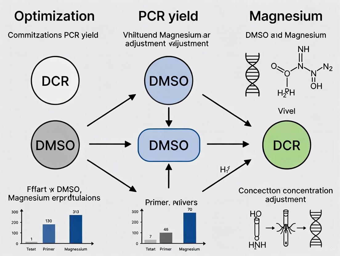
Optimizing PCR Yield: A Strategic Guide to DMSO and Magnesium Concentration
This article provides a comprehensive guide for researchers and drug development professionals on systematically enhancing Polymerase Chain Reaction (PCR) yield and specificity.
Optimizing PCR Yield: A Strategic Guide to DMSO and Magnesium Concentration
Abstract
This article provides a comprehensive guide for researchers and drug development professionals on systematically enhancing Polymerase Chain Reaction (PCR) yield and specificity. Focusing on the critical adjustment of Dimethyl Sulfoxide (DMSO) and magnesium ion (Mg²⁺) concentrations, the content spans from foundational principles and mechanistic insights to advanced methodological protocols, troubleshooting strategies, and validation techniques. Readers will gain actionable knowledge to overcome common amplification challenges, particularly with difficult templates like GC-rich sequences, enabling robust and reproducible results for sensitive downstream applications in biomedical and clinical research.
The Science Behind DMSO and Magnesium in PCR Amplification
Understanding the Fundamental Role of Magnesium as an Essential Cofactor
The Biochemical Mechanism of Magnesium in PCR
Magnesium ions (Mg²⁺) are indispensable for a successful Polymerase Chain Reaction (PCR), primarily functioning as a critical cofactor for DNA polymerase enzymes. As a cofactor, Mg²⁺ is a non-protein chemical compound that the enzyme requires for its catalytic activity. Without it, DNA polymerases like Taq are virtually inactive, unable to initiate the synthesis of new DNA strands [1].
The ion executes two primary biochemical functions:
- Enhancing DNA Polymerase Activity: At the molecular level, the Mg²⁺ ion binds to a dNTP at its alpha phosphate group. This binding facilitates the removal of the beta and gamma phosphates, allowing the resulting dNMP (deoxynucleoside monophosphate) to form a phosphodiester bond with the 3' hydroxyl (OH) group of the preceding nucleotide in the growing DNA chain. This process is the fundamental reaction of DNA elongation [1].
- Facilitating Primer Binding and Template Stability: Mg²⁺ influences the thermodynamics of DNA by binding to the negatively charged phosphate ions along the DNA backbone. This binding neutralizes the electrostatic repulsion between the negatively charged strands, stabilizing the double-stranded structure and facilitating the proper annealing of primers to their complementary sequences on the template DNA. This action also increases the melting temperature (Tm) of the DNA, making the primer-template hybrid more stable [2] [1].
Diagram: The Dual Role of Mg²⁺ in PCR. The diagram illustrates magnesium's critical functions as a polymerase cofactor and a DNA structure stabilizer.
Troubleshooting Guide: Magnesium-Related PCR Issues
The following table outlines common PCR problems stemming from incorrect magnesium concentration, their causes, and recommended solutions.
| Problem | Possible Causes Related to Mg²⁺ | Recommended Solutions |
|---|---|---|
| No or Weak Amplification [3] [1] [4] | Insufficient free Mg²⁺ concentration; Co-purified EDTA chelating Mg²⁺; High dNTP concentrations consuming free Mg²⁺. | - Titrate MgCl₂ concentration upward in 0.5 mM increments [5] [3]. - Re-purify DNA template to remove EDTA or other chelators [3]. - Ensure balanced, equimolar dNTP concentrations [2] [3]. |
| Nonspecific Amplification / Multiple Bands [2] [3] [4] | Excessive Mg²⁺ concentration reduces reaction stringency and promotes mispriming. | - Titrate MgCl₂ concentration downward [3]. - Use a hot-start DNA polymerase [6] [3]. - Increase the annealing temperature [6] [3]. |
| Low Fidelity (High Error Rate) [3] | Excess Mg²⁺ concentration can reduce polymerase fidelity and increase misincorporation of nucleotides. | - Optimize and lower Mg²⁺ concentration to the minimum effective level [3]. - Use a high-fidelity, proofreading DNA polymerase [6]. - Reduce PCR cycle number [3]. |
| Primer-Dimer Formation [1] [4] | High Mg²⁺ concentrations can facilitate non-specific priming and primer-dimer artifacts. | - Lower Mg²⁺ concentration [3]. - Optimize primer concentrations (typically 0.1-1 µM) to avoid excess [2] [3]. - Improve primer design to avoid 3'-end complementarity [2] [7]. |
Magnesium Optimization and Interaction with Other Components
Quantitative Guidelines for Magnesium Concentration
A 2025 meta-analysis of 61 studies provided evidence-based guidelines for MgCl₂ optimization, highlighting optimal ranges and effects on reaction thermodynamics [5] [8].
| Parameter | Optimal Range or Quantitative Effect | Notes and Context |
|---|---|---|
| General Optimal Range [5] | 1.5 mM to 3.0 mM | Found to be optimal for efficient PCR performance across multiple studies. |
| Standard Starting Point [1] | ~2.0 mM | A commonly used initial concentration for standard PCR. |
| Effect on Melting Temp (Tm) [5] | +1.2 °C per 0.5 mM MgCl₂ | Within the 1.5-3.0 mM range; a logarithmic relationship. |
| Template-Specific Needs [5] | Genomic DNA > Plasmid DNA | Template complexity influences requirements; genomic DNA often needs higher concentrations. |
| Maximum Typical Concentration [4] | Up to 4.5 mM | May be required for challenging templates like those with high GC content. |
Interaction with Critical Reaction Components
Magnesium concentration does not act in isolation; its availability and optimal level are influenced by other components in the PCR mix.
- dNTPs: Mg²⁺ ions bind to dNTPs to form the actual substrate for the DNA polymerase. The recommended final concentration of each dNTP is typically 0.2 mM [2]. Because dNTPs chelate Mg²⁺, a high dNTP concentration can significantly reduce the amount of free Mg²⁺ available for the polymerase, effectively inhibiting the reaction. If dNTP concentrations are increased, the Mg²⁺ concentration often needs to be increased proportionally [3].
- PCR Enhancers and Additives: Common additives like DMSO are used to help amplify difficult templates such as GC-rich sequences [6] [9]. These additives can alter DNA structure and stability, which can, in turn, affect the optimal Mg²⁺ requirement. When using such additives, re-optimization of Mg²⁺ may be necessary [3].
- Potassium Chloride (KCl): The salt KCl is another component that stabilizes primer-template binding. Its interaction with Mg²⁺ is complex, and its concentration (typically 50 mM) can influence the overall stringency of the reaction [9] [7].
Experimental Protocol: Optimizing MgCl₂ Concentration
Reagent Solutions for Optimization Experiments
| Reagent | Function in the Experiment | Typical Stock Concentration | Notes |
|---|---|---|---|
| MgCl₂ Solution | The variable being optimized; essential cofactor. | 25 mM or 50 mM | Supplied separately with many PCR buffers for optimization [9]. |
| DNA Polymerase & Buffer | Catalyzes DNA synthesis; buffer provides pH and salt conditions. | 10X concentration | Use the matching buffer provided with the enzyme. Note if it already contains Mg²⁺. |
| dNTP Mix | Building blocks for new DNA strands. | 10 mM total (2.5 mM each) | Use equimolar concentrations of all four dNTPs [2] [7]. |
| Template DNA | The DNA containing the target sequence to be amplified. | Varies (e.g., 10-100 ng/µL) | Use a consistent, high-quality template amount across reactions [2]. |
| Primers (Forward & Reverse) | Short oligonucleotides that define the sequence to be amplified. | 10-20 µM | Design with Tms of 55-70°C and avoid self-complementarity [2] [6]. |
Step-by-Step Titration Methodology
This protocol provides a systematic approach to determining the optimal MgCl₂ concentration for a specific PCR assay.
- Prepare a Master Mix: Create a master mix containing all the common reagents for the number of reactions you are running (n), plus one extra to account for pipetting error. This includes sterile water, PCR buffer (without Mg²⁺), dNTPs, primers, template DNA, and DNA polymerase [7].
- Aliquot the Master Mix: Dispense equal volumes of the master mix into thin-walled PCR tubes.
- Spike with MgCl₂: Add a different volume of MgCl₂ stock solution to each tube to create a concentration gradient. A recommended range is from 1.0 mM to 4.0 mM in increments of 0.5 mM [5] [3] [7].
- Run the PCR: Place the tubes in a thermal cycler and run the PCR with the appropriate cycling conditions for your primers and template.
- Analyze the Results: Analyze the PCR products using agarose gel electrophoresis. The optimal condition is the lowest Mg²⁺ concentration that produces a strong, specific band of the expected size with minimal to no non-specific background [3].
Diagram: Mg²⁺ Optimization Workflow. This flowchart outlines the step-by-step process for empirically determining the ideal magnesium concentration.
Frequently Asked Questions (FAQs)
Q1: Why is magnesium concentration so critical for PCR success?
Magnesium is an essential cofactor for DNA polymerase enzyme activity. It is directly involved in the catalytic mechanism of DNA synthesis and stabilizes the interaction between primers and the template DNA. Too little Mg²⁺ results in no amplification, while too much promotes non-specific binding and errors, making its precise concentration vital [2] [1].
Q2: What is a good starting point for Mg²⁺ concentration?
A final concentration of 2.0 mM MgCl₂ is a common and effective starting point for many standard PCR reactions [1]. However, a meta-analysis suggests an optimal range of 1.5 to 3.0 mM for many templates [5] [8].
Q3: How does Mg²⁺ interact with DMSO in PCR?
DMSO is an additive that helps denature stable secondary structures in GC-rich templates. It alters the DNA structure, which can affect how much Mg²⁺ is needed for optimal primer binding and polymerase activity. When adding DMSO, it is often necessary to re-optimize the Mg²⁺ concentration, as the interaction can change the reaction's stringency [6] [3] [9].
Q4: My PCR has multiple bands. Should I increase or decrease Mg²⁺?
Decrease Mg²⁺. Multiple bands indicate non-specific amplification, which is often caused by excessive Mg²⁺ concentration that reduces reaction stringency and allows primers to bind to incorrect sites. Titrating Mg²⁺ downward in 0.5 mM increments can help eliminate these spurious bands [3] [4].
Q5: How does template type affect the required Mg²⁺ concentration?
Template complexity matters. Genomic DNA, with its high complexity and potential for co-purified inhibitors, often requires a higher Mg²⁺ concentration (e.g., at the upper end of the 1.5-3.0 mM range) compared to simpler templates like plasmid or lambda DNA [2] [5].
How DMSO Modifies DNA Melting Temperature and Disrupts Secondary Structures
FAQs and Troubleshooting Guides
Q1: How does DMSO improve PCR amplification of GC-rich templates?
DMSO (Dimethyl sulfoxide) enhances PCR amplification of GC-rich DNA primarily by reducing the DNA's melting temperature (Tm) and disrupting stable secondary structures [10] [11]. GC-rich sequences form strong hydrogen bonds and stable secondary structures like hairpins due to three hydrogen bonds between G and C bases, compared to two between A and T [12]. These structures can cause polymerase extension to terminate prematurely, resulting in truncated amplicons [12] [13]. DMSO interferes with hydrogen bonding and base stacking interactions within the DNA helix [14] [10]. This action facilitates strand separation at lower temperatures, allowing primers to access their binding sites and polymerases to extend without being blocked by stubborn secondary structures [15] [10].
Q2: What is the effective concentration range for DMSO in PCR?
The effective concentration of DMSO typically ranges from 2% to 10% (v/v) [10]. However, most protocols use a narrower range of 2.5% to 5% [13] [16]. The effect is concentration-dependent. A recent biophysical study found that DMSO concentrations up to 20% moderately and linearly decrease DNA's bending persistence length (a measure of flexibility) and compact DNA conformations [14]. However, high DMSO concentrations can inhibit Taq DNA polymerase activity [10]. It is crucial to titrate DMSO for each specific reaction, as the optimal concentration balances the benefits of secondary structure disruption against potential enzyme inhibition [10].
Q3: How does DMSO interact with magnesium chloride in PCR optimization?
DMSO and magnesium chloride (MgCl₂) are two critical, independent variables that must be optimized together for GC-rich PCR. Mg²⁺ is an essential cofactor for DNA polymerase, stabilizing the enzyme and its interaction with the DNA template [10] [2]. The optimal MgCl₂ concentration often falls between 1.5 mM and 2.0 mM for GC-rich targets, but this can vary [16]. When adding DMSO, re-optimization of Mg²⁺ is recommended because DMSO can alter the DNA structure and potentially affect the reaction environment. The goal is to find a combination that provides sufficient Mg²⁺ for polymerase activity while DMSO keeps the GC-rich template accessible.
Q4: What other PCR additives can help with difficult templates?
Several additives can aid in amplifying difficult templates, often with slightly different mechanisms:
- Betaine: An isostabilizing agent that equilibrates the melting temperature between AT and GC base pairs, reducing the formation of secondary structures [12] [11]. It is often used at a concentration of 1-1.7 M [10].
- Formamide: Disrupts hydrogen bonding and weakens base pairing, which can help denature stable DNA structures. It is typically used at 1.25-10% [15].
- Non-ionic detergents (e.g., Tween 20, Triton X-100): Can stabilize DNA polymerases and help prevent secondary structure formation [15]. They are commonly used at concentrations of 0.1-1% [10].
Q5: What are the non-PCR experimental effects of DMSO on DNA structure?
Beyond PCR, studies show that DMSO has measurable effects on DNA mechanics and conformation even at low concentrations. Single-molecule and spectroscopic studies have revealed that DMSO can induce changes in DNA topology and form. One study using FT-IR spectroscopy on cells treated with low-dose DMSO (0.1-1.5%) suggested the formation of Z-DNA, an alternative left-handed DNA conformation, indicating that DMSO can significantly alter DNA topology [17]. Furthermore, magnetic tweezers experiments demonstrate that DMSO linearly reduces the bending persistence length of DNA and causes a compaction of its overall conformation [14]. These findings are critical for any experimental system where DMSO is present, as it may influence DNA-protein interactions and overall chromatin architecture.
Table 1: Effects of DMSO Concentration on DNA Physical Properties [14]
| DMSO Concentration (%) | Effect on Bending Persistence Length | Effect on Helical Twist | Effect on Mean-Squared End-to-End Distance |
|---|---|---|---|
| Up to 20% | Linear decrease | Largely unchanged | Linear decrease |
| >20% to 60% | - | Slight unwinding | - |
| Rate of Change | (0.43 ± 0.02%) per %-DMSO | - | 1.2% per %-DMSO |
Table 2: Optimized PCR Protocol Components for GC-Rich Templates
| Component | Standard Recommendation | Optimization for GC-Rich Targets | Key References |
|---|---|---|---|
| DMSO | 0% | 2.5% - 5% (v/v) | [13] [16] |
| MgCl₂ | 1.5 mM (varies) | 1.5 mM - 2.0 mM (requires titration) | [16] |
| Annealing Temperature | Calculated Tm of primers | Often 5-7°C higher than calculated Tm | [16] |
| DNA Template | Varies by source | Higher concentration may be needed (e.g., ≥2 µg/ml) | [16] |
| Denaturation Temperature | 94-95°C | 98°C for more complete denaturation | [13] |
Experimental Protocols
Protocol 1: Systematic Optimization of DMSO and MgCl₂ for GC-Rich PCR
This protocol is adapted from a study that successfully amplified an extremely GC-rich (75.45%) EGFR promoter region [16].
Reaction Setup:
- Prepare a master mix containing 1X PCR buffer, 0.2 µM of each primer, 0.25 mM of each dNTP, and 0.625 U of Taq DNA polymerase.
- Aliquot the master mix into separate tubes.
- Create a matrix of reactions with MgCl₂ concentrations spanning 0.5 mM to 2.5 mM (e.g., 0.5, 1.0, 1.5, 2.0, 2.5 mM).
- To each MgCl₂ level, add different DMSO concentrations (e.g., 0%, 1%, 3%, 5%).
- Add a consistent amount of template DNA (at least 2 µg/ml for difficult genomic DNA) and nuclease-free water to a final volume of 25 µL.
Thermal Cycling:
- Initial Denaturation: 94°C for 3 minutes.
- Amplification (45 cycles):
- Denaturation: 94°C for 30 seconds.
- Annealing: Test a gradient (e.g., 61°C, 63°C, 65°C, 67°C, 69°C) for 20 seconds. The study found 63°C optimal, which was 7°C higher than the calculated Tm [16].
- Extension: 72°C for 60 seconds.
- Final Extension: 72°C for 7 minutes.
Analysis:
- Analyze 5-10 µL of each PCR product by gel electrophoresis (e.g., 2% agarose).
- Identify the condition that yields the strongest, most specific band of the expected size with the least non-specific amplification or primer-dimer. The cited study found 5% DMSO and 1.5 mM MgCl₂ to be optimal [16].
Protocol 2: Assessing DMSO-Induced DNA Conformational Changes via AFM Imaging
This methodology summarizes the single-molecule approach used to quantify DMSO's effect on DNA conformation [14].
Sample Preparation:
- Dilute DNA (e.g., lambda DNA) in a buffer containing a range of DMSO concentrations (0% to 60%).
- Incubate the samples to allow equilibrium conformation to be reached.
AFM Imaging:
- Deposit a small volume of the DNA-DMSO solution onto a freshly cleaved mica surface.
- After adsorption, rinse the surface gently with deionized water and dry under a gentle stream of nitrogen gas.
- Image the samples using an Atomic Force Microscope (AFM) in tapping mode in air.
Data Analysis:
- Measure the end-to-end distance and the contour length of multiple individual DNA molecules from the AFM images.
- Calculate the mean-squared end-to-end distance for each DMSO condition.
- The study observed a systematic decrease in the mean-squared end-to-end distance by 1.2% per %-DMSO, indicating a compaction of DNA conformation [14].
Experimental Workflow and Mechanism
The Scientist's Toolkit: Research Reagent Solutions
Table 3: Essential Reagents for DMSO and Mg²⁺ PCR Optimization Studies
| Reagent/Material | Function/Description | Example Use Case |
|---|---|---|
| DMSO (Dimethyl Sulfoxide) | A polar aprotic solvent that disrupts DNA secondary structures by interfering with hydrogen bonding, thereby lowering the melting temperature (Tm). | Added at 2.5-5% (v/v) to PCR mixes to facilitate amplification of GC-rich templates [15] [13] [16]. |
| Magnesium Chloride (MgCl₂) | An essential cofactor for thermostable DNA polymerases; stabilizes the enzyme-DNA-dNTP complex. Concentration critically affects specificity and yield. | Titrated between 1.0-4.0 mM to find the optimal concentration for a specific PCR reaction, often around 1.5-2.0 mM for GC-rich targets [16] [2]. |
| High-Fidelity DNA Polymerase | Engineered polymerases, often with proofreading (3'→5' exonuclease) activity, capable of efficiently elongating through complex secondary structures. | Preferred over standard Taq for amplifying long or GC-rich targets due to higher processivity and resistance to stalling [15] [11]. |
| Betaine | An isostabilizing agent that reduces the differential in Tm between AT and GC base pairs, helping to denature GC-rich regions. | Used as an alternative or supplement to DMSO at 1-1.7 M concentration for GC-rich PCR [12] [10]. |
| dNTP Mix | The building blocks (dATP, dCTP, dGTP, dTTP) for new DNA strand synthesis. | Used at equimolar concentrations, typically 0.2 mM each, to ensure balanced incorporation and maintain polymerase fidelity [2]. |
The Interplay Between Mg²⁺ Concentration, Enzyme Fidelity, and Specificity
FAQs: Magnesium in PCR
What is the fundamental role of Mg²⁺ in PCR? Magnesium ions (Mg²⁺) are an essential cofactor for all thermostable DNA polymerases [15] [6] [18]. They are directly involved in the catalytic mechanism of DNA synthesis and are critical for enzyme activity. Without adequate free Mg²⁺, DNA polymerases are inactive, leading to PCR failure [18]. Specifically, Mg²⁺ facilitates the nucleophilic attack of the 3'-OH group of the primer on the alpha-phosphate of the incoming dNTP and helps stabilize the negative charge on the triphosphate leaving group [19].
How does Mg²⁺ concentration affect amplification yield and specificity? The concentration of Mg²⁺ must be carefully optimized, as it has a direct and significant impact on both PCR yield and specificity [6] [3].
- Low Mg²⁺ Concentration: Results in reduced enzyme activity, leading to low or no amplification yield [6] [18].
- High Mg²⁺ Concentration: Promotes non-specific amplification, such as primer-dimer formation and off-target products, by reducing the stringency of primer-template binding [6] [3]. Excess Mg²⁺ can also reduce enzyme fidelity (see below) [18].
What is the relationship between Mg²⁺ concentration and enzyme fidelity? Mg²⁺ concentration is a key determinant of DNA polymerase fidelity, which is the accuracy of nucleotide incorporation [6] [3].
- High Fidelity: Lower, more physiological Mg²⁺ concentrations are generally associated with higher fidelity [20] [18]. For instance, some high-fidelity polymerases are recommended for use at a final Mg²⁺ concentration of 1 mM to maximize accuracy [18] [21].
- Low Fidelity: Excess free Mg²⁺ reduces enzyme fidelity and increases the misincorporation of nucleotides [6] [18]. This is a critical consideration for applications like cloning and sequencing, where accurate DNA replication is paramount [3].
What is the typical optimal range for Mg²⁺ concentration, and how is it determined? The optimal Mg²⁺ concentration typically falls between 1.5 and 2.5 mM for many standard PCR reactions [6]. However, the ideal concentration must be determined empirically for each primer-template system because the "free" Mg²⁺ concentration is affected by several factors in the reaction mix [18]. Key factors that chelate Mg²⁺ and reduce its availability include:
- dNTPs: These are strong chelators; higher dNTP concentrations require higher Mg²⁺ concentrations [3].
- EDTA: A potent chelator that may be carried over from DNA purification protocols [6] [3].
- Citrate: Another chelator that may be present in samples [18].
A titration experiment, often in 0.5 mM increments across a range from 0.5 mM to 5.0 mM, is the standard method for optimization [15] [6].
Troubleshooting Guide: Common Problems and Solutions Related to Mg²⁺
This guide addresses common PCR issues where adjusting Mg²⁺ concentration is a primary solution.
| Problem | Possible Causes Related to Mg²⁺ | Recommended Solutions |
|---|---|---|
| No/Low Yield [22] | Insufficient free Mg²⁺ for polymerase activity due to low concentration or chelation by dNTPs/EDTA [6] [3]. | - Titrate Mg²⁺ concentration upward in 0.5 mM increments [6].- Ensure Mg²⁺ is in excess of total dNTP concentration [3].- Re-purify DNA template to remove EDTA [6]. |
| Non-Specific Amplification (e.g., multiple bands, smearing) [22] [3] | Excessive free Mg²⁺ concentration, which stabilizes non-specific primer-template binding [6] [18]. | - Titrate Mg²⁺ concentration downward [6] [3].- Use a hot-start DNA polymerase to prevent activity at low temperatures [15] [22].- Increase the annealing temperature [3]. |
| Low Fidelity (High Error Rate) [3] | High Mg²⁺ concentration reduces base-pairing specificity, increasing misincorporation [6] [18]. | - Use a high-fidelity polymerase with proofreading (3'→5' exonuclease) activity [15] [6].- Lower the Mg²⁺ concentration to the optimal range for your enzyme [18] [21].- Ensure dNTP concentrations are balanced and not in excess [3]. |
| Primer-Dimer Formation [22] | High Mg²⁺ and/or high primer concentration promotes primer self-annealing [22] [6]. | - Optimize (lower) Mg²⁺ concentration [6].- Reduce primer concentration [22] [3].- Improve primer design to avoid 3'-end complementarity [15] [6]. |
Experimental Protocols
Protocol 1: Mg²⁺ Titration for Reaction Optimization
Purpose: To empirically determine the optimal Mg²⁺ concentration for maximum yield and specificity of a given PCR assay [6].
Materials:
- Template DNA
- Forward and Reverse Primers
- 10X PCR Buffer (without Mg²⁺)
- 25 mM or 50 mM MgCl₂ stock solution
- dNTP Mix
- DNA Polymerase
- Nuclease-free Water
Method:
- Prepare a master mix containing all PCR components except the MgCl₂ and template DNA.
- Aliquot the master mix into multiple PCR tubes.
- Add MgCl₂ to each tube to create a series of final concentrations. A standard range is 0.5, 1.0, 1.5, 2.0, 2.5, 3.0, 3.5, 4.0, and 5.0 mM.
- Add the template DNA to each tube and mix thoroughly.
- Run the PCR using cycling conditions appropriate for your primer pair and polymerase.
- Analyze the results using agarose gel electrophoresis. The condition that produces the strongest, cleanest band of the expected size with the least background or non-specific products indicates the optimal Mg²⁺ concentration [6].
Protocol 2: Assessing Fidelity Using a lacZα-based Complementation Assay
Purpose: To quantitatively measure the mutation rate and fidelity of a DNA polymerase under different Mg²⁺ conditions [20].
Background: This assay measures the loss of function of the lacZα gene due to mutations introduced during DNA synthesis. The number of white (mutant) versus blue (functional) colonies allows for the calculation of error frequency [20].
Materials:
- RNA or DNA template encoding the lacZα peptide
- Reverse Transcriptase or DNA Polymerase of interest
- Reaction Buffer (with variable Mg²⁺)
- dNTPs
- Primers for lacZα synthesis
- E. coli competent cells for transformation
- X-gal/IPTG containing agar plates
Method:
- DNA Synthesis: Perform the nucleic acid synthesis reaction (e.g., RNA-templated cDNA synthesis for RTs) in two different conditions: Low Mg²⁺ (0.5 mM) and High Mg²⁺ (6 mM). Use otherwise identical buffer, dNTP, and enzyme concentrations [20].
- Product Purification: Purify the synthesized DNA product.
- Cloning: Clone the products into a suitable vector and transform into an E. coli host strain.
- Screening: Plate transformed cells on media containing X-gal and IPTG. Incubate to allow colony formation.
- Analysis: Count the total colonies and the number of white (mutant) colonies. The mutation frequency is calculated as (Number of white colonies) / (Total number of colonies). A lower mutation frequency in the 0.5 mM Mg²⁺ condition demonstrates higher fidelity, as was shown for HIV-1 RT [20].
Research Reagent Solutions
This table details key reagents essential for investigating and optimizing Mg²⁺ interactions in enzymatic reactions.
| Reagent / Material | Function in Research |
|---|---|
| MgCl₂ or MgSO₄ Stock Solutions | Provides the divalent cation cofactor. The choice of salt and its concentration is the primary variable for fidelity and specificity studies [6] [3]. |
| High-Fidelity DNA Polymerases (e.g., Pfu, KOD) | Engineered enzymes with 3'→5' exonuclease (proofreading) activity. Essential for high-accuracy applications and for studying the limits of fidelity optimization with Mg²⁺ [15] [6]. |
| Hot-Start Polymerases | Inactive until a high-temperature activation step. Prevents non-specific amplification and primer-dimer formation during reaction setup, providing a clearer baseline for Mg²⁺ optimization [15] [3]. |
| dNTP Mix | The building blocks of DNA. Concentrations must be balanced and optimized, as dNTPs chelate Mg²⁺, directly affecting the free Mg²⁺ available to the polymerase [6] [3]. |
| PCR Additives (DMSO, Betaine) | Assist in amplifying complex templates (e.g., GC-rich sequences). They can alter DNA melting behavior and may interact with or change the optimal Mg²⁺ concentration, requiring re-optimization [15] [6]. |
| Chelators (EDTA, Citrate) | Used to control or scavenge free metal ions. Understanding their presence is critical for accurately calculating and maintaining desired free Mg²⁺ concentrations [6] [18]. |
Conceptual Diagrams
Mg2+ Impact on PCR Outcomes
Two-Metal-Ion Catalytic Mechanism
FAQs and Troubleshooting Guides
Why is my PCR producing multiple bands or a smear on the gel?
This is a common sign of non-specific amplification, where your primers are binding to incorrect sites on the DNA template.
| Possible Cause | Recommended Solution |
|---|---|
| Annealing temperature too low | Increase the annealing temperature in 1-2°C increments. Use a gradient thermal cycler to find the optimal temperature. [3] [24] |
| Poor primer design | Verify primers are specific and lack self-complementarity (which can cause hairpins) or complementarity to each other (which causes primer-dimers). [3] [2] |
| Excess Mg²⁺ concentration | Optimize Mg²⁺ concentration by testing in 0.2-1.0 mM increments. High Mg²⁺ can reduce fidelity and promote non-specific binding. [3] [24] [25] |
| High primer concentration | Optimize primer concentration, typically between 0.1-1 μM. High concentrations promote mispriming and primer-dimer formation. [3] [2] |
| Enzyme activity at low temp | Use a hot-start DNA polymerase to prevent enzyme activity during reaction setup, thereby eliminating premature replication. [3] [22] [24] |
| Too many cycles | Reduce the number of PCR cycles to prevent the accumulation of non-specific products in later cycles. [3] |
How can I successfully amplify GC-rich templates?
GC-rich sequences (≥60% GC content) are challenging due to their high thermal stability and tendency to form secondary structures like hairpins [26] [27].
| Strategy | Protocol & Application Notes |
|---|---|
| Polymerase Choice | Use polymerases specifically engineered for GC-rich templates (e.g., Q5 High-Fidelity, OneTaq with GC Buffer). These often come with a proprietary GC Enhancer. [26] [24] |
| PCR Additives | Add co-solvents like DMSO (1-10%), glycerol, or betaine (0.8-1.3 M) to help denature stable secondary structures. [26] [28] [27] |
| Denaturation Temperature | Use a higher denaturation temperature (e.g., 98°C) for the first few cycles. Avoid temperatures above 95°C for extended periods to preserve enzyme activity. [27] [25] |
| Mg²⁺ Concentration | Test a gradient of MgCl₂, typically between 1.0-4.0 mM, to find the optimal concentration for your specific template. [26] |
| Annealing Temperature | Use primers with a higher Tm (>68°C) and perform a touchdown PCR, starting with a higher annealing temperature to increase specificity. [25] |
What should I do if I get no PCR product or a very low yield?
A lack of product often points to issues with reaction components or cycling conditions.
| Area to Investigate | Action Plan |
|---|---|
| Template DNA | Check quality (degradation, purity from inhibitors like phenol) and quantity. For genomic DNA, use 5-50 ng; for plasmid, 0.1-1 ng is often sufficient. [3] [2] |
| Primers | Confirm primer design, specificity, and concentration. Ensure they are resuspended and stored correctly. [3] [24] |
| Thermal Cycling | Verify denaturation is efficient. Increase the number of cycles (e.g., up to 40) if the template copy number is very low. [3] |
| Mg²⁺ & dNTPs | Ensure sufficient Mg²⁺ is available (dNTPs chelate Mg²⁺). Use fresh, balanced dNTP mixtures. [3] [2] [24] |
| PCR Enzyme | Confirm the polymerase is active and added in the correct amount. Increase the amount if inhibitors are suspected. [3] [2] |
How do I prevent primer-dimer formation?
Primer-dimers are short, artifactual products visible as a band near 50 bp on a gel. They form when primers anneal to each other [23].
- Key Prevention Methods:
- Optimize Primer Design: Avoid 3'-end complementarity between primers and keep the GC content at 40-60% [2] [22].
- Lower Primer Concentration: Test concentrations in the range of 0.1–0.5 μM [3] [23].
- Use Hot-Start Polymerases: This prevents enzymatic activity at low temperatures during setup, a common time for primer-dimer formation [22] [24].
- Increase Annealing Temperature: A higher temperature reduces the chance of primers loosely binding to each other [22].
The Scientist's Toolkit: Research Reagent Solutions
The following reagents are essential for troubleshooting and optimizing challenging PCRs.
| Reagent | Function in PCR Optimization |
|---|---|
| Hot-Start DNA Polymerase | Polymerase that is inactive at room temperature, preventing non-specific amplification and primer-dimer formation during reaction setup. [3] [22] |
| Specialized Polymerase Mixes | Polymerases like Q5 or OneTaq are engineered for high fidelity, long amplification, or efficient amplification of GC-rich templates. [26] [24] |
| DMSO (Dimethyl Sulfoxide) | A common additive that helps denature GC-rich DNA templates by disrupting base pairing, reducing secondary structure formation. [26] [28] [27] |
| Betaine | An additive (used at 0.8-1.3 M) that equalizes the stability of AT and GC base pairs, aiding in the amplification of GC-rich regions and reducing secondary structures. [28] |
| GC Enhancer | Proprietary buffer supplements (e.g., from NEB) that contain a mix of agents to improve yield and specificity for GC-rich targets. [26] |
| MgCl₂ Solution | A separate, optimized source of magnesium ions, a crucial cofactor for DNA polymerase activity. Its concentration is a key variable for optimization. [3] [25] |
| dNTP Mix | The building blocks for new DNA strands. Use a balanced, high-quality mixture to prevent incorporation errors. [3] [2] |
Experimental Protocols for Key Optimizations
Protocol 1: Systematic Optimization of Mg²⁺ and DMSO Concentrations
This protocol is central to a thesis focused on improving PCR yield through adjustment of these key reagents [26] [24].
- Prepare Master Mix: Create a master mix containing all standard PCR components (buffer, dNTPs, primers, template, polymerase) but omit Mg²⁺ and DMSO.
- Set Up Gradient Reactions: Aliquot the master mix into multiple tubes.
- For Mg²⁺ titration, add MgCl₂ to achieve a final concentration gradient (e.g., 1.0, 1.5, 2.0, 2.5, 3.0, 3.5, 4.0 mM).
- For DMSO titration, add DMSO to achieve a final concentration gradient (e.g., 0%, 2%, 4%, 6%, 8%, 10%).
- Run PCR: Use optimized thermal cycling conditions with an annealing temperature gradient if possible.
- Analyze Results: Evaluate amplification yield and specificity via agarose gel electrophoresis. Identify the Mg²⁺ and DMSO concentrations that produce the highest yield of the specific product with minimal background.
Protocol 2: Amplification of a GC-Rich Template
This detailed methodology is adapted from recommended practices for challenging GC-rich targets [26] [25].
- Select a Specialized Polymerase: Choose a polymerase known for high processivity and GC-rich amplification, such as Q5 High-Fidelity DNA Polymerase or OneTaq DNA Polymerase with GC Buffer.
- Prepare the Reaction Mixture:
- DNA Template: 10-50 ng of high-quality genomic DNA or equivalent.
- Primers: 0.5 μM each, designed with Tms above 68°C.
- PCR Buffer: Use the manufacturer's recommended buffer.
- Additives: Include 1X GC Enhancer (if supplied) and 5% DMSO.
- Mg²⁺: Start with the manufacturer's recommended concentration (often 1.5-2.0 mM).
- Polymerase: 1-2 units per 50 μL reaction.
- Thermal Cycling Conditions:
- Initial Denaturation: 98°C for 30 seconds.
- Amplification (35 cycles):
- Denaturation: 98°C for 10 seconds.
- Annealing: 72°C for 20 seconds (for a two-step protocol if primer Tms allow) or 5°C above the calculated Tm for a three-step protocol.
- Extension: 68°C for 30 seconds per kb.
- Final Extension: 72°C for 2 minutes.
Troubleshooting PCR Workflows
The following diagrams outline logical workflows for diagnosing and resolving common PCR issues.
Step-by-Step Protocols for DMSO and Magnesium Titration
Core Components of a Standard PCR Protocol
This section details the essential reagents and their standard concentrations for a foundational PCR protocol, designed to be robust and reproducible without additives.
Table 1: Standard Reaction Components for a 50 µL PCR
| Component | Final Concentration/Amount | Purpose & Notes |
|---|---|---|
| Template DNA | 1 pg – 100 ng [29] | Amount depends on complexity [30]. |
| Forward & Reverse Primers | 0.1 – 1 µM each [3] [29] | Typically 20-30 nucleotides; Tm within 5°C of each other [29]. |
| dNTP Mix | 200 µM of each dNTP [29] | Use balanced, equimolar concentrations [3]. |
| PCR Buffer (10X) | 1X | Typically supplied with Taq polymerase; may contain MgCl₂ [7]. |
| Magnesium Chloride (MgCl₂) | 1.5 – 2.0 mM [29] | Critical cofactor; optimize if not pre-included in buffer [3] [31]. |
| Taq DNA Polymerase | 0.5 – 2.5 units per 50 µL reaction [7] [29] | Follow manufacturer's specific recommendation [29]. |
| Nuclease-Free Water | To volume | Brings the total reaction volume to 50 µL. |
Standard Thermal Cycling Conditions
The following protocol provides a reliable starting point for amplifying a typical 0.5 - 2 kb fragment from a genomic DNA template [29].
Table 2: Standard Thermal Cycling Protocol
| Step | Temperature | Duration | Notes |
|---|---|---|---|
| Initial Denaturation | 95°C | 2 minutes | Ensures complete denaturation of complex templates [29]. |
| Denaturation | 95°C | 15 – 30 seconds | |
| Annealing | 50–60°C (5°C below primer Tm) | 15 – 30 seconds | Critical for specificity; optimize using a gradient cycler [3] [29]. |
| Extension | 68°C | 1 minute per kb | For products <1 kb, 45-60 seconds is sufficient [29]. |
| Final Extension | 68°C | 5 minutes | Ensures all amplicons are fully extended [3]. |
| Hold | 4–10°C | ∞ |
The workflow for setting up this baseline PCR experiment is outlined below.
FAQs on the Baseline Protocol
What is the primary purpose of establishing a baseline protocol?
A baseline protocol without additives provides a controlled starting point for your experiments. Once this standard reaction is working reliably, it becomes a benchmark. You can then systematically introduce variables like DMSO or adjust magnesium levels to troubleshoot specific issues (like GC-rich templates) and accurately measure their impact on yield and specificity [3] [32].
My baseline PCR shows no product. What should I check first?
Begin troubleshooting with the most common culprits [3]:
- Template Quality and Quantity: Ensure your DNA is intact and free of inhibitors like phenol or EDTA. Verify the amount used falls within the recommended range [3] [29].
- Mg²⁺ Concentration: If your PCR buffer does not contain MgCl₂, it must be added separately. Test a range from 1.0 to 4.0 mM in 0.5 mM increments [29] [31].
- Annealing Temperature: This is a frequent source of failure. Use a gradient thermal cycler to test temperatures 5°C above and below the calculated Tm of your primers [3] [6].
I get nonspecific amplification (multiple bands) with the baseline protocol. How can I improve specificity?
- Increase Annealing Temperature: Raise the temperature in 1-2°C increments to increase stringency [3].
- Optimize Primer Concentration: High primer concentrations can promote off-target binding. Titrate primer concentrations down, starting from 1 µM [3].
- Reduce Cycle Number: A high number of cycles can accumulate nonspecific products. Try reducing to 25-30 cycles [3].
- Switch to a Hot-Start Taq: Hot-start polymerases remain inactive until the initial denaturation step, preventing primer-dimer formation and mispriming during reaction setup [3] [6].
The Scientist's Toolkit: Key Research Reagent Solutions
Table 3: Essential Reagents for Your Baseline PCR
| Reagent | Function | Key Considerations |
|---|---|---|
| Taq DNA Polymerase | Enzyme that synthesizes new DNA strands. | Choose standard Taq for routine amplification. Select hot-start versions to minimize nonspecific amplification [6]. |
| dNTP Mix | Building blocks (A, T, C, G) for DNA synthesis. | Use equimolar concentrations to maintain polymerase fidelity. Avoid repeated freeze-thaw cycles [3] [31]. |
| Oligonucleotide Primers | Define the start and end of the target sequence. | Design primers with 40-60% GC content and a Tm of 55-65°C. Avoid self-complementarity and dimer formation [7] [29]. |
| PCR Buffer (with MgCl₂) | Provides optimal chemical environment (pH, salts) for the polymerase. | Often supplied with the enzyme. If Mg²⁺ is separate, it requires independent optimization [29] [31]. |
| Nuclease-Free Water | Solvent for the reaction. | Essential to avoid degradation of reaction components by environmental nucleases. |
Determining the Optimal DMSO Concentration (2-10%) for Your Template
Dimethyl sulfoxide (DMSO) is a pivotal additive in polymerase chain reaction (PCR) for optimizing the amplification of difficult templates. Its primary role is to disrupt the secondary structures and stabilize the DNA, which is especially beneficial for templates with high GC content. Integrating DMSO and magnesium level adjustments is a proven strategy to significantly improve PCR yield and specificity. This guide provides detailed methodologies and troubleshooting advice to help you effectively incorporate DMSO into your PCR experiments.
▣ How DMSO Enhances PCR Amplification
DMSO improves PCR results through two main mechanisms:
- Reduces DNA Melting Temperature: DMSO interacts with DNA bases, particularly cytosine, making them more heat-labile. This lowers the overall melting temperature ((T_m)) of the template, facilitating primer annealing without requiring excessively high temperatures that could damage the DNA [33].
- Prevents Secondary Structure Formation: In GC-rich templates, strong hydrogen bonds can form stable hairpin loops and other secondary structures. DMSO weakens these hydrogen bonds, prevents the reannealing of denatured DNA strands, and reduces DNA supercoiling. This ensures the template remains accessible for primer binding, thereby increasing reaction specificity and yield [34] [33].
Optimizing DMSO Concentration: A Step-by-Step Guide
The optimal concentration of DMSO is template-dependent. The following table summarizes the general starting guidelines, which should be further refined through experimental optimization.
Table 1: Recommended DMSO Concentrations for Different Template Types
| Template Characteristic | Recommended DMSO Starting Concentration | Key Considerations |
|---|---|---|
| Standard/Routine | 0% (or as included in proprietary buffer) | Standard PCR buffers may already contain small, undefined amounts of DMSO [33]. |
| Moderate GC-richness | 3% | A starting point for templates that fail under standard conditions, even with GC content around 52% [33]. |
| High GC-richness (>60%) | 5% | The most commonly cited starting concentration for GC-rich targets [34] [6] [33]. |
| Very High GC-richness/Complex | 5.5% to 10% | Requires careful optimization. Concentrations above 5% can be tested if 5% is ineffective [34] [33]. |
Experimental Protocol for DMSO Titration
To determine the ideal DMSO concentration for your specific template, perform a titration experiment.
- Prepare Reaction Master Mix: Create a master mix containing all standard PCR components (buffer, dNTPs, primers, polymerase, template) for all test reactions.
- Aliquot and Add DMSO: Aliquot the master mix into several PCR tubes. Add DMSO to each tube to create a final concentration gradient (e.g., 0%, 2%, 4%, 6%, 8%, 10%). Use molecular-grade DMSO for consistency [33].
- Run PCR: Perform amplification using your standard thermal cycling program. Note: The presence of DMSO lowers the effective annealing temperature by approximately 0.5-1.0°C per 1% DMSO. You may need to adjust the annealing temperature downward slightly for the reactions containing DMSO [6] [33].
- Analyze Results: Analyze the PCR products using gel electrophoresis. The optimal condition will be the one that produces the highest yield of the desired specific product with the least or no non-specific amplification or primer-dimer.
The workflow for this optimization process is outlined below.
Common DMSO-Related Problems and Solutions
Despite its benefits, improper use of DMSO can lead to experimental failure. Here are common issues and their solutions.
Table 2: DMSO Troubleshooting Guide
| Problem | Possible Cause | Solution |
|---|---|---|
| No Product or Low Yield | DMSO concentration too low for a difficult template. | Titrate DMSO upward in 1% increments [33]. |
| Non-Specific Bands or Smearing | DMSO concentration too high, reducing annealing stringency. | Titrate DMSO downward. Increase the annealing temperature by 1-2°C to counter the Tm-lowering effect [33]. |
| Complete PCR Failure | Excessive DMSO inhibits DNA polymerase activity. | Do not exceed 10% DMSO. Re-optimize at lower concentrations (2-6%) [33]. |
| High Error Rate (Low Fidelity) | High DMSO concentration can compromise polymerase fidelity, leading to misincorporation. | Use the minimum effective DMSO concentration. For sequencing applications, avoid DMSO if possible or use minimal amounts [33]. |
Interaction of DMSO and Magnesium in PCR
Magnesium (Mg²⁺) is an essential cofactor for DNA polymerase, and its concentration is a critical factor for PCR success. DMSO and Mg²⁺ concentrations can interact, making simultaneous optimization a powerful strategy [34] [6] [35].
- DMSO and Mg²⁺ Synergy: Both components affect DNA duplex stability. The combined influence of magnesium concentration and specificity enhancers like DMSO must be considered for optimal results [36].
- Optimization Strategy: If you are optimizing both, start by finding the optimal DMSO concentration first while keeping Mg²⁺ at a standard level (e.g., 1.5 mM). Then, perform a Mg²⁺ titration (e.g., from 0.5 mM to 3.0 mM in 0.2-0.5 mM increments) at the optimal DMSO level [35].
The Scientist's Toolkit: Essential Reagents for PCR Optimization with DMSO
Table 3: Key Research Reagent Solutions
| Reagent | Function in PCR | Consideration for Use with DMSO |
|---|---|---|
| Molecular Grade DMSO | An additive to improve yield and specificity for GC-rich templates. | Use high-purity, sterile-filtered DMSO. Aliquot to prevent contamination and oxidation [37] [38]. |
| MgCl₂ or MgSO₄ Solution | An essential cofactor for DNA polymerase activity. | Concentration is critical; titrate for optimal results, especially when used in combination with DMSO [34] [35]. |
| High-Fidelity DNA Polymerase | Enzyme with proofreading activity for high-accuracy amplification. | Check manufacturer's guidelines, as some advanced polymerases are supplied with optimized buffers that may not require additional DMSO [34] [6]. |
| Betaine | An alternative additive to DMSO that homogenizes DNA template stability. | Can be used as an alternative or in conjunction with DMSO for exceptionally difficult templates [6]. |
| GC-Rich Specific Kits | Commercial kits containing pre-optimized buffers and enzymes. | Often include DMSO or similar enhancers; a good starting point before manual optimization [34] [35]. |
FAQs on DMSO Use in PCR
Q1: Can I use DMSO with any DNA polymerase? A1: While many standard polymerases are compatible with DMSO, it is crucial to consult the manufacturer's instructions. Some specialized polymerases, such as PrimeSTAR Max, are explicitly noted to work well with DMSO, while others may be inhibited [34]. When in doubt, perform a compatibility test.
Q2: What is the maximum safe concentration of DMSO? A2: Concentrations above 10% are generally not recommended as they can significantly inhibit polymerase activity and increase non-specific binding. Most successful protocols use DMSO between 2% and 5%, with 10% representing a practical upper limit for testing [34] [33].
Q3: How does DMSO affect the primer annealing temperature? A3: DMSO lowers the melting point of DNA. As a rule of thumb, the presence of 5% DMSO can reduce the effective annealing temperature by approximately 2.5°C. You may need to lower your calculated annealing temperature when adding DMSO to your reaction [33].
Q4: When should I avoid using DMSO in PCR? A4: DMSO should be avoided or used with extreme caution when the PCR product is intended for downstream sequencing, as high concentrations can increase the error rate of the polymerase. For standard templates that amplify efficiently, DMSO is unnecessary [33].
Frequently Asked Questions (FAQs)
Q1: Why is MgCl₂ concentration so critical for a successful PCR? Mg²⁺ ions are an essential cofactor for Taq DNA polymerase. They facilitate the enzyme's catalytic activity by binding to its active site and coordinating with incoming dNTPs to enable the formation of phosphodiester bonds during DNA synthesis. The concentration directly influences primer annealing, DNA duplex stability, and the overall fidelity of the amplification [39].
Q2: What are the symptoms of a sub-optimal MgCl₂ concentration in my reaction?
- Too Low (e.g., <1.0 mM): You will typically see little to no PCR product. This is because there are insufficient Mg²⁺ ions to activate the DNA polymerase effectively, leading to poor enzyme activity and failed primer extension [40] [39].
- Too High (e.g., >4.0 mM): You will often observe non-specific amplification, such as multiple bands or smears on an agarose gel. Excess Mg²⁺ stabilizes DNA duplexes non-specifically, which can cause primers to bind to incorrect sites. It can also promote the formation of primer-dimers [40] [39].
Q3: How does the type of DNA template influence the optimal MgCl₂ concentration? The complexity and nature of your DNA template significantly impact the Mg²⁺ requirement. A recent meta-analysis confirmed that genomic DNA templates generally require higher MgCl₂ concentrations than simpler templates, such as plasmids or viral DNA. This is due to the greater complexity and potential for secondary structures in genomic DNA [8].
Q4: Can I use additives alongside MgCl₂ titration to further improve my PCR? Yes, additives like DMSO, formamide, and betaine are commonly used to enhance PCR specificity and yield, particularly for challenging templates like those with high GC content. A study found that 2 mM TMA oxalate could dramatically improve specificity and efficiency. Similarly, DMSO (1-10%) and formamide (1.25-10%) can be included in the reaction to help denature stable secondary structures [7] [41]. When titrating MgCl₂ in the presence of these additives, you may need to re-optimize, as they can interact with the reaction components.
Troubleshooting Guide: Common MgCl₂-Related Problems
| Problem Observed | Potential Cause | Recommended Solution |
|---|---|---|
| No PCR product | MgCl₂ concentration is too low. | Increase the concentration in 0.5 mM increments, testing up to 4.0 mM [40]. |
| Multiple non-specific bands or smearing | MgCl₂ concentration is too high. | Decrease the concentration in 0.5 mM increments, starting from your current level [40]. |
| Weak or faint target band | MgCl₂ concentration is sub-optimal, or cycle number is too low. | Perform a full titration (e.g., 0.5 mM to 5.0 mM). Also, consider increasing cycles to 35-40 for low-copy templates [8] [42]. |
| PCR failure with complex genomic DNA | Standard MgCl₂ concentration is insufficient for a complex template. | Titrate towards the higher end of the range (e.g., 2.0 mM to 5.0 mM), as genomic DNA often requires more Mg²⁺ [8]. |
Quantitative Data and Protocols
The following table synthesizes key quantitative relationships identified through meta-analysis and experimental studies.
| Parameter | Effect of MgCl₂ | Quantitative Relationship / Optimal Range | Notes |
|---|---|---|---|
| Optimal Concentration Range | Baseline for standard PCR | 1.5 - 3.0 mM [8] | The ideal concentration must be determined empirically for each primer-template system. |
| DNA Melting Temperature (Tm) | Increases Tm | +1.2 °C per 0.5 mM increase (within 1.5-3.0 mM range) [8] | Mg²⁺ stabilizes the DNA duplex by neutralizing the negative charge on the phosphate backbone. |
| Reaction Specificity | Highly concentration-dependent | Maximal specificity at 2.0 mM (with TMA oxalate) [41] | Specificity decreases at both lower and higher concentrations due to poor polymerization or non-specific priming. |
| Template Dependency | Varies by template type | Genomic DNA requires higher concentrations than plasmid DNA [8] | A one-size-fits-all approach does not work. |
Detailed MgCl₂ Titration Protocol
This protocol provides a methodology for empirically determining the optimal MgCl₂ concentration for your specific PCR assay.
Materials and Reagents
- Taq DNA Polymerase and its corresponding 10X PCR Buffer (often supplied without MgCl₂ or with a standard concentration like 15 mM) [7]
- 25 mM or 50 mM MgCl₂ stock solution
- dNTP Mix (e.g., 10 mM)
- Forward and Reverse Primers (e.g., 20 µM)
- DNA Template
- Nuclease-Free Water
- PCR Tubes and Thermal Cycler
Experimental Workflow:
Procedure:
- Prepare a Master Mix: Calculate the volumes needed for 8 reactions (including an extra to account for pipetting error). Combine the following components in a tube on ice [7]:
- Nuclease-Free Water (Q.S. to final volume)
- 10X PCR Buffer (1X final concentration)
- dNTP Mix (200 µM of each dNTP final)
- Forward Primer (0.1-0.5 µM final)
- Reverse Primer (0.1-0.5 µM final)
- Taq DNA Polymerase (0.5-2.5 units/50 µl reaction)
Aliquot and Titrate MgCl₂: Vortex the Master Mix to ensure homogeneity and aliquot equal volumes into 8 labeled PCR tubes. Add a different volume of your MgCl₂ stock solution to each tube to achieve the following final concentrations:
Add Template and Run PCR: Add an equal amount of your DNA template to each tube. Gently mix the reactions and briefly centrifuge to collect the contents. Transfer the tubes to a pre-heated thermal cycler and run using your standard PCR program [7].
Analyze Results: Separate the PCR products on an agarose gel. Identify the MgCl₂ concentration that produces the strongest, single band of the expected size with the least background or non-specific amplification. This is your optimal concentration.
Research Reagent Solutions
The following table lists key reagents essential for performing a MgCl₂ titration experiment.
| Reagent | Function in the Experiment | Key Considerations |
|---|---|---|
| Taq DNA Polymerase | Enzyme that synthesizes new DNA strands. | Requires Mg²⁺ as a cofactor. Hot-start versions can improve specificity by reducing primer-dimer formation [42]. |
| MgCl₂ Stock Solution | Source of Mg²⁺ ions for the reaction. | Must be of high purity and concentration accurately known. Typically used at a 25-50 mM stock concentration [7]. |
| dNTPs (dATP, dCTP, dGTP, dTTP) | The building blocks for DNA synthesis. | Final concentration is typically 200 µM of each dNTP. Higher concentrations can chelate Mg²⁺, effectively reducing its availability [40]. |
| PCR Buffer | Provides the optimal chemical environment (pH, ionic strength). | Often supplied with the polymerase. May or may not contain MgCl₂; check the formulation as this is the starting point for your titration [7]. |
| Primers | Short DNA sequences that define the start and end of the amplified region. | Should be well-designed (Tm within 5°C of each other, 40-60% GC content). Final concentration of 0.1-0.5 µM is typical [7] [40]. |
| PCR Enhancers (e.g., DMSO) | Additives that can help amplify difficult templates. | DMSO (1-10%) can destabilize DNA secondary structures. If used, the optimal MgCl₂ concentration may shift and require re-titration [7] [41]. |
FAQs and Troubleshooting Guides
Q1: What are the specific roles of DMSO and Mg²⁺ in PCR, and why are they considered synergistic?
A: DMSO and Mg²⁺ are two of the most critical additives for optimizing PCR, particularly for difficult templates. They function through distinct but complementary mechanisms:
Mg²⁺ (as MgCl₂): This is an essential cofactor for thermostable DNA polymerases [1] [43]. It directly activates the enzyme, facilitating the formation of phosphodiester bonds between nucleotides during DNA synthesis [2]. Additionally, Mg²⁺ stabilizes the interaction between the primer and the DNA template by neutralizing the negative charges on their phosphate backbones, which promotes proper annealing [1] [2].
DMSO (Dimethyl Sulfoxide): This agent acts as a secondary structure disruptor [44]. It is particularly beneficial for amplifying GC-rich templates (>65% GC), as it interferes with the formation of stable hydrogen bonds in GC base pairs, thereby reducing the stability of secondary structures like hairpins and G-quadruplexes that can block polymerase progression [43] [44].
The synergy arises because Mg²⁺ ensures the DNA polymerase is maximally active and promotes primer binding, while DMSO helps ensure the template is accessible by preventing these secondary structures from forming. Using them together can make the amplification of complex targets like GC-rich regions more successful than using either additive alone [16] [44].
Q2: How do I optimize the concentrations of Mg²⁺ and DMSO in a combined approach?
A: A systematic, iterative optimization strategy is recommended. The table below summarizes the standard starting points and optimization ranges based on template type.
Table 1: Optimization Guidelines for Mg²⁺ and DMSO
| Template Type | Recommended Starting [Mg²⁺] | Recommended Starting [DMSO] | Optimization Strategy |
|---|---|---|---|
| Standard Template | 1.5 - 2.0 mM [8] [7] | 0% | Use as a baseline. Adjust Mg²⁺ if specificity or yield is low. |
| GC-Rich Template | 1.5 - 2.0 mM [16] | 2.5% - 5% [16] [43] | Start with 2.5% DMSO and 1.5 mM Mg²⁺. Titrate DMSO first, then fine-tune Mg²⁺. |
| Complex/Long Template | 1.5 - 3.0 mM | 1% - 5% | Higher Mg²⁺ may aid processivity; DMSO helps with secondary structures. |
Step-by-Step Protocol:
- Establish a Baseline: First, run your PCR with a standard Mg²⁺ concentration (e.g., 1.5 mM) and no DMSO.
- Titrate DMSO: If amplification is poor (e.g., no product or weak band), set up a series of reactions with the baseline Mg²⁺ and increasing DMSO concentrations (e.g., 1%, 2.5%, 5%, 10%) [16] [7].
- Fine-tune Mg²⁺: From the best DMSO condition, set up another series where you vary the Mg²⁺ concentration (e.g., 0.5 mM, 1.0 mM, 1.5 mM, 2.0 mM, 2.5 mM) [16].
- Analyze Results: Assess each reaction for product yield and specificity using agarose gel electrophoresis. The optimal condition will show a strong, specific band with minimal primer-dimers or non-specific products.
Q3: What are the consequences of using too much or too little Mg²⁺ and DMSO?
A: Imbalanced concentrations are a common source of PCR failure. The effects are summarized in the table below.
Table 2: Troubleshooting Effects of Mg²⁺ and DMSO Concentration
| Reagent | Concentration Too Low | Concentration Too High |
|---|---|---|
| Mg²⁺ | - Weak or failed amplification [4] [43]- Primers fail to bind efficiently [1] | - Non-specific binding and amplification [1] [43]- Increased formation of primer-dimers [4]- Reduced enzyme fidelity [43] |
| DMSO | - Inefficient denaturation of GC-rich secondary structures [44] | - Can inhibit Taq DNA polymerase activity [43]- May reduce overall PCR efficiency |
Q4: For a GC-rich EGFR promoter region, what specific conditions were found to be optimal?
A: A study targeting an EGFR promoter region with ~75% GC content successfully optimized PCR using a combination of DMSO and MgCl₂ [16]. The critical parameters were:
- DMSO Concentration: 5% was necessary for specific amplification [16].
- MgCl₂ Concentration: An optimal concentration of 1.5 mM was determined [16].
- Additional Conditions: The annealing temperature was optimized to 63°C, which was 7°C higher than the calculated Tm, and a high DNA template concentration was required [16].
This case study highlights that for extremely challenging templates, a combined and optimized approach is essential.
Experimental Protocols
Detailed Methodology for Combined DMSO and Mg²⁺ Titration
This protocol provides a detailed guide for empirically determining the optimal concentrations of DMSO and MgCl₂ for a specific PCR assay.
1. Reagents and Materials
- DNA Template: Purified genomic DNA, cDNA, or plasmid DNA.
- Primers: Forward and reverse primers, resuspended in nuclease-free water.
- PCR Master Mix Components: 10X PCR Buffer (often supplied Mg²⁺-free), dNTP Mix (10 mM total), Taq DNA Polymerase (e.g., 5 U/μL), 25 mM MgCl₂ solution, Nuclease-free Water.
- Additive: Molecular biology grade DMSO.
- Equipment: Thermal cycler, microcentrifuge tubes, pipettes and tips, agarose gel electrophoresis system.
2. Procedure
- Step 1: Prepare DMSO Master Mixes Create two separate master mixes to minimize pipetting error. Master Mix A contains no DMSO, and Master Mix B contains 10% DMSO. Prepare sufficient volume for n+1 reactions.
Table 3: Master Mix Formulations
| Component | Master Mix A (No DMSO) | Master Mix B (10% DMSO) |
|---|---|---|
| 10X PCR Buffer | 1X | 1X |
| dNTP Mix (10 mM) | 0.2 mM each | 0.2 mM each |
| Forward Primer (20 μM) | 0.4 μM | 0.4 μM |
| Reverse Primer (20 μM) | 0.4 μM | 0.4 μM |
| Taq DNA Polymerase | 1.25 U/50 μL rxn | 1.25 U/50 μL rxn |
| DMSO | 0% | 10% |
| Nuclease-free Water | To volume | To volume |
Step 2: Aliquot and Add MgCl₂ Aliquot the DMSO master mixes into PCR tubes. Then, add MgCl₂ to achieve the desired final concentrations across a series of reactions. A suggested matrix is below.
Table 4: Reaction Setup Matrix (Final Concentrations in 50 μL Reaction)
Tube DMSO Master Mix Final [DMSO] Volume of 25 mM MgCl₂ to Add Final [MgCl₂] 1 A 0% 1.0 μL 0.5 mM 2 A 0% 2.0 μL 1.0 mM 3 A 0% 3.0 μL 1.5 mM 4 A 0% 4.0 μL 2.0 mM 5 A 0% 5.0 μL 2.5 mM 6 B 2% 1.0 μL 0.5 mM 7 B 2% 2.0 μL 1.0 mM 8 B 2% 3.0 μL 1.5 mM 9 B 2% 4.0 μL 2.0 mM 10 B 2% 5.0 μL 2.5 mM ...continue for higher DMSO... Step 3: Add Template and Run PCR Add a constant amount of DNA template to each reaction tube. Include a no-template control (NTC) for each DMSO condition to check for contamination. Run the PCR using your standard cycling parameters.
Step 4: Analyze Results Separate the PCR products by agarose gel electrophoresis. Identify the condition that produces the strongest specific band with the least background smearing or non-specific bands.
Signaling Pathways and Workflows
PCR Optimization Workflow
The following diagram illustrates the logical decision-making process for optimizing a PCR using DMSO and Mg²⁺.
Molecular Mechanism of Mg²⁺ and DMSO
This diagram visualizes the synergistic molecular mechanisms of Mg²⁺ and DMSO in a PCR reaction.
The Scientist's Toolkit: Research Reagent Solutions
Table 5: Essential Reagents for PCR Optimization with DMSO and Mg²⁺
| Reagent / Kit | Function / Role in Optimization |
|---|---|
| MgCl₂ Solution | A separate, sterile solution (e.g., 25 mM or 50 mM) is crucial for titration experiments without altering the buffer's salt concentration [43]. |
| Molecular Grade DMSO | A high-purity, sterile DMSO is used to disrupt secondary structures in GC-rich templates, improving amplification efficiency and specificity [16] [44]. |
| PCR Kits with Enhanced Buffers | Kits like "PCR Kit V2" often contain pre-optimized, proprietary buffers that may include betaine, DMSO, and other stabilizers designed to work synergistically for amplifying difficult templates [44]. |
| Thermostable DNA Polymerase | The core enzyme of the reaction. Different polymerases (e.g., standard Taq, high-fidelity, GC-enhanced) have varying tolerances to additives and are suited for different template types [43] [2]. |
| dNTP Mix | The building blocks for new DNA strands. The concentration of dNTPs must be balanced with Mg²⁺, as Mg²⁺ binds to dNTPs, reducing the amount of free cofactor available for the polymerase [43] [2]. |
Troubleshooting Guides
FAQ: Annealing Temperature and Additives
1. Why must I adjust the annealing temperature when using PCR additives like DMSO? PCR additives such as DMSO alter the physical environment of the PCR reaction. DMSO affects the hydrogen bonding between DNA strands, which lowers the melting temperature (Tm) of the primer-template duplex [45] [46]. If the annealing temperature is not reduced to compensate for this effect, the primers may not bind efficiently to the template DNA, leading to reduced yield or complete amplification failure. A rule of thumb is that 10% DMSO can decrease the annealing temperature by approximately 5.5–6.0°C [46].
2. I am getting no PCR product after adding DMSO. What should I do? This is a common sign that the annealing temperature is now too high for effective primer binding. Begin by systematically lowering your annealing temperature in increments of 2–3°C [46]. Additionally, verify the concentration of your additives; for DMSO, the typical working concentration is 1-10%, but high concentrations can be inhibitory [7]. Ensure you are using a high-quality DNA polymerase robust to such additives.
3. My gel shows smeared or multiple non-specific bands after optimizing with Mg2+ and DMSO. How can I increase specificity? Non-specific amplification often occurs when the annealing temperature is too low or the Mg2+ concentration is too high [3] [6]. To resolve this:
- Increase Annealing Temperature: Raise the temperature in 2–3°C increments to increase stringency [46].
- Optimize Mg2+ Concentration: Titrate Mg2+ in 0.5 mM increments. High Mg2+ promotes non-specific binding and reduces fidelity [6] [22].
- Use a Hot-Start Polymerase: This prevents enzyme activity at room temperature, reducing primer-dimer formation and non-specific amplification during reaction setup [3] [6].
4. How do I simultaneously optimize both Mg2+ concentration and annealing temperature? This requires a two-dimensional optimization strategy. Set up a series of reactions with a gradient of Mg2+ concentrations (e.g., 1.0, 1.5, 2.0, 2.5, 3.0 mM) and, using a thermal cycler with a gradient function, run them across a range of annealing temperatures (e.g., 5°C above and below the calculated Tm) [6] [45]. Analyze the results by gel electrophoresis to identify the combination that gives the highest yield and specificity.
Quantitative Data for Optimization
Table 1: Common PCR Additives and Their Effects
| Additive | Common Concentrations | Primary Function | Effect on Annealing Temperature (Ta) |
|---|---|---|---|
| DMSO | 1-10% [7] | Disrupts secondary structure, especially in GC-rich templates [6] [45] | Decreases Ta [46] |
| Betaine | 0.5 M - 2.5 M [7] | Homogenizes DNA melting temperatures; beneficial for GC-rich templates [6] | Can lower effective Ta |
| Formamide | 1.25-10% [7] | Increases primer stringency, denatures DNA [45] | Decreases Ta [46] |
| GC Enhancer | Varies by manufacturer | Proprietary mix to inhibit secondary structure [45] | Follow manufacturer's guidelines |
Table 2: Optimization Guide for Magnesium and Annealing Temperature
| Symptom | Potential Cause | Suggested Adjustment for Mg2+ | Suggested Adjustment for Annealing Temperature (Ta) |
|---|---|---|---|
| No/Low Yield | Too stringent, inefficient priming | Increase in 0.5 mM steps (1.0 - 4.0 mM range) [45] | Decrease in 2-3°C increments [46] |
| Non-specific Bands/Smearing | Low stringency, non-specific binding | Decrease concentration to improve specificity [3] [6] | Increase in 2-3°C increments for greater stringency [46] |
| Primer-Dimer Formation | Excess primers, low Ta | Optimize concentration (often reduce) [22] | Increase temperature; ensure 3' ends are clamped [7] |
Experimental Protocols
Detailed Methodology: Co-optimization of Annealing Temperature and Mg2+ with DMSO
This protocol provides a systematic approach to optimizing PCR conditions for challenging targets, such as GC-rich sequences, by simultaneously adjusting annealing temperature and Mg2+ concentration in the presence of DMSO.
1. Materials and Reagents
- DNA Template: High-quality, purified DNA (e.g., 1 pg–1 µg genomic DNA) [47].
- Primers: Designed with Tms within 5°C of each other, resuspended to a stock concentration of 20 µM [7].
- PCR Buffer: 10X buffer supplied with your DNA polymerase.
- MgCl2 Stock Solution: 25 mM (if not included in the buffer at a sufficient concentration).
- dNTP Mix: 10 mM total dNTPs.
- DNA Polymerase: Thermostable enzyme (e.g., Taq or a high-fidelity polymerase). Hot-start enzymes are recommended [3].
- DMSO: Molecular biology grade.
- Sterile Water: Nuclease-free.
2. Reaction Setup and Procedure
- Step 1: Master Mix Preparation Prepare a master mix for 12 reactions to ensure consistency. Calculate the volumes to achieve the final concentrations below, accounting for the addition of Mg2+, DMSO, and polymerase in the next steps.
Step 2: Aliquot and Add Mg2+ Aliquot the master mix into 12 thin-walled PCR tubes. Add MgCl2 stock solution to create a concentration gradient across the tubes. A suggested range is 1.0 mM to 3.5 mM in 0.5 mM increments [45].
Step 3: Add DMSO Add DMSO to all tubes to a final concentration of 3%. Pipette mix gently.
Step 4: Add DNA Polymerase Add 0.5-2.5 units of DNA polymerase to each tube [7]. Mix thoroughly by pipetting.
Step 5: Thermal Cycling Place the tubes in a thermal cycler with a gradient function for the annealing step. Set the annealing temperature gradient to span a range of 5°C above and below the calculated Tm of your primers (adjusted for DMSO). A sample three-step cycling program is below.
Table 4: Thermal Cycling Protocol
Step Temperature Time Cycles Initial Denaturation 94-98°C 1-3 min 1 Denaturation 94-98°C 15-30 sec Annealing (Gradient) Variable (e.g., 55-65°C) 15-30 sec 25-35 Extension 68-72°C 1 min/kb Final Extension 68-72°C 5-15 min 1 Hold 4-10°C ∞
3. Analysis Analyze the PCR products using agarose gel electrophoresis. The optimal condition will be the combination of Mg2+ concentration and annealing temperature that produces a single, sharp band of the expected size with the highest intensity and minimal background.
The Scientist's Toolkit: Research Reagent Solutions
Table 5: Essential Reagents for PCR Optimization with Additives
| Reagent / Solution | Function / Explanation |
|---|---|
| Hot-Start DNA Polymerase | An enzyme engineered to be inactive at room temperature, preventing non-specific amplification and primer-dimer formation during reaction setup. Essential for robust, specific assays [3] [6]. |
| GC Enhancer | A proprietary buffer additive, often containing agents like betaine, designed to destabilize secondary structures in GC-rich templates, thereby improving yield and specificity without manual additive optimization [45]. |
| MgCl2 Stock Solution | A titratable source of Mg2+ ions, an essential cofactor for DNA polymerase activity. Its concentration directly affects enzyme processivity, fidelity, and primer-template stability [6] [7]. |
| Molecular Biology Grade DMSO | A high-purity, sterile additive that reduces DNA secondary structure and lowers the Tm of the primer-template duplex. Crucial for amplifying difficult templates [6] [45]. |
| Gradient Thermal Cycler | An instrument that allows different annealing temperatures to be tested across a single block of tubes in a single run, drastically speeding up the optimization of annealing parameters [46]. |
Workflow and Logical Diagrams
Systematic PCR Optimization Workflow
This diagram outlines a logical troubleshooting pathway for improving PCR yield and specificity by adjusting key parameters like annealing temperature, Mg2+ concentration, and additives. The process is iterative, with gel analysis guiding the next optimization step until a robust, specific amplification is achieved.
Advanced Troubleshooting for Low Yield and Specificity Issues
Troubleshooting Guides
This guide addresses the most common PCR failure modes, providing targeted solutions to help you achieve specific amplification and high yield.
Why is there no PCR product on my gel?
A complete absence of product can be frustrating. The following issues are the most common culprits and should be investigated first.
- Check template quality and quantity: The most common issue is problematic template DNA. Ensure you have between 10^4 and 10^7 molecules of target DNA (typically 1-1000 ng for genomic DNA) [7]. Re-isolate your DNA if you suspect degradation, which can be checked by running a small amount on a gel [48].
- Verify enzyme activity and reagent integrity: Use fresh aliquots of your PCR reagents, particularly the DNA polymerase. Contamination or inactivation of reagents is a frequent cause of failure [48]. Ensure you are using the correct number of units of enzyme (typically 1-2.5 units per 50 µL reaction) and that it has been stored properly [49] [15].
- Optimize cycling conditions: An insufficient number of cycles (less than 25) can result in no visible product, especially for low-copy-number targets. Increasing cycles to 30-35 may help [48] [50]. Also, verify that the denaturation temperature and time are sufficient, particularly for genomic DNA, which may require an initial denaturation of up to 10 minutes [50].
- Evaluate primer design and concentration: Poorly designed primers are a major cause of PCR failure. Confirm that your primers do not form hairpins or primer-dimers, have a GC content of 40-60%, and possess similar melting temperatures (Tm) within 5°C of each other [2] [7] [15]. Use a concentration of 0.1-1 µM for each primer [2].
Why does my gel show a smeared band instead of a sharp one?
Smearing indicates non-specific amplification or DNA degradation. The solutions typically involve increasing the reaction stringency.
- Reduce template amount: Using too much template DNA is a primary cause of smearing. Try reducing the template concentration in your reaction [48].
- Increase annealing temperature: An annealing temperature that is too low allows primers to bind non-specifically. Increase the temperature in 2-5°C increments. Using a gradient PCR block to test a range of temperatures is highly effective [48] [50].
- Lower cycle number and shorten extension time: Excessive cycling can lead to smearing. Keep your cycles within the 20-35 range [48]. Also, ensure your extension time is appropriate (typically 1 min/kb) but not excessively long [48] [51].
- Check reagent concentrations: High concentrations of Mg2+, primers, or dNTPs can reduce fidelity and cause smearing. Titrate Mg2+ starting from 1.5 mM and ensure primer concentrations do not exceed 1 µM [50] [15]. Using a hot-start polymerase can prevent non-specific amplification that occurs during reaction setup [50].
What causes multiple non-specific bands, and how can I eliminate them?
The appearance of multiple bands points to a lack of specificity in the amplification, often requiring a multi-pronged optimization approach.
- Optimize the annealing temperature: This is the most critical step. Use a gradient PCR to empirically determine the optimal annealing temperature (Ta). A good starting point is 5°C below the calculated Tm of your primers [6]. A Ta that is too low is the most common reason for non-specific amplification [6].
- Check Mg2+ concentration: Excess Mg2+ reduces enzyme fidelity and promotes non-specific binding. If your buffer system allows it, titrate MgCl2 in 0.5 mM increments between 1.0 and 5.0 mM to find the optimal concentration for your specific primer-template combination [50] [15].
- Use PCR enhancers or additives: For difficult templates, especially GC-rich ones (>65% GC), additives can be crucial. DMSO (1-10%) helps disrupt secondary structures [51] [6] [15]. For GC-rich targets, a combination of 1.0 M betaine with DMSO can also be highly effective [50].
- Employ a hot-start polymerase and review primer design: A hot-start enzyme prevents activity until the first denaturation step, eliminating pre-amplification mis-priming [50]. Re-analyze your primer sequences to ensure the 3' ends are not complementary to each other (to prevent primer-dimers) and are specific to your target [2] [50].
Experimental Protocols
Systematic Optimization of DMSO and Magnesium Levels
This protocol is designed to methodically optimize PCR yield and specificity by fine-tuning two key reaction components: Magnesium ions (Mg2+), an essential polymerase cofactor, and Dimethyl sulfoxide (DMSO), an additive that aids in denaturing complex templates [51] [15].
Background: Mg2+ is a critical cofactor for DNA polymerase activity. Its concentration directly affects enzyme processivity, fidelity, and primer annealing [2] [6]. DMSO improves the amplification of GC-rich templates (>65% GC) by lowering the DNA melting temperature and preventing the formation of secondary structures [51] [6] [15].
Materials:
- Template DNA (e.g., 10-100 ng human genomic DNA)
- Forward and reverse primers (resuspended to 20 µM)
- 10X PCR Buffer (without MgCl2)
- 25 mM MgCl2 stock solution
- 100% DMSO
- dNTP Mix (10 mM each)
- DNA Polymerase (e.g., Taq polymerase)
- Nuclease-free water
- Thermal cycler
Procedure:
- Prepare a Master Mix: Create a master mix for all reactions to ensure consistency. Calculate for n+1 reactions to account for pipetting error.
Reagent Volume per 50 µL Reaction 10X PCR Buffer (without MgCl2) 5 µL dNTP Mix (10 mM each) 1 µL Forward Primer (20 µM) 1 µL Reverse Primer (20 µM) 1 µL Template DNA 2 µL DNA Polymerase (5 U/µL) 0.5 µL Nuclease-free Water Variable (to a final vol. of 50 µL)
- Aliquot and Add Variable Reagents: Aliquot the master mix into 12 separate PCR tubes. Add MgCl2 and DMSO according to the following test matrix to create a grid of conditions.
Optimization Matrix: MgCl2 and DMSO Concentrations
| MgCl2 / DMSO | 0% DMSO | 2.5% DMSO | 5% DMSO |
|---|---|---|---|
| 1.5 mM Mg2+ | Tube 1: 3 µL MgCl2, 0 µL DMSO | Tube 2: 3 µL MgCl2, 1.25 µL DMSO | Tube 3: 3 µL MgCl2, 2.5 µL DMSO |
| 2.5 mM Mg2+ | Tube 4: 5 µL MgCl2, 0 µL DMSO | Tube 5: 5 µL MgCl2, 1.25 µL DMSO | Tube 6: 5 µL MgCl2, 2.5 µL DMSO |
| 3.5 mM Mg2+ | Tube 7: 7 µL MgCl2, 0 µL DMSO | Tube 8: 7 µL MgCl2, 1.25 µL DMSO | Tube 9: 7 µL MgCl2, 2.5 µL DMSO |
| 4.5 mM Mg2+ | Tube 10: 9 µL MgCl2, 0 µL DMSO | Tube 11: 9 µL MgCl2, 1.25 µL DMSO | Tube 12: 9 µL MgCl2, 2.5 µL DMSO |
Note: The volume of nuclease-free water must be adjusted downward to account for the added volumes of MgCl2 and DMSO.
Run PCR: Use the following standard cycling conditions, adjusting the annealing temperature (Ta) as needed for your primers.
Analyze Results: Separate the PCR products on an agarose gel. Identify the condition that produces the strongest, cleanest band of the expected size with the least background smearing or non-specific bands. This condition represents the optimal Mg2+ and DMSO concentrations for your assay.
Diagnostic Workflow for PCR Failures
This decision tree helps to systematically identify the cause of a failed PCR and suggests the most effective corrective actions.
Frequently Asked Questions (FAQs)
What is the single most important factor for preventing multiple bands?
The annealing temperature is critically important. An annealing temperature that is too low is the most common cause of non-specific amplification and multiple bands [6]. Using a gradient PCR block to empirically determine the optimal temperature for your specific primer-template pair is the most effective strategy.
When should I use DMSO or other additives in my PCR?
Additives like DMSO are most beneficial when amplifying challenging templates, particularly those with high GC content (above 65%) [51] [6]. DMSO (at 2.5-5%) helps denature stable secondary structures that can form in GC-rich regions [51]. Other additives like betaine (0.5-2.5 M) can also be used to homogenize the stability of DNA and are especially useful for long-range PCR [6] [15].
How does Mg2+ concentration affect my PCR results?
Magnesium ion (Mg2+) is an essential cofactor for DNA polymerase activity [2]. Its concentration must be carefully optimized:
- Too low (e.g., <1.0 mM): The polymerase has low activity, leading to reduced or no yield [6].
- Too high (e.g., >4.0 mM): The polymerase loses specificity, resulting in non-specific amplification, smearing, or multiple bands, and also exhibits lower fidelity [50] [6]. A typical starting concentration is 1.5 mM, but the optimal concentration should be determined by titration for each new primer set [15].
My primers were designed in-silico and should work. What now?
If in-silico design confirms good primers, the problem likely lies in the experimental conditions. First, run a positive control with a known working primer and template to rule out issues with your core reagents or thermal cycler [52]. If the positive control works, systematically optimize the annealing temperature using a gradient and titrate the Mg2+ concentration [6] [52]. If problems persist, consider using a touch-down PCR protocol or re-designing primers for an alternate region [51] [52].
The Scientist's Toolkit: Research Reagent Solutions
This table details key reagents essential for successful PCR setup and optimization.
Key Reagents for PCR Optimization
| Reagent | Function | Typical Stock Concentration | Optimal Final Concentration (50 µL Reaction) |
|---|---|---|---|
| DNA Polymerase | Enzymatically synthesizes new DNA strands [2]. | 5 U/µL | 1-2.5 Units [2] [15] |
| 10X PCR Buffer | Provides optimal chemical environment (pH, salts) for enzyme activity [49]. | 10X | 1X [15] |
| MgCl2 | Essential cofactor for polymerase activity; concentration critically affects specificity and yield [2] [6]. | 25 mM | 1.5 - 5.0 mM (requires titration) [7] [15] |
| dNTPs | Building blocks (dATP, dCTP, dGTP, dTTP) for new DNA synthesis [2]. | 10 mM (each) | 200 µM (each) [7] [15] |
| Primers | Short sequences that define the start and end of the target region for amplification [2]. | 20 µM | 0.1 - 1 µM each (20-50 pmol per reaction) [2] [7] |
| DMSO | Additive that aids in denaturing GC-rich templates and secondary structures [51] [15]. | 100% | 2.5 - 5% [51] |
Correcting for Too High or Too Low DMSO Concentrations
FAQs and Troubleshooting Guides
What are the symptoms of incorrect DMSO concentration in a PCR reaction?
The table below summarizes common symptoms and their likely causes related to improper DMSO concentration in PCR.
| Symptom | Likely Cause | Underlying Mechanism |
|---|---|---|
| No amplification or very weak band [16] | DMSO concentration too low (for GC-rich templates) | Failure to disrupt stable GC-rich secondary structures, preventing primer binding and polymerase elongation [53]. |
| Smear of non-specific amplification [54] | DMSO concentration potentially too high | Reduced polymerase activity and decreased primer annealing stringency, leading to off-target binding [53]. |
| Complete PCR failure or significant yield reduction [53] | DMSO concentration too high | Inhibition of Taq DNA polymerase activity [53]. |
How do I troubleshoot a PCR reaction with suspected DMSO-related issues?
Follow the logical troubleshooting path below to diagnose and correct DMSO-related problems.
What is a standard experimental protocol to optimize DMSO concentration?
This protocol is adapted from a study that successfully amplified an extremely GC-rich EGFR promoter region [16].
1. Prepare Reaction Master Mix:
- Set up a standard PCR master mix containing your template DNA, primers, dNTPs, and polymerase.
- Divide the master mix into several equal-volume aliquots.
2. Set Up DMSO Gradient:
- Add DMSO to the aliquots to create a final concentration gradient. A typical range is 2% to 10% (v/v) [53] [16].
- Example: For a 25 µL reaction, prepare tubes with 0.5 µL, 1.0 µL, 1.5 µL, 2.0 µL, and 2.5 µL of DMSO to achieve 2%, 4%, 6%, 8%, and 10% final concentration, respectively.
- Include a negative control with no DMSO for comparison.
3. Run PCR and Analyze:
- Run the PCR cycles using your standard or optimized thermal cycling program.
- Analyze the results using agarose gel electrophoresis.
- Identify the DMSO concentration that provides the strongest specific amplification with the least background.
4. Corroborating Evidence:
- In the referenced study, a 5% DMSO concentration was necessary for the successful amplification of a GC-rich EGFR promoter sequence, where lower concentrations (1% and 3%) failed to produce the desired amplicon [16].
How does DMSO interact with other PCR components like magnesium?
DMSO does not operate in isolation; its effect is often interconnected with magnesium ion (Mg²⁺) concentration and annealing temperature. The optimization process for these parameters is synergistic.
The table below outlines the key parameters to optimize alongside DMSO.
| Parameter | Role in PCR | Interaction with DMSO | Optimization Range |
|---|---|---|---|
| MgCl₂ Concentration | Essential cofactor for DNA polymerase activity; stabilizes DNA and primer binding [53] [54]. | DMSO can affect the overall reaction environment. Both must be balanced for maximum efficiency [16]. | 1.0 to 4.0 mM (test in 0.5 mM increments) [54]. An optimum of 1.5 mM was found for GC-rich amplification [16]. |
| Annealing Temperature (Tₐ) | Temperature at which primers bind to the template. Critical for specificity [54]. | DMSO lowers the DNA melting temperature (Tₘ) [53]. This may allow for the use of a higher, more specific Tₐ. | Test a gradient. For GC-rich targets, the optimal Tₐ may be 7°C higher than the calculated value [16]. |
| Polymerase Choice | Different enzymes have varying processivity and tolerance to additives [54]. | Some specialized polymerases are supplied with GC enhancers that may include DMSO or similar additives [54]. | Consider polymerases specifically designed for GC-rich or difficult templates [54]. |
The Scientist's Toolkit: Key Reagent Solutions
This table details essential reagents mentioned for troubleshooting PCR, particularly for challenging templates.
| Reagent | Function in PCR Optimization |
|---|---|
| Dimethyl Sulfoxide (DMSO) | An additive that reduces the secondary structure stability of DNA by interfering with hydrogen bonding, thereby lowering the melting temperature. This facilitates the amplification of GC-rich templates [53] [16]. |
| Betaine | An osmoprotectant that can improve the amplification of GC-rich regions by reducing the formation of secondary structures and eliminating the base-pair composition dependence of DNA melting [53] [54]. |
| MgCl₂ | A necessary cofactor for DNA polymerase enzyme activity. Its concentration is critical for reaction efficiency and specificity and often requires optimization [54] [16]. |
| GC Enhancer | A proprietary buffer additive, often supplied with specialized polymerases, which contains a mixture of agents (which may include DMSO, betaine, or others) to facilitate the amplification of GC-rich sequences [54]. |
| dNTPs | The building blocks (deoxyribonucleotide triphosphates) for DNA synthesis. Consistent quality and correct concentration are fundamental for successful PCR. |
| High-Fidelity or Specialized Polymerase | DNA polymerases engineered for high processivity through difficult secondary structures or with enhanced fidelity to reduce replication errors [54]. |
Balancing Mg²⁺ Levels to Avoid Non-Specific Amplification or Reduced Fidelity
FAQs
What is the fundamental role of Mg²⁺ in a PCR reaction? Magnesium ion (Mg²⁺) is an essential cofactor for thermostable DNA polymerases. The enzyme requires adequate free Mg²⁺ to be active, as it facilitates the binding of the polymerase to the DNA template and is directly involved in the catalytic reaction for DNA synthesis. Without sufficient free Mg²⁺, PCR polymerases exhibit little to no activity [18].
What are the consequences of incorrect Mg²⁺ concentrations? Both insufficient and excess Mg²⁺ can derail a PCR experiment:
- Inadequate free Mg²⁺: Results in reduced or completely absent polymerase activity, leading to low or no PCR yield [18].
- Excess free Mg²⁺: Reduces the fidelity (accuracy) of the DNA polymerase and significantly increases the risk of non-specific amplification, where primers bind to incorrect sites on the template DNA, generating unwanted products [18] [55] [3].
What is a typical starting point for Mg²⁺ concentration? A meta-analysis of PCR optimization studies suggests a general optimal range of 1.5 mM to 3.0 mM for MgCl₂ [8]. However, the ideal concentration depends on several factors, and many standard polymerase buffers are supplied with MgCl₂ at a concentration of around 3.5 mM [18]. Some high-fidelity polymerases have specific requirements; for instance, PrimeSTAR GXL and PrimeSTAR MAX DNA Polymerases are optimized at a final Mg²⁺ concentration of 1 mM [18].
Troubleshooting Guide
The following tables outline common PCR issues related to Mg²⁺, their causes, and evidence-based solutions.
Table 1: Addressing Non-Specific Amplification
| Observation | Possible Cause | Recommended Solution |
|---|---|---|
| Multiple bands or smears on a gel [23] | Excess free Mg²⁺ concentration [18] | Optimize Mg²⁺ by testing reductions in 0.2 - 1.0 mM increments [55] [3]. |
| Non-specific products | Primer annealing temperature too low | Increase annealing temperature stepwise (1-2°C increments) and/or use a hot-start polymerase [55] [3]. |
| Non-specific products | Excess DNA polymerase | Decrease the amount of polymerase in the reaction [3]. |
Table 2: Addressing Low Yield or Fidelity Issues
| Observation | Possible Cause | Recommended Solution |
|---|---|---|
| No product or faint bands | Insufficient free Mg²⁺ concentration [18] | Optimize Mg²⁺ by testing increases in 0.2 - 1.0 mM increments [55]. Ensure the buffer is thoroughly mixed [55]. |
| Poor fidelity (sequence errors) | Excess free Mg²⁺ concentration [18] [55] | Reduce Mg²⁺ concentration to minimize misincorporation of nucleotides [55] [3]. |
| Poor fidelity | Unbalanced dNTP concentrations | Ensure equimolar concentrations of all four dNTPs are used, as unbalanced mixes increase error rates [55] [3]. |
| Poor fidelity | Low-fidelity polymerase | Switch to a high-fidelity polymerase with proofreading (3'→5' exonuclease) activity [55] [56]. |
Experimental Protocols
Protocol 1: Standard Mg²⁺ Optimization
This protocol provides a method for empirically determining the optimal Mg²⁺ concentration for a specific primer-template system.
1. Materials and Reagents
- DNA template (e.g., 1-10 ng genomic DNA)
- Forward and reverse primers
- 10X PCR buffer (without MgCl₂)
- 25 mM or 50 mM MgCl₂ solution
- dNTP mix
- Thermostable DNA polymerase
- Nuclease-free water
2. Experimental Workflow
- Prepare a master mix containing all reaction components except the MgCl₂.
- Aliquot the master mix into multiple PCR tubes.
- Add MgCl₂ to each tube to create a concentration gradient (e.g., 0.5, 1.0, 1.5, 2.0, 2.5, 3.0, 3.5, 4.0, 5.0 mM).
- Run the PCR using optimized cycling conditions.
- Analyze the results using agarose gel electrophoresis. The condition that produces the strongest specific band with the least background smearing or non-specific bands indicates the optimal Mg²⁺ concentration.
Protocol 2: Troubleshooting a Problematic Reaction
If a PCR is producing non-specific amplification, this systematic approach can help identify and correct the issue.
1. Initial Assessment
- Verify the integrity and concentration of the DNA template.
- Check primer design for specificity and secondary structures.
- Confirm thermal cycler programming.
2. Optimization Workflow Begin by increasing the annealing temperature. If non-specific amplification persists, proceed with a Mg²⁺ titration as described in Protocol 1, focusing on concentrations below the initial starting point. If the problem continues, consider using a hot-start polymerase and re-evaluating primer design [55] [3].
The Scientist's Toolkit: Key Research Reagent Solutions
Table 3: Essential Reagents for PCR Optimization
| Reagent / Material | Critical Function | Considerations for Use |
|---|---|---|
| MgCl₂ Solution | Source of Mg²⁺ cofactor. | Supplied separately from the buffer for many polymerases to allow for optimization. Concentration typically 25-50 mM [18]. |
| Mg²⁺-Free Buffer | Provides pH and ionic strength. | Allows the researcher complete control over final Mg²⁺ concentration without a background contribution. |
| High-Fidelity Polymerase | Amplifies target with high accuracy. | Possesses proofreading (3'→5' exonuclease) activity. Error rates can be >50x lower than Taq [56] [57]. |
| Hot-Start Polymerase | Reduces non-specific amplification. | Inactive until a high-temperature activation step, preventing primer-dimer formation and mispriming during reaction setup [3]. |
| dNTP Mix | Building blocks for new DNA strands. | Must be equimolar and of high quality. Excess dNTPs can chelate Mg²⁺, reducing free [Mg²⁺] available for the polymerase [18] [3]. |
The Dual Role of Mg²⁺ in PCR Fidelity and Specificity
Mg²⁺ is central to the delicate balance between reaction efficiency, specificity, and fidelity. Its role extends beyond being a simple polymerase cofactor.
The Fidelity Mechanism DNA polymerase fidelity refers to the accuracy with which a polymerase copies a DNA template. The geometry of the polymerase active site, coordinated by Mg²⁺ ions, is critical for selecting the correct incoming nucleotide. Some high-fidelity enzymes also contain a proofreading domain (3'→5' exonuclease) that excises misincorporated nucleotides. The presence of proofreading activity can improve fidelity by over 100-fold [56].
Interplay with Other Components The concentration of free Mg²⁺ is what the polymerase experiences. This level is significantly affected by other reaction components that chelate Mg²⁺:
- dNTPs: Are strong chelators. Higher dNTP concentrations require higher total Mg²⁺ to maintain adequate free Mg²⁺ [18].
- EDTA: A potent chelator often present in template DNA storage buffers (e.g., TE buffer). It can severely deplete free Mg²⁺ if not accounted for [18] [3].
- Citrate: Can be a contaminant from sample preparation and acts as a chelator [18].
Therefore, when optimizing a reaction, changes in template quality, dNTP concentration, or buffer composition may necessitate re-optimization of the Mg²⁺ concentration to maintain the perfect balance for specific and high-fidelity amplification.
Technical Support Center
Frequently Asked Questions (FAQs)
1. Why is the EGFR promoter region particularly challenging to amplify by standard PCR?
The core challenge lies in its extremely high Guanine-Cytosine (GC) content, which is approximately 75-88% for this region [16] [58]. DNA sequences with such high GC content form stable and complex secondary structures, such as hairpin loops. These structures are difficult to fully denature (separate into single strands) during standard PCR heating steps. This incomplete denaturation can physically block the DNA polymerase enzyme, leading to inefficient amplification, truncated products, or complete PCR failure [16] [59].
2. What is the role of DMSO in optimizing this reaction?
Dimethyl sulfoxide (DMSO) acts as a PCR additive that enhances the amplification of GC-rich templates. Its mechanism involves interacting with water molecules and the DNA strand, which reduces the stability of DNA secondary structures. This effectively lowers the melting temperature (Tm) of the DNA, helping it to denature more completely at a given temperature and allowing the primers to bind to the template [60]. In the case of the GC-rich EGFR promoter, the addition of 5% DMSO was found to be necessary for successful amplification [16] [58].
3. How does magnesium concentration affect the PCR, and what is the optimal range?
Magnesium ions (Mg²⁺) are an essential cofactor for all DNA polymerases. They are required for the enzyme's activity and stability, and they facilitate the binding of dNTPs during the synthesis of the new DNA strand [60]. The optimal concentration is critical:
- If [Mg²⁺] is too low: The DNA polymerase is not active, and no PCR product will be formed [61].
- If [Mg²⁺] is too high: It can reduce enzyme fidelity and promote non-specific amplification, leading to undesired PCR products [61] [3]. For the EGFR promoter amplification, the adequate concentration of MgCl₂ was optimized to a range of 1.5 to 2.0 mM [16].
4. Should the annealing temperature be adjusted for a GC-rich target?
Yes, significantly. While the calculated annealing temperature for the primers might be lower, empirical optimization for GC-rich sequences often requires a higher temperature. In the featured case study, the calculated annealing temperature was 56°C, but the optimal result was achieved at 63°C [16]. A higher annealing temperature increases stringency, meaning only the perfectly matched primers will bind, thereby reducing non-specific amplification [59] [3].
Troubleshooting Guide
| Symptom | Possible Cause | Recommended Solution |
|---|---|---|
| No PCR product | • Incomplete denaturation of GC-rich template• Insufficient Mg²⁺ concentration• DNA template concentration too low | • Increase denaturation temperature (e.g., to 98°C) or duration [59] [3]• Optimize MgCl₂ concentration, starting from 1.5 mM [16]• Ensure DNA concentration is at least 2 μg/mL [16] |
| Faint or low yield of desired product | • Non-optimal Mg²⁺ concentration• Additive concentration not ideal• Annealing temperature too high | • Titrate MgCl₂ in 0.5 mM increments [61]• Test DMSO concentrations between 2.5-5% [16] [59]• Use a thermal cycler with a gradient to test a range of annealing temperatures [3] |
| Non-specific amplification (multiple bands) | • Annealing temperature too low• Excess Mg²⁺ concentration• Excess primers or enzyme | • Increase annealing temperature in 1-2°C increments [3]• Reduce MgCl₂ concentration [61]• Lower primer concentration to 0.1-0.5 μM; use a hot-start DNA polymerase [61] [2] |
Experimental Protocol: Optimized for GC-Rich EGFR Promoter Amplification
The following methodology is adapted from the successful optimization study [16].
1. Reagent Setup Assemble the following reaction components in a sterile, thin-walled 0.2 mL PCR tube on ice. It is recommended to prepare a master mix when setting up multiple reactions.
- Component & Final Concentration/Amount
- 10X PCR Buffer (as supplied with polymerase) & 1X
- dNTP Mix & 0.25 mM each
- Forward Primer (20 μM) & 0.2 μM
- Reverse Primer (20 μM) & 0.2 μM
- MgCl₂ (25 mM stock) & 1.5 - 2.0 mM
- Dimethyl Sulfoxide (DMSO) & 5%
- Taq DNA Polymerase & 0.625 U/25 μL rxn
- Genomic DNA Template & 2 μg/mL (minimum)
- Sterile Distilled Water & To volume
Total Reaction Volume: 25 μL [16]
2. Thermal Cycling Conditions Run the PCR using the following optimized cycling protocol:
| Step | Temperature | Duration | Notes |
|---|---|---|---|
| Initial Denaturation | 94°C | 3 minutes | Ensures complete denaturation of complex genomic DNA. |
| Cycling (45 cycles) | |||
| › Denaturation | 94°C | 30 seconds | |
| › Annealing | 63°C | 20 seconds | Critical optimized step. |
| › Extension | 72°C | 60 seconds | 1 min/kb is a standard rule. |
| Final Extension | 72°C | 7 minutes | Ensures all amplicons are fully extended. |
| Hold | 4-10°C | ∞ | [16] |
3. Product Analysis Analyze the resulting PCR products by loading them onto a 2% agarose gel stained with an appropriate DNA stain (e.g., SYBR Safe) for electrophoresis. A single, sharp band at the expected size (197 bp for the EGFR promoter amplicon in the original study) indicates successful and specific amplification [16].
Research Reagent Solutions
The following table details key reagents essential for successfully amplifying difficult templates like the GC-rich EGFR promoter.
| Reagent | Function in GC-Rich PCR | Optimization Notes |
|---|---|---|
| Specialized Polymerases | Enzymes like PrimeSTAR GXL or LA Taq are engineered for higher processivity and better performance through secondary structures and high GC content [59]. | Consider polymerases specifically marketed for GC-rich or long-range PCR. |
| DMSO | Additive that disrupts DNA secondary structures by reducing hydrogen bonding, aiding in the denaturation of GC-rich templates [60]. | Test at 2.5-5% final concentration. Higher concentrations can inhibit the polymerase [16] [60]. |
| Betaine | An osmoprotectant that can help denature GC-rich DNA by eliminating the dependence of DNA melting on base pair composition. It equalizes the stability of AT and GC pairs [60]. | Can be used as an alternative to DMSO. Recommended concentration is 0.5 M to 2.5 M [7] [60]. |
| MgCl₂ | An essential cofactor for DNA polymerase activity. Its concentration directly affects enzyme efficiency, fidelity, and primer-template specificity [2] [60]. | Must be optimized for each primer-template system. Titrate from 1.0 mM to 4.0 mM in 0.5 mM steps [61]. |
| dNTPs | The building blocks for new DNA strand synthesis. | Use balanced equimolar concentrations (typically 200 μM of each dNTP). Unbalanced concentrations can increase error rate [61] [2]. |
Optimization Workflow and Logic
The following diagram illustrates the systematic approach to troubleshooting and optimizing a challenging PCR.
Experimental Factor Interactions
This diagram summarizes the key parameters optimized in the case study and their interrelationships in achieving a successful outcome.
Systematic Optimization Checklist for Complex Templates
Frequently Asked Questions (FAQs)
What are the critical factors for amplifying long genomic targets?
Answer: Successful amplification of long genomic targets (>10 kb) requires attention to three key areas:
- Template Quality: DNA integrity is critical. Avoid DNA breakage during isolation and depurination at elevated temperatures or low pH. DNA is most stable at pH 7–8 or in buffered solutions; avoid resuspending DNA in pure water [62].
- PCR Conditions: Keep denaturation time to a minimum to decrease depurination events. Use touchdown PCR and design primers with melting temperatures (Tm) above 68°C [62].
- Polymerase Selection: Use polymerases optimized for long-range PCR, such as Takara LA Taq or PrimeSTAR GXL DNA Polymerase, depending on GC content and target size [62].
How can I optimize PCR for GC-rich templates?
Answer: GC-rich templates (>65% GC) are challenging due to stable secondary structures. Optimization strategies include:
- PCR Conditions: Use higher denaturation temperatures (e.g., 98°C) for complete template denaturation and keep annealing times short. Use primers with a higher Tm (>68°C) [62].
- Polymerase Selection: Choose a polymerase specifically optimized for GC-rich sequences [62].
- Additives: Adding DMSO at a concentration of 2.5% to 5% can improve amplification of GC-rich templates by helping to resolve secondary structures [62] [11]. Betaine (1-2 M) can also be used to homogenize DNA stability [6].
What is the role of magnesium and DMSO in PCR optimization?
Answer:
- Magnesium (Mg²⁺): This is an essential cofactor for thermostable DNA polymerases. The free Mg²⁺ concentration affects enzyme activity, fidelity, and specificity. Without adequate Mg²⁺, polymerases are inactive; excess Mg²⁺ can reduce fidelity and increase nonspecific amplification. The optimal concentration must be determined experimentally, typically between 0.5-5.0 mM, and is influenced by dNTP concentration, as dNTPs chelate Mg²⁺ [62] [15] [63].
- DMSO (Dimethyl Sulfoxide): DMSO is an additive that improves the amplification of complex templates, particularly GC-rich regions, by lowering the DNA melting temperature and preventing the formation of secondary structures. The recommended concentration is typically between 2-10% [15] [11].
How do I determine the optimal annealing temperature?
Answer: The annealing temperature (Ta) is primer-specific and critical for specificity.
- Calculation: A common method is to set the Ta at 3–5°C below the calculated melting temperature (Tm) of the primers. The Tm can be calculated using the formula: Tm = 2(A+T) + 4(G+C) [63].
- Experimental Determination: The most accurate method is to use a thermal cycler with a gradient function to test a range of annealing temperatures simultaneously [64] [63]. A 2D-gradient function can further optimize both annealing and denaturation temperatures in a single run [64].
- Touchdown PCR: This technique starts with an annealing temperature higher than the expected Tm and gradually decreases it in subsequent cycles. This ensures that the first, most specific amplifications are favored [62] [63].
What are the recommended template amounts for PCR?
Answer: The optimal amount depends on the template complexity and target copy number. Approximately 10⁴ copies of the target DNA are needed for detection in 25-30 cycles [62] [15].
Table 1: Recommended DNA Template Quantities for PCR
| Template Source | Recommended Amount | Notes |
|---|---|---|
| Human Genomic DNA | 30–100 ng | For high-copy targets (e.g., housekeeping genes), 10 ng may be sufficient [62] [15]. |
| E. coli Genomic DNA | 100 pg–1 ng | [62] |
| Lambda DNA | ~100 pg | [62] |
| cDNA | As little as 10 pg (RNA equivalent) | Amount depends on the abundance of the target transcript [62]. |
| Plasmid DNA | ≤ 1 ng | High sensitivity requires minimal template to maintain specificity [63]. |
When should I use a two-step versus a three-step PCR protocol?
Answer:
- Three-step PCR: This is the standard protocol (denaturation, annealing, extension) and should be used when the primer Tm is lower than the extension temperature or is less than 68°C. It is also recommended for amplifying short sequences (<1 kb) [62].
- Two-step PCR: This protocol combines the annealing and extension steps. It is recommended when the primer Tm is close to or a few degrees lower than the extension temperature (e.g., 72°C). It is also useful for GC-rich targets or long sequences (>10 kb) [62].
Experimental Protocols
Protocol 1: Systematic Optimization of Mg²⁺ and DMSO Concentrations
Objective: To empirically determine the optimal concentrations of MgCl₂ and DMSO for amplifying a complex template.
Materials:
- Template DNA (e.g., GC-rich genomic DNA)
- Forward and reverse primers (0.4–0.5 µM final concentration each)
- 2X HotStart PCR Master Mix
- 25 mM MgCl₂ solution
- DMSO
- Nuclease-free water
Table 2: Reaction Setup for Mg²⁺ and DMSO Titration
| Reagent | Volume (µL) - Master Mix | Final Concentration (in 50 µL reaction) |
|---|---|---|
| 2X PCR Master Mix | 25 µL | 1X |
| Primers (10 µM each) | 2 µL (each) | 0.4 µM each |
| Template DNA | Variable | As per Table 1 |
| 25 mM MgCl₂ | Variable (see table below) | 1.0 - 4.0 mM |
| DMSO | Variable (see table below) | 0 - 8% |
| Nuclease-free Water | To a final volume of 50 µL | - |
Method:
- Prepare Reaction Tubes: Label a set of PCR tubes for the different conditions.
- Create Master Mixes: Prepare separate master mixes for the different DMSO concentrations to be tested (e.g., 0%, 2%, 4%, 6%, 8%). Exclude MgCl₂ and template at this stage.
- Aliquot Master Mix: Dispense the DMSO master mixes into the labeled tubes.
- Add MgCl₂: Spike in MgCl₂ to achieve the desired final concentration range (e.g., 1.0, 1.5, 2.0, 3.0, 4.0 mM).
- Add Template: Add the template DNA to each tube.
- Run PCR: Use optimized cycling conditions, including a gradient annealing step if available.
- Analyze Results: Resolve PCR products on an agarose gel. The optimal condition will show a strong, specific band with minimal non-specific products or primer-dimers.
Protocol 2: Touchdown PCR for Enhanced Specificity
Objective: To increase amplification specificity by starting with high-stringency annealing temperatures.
Materials: As listed in Protocol 1, using the optimized Mg²⁺ and DMSO concentrations.
Method:
- Set Up PCR Reaction: Prepare the reaction mix as optimized.
- Program Thermal Cycler: Use the following cycling parameters:
- Initial Denaturation: 98°C for 2 minutes (if required by polymerase).
- Touchdown Cycles: 10 cycles of:
- Denaturation: 98°C for 10-30 seconds.
- Annealing: Start at 72°C and decrease by 1°C per cycle (e.g., 72°C, 71°C... down to 63°C). Extension time as required.
- Standard Cycles: 25 cycles of:
- Denaturation: 98°C for 10-30 seconds.
- Annealing: 62°C for 15-30 seconds.
- Extension: 72°C at 10-60 sec/kb.
- Final Extension: 72°C for 5 minutes [62] [63].
The Scientist's Toolkit: Research Reagent Solutions
Table 3: Essential Reagents for Optimizing PCR of Complex Templates
| Reagent / Material | Function / Role in Optimization |
|---|---|
| High-Fidelity DNA Polymerase (e.g., Pfu, KOD) | Possesses 3'→5' exonuclease (proofreading) activity, resulting in higher fidelity and lower error rates than Taq, essential for cloning and sequencing [6] [15]. |
| Polymerase for GC-Rich/Long Templates | Specialized enzymes (e.g., PrimeSTAR GXL, LA Taq) are designed to handle complex secondary structures and long amplicons [62] [42]. |
| Hot-Start Polymerase | Remains inactive at room temperature, preventing non-specific amplification and primer-dimer formation before the initial denaturation step [6] [15]. |
| MgCl₂ Solution | A separate Mg²⁺ solution allows for fine-tuning the concentration of this critical polymerase cofactor to maximize yield and specificity [62] [63]. |
| DMSO | Additive that disrupts secondary structures in GC-rich templates by lowering the DNA melting temperature [62] [15] [11]. |
| Betaine | Additive that homogenizes the thermodynamic stability of DNA, useful for GC-rich templates and long-range PCR [6]. |
| dNTP Mix | The building blocks for DNA synthesis. Balanced concentrations (typically 20-200 µM each) are crucial for efficient amplification [15]. |
| Thermal Cycler with Gradient Function | Instrumental for rapidly and empirically determining the optimal annealing temperature for a primer pair across a range of temperatures in a single run [64] [63]. |
Optimization Workflow and Parameter Relationships
Diagram 1: A systematic flowchart for troubleshooting and optimizing PCR experiments, showing the logical progression from basic checks to advanced parameter adjustments.
Diagram 2: The effects of critical reagent concentrations on PCR outcomes, illustrating the balance required for successful amplification.
Validating Results and Comparing Enhancement Strategies
Troubleshooting Guides
Gel Electrophoresis Troubleshooting
This section addresses common problems encountered when visualizing PCR results via agarose gel electrophoresis, directly impacting the assessment of amplification yield.
Table 1: Troubleshooting Common Gel Electrophoresis Issues
| Problem & Visual Description | Potential Causes | Recommended Solutions |
|---|---|---|
| Faint or No Bands [65] [66] |
|
|
| Smeared Bands [65] [66] |
|
|
| Poorly Separated Bands [65] |
|
|
| "Smiling" Bands (Curved Bands) [66] |
|
|
| DNA Ladder Issues [67] |
|
PCR Yield Optimization Troubleshooting
Problems with gel electrophoresis often originate from the PCR itself. The following table addresses issues related to amplification yield and specificity, which are critical for successful downstream analysis.
Table 2: Troubleshooting PCR Yield and Specificity
| Problem & Description | Potential Causes | Recommended Solutions |
|---|---|---|
| Low or No Yield [6] [15] |
|
|
| Non-Specific Amplification (Multiple Bands) [6] [15] |
|
|
| Poor Amplification of GC-Rich Templates [6] [68] |
|
Workflow for Troubleshooting PCR and Gel Analysis
The following diagram outlines a systematic workflow for diagnosing and resolving common PCR and gel electrophoresis issues.
Frequently Asked Questions (FAQs)
Gel Electrophoresis FAQs
Q1: My DNA ladder is faint or missing. What should I do? A: A faint ladder usually indicates too little DNA was loaded. Increase the volume loaded to the manufacturer's recommended amount (typically 3-5 μL). A completely missing ladder could mean it was forgotten, it degraded due to nuclease contamination, or it ran off the gel. Always use DNase-free tips and ensure the run time is not excessive [67].
Q2: Why are my bands smeared, and how can I fix this? A: Smearing is commonly caused by DNA degradation, overloading the sample, or running the gel at too high a voltage. To fix it, ensure your DNA is intact and not degraded, load less DNA (for a PCR product, 3-5 μL is often sufficient), and run the gel at a moderate voltage of 110-130V [65] [66].
Q3: How do I estimate the size of my PCR product from the gel? A: Compare the migration distance of your PCR product band to the bands of a DNA ladder run in an adjacent lane. Plot the log of the known base-pair sizes of the ladder bands against their migration distance. The resulting standard curve allows you to interpolate the size of your unknown PCR product [70].
PCR Yield and Optimization FAQs
Q4: What is the most critical factor for preventing non-specific amplification in PCR? A: The most common cause is an annealing temperature that is too low, which reduces the stringency of primer binding. The most critical step is to calibrate the annealing temperature (Ta) using a gradient PCR thermocycler to find the optimal temperature for your specific primer-template pair [6] [15].
Q5: How do DMSO and magnesium (Mg²⁺) affect PCR yield? A: Mg²⁺ is an essential cofactor for DNA polymerase. Its concentration must be carefully optimized (typically 1.0-4.0 mM); too little results in low yield, while too much promotes non-specific products and reduces fidelity [6] [68]. DMSO is an additive that helps amplify difficult templates (like GC-rich regions) by reducing their secondary structure and lowering the melting temperature (Tm). It is typically used at 2-10% concentration, but higher levels can inhibit the polymerase [68] [41].
Q6: When should I use a hot-start DNA polymerase? A: Hot-start polymerases are recommended for most PCR applications. They remain inactive until the initial high-temperature denaturation step, preventing non-specific priming and primer-dimer formation that can occur when reactions are assembled at room temperature. This leads to higher yields of the specific target product [15] [69].
The Scientist's Toolkit: Essential Research Reagents
Table 3: Key Reagents for PCR and Gel Electrophoresis
| Reagent Category | Specific Examples | Function & Application |
|---|---|---|
| DNA Polymerases | Standard Taq, Pfu, Q5 High-Fidelity [6] [15] | Enzymes that synthesize new DNA strands. High-fidelity polymerases (e.g., Pfu) have proofreading (3'→5' exonuclease) activity for lower error rates, essential for cloning and sequencing [6]. |
| PCR Additives | DMSO, Betaine, Formamide [68] [41] | Enhance specificity and yield for challenging templates. DMSO and betaine help denature GC-rich secondary structures. Formamide increases primer annealing specificity [68]. |
| Nucleic Acid Stains | Ethidium Bromide (EB), GelRed, GelGreen, SYBR Safe [66] | Intercalating dyes that fluoresce under UV or blue light to visualize DNA in gels. Safer alternatives (e.g., GelRed) are now preferred over toxic ethidium bromide [66]. |
| DNA Ladders | 100 bp DNA Ladder, 1 kb DNA Ladder [66] [67] | A mixture of DNA fragments of known sizes, run alongside samples on a gel. Essential for estimating the size of PCR amplicons and assessing the success of the run [67]. |
| Buffers & Cofactors | MgCl₂, 10X PCR Buffer, TAE/TBE Gel Buffer [6] [15] | Mg²⁺ is a critical cofactor for polymerase activity. PCR buffer provides optimal pH and salt conditions. TAE or TBE is used to prepare and run agarose gels, providing the ions necessary for conductivity [6]. |
Experimental Protocols
Protocol 1: Optimizing Mg²⁺ Concentration in PCR
Background: Magnesium ion (Mg²⁺) concentration is a critical variable, as it acts as a cofactor for the DNA polymerase. Suboptimal concentrations are a primary cause of PCR failure, low yield, or non-specific products [6] [68].
Methodology:
- Prepare Master Mix: Create a master mix containing all standard PCR components (buffer without Mg²⁺, dNTPs, primers, template, polymerase, and water).
- Set Up Titration Series: Aliquot the master mix into 6-8 PCR tubes. Add MgCl₂ stock solution to each tube to create a final concentration series, for example: 0.5 mM, 1.0 mM, 1.5 mM, 2.0 mM, 2.5 mM, 3.0 mM, 3.5 mM, and 4.0 mM.
- Run PCR: Place the tubes in a thermal cycler and run the standard PCR program optimized for your primer pair.
- Analyze Results: Analyze the PCR products by agarose gel electrophoresis. Identify the Mg²⁺ concentration that produces the highest yield of the specific product with the least background smearing [6].
Protocol 2: Using DMSO as a PCR Enhancer
Background: Dimethyl sulfoxide (DMSO) improves the amplification of DNA templates with high GC content or strong secondary structures by lowering the DNA's melting temperature (Tm) and destabilizing these structures [68] [41].
Methodology:
- Prepare Reaction Tubes: Set up a series of PCR tubes with identical reagent concentrations.
- Add DMSO: Add DMSO to the tubes to create a final concentration series of 0%, 2%, 4%, 6%, 8%, and 10%. Note that the total reaction volume must account for the DMSO addition.
- Run and Analyze: Perform PCR and analyze the products on a gel. Compare the yield and specificity of the target band across the different DMSO concentrations. Caution: DMSO can inhibit Taq polymerase at higher concentrations (typically >10%), so optimization is crucial [68].
FAQs on PCR Product Sequencing
1. Why did my sequencing reaction fail, showing a messy trace with mostly N's?
Sequencing reactions can fail completely, yielding unreadable data with mostly N's in the chromatogram. The most common reason is suboptimal template concentration or quality [71].
- Cause: The template concentration may be too low, too high, or the DNA may be contaminated with PCR inhibitors like salts, residual primers, or enzymes [71] [72]. Impurities can inhibit the sequencing polymerase.
- Solutions:
- Quantify Accurately: Ensure your purified PCR product concentration is between 50-100 ng/µL for a standard 20 µL sequencing reaction. Use a method like Nanodrop and ensure the A260 reading is between 0.1 and 0.8 for accuracy [72].
- Assess Purity: Check the A260/A280 ratio; a value of ~1.8 indicates pure DNA [73].
- Purify PCR Products: Always clean up your PCR reaction before sequencing to remove primers, dNTPs, salts, and enzymes. Most PCR purification kits are suitable for this [72].
2. Why does my sequence start clearly but then suddenly stop or become mixed?
Premature termination or the appearance of overlapping sequences (mixed traces) after a region of good-quality data often points to specific structural challenges in the template [71].
- Cause: Secondary structures, such as hairpins formed by GC-rich regions or repetitive sequences, can block the sequencing polymerase [71]. A "mixed" trace can also result from sequencing a mixture of templates, which can happen if multiple colonies were picked or if the PCR product was not specific [71].
- Solutions:
- Use Specialized Protocols: Some sequencing cores offer "difficult template" protocols using different dye chemistries that can help the polymerase read through secondary structures [71].
- Redesign Primers: Design a new sequencing primer that binds just after the problematic region or sequences toward it from the reverse direction to shorten the read through the difficult area [71].
- Ensure Clonal Template: Verify that your PCR produced a single, specific band on a gel and that you picked a single colony for plasmid templates [72].
3. Why is the beginning of my sequence trace noisy and mixed, but becomes clean further down?
This specific pattern often indicates an issue with the sequencing primer itself [71].
- Cause: Primer-dimer formation or non-specific binding of the primer at the beginning of the reaction can cause a mixed signal until the polymerase reaches a region of unique template [71].
- Solutions:
4. How does the choice of PCR polymerase influence sequencing fidelity?
The DNA polymerase used to generate the PCR product can introduce errors that are then detected during sequencing.
- High-Fidelity Polymerases: Enzymes with proofreading (3'→5' exonuclease) activity, such as Phusion or Q5, have significantly higher replication fidelity—up to 100 times greater than Taq polymerase [74]. This drastically reduces the introduction of unintended mutations during PCR, ensuring that the sequence you get reflects the original template.
- Standard Polymerases: Non-proofreading enzymes like Taq polymerase have a higher error rate, which can lead to a heterogeneous population of PCR products. This can manifest as background noise or apparent "mutations" in the sequencing chromatogram that are actually PCR artifacts [74]. For cloning or variant detection, a high-fidelity enzyme is strongly recommended.
Troubleshooting Guide: From PCR to Sequence
The table below links common PCR issues with their solutions and their impact on downstream sequencing success.
| Problem | Possible Cause | Solution | Impact on Sequencing |
|---|---|---|---|
| No/Low Yield [3] [73] | Degraded template, insufficient cycles, suboptimal annealing temperature | Increase template quality/amount, optimize cycling parameters, use hot-start polymerase [3] | Inadequate template for sequencing reaction, leading to failure or noisy data [71] |
| Non-Specific Bands [3] [75] | Low annealing temperature, high primer concentration, excess Mg²⁺ | Increase annealing temperature incrementally, optimize primer/Mg²⁺ concentrations [3] | Mixed template sequence, unreadable chromatogram with multiple peaks per position [71] |
| Primer-Dimers [3] [7] | Primer self-complementarity, high primer concentration | Redesign primers to avoid complementarity, lower primer concentration [3] [2] | Noisy sequence at the start of the read, poor quality data [71] |
| GC-Rich Targets [3] [76] | Secondary structures, inefficient denaturation | Use PCR additives (DMSO, Betaine), choose polymerases for GC-rich templates, increase denaturation temperature [3] [76] | Sequencing polymerase blockage, causing sudden stops or data decay [71] |
Experimental Protocol: Verifying PCR Product Specificity by Sequencing
This protocol outlines the steps to prepare your PCR product for sequencing to confirm its identity and fidelity.
1. PCR Amplification
- Primer Design: Follow best practices: primers 18-30 bases long, Tm within 55-70°C, 40-60% GC content, and avoid 3' end complementarity to prevent primer-dimers [2] [7].
- Reaction Setup: Use a high-fidelity DNA polymerase to minimize incorporation errors [74]. For difficult templates (GC-rich), include 1-5% DMSO as an additive to help denature secondary structures [76].
- Mg²⁺ Optimization: Titrate MgCl₂ concentration (e.g., 0.5-5.0 mM) as it is a critical cofactor. Excess Mg²⁺ can reduce fidelity and increase nonspecific binding, while too little can result in low yield [3] [7].
2. Post-PCR Purification
- Verify Amplicon: Run 5-10 µL of the PCR product on an agarose gel to confirm a single, sharp band of the expected size. The presence of multiple bands or smears indicates a mixed template that will give a poor sequence [72].
- Cleanup: Use a PCR purification kit or perform gel extraction to remove primers, dNTPs, salts, and enzyme. Elute the purified DNA in molecular-grade water or TE buffer [72].
3. Sequencing Submission
- Accurate Quantification: Dilute the purified PCR product and measure concentration spectrophotometrically. For Sanger sequencing, a typical reaction requires 50-100 ng of template per 20 µL reaction [71] [72].
- Primer Submission: Provide the appropriate sequencing primer at a specified concentration. Ensure it is a single, specific primer, not a mixture of forward and reverse [71].
Workflow: Confirming PCR Product Fidelity by Sequencing
The diagram below illustrates the critical steps for ensuring your PCR product is sequence-ready.
Research Reagent Solutions
The following table lists key reagents and their roles in generating high-fidelity PCR products suitable for sequencing.
| Item | Function | Consideration for Sequencing |
|---|---|---|
| High-Fidelity DNA Polymerase (e.g., Phusion, Q5) | Amplifies target with very low error rate due to proofreading activity [74]. | Critical for obtaining an accurate sequence; minimizes background mutations in the final data [74]. |
| PCR Additives (DMSO, Betaine) | Reduces secondary structures in GC-rich templates, improving amplification efficiency and yield [3] [76]. | Helps produce a clean, specific product and prevents sequencing polymerase blockage [71]. |
| Magnesium Chloride (MgCl₂) | Essential cofactor for DNA polymerase activity. Concentration must be optimized [3] [2]. | Excess Mg²⁺ reduces fidelity and increases nonspecific products, leading to mixed sequencing reads [3]. |
| PCR Purification Kit | Removes primers, enzymes, salts, and dNTPs from the finished PCR reaction [72]. | Essential step. Contaminants will inhibit the sequencing reaction, causing failure or poor-quality data [71] [72]. |
Polymerase Chain Reaction (PCR) is a foundational technique in molecular biology, but its success often hinges on optimizing reaction conditions, particularly when challenging templates like GC-rich sequences are involved. PCR additives are specialized reagents used to overcome common amplification obstacles such as complex secondary structures, non-specific binding, and enzyme inefficiency. These compounds work through distinct biochemical mechanisms to enhance the yield and specificity of the desired amplicon.
This article focuses on four key additives: Dimethyl Sulfoxide (DMSO), betaine, formamide, and Bovine Serum Albumin (BSA). Within the broader context of improving PCR yield by adjusting DMSO and magnesium levels, we provide a comparative analysis of these additives to help you select the most appropriate one for your specific experimental needs.
Additive Comparison Tables
The table below summarizes the primary mechanisms and typical use cases for each additive to guide your initial selection.
| Additive | Primary Mechanism | Best For | Key Considerations |
|---|---|---|---|
| DMSO | Reduces DNA secondary structures by interfering with base pairing; lowers DNA melting temperature (Tm) [77] [32] [16]. | GC-rich templates (>65% GC) [6] [16]. | Can inhibit Taq polymerase; requires concentration optimization, typically 2-10% [77] [32]. |
| Betaine | Homogenizes the thermodynamic stability of DNA; eliminates base-pair composition dependence of melting [77] [6] [32]. | GC-rich templates and long-range PCR [6] [78]. | Use betaine or betaine monohydrate, not betaine HCl, at 1.0-1.7 M [32]. |
| Formamide | Destabilizes DNA double helix by binding to major and minor grooves; lowers melting temperature [77] [32] [79]. | Improving specificity, especially in GC-rich regions [32]. | Effective within a narrow concentration range (1-5%); effects can be template-dependent [32] [79]. |
| BSA (Bovine Serum Albumin) | Binds to PCR inhibitors (e.g., phenolics); stabilizes reaction components and prevents adhesion to tubes [77] [32] [79]. | Reactions with potential contaminants or inhibitors; can enhance effects of other additives [77] [79]. | Effective at 0.1-0.8 mg/mL; acts as a co-enhancer, particularly with DMSO or formamide [32] [79]. |
Quantitative Optimization Parameters
Optimal performance requires using the correct concentration. The following table outlines standard working concentrations and key optimization parameters.
| Additive | Common Working Concentration | Optimal Magnesium (Mg²⁺) Range | Reported Enhancement |
|---|---|---|---|
| DMSO | 2-10% [77] [6]; 5% found highly effective for GC-rich templates [78] [16]. | 1.5 - 2.0 mM (may require adjustment) [16]. | Increased PCR success rate from 42% to 91.6% for plant ITS2 barcodes [78]. |
| Betaine | 1.0 M - 1.7 M [6] [78] [32]. | Titrate within 1.0 - 4.0 mM, as with standard PCR [6]. | 75% PCR success rate for challenging plant DNA [78]. |
| Formamide | 1-5% [77] [32]. | Titrate within 1.0 - 4.0 mM [6]. | Most effective for fragments up to ~2.5 kb; enhances specificity [79]. |
| BSA | 0.1 - 0.8 mg/mL [32] [79]. | Titrate within 1.0 - 4.0 mM [6]. | Significant yield increase when used as a co-additive with DMSO or formamide [79]. |
Troubleshooting Guide: Frequently Asked Questions (FAQs)
Additive Selection and Use
Q1: My PCR targeting a GC-rich promoter region (88% GC) is failing. Which additive should I try first?
A: For extremely GC-rich templates, DMSO is often the first-choice additive. A documented protocol for amplifying an EGFR promoter region with 88% GC content achieved success using 5% DMSO, an annealing temperature of 63°C (7°C higher than the calculated Tm), and 1.5-2.0 mM MgCl₂ [16]. Begin with 5% DMSO and a magnesium titration series. If DMSO alone fails, substitute it with 1 M betaine, as this sequential approach has been shown to achieve a 100% success rate for difficult amplifications [78].
Q2: I am getting multiple non-specific bands when using degenerate primers. What can help?
A: Tetramethylammonium chloride (TMAC) is the preferred additive for reactions using degenerate primers [77]. TMAC increases hybridization specificity and melting temperature, which helps eliminate non-specific priming and potential mismatches [77] [32]. Use TMAC at a final concentration of 15-100 mM [32]. Additionally, ensure you are using a hot-start DNA polymerase and optimize your annealing temperature upwards.
Q3: How can I improve the yield of a long-range PCR (>5 kb) with high GC content?
A: For long, GC-rich targets, betaine is highly effective because it homogenizes DNA melting stability [6]. Use it at a final concentration of 1 M to 1.7 M [6] [32]. Combine this with a DNA polymerase engineered for long-range amplification and ensure your template DNA is of high integrity to prevent truncation [80].
Q4: My template DNA is from a soil sample and may contain inhibitors. What additive strategy should I use?
A: BSA is excellent for combating PCR inhibitors commonly found in environmental, fecal, or plant tissue samples [32] [79]. BSA binds to inhibitors like phenolic compounds, preventing them from interfering with the polymerase [77]. Use BSA at a concentration of up to 0.8 mg/mL [32]. For compounded challenges (e.g., inhibitors plus a GC-rich template), use BSA as a co-additive with DMSO or formamide to significantly boost yields [79].
Optimization and Compatibility
Q5: Can I combine DMSO and betaine in the same reaction for a stronger effect?
A: Generally, no. Empirical studies show that combining DMSO and betaine in the same reaction does not improve PCR success and can be counterproductive [78]. The recommended strategy is to test them sequentially: first try your reaction with 5% DMSO, and if it fails, repeat with 1 M betaine as a substitute [78].
Q6: How do these additives affect the optimal magnesium concentration in the reaction?
A: Additives can interact with reaction components and affect the amount of free Mg²⁺, which is an essential cofactor for DNA polymerases [77] [6]. While standard optimization of Mg²⁺ (e.g., from 1.0 to 4.0 mM in 0.5 mM intervals) is always recommended [6] [3], note that a study using 5% DMSO found the optimal MgCl₂ concentration to be 1.5 mM [16]. When introducing any new additive, a magnesium titration should be performed.
Q7: The additive improved yield but reduced specificity. What should I do?
A: This is a common issue, particularly with DMSO and non-ionic detergents [77] [32]. To regain specificity, take the following steps:
- Increase the annealing temperature: Start by increasing it in 1-2°C increments [6] [3].
- Reduce the additive concentration: Use the lowest effective concentration [3].
- Switch additives: If DMSO causes smearing, try formamide, which is known to enhance specificity [32].
- Use a hot-start polymerase: This is critical to prevent non-specific amplification during reaction setup [6] [3].
Experimental Workflow and Protocols
Additive Selection Workflow
The following diagram illustrates a logical workflow for selecting and testing PCR additives based on the specific amplification problem.
Detailed Optimization Protocol for GC-Rich Templates
The protocol below is adapted from a published study that successfully amplified an extremely GC-rich (88%) EGFR promoter region [16].
Objective: To optimize PCR amplification of a difficult GC-rich template. Reagents:
- Taq DNA Polymerase with supplied 10X buffer.
- dNTP mix (10 mM each).
- Forward and Reverse Primers (20 μM each).
- Template DNA (≥ 2 μg/mL recommended) [16].
- MgCl₂ solution (25 mM).
- DMSO (Molecular Biology Grade).
- Sterile distilled water.
Procedure:
- Prepare Reaction Master Mix: On ice, prepare a master mix for multiple reactions plus 10% excess. The final 25 μL reaction should contain:
- 1X PCR Buffer
- 0.2 μM of each primer
- 0.25 mM of each dNTP
- 1.5 - 2.0 mM MgCl₂ (test a range, e.g., 1.5, 2.0, 2.5 mM)
- 5% DMSO (v/v)
- 0.625 U Taq DNA Polymerase
- 1 μL template DNA
- Sterile water to 25 μL
Thermal Cycling: Run the following program in a thermal cycler:
- Initial Denaturation: 94°C for 3 minutes.
- 45 Cycles:
- Denaturation: 94°C for 30 seconds.
- Annealing: Test a gradient (e.g., 61°C, 63°C, 65°C, 67°C, 69°C) for 20 seconds. The optimal temperature may be significantly higher than the calculated Tm [16].
- Extension: 72°C for 60 seconds.
- Final Extension: 72°C for 7 minutes.
- Hold: 4°C.
Analysis: Analyze PCR products by agarose gel electrophoresis.
Troubleshooting Notes:
- No Product: If no product is observed with DMSO, repeat the reaction substituting DMSO with 1 M betaine [78].
- Low Yield: Ensure template DNA concentration is sufficient. For FFPE-derived DNA, concentrations below 1.86 μg/mL may fail [16].
- Non-specific Bands: Increase the annealing temperature stepwise. If using a gradient cycler, select the highest temperature that still provides a good yield of the desired product [6] [3].
The Scientist's Toolkit: Research Reagent Solutions
The table below lists key reagents essential for effective PCR optimization as discussed in this guide.
| Reagent / Solution | Function / Purpose | Handling Notes |
|---|---|---|
| DMSO (100%) | Disrupts secondary structures in GC-rich DNA [77] [16]. | Test at 2-10%; higher concentrations can inhibit polymerase [77]. Use molecular biology grade. |
| Betaine (Monohydrate) | Homogenizes DNA melting temperatures; good for long-range and GC-rich PCR [6] [78]. | Use at 1.0-1.7 M final concentration. Do not use Betaine-HCl [32]. |
| Formamide | Destabilizes DNA double helix, can improve specificity [77] [32]. | Effective within a narrow range (1-5%); effects are template-dependent [79]. |
| Molecular Grade BSA | Binds inhibitors, stabilizes enzymes, prevents surface adsorption [77] [79]. | Use at 0.1-0.8 mg/mL. Acts as a co-enhancer with other additives [79]. |
| Magnesium Chloride (MgCl₂) | Essential cofactor for DNA polymerases; concentration critically affects yield and fidelity [77] [6]. | Titrate from 1.0-4.0 mM. Vortex stock solution thoroughly before use to avoid concentration gradients [32] [3]. |
| TMAC | Increases hybridization stringency, ideal for degenerate primers [77]. | Use at 15-100 mM to reduce non-specific priming [32]. |
| Hot-Start DNA Polymerase | Reduces non-specific amplification prior to thermal cycling [6] [3]. | Essential for complex reactions. Follow manufacturer's activation instructions. |
FAQs and Troubleshooting Guides
General PCR Optimization
What is the most common reason for non-specific amplification in a standard PCR assay?
The most frequent cause is an annealing temperature that is too low, which reduces the stringency of primer-template binding and allows primers to anneal to off-target sites [6]. Other common causes include excessive magnesium ion concentration, poor primer design, or insufficiently pure template DNA [3] [81].
How do I optimize magnesium concentration for high-fidelity PCR?
Magnesium ion (Mg²⁺) is an essential cofactor for DNA polymerases, but its concentration must be carefully balanced [82] [31]. Optimize by testing a range from 1.0 to 4.0 mM in 0.5–1.0 mM increments [32]. High-fidelity enzymes often perform best at the lower end of this range (e.g., 1-2 mM) [83]. Remember that dNTPs and chelating agents like EDTA compete for magnesium, so adjustments may be needed if their concentrations change [3] [6].
When should I use PCR additives like DMSO, and what are the optimal concentrations?
Additives are crucial for challenging templates, such as those with high GC content. The table below summarizes common additives and their usage.
| Additive | Primary Mechanism | Recommended Concentration | Key Application Considerations |
|---|---|---|---|
| DMSO | Reduces DNA secondary structure; lowers Tm [82] [16] | 2–10% [82] [32]; often 5% is optimal [16] | Can inhibit Taq polymerase at >2% [31]. Ideal for GC-rich templates (>65% GC) [6]. |
| Betaine | Homogenizes DNA thermodynamic stability; reduces secondary structure [82] [6] | 0.5–2.5 M; typically 1–1.7 M [82] [31] | Use betaine or betaine monohydrate, not betaine HCl [82]. Enhances amplification of GC-rich and long templates [6]. |
| Formamide | Destabilizes DNA double helix; lowers Tm; increases stringency [82] | 1–5% [82] [32] | Reduces non-specific priming [82]. |
| TMAC | Increases hybridization specificity; increases Tm [82] [31] | 15–100 mM [82] [31] | Particularly useful for PCR with degenerate primers [82]. |
| BSA | Binds and neutralizes inhibitors in the reaction [82] | Up to 0.8 mg/ml [82] [32] | Helps combat contaminants like phenolic compounds [82]. |
Application-Specific Troubleshooting
How can I reduce sequencing errors in PCR products intended for cloning?
To ensure high fidelity for cloning:
- Choose a High-Fidelity Polymerase: Use enzymes with proofreading activity (3'→5' exonuclease activity), such as Q5, Pfu, or Phusion, which can reduce error rates by up to 50-fold compared to standard Taq [6] [81].
- Optimize Reaction Conditions: Reduce the number of cycles, use balanced dNTP concentrations, and avoid excessive Mg²⁺ concentration, as it can reduce fidelity by promoting misincorporation [3] [6] [81].
- Minimize Template Damage: Limit UV exposure when analyzing or excising products from a gel [3] [81].
Why is there no amplification product when targeting a GC-rich promoter region?
GC-rich templates (e.g., >65% GC) form stable secondary structures that block polymerase progression [16]. A multi-pronged optimization strategy is required:
- Add Enhancers: Incorporate 5% DMSO and/or 1–1.7 M Betaine to help denature these structures [82] [16].
- Adjust Thermal Cycling: Use a higher denaturation temperature (e.g., 98°C) and optimize the annealing temperature, which may be 5–7°C higher than the calculated Tm [16] [83].
- Ensure Sufficient Template: For very difficult targets like the EGFR promoter, a DNA concentration of at least 2 µg/ml may be necessary [16].
What are the critical factors for amplifying long genomic targets for sequencing?
Successful long-range PCR requires impeccable template quality and specialized conditions [83].
- Template Quality: DNA integrity is critical. Avoid template degradation and depurination by storing DNA in buffered solutions (pH 7–8), not water [3] [83].
- Polymerase Selection: Use a polymerase blend specifically designed for long amplification, such as LA Taq or PrimeSTAR GXL [3] [83].
- PCR Conditions:
How can I improve the sensitivity and specificity of a diagnostic PCR assay?
- Use Hot-Start Polymerases: These enzymes remain inactive until a high-temperature activation step, preventing non-specific amplification and primer-dimer formation during reaction setup [3] [84].
- Optimize Primer Design: Ensure primers are specific, have a Tm of 55–65°C, and avoid 3' end complementarity [6].
- Consider Primer Modifications: Research shows that using thiol-modified primers can enhance PCR sensitivity by more than 100-fold and improve yield, though they are highly sensitive to contaminants like proteins [84].
- Purify Template: Remove PCR inhibitors (e.g., from blood, soil) through alcohol precipitation or column purification [3] [81].
Experimental Optimization Protocols
Protocol 1: Systematic Optimization of Mg²⁺ and DMSO
This protocol is ideal for initially optimizing a new PCR assay, especially for cloning and sequencing where fidelity and yield are critical [16].
Materials:
- PCR reagents: polymerase, buffer (without Mg²⁺), dNTPs, primers, template
- MgCl₂ stock solution (e.g., 25 mM)
- DMSO
- Thermal cycler
Method:
- Prepare a master mix containing all PCR components except MgCl₂ and DMSO.
- Aliquot the master mix into multiple tubes.
- Create a matrix of reactions with Mg²⁺ concentrations from 1.0 mM to 3.0 mM (in 0.5 mM increments) and DMSO concentrations of 0%, 2.5%, and 5%.
- Run the PCR using a gradient thermal cycler to simultaneously test a range of annealing temperatures (e.g., ±5°C from the primer Tm).
- Analyze the results by gel electrophoresis. The optimal condition produces the highest yield of the specific product with the least background.
This workflow for methodical optimization of reaction conditions can be visualized as follows:
Protocol 2: Amplification of a GC-Rich EGFR Promoter Sequence
This validated protocol is adapted from a published study that successfully amplified an extremely GC-rich (75.45%) region [16].
Materials:
- Template: Genomic DNA (≥ 1.86 µg/ml)
- Primers specific for the EGFR promoter
- Taq DNA Polymerase with 10x PCR Buffer
- dNTP mix
- MgCl₂
- DMSO
Method:
- Prepare a 25 µl reaction mix containing:
- 1 µl genomic DNA
- 0.2 µM of each primer
- 0.25 mM of each dNTP
- 0.625 U Taq DNA Polymerase
- 1x PCR Buffer
- 1.5 mM MgCl₂
- 5% DMSO
- Use the following thermal cycling profile:
- Initial Denaturation: 94°C for 3 minutes
- 45 Cycles:
- Denaturation: 94°C for 30 seconds
- Annealing: 63°C for 20 seconds
- Extension: 72°C for 60 seconds
- Final Extension: 72°C for 7 minutes
- Detect the 197 bp product by gel electrophoresis on a 2% agarose gel.
The Scientist's Toolkit: Research Reagent Solutions
| Reagent / Solution | Function / Purpose | Application Notes |
|---|---|---|
| High-Fidelity DNA Polymerase | Provides 3'→5' proofreading activity for high-accuracy amplification. | Essential for cloning and sequencing (e.g., Q5, Pfu, Phusion). Error rates can be 50x lower than standard Taq [6] [81]. |
| Hot-Start Polymerase | Prevents non-specific amplification during reaction setup by requiring heat activation. | Critical for diagnostic PCR and assays requiring high specificity [3] [84]. |
| DMSO | Additive that disrupts DNA secondary structures by reducing hydrogen bonding. | Use at 2-10% for GC-rich templates. Balance is key as it can inhibit polymerase [82] [16]. |
| Betaine | Additive that homogenizes DNA stability, preventing secondary structure formation. | Use at 1-1.7 M for GC-rich templates. Do not use betaine hydrochloride [82] [6]. |
| BSA (Bovine Serum Albumin) | Stabilizes enzymes and neutralizes common PCR inhibitors (e.g., phenols). | Use at up to 0.8 mg/ml when processing complex samples (e.g., from blood, plants) [82] [31]. |
| TMAC | Increases hybridization stringency, suppressing non-specific priming. | Use at 15-100 mM, especially with degenerate primers [82] [31]. |
| dNTP Mix | Provides the essential nucleotides (dATP, dCTP, dGTP, dTTP) for DNA synthesis. | Use balanced, equimolar concentrations. Unbalanced dNTPs increase error rate [3] [6]. |
| MgCl₂ Solution | Supplies Mg²⁺, an essential cofactor for DNA polymerase activity. | Concentration is critical and must be optimized for each primer-template system [82] [32]. |
The relationships between different PCR challenges and the corresponding reagent-based solutions are summarized in the following diagram:
Best Practices for Documentation and Ensuring Reproducibility
Troubleshooting Guide: Improving PCR Yield by Adjusting DMSO and Magnesium Levels
This guide addresses common challenges researchers face when optimizing Polymerase Chain Reaction (PCR) experiments, specifically through the adjustment of dimethyl sulfoxide (DMSO) and magnesium levels, to improve yield and ensure reproducibility.
FAQ 1: How does magnesium concentration affect my PCR reaction, and what are the symptoms of suboptimal levels?
Magnesium (Mg²⁺) is an essential cofactor for thermostable DNA polymerases. Its concentration directly influences enzyme activity, fidelity, and specificity [85].
- If [Mg²⁺] is too low: The DNA polymerase is not sufficiently active, which can result in little to no PCR product [86].
- If [Mg²⁺] is too high: Enzyme fidelity is reduced, and nonspecific amplification (e.g., smearing or extra bands on a gel) is likely to occur [85] [86].
- Optimal Range: A final concentration of 1.5–2.0 mM is typically optimal for Taq DNA Polymerase, but this can vary based on template, buffer, and dNTP concentrations, as these components can chelate magnesium [86].
FAQ 2: When and why should I add DMSO to my PCR reaction?
DMSO is a common additive used to improve the amplification of difficult templates, particularly those with high GC content [85].
- Primary Use: DMSO aids in the amplification of GC-rich templates (>65% GC content). These regions can form stable secondary structures (e.g., hairpins) that do not denature efficiently during the PCR cycle, hindering polymerase progression. DMSO helps to disrupt these structures [85].
- Mechanism: It alters DNA melting dynamics and reduces secondary structure formation, allowing for more complete denaturation and primer access.
- Recommended Concentration: A final concentration of 2.5% to 5% is recommended for improving amplification of GC-rich templates using enzymes like PrimeSTAR MAX DNA Polymerase or CloneAmp HiFi PCR Premix [85].
FAQ 3: I am getting nonspecific PCR products. What adjustments to DMSO and magnesium can I try?
Nonspecific amplification is often linked to low reaction stringency.
- Adjust Magnesium: Since high Mg²⁺ can reduce fidelity, try lowering the concentration in 0.5 mM increments. Alternatively, if you are using a pre-mixed buffer with Mg²⁺, you may need to switch to a system that allows for optimization [85] [86].
- Use DMSO Cautiously: While DMSO helps with GC-rich templates, it can also lower the melting temperature (Tm) of the DNA. In some cases, this might reduce specificity. Ensure you are not using a higher concentration than necessary (e.g., start at 2.5%) [85].
- Combine with Other Optimizations: The most effective approach is often to combine magnesium and DMSO optimization with an increase in annealing temperature to enhance specificity [85] [86].
FAQ 4: How should I systematically optimize DMSO and Mg²⁺ concentrations?
A grid optimization is the most reliable method. The table below outlines a sample experiment to test the interaction between these two components.
| MgCl₂ Concentration (mM) | DMSO Concentration (%) | Expected Impact & Use Case |
|---|---|---|
| 1.5 | 0 | Standard starting condition; baseline for comparison [86]. |
| 1.5 | 2.5 | For potential mild GC-richness or secondary structures [85]. |
| 2.0 | 0 | Standard recommended concentration for many templates [86]. |
| 2.0 | 2.5 | Common optimization for moderately GC-rich templates [85]. |
| 2.5 | 0 | May increase yield for some simple templates but risk of nonspecific bands [86]. |
| 2.5 | 5.0 | For challenging, high-GC templates; high risk of nonspecificity if not needed [85]. |
| 3.0 | 2.5 | For difficult templates requiring both high Mg²⁺ and DMSO; use with caution [85]. |
| 3.0 | 5.0 | Aggressive conditions for the most stubborn, high-GC targets [85]. |
Experimental Protocol for Grid Optimization:
- Prepare Master Mix: Create a standard master mix containing buffer, dNTPs, primers, polymerase, and template. Divide it into separate tubes for each condition.
- Additives: Supplement each tube with MgCl₂ and DMSO to achieve the final concentrations outlined in your grid (e.g., the table above).
- Thermocycling: Run the PCR using your standard cycling protocol. If possible, use a gradient thermocycler to simultaneously test a range of annealing temperatures.
- Analysis: Analyze the results on an agarose gel. Identify the condition that provides the strongest specific band with the least background.
The Scientist's Toolkit: Research Reagent Solutions
The following table details key reagents and their functions in PCR optimization.
| Item | Function & Application in Optimization |
|---|---|
| Taq DNA Polymerase | The standard enzyme for routine PCR. Optimization often begins with this polymerase [86]. |
| High-Fidelity Polymerases | Enzymes like PrimeSTAR GXL are recommended for long-range PCR, GC-rich templates, or when high fidelity is critical [85]. |
| MgCl₂ Solution | A separate magnesium chloride solution is required for polymerases supplied with magnesium-free buffers to allow for concentration optimization [85]. |
| DMSO | Additive to disrupt secondary structures in GC-rich templates, improving yield [85]. |
| dNTP Mix | Deoxynucleotides (dATP, dCTP, dGTP, dTTP) are the building blocks for DNA synthesis. Consistent quality and concentration (typically 200 µM each) are vital [86]. |
| PCR Buffer (with & without Mg²⁺) | Provides the optimal salt (e.g., KCl) and pH environment for the polymerase. The choice of buffer depends on whether you need to control magnesium levels [85] [86]. |
PCR Optimization Workflow
The following diagram outlines a logical workflow for troubleshooting PCR experiments by adjusting key parameters like DMSO and magnesium.
Key Documentation Practices for Reproducibility
To ensure your PCR experiments are reproducible, document the following details meticulously [87] [88]:
- Reaction Conditions: Final concentrations of all components, including template, primers, dNTPs, polymerase (including units), MgCl₂, DMSO, and buffer.
- Thermocycler Parameters: Record the exact protocol: initial denaturation (temperature/time), cycling parameters (denaturation, annealing, extension temperatures and times for all cycles), and final extension.
- Instrument and Reagents: Note the thermocycler model, polymerase brand and catalog number, and lot numbers for critical reagents if applicable.
- Template and Primer Details: Document template source, quality (e.g., A260/280), and concentration. For primers, record sequences and calculated Tm values.
- Analysis Method: Specify how the product was analyzed (e.g., agarose gel percentage, staining method).
Conclusion
Mastering the synergistic adjustment of DMSO and magnesium concentrations is a cornerstone of robust PCR optimization. This systematic approach enables researchers to reliably amplify even the most challenging templates, such as GC-rich regions, by strategically lowering DNA melting temperature, preventing secondary structures, and providing optimal enzyme cofactor conditions. The foundational knowledge, practical protocols, and troubleshooting strategies outlined herein empower scientists to achieve high yields and exceptional specificity, which is paramount for downstream applications including cloning, next-generation sequencing, and clinical diagnostics. Future directions involve integrating these wet-lab techniques with computational modeling for predictive optimization and adapting these principles to emerging PCR-based technologies in personalized medicine and point-of-care testing.
References
- 1. What Is the Role of MgCl2 in PCR Amplification Reactions? [https://www.excedr.com/resources/what-is-the-role-of-mgcl2-in-pcr]
- 2. PCR Setup—Six Critical Components to Consider [https://www.thermofisher.com/us/en/home/life-science/cloning/cloning-learning-center/invitrogen-school-of-molecular-biology/pcr-education/pcr-reagents-enzymes/pcr-component-considerations.html]
- 3. PCR Troubleshooting Guide | Thermo Fisher Scientific - US [https://www.thermofisher.com/us/en/home/life-science/cloning/cloning-learning-center/invitrogen-school-of-molecular-biology/pcr-education/pcr-reagents-enzymes/pcr-troubleshooting.html]
- 4. How does magnesium concentration affect PCR? [https://www.aatbio.com/resources/faq-frequently-asked-questions/how-does-magnesium-concentration-affect-pcr]
- 5. Investigating concentration effects on reaction efficiency ... [https://www.sciencedirect.com/science/article/abs/pii/S0003269725001472]
- 6. Optimizing PCR Reaction Conditions for High Fidelity and ... [https://www.labx.com/resources/optimizing-pcr-reaction-conditions-for-high-fidelity-and-yield/5579]
- 7. Polymerase Chain Reaction: Basic Protocol Plus ... [https://pmc.ncbi.nlm.nih.gov/articles/PMC4846334/]
- 8. Comprehensive review and meta-analysis of magnesium ... [https://pubmed.ncbi.nlm.nih.gov/40436087/]
- 9. Optimizing your PCR [https://www.takarabio.com/learning-centers/pcr/faq/optimization?srsltid=AfmBOoq8UERxmTftaE1czYycGjorn1VJF-JiI-9X_mm0vuCCAZ2PntgO]
- 10. Enhancement of DNA amplification efficiency by using ... [https://www.bocsci.com/blog/enhancement-of-dna-amplification-efficiency-by-using-pcr-additives/?srsltid=AfmBOooaDMcioNaFN-FRCd05BmHGAUcJ1iwBorYWtge7sClmyViVaWRc]
- 11. How to optimize PCR conditions for GC-rich regions? [https://synapse.patsnap.com/article/how-to-optimize-pcr-conditions-for-gc-rich-regions]
- 12. DMSO and Betaine Greatly Improve Amplification of GC- ... [https://pmc.ncbi.nlm.nih.gov/articles/PMC2883997/]
- 13. Optimizing your PCR [https://www.takarabio.com/learning-centers/pcr/faq/optimization?srsltid=AfmBOooqpBgww6Mwfa3ijF9XoyBwNI-BABcwtHWcIsP5uRan_ckFivZT]
- 14. The effects of DMSO on DNA conformations and mechanics [https://www.sciencedirect.com/science/article/pii/S0006349525004187]
- 15. Optimizing PCR: Proven Tips and Troubleshooting Tricks [https://www.the-scientist.com/optimizing-pcr-proven-tips-and-troubleshooting-tricks-71660]
- 16. Optimization of PCR Conditions for Amplification of GC‐ ... [https://pmc.ncbi.nlm.nih.gov/articles/PMC6807403/]
- 17. Low dose dimethyl sulfoxide driven gross molecular ... [https://pmc.ncbi.nlm.nih.gov/articles/PMC6172209/]
- 18. What is the role of magnesium in PCR, and what ... [https://help.takarabio.com/36168/kb/article/110034/what-is-the-role-of-magnesium-in-pcr-and-what-is-the-optimal-concentration]
- 19. Distinct Roles of the Active-site Mg2+ Ligands, Asp882 and ... [https://pmc.ncbi.nlm.nih.gov/articles/PMC3030377/]
- 20. Physiological magnesium concentrations increase fidelity ... [https://pmc.ncbi.nlm.nih.gov/articles/PMC10019084/]
- 21. Optimizing your PCR [https://www.takarabio.com/learning-centers/pcr/faq/optimization?srsltid=AfmBOormTERcyFYrR4WzxldUI3tNxIkPo_zCATKupwkn2YoWpz1Xg3mQ]
- 22. PCR Troubleshooting: Common Problems and Solutions [https://www.mybiosource.com/learn/pcr-troubleshooting-common-problems-and-solutions/]
- 23. Troubleshooting Non-Specific Amplification [https://bento.bio/resources/bento-lab-advice/troubleshooting-non-specific-amplification-with-bento-lab/]
- 24. PCR Troubleshooting Guide [https://www.neb.com/en-us/tools-and-resources/troubleshooting-guides/pcr-troubleshooting-guide?srsltid=AfmBOooTTplUd6NIlpXj0vcB-XvSzWVwR8l3igsaYB95kccQw0yACBr6]
- 25. Optimizing your PCR [https://www.takarabio.com/learning-centers/pcr/faq/optimization?srsltid=AfmBOooVyrA7H-yOYUsyPwTSnYz2s_x7ORFO_xngEcOYYeqkgsM8s4Vw]
- 26. Four tips for PCR amplification of GC-rich sequences [https://www.neb.com/en-us/nebinspired-blog/four-tips-for-pcr-amplification-of-gc-rich-sequences?srsltid=AfmBOorJxflbX6KmMntmu09IrYIc1Tho7D3ac-ablGPx9W_VvMJ523iS]
- 27. 5 easy tips to address problems amplifying GC-rich regions [https://bitesizebio.com/24002/problems-amplifying-gc-rich-regions-problem-solved/]
- 28. Long and Accurate PCR Amplification of DNA (D8045) [https://www.sigmaaldrich.com/US/en/technical-documents/protocol/genomics/pcr/long-and-accurate-pcr-amplification-of-dna?srsltid=AfmBOorrxdgUOKKtN3Z3SarjmLBL5NxA_AQmp7UbmH4qgfq_5CMiWliD]
- 29. Guidelines for PCR Optimization with Taq DNA Polymerase [https://www.neb.com/en-us/tools-and-resources/usage-guidelines/guidelines-for-pcr-optimization-with-taq-dna-polymerase?srsltid=AfmBOooD088nIv9wdYgYXvDxo4GgYYsWidE9hm7We7prsn3BVm9e7oDD]
- 30. Optimizing your PCR [https://www.takarabio.com/learning-centers/pcr/faq/optimization?srsltid=AfmBOorGuneVZiaOOUJElo4CAL5fVzs9BZ5WYnpZjwviCjcaa5wemKTL]
- 31. Additives & Buffer Selection | AAT Bioquest PCR [https://www.aatbio.com/resources/application-notes/pcr-additives-buffer-selection]
- 32. Just What Do All These Additives Do? [https://bitesizebio.com/19420/just-what-do-all-these-additives-do/]
- 33. Role of DMSO in PCR [https://geneticeducation.co.in/role-of-dmso-in-pcr/]
- 34. Optimizing your PCR [https://www.takarabio.com/learning-centers/pcr/faq/optimization?srsltid=AfmBOoqhyVUr3zY_ltWa3fBAguXbbgUfQrtmB6g7wBRrIBPHoB0HDmRi]
- 35. PCR Troubleshooting Guide [https://www.neb.com/en-us/tools-and-resources/troubleshooting-guides/pcr-troubleshooting-guide?srsltid=AfmBOoqofSCUQKuP_KKK24sZqRdKDHzGxmDEUp0rGRdl4H93-SwF6B70]
- 36. Combined influence of magnesium concentration and ... [https://pubmed.ncbi.nlm.nih.gov/1624113/]
- 37. If my solvent includes DMSO, what do I need to know for ... [https://www.wyatt.com/ask-the-expert/if-my-solvent-includes-dmso-what-do-i-need-to-know-for-dynapro-plate-reader-or-nanostar-measurements.html]
- 38. The lab's all-purpose oil DMSO [https://www.yearnintl.com/problems/the-labs-all-purpose-oil-dmso/]
- 39. MgCl2 [https://www.clinisciences.com/es/comprar/cat-mgcl2-2089.html]
- 40. Guidelines for PCR Optimization with Taq DNA Polymerase [https://www.neb.com/en-us/tools-and-resources/usage-guidelines/guidelines-for-pcr-optimization-with-taq-dna-polymerase?srsltid=AfmBOoox2tqAVTKSS7cw4Mei7PTX2jzOJCOrEOCK-Y13YiZGVT6SeUhs]
- 41. New specificity and yield enhancer of polymerase chain ... [https://pmc.ncbi.nlm.nih.gov/articles/PMC102717/]
- 42. Free Guide: Top 5 PCR Optimization Tips - Yeasen [https://www.yeasenbio.com/blogs/molecular-biology/free-guide-top-5-pcr-optimization-tips]
- 43. Optimizing your PCR [https://www.takarabio.com/learning-centers/pcr/faq/optimization?srsltid=AfmBOoqCBYcfQnjT7HiW7iGZKHSetrCgQ0JO2KSMs8yyvQANWUrY_ZrB]
- 44. Understanding the Buffer Chemistry in PCR Kit V2 [https://thearkdb.org/understanding-the-buffer-chemistry-in-pcr-kit-v2-how-it-improves-amplification-of-difficult-templates/]
- 45. Four tips for PCR amplification of GC-rich sequences [https://www.neb.com/en-us/nebinspired-blog/four-tips-for-pcr-amplification-of-gc-rich-sequences?srsltid=AfmBOooCjQEOfx84EBsGSGqGQiohfYufr11DhIjJ7cb2qh39wH8bPTtW]
- 46. PCR Cycling Parameters—Six Key Considerations for ... [https://www.thermofisher.com/us/en/home/life-science/cloning/cloning-learning-center/invitrogen-school-of-molecular-biology/pcr-education/pcr-reagents-enzymes/pcr-cycling-considerations.html]
- 47. Optimization of Annealing Temperature and other PCR ... [https://www.genaxxon.com/shop/en/blog/optimization-of-annealing-temperature-and-other-pcr-parameters]
- 48. Weak PCR Band Results or Smearing - What To Do [https://www.goldbio.com/blogs/articles/troubleshooting-pcr-part-three-solutions-for-weak-bands-and-smearing?srsltid=AfmBOopympke7FTCaT8t8_XGVqdacxwrAhekf9nytUSL44qrDZ-QTivC]
- 49. Standard PCR Protocol [https://www.sigmaaldrich.com/US/en/technical-documents/protocol/genomics/pcr/standard-pcr?srsltid=AfmBOorAaYFp5IIYNUXPgq46KWuBND5mvW0_eoy1dJpDqcR5KLCV4VFo]
- 50. End-point PCR and PCR Primers Support - Troubleshooting [https://www.thermofisher.com/us/en/home/technical-resources/technical-reference-library/pcr-cdna-synthesis-support-center/end-point-pcr-primers-support/end-point-pcr-primers-support-troubleshooting.html]
- 51. Optimizing your PCR [https://www.takarabio.com/learning-centers/pcr/faq/optimization?srsltid=AfmBOoosZIiQbaMjbvnPpO49SMeFhmvaJ3JwSqV-v6Bvs4eqU2WpWAWH]
- 52. Help troubleshoot multiple Bands on gel (Conventional PCR) [https://biology.stackexchange.com/questions/115743/help-troubleshoot-multiple-bands-on-gel-conventional-pcr]
- 53. Enhancement of DNA amplification efficiency by using ... [https://www.bocsci.com/blog/enhancement-of-dna-amplification-efficiency-by-using-pcr-additives/?srsltid=AfmBOoqqbqCH_VZ-XtrMBiVEdVocnqmwytphap_QOxBTmIqIxUC5Z9PW]
- 54. Four tips for PCR amplification of GC-rich sequences [https://www.neb.com/en-us/nebinspired-blog/four-tips-for-pcr-amplification-of-gc-rich-sequences?srsltid=AfmBOoogieEx1tuiXn_sRSevBufNSRMr33GtkVv_PRy8hqJk9HrRXLf2]
- 55. PCR Troubleshooting Guide [https://www.neb.com/en-us/tools-and-resources/troubleshooting-guides/pcr-troubleshooting-guide?srsltid=AfmBOorolWtvMxO8Kz2w0wDo4PxA9csrlCLB8gGV7MkETipgowp6K87C]
- 56. What does DNA polymerase fidelity mean? [https://www.neb.com/en-us/tools-and-resources/feature-articles/polymerase-fidelity-what-is-it-and-what-does-it-mean-for-your-pcr?srsltid=AfmBOoolPlewdqfGuVWLRmYgQ5WaGDwdSXC0oFsG6eZo15jV2KJQcXqW]
- 57. Error Rate Comparison during Polymerase Chain Reaction ... [https://pmc.ncbi.nlm.nih.gov/articles/PMC4150459/]
- 58. Optimization of PCR conditions for amplification of GC-Rich ... [https://pubmed.ncbi.nlm.nih.gov/24218132/]
- 59. Optimizing your PCR [https://www.takarabio.com/learning-centers/pcr/faq/optimization?srsltid=AfmBOopdEPSCCegeDEjWZ4472pVqs88rfBdMr1wuTdGHIh3UN5HNCilf]
- 60. Enhancement of DNA amplification efficiency by using ... [https://www.bocsci.com/blog/enhancement-of-dna-amplification-efficiency-by-using-pcr-additives/?srsltid=AfmBOoryWJmC8OSqxI9fxDDKaTEAljm0M4uBLnUidboWhZhj7--Z_-yG]
- 61. Guidelines for PCR Optimization with Taq DNA Polymerase [https://www.neb.com/en-us/tools-and-resources/usage-guidelines/guidelines-for-pcr-optimization-with-taq-dna-polymerase?srsltid=AfmBOopbGncnWPrac2bLIynj1yZ8jcBOKGI2f0uHitcfsHVpflewKWbi]
- 62. Optimizing your PCR [https://www.takarabio.com/learning-centers/pcr/faq/optimization?srsltid=AfmBOoqsDJm9SKI4oOjX3Rx3ttaP8I7r2_2aq6OLbAQlnBOIf4FRTg-h]
- 63. Faster PCR Optimization [https://bitesizebio.com/31937/optimizing-your-pcr-faster/]
- 64. Improve Yield and Specificity of Your PCR Using the 2D- ... [https://www.eppendorf.com/fj-en/beyond-science/method-applications/molecular-biology/improve-yield-and-specificity-of-your-pcr-using-the-2d-gradient-function/]
- 65. Nucleic Acid Gel Electrophoresis Troubleshooting [https://www.thermofisher.com/us/en/home/life-science/cloning/cloning-learning-center/invitrogen-school-of-molecular-biology/na-electrophoresis-education/na-electrophoresis-troubleshooting.html]
- 66. Troubleshooting Nucleic Acid Gel Electrophoresis—Solved in ... [https://www.yeasenbio.com/blogs/molecular-biology/troubleshooting-nucleic-acid-gel-electrophoresis-solved-in-one-go]
- 67. Troubleshooting DNA Ladders [https://www.goldbio.com/blogs/articles/troubleshooting-dna-ladders?srsltid=AfmBOoqsOSS9Z62fbr-jQYYLNy-qhct5Pk2ZjQqpy_FssMZxxEmOsSIR]
- 68. Enhancement of DNA amplification efficiency by using ... [https://www.bocsci.com/blog/enhancement-of-dna-amplification-efficiency-by-using-pcr-additives/?srsltid=AfmBOoo4GjYWTkJfe7erFBoWZ85GbxmM2yqo6hMp8bPUyScw_S3xSqn2]
- 69. PCR Amplification | An Introduction to PCR Methods [https://www.promega.com/resources/guides/nucleic-acid-analysis/pcr-amplification/]
- 70. Interpreting Electrophoresis Gels [https://bento.bio/resources/bento-lab-advice/interpreting-electrophoresis-gels-with-bento-lab/]
- 71. Sanger DNA Sequencing: Troubleshooting [https://dnacore.mgh.harvard.edu/new-cgi-bin/site/pages/sequencing_pages/seq_troubleshooting.jsp]
- 72. Sanger Sequencing Best Practices - A guide to sample ... [https://genome.center.uconn.edu/cgi-services/sequencing-best-practices-a-guide-to-sample-preparation-and-troubleshooting/]
- 73. PCR Troubleshooting Guide [https://www.genscript.com/pcr-troubleshooting-guide.html]
- 74. Impact of Polymerase Fidelity on Background Error Rates ... [https://www.nature.com/articles/s41598-019-39762-6]
- 75. Common Mistakes in PCR Experiments and How to Avoid Them [https://shop.cgenomix.com/archives/54051/common-mistakes-in-pcr-experiments-and-how-to-avoid-them/?srsltid=AfmBOooCGR8oXMVX7lTvqj2x4q2wxfQJ3gCUGkaO3Fneii9tZGD_fqrz]
- 76. Optimizing your PCR [https://www.takarabio.com/learning-centers/pcr/faq/optimization?srsltid=AfmBOoqOKzCbnEXI8aXNwgjJXY6rAp8O_Ju81v1P6nckdyalR7D_V61C]
- 77. What are the common PCR additives? | AAT Bioquest [https://www.aatbio.com/resources/faq-frequently-asked-questions/what-are-the-common-pcr-additives]
- 78. DMSO and betaine significantly enhance the PCR ... [https://pubmed.ncbi.nlm.nih.gov/32433893/]
- 79. Bovine serum albumin further enhances the effects of organic ... [https://pmc.ncbi.nlm.nih.gov/articles/PMC3466135/]
- 80. Optimizing your PCR [https://www.takarabio.com/learning-centers/pcr/faq/optimization?srsltid=AfmBOoqtLTMaE4XPDmtmZKbU4_1naDu5ZD_9gJrl0XEuOzeNXzfSi6Lf]
- 81. PCR Troubleshooting Guide [https://www.neb.com/en-us/tools-and-resources/troubleshooting-guides/pcr-troubleshooting-guide?srsltid=AfmBOopCfhJZzc9JJh3a2tB4Q_6OBE3adQqXKy_xXb1R7iD6xeEfeOef]
- 82. Enhancement of DNA amplification efficiency by using ... [https://www.bocsci.com/blog/enhancement-of-dna-amplification-efficiency-by-using-pcr-additives/?srsltid=AfmBOoo765WSu75KFS8JZUMXdLcFI3KCcfZJM9j6xrR28Tg1A9iT7viK]
- 83. Optimizing your PCR [https://www.takarabio.com/learning-centers/pcr/faq/optimization?srsltid=AfmBOoqadYYsgZbB21_DHSBKoQFLz8UgDbNhvoq50v_pi2i52zV18jvB]
- 84. Enhancement of PCR Sensitivity and Yield Using Thiol-modified Primers [https://www.nature.com/articles/s41598-018-33223-2?error=cookies_not_supported&code=38c77c8f-7c8b-4889-96fc-689e1ffe29af]
- 85. Optimizing your PCR [https://www.takarabio.com/learning-centers/pcr/faq/optimization?srsltid=AfmBOoofmeDolUNl2DR39IVJxyO6GqRAs598dG-jtg2lgnyEFgYV03WZ]
- 86. Guidelines for PCR Optimization with Taq DNA Polymerase [https://www.neb.com/en-us/tools-and-resources/usage-guidelines/guidelines-for-pcr-optimization-with-taq-dna-polymerase?srsltid=AfmBOooGsI5eVQrmFv25nRKx4ZeXLBxm0BPykDKUINwhwJwW9n-dcJtH]
- 87. Yale Experts Lead Sessions on Research Integrity and Best ... [https://isps.yale.edu/news/blog/2025/10/reproducibility-matters-yale-experts-lead-sessions-on-research-integrity-and-best]
- 88. CVPR 2025 Suggested Practices for Authors [https://cvpr.thecvf.com/Conferences/2025/AuthorSuggestedPractices]