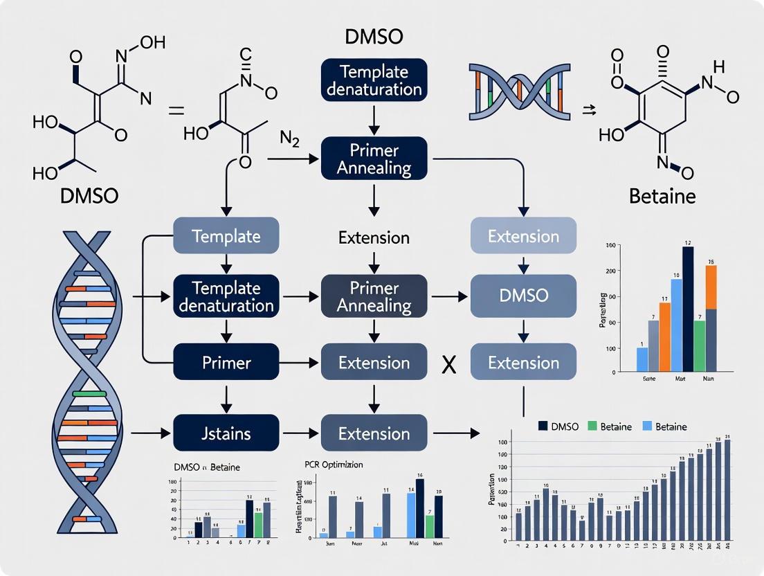
Optimizing PCR for GC-Rich Regions: A Comprehensive Guide to Using DMSO and Betaine
Amplifying GC-rich DNA sequences (>60% GC content) remains a significant challenge in molecular biology, often leading to PCR failure due to stable secondary structures and high melting temperatures.
Optimizing PCR for GC-Rich Regions: A Comprehensive Guide to Using DMSO and Betaine
Abstract
Amplifying GC-rich DNA sequences (>60% GC content) remains a significant challenge in molecular biology, often leading to PCR failure due to stable secondary structures and high melting temperatures. This article provides a comprehensive, evidence-based protocol for researchers and drug development professionals struggling with these difficult templates. We detail a multipronged optimization strategy, with a core focus on the synergistic use of additives like DMSO and betaine. The guide covers foundational principles, step-by-step methodological application, advanced troubleshooting techniques, and comparative validation data to ensure robust, specific, and efficient amplification of GC-rich targets for downstream applications in genomics, cloning, and diagnostic assay development.
Understanding the Challenge: Why GC-Rich DNA Hampers PCR Efficiency
In genomic research, GC-rich sequences are defined as DNA regions where the proportion of guanine (G) and cytosine (C) bases equals or exceeds 60% of the total nucleotide composition [1]. These regions are of profound biological importance due to their significant overrepresentation in essential regulatory areas of the genome. Although they constitute only approximately 3% of the entire human genome, GC-rich sequences are disproportionately concentrated in functional elements that control gene expression [1]. This non-random distribution highlights their critical role in transcriptional regulation and genome organization.
The biological significance of GC-rich regions stems from the unique biochemical properties of G-C base pairing. Unlike A-T pairs which form two hydrogen bonds, each G-C base pair establishes three hydrogen bonds, creating a more stable and thermodynamically robust duplex structure [1]. This enhanced stability directly influences DNA conformation, protein-DNA interactions, and the formation of higher-order genomic structures that collectively regulate gene expression patterns and cellular function.
Quantitative Analysis of GC-Rich Regions in Genomic Elements
GC-rich sequences are not uniformly distributed throughout the genome but are strategically concentrated in specific regulatory domains. The following table summarizes their prevalence across different genomic elements:
Table 1: Prevalence of GC-Rich Regions in Genomic Elements
| Genomic Element | GC Content Range | Biological Significance |
|---|---|---|
| Gene Promoters | Often >60% [1] | Regulatory hubs for transcription initiation; contain transcription factor binding sites |
| Housekeeping Gene Promoters | Consistently high | Maintain basal cellular functions [1] |
| Tumor Suppressor Gene Promoters | Consistently high | Regulation of cell cycle and apoptosis [1] |
| Enhancers/Cis-regulatory Elements | Often elevated [2] | Remote regulation of gene expression |
| EGFR Gene Promoter | Up to 88% [3] | Extreme example in clinically relevant cancer gene |
These quantitative distributions reflect the functional importance of GC-rich regions in maintaining accessible chromatin configurations and facilitating the binding of transcription factors and other regulatory proteins. The elevated GC content in promoter regions, particularly for housekeeping and tumor suppressor genes, creates a distinct biochemical environment that influences nucleosome positioning, DNA methylation patterns, and ultimately transcriptional competence [1].
The Molecular Challenges of GC-Rich Sequence Amplification
Amplifying GC-rich templates via polymerase chain reaction (PCR) presents substantial technical challenges that stem from their unique biochemical properties. The primary obstacles include:
Secondary Structure Formation
The strong hydrogen bonding in GC-rich regions promotes formation of stable intra-strand secondary structures, particularly hairpins and stem-loops [1]. These structures occur when complementary regions within a single DNA strand fold back on themselves, creating physical barriers that impede polymerase progression during extension phases. This results in premature termination and accumulation of truncated amplification products [4].
Incomplete Denaturation
The thermal stability of GC-rich duplexes requires higher denaturation temperatures. Standard denaturation at 94°C may be insufficient for complete strand separation of templates with GC content exceeding 70%, leading to reannealing during primer annealing and extension steps [2]. This incomplete denaturation significantly reduces amplification efficiency and product yield.
Non-Specific Amplification
High GC content increases melting temperatures (Tm) for both templates and primers, potentially causing mispriming events when using standard annealing temperatures [4]. This often manifests as multiple bands or smearing on agarose gels, indicating amplification of non-target sequences [1].
The following diagram illustrates these molecular challenges and their impacts on PCR efficiency:
Chemical Optimization Strategies: DMSO and Betaine
The strategic application of chemical additives represents a cornerstone approach for overcoming GC-rich amplification challenges. Dimethyl sulfoxide (DMSO) and betaine have emerged as particularly effective agents with distinct but complementary mechanisms of action.
DMSO (Dimethyl Sulfoxide)
DMSO functions primarily as a secondary structure disruptor by interfering with the formation of stable DNA duplexes and hairpins. It achieves this through several mechanisms: DMSO alters DNA solvation by reducing the strength of hydrogen bonding between complementary strands, facilitates strand separation at lower temperatures by destabilizing duplex DNA, and prevents reannealing of GC-rich templates during critical PCR steps [4]. Multiple studies have demonstrated that DMSO concentrations between 2.5% to 5% (v/v) significantly improve amplification efficiency of GC-rich targets, with 5% providing optimal results for extremely challenging templates like the EGFR promoter region [3].
Betaine
Betaine (N,N,N-trimethylglycine) operates through a different mechanism known as isostabilization, which equalizes the thermal stability of AT-rich and GC-rich DNA regions. It functions by: preferentially hydrating AT base pairs to increase their melting temperature while simultaneously reducing the Tm of GC-rich regions through direct interaction, effectively compressing the melting temperature range across the entire template to minimize secondary structure formation, and enhancing primer specificity by reducing mispriming at partially complementary sites [5] [4]. Betaine is typically used at concentrations ranging from 0.5 M to 1.5 M, depending on template complexity and GC content.
Table 2: Optimization Parameters for GC-Rich PCR
| Parameter | Standard Conditions | Optimized Conditions for GC-Rich Templates |
|---|---|---|
| DMSO Concentration | 0% | 2.5–5% [3] [2] |
| Betaine Concentration | 0 M | 0.5–1.5 M [5] [4] |
| Denaturation Temperature | 94–95°C | 98°C [2] |
| Denaturation Time | 30 sec | 10 sec at 98°C [2] |
| Annealing Temperature | Calculated Tm −5°C | 7°C higher than calculated [3] |
| MgCl₂ Concentration | 1.5–2.0 mM | Gradient optimization 1.0–4.0 mM [1] |
| Polymerase Selection | Standard Taq | Specialized high-fidelity GC-rich polymerases [1] |
Comprehensive Experimental Protocol for GC-Rich Amplification
This optimized protocol integrates DMSO and betaine for reliable amplification of GC-rich regulatory elements, incorporating established methodologies from published studies [5] [4] [3].
Reagent Setup and Preparation
- DNA Template: Use ≥2 μg/mL of high-quality genomic DNA; for FFPE samples, increase concentration to compensate for potential degradation [3]
- Primer Design: Design primers with melting temperatures ≥68°C to facilitate higher annealing temperatures and improved specificity [2]
- Reaction Mixture (25 μL total volume):
- 1X PCR buffer (provided with polymerase)
- 0.2 μM each forward and reverse primer
- 0.25 mM each dNTP
- 1.5–2.0 mM MgCl₂ (optimize using gradient PCR) [3]
- 5% DMSO (v/v) [3] OR 1 M betaine [5] [4]
- 0.625–1.25 U DNA polymerase (specialized for GC-rich templates)
- 2–5 μL template DNA (concentration dependent on source)
- Nuclease-free water to volume
Thermal Cycling Conditions
- Initial Denaturation: 98°C for 2–5 minutes (complete denaturation of complex templates) [2]
- Amplification Cycles (35–45 cycles):
- Final Extension: 72°C for 5–7 minutes (complete synthesis of all products)
- Hold: 4°C indefinitely
Troubleshooting and Optimization Guidelines
- For templates with GC content >80%: Combine DMSO (2.5%) and betaine (0.5–1.0 M) for synergistic effects [5]
- If non-specific amplification persists: Increase annealing temperature in 2°C increments or implement touchdown PCR
- If yield remains low: Extend extension time to 2 minutes per kb and increase template concentration
- For complex secondary structures: Incorporate 7-deaza-2′-deoxyguanosine (dGTP analog) to reduce hairpin stability [1]
The following workflow diagram summarizes the optimized experimental procedure:
The Scientist's Toolkit: Essential Research Reagents
Successful investigation of GC-rich regulatory elements requires specialized reagents and tools. The following table outlines essential components for researching and amplifying these challenging sequences:
Table 3: Essential Research Reagents for GC-Rich Sequence Analysis
| Reagent/Tool | Specific Function | Application Notes |
|---|---|---|
| Specialized DNA Polymerases | High processivity for complex templates; often supplied with GC buffers | Examples: OneTaq (NEB), Q5 High-Fidelity (NEB), PrimeSTAR GXL (Takara) [1] |
| DMSO (Dimethyl Sulfoxide) | Disrupts secondary structures; reduces template thermostability | Use at 2.5–5% (v/v); improves specificity and yield [3] [2] |
| Betaine | Isostabilizing agent; equalizes Tm across sequence | Use at 0.5–1.5 M; compatible with DMSO for challenging templates [5] [4] |
| MgCl₂ Solution | Cofactor for polymerase activity; concentration critical | Optimize from 1.0–4.0 mM; affects specificity and efficiency [1] |
| GC Enhancer Buffers | Proprietary additive mixtures for difficult amplicons | Often included with specialized polymerases [1] |
| 7-deaza-dGTP | dGTP analog that reduces secondary structure | Incorporation reduces hairpin stability [1] |
| Bioinformatic Tools | GC content analysis; primer design; sequence annotation | GC-Profile, EMBOSS CpGPlot, Polygraph framework [6] [7] |
GC-rich sequences represent critical functional elements within genomes, particularly concentrated in gene promoters and regulatory regions where they influence transcription factor binding, chromatin organization, and gene expression patterns. Their biochemical properties present significant challenges for molecular analysis, especially PCR amplification. However, through strategic application of chemical additives like DMSO and betaine, combined with optimized thermal cycling parameters and specialized polymerases, these challenges can be systematically overcome. The protocols and methodologies presented here provide researchers with a comprehensive framework for investigating these important genomic elements, enabling more reliable study of gene regulatory mechanisms and their implications in development, homeostasis, and disease.
The amplification of GC-rich DNA sequences presents a significant challenge in molecular biology, primarily due to the intrinsic molecular stability of these regions. A DNA template is considered GC-rich when 60% or more of its bases are guanine (G) or cytosine (C) [8] [9]. While these regions constitute only about 3% of the human genome, they are frequently found in critical areas such as the promoter regions of housekeeping and tumor suppressor genes, making their amplification essential for many research and diagnostic applications [8].
The formidable challenge in amplifying these sequences stems from two fundamental physical interactions: hydrogen bonding and base stacking. The strong triple hydrogen bonds between G-C base pairs, compared to the double bonds in A-T pairs, confer greater thermostability, requiring more energy to separate the DNA strands [8]. Concurrently, base stacking interactions between adjacent nucleotide pairs provide even greater stabilization to the DNA double helix than hydrogen bonding alone [9]. This combined stability results in DNA with higher melting temperatures and a pronounced tendency to form stable secondary structures, such as hairpin loops, which can block polymerase progression and lead to amplification failure [8] [9].
This application note explores the scientific basis of these stability challenges and provides detailed, optimized protocols for the successful amplification of GC-rich sequences, with particular emphasis on the synergistic use of PCR additives such as DMSO and betaine.
The Fundamental Forces Governing DNA Stability
Hydrogen Bonding and Its Role in PCR
Hydrogen bonding represents a primary force contributing to the stability of the DNA double helix. In standard Watson-Crick base pairing, guanine-cytosine (G-C) pairs form three hydrogen bonds, while adenine-thymine (A-T) pairs form only two [8]. This difference in bond number has direct implications for the thermal stability of DNA during the polymerase chain reaction.
- Thermodynamic Impact: The additional hydrogen bond in G-C pairs increases the energy required to denature the DNA duplex. During PCR, this translates to a higher melting temperature (Tm) for GC-rich templates, often exceeding standard denaturation temperatures used in typical protocols [8].
- Proofreading Dependency: Research on DNA polymerases has revealed that hydrogen bonding is essential for proper proofreading activity. Enzymes like the human mitochondrial DNA polymerase utilize hydrogen bond formation as a critical criterion for correct base pair recognition, with non-hydrogen-bonded base pairs being excised as if they were mismatches regardless of their steric properties [10].
Base Stacking: The Dominant Stabilizing Force
Contrary to common perception, the dominant stabilization force in DNA comes from base stacking interactions, not hydrogen bonding [9]. Base stacking refers to the vertical, hydrophobic interactions between the aromatic rings of adjacent nucleotide pairs in the DNA helix.
- Energetic Superiority: These stacking interactions contribute more significantly to the overall stability of the DNA duplex than hydrogen bonds. The stacking free energy helps maintain the double-stranded structure, particularly in GC-rich regions where the planar nature of guanine and cytosine rings facilitates optimal overlap [9].
- Structural Consequences: The pronounced base stacking in GC-rich sequences makes these regions exceptionally "bendable," readily forming stable secondary structures such as hairpins and stem-loops that can persist even at standard PCR denaturation temperatures [8]. These structures physically impede polymerase progression, leading to incomplete amplification products or complete PCR failure.
Diagram 1: Molecular forces creating challenges in GC-rich PCR amplification.
Optimization Strategies for GC-Rich Amplification
Successful amplification of GC-rich templates requires a multifaceted approach that addresses both hydrogen bonding and base stacking stability. The following strategies can be implemented individually or in combination to overcome these challenges.
PCR Additives and Their Mechanisms of Action
Organic additives represent powerful tools for modulating DNA stability during amplification. They work through distinct mechanisms to either reduce secondary structure formation or increase primer annealing stringency [8].
Table 1: PCR Additives for GC-Rich Amplification
| Additive | Recommended Concentration | Primary Mechanism | Effect on PCR |
|---|---|---|---|
| Betaine | 1.0 - 1.3 M | Equalizes template stability by reducing base stacking energy | Disrupts secondary structures; reduces nonspecific background |
| DMSO | 3 - 10% (typically 5%) | Interferes with hydrogen bonding; alters DNA solvation | Lowers melting temperature; prevents secondary structure formation |
| 7-deaza-dGTP | 50 μM (partial replacement of dGTP) | Analog that incorporates into DNA but reduces hydrogen bonding | Improves polymerase progression; reduces stalling |
| Glycerol | 5 - 10% | Protein-stabilizing agent; may reduce DNA melting temperature | Stabilizes polymerase; aids in denaturation of stable structures |
| Formamide | 1 - 5% | Denaturing agent that disrupts hydrogen bonding | Increases primer stringency; reduces secondary structures |
The synergistic combination of betaine, DMSO, and 7-deaza-dGTP has proven particularly effective for extremely challenging templates with GC content exceeding 75% [11]. This combination simultaneously addresses both hydrogen bonding and base stacking stabilization, providing a comprehensive solution for the most refractory sequences.
Polymerase Selection and Reaction Condition Optimization
The choice of DNA polymerase significantly impacts the success of GC-rich amplification. Standard Taq polymerase often struggles with these templates, while specially engineered polymerases such as Q5 High-Fidelity DNA Polymerase (NEB #M0491) and OneTaq DNA Polymerase (NEB #M0480) demonstrate superior performance [8]. These enzymes are often supplied with GC Enhancer formulations that contain optimized mixtures of additives to inhibit secondary structure formation and increase primer stringency [8].
Magnesium concentration optimization represents another critical parameter. Magnesium ions (Mg²⁺) serve as essential cofactors for polymerase activity, but inappropriate concentrations can exacerbate amplification problems. Testing a concentration gradient from 1.0 to 4.0 mM MgCl₂ in 0.5 mM increments can identify the optimal concentration that maximizes yield while minimizing non-specific amplification [8].
Annealing temperature adjustment provides additional control over amplification specificity. For problematic GC-rich templates, implementing a "touchdown" approach with higher annealing temperatures in the initial cycles can improve specificity, while subsequent cycles at lower temperatures boost product yield [8]. The NEB Tm Calculator tool can assist in selecting appropriate annealing temperatures based on the specific enzyme and buffer system [8].
Experimental Protocols
Protocol 1: Standard PCR with Additives for GC-Rich Templates
This protocol provides a robust starting point for amplifying GC-rich sequences (60-75% GC content) using a standard thermal cycler and common reagents.
Reagents and Equipment:
- DNA template (10-100 ng)
- High-fidelity DNA polymerase with buffer (e.g., Q5 or OneTaq)
- 10 mM dNTP mix (with optional 7-deaza-dGTP substitution)
- Forward and reverse primers (10 μM each)
- Betaine (5 M stock solution)
- DMSO (Molecular biology grade)
- MgCl₂ (50 mM stock solution)
- Nuclease-free water
- Thermal cycler with gradient capability
Procedure:
- Prepare Master Mix (50 μL reaction):
- 1X polymerase buffer (provided)
- 200 μM each dNTP (replace 25-50% of dGTP with 7-deaza-dGTP for difficult templates)
- 0.5 μM forward primer
- 0.5 μM reverse primer
- 1.0 M betaine (from 5M stock)
- 5% DMSO
- 1.5-3.0 mM MgCl₂ (optimize using gradient)
- 1.25 units DNA polymerase
- 10-100 ng template DNA
- Nuclease-free water to 50 μL
Thermal Cycling Conditions:
- Initial denaturation: 98°C for 30 seconds
- 35 cycles of:
- Denaturation: 98°C for 10 seconds
- Annealing: 68-72°C for 30 seconds (optimize based on Tm)
- Extension: 72°C for 30 seconds per kb
- Final extension: 72°C for 2 minutes
- Hold at 4°C
Analysis:
- Analyze 5 μL of PCR product by agarose gel electrophoresis
- For non-specific amplification, increase annealing temperature by 2-3°C or titrate MgCl₂ downward
- For no product, decrease annealing temperature, increase MgCl₂, or add additional DMSO (up to 10%)
Protocol 2: Enhanced Protocol for Extremely GC-Rich Targets (>75% GC)
This specialized protocol incorporates the powerful triple-additive combination for the most challenging templates, such as promoter regions with GC content exceeding 80% [11].
Reagents and Equipment:
- All reagents from Protocol 1
- 7-deaza-dGTP (50 mM stock solution)
- Additional MgCl₂ (50 mM stock solution)
Procedure:
- Prepare Master Mix (25 μL reaction):
- 1X polymerase buffer
- 200 μM dATP, dCTP, dTTP
- 150 μM dGTP
- 50 μM 7-deaza-dGTP
- 0.5 μM forward primer
- 0.5 μM reverse primer
- 1.3 M betaine
- 5% DMSO
- 2.5 mM MgCl₂ (adjust based on optimization)
- 1.25 units DNA polymerase
- 50-100 ng template DNA
- Nuclease-free water to 25 μL
Modified Thermal Cycling Conditions:
- Initial denaturation: 95°C for 3 minutes
- 10 "Touchdown" cycles:
- Denaturation: 95°C for 30 seconds
- Annealing: Start at 5°C above calculated Tm, decrease 0.5°C per cycle
- Extension: 72°C for 45 seconds per kb
- 25-30 standard cycles:
- Denaturation: 95°C for 30 seconds
- Annealing: Use final touchdown temperature for 30 seconds
- Extension: 72°C for 45 seconds per kb
- Final extension: 72°C for 5 minutes
Troubleshooting:
Diagram 2: Systematic workflow for optimizing GC-rich PCR amplification.
Research Reagent Solutions
Table 2: Essential Reagents for GC-Rich PCR
| Reagent Category | Specific Examples | Function & Application Notes |
|---|---|---|
| Specialized Polymerases | Q5 High-Fidelity DNA Polymerase (NEB #M0491), OneTaq DNA Polymerase (NEB #M0480), AccuPrime GC-Rich DNA Polymerase | Engineered for high processivity on difficult templates; some include GC enhancers |
| GC Enhancers | OneTaq High GC Enhancer, Q5 High GC Enhancer | Proprietary formulations that combine multiple additives for maximum effect |
| Chemical Additives | Betaine (1.0-1.3 M), DMSO (3-10%), 7-deaza-dGTP (50 μM) | Work synergistically to disrupt hydrogen bonding and base stacking |
| Modified Nucleotides | 7-deaza-2'-deoxyguanosine triphosphate | dGTP analog that reduces hydrogen bonding without steric hindrance |
| Optimization Kits | Magnesium Spinner kits, Temperature Gradient kits | Enable systematic optimization of critical reaction parameters |
The successful amplification of GC-rich DNA sequences requires a fundamental understanding of the molecular forces governing DNA stability, particularly the complementary roles of hydrogen bonding and base stacking interactions. Through strategic implementation of specialized polymerases, optimized reaction conditions, and synergistic additive combinations—notably betaine and DMSO—researchers can overcome the formidable challenges posed by these templates.
The protocols presented herein provide a systematic approach from standard optimization to enhanced methods for extremely recalcitrant sequences. As GC-rich regions frequently occur in biologically significant genomic contexts, mastery of these techniques empowers researchers and drug development professionals to advance their investigations with greater reliability and efficiency, ultimately contributing to enhanced molecular diagnostic capabilities and therapeutic development.
In vitro DNA polymerization is a cornerstone of modern molecular biology, forming the basis for techniques including quantitative PCR (qPCR), digital PCR (dPCR), and massively parallel sequencing (MPS) [12]. A significant challenge in these applications is the presence of intramolecular secondary structures within DNA templates, such as hairpins, which can severely inhibit polymerase activity [13]. These structures are particularly prevalent in GC-rich sequences, where the three hydrogen bonds between guanine and cytosine create exceptionally stable and thermostable formations [14] [15]. When a polymerase encounters these stable secondary structures, it can stall or undergo "polymerase jumping," leading to reduced assay sensitivity, lower yield, higher error rates, and in some cases, complete amplification failure [13]. For researchers, particularly in drug development and clinical diagnostics where precision is critical, understanding and overcoming these structural hurdles is essential. This application note, framed within broader research on PCR protocols with DMSO and betaine for GC-rich regions, details the mechanisms of inhibition and provides optimized protocols to ensure successful amplification.
Mechanisms of Inhibition
Stable secondary structures impair the polymerase chain reaction through several distinct biochemical mechanisms.
- Polymerase Stalling and Premature Termination: Complex secondary structures act as physical barriers to the progressing DNA polymerase. When the enzyme stalls, the result is truncated, incomplete amplification products [13] [14]. This is a common issue with GC-rich templates, which are "bendable" and readily form hairpins that block the polymerase [14].
- Endonucleolytic Cleavage by Taq Polymerase: Recent research has elucidated another mechanism where the 5′-3′ exonuclease activity of Taq polymerase cleaves the template DNA within these stable secondary structures. This unintended cleavage destroys the template, thereby preventing the amplification of the full-length target product [13].
- Inhibition of Primer Annealing: Secondary structures can form within the single-stranded template before primers have a chance to bind. Because reaction kinetics favor intramolecular folding over intermolecular primer binding, this can prevent the formation of a stable primer-template hybrid, which is a prerequisite for polymerase activity [13].
- Fluorescence Quenching: It is also important to note that some inhibitory substances, though distinct from the templates themselves, can interfere with fluorescence-based detection—a critical component of qPCR, dPCR, and MPS. These quenchers can operate through collisional or static quenching mechanisms, leading to inaccurate quantification [12].
Quantitative Impact of Secondary Structures
The following table summarizes the quantitative effects of various inhibitory structures and the performance of different solutions as reported in the literature.
Table 1: Quantitative Impact of Secondary Structures and Solution Performance
| Template/Challenge | Key Metric | Performance without Solution | Performance with Solution | Citation |
|---|---|---|---|---|
| rAAV ITR Sequences (Ultra-stable hairpins) | PCR Amplification Success | Extremely difficult / No product | Successful amplification | [13] |
| EGFR Target A (Stable secondary structure) | qPCR Efficiency (at 10 template copies) | Significant inhibition | ~100% efficiency with disruptors | [13] |
| GC-rich IGF2R & BRAF | PCR Product Specificity & Yield | Low specificity, poor yield | Greatly improved with DMSO/Betaine | [4] |
| General GC-rich Templates | Polymerase Processivity | Stalling and low yield | Robust amplification with specialized polymerases + GC Enhancer | [14] |
| Guide RNA with Hairpins (for CRISPR/Cas9) | Off-target Editing Rate | High off-target effects | 50-fold higher specificity | [16] |
Experimental Protocols
Protocol 1: Amplifying GC-Rich Templates Using Additives
This protocol is designed for the robust amplification of difficult GC-rich targets (>60% GC content) using reagent additives and an inhibitor-tolerant polymerase [4] [14] [17].
Step 1: Reagent Preparation
- Prepare a master mix with the following components and concentrations:
- DNA Polymerase: Use an inhibitor-tolerant, highly processive polymerase such as OneTaq or Q5 High-Fidelity DNA Polymerase [14].
- Buffer: Use the specialized GC Buffer supplied with the polymerase.
- GC Enhancer: Add the proprietary GC Enhancer at the recommended concentration (e.g., 10-20% v/v) [14]. This solution often contains a blend of additives like DMSO and betaine.
- Additives (if not using GC Enhancer): As an alternative, include DMSO (1-10% v/v) or Betaine (0.5-1.5 M) in the reaction [4] [14] [15].
- MgCl2: Consider testing a concentration gradient from 1.0 mM to 4.0 mM in 0.5 mM increments to optimize cofactor concentration [14].
- dNTPs: Standard concentration (e.g., 200 μM of each).
- Primers: Designed for the GC-rich target, typically with a Tm of 50-72°C.
- Template: High-quality DNA.
- Prepare a master mix with the following components and concentrations:
Step 2: Thermal Cycling
- Use the following thermal cycling parameters, optimized for GC-rich templates:
- Initial Denaturation: 98°C for 30 seconds [17].
- Amplification (35 cycles):
- Denaturation: 98°C for 5-10 seconds. A higher denaturation temperature helps melt stable GC bonds [17].
- Annealing: Use a temperature gradient to determine the optimal Ta. Start with a Ta 5°C above the calculated Tm of the primers and gradually decrease in subsequent cycles (Touchdown PCR) to enhance specificity [17] [15].
- Extension: 72°C. Allow 15-30 seconds per kb.
- Final Extension: 72°C for 2 minutes.
- Use the following thermal cycling parameters, optimized for GC-rich templates:
Step 3: Analysis
- Analyze the PCR product using agarose gel electrophoresis to confirm amplicon size and purity.
The following workflow diagram illustrates this experimental process.
Protocol 2: Using Disruptor Oligonucleotides for Ultra-Stable Structures
For the most challenging templates, such as the inverted terminal repeats (ITRs) of adeno-associated virus (AAV) vectors, conventional additives may fail. This protocol uses specialized "disruptor" oligonucleotides to physically unwind secondary structures [13].
Step 1: Design of Disruptor Oligonucleotides
- Design disruptors to be reverse-complementary to the template sequence, specifically overlapping the duplex region of the intramolecular secondary structure.
- Each disruptor should contain three functional components:
- Anchor: A sequence at the 3' end designed to initiate specific binding to the template.
- Effector: A central sequence that mediates strand displacement to unwind the secondary structure.
- 3' Blocker: A chemical modification (e.g., C3-Spacer) at the 3' end to prevent the disruptor itself from being elongated by the DNA polymerase [13].
Step 2: PCR Reaction Setup
- Prepare a standard PCR master mix suitable for the target amplicon length.
- Add the designed disruptor oligonucleotide(s) to the reaction. The optimal final concentration should be determined empirically but is typically in the range of 0.1–0.5 μM.
- Note: In the referenced study, DMSO and betaine were completely ineffective on rAAV ITR sequences, whereas disruptors enabled successful amplification [13].
Step 3: Thermal Cycling and Analysis
- Use standard thermal cycling conditions appropriate for the primer pair and polymerase.
- Analyze the product by gel electrophoresis or sequencing.
The Scientist's Toolkit: Research Reagent Solutions
The following table lists key reagents for overcoming secondary structures in PCR.
Table 2: Essential Reagents for Overcoming Structural Hurdles in PCR
| Reagent / Material | Function / Mechanism of Action | Example Use Cases |
|---|---|---|
| DMSO (Dimethyl Sulfoxide) | Polar additive that disrupts base pairing by interfering with hydrogen bonding; lowers DNA melting temperature (Tm) [14] [15]. | Amplification of GC-rich templates; reduces secondary structure formation [4]. |
| Betaine | Amino acid analog that equilibrates Tm differences between GC and AT base pairs; acts as an isostabilizing agent [4] [14]. | PCR amplification of templates with extreme GC content; improves specificity and yield [4]. |
| Specialized DNA Polymerase (e.g., OneTaq, Q5) | Engineered enzymes with high processivity and stability; often supplied with proprietary GC buffers and enhancers [14]. | Robust amplification of difficult amplicons (long, GC-rich, or impure samples) [14] [17]. |
| Disruptor Oligonucleotides | Sequence-specific oligonucleotides that bind and unwind stable intramolecular secondary structures via strand displacement [13]. | Amplifying and sequencing ultra-stable structures like rAAV ITRs where traditional additives fail [13]. |
| 7-deaza-2'-deoxyguanosine | dGTP analog that reduces the strength of hydrogen bonding between guanosine and cytosine by replacing a nitrogen atom with a carbon at the 7 position [13] [14]. | Alternative for amplifying highly GC-rich regions; may require adjustments to staining as it stains poorly with ethidium bromide [14]. |
| GC Enhancer | A proprietary, pre-optimized blend of additives (which may include DMSO, betaine, and other components) designed to inhibit secondary structure formation [14]. | A convenient, single-solution additive for improving PCR of GC-rich targets without needing to optimize individual reagent concentrations [14]. |
The challenges posed by hairpins and secondary structures in PCR are significant but surmountable. Understanding the mechanisms—from polymerase stalling to template cleavage—provides a rational basis for selecting the right solution. For most GC-rich templates, a combination of specialized polymerases and chemical additives like DMSO and betaine offers a reliable path to successful amplification. However, for the most recalcitrant structures, such as those found in rAAV ITRs, innovative approaches like disruptor oligonucleotides represent a breakthrough, enabling research and therapeutic development that was previously hampered by technical limitations. By applying the optimized protocols and reagents detailed in this application note, researchers can systematically overcome these structural hurdles and achieve robust and reliable DNA amplification.
In polymerase chain reaction (PCR) experiments, achieving specific and efficient amplification is paramount for accurate results. However, researchers frequently encounter artifacts that compromise data integrity, including primer dimers, nonspecific amplification, and truncated products. These artifacts are particularly prevalent when amplifying challenging templates, such as GC-rich regions, which are common in promoter regions of housekeeping and tumor suppressor genes [18]. The formation of these unwanted products competes with target amplification for reagents, reduces overall yield, and can lead to both false-positive and false-negative interpretations [19]. This application note details the consequences of these common PCR artifacts and provides optimized protocols to mitigate them, with a specific focus on the use of additives like DMSO and betaine within the context of GC-rich amplification challenges.
Consequences of Common PCR Artifacts
Primer Dimer Formation
Primer dimers are small, unintended DNA fragments that form when primers anneal to each other instead of the target template. They are a significant source of PCR inefficiency, particularly in quantitative PCR (qPCR) [20] [21].
Mechanisms of Formation:
- Self-dimerization: A single primer contains regions that are self-complementary.
- Cross-dimerization: Two primers have complementary regions that allow them to bind to each other [20]. In both cases, the DNA polymerase can extend the annealed primers, creating a short, stable product that is amplified in subsequent cycles. The risk of primer dimer formation is highest before the PCR cycle begins, when reagents are mixed at non-stringent temperatures [20] [19].
Impact on PCR:
- Consumption of Reagents: Primer dimers compete for primers, dNTPs, and polymerase activity, reducing the resources available for target amplification [19].
- Inhibition of Target Amplification: This competition can lead to reduced yield of the desired product and a higher cycle threshold (Ct) in qPCR, potentially causing false-negative results in low-template samples [19].
- False Positives: In SYBR Green-based qPCR, the dye will bind to primer dimer products, generating a fluorescent signal that can be mistaken for specific amplification, especially in no-template controls (NTCs) [19].
Identification: On an agarose gel, primer dimers typically appear as a smeary band or a sharp band below 100 bp [20]. The use of a no-template control (NTC) is crucial for identifying primer-derived artifacts [20] [21].
Non-Specific Binding and Amplification
Nonspecific amplification occurs when primers anneal to partially complementary, off-target sites on the template DNA, leading to the synthesis of unwanted products of varying sizes [22].
- Causes: The primary cause is low stringency in annealing conditions, often due to an annealing temperature that is too low [22] [18]. Excess magnesium ions (Mg²⁺) or high primer concentration can also contribute to mis-priming [18].
- Consequences:
- Multiple Bands on Gels: Complicates the interpretation of results and can obscure the specific band during gel extraction.
- Reduced Sensitivity and Efficiency: Amplification of off-target sequences depletes reaction components, thereby reducing the yield of the desired amplicon.
- Inaccurate Quantification: In qPCR, nonspecific products can generate background fluorescence, interfering with accurate quantification of the target [21].
- Generation of Chimeric Artifacts: In extreme cases, particularly with repetitive sequences like Alu elements, nonspecific priming can lead to the amplification of recombinant artifacts that do not exist in the original sample, fundamentally compromising data validity [23].
Truncated Products
Truncated products are incomplete amplification fragments that result from the polymerase failing to fully extend the DNA strand during each cycle.
- Primary Cause in GC-Rich Templates: The main cause is the formation of stable secondary structures, such as hairpins and stem-loops, due to the strong triple hydrogen bonding of G-C base pairs. These structures can physically block polymerase progression [4] [18].
- Consequences:
- Smeared Gels: Instead of a clean, discrete band, a smear of DNA of various sizes is observed.
- Low Yield of Full-Length Product: The accumulation of truncated fragments means the full-length amplicon is under-represented.
- Assay Failure: In severe cases, amplification of the target may fail entirely.
Table 1: Summary of PCR Artifacts and Their Consequences
| Artifact | Primary Cause | Key Consequences | Common Indicators |
|---|---|---|---|
| Primer Dimer [20] [19] | Primer self-/cross-complementarity; low pre-PCR temperatures | False positives in qPCR (SYBR Green); reduced amplification efficiency; higher Ct values | Smear/band <100 bp in NTC; early amplification in NTC |
| Non-Specific Binding [21] [22] [23] | Low annealing stringency; high [Mg²⁺]; high [primer] | Multiple bands; reduced target yield; inaccurate quantification; chimeric artifacts | Multiple bands on agarose gel |
| Truncated Products [4] [18] | Secondary structures in GC-rich templates | Smeared gels; low yield of full-length product; PCR failure | DNA smear on agarose gel |
The Scientist's Toolkit: Research Reagent Solutions
Selecting the right reagents is a critical first step in designing a robust PCR assay, especially for difficult targets. The following table outlines key solutions for preventing common artifacts.
Table 2: Essential Reagents for Mitigating PCR Artifacts
| Reagent / Solution | Function and Rationale | Specific Use Case |
|---|---|---|
| Hot-Start DNA Polymerase [20] [24] | Antibody- or aptamer-bound enzyme inactive until initial denaturation step. Prevents primer extension during reaction setup at low temperatures, reducing primer dimer formation. | Essential for all PCR, especially multiplex and low-template qPCR. |
| High-Fidelity Polymerase Mixes [24] [18] | Blends of non-proofreading and proofreading enzymes (e.g., Taq and Pfu). Improve accuracy and efficiency for long or difficult amplicons. | Amplification of long targets (>5kb) or complex templates. |
| Specialized GC Buffers & Enhancers [18] | Proprietary buffers with additives that disrupt secondary structures. Offers a standardized, optimized solution for GC-rich PCR without user optimization of individual additives. | First-choice solution for amplifying GC-rich regions (e.g., promoter sequences). |
| DMSO (Dimethyl Sulfoxide) [4] [18] | Disrupts secondary structure by interfering with hydrogen bonding and base stacking. Lowers the melting temperature (Tm) of DNA, facilitating denaturation of stable structures. | Amplification of GC-rich templates (typical use 3-10%). |
| Betaine [4] [18] | An isostabilizing agent that equilizes the Tm difference between GC and AT base pairs. Reduces secondary structure formation and increases primer annealing specificity. | Amplification of GC-rich templates and mitigation of hairpin formation (typical use 1-1.5 M). |
| MgCl₂ [18] | Cofactor for DNA polymerase. Concentration is critical for enzyme activity, fidelity, and primer annealing. Too little reduces yield; too much increases nonspecific binding. | Optimization of specificity and yield (test 1.0 - 4.0 mM in gradients). |
Detailed Experimental Protocols
Protocol 1: Standard PCR Protocol with Optimization for GC-Rich Regions
This protocol is designed for routine amplification but includes specific modifications for challenging, GC-rich templates using DMSO and betaine, as validated in studies on synthetic gene construction [4].
Materials:
- Template DNA
- Forward and Reverse Primers
- High-Fidelity DNA Polymerase (e.g., Q5 or OneTaq) with supplied GC Buffer/Enhancer [18]
- dNTP Mix
- Nuclease-free Water
- Additives: DMSO (PCR-grade) and/or Betaine (5M stock) [4]
Method:
- Reaction Assembly:
Prepare a master mix on ice with the following components in a 50 µL reaction:
- Nuclease-free water: to 50 µL
- 10X PCR Buffer (or 2X GC Master Mix): 1X final concentration
- dNTP Mix (10 mM each): 200 µM each
- Forward Primer (10 µM): 0.5 µM final
- Reverse Primer (10 µM): 0.5 µM final
- DNA Polymerase: 1-2 units
- Optional Additives:
- Template DNA: 10-100 ng genomic DNA
Thermal Cycling:
- Initial Denaturation: 98°C for 30-60 seconds.
- Amplification (35 cycles):
- Denaturation: 98°C for 10-15 seconds.
- Annealing: Temperature gradient of 60-72°C for 20-30 seconds. See Protocol 2 for optimization.
- Extension: 72°C for 20-60 seconds/kb.
- Final Extension: 72°C for 2 minutes.
- Hold: 4°C.
Analysis: Analyze 5-10 µL of the PCR product by agarose gel electrophoresis.
Protocol 2: Touch-Down PCR for Enhanced Specificity
Touch-down PCR is highly effective for reducing nonspecific amplification and primer dimer formation by starting with high stringency and gradually lowering it [22].
Method:
- Reaction Assembly: Follow Protocol 1.
- Thermal Cycling:
- Initial Denaturation: 95°C for 5 minutes.
- Touch-Down Phase (10 cycles):
- Denaturation: 94°C for 30 seconds.
- Annealing: Start at 5-10°C above the calculated Tm of the primers. Decrease the annealing temperature by 1°C per cycle. (e.g., from 72°C to 63°C).
- Extension: 72°C for 1 minute/kb.
- Standard Phase (25 cycles):
- Denaturation: 94°C for 30 seconds.
- Annealing: Use the final annealing temperature from the touch-down phase.
- Extension: 72°C for 1 minute/kb.
- Final Extension: 72°C for 5 minutes.
- Hold: 4°C.
Protocol 3: Using a No-Template Control (NTC) to Identify Contamination
The NTC is a critical control that must be included in every PCR run to diagnose reagent contamination and primer dimer formation [20] [22].
Method:
- Prepare a reaction tube identical to those containing template DNA.
- Replace the template DNA with nuclease-free water.
- Run this NTC reaction alongside all other samples through the entire thermal cycling protocol.
- Interpretation:
- No Amplification: Indicates clean reagents and specific amplification in the test samples.
- Amplification in NTC: Signifies contamination of reagents with template or the presence of primer dimers. The product should be analyzed by gel electrophoresis (primer dimers appear as a smear ~50-100 bp [20]) and the assay requires re-optimization (e.g., higher annealing temperature, hot-start polymerase, primer redesign).
The following diagram illustrates the decision-making workflow for identifying and troubleshooting common PCR artifacts, integrating the protocols and solutions discussed in this note.
PCR artifacts like primer dimers, nonspecific products, and truncated fragments pose significant challenges to molecular biology research and diagnostic assay development. A systematic approach involving careful primer design, stringent thermal cycling protocols, and the selective use of specialized reagents—such as hot-start polymerases and structure-disrupting additives like DMSO and betaine—is essential for successful amplification. By understanding the consequences of these artifacts and implementing the detailed protocols and troubleshooting workflows provided herein, researchers can significantly improve the specificity, efficiency, and reliability of their PCR experiments, particularly when working with demanding GC-rich DNA templates.
The Core Protocol: A Step-by-Step Guide to Incorporating DMSO and Betaine
The amplification of GC-rich DNA sequences (typically defined as having a guanine-cytosine content exceeding 60%) remains a significant technical challenge in molecular biology, particularly for applications in genetic research and drug development [25]. These sequences form stable secondary structures due to the three hydrogen bonds in G-C base pairs, leading to polymerase stalling, incomplete amplification, and non-specific products [25] [26] [27]. Successful polymerase chain reaction (PCR) amplification of these difficult templates requires meticulous preparation and handling of specific reagents, primarily specialized polymerases and amplification-enhancing additives like dimethyl sulfoxide (DMSO) and betaine. This application note provides detailed protocols for sourcing, preparing, and handling these critical reagents within the context of optimizing PCR for GC-rich regions, forming part of a broader thesis on advanced molecular techniques.
Research Reagent Solutions: A Curated Toolkit
The following table details the essential reagents required for establishing robust PCR protocols for GC-rich targets, along with their specific functions and considerations for use.
Table 1: Essential Reagents for GC-Rich PCR Amplification
| Reagent Category | Specific Examples | Primary Function in GC-Rich PCR | Key Considerations |
|---|---|---|---|
| DNA Polymerases | OneTaq DNA Polymerase (NEB #M0480), Q5 High-Fidelity DNA Polymerase (NEB #M0491), PrimeSTAR GXL, Phusion High-Fidelity, Platinum SuperFi [25] [26] [27] | Catalyzes DNA synthesis; high-fidelity and specialized polymerases are engineered to overcome secondary structures that cause stalling. | Fidelity, processivity, and presence of proofreading activity are critical. Many are supplied with proprietary GC Enhancer buffers [25]. |
| Chemical Additives | Dimethyl Sulfoxide (DMSO), Betaine (also known as glycine betaine) [4] [26] [11] | Disrupts secondary structures (DMSO) and equilibrates DNA melting temperatures (Betaine), facilitating primer annealing and polymerase progression. | Often used in combination for synergistic effects. Concentration must be optimized to avoid inhibition of polymerase activity [11]. |
| Nucleotide Analogs | 7-deaza-2'-deoxyguanosine (7-deaza-dGTP) [11] | dGTP analog that incorporates into DNA and reduces hydrogen bonding, thereby lowering the stability of secondary structures. | Can be used in partial replacement of dGTP. May not stain well with ethidium bromide [25]. |
| Enhanced Buffer Systems | Q5 High GC Enhancer, OneTaq High GC Enhancer [25] | Proprietary buffer/additive mixes designed to inhibit secondary structure formation and increase primer stringency for specific polymerases. | Offers a standardized alternative to manual optimization of individual additive concentrations. |
Reagent Sourcing and Preparation Protocols
Polymerase Selection and Reconstitution
Choosing the appropriate DNA polymerase is the most critical step for successful GC-rich PCR. Standard Taq polymerase often fails with these templates, necessitating the use of specialized enzymes [25].
- Selection Criteria: Prioritize polymerases known for high processivity and fidelity. For instance, Q5 High-Fidelity DNA Polymerase offers more than 280 times the fidelity of Taq and is ideal for long or difficult amplicons [25]. PrimeSTAR GXL has also been successfully used for amplifying long GC-rich targets (>1 kb) from genomes like Mycobacterium bovis [27].
- Reconstitution and Storage: Always follow the manufacturer's instructions precisely. Use nuclease-free water for dilution if required. Store enzymes at -20°C and avoid multiple freeze-thaw cycles by aliquoting if necessary. Master mixes containing the polymerase are ideal for convenience but offer less flexibility for optimization compared to standalone enzymes [25].
DMSO: Handling and Preparation
DMSO is a polar organic solvent that reduces DNA secondary structure formation and lowers the melting temperature of DNA [15].
- Sourcing: Use molecular biology or PCR-grade DMSO of the highest purity (>99.9%) to avoid contaminants that can inhibit PCR.
- Handling and Safety: DMSO is hygroscopic and readily absorbs water from the atmosphere. Store it in sealed containers under anhydrous conditions at room temperature. It is also a potent solvent that can facilitate the transport of other molecules through the skin; therefore, wear appropriate personal protective equipment (PPE) including gloves and safety glasses when handling.
- Preparation for PCR: DMSO is typically added to the PCR reaction at a final concentration of 1-10%, with 5% being a common starting point for optimization [28] [11]. Prepare a sterile, stock solution to be added to the master mix. Note that cytotoxicity has been observed in cell cultures at concentrations as low as 0.5%, highlighting the importance of careful dosage [29].
Betaine: Handling and Preparation
Betaine (N,N,N-trimethylglycine) is a natural zwitterionic osmoprotectant that acts as an isostabilizing agent. It homogenizes the melting temperature of DNA by neutralizing the differential stability of GC and AT base pairs [30] [27].
- Sourcing: Source molecular biology-grade betaine to ensure purity and performance.
- Preparation for PCR: Betaine is typically used at a high final concentration, often 1.0 M to 1.3 M [11]. It is commonly prepared as a sterile 5 M stock solution in nuclease-free water. This solution is viscous; ensure it is mixed thoroughly and pipetted accurately when adding to the PCR master mix. Betaine is known for its low cytotoxicity compared to DMSO, making it a favorable additive [30].
Standardized Experimental Workflow and Protocols
Experimental Workflow for GC-Rich PCR Optimization
The following diagram illustrates the logical workflow for developing and troubleshooting a PCR protocol for a GC-rich target.
Diagram 1: GC-rich PCR optimization workflow.
Core PCR Protocol with Additives
This protocol is adapted from established basic PCR methods and enhanced with specific considerations for GC-rich templates [28] [11].
Reaction Setup (50 µL final volume):
- Template DNA: 1-1000 ng (typically 10-100 ng genomic DNA)
- Forward & Reverse Primers: 20-50 pmol each (final conc. 0.2-1.0 µM)
- dNTP Mix: 200 µM of each dNTP
- PCR Buffer (10X): 5 µL (supplied with polymerase)
- MgCl₂ (25 mM): 0-3.2 µL (if not in buffer; final conc. 1.0-4.0 mM) [25]
- DMSO: 0.5-5.0 µL (final conc. 1-10%)
- Betaine (5 M Stock): 10-13 µL (final conc. 1.0-1.3 M) [11]
- DNA Polymerase: 0.5-2.5 units
- Nuclease-Free Water: to 50 µL
Thermal Cycling Conditions (Example):
- Initial Denaturation: 98°C for 30 sec to 5 min (polymerase-dependent)
- Amplification (30-40 cycles):
- Denaturation: 98°C for 10-30 sec
- Annealing: 60-72°C for 15-30 sec (optimize using gradient PCR)
- Extension: 72°C for 15-60 sec/kb
- Final Extension: 72°C for 2-5 min
- Hold: 4°C
Note: For extremely GC-rich targets, a "2-step PCR" protocol, which combines annealing and extension at a higher temperature (e.g., 68°C), has proven superior [27].
Advanced Combinatorial Additive Protocol
For sequences refractory to standard optimization, a powerful combination of additives can be employed [31] [11].
- Reaction Modifications:
- Include 1.3 M betaine, 5% DMSO, and 50 µM 7-deaza-dGTP in the reaction mix.
- 7-deaza-dGTP can be used to partially or fully replace dGTP. If used as a full replacement, adjust the dNTP mix accordingly (e.g., 200 µM dATP, dCTP, dTTP; 50 µM 7-deaza-dGTP, and 150 µM dGTP is not recommended—consult specific protocols).
- Application: This combination was essential for the specific amplification of disease gene sequences with GC contents ranging from 67% to 79% [11].
Formulating Stock Solutions and Master Mixes
For reproducibility and efficiency, especially when screening multiple conditions, prepare stock solutions and master mixes.
Table 2: Formulation of a Standard 50 µL PCR Reaction with Additives
| Reagent | Stock Concentration | Volume per 50 µL Reaction | Final Concentration |
|---|---|---|---|
| Nuclease-Free Water | - | Variable (Q.S. to 50 µL) | - |
| PCR Buffer | 10X | 5 µL | 1X |
| dNTP Mix | 10 mM (each) | 1 µL | 200 µM (each) |
| MgCl₂ | 25 mM | 0 - 3.2 µL | 1.0 - 4.0 mM |
| Forward Primer | 20 µM | 1.25 µL | 0.5 µM |
| Reverse Primer | 20 µM | 1.25 µL | 0.5 µM |
| DMSO | 100% | 2.5 µL | 5% |
| Betaine | 5 M | 10 µL | 1.0 M |
| Template DNA | Variable | Variable | 1-1000 ng |
| DNA Polymerase | 1 U/µL | 0.5 µL | 0.5 U |
Master Mix Preparation Instructions:
- Calculate the number of reactions (n) and prepare a master mix for n+1 to account for pipetting error.
- In a sterile 1.5 mL microcentrifuge tube, combine all common reagents: water, buffer, dNTPs, MgCl₂, DMSO, betaine, and polymerase. Mix by gentle vortexing and brief centrifugation.
- Aliquot the master mix into individual PCR tubes.
- Add template DNA and primers to each tube. Include a negative control (no template DNA).
- Proceed with thermal cycling.
The successful amplification of GC-rich DNA sequences is a cornerstone technique for advanced genetic research. This application note underscores that achieving robust and specific amplification relies not only on the strategic selection of polymerases and additives like DMSO and betaine but also on the meticulous preparation and handling of these reagents. By adhering to the detailed sourcing guidelines, preparation protocols, and optimization workflows outlined herein, researchers can systematically overcome the challenges posed by high-GC templates, thereby ensuring the reliability and reproducibility of their data for downstream applications in drug development and scientific discovery.
The polymerase chain reaction (PCR) stands as a cornerstone technique in molecular biology, yet the amplification of GC-rich templates (defined as sequences with ≥60% guanine-cytosine content) presents a formidable challenge for researchers and drug development professionals [32] [33]. The core of the problem lies in the inherent molecular stability of GC-rich regions; the three hydrogen bonds forming a G-C base pair confer greater thermostability compared to the two bonds of an A-T pair [33]. This elevated melting temperature promotes two primary issues: first, the incomplete denaturation of DNA templates during the PCR thermal cycling, and second, the formation of stable, intricate secondary structures such as hairpins and stem-loops [32] [4]. These structures physically impede the progression of DNA polymerase, leading to enzymatic stalling, premature termination, and ultimately, amplification failure or the generation of non-specific products [32] [33].
Within the context of drug development, particularly for targets like the nicotinic acetylcholine receptor subunits, overcoming these amplification hurdles is not merely a technical exercise but a prerequisite for downstream functional and structural studies [32]. The inability to reliably amplify these sequences can stall research into their role as potential drug targets. A multipronged optimization strategy, moving beyond standard PCR protocols, is therefore essential. The strategic formulation of the reaction cocktail—specifically the incorporation of chemical additives like DMSO and betaine, and the careful calibration of their concentrations—is critical to disrupting the secondary structures and homogenizing the melting behavior of the DNA, thereby enabling efficient and specific amplification of these recalcitrant sequences [32] [4].
Core Principles of PCR-Enhancing Additives
Chemical additives function as reaction isostabilizers that modify the physicochemical environment of the PCR to counteract the challenges posed by GC-rich DNA. Their primary mechanisms involve lowering the overall melting temperature of double-stranded DNA and disrupting the strong hydrogen bonding that facilitates secondary structure formation.
Betaine (N,N,N-trimethylglycine): This zwitterionic molecule operates by homogenizing base pair stability. It penetrates the DNA duplex and equally stabilizes both G-C and A-T base pairs by neutralizing the differential in their melting temperatures ( [4] [33]). This action prevents the localized "breathing" and re-annealing of GC-rich stretches that lead to secondary structures, allowing the polymerase to traverse these regions with greater efficiency. The typical working concentration for betaine is in the range of 1.0 M to 1.3 M [4].
Dimethyl Sulfoxide (DMSO): DMSO acts as a DNA duplex destabilizer. By interfering with the formation of hydrogen bonds and altering the solvation of the DNA molecule, it effectively lowers the melting point of the template, facilitating strand separation and preventing the formation of hairpins and other secondary structures that hinder polymerase progression [4] [33]. Standard protocols often recommend a final concentration of 3% to 10% (v/v), with 5% being a common starting point for optimization [4] [33].
Combined Use: Research demonstrates that DMSO and betaine are highly compatible and can be used together in a single reaction without the need for extensive protocol modifications [4]. This combination can be particularly effective for extremely challenging templates, as the additives target the problem through complementary, synergistic mechanisms. A study on the de novo synthesis of GC-rich genes such as IGF2R and BRAF confirmed that both additives "greatly improved target product specificity and yield during PCR amplification" [4].
Determining Optimal Concentrations: A Data-Driven Approach
Establishing the correct concentration for each additive is paramount, as the optimal level is often template-specific. The following table synthesizes quantitative data and recommended concentration ranges from key studies to serve as a foundational guide for experimental design.
Table 1: Optimized Concentration Ranges for Key PCR Cocktail Components
| Reagent | Standard Concentration | Optimized Range for GC-Rich PCR | Key Consideration |
|---|---|---|---|
| Betaine | - | 1.0 M - 1.3 M [4] | Homogenizes base pair stability; commonly used at 1.3 M. |
| DMSO | - | 3% - 10% (v/v) [33] | Disrupts secondary structures; 5% is a frequent starting point. |
| MgCl₂ | 1.5 - 2.0 mM | 1.0 - 4.0 mM [33] | Essential cofactor; titrate in 0.5 mM increments [33]. |
| DNA Polymerase | 1 - 2 units/50 µL | Increased concentrations may be needed [34] | Resists stalling; use enzymes specifically optimized for GC-rich templates. |
Beyond the primary additives, the concentration of magnesium ions (Mg²⁺) requires careful titration. Mg²⁺ is an essential cofactor for polymerase activity, but its optimal concentration can shift in the presence of additives and with different templates. A suboptimal Mg²⁺ concentration is a common cause of PCR failure; too little leads to reduced enzyme activity and low yield, while too much promotes non-specific amplification and reduces fidelity [35] [33]. A systematic titration across a range of 1.0 mM to 4.0 mM in 0.5 mM increments is recommended to identify the ideal concentration for a specific GC-rich target [33].
Detailed Experimental Protocol for GC-Rich PCR Amplification
This section provides a step-by-step methodology for optimizing and performing PCR amplification of a GC-rich template, incorporating the principles and concentrations discussed above.
Preliminary Primer and Template Design
- Primer Design: Design primers with a melting temperature (Tm) between 55°C and 70°C, ensuring the Tm for the forward and reverse primers are within 1-2°C of each other [35]. Aim for a GC content of 40-60% and avoid runs of three or more G or C bases at the 3' end to minimize mispriming [34]. Analyze primers for potential secondary structures and self-complementarity using dedicated software.
- Template Preparation: Use high-quality, purified DNA. For genomic DNA, a starting amount of 5-50 ng per 50 µL reaction is recommended. If inhibitors are suspected, dilute the template or use a polymerase engineered for inhibitor resistance [36] [34].
Reaction Cocktail Assembly
Prepare a master mix on ice according to the following formulation for a 50 µL reaction. Note that the volumes for Betaine, DMSO, and MgCl₂ are starting points for optimization.
Table 2: Protocol for GC-Rich PCR Reaction Setup
| Component | Final Concentration/Amount | Volume (µL) - Example | Notes |
|---|---|---|---|
| Nuclease-free Water | - | To 50 µL | Calculated to achieve final volume. |
| 10X Reaction Buffer | 1X | 5 | Use the buffer supplied with the polymerase. |
| dNTP Mix | 0.2 mM each | 1 (from 10 mM stock) | Higher concentrations may inhibit PCR [34]. |
| Forward Primer | 0.1 - 1 µM | 0.5 (from 10 µM stock) | Optimize concentration to reduce mispriming. |
| Reverse Primer | 0.1 - 1 µM | 0.5 (from 10 µM stock) | Optimize concentration to reduce mispriming. |
| Template DNA | e.g., 10 ng | Variable | Amount depends on source and complexity. |
| Betaine (5 M stock) | 1.3 M | 13 | Add sterile, molecular-grade stock solution. |
| DMSO | 5% (v/v) | 2.5 | Use high-purity, sterile grade. |
| MgCl₂ (25 mM stock) | 2.0 mM (initial) | 4 | This component requires titration. |
| DNA Polymerase | 1 - 2 units | 0.5 - 1 | Use a high-fidelity, GC-enhanced enzyme. |
| Total Volume | 50 µL |
Thermal Cycling Conditions
Utilize the following thermal cycling protocol as a foundation, adjusting the annealing temperature (Ta) based on empirical results.
- Initial Denaturation: 98°C for 30 seconds to 2 minutes (duration depends on polymerase and template complexity).
- Amplification Cycles (25-35 cycles):
- Denaturation: 98°C for 10-20 seconds.
- Annealing: Use a gradient from 5°C below the lowest primer Tm to 5°C above it for the first 5-10 cycles to promote stringent binding, then complete the remaining cycles at the optimal Ta [33]. Alternatively, perform all cycles at the empirically determined optimal Ta.
- Extension: 72°C at 1 minute per kilobase of amplicon.
- Final Extension: 72°C for 5-10 minutes.
- Hold: 4°C.
Optimization and Troubleshooting Workflow
The process of optimizing a GC-rich PCR is iterative. The following diagram outlines a logical workflow for systematic troubleshooting.
The Scientist's Toolkit: Essential Research Reagent Solutions
Successful amplification of GC-rich templates relies on a curated set of laboratory reagents and tools. The following table details the essential components for this specialized application.
Table 3: Essential Reagents and Tools for GC-Rich PCR Research
| Item | Function/Description | Example Products & Notes |
|---|---|---|
| High-Fidelity DNA Polymerase | Engineered enzymes with proofreading (3'→5' exonuclease) activity for superior accuracy and performance on difficult templates including GC-rich sequences. | Q5 High-Fidelity (NEB), OneTaq Hot Start (NEB), KOD Polymerase [35] [33]. |
| GC Enhancer Solution | Proprietary, pre-optimized blends of additives (e.g., betaine, DMSO) designed to inhibit secondary structure formation and increase primer stringency. | Q5 High GC Enhancer, OneTaq GC Buffer & Enhancer [33]. |
| Chemical Additives | Molecular biology grade reagents used to destabilize DNA secondary structures and homogenize base pair melting temperatures. | Betaine (1.3 M), DMSO (5%) [32] [4] [33]. |
| Gradient Thermal Cycler | Instrument capable of creating a temperature gradient across the block during the annealing step, allowing for rapid empirical determination of the optimal ( T_a ) [35]. | Various manufacturers. Essential for protocol optimization. |
| Primer Design Software | Bioinformatics tools for designing primers with appropriate ( T_m ), GC content, and minimal secondary structures. | NEB Tm Calculator, PrimerQuest, and other web-based tools [34] [33]. |
The reliable amplification of GC-rich sequences is achievable through a meticulously optimized reaction cocktail. The synergistic combination of 1.3 M betaine and 5% DMSO has been demonstrated to effectively overcome the challenges of DNA secondary structures and high thermostability by functioning as isostabilizing agents [32] [4]. This optimization must be part of an integrated strategy that includes the selection of a high-fidelity DNA polymerase, systematic titration of Mg²⁺ concentration, and empirical determination of the optimal annealing temperature [35] [33]. By adhering to the detailed protocols and data-driven concentrations outlined in this application note, researchers and drug development scientists can robustly amplify even the most challenging GC-rich targets, thereby accelerating downstream research into critical genetic elements and potential drug targets.
The polymerase chain reaction (PCR) is a cornerstone technique in molecular biology and diagnostics, yet the amplification of Guanine-Cytosine (GC)-rich DNA sequences remains a significant challenge. Regions with GC content exceeding 60% are prone to forming stable secondary structures that impede polymerase progression, leading to nonspecific amplification or complete amplification failure [11] [37] [38]. This application note details a powerful synergistic strategy employing a combination of three additives—betaine, dimethyl sulfoxide (DMSO), and 7-deaza-dGTP—to reliably amplify GC-rich sequences with GC content ranging from 67% to over 80% [11] [37]. Framed within broader research on PCR optimization for GC-rich regions, we provide validated protocols, quantitative data, and practical guidance for researchers and drug development professionals working with refractory DNA templates, such as those found in gene promoters and trinucleotide repeat regions associated with human diseases.
GC-rich DNA sequences present a formidable obstacle in PCR due to the formation of stable secondary structures, including hairpins and loops, favored by the three hydrogen bonds of G-C base pairs compared to the two in A-T pairs [38]. These structures resist complete denaturation at standard temperatures, hinder primer annealing, and cause DNA polymerases to stall, resulting in inefficient or nonspecific amplification [37] [38]. Such challenges are frequently encountered in the analysis of gene promoters, many of which are located within GC-rich regions of the genome, and in the diagnosis of genetic disorders caused by the expansion of GC-rich trinucleotide repeats, such as Fragile X syndrome (FMR1 gene) and Huntington's disease [37].
While individual additives like DMSO, betaine, or 7-deaza-dGTP can partially alleviate these issues, research demonstrates that a synergistic combination of all three is often essential for successful amplification of the most challenging templates [11]. This protocol outlines the application of this potent mixture, providing a reliable solution for a persistent problem in molecular biology.
Optimized Additive Formulations
Extensive experimental data supports the use of specific concentration ranges for each additive. The following table summarizes the effective and optimal concentrations for the synergistic mixture as derived from published studies.
Table 1: Optimized Concentration Ranges for PCR Additives in GC-Rich Amplification
| Additive | Role in PCR Enhancement | Effective Concentration Range | Exemplar Optimal Concentration |
|---|---|---|---|
| Betaine | Equalizes DNA melting temperatures, destabilizes secondary structures, reduces non-specific background [11] [37]. | 1 M - 2 M [39] [37] | 1.3 M [11] |
| DMSO | Disrupts secondary structure formation, improves primer annealing stringency, enhances yield of large-sized amplicons [11] [40] [38]. | 5% - 10% (v/v) [39] | 5% (v/v) [11] [37] |
| 7-deaza-dGTP | dGTP analog that reduces hydrogen bonding, preventing stable intramolecular base pairing; improves amplification of longer products [11] [41] [38]. | 50 µM - 150 µM (as a partial substitute for dGTP) [11] [37] | 50 µM (in a 40:60 to 50:50 ratio with dGTP) [11] [41] |
Materials: The Scientist's Toolkit
Table 2: Essential Research Reagents and Materials
| Item | Specification / Function | Example Source / Catalog |
|---|---|---|
| DNA Polymerase | Thermostable polymerase (e.g., Taq, OneTaq, Q5). Choice depends on fidelity needs; some are supplied with specialized GC buffers. | Eppendorf-5 Prime [11], NEB #M0491 [38] |
| dNTP Mix | Standard solution of dATP, dCTP, dTTP. | Promega [37] |
| 7-deaza-dGTP | Nucleotide analog for partial substitution of dGTP. | Roche Diagnostics [11] |
| Betaine | Molecular biology grade, for use as a PCR additive. | Sigma-Aldrich [11] [37] |
| DMSO | Molecular biology grade, for use as a PCR additive. | Sigma-Aldrich [11] [37] |
| PCR Buffer | 10x concentration, typically supplied with polymerase. May require MgCl₂ supplementation. | Promega [37] |
| MgCl₂ | Essential cofactor for polymerase activity; concentration may require optimization (1.5-4 mM) [38]. | Promega [37] |
| Primers | Oligonucleotides designed for the GC-rich target, resuspended in nuclease-free water. | Custom synthesis [37] |
| Template DNA | Genomic DNA, cDNA, or other sample of interest. | - |
Detailed Experimental Protocol
Protocol 1: Standard Workflow for GC-Rich Amplicons
This protocol is adapted from the seminal work by Musso et al. (2006) and is designed for amplifying GC-rich sequences from genomic DNA [11].
Diagram 1: Standard protocol workflow
Procedure:
Prepare the Reaction Master Mix: In a nuclease-free tube, assemble the following components on ice in the order listed to a final volume of 25 µL.
Table 3: Reaction Setup for Standard Protocol [11]
Component Final Concentration Volume for 25 µL Reaction Nuclease-Free Water - To 25 µL 10x PCR Buffer 1X 2.5 µL MgCl₂ (25 mM) 2.0 - 2.5 mM 2.0 - 2.5 µL dNTP Mix (10 mM each) 200 µM 0.5 µL 7-deaza-dGTP (10 mM) 50 µM 0.125 µL dGTP (10 mM) 150 µM 0.375 µL Forward Primer (10 µM) 0.4 µM 1.0 µL Reverse Primer (10 µM) 0.4 µM 1.0 µL Betaine (5 M stock) 1.3 M 6.5 µL DMSO 5% (v/v) 1.25 µL DNA Polymerase (5 U/µL) 1.25 U 0.25 µL Template DNA 50-100 ng X µL Thermal Cycling: Program your thermal cycler with the following parameters. The use of a "touchdown" or elevated annealing temperature can further enhance specificity.
Table 4: Thermal Cycling Conditions [11] [37]
Step Temperature Time Cycles Initial Denaturation 94-95°C 3-5 minutes 1 Cycling 25-40 Denaturation 94-95°C 30 seconds Annealing 60-68°C* 30-60 seconds Extension 72°C 45-60 seconds/kb Final Extension 72°C 5-10 minutes 1 Hold 4-12°C ∞ 1 Note: The optimal annealing temperature is primer-specific. A temperature gradient (e.g., 60°C to 68°C) is recommended for initial optimization [38].
Post-Amplification Analysis: Analyze 5-10 µL of the PCR product by agarose gel electrophoresis to verify specific amplification and product size.
Protocol 2: Advanced Workflow with Subcycling for Complex Templates
For exceptionally challenging templates, such as those with a broad spectrum of GC content (e.g., 10% to 90%) or in multiplexed amplification reactions, incorporating a subcycling approach can significantly improve performance [41]. This method involves multiple, short cycles of alternating annealing and extension steps within each main PCR cycle, which helps polymerase navigate through complex secondary structures.
Diagram 2: Advanced protocol with subcycling
Procedure:
- Reaction Setup: Prepare the master mix as described in Protocol 1 (Table 3). The use of 7-deaza-dGTP is particularly beneficial in this context [41].
- Thermal Cycling with Subcycling: Use the following modified cycling conditions, adapted from Guido et al. (2016) [41]:
- Initial Denaturation: 95°C for 5 minutes.
- Main Cycles: 29 cycles of:
- Denaturation: 98°C for 20 seconds.
- Subcycling: 4 cycles of:
- Annealing: 60°C for 15 seconds.
- Extension: 65°C for 15 seconds.
- Final Extension: 65°C for 5 minutes.
- Hold: 12°C.
Mechanism of Action: How the Synergy Works
The powerful effect of this triple-additive mixture stems from the complementary mechanisms through which each component mitigates the challenges of GC-rich DNA.
Diagram 3: Additive synergy mechanism
- Betaine acts as a chemical chaperone. It is believed to destabilize DNA secondary structures by preventing the DNA strand from adopting a condensed, ordered conformation. Furthermore, betaine equalizes the melting temperature (Tm) of DNA across different sequence compositions, which is particularly beneficial for amplifying regions with uneven GC distribution [11] [37].
- DMSO interferes with the formation of hydrogen bonds and disrupts base stacking interactions. This action helps to unwind and melt stable secondary structures like hairpins and loops, making the DNA template more accessible to the polymerase and primers [38].
- 7-deaza-dGTP is a guanine derivative that lacks the nitrogen atom at position 7 of the purine ring. This modification prevents the formation of non-standard Hoogsteen base pairs, which are key stabilizers of DNA triplexes and other complex structures common in GC-rich sequences. When partially substituted for dGTP, it integrates into the newly synthesized DNA strand, reducing the overall stability of secondary structures without compromising the fidelity of Watson-Crick base pairing [11] [38].
Validation and Application Notes
The efficacy of the betaine/DMSO/7-deaza-dGTP mixture is proven in multiple experimental contexts:
- Disease Gene Analysis: This combination was essential for the specific amplification of a 392 bp region of the RET proto-oncogene promoter (79% GC), a region of the LMX1B gene (67.8% GC), and PHOX2B exon 3 (72.7% GC), where standard PCR or any two-additive combination failed or produced nonspecific products [11].
- Trinucleotide Repeat Diagnostics: Optimizing PCR with a combination of 1M betaine and 5% DMSO enabled reproducible amplification of the GC-rich 5' untranslated region of the FMR1 gene, which is responsible for Fragile X syndrome [37].
- De Novo Gene Synthesis: In multiplexed PCR for gene assembly, the combination of subcycling and 7-deaza-dGTP enabled efficient amplification of DNA templates with a remarkably broad GC content range (10% to 90%) [41].
Troubleshooting Tips:
- No Amplification: Ensure the 7-deaza-dGTP is only a partial substitute for dGTP; a 40:60 or 50:50 ratio with standard dGTP is typical [11] [41]. Verify MgCl₂ concentration and optimize annealing temperature.
- Nonspecific Bands: Increase the annealing temperature in 2°C increments. Titrate the concentration of DMSO or betaine, as excessively high concentrations can be inhibitory [39] [38].
- Weak Yield: Consider using a polymerase specifically engineered for GC-rich templates and/or increasing the number of PCR cycles. Ensure fresh, high-quality additives are used.
The synergistic combination of betaine, DMSO, and 7-deaza-dGTP represents a powerful and robust strategy for overcoming the pervasive challenge of amplifying GC-rich DNA sequences. The protocols and data presented herein provide a clear roadmap for researchers to implement this technique effectively. By understanding the complementary mechanisms of these additives and systematically optimizing their concentrations, scientists can achieve reliable amplification of even the most refractory templates, thereby accelerating research and diagnostic applications in genetics and drug development.
Within the broader context of optimizing PCR protocols for GC-rich regions using additives like DMSO and betaine, the precise control of cycling parameters emerges as a critical determinant of success. The amplification of GC-rich templates (≥60% GC content) presents formidable challenges due to the increased thermostability of G-C bonds—which feature three hydrogen bonds compared to the two in A-T pairs—and the propensity of these sequences to form complex secondary structures that hinder polymerase progression [42] [43]. While reagent-based solutions such as specialized polymerases and buffer additives provide a foundation for success, fine-tuning the physical parameters of the PCR cycle itself is equally vital for efficient, specific amplification of these difficult targets. This application note details evidence-based protocols for optimizing denaturation temperature and annealing times specifically within the framework of GC-rich PCR, providing researchers and drug development professionals with actionable methodologies to overcome these pervasive amplification barriers.
The Critical Role of Denaturation Temperature
Complete denaturation of the DNA template is the essential first step for successful PCR. For GC-rich sequences, standard denaturation temperatures (e.g., 94–95°C) are often insufficient to fully separate strands, leading to inefficient primer annealing and low product yield [44] [45].
Quantitative Data on Denaturation Conditions
Table 1: Denaturation Temperature and Time Optimization for GC-Rich PCR
| Template Type | Recommended Temperature | Recommended Time | Experimental Basis |
|---|---|---|---|
| Standard GC-rich template | 98°C | 10–30 seconds | Higher temperature improves separation of stable G-C bonds [43] [45] |
| Complex/secondary structure | 98°C | Up to 2 minutes | Longer initial denaturation improves yield of GC-rich, 0.7 kb human DNA fragment [44] |
| Initial denaturation (genomic DNA) | 94–98°C | 1–3 minutes | Required for complex templates; time varies with DNA complexity and salt concentration [44] |
| Subsequent cycles | 98°C | 10–30 seconds | Short, high-temperature denaturation preserves enzyme activity [45] |
Mechanism of Action
Elevated denaturation temperatures provide the necessary energy to break the three hydrogen bonds of G-C base pairs and disrupt stable secondary structures like hairpins. The use of highly thermostable polymerases (e.g., those derived from Archaea) is crucial when implementing these high-temperature protocols, as they can withstand prolonged incubation at 98°C without significant activity loss [44]. Furthermore, additives like DMSO and betaine aid in DNA denaturation by reducing the melting temperature of double-stranded DNA, thereby working synergistically with elevated thermal conditions to improve strand separation [44] [43].
Optimizing Annealing Temperature and Time
The annealing step represents a critical balance between specificity and efficiency. For GC-rich templates, this balance is particularly delicate due to the high melting temperatures of primer-template complexes and the potential for nonspecific amplification.
Annealing Temperature Optimization
The optimal annealing temperature (Ta) is primarily determined by the melting temperature (Tm) of the primers. For GC-rich templates, primers with higher Tm values (>68°C) are recommended, allowing annealing to occur at higher temperatures which enhances specificity [45]. The Tm can be calculated using several methods:
- Basic Formula: Tm = 4(G + C) + 2(A + T) [44]
- Salt-Adjusted Formula: Tm = 81.5 + 16.6(log[Na+]) + 0.41(%GC) – 675/primer length [44]
- Nearest Neighbor Method: Most accurate method, considers thermodynamic stability of every adjacent dinucleotide pair [44]
When using co-solvents like DMSO, the Ta must be adjusted downward, as 10% DMSO can decrease the annealing temperature by 5.5–6.0°C [44]. A general starting point is to set the Ta 3–5°C below the calculated Tm, then optimize using a temperature gradient [44] [43].
Table 2: Annealing Parameter Adjustments for GC-Rich Templates
| Parameter | Standard PCR | GC-Rich PCR | Rationale |
|---|---|---|---|
| Annealing Temperature | 3–5°C below primer Tm | Gradient optimization recommended; may require higher Ta | Increases specificity, reduces mispriming in GC-rich regions [43] |
| Annealing Time | 30–60 seconds | As short as 5–15 seconds with high-efficiency polymerases | Short times reduce mispriming-induced nonspecific amplification [45] |
| Primer Tm | 55–70°C | >68°C recommended | Enables higher annealing temperature for improved specificity [45] |
| Two-Step PCR | When primer Tm is close to extension temperature | Beneficial for GC-rich or long targets (>10 kb) | Combines annealing/extension; no temperature switching [44] [45] |
Experimental Workflow for Annealing Optimization
The following diagram illustrates a systematic workflow for optimizing annealing conditions for GC-rich PCR targets:
Integrated Protocol for GC-Rich Amplification
This comprehensive protocol synthesizes optimized denaturation and annealing parameters with the use of DMSO and betaine for effective amplification of GC-rich targets.
Materials and Reagents
Table 3: Research Reagent Solutions for GC-Rich PCR
| Reagent | Function/Application | Recommended Concentration |
|---|---|---|
| High-Thermostability DNA Polymerase (e.g., Q5 High-Fidelity, OneTaq DNA Polymerase) | Withstands prolonged high denaturation temperatures; some optimized for GC-rich templates [43] | 1–2 units per 50 μL reaction [34] |
| GC Enhancer/Betaine | Reduces secondary structure formation; equalizes Tm differences between A-T and G-C base pairs [42] [43] | Varies by system; 1M betaine common [42] |
| DMSO (Dimethyl Sulfoxide) | Aids denaturation of GC-rich DNA; lowers Tm of primer-template complex [44] [45] | 2.5–10% (v/v); typically 2.5–5% [43] [45] |
| MgCl₂ | Cofactor for DNA polymerase; concentration affects specificity and yield [43] [34] | 1.5–4 mM (optimize in 0.5 mM increments) [43] |
| dNTPs | Building blocks for DNA synthesis [34] | 0.2 mM each dNTP [34] |
Step-by-Step Methodology
Reaction Assembly:
- Prepare a 50 μL reaction containing:
- 1X specialized PCR buffer (provided with polymerase)
- 1.5–3.5 mM MgCl₂ (optimization required)
- 0.2 mM each dNTP
- 0.3–1 μM forward and reverse primers
- 10–50 ng template DNA (dependent on complexity)
- 1–2 units GC-optimized DNA polymerase
- 2.5–5% DMSO and/or GC enhancer as recommended
- Include appropriate positive and negative controls.
- Prepare a 50 μL reaction containing:
Thermal Cycling Protocol:
- Initial Denaturation: 98°C for 2 minutes (for complete denaturation of GC-rich templates) [45]
- Amplification Cycles (35 cycles):
- Denaturation: 98°C for 10–30 seconds
- Annealing: Temperature gradient from 65–75°C for 15–30 seconds (optimize based on primer Tm)
- Extension: 72°C for 20–60 seconds/kb (depending on polymerase speed)
- Final Extension: 72°C for 5–10 minutes (ensures complete extension of all amplicons) [44]
Product Analysis:
- Analyze 5–10 μL of PCR product by agarose gel electrophoresis.
- For nonspecific amplification: Increase annealing temperature in 2–3°C increments or reduce MgCl₂ concentration.
- For no amplification: Lower annealing temperature in 2–3°C increments, extend denaturation time, or increase MgCl₂ concentration.
Mechanism of Additives in Conjunction with Thermal Optimization
The following diagram illustrates how thermal parameters work synergistically with chemical additives to overcome amplification challenges in GC-rich templates:
Optimizing denaturation temperature and annealing times represents a crucial component in the amplification of GC-rich sequences, working synergistically with chemical additives like DMSO and betaine to overcome the unique challenges these templates present. The implementation of higher denaturation temperatures (up to 98°C), shorter annealing times, and precisely calculated annealing temperatures through gradient optimization enables researchers to achieve specific and efficient amplification where standard protocols fail. When combined with specialized polymerases and buffer systems formulated for GC-rich amplification, these thermal cycling adjustments provide a comprehensive solution for researchers and drug development professionals working with challenging templates, particularly in the context of amplifying promoter regions of genes and other GC-rich genomic elements of therapeutic interest.
The amplification of deoxyribonucleic acid (DNA) sequences with high guanine-cytosine (GC) content remains a significant challenge in molecular biology, affecting applications from basic research to diagnostic assays. GC-rich regions (typically >60%) exhibit elevated melting temperatures and a strong propensity to form stable secondary structures, such as hairpins and tetraplexes, which hinder DNA polymerase progression and primer annealing [26] [46]. This often results in polymerase chain reaction (PCR) failure, characterized by absent, truncated, or non-specific products [9]. The problem is particularly acute in promoter regions of many genes, including the RET proto-oncogene, where GC content can exceed 75% [11]. This case study details a optimized protocol that successfully amplified a 392-base pair (bp) region of the RET promoter with a GC content of 79%, a sequence previously refractory to standard amplification methods. The strategy employs a potent combination of chemical additives—betaine, dimethyl sulfoxide (DMSO), and 7-deaza-2'-deoxyguanosine triphosphate (7-deaza-dGTP)—to overcome these thermodynamic and structural barriers [11]. The findings are contextualized within broader research on using DMSO and betaine for GC-rich templates, providing a validated roadmap for researchers and drug development professionals grappling with similar challenges.
Background and Challenge
The target for this case study is a 392 bp sequence encompassing the transcription start site of the RET tyrosine kinase receptor gene [11]. RET is a critical protein, with gain-of-function or loss-of-function mutations implicated in diseases such as medullary thyroid carcinoma and Hirschsprung disease [11]. Analysis of the target sequence revealed an overall GC content of 79%, with a specific region between nucleotides 100 and 150 where the GC content peaks at approximately 90% [11].
Initial attempts to amplify this region using standard PCR conditions resulted in failure. Instead of the desired specific product, at least five major non-specific amplification products were observed [11]. The primary challenges posed by this template are:
- Exceptional Thermodynamic Stability: The high density of GC base pairs, which are linked by three hydrogen bonds instead of the two in AT pairs, drastically increases the melting temperature (Tm) of the DNA, preventing complete denaturation under standard cycling conditions [46] [9].
- Stable Secondary Structures: The GC-rich sequence readily forms intramolecular secondary structures, such as hairpin loops and knots. These structures act as physical barriers, causing the DNA polymerase to stall and leading to premature termination and truncated products [26] [9].
- Mispriming: The high Tm can also lead to non-specific primer annealing, resulting in the amplification of off-target sequences, as evidenced by the background bands in initial experiments [11].
Materials and Methods
Research Reagent Solutions
The following table details the key reagents essential for replicating this optimized protocol.
Table 1: Essential Research Reagents and Their Functions
| Reagent | Function/Description |
|---|---|
| DNA Polymerase | Standard Taq Polymerase (e.g., from Eppendorf-5 Prime or Applied Biosystems Gold Taq) [11]. |
| Primers | RET-specific primers: RET f (5'-CCCGCACTGAGCTCCTACAC-3') and RET r (5'-GGACGTCGCCTTCGCCATCG-3') [11]. |
| Additive: Betaine | Also known as trimethylglycine. Final working concentration: 1.3 M. Functions as a universal PCR enhancer by reducing DNA melting temperature, disrupting secondary structures, and equalizing the stability of AT and GC base pairs [26] [47] [11]. |
| Additive: DMSO | Dimethyl Sulfoxide. Final working concentration: 5% (v/v). Acts as a duplex-destabilizing agent, helping to denature GC-rich DNA and inhibit the formation of secondary structures [26] [15] [11]. |
| Additive: 7-deaza-dGTP | A guanosine analog. Final working concentration: 50 µM, used as a partial substitute for dGTP. Incorporates into nascent DNA and prevents the formation of secondary structures by impairing standard base pairing, thereby facilitating polymerase processivity [48] [11]. |
| MgCl₂ | Magnesium Chloride. A critical cofactor for DNA polymerase activity. The working concentration was 2.5 mM for standard Taq and 2.0 mM for Gold Taq [11]. |
Optimized PCR Protocol
The experimental workflow for optimizing and executing the amplification is outlined below.
Reaction Setup
- PCR Reaction Mix: The following components were combined in a thin-walled 200 µL or 500 µL PCR tube to a final volume of 25 µL [11]:
- Template DNA: 100 ng of genomic DNA (from IMR-32 neuroblastoma cell line).
- Primers: 10 nmol (final concentration not specified) of each forward and reverse primer.
- dNTPs: 200 µM of each dATP, dTTP, dCTP, and a 40:60 mixture of 7-deaza-dGTP to dGTP (i.e., 50 µM 7-deaza-dGTP + 150 µM dGTP) [11].
- PCR Buffer: 1X concentration, supplemented with MgCl₂ to the final concentration specified in Table 1.
- Chemical Additives: 1.3 M betaine, 5% (v/v) DMSO.
- DNA Polymerase: 1.25 units of Taq polymerase.
Thermal Cycling Conditions
The amplification was performed using the following cycling protocol [11]:
- Initial Denaturation: 94°C for 5 minutes.
- Amplification Cycles (40 cycles):
- Denaturation: 94°C for 30 seconds.
- Annealing: 60°C for 30 seconds.
- Extension: 72°C for 45 seconds.
- Final Extension: 72°C for 5 minutes.
- Hold: 4°C.
Analysis of PCR Products
- Agarose Gel Electrophoresis: 5 µL of each PCR product was resolved on a 1.2% agarose gel stained with ethidium bromide and visualized under UV light [11].
- DNA Sequencing: To confirm the identity and fidelity of the amplified product, 5 µL of the PCR product was treated with a proprietary enzyme cleanup system (Exo-Sap) and subjected to Sanger sequencing using the BigDye Terminator v3.1 Cycle Sequencing Kit [11].
Results and Data Analysis
Systematic Optimization of PCR Additives
The success of the amplification was contingent on a systematic evaluation of PCR additives, both individually and in combination. The results of this optimization process are summarized in the table below.
Table 2: Efficacy of Additive Combinations in Amplifying the 79% GC-Rich RET Promoter
| Additive(s) in Reaction | Amplification Outcome | Specific Band (392 bp) Intensity | Non-Specific Background |
|---|---|---|---|
| No additives (Control) | Multiple non-specific products | None | High (5+ bands) |
| DMSO (5%) only | Non-specific products | None | Moderate |
| 7-deaza-dGTP (50 µM) only | Non-specific products | None | Moderate |
| Betaine (1.3 M) only | One dominant non-specific product (~344 bp) | None | Low (1 dominant band) |
| Betaine + DMSO | One dominant non-specific product (~344 bp) | None | Low (1 dominant band) |
| Betaine + 7-deaza-dGTP | Specific and non-specific co-amplification | Strong | Moderate (trail of bands) |
| Betaine + DMSO + 7-deaza-dGTP | Single, specific product | Strong | None |
Outcome of the Optimized Protocol
The implementation of the triple-additive cocktail yielded a single, sharp band of the expected size (392 bp) on an agarose gel, with no detectable non-specific amplification [11]. Subsequent Sanger sequencing of this product confirmed it to be the correct RET promoter sequence, demonstrating that the combination of additives did not compromise amplification fidelity [11]. The specific 344 bp non-specific product amplified in several conditions was sequenced and found to originate from an alternate genomic location with 50.3% GC content, highlighting how betaine can reduce general background but may not prevent all mispriming events without the synergistic action of DMSO and 7-deaza-dGTP [11].
Discussion
Mechanism of Action of the Additive Cocktail
The success of the betaine-DMSO-7-deaza-dGTP cocktail lies in the synergistic action of its components, each addressing a distinct aspect of the GC-rich amplification challenge.
- Betaine: As a homostabilizing agent, betaine (at 1.3 M) equalizes the contribution of GC and AT base pairs to DNA duplex stability. This reduces the overall melting temperature of the GC-rich template, facilitating denaturation and preventing the formation of stable secondary structures without disproportionately affecting primer annealing [26] [47]. It also enhances the thermostability of DNA polymerases [47].
- DMSO: This duplex-destabilizing agent (at 5%) further interferes with the hydrogen bonding network and base stacking interactions that stabilize DNA secondary structures. By lowering the nucleic acid melt temperature, it ensures more complete strand separation during the denaturation step, making the template accessible to primers and polymerase [26] [15].
- 7-deaza-dGTP: This analog, when partially substituted for dGTP, incorporates into the nascent DNA strand. The nitrogen at the 7-position of guanine, which is involved in base-stacking interactions, is replaced by a carbon in 7-deaza-dGTP. This modification impedes the formation of stable secondary structures like hairpins in the newly synthesized DNA, allowing the polymerase to read through regions where it would normally stall [48] [11].
Broader Implications for GC-Rich PCR
This case study aligns with and reinforces broader research on PCR optimization for difficult templates. Recent systematic comparisons have confirmed that betaine outperforms other common enhancers like DMSO, formamide, and glycerol in amplifying GC-rich fragments, while also providing superior thermostabilization for DNA polymerases and tolerance to PCR inhibitors [47]. The multipronged approach of combining additives is frequently emphasized as a superior strategy, as a single method rarely provides a universal solution [26]. Furthermore, this protocol validates strategies recommended by commercial manufacturers, such as using specialized polymerases and buffer systems designed for GC-rich templates, which often contain similar enhancer cocktails [46] [49].
For other challenging sequences, such as the LMX1B gene (67.8% GC) and the PHOX2B gene (72.7% GC), the same triple-additive cocktail was essential for obtaining a clean, specific amplification product [11]. This demonstrates the protocol's robustness and potential for broader application beyond the RET promoter.
Troubleshooting and Further Recommendations
While the triple-additive cocktail is powerful, researchers may need to consider additional optimization parameters:
- Polymerase Selection: While standard Taq was successful here, high-fidelity polymerases with proofreading activity (e.g., Q5, Phusion) or those specifically engineered for GC-rich templates (e.g., OneTaq with GC Buffer, AccuPrime GC-Rich DNA Polymerase) can offer improved results and are often supplied with proprietary GC enhancers [46] [9].
- Magnesium Concentration: Mg²⁺ is a critical cofactor. The concentration used in this study (2.0-2.5 mM) is a common starting point. If optimization is needed, a gradient of MgCl₂ from 1.0 mM to 4.0 mM in 0.5 mM increments is recommended to balance yield and specificity [46] [49].
- Thermal Cycling Adjustments:
- Higher Denaturation Temperature: Using a higher denaturation temperature (e.g., 98°C) for a short duration can improve the melting of exceptionally stable templates [9] [49].
- Annealing Temperature: A temperature gradient PCR can help identify the optimal annealing temperature, which may be higher than calculated for GC-rich primers [46] [15].
- Touchdown PCR: This technique, which starts with a high annealing temperature and gradually decreases it over cycles, can enhance specificity in the early stages of amplification [15] [49].
This application note presents a validated and detailed protocol for the successful amplification of a highly GC-rich RET promoter sequence that was previously refractory to standard PCR. The key to success was the synergistic use of a triple-additive cocktail comprising 1.3 M betaine, 5% DMSO, and 50 µM 7-deaza-dGTP. This combination effectively mitigated the challenges of high thermodynamic stability and secondary structure formation. The methodology and findings presented provide researchers and drug development professionals with a powerful strategic framework for amplifying similarly difficult GC-rich targets, thereby facilitating advanced genetic analysis, mutation screening, and functional studies of critical genomic regions.
Beyond the Basics: Advanced Troubleshooting for Stubborn Targets
Polymersse chain reaction (PCR) is a foundational technique in molecular biology, but the amplification of complex DNA templates, particularly those with high guanine-cytosine (GC) content, remains a significant challenge. GC-rich regions tend to form stable secondary structures that impede polymerase progression, leading to amplification failure, biased representation in next-generation sequencing (NGS), and the generation of truncated products [50] [11]. The selection of an appropriate DNA polymerase is therefore critical for experimental success, necessitating a careful balance between high fidelity—the accuracy of DNA synthesis—and the ability to efficiently amplify difficult templates. This application note provides a structured comparison of high-fidelity and GC-optimized enzymes and details optimized protocols for their use, specifically within the context of amplifying GC-rich DNA with the additives DMSO and betaine.
Comparative Analysis of PCR Enzymes
High-Fidelity vs. Standard DNA Polymerases
High-fidelity DNA polymerases are distinguished from standard enzymes like Taq by their incorporation of a 3'→5' exonuclease proofreading activity. This activity allows the enzyme to detect and correct misincorporated nucleotides during DNA synthesis, dramatically reducing error rates [35] [51]. While standard Taq polymerase has an error rate of approximately 2 × 10⁻⁴ to 2 × 10⁻⁵ errors per base pair per duplication, high-fidelity enzymes can achieve error rates as low as 10⁻⁶, representing a 10-fold to 300-fold improvement in accuracy [51] [52] [53]. This makes high-fidelity enzymes indispensable for applications like cloning, sequencing, and mutagenesis, where sequence integrity is paramount.
Table 1: Comparison of DNA Polymerase Types and Their Properties
| Polymerase Type | Proofreading Activity | Typical Error Rate (errors/bp/duplication) | Fidelity Relative to Taq | Primary Applications |
|---|---|---|---|---|
| Standard Taq | No | 1 × 10⁻⁵ – 2 × 10⁻⁴ | 1x | Routine screening, genotyping [35] [52] |
| AccuPrime-Taq HF | Yes | ~1 × 10⁻⁵ | ~9x better than Taq | High-fidelity PCR [52] |
| KOD Hot Start | Yes | ~4 × 10⁻⁶ | ~50x better than Taq | High-fidelity and long-range PCR [52] |
| Pfu, Pwo | Yes | 1 × 10⁻⁶ – 2 × 10⁻⁶ | 6-10x better than Taq | Cloning, mutagenesis [51] [52] |
| Phusion Hot Start | Yes | 4 × 10⁻⁷ – 9.5 × 10⁻⁷ | >50x better than Taq (HF buffer) | High-throughput cloning, NGS [52] |
| Platinum SuperFi II | Yes (Engineered) | >300x fidelity of Taq | >300x better than Taq | Cloning, sequencing, GC-rich targets [53] |
Performance Evaluation of Commercial Enzymes
Recent, comprehensive studies have evaluated numerous commercially available enzymes for demanding applications like NGS library preparation, where unbiased amplification is critical. These studies have identified several top-performing polymerases that outperform the previous benchmark, Kapa HiFi [50].
Table 2: Top-Performing High-Fidelity Enzymes for NGS and Complex Templates
| Enzyme Name | Key Features | Demonstrated Performance |
|---|---|---|
| Quantabio RepliQa Hifi Toughmix | High fidelity, robust amplification | Consistent performance across genomes; best for long fragment amplification ahead of long-read sequencing; closely mirrors PCR-free NGS data [50]. |
| Watchmaker 'Equinox' | Library Amplification Hot Start Master Mix | Consistent performance over all genomes tested, matching PCR-free NGS dataset coverage uniformity [50]. |
| Takara Ex Premier | High fidelity, hot-start | Consistent performance across a range of genomic templates with varying GC content [50]. |
| Platinum SuperFi II | >300x fidelity of Taq, universal 60°C annealing, high inhibitor tolerance | Robust amplification of GC-rich targets, long sequences (up to 14 kb), and DNA of suboptimal purity; high sensitivity for low-copy number templates [53]. |
The bias introduced by suboptimal PCR enzymes during NGS library prep is a significant concern, often resulting in the overrepresentation of GC-neutral and smaller fragments and the loss of sequences from extreme GC-content regions. The enzymes listed in Table 2 have been shown to minimize this bias, producing coverage uniformity that closely resembles that of PCR-free datasets [50].
Optimized Protocols for GC-Rich DNA Amplification
Strategic Use of Additives: DMSO and Betaine
For particularly challenging, GC-rich templates (GC content >65%), the use of buffer additives is often necessary. Betaine and DMSO work through different but complementary mechanisms to facilitate the amplification of these regions.
- Betaine (also known as N,N,N-trimethylglycine) homogenizes the thermodynamic stability of DNA duplexes by eliminating the base pair composition dependence of DNA melting. This prevents the formation of stable secondary structures like hairpins and G-quadruplexes that can halt polymerase progression [54] [11].
- DMSO interferes with the hydrogen bonding of DNA bases, effectively lowering the melting temperature (Tm) of the DNA template. This helps to denature stable GC-rich secondary structures during the PCR cycling [51] [11].
A powerful strategy for amplifying extremely GC-rich sequences (e.g., 67-79% GC) is the combination of betaine, DMSO, and 7-deaza-dGTP. 7-deaza-dGTP is an analog of dGTP that incorporates into the nascent DNA strand and reduces the stability of GC base pairs by disrupting Hoogsteen base pairing, further preventing secondary structure formation [11].
Detailed Protocol: Amplification of GC-Rich Regions
The following protocol is adapted from published methodology demonstrating successful amplification of sequences with GC content up to 79% [11].
Research Reagent Solutions
| Reagent | Function | Notes |
|---|---|---|
| High-Fidelity DNA Polymerase | Enzymatic amplification with proofreading | e.g., Platinum SuperFi II, Pfu, or Phusion. |
| 10X Reaction Buffer | Provides optimal pH and salt conditions | Use the buffer supplied with the enzyme. |
| MgCl₂ Solution (25-50 mM) | Essential polymerase cofactor | Typically optimized between 1.5-2.5 mM final concentration. |
| dNTP Mix (10 mM each) | Nucleotide building blocks | |
| Betaine (5M Stock) | Homogenizes DNA melting temps | Final concentration of 1-1.3 M. |
| DMSO | Disrupts secondary structure | Final concentration of 5%. |
| 7-deaza-dGTP (optional, 10 mM) | Reduces duplex stability | Use at 50 µM final concentration; may require partial substitution for dGTP. |
| Template DNA | Target for amplification | 10-100 ng genomic DNA or 1-10 ng plasmid DNA. |
| Oligonucleotide Primers | Target-specific amplification | 0.1-1 µM each, designed with Tm of ~60°C. |
Procedure:
Reaction Setup: Prepare a PCR master mix on ice with the following components for a 25 µL reaction:
- Sterile Nuclease-Free Water: to 25 µL final volume
- 10X Reaction Buffer: 1X final concentration
- MgCl₂: As recommended by the enzyme manufacturer (e.g., 2-2.5 mM final)
- dNTP Mix: 200 µM each dNTP final
- Forward Primer: 0.1-1 µM final
- Reverse Primer: 0.1-1 µM final
- Betaine (5M Stock): 1.3 M final concentration
- DMSO: 5% (v/v) final concentration
- 7-deaza-dGTP (optional): 50 µM final (if used, reduce dGTP concentration accordingly)
- Template DNA: As specified in table above
- High-Fidelity DNA Polymerase: 0.5-1.25 units per reaction
Thermal Cycling: Use the following cycling conditions, optimized for a complex GC-rich template:
- Initial Denaturation: 98°C for 2-3 minutes (or as recommended for the polymerase)
- Amplification (30-40 cycles):
- Denaturation: 98°C for 10-30 seconds
- Annealing: 60-68°C for 15-30 seconds
- Extension: 72°C for 1-2 minutes per kb of amplicon
- Final Extension: 72°C for 5-10 minutes
- Hold: 4°C
Analysis: Analyze 5 µL of the PCR product by agarose gel electrophoresis to verify specificity and yield.
Troubleshooting Notes:
- If non-specific amplification occurs, increase the annealing temperature in 2°C increments or use a gradient PCR thermocycler to determine the optimal temperature [35].
- If yield is low, ensure the Mg²⁺ concentration is optimized, as it is a critical cofactor. Titrate Mg²⁺ from 1-4 mM final concentration [35] [51].
- For the most challenging targets, a "touchdown" PCR protocol, where the annealing temperature is gradually decreased over the first cycles, can improve specificity.
Workflow for PCR Enzyme Selection and Optimization
The following diagram illustrates the logical decision-making process for selecting and optimizing a PCR protocol for challenging templates.
The selective amplification of GC-rich DNA templates requires a strategic approach combining enzyme selection with chemical optimization. High-fidelity polymerases with proofreading capabilities, such as Quantabio RepliQa, Takara Ex Premier, and engineered enzymes like Platinum SuperFi II, provide the accuracy necessary for sensitive downstream applications while also demonstrating robust performance on complex templates. When standard protocols fail, the systematic incorporation of additives—primarily a combination of betaine and DMSO—provides a powerful means to overcome the challenges posed by extreme GC content, enabling successful and reliable amplification for advanced research and drug development.
Within the broader research on PCR protocols utilizing DMSO and betaine for amplifying GC-rich regions, the critical role of magnesium ion (Mg²⁺) concentration cannot be overstated. Mg²⁺ serves as an essential cofactor for DNA polymerase activity, influencing the enzyme's kinetics, fidelity, and overall amplification efficiency [55] [34]. In GC-rich templates, which are prone to forming stable secondary structures that impede polymerase progression, precise Mg²⁺ titration becomes paramount for achieving sufficient product yield without compromising specificity [56]. This application note provides detailed protocols and quantitative data for optimizing Mg²⁺ concentrations to overcome the unique challenges posed by GC-rich amplicons, particularly within research frameworks employing DMSO and betaine as secondary structure-disrupting agents.
The Critical Role of Mg²⁺ in PCR Thermodynamics and Kinetics
Magnesium ions are fundamental to the PCR process through two primary mechanisms. First, Mg²⁺ is a required cofactor for DNA polymerase enzymatic activity. It facilitates the formation of phosphodiester bonds by binding to the α-phosphate group of incoming dNTPs, enabling the nucleophilic attack by the 3'-OH group of the primer and the subsequent release of pyrophosphate [34]. Second, Mg²⁺ stabilizes the interaction between primers and template DNA by neutralizing the negative charges on the phosphate backbones of DNA strands, thereby reducing electrostatic repulsion and facilitating proper annealing [57].
The concentration of Mg²⁺ directly influences PCR thermodynamics, particularly the melting temperature (Tm) of DNA. A comprehensive meta-analysis established a logarithmic relationship between MgCl₂ concentration and DNA melting temperature, quantifying that every 0.5 mM increase in MgCl₂ within the 1.5–3.0 mM range raises the DNA melting temperature by approximately 1.2°C [55]. This relationship underscores the profound effect of Mg²⁺ on reaction stringency and the necessity for precise concentration control, especially for GC-rich templates with inherently higher melting temperatures.
Table 1: Quantitative Effects of MgCl₂ Concentration on PCR Parameters
| MgCl₂ Concentration (mM) | Impact on Melting Temperature | Effect on Polymerase Activity | Typical Application |
|---|---|---|---|
| 1.0–1.5 | Lower Tm, increased stringency | Reduced activity, potential incomplete amplification | Standard templates with low GC content |
| 1.5–2.0 | Optimal for most reactions | Balanced activity and fidelity | Routine PCR, plasmid DNA |
| 2.0–3.0 | Elevated Tm, reduced stringency | Enhanced activity, potential reduced fidelity | GC-rich templates, genomic DNA |
| 3.0–4.0+ | Significantly elevated Tm | High activity with increased non-specific binding | Challenging templates requiring optimization |
For GC-rich sequences, which constitute approximately 60% or more guanine and cytosine bases, the challenges are multifaceted [56]. The triple hydrogen bonds of GC base pairs confer greater thermostability than AT pairs, requiring higher denaturation temperatures. Furthermore, GC-rich regions are structurally "bendable" and readily form stable secondary structures such as hairpins, which can cause polymerase stalling and result in truncated amplification products [4] [56]. The interplay between Mg²⁺ concentration and these structural complexities necessitates careful optimization to disrupt secondary structures while maintaining sufficient enzyme processivity and primer annealing specificity.
Comprehensive Mg²⁺ Optimization Protocol
Preliminary Considerations and Reagent Preparation
Before commencing Mg²⁺ titration, ensure all reaction components are of high quality. Use purified DNA templates, with recommended inputs of 1 pg–10 ng for plasmid DNA or 1 ng–1 μg for genomic DNA [58]. Design primers with 40–60% GC content and melting temperatures between 55–70°C, ensuring both primers have Tms within 5°C of each other [34] [58]. Prepare a 25 mM MgCl₂ stock solution in nuclease-free water, and ensure dNTPs are at a standard concentration of 200 μM of each nucleotide [58].
Critical Note: Be aware that dNTPs chelate Mg²⁺ ions, effectively reducing the free Mg²⁺ available for polymerase function. The total Mg²⁺ concentration must therefore exceed the combined concentration of dNTPs in the reaction [34] [57].
Mg²⁺ Titration Experimental Procedure
This protocol is designed for a 50 μL reaction volume and utilizes a gradient thermal cycler to test multiple Mg²⁺ concentrations simultaneously.
Prepare Master Mix (for n reactions + 10% extra):
- 10X PCR Buffer (without MgCl₂): 5 μL per reaction
- dNTP Mix (10 mM each): 1 μL per reaction
- Forward Primer (10 μM): 1.5 μL per reaction
- Reverse Primer (10 μM): 1.5 μL per reaction
- DNA Template: variable (optimized concentration)
- Nuclease-free Water: to 45 μL final volume per reaction
- DNA Polymerase: 1.25 units per reaction (add last)
Aliquot Master Mix into n PCR tubes, then supplement with MgCl₂ stock solution to achieve the desired final concentrations:
Table 2: Recommended MgCl₂ Titration Range for GC-Rich Templates
| Tube Number | MgCl₂ Stock (25 mM) to Add | Final MgCl₂ Concentration |
|---|---|---|
| 1 | 1.0 μL | 1.0 mM |
| 2 | 1.5 μL | 1.5 mM |
| 3 | 2.0 μL | 2.0 mM |
| 4 | 2.5 μL | 2.5 mM |
| 5 | 3.0 μL | 3.0 mM |
| 6 | 3.5 μL | 3.5 mM |
| 7 | 4.0 μL | 4.0 mM |
Thermal Cycling Parameters:
- Initial Denaturation: 95°C for 2 minutes
- 25–35 Cycles:
- Denaturation: 95°C for 15–30 seconds
- Annealing: Temperature gradient (55–72°C) for 15–30 seconds
- Extension: 68°C for 45–60 seconds per kb
- Final Extension: 68°C for 5–10 minutes
- Hold: 4–10°C
Product Analysis:
- Analyze 5–10 μL of each reaction on an agarose gel appropriate for the expected product size.
- Identify the Mg²⁺ concentration that yields the strongest specific band with minimal non-specific amplification.
Diagram 1: Mg²⁺ Optimization Workflow. This flowchart illustrates the systematic approach to identifying the optimal magnesium concentration for specific PCR applications.
Integration with DMSO and Betaine for GC-Rich Templates
For particularly challenging GC-rich targets (>70% GC content), combining Mg²⁺ optimization with structure-disrupting additives often yields superior results. DMSO and betaine function through different mechanisms to facilitate amplification of difficult templates.
DMSO disrupts secondary structure formation by interfering with hydrogen bonding and base stacking interactions, thereby reducing DNA thermostability [4] [56]. Betaine (N,N,N-trimethylglycine) is an isostabilizing agent that equilibrates the differential melting temperatures between AT and GC base pairs, effectively reducing the overall Tm of GC-rich regions and preventing secondary structure formation [4].
Combined Optimization Protocol:
Prepare Master Mix as described in section 3.2, but include either:
- DMSO at 3–10% (v/v) final concentration, OR
- Betaine at 0.5–1.5 M final concentration
Proceed with Mg²⁺ titration as outlined in Table 2.
Include controls without additives to assess their impact.
Note: Some specialized polymerases are supplied with proprietary GC enhancers that may contain similar additives [56]. When using such systems, consult manufacturer guidelines as additive concentrations may require adjustment.
Advanced Considerations and Troubleshooting
Template-Specific Magnesium Requirements
The complexity and composition of DNA templates significantly influence optimal Mg²⁺ requirements. Genomic DNA templates typically require higher Mg²⁺ concentrations (2.0–3.0 mM) compared to more straightforward templates like plasmid DNA (1.5–2.0 mM) [55]. This difference stems from the greater complexity and potential secondary structure formation in genomic DNA.
Longer amplicons (>3 kb) and those with exceptionally high GC content (>80%) may benefit from Mg²⁺ concentrations at the higher end of the optimization range (2.5–4.0 mM) to enhance polymerase processivity. However, excessive Mg²⁺ can reduce fidelity by decreasing primer annealing stringency, leading to non-specific products [34] [57].
Interpretation of Results and Troubleshooting
Table 3: Troubleshooting Guide for Mg²⁺ Optimization
| Observation | Potential Cause | Solution |
|---|---|---|
| No amplification | Mg²⁺ concentration too low | Increase Mg²⁺ in 0.5 mM increments |
| Multiple bands | Mg²⁺ concentration too high | Decrease Mg²⁺ concentration; increase annealing temperature |
| Smear on gel | Excessive Mg²⁺ or insufficient specificity | Titrate Mg²⁺ downward; incorporate DMSO or betaine |
| Faint target band | Suboptimal Mg²⁺ or secondary structures | Fine-tune Mg²⁺; add GC enhancers; increase polymerase amount |
When non-specific amplification persists despite Mg²⁺ optimization, consider implementing a "hot-start" protocol and increasing the annealing temperature in 2–3°C increments [56] [58]. For multiplex PCR applications, where multiple primer pairs are used simultaneously, a compromise Mg²⁺ concentration must be established that supports efficient amplification of all targets.
Diagram 2: Magnesium's Mechanism in PCR. This diagram illustrates the multifaceted role of magnesium ions in influencing PCR outcomes through various biochemical functions.
Research Reagent Solutions
Table 4: Essential Reagents for Magnesium Optimization in GC-Rich PCR
| Reagent | Function | Optimization Notes |
|---|---|---|
| MgCl₂ stock solution (25 mM) | Magnesium ion source for polymerase cofactor function | Titrate from 1.0–4.0 mM in 0.5 mM increments |
| DMSO (Dimethyl sulfoxide) | Disrupts secondary structures in GC-rich DNA | Use at 3–10% (v/v); higher concentrations may inhibit polymerase |
| Betaine | Equalizes Tm differences between AT and GC base pairs | Use at 0.5–1.5 M; enhances specificity in GC-rich amplification |
| GC enhancer systems | Proprietary additive mixes for challenging templates | Follow manufacturer recommendations; often contains betaine or DMSO analogs |
| High-fidelity DNA polymerases | Specialized enzymes for GC-rich or long amplicons | Q5 and OneTaq systems show improved GC-rich amplification [56] |
| dNTP mix | Nucleotide substrates for DNA synthesis | Standard 200 μM each; affects free Mg²⁺ via chelation |
Fine-tuning Mg²⁺ concentration represents a fundamental aspect of PCR optimization, particularly when amplifying GC-rich templates with DMSO and betaine. The quantitative relationship between MgCl₂ concentration and DNA melting temperature establishes a scientific basis for systematic optimization rather than empirical approaches. The protocols outlined herein provide researchers with a methodological framework for identifying ideal Mg²⁺ concentrations that balance the competing demands of specificity, yield, and fidelity. Through careful titration and integration with structure-disrupting additives, even the most challenging GC-rich targets can be efficiently amplified, advancing research in gene regulation, promoter analysis, and therapeutic development where these difficult sequences are frequently encountered.
The polymerase chain reaction (PCR) is a foundational technique in molecular biology, yet the amplification of templates with high GC content (>60%) presents significant challenges due to strong hydrogen bonding and secondary structure formation [26]. These challenges are frequently encountered in critical research areas, including the study of gene promoters and the nicotinic acetylcholine receptor subunits, which are important drug targets [26] [59]. Within the context of optimizing PCR protocols with DMSO and betaine for GC-rich regions, precise temperature control emerges as a paramount factor for success. This application note details two key temperature optimization strategies—Gradient PCR and Touchdown PCR—providing detailed methodologies and quantitative data to enable researchers to overcome the hurdles of non-specific amplification and PCR failure.
The Challenge of GC-Rich Templates and Temperature
GC-rich DNA sequences possess a higher melting temperature (Tm) due to the three hydrogen bonds between guanine and cytosine bases, compared to the two bonds in AT base pairs [59]. This inherent stability leads to two primary complications:
- Incomplete Denaturation: Under standard denaturation temperatures (e.g., 95°C), GC-rich regions may not fully melt, preventing primer access [17].
- Formation of Stable Secondary Structures: Regions can fold into hairpins, knots, and tetraplexes that block polymerase progression [26]. A suboptimal annealing temperature exacerbates these issues. If too low, it promotes non-specific primer binding and primer-dimer formation; if too high, it results in no amplification [35]. The following diagram illustrates the strategic approach to temperature optimization covered in this note.
Optimization Strategies and Protocols
Gradient PCR for Annealing Temperature Determination
Gradient PCR is the primary method for empirically determining the optimal annealing temperature (Ta) for a primer-template pair. It allows for the simultaneous testing of a range of annealing temperatures within a single run [35].
Experimental Protocol:
- Reaction Setup: Prepare a master mix containing all standard PCR components: template DNA, forward and reverse primers, DNA polymerase, dNTPs, and reaction buffer with MgCl₂. For GC-rich templates, include DMSO (at a final concentration of 2-10%) or betaine (1-2 M) at this stage [59] [35].
- Thermal Cycling: Aliquot the master mix into identical tubes and run on a thermal cycler with a gradient function across the block. The program should include:
- Initial Denaturation: 95°C for 2-5 minutes.
- Amplification Cycles (30-35 cycles):
- Denaturation: 95°C for 20-30 seconds.
- Gradient Annealing: Set a temperature range (e.g., 50°C to 70°C) for 20-30 seconds.
- Extension: 72°C for 1 minute per kb.
- Final Extension: 72°C for 5-10 minutes.
- Analysis: Resolve PCR products by agarose gel electrophoresis. The optimal Ta is identified as the highest temperature that yields a single, intense band of the expected amplicon size.
Touchdown PCR for Enhanced Specificity
Touchdown PCR is a powerful technique to increase amplification specificity by progressively lowering the annealing temperature during the initial cycles. This ensures that the first, most specific amplification products are preferentially enriched [17] [60].
Experimental Protocol:
- Reaction Setup: Prepare the master mix as described for Gradient PCR, including necessary additives for GC-rich templates.
- Thermal Cycling: The cycling protocol consists of two phases.
- Phase 1: Touchdown Cycles
- Initial Denaturation: 95°C for 2-5 minutes (with Hot-Start activation if required).
- 10-15 cycles with the following steps:
- Denaturation: 95°C for 20-30 seconds.
- Annealing: Start at a temperature 5-10°C above the estimated Tm of the primers. Decrease the annealing temperature by 1°C every cycle [60].
- Extension: 72°C for 1 minute per kb.
- Phase 2: Standard Cycles
- 15-25 cycles with the following steps:
- Denaturation: 95°C for 20-30 seconds.
- Annealing: Use the final, lowered temperature from Phase 1 (or 1-2°C below the calculated Tm) for all remaining cycles [60].
- Extension: 72°C for 1 minute per kb.
- 15-25 cycles with the following steps:
- Final Extension: 72°C for 5-10 minutes.
- Phase 1: Touchdown Cycles
Table 1: Example of a Touchdown PCR Protocol Based on a Primer Tm of 57°C [60]
| Step | Temperature (°C) | Time | Stage and Number of Cycles |
|---|---|---|---|
| 1. Initial Denature | 95 | 3:00 | |
| 2. Denature | 95 | 0:30 | Stage 1: 10 cycles |
| 3. Anneal | 67 (Tm+10) | 0:45 | |
| 4. Extension | 72 | 0:45 | |
| 5. Denature | 95 | 0:30 | Stage 2: 15-20 cycles |
| 6. Anneal | (Last anneal temp -1°C) | 0:45 | |
| 7. Extension | 72 | 0:45 | |
| 8. Final Extension | 72 | 15:00 |
Key Optimization Parameters and Data
Successful temperature optimization for GC-rich regions is supported by the precise adjustment of several reaction components. The following table summarizes critical reagents and their functions.
Table 2: Research Reagent Solutions for GC-Rich PCR
| Reagent | Function in GC-Rich PCR | Optimal Concentration | Key Considerations |
|---|---|---|---|
| DMSO | Disrupts secondary structures, lowers DNA melting temperature [26] [59]. | 2-10% [35] | Lowering Ta by 1.5-2°C for every 5% DMSO added may be necessary [35]. |
| Betaine | Homogenizes base-pair stability, denatures secondary structures [26] [35]. | 1-2 M [35] | Often used in combination with DMSO for synergistic effects [26]. |
| MgCl2 | Essential polymerase cofactor; concentration directly affects Tm and specificity [55]. | 1.5-3.0 mM [55] | Every 0.5 mM increase raises DNA Tm by ~1.2°C. Titrate in 0.5 mM increments [59] [55]. |
| High-Fidelity Polymerase | Enzymes with high processivity robustly amplify through complex secondary structures [17] [59]. | As per mfr. protocol | Use polymerases supplied with specialized GC buffers or enhancers [59]. |
The workflow below integrates these reagents with the temperature cycling strategies into a complete optimization process.
Temperature optimization through Gradient and Touchdown PCR is a critical component in the broader strategy for amplifying GC-rich templates, which often includes the use of additives like DMSO and betaine. By systematically employing Gradient PCR to define the optimal annealing stringency and Touchdown PCR to enforce high initial specificity, researchers can overcome the significant challenges posed by high GC content. The integration of these thermal cycling strategies with optimized reagent compositions, as detailed in this note, provides a robust and reliable framework for successful amplification in advanced research and drug development applications.
Within the broader scope of optimizing PCR protocols for GC-rich regions using DMSO and betaine, meticulous primer design serves as the critical foundation for experimental success. Amplifying guanine-cytosine (GC)-rich sequences (typically defined as >60% GC content) presents a substantial challenge in molecular biology due to the formation of stable secondary structures and the higher thermodynamic stability of GC base pairs, which possess three hydrogen bonds compared to the two in AT pairs [42] [61]. These challenges often manifest as PCR failure, non-specific amplification, or significantly reduced yield. While additives like DMSO and betaine are powerful facilitators, their efficacy is maximized only when paired with rationally designed primers. This document provides detailed application notes and protocols, framing primer design adjustments—specifically regarding length, GC clamp handling, and melting temperature (Tm) calculation—within the essential context of overcoming the inherent difficulties of GC-rich templates.
Core Principles of Primer Design for GC-Rich Regions
The standard principles of primer design require heightened stringency when targeting GC-rich sequences. The primary objectives are to ensure specificity, maximize amplification efficiency, and minimize the potential for secondary structure formation within the primer itself.
The following table summarizes the key quantitative parameters for primer design, integrating general guidelines with specific considerations for GC-rich targets.
Table 1: Optimal Primer Design Parameters for Standard and GC-Rich PCR
| Parameter | General Recommendation | Specific Consideration for GC-Rich Targets |
|---|---|---|
| Primer Length | 18–30 nucleotides [62] [63] [64] | Favor the longer end (e.g., 24-30 nt) for increased specificity, but ensure the Tm remains within an acceptable range [63]. |
| GC Content | 40–60% [62] [65] [63] | Stay within this range. Avoid excessive GC content that elevates Tm and promotes self-structures. A balanced distribution of GC-rich and AT-rich domains is crucial [62]. |
| GC Clamp | The 3' end should end in G or C (GC clamp) to promote binding [62] [63]. | Avoid more than 3 G or C bases in the last 5 nucleotides at the 3' end to prevent non-specific binding [62] [65] [63]. |
Melting Temperature (Tm) |
60–65°C; primers in a pair should be within 2°C of each other [62] [64]. | Tm calculation must be accurate. The annealing temperature (Ta) may need to be optimized and can be 7°C or more higher than the calculated Tm for GC-rich targets [3]. |
| Avoid Runs/Repeats | Avoid runs of 4 or more of the same base, or dinucleotide repeats [62] [65]. | Critical for GC-rich templates to prevent mispriming and slippage, which are common sources of failure. |
The "GC Clamp": A Nuanced Approach
The concept of a "GC clamp" is often misunderstood. While a G or C at the 3’-terminus of a primer strengthens binding due to the more stable hydrogen bonding [62] [63], a consecutive run of GC bases is detrimental. Such a clamp can be a double-edged sword; it should promote specific binding at the 3' end where polymerase extension initiates, but a strong clamp with multiple Gs or Cs can instead promote non-specific priming and false-positive results [63]. Therefore, the optimal design employs a single G or C at the 3’ end, or at most, two, while explicitly avoiding more than three in the final five bases [62] [65].
Calculating Tm and Determining Annealing Temperature (Ta)
Tm Calculation Methods
The melting temperature (Tm) is the temperature at which 50% of the primer-template duplexes dissociate. Accurate calculation is paramount. Two common methods are:
- The Basic Rule (
Tm = 4(G+C) + 2(A+T)): This simple formula provides a quick estimate but lacks precision as it does not account for salt concentrations or the sequence context (nearest-neighbor effects) [3] [63]. - Nearest-Neighbor Method: This is the more accurate and recommended approach. It considers the thermodynamics of dinucleotide pairs and reaction buffer conditions. This method is used by sophisticated online tools like the IDT OligoAnalyzer [64] and should be the default for critical work [64].
When using these tools, it is essential to input your specific reaction conditions, particularly the Mg2+ concentration, as it significantly impacts the Tm [64]. For GC-rich templates, the Tm calculated by these tools is a starting point, and empirical optimization of the annealing temperature (Ta) is almost always required.
FromTmto Annealing Temperature (Ta)
The annealing temperature (Ta) is the experimental parameter set in the thermal cycler protocol. A standard starting point is to set the Ta 2–5°C below the calculated Tm of the lower Tm primer in the pair [65] [64]. However, for GC-rich targets, this rule often fails. Due to the formation of secondary structures that impede primer access, the optimal Ta can be significantly higher. One study on an extremely GC-rich EGFR promoter region found the optimal Ta to be 63°C, which was 7°C higher than the calculated Tm of 56°C [3]. Therefore, a gradient PCR is highly recommended to empirically determine the optimal Ta for each primer pair and template.
The diagram below illustrates the interconnected workflow for designing and optimizing primers for GC-rich regions.
Integrated Protocol: Primer Design and PCR Optimization for GC-Rich Targets
This protocol combines in silico primer design with a wet-lab optimization workflow that systematically incorporates DMSO and betaine.
Step-by-Step Primer Design Workflow
- Define Target and Retrieve Sequence: Obtain the exact target sequence from a curated database (e.g., NCBI RefSeq). For sequencing or SNP detection, ensure primers flank the region of interest.
- Use Automated Design Tools: Utilize tools like NCBI Primer-BLAST or the IDT PrimerQuest Tool. Set parameters to the values in [Table 1] (Length: 18-30 bp;
Tm: 60-65°C;Max Tm Difference: 2°C; GC%: 40-60%) [65] [64]. Primer-BLAST is critical as it integrates design with specificity checking. - Evaluate and Filter Candidates: Screen the suggested primer pairs using analysis tools (e.g., IDT OligoAnalyzer). Check for:
- Secondary Structures: Hairpins with a free energy (ΔG) more negative than -9.0 kcal/mol should be rejected [64].
- Self- and Cross-Dimers: Similarly, dimer formations with ΔG < -9.0 kcal/mol can deplete primer concentration and should be avoided [65] [64].
- Specificity: Rely on the Primer-BLAST report to ensure minimal off-target binding.
- In Silico Validation: Perform an in silico PCR (e.g., using UCSC tools) to confirm the amplicon size and specificity.
Experimental Optimization with DMSO and Betaine
Even well-designed primers may require reaction optimization for GC-rich targets. The following table outlines key reagents and their roles in troubleshooting.
Table 2: Research Reagent Solutions for GC-Rich PCR Optimization
| Reagent | Function in GC-Rich PCR | Recommended Concentration Range | Notes |
|---|---|---|---|
| High-Fidelity DNA Polymerase (e.g., Q5, OneTaq) | Some are engineered to handle complex secondary structures more effectively than standard Taq [61]. | As per manufacturer | Many are supplied with specialized GC Buffers or GC Enhancers [61]. |
| DMSO (Dimethyl Sulfoxide) | Disrupts secondary structure by preventing re-annealing of DNA strands, facilitating primer access [66] [61]. | 3–10% (v/v); common optimum at 5% [3] [66] | A common starting point is 5%. Higher concentrations can inhibit the polymerase. |
| Betaine | Isostabilizing agent that equalizes the Tm of AT and GC base pairs, reducing the stability of GC-rich secondary structures [66] [42]. |
1–1.5 M [66] | Can be used in combination with DMSO for a synergistic effect [66]. |
| MgCl₂ | Cofactor for DNA polymerase; concentration affects enzyme activity, primer annealing, and product specificity [3] [61]. | 1.5–2.5 mM; test gradients from 1.0 to 4.0 mM [3] [61] | Excess can lead to non-specific bands; too little reduces yield. |
Optimization Protocol:
- Master Mix Setup: Prepare a standard PCR master mix containing your selected high-fidelity polymerase, dNTPs, and primers. Divide the master mix into aliquots for testing additives.
- Additive Testing:
- Test 1: Control (no additives).
- Test 2: 5% DMSO.
- Test 3: 1 M Betaine.
- Test 4: 5% DMSO + 1 M Betaine.
- Gradient PCR: Run the reactions using a thermal cycler with a gradient function across an annealing temperature range. A logical starting range is from the calculated
Tmof your primers to 10°C above it (e.g., 55°C to 70°C). - Analysis: Analyze the PCR products on an agarose gel. The optimal condition will show a single, bright band of the correct size.
- Fine-Tuning Mg²⁺: If non-specific amplification persists or yield is low at the optimal
Taand additive condition, perform a final optimization using a MgCl₂ concentration gradient (e.g., 1.0, 1.5, 2.0, 2.5, 3.0 mM) [3] [61].
Successful amplification of GC-rich regions is a common hurdle in molecular research and drug development, particularly when working with promoter regions of genes. A methodical approach that couples stringent primer design—with careful attention to length, a nuanced use of a GC clamp, and accurate Tm calculation—with a systematic wet-lab optimization protocol incorporating DMSO and/or betaine, is highly effective. By following the detailed application notes and protocols outlined herein, researchers can robustly overcome the challenges posed by GC-rich templates, ensuring reliable and reproducible results for their thesis research and beyond.
Polymerase chain reaction (PCR) is a foundational technique in molecular biology, yet researchers frequently encounter artifacts that compromise experimental results. These challenges are particularly pronounced when amplifying GC-rich regions (sequences with >60% GC content), which are common in gene promoters, including those of housekeeping and tumor suppressor genes [67]. The strong hydrogen bonding (three bonds for G-C versus two for A-T) and propensity for forming stable secondary structures like hairpins make these templates notoriously difficult to amplify [42] [67]. This application note details a systematic, evidence-based approach for troubleshooting common PCR artifacts—no product, smearing, and multiple bands—within the specific context of optimizing protocols for GC-rich regions using additives such as DMSO and betaine.
Decoding PCR Artifacts: Causes and Systematic Solutions
The first step in effective troubleshooting is accurately identifying the problem. The table below summarizes the common artifacts, their primary causes, and immediate corrective actions.
Table 1: Common PCR Artifacts: Identification and Initial Troubleshooting Steps
| Artifact | Primary Causes | Immediate Corrective Actions |
|---|---|---|
| No Product | • Overly stringent conditions (annealing temperature too high) [68]• Insufficient denaturation of GC-rich secondary structures [67]• Low template concentration/quality [69]• PCR inhibitors present [68] | • Lower annealing temperature in 2°C increments [68]• Increase initial denaturation time/temperature [44]• Check template concentration and quality; re-isolate if degraded [69] |
| Smearing | • Excess template DNA [69] [70]• Excessive cycle number leading to over-amplification and errors [68]• Low annealing temperature causing non-specific priming [69]• Degraded primers or reagents [69] | • Reduce amount of template DNA [69] [68]• Reduce number of PCR cycles (keep within 20-35) [69] [68]• Increase annealing temperature; use touchdown PCR [69] [68] |
| Multiple Bands | • Non-specific primer binding (annealing temperature too low) [68] [44]• Excess primers, Mg2+, or template [69] [68]• Primers binding to non-target sites [68] | • Increase annealing temperature in 2°C increments [68] [44]• Perform BLAST alignment of primers; redesign if necessary [68]• Optimize Mg2+ concentration and reduce primer/template amount [67] [68] |
A Targeted Approach for GC-Rich Templates
GC-rich templates require specialized strategies that go beyond general troubleshooting. The following combined approach is critical for success.
Strategic Use of PCR Additives
Organic additives are crucial for disrupting the stable secondary structures of GC-rich DNA. They function through two primary mechanisms: reducing secondary structures and increasing primer stringency [67]. The optimal combination and concentration often require empirical testing for each specific target [67].
Table 2: Additives for Amplifying GC-Rich Templates
| Additive | Mechanism of Action | Recommended Final Concentration | Considerations |
|---|---|---|---|
| Betaine | Equalizes the thermodynamic stability of GC and AT base pairs, reducing secondary structure formation and lowering melting temperature [11]. | 1 - 1.3 M [11] | Often used as a first-line additive. Can drastically reduce non-specific background [11]. |
| DMSO | Disrupts hydrogen bonding and base stacking, aiding DNA denaturation and reducing DNA polymerase stalling [67] [11]. | 5 - 10% [67] [11] | Lowers the primer-template melting temperature (Tm); adjust annealing temperature accordingly [44]. |
| 7-Deaza-dGTP | A dGTP analog that incorporates into DNA and disrupts Hoogsteen base pairing, preventing hairpin formation [67] [11]. | 50 µM (as a substitute for a portion of dGTP) [11] | Does not stain well with ethidium bromide; may require alternative DNA stains [67]. |
| Combination (Betaine, DMSO, 7-deaza-dGTP) | Powerful synergistic effect proven to successfully amplify extremely GC-rich (67-79%) sequences refractory to other methods [11]. | 1.3 M Betaine, 5% DMSO, 50 µM 7-deaza-dGTP [11] | Consider this potent combination when single additives fail [11]. |
Polymerase Selection and Reaction Component Optimization
The choice of DNA polymerase is a critical decision. Standard Taq polymerase often stalls at complex secondary structures, whereas specialized enzymes are engineered to overcome these challenges [67]. Polymerases such as OneTaq and Q5 High-Fidelity are supplied with GC Enhancers—buffers containing optimized mixtures of additives that inhibit secondary structure formation and increase primer stringency [67]. Other enzymes, like PrimeSTAR GXL DNA Polymerase, are specifically engineered for robust amplification of GC-rich templates without requiring additives, though they represent another effective strategy [71].
Magnesium ion (Mg2+) concentration is another vital parameter. As a polymerase cofactor, its concentration must be carefully balanced: too little reduces enzyme activity, while too much promotes non-specific binding [67]. For GC-rich PCR, titrating MgCl2+ in 0.5 mM increments between 1.0 and 4.0 mM can help find the optimal concentration [67].
Experimental Protocol: Amplification of GC-Rich Sequences
This protocol provides a detailed methodology for amplifying challenging GC-rich targets, incorporating the strategies discussed above.
Research Reagent Solutions
Table 3: Essential Reagents for GC-Rich PCR
| Item | Function/Application |
|---|---|
| High-Fidelity DNA Polymerase (e.g., Q5, OneTaq, PrimeSTAR GXL) | Provides high processivity and fidelity to navigate through complex secondary structures and long amplicons [67] [71]. |
| GC Enhancer / Additives (Betaine, DMSO) | Critical for reducing secondary structures and stabilizing the polymerase, enabling amplification of high-GC targets [67] [11]. |
| dNTP Mix (including 7-deaza-dGTP) | Provides nucleotides for DNA synthesis; 7-deaza-dGTP is a specialized nucleotide used to disrupt GC-rich hairpins [11]. |
| Template DNA (High-Quality) | Intact, purified DNA template is essential. The presence of inhibitors or degradation can prevent amplification [69] [68]. |
| Optimized Primers | Primers designed for high specificity, potentially with increased length, and avoiding GC-clamps at the 3' end [15]. |
Step-by-Step Workflow
Reaction Setup:
- Prepare a 25 µL PCR mixture containing:
- 1X polymerase reaction buffer (or specialized GC buffer)
- Template DNA (50-100 ng human genomic DNA)
- Forward and reverse primers (10 nmol each [11])
- dNTPs (200 µM each) [68]. For challenging cases, substitute dGTP partially with 7-deaza-dGTP (50 µM final) [11].
- Betaine (1.3 M final) and DMSO (5% final) [11].
- DNA polymerase (1.25 units) [11].
- Mix gently and centrifuge to collect the reaction at the bottom of the tube.
- Prepare a 25 µL PCR mixture containing:
Thermal Cycling:
- The following cycling conditions are adapted from published work on GC-rich amplification [11] and can be performed on a standard thermal cycler.
- The workflow for this optimized protocol is summarized in the diagram below:
Advanced Troubleshooting and Technique Refinement
If the core protocol does not yield optimal results, consider these advanced techniques:
- Touchdown PCR: This technique starts with an annealing temperature 5-10°C above the calculated Tm and gradually decreases it in subsequent cycles. This ensures that the most specific primer-template hybrids are amplified during the early cycles, improving specificity [15].
- Primer Design Optimization: For GC-rich targets, primers should be carefully designed. Consider increasing primer length for enhanced specificity and avoid excessive G/C bases at the 3'-end ("GC-clamps") which can promote non-specific binding [15].
- Temperature and Time Adjustments: For GC-rich templates, increasing the denaturation temperature (e.g., to 98°C) and/or duration during each cycle can be necessary to fully melt the template [44]. Similarly, ensure the final extension step is long enough (e.g., 10-15 minutes) to allow for complete synthesis of all amplicons, which is visible by the disappearance of smearing below the desired band [44].
Successfully resolving PCR artifacts from GC-rich templates requires a systematic and multifaceted strategy. Key to this process is the rational use of additives like DMSO and betaine, the selection of an appropriate high-fidelity DNA polymerase, and the meticulous optimization of thermal cycling parameters. By adhering to the detailed protocols and troubleshooting guidance provided in this application note, researchers and drug development professionals can significantly improve the reliability and yield of their PCRs, thereby advancing their molecular research and diagnostic goals.
Proving the Protocol: Efficacy, Validation, and Alternative Comparisons
Within the broader research on optimizing PCR protocols with DMSO and betaine for amplifying GC-rich regions, analytical gel electrophoresis serves as a critical, indispensable technique for evaluating experimental success. It provides a direct, visual method to assess key parameters such as product specificity, reaction yield, and sample purity—factors paramount to downstream applications in drug development and basic research [72]. This application note details the use of analytical gel electrophoresis to evaluate the outcomes of PCR and other enzymatic reactions, with a specific focus on challenging GC-rich templates.
GC-rich sequences (those with >60% GC content) are notoriously difficult to amplify due to their propensity to form stable secondary structures, such as hairpins, which can cause polymerase stalling and result in truncated products observable on gels as smears or multiple bands [4] [73]. The use of additives like DMSO and betaine has been shown to greatly improve amplification specificity and yield of such regions by disrupting secondary structures and equilibrating the melting temperature between AT and GC base pairs [4]. Here, we demonstrate how analytical gel electrophoresis is employed to confirm these enhancements and ensure the integrity of nucleic acid samples throughout the experimental workflow.
Applications of Analytical Gel Electrophoresis
Analytical gel electrophoresis is primarily used to examine the results of prior experimental steps before proceeding further. Its applications in assessing product specificity and purity are multifaceted [72].
Analysis of Enzymatic Reactions
Gel electrophoresis is a fundamental tool for determining the success and efficiency of various enzymatic synthesis and digestion experiments central to molecular biology [72].
- PCR and Amplification Assessment: Following endpoint PCR, electrophoresis is performed to confirm the amplification of the target DNA sequence and to estimate its yield. A single, sharp band of the expected size indicates specific amplification, whereas smears or multiple bands suggest mispriming or non-specific products [72]. This is particularly crucial when optimizing PCR for GC-rich targets, where additives like DMSO and betaine can be evaluated for their ability to reduce secondary structure and improve the specificity and yield of the target amplicon [4].
- Restriction Digestion: Electrophoresis is used to evaluate the cleavage pattern of DNA treated with restriction enzymes. A successful digest will show a fragment pattern that matches the predicted profile, confirming both the completion of the digestion and the identity of the DNA construct.
- Ligation Efficiency: In molecular cloning, gel electrophoresis can assess the efficiency of ligation reactions. The successful insertion of a DNA fragment into a vector can often be visualized by a shift in the vector's mobility, as seen in Figure 2 where ligated DNA appears as a prominent higher molecular weight band [72].
Assessment of Sample Quality
Beyond confirming reaction outcomes, gel electrophoresis is routinely used to evaluate the quality and integrity of nucleic acid samples [72].
- Nucleic Acid Quantitation: While spectrophotometric methods are common, quantitation by gel electrophoresis is often more reliable as it is less susceptible to contamination from nucleotides and primers. By comparing the band intensity of a sample to a ladder of known quantities, researchers can estimate the mass of DNA or RNA fragments more accurately (Figure 3A, 3B) [72].
- Sample Purity and Integrity: Gel electrophoresis can reveal the presence of contaminants, such as genomic DNA in RNA samples. The integrity of total RNA is typically assessed by examining the sharpness of the ribosomal RNA bands (28S and 18S) and their intensity ratio (ideally 2:1), with smearing indicating degradation (Figure 4A, 4B) [72].
- Analysis of Oligonucleotide Synthesis: After oligonucleotide synthesis, denaturing polyacrylamide gel electrophoresis (PAGE) can differentiate full-length products from shorter "failure sequences" that arise due to less-than-100% coupling efficiency during synthesis [72].
Quantitative Data from Gel Analysis
The effectiveness of PCR additives for GC-rich amplification can be quantitatively assessed by analyzing band intensities on gels. The following table summarizes experimental data from the synthesis of GC-rich gene fragments, demonstrating the impact of assembly method and PCR additives on product yield [4].
Table 1: Impact of Assembly Method and PCR Additives on GC-Rich Gene Synthesis Yield
| Gene Construct | Assembly Method | PCR Additive | Target Band Intensity (Relative Units) | Non-Specific Products |
|---|---|---|---|---|
| IGF2R | PCA | None | 15 | Significant |
| IGF2R | PCA | DMSO | 85 | Minimal |
| IGF2R | PCA | Betaine | 80 | Minimal |
| IGF2R | LCR | None | 60 | Moderate |
| IGF2R | LCR | DMSO | 98 | None |
| IGF2R | LCR | Betaine | 95 | None |
| BRAF | PCA | None | 10 | Significant |
| BRAF | PCA | DMSO | 75 | Minimal |
| BRAF | PCA | Betaine | 78 | Minimal |
| BRAF | LCR | None | 55 | Moderate |
| BRAF | LCR | DMSO | 96 | None |
| BRAF | LCR | Betaine | 94 | None |
Table notes: PCA (Polymerase Chain Assembly); LCR (Ligase Chain Reaction). Data adapted from a study comparing de novo synthesis of GC-rich genes [4].
Furthermore, the recovery of nucleic acids for downstream applications can be quantified. The table below compares the yields of a modern micropreparative PAGE (MP-PAGE) method with a traditional technique.
Table 2: Comparison of DNA Recovery Yields: MP-PAGE vs. Crush-and-Soak Method
| DNA Fragment Size | Crush-and-Soak Method Recovery Yield (%) | MP-PAGE Recovery Yield (%) |
|---|---|---|
| 25 bp | 58 | 90 |
| 50 bp | 54 | 80 |
| 75 bp | 24 | 77 |
Data adapted from a study on micropreparative gel purification [74].
Experimental Protocols
Protocol 1: Agarose Gel Electrophoresis for PCR Product Analysis
This protocol is designed for the routine analysis of PCR products, such as those from GC-rich amplification assays, using an agarose gel [75].
- 1. Gel Preparation: Prepare a 1-2% agarose gel by dissolving electrophoresis-grade agarose powder in 1X TAE or TBE buffer by heating. Allow the solution to cool slightly, then add a fluorescent nucleic acid stain (e.g., ethidium bromide or a safer alternative). Pour the gel into a casting tray with a well comb and allow it to solidify completely.
- 2. Sample and Ladder Preparation: Mix the PCR samples and a suitable DNA ladder with a loading dye containing a dense agent (e.g., glycerol) and a tracking dye (e.g., bromophenol blue).
- 3. Electrophoretic Run: Submerge the solidified gel in the electrophoresis tank filled with 1X running buffer. Carefully load the prepared samples and ladder into the wells. Run the gel at 5-10 V/cm (distance between electrodes) until the tracking dye has migrated an appropriate distance.
- 4. Visualization and Documentation: Visualize the separated DNA bands using a gel documentation system with UV or blue light transillumination. Capture an image for analysis.
Protocol 2: Evaluating GC-Rich PCR Additives via Gel Electrophoresis
This protocol outlines a method to test the efficacy of DMSO and betaine in improving the amplification of GC-rich templates, with analysis via agarose gel electrophoresis [4] [73].
- 1. PCR Setup: Prepare PCR reactions containing:
- 1X polymerase buffer (often supplied with MgCl₂).
- High-fidelity DNA polymerase (e.g., Q5 or OneTaq).
- 200 µM of each dNTP.
- Forward and reverse primers (0.2-1.0 µM each).
- Template DNA.
- Experimental additive(s):
- Condition A: No additive.
- Condition B: DMSO (3-10% v/v).
- Condition C: Betaine (0.5-1.5 M).
- A GC Enhancer solution can be used as a positive control if provided with the polymerase.
- 2. Thermocycling: Use the following cycling parameters, which may require optimization:
- Initial Denaturation: 98°C for 30 sec.
- 35 Cycles:
- Denaturation: 98°C for 10 sec.
- Annealing: Temperature gradient (e.g., 55-72°C) for 30 sec.
- Extension: 72°C for 30 sec/kb.
- Final Extension: 72°C for 2 min.
- 3. Gel Analysis: Analyze 5-10 µL of each PCR reaction on a 1-1.5% agarose gel as described in Protocol 1. Assess the gel for:
- Specificity: Presence of a single, sharp band at the expected size.
- Yield: Intensity of the target band relative to the ladder.
- Purity: Absence of smearing or extra bands.
Workflow Visualization
The following diagram illustrates the logical workflow for using analytical gel electrophoresis to assess PCR outcomes, particularly in the context of optimizing reactions for GC-rich templates.
Figure 1: Workflow for PCR Analysis
This diagram outlines the specific protocol for testing PCR additives like DMSO and betaine to improve results from challenging GC-rich templates.
Figure 2: Additive Testing Protocol
Research Reagent Solutions
The following table details key reagents and their functions in the gel electrophoresis analysis of PCR reactions, especially those involving GC-rich sequences.
Table 3: Essential Reagents for Gel Electrophoresis in PCR Analysis
| Reagent | Function/Benefit | Application Note |
|---|---|---|
| Agarose | Polysaccharide polymer that forms a porous gel matrix for size-based separation of nucleic acids. | Choose concentration based on fragment size: 1-2% for most PCR products (0.1-3 kb) [75]. |
| DNA Ladder | A mixture of DNA fragments of known sizes used to estimate the size of unknown samples. | Essential for confirming the amplicon is the expected molecular weight. |
| Fluorescent Stain | Binds to nucleic acids and fluoresces under specific light, enabling visualization. | Examples are ethidium bromide, SYBR Safe. More sensitive stains allow for better detection of low-yield products. |
| DMSO | Additive that disrupts secondary structures in GC-rich DNA, improving PCR specificity and yield. | Typically used at 3-10% (v/v) in the PCR reaction [4] [73]. |
| Betaine | Additive that equilibrates Tm between AT and GC base pairs, reducing secondary structure formation. | Typically used at 0.5-1.5 M concentration. Can be used in conjunction with or as an alternative to DMSO [4]. |
| GC Enhancer | A proprietary solution containing a mix of agents (e.g., DMSO, betaine) optimized to facilitate amplification of difficult templates. | Supplied with some specialized polymerases (e.g., from NEB). Provides a standardized, pre-optimized option [73]. |
| High-Fidelity Polymerase | DNA polymerase with proofreading activity for accurate amplification of long or difficult targets like GC-rich regions. | Enzymes like Q5 or OneTaq are often recommended over standard Taq for GC-rich PCR [73]. |
The polymerase chain reaction (PCR) serves as a fundamental technique in molecular biology, yet the amplification of templates with high GC content (>60%) presents significant challenges that can compromise sequencing fidelity. These GC-rich regions form strong hydrogen bonds and stable secondary structures, which hinder DNA polymerase activity and reduce primer annealing efficiency, ultimately leading to amplification failures and potential misincorporations that require thorough verification [42]. Within drug development and basic research, ensuring the fidelity of amplified products is particularly crucial when working with targets such as the nicotinic acetylcholine receptor subunits, which are important therapeutic targets [42].
This document establishes application notes and protocols for verifying sequencing fidelity within the broader context of optimizing PCR protocols utilizing DMSO and betaine for GC-rich regions. The verification methodologies outlined herein ensure that amplified products maintain sequence integrity free from misincorporations, thereby supporting reliable downstream applications including cloning, functional studies, and diagnostic assay development.
Optimization Strategies for GC-Rich PCR
Successfully amplifying GC-rich templates requires a multipronged approach that addresses both secondary structure formation and enzyme processivity. The tailored integration of specialized reagents, polymerase selection, and cycling parameters is essential for obtaining high yields of faithful amplification products.
Organic Additives and Reaction Components
Dimethyl Sulfoxide (DMSO): This organic additive interferes with the hydrogen bonding between DNA strands, effectively reducing the melting temperature (Tm) of GC-rich duplexes and preventing the formation of secondary structures that impede polymerase progression [42]. A typical working concentration ranges from 3-10%.
Betaine (Trimethylglycine): Betaine functions by equalizing the contribution of base pairs to DNA stability, effectively reducing the discrepancy between GC and AT pairing stability. This results in a more uniform melting temperature across the template and enhances amplification efficiency [42]. Standard protocols often utilize betaine at a final concentration of 1.0-1.5 M.
Co-solvent Synergy: The combination of DMSO (e.g., 5% final concentration) and Betaine (e.g., 1 M final concentration) has been demonstrated to have an additive effect in facilitating the amplification of particularly challenging templates, as evidenced by their use in optimizing the amplification of nAChR subunits from Ixodes ricinus and Apis mellifera [42].
Polymerase Selection and Cycling Conditions
The choice of DNA polymerase is critical. Standard polymerases often fail with complex templates, whereas specialized enzyme blends engineered for robustness offer significant advantages. PrimeSTAR GXL DNA Polymerase, for instance, has demonstrated the capability to amplify GC-rich targets exceeding 75% GC content without requiring additional optimization or additives, though other enzymes may achieve success when supplemented with DMSO and betaine [76].
PCR cycling parameters must be adjusted to accommodate difficult templates. This often involves:
- Higher Denaturation Temperatures: Using 98°C for denaturation to ensure complete strand separation.
- Two-Step PCR Protocols: Employing cycles consisting only of denaturation and combined annealing/extension steps (e.g., 98°C for 10 sec, 68°C for 15 sec) to minimize time spent at permissive temperatures for secondary structure formation [76].
- Increased Enzyme Concentration: Adding a higher concentration of polymerase to overcome reaction inhibitors and structural barriers [42].
Experimental Protocol: Amplification and Verification
Optimized PCR Protocol for GC-Rich Targets
Objective: To reliably amplify a GC-rich target (e.g., a nAChR subunit gene) for downstream sequencing verification [42].
Table 1: Reaction Setup for GC-Rich PCR
| Component | Final Concentration/Amount | Notes |
|---|---|---|
| Template DNA | 10-100 ng | High purity genomic or cDNA |
| Forward/Reverse Primer | 0.2-0.5 µM each | Optimally 25-30 bp; adjust Tm for additives |
| dNTP Mix | 200 µM each | |
| Specialized Polymerase Buffer | 1X | Use manufacturer's provided buffer |
| DMSO | 5% (v/v) | |
| Betaine | 1 M | From 5M stock solution |
| DNA Polymerase | 1.25 U | e.g., PrimeSTAR GXL or equivalent |
| Nuclease-free Water | To final volume | |
| Total Volume | 50 µL |
Thermal Cycling Conditions:
- Initial Denaturation: 98°C for 2 minutes.
- Amplification (35 cycles):
- Denaturation: 98°C for 10 seconds.
- Annealing/Extension: 60-68°C for 15-60 seconds/kilobase.
- Final Extension: 68°C for 5 minutes.
- Hold: 4°C.
Post-Amplification Analysis:
- Verify amplification specificity and yield by agarose gel electrophoresis.
- Purify the PCR product using a gel extraction or PCR purification kit.
- Quantify the purified DNA using a spectrophotometer or fluorometer.
Sequencing Verification Workflow
Objective: To confirm the fidelity of the amplified GC-rich product and check for polymerase-induced misincorporations.
Table 2: Sequencing Verification Methods Comparison
| Method | Principle | Key Applications | Fidelity Assessment |
|---|---|---|---|
| Sanger Sequencing | Chain termination with dideoxynucleotides | Verification of clonal inserts, confirmation of specific variants | Qualitative sequence comparison; visual inspection of chromatograms for miscalls |
| Next-Generation Sequencing (NGS) | Massively parallel sequencing of fragmented DNA | Comprehensive variant discovery, haplotype phasing, low-frequency mutation detection | Quantitative; statistical analysis of base calls across thousands of reads to identify misincorporation rates |
| Clonal Sanger Verification | Sanger sequencing of individual cloned alleles | Resolving complex mixtures, confirming haplotypes in heterogeneous samples | Qualitative; identifies misincorporations fixed in individual clones |
Detailed Verification Steps:
Sequencing Library Preparation:
- For Sanger sequencing: Prepare purified PCR product at optimal concentration (e.g., 5-20 ng/µL) and submit with appropriate sequencing primer (10 µM).
- For NGS: Fragment the purified PCR product, prepare libraries using a commercial kit (accounting for GC content during adapter ligation and amplification), and quantify the final library.
Data Analysis for Fidelity:
- Sanger Data: Align the obtained sequence to the reference sequence using bioinformatics software (e.g., BLAST, Geneious). Manually inspect the chromatogram for:
- Double peaks indicating heterogeneous sequences or misincorporations.
- Sudden drops in signal strength suggesting secondary structures.
- Any base substitutions compared to the reference, especially those consistent with polymerase errors.
- NGS Data: Map the high-quality reads to the reference sequence using an aligner like BWA or Bowtie2. Use variant calling software (e.g., GATK) with stringent filters to distinguish true low-frequency variants from sequencing artifacts. A significantly elevated rate of specific base substitutions across the amplicon may indicate a systematic polymerase error.
- Sanger Data: Align the obtained sequence to the reference sequence using bioinformatics software (e.g., BLAST, Geneious). Manually inspect the chromatogram for:
The Scientist's Toolkit: Research Reagent Solutions
Table 3: Essential Reagents for GC-Rich PCR and Verification
| Reagent | Function/Description | Example Use Case |
|---|---|---|
| PrimeSTAR GXL DNA Polymerase | Robust, high-fidelity enzyme for challenging templates (GC-rich, long amplicons) [76]. | Amplification of >75% GC content targets without additive optimization [76]. |
| DMSO (Dimethyl Sulfoxide) | Disrupts secondary structures by reducing DNA melting temperature [42]. | Added at 5% (v/v) to facilitate primer annealing in GC-rich regions [42]. |
| Betaine | Equalizes base pair stability, reducing DNA thermal stability variability [42]. | Used at 1.0-1.5 M concentration to improve amplification efficiency and yield [42]. |
| GC-Rich Specific Kits | Commercial master mixes formulated with proprietary polymers and stabilizers. | Provides a standardized, optimized solution for routine amplification of GC-rich targets. |
| PCR Purification Kits | Remove enzymes, dNTPs, primers, and salts post-amplification. | Essential clean-up step prior to sequencing reactions to ensure high-quality results. |
| Sequencing Kit (e.g., Sanger) | Contains fluorescently labeled terminators and polymerase for sequencing. | Used for the final verification step to confirm sequence fidelity of the amplified product. |
Within polymerase chain reaction (PCR) research, the amplification of GC-rich DNA templates (those with a guanine-cytosine content exceeding 60%) presents a significant challenge. These sequences are prevalent in critical genomic regions, including gene promoters for housekeeping and tumor suppressor genes [77] [78]. The primary obstacle is the formation of stable secondary structures, such as hairpins and loops, due to the three hydrogen bonds in G-C base pairs compared to two in A-T pairs. These structures hinder DNA polymerase progression and reduce primer annealing efficiency, leading to PCR failure or nonspecific amplification [42] [77] [78].
To overcome these challenges, chemical enhancers like Dimethyl Sulfoxide (DMSO) and betaine are employed. This application note provides a side-by-side comparison of PCR performance with and without these additives, framed within the context of optimizing protocols for GC-rich regions. We summarize quantitative data, detail experimental methodologies, and explain the mechanisms of action to guide researchers and drug development professionals in effectively amplifying difficult targets.
Mechanism of Action: How Enhancers Work
GC-rich DNA templates form complex secondary structures because the strong hydrogen bonding between G and C bases leads to high thermostability and intra-strand folding [77] [78]. DMSO and betaine act through distinct but complementary mechanisms to disrupt these structures and facilitate amplification.
- DMSO (Dimethyl Sulfoxide): Functions as a destabilizing agent by disrupting inter- and intrastrand re-annealing of DNA. It interferes with hydrogen bonding and base stacking forces, effectively lowering the melting temperature (Tm) of the DNA. This action helps denature stable secondary structures that would otherwise block polymerase access [4] [15] [78].
- Betaine (Trimethylglycine): Acts as an isostabilizing agent. As an amino acid analog with both positive and negative charges near neutral pH, betaine equilibrates the differential melting temperatures between AT-rich and GC-rich regions. It reduces the energy required to separate DNA strands in GC-rich areas by increasing hydration around these domains, thereby promoting uniform strand separation without preferentially binding to the DNA [4] [77].
The following diagram illustrates how these additives overcome the challenges of amplifying GC-rich DNA.
Quantitative Performance Comparison
The efficacy of DMSO and betaine in enhancing PCR amplification is well-documented. The table below summarizes key quantitative findings from published studies, comparing PCR success rates, optimal concentrations, and specific applications with and without these additives.
Table 1: Quantitative Comparison of PCR Performance With vs. Without Enhancers
| Parameter | PCR without Enhancers | PCR with DMSO | PCR with Betaine | Experimental Context |
|---|---|---|---|---|
| PCR Success Rate | 42% (standard conditions) [79] | 91.6% (with 5% DMSO) [79] | 75% (with 1 M betaine) [79] | Amplification of ITS2 DNA barcodes from plants [79] |
| Optimal Concentration | Not applicable | 3–10% (commonly 5%) [79] [15] | 0.5–1.5 M (commonly 1 M) [79] [4] | De novo synthesis of GC-rich genes (e.g., IGF2R, BRAF) [4] |
| Primary Mechanism | N/A | Reduces DNA secondary structure, lowers Tm [4] [15] | Equalizes Tm of AT/GC base pairs, destabilizes secondary structures [4] [77] | Fundamental study on GC-rich template amplification [77] |
| Effect on Specificity | Variable; often prone to nonspecific products [77] | Can improve specificity by reducing mispriming [78] | Can improve specificity and yield [4] | Optimization of nicotinic acetylcholine receptor subunits [42] |
| Combined Use | N/A | Not recommended to combine with betaine for ITS2 [79] | Can be used sequentially if one fails [79] | Strategy for 100% PCR success in 50 plant species [79] |
Detailed Experimental Protocols
Protocol: Systematic Testing of Enhancers for GC-Rich PCR
This protocol is adapted from studies on amplifying difficult targets like ITS2 DNA barcodes and GC-rich gene fragments [79] [4]. The workflow provides a systematic approach for identifying the optimal enhancer conditions for a specific GC-rich target.
Materials and Reagents
Table 2: Research Reagent Solutions for GC-Rich PCR
| Reagent / Tool | Function / Rationale | Example / Recommended Concentration |
|---|---|---|
| High-Fidelity DNA Polymerase | Engineered to withstand difficult templates and reduce errors. | OneTaq Hot Start or Q5 High-Fidelity DNA Polymerase [78] |
| dNTP Mix | Building blocks for new DNA strands. | 0.2 mM of each dNTP [34] |
| Magnesium Chloride (MgCl₂) | Essential cofactor for polymerase activity; may require optimization. | 1.5–2.0 mM standard; titrate 1.0–4.0 mM for GC-rich targets [78] |
| Forward & Reverse Primers | Designed for unique binding to flanking sequences. | 0.1–1.0 μM; design with Tm 55–70°C, avoid 3' GC clamps [34] |
| Template DNA | The GC-rich target to be amplified. | 5–50 ng genomic DNA; quality is critical [34] |
| DMSO (100%) | Additive to disrupt DNA secondary structures. | Test at 3%, 5%, and 10% final concentration [79] [15] |
| Betaine (5M Stock) | Additive to equalize DNA melting temperature. | Test at 0.5 M, 1.0 M, and 1.5 M final concentration [79] [4] |
| PCR Thermocycler | Instrument for precise temperature cycling. | Capable of fast ramping and short hold times [77] |
Step-by-Step Procedure
Master Mix Preparation: On ice, prepare a master mix for four 50 μL reactions containing the following per reaction:
- 1X DNA Polymerase Reaction Buffer
- 0.2 mM of each dNTP
- 1.5 mM MgCl₂ (if not already in the buffer)
- 0.5 μM forward primer
- 0.5 μM reverse primer
- 1–2 units of DNA Polymerase
- 20–50 ng of template DNA
- Nuclease-free water to 45 μL. Mix the master mix thoroughly by gentle pipetting.
Aliquot and Add Enhancers: Aliquot 45 μL of the master mix into each of four PCR tubes.
- Tube 1 (Control): Add 5 μL of nuclease-free water.
- Tube 2 (DMSO): Add 2.5 μL of 100% DMSO (for a 5% final concentration).
- Tube 3 (Betaine): Add 10 μL of 5M betaine stock solution (for a 1 M final concentration).
- Tube 4 (Reserve): Add 5 μL of water (for potential substitution). Bring the total volume of each reaction to 50 μL.
Thermal Cycling: Place the tubes in a thermocycler and run the following program:
- Initial Denaturation: 98°C for 2 minutes (or as recommended for the polymerase).
- 35–40 Cycles of:
- Denaturation: 98°C for 10–20 seconds.
- Annealing: Use a temperature 5°C above the calculated Tm of the primers. Critical: For GC-rich targets, keep the annealing time short (3–6 seconds) to minimize mispriming [77].
- Extension: 72°C for 15–30 seconds per kb.
- Final Extension: 72°C for 2–5 minutes.
- Hold: 4°C.
Product Analysis: Analyze 5–10 μL of each PCR product by agarose gel electrophoresis. Compare the intensity and specificity of the target band against the control.
Troubleshooting and Substitution: If the control fails and one enhancer shows success, proceed with that condition. If both enhancer reactions fail, use Tube 4 (Reserve) to test a combination of DMSO and betaine, or a different concentration, based on the results. A sequential strategy—using 5% DMSO by default and substituting with 1 M betaine only upon failure—has been shown to achieve a 100% success rate in some studies [79].
Protocol Modifications for GC-Rich Templates
Beyond additives, several key parameters require optimization for GC-rich templates:
- Polymerase Choice: Select enzymes specifically designed or validated for GC-rich amplification. These often come with specialized buffers or GC enhancers that contain proprietary mixtures of effective additives [42] [78].
- Annealing Temperature and Time: Implement a temperature gradient PCR to determine the optimal annealing temperature empirically. As indicated by the workflow diagram, shorter annealing times (3–6 seconds) can be more efficient for GC-rich templates by reducing non-specific primer binding [77].
- Magnesium Concentration: Titrate MgCl₂ in 0.5 mM increments from 1.0 mM to 4.0 mM. Increased Mg²⁺ can stabilize the DNA template but may also promote non-specific amplification [78].
The strategic use of DMSO and betaine is a powerful tool for overcoming the formidable challenge of amplifying GC-rich DNA templates. As the comparative data shows, the inclusion of 5% DMSO can elevate PCR success rates dramatically, from 42% to over 90% in specific applications [79]. While DMSO and betaine operate via distinct mechanisms, their shared goal is to neutralize the secondary structures and high thermostability that characterize GC-rich sequences.
A systematic, empirical approach to optimization is crucial. Researchers are advised to begin with a standardized test protocol, comparing no-additive controls against reactions containing DMSO or betaine individually. The sequential use of these additives—rather than initial combination—often yields the clearest path to success. By integrating these enhancers with other optimized parameters, such as polymerase selection, magnesium concentration, and stringent cycling conditions, scientists can reliably unlock the analysis of previously intractable GC-rich genomic targets, accelerating research in gene regulation, diagnostics, and drug development.
The amplification of Guanine-Cytosine (GC)-rich DNA templates, typically defined as sequences exceeding 60% GC content, presents a significant challenge in molecular biology [80]. These regions are biologically critical, as they are often found in the promoter regions of housekeeping genes, tumor suppressor genes, and other regulatory domains, yet their experimental manipulation remains notoriously difficult [81]. The core of the problem lies in the molecular stability of GC base pairs, which form three hydrogen bonds compared to the two bonds in AT base pairs. This increased stability results in higher thermostability, requiring more energy to separate strands, and a pronounced tendency to form complex secondary structures, such as hairpins and stem-loops, which can stall polymerase progression [80]. These obstacles frequently manifest in the laboratory as failed reactions, smeared gel bands, or complete absence of the desired amplicon.
To overcome these hurdles, scientists primarily employ two strategic pathways: optimized commercial master mixes or custom-formulated "homebrew" reagent mixtures. Commercial kits offer convenience and reliability, while homebrew solutions provide flexibility for troubleshooting exceptionally difficult targets. Both approaches frequently utilize a common set of chemical additives, with Dimethyl Sulfoxide (DMSO) and betaine being two of the most prominent and effective agents cited in the literature [11] [66]. This application note provides a detailed comparison of these two pathways, offering structured protocols and data to guide researchers in selecting the optimal strategy for their specific experimental context, particularly within the framework of a broader research thesis on PCR optimization.
Commercial Kits vs. Homebrew Additives: A Comparative Analysis
The choice between a pre-formulated commercial kit and a custom homebrew mix depends on factors such as experimental throughput, required fidelity, cost, and the need for protocol flexibility. The table below summarizes the key characteristics of both approaches.
Table 1: Comparison of Commercial Kits and Homebrew Additives for GC-Rich PCR
| Feature | Commercial Kits | Homebrew Additives |
|---|---|---|
| Key Examples | OneTaq Hot Start GC Buffer, Q5 High GC Enhancer, Phusion GC Buffer [80] [82] | Betaine, DMSO, 7-deaza-dGTP, Formamide, Glycerol [83] [11] |
| Primary Mechanism | Proprietary buffer systems often containing a combination of destabilizing agents and fidelity-enhancing components [80] | Betaine equilibrates DNA melting temps; DMSO disrupts secondary structures; 7-deaza-dGTP prevents hairpin formation [83] [11] [66] |
| Ease of Use | High; pre-mixed for convenience, minimizing pipetting steps and variability [80] | Low to moderate; requires manual preparation and optimization of individual component concentrations |
| Flexibility | Low; fixed formulation limits fine-tuning for specific templates [80] | High; allows for empirical testing of additive types and ratios for challenging targets [11] |
| Typified Use Case | Routine amplification of moderately GC-rich templates; high-throughput workflows | Troubleshooting extremely GC-rich sequences (>75%); research requiring specific, non-standard conditions |
| Cost Consideration | Higher cost per reaction | Lower cost per reaction, but requires investment in optimization time and bulk reagents |
The Scientist's Toolkit: Essential Reagents for GC-Rich PCR
The following table details key reagents used in both commercial and homebrew contexts for amplifying GC-rich regions.
Table 2: Research Reagent Solutions for GC-Rich PCR
| Reagent | Function / Mechanism | Typical Working Concentration |
|---|---|---|
| Betaine (N,N,N-trimethylglycine) | Equalizes the melting temperature between GC and AT base pairs, reducing the energy required to denature secondary structures [11] [66]. | 0.5 M - 1.3 M [11] [66] |
| Dimethyl Sulfoxide (DMSO) | Disrupts hydrogen bonding and base stacking, thereby destabilizing DNA secondary structures and lowering the overall melting temperature [83] [66]. | 1% - 10% (v/v); commonly 5% [83] [11] |
| 7-deaza-2'-deoxyguanosine (dc7GTP) | A dGTP analog that incorporates into nascent DNA and prevents the formation of stable secondary structures like hairpins by impairing Hoogsteen base pairing [11]. | 50 µM (used in a 3:1 or 4:1 ratio with standard dGTP) [11] |
| Formamide | Acts as a denaturant, increasing primer annealing stringency and helping to disrupt DNA secondary structures [83]. | 1% - 5% (v/v) [83] |
| Mg2+ (Magnesium Ions) | Essential cofactor for DNA polymerase activity. Optimal concentration is critical, as too little reduces yield, and too much promotes non-specific amplification [80] [34]. | 1.0 - 4.0 mM (requires gradient optimization) [80] |
| Q5 High-Fidelity DNA Polymerase | A high-fidelity enzyme engineered for robust performance on long and difficult amplicons, including GC-rich DNA. Often sold with a proprietary GC Enhancer [80]. | As per manufacturer's instructions (typically 1-2 units per 50 µL reaction) [80] [34] |
| BSA (Bovine Serum Albumin) | Binds to inhibitors that may be present in the sample, neutralizing their effects and stabilizing the polymerase enzyme [83]. | Up to 0.8 mg/mL [83] |
Detailed Experimental Protocols
Protocol 1: Utilizing a Commercial GC Enhancer Kit
This protocol uses New England Biolabs' (NEB) Q5 High-Fidelity DNA Polymerase system as a representative example of a high-performance commercial option [80].
Materials:
- Q5 High-Fidelity DNA Polymerase (NEB #M0491 or M0492 for master mix)
- 5X Q5 Reaction Buffer
- Q5 High GC Enhancer
- 10 mM dNTPs
- Template DNA (e.g., 5-50 ng genomic DNA)
- Forward and Reverse Primers (0.1-1 µM final)
- Nuclease-free water
Method:
- Reaction Setup: Assemble the following components on ice in a thin-walled PCR tube:
- 5 µL of 5X Q5 Reaction Buffer
- 5 µL of Q5 High GC Enhancer
- 0.5 µL of 10 mM dNTPs
- 0.25 µL of Q5 High-Fidelity DNA Polymerase
- 1-2 µL of template DNA
- 0.5-1 µL each of forward and reverse primers (10 µM stock)
- Nuclease-free water to a final volume of 25 µL
Thermal Cycling: Run the following program in a thermal cycler:
- Initial Denaturation: 98°C for 30 seconds
- Amplification (35 cycles):
- Denaturation: 98°C for 5-10 seconds
- Annealing: Use NEB's Tm calculator for precise temperature; typically 5-7°C below primer Tm for 10-30 seconds [80]
- Extension: 72°C at 30 seconds/kb
- Final Extension: 72°C for 2 minutes
- Hold: 4°C
Analysis: Analyze 5 µL of the PCR product by agarose gel electrophoresis.
Troubleshooting Notes:
- If non-specific bands are observed, increase the annealing temperature in 2°C increments or use a temperature gradient [80].
- If yield is low, ensure the Mg2+ concentration is optimal. While the buffer contains Mg2+, supplemental MgCl2 can be tested in 0.5 mM increments from 1.0 to 4.0 mM [80] [83].
Protocol 2: Homebrew Additive Mix for Extreme GC-Rich Targets
This protocol is adapted from a study that successfully amplified sequences with GC contents of 67% to 79% using a combination of three additives [11].
Materials:
- Thermostable DNA Polymerase (e.g., standard Taq or a high-fidelity enzyme) and its corresponding buffer
- 1.3 M Betaine solution
- Dimethyl Sulfoxide (DMSO)
- 7-deaza-2'-deoxyguanosine 5'-triphosphate (dc7GTP)
- 10 mM dNTP mix (dATP, dCTP, dGTP, dTTP)
- Template DNA and primers
- Nuclease-free water
Method:
- Reaction Setup: Prepare a master mix containing the following per reaction:
- 1X Polymerase Buffer (supplemented with 2-2.5 mM MgCl2 if not included)
- 200 µM of each dATP, dCTP, and dTTP
- 50 µM dc7GTP + 150 µM dGTP (creating a 3:1 ratio of dGTP:dc7GTP) [11]
- 0.2-1.0 µM of each primer
- 1.3 M Betaine (final concentration)
- 5% DMSO (v/v, final concentration)
- 1-2 units of DNA Polymerase
- 50-100 ng of template DNA
- Water to 25 µL
Thermal Cycling: A suggested cycling profile is:
- Initial Denaturation: 94°C for 3-5 minutes
- Amplification (30-40 cycles):
- Denaturation: 94°C for 10-30 seconds
- Annealing: Use a higher temperature (e.g., 60-68°C) for a SHORT duration (3-6 seconds). Shorter times minimize mispriming at incorrect sites on GC-rich templates [81].
- Extension: 72°C at 30-60 seconds/kb
- Final Extension: 72°C for 5-10 minutes
Analysis: Verify amplification specificity and yield by gel electrophoresis. For downstream sequencing, note that dc7GTP-containing DNA does not stain well with ethidium bromide [83].
Diagram 1: A strategic workflow for troubleshooting GC-rich PCR, integrating both commercial and homebrew approaches.
Representative Data and Validation
The efficacy of additive combinations, particularly for extreme GC-rich targets, is well-documented in literature. A seminal study demonstrated that for a 392 bp RET promoter region with 79% GC content, individual additives or even two-additive combinations failed to produce a specific product, yielding instead non-specific amplification or a single incorrect band. Only the combination of betaine, DMSO, and 7-deaza-dGTP resulted in a unique, specific PCR product, which was confirmed by DNA sequencing [11]. Similar results were reported for other genes like LMX1B (67.8% GC) and PHOX2B (72.7% GC), where the triple-additive cocktail was essential for clean amplification, crucial for molecular diagnosis [11].
Furthermore, the impact of cycling parameters must not be underestimated. Independent research on amplifying the human ARX gene (78.7% GC) found that shorter annealing times (3-6 seconds) were not only sufficient but necessary for specific product formation. Annealing times longer than 10 seconds consistently resulted in smeared amplification products, highlighting the critical interplay between reagent composition and thermal cycling conditions [81].
The amplification of GC-rich DNA sequences requires a strategic and often iterative approach. The choice between commercial kits and homebrew formulations is not a matter of which is universally better, but which is more appropriate for a given context.
- For routine applications and high-throughput labs, commercial kits like NEB's Q5 with GC Enhancer provide a reliable, off-the-shelf solution that minimizes optimization time and variability [80].
- For troubleshooting the most challenging templates or for research demanding high customization, a homebrew approach utilizing a combination of 1.3 M betaine, 5% DMSO, and 50 µM 7-deaza-dGTP has proven exceptionally powerful for targets with GC content exceeding 75% [11].
Regardless of the path chosen, optimization of annealing temperature and time, as well as Mg2+ concentration, remains critical. Researchers are encouraged to use the structured workflow provided in this note to systematically overcome the challenges of GC-rich PCR, thereby enabling the study of these biologically significant but technically demanding genomic regions.
The analysis of circulating tumor DNA (ctDNA) has emerged as a transformative tool in precision oncology, enabling non-invasive monitoring of treatment response and minimal residual disease (MRD) [84]. A significant technical challenge in this field involves the polymerase chain reaction (PCR) amplification of guanine-cytosine (GC)-rich genomic regions, which are prevalent in gene promoters—including those of housekeeping and tumor suppressor genes [85]. These regions, defined as sequences comprising 60% or greater GC content, form stable secondary structures that impede polymerase progression and result in inefficient amplification [4] [85].
This application note details optimized molecular protocols for reliable amplification of GC-rich templates, with specific application to ctDNA analysis in biomedical research. We demonstrate how chemical additives like dimethyl sulfoxide (DMSO) and betaine overcome these technical barriers, enabling robust detection of cancer biomarkers critical for therapeutic monitoring.
Technical Challenges in GC-Rich Amplification
GC-rich sequences present three primary challenges for PCR amplification. First, the triple hydrogen bonds between G-C base pairs confer higher thermostability compared to A-T pairs (two bonds), requiring greater energy for strand separation [85]. Second, these sequences are "bendable" and readily form intramolecular secondary structures such as hairpins and stem-loops, which cause polymerase stalling and premature termination [4] [85]. Third, high melting temperature (Tm) overlaps promote mispriming and primer-dimer formation, reducing amplification efficiency and specificity [4].
In ctDNA research, these challenges are particularly acute given the low abundance of target DNA in circulation, where any amplification inefficiency can significantly impact detection sensitivity and quantitative accuracy [86] [84].
Solution Development: Chemical Additives for PCR Enhancement
Mechanism of Action of Chemical Additives
Chemical additives improve GC-rich amplification through distinct molecular mechanisms. DMSO disrupts inter- and intrastrand reannealing by interfering with hydrogen bonding and base stacking interactions, effectively destabilizing secondary structures [4] [85]. Betaine (an amino acid analog) acts as an isostabilizing agent by equilibrating the differential Tm between AT and GC base pairings, thereby reducing the overall melting temperature of GC-rich DNA and facilitating strand separation [4] [85]. These additives do not interfere with standard PCR components and require no additional protocol modifications [4].
Optimized PCR Protocol for GC-Rich Templates
Materials & Reagents
- DNA template (e.g., ctDNA extract)
- High-fidelity DNA polymerase (e.g., Q5 High-Fidelity or OneTaq DNA Polymerase)
- Appropriate polymerase buffer
- dNTP mix
- Target-specific forward and reverse primers
- Molecular grade DMSO and/or betaine
- Nuclease-free water
Procedure
- Reaction Setup: Prepare a master mix containing:
- 1X polymerase buffer
- 200 µM of each dNTP
- 0.5 µM forward primer
- 0.5 µM reverse primer
- 0.5-2 U DNA polymerase
- 1-5 µL DNA template
- Additive Optimization:
- DMSO: 3-10% (v/v) final concentration
- Betaine: 1-1.5 M final concentration
- Adjust total volume to 25-50 µL with nuclease-free water
Thermal Cycling Conditions:
- Initial denaturation: 98°C for 30 seconds
- 35-40 cycles of:
- Denaturation: 98°C for 5-10 seconds
- Annealing: Temperature gradient of 55-72°C for 15-30 seconds (optimize based on primer Tm)
- Extension: 72°C for 30-60 seconds per kb
- Final extension: 72°C for 2-5 minutes
- Hold at 4°C
Product Analysis: Analyze amplification products by agarose gel electrophoresis or downstream sequencing.
Troubleshooting Notes
- For persistent secondary structures, combine DMSO and betaine at lower concentrations (e.g., 3% DMSO + 0.5 M betaine)
- If non-specific amplification occurs, increase annealing temperature by 2-3°C increments or incorporate a touchdown protocol
- For weak amplification, test MgCl₂ concentration gradients (1.0-4.0 mM) as magnesium is a critical polymerase cofactor [85]
Experimental Workflow for GC-Rich ctDNA Analysis
The following diagram illustrates the integrated workflow for analyzing GC-rich ctDNA regions, from sample preparation through data interpretation:
Application in ctDNA Research: Enabling Precision Oncology
ctDNA as a Biomarker for Treatment Response Monitoring
Circulating tumor DNA has emerged as a powerful biomarker for monitoring treatment response in solid tumors. The Friends of Cancer Research ctMoniTR project, aggregating data from multiple randomized clinical trials, demonstrated that ctDNA reductions in patients with advanced non-small cell lung cancer (NSCLC) are significantly associated with improved overall survival [87] [88]. Molecular response, defined as a decrease in ctDNA levels, can be assessed using different thresholds (≥50% decrease, ≥90% decrease, or 100% clearance) at early (up to 7 weeks) and later (7-13 weeks) timepoints post-treatment initiation [88].
Table 1: ctDNA Molecular Response Thresholds and Clinical Associations in Advanced NSCLC
| Molecular Response Threshold | Treatment Modality | Association with Overall Survival | Optimal Timing (Weeks) |
|---|---|---|---|
| ≥50% decrease | Anti-PD(L)1 therapy | Significant improvement | 7-13 (T2) |
| ≥90% decrease | Anti-PD(L)1 therapy | Significant improvement | 7-13 (T2) |
| 100% clearance | Anti-PD(L)1 therapy | Significant improvement | 7-13 (T2) |
| ≥50% decrease | Chemotherapy | Weaker association | 7-13 (T2) |
| ≥90% decrease | Chemotherapy | More pronounced association | 7-13 (T2) |
| 100% clearance | Chemotherapy | More pronounced association | 7-13 (T2) |
Novel Approaches in ctDNA Quantification
Advanced detection methodologies have enhanced the precision of ctDNA monitoring. The Northstar Response assay employs quantitative counting template (QCT) technology to quantify methylated ctDNA molecules at over 500 genomic locations, achieving a coefficient of variation <10% at 1% tumor fraction [86]. This tumor-naive approach demonstrates superior precision compared to variant allele frequency (VAF)-based methods, which suffer from Poisson sampling noise particularly at low VAF levels [86].
Similarly, the MinerVa-Delta algorithm represents an innovative bioinformatic approach that quantifies ctDNA dynamics by calculating weighted mutation changes in samples with multiple tracked variants, accounting for sequencing depth and VAF variance [89]. In validation studies with lung squamous cell carcinoma patients, MinerVa-Delta effectively identified molecular responders who showed significantly improved progression-free survival (hazard ratio = 0.19) and overall survival (hazard ratio = 0.24) compared to non-responders [89].
Table 2: Comparison of ctDNA Detection and Analysis Methodologies
| Methodology | Approach | Sensitivity | Key Advantages | Applications |
|---|---|---|---|---|
| Northstar Response | Methylation-based molecule counting | <10% CV at 1% tumor fraction | Tumor-naive, high precision | Therapy response monitoring |
| MinerVa-Delta | Weighted VAF change algorithm | HR=0.19 for PFS prediction | Accounts for sequencing depth & variance | Molecular response classification |
| ddPCR | Targeted mutation detection | Limit of detection: 0.1%-0.5% VAF | High sensitivity, rapid turnaround | Variant tracking |
| TEC-Seq | Targeted error correction sequencing | ~0.01% VAF for known mutations | Ultra-sensitive, error-corrected | MRD detection |
| CAPP-Seq | Targeted NGS with optimization | 0.02% VAF for known mutations | Combines breadth and sensitivity | Comprehensive profiling |
The Scientist's Toolkit: Essential Reagents for GC-Rich ctDNA Analysis
Table 3: Key Research Reagent Solutions for GC-Rich ctDNA Analysis
| Reagent / Solution | Function | Application Notes |
|---|---|---|
| DMSO (3-10%) | Disrupts secondary structures, reduces DNA thermostability | Compatible with most polymerases; improves specificity |
| Betaine (1-1.5 M) | Equalizes Tm differences between GC and AT base pairs | Enhances yield of GC-rich targets; can combine with DMSO |
| Q5 High-Fidelity Polymerase | High-fidelity amplification of difficult templates | ~280x fidelity of Taq; compatible with GC Enhancer |
| OneTaq DNA Polymerase | Balanced fidelity and processivity for GC-rich targets | 2x fidelity of Taq; supplied with GC Buffer |
| 7-deaza-2'-deoxyguanosine | dGTP analog that reduces secondary structure formation | Does not stain well with ethidium bromide |
| GC Enhancer | Proprietary additive mixtures that inhibit secondary structure formation | Polymerase-specific formulations available |
| MgCl₂ (1.0-4.0 mM) | Essential polymerase cofactor; improves enzyme processivity | Concentration requires optimization for specific targets |
Robust PCR amplification of GC-rich sequences using optimized protocols with DMSO and betaine is foundational to advancing ctDNA research and application in precision oncology. These technical enhancements enable reliable detection and quantification of critical cancer biomarkers, supporting molecular response assessment and treatment monitoring. As ctDNA continues to gain validation as an intermediate endpoint in oncology drug development [87] [88] [89], the foundational methods described herein for overcoming GC-rich amplification challenges will remain essential components of the molecular researcher's toolkit.
The integration of chemical additive optimization with novel analytical approaches like methylation-based counting and weighted VAF algorithms represents the cutting edge of liquid biopsy science, providing increasingly precise tools for cancer management and therapeutic decision-making.
Conclusion
Successfully amplifying GC-rich regions requires a fundamental understanding of DNA biophysics and a strategic, multipronged optimization approach. The combined use of DMSO and betaine has been rigorously validated as a powerful, low-cost solution to destabilize secondary structures and promote specific polymerase activity. As demonstrated in studies on nicotinic acetylcholine receptors and promoter regions of disease genes, this protocol is indispensable for advancing research in gene regulation, oncology, and molecular diagnostics. Future directions will involve refining these methods for emerging techniques like ddPCR-based ctDNA analysis and leveraging next-generation polymerases to push the boundaries of amplifying the most challenging genomic targets.
References
- 1. Four tips for PCR amplification of GC-rich sequences [https://www.neb.com/en-us/nebinspired-blog/four-tips-for-pcr-amplification-of-gc-rich-sequences?srsltid=AfmBOorHD8v4pEf8QAlifDXYbbEVcBS36IyiBcXNYte3miXCq8NSl7Ts]
- 2. Optimizing your PCR [https://www.takarabio.com/learning-centers/pcr/faq/optimization?srsltid=AfmBOopGwLOKSPyQ-8hgSeWByX9HQYbzXP6KHDtHNgjcqQ3ibYTnWy6J]
- 3. Optimization of PCR Conditions for Amplification of GC‐ ... [https://pmc.ncbi.nlm.nih.gov/articles/PMC6807403/]
- 4. DMSO and Betaine Greatly Improve Amplification of GC- ... [https://pmc.ncbi.nlm.nih.gov/articles/PMC2883997/]
- 5. DMSO and betaine greatly improve amplification of GC-rich ... [https://pubmed.ncbi.nlm.nih.gov/20552011/]
- 6. Polygraph: a software framework for the systematic ... [https://genomebiology.biomedcentral.com/articles/10.1186/s13059-025-03584-9]
- 7. GC-Profile: a web-based tool for visualizing and analyzing ... [https://pmc.ncbi.nlm.nih.gov/articles/PMC1538862/]
- 8. Four tips for PCR amplification of GC-rich sequences [https://www.neb.com/en-us/nebinspired-blog/four-tips-for-pcr-amplification-of-gc-rich-sequences]
- 9. 5 easy tips to address problems amplifying GC-rich regions [https://bitesizebio.com/24002/problems-amplifying-gc-rich-regions-problem-solved/]
- 10. Base Pair Hydrogen Bonds Are Essential for Proofreading ... [https://pmc.ncbi.nlm.nih.gov/articles/PMC2386942/]
- 11. Betaine, Dimethyl Sulfoxide, and 7-Deaza-dGTP, a Powerful ... [https://pmc.ncbi.nlm.nih.gov/articles/PMC1876170/]
- 12. PCR inhibition in qPCR, dPCR and MPS—mechanisms and ... [https://pmc.ncbi.nlm.nih.gov/articles/PMC7072044/]
- 13. Disruptors, a new class of oligonucleotide reagents ... [https://www.sciencedirect.com/science/article/abs/pii/S0003269721000701]
- 14. Four tips for PCR amplification of GC-rich sequences [https://www.neb.com/en-us/nebinspired-blog/four-tips-for-pcr-amplification-of-gc-rich-sequences?srsltid=AfmBOoqt1XDb7fPGSeaf9gPVXdNYdFh8UsvW4bfuJASVFPGyRzllzKRt]
- 15. How to optimize PCR conditions for GC-rich regions? [https://synapse.patsnap.com/article/how-to-optimize-pcr-conditions-for-gc-rich-regions]
- 16. Engineered RNA Hairpins Improve CRISPR Specificity by ... [https://www.synthego.com/blog/rna-hairpins-improve-crispr-specificity/]
- 17. PCR Methods - Top Ten Strategies [https://www.thermofisher.com/us/en/home/life-science/cloning/cloning-learning-center/invitrogen-school-of-molecular-biology/pcr-education/pcr-reagents-enzymes/pcr-methods.html]
- 18. Four tips for PCR amplification of GC-rich sequences [https://www.neb.com/en-us/nebinspired-blog/four-tips-for-pcr-amplification-of-gc-rich-sequences?srsltid=AfmBOooMx4u1yJyS2Hf-cmZPV_gq00xd09UmbFNjZjkS74mYYS77KWZ6]
- 19. The Pain of Primer Dimer [https://kilobaser.com/post/the-pain-of-primer-dimer]
- 20. Troubleshooting primer dimer in PCR [https://www.minipcr.com/primer-dimer-pcr/]
- 21. Amplification of nonspecific products in quantitative ... [https://pmc.ncbi.nlm.nih.gov/articles/PMC5727009/]
- 22. 4 PCR Tips When Encountering Nonspecific Binding [https://www.goldbio.com/blogs/articles/troubleshooting-pcr-part-1-4-pcr-tips-when-encountering-nonspecific-binding?srsltid=AfmBOopg5U5bvXHz9V0YcmlaW567cN04_x4d_T-vpZ3ElZ65uq4Yxwf3]
- 23. Alu-mediated PCR artifacts and the constitutional t(11 - PubMed [https://pubmed.ncbi.nlm.nih.gov/11063731/]
- 24. PCR Amplification | An Introduction to PCR Methods [https://www.promega.com/resources/guides/nucleic-acid-analysis/pcr-amplification/]
- 25. Four tips for PCR amplification of GC-rich sequences [https://www.neb.com/en-us/nebinspired-blog/four-tips-for-pcr-amplification-of-gc-rich-sequences?srsltid=AfmBOopvnMlI0kaYLE44imNANgiRxltjcrwuJn1AtMws3v1jzIf6khEG]
- 26. Optimizing PCR amplification of GC-rich nicotinic ... [https://pmc.ncbi.nlm.nih.gov/articles/PMC12145843/]
- 27. PCR procedures to amplify GC-rich DNA sequences of ... [https://www.sciencedirect.com/science/article/abs/pii/S016770122030837X]
- 28. Polymerase Chain Reaction: Basic Protocol Plus ... [https://pmc.ncbi.nlm.nih.gov/articles/PMC4846334/]
- 29. Metabolic disruptions induced by low concentrations of ... [https://www.sciencedirect.com/science/article/pii/S0166445X25001195]
- 30. Natural zwitterionic betaine enables cells to survive ... [https://www.nature.com/articles/srep37458]
- 31. Betaine, Dimethyl Sulfoxide, and 7-Deaza-dGTP, a ... [https://www.sciencedirect.com/science/article/abs/pii/S1525157810603431]
- 32. Optimizing PCR amplification of GC-rich nicotinic ... [https://pubmed.ncbi.nlm.nih.gov/40486497/]
- 33. Four tips for PCR amplification of GC-rich sequences [https://www.neb.com/en-us/nebinspired-blog/four-tips-for-pcr-amplification-of-gc-rich-sequences?srsltid=AfmBOorKXhqLG7AE30jvga8VrXq03e8EN5HvRJLJUwbkoNeXn00sTRnF]
- 34. PCR Setup—Six Critical Components to Consider [https://www.thermofisher.com/us/en/home/life-science/cloning/cloning-learning-center/invitrogen-school-of-molecular-biology/pcr-education/pcr-reagents-enzymes/pcr-component-considerations.html]
- 35. Optimizing PCR Reaction Conditions for High Fidelity and ... [https://www.labx.com/resources/optimizing-pcr-reaction-conditions-for-high-fidelity-and-yield/5579]
- 36. Direct DNA Amplification from Crude Clinical Samples ... [https://pmc.ncbi.nlm.nih.gov/articles/PMC2871721/]
- 37. Polymerase chain reaction optimization for amplification of ... [https://pmc.ncbi.nlm.nih.gov/articles/PMC3722634/]
- 38. Four tips for PCR amplification of GC-rich sequences [https://www.neb.com/en-us/nebinspired-blog/four-tips-for-pcr-amplification-of-gc-rich-sequences?srsltid=AfmBOop29x-GNZeZHDjdR38AKyeCo-fPuFsYzjn2h0IuR2T2L_ZyCXLo]
- 39. Effects of DMSO, glycerol, betaine and their combinations ... [https://www.sciencedirect.com/science/article/abs/pii/S0731708516302473]
- 40. DMSO Improves the Ski-Slope Effect in Direct PCR [https://www.mdpi.com/2076-3417/11/4/1943]
- 41. Improved PCR Amplification of Broad Spectrum GC DNA ... [https://journals.plos.org/plosone/article?id=10.1371/journal.pone.0156478]
- 42. Optimizing PCR amplification of GC-rich nicotinic ... [https://www.sciencedirect.com/science/article/pii/S2405580825001396]
- 43. Four tips for PCR amplification of GC-rich sequences [https://www.neb.com/en-us/nebinspired-blog/four-tips-for-pcr-amplification-of-gc-rich-sequences?srsltid=AfmBOopL-2VzcOAWQYdryNf_WeXy53C8UmlzJqEp7p3F7ApMxlawbDuC]
- 44. PCR Cycling Parameters—Six Key Considerations for ... [https://www.thermofisher.com/us/en/home/life-science/cloning/cloning-learning-center/invitrogen-school-of-molecular-biology/pcr-education/pcr-reagents-enzymes/pcr-cycling-considerations.html]
- 45. Optimizing your PCR [https://www.takarabio.com/learning-centers/pcr/faq/optimization?srsltid=AfmBOorfEjk8zfVuyBw7LT1qIuDsXnWiTVo-1Jg2LdVa99VZczXSGQbv]
- 46. Four tips for PCR amplification of GC-rich sequences [https://www.neb.com/en-us/nebinspired-blog/four-tips-for-pcr-amplification-of-gc-rich-sequences?srsltid=AfmBOorsvNLPBk70ZchzZEhVUk-LX_eJaLx8rhZjWJtAuvI4xd2nZmbb]
- 47. Sweet enhancers of polymerase chain reaction - PMC [https://pmc.ncbi.nlm.nih.gov/articles/PMC11521273/]
- 48. Improved PCR Amplification of Broad Spectrum GC DNA ... [https://pmc.ncbi.nlm.nih.gov/articles/PMC4896431/]
- 49. Optimizing your PCR [https://www.takarabio.com/learning-centers/pcr/faq/optimization?srsltid=AfmBOordWTpY01LQlS2aGh5_qaSnxgxYH9IRF8JKSjL1aEM-cBQzef1P]
- 50. Identifying the best PCR enzyme for library amplification in NGS [https://pmc.ncbi.nlm.nih.gov/articles/PMC11092157/]
- 51. : Proven Tips and Troubleshooting Tricks | The Scientist Optimizing PCR [https://www.the-scientist.com/optimizing-pcr-proven-tips-and-troubleshooting-tricks-71660]
- 52. Error Rate Comparison during Polymerase Chain Reaction ... [https://pmc.ncbi.nlm.nih.gov/articles/PMC4150459/]
- 53. Platinum SuperFi II DNA Polymerase-High Fidelity PCR ... [https://www.thermofisher.com/us/en/home/life-science/pcr/pcr-enzymes-master-mixes/platinum-high-fidelity-pcr-enzyme.html]
- 54. Betaine improves the PCR amplification of GC-rich DNA ... [https://pmc.ncbi.nlm.nih.gov/articles/PMC146979/]
- 55. Investigating concentration effects on reaction efficiency ... [https://www.sciencedirect.com/science/article/abs/pii/S0003269725001472]
- 56. Four tips for PCR amplification of GC-rich sequences [https://www.neb.com/en-us/nebinspired-blog/four-tips-for-pcr-amplification-of-gc-rich-sequences?srsltid=AfmBOorfF8FOrW-QffJExZygSRmfcMOOEwjRgGb-vJnoiB6wdcPQQjFT]
- 57. PCR conditions | Primer annealing specificity | PCR buffers [https://www.qiagen.com/us/knowledge-and-support/knowledge-hub/bench-guide/pcr/introduction/pcr-conditions]
- 58. Guidelines for PCR Optimization with Taq DNA Polymerase [https://www.neb.com/en-us/tools-and-resources/usage-guidelines/guidelines-for-pcr-optimization-with-taq-dna-polymerase?srsltid=AfmBOopY7FN5us70p599yRaxPw1cvymIuMeYtLMCQ_Y_hUzi_RiKajjx]
- 59. Four tips for PCR amplification of GC-rich sequences [https://www.neb.com/en-us/nebinspired-blog/four-tips-for-pcr-amplification-of-gc-rich-sequences?srsltid=AfmBOoqlx873js3uWoMNOBdHnZUoe3Skg2D69HXsx_xAexBu6BPEW1FQ]
- 60. Touchdown PCR: Easy Intro Plus 5 Expert Tips [https://bitesizebio.com/2203/touchdown-pcr-a-primer-and-some-tips/]
- 61. Having trouble when amplifying GC-rich sequences? [https://bionordika.no/science-hub/blog-articles/having-trouble-when-amplifying-gc-rich-sequences-here-are-four-tips-on-how-to-troubleshoot]
- 62. PCR Primer Design Tips - Behind the Bench [https://www.thermofisher.com/blog/behindthebench/pcr-primer-design-tips/]
- 63. Primer design guide - 5 tips for best PCR results [https://the-dna-universe.com/2022/09/05/primer-design-guide-the-top-5-factors-to-consider-for-optimum-performance/]
- 64. Rules and Tips for PCR & qPCR Primer Design | IDT [https://www.idtdna.com/page/support-and-education/decoded-plus/how-to-design-primers-and-probes-for-pcr-and-qpcr/]
- 65. How to Design Primers for DNA Sequencing | Practical Lab ... [https://www.cd-genomics.com/resource-how-to-design-primers-for-dna-sequencing.html]
- 66. DMSO and Betaine Greatly Improve Amplification of GC-Rich ... [https://journals.plos.org/plosone/article?id=10.1371/journal.pone.0011024]
- 67. Four tips for PCR amplification of GC-rich sequences [https://www.neb.com/en-us/nebinspired-blog/four-tips-for-pcr-amplification-of-gc-rich-sequences?srsltid=AfmBOooqV0R3IXE88QkB3ifCX2IOYmjxpTuXbJKxVp-66tSvUp-iV0e2]
- 68. Troubleshooting your PCR [https://www.takarabio.com/learning-centers/pcr/faq/troubleshooting?srsltid=AfmBOorkEzdQUkPVW7yuab66Dx1ss-2Zetugc6kKNQhzzxsPVVz7F_Md]
- 69. Weak PCR Band Results or Smearing - What To Do [https://www.goldbio.com/blogs/articles/troubleshooting-pcr-part-three-solutions-for-weak-bands-and-smearing?srsltid=AfmBOoqOycLky0CmlclghRnbURwfSpqJZ_vTA7P8oCs1HsSzwYc4O6HE]
- 70. Troubleshooting Non-Specific Amplification [https://bento.bio/resources/bento-lab-advice/troubleshooting-non-specific-amplification-with-bento-lab/]
- 71. Superior amplification of GC-rich templates ... [https://www.takarabio.com/learning-centers/pcr/technical-notes/amplifying-gc-rich-templates?srsltid=AfmBOooT2yF0F5XxnSbXlYohL9jgqLzLW5dWXY8V0Dq77Xqqg9g50jxl]
- 72. Applications | Thermo Fisher Scientific - ID Gel Electrophoresis [https://www.thermofisher.com/id/en/home/life-science/cloning/cloning-learning-center/invitrogen-school-of-molecular-biology/na-electrophoresis-education/na-electrophoresis-applications.html]
- 73. Four tips for PCR amplification of GC-rich sequences [https://www.neb.com/en-us/nebinspired-blog/four-tips-for-pcr-amplification-of-gc-rich-sequences?srsltid=AfmBOoqp9S569QELOUWO85LcZPeK9TsZND_z6oNQ9qCAQqBl4xZxkJFP]
- 74. Micropreparative Gel for Purification of Nanoscale... Electrophoresis [https://pmc.ncbi.nlm.nih.gov/articles/PMC10885001/]
- 75. Steps in Nucleic Acid Gel Electrophoresis [https://www.thermofisher.com/us/en/home/life-science/cloning/cloning-learning-center/invitrogen-school-of-molecular-biology/na-electrophoresis-education/na-electrophoresis-workflow.html]
- 76. Superior amplification of GC-rich templates ... [https://www.takarabio.com/learning-centers/pcr/technical-notes/amplifying-gc-rich-templates?srsltid=AfmBOooNypadb_cqZMrYXLnC1XQX6LkAbGPohIzNFcdeblbhuzNZJ_M-]
- 77. A fundamental study of the PCR amplification of GC-rich ... [https://www.sciencedirect.com/science/article/abs/pii/S1476927108000881]
- 78. Four tips for PCR amplification of GC-rich sequences [https://www.neb.com/en-us/nebinspired-blog/four-tips-for-pcr-amplification-of-gc-rich-sequences?srsltid=AfmBOooXssvbWUvx_5lfbmnZrx6dt0ONBQTuZuZ7xozGBR36osgd6mye]
- 79. DMSO and betaine significantly enhance the PCR ... [https://pubmed.ncbi.nlm.nih.gov/32433893/]
- 80. Four tips for PCR amplification of GC-rich sequences [https://www.neb.com/en-us/nebinspired-blog/four-tips-for-pcr-amplification-of-gc-rich-sequences?srsltid=AfmBOoqYSqZqR31Pl0Pp2nep9lywfa6qz-lV_Guy_IJ1BdOSjhHmzsIC]
- 81. A Fundamental Study of the PCR Amplification of GC-Rich ... [https://pmc.ncbi.nlm.nih.gov/articles/PMC2727727/]
- 82. Purifying commonly used enzymes / Homebrew pfu-sso7d [https://pipettejockey.com/2017/08/18/purifying-commonly-used-enzymes-homebrew-phusion/]
- 83. PCR Additives & Buffer Selection [https://www.aatbio.com/resources/application-notes/pcr-additives-buffer-selection]
- 84. Circulating tumor DNA to monitor treatment response in ... [https://www.nature.com/articles/s41698-025-00876-y]
- 85. Four tips for PCR amplification of GC-rich sequences [https://www.neb.com/en-us/nebinspired-blog/four-tips-for-pcr-amplification-of-gc-rich-sequences?srsltid=AfmBOoqLtbjuOUkwQ2D_lULES-VXkU8q7LRi34PzxLBMyHnpG6aPNFQO]
- 86. Molecular counting enables accurate and precise ... [https://www.nature.com/articles/s41598-025-90013-3]
- 87. Friends study points to ctDNA's potential to serve as an ... [https://cancerletter.com/regulatory-news/20251024_1/]
- 88. Molecular response cutoffs and ctDNA collection ... [https://pmc.ncbi.nlm.nih.gov/articles/PMC12458711/]
- 89. Quantitative and Dynamic ctDNA as a Biomarker of ... [https://www.sciencedirect.com/science/article/pii/S1556086425007518]