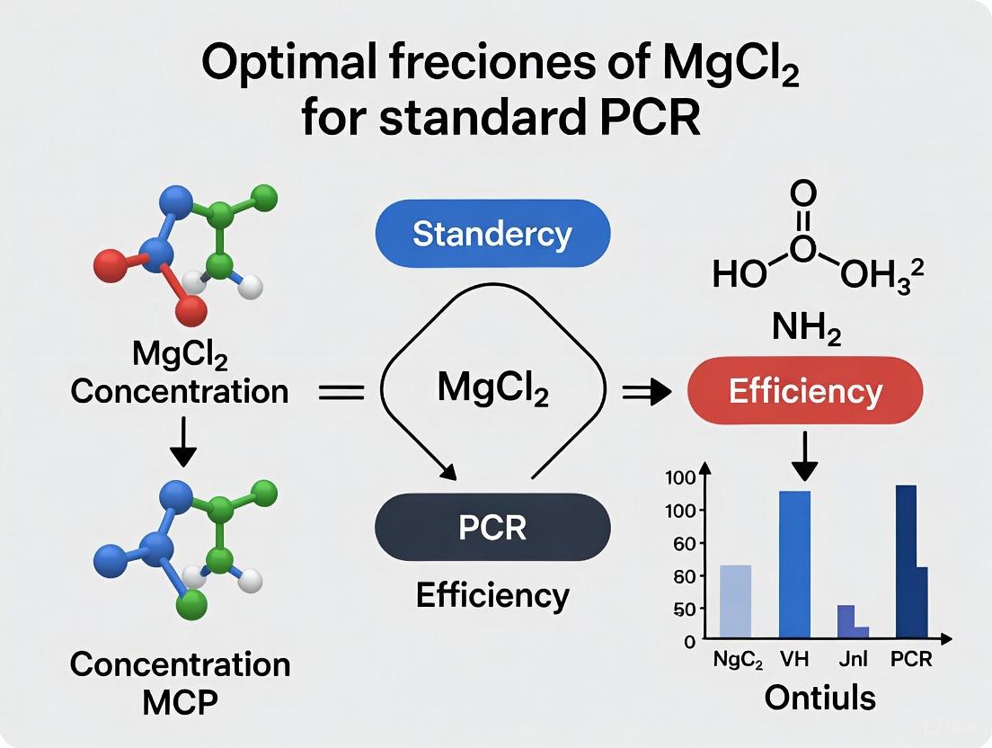
Optimizing MgCl₂ Concentration for Standard PCR: A Comprehensive Evidence-Based Guide for Researchers
This article provides a comprehensive, evidence-based guide for researchers and drug development professionals on optimizing magnesium chloride (MgCl₂) concentration in standard Polymerase Chain Reaction (PCR).
Optimizing MgCl₂ Concentration for Standard PCR: A Comprehensive Evidence-Based Guide for Researchers
Abstract
This article provides a comprehensive, evidence-based guide for researchers and drug development professionals on optimizing magnesium chloride (MgCl₂) concentration in standard Polymerase Chain Reaction (PCR). Synthesizing the latest research, including a 2025 meta-analysis, we detail the foundational role of Mg²⁺ as an essential polymerase cofactor, establish optimal concentration ranges (1.5-4.5 mM), and present methodological frameworks for systematic optimization tailored to template characteristics. The content covers advanced troubleshooting for common amplification issues, comparative analysis of buffer systems and DNA polymerases, and validation strategies to ensure reproducible, high-fidelity results for biomedical and clinical applications.
The Critical Role of MgCl₂ in PCR: Understanding the Biochemical Foundation
Mg²⁺ as an Essential Cofactor for DNA Polymerase Activity
Magnesium chloride (MgCl₂) is an indispensable component of the polymerase chain reaction (PCR), serving as a critical cofactor for DNA polymerase enzyme activity. This whitepaper synthesizes current research to define the optimal MgCl₂ concentration for standard PCR research, establishing a general range of 1.5 mM to 4.5 mM with a most common optimum near 2.0 mM [1] [2] [3]. The precise concentration, however, is not a single value but is significantly influenced by reaction-specific variables, particularly template DNA complexity [4] [5]. This guide provides researchers and drug development professionals with evidence-based guidelines, detailed methodologies, and optimization strategies to harness the full thermodynamic and kinetic potential of Mg²⁺ in nucleic acid amplification.
In the polymerase chain reaction, Mg²⁺ ions function as an essential catalytic cofactor for thermostable DNA polymerases such as Taq polymerase. The ion's primary role is to facilitate the formation of phosphodiester bonds between adjacent nucleotides during the extension phase of PCR [2]. Mechanistically, Mg²⁺ binds to the dNTP substrate at its alpha phosphate group, creating an electrostatically favorable complex that allows the 3'-hydroxyl group of the primer to execute a nucleophilic attack, thereby releasing pyrophosphate and incorporating the nucleotide into the growing DNA chain [2]. Without Mg²⁺, DNA polymerase exhibits minimal to no activity, leading to PCR failure [6].
Beyond its catalytic role, Mg²⁺ profoundly influences PCR thermodynamics by stabilizing nucleic acid interactions. The ion binds to the negatively charged phosphate backbone of DNA, effectively shielding the inherent electrostatic repulsion between the primer and template strands and between the two strands of the DNA duplex itself [6] [2]. This shielding effect increases the stability of the primer-template complex and raises the observed melting temperature (Tm), with a quantified relationship showing an approximately 1.2 °C increase in Tm for every 0.5 mM increase in MgCl₂ concentration within the critical 1.5-3.0 mM range [4] [5]. This dual functionality—catalytic and thermodynamic—makes precise MgCl₂ concentration a paramount variable for achieving optimal amplification efficiency, specificity, and yield in research applications.
Quantitative Analysis of Optimal MgCl₂ Concentrations
Established Concentration Ranges and Effects
A systematic meta-analysis of 61 peer-reviewed studies delineates a foundational MgCl₂ concentration range of 1.5 to 3.0 mM for efficient PCR performance [4] [5]. Broader analyses, accounting for a wider variety of templates and polymerase systems, extend this upper limit to 4.5 mM [1] [3]. The concentration must be meticulously optimized, as deviations directly impact outcomes.
Table 1: Effects of Suboptimal and Optimal MgCl₂ Concentrations in PCR
| Condition | Impact on Specificity | Impact on Yield | Overall Result |
|---|---|---|---|
| Too Low (<1.5 mM) | Primers fail to bind stably | Greatly reduced or failed amplification | Weak or no PCR product [1] [2] |
| Optimal (1.5-4.5 mM) | High specificity; primers bind correctly | Robust amplification of target | Strong, specific band(s) [4] [1] |
| Too High (>4.5 mM) | Decreased specificity; non-specific binding | Amplification of non-target products | Multiple bands or smears on a gel [1] [2] |
Template-Dependent Optimization Guidelines
The optimal MgCl₂ concentration is not universal but is contingent on the characteristics of the DNA template. Complex templates like genomic DNA (gDNA) generally require higher Mg²⁺ concentrations compared to simpler templates such as plasmid DNA or cDNA [4] [5]. This is attributed to the greater structural complexity and potential for secondary structure in gDNA, which requires more Mg²⁺ for effective strand separation and primer annealing. Furthermore, templates with high GC content often benefit from elevated MgCl₂, as the ion enhances the stability of GC-rich duplexes [2]. The presence of PCR inhibitors in the sample, such as those co-purified with DNA from complex biological samples, may also necessitate increased MgCl₂, as these inhibitors can chelate or otherwise sequester free Mg²⁺ ions, reducing their effective concentration [2].
Table 2: Template-Specific MgCl₂ Concentration Recommendations
| Template Type | Recommended [MgCl₂] Range | Rationale & Notes |
|---|---|---|
| Plasmid DNA, cDNA | 1.5 - 2.5 mM | Less complex structure; lower Mg²⁺ requirement [4] [7]. |
| Genomic DNA (gDNA) | 2.0 - 4.5 mM | Higher complexity and potential for inhibitors; requires more cofactor [4] [5]. |
| GC-Rich Templates | 2.5 - 4.5+ mM | Elevated Mg²⁺ stabilizes high-energy GC bonds; may require fine-tuning [2]. |
| Standard RAPD | 1.5 - 4.5 mM | Optimum is primer-dependent; requires empirical testing [3]. |
Experimental Protocol for MgCl₂ Optimization
Establishing a MgCl₂ Gradient
A robust optimization experiment involves setting up a MgCl₂ concentration gradient. The following protocol, adapted from standardized molecular biology methods, provides a systematic approach [8].
Materials & Reagents:
- DNA polymerase with its corresponding 10X reaction buffer (without MgCl₂).
- 25 mM MgCl₂ stock solution.
- DNA template (e.g., 10-100 ng genomic DNA/reaction).
- Forward and reverse primers (e.g., 10-20 pmol each per reaction).
- 10 mM dNTP mix.
- Nuclease-free water.
- Thermal cycler.
Procedure:
- Prepare a Master Mix: Calculate the reagents for (n+1) reactions, where 'n' is the number of MgCl₂ conditions to be tested. Combine in order:
- Nuclease-free water (to a final volume of 50 µL per reaction).
- 10X PCR Buffer (5 µL per reaction).
- 10 mM dNTP mix (1 µL per reaction).
- Forward Primer (e.g., 1 µL of 20 µM stock).
- Reverse Primer (e.g., 1 µL of 20 µM stock).
- DNA Polymerase (0.5-1.25 U per reaction).
- Mix gently by pipetting.
Aliquot the Master Mix: Dispense equal volumes (e.g., 45 µL) of the Master Mix into individual PCR tubes.
Add MgCl₂: Spike each tube with a different volume of the 25 mM MgCl₂ stock solution to create a final concentration gradient. For a 50 µL reaction:
- Tube 1 (1.0 mM): Add 2 µL of 25 mM MgCl₂.
- Tube 2 (1.5 mM): Add 3 µL of 25 mM MgCl₂.
- Tube 3 (2.0 mM): Add 4 µL of 25 mM MgCl₂.
- Tube 4 (2.5 mM): Add 5 µL of 25 mM MgCl₂.
- Tube 5 (3.0 mM): Add 6 µL of 25 mM MgCl₂.
- Tube 6 (4.0 mM): Add 8 µL of 25 mM MgCl₂.
- Tube 7 (5.0 mM): Add 10 µL of 25 mM MgCl₂.
Add Template and Run PCR: Add a consistent amount of DNA template to each tube. Include a negative control (no template) for one MgCl₂ concentration. Place tubes in a thermal cycler and run using standard cycling parameters appropriate for your primer pair and amplicon.
Analyze Results: Separate the PCR products by agarose gel electrophoresis. Identify the MgCl₂ concentration that yields the strongest target band with the least non-specific amplification or primer-dimer formation.
Workflow Visualization
The following diagram illustrates the logical workflow for the MgCl₂ optimization experiment.
The Scientist's Toolkit: Essential Research Reagents
Successful MgCl₂ optimization and PCR execution rely on high-quality, specific reagents. The following table details the essential materials required for the experiments described in this guide.
Table 3: Essential Reagents for PCR and MgCl₂ Optimization
| Reagent / Material | Typical Working Concentration | Critical Function |
|---|---|---|
| MgCl₂ Solution (e.g., 25 mM stock) [9] | 1.5 - 4.5 mM (final) | DNA polymerase cofactor; stabilizes primer-template binding. |
| DNA Polymerase (e.g., Taq Polymerase) [7] | 0.5 - 2.5 U/50 µL reaction | Enzyme that synthesizes new DNA strands. |
| 10X PCR Buffer (Mg²⁺-free) [8] | 1X (final) | Provides optimal pH and ionic strength (e.g., Tris-HCl, KCl). |
| dNTP Mix [7] | 200 µM each (final) | Building blocks (dATP, dCTP, dGTP, dTTP) for new DNA synthesis. |
| Oligonucleotide Primers [7] [8] | 0.1 - 1.0 µM (final) | Define the start and end points of the DNA segment to be amplified. |
| DNA Template (gDNA, plasmid, etc.) [7] | 1 pg - 1 µg (per 50 µL reaction) | The source DNA containing the target sequence to be copied. |
Mg²⁺ is far more than a simple buffer component; it is a foundational element that dictates the catalytic efficiency of DNA polymerase and the thermodynamic landscape of the entire PCR reaction. The "optimal" concentration, while generally falling between 1.5 and 4.5 mM, must be determined empirically for each novel primer-template system, with genomic and GC-rich templates typically demanding higher concentrations. The quantitative relationship between MgCl₂ and DNA melting temperature provides a theoretical framework for optimization, but the definitive test remains empirical analysis via a concentration gradient and gel electrophoresis. By adhering to the detailed protocols and guidelines presented herein, researchers can systematically overcome the challenge of MgCl₂ optimization, leading to highly specific, efficient, and reliable PCR outcomes essential for advanced research and drug development.
Within the polymerase chain reaction (PCR), the precise stabilization of the short, transient hybrid formed between the primer and the DNA template is a critical determinant of success. This process is profoundly influenced by magnesium ions (Mg²⁺), which act as a linchpin in the assembly of a functional replication complex. This technical guide delves into the molecular mechanisms by which Mg²⁺ stabilizes DNA duplexes, directly linking this fundamental interaction to the practical challenge of determining the optimal MgCl2 concentration for standard PCR research. We synthesize quantitative data on concentration-dependent effects, provide detailed methodologies for empirical optimization, and present visual models of the underlying mechanisms, providing researchers with a comprehensive framework to enhance the specificity and efficiency of their DNA amplification assays.
The polymerase chain reaction is an enzymatic process that hinges on a series of precisely orchestrated molecular interactions. Among the reagents required, magnesium chloride (MgCl2) is unique in its ability to influence nearly every stage of the amplification process, from initial primer binding to the final catalytic act of phosphodiester bond formation [2]. Its role extends beyond a simple cofactor; it is an integral structural and catalytic component of the PCR machinery.
The core challenge for researchers lies in the fact that Mg²⁺ concentration is not a one-size-fits-all parameter. The optimal concentration must be determined experimentally, as it is influenced by the specific primer-template system, the composition of the PCR buffer, and the concentration of other components that can chelate or otherwise interact with the free Mg²⁺ ions [10] [1]. This guide frames the discussion of Mg²⁺'s role in duplex stabilization within the essential research practice of MgCl2 optimization, which is critical for achieving high specificity and yield in standard PCR applications.
Molecular Mechanisms of Mg²⁺-Mediated Duplex Stabilization
The negatively charged phosphate backbone of DNA creates a natural electrostatic repulsion between two complementary nucleic acid strands, which is a significant barrier to the formation of the short-lived primer-template hybrid. Divalent cations like Mg²⁺ are exceptionally effective at mitigating this repulsion and facilitating duplex formation through direct and indirect mechanisms.
Electrostatic Shielding and Direct Binding
The primary mechanism by which Mg²⁺ stabilizes the DNA duplex is through electrostatic shielding. The positively charged Mg²⁺ ions congregate around the negatively charged phosphate groups, effectively neutralizing the repulsive forces that would otherwise prevent the primer from closely associating with the template strand [2]. This reduction in repulsion allows the two strands to come into close proximity, enabling the hydrogen bonding between complementary bases that defines the specific hybrid.
Furthermore, Mg²⁺ can engage in direct inner-sphere binding to specific atoms on the DNA bases and the phosphate oxygens, leading to the formation of structurally well-defined and stabilized complexes [11]. This direct coordination can subtly influence the geometry of the duplex, further enhancing its stability.
Thermodynamic and Kinetic Consequences
The stabilization provided by Mg²⁺ has direct thermodynamic consequences, most notably an increase in the melting temperature (Tm) of the primer-template hybrid. The Tm is defined as the temperature at which half of the DNA duplexes dissociate into single strands. A meta-analysis of PCR optimization studies revealed a clear logarithmic relationship between MgCl2 concentration and DNA melting temperature, with every 0.5 mM increase in MgCl2 within the 1.5 to 3.0 mM range associated with an average increase in Tm of 1.2 °C [5]. This increase in Tm permits the use of higher annealing temperatures in the PCR protocol, which in turn enhances the reaction specificity by preventing primers from binding to non-complementary or partially complementary sequences.
Table 1: Quantitative Effects of MgCl2 Concentration on PCR Thermodynamics and Output
| MgCl2 Concentration | Effect on DNA Tm | Effect on PCR Product | Typical Indication |
|---|---|---|---|
| Too Low (< 1.5 mM) | Reduced Tm | Weak or no amplification | Primer fails to bind template efficiently [2] [1] |
| Optimal (1.5 - 4.5 mM) | Maximal, specific Tm increase | Strong, specific amplification of the target amplicon | Ideal conditions for specific primer binding [5] [1] |
| Too High (> 4.5 mM) | Non-specific Tm increase | Non-specific bands; primer-dimer formation | Primers bind to incorrect sites [2] [1] |
From a kinetic perspective, the stabilized hybrid provides a more secure platform for the DNA polymerase to bind and initiate synthesis. Research on the Klenow fragment of DNA polymerase I indicates that Mg²⁺ ligands are crucial for the fingers-closing step that converts the open ternary complex into the closed, catalytically active conformation [11]. Thus, Mg²⁺-mediated duplex stabilization is a prerequisite for the subsequent structural transitions that lead to efficient catalysis.
Determining the Optimal MgCl2 Concentration for PCR
The established optimal range for MgCl2 in standard PCR lies between 1.5 mM and 4.5 mM, with 2.0 mM being a commonly used starting point [1]. However, achieving true optimization requires a systematic empirical approach, as the ideal concentration is dependent on the specific experimental context, including the primer sequence, template complexity, and the specific buffer composition.
The MgCl2 Optimization Experiment
A standard MgCl2 titration protocol is the most reliable method for identifying the optimal concentration for a given assay. The following provides a detailed methodology adapted from established optimization practices [10].
Research Reagent Solutions & Essential Materials
Table 2: Key Reagents for MgCl2 Optimization in PCR
| Reagent / Material | Function / Explanation |
|---|---|
| MgCl2 Stock Solutions | A set of solutions (e.g., 1 mM, 5 mM, 10 mM, 25 mM) to create a concentration gradient in the final reaction. |
| PCR Buffer (without MgCl2) | Provides the core chemical environment (pH, salt) but allows for the precise adjustment of Mg²⁺ concentration. |
| DNA Template | The sample containing the target sequence to be amplified. Quality and quantity must be consistent. |
| Forward & Reverse Primers | Short, specific DNA sequences that define the start and end of the target amplicon. |
| Thermostable DNA Polymerase | The enzyme that synthesizes new DNA strands (e.g., Taq polymerase). |
| dNTP Mix | The building blocks (dATP, dCTP, dGTP, dTTP) for the new DNA strands. |
| Thermal Cycler | The instrument that automates the temperature cycles for denaturation, annealing, and extension. |
| Agarose Gel Electrophoresis System | For visualizing and assessing the specificity and yield of the PCR products post-amplification. |
Experimental Protocol:
Preparation of Master Mix: Create a master mix containing all PCR components except for the DNA template and MgCl2. This includes nuclease-free water, PCR buffer (without MgCl2), dNTPs, primers, and DNA polymerase. Preparing a master mix ensures consistency across all reactions.
Aliquoting and MgCl2 Titration: Aliquot the master mix into individual PCR tubes. To each tube, add a different volume of a MgCl2 stock solution to create a final concentration gradient. A typical range to test is from 0.5 mM to 5.0 mM in 0.5 mM increments. Include a negative control (no template DNA) for each MgCl2 concentration to check for contamination.
Thermal Cycling: Add a constant amount of DNA template to each tube (except the negative controls) and initiate the PCR run using the standard thermal cycling program for your assay.
Product Analysis: Analyze the PCR products using agarose gel electrophoresis. Include a DNA ladder for size comparison. The goal is to identify the MgCl2 concentration that produces a single, intense band of the expected size with a clear or minimal background.
Data Interpretation and Troubleshooting
The results of the titration experiment will fall into one of several classic patterns, which directly reflect the mechanisms of Mg²⁺ action described in Section 2.
- Optimal Concentration: The MgCl2 concentration that yields a single, strong band of the correct size indicates successful optimization. At this point, the Mg²⁺ concentration is sufficient to stabilize the specific primer-template hybrid and support polymerase activity without promoting non-specific interactions.
- Sub-Optimal Concentration: Reactions with little to no visible product indicate that the Mg²⁺ concentration is too low. This leads to inefficient primer annealing and poor polymerase activity [2] [1].
- Supra-Optimal Concentration: Reactions showing multiple bands or a high-molecular-weight smear indicate excessive MgCl2. High concentrations stabilize any duplex, even those with mismatched bases, leading to non-specific amplification and primer-dimer artifacts [2] [1].
Table 3: Troubleshooting Guide for MgCl2 Concentrations in PCR
| Observed Result | Probable Cause | Recommended Action |
|---|---|---|
| No amplification band | MgCl2 concentration too low; primers cannot bind. | Increase MgCl2 concentration in 0.5 mM steps. Verify primer design and template quality. |
| Faint target band | MgCl2 concentration is sub-optimal. | Fine-tune MgCl2 increase (e.g., 0.2 mM steps). Optimize primer concentration and annealing temperature. |
| Multiple non-specific bands | MgCl2 concentration too high; low annealing stringency. | Decrease MgCl2 concentration. Increase the annealing temperature in the PCR protocol. |
| Primer-dimer formation | Excess MgCl2 and/or low annealing temperature. | Decrease MgCl2 and increase annealing temperature. Optimize primer design to avoid 3'-complementarity. |
It is critical to note that dNTPs also chelate Mg²⁺ ions. Therefore, the effective concentration of free Mg²⁺ available for the polymerase is the total Mg²⁺ minus the concentration chelated by dNTPs. Any change in the standard dNTP concentration in a protocol may necessitate a re-optimization of the MgCl2 concentration.
The influence of Mg²⁺ on the stabilization of primer-template hybrids is a cornerstone of successful PCR. By neutralizing electrostatic repulsion and increasing the duplex melting temperature, Mg²⁺ directly controls the specificity and efficiency of the initial annealing step. Moving beyond a generic "one-concentration-fits-all" approach to a systematic, empirical determination of the optimal MgCl2 level is a fundamental practice for any rigorous research program. The protocols and guidelines provided here offer a pathway to achieve such optimization, ensuring that the powerful technique of PCR delivers reliable and reproducible results for scientific and diagnostic applications.
The optimization of magnesium chloride (MgCl₂) concentration represents a critical parameter in polymerase chain reaction (PCR) protocol development, with its influence on DNA melting temperature (Tm) following a distinct logarithmic relationship. This technical guide synthesizes current evidence to delineate the quantitative effects of MgCl₂ on PCR thermodynamics and kinetics, providing researchers with a framework for evidence-based protocol optimization. Within the broader context of determining optimal MgCl₂ concentrations for standard PCR research, this review establishes that precise modulation of Mg²⁺ levels, tailored to specific template characteristics and reaction conditions, can significantly enhance both amplification efficiency and product specificity. The findings offer a robust theoretical foundation for developing template-specific optimization strategies and advancing the design of reliable, effective PCR protocols.
The Polymerase Chain Reaction (PCR) is a foundational technique in molecular biology, enabling the exponential amplification of specific DNA sequences. Its success hinges on the careful optimization of multiple reaction components, among which MgCl₂ is particularly crucial. Acting as an essential cofactor for DNA polymerase enzyme activity, Mg²⁺ ions facilitate the catalytic incorporation of nucleotides into the growing DNA strand [2]. Beyond its enzymatic role, MgCl₂ also critically influences the annealing efficiency of primers to the template DNA by stabilizing the primer-template duplex [7].
The central challenge in MgCl₂ optimization lies in its dualistic nature; both deficient and excessive concentrations can be detrimental to PCR outcomes. A concentration that is too low fails to support adequate polymerase activity and primer annealing, leading to weak or non-existent amplification [12]. Conversely, a concentration that is too high promotes non-specific primer binding, resulting in spurious amplification products and primer-dimer formation [12]. Consequently, understanding the quantitative relationship between MgCl₂ concentration and DNA melting temperature is paramount for predicting and controlling PCR behavior. This guide explores this relationship in depth, framing it within the overarching objective of determining optimal MgCl₂ concentrations for robust and specific PCR research applications.
The Biochemical Role of Mg²⁺ in PCR
The magnesium ion (Mg²⁺) is a multifunctional component in PCR, with its roles spanning enzymatic catalysis and nucleic acid stability.
Cofactor for DNA Polymerase
The primary role of Mg²⁺ is to serve as an essential cofactor for thermostable DNA polymerases like Taq polymerase. The ion is directly involved in the catalytic mechanism of the enzyme. During the extension phase of PCR, the Mg²⁺ ion facilitates the formation of the phosphodiester bond between the 3'-hydroxyl group of the primer and the phosphate group of the incoming deoxynucleoside triphosphate (dNTP) [2]. More specifically, Mg²⁺ binds to the dNTP at its alpha phosphate group, enabling the removal of beta and gamma phosphates and allowing the resulting dNMP to form a phosphodiester bond with the 3' OH of the adjacent nucleotide [2]. In the absence of Mg²⁺, DNA polymerase exhibits negligible activity, halting the amplification process entirely.
Influence on Nucleic Acid Thermodynamics
A second, equally critical function of Mg²⁺ is its influence on the thermodynamic stability of nucleic acids. The DNA backbone is highly negatively charged due to its phosphate groups, creating electrostatic repulsion between complementary strands [2]. Divalent cations like Mg²⁺ bind to the phosphate backbone, effectively shielding this negative charge and reducing inter-strand repulsion [2] [7]. This action stabilizes the double-stranded structure, leading to a significant increase in the observed melting temperature (Tm) of the DNA duplex. This effect is fundamental to the logarithmic relationship between MgCl₂ concentration and Tm, as it directly affects the efficiency with which primers anneal to their target sequences during the PCR cycling process.
The Logarithmic Relationship Between MgCl₂ and DNA Melting Temperature
Quantitative Analysis of the Relationship
A systematic meta-analysis of 61 peer-reviewed studies provides compelling quantitative evidence for the relationship between MgCl₂ concentration and DNA melting temperature. The analysis established a clear logarithmic relationship, with optimal PCR performance generally observed within a MgCl₂ concentration range of 1.5 to 3.0 mM [5]. Within this functional range, every 0.5 mM increase in MgCl₂ concentration is associated with an average increase in melting temperature of 1.2 °C [5]. This quantitative finding provides researchers with a predictive model for fine-tuning annealing temperatures during protocol optimization.
The underlying mechanism for this logarithmic effect involves the non-linear nature of electrostatic shielding. As Mg²⁺ concentration increases, the number of ions available to neutralize the negatively charged phosphate groups on the DNA backbone also increases, but the relationship is not linear. The initial additions of MgCl₂ have a more pronounced effect on Tm, with subsequent increments yielding progressively smaller increases in stability, which is characteristic of a logarithmic or saturation binding model.
The Impact of Template Complexity
The meta-analysis further revealed that the optimal MgCl₂ concentration is not universal but is significantly influenced by the complexity of the DNA template. Genomic DNA, with its large size and potential for complex secondary structures, typically requires higher concentrations of MgCl₂ compared to simpler templates like plasmid DNA or cDNA [5]. This is likely because complex templates present more binding sites for Mg²⁺ and may require greater stabilization to facilitate efficient primer annealing and polymerase progression.
Consequences of Mg²⁺ Deviation
Straying from the optimal MgCl₂ range has direct and measurable consequences on PCR yield and specificity, as summarized in the table below.
Table 1: Effects of MgCl₂ Concentration on PCR Outcomes
| MgCl₂ Status | Concentration Range | Impact on PCR Efficiency | Impact on PCR Specificity |
|---|---|---|---|
| Too Low | < 1.5 mM | Weak or failed amplification due to insufficient polymerase activity and poor primer annealing [12]. | Not applicable (no products). |
| Optimal | 1.5 - 4.0 mM | Efficient and robust amplification of the target sequence [13] [14]. | High specificity; clear band of expected size with minimal background [12]. |
| Too High | > 4.0 - 4.5 mM | May maintain strong amplification yield. | Increased non-specific binding, primer-dimer formation, and multiple background bands [12]. |
The general recommended range for standard PCR is 1.5 mM to 4.5 mM, with 2.0 mM being a common starting point for optimization [13] [2]. However, certain applications, such as those involving PCR inhibitors or templates with high GC content, may necessitate concentrations as high as 7 mM to compensate for ions that become sequestered and unavailable [13] [2].
Experimental Protocols for MgCl₂ Optimization
MgCl₂ Titration Methodology
A standard approach for empirically determining the optimal MgCl₂ concentration for a given PCR assay is through a titration experiment. The following protocol, adapted from commercial and research sources, provides a reliable methodology [14].
Table 2: Reagent Setup for a MgCl₂ Titration Master Mix
| Reagent | Volume for 8 reactions (of 20 µL) |
|---|---|
| 5X PCR Master Mix | 32 µL |
| PCR Grade Water | 9.6 µL |
| Primer 1 (10 µM) | 3.2 µL |
| Primer 2 (10 µM) | 3.2 µL |
| DNA Template | 16 µL |
| Total Volume | 64 µL |
Procedure:
- Prepare a master mix as detailed in Table 2, excluding additional MgCl₂. The master mix already contains a baseline concentration of MgCl₂ (e.g., 1.5 mM).
- Aliquot 8 µL of the master mix into each of seven labeled PCR tubes.
- Prepare a working stock of 5 mM MgCl₂ by diluting a 25 mM stock solution in PCR-grade water.
- To create a MgCl₂ concentration gradient, add the volumes of 5 mM MgCl₂ and PCR-grade water to each tube as specified in Table 3. The final volume in each tube should be 12 µL before adding the master mix.
- Add the 8 µL master mix aliquot to each tube, bringing the final reaction volume to 20 µL. Flick the tubes to mix thoroughly.
- Run the PCR using the standard thermal cycling parameters for your template and primers.
- Analyze the results using agarose gel electrophoresis. The optimal concentration is typically identified as the one that produces the clearest, most intense band of the expected size with the least non-specific amplification or smearing [14].
Table 3: Setup for Creating a MgCl₂ Concentration Gradient
| Final MgCl₂ Concentration | PCR Grade Water Added | 5 mM MgCl₂ Added |
|---|---|---|
| 1.5 mM (Baseline) | 12 µL | 0 µL |
| 2.0 mM | 10 µL | 2 µL |
| 2.5 mM | 8 µL | 4 µL |
| 3.0 mM | 6 µL | 6 µL |
| 3.5 mM | 4 µL | 8 µL |
| 4.0 mM | 2 µL | 10 µL |
| 4.5 mM | 0 µL | 12 µL |
Workflow and Decision-Making
The following diagram visualizes the experimental workflow for MgCl₂ optimization and the decision-making process based on the results.
The Scientist's Toolkit: Essential Reagents for PCR Optimization
Successful PCR optimization relies on a suite of high-quality reagents. Below is a table detailing the key components and their specific functions related to MgCl₂ optimization and overall reaction performance.
Table 4: Essential Reagents for PCR Optimization
| Reagent / Solution | Function / Role in Optimization |
|---|---|
| MgCl₂ Solution (25 mM) | Used as a supplement to titrate and optimize the final Mg²⁺ concentration in the PCR. Stored at -20°C [14]. |
| Thermostable DNA Polymerase | Enzyme that synthesizes new DNA strands. Its activity is absolutely dependent on Mg²⁺ as a cofactor [2] [8]. |
| dNTP Mix | The building blocks (dATP, dCTP, dGTP, dTTP) for new DNA synthesis. Mg²⁺ binds to dNTPs, so their concentration can affect free Mg²⁺ availability [7]. |
| Oligonucleotide Primers | Short, specific sequences that define the start and end of the target amplicon. Their annealing is stabilized by Mg²⁺ [7] [8]. |
| PCR Buffer (10X) | Provides the optimal pH and salt conditions (e.g., KCl, Tris-HCl) for the reaction. Often comes with a baseline MgCl₂ concentration [8]. |
| Nuclease-Free Water | Ensures the reaction is not degraded by nucleases and that the final volume is accurate. Critical for making precise dilutions [15]. |
The logarithmic relationship between MgCl₂ concentration and DNA melting temperature is a fundamental principle underpinning successful PCR optimization. The quantitative finding that a 0.5 mM increase in MgCl₂ raises the Tm by approximately 1.2°C provides a powerful predictive tool for researchers [5]. However, this relationship must be applied within the context of the specific reaction, as template complexity and other components like dNTPs directly influence the effective Mg²⁺ concentration.
Moving beyond a one-size-fits-all approach is crucial. The optimal MgCl₂ concentration for standard PCR research is not a single value, but a range—typically 1.5 mM to 4.0 mM—that must be empirically determined for each new set of primers and template [5] [13] [12]. The titration protocol and troubleshooting guidance provided herein offer a clear pathway for this essential empirical optimization. By systematically leveraging the quantitative effects of MgCl₂, scientists can significantly enhance the efficiency, specificity, and reliability of their PCR assays, thereby ensuring robust and reproducible results in their research and diagnostic endeavors.
The optimization of the Polymerase Chain Reaction (PCR) remains a central challenge in molecular biology, with the concentration of magnesium chloride (MgCl₂) being one of the most critical parameters determining its success [16] [4]. While Mg²⁺ is widely recognized as an essential cofactor for DNA polymerase activity, its functions extend far beyond simple polymerization, profoundly influencing the fundamental thermodynamics of the entire reaction system [4] [7]. A precise understanding of how Mg²⁺ modulates reaction kinetics and DNA duplex stability is crucial for developing efficient, reliable, and specific PCR protocols, particularly in pharmaceutical research and diagnostic applications where reproducibility is paramount [16] [17].
The multifaceted role of Mg²⁺ creates a complex optimization landscape. Its concentration affects multiple interdependent processes simultaneously, meaning that optimizing for one parameter may inadvertently compromise another [18]. This technical guide synthesizes current evidence to elucidate the thermodynamic mechanisms through which Mg²⁺ operates and provides evidence-based frameworks for determining the optimal MgCl₂ concentration for specific PCR applications within research contexts.
Multifunctional Roles of Mg²⁺ in PCR Thermodynamics
Essential Cofactor for DNA Polymerase Activity
The most established role of Mg²⁺ in PCR is as an indispensable cofactor for DNA polymerase enzyme activity. Without adequate free Mg²⁺, thermostable DNA polymerases exhibit minimal to no catalytic function [19]. The mechanistic basis for this requirement lies in the metal ion's direct participation in the phosphoryl transfer reaction during nucleotide incorporation [2] [7].
Mg²⁺ ions at the enzyme's active site facilitate the formation of phosphodiester bonds by coordinating the negative charges on the phosphate groups of incoming dNTPs [7]. This coordination catalyzes the nucleophilic attack of the 3'-hydroxyl group of the primer on the alpha-phosphate of the dNTP, enabling DNA chain elongation [2]. The binding of Mg²⁺ to dNTPs also creates the correct substrate conformation for efficient incorporation by the polymerase enzyme [2]. When Mg²⁺ concentration is insufficient, polymerase activity decreases significantly, leading to reduced amplification efficiency or complete PCR failure [2] [19].
Modulator of Nucleic Acid Thermodynamics
Beyond its enzymatic role, Mg²⁺ critically influences the stability and hybridization behavior of nucleic acids through its effect on DNA melting temperature (Tm) [16] [4]. Divalent magnesium cations stabilize the DNA double helix by neutralizing the negative charges on the phosphate backbone of DNA strands, thereby reducing the electrostatic repulsion between complementary strands [2] [7].
This charge neutralization has quantifiable effects on DNA duplex stability. Recent meta-analyses of 61 studies have demonstrated a strong logarithmic relationship between MgCl₂ concentration and DNA melting temperature, with every 0.5 mM increase in MgCl₂ within the 1.5-3.0 mM range associated with an approximately 1.2°C increase in melting temperature [16] [4] [5]. This modulation directly impacts the annealing efficiency of primers to template DNA and the denaturation efficiency of amplified products, making Mg²⁺ concentration a powerful tool for fine-tuning reaction specificity [16].
Influence on Reaction Fidelity and Specificity
The Mg²⁺ concentration presents a critical trade-off between amplification efficiency and reaction fidelity. Excess free Mg²⁺ reduces enzyme fidelity and increases the likelihood of nonspecific amplification [19]. This occurs because elevated Mg²⁺ concentrations overstabilize primer-template interactions, allowing primers to anneal to partially complementary sequences with sufficient stability to initiate polymerization [2] [18].
Conversely, insufficient Mg²⁺ not only limits polymerase activity but can also increase stringency to the point where specific primer-template interactions become unstable, resulting in weak or non-existent amplification of the desired target [2] [19]. This delicate balance necessitates precise optimization based on template characteristics and primer properties [16] [18].
Figure 1: Multifunctional Impact of Mg²⁺ on PCR Thermodynamics. The diagram illustrates how Mg²⁺ concentration simultaneously influences three critical aspects of PCR through different thermodynamic mechanisms.
Quantitative Guidelines for MgCl₂ Optimization
Evidence-Based Concentration Ranges
Comprehensive meta-analyses of peer-reviewed studies have established quantitative relationships between MgCl₂ concentration and PCR performance metrics [16] [4] [5]. The optimal concentration range depends on multiple factors, with template characteristics being particularly influential.
Table 1: Optimal MgCl₂ Concentration Ranges for Different Template Types
| Template Type | Optimal MgCl₂ Range | Key Considerations | Impact on Melting Temperature |
|---|---|---|---|
| Standard Templates | 1.5 - 3.0 mM | Balanced efficiency and specificity | +1.2°C per 0.5 mM increase [16] |
| Genomic DNA | Higher concentrations required | Increased complexity demands more Mg²⁺ [16] | More pronounced stabilization effect |
| GC-Rich Templates | Often >2.0 mM | Enhanced duplex stability needed [2] | Greater Tm increase per mM Mg²⁺ |
| Plasmid DNA | Lower concentrations often sufficient | Less complex structure [16] | Moderate Tm effect |
The 1.5-3.0 mM range represents the optimal starting point for most standard PCR applications, providing the best balance between amplification efficiency and product specificity [16] [4]. This range has been validated across multiple experimental systems and template types through systematic analysis [16].
Template-Dependent Optimization
Template characteristics significantly influence optimal Mg²⁺ requirements. Genomic DNA templates, with their higher complexity and longer sequences, typically require elevated MgCl₂ concentrations compared to simpler plasmid DNA or synthetic oligonucleotides [16]. The GC content of the target sequence also modulates Mg²⁺ requirements, as GC-rich sequences form more stable duplexes that may require fine-tuned Mg²⁺ concentrations for optimal amplification [2] [18].
Template quality represents another consideration, as samples containing PCR inhibitors (e.g., EDTA, citrate) may require higher MgCl₂ concentrations to compensate for chelation of free Mg²⁺ ions [2] [19]. The presence of such chelating agents effectively reduces the available Mg²⁺, necessitating adjustment of the total concentration added to the reaction mixture.
Experimental Optimization Methodologies
Systematic Titration Approach
A methodical titration of MgCl₂ concentration represents the most reliable approach for protocol optimization. The following step-by-step methodology adapts established best practices for determining template-specific optimal concentrations [16] [17] [18]:
Prepare Stock Solutions: Create a MgCl₂ stock solution series ranging from 0.5 mM to 5.0 mM in 0.5 mM increments. Include both lower and higher concentrations to define the response curve boundaries.
Setup Reaction Matrix: Using otherwise identical master mix aliquots, establish reactions across the MgCl₂ concentration series. Maintain consistent template, primer, dNTP, and polymerase concentrations across all reactions.
Amplification Parameters: Execute PCR using predetermined cycling conditions appropriate for the specific template and primer set. Avoid altering other thermal cycler parameters during Mg²⁺ optimization.
Product Analysis: Resolve amplification products by agarose gel electrophoresis. Alternatively, for quantitative assessment, utilize real-time PCR systems to monitor amplification efficiency.
Optimal Concentration Identification: Identify the MgCl₂ concentration that produces the strongest specific amplification with minimal nonspecific products or primer-dimer formation.
Figure 2: Experimental Workflow for Systematic Mg²⁺ Optimization. The step-by-step process illustrates the methodical approach for determining template-specific optimal MgCl₂ concentrations.
Integrated Factor Optimization
Mg²⁺ concentration does not operate in isolation but interacts with several other reaction components in determining PCR outcomes [7] [19]. These interactions must be considered during optimization:
- dNTP Concentration: Mg²⁺ binds to dNTPs in approximately equimolar ratios, reducing free Mg²⁺ availability. The total Mg²⁺ concentration should exceed the total dNTP concentration by 0.5-2.5 mM to maintain adequate free Mg²⁺ for polymerase function [7].
- Primer Design and Concentration: Primer characteristics influence Mg²⁺ requirements. Suboptimal primers with secondary structure or low specificity may appear to perform better at non-optimal Mg²⁺ concentrations, but primer redesign represents a superior solution [17] [18].
- Buffer Composition: Commercial PCR buffers often contain proprietary additives that can modulate Mg²⁺ effects. Consistency in buffer selection is important once optimal conditions are established [19].
Table 2: Interaction Effects Between MgCl₂ and Other PCR Components
| Component | Interaction with Mg²⁺ | Optimization Consideration |
|---|---|---|
| dNTPs | Mg²⁺ binds dNTPs (~1:1 ratio) | Maintain [Mg²⁺] > [total dNTP] + 0.5 mM [7] |
| Primers | Mg²⁺ stabilizes primer-template duplex | Higher Mg²⁺ may compensate for suboptimal primers [2] |
| Template Quality | Inhibitors may chelate Mg²⁺ | Increase Mg²⁺ with contaminated samples [2] |
| DNA Polymerase | Cofactor requirement varies by enzyme | Follow manufacturer recommendations [19] |
The Scientist's Toolkit: Research Reagent Solutions
Successful implementation of Mg²⁺ optimization requires specific laboratory reagents and methodologies. The following toolkit outlines essential materials and their functions for systematic investigation of Mg²⁺ effects in PCR thermodynamics.
Table 3: Essential Reagents and Methodologies for Mg²⁺ Optimization Research
| Reagent/Method | Function/Application | Technical Considerations |
|---|---|---|
| MgCl₂ Stock Solutions | Precise concentration manipulation | Prepare fresh, filter-sterilized stocks; avoid repeated freeze-thaw cycles |
| Magnesium-Free Buffers | Baseline for optimization | Available with many polymerase systems (e.g., Takara Ex Taq) [19] |
| Gradient Thermal Cycler | Parallel temperature optimization | Enables simultaneous testing of multiple annealing temperatures [17] |
| qPCR with Melting Curve Analysis | Quantification of amplification efficiency and specificity | Provides real-time assessment of Mg²⁺ effects on kinetics and products [20] |
| High-Fidelity DNA Polymerases | Assessment of fidelity impact | Monitor error rates at different Mg²⁺ concentrations [18] |
The roles of Mg²⁺ in PCR extend well beyond its function as a polymerase cofactor, encompassing critical influences on DNA duplex stability, reaction specificity, and overall amplification efficiency [16] [4] [7]. The optimal MgCl₂ concentration for standard PCR research falls within the evidence-based range of 1.5-3.0 mM, with template-specific adjustments required for complex genomic DNA, GC-rich targets, or suboptimal sample quality [16] [5].
The quantitative relationship between MgCl₂ concentration and DNA melting temperature—approximately 1.2°C increase per 0.5 mM within the optimal range—provides researchers with a predictable framework for systematic optimization [16] [4]. Through methodical titration and careful consideration of interacting factors, scientists can harness the multifunctional thermodynamic properties of Mg²⁺ to develop highly specific, efficient, and robust PCR protocols suitable for the exacting requirements of pharmaceutical research and diagnostic development.
Establishing Your MgCl₂ PCR Protocol: From Standard Ranges to Template-Specific Optimization
Magnesium chloride (MgCl₂) is an indispensable cofactor in the Polymerase Chain Reaction (PCR), serving dual functions that are critical for successful DNA amplification. Acting as a chemical chaperone, the magnesium ion (Mg²⁺) is essential for the catalytic activity of DNA polymerase enzymes; it facilitates the formation of phosphodiester bonds by enabling the nucleotide incorporation process [2]. Concurrently, Mg²⁺ influences the reaction thermodynamics by stabilizing the interaction between primers and the template DNA. It achieves this by binding to the negatively charged phosphate backbone of DNA, effectively reducing electrostatic repulsion and thereby increasing the melting temperature (Tm) for more stable duplex formation [5] [2]. The precise concentration of this ion is therefore not merely a component, but a pivotal regulator that balances reaction efficiency with amplification specificity.
Establishing the Standard Operating Range
A consensus across technical literature and commercial reagent guidelines identifies 1.5 mM to 4.5 mM as the standard operating range for MgCl₂ concentration in PCR, with 2.0 mM often serving as the most common starting point [21] [1] [13]. This range is supported by a comprehensive meta-analysis of 61 peer-reviewed studies, which confirmed that optimal concentrations predominantly fall between 1.5 and 3.0 mM [5]. The analysis further established a quantifiable logarithmic relationship between MgCl₂ concentration and DNA melting temperature, where every 0.5 mM increase within this range corresponds to an average increase of 1.2 °C in the melting temperature [5].
Deviating from this optimal range consistently produces suboptimal results. Excessive MgCl₂ (e.g., >4.5-5.0 mM) promotes non-specific primer binding, leading to spurious amplification products and primer-dimer formation [2] [1]. Conversely, insufficient MgCl₂ (e.g., <1.5 mM) can lead to complete PCR failure or dramatically reduced yield because primers fail to anneal effectively and the DNA polymerase enzyme lacks the cofactor required for catalysis [21] [2] [1].
Template-Dpecific Magnesium Requirements
The optimal concentration within the 1.5-4.5 mM window is not universal; it is significantly influenced by the nature of the DNA template. The meta-analysis by Tbahriti et al. provides quantitative evidence that template complexity dictates MgCl₂ requirements [5]. The following table summarizes the adjusted starting ranges based on template type, synthesizing data from the meta-analysis and manufacturer guidelines [5] [21].
Table 1: Template-Specific MgCl₂ Optimization Guidelines
| Template Type | Recommended MgCl₂ Starting Range | Rationale and Considerations |
|---|---|---|
| Plasmid or Viral DNA | 1.5 - 2.0 mM | Less complex templates require lower Mg²⁺ for specificity. Higher concentrations increase risk of non-specific bands. |
| Genomic DNA | 2.0 - 3.0 mM (or higher) | Increased complexity and size require higher Mg²⁺ concentrations to stabilize primer binding and polymerase processivity. |
| High GC-Content DNA | 2.5 - 4.5 mM | Elevated Mg²⁺ is needed to neutralize the stronger secondary structure and higher thermodynamic stability of GC-rich sequences. |
| PCR with Inhibitors | 3.0 - 4.5 mM | Higher Mg²⁺ compensates for ions chelated by impurities (e.g., from blood or soil) in the DNA sample. |
Experimental Protocol for MgCl₂ Optimization
A systematic approach to MgCl₂ optimization is fundamental for developing robust and reproducible PCR assays, particularly for novel targets or under standardized research conditions.
Reagent Preparation and Titration Setup
Begin by preparing a master mix containing all standard PCR components except MgCl₂ and the DNA template. The MgCl₂ is then titrated across a series of reactions. A standard optimization experiment involves setting up a dilution series of MgCl₂, typically in 0.5 mM increments, spanning from 1.0 mM to 5.0 mM [21] [18]. This granularity is sufficient to identify the optimal concentration without unnecessary effort.
Table 2: Example of a MgCl₂ Titration Series Setup for a 50 µL Reaction
| Tube | 1X PCR Buffer (µL) | dNTPs (µL) | Primers (µL) | Template (µL) | Taq Polymerase (µL) | MgCl₂ (25 mM Stock) (µL) | Water (µL) | Final [MgCl₂] |
|---|---|---|---|---|---|---|---|---|
| 1 | 5 | 1 | 2 | 2 | 0.5 | 1.0 | 38.5 | 1.0 mM |
| 2 | 5 | 1 | 2 | 2 | 0.5 | 1.5 | 38.0 | 1.5 mM |
| 3 | 5 | 1 | 2 | 2 | 0.5 | 2.0 | 37.5 | 2.0 mM |
| 4 | 5 | 1 | 2 | 2 | 0.5 | 2.5 | 37.0 | 2.5 mM |
| 5 | 5 | 1 | 2 | 2 | 0.5 | 3.0 | 36.5 | 3.0 mM |
| 6 | 5 | 1 | 2 | 2 | 0.5 | 3.5 | 36.0 | 3.5 mM |
| 7 | 5 | 1 | 2 | 2 | 0.5 | 4.0 | 35.5 | 4.0 mM |
| 8 | 5 | 1 | 2 | 2 | 0.5 | 4.5 | 35.0 | 4.5 mM |
| 9 | 5 | 1 | 2 | 2 | 0.5 | 5.0 | 34.5 | 5.0 mM |
Analysis and Interpretation of Results
Following thermocycling, the products from each titration point are analyzed using agarose gel electrophoresis [22]. The optimal MgCl₂ concentration is identified as the point that produces the following outcomes:
- A single, intense amplicon band of the expected size.
- Minimal to no non-specific products (e.g., extra bands of incorrect sizes).
- Absence of primer-dimer artifacts [21] [18] [2].
A diagram of the optimization workflow is presented below.
The Scientist's Toolkit: Essential Reagents for PCR Optimization
Successful PCR optimization relies on high-quality reagents. The following table details key components and their functions, with a specific focus on MgCl₂ solutions.
Table 3: Essential Reagents for PCR Optimization
| Reagent / Solution | Critical Function in PCR | Considerations for MgCl₂ Optimization |
|---|---|---|
| Magnesium Chloride (MgCl₂) | - Cofactor for DNA polymerase activity [2].- Stabilizes primer-template binding [5] [2]. | Use a dedicated, high-purity solution (e.g., 25 mM stock) for precise titration, separate from the core buffer [23]. |
| Thermostable DNA Polymerase (e.g., Taq) | Enzyme that synthesizes new DNA strands. | Its activity is directly dependent on Mg²⁺ concentration. Standard use is 0.5-2.0 units/50 µL reaction [21]. |
| dNTP Mix (dATP, dCTP, dGTP, dTTP) | Building blocks for new DNA synthesis. | dNTPs chelate Mg²⁺. A standard 0.2 mM concentration of each dNTP must be balanced by sufficient Mg²⁺ (typically in excess) [7]. |
| Oligonucleotide Primers | Define the start and end of the target sequence. | Primer design quality (Tm, GC%, length) influences the required Mg²⁺ for specific annealing. Final concentration typically 0.1-0.5 µM [21] [7]. |
| 10X PCR Buffer | Provides ionic strength and pH stability (e.g., Tris-HCl, KCl). | Many "PCR Buffer" formulations are supplied without MgCl₂ ("Mg-free"), allowing for flexible and precise optimization by the user [22] [23]. |
The empirical evidence firmly establishes 1.5 mM to 4.5 mM as the standard operating range for MgCl₂ concentration in PCR. This range, with a common starting point of 2.0 mM, provides a robust foundation for reaction setup. However, the ultimate optimal concentration is not a single value but a variable determined by a specific reaction's components, most notably the template DNA. Therefore, the systematic titration of MgCl₂ within this established range remains an indispensable, evidence-based practice for achieving maximum PCR efficiency, specificity, and reliability in scientific research and drug development.
In polymerase chain reaction (PCR) optimization, magnesium chloride (MgCl₂) is far more than a simple buffer component; it is a fundamental catalytic cofactor that directly dictates the success of DNA amplification. The magnesium ion (Mg²⁺) enhances the enzymatic activity of DNA polymerase and facilitates primer binding by stabilizing the interaction between primers and the template DNA [2]. However, a "one-size-fits-all" concentration proves ineffective across different template types. Template-driven optimization is therefore essential, as the structural complexity and sequence composition of the DNA template directly influence the optimal Mg²⁺ concentration required for efficient and specific amplification [5].
Genomic DNA (gDNA), with its vast size and complex secondary structures, presents different challenges compared to the smaller, supercoiled nature of plasmid DNA. A clear understanding of how these template characteristics influence Mg²⁺ requirements enables researchers to develop efficient and reliable protocols. This guide provides a detailed framework for adjusting Mg²⁺ concentrations based on template type, supported by quantitative data and step-by-step experimental protocols, to achieve robust PCR results in research and drug development.
Quantitative Mg²⁺ Requirements for Different DNA Templates
The optimal magnesium concentration is profoundly influenced by the nature of the DNA template. A comprehensive meta-analysis of peer-reviewed studies established clear distinctions in Mg²⁺ requirements, revealing a strong logarithmic relationship between MgCl₂ concentration and DNA melting temperature [5].
Table 1: Template-Specific Optimal MgCl₂ Concentrations and Their Effects
| Template Type | Recommended MgCl₂ Range | Typical Optimal Starting Point | Primary Effect of Deviation |
|---|---|---|---|
| Plasmid DNA | 1.5 – 2.5 mM [5] [24] | 1.5 – 2.0 mM [7] [24] | High concentration rapidly leads to non-specific bands and primer-dimers [7] [2]. |
| Genomic DNA (gDNA) | 1.5 – 3.0 mM [5] | 2.0 mM | Lower concentrations cause smearing and weak or failed amplification, especially with low-copy targets [5] [25]. |
| GC-Rich Templates | Up to 3.0 mM or higher [2] | 2.5 mM | Higher concentrations help denature strong secondary structures and facilitate polymerase processivity [2]. |
The meta-analysis showed that every 0.5 mM increase in MgCl₂ within the optimal range is associated with a 1.2 °C increase in melting temperature, which critically impacts primer annealing efficiency [5]. Genomic DNA templates consistently require higher Mg²⁺ concentrations than plasmid DNA due to their greater complexity and the presence of potential PCR inhibitors in the sample that can chelate and reduce the availability of free Mg²⁺ ions [5] [2].
Table 2: Troubleshooting PCR Based on Mg²⁺ Concentration and Template Type
| Observed Result | Likely Cause | Template-Specific Solution |
|---|---|---|
| Smearing on gel | Mg²⁺ concentration is too low, reducing polymerase activity and causing incomplete amplification [25]. | Particularly critical for gDNA and low-copy plasmids. Increase MgCl₂ in 0.5 mM increments [25]. |
| Multiple non-specific bands | Mg²⁺ concentration is too high, stabilizing weak primer-binding events [2] [24]. | More common with plasmid templates. Decrease MgCl₂ concentration and/or increase annealing temperature [24]. |
| No product | Severely limiting Mg²⁺, primer/Tm mismatch, or failed reaction [24]. | Verify Mg²⁺ is present. For gDNA, check template quality and increase Mg²⁺ if inhibitors are suspected [2] [26]. |
| Weak amplification | Suboptimal Mg²⁺, low template input, or low primer efficiency [7]. | For gDNA, titrate MgCl₂ upward. For plasmid, ensure template amount is 0.1–1 ng [7] [24]. |
Experimental Protocol for Systematic Mg²⁺ Optimization
A systematic approach to Mg²⁺ optimization is crucial for reconciling the differing requirements of genomic and plasmid DNA. The following step-by-step protocol ensures that the unique characteristics of each template are accounted for, leading to maximum yield and specificity.
Reagent Preparation and Primer Design
Begin by assembling high-quality reagents. Use a PCR buffer without Mg²⁺ to allow for precise concentration adjustments by adding MgCl₂ separately [25]. For the DNA template, ensure it is pure and intact. The recommended starting amount is 5–50 ng for genomic DNA and 0.1–1 ng for plasmid DNA in a 50 µL reaction [7] [24].
Primer design is equally critical. Primers should be 20–30 nucleotides in length with a melting temperature (Tm) between 55–70°C, and the Tms of the forward and reverse primer should be within 5°C of each other [7] [24]. The GC content should be ideally between 40–60% [7]. Using a Tm calculator based on nearest-neighbor thermodynamics and accounting for the actual monovalent (Na⁺ ~50 mM) and divalent (Mg²⁺ ~1.5-2.5 mM) ion concentrations in your buffer is essential for accurate Tm prediction, as salt concentrations significantly impact duplex stability [27].
Mg²⁺ Titration and Thermal Cycling
Prepare a series of PCR reactions with MgCl₂ concentrations spanning the expected optimal range. A standard titration series is recommended below.
Table 3: Example MgCl₂ Titration Series for Optimization
| Reaction Tube | 1 | 2 | 3 | 4 | 5 | 6 |
|---|---|---|---|---|---|---|
| Final MgCl₂ Concentration | 1.0 mM | 1.5 mM | 2.0 mM | 2.5 mM | 3.0 mM | 3.5 mM |
For genomic DNA, focus on the higher end of the range (e.g., 1.5 mM to 3.0 mM), while for plasmid DNA, the lower to middle range (e.g., 1.0 mM to 2.5 mM) is often sufficient [5] [24]. It is critical to keep all other reaction components—including template, primers, dNTPs, and polymerase—constant across all reactions.
Thermal cycling should then be performed. After an initial denaturation at 95°C for 2 minutes, run 25-35 cycles of:
- Denaturation: 95°C for 15-30 seconds.
- Annealing: Use a temperature 5°C below the calculated Tm of your primers for the first few cycles. If using a gradient thermal cycler, a temperature gradient can be applied simultaneously with the Mg²⁺ titration for multi-parameter optimization.
- Extension: 68–72°C for 1 minute per 1,000 base pairs [24].
This is followed by a final extension at 68–72°C for 5–10 minutes [24]. If specificity is a concern, consider using hot-start PCR to inhibit polymerase activity until the first denaturation step, thereby preventing non-specific amplification and primer-dimer formation during reaction setup [28].
Analysis and Validation of Results
Analyze the PCR products using agarose gel electrophoresis. The optimal condition is identified by the Mg²⁺ concentration that produces a single, sharp band of the expected size with minimal background smearing or non-specific products [25] [24].
For reactions where the initial titration does not yield a clear optimum, a second, finer titration (e.g., in 0.25 mM increments) around the best-performing concentration from the first experiment may be necessary. Once optimal conditions are identified, the amplified product should be purified to remove leftover primers, dNTPs, and enzymes before any downstream application, such as sequencing or cloning [26].
The workflow for this optimization process is summarized in the following diagram:
The Scientist's Toolkit: Essential Reagents for Mg²⁺ Optimization
Successful template-driven optimization relies on a set of key reagents and tools, each serving a specific function in the process.
Table 4: Essential Research Reagent Solutions for PCR Optimization
| Reagent / Tool | Function in Optimization | Key Considerations |
|---|---|---|
| MgCl₂ Stock Solution | To titrate the final Mg²⁺ concentration in the reaction without altering other buffer components. | Use a high-purity, sterile solution. Add separately from the 10X buffer for precise control [25]. |
| Mg²⁺-Free PCR Buffer | Provides the basic chemical environment (pH, salts) but allows flexible adjustment of Mg²⁺. | Essential for a controlled optimization experiment. Often supplied with the polymerase [24]. |
| High-Fidelity DNA Polymerase | Catalyzes DNA synthesis. Hot-start versions prevent non-specific amplification at room temperature. | 1–2 units per 50 µL reaction. Hot-start is recommended for improved specificity [7] [28]. |
| dNTP Mix | The building blocks (dATP, dCTP, dGTP, dTTP) for new DNA strands. | Typical concentration is 200 µM of each dNTP. Note that dNTPs chelate Mg²⁺, reducing its free concentration [7] [24]. |
| Template DNA | The DNA to be amplified (gDNA or plasmid). | Quality and quantity are critical. Use spectrophotometry/fluorometry and gel electrophoresis for QC [26]. |
| Oligonucleotide Primers | Define the start and end of the target sequence to be amplified. | Design for specificity and uniform Tm. Purify by HPLC or gel filtration for critical applications [7] [27]. |
| Thermal Cycler with Gradient | Instrument that automates PCR temperature cycles. | A gradient function allows simultaneous testing of a range of annealing temperatures alongside Mg²⁺ titration. |
The paradigm of template-driven optimization underscores that Mg²⁺ concentration is not a universal setting but a variable finely tuned to template characteristics. Genomic DNA, with its inherent complexity and potential for inhibitors, systematically demands higher MgCl₂ concentrations (1.5–3.0 mM) than the simpler plasmid templates (1.5–2.5 mM) [5]. Adhering to a rigorous optimization workflow involving Mg²⁺ titration, coupled with careful attention to primer design and reaction conditions, allows researchers to transform PCR from an unpredictable reaction into a robust and reliable technique. This precision is fundamental to achieving consistent and valid results in genetic analysis, diagnostic assay development, and therapeutic drug discovery.
The polymerase chain reaction (PCR) is a foundational technique in molecular biology, yet its success critically depends on the precise optimization of reaction components. Central to this is the molar balance between magnesium ions (Mg²⁺) and deoxynucleoside triphosphates (dNTPs). This technical guide details the biochemical principles, quantitative relationships, and practical methodologies for calculating and optimizing the dNTP-Mg²⁺ ratio to achieve specific and efficient DNA amplification. Framed within the broader objective of determining optimal MgCl₂ concentration for standard PCR, this whitepaper provides researchers and drug development professionals with evidence-based protocols and troubleshooting frameworks to enhance experimental reproducibility and outcomes.
In PCR, magnesium chloride (MgCl₂) is not merely a buffer component but an essential cofactor for DNA polymerase activity. The Mg²⁺ ion facilitates the catalytic function of the enzyme by enabling the formation of a productive complex between the polymerase and the dNTP substrate [2]. Specifically, the ion binds to the phosphate groups of dNTPs, neutralizing their negative charge and making them suitable substrates for the nucleophilic attack by the 3'-OH group of the primer terminus [29] [30].
However, the relationship is complicated by a critical interaction: Mg²⁺ chelates dNTPs within the reaction mixture. Consequently, the total Mg²⁺ concentration is partitioned into a fraction that is tightly bound to dNTPs and a remaining fraction that exists as free Mg²⁺. It is this free Mg²⁺ that is available to act as a cofactor for the DNA polymerase enzyme [29]. An imbalance in this ratio directly impacts reaction efficiency, influencing specificity, fidelity, and yield. Therefore, optimizing PCR is not about determining absolute concentrations of either component in isolation, but about defining the precise molar ratio that maintains an optimal concentration of free Mg²⁺.
Biochemical Principles of the dNTP-Mg²⁺ Interaction
Molecular Mechanisms of Magnesium in PCR
Magnesium ions play two non-redundant roles in the PCR process, both crucial for successful amplification:
Catalytic Cofactor for Polymerase Activity: At the enzyme's active site, the Mg²⁺ ion coordinates with the phosphate groups of the incoming dNTP, facilitating the removal of the beta and gamma phosphates and the subsequent formation of a phosphodiester bond with the growing DNA chain [2]. Without this, the polymerase exhibits drastically reduced activity or is completely inactive.
Nucleic Acid Stabilizer: Mg²⁺ stabilizes the double-stranded DNA structure by neutralizing the negative charge of the phosphate backbone. This reduces electrostatic repulsion between the primer and the template strand, thereby lowering the melting temperature (Tm) and facilitating proper annealing [27] [2]. The ion's concentration thus directly influences the stringency of primer binding.
The Chelation Effect and Free Mg²⁺ Concentration
The core challenge in optimization stems from the strong chelation of Mg²⁺ by dNTPs. Each dNTP molecule can bind a Mg²⁺ ion, meaning the total dNTP concentration directly depletes the pool of available Mg²⁺. The following diagram illustrates the competitive dynamics of Mg²⁺ binding in a PCR reaction and its critical outcomes.
This competition means that the free Mg²⁺ concentration is the critical variable. A reaction with a high dNTP concentration requires a correspondingly high total Mg²⁺ concentration to ensure a sufficient residual level of free ions for the polymerase. A meta-analysis of optimization studies confirms that modulating this balance is one of the most effective ways to troubleshoot PCR performance [5].
Quantitative Guidelines for dNTP-Mg²⁺ Ratios
Successful PCR optimization requires a foundation in standard concentration ranges and an understanding of how their interplay affects the reaction. The following table summarizes the standard and optimal concentrations for key components in a typical PCR setup.
Table 1: Standard Concentration Ranges for Key PCR Components
| Component | Standard Range | Common Starting Point | Key Consideration |
|---|---|---|---|
| MgCl₂ | 1.0 – 5.0 mM [31] [14] | 1.5 mM [14] | Concentration refers to total Mg²⁺; a portion is chelated by dNTPs. |
| dNTPs (each) | 0.05 – 0.2 mM [30] [31] | 0.2 mM [31] | High dNTPs chelate Mg²⁺, reducing free [Mg²⁺]; low dNTPs cause early exhaustion. |
| Free Mg²⁺ | ~0.5 – 2.5 mM (inferred) | ~1.0 mM (estimated) | The critical variable for polymerase activity; not directly added but a result of balance. |
Calculating the dNTP-Mg²⁺ Molar Relationship
The chelation of Mg²⁺ by dNTPs is a stoichiometric relationship. To a first approximation, each dNTP molecule binds one Mg²⁺ ion. Therefore, the molar amount of Mg²⁺ chelated is roughly equal to the total molar amount of dNTPs.
The formula for estimating the free Mg²⁺ concentration is:
[Free Mg²⁺] ≈ [Total Mg²⁺] - [Total dNTPs]
Sample Calculation: For a 50 µL reaction with:
- Total Mg²⁺ = 2.0 mM (or 100 nmol in 50 µL)
- Total dNTPs = 0.8 mM (0.2 mM each of dATP, dCTP, dGTP, dTTP, or 40 nmol in 50 µL)
The estimated Free Mg²⁺ = 2.0 mM - 0.8 mM = 1.2 mM
This 1.2 mM of free Mg²⁺ is the concentration available to serve as a polymerase cofactor and stabilize the nucleic acids. This example illustrates a likely functional balance. A meta-analysis of 61 studies established that an optimal free Mg²⁺ concentration typically lies between 1.5 and 3.0 mM, with every 0.5 mM increase within this range increasing the DNA melting temperature by approximately 1.2°C [5].
Effects of Imbalance and Template-Specific Considerations
Deviations from the optimal balance have predictable consequences, which are summarized in the table below.
Table 2: Troubleshooting PCR Based on dNTP-Mg²⁺ Balance
| Condition | Effect on Free Mg²⁺ | Observed PCR Result | Corrective Action |
|---|---|---|---|
| High [dNTPs] | Too Low | Reduced or failed amplification; smearing on gel [25] | Increase Total [Mg²⁺] and/or decrease [dNTPs] |
| Low [dNTPs] | Too High | Non-specific amplification; primer-dimer formation [29] [2] | Decrease Total [Mg²⁺] and/or increase [dNTPs] |
| High [Mg²⁺] | Too High | Non-specific bands; accumulation of misprimed products [29] [14] | Decrease Total [Mg²⁺] |
| Low [Mg²⁺] | Too Low | Weak or no amplification; smearing [25] | Increase Total [Mg²⁺] |
Furthermore, the optimal ratio can be influenced by template characteristics. Complex templates like genomic DNA often require higher total Mg²⁺ concentrations (and thus a different ratio) compared to simpler plasmid DNA templates [5]. Similarly, templates with high GC content may benefit from slightly elevated Mg²⁺ levels to stabilize the higher number of hydrogen bonds [2].
Experimental Protocol for Systematic Optimization
For novel PCR applications or when troubleshooting persists, empirical optimization is necessary. The following protocol provides a robust method for determining the optimal Mg²⁺ concentration.
Mg²⁺ Titration Gradient Setup
This protocol is designed to identify the optimal MgCl₂ concentration for a specific primer-template system [14].
Research Reagent Solutions
| Item | Function in Experiment |
|---|---|
| MgCl₂ (25 mM stock) | Used to create a gradient of final Mg²⁺ concentrations. |
| PCR Buffer (without Mg²⁺) | Provides stable pH and ionic strength; allows precise Mg²⁺ adjustment. |
| Hot-Start DNA Polymerase | Reduces non-specific amplification during reaction setup [29]. |
| dNTP Mix (e.g., 10 mM) | Source of nucleotides; concentration must be kept constant during Mg²⁺ titration. |
| Template DNA & Primers | The specific targets whose amplification is being optimized. |
Step-by-Step Procedure:
Prepare a Master Mix: Combine all common reaction components except MgCl₂ and water in a 1.5 mL tube. Calculations are for a single 20 µL reaction; multiply by the number of tests plus an excess to account for pipetting error.
- PCR Buffer (without Mg²⁺): 1X final concentration
- Forward Primer (10 µM): 0.2 µM final concentration
- Reverse Primer (10 µM): 0.2 µM final concentration
- dNTP Mix (10 mM): 0.2 mM final concentration for each dNTP
- Template DNA: variable (e.g., 5–50 ng genomic DNA)
- Hot-Start DNA Polymerase: as per manufacturer's unit recommendation
- Nuclease-free water: to a final volume of 20 µL after Mg²⁺ addition
Aliquot the Master Mix: Dispense equal volumes of the Master Mix into each PCR tube designated for the gradient.
Create the Mg²⁺ Gradient: Prepare a working stock of 5 mM MgCl₂ by diluting the 25 mM stock. Add different volumes of the 5 mM MgCl₂ stock and water to each tube to achieve the desired final concentration gradient, as shown in the workflow below.
Table 3: Example Setup for a Mg²⁺ Titration in a 20 µL Reaction [14]
| Final [MgCl₂] (mM) | Volume of 5 mM MgCl₂ (µL) | Volume of Nuclease-Free Water (µL) | Total Volume with Master Mix (µL) |
|---|---|---|---|
| 1.5 | 0.0 | 12.0 | 20 |
| 2.0 | 2.0 | 10.0 | 20 |
| 2.5 | 4.0 | 8.0 | 20 |
| 3.0 | 6.0 | 6.0 | 20 |
| 3.5 | 8.0 | 4.0 | 20 |
| 4.0 | 10.0 | 2.0 | 20 |
| 4.5 | 12.0 | 0.0 | 20 |
Run the PCR: Place the tubes in a thermal cycler and run the appropriate amplification protocol, ideally using a temperature gradient for the annealing step to concurrently optimize this parameter.
Analyze Results: Resolve the PCR products using agarose gel electrophoresis. The optimal MgCl₂ concentration is the one that produces a sharp, intense band of the expected size with the least amount of background smearing or non-specific bands [14].
Advanced Applications and Considerations
Specialized PCR Formulations
The "Hot Start – With Buffer – With MgCl₂ – Without dNTP" system exemplifies a premium approach for maximum experimental control. This configuration allows researchers to independently titrate dNTPs and Mg²⁺, which is crucial for advanced applications [29]:
- Incorporation of Modified Nucleotides: Fluorescently labelled or biotinylated dNTPs often have different incorporation efficiencies and may alter Mg²⁺ binding kinetics.
- Site-Directed Mutagenesis: Intentional manipulation of dNTP ratios to promote misincorporation requires precise, independent control over Mg²⁺ to manage fidelity.
- Long-Range PCR: Delaying dNTP addition until after the initial high-temperature denaturation can enhance specificity for long amplicons.
Impact on Fidelity and High-Fidelity PCR
The dNTP-Mg²⁺ balance directly influences the error rate of DNA polymerases. For standard, non-proofreading enzymes like Taq, fidelity can be improved by using lower dNTP concentrations (0.01–0.05 mM each), which reduces misincorporation rates [30]. This must be accompanied by a proportional reduction in Mg²⁺ to maintain the free Mg²⁺ level required for basal polymerase activity. When using proofreading enzymes for high-fidelity PCR, always consult the manufacturer's specific recommendations for both dNTP and Mg²⁺ concentrations, as these systems are often finely tuned.
Achieving optimal PCR efficiency and specificity is a exercise in biochemical balance. The molar ratio between Mg²⁺ and dNTPs is not a mere suggestion but a fundamental determinant of success, as it directly governs the concentration of free Mg²⁺ available to catalyze DNA synthesis and stabilize reaction intermediates. The standard starting point of 1.5-2.0 mM Mg²⁺ and 0.2 mM of each dNTP provides a functional foundation, but true optimization requires a systematic, empirical approach tailored to the specific template, primers, and enzyme in use. By understanding the principles and applying the titration protocols outlined in this guide, researchers can transform PCR from an unpredictable art into a robust and reproducible scientific tool, thereby accelerating discovery and development in diagnostics and therapeutics.
In the realm of molecular biology, the polymerase chain reaction (PCR) stands as a foundational technique, yet achieving optimal conditions remains a persistent challenge. The concentration of magnesium chloride (MgCl₂) represents one of the most crucial variables influencing reaction success, acting as a key cofactor for DNA polymerase activity and significantly impacting DNA strand separation dynamics [4] [2]. The precise modulation of MgCl₂ concentration, tailored to specific experimental parameters, can dramatically improve both the efficiency and specificity of PCR amplification [5].
This guide provides a systematic framework for the empirical determination of optimal MgCl₂ concentration, moving beyond standardized protocols to address the unique requirements of individual template-primer systems. The deceptive simplicity of PCR often belies the complexity of its biochemical underpinnings, where Mg²⁺ ions participate directly in the catalytic mechanism of phosphodiester bond formation and stabilize the interaction between primers and template DNA [2] [7]. Understanding these relationships is particularly crucial for challenging templates, specialized applications, and rigorous scientific reproducibility [4] [32].
MgCl₂ in PCR: Mechanism and Concentration Effects
Biochemical Mechanisms
Magnesium ions serve as an essential cofactor for DNA polymerase activity, without which the enzyme demonstrates minimal functionality [2]. The molecular mechanism involves Mg²⁺ ions binding to dNTPs at the alpha phosphate group, facilitating the removal of beta and gamma phosphates and enabling the resulting dNMP to form a phosphodiester bond with the 3' hydroxyl group of the adjacent nucleotide [2]. Additionally, MgCl₂ significantly influences primer binding specificity by modulating the primer melting temperature (Tm). The positively charged magnesium ions bind to negatively charged phosphate groups in the DNA backbone, reducing electrostatic repulsion between complementary strands and thereby promoting stable hybridization [2].
Effects of Suboptimal Concentrations
The consequences of improper MgCl₂ concentration manifest in distinct amplification artifacts:
- Excessive MgCl₂ (typically >3.0-4.5 mM, depending on the system) promotes non-specific primer binding, resulting in erroneous DNA replication and multiple extraneous bands on agarose gels [1] [2]. In some cases, elevated concentrations may also foster primer-dimer formation [2].
- Insufficient MgCl₂ (generally <1.5 mM) impedes primer annealing and polymerase activity, leading to weak amplification or complete PCR failure as primers cannot effectively base pair with the DNA template [1] [2].
Quantitative analysis reveals a strong logarithmic relationship between MgCl₂ concentration and DNA melting temperature, with every 0.5 mM increase in MgCl₂ within the 1.5-3.0 mM range associated with an approximately 1.2°C increase in melting temperature [4] [5].
Table 1: Effects of MgCl₂ Concentration on PCR Performance
| MgCl₂ Concentration | Amplification Efficiency | Specificity | Common Artifacts |
|---|---|---|---|
| <1.5 mM | Significantly reduced | High | Faint or no bands; PCR failure |
| 1.5–3.0 mM | Optimal | High | Clean, specific bands |
| 3.0–4.5 mM | High | Reduced | Minor non-specific products |
| >4.5 mM | Variable | Poor | Multiple bands; primer dimers |
Establishing the Titration Framework
Preliminary Considerations
Before commencing empirical titration, researchers must recognize that template characteristics profoundly influence optimal MgCl₂ requirements. Meta-analysis of optimization studies demonstrates that genomic DNA templates typically require higher MgCl₂ concentrations than simpler plasmid DNA or synthetic oligonucleotides [4] [5]. Template complexity, GC content, and amplicon length collectively contribute to these differential requirements. Similarly, the presence of PCR inhibitors in DNA extracts may necessitate increased MgCl₂ concentrations, as these compounds often chelate Mg²⁺ ions and reduce their effective availability [2].
The experimental design should account for these variables through appropriate control reactions and replicate sampling. For novel template-primer systems, initial optimization is always recommended, while established protocols may require re-optimization when transitioning between template types, thermal cyclers, or reagent lots [33] [8].
Recommended Titration Range
Based on comprehensive meta-analysis of 61 peer-reviewed studies, the optimal MgCl₂ concentration for most PCR applications falls within 1.5 to 4.5 mM [1] [4] [3]. The narrower evidence-based range of 1.5 to 3.0 mM represents the ideal starting point for systematic optimization [4] [5]. The titration series should encompass a minimum of 6-8 concentrations within this spectrum to adequately capture the concentration-response relationship.
Table 2: Template-Specific MgCl₂ Concentration Guidelines
| Template Type | Recommended Starting Range | Notes |
|---|---|---|
| Plasmid DNA | 1.5–2.5 mM | Less complex templates generally require lower concentrations |
| cDNA | 1.5–3.0 mM | Dependent on reverse transcription efficiency |
| Genomic DNA | 2.0–3.5 mM | Higher complexity necessitates increased Mg²⁺ |
| GC-rich templates | 2.5–4.0 mM | Increased stability of DNA duplex requires higher Mg²⁺ |
| Multiplex PCR | 2.5–4.5 mM | Multiple primer pairs may require elevated concentrations |
Experimental Protocol: Systematic MgCl₂ Titration
Reagent Preparation
Establish a master mix containing all common reaction components to minimize pipetting error and ensure reaction uniformity. The following protocol assumes a standard 50 μL reaction volume [8]:
- 10X PCR Buffer (without MgCl₂): 5 μL
- dNTP Mix (10 mM total): 1 μL
- Forward Primer (20 μM): 1 μL
- Reverse Primer (20 μM): 1 μL
- DNA Template: 0.5–5 μL (optimized for specific template)
- DNA Polymerase: 0.5–2.5 units
- Sterile Water: variable, to adjust final volume
- MgCl₂ Stock Solution (25 mM): variable, to achieve desired final concentration
The MgCl₂ stock solution should be prepared accurately and quality-controlled, as the precision of this component directly influences titration reliability.
Titration Series Setup
Prepare a dilution series of MgCl₂ to achieve the following final concentrations in individual reaction tubes:
Table 3: Recommended Titration Series
| Tube Number | Volume of 25 mM MgCl₂ Stock (μL) | Final Concentration (mM) |
|---|---|---|
| 1 | 3.0 | 1.5 |
| 2 | 4.0 | 2.0 |
| 3 | 5.0 | 2.5 |
| 4 | 6.0 | 3.0 |
| 5 | 7.0 | 3.5 |
| 6 | 8.0 | 4.0 |
| 7 | 9.0 | 4.5 |
Include a negative control (no template DNA) for each MgCl₂ concentration to monitor contamination and primer-dimer formation. For critical applications, technical replicates (n=3) are recommended to assess reproducibility [8] [32].
Thermal Cycling Parameters
Execute PCR amplification using standardized cycling conditions appropriate for your specific primer-template system:
- Initial Denaturation: 94–95°C for 2–5 minutes
- Amplification Cycles (25–35 cycles):
- Denaturation: 94–95°C for 30–60 seconds
- Annealing: Primer-specific temperature (50–65°C) for 30–60 seconds
- Extension: 72°C for 1 minute per kb of amplicon
- Final Extension: 72°C for 5–10 minutes
- Hold: 4–10°C indefinitely
Maintain consistent thermal cycling parameters across the entire titration series to isolate the effect of MgCl₂ concentration [33] [8].
Analysis and Interpretation
Resolve PCR products by agarose gel electrophoresis and quantify band intensity to determine optimal amplification conditions. The ideal MgCl₂ concentration produces a single, robust band of the expected size with minimal background or non-specific amplification [1] [8].
For quantitative applications, determine amplification efficiency by generating standard curves with serial template dilutions at each MgCl₂ concentration. Calculate efficiency using the formula: E = 10(-1/slope), with ideal efficiency approaching 100% (E=2) [32]. Modern meta-analysis suggests that robust assays should demonstrate linearity over at least six orders of magnitude with correlation coefficients (R²) >0.98 [4] [32].
Advanced Optimization Strategies
Template-Specific Modifications
Template characteristics significantly influence optimal MgCl₂ requirements. GC-rich templates often necessitate elevated MgCl₂ concentrations (2.5–4.0 mM) to counteract the increased stability of DNA duplexes [4]. For long amplicon amplification (>5 kb), incremental MgCl₂ increases (0.5 mM steps) within the 2.0–4.0 mM range may improve yield while maintaining specificity. When working with multiplex PCR systems employing multiple primer pairs, expanded titration series (2.5–4.5 mM) are recommended to accommodate the diverse thermodynamic requirements of simultaneous amplification [2].
Complementary Buffer Modifications
MgCl₂ concentration interacts with other buffer components, particularly dNTPs, which chelate Mg²⁺ ions and reduce effective concentration [7]. The molar relationship between Mg²⁺ and dNTPs should be maintained such that [Mg²⁺]free = [Mg²⁺]total - [dNTP]total remains positive. For specialized applications requiring enhanced specificity, additive incorporation may prove beneficial:
- DMSO (1–10%): Enhances amplification of GC-rich templates
- Betaine (0.5–2.5 M): Reduces secondary structure formation
- Formamide (1.25–10%): Improves stringency
- BSA (10–100 μg/mL): Counteracts inhibitors in DNA preparations [8]
These additives may modify MgCl₂ requirements, necessitating iterative optimization when introducing them to established protocols.
The Scientist's Toolkit: Essential Reagents and Materials
Table 4: Essential Research Reagent Solutions for MgCl₂ Titration
| Reagent/Material | Function in Optimization | Recommended Specifications |
|---|---|---|
| MgCl₂ Stock Solution | Magnesium ion source for titration | Molecular biology grade, 25 mM in sterile water |
| 10X PCR Buffer (Mg-free) | Reaction environment foundation | Supplied with enzyme or prepared without Mg²⁺ |
| dNTP Mix | DNA synthesis substrates | Balanced 10 mM solution, pH 7.0 |
| DNA Polymerase | Enzymatic DNA amplification | Thermostable (e.g., Taq), high purity |
| Primer Pair | Sequence-specific amplification | HPLC-purified, 20 μM working stocks |
| Template DNA | Target for amplification | Quantified, minimal inhibitor content |
| Agarose | Electrophoretic separation | Molecular biology grade, 1-3% gels |
| Nucleic Acid Stain | DNA visualization | Intercalating dye (e.g., ethidium bromide) |
| Molecular Weight Marker | Product size determination | DNA ladder covering expected amplicon size |
Systematic titration of MgCl₂ represents a fundamental empirical approach for optimizing PCR efficiency and specificity. The protocol outlined herein provides a rigorous framework for establishing template-specific and application-appropriate magnesium concentrations, moving beyond standardized conditions to address the unique requirements of individual experimental systems. Through methodical concentration testing and comprehensive result analysis, researchers can achieve the delicate balance between amplification yield and reaction specificity that defines robust, reproducible molecular assays.
The quantitative relationships and methodological principles detailed in this guide support the development of evidence-based PCR protocols, contributing to improved experimental reproducibility across the life sciences. As PCR technologies continue to evolve, the fundamental importance of Mg²⁺ optimization remains constant, underpinning successful genetic analysis, diagnostic testing, and fundamental biological research [4] [5].
Troubleshooting MgCl₂-Related PCR Failure: A Problem-Solving Framework
The polymerase chain reaction (PCR) stands as a cornerstone technique in molecular biology, with its success critically dependent on the precise optimization of reaction components. Among these, magnesium chloride (MgCl₂) concentration represents one of the most crucial yet frequently variable parameters affecting reaction outcomes. Insufficient Mg²⁺ cofactor activity directly manifests as low or failed amplification, presenting a significant challenge for researchers across genetic analysis, diagnostic testing, and fundamental biological research [4]. The magnesium ion (Mg²⁺) serves not merely as a passive buffer component but as an essential catalytic cofactor for DNA polymerase activity and a key modulator of nucleic acid interaction dynamics [2]. This technical guide examines the mechanistic role of Mg²⁺ in PCR failure, provides evidence-based optimization strategies, and establishes systematic troubleshooting protocols to address insufficient Mg²⁺ cofactor activity within the broader context of determining optimal MgCl₂ concentrations for standard PCR research.
Molecular Mechanisms: How Mg²⁺ Influences PCR Biochemistry
Enzymatic Cofactor Function
The magnesium ion is an indispensable cofactor for all DNA polymerases used in PCR, including the commonly employed Taq polymerase. Mechanistically, Mg²⁺ facilitates the enzymatic activity through direct interaction with the dNTP substrate [2] [34]. The ion binds to the alpha-phosphate group of the incoming dNTP, enabling the nucleophilic attack by the 3'-hydroxyl group of the primer terminus and subsequent release of pyrophosphate during phosphodiester bond formation [35]. Without sufficient Mg²⁺ concentration, this catalytic process cannot proceed efficiently, leading to reduced polymerase processivity or complete enzymatic failure [36].
Nucleic Acid Thermodynamics
Beyond its enzymatic role, Mg²⁺ significantly influences the stability and hybridization behavior of nucleic acids. As a divalent cation, Mg²⁺ neutralizes the negative charges on the phosphate backbone of DNA, thereby reducing electrostatic repulsion between primer and template strands [2]. This shielding effect stabilizes the primer-template duplex, directly increasing the observed melting temperature (Tm) [4]. Quantitative analysis reveals a logarithmic relationship between MgCl₂ concentration and DNA melting temperature, with every 0.5 mM increase in MgCl₂ within the 1.5–3.0 mM range associated with an approximately 1.2°C increase in melting temperature [4] [5]. This thermodynamic influence directly affects primer binding efficiency and specificity throughout the amplification process.
Quantitative Optimization Parameters
Establishing Baseline Concentration Ranges
The optimal MgCl₂ concentration varies significantly based on template characteristics and reaction composition. The following table summarizes evidence-based concentration ranges for different PCR scenarios:
| Application Context | Recommended [MgCl₂] | Key Considerations | Primary Citations |
|---|---|---|---|
| Standard PCR | 1.5–2.0 mM | Most common starting concentration; suitable for routine amplification | [2] [34] |
| Broad Optimization Range | 1.5–4.5 mM | General satisfactory yield range for most applications | [1] [3] [36] |
| GC-Rich Templates | 2.0–4.0+ mM | Higher stability demands; may require incremental optimization | [34] [35] |
| Genomic DNA Templates | Higher end of optimal range | Increased complexity requires elevated Mg²⁺ | [4] [5] |
| Inhibitor Compensation | Up to 4.5 mM or more | Compensates for chelators in DNA extracts | [2] [14] |
Concentration-Dependent Effects on Reaction Outcomes
The Mg²⁺ concentration directly dictates PCR efficiency and specificity through predictable physiological effects:
Insufficient Mg²⁺ (<1.5 mM): Primers fail to base pair effectively with the DNA template due to insufficient duplex stabilization, resulting in weak amplification or complete PCR failure [2] [36]. DNA polymerase activity is significantly reduced due to inadequate cofactor availability, diminishing dNTP incorporation efficiency [35].
Optimal Mg²⁺ (1.5–4.5 mM): Balanced primer binding specificity with efficient polymerase activity yields high-specificity amplification of the target sequence with minimal off-target products [4] [1].
Excessive Mg²⁺ (>4.5–5.0 mM): Non-specific primer binding increases due to over-stabilization of primer-template interactions, resulting in multiple amplification bands [2] [1]. Primer-dimer formation becomes more prevalent, and polymerase fidelity may decrease due to reduced nucleotide incorporation stringency [35].
Experimental Optimization Protocol
MgCl₂ Titration Methodology
A systematic approach to MgCl₂ optimization provides the most reliable results for challenging templates. The following protocol adapts established titration methodologies for standard research applications [14]:
Master Mix Preparation: Create a 1X master mix for 8 reactions of 20 μL to accommodate MgCl₂ concentrations from 1.5 mM to 4.5 mM. For a 5X PCR Master Mix with a standard 1.5 mM MgCl₂ final concentration, combine:
- 32 μL 5X Master Mix
- 9.6 μL PCR-grade water
- 3.2 μL Primer 1 (10 μM)
- 3.2 μL Primer 2 (10 μM)
- 16 μL DNA template
- Total volume: 64 μL
Aliquot Distribution: Dispense 8 μL of the master mix into each of 7 PCR tubes.
MgCl₂ Gradient Setup: Prepare a 5 mM MgCl₂ working solution (dilute from 25 mM stock). Add MgCl₂ and water according to the following scheme to create a concentration gradient:
| Target [MgCl₂] | PCR-grade Water | 5 mM MgCl₂ |
|---|---|---|
| 1.5 mM (baseline) | 12 μL | 0 μL |
| 2.0 mM | 10 μL | 2 μL |
| 2.5 mM | 8 μL | 4 μL |
| 3.0 mM | 6 μL | 6 μL |
| 3.5 mM | 4 μL | 8 μL |
| 4.0 mM | 2 μL | 10 μL |
| 4.5 mM | 0 μL | 12 μL |
- Amplification and Analysis: Run PCR using established thermal cycling parameters. Analyze results via agarose gel electrophoresis. The optimal concentration produces the clearest bands of expected sizes with minimal non-specific amplification [14].
Template-Specific Considerations
Template characteristics significantly influence optimal Mg²⁺ requirements. Genomic DNA templates typically require higher MgCl₂ concentrations (2.5–4.5 mM) than plasmid or synthetic templates due to their complexity [4] [5]. GC-rich templates (≥60% GC content) present particular challenges due to their tendency to form stable secondary structures that resist denaturation [34]. For these difficult templates, combining Mg²⁺ optimization with specialized additives often yields the best results:
- DMSO (2-10%): Disrupts secondary structure formation
- Betaine (1-2 M): Homogenizes base-pair stability
- GC Enhancers: Commercial formulations containing multiple additives [34]
Complementary Optimization Strategies
Polymerase Selection
The choice of DNA polymerase significantly influences Mg²⁺ requirements and tolerance. While standard Taq polymerase functions adequately with typical Mg²⁺ concentrations (1.5-2.0 mM), specialized polymerases often include optimized buffer systems [34]. For GC-rich templates (>80% GC content), polymerases specifically engineered for challenging amplifications (such as OneTaq or Q5 High-Fidelity) with companion GC enhancers often yield superior results [34]. High-fidelity polymerases with proofreading capabilities may have distinct Mg²⁺ optima compared to standard Taq, necessitating manufacturer recommendation consultation [35].
Annealing Temperature Adjustment
Given the established relationship between Mg²⁺ concentration and DNA melting temperature (approximately 1.2°C increase per 0.5 mM MgCl₂ within the 1.5-3.0 mM range) [4], simultaneous optimization of annealing temperature (Ta) may be necessary. When increasing MgCl₂ concentration, consider elevating the annealing temperature by 1-2°C to maintain primer specificity. Conversely, when decreasing MgCl₂, a slight reduction in annealing temperature may improve primer binding efficiency. Implementing a touchdown PCR approach or temperature gradient during initial optimization can identify ideal conditions for specific primer-template systems [34].
The Scientist's Toolkit: Essential Research Reagents
| Reagent/Tool | Primary Function | Application Notes |
|---|---|---|
| MgCl₂ Solution (25 mM) | Adjust Mg²⁺ concentration in PCR; essential cofactor | Titrate from 1.5-4.5 mM final concentration; aliquot to prevent contamination [14] |
| High-Fidelity DNA Polymerase | Amplification with proofreading capability | Ideal for complex templates; often supplied with optimized buffers [34] |
| GC Enhancer | Suppresses secondary structures in GC-rich DNA | Used with GC-rich templates (>60% GC); often proprietary formulations [34] |
| DMSO | Additive that disrupts DNA secondary structures | Typical concentration: 2-10%; improves GC-rich amplification [35] |
| Betaine | Additive that homogenizes base-pair stability | Typical concentration: 1-2 M; equivalent to N,N-dimethylglycine [35] |
| dNTP Mix | Nucleotide substrates for DNA synthesis | Balanced solution of dATP, dCTP, dGTP, dTTP; excess can chelate Mg²⁺ [2] |
Diagnosing and addressing insufficient Mg²⁺ cofactor activity requires a systematic understanding of magnesium's dual roles in PCR biochemistry—as an essential enzymatic cofactor and a modulator of DNA hybridization thermodynamics. The evidence-based concentration ranges and optimization protocols presented here provide a robust framework for resolving amplification failures related to Mg²⁺ availability. Through meticulous titration across the 1.5-4.5 mM spectrum, coupled with template-specific adjustments and complementary reagent selection, researchers can consistently achieve high-efficiency amplification across diverse experimental contexts. The quantitative relationship between MgCl₂ concentration and reaction outcomes underscores that precise Mg²⁺ optimization remains indispensable for PCR reliability and reproducibility in scientific research.
In the pursuit of defining the optimal MgCl₂ concentration for standard PCR research, the critical challenge of eliminating non-specific bands and smearing often arises from an imbalance in this very component. Magnesium chloride (MgCl₂) is an indispensable cofactor for thermostable DNA polymerases, yet its concentration requires precise titration. Excessive Mg²⁺ is a frequent culprit behind spurious amplification, which compromises assay specificity and yield. This guide provides researchers and drug development professionals with a detailed, evidence-based framework for diagnosing and correcting PCR errors induced by high Mg²⁺ concentrations, ensuring the integrity of molecular data.
The Dual Role of Magnesium in PCR
Magnesium ions (Mg²⁺) are a fundamental cofactor in PCR, directly influencing enzyme kinetics and nucleic acid hybridization. Understanding its dual mechanism is key to effective optimization.
Cofactor for DNA Polymerase: Mg²⁺ is a required cofactor for thermostable DNA polymerases. The ion is utilized at the enzyme's active site to facilitate the formation of phosphodiester bonds between the 3′-OH end of the primer and the phosphate group of the incoming dNTP, thereby driving the catalytic reaction of DNA synthesis [2] [7]. Without adequate free Mg²⁺, DNA polymerases exhibit little to no activity [37].
Stabilizer of Nucleic Acid Duplexes: Mg²⁺ influences the binding of primers to the template DNA by stabilizing the double-stranded structure. The ion binds to the negatively charged phosphate backbone of DNA, reducing electrostatic repulsion between the two strands [2] [7]. This binding increases the melting temperature (Tm) of the DNA, promoting more stable primer-template hybrids and facilitating the annealing step [2] [5]. A recent meta-analysis confirmed a strong logarithmic relationship between MgCl₂ concentration and DNA melting temperature, with every 0.5 mM increase within the optimal range associated with a 1.2 °C rise in Tm [5].
Consequences of Excessive Mg²⁺ Concentration
Deviating from the optimal magnesium window, particularly towards higher concentrations, introduces several specific artifacts that degrade PCR quality.
Non-Specific Primer Annealing: Excess Mg²⁺ over-stabilizes the DNA duplex, which can cause primers to bind to non-complementary or partially complementary sequences on the template DNA [2] [18]. This mispriming results in the amplification of unintended products, visible as multiple extra bands or a smeared background on an agarose gel [2] [38] [37].
Formation of Primer-Dimers: High Mg²⁺ concentrations can also promote the annealing of primers to each other due to sequence complementarity, leading to the formation of primer-dimers [2]. These short, artifactual products compete with the target amplification for reagents, often reducing the yield of the desired product.
Reduced Enzymatic Fidelity: While paramount for activity, excess free Mg²⁺ can reduce the fidelity of some DNA polymerases, potentially increasing the rate of nucleotide misincorporation [37]. This can be a critical concern in applications like cloning or sequencing where high accuracy is required.
Table 1: Diagnosing PCR Results on an Agarose Gel
| Observation | Potential Cause | Underlying Mechanism |
|---|---|---|
| Multiple bands or smearing | Excessive Mg²⁺ [2] [38] | Non-specific primer annealing and stabilization of mismatched hybrids [2] [18]. |
| Weak or no amplification | Insufficient Mg²⁺ [2] [14] | Inadequate cofactor for DNA polymerase activity and poor primer annealing [2]. |
| Single, clear band of expected size | Optimal Mg²⁺ | Specific primer binding and efficient polymerase activity. |
| Primer-dimer formation | Excessive Mg²⁺ or high primer concentration [2] | Stabilization of primer-primer interactions [2]. |
Determining the Optimal Mg²⁺ Concentration
The "optimal" MgCl₂ concentration is not a single universal value but must be determined empirically for each specific PCR assay, as it is influenced by multiple reaction components.
General Guidelines and Starting Points
For standard PCR with Taq DNA polymerase, the optimal MgCl₂ concentration typically falls within the range of 1.5 mM to 2.0 mM [38]. A final concentration of 2.0 mM is most commonly used as a starting point for optimization [2]. Broader reviews suggest that satisfactory yields can be achieved between 1.5 mM and 4.5 mM, with some specialized reactions requiring up to 7 mM [13]. The optimal range can be affected by template complexity, with genomic DNA often requiring higher concentrations than simpler plasmid or viral templates [5].
Factors Influencing Free Mg²⁺ Availability
The critical parameter is the concentration of free Mg²⁺ ions in the solution, which is chelated by several reaction components:
- dNTPs: dNTPs bind Mg²⁺ stoichiometrically. A typical dNTP concentration of 200 µM for each nucleotide chelates an equivalent amount of Mg²⁺ [38] [7].
- Template DNA: The phosphate backbone of the DNA template can chelate Mg²⁺ [37].
- EDTA: If present in the template sample (e.g., from DNA purification kits), the chelator EDTA will bind Mg²⁺ and render it unavailable [37].
Therefore, the total Mg²⁺ concentration must be sufficient to saturate all chelators and still provide an adequate level of free ions for the polymerase.
Table 2: MgCl₂ Optimization Guide Based on Template Type
| Template Type | Recommended Starting [MgCl₂] | Considerations for Optimization |
|---|---|---|
| Standard Amplicon | 1.5 - 2.0 mM | Ideal for most routine targets with balanced GC content [38]. |
| Genomic DNA | 2.0 - 3.0 mM | Higher complexity and potential for inhibitors may require more Mg²⁺ [5]. |
| GC-Rich Targets | 2.0 mM and above | May require higher concentrations or additives like DMSO to aid denaturation [2] [37]. |
| High-Fidelity PCR | ~1 mM (for specific enzymes) | Lower Mg²⁺ can enhance fidelity for certain engineered polymerases [37]. |
| Presence of Inhibitors | May require increase | Compensates for Mg²⁺ ions bound by inhibitors in the sample [2] [14]. |
Experimental Protocols for Optimization and Troubleshooting
Systematic Mg²⁺ Titration Protocol
A rigorous titration is the most reliable method for identifying the optimal MgCl₂ concentration for a new assay.
- Prepare a Master Mix: Create a master mix containing all standard PCR components except MgCl₂ and the DNA template. If using a pre-mixed buffer containing MgCl₂, note its final concentration [14].
- Set Up the Titration Series: Aliquot the master mix into a series of PCR tubes. Supplement each tube with MgCl₂ to create a gradient of final concentrations. A recommended range is 1.0 mM to 4.0 mM in increments of 0.5 mM [38] [14].
- Add Template and Run PCR: Add the DNA template to each tube and initiate the thermocycling program using standard conditions for your target.
- Analyze Results: Resolve the PCR products by agarose gel electrophoresis. The optimal condition is the lowest Mg²⁺ concentration that produces a strong, specific band with minimal to no non-specific products or primer-dimers [14].
The following workflow outlines the systematic process for Mg²⁺ optimization and troubleshooting:
Correcting a PCR with Confirmed Excessive Mg²⁺
If a reaction is known to contain too much MgCl₂, the most straightforward correction is to set up a new reaction with a lower, optimized concentration. However, if the amplified product from a high-Mg²⁺ reaction is the only available material and must be used, the following salvage protocol is recommended:
- Purify the PCR Product: Use a standard PCR purification kit to remove proteins, salts, excess primers, dNTPs, and other reaction components, including the excessive Mg²⁺.
- Re-amplify with Optimized Conditions: Use a small volume (e.g., 1 µL) of the purified product as a template in a fresh, optimized PCR reaction. The purified DNA should be sufficiently clean to not carry over the original Mg²⁺ concentration. It is critical to use the newly determined, optimal MgCl₂ concentration in this re-amplification step [7].
Complementary Optimization Strategies
Since PCR components are interdependent, addressing Mg²⁺ should be part of a holistic approach.
- Primer Design and Annealing Temperature: Ensure primers are well-designed (Tm 55–70°C, within 5°C of each other, 40–60% GC content) [38] [7]. If non-specific bands persist after Mg²⁺ optimization, increase the annealing temperature in 2–5°C increments to enhance stringency [38] [37].
- Hot-Start Polymerases: Use a hot-start DNA polymerase to minimize primer-dimer formation and non-specific priming during reaction setup before the initial denaturation step [18].
- Additives for Difficult Templates: For GC-rich templates that may require higher denaturation temperatures and Mg²⁺ concentrations, additives like DMSO (2.5–5%) can be included to help denature secondary structures and improve specificity [37].
The Scientist's Toolkit: Essential Reagents for PCR Optimization
Table 3: Key Research Reagent Solutions
| Reagent / Material | Function in Optimization |
|---|---|
| MgCl₂ Solution (e.g., 25 mM) | Allows for precise supplementation of Mg²⁺ concentration in PCR reactions, essential for titration experiments [14]. |
| Hot-Start DNA Polymerase | Reduces non-specific amplification and primer-dimer formation by inhibiting polymerase activity until the first high-temperature denaturation step [18]. |
| PCR Purification Kit | Enables cleanup of PCR products for downstream applications or for salvage of reactions amplified under suboptimal conditions [7]. |
| dNTP Mix | Provides the nucleotide building blocks. Concentration should be balanced (typically 200 µM each) and considered when calculating free Mg²⁺ [38] [7]. |
| Optimized Primer Pairs | Well-designed primers are the foundation of specific amplification. They should be unique, have appropriate Tm, and lack secondary structures [38] [7]. |
Within the broader thesis of defining optimal MgCl₂ for standard PCR, the correction of excessive Mg²⁺ concentrations emerges as a critical, controllable variable. The path to eliminating non-specific bands and smearing is systematic: it begins with recognizing the symptoms, followed by empirical titration to pinpoint the narrow concentration window that supports robust amplification of the intended target without ancillary products. By integrating Mg²⁺ optimization with rigorous primer design and appropriate cycling conditions, researchers can achieve the high levels of specificity and efficiency required for reliable data in both basic research and advanced drug development pipelines.
The polymerase chain reaction (PCR) stands as a cornerstone technique in molecular biology, enabling groundbreaking discoveries and practical applications across numerous fields, including drug development and diagnostic testing [4]. However, achieving optimal PCR conditions remains a significant challenge, particularly when working with challenging templates such as GC-rich sequences and long amplicons. Within this optimization landscape, magnesium chloride (MgCl₂) concentration emerges as one of the most crucial parameters affecting reaction success [4]. This technical guide explores the evidence-based optimization of MgCl₂ concentration, framed within the broader thesis of determining the optimal MgCl₂ concentration for standard PCR research. MgCl₂ serves not merely as a passive buffer component but as an active participant in the PCR process, functioning as an essential cofactor for DNA polymerase activity while simultaneously influencing DNA strand separation dynamics and primer annealing specificity [2] [7]. The precise modulation of MgCl₂ concentration, tailored to specific template characteristics, can significantly improve both efficiency and specificity, providing a robust theoretical framework for developing template-specific optimization strategies [4].
MgCl₂ Mechanisms: Biochemical Foundations in PCR
Molecular Functions of Magnesium Ions
Magnesium ions (Mg²⁺) derived from MgCl₂ play two fundamental roles in PCR amplification. First, they act as an essential cofactor for Taq DNA polymerase activity by enabling the enzyme to bind both the DNA template and nucleotides (dNTPs), thereby activating the polymerase's catalytic function [2] [39]. At the molecular level, Mg²⁺ ions bind to a dNTP at its alpha phosphate group and facilitate the removal of beta and gamma phosphates, allowing the resulting dNMP to form a phosphodiester bond with the 3' OH (hydroxyl) group of the adjacent nucleotide [2]. Second, Mg²⁺ facilitates stable primer-template binding by neutralizing the negative charges on the phosphate backbones of DNA strands, reducing electrostatic repulsion between primer and template, thereby increasing primer melting temperature (Tm) and ensuring accurate annealing [2] [7].
Consequences of Improper MgCl₂ Concentration
The concentration of MgCl₂ directly determines PCR success through its effects on reaction specificity and yield. Insufficient Mg²⁺ concentrations lead to poor polymerase activity, resulting in weak amplification or complete PCR failure as primers cannot effectively base pair with the DNA template [1] [2]. Conversely, excessive MgCl₂ increases non-specific primer binding, leading to erroneous DNA replication products, primer-dimer formation, and multiple band artifacts on electrophoresis gels [1] [2]. This delicate balance underscores why MgCl₂ optimization represents a critical step in PCR protocol development, particularly for challenging templates where reaction conditions push the limits of standard parameters.
Figure 1: MgCl₂ Concentration Effects on PCR Outcomes. This workflow diagram illustrates the cascade of molecular events resulting from varying MgCl₂ concentrations, culminating in distinct amplification results.
Template-Specific Optimization Strategies
GC-Rich Sequences
GC-rich templates present particular challenges in PCR amplification due to their high thermodynamic stability, which impedes DNA denaturation and primer annealing. These sequences form strong triple-hydrogen-bonded base pairs that resist strand separation, often leading to incomplete denaturation and subsequent amplification failure. For such challenging templates, elevated MgCl₂ concentrations within the range of 2.5-4.5 mM are often necessary [2]. The increased Mg²⁺ concentration stabilizes DNA duplexes by further neutralizing the negative charges on phosphate backbones, effectively raising the melting temperature (Tm) of primer-template hybrids and promoting more stable annealing in GC-rich regions [4]. Additionally, the enhanced Mg²⁺ availability ensures sufficient cofactor presence for the DNA polymerase to efficiently synthesize new strands through these structurally challenging regions. When working with GC-content exceeding 60%, researchers should consider implementing a gradient PCR approach to empirically determine the ideal MgCl₂ concentration, as sequence-specific characteristics may necessitate deviations from general guidelines.
Long Amplicons and Complex Templates
Amplification of long DNA fragments (typically >3kb) introduces additional complexities that impact MgCl₂ requirements. These extended amplification processes place greater demands on DNA polymerase processivity and fidelity while increasing the likelihood of polymerase dissociation or synthesis errors. Genomic DNA templates, with their inherent structural complexity and potential secondary structures, similarly challenge standard PCR conditions. Recent meta-analyses demonstrate that template complexity significantly influences optimal MgCl₂ requirements, with genomic DNA templates consistently requiring higher concentrations than more straightforward templates such as plasmid DNA or synthetic oligonucleotides [4]. For long amplicon amplification (>5kb), MgCl₂ concentrations in the range of 2.0-4.0 mM are typically necessary to maintain polymerase processivity throughout the extended elongation phase. Furthermore, when processing DNA samples containing known PCR inhibitors (e.g., heparin, EDTA, or heme compounds), increased MgCl₂ concentrations may be required to compensate for the chelation of free Mg²⁺ ions, thereby ensuring sufficient availability for enzymatic function [2].
Quantitative Relationships: MgCl₂ Concentration and PCR Parameters
MgCl₂ and Melting Temperature
Comprehensive meta-analysis of 61 peer-reviewed studies has revealed a strong logarithmic relationship between MgCl₂ concentration and DNA melting temperature, providing a quantitative framework for optimization strategies [4] [5]. Within the optimal concentration range of 1.5-3.0 mM, each 0.5 mM increment in MgCl₂ concentration produces an average increase in melting temperature of approximately 1.2°C [4]. This predictable relationship enables researchers to precisely adjust annealing temperatures when modifying MgCl₂ concentrations or, conversely, to calculate required MgCl₂ adjustments when working with primers of known melting temperatures. The thermodynamic basis for this relationship stems from the charge-shielding effect of Mg²⁺ ions, which neutralizes the electrostatic repulsion between negatively charged phosphate groups on complementary DNA strands, thereby stabilizing the duplex structure and increasing the energy required for strand separation.
Table 1: MgCl₂ Effects on PCR Parameters Across Template Types
| Template Type | Optimal MgCl₂ Range (mM) | Effect on Melting Temperature | Primary Optimization Consideration |
|---|---|---|---|
| Standard Templates | 1.5 - 2.5 [39] | +1.2°C per 0.5 mM MgCl₂ [4] | Balance specificity and yield |
| GC-Rich Sequences | 2.5 - 4.5 [2] | Enhanced stabilization of GC bonds | Overcome high thermodynamic stability |
| Long Amplicons (>3kb) | 2.0 - 4.0 [2] | Improved polymerase processivity | Maintain enzyme activity through extended synthesis |
| Genomic DNA | 2.0 - 3.5 [4] | Higher requirements due to complexity | Compensate for structural complexity |
| Plasmid DNA | 1.5 - 2.5 [7] | Standard stabilization | Lower complexity reduces requirement |
Comprehensive Concentration Ranges
The optimal MgCl₂ concentration varies substantially based on template characteristics and amplification goals. While standard PCR applications typically utilize concentrations between 1.5-2.5 mM, challenging templates often require expansion to 1.5-4.5 mM, with precise determination necessitating empirical optimization [1] [3]. This optimization range aligns with findings from random-amplified polymorphic DNA (RAPD) studies, where approximately 77% of primers tested demonstrated optimal performance within the 1.5-4.5 mM range [3]. Beyond concentration effects on melting temperature, MgCl₂ also influences dNTP incorporation efficiency by forming soluble complexes with dNTPs that serve as the actual substrates for DNA polymerase. This relationship creates an important interdependence between MgCl₂ and dNTP concentrations, wherein elevated dNTP concentrations may require proportional increases in MgCl₂ to maintain sufficient free Mg²⁺ for enzymatic function [7].
Table 2: MgCl₂ Optimization Guidelines Based on Template Characteristics
| Template Characteristic | Recommended Starting MgCl₂ (mM) | Adjustment Strategy | Expected Impact on Efficiency |
|---|---|---|---|
| GC Content <50% | 1.5 | Standard concentration | Baseline efficiency |
| GC Content 50-70% | 2.5 | Increase in 0.5 mM increments | Up to 40% improvement in specificity [39] |
| GC Content >70% | 3.0 | Combine with additives (DMSO, betaine) | Essential for amplification success |
| Amplicon Length <1kb | 1.5 - 2.0 | Standard concentration | Sufficient for standard applications |
| Amplicon Length 1-3kb | 2.0 - 2.5 | Moderate increase | Maintains processivity for longer extensions |
| Amplicon Length >3kb | 2.5 - 3.5 | Significant increase with optimization | Prevents truncated amplification products |
| Pure Plasmid Template | 1.5 - 2.0 | Minimal requirement | High efficiency expected |
| Complex Genomic DNA | 2.5 - 3.0 | Increased concentration | Compensates for structural complexity [4] |
Experimental Protocols for MgCl₂ Optimization
Systematic MgCl₂ Titration Approach
To establish the optimal MgCl₂ concentration for a specific PCR application, implement a systematic titration protocol. Begin by preparing a master mixture containing all standard PCR components—1X PCR buffer, 0.2 mM of each dNTP, 20-50 pmol of each primer, 0.5-2.5 units of DNA polymerase, and template DNA (typically 1-1000 ng depending on source)—while omitting MgCl₂ [8]. Aliquot this master mixture into individual PCR tubes, then supplement each with MgCl₂ to create a concentration series spanning 1.0-5.0 mM in increments of 0.5 mM [2] [8]. Include a negative control without template DNA to detect contamination or non-specific amplification. Subject all reactions to standardized thermal cycling parameters appropriate for the template and primer system, beginning with an initial denaturation at 95°C for 2-5 minutes, followed by 30-35 cycles of denaturation (95°C for 30 seconds), annealing (temperature based on primer Tm for 30 seconds), and extension (72°C for 1 minute per kb of expected product), concluding with a final extension at 72°C for 5-10 minutes [8].
Analysis and Interpretation of Results
Following amplification, analyze PCR products using agarose gel electrophoresis to evaluate amplification efficiency and specificity. Successful optimization yields a single, intense band of the expected size with minimal background smearing or secondary bands. Compare results across the MgCl₂ concentration gradient to identify the range producing maximal specific amplification with minimal non-specific products [32]. For quantitative applications, further validate optimal conditions through real-time PCR efficiency calculations by generating standard curves with serial template dilutions; optimal reactions typically demonstrate efficiencies between 90-105% [32]. When working with SYBR Green-based detection systems, perform post-amplification melt curve analysis to verify product homogeneity, as the presence of multiple peaks may indicate persistent non-specific amplification requiring further optimization [32].
Figure 2: Experimental Workflow for MgCl₂ Optimization. This diagram outlines the systematic approach for determining optimal MgCl₂ concentration, including key validation checkpoints.
The Scientist's Toolkit: Essential Research Reagents
Table 3: Key Research Reagents for PCR Optimization with Challenging Templates
| Reagent | Function in PCR | Optimization Consideration |
|---|---|---|
| MgCl₂ | Essential cofactor for DNA polymerase; stabilizes primer-template binding [2] [7] | Critical optimization parameter (1.5-4.5 mM range); concentration affects specificity and yield [4] [1] |
| Taq DNA Polymerase | Thermostable enzyme that synthesizes new DNA strands [20] | Typically 0.5-2.5 units/50 μL reaction; higher concentrations may increase non-specific products [7] |
| dNTPs | Building blocks for new DNA strands (dATP, dCTP, dGTP, dTTP) [7] | Typically 200 μM each; balance with Mg²⁺ as Mg²⁺ binds dNTPs [7] |
| PCR Primers | Synthetic oligonucleotides that define amplification targets [7] | Typically 0.1-1 μM; design with Tm 55-70°C, 40-60% GC content [7] |
| Template DNA | Source of target sequence for amplification [7] | Amount depends on complexity: 0.1-1 ng plasmid DNA, 5-50 ng genomic DNA [7] |
| DMSO | Additive that disrupts base pairing, especially helpful for GC-rich templates [8] | Typically 1-10%; reduces secondary structure in GC-rich regions [8] |
| Betaine | Additive that equalizes Tm differences in GC-rich regions [8] | Typically 0.5-2.5 M; improves amplification of problematic templates [8] |
The optimization of MgCl₂ concentration represents a fundamental aspect of PCR protocol development, particularly when addressing the challenges inherent in amplifying GC-rich sequences and long amplicons. Through systematic investigation and evidence-based optimization, researchers can overcome the thermodynamic and kinetic barriers that impede successful amplification of these difficult templates. The quantitative relationships established between MgCl₂ concentration and DNA melting temperature, coupled with template-specific adjustment strategies, provide a robust framework for developing highly efficient and specific PCR assays. As molecular techniques continue to evolve and application demands intensify, the precise modulation of MgCl₂ concentration will remain an essential skill in the molecular biologist's repertoire, enabling reliable amplification of even the most challenging templates for advanced research and diagnostic applications.
In polymerase chain reaction (PCR) optimization, magnesium chloride (MgCl₂) serves as a fundamental cofactor for DNA polymerase activity, enabling phosphodiester bond formation and facilitating primer binding to template DNA [2]. Its concentration directly influences reaction efficiency and specificity, with optimal ranges typically falling between 1.5 mM and 4.5 mM for standard applications [13]. However, when amplifying challenging templates such as GC-rich sequences, researchers often employ enhancing additives like dimethyl sulfoxide (DMSO) and betaine to overcome barriers to efficient amplification. These additives function through distinct biochemical mechanisms—DMSO reduces DNA melting temperature by binding to cytosine bases and altering DNA topology, while betaine acts as a chaotrope that disrupts base stacking and hydrogen bonding, effectively homogenizing the thermal stability of DNA duplexes [40] [41].
The coordination between MgCl₂ concentration and these enhancing additives presents a critical optimization challenge, as their effects are frequently interdependent. A comprehensive meta-analysis of 61 peer-reviewed studies established a clear logarithmic relationship between MgCl₂ concentration and DNA melting temperature, with every 0.5 mM increase in MgCl₂ within the optimal range associated with a 1.2°C increase in melting temperature [5]. This relationship becomes particularly significant when additives that alter melting behavior are introduced, necessitating careful rebalancing of MgCl₂ concentrations to maintain optimal primer binding and polymerase activity. This technical guide examines the interactive effects of MgCl₂ with common PCR additives, providing evidence-based strategies for researchers to develop highly efficient and specific amplification protocols, with particular emphasis on challenging template types.
Biochemical Mechanisms and Interactive Effects
Molecular Functions of MgCl₂ in PCR
Magnesium ions play two indispensable roles in PCR amplification, functioning as both an enzymatic cofactor and a nucleic acid stabilizer. As a cofactor for DNA polymerase, Mg²⁺ binds to dNTPs at the α-phosphate group, facilitating the removal of β and γ phosphates and enabling the formation of phosphodiester bonds between the resulting dNMP and the 3' hydroxyl group of the adjacent nucleotide [2] [42]. This catalytic function is irreplaceable—DNA polymerases exhibit minimal activity in the absence of magnesium cofactors.
Concurrently, Mg²⁺ serves as a neutralizer of electrostatic repulsion between negatively charged phosphate groups along the DNA backbone [2]. By binding to these phosphate groups, magnesium ions reduce the charge-based barrier to hybridization, thereby stabilizing the primer-template complex and influencing the effective melting temperature (Tm) of the duplex. Research demonstrates that MgCl₂ concentration exhibits a logarithmic relationship with DNA melting temperature, with each 0.5 mM increase within the optimal range corresponding to approximately 1.2°C increase in Tm [5]. This dual functionality makes precise MgCl₂ concentration critical for balancing enzymatic efficiency with hybridization specificity.
Mechanism of Action of Common Additives
DMSO (Dimethyl Sulfoxide) functions through multiple mechanisms to enhance amplification of challenging templates. As a polar solvent, DMSO interacts with DNA bases, particularly cytosine, increasing their heat lability and consequently decreasing the overall melting temperature of the DNA duplex [40]. This property is particularly valuable for GC-rich templates where high thermodynamic stability prevents efficient denaturation. Additionally, DMSO disrupts the formation of secondary structures—such as hairpins and stem-loops—that commonly form in GC-rich sequences and can block polymerase progression [42] [40]. By binding to single-stranded DNA, DMSO prevents reannealing of these stable structures, maintaining template accessibility for primers. However, at elevated concentrations (typically >10%), DMSO can inhibit DNA polymerase activity and reduce reaction fidelity, potentially introducing mutations during amplification [40].
Betaine (also known as N,N,N-trimethylglycine) operates primarily as a isostabilizing agent that equalizes the contribution of GC and AT base pairs to duplex stability [41]. At high concentrations (typically 0.5-1.5 M), betaine penetrates the DNA double helix and disrupts the base stacking interactions that make GC base pairs more thermostable than AT pairs. This action effectively homogenizes the thermal stability across the template, reducing the energy required to denature GC-rich regions without significantly affecting the melting of AT-rich areas [41]. Unlike DMSO, betaine does not substantially alter the overall melting temperature but rather compresses the temperature range over which DNA denaturation occurs, facilitating more uniform amplification across heterogeneous templates.
Interplay Between MgCl₂ and Additives
The interaction between MgCl₂ and PCR additives creates a complex thermodynamic environment where adjustments to one component frequently necessitate re-optimization of the other. Additives like DMSO that lower DNA melting temperature may require compensatory increases in MgCl₂ concentration to restore primer-template stability, while betaine's isostabilizing effect can alter the optimal magnesium concentration needed for polymerase activity [42] [40].
This interplay arises because both components influence similar aspects of DNA biochemistry—particularly duplex stability and primer annealing efficiency. For instance, when DMSO reduces electrostatic repulsion between DNA strands (a function also served by Mg²⁺), the requirement for magnesium-mediated charge stabilization may be partially alleviated, potentially allowing for lower MgCl₂ concentrations in some contexts [40]. Conversely, the presence of betaine may alter the magnesium concentration needed to achieve optimal primer specificity, as both affect the stringency of hybridization.
Evidence suggests that template characteristics significantly influence these interactions. A meta-analysis demonstrated that genomic DNA templates consistently require higher MgCl₂ concentrations (typically 1.5-3.0 mM) compared to simpler plasmid DNA templates, and this requirement shifts further when additives are introduced to overcome template-specific challenges like high GC content or secondary structure formation [5].
Table 1: Interactive Effects of MgCl₂ and Common PCR Additives
| Additive | Common Concentration Range | Primary Mechanism | Effect on MgCl₂ Requirement | Best Applications |
|---|---|---|---|---|
| DMSO | 3-10% (typically 5%) [40] | Decreases DNA Tm, prevents secondary structures [40] | Often requires increase (1.5-2.5 mM) [43] [42] | GC-rich templates (>60% GC), prevents hairpins [40] |
| Betaine | 0.5-1.5 M [41] | Equalizes GC/AT stability, promotes uniform melting [41] | Variable, typically requires optimization (1.5-3.0 mM) [41] | Extremely GC-rich templates (>80% GC) [41] |
| GC Enhancers | 1X-2X (commercial formulations) [42] | Combination approach, often includes betaine, DMSO derivatives [42] | Pre-optimized in specialized buffers | Challenging amplicons with complex secondary structures [42] |
Quantitative Optimization Data
MgCl₂ Concentration Ranges Across Template Types
The optimal concentration of MgCl₂ varies significantly depending on template characteristics, with more complex templates generally requiring higher magnesium concentrations. A comprehensive meta-analysis established that the typical optimal range for MgCl₂ spans from 1.5 to 3.0 mM for most conventional PCR applications [5]. This analysis, encompassing 61 peer-reviewed studies, further revealed that genomic DNA templates consistently require higher MgCl₂ concentrations compared to plasmid DNA or PCR products, reflecting the greater complexity and potential inhibitor content of genomic preparations.
When additives are introduced to address specific template challenges, the optimal MgCl₂ range frequently shifts. For GC-rich templates amplified with DMSO, the optimal MgCl₂ concentration typically falls between 1.5 and 2.0 mM [43], slightly narrower than the general range. This compression reflects the balancing act between sufficient magnesium to support polymerase activity and stabilize primer binding, while avoiding the non-specific amplification that can occur with excess Mg²⁺ in the presence of melting-temperature-lowering additives.
Table 2: Optimal MgCl₂ Concentrations for Different Template and Additive Conditions
| Template Type | Additive Condition | Recommended MgCl₂ Range | Key Considerations |
|---|---|---|---|
| Standard Templates (45-55% GC) | None | 1.5-3.0 mM [5] [13] | Balance between yield and specificity; 2.0 mM is common starting point [2] |
| GC-rich Templates (>60% GC) | None | 1.5-4.5 mM (may extend to 7 mM) [13] [42] | Higher concentrations help denature stable GC structures; increment by 0.5 mM [42] |
| GC-rich Templates | 5% DMSO | 1.5-2.0 mM [43] | DMSO reduces Tm, requiring less Mg²⁺ for denaturation but precise balance needed for specificity |
| Extreme GC-rich (>80% GC) | Betaine (1.0 M) | 1.5-3.0 mM [41] | Betaine homogenizes melting profile; Mg²⁺ optimization still required for polymerase activity |
| Genomic DNA | None | 1.5-3.0 mM (higher end typical) [5] | Complex templates and potential inhibitors may require increased Mg²⁺ [5] |
| Plasmid DNA | None | 1.5-2.5 mM (lower end typical) [5] | Less complex templates require less Mg²⁺ for efficient amplification [5] |
Effects of Concentration Deviations
Deviations from optimal MgCl₂ concentrations produce predictable yet problematic effects on PCR outcomes that are often exacerbated by the presence of additives. Excessive MgCl₂ (typically >3.0 mM in standard reactions) reduces primer hybridization stringency, leading to non-specific amplification manifested as multiple bands on agarose gels, increased primer-dimer formation, and overall reduced reaction specificity [2] [42]. This effect is particularly pronounced when DMSO is present, as both components contribute to reduced hybridization stringency.
Conversely, insufficient MgCl₂ (<1.5 mM for most applications) results in poor polymerase processivity, dramatically reduced product yield, and potentially complete amplification failure [2]. In the presence of betaine, which already compresses the melting temperature range, inadequate MgCl₂ can disproportionately affect amplification efficiency due to insufficient support for the polymerase catalytic function. The quantitative relationship between MgCl₂ concentration and polymerase activity follows a typical enzyme-cofactor curve, with activity dropping sharply below the Km for magnesium, which is approximately 0.010-0.015 mM for free dNTPs under standard conditions [7].
Experimental Optimization Strategies
Systematic Optimization Approaches
Effective optimization of MgCl₂ with additives requires a structured experimental approach that accounts for their interactive effects. The following stepwise protocol provides a methodology for establishing optimal conditions for challenging templates:
Establish Baseline Conditions: Begin with standard recommended concentrations for all components—typically 2.0 mM MgCl₂, 0.2 mM of each dNTP, 0.2-1.0 μM primers, and 1-2 units of DNA polymerase in an appropriate buffer [7] [18]. Amplify using standard cycling conditions with an annealing temperature approximately 5°C below the calculated primer Tm [18].
Introduce Additives Based on Template Challenges: For GC-rich templates (>60% GC), add 5% DMSO or 1.0 M betaine to the baseline reaction [43] [40]. For extremely GC-rich templates (>80%), consider using commercial GC enhancer formulations that often contain optimized combinations of multiple additives [42].
Optimize MgCl₂ Concentration: Prepare a series of reactions with MgCl₂ concentrations ranging from 1.0 mM to 4.0 mM in 0.5 mM increments [42]. Include the selected additive at the chosen concentration in all reactions. Amplify using the baseline cycling conditions.
Evaluate Results and Refine: Analyze amplification products by agarose gel electrophoresis. Identify the MgCl₂ concentration that produces the strongest specific band with minimal non-specific products. If no clear optimum emerges, expand the range or adjust the increment size.
Fine-Tune Additive Concentration: Using the optimal MgCl₂ concentration identified in step 3, prepare a second series varying the additive concentration—for DMSO, test 3%, 5%, and 7%; for betaine, test 0.5 M, 1.0 M, and 1.5 M [40] [41].
Final Optimization of Annealing Temperature: With optimized MgCl₂ and additive concentrations, perform a gradient PCR with annealing temperatures spanning ±5°C around the calculated Tm to maximize specificity and yield [43] [18].
This systematic approach typically identifies conditions that yield specific amplification even for challenging templates, though additional fine-tuning may be necessary for particularly difficult amplicons.
Troubleshooting Common Issues
Several common amplification problems can be addressed through coordinated adjustment of MgCl₂ and additives:
Non-specific Amplification (Multiple Bands): This frequently indicates excessive MgCl₂ concentration and/or insufficiently high annealing temperature [42] [18]. Reduce MgCl₂ in 0.25 mM increments while simultaneously increasing the annealing temperature by 1-2°C. If using DMSO, consider reducing its concentration as it also reduces hybridization stringency.
Weak or No Amplification: This suggests insufficient MgCl₂, ineffective denaturation of GC-rich regions, or polymerase inhibition [2]. Increase MgCl₂ concentration incrementally while ensuring adequate denaturation temperature and time. For GC-rich templates, consider adding or increasing DMSO (up to 7%) or betaine (up to 1.5 M) to improve template denaturation [42] [40].
PCR Failure with Additives: High concentrations of additives, particularly DMSO, can inhibit polymerase activity [40]. If amplification fails after additive introduction, reduce the additive concentration while maintaining MgCl₂ in the moderate range (1.5-2.0 mM). Alternatively, switch to a polymerase specifically engineered for compatibility with additives [42].
Inconsistent Results Across Templates: Remember that optimal conditions are template-specific [5] [42]. What works for one GC-rich amplicon may not work for another, even with similar GC content. Document optimization results systematically to build a knowledge base for future experiments.
Research Reagent Solutions
Successful optimization requires high-quality reagents specifically selected for their compatibility and performance in challenging amplification scenarios. The following table details essential reagents for coordinating MgCl₂ with additives in PCR optimization:
Table 3: Essential Research Reagents for PCR Optimization with MgCl₂ and Additives
| Reagent Category | Specific Examples | Function & Application Notes |
|---|---|---|
| Magnesium Source | MgCl₂ (1M stock) [7] | PCR cofactor; use high-purity, nuclease-free solutions; concentration significantly affects specificity [2] |
| PCR Additives | DMSO (Molecular Biology Grade) [40] | Reduces secondary structures in GC-rich templates; use at 3-10% (typically 5%); decreases Tm [40] |
| Betaine (5M stock) [41] | Equalizes DNA melting temperatures; use at 0.5-1.5 M for extreme GC-rich templates [41] | |
| Specialized Polymerases | OneTaq DNA Polymerase with GC Buffer [42] | Engineered for difficult amplicons; supplied with optimized buffer systems containing enhancing agents |
| Q5 High-Fidelity DNA Polymerase [42] | High-fidelity enzyme with companion GC Enhancer for challenging templates including GC-rich targets | |
| Optimization Kits | MgCl₂ Optimization Kits | Pre-formulated magnesium gradients (0.5-5.0 mM) for systematic screening |
| PCR Enhancer Screening Kits | Includes multiple additives at varying concentrations for combinatorial testing |
Workflow and Decision Pathways
The complex interplay between MgCl₂ concentration and PCR additives necessitates a structured decision-making process for optimization. The following workflow diagram provides a visual guide to developing optimized PCR conditions for challenging templates, particularly those with high GC content:
Figure 1: Decision pathway for optimizing MgCl₂ concentration with PCR additives. This workflow provides a systematic approach to addressing amplification challenges, beginning with baseline conditions and proceeding through targeted troubleshooting based on template characteristics and initial results.
The coordinated optimization of MgCl₂ concentration with buffer additives represents a critical strategy for successful amplification of challenging DNA templates, particularly those with high GC content. Evidence consistently demonstrates that MgCl₂ concentrations between 1.5 and 2.0 mM work most effectively with 5% DMSO for GC-rich templates, while the optimal range may extend higher when using betaine or commercial enhancer formulations [43] [42]. The interactive effects between these components necessitate systematic optimization approaches, as changes to one parameter frequently alter the optimal concentration of the other.
Researchers should recognize that template characteristics significantly influence optimal conditions, with genomic DNA and GC-rich sequences requiring more extensive optimization than simple plasmid templates [5]. The experimental protocols outlined in this guide provide a structured methodology for identifying optimal conditions through sequential testing of MgCl₂ concentrations and additive levels. By documenting these optimization results and building institutional knowledge, research teams can develop increasingly efficient pathways to robust PCR conditions, ultimately enhancing reproducibility and reliability in molecular research and diagnostic applications.
Validation and Advanced Applications: Ensuring Reproducibility Across Systems
Magnesium chloride (MgCl₂) concentration is a critical determinant of polymerase chain reaction (PCR) efficiency, specificity, and fidelity. This comprehensive technical analysis examines the distinct MgCl₂ optimization requirements for Standard Taq and High-Fidelity DNA polymerases. While Standard Taq polymerase typically functions optimally within a broad range of 1.5-5.0 mM MgCl₂, High-Fidelity enzymes such as Q5 and PrimeSTAR GXL operate effectively at narrower, often lower concentrations (e.g., 1.0-2.0 mM) to maintain their superior accuracy. This whitepaper synthesizes current optimization strategies, provides detailed experimental protocols for MgCl₂ titration, and presents a structured framework for researchers to systematically optimize PCR conditions based on their specific polymerase selection and application requirements, ultimately enhancing experimental reproducibility and success across diverse molecular biology applications.
Magnesium ion (Mg²⁺) serves as an indispensable cofactor in PCR, fulfilling multiple essential biochemical roles that directly impact reaction success. As a divalent cation, Mg²⁺ facilitates the enzymatic activity of DNA polymerases by coordinating the interaction between the enzyme's active site and the dNTP substrates [44] [45]. Specifically, Mg²⁺ binds to the α-phosphate group of incoming dNTPs, enabling the removal of β and gamma phosphates and catalyzing the formation of phosphodiester bonds between the dNMP and the 3' OH group of the growing DNA chain [45]. Beyond its role in catalysis, Mg²⁺ significantly influences nucleic acid hybridization by neutralizing the negative charges on the phosphate backbones of DNA strands, thereby reducing electrostatic repulsion and stabilizing primer-template complexes [44] [45].
The concentration of MgCl₂ requires precise optimization because it creates a delicate balance between reaction efficiency and specificity. Insufficient Mg²⁺ concentration results in weak or failed amplification due to reduced polymerase activity and inefficient primer binding [46] [14]. Conversely, excess Mg²⁺ promotes non-specific amplification by stabilizing mismatched primer-template interactions and can decrease enzyme fidelity, leading to increased error rates in the amplified products [44] [46]. This balance becomes particularly crucial when comparing Standard Taq and High-Fidelity polymerases, as their distinct biochemical properties and proofreading capabilities necessitate different optimal Mg²⁺ concentrations for maximum performance.
Fundamental Differences Between Standard Taq and High-Fidelity Polymerases
Understanding the inherent biochemical differences between Standard Taq and High-Fidelity DNA polymerases is prerequisite to rational MgCl₂ optimization. These polymerases differ significantly in their structural features, enzymatic activities, and consequent magnesium dependencies, which directly impact experimental outcomes.
Table 1: Comparative Characteristics of Standard Taq vs. High-Fidelity Polymerases
| Feature | Standard Taq Polymerase | High-Fidelity Polymerases |
|---|---|---|
| Proofreading Activity | Lacks 3'→5' exonuclease activity [47] | Contains 3'→5' exonuclease activity (proofreading) [47] |
| Typical Error Rate | ~1 error per 2-5 x 10⁴ bases [47] | ~280x higher fidelity than Taq (e.g., Q5 polymerase) [47] |
| Structural Features | Standard polymerase domain | Often fused to processivity-enhancing domains (e.g., Sso7d in Q5) [47] |
| Biochemical Requirements | Higher Mg²⁺ tolerance | More stringent Mg²⁺ requirements for proofreading activity [44] [47] |
| Primary Applications | Routine PCR, genotyping, colony PCR [48] | Cloning, sequencing, long or difficult amplicons [48] [47] |
Standard Taq polymerase, derived from Thermus aquaticus, represents the foundational enzyme for conventional PCR applications. It lacks 3'→5' exonuclease (proofreading) activity, resulting in a relatively higher error rate compared to High-Fidelity alternatives—approximately 1 error per 2-5 x 10⁴ bases incorporated [47]. This biochemical limitation conversely grants Taq greater tolerance to varying Mg²⁺ concentrations, typically functioning across a broad range of 1.0-5.0 mM [46] [13]. Taq polymerase is sufficient for many routine applications including genotyping, colony PCR, and applications where ultimate fidelity is not critical.
In contrast, High-Fidelity DNA polymerases such as Q5 (New England Biolabs) and PrimeSTAR GXL (Takara Bio) incorporate 3'→5' exonuclease activity that enables proofreading during DNA synthesis, providing error rates approximately 280-fold lower than Taq polymerase [47]. These enzymes often feature structural enhancements; Q5 polymerase, for instance, is fused to the Sso7d DNA-binding domain, which improves processivity, fidelity, and reliability [47]. This sophisticated enzymatic machinery necessitates more stringent Mg²⁺ conditions, typically operating optimally at lower concentrations (1.0-2.0 mM) to maintain both high fidelity and efficient amplification [44] [47]. The proofreading activity itself requires precise Mg²⁺ coordination for exonucleolytic cleavage, making these enzymes more sensitive to magnesium concentration fluctuations.
MgCl₂ Optimization Parameters by Polymerase Type
Standard Taq Polymerase Optimization
Standard Taq polymerase demonstrates considerable flexibility in MgCl₂ requirements, functioning across a concentration range of 1.0-5.0 mM, with 1.5-2.0 mM being most common for standard applications [46] [14]. This breadth stems from Taq's relative biochemical simplicity compared to high-fidelity enzymes. However, template characteristics significantly influence the ideal concentration within this range. For instance, amplification of long templates (>4 kb) often benefits from slightly reduced Mg²⁺ concentrations, which may help minimize non-specific initiation events that could lead to truncated products [44]. Conversely, amplification of shorter products (<1 kb) or reactions containing potential PCR inhibitors may require elevated Mg²⁺ concentrations up to 4.0-4.5 mM to ensure efficient primer binding and polymerase activity [14].
Several factor-specific considerations warrant attention:
- Template Complexity: Genomic DNA templates typically require higher Mg²⁺ concentrations (2.0-3.0 mM) compared to plasmid or cDNA templates due to their greater complexity and potential for secondary structure formation [5].
- dNTP Concentration: Mg²⁺ concentrations must be balanced against dNTP concentrations, as Mg²⁺ also chelates dNTPs. A general guideline is to maintain Mg²⁺ concentration approximately 0.5-1.0 mM higher than the total dNTP concentration [8].
- Buffer Composition: Commercial Taq formulations often include proprietary buffers with optimized salt concentrations. The presence of potassium chloride (KCl) at 50-100 mM can influence Mg²⁺ requirements, with higher KCl concentrations potentially necessitating Mg²⁺ adjustment [44].
Table 2: MgCl₂ Optimization Guide for Standard Taq Polymerase
| Application | Recommended MgCl₂ Range | Key Considerations |
|---|---|---|
| Routine PCR (0.5-3 kb) | 1.5-2.0 mM | Standard starting point for most applications [46] [18] |
| Long Amplicons (>4 kb) | 1.0-1.5 mM | Lower concentrations may reduce non-specific binding [44] |
| GC-Rich Templates | 2.0-4.0 mM | May require titration in 0.5 mM increments [45] |
| AT-Rich Templates | 1.5-2.5 mM | Lower extension temperatures (65°C) may be beneficial [44] |
| Presence of Inhibitors | 3.0-4.5 mM | Higher Mg²⁺ compensates for chelation by impurities [14] |
High-Fidelity Polymerase Optimization
High-Fidelity polymerases exhibit markedly different MgCl₂ requirements compared to Standard Taq, typically functioning optimally at lower concentrations ranging from 1.0 to 2.0 mM [44] [47]. This constrained range reflects the enzymatic precision of these proofreading enzymes. For instance, PrimeSTAR GXL and PrimeSTAR MAX DNA Polymerases are specifically formulated to operate at 1 mM Mg²⁺, a concentration that maximizes their fidelity profile [44]. Similarly, Q5 High-Fidelity DNA Polymerase is supplied with a reaction buffer containing 2 mM Mg²⁺ at the final 1X concentration, which supports robust amplification across diverse template types while maintaining ultra-low error rates [47].
The fundamental reason for this narrowed Mg²⁺ optimum lies in the dual enzymatic requirements of High-Fidelity polymerases. These enzymes must coordinate both polymerase and proofreading exonuclease activities, both of which are Mg²⁺-dependent processes. Excessive Mg²⁺ can disrupt this delicate balance, potentially reducing fidelity by promoting non-specific amplification while also potentially impairing exonuclease activity [44]. This sensitivity makes High-Fidelity enzymes less forgiving than Taq regarding Mg²⁺ deviations, necessitating more precise optimization for challenging templates.
For specialized applications with High-Fidelity polymerases:
- GC-Rich Targets: While the standard Mg²⁺ concentration often suffices, particularly challenging GC-rich targets (≥65% GC) may benefit from the use of specialized enhancers rather than Mg²⁺ adjustment. Q5 High-Fidelity DNA Polymerase, for example, offers a proprietary GC Enhancer that improves amplification of difficult templates without altering Mg²⁺ concentration [47] [45].
- Long-Range PCR: PrimeSTAR GXL DNA Polymerase maintains high fidelity with longer extension times (1 min/kb) when amplifying from samples containing excess template (up to 1 µg), demonstrating consistent performance at its optimized Mg²⁺ concentration [44].
- Buffer Systems: High-Fidelity polymerases are typically supplied with optimized buffer systems that maintain Mg²⁺ at consistent levels. The 5X Q5 Reaction Buffer contains 2 mM Mg²⁺ at final concentration, and alternative formulations like the 5X Q5 High GC Enhancer are available for particularly challenging amplifications [47].
Experimental Methodology for MgCl₂ Titration
Standardized Titration Protocol
A systematic approach to MgCl₂ titration ensures identification of optimal concentrations for specific PCR applications. The following protocol adapts established methodologies for both Standard Taq and High-Fidelity polymerases [8] [14].
Materials Required:
- DNA polymerase (Standard Taq or High-Fidelity) with corresponding reaction buffer
- 25 mM MgCl₂ solution [14]
- Primer pair (10 µM each)
- DNA template
- dNTP mix (10 mM total)
- Sterile PCR-grade water
- PCR tubes and thermal cycler
Procedure:
- Prepare MgCl₂ Working Solution: If using concentrated MgCl₂ (e.g., 25 mM), create a 5 mM working solution by diluting 20 µL of stock in 80 µL PCR-grade water [14].
- Master Mix Formulation: Prepare a master mix containing all reaction components except MgCl₂ and template DNA according to manufacturer recommendations. Scale volumes to accommodate the number of titration points planned.
- Aliquot Master Mix: Distribute equal volumes of master mix into individual PCR tubes.
- MgCl₂ Titration Series: Add varying volumes of the 5 mM MgCl₂ working solution to achieve the desired final concentration range (e.g., 1.0, 1.5, 2.0, 2.5, 3.0, 3.5, and 4.0 mM). Maintain constant reaction volume by compensating with PCR-grade water [14].
- Control Reactions: Include both positive controls (previously optimized conditions) and negative controls (no template) to validate results.
- Thermal Cycling: Perform amplification using manufacturer-recommended cycling parameters.
- Product Analysis: Resolve PCR products by agarose gel electrophoresis. Identify the MgCl₂ concentration that yields the strongest specific amplification with minimal nonspecific products [14].
MgCl₂ Titration Workflow
Troubleshooting Common MgCl₂-Related Issues
Excessive Nonspecific Amplification: Multiple bands or smearing on agarose gels often indicates excessive MgCl₂ concentration, which stabilizes non-specific primer binding [45]. Remedial actions include:
- Decreasing MgCl₂ concentration in 0.5 mM increments
- Increasing annealing temperature gradually (2-3°C increments)
- Implementing a "hot start" protocol to minimize mispriming during reaction setup
- Reducing primer concentration if above 0.5 µM
Weak or No Amplification: Insufficient product yield may result from inadequate MgCl₂ concentration, leading to reduced polymerase activity and inefficient primer binding [46] [14]. Corrective measures include:
- Increasing MgCl₂ concentration systematically (0.5 mM increments)
- Verifying template quality and concentration
- Ensuring dNTP concentrations do not exceed Mg²⁺ concentrations
- Checking primer design and annealing temperature compatibility
Inconsistent Results Between Replicates: Variability in amplification efficiency often stems from inaccurate MgCl₂ pipetting due to solution viscosity. Implementation strategies include:
- Preparing large-volume master mixes to minimize pipetting error
- Using calibrated pipettes with appropriate volume ranges
- Creating pre-aliquoted MgCl₂ solutions at working concentrations
- Ensuring complete mixing of reaction components before thermal cycling
Advanced Considerations for Challenging Templates
GC-Rich Templates
GC-rich templates (≥60% GC content) present unique challenges for MgCl₂ optimization due to their propensity for forming stable secondary structures and higher melting temperatures [44] [45]. These templates require integrated optimization strategies:
Polymerase Selection: Specialized polymerases such as OneTaq DNA Polymerase with GC Buffer or Q5 High-Fidelity DNA Polymerase with GC Enhancer demonstrate superior performance with GC-rich templates [45]. These formulations include proprietary additives that disrupt secondary structures without requiring dramatic MgCl₂ adjustments.
MgCl₂ Considerations: While Standard Taq may require elevated MgCl₂ concentrations (2.0-4.0 mM) for GC-rich amplification, High-Fidelity enzymes typically maintain their standard MgCl₂ requirements (1.0-2.0 mM) when supplemented with specialized buffers [45]. Titration should be performed in 0.5 mM increments within these ranges.
Complementary Approaches:
- Initial denaturation at higher temperatures (98°C instead of 94°C) to ensure complete strand separation [44]
- Incorporation of additives such as DMSO (2.5-5%), betaine (0.5-2.5 M), or formamide (1.25-10%) to reduce secondary structure formation [44] [8] [45]
- Use of primers with higher Tm values (>68°C) to permit higher annealing temperatures [44]
- Implementation of touchdown PCR protocols to enhance specificity during initial cycles [44]
Long Amplicon Amplification
Amplification of long DNA fragments (>5 kb) demands meticulous optimization of both MgCl₂ concentration and reaction conditions to ensure complete product synthesis:
Template Quality: DNA integrity is paramount for long-range PCR. Damage from isolation procedures or depurination during thermal cycling significantly reduces amplification efficiency. Template should be carefully purified and stored in buffered solutions at pH 7-8 to minimize acid-mediated degradation [44].
MgCl₂ Optimization: Long amplicons typically benefit from moderate MgCl₂ concentrations (1.0-2.0 mM for both Standard Taq and High-Fidelity polymerases) to balance processivity with specificity [44]. Excessive MgCl₂ promotes non-specific initiation events that can result in truncated products.
Reaction Modifications:
- Reduced denaturation times to minimize depurination and template damage [44]
- Extended extension times (1 min/kb for conventional polymerases, 10-20 sec/kb for high-speed enzymes) [44]
- Two-step PCR protocols when primer Tm values approach the extension temperature [44]
- Lower extension temperatures (68°C instead of 72°C) for particularly long fragments (>4 kb) to reduce depurination rates [44]
The Scientist's Toolkit: Essential Research Reagents
Table 3: Essential Reagents for MgCl₂ Optimization in PCR
| Reagent/Category | Function/Application | Representative Examples |
|---|---|---|
| Standard Taq Polymerases | Routine PCR applications with broad Mg²⁺ tolerance | Takara Ex Taq [44], Sigma Taq [8] |
| High-Fidelity Polymerases | Applications requiring ultra-low error rates | Q5 High-Fidelity DNA Polymerase [47], PrimeSTAR GXL [44] |
| Specialized Polymerase Systems | Challenging templates (GC-rich, long amplicons) | OneTaq with GC Buffer [45], ExtremeTaq HiFi Mix [48] |
| Magnesium Solutions | Optimization of Mg²⁺ concentration | 25 mM MgCl₂ solutions [14] |
| PCR Enhancers/Additives | Improve amplification of difficult templates | DMSO [44] [45], Betaine [8], GC Enhancers [47] [45] |
| Optimization Buffers | Provide optimal salt and pH conditions | 5X Q5 Reaction Buffer [47], GC Buffers [45] |
MgCl₂ optimization represents a fundamental aspect of PCR protocol development that demands polymerase-specific strategies. Standard Taq polymerase offers considerable flexibility, functioning effectively across a broad MgCl₂ range of 1.0-5.0 mM, with optimal performance typically between 1.5-2.0 mM for routine applications. In contrast, High-Fidelity DNA polymerases require more stringent MgCl₂ control, operating optimally within a narrower range of 1.0-2.0 mM to maintain their superior accuracy. This differential reflects intrinsic biochemical differences between these enzyme classes, particularly the presence of proofreading activity in High-Fidelity variants that necessitates precise cofactor coordination.
Successful MgCl₂ optimization requires systematic titration approaches, beginning with established baselines and implementing incremental adjustments based on template characteristics and amplification objectives. For challenging templates such as GC-rich sequences or long amplicons, integrated optimization strategies that combine MgCl₂ adjustment with specialized polymerases, buffer systems, and reaction additives yield the most reliable results. By understanding and applying these polymerase-specific principles, researchers can significantly enhance PCR efficiency, specificity, and reproducibility across diverse molecular biology applications.
Magnesium chloride (MgCl₂) serves as an essential cofactor in polymerase chain reaction (PCR) processes, with its concentration critically influencing enzyme efficiency, primer annealing specificity, and overall amplification success. This technical guide explores the established optimal concentration range of 1.5 to 2.0 mM MgCl₂ for standard PCR applications while addressing the necessity for cross-platform validation across different thermocyclers. Through systematic meta-analysis of recent scientific literature and experimental data, we demonstrate how subtle variations in thermal cycler performance, reaction composition, and template characteristics necessitate instrument-specific verification of magnesium concentrations. Our findings provide researchers with a standardized framework for optimizing MgCl₂ concentrations to ensure reproducible, specific, and efficient amplification results across diverse laboratory environments.
Magnesium chloride (MgCl₂) is an indispensable component of PCR master mixes, functioning primarily as a catalytic cofactor for DNA polymerase activity. The magnesium ion (Mg²⁺) facilitates the formation of phosphodiester bonds between adjacent nucleotides by binding to dNTPs at their alpha phosphate groups, enabling the removal of beta and gamma phosphates and subsequent nucleotide incorporation [2]. Beyond its enzymatic role, Mg²⁺ stabilizes the interaction between primers and template DNA by neutralizing the negative charges on phosphate backbones of DNA strands, thereby reducing electrostatic repulsion and facilitating proper annealing [7] [2]. This dual functionality makes magnesium concentration optimization paramount for PCR success, as improper levels can lead to either amplification failure or nonspecific products.
The concentration of MgCl₂ in PCR reactions directly affects DNA melting temperature (Tm), with studies demonstrating a logarithmic relationship where every 0.5 mM increase in MgCl₂ within the optimal range correlates with approximately 1.2°C increase in melting temperature [5]. This thermodynamic influence underscores the importance of precise magnesium concentration control, particularly when validating methods across different thermal cycling platforms that may exhibit subtle variations in temperature calibration, ramp rates, and heat block uniformity.
Establishing the Optimal MgCl₂ Concentration Range
Quantitative Analysis of Magnesium Chloride Concentrations
Extensive research across multiple experimental systems has established definitive concentration ranges for MgCl₂ in PCR applications. The table below summarizes evidence-based recommendations from comprehensive studies:
Table 1: Established MgCl₂ Concentration Ranges for PCR Applications
| Application Type | Optimal [MgCl₂] Range | Key Observations | Primary Sources |
|---|---|---|---|
| Standard PCR | 1.5 - 2.0 mM | Balanced specificity and yield; suitable for most templates | [49] |
| Broad-Range PCR | 1.5 - 4.5 mM | Accommodates various template types and complexities | [5] [13] [1] |
| GC-Rich Templates | 2.0 - 4.5 mM | Higher concentrations improve efficiency with challenging secondary structures | [2] |
| Genomic DNA Templates | 2.0 - 4.0 mM | Compensates for template complexity and potential inhibitors | [5] |
| Plasmid DNA Templates | 1.5 - 2.5 mM | Lower requirements due to simpler template structure | [7] [5] |
Meta-analysis of 61 peer-reviewed studies confirms that optimal MgCl₂ concentrations predominantly fall between 1.5 and 3.0 mM, with template complexity being the primary determinant of specific requirements [5]. Genomic DNA templates typically require higher magnesium concentrations (2.0-4.0 mM) compared to more straightforward templates like plasmid DNA (1.5-2.5 mM), reflecting the differential magnesium binding capacities of these templates and their associated contaminants [7] [5].
Consequences of Suboptimal MgCl₂ Concentrations
Deviation from optimal MgCl₂ concentrations produces characteristic experimental failures:
Excessive MgCl₂ ( > 3.0 mM): Promotes non-specific primer binding, resulting in multiple amplification products visible as extraneous bands on agarose gels [2] [1] [49]. Elevated concentrations may also facilitate primer-dimer formation through stabilized annealing between primers themselves [2].
Insufficient MgCl₂ ( < 1.5 mM): Compromises DNA polymerase activity, leading to significantly reduced yields or complete amplification failure [2] [1] [49]. Inadequate magnesium availability prevents efficient primer extension due to insufficient cofactor saturation of the polymerase enzyme [7].
The quantitative relationship between MgCl₂ concentration and PCR efficiency follows a Gaussian distribution, with sharp declines in performance at both concentration extremes. This nonlinear response underscores the critical importance of precise optimization within narrow operational windows [5] [3].
Experimental Protocols for MgCl₂ Optimization
Standardized Titration Methodology
A rigorous MgCl₂ optimization protocol should be implemented when establishing PCR conditions for new targets or when transferring methods between thermal cyclers. The following procedure ensures systematic evaluation:
Table 2: Master Mix Preparation for MgCl₂ Titration Experiments
| Reagent | Initial Concentration | Volume per 50 μL Reaction | Final Concentration Range |
|---|---|---|---|
| PCR Buffer (without MgCl₂) | 10X | 5 μL | 1X |
| dNTP Mix | 10 mM each | 1 μL | 200 μM each |
| Forward Primer | 20 μM | 1 μL | 0.4 μM |
| Reverse Primer | 20 μM | 1 μL | 0.4 μM |
| Template DNA | Variable | Variable | 104-107 copies |
| Taq DNA Polymerase | 5 U/μL | 0.25 μL | 1.25 U |
| MgCl₂ Stock | 25 mM | Variable | 1.0 - 4.5 mM |
| Nuclease-free Water | - | Variable | - |
| Total Volume | - | 50 μL | - |
Procedure:
- Prepare a master mix containing all reaction components except MgCl₂ and template DNA to minimize tube-to-tube variation [8].
- Aliquot equal volumes of the master mix into individual PCR tubes.
- Add varying volumes of 25 mM MgCl₂ stock solution to achieve a concentration series (e.g., 1.0, 1.5, 2.0, 2.5, 3.0, 3.5, 4.0, and 4.5 mM) in separate reactions [49].
- Add template DNA to each reaction tube.
- Execute the following thermal cycling protocol:
- Analyze amplification products by agarose gel electrophoresis to identify the MgCl₂ concentration producing the strongest specific band with minimal nonspecific amplification.
Cross-Platform Validation Workflow
The transfer of optimized PCR conditions between different thermocycler models requires verification through a standardized workflow:
Figure 1: Cross-platform validation workflow for MgCl₂ concentration optimization. This iterative process ensures consistent PCR performance when transferring methods between different thermocycler models.
The Scientist's Toolkit: Essential Reagents for MgCl₂ Optimization
Table 3: Essential Research Reagent Solutions for PCR Optimization
| Reagent/Category | Function in PCR | Optimization Considerations |
|---|---|---|
| MgCl₂ Solution | DNA polymerase cofactor; stabilizes nucleic acid interactions | Titration between 1.5-4.5 mM; concentration affects specificity and yield [2] [13] |
| DNA Polymerase | Enzymatic synthesis of new DNA strands | 0.5-2.5 units/50 μL reaction; thermostability affects magnesium requirements [7] [49] |
| dNTP Mix | Building blocks for DNA synthesis | 200 μM each dNTP standard; competes with primers for Mg²⁺ binding [7] [49] |
| PCR Primers | Target sequence recognition and initiation points | 0.1-1.0 μM each primer; design affects magnesium-dependent annealing [7] [8] |
| Template DNA | Source of target sequence for amplification | 1pg-10ng plasmid DNA; 1ng-1μg genomic DNA; complexity affects Mg²⁺ needs [7] [49] |
| PCR Buffer | Maintains optimal pH and ionic strength | Typically contains Tris-HCl, KCl; may include initial MgCl₂ [8] [49] |
| Enhancement Additives | Improve amplification of difficult templates | DMSO, betaine, or BSA may reduce magnesium requirements for GC-rich targets [8] |
Discussion: Implications for Cross-Platform Validation
Instrument-Specific Factors Influencing MgCl₂ Optimization
The observed variations in optimal MgCl₂ concentrations across different thermal cyclers can be attributed to several instrument-specific characteristics:
Thermal Block Uniformity: Differences in heat distribution across block wells can create microenvironments with varying annealing efficiencies, necessitating adjustments in magnesium concentrations to maintain reaction specificity [5].
Temperature Calibration Accuracy: Systematic offsets in reported versus actual temperatures, particularly during the annealing phase, alter the effective stringency of primer binding, which can be compensated through MgCl₂ concentration modifications [5].
Ramp Rate Performance: The speed of temperature transitions between PCR cycle stages affects reaction kinetics and can influence the time available for magnesium-dependent primer-template interactions [5].
These factors collectively contribute to the practical observation that MgCl₂ concentrations optimized on one thermocycler platform may require recalibration when transferred to another instrument, even when using identical reaction compositions and cycling parameters.
Template-Dependent Magnesium Requirements
The complexity and characteristics of DNA templates significantly influence optimal MgCl₂ concentrations, creating a template-instrument interaction effect that must be addressed during cross-platform validation:
GC-Rich Templates: DNA with high GC content (>60%) typically requires elevated MgCl₂ concentrations (2.0-4.5 mM) to overcome strong secondary structures that impede amplification [2]. The additional magnesium stabilizes DNA duplexes and facilitates polymerase processivity through difficult regions.
Inhibitor-Containing Samples: Clinical or environmental samples often contain PCR inhibitors that chelate magnesium ions, effectively reducing the available Mg²⁺ concentration. In such cases, increasing MgCl₂ concentrations by 0.5-2.0 mM above standard levels can restore amplification efficiency [2].
These template-specific effects interact with instrument characteristics, creating a complex optimization landscape that necessitates empirical determination of ideal MgCl₂ concentrations for each unique application platform combination.
Cross-platform validation of MgCl₂ concentrations represents a critical step in ensuring robust and reproducible PCR performance across diverse laboratory environments. While the established optimal range of 1.5-2.0 mM provides a validated starting point for standard applications, our analysis demonstrates that template characteristics, reaction composition, and instrument-specific performance attributes collectively determine the ideal magnesium concentration for any given application. The experimental framework presented herein offers researchers a systematic approach to MgCl₂ optimization that accommodates these variables, facilitating technology transfer and methodological standardization across research teams and facilities. Through implementation of these validation protocols, laboratories can achieve consistent amplification performance independent of platform-specific variations, thereby enhancing the reliability and cross-comparability of PCR-based research outcomes.
Magnesium chloride (MgCl₂) is an indispensable cofactor in polymerase chain reaction (PCR) and quantitative PCR (qPCR) workflows, serving dual critical functions in reaction thermodynamics and enzyme kinetics. As a divalent cation, Mg²⁺ facilitates precise primer-template binding by stabilizing the negative charges on DNA phosphate backbones and serves as an essential cofactor for Taq DNA polymerase activity [2] [7]. The concentration of Mg²⁺ directly influences DNA melting temperature (Tm), reaction efficiency, and amplification specificity, making its optimization fundamental to reliable genetic quantification [4] [5].
Despite its critical importance, Mg²⁺ requirement is not universal across experimental conditions. Recent meta-analyses demonstrate that template characteristics significantly influence optimal Mg²⁺ concentrations, with complex genomic DNA templates often requiring higher concentrations than simpler templates [4]. Furthermore, the presence of PCR inhibitors in sample matrices can sequester available Mg²⁺, necessitating concentration adjustments [50]. This technical guide establishes a framework for using qPCR efficiency metrics as quantitative tools for validating Mg²⁺ concentrations, providing researchers with methodologies to optimize reaction conditions for superior data quality in diagnostic and research applications.
Mg²⁺ in PCR: Mechanisms and Optimal Concentration Ranges
Molecular Mechanisms of Magnesium Action
Magnesium ions participate in PCR through two primary biochemical mechanisms. First, Mg²⁺ acts as a molecular bridge that neutralizes the negative charges on the phosphate backbones of DNA strands, reducing electrostatic repulsion and facilitating proper annealing of primers to template DNA [2] [7]. Second, at the enzymatic level, Mg²⁺ is an essential cofactor that binds to the active site of DNA polymerase, enabling the catalytic transfer of a nucleotide to the growing DNA chain by coordinating the interaction between the primer's 3'-OH group and the incoming dNTP's phosphate group [2] [7].
The equilibrium between bound and free Mg²⁺ is crucial, as only the free ions are biologically active in these processes. This available Mg²⁺ pool is influenced by multiple reaction components, including dNTPs (which chelate Mg²⁺), EDTA in DNA storage buffers, and potential inhibitors present in complex samples like soil or blood [50] [51]. Consequently, the optimal Mg²⁺ concentration represents a balance between providing sufficient cofactor for polymerase activity while maintaining appropriate stringency for specific primer-template binding.
Established Optimal Concentration Ranges
Comprehensive analyses have quantified the relationship between MgCl₂ concentration and PCR performance. A recent systematic meta-analysis of 61 peer-reviewed studies established an optimal MgCl₂ range of 1.5–3.0 mM for standard PCR applications [4] [5]. Within this range, a precise logarithmic relationship exists between MgCl₂ concentration and DNA melting temperature, with every 0.5 mM increase in MgCl₂ raising the melting temperature by approximately 1.2°C [4].
Table 1: MgCl₂ Concentration Effects on PCR Performance
| MgCl₂ Concentration | Impact on PCR Efficiency | Impact on Specificity | Template Considerations |
|---|---|---|---|
| < 1.5 mM | Reduced or failed amplification due to insufficient polymerase activity | High specificity but potentially no product | Generally suboptimal for all templates |
| 1.5–3.0 mM | Optimal amplification efficiency | High specificity with minimal non-specific products | Suitable for most standard templates |
| 3.0–4.5 mM | Increased efficiency but risk of errors | Reduced specificity; non-specific binding | May benefit GC-rich templates |
| > 4.5 mM | High non-specific background | Significantly compromised specificity | Generally not recommended |
These ranges align with manufacturer recommendations, with many commercial suppliers suggesting a working range of 1.5–4.5 mM for initial optimization experiments [1]. However, template-specific adjustments are often necessary, as genomic DNA templates typically require higher Mg²⁺ concentrations than plasmid DNA or cDNA due to their structural complexity [4] [51].
qPCR Efficiency as a Validation Metric
Fundamentals of qPCR Efficiency
qPCR efficiency represents the proportionality between the initial quantity of a target nucleic acid and its amplified product during the exponential phase of amplification [52]. Theoretically, a perfect doubling of amplicons each cycle corresponds to 100% efficiency, with the efficiency (E) calculated from the slope of a standard curve according to the equation: E = -1 + 10^(-1/slope) [52]. An ideal reaction with 100% efficiency produces a slope of -3.32, while suboptimal conditions alter this value, reflecting changes in amplification kinetics.
Efficiency values between 90–110% (slope of -3.6 to -3.1) are generally considered acceptable for reliable quantification [52]. Deviations outside this range indicate potential issues with reaction components, with Mg²⁺ concentration being a primary contributor. The quantitative nature of efficiency measurement makes it an excellent metric for systematically evaluating Mg²⁺ concentrations across a dilution series.
Interpreting Efficiency Values in Mg²⁺ Optimization
When Mg²⁺ concentrations deviate from the optimal range, distinct patterns emerge in efficiency metrics:
Low Mg²⁺ Concentrations (< 1.5 mM): Insufficient Mg²⁺ reduces polymerase processivity and primer annealing stability, resulting in efficiencies below 90% (slope > -3.6). This manifests as delayed quantification cycles (Cq), reduced sensitivity, and potentially amplification failure [1] [2].
High Mg²⁺ Concentrations (> 4.5 mM): Excessive Mg²⁺ promotes non-specific primer binding and reduces enzyme fidelity, potentially creating efficiencies exceeding 110% (slope < -3.1) due to non-specific amplification artifacts and primer-dimer formation [1] [52]. This can also occur when inhibitors present in concentrated samples are diluted out in standard curve points, flattening the curve [52].
Optimal Mg²⁺ Range (1.5–3.0 mM): Reactions typically demonstrate efficiencies between 90–110% with minimal deviation between replicates, indicating specific amplification of the target sequence without competing products [4] [52].
Table 2: Troubleshooting qPCR Efficiency Abnormalities Related to Mg²⁺
| Efficiency Pattern | Potential Mg²⁺ Issue | Additional Contributing Factors | Corrective Actions |
|---|---|---|---|
| Efficiency < 90% (Slope > -3.6) | Mg²⁺ concentration too low | Low primer quality, enzyme inhibitors, poor primer design | Increase MgCl₂ in 0.5 mM increments; verify primer design |
| Efficiency > 110% (Slope < -3.1) | Mg²⁺ concentration too high | Primer-dimer formation, non-specific amplification, sample inhibitors | Decrease MgCl₂ in 0.5 mM increments; optimize annealing temperature |
| Inconsistent efficiency across replicates | Variable Mg²⁺ availability due to chelators | Sample carryover inhibitors, pipetting inaccuracies | Purify template DNA; use consistent pipetting technique; consider master mixes |
Experimental Framework: Validating Mg²⁺ Concentrations Using qPCR Efficiency
Reagent Preparation and Mg²⁺ Titration Series
Begin by preparing a standardized qPCR master mix containing all reaction components except MgCl₂ and template DNA. To establish a Mg²⁺ titration series, prepare working stocks of MgCl₂ at concentrations spanning 0.5 mM to 5.0 mM in 0.5 mM increments [4] [51]. This range systematically covers suboptimal to supra-optimal concentrations while capturing the established optimal window of 1.5–3.0 mM [4].
Essential Research Reagent Solutions:
- DNA Polymerase: Select based on application (e.g., standard Taq for routine PCR, high-fidelity enzymes for cloning) [7]
- dNTP Mix: Use balanced equimolar concentrations (typically 0.2 mM each) to prevent misincorporation [7]
- Primer Pairs: Validate specificity and minimize primer-dimer potential using design tools [53]
- Template DNA: Quality-check via spectrophotometry (A260/A280 ~1.8-2.0) and minimize contaminating inhibitors [52]
- Buffer System: Use appropriate buffer matching polymerase specifications, with or without pre-optimized Mg²⁺ [51]
For the standard curve, prepare a template DNA dilution series spanning at least three orders of magnitude (e.g., 1:10 serial dilutions) using nuclease-free water or the same buffer as your samples to maintain consistency [53] [52]. Include no-template controls (NTCs) for each Mg²⁺ concentration to monitor contamination and primer-dimer formation.
qPCR Run Conditions and Data Collection
Program the thermal cycler with an appropriate amplification protocol, typically including an initial denaturation step (95°C for 2–5 minutes), followed by 35–45 cycles of denaturation (95°C for 10–30 seconds), annealing (primer-specific temperature for 15–30 seconds), and extension (68–72°C for 15–60 seconds depending on amplicon length) [53] [51]. Include a melting curve analysis step following amplification to verify amplification specificity across the Mg²⁺ gradient.
Following the run, collect the quantification cycle (Cq) values for each reaction and plot the standard curve for each Mg²⁺ concentration tested. Most modern qPCR instruments will automatically calculate the slope, y-intercept, and R² value for each standard curve, with these metrics serving as the foundation for efficiency calculations.
Data Analysis and Interpretation
For each Mg²⁺ concentration, calculate the amplification efficiency using the standard curve slope: E = -1 + 10^(-1/slope) [52]. Plot efficiency values against Mg²⁺ concentrations to visualize the optimal range. The concentration(s) producing efficiencies closest to 100% (slope ≈ -3.32) with minimal replicate variation represent the optimal Mg²⁺ conditions for your specific experimental setup.
Complement efficiency data with melting curve analysis. Optimal Mg²⁺ concentrations typically produce single, sharp peaks in melting curves, indicating specific amplification of a single product. Multiple peaks or broad melting transitions suggest non-specific amplification, often associated with excessive Mg²⁺ concentrations [53].
Advanced Considerations for Specific Applications
Template-Specific Optimization
The complexity and characteristics of DNA templates significantly influence Mg²⁺ requirements. Recent meta-analyses demonstrate that genomic DNA templates typically require higher Mg²⁺ concentrations than plasmid DNA or simpler templates due to their structural complexity and potential co-purified inhibitors [4]. Furthermore, templates with extreme GC content often necessitate specialized optimization:
GC-Rich Templates (>65% GC content): Often benefit from slightly elevated Mg²⁺ concentrations (up to 3.5–4.0 mM) to overcome secondary structure stability, potentially combined with additives like DMSO (2.5–5%) to facilitate denaturation [51].
AT-Rich Templates: May perform better at the lower end of the optimal Mg²⁺ range (1.5–2.0 mM) and sometimes with reduced extension temperatures (as low as 60°C) to maintain amplification efficiency [51].
Addressing PCR Inhibition
Complex biological samples often contain substances that inhibit PCR by chelating Mg²⁺ or interacting with polymerase. Common inhibitors include humic acids in soil, hemoglobin in blood, heparin, and polysaccharides [50] [52]. When working with such samples:
Consider Mg²⁺ spiking experiments to determine if additional Mg²⁺ can overcome inhibition by ensuring sufficient free Mg²⁺ remains available after chelation by inhibitors [50].
Implement sample dilution to reduce inhibitor concentration while maintaining detectable target levels, recognizing that this approach may diminish sensitivity [52].
Use polymerase enzymes engineered for inhibitor tolerance or DNA extraction methods with enhanced purification steps to remove potential Mg²⁺ chelators prior to amplification [50].
Systematic validation of Mg²⁺ concentrations using qPCR efficiency metrics provides a robust, quantitative framework for PCR optimization that transcends empirical approaches. The established optimal range of 1.5–3.0 mM serves as an excellent starting point, but template-specific adjustments based on efficiency calculations (90–110%) and melting curve profiles are essential for method rigor [4] [52]. This approach is particularly valuable when establishing new assays, transitioning between sample types, or troubleshooting suboptimal amplification. By adopting this quantitative validation strategy, researchers can ensure maximum sensitivity, specificity, and reproducibility in genetic quantification assays, ultimately enhancing the reliability of research findings and diagnostic applications.
This technical guide explores the critical role of magnesium chloride (MgCl₂) optimization in polymerase chain reaction (PCR) through a detailed case study on malaria parasite detection. MgCl₂ concentration significantly influences PCR efficiency, specificity, and fidelity by acting as a DNA polymerase cofactor and stabilizing primer-template interactions. Evidence from a malaria diagnostic study demonstrates that precise MgCl₂ adjustment to 2.5 mM enabled clear differentiation of Plasmodium species via high-resolution melting (HRM) analysis. This optimization proved essential for accurate genotyping in complex clinical samples, establishing a framework for developing robust PCR-based diagnostics across research and drug development applications.
Magnesium chloride (MgCl₂) serves as an indispensable PCR component, functioning primarily as a cofactor for DNA polymerase activity [2]. The magnesium ion (Mg²⁺) facilitates phosphodiester bond formation between the 3′-OH group of a primer and the phosphate group of an incoming deoxynucleoside triphosphate (dNTP) during DNA strand elongation [7]. Beyond its enzymatic role, Mg²⁺ stabilizes the duplex formation between primers and template DNA by neutralizing the negative charges on phosphate backbones, thereby reducing electrostatic repulsion and influencing primer melting temperature (Tm) [5] [2].
The optimal MgCl₂ concentration represents a critical balance in PCR optimization. Insufficient Mg²⁺ results in poor polymerase activity and weak or failed amplification, while excess Mg²⁺ promotes non-specific binding, primer-dimer formation, and reduced enzyme fidelity [1] [2] [19]. This balance is particularly crucial in complex genotyping applications where distinguishing between highly similar sequences is necessary for accurate diagnosis or analysis.
The Critical Role of MgCl₂: Mechanisms and Optimal Ranges
Molecular Mechanisms of Magnesium Ions
Mg²⁺ operates through two primary molecular mechanisms in PCR. First, it enables catalytic activity by binding to dNTPs at the alpha phosphate group, facilitating the removal of beta and gamma phosphates and allowing the resulting dNMP to form a phosphodiester bond with the 3' OH group of the adjacent nucleotide [2]. Second, it stabilizes nucleic acid interactions by binding to the negatively-charged phosphate ions in DNA, reducing electrostatic repulsion between complementary strands and facilitating proper primer annealing [2]. This dual functionality makes Mg²⁺ concentration crucial for both reaction efficiency and specificity.
Established Optimal Concentration Ranges
Extensive research has established definitive MgCl₂ concentration parameters for PCR applications. A comprehensive meta-analysis of 61 peer-reviewed studies published between 1973 and 2024 revealed a strong logarithmic relationship between MgCl₂ concentration and DNA melting temperature, with optimal ranges between 1.5 and 3.0 mM [5]. Within this range, every 0.5 mM increase in MgCl₂ was associated with a 1.2°C increase in melting temperature [5].
Table 1: Recommended MgCl₂ Concentration Ranges for PCR Applications
| Application Context | Recommended [MgCl₂] | Key Considerations | Primary References |
|---|---|---|---|
| Standard PCR | 1.5–2.0 mM | Default starting point for most applications with Taq DNA Polymerase | [54] |
| Broad-Range Optimization | 1.5–4.5 mM | Covers requirements for most primer-template combinations | [3] [1] [13] |
| Template-Specific Adjustments | 1.5–3.0 mM (genomic DNA)Lower for plasmid DNA | Genomic DNA templates typically require higher concentrations | [5] [7] |
| Challenging Templates | Up to 7.0 mM | May be necessary for high GC content, problematic primers, or inhibitor presence | [13] |
These concentration ranges require careful adjustment based on specific reaction components. dNTPs chelate Mg²⁺ ions effectively, necessitating proportional Mg²⁺ increases when using elevated dNTP concentrations [7] [54]. Similarly, DNA templates with high GC content or reactions containing PCR inhibitors may require increased MgCl₂ to compensate for reduced ion availability [2].
Case Study: MgCl₂ Optimization for Malaria Species Identification
Experimental Context and Diagnostic Challenge
A 2025 study published in Scientific Reports aimed to develop optimal malaria diagnostic methods using real-time PCR with high-resolution melting (HRM) analysis [55]. The research team faced the significant challenge of differentiating between Plasmodium falciparum and Plasmodium vivax species in 300 clinical samples from suspected malaria patients in Sistan and Baluchistan province, Iran [55]. Accurate species identification is crucial for appropriate treatment, as these species cause infections with different characteristics and may require different therapeutic approaches.
The molecular diagnostic approach targeted the 18S SSU rRNA region, which contains sufficient genetic variation to distinguish Plasmodium species but requires precise amplification conditions to avoid cross-reactivity or non-specific amplification [55]. Previous methods, including microscopic examination and conventional PCR, showed variable sensitivity and specificity, necessitating an optimized protocol with enhanced discriminatory power.
MgCl₂ Optimization Protocol
The research team implemented a systematic approach to MgCl₂ optimization:
- Initial Concentration: Began with 2.5 mM MgCl₂ based on previous experience with Plasmodium DNA amplification [55]
- Reaction Composition: Prepared 20 μL reactions containing:
- 1× PCR buffer
- 2.5 mM MgCl₂
- 200 μM of each dNTP
- 200 nM of forward (MEH) and reverse (UNR) primers
- 1 U Taq DNA polymerase
- Approximately 10 ng of DNA template extracted using Qiagen DNA Mini Kit [55]
- Thermal Cycling Conditions:
- Initial denaturation: 95°C for 5 minutes
- 40 cycles of:
- Denaturation: 94°C for 45 seconds
- Annealing: 60°C for 45 seconds
- Extension: 72°C for 70 seconds
- Final elongation: 72°C for 10 minutes [55]
- HRM Analysis: Performed on Light Cycler 96 Instrument (Roche) with temperature resolution sufficient to detect 2.73°C difference between species melting profiles [55]
Diagram 1: Experimental workflow for malaria species identification showing the critical role of MgCl₂ optimization in the PCR process.
Research Reagent Solutions
Table 2: Essential Research Reagents for MgCl₂ Optimization in Genotyping Applications
| Reagent/Equipment | Specification/Function | Role in Optimization |
|---|---|---|
| MgCl₂ Solution | Separate 25 mM solution for titration | Enables precise concentration adjustment without altering buffer composition [19] |
| Taq DNA Polymerase | Thermostable enzyme (1-2 units/50 μL reaction) | Primary target for Mg²⁺ cofactor activity; concentration may require adjustment with difficult templates [7] [54] |
| dNTP Mix | 200 μM of each dNTP standard concentration | Chelates Mg²⁺; concentration changes require proportional Mg²⁺ adjustment [55] [54] |
| Template DNA | 10 ng genomic DNA per reaction (varies by source) | Complexity affects optimal Mg²⁺ requirements; genomic DNA typically needs more than plasmid DNA [5] [7] |
| Primers | 200 nM each, species-specific design | Mg²⁺ affects annealing efficiency and melting temperature; design considerations critical for specificity [55] |
| Real-time PCR System | Light Cycler 96 Instrument with HRM capability | Enables monitoring of amplification and melting curve analysis for specificity assessment [55] |
Results and Impact of Optimization
The meticulous optimization of MgCl₂ concentration to 2.5 mM yielded significant improvements in diagnostic accuracy. The HRM method successfully distinguished between Plasmodium falciparum and Plasmodium vivax with a melting temperature difference of 2.73°C, enabling clear species differentiation [55]. Comparative analysis revealed that the optimized HRM method detected 15 cases (5%) of P. falciparum and 14 cases (4.66%) of P. vivax, demonstrating high concordance with sequencing results that identified 13 cases (4.33%) of P. falciparum and 16 cases (5.33%) of P. vivax [55].
This optimized protocol provided several advantages over conventional methods. The closed-tube HRM approach reduced contamination risk while offering rapid turnaround time compared to nested PCR and sequencing. The specific MgCl₂ concentration of 2.5 mM proved critical for balancing amplification efficiency with specificity, ensuring robust signal while maintaining clear discrimination between similar Plasmodium species [55].
Generalized MgCl₂ Optimization Strategy for Complex Genotyping
Systematic Optimization Approach
Based on the malaria case study and broader evidence, successful MgCl₂ optimization for complex genotyping applications follows a structured methodology:
- Initial Concentration Selection: Begin with 1.5-2.0 mM MgCl₂ for standard applications, or 2.0-2.5 mM for complex templates like genomic DNA [5] [54]
- Titration Series Implementation: Test a range of concentrations (e.g., 1.0, 1.5, 2.0, 2.5, 3.0, 3.5, 4.0 mM) in parallel reactions using identical template and primer conditions [54]
- Assessment Parameters: Evaluate results based on:
- Amplification yield (band intensity or fluorescence)
- Specificity (presence of single vs. multiple bands)
- Signal-to-noise ratio in detection systems [18]
- Fine-Tuning: Adjust in 0.5 mM increments once the optimal range is identified [54]
Template-Specific Considerations
Template characteristics significantly influence optimal MgCl₂ concentration. The meta-analysis by Tbahriti et al. (2025) demonstrated that template complexity directly affects Mg²⁺ requirements, with genomic DNA templates generally requiring higher concentrations than simpler plasmid templates [5]. This relationship necessitates customization of MgCl₂ conditions based on the specific application and template type.
Diagram 2: Molecular mechanisms of MgCl₂ in PCR showing how concentration affects multiple reaction components and outcomes.
This case study demonstrates that systematic MgCl₂ optimization represents a fundamental requirement for successful PCR-based genotyping in complex diagnostic applications. The establishment of 2.5 mM MgCl₂ as optimal for malaria species differentiation via HRM analysis underscores the importance of empirical determination rather than relying on standardized concentrations. The 2.73°C melting temperature difference achieved through this optimization enabled clear discrimination between Plasmodium falciparum and Plasmodium vivax, highlighting the practical significance of precise Mg²⁺ adjustment.
These findings align with broader evidence indicating optimal MgCl₂ ranges of 1.5-3.0 mM for most applications, with template complexity serving as a key determinant of specific requirements [5]. The systematic approach outlined—beginning with established ranges, implementing titration series, and evaluating based on application-specific parameters—provides a validated framework for researchers developing PCR-based assays across diverse fields including clinical diagnostics, pharmacogenomics, and biomedical research.
Conclusion
Optimal MgCl₂ concentration is not a single value but a carefully optimized parameter that balances polymerase activity, reaction specificity, and fidelity. The established range of 1.5-4.5 mM serves as a critical starting point, with precise optimization required based on template complexity, primer characteristics, and polymerase selection. Evidence confirms that every 0.5 mM increment within the 1.5-3.0 mM range increases DNA melting temperature by approximately 1.2°C, directly impacting reaction efficiency. For biomedical and clinical research, systematic MgCl₂ titration is essential for developing robust, reproducible PCR protocols. Future directions include leveraging machine learning for predictive optimization and developing specialized buffer systems for point-of-care diagnostic applications, ultimately enhancing the reliability of genetic analysis in drug development and clinical diagnostics.
References
- 1. What is the ideal concentration of MgCl2? [https://www.aatbio.com/resources/faq-frequently-asked-questions/what-is-the-ideal-concentration-of-mgcl2]
- 2. What Is the Role of MgCl2 in PCR Amplification Reactions? [https://www.excedr.com/resources/what-is-the-role-of-mgcl2-in-pcr]
- 3. Effect of concentration of MgCl2 on random-amplified DNA ... [https://pubmed.ncbi.nlm.nih.gov/8024785/]
- 4. Investigating concentration effects on reaction efficiency ... [https://www.sciencedirect.com/science/article/abs/pii/S0003269725001472]
- 5. Investigating concentration effects on reaction efficiency ... [https://pubmed.ncbi.nlm.nih.gov/40436087/]
- 6. What is the function of MgCl2 in PCR? [https://www.aatbio.com/resources/faq-frequently-asked-questions/What-is-the-function-of-MgCl2-in-PCR]
- 7. PCR Setup—Six Critical Components to Consider [https://www.thermofisher.com/us/en/home/life-science/cloning/cloning-learning-center/invitrogen-school-of-molecular-biology/pcr-education/pcr-reagents-enzymes/pcr-component-considerations.html]
- 8. Polymerase Chain Reaction: Basic Protocol Plus ... [https://pmc.ncbi.nlm.nih.gov/articles/PMC4846334/]
- 9. Magnesium Chloride (MgCl 2 ) Solution [https://www.neb.com/en-us/products/b9021-magnesium-chloride-mgcl2-solution?srsltid=AfmBOooKySRF0R_raRh-UEk6Ulxf1EXpKIC6dx0kLFbtidkMAEPv6Fnl]
- 10. PCR Assay Optimization and Validation [https://www.sigmaaldrich.com/US/en/technical-documents/technical-article/genomics/pcr/assay-optimization-and-validation?srsltid=AfmBOor-DSGRRGx-e9lUIL9cGtp0HCKKCBEkc1S55B7EQwh0GA584iuU]
- 11. Distinct Roles of the Active-site Mg2+ Ligands, Asp882 and ... [https://pmc.ncbi.nlm.nih.gov/articles/PMC3030377/]
- 12. How does magnesium concentration affect PCR? [https://www.aatbio.com/resources/faq-frequently-asked-questions/how-does-magnesium-concentration-affect-pcr]
- 13. What is the ideal concentration of MGCI2 in a PCR reaction? [https://www.aatbio.com/resources/faq-frequently-asked-questions/what-is-the-ideal-concentration-of-mgci2-in-a-pcr-reaction]
- 14. Magnesium Chloride (25 mM MgCl2) [https://bento.bio/resources/datasheets/magnesium-chloride-25-mm-mgcl2/]
- 15. A Comprehensive Guide to Polymerase Chain Reaction (PCR) [https://www.clyte.tech/post/unlocking-the-power-of-dna-a-comprehensive-guide-to-polymerase-chain-reaction-pcr?srsltid=AfmBOopdFpnXZkLpXtzWYx2rKbYes2VuLLzbx7TS73zsJ2X_xuCkRaL7]
- 16. Репозиторий научных публикаций СКФУ: Comprehensive review and... [https://dspace.ncfu.ru/handle/123456789/30526]
- 17. Assay Optimization and Validation PCR [https://www.sigmaaldrich.com/ZA/en/technical-documents/technical-article/genomics/pcr/assay-optimization-and-validation]
- 18. PCR Optimization: Factors Affecting Reaction Specificity ... [https://www.mybiosource.com/learn/pcr-optimization-factors-affecting-reaction-specificity-and-efficien/]
- 19. What is the role of magnesium in PCR, and what ... [https://help.takarabio.com/36168/kb/article/110034/what-is-the-role-of-magnesium-in-pcr-and-what-is-the-optimal-concentration]
- 20. A robust strategy for overexpression of DNA polymerase ... [https://www.nature.com/articles/s41598-025-89902-4]
- 21. Guidelines for PCR Optimization with Taq DNA Polymerase [https://www.neb.com/en-us/tools-and-resources/usage-guidelines/guidelines-for-pcr-optimization-with-taq-dna-polymerase?srsltid=AfmBOopG4ZSuGnX93XnQwcf-iUVQdSfSUCs4CYnxCMfinLL8EdorLCkS]
- 22. Standard PCR Protocol [https://www.sigmaaldrich.com/US/en/technical-documents/protocol/genomics/pcr/standard-pcr?srsltid=AfmBOoqRtXBl3Ff-ovGuSxDoPIyY12g-gHpDUsaMmcSQdyO2RL5cDsI0]
- 23. Magnesium Chloride (MgCl 2 ) Solution [https://www.neb.com/en-us/products/b9021-magnesium-chloride-mgcl2-solution?srsltid=AfmBOoqAbBS7EOt54bt8u17iYEMq5d0OmNfi988LirMKEIq1UuOz75xk]
- 24. Guidelines for PCR Optimization with Taq DNA Polymerase [https://www.neb.com/en-us/tools-and-resources/usage-guidelines/guidelines-for-pcr-optimization-with-taq-dna-polymerase?srsltid=AfmBOooZ8vsDvCX2LZA0cRnKXUfl7RiF6y_ttbjwP42uB2HomevBXBL2]
- 25. Smearing in PCR Amplification | NTA NET LIFE SCIENCE [https://www.letstalkacademy.com/smearing-in-pcr-amplification-due-to-low-mg-ions-concentration/]
- 26. How to Sequence a Gene: Step-by-Step Experimental ... [https://www.cd-genomics.com/resource-how-to-sequence-a-gene.html]
- 27. 2025 Oligonucleotide Design Guide: Complete User Manual [https://oligopool.com/resources/user-guide]
- 28. PCR Methods - Top Ten Strategies [https://www.thermofisher.com/us/en/home/life-science/cloning/cloning-learning-center/invitrogen-school-of-molecular-biology/pcr-education/pcr-reagents-enzymes/pcr-methods.html]
- 29. Hot Start PCR – With Buffer, With MgCl₂, Without dNTP [https://www.topigen.com/hot-start-pcr-with-buffer-with-mgcl%E2%82%82-without-dntp-advanced-optimization-and-technical-analysis/]
- 30. Setup—Six Critical Components to... | Thermo Fisher Scientific - JM PCR [https://www.thermofisher.com/jm/en/home/life-science/cloning/cloning-learning-center/invitrogen-school-of-molecular-biology/pcr-education/pcr-reagents-enzymes/pcr-component-considerations.html]
- 31. Polymerase Chain Reaction [https://www.sigmaaldrich.com/ER/en/technical-documents/technical-article/genomics/pcr/polymerase-chain-reaction]
- 32. Article 4: Optimisation of the PCR step of a qPCR assay [https://www.europeanpharmaceuticalreview.com/article/756/article-4-optimisation-of-the-pcr-step-of-a-qpcr-assay/]
- 33. Standard PCR Protocol [https://www.sigmaaldrich.com/US/en/technical-documents/protocol/genomics/pcr/standard-pcr?srsltid=AfmBOop6P2CnwmORBIgRgz33msFzS-YpyqodKefCVAa_DZ9BLdWhRovA]
- 34. Four tips for PCR amplification of GC-rich sequences [https://www.neb.com/en-us/nebinspired-blog/four-tips-for-pcr-amplification-of-gc-rich-sequences?srsltid=AfmBOooBfubTLpJktdX7WiRzE7kBoB-oXxE33IMnJG7YHgtiuK46fphq]
- 35. Optimizing PCR Reaction Conditions for High Fidelity and ... [https://www.labx.com/resources/optimizing-pcr-reaction-conditions-for-high-fidelity-and-yield/5579]
- 36. What happens if I add too little MgCI2 in my PCR reaction? [https://www.aatbio.com/resources/faq-frequently-asked-questions/what-happens-if-i-add-too-little-mgci2-in-my-pcr-reaction]
- 37. Optimizing your PCR [https://www.takarabio.com/learning-centers/pcr/faq/optimization?srsltid=AfmBOoos636M4xt6GsyaLKg-ds1jd9k4RAQwk1njXcnAaqLWva9F7z1B]
- 38. Guidelines for PCR Optimization with Taq DNA Polymerase [https://www.neb.com/en-us/tools-and-resources/usage-guidelines/guidelines-for-pcr-optimization-with-taq-dna-polymerase?srsltid=AfmBOoq-gExuMXzJsZ25Nl9WfJfJMl_-056mlB6uVb366j1dNcp17hlw]
- 39. What is Magnesium Chloride and Why Is It Important for ... [https://www.linkedin.com/posts/dip-ghosh-2329b3291_pcr-mgcl2-molecularbiology-activity-7356124849922035713-5KhE]
- 40. Role of DMSO in PCR [https://geneticeducation.co.in/role-of-dmso-in-pcr/]
- 41. Betaine and DMSO : Enhancing Agents for PCR [https://www.semanticscholar.org/paper/Betaine-and-DMSO-%3A-Enhancing-Agents-for-PCR-Frackman-Kobs/0bc6fc3bcc9a6333b098fa1cc374e6388fc67ac1]
- 42. Four tips for PCR amplification of GC-rich sequences [https://www.neb.com/en-us/nebinspired-blog/four-tips-for-pcr-amplification-of-gc-rich-sequences?srsltid=AfmBOoqvZulM4TJZoD3lXh9Ld4LEhvyn0QHMM4wBJWUU0AE8dkWTf3ih]
- 43. Optimization of PCR Conditions for Amplification of GC‐ ... [https://pmc.ncbi.nlm.nih.gov/articles/PMC6807403/]
- 44. Optimizing your PCR [https://www.takarabio.com/learning-centers/pcr/faq/optimization?srsltid=AfmBOoqiRos-8bzAGNNeJCexfeubCvM6QBS2Pa_YSVwJQe5ltM27kaA4]
- 45. Four tips for PCR amplification of GC-rich sequences [https://www.neb.com/en-us/nebinspired-blog/four-tips-for-pcr-amplification-of-gc-rich-sequences?srsltid=AfmBOoru1LCikSNENRvhgtalWP8ln_jEwbXcN_dqvYtWKEP3L3seizMP]
- 46. What is the optimal magnesium concentration for Taq ... [https://www.aatbio.com/resources/faq-frequently-asked-questions/what-is-the-optimal-magnesium-concentration-for-taq-polymerase]
- 47. Q5® High-Fidelity DNA Polymerase [https://www.neb.com/en-us/products/m0491-q5-high-fidelity-dna-polymerase?srsltid=AfmBOorXPKFZC24rR3sRT5I_bO9n9q05D2kiTeyVF3v3JSx6d7Esx8aA]
- 48. ExtremeTaq™ HiFi Mix [https://azuragenomics.com/products/extremetaq-hifi-mix]
- 49. Guidelines for PCR Optimization with Taq DNA Polymerase [https://www.neb.com/en-us/tools-and-resources/usage-guidelines/guidelines-for-pcr-optimization-with-taq-dna-polymerase?srsltid=AfmBOornF_qUbumbclzaO75cMPmH-2_Tg17qA5sev8JB99prPQEBfRU2]
- 50. Discrepancies in qPCR-based gene quantification and their ... [https://pubs.rsc.org/en/content/articlehtml/2025/ra/d5ra02689j]
- 51. Optimizing your PCR [https://www.takarabio.com/learning-centers/pcr/faq/optimization?srsltid=AfmBOor15_ywGdURaPFXZKSYL8Fh3kwhepWZf6P3F_tXeUJrkVTcPbDX]
- 52. Understanding qPCR Efficiency and Why It Exceeds 100% [https://biosistemika.com/blog/qpcr-efficiency-over-100/]
- 53. PCR Assay Optimization and Validation [https://www.sigmaaldrich.com/US/en/technical-documents/technical-article/genomics/pcr/assay-optimization-and-validation?srsltid=AfmBOoq2pbvTcQ8XVA1zHFC-aJamtWYD_eKZfdaScyE6Tan_kEySTsFJ]
- 54. Guidelines for PCR Optimization with Taq DNA Polymerase [https://www.neb.com/en-us/tools-and-resources/usage-guidelines/guidelines-for-pcr-optimization-with-taq-dna-polymerase?srsltid=AfmBOoop7YIEJiiR950Nvp_zj2j1_mpUMCqj52KpXujswQ6zxz5xDfYB]
- 55. Optimization of the real-time PCR platform method using ... [https://www.nature.com/articles/s41598-025-16972-9]