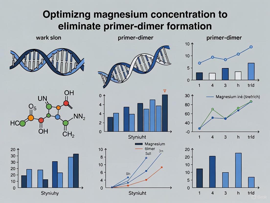
Optimizing Magnesium Concentration to Eliminate Primer-Dimer Formation: A Strategic Guide for Researchers
Primer-dimer formation is a pervasive challenge in PCR that consumes reagents, reduces amplification efficiency, and compromises assay sensitivity and specificity.
Optimizing Magnesium Concentration to Eliminate Primer-Dimer Formation: A Strategic Guide for Researchers
Abstract
Primer-dimer formation is a pervasive challenge in PCR that consumes reagents, reduces amplification efficiency, and compromises assay sensitivity and specificity. This article provides a comprehensive, evidence-based guide for researchers and drug development professionals on the critical role of magnesium ion (Mg²⁺) concentration in controlling this artifact. We explore the foundational biochemistry of Mg²⁺, present systematic methodological approaches for optimization, detail advanced troubleshooting strategies, and outline rigorous validation techniques. By synthesizing current best practices, this resource aims to equip scientists with the knowledge to precisely calibrate Mg²⁺ levels, thereby eliminating primer-dimer interference and enhancing the reliability of molecular diagnostics, SNP detection, and other precision PCR applications.
The Biochemistry of Magnesium in PCR: Understanding Its Dual Role as Essential Cofactor and Primer-Dimer Catalyst
The Essential Role of Magnesium Ions
Why is magnesium absolutely essential for DNA polymerase activity?
Magnesium ions (Mg²⁺) are non-negotiable cofactors for DNA polymerases because they directly participate in the catalytic mechanism of nucleotidyl transfer. Without Mg²⁺, the enzymatic reaction cannot proceed.
Molecular Mechanism: DNA polymerases employ a two-metal-ion mechanism to catalyze the formation of phosphodiester bonds. The two ions, often referred to as Metal A (catalytic metal) and Metal B (nucleotide-binding metal), are coordinated by conserved aspartate residues in the enzyme's active site [1] [2].
- Metal A (Catalytic Mg²⁺): This ion activates the 3'-OH group of the primer terminus by facilitating deprotonation, enhancing its nucleophilicity for an in-line attack on the α-phosphate of the incoming dNTP [2] [3].
- Metal B (Nucleotide-binding Mg²⁺): This ion coordinates the triphosphate moiety of the incoming dNTP, stabilizing the negative charge and assisting in the departure of the pyrophosphate (PPi) leaving group [1] [2].
The table below summarizes the distinct roles of these two metal ions in the catalytic mechanism.
Table 1: Roles of the Two Catalytic Magnesium Ions in DNA Polymerase Activity
| Metal Ion | Common Name | Primary Role | Key Ligands |
|---|---|---|---|
| Metal A | Catalytic Metal | Lowers pKa of primer 3'-OH; stabilizes transition state [2] | Primer 3'-OH, invariant aspartates (e.g., Asp705 in Pol I Klenow Fragment) [1] |
| Metal B | Nucleotide-binding Metal | Coordinates dNTP triphosphate; stabilizes leaving group (PPi) [1] [2] | β- and γ-phosphates of dNTP, invariant aspartates (e.g., Asp882 in Pol I Klenow Fragment) [1] |
How does Mg²⁺ influence pre-catalytic conformational changes?
Beyond the chemical step, Mg²⁺ plays a critical role in the pre-catalytic conformational changes that prepare the active site for chemistry. Kinetic studies on DNA polymerase I (Klenow Fragment) reveal that the aspartate residues coordinating the metals are required at specific stages:
- The D882A mutant, which likely disrupts the Metal B site, inhibits the fingers-closing conformational transition that creates the active site geometry for catalysis [1].
- The D705A mutant, which likely disrupts the Metal A site, does not prevent fingers-closing but is required for a subsequent step, possibly the entry of the second Mg²⁺ into the active site [1].
- Weak binding of the catalytic Mg²⁺ (Metal A) contributes to fidelity by allowing sampling of the correctly aligned substrate without perturbing the equilibrium for nucleotide binding [3].
The following diagram illustrates the sequence of metal ion binding and key conformational changes in the DNA polymerase catalytic cycle.
Diagram: Sequential Role of Mg²⁺ in DNA Polymerase Catalytic Cycle
Magnesium Optimization and Primer-Dimer Troubleshooting
How does magnesium concentration specifically affect PCR and primer-dimer formation?
The concentration of free Mg²⁺ in a PCR is a critical determinant of both specificity and efficiency. It acts as a cofactor for the DNA polymerase, but its concentration must be carefully optimized.
Table 2: Effects of Magnesium Chloride (MgCl₂) Concentration in PCR
| [MgCl₂] Condition | Impact on PCR Efficiency | Impact on Specificity & Primer-Dimer Formation |
|---|---|---|
| Too Low(Insufficient free Mg²⁺) | DNA polymerase activity is drastically reduced, leading to weak or failed amplification [4] [5]. | Primers cannot bind stably to the template, but primer-dimer formation is also suppressed due to lack of polymerase activity. |
| Optimal Range(Typically 1.5 - 4.5 mM) | Robust amplification of the specific target. The enzyme is fully active, and primers anneal correctly [4] [6]. | Specific primer-template binding is favored. Proper Mg²⁺ levels help maintain polymerase fidelity, reducing misincorporation and spurious amplification [6]. |
| Too High(Excess free Mg²⁺) | Non-specific binding of primers is enhanced due to increased stability of mismatched duplexes [4] [5]. | Marked increase in primer-dimer formation and other non-specific products. Polymerase fidelity is decreased, leading to more errors [4] [6] [5]. |
What are the best strategies to optimize Mg²⁺ concentration and prevent primer-dimers?
A multi-faceted approach is required to tackle primer-dimer formation, with Mg²⁺ optimization at its core.
1. Magnesium Titration Protocol:
- Preparation: Set up a series of PCR reactions where the concentration of MgCl₂ is varied. A standard range is 0.5 mM to 5.0 mM, in increments of 0.5 mM.
- Execution: Run the PCR with the gradient of Mg²⁺ concentrations.
- Analysis: Analyze the products by agarose gel electrophoresis. The optimal concentration is the one that yields the strongest target band with the least background (e.g., primer-dimer smears) [4] [5].
2. Comprehensive Troubleshooting Guide for Primer-Dimers:
- Hot-Start PCR: Use a hot-start DNA polymerase. These enzymes are inactive during reaction setup at room temperature, preventing spurious initiation and primer-dimer extension during preparation. Activation occurs only at the high temperature of the initial denaturation step [7].
- Primer Design: Utilize software to design primers with minimal self-complementarity, especially at the 3' ends. Avoid long stretches of complementary bases between forward and reverse primers [7].
- Touchdown PCR: Employ a touchdown protocol, starting with an annealing temperature higher than the calculated Tm and gradually decreasing it in subsequent cycles. This enriches specific products early in the reaction.
- Chemical Suppressors: Add additives like betaine or DMSO for GC-rich templates, which can help by equalizing the melting temperatures of primer-template duplexes and reducing secondary structure.
- Advanced Primer Chemistry: Consider using Self-Avoiding Molecular Recognition Systems (SAMRS). SAMRS are nucleotide analogs that pair with natural bases but not with other SAMRS. Incorporating them into primers can significantly reduce primer-primer interactions, thereby eliminating primer-dimer formation [8] [7].
Experimental Protocols & Reagent Toolkit
Detailed Protocol: Magnesium Titration for PCR Optimization
This protocol provides a step-by-step method for empirically determining the optimal Mg²⁺ concentration for a specific PCR assay.
Materials Required:
- Template DNA
- Forward and Reverse Primers
- dNTP Mix
- 10X PCR Buffer (without MgCl₂)
- MgCl₂ stock solution (e.g., 25 mM or 50 mM)
- Thermostable DNA Polymerase (e.g., Taq polymerase)
- Nuclease-free Water
- Thermal Cycler
Procedure:
- Prepare Master Mix: Create a master mix for N+1 reactions (where N is the number of Mg²⁺ conditions) to ensure consistency. For each 50 µL reaction, combine the following on ice:
- 5.0 µL of 10X PCR Buffer (Mg-free)
- 1.0 µL of dNTP Mix (10 mM each)
- 1.0 µL of Forward Primer (10 µM)
- 1.0 µL of Reverse Primer (10 µM)
- 0.5 µL of DNA Polymerase (e.g., 2.5 U/µL)
- X µL of Template DNA (variable, e.g., 100 ng)
- Y µL of Nuclease-free Water (variable volume to adjust for Mg²⁺ addition)
- Aliquot and Add Mg²⁺: Aliquot the master mix into N PCR tubes. Add a different volume of MgCl₂ stock solution to each tube to achieve the desired final concentration range (e.g., 0.5, 1.0, 1.5, 2.0, 2.5, 3.0, 4.0, 5.0 mM).
- Run PCR: Place the tubes in the thermal cycler and run the standard PCR program optimized for your primer pair and template.
- Analyze Results: Resolve the PCR products on an agarose gel. Identify the Mg²⁺ concentration that provides the strongest specific amplification with minimal non-specific bands or primer-dimer smears.
The Scientist's Toolkit: Essential Research Reagents
Table 3: Key Reagents for Studying Mg²⁺ in DNA Polymerase Systems
| Reagent / Material | Function / Role | Example & Notes |
|---|---|---|
| MgCl₂ Solution | Source of Mg²⁺ cofactor. Concentration is critical for activity and fidelity. | Supplied separately with many polymerase systems (e.g., Takara Ex Taq) for optimization [6]. |
| dNTPs | Substrates for DNA synthesis. | dNTPs chelate Mg²⁺; therefore, their concentration affects free [Mg²⁺]. Keep dNTP concentration constant during Mg²⁺ titration [5]. |
| Mg-free PCR Buffer | Provides optimal ionic strength and pH for the polymerase. | Allows for precise control over Mg²⁺ concentration without interference (e.g., Takara Ex Taq buffer) [6]. |
| Hot-Start DNA Polymerase | Enzyme engineered to be inactive at low temperatures, preventing mis-priming and primer-dimer formation. | Available via antibody-based, aptamer-based, or chemical modification (e.g., Takara's Hot Start Taq) [7]. |
| Non-hydrolysable dNTP Analogs | Used in X-ray crystallography to trap pre-catalytic complexes for structural studies. | Allows for visualization of the active site with catalytic Mg²⁺ and the primer 3'-OH in place [2]. |
| SAMRS Phosphoramidites | Nucleotide analogs for synthesizing primers that avoid primer-primer interactions. | Used to synthesize primers that bind to natural DNA templates but not to other SAMRS-containing primers, drastically reducing dimer formation [8]. |
| Rapid Quench-Flow Instrument | For pre-steady-state kinetic analysis to measure rapid conformational and chemical steps. | Used to determine the roles of specific aspartate residues and metal ions in the polymerization cycle (e.g., KinTek RQF-3) [1]. |
Troubleshooting Guide: Resolving Primer-Dimer Formation
This guide addresses the most common experimental issues leading to primer-dimer formation and provides targeted solutions to restore assay performance.
Q1: My PCR results show a smeary band or a sharp band below 100 bp on an agarose gel. How can I confirm this is a primer-dimer?
- Interpretation: A smeary or sharp band below 100 bp is a classic signature of primer-dimer [9]. These are short, unintended DNA fragments formed by primers ann.ealing to each other instead of the target template.
- Confirmation Experiment: Run a No-Template Control (NTC). Set up a duplicate PCR reaction using all components except the DNA template. If the same low molecular weight band appears in the NTC lane, it confirms the amplification product is a primer-dimer, as it forms independently of the template [9].
Q2: I have verified my primer sequences are specific. What are the primary reaction condition culprits for primer-dimer formation?
Several reaction conditions can promote primer-dimer, even with well-designed primers. The table below summarizes the main causes and immediate corrective actions.
- Table: Common Reaction Condition Culprits and Solutions
| Cause of Primer-Dimer | Underlying Mechanism | Corrective Action |
|---|---|---|
| Excess Primer Concentration [10] [11] | High primer concentration increases the probability of primer-primer interactions. | Optimize primer concentration, typically between 0.1–1 μM. Start with a lower concentration [10]. |
| Low Annealing Temperature [10] | Allows primers to anneal to non-specific sequences or to each other with imperfect complementarity. | Increase the annealing temperature stepwise in 1–2°C increments. Use a gradient thermal cycler if available [10]. |
| Use of Non-Hot-Start Polymerase [10] [9] | The polymerase is active during reaction setup at low temperatures, where nonspecific priming and primer-dimer formation are favored. | Switch to a hot-start DNA polymerase. These enzymes are inactive until a high-temperature activation step, preventing pre-amplification artifacts [10] [9]. |
| Excessive Magnesium Ion (Mg²⁺) Concentration [10] [11] | Mg²⁺ is a cofactor for DNA polymerase and stabilizes DNA duplexes. High concentrations stabilize even weak primer-primer interactions. | Optimize Mg²⁺ concentration. Review and titrate the Mg²⁺ concentration downward, as excess Mg²⁺ promotes nonspecific amplification [10]. |
| Long Annealing Times [10] | Provides more time for primers to bind to incorrect sequences or to each other. | Shorten the annealing time to minimize off-target binding [10]. |
Q3: My assay requires a high degree of multiplexing (many primer pairs in one tube). How can I minimize primer-dimer formation systematically?
In highly multiplexed PCR, the number of potential primer-dimer interactions grows quadratically with the number of primers, making design critical [12].
- Strategy: Employ advanced computational design algorithms. Tools like SADDLE (Simulated Annealing Design using Dimer Likelihood Estimation) use stochastic algorithms to select primer sequences from countless possibilities that collectively minimize a "Badness" function, which estimates the severity of primer-dimer interactions across the entire set [12].
- Protocol: The SADDLE algorithm involves 1) generating primer candidates for each target, 2) selecting an initial random set, 3) evaluating the total dimer potential (Loss function), and 4) iteratively refining the set by randomly changing primers and probabilistically accepting changes that reduce the total dimer likelihood [12]. This approach can reduce dimer formation from over 90% to under 5% in a 96-plex assay [12].
Q4: Are there any novel chemistry approaches to prevent primer-dimer?
- Strategy: Utilize Self-Avoiding Molecular Recognition Systems (SAMRS) [8].
- Mechanism: SAMRS nucleobases (e.g., a, t, g, c) pair with their complementary natural bases (A, T, G, C) with strengths comparable to an A:T pair. However, SAMRS components do not pair with each other. When incorporated into primers, SAMRS allows them to bind perfectly to the natural DNA template but significantly weakens primer-primer interactions, thereby preventing dimer formation [8].
- Design Rule: The number and strategic placement of SAMRS components in a primer are crucial for optimal performance without compromising PCR efficiency [8].
Experimental Protocol: Optimizing Magnesium Concentration to Eliminate Primer-Dimer
This protocol provides a detailed methodology for titrating magnesium concentration, a critical factor in minimizing primer-dimer formation while maintaining robust target amplification [10] [13].
Background and Principle
Magnesium ions (Mg²⁺) are an essential cofactor for DNA polymerase activity. However, excessive Mg²⁺ concentration stabilizes DNA duplexes, including nonspecific primer-template hybrids and primer-primer dimers, by reducing the electrostatic repulsion between phosphate groups on the DNA backbone [10] [11]. This experiment systematically varies the Mg²⁺ concentration to find the optimal level that supports efficient amplification of the desired product while eliminating or drastically reducing primer-dimer artifacts.
Materials and Reagents
- Table: Research Reagent Solutions for Magnesium Optimization
| Reagent | Function | Notes for Protocol |
|---|---|---|
| Template DNA | The target DNA to be amplified. | Use a positive control template of known concentration and quality. |
| Primer Pair (Forward & Reverse) | Sequences complementary to the flanking regions of the target. | Designed with optimal characteristics (e.g., Tm 55-72°C, GC content 40-60%) [14]. |
| dNTP Mix | Nucleotides (dATP, dCTP, dGTP, dTTP) for DNA synthesis. | Use balanced equimolar concentrations (e.g., 200 μM of each dNTP final) [10] [13]. |
| 10X PCR Buffer (without MgCl₂) | Provides optimal pH and ionic conditions for the reaction. | Using a Mg-free buffer is essential for this titration experiment. |
| Magnesium Chloride (MgCl₂) Solution | The variable component; source of Mg²⁺ ions. | A common stock concentration is 25 mM or 50 mM. |
| Hot-Start DNA Polymerase | Enzyme that synthesizes new DNA strands. | Hot-start is preferred to prevent pre-activation activity [10] [9]. |
| Nuclease-Free Water | Solvent to bring the reaction to final volume. | - |
Step-by-Step Procedure
Prepare Reaction Master Mix (MM): Calculate the reagents for ( n + 1 ) reactions, where ( n ) is the number of Mg²⁺ conditions to be tested. Combine all common components in a 1.8 ml microcentrifuge tube on ice [13]:
- Nuclease-Free Water (Q.S. to final volume)
- 10X PCR Buffer (without MgCl₂)
- dNTP Mix
- Forward Primer
- Reverse Primer
- Hot-Start DNA Polymerase Mix gently by pipetting up and down.
Aliquot MM: Dispense equal volumes of the Master Mix into each PCR tube.
Add MgCl₂: Add a different volume of the MgCl₂ stock solution to each tube to create a concentration gradient. A typical range is 1.0 mM to 5.0 mM final concentration in 0.5 mM increments [10] [13].
- Example: For a 50 μl reaction and a 25 mM MgCl₂ stock, to achieve 1.5 mM final, add 3.0 μl; for 2.0 mM, add 4.0 μl, etc.
Add Template and Control: Add the template DNA to all experimental tubes. For one tube, add water instead of template to serve as the No-Template Control (NTC). This is crucial for identifying primer-dimer.
Perform Thermal Cycling: Place tubes in a thermal cycler and run the standard PCR protocol for your target, ensuring the annealing temperature is appropriately set (typically 3–5°C below the primer Tm) [10] [13].
Analyze Results: Analyze the PCR products using agarose gel electrophoresis. Include a DNA ladder to determine product sizes.
Data Analysis and Interpretation
- Identify the Optimal Range: The optimal Mg²⁺ concentration is the lowest concentration that yields a strong, specific band of the expected amplicon size.
- Assay Performance: Accompanying the guide above, the following workflow visualizes the experimental and analytical process for magnesium optimization.
- Visual Guide: The diagram below summarizes the experimental and analytical process for magnesium optimization.
Frequently Asked Questions (FAQs)
Q: What is the fundamental mechanism of primer-dimer formation? A: Primer-dimer occurs when two primers (either identical or forward/reverse) anneal to each other via complementary sequences, instead of to the target template DNA [11]. This creates a free 3' hydroxyl end that DNA polymerase recognizes and extends, synthesizing a short, unintended DNA fragment that consumes reaction resources [8] [11].
Q: How does primer design specifically influence dimer formation? A: The 3' ends of the primers are critical. If the 3' ends of the forward and reverse primers are complementary, even by just 3-4 bases, they can easily anneal and be extended by the polymerase, forming a "primer dimer" [13] [14]. Design tools check for this 3'-complementarity to avoid cross-dimerization. Primers should also be checked for self-complementarity that can lead to hairpin structures [13].
Q: Can primer-dimer lead to false positives in quantitative PCR (qPCR) or diagnostic assays? A: Yes, this is a significant risk. In techniques like LAMP or qPCR that use fluorescent dyes, the amplification of primer-dimers can generate a fluorescent signal that is indistinguishable from the signal of the specific target amplicon, leading to false-positive results [11] [15]. This is why a No-Template Control is essential for validation.
Q: Are there any specialized primer design strategies to inherently avoid dimers? A: Yes, advanced strategies exist. One innovative approach is Self-Avoiding Molecular Recognition Systems (SAMRS), where primers are synthesized with modified nucleotides that bind to natural DNA but have greatly reduced affinity for other SAMRS-containing primers, thus avoiding primer-primer interactions [8]. For highly multiplexed panels, computational algorithms like SADDLE are designed to select primer sets that minimize a "dimer potential" score across thousands of possible primer interactions [12].
Q: Beyond magnesium and temperature, what other reaction components can I adjust? A: Consider additives that reduce secondary structure or alter duplex stability. DMSO (1-10%) or formamide (1.25-10%) can help by disrupting weak nonspecific bonds that stabilize primer-dimers, particularly in GC-rich sequences [10] [13]. Additionally, ensure your dNTP concentrations are not excessively high, as this can chelate Mg²⁺ and indirectly affect reaction fidelity [10].
Frequently Asked Questions (FAQs)
FAQ 1: What is the fundamental role of magnesium (Mg²⁺) in a PCR reaction, and why is its concentration critical? Magnesium is an essential cofactor for thermostable DNA polymerases, the enzymes that build new DNA strands during PCR [16] [17]. It facilitates the binding of the enzyme to the DNA template and is directly involved in the catalytic process of incorporating nucleotides into the growing chain [17]. The concentration of Mg²⁺ is critical because it directly influences enzyme activity, reaction specificity, and fidelity [16]. An incorrect concentration can lead to a range of issues, including the formation of primer-dimers and other non-specific products [18] [16].
FAQ 2: How does magnesium concentration directly promote primer-dimer formation? Mg²⁺ stabilizes all nucleic acid duplexes formed during the reaction, which includes not only the desired primer-template hybrids but also unintended structures like primer-dimers [16]. Elevated Mg²⁺ concentrations can reduce the annealing stringency, making it easier for primers to bind to each other through short complementary sequences, even at higher temperatures [16]. Once formed, these primer-duplexes are stabilized by Mg²⁺ and efficiently extended by the DNA polymerase, consuming reagents and potentially outcompeting the amplification of the desired target [8].
FAQ 3: What is the typical optimal range for magnesium concentration in a standard PCR? For Taq DNA Polymerase, the optimal magnesium concentration is typically in the range of 1.5 mM to 2.0 mM [18]. However, this is a starting point, and the ideal concentration must be determined empirically for each specific primer-template combination, as it depends on factors that chelate magnesium, such as the concentration of dNTPs and the DNA template itself [18].
FAQ 4: What are the observable consequences of sub-optimal Mg²⁺ levels in a PCR? The effects of incorrect Mg²⁺ concentration can be observed through gel electrophoresis or real-time PCR melt curves:
- Too Low (< 1.5 mM): Results in low or no yield of the desired PCR product due to inefficient polymerase activity [18].
- Too High (> 2.0 mM): Leads to increased non-specific amplification, including smeared bands on a gel, spurious bands, and prominent primer-dimer formation [18] [16].
FAQ 5: Besides Mg²⁺ optimization, what other strategies are proven to reduce primer-dimer formation? A multi-faceted approach is most effective. Key strategies include:
- Primer Design: Designing primers with minimal self-complementarity and 3'-end complementarity is the most effective preventative measure [19] [9].
- Hot-Start Polymerases: Using polymerases that are inactive at room temperature prevents spurious extension during reaction setup [9] [17].
- Annealing Temperature: Increasing the annealing temperature enhances specificity, reducing the chance for primers to bind to each other [9] [20].
- Primer Concentration: Lowering the concentration of primers reduces the likelihood of primer-primer interactions [19] [9] [20].
Troubleshooting Guide: Primer-Dimers and Magnesium
Problem: Strong primer-dimer band on gel electrophoresis or a peak in the low-temperature region of a qPCR melt curve.
Step-by-Step Diagnostic and Resolution
Step 1: Review Primer Design Before wet-lab optimization, computationally analyze your primers.
- Action: Use primer design software to check for self-dimerization and cross-dimerization potential. The strongest 3'-end dimers (with a ΔG < -2.0 kcal/mol) should be avoided, as they are highly likely to be extended [19].
- Goal: Ensure primers have low self-complementarity, especially at the 3' ends.
Step 2: Optimize Magnesium Concentration Empirically Systematically titrate Mg²⁺ to find the concentration that maximizes specific product yield and minimizes dimers.
- Action: Set up a series of reactions where the Mg²⁺ concentration is varied, typically from 1.0 mM to 3.0 mM in 0.5 mM increments [18].
- Protocol:
- Prepare a master mix containing all PCR components except MgCl₂ and the DNA template.
- Aliquot the master mix into several tubes.
- Add MgCl₂ to each tube to achieve the desired final concentration range.
- Add template DNA to each tube.
- Run the PCR using a standardized cycling program.
- Analyze the results using gel electrophoresis.
- Goal: Identify the lowest Mg²⁺ concentration that yields robust, specific amplification with minimal dimer.
Step 3: Adjust Thermal Cycling Conditions If dimers persist after Mg²⁺ optimization, refine the thermal profile.
- Action: Increase the annealing temperature in a step-wise manner (e.g., +2°C increments) or use a gradient PCR instrument to determine the optimal temperature [19] [20].
- Rationale: A higher annealing temperature increases stringency, preventing primers from annealing to partially complementary sequences on other primers [9].
Step 4: Fine-Tune Reaction Components Further optimize reagent concentrations to disfavor dimer formation.
- Action: Titrate primer concentrations downwards. Test a range from 0.05 µM to 0.5 µM of each primer [19] [18]. High primer concentrations increase the chance of primer-primer interactions [20].
- Advanced Strategy: Employ a "hot-start" technique by using a hot-start DNA polymerase. This prevents polymerase activity during reaction setup at room temperature, a period when primer-dimers are frequently initiated [9] [17].
Experimental Protocol: Magnesium Titration for Primer-Dimer Elimination
Objective: To determine the optimal Mg²⁺ concentration for a specific PCR assay that minimizes primer-dimer formation while maintaining high amplification efficiency.
Materials:
- Template DNA
- Forward and Reverse Primers
- 10X PCR Buffer (without MgCl₂)
- 25 mM MgCl₂ stock solution
- 10 mM dNTP mix
- Hot-Start DNA Polymerase
- Nuclease-free water
Methodology:
- Prepare a master mix on ice according to the table below for a 50 µL reaction, calculating for one extra reaction to account for pipetting error. Omit MgCl₂ and template at this stage.
Aliquot 49 µL of the master mix into each of five PCR tubes.
Add 1 µL of template DNA to each tube. Include a No-Template Control (NTC) by adding 1 µL of nuclease-free water to a separate tube to monitor for contamination and primer-dimer formation in the absence of target.
Add the appropriate volume of 25 mM MgCl₂ to each tube to achieve the final concentrations listed in the table below.
Run the PCR using the following typical cycling conditions:
- Initial Denaturation: 95°C for 2 minutes
- 35 Cycles:
- Denaturation: 95°C for 15 seconds
- Annealing: Use the calculated Tm for your primers for 30 seconds
- Extension: 72°C for 1 minute/kb
- Final Extension: 72°C for 5 minutes
- Hold: 4°C
Analyze 5-10 µL of each reaction on a 2-3% agarose gel. Identify the reaction with the strongest specific band and the faintest primer-dimer signal.
Master Mix Composition (for one 50 µL reaction):
| Component | Volume (µL) | Final Concentration |
|---|---|---|
| 10X PCR Buffer (Mg-free) | 5.0 | 1X |
| 10 mM dNTP Mix | 1.0 | 200 µM |
| Forward Primer (10 µM) | 1.0 | 0.2 µM |
| Reverse Primer (10 µM) | 1.0 | 0.2 µM |
| Hot-Start DNA Polymerase | 0.5 | 1.25 U |
| Nuclease-free Water | 39.5 | - |
| Template DNA | 1.0 | Variable |
| Total Volume | 49.0 |
Mg²⁺ Titration Scheme:
| Tube No. | Volume of 25 mM MgCl₂ (µL) | Final [Mg²⁺] (mM) |
|---|---|---|
| 1 | 1.0 | 1.0 |
| 2 | 1.5 | 1.5 |
| 3 | 2.0 | 2.0 |
| 4 | 2.5 | 2.5 |
| 5 | 3.0 | 3.0 |
Visualization: Magnesium Optimization Workflow
The following diagram illustrates the logical workflow for troubleshooting primer-dimers by optimizing magnesium concentration and other key parameters.
The Scientist's Toolkit: Essential Research Reagents
This table details key reagents and their specific functions in the context of optimizing PCR to suppress primer-dimer formation.
| Research Reagent | Function in Primer-Dimer Suppression | Key Considerations |
|---|---|---|
| MgCl₂ Solution | Essential cofactor for DNA polymerase; concentration critically affects reaction specificity and primer-dimer stability [18] [16]. | Titration is mandatory. High concentrations stabilize primer-duplexes, promoting dimers [16]. |
| Hot-Start DNA Polymerase | Enzyme is inactive at room temperature, preventing spurious extension during reaction setup—a common source of primer-dimers [9] [17]. | Activated during the initial denaturation step, ensuring primers only extend at stringent temperatures. |
| High-Purity Primers | Primers purified via HPLC or similar methods have reduced truncated sequences, which are prone to non-specific annealing and dimer formation [20]. | Reduces the population of error-prone primers that can initiate off-target amplification. |
| PCR Additives (DMSO, BSA) | Can help denature complex templates (DMSO) or neutralize inhibitors (BSA), indirectly promoting specific primer binding over dimer formation [17]. | Use judiciously, as they can also alter the effective stringency of the reaction. |
| dNTP Mix | The building blocks for DNA synthesis. Concentration must be balanced with Mg²⁺, as dNTPs chelate Mg²⁺ ions [18] [17]. | A consistent dNTP concentration is required for valid Mg²⁺ titration. |
Magnesium ions (Mg²⁺) function as an essential cofactor for DNA polymerase activity, enabling the incorporation of dNTPs during polymerization [21] [22]. Beyond its fundamental enzymatic role, Mg²⁺ concentration critically influences reaction specificity by stabilizing the binding of primers to template DNA [23] [21]. An imbalance in Mg²⁺ concentration directly promotes the formation of primer-dimers—small, unintended amplification artifacts where primers anneal to each other instead of the target template [9] [10] [24]. However, Mg²⁺ does not act in isolation. Its optimal concentration is profoundly affected by three key factors: primer design, template quality, and overall buffer composition. Understanding these interactions is fundamental to developing robust, specific PCR assays free from primer-dimer artifacts.
Understanding Primer-Dimers
What is a primer dimer?
A primer dimer is a small, unintended DNA fragment that can form during a polymerase chain reaction (PCR) [9]. These artifacts are typically short, often below 100 base pairs, and appear on an agarose gel as a fuzzy smear or a bright band at the bottom of the gel, well below the expected amplicon size [9] [24].
How do primer dimers form?
Primer dimers form when primers anneal to each other instead of binding to their intended target in the template DNA. This occurs through two primary mechanisms [9]:
- Self-dimerization: A single primer contains regions that are complementary to itself.
- Cross-dimerization: The forward and reverse primers have complementary regions that allow them to bind to each other.
In both cases, the 3' ends of the primers provide a free end that DNA polymerase can extend, creating a short, amplifiable duplex that competes with the target amplicon for reaction resources [9].
The Interplay Between Mg²⁺ and Primer Design
Primer design is the foremost factor determining Mg²⁺ sensitivity and primer-dimer propensity. Poorly designed primers often require suboptimal Mg²⁺ concentrations to work, creating a cycle of non-specific amplification.
Table 1: Primer Design Parameters and Their Impact on Mg²⁺ Optimization
| Design Parameter | Recommended Value | Effect on Mg²⁺ Requirement & Primer-Dimer Risk |
|---|---|---|
| Length | 18-30 nucleotides [25] [21] [26] | Longer primers (within range) increase specificity, allowing use of higher, more specific annealing temperatures and lower Mg²⁺. |
| Melting Temp (Tm) | 55-70°C; within 5°C for a pair [25] [21] | Matching Tms enable a single optimal annealing temperature, preventing the need for excessive Mg²⁺ to stabilize the lower-Tm primer. |
| GC Content | 40-60% [25] [21] [26] | High GC content requires higher denaturation temperatures and can increase non-specific binding with high Mg²⁺. |
| 3'-End Sequence | Avoid >3 G/C residues [21]; one G/C is beneficial [21] | A strong GC clamp at the 3' end can promote mispriming and dimer extension, especially with high Mg²⁺ [25] [26]. |
| Self-Complementarity | Avoid complementarity, especially at 3' ends [21] | Directly enables primer-dimer formation; may require lowering Mg²⁺ to reduce stability, which can also reduce target yield. |
Key Interaction: Excessive Mg²⁺ concentration can stabilize the short, imperfect complementary sequences between primers, making dimer formation thermodynamically favorable [23] [10]. Conversely, primers with high self-complementarity or 3'-end complementarity force the use of lower Mg²⁺ to suppress dimers, which may concurrently reduce the efficiency of target amplification [10]. Proper primer design creates a wide Mg²⁺ optimization window where target amplification is efficient while primer-dimer formation is minimized.
Template Quality and Quantity: The Substrate for Specificity
The quality and quantity of the template DNA significantly influence the Mg²⁺ concentration required for specific amplification.
Table 2: Template Considerations and Their Synergy with Mg²⁺
| Template Factor | Recommendation | Interaction with Mg²⁺ |
|---|---|---|
| Purity | High purity, free of inhibitors (phenol, EDTA, proteins) [27] [10] | Contaminants like EDTA chelate Mg²⁺, making it unavailable for the polymerase and leading to reaction failure [10]. |
| Integrity | Intact, non-degraded DNA [10] | Degraded DNA presents more potential non-specific binding sites; higher Mg²⁺ can stabilize these spurious interactions, causing smearing [10] [24]. |
| Concentration | Appropriate amount (e.g., 10-100 ng for genomic DNA) [21] [27] | Excess template DNA increases the chance of non-specific binding and can require lower Mg²⁺ to maintain specificity, similar to its effect on primer-dimers [21] [24]. |
| Complexity | Additives for GC-rich templates [25] [10] | Complex templates (GC-rich, secondary structures) may require higher Mg²⁺ for efficient primer extension, but this must be balanced against increased non-specific binding risks. |
Key Interaction: Contaminants that chelate Mg²⁺ (e.g., EDTA) or inhibitors that reduce polymerase processivity can create a false signal of Mg²⁺ deficiency [10]. Researchers may respond by increasing Mg²⁺ concentration, which, if the true cause is contamination, will only increase non-specific products like primer-dimers without solving the underlying problem. Similarly, degraded or excess template provides more opportunities for primers to bind non-specifically, a process that is stabilized by high Mg²⁺ concentrations [10] [24].
Buffer Composition: The Chemical Environment
The PCR buffer provides the chemical environment that governs all interactions. Its components, particularly monovalent cations and additives, directly interact with Mg²⁺.
Monovalent Cations: Specialized cation combinations in some commercial PCR buffers can help maintain high primer annealing specificity across a broader range of annealing temperatures. This buffer feature can reduce the need for meticulous, individual optimization of Mg²⁺ for every primer-template system [23].
dNTP Concentration: dNTPs bind Mg²⁺. The concentration of free Mg²⁺ available for the polymerase is the total Mg²⁺ minus that bound by dNTPs [21]. A typical recommended final concentration for each dNTP is 0.2 mM [21]. If dNTP concentrations are increased, the Mg²⁺ concentration must be proportionally increased to ensure an adequate level of free cofactor. Failure to do so can reduce PCR efficiency. Unbalanced dNTP concentrations can also increase the PCR error rate [10].
Additives: Reagents like DMSO, formamide, and glycerol are often used to amplify difficult templates (e.g., GC-rich regions) by lowering the template's melting temperature [23]. However, these additives can also weaken primer binding to the target [10]. This often necessitates a compensatory decrease in the annealing temperature and may also affect the optimal Mg²⁺ range, as the overall reaction stringency is altered [10].
Integrated Experimental Protocol: Optimizing Mg²⁺ to Eliminate Primer-Dimers
The following workflow provides a systematic, evidence-based methodology for optimizing Mg²⁺ concentration in the context of synergistic factors to eliminate primer-dimer formation.
Diagram Title: Experimental workflow for Mg²⁺ optimization.
Step-by-Step Methodology
Step 1: Primer Design Audit
- Verify that forward and reverse primers meet the criteria in Table 1 [25] [21]. Use oligonucleotide analysis software to check for self-dimers and cross-dimers.
- Synergistic Consideration: Well-designed primers with high specificity reduce the dependency on precise Mg²⁺ titration to suppress artifacts.
Step 2: Template Quality Control
- Quantify template DNA using a spectrophotometer (e.g., Nanodrop) and assess integrity by running an aliquot on an agarose gel. A clean, high-molecular-weight band indicates good integrity, while a smear suggests degradation [10].
- Synergistic Consideration: Using intact, pure template prevents misdiagnosis of Mg²⁺ requirements and avoids the smearing often confused with primer-dimer background [24].
Step 3: Mg²⁺ Gradient PCR Setup
- Prepare a master mix containing all components except Mg²⁺. Aliquot the master mix into individual PCR tubes.
- Create a Mg²⁺ gradient (e.g., 1.0, 1.5, 2.0, 2.5, 3.0, 3.5, 4.0 mM) by adding the appropriate volume of MgCl₂ or MgSO₄ stock solution to each tube. The specific salt (MgCl₂ vs. MgSO₄) should be selected based on the DNA polymerase's preference [10].
- Critical: Include a No-Template Control (NTC) for each Mg²⁺ concentration tested. The NTC contains all reagents except the template DNA and is essential for distinguishing primer-dimers from non-specific amplification [23] [9].
Step 4: Analysis and Interpretation
- Resolve PCR products by agarose gel electrophoresis.
- Identify the Mg²⁺ concentration that produces the strongest, most specific target band with the cleanest background.
- Examine the NTC lanes. A band in the NTC that is also present in the sample lane at the same Mg²⁺ concentration is a primer-dimer, not a specific product [9].
Step 5: Iterative Fine-Tuning
- Based on the results from the initial gradient, perform a second, finer Mg²⁺ titration (e.g., in 0.2 mM increments) around the best-performing concentration from the first experiment.
- Validate the final optimized condition by running a full PCR with positive and negative controls.
The Scientist's Toolkit: Essential Research Reagents
Table 3: Key Reagents for PCR and Primer-Dimer Troubleshooting
| Reagent / Tool | Function / Purpose |
|---|---|
| Hot-Start DNA Polymerase | Enzyme inactive at room temperature, preventing primer-dimer formation during reaction setup; activated at high initial denaturation temperature [9] [10]. |
| dNTP Mix | Building blocks for new DNA strands; must be used at equimolar concentrations (typically 0.2 mM each) to prevent misincorporation and to allow correct calculation of free Mg²⁺ [21] [10]. |
| MgCl₂ or MgSO₄ Stock | Source of Mg²⁺ cofactor; concentration requires optimization for each primer-template system [21] [10]. |
| PCR Additives (e.g., DMSO, BSA) | Assist in denaturing complex templates (GC-rich, secondary structures); can alter reaction stringency and thus optimal Mg²⁺ [23] [10]. |
| Nuclease-Free Water | Ensures reaction setup is free of contaminating nucleases that could degrade primers and template, which can cause smearing [22]. |
| Gradient Thermal Cycler | Allows testing of a range of annealing temperatures or Mg²⁺ concentrations in a single run, drastically speeding up optimization [10]. |
Frequently Asked Questions (FAQs)
Q1: I see a strong band in my No-Template Control (NTC). Is this a primer-dimer, and how can I fix it? A: A band in the NTC is very likely a primer-dimer or other non-specific product. To resolve this, first, check your primer design for 3'-end complementarity. If the primers are well-designed, systematically troubleshoot by:
- Increasing the annealing temperature in 1-2°C increments [9] [10].
- Lowering the primer concentration (e.g., from 1 μM to 0.3 μM) [9] [21].
- Reducing the Mg²⁺ concentration, as high Mg²⁺ stabilizes the weak bonds in primer-dimers [10].
- Using a hot-start polymerase to prevent pre-PCR amplification [9] [10].
Q2: My PCR has a smear and primer-dimers. Should I lower or raise the Mg²⁺? A: A smear with primer-dimers suggests multiple issues. Start by lowering the Mg²⁺ concentration, as high Mg²⁺ stabilizes all primer-template interactions, both specific and non-specific, leading to smearing and dimers [10] [24]. Simultaneously, verify your template DNA is not degraded (which causes smearing) and is at an optimal concentration, as too much template can also cause smearing [24].
Q3: How does primer concentration interact with Mg²⁺ to cause primer-dimers? A: High primer concentration increases the probability that primer molecules will encounter each other and form dimers. Mg²⁺ stabilizes these interactions. Therefore, a combination of high primer concentration and high Mg²⁺ concentration creates the perfect conditions for prolific primer-dimer formation that can outcompete target amplification [9] [21] [10]. The optimal strategy is to use the lowest primer concentration that still yields a robust specific product, which then allows for the use of a lower, more specific Mg²⁺ concentration.
Q4: I have optimized Mg²⁺, but my assay is still not specific. What is the next step? A: If Mg²⁺ optimization alone fails, the problem may be rooted in primer design. Re-design your primers, paying close attention to avoiding self-complementarity and ensuring specificity to the target sequence. Consider using specialized primer design algorithms or technologies (e.g., Co-Primers technology) that are explicitly engineered to minimize primer-dimer interactions, especially in multiplexed assays [28].
A Step-by-Step Protocol for Systematically Optimizing Magnesium Concentration in Your Assays
Why is Magnesium Concentration Critical?
In polymerase chain reaction (PCR) experiments, magnesium ions (Mg²⁺) serve as an essential cofactor for DNA polymerase enzyme activity. The Mg²⁺ concentration in your reaction mixture directly influences enzyme efficiency, fidelity, and specificity. An incorrect concentration is a common source of PCR failure, often resulting in the formation of primer-dimer artifacts or non-specific amplification products that compromise your experimental results. Establishing the proper magnesium baseline between 1.5 mM and 4.5 mM is therefore a fundamental step in optimizing your amplification conditions, particularly for research aimed at eliminating primer-dimer formation.
The Chemistry of Magnesium Ions
Magnesium is a silvery metal that exists predominantly in the +2 oxidation state in biological systems. In aqueous solutions, magnesium ions exhibit characteristic reactions that are crucial to their function in PCR. When combined with hydroxide ions, magnesium forms a white precipitate of magnesium hydroxide, Mg(OH)₂ [29] [30]. This property becomes relevant when considering buffer composition and pH effects. Magnesium ions rarely form complex ions, and most of their salts are white and soluble in water [29], making them suitable for inclusion in PCR master mixes.
Troubleshooting Guide: Magnesium-Related PCR Issues
Common Problems and Solutions
| Problem Observed | Possible Magnesium-Related Cause | Recommended Solution |
|---|---|---|
| No amplification products | Mg²⁺ concentration too low (<1.0 mM) | Increase Mg²⁺ concentration in 0.5 mM increments |
| Non-specific bands/smearing | Mg²⁺ concentration too high (>4.5 mM) | Decrease Mg²⁺ concentration; enhance specificity |
| Primer-dimer formation | Suboptimal Mg²⁺ reducing specificity | Titrate Mg²⁺; optimize annealing temperature |
| Inconsistent results | Variable Mg²⁺ in buffer preparations | Use consistent Mg²⁺ source; prepare fresh stocks |
Advanced Troubleshooting Scenarios
Unexpected Precipitation: While magnesium sulfate and sodium bicarbonate have been shown to be physically compatible in solution without precipitation at specific concentrations [31], be aware that magnesium can form insoluble complexes under certain conditions. If you observe cloudiness in your reaction mixture, consider the compatibility of all buffer components.
Measurement Inconsistencies: Inconsistent magnesium measurements can occur with different testing methodologies [32]. When preparing stock solutions, use calibrated instruments and consistent measurement techniques to ensure accurate Mg²⁺ concentrations across experiments.
Frequently Asked Questions (FAQs)
Q1: Why is the typical Mg²⁺ concentration range 1.5 mM to 4.5 mM for PCR?
This range represents the concentrations where Taq DNA polymerase functions optimally. Below 1.5 mM, polymerase activity is significantly reduced due to insufficient cofactor availability. Above 4.5 mM, enzyme specificity decreases, leading to increased non-specific binding and primer-dimer formation [13].
Q2: How does Mg²⁺ concentration specifically affect primer-dimer formation?
Mg²⁺ stabilizes DNA duplex formation. At high concentrations, it promotes stabilization of even short, non-specific interactions between primers, facilitating primer-dimer formation. By carefully titrating Mg²⁺ within the 1.5-4.5 mM range, you can find a concentration that supports specific primer-template binding while minimizing non-specific primer interactions.
Q3: My PCR buffer already contains MgCl₂. Should I still optimize?
Yes. Most commercial PCR buffers contain MgCl₂ at approximately 1.5 mM, but this is a starting point. The optimal Mg²⁺ concentration varies based on your specific primer-template system, and supplementation is often necessary. Always check the manufacturer's specification for baseline Mg²⁺ concentration in your buffer.
Q4: How do I accurately prepare and store Mg²⁺ stock solutions?
Prepare MgCl₂ stock solutions using nuclease-free water and sterile techniques. Filter-sterilize if necessary rather than autoclaving, which can lead to oxidation. Aliquot and store at -20°C to prevent contamination and degradation. Avoid repeated freeze-thaw cycles.
Q5: Can I use magnesium sulfate instead of magnesium chloride?
MgCl₂ is standard for PCR, but some protocols successfully use MgSO₄. Note that the ionic strength and effects on polymerase activity may differ slightly. If substituting, you may need to re-optimize the concentration and maintain consistency once established.
Experimental Protocol: Mg²⁺ Titration for Primer-Dimer Elimination
Materials and Reagents
Research Reagent Solutions
| Item | Function in Experiment | Specification Notes |
|---|---|---|
| Taq DNA Polymerase | Enzyme for DNA amplification | 0.5-2.5 units per 50 μL reaction [13] |
| 10X PCR Buffer | Reaction environment | May contain 15 mM MgCl₂; check composition [13] |
| dNTP Mix | Nucleotide substrates | 200 μM final concentration of each dNTP [13] |
| Magnesium Chloride | Polymerase cofactor | 25-50 mM stock solution for supplementation [13] |
| Primers | Target sequence flanking | 20-50 pmol each per reaction; well-designed to avoid hairpins [13] |
| Template DNA | Amplification target | 1-1000 ng genomic DNA per 50 μL reaction [13] |
| Sterile Water | Volume adjustment | Nuclease-free to prevent degradation |
Step-by-Step Titration Procedure
Preliminary Setup: Wear gloves throughout the procedure to prevent contamination. Arrange all reagents in a freshly filled ice bucket and allow them to thaw completely before setting up reactions. Keep reagents on ice throughout the experiment [13].
Master Mix Preparation: For efficiency and consistency, prepare a master mixture containing all reaction components except MgCl₂ and template DNA. Scale volumes appropriately based on the number of reactions. Include negative controls without template DNA [13].
Mg²⁺ Titration Series: Aliquot the master mix into individual PCR tubes. Add MgCl₂ to achieve final concentrations across your test range. A recommended series is: 1.5 mM, 2.0 mM, 2.5 mM, 3.0 mM, 3.5 mM, 4.0 mM, and 4.5 mM.
Thermal Cycling: Use the following standard cycling conditions with modifications as needed:
- Initial Denaturation: 94-95°C for 2-5 minutes
- 25-35 cycles of:
- Denaturation: 94-95°C for 30 seconds
- Annealing: 45-65°C for 30-60 seconds (primer-specific)
- Extension: 72°C for 1 minute per kb of product
- Final Extension: 72°C for 5-10 minutes
- Hold: 4°C
Product Analysis: Separate PCR products by agarose gel electrophoresis. Include an appropriate molecular weight standard. Analyze for:
- Presence of a single, specific band of expected size
- Absence of multiple non-specific bands
- Minimal to no primer-dimer formation (visible as a low molecular weight smear)
Data Interpretation and Next Steps
After completing your Mg²⁺ titration experiment, analyze the results to determine the optimal concentration for your specific application:
Interpreting Titration Results: Compare the amplification efficiency and specificity across the Mg²⁺ concentration range. The optimal concentration typically produces a strong, specific band of the expected size with minimal background or primer-dimer formation. Document your findings in a systematic table:
| Mg²⁺ Concentration | Band Intensity | Specificity | Primer-Dimer | Rating |
|---|---|---|---|---|
| 1.5 mM | Weak | High | None | Poor efficiency |
| 2.0 mM | Moderate | High | Minimal | Good |
| 2.5 mM | Strong | High | None | Optimal |
| 3.0 mM | Strong | Moderate | Moderate | Acceptable |
| 3.5 mM | Strong | Low | Significant | Poor specificity |
| 4.0 mM | Variable | Low | Extensive | Unacceptable |
| 4.5 mM | Variable | Low | Extensive | Unacceptable |
Further Optimization: Once you have established the optimal Mg²⁺ concentration, you may need to fine-tune other reaction parameters such as annealing temperature or cycling conditions to completely eliminate primer-dimer formation while maintaining strong specific amplification.
Establishing the correct Mg²⁺ concentration baseline is a fundamental step in PCR optimization that directly impacts the success of your experiments. The titration approach outlined here provides a systematic method for determining the ideal Mg²⁺ concentration between 1.5 mM and 4.5 mM for your specific primer-template system. By carefully controlling this critical parameter, researchers can significantly reduce or eliminate primer-dimer formation, thereby enhancing the specificity and reliability of their amplification results. This optimization process is particularly valuable in drug development and diagnostic applications where assay precision is paramount.
Core Experimental Protocol
This section outlines the standard method for quantifying magnesium ions using complexometric titration, a foundational technique for researchers optimizing magnesium concentration to eliminate primer-dimer formation in PCR.
Methodology: Complexometric Titration with EDTA
The estimation of magnesium is reliably performed using complexometric titration against Ethylenediaminetetraacetic acid (EDTA). The procedure involves two key stages: standardizing the EDTA solution and then titrating the unknown magnesium sample [33].
Step-by-Step Procedure:
- EDTA Standardization: Standardize a solution of EDTA against a primary standard, such as zinc sulfate. This establishes the exact concentration of the EDTA titrant, which is crucial for all subsequent calculations [33].
- Sample Titration:
- Transfer a known volume of your magnesium sulfate sample into a clean titration vessel (beaker or Erlenmeyer flask) [33].
- Add a buffer solution to maintain the pH at 10. This pH is essential for the proper function of the Eriochrome Black T indicator and for the reaction between EDTA and magnesium [34].
- Add 2-3 drops of Eriochrome Black T (EBT) indicator. The solution should turn a wine-red color due to the formation of a complex between Mg²⁺ ions and the indicator [34] [33].
- Titrate with the standardized EDTA solution while swirling the flask continuously.
- The end point is reached when the solution changes sharply from wine-red to a pure blue. This indicates that all the magnesium ions have been complexed by the EDTA, releasing the free indicator into solution [34] [33].
- Record the volume of EDTA used.
- Calculation: The molarity and mass of magnesium in the original sample are calculated based on the volume and concentration of EDTA used, knowing that one mole of EDTA complexes one mole of Mg²⁺ [34] [33].
The Scientist's Toolkit: Essential Reagents and Equipment
The following table details the key materials required to perform a Mg²⁺ titration experiment [34].
| Item | Function / Specification |
|---|---|
| EDTA Solution | Ethylenediaminetetraacetic acid, the titrant that forms stable complexes with Mg²⁺ ions [33]. |
| Eriochrome Black T (EBT) | Indicator that changes color from wine-red to blue at the endpoint [34] [33]. |
| Buffer Solution (pH 10) | Maintains constant pH for proper reaction and indicator function [34]. |
| Burette | Precision glassware for dispensing titrant; a 50 mL burette has a tolerance of ±0.05 mL [35]. |
| Volumetric Flask | For accurate preparation and dilution of standard solutions [34]. |
| Pipette | For precise transfer of sample aliquots [34]. |
| Titration Vessel | Beaker or Erlenmeyer flask for holding the sample [33]. |
Troubleshooting Guide
This section addresses common problems encountered during Mg²⁺ titration to ensure accurate and reproducible results.
Troubleshooting Logic Workflow
Common Problems and Solutions
Q1: My titration has an unclear or missing endpoint. What should I check?
- Indicator Issues: Ensure you are using the correct indicator (Eriochrome Black T for Mg²⁺) and that it is fresh. Contaminated or degraded indicators will not produce a clear color change [35] [36].
- Improper pH: The buffer must maintain a pH of 10 for the EBT indicator to function correctly. Verify the pH of your solution and the quality of your buffer [34].
- Instrumentation: If using a potentiometric method, the pH electrode may be faulty, blocked, or uncalibrated. Inspect the electrode for damage and ensure it is properly maintained and calibrated [37].
Q2: My results are inconsistent between replicates. How can I improve reproducibility?
- Equipment Care: Electrodes are consumable items and wear out. Inspect the electrode's measuring membrane and reference diaphragm for scratches or blockages, which cause irregular results. Replace if necessary [37].
- Reagent Integrity: Titrants like EDTA can degrade. Sodium hydroxide-based titrants, for example, absorb CO₂ from the air, reducing their concentration. Store reagents properly in appropriate containers and check expiration dates. Prepare fresh solutions if contamination is suspected [35] [36].
- Technique Consistency: Perform each titration carefully and consistently. Add titrant slowly near the endpoint, swirl the flask continuously, and read the burette at eye level to avoid parallax errors. Repeating the titration at least three times is recommended to obtain a reliable average [38] [36].
Q3: I suspect a systematic error in my method. What are the common sources?
- Titer Determination: Neglecting to standardize (determine the titer of) the EDTA solution is a common pitfall. Do not rely solely on the nominal concentration on the bottle. Standardize your titrant regularly against a primary standard for accurate results [35] [38].
- Burette Calibration: Using an ill-calibrated burette or a burette that is too large for the volume being dispensed can introduce significant error. Ensure all volumetric devices are calibrated periodically. For a typical titration volume, select a burette size that minimizes relative error [35] [36].
- Temperature Fluctuations: Solutions have a specific coefficient of thermal expansion. A change in lab temperature can alter the volume of the titrant, leading to inaccuracies. Regulating laboratory temperature or using an automated titrator with temperature compensation can mitigate this [35].
Frequently Asked Questions (FAQs)
Q: Why is optimizing Mg²⁺ concentration critical in PCR research, and how does this titration method help? A: In PCR, Mg²⁺ ions are essential cofactors for DNA polymerase activity. Low Mg²⁺ levels reduce polymerase efficiency, leading to incomplete amplification and a smear on gel electrophoresis. Conversely, high Mg²⁺ levels can stabilize non-specific primer-template interactions, increasing primer-dimer formation and non-specific bands [39]. This titration method provides a precise and quantitative way to measure and adjust Mg²⁺ concentration in your PCR buffer solutions, enabling you to systematically optimize this critical parameter and eliminate such artifacts.
Q: Can I use this method if my sample contains both calcium and magnesium ions? A: Yes, but with an important consideration. The Eriochrome Black T indicator at pH 10 will respond to both Ca²⁺ and Mg²⁺ ions, giving a combined measurement [34]. To determine the magnesium content specifically, you can perform a correction. One approach is to repeat the titration at a high pH (≥12) using a different indicator like Murexide, which is specific for calcium. The magnesium concentration can then be found by difference [34].
Q: What is the single most important step to ensure titration accuracy? A: While the entire protocol is important, proper titrant standardization and regular titer determination are foundational. An error in the titrant's known concentration is a systematic error that will propagate through all your calculations and render all results inaccurate, even if the technique is otherwise perfect [35] [38].
Q: When should I consider switching from manual to automated titration? A: Automated titration is highly recommended when you require high throughput, maximum reproducibility, or are troubleshooting persistent manual errors. Autotitrators eliminate subjective errors like visual perception of color changes (parallax error) and offer higher dosing resolution [35]. They are particularly valuable for standardizing protocols across multiple users in a lab.
Troubleshooting Guides
Gel Electrophoresis Troubleshooting
This section addresses common issues encountered when analyzing PCR products via gel electrophoresis.
Table 1: Troubleshooting Guide for Gel Electrophoresis
| Problem | Possible Causes | Recommended Solutions |
|---|---|---|
| Faint or No Bands | Low quantity of sample [40] | Load 0.1–0.2 μg of DNA/RNA per mm of well width [40]. |
| Sample degradation [40] | Use molecular biology-grade reagents, wear gloves, and use nuclease-free labware [40]. | |
| Reversed electrodes [40] | Ensure gel wells are on the negative electrode (cathode) side [40]. | |
| Smeared Bands | Sample overloading [40] | Load an appropriate sample amount (0.1–0.2 μg/mm of well width) and avoid wells with a pipette tip [40]. |
| Sample degradation [40] | Follow good lab practices to prevent nuclease contamination [40]. | |
| High-salt buffer in sample [40] | Dilute, purify, or precipitate the sample to remove excess salt and resuspend in nuclease-free water [40]. | |
| Incorrect voltage [40] | Apply the recommended voltage for the nucleic acid size; very low or high voltage causes suboptimal resolution [40]. | |
| Poorly Separated Bands | Incorrect gel percentage [40] | Use a higher percentage gel for smaller fragments; for agarose, adjust water volume after boiling to prevent increased percentage [40]. |
| Suboptimal gel type [40] | Use polyacrylamide gels for nucleic acids <1,000 bp [40]. | |
| Sample overloading [40] | Do not overload wells; fused bands are a characteristic of overloaded gels [40]. |
Melt Curve Analysis Troubleshooting
This section focuses on interpreting and troubleshooting melt curve data from SYBR Green qPCR assays, a key tool for assessing amplicon specificity and detecting primer-dimer.
Table 2: Troubleshooting Guide for Melt Curve Analysis
| Problem | Possible Causes | Recommended Solutions |
|---|---|---|
| Double Peaks (Minor Peak <80°C) | Primer-dimer formation [41] | Lower primer concentration or redesign primers to avoid self-complementarity [41]. |
| Double Peaks (Minor Peak >80°C) | Non-specific amplification [41] | Raise the annealing temperature, use a hot-start polymerase, or redesign primers [41]. |
| Single Peak, But Not Sharp | Broad melting temperature range [41] | If the temperature span is ≤7°C, the result is often still usable. Confirm specificity with high-concentration agarose gel [41]. |
| Single Peak, But Tm <80°C | Primer-dimer amplification (no true product) [41] | Redesign primers. A low Tm is expected only if the genuine product is <100 bp [41]. |
| Irregular or Noisy Peaks | Template contamination [41] | Check template quality and prepare a fresh sample if necessary [41]. |
| No Melt Curve Detected | Incorrect instrument settings [41] | Ensure fluorescence acquisition is enabled for the melt curve step in the qPCR setup [41]. |
Cq Interpretation Troubleshooting
This section addresses issues related to the Quantification Cycle (Cq), which is critical for accurate quantification in qPCR.
Table 3: Troubleshooting Guide for Cq Interpretation
| Problem | Possible Causes | Recommended Solutions |
|---|---|---|
| High Cq (Late Amplification) | Low template concentration or quality [42] | Check DNA/RNA integrity and purity (A260/A280 ratio of 1.8-2.0). Use high-quality, purified template [42]. |
| Inefficient reverse transcription [43] | Use a robust reverse transcriptase and generate a standard curve to check for cDNA synthesis bias [43]. | |
| Inefficient PCR amplification [44] | Optimize reagent concentrations (Mg2+, dNTPs, primers) and cycling conditions [44]. | |
| Low Cq (Unexpectedly Early Amplification) | Contamination [42] | Use separate work areas, uracil-DNA-glycosylase (UNG) treatment, and no-template controls [42]. |
| High Replicate Variability | Pipetting errors [42] | Master mix preparation and accurate pipetting are crucial [42]. |
| Instrument calibration issues [41] | Perform routine instrument maintenance and calibration [41]. | |
| Inconsistent Cq with Standard Curve | PCR inhibition [42] | Dilute the template or re-purify to remove inhibitors like salts or proteins [40]. |
| Suboptimal Mg2+ concentration [44] | Optimize Mg2+ concentration, as it is a critical cofactor for Taq DNA polymerase [44]. |
Experimental Protocols
Protocol: Systematic Optimization of Magnesium Concentration
Objective: To determine the optimal Mg2+ concentration for a specific PCR assay to maximize specificity, yield, and minimize primer-dimer formation [44].
Background: Magnesium ions (Mg2+) are an essential cofactor for thermostable DNA polymerases. The optimal concentration depends on the specific template, primers, and buffer components, all of which can chelate Mg2+. Insufficient Mg2+ can result in no product, while excess Mg2+ can promote non-specific amplification and primer-dimer formation [44].
Materials:
- Taq DNA Polymerase (e.g., NEB M0267) and its corresponding 10X PCR Buffer [44]
- 25 mM MgCl2 solution (if not included in the buffer at a sufficient concentration) [44]
- dNTP Mix (10 mM each)
- Forward and Reverse Primers (20 μM each)
- Template DNA
- Nuclease-free water
- PCR tubes and thermal cycler
Method:
- Prepare a Master Mix: Calculate for one 50 μL reaction and multiply by the number of reactions (n+1). Combine the following in a nuclease-free tube on ice:
- 1X PCR Buffer (e.g., 5 μL of 10X buffer)
- 200 μM of each dNTP (e.g., 1 μL of 10 mM dNTP mix)
- 0.1-0.5 μM of each primer (typical final concentration) [44]
- ~105 molecules of template DNA (1pg–10 ng of plasmid; 1ng–1μg of genomic DNA) [44]
- 1.25 units of Taq DNA Polymerase [44]
- Nuclease-free water to a final volume of 45 μL.
Set Up Mg2+ Titration: Aliquot 45 μL of the master mix into each PCR tube. Add MgCl2 to achieve the final concentrations listed in the table below. A typical titration range is 0.5 mM to 4.0 mM [44].
Table 4: Magnesium Titration Setup
Tube Volume of 25 mM MgCl2 (μL) Final [Mg2+] (mM) 1 0.0 *Baseline (e.g., 1.5 from buffer) 2 0.5 Baseline + 0.5 3 1.0 Baseline + 1.0 4 1.5 Baseline + 1.5 5 2.0 Baseline + 2.0 6 3.0 Baseline + 3.0 *Check the composition of your 10X PCR buffer, as it may already contain Mg2+.
Run PCR: Use the following cycling conditions, adjusting the annealing temperature (Ta) as needed for your primers [44]:
- Initial Denaturation: 95°C for 2 minutes
- 25-35 Cycles:
- Denaturation: 95°C for 15-30 seconds
- Annealing: Ta (e.g., 5°C below the primer Tm) for 15-30 seconds
- Extension: 68°C for 1 minute per kb
- Final Extension: 68°C for 5 minutes
- Hold: 4°C
Analyze Results:
- Analyze 5-10 μL of each reaction by gel electrophoresis.
- Identify the Mg2+ concentration that produces the strongest specific band with the least or no non-specific products or primer-dimer.
- For qPCR, the optimal concentration will yield the lowest Cq with a single, sharp melt curve peak.
Protocol: Assessment of PCR Specificity Using Melt Curve Analysis
Objective: To confirm the specificity of a SYBR Green qPCR assay by verifying the amplification of a single, specific product.
Background: SYBR Green dye binds to any double-stranded DNA. Melt curve analysis differentiates the desired amplicon from non-specific products and primer-dimers based on their distinct melting temperatures (Tm) [43].
Materials:
- Optimized qPCR reaction mix (including SYBR Green dye)
- Thermal cycler with real-time and melt curve capabilities
Method:
- Program the qPCR Run: Include the melt curve step after the amplification cycles. A typical setup is:
- Amplification Cycles: As optimized for your assay.
- Melt Curve Step:
- Denature: 95°C for 15 seconds.
- Anneal: 60°C for 1 minute.
- Melt: Continuously measure fluorescence while heating from 60°C to 95°C at a slow rate (e.g., 0.1°C/second) [45].
Interpret the Results: View the data as the derivative of the fluorescence (-dF/dT) versus temperature.
Confirm with Gel Electrophoresis: Run the qPCR products on a high-percentage agarose gel (e.g., 3%). A single, discrete band corresponding to the expected amplicon size confirms specificity [45].
Visual Workflows
Research Reagent Solutions
Table 5: Essential Reagents for PCR and qPCR Analysis
| Reagent | Function | Key Considerations |
|---|---|---|
| Hot-Start DNA Polymerase | Enzyme for DNA amplification; remains inactive until high temperature to reduce primer-dimer [17]. | Choose based on fidelity, processivity, and extension rate. Hot-start is crucial for specificity [17]. |
| SYBR Green Dye | Intercalating dye for real-time detection of double-stranded DNA in qPCR [43]. | Cost-effective but binds to any dsDNA, making melt curve analysis essential for confirming specificity [43]. |
| dNTPs | Building blocks for new DNA strands [44]. | Use balanced concentrations (typically 200 µM each). Higher concentrations can reduce fidelity [44]. |
| Magnesium Chloride (MgCl2) | Essential cofactor for DNA polymerase activity [44]. | Concentration must be optimized (typically 1.5-2.0 mM). It is a critical variable for eliminating primer-dimer [44]. |
| Primers | Short sequences that define the start and end of the amplicon [46]. | Design with 18-30 bp, 40-60% GC content, and Tm of 60-64°C. Avoid self-complementarity to prevent dimer formation [46]. |
| Nuclease-Free Water | Solvent for reactions. | Ensures reactions are not degraded by environmental nucleases. |
| Agarose | Matrix for gel electrophoresis to separate DNA fragments by size [40]. | Use appropriate percentage (e.g., 1-3%) for the fragment size. Thicker gels (>5mm) can cause band diffusion [40]. |
FAQ: Magnesium's Role in PCR and Primer-Dimer Formation
What is the fundamental role of Mg²⁺ in PCR?
Magnesium ions (Mg²⁺) are an essential cofactor for DNA polymerase activity [47] [39]. They form a soluble complex with the phosphate groups of dNTPs (deoxynucleotide triphosphates), which is a prerequisite for the polymerase enzyme to catalyze DNA strand elongation [39]. Without adequate Mg²⁺, polymerase activity is significantly reduced.
How does Mg²⁺ concentration specifically influence primer-dimer formation?
Mg²⁺ concentration directly affects the stability of DNA duplexes. Low Mg²⁺ levels reduce general polymerase efficiency but can also paradoxically increase primer-dimer formation by reducing the reaction's stringency, allowing primers to anneal to each other more easily [39]. Conversely, excessively high Mg²⁺ levels can over-stabilize weak interactions, including non-specific primer binding and primer-dimer artifacts [39]. The optimal concentration stabilizes the specific primer-template binding without supporting spurious interactions.
Why are multiplex SNP assays particularly prone to primer-dimer issues?
Multiplex assays, which amplify multiple targets in a single reaction, use a higher total concentration of primers than standard PCR [47]. This increased primer load raises the statistical probability that any two primers will have complementary sequences, especially at their 3' ends, leading to cross-primer dimerization [48] [49]. This makes meticulous optimization of reaction conditions, including Mg²⁺ concentration, absolutely critical for success.
Troubleshooting Guide: Resolving Primer-Dimer via Mg²⁺ Optimization
Problem: High Background from Primer-Dimer in Multiplex SNP Genotyping
In a multiplex SNP detection assay using allele-specific PCR, the gel analysis or capillary electrophoresis shows smearing and low-molecular-weight bands (~50-100 bp) in addition to the specific amplicons. This indicates primer-dimer formation, which competes for reagents and can lead to failed or inaccurate genotyping calls, as sequencers like the NovaSeq 6000 may not tolerate any primer dimer [50].
Solution: A Systematic Mg²⁺ Titration Protocol
The following step-by-step protocol is designed to identify the optimal Mg²⁺ concentration that suppresses primer-dimer formation while maintaining robust amplification of the specific target sequences.
Step 1: Prepare a Mg²⁺ Titration Series
Begin with a standard Mg²⁺ concentration, often 1.5 mM, and create a titration series to test a range around this value [39]. If using a master mix with pre-formulated Mg²⁺, you will need to use a separate Mg²⁺-free buffer and add MgCl₂ separately for this optimization.
Recommended Titration Series:
| Tube # | Final MgCl₂ Concentration |
|---|---|
| 1 | 1.0 mM |
| 2 | 1.5 mM |
| 3 | 2.0 mM |
| 4 | 2.5 mM |
| 5 | 3.0 mM |
| 6 | 3.5 mM |
Step 2: Set Up the Optimization Reactions
For each Mg²⁺ concentration in your series, set up a complete PCR reaction. It is crucial to include both a no-template control (NTC) and a positive control with known good template DNA in the series [9]. The NTC is vital for identifying primer-dimer, as it will amplify these artifacts in the absence of any legitimate target.
Step 3: Perform Thermal Cycling with Gradient Annealing
While optimizing Mg²⁺, it is highly advantageous to simultaneously use a thermal cycler with a gradient annealing temperature function [49]. This allows you to test different annealing temperatures (typically between 55°C and 65°C) across the same Mg²⁺ series in a single run, identifying the best combination of parameters.
Step 4: Analyze Results
Analyze the PCR products using agarose gel electrophoresis or a fragment analyzer like Tapestation [50]. Evaluate the results based on the following criteria:
| Mg²⁺ Level | Target Amplification | Primer-Dimer | Action |
|---|---|---|---|
| Too Low (<1.5 mM) | Weak or no bands | Smearing may be present | Increase concentration |
| Optimal (e.g., 2.0 mM) | Strong, specific bands | Absent or minimal | Ideal condition |
| Too High (>3.0 mM) | Multiple non-specific bands | May be present | Reduce concentration |
The optimal condition is the one that yields a strong, specific amplicon with no visible primer-dimer in the NTC lane.
Experimental Workflow: Mg²⁺ Optimization
The following diagram illustrates the logical workflow for troubleshooting primer-dimer formation through Mg²⁺ optimization.
The Scientist's Toolkit: Key Research Reagent Solutions
The following table details essential materials and their functions for setting up and optimizing a multiplex SNP genotyping assay.
| Item | Function & Importance in SNP Genotyping |
|---|---|
| High-Quality DNA Polymerase | Enzyme that synthesizes new DNA strands. Hot-start versions are recommended to minimize primer-dimer formation during reaction setup [9]. |
| dNTP Mix | The four deoxynucleotide triphosphates (dATP, dCTP, dGTP, dTTP) serve as the building blocks for new DNA strands [47]. |
| Optimized Primer Pairs | Short, single-stranded DNA sequences that define the boundaries of the target SNP. For multiplexing, they must be designed for compatibility and have closely matched melting temperatures (Tm difference ≤ 2°C) [48] [49]. |
| MgCl₂ Solution | Source of Mg²⁺ ions. Using a separate solution allows for fine-tuning the concentration, which is critical for optimizing reaction specificity and yield [39]. |
| Reaction Buffer | Maintains the optimal pH and ionic conditions for polymerase activity. The buffer composition can influence Mg²⁺ availability and primer annealing [47]. |
| Nuclease-Free Water | A pure, contaminant-free solvent to make up the reaction volume without degrading sensitive reaction components. |
Pro Tips for Success
- Use In Silico Tools: Before wet-lab work, use primer design software like OligoAnalyzer or Primer-BLAST to screen for potential self-dimers and cross-dimers. Aim for a weak ΔG value (e.g., ≥ -2.0 kcal/mol for 3'-end dimers) [48] [49].
- Consider Master Mixes: For standardized workflows, consider specialized genotyping master mixes (e.g., PACE Genotyping Master Mix). These are pre-optimized for specificity and can simplify setup, though they may offer less flexibility for Mg²⁺ adjustment [47].
- The No-Template Control (NTC) is Your Best Friend: Any amplification in the NTC is a definitive sign of contamination or primer-dimer. A clean NTC is a non-negotiable indicator of a specific assay [9].
Advanced Troubleshooting: Resolving Persistent Primer-Dimer Formation Despite Mg²⁺ Adjustment
FAQs: Magnesium Concentration and PCR Performance
1. How does magnesium concentration fundamentally affect a PCR reaction?
Magnesium chloride (MgCl₂) is an essential cofactor for Taq DNA polymerase. The Mg²⁺ ions activate the enzyme, enabling it to bind to the DNA template and catalyze the incorporation of nucleotides into the growing DNA strand. Furthermore, Mg²⁺ stabilizes the double-stranded DNA structure by neutralizing the negative charges on the phosphate backbone, which facilitates the binding of primers to the template DNA. An optimal concentration is critical; too little or too much can lead to reaction failure or nonspecific products [4] [51].
2. What are the visual symptoms of low Mg²⁺ concentration in my PCR results?
- Amplification Curves: You will typically observe weak or delayed amplification curves. In severe cases, there may be no amplification at all, resulting in a flat line. The reaction efficiency is very low [4] [52].
- Gel Electrophoresis: The agarose gel will show a very faint band of the expected product or no band whatsoever. The yield is insufficient [4].
3. What are the visual symptoms of high Mg²⁺ concentration in my PCR results?
- Amplification Curves: The curves may show a lower quantification cycle (Cq), but a melt curve analysis will often reveal multiple peaks, indicating the presence of non-specific products alongside the correct amplicon [53] [43].
- Gel Electrophoresis: The gel will display multiple bands or a smeared appearance, showing that primers have bound to non-target sequences. A prominent primer-dimer band (typically around 30-50 bp) is also a common symptom [4] [52].
4. What is the typical optimal range for Mg²⁺ concentration in PCR?
The ideal concentration of MgCl₂ for creating optimal PCR conditions typically falls within a range of 1.5 mM to 4.5 mM [54] [4]. For many standard reactions, a narrower range of 1.5–2.5 mM is often effective, but optimization is required for each specific assay [51].
Diagnostic Guide: Symptoms of Mg²⁺ Imbalance
The following table summarizes the key differences in symptoms caused by non-optimal magnesium concentrations to aid in diagnosing your PCR issues.
| Symptom | Low Mg²⁺ | Optimal Mg²⁺ | High Mg²⁺ |
|---|---|---|---|
| Amplification Curve | Weak, delayed, or no amplification | Robust, single sigmoidal curve with high efficiency | Early Cq but non-specific signals; multiple peaks in melt curve |
| Gel Electrophoresis | Faint or no target band | Single, crisp band of expected size | Multiple bands, smearing, and/or strong primer-dimer band |
| Primary Cause | Insufficient polymerase activity; unstable primer-template binding | Correct enzyme activity and reaction stringency | Reduced reaction stringency; promiscuous primer binding |
Experimental Protocol: Optimizing Mg²⁺ Concentration to Eliminate Primer-Dimer
This protocol provides a detailed methodology for empirically determining the ideal Mg²⁺ concentration for your assay, with a specific focus on minimizing primer-dimer formation.
Objective: To identify the Mg²⁺ concentration that yields the highest specific product yield while eliminating non-specific amplification and primer-dimer.
Materials (Research Reagent Solutions):
| Reagent | Function |
|---|---|
| Hot-start DNA Polymerase | Reduces non-specific amplification and primer-dimer formation by remaining inactive until high temperatures are reached [52]. |
| 10X PCR Buffer (without MgCl₂) | Provides the core reaction environment (pH, salts). Using a Mg-free buffer is essential for this optimization. |
| 25-50 mM MgCl₂ Stock Solution | The variable cofactor being optimized. |
| Template DNA | The DNA sample containing the target sequence. Use a concentration within the linear dynamic range of your assay. |
| Primers (Forward & Reverse) | Sequence-specific oligonucleotides designed to flank your target amplicon. |
| dNTP Mix | The building blocks (nucleotides) for new DNA synthesis. |
| SYBR Green I Master Mix (Optional) | A fluorescent dye used in qPCR to monitor amplification in real-time and perform essential melt curve analysis [43]. |
Procedure:
Prepare a Master Mix: Create a master mix containing all the common reaction components except for the MgCl₂ stock solution. This includes nuclease-free water, PCR buffer (without MgCl₂), dNTPs, primers, template DNA, and hot-start polymerase.
Set Up the Mg²⁺ Gradient: Aliquot equal volumes of the master mix into a series of PCR tubes or a qPCR plate. Add the MgCl₂ stock solution to each tube to create a gradient of final concentrations. A standard range is from 1.0 mM to 4.5 mM in 0.5 mM increments.
Run the PCR Amplification: Place the samples in your thermal cycler and run the optimized cycling program for your primers and template.
Analyze the Results:
- For qPCR with SYBR Green I: Analyze the amplification plots and the subsequent melt curve analysis. The optimal condition is the lowest Mg²⁺ concentration that produces a low Cq value with a single, sharp peak in the melt curve, indicating a single, specific product without primer-dimer [53] [43].
- For Conventional PCR: Analyze the products using agarose gel electrophoresis. The optimal condition is the lowest Mg²⁺ concentration that produces a single, intense band of the correct size and a clear background with no smearing or primer-dimer bands [52].
Optimization Workflow for Mg²⁺
The following diagram illustrates the logical decision-making process for diagnosing and correcting Mg²⁺-related issues in PCR, positioning Mg²⁺ optimization within a broader troubleshooting context.
Key Considerations for Effective Optimization
- Systematic Approach: Always change only one variable at a time. If adjusting Mg²⁺ concentration does not resolve the issue, systematically investigate other parameters such as annealing temperature and primer concentration [53] [52].
- Primer Design is Foundational: The most careful Mg²⁺ optimization cannot compensate for poorly designed primers. Ensure your primers are specific, have minimal self-complementarity (especially at the 3'-ends), and are designed to avoid stable dimers (ΔG ≥ -6.0 kcal/mol for total dimer, and ΔG ≥ -2.0 kcal/mol at the 3'-end) [53] [55].
- The Role of Hot-Start Polymerases: Using a hot-start enzyme is highly recommended for optimization work. It minimizes artifacts generated during reaction setup by inhibiting polymerase activity at room temperature, thereby providing a clearer picture of how Mg²⁺ alone affects the reaction [52].
FAQs on Eliminating Primer-Dimer Formation
What are primer dimers and why are they a problem in PCR?
Primer dimers are small, unintended DNA fragments that form when PCR primers anneal to each other instead of to the intended target DNA template. This occurs through self-dimerization (a single primer with self-complementary regions) or cross-dimerization (two primers with complementary regions binding to each other) [9]. DNA polymerase can then extend these bound primers, creating an amplifiable product.
Primer dimers compete with the target amplification for PCR reagents (dNTPs, polymerase, primers), which can significantly reduce the efficiency and yield of your desired PCR product, especially for low copy number targets [56] [9]. They typically appear as a fuzzy smear or a sharp band below 100 bp on an agarose gel [9] [24].
How does Hot-Start PCR work to prevent non-specific amplification like primer dimers?
Hot-Start PCR employs specialized techniques to inhibit DNA polymerase activity during the reaction setup at room temperature. The goal is to prevent the polymerase from extending primers that have bound non-specifically or to each other under low-stringency conditions before cycling begins [56] [57].
The polymerase is only activated after the first high-temperature denaturation step in the thermal cycler, ensuring that the initial priming events occur under the more stringent conditions of the optimized annealing temperature [57]. This approach substantially improves the specificity and sensitivity of amplification [56].
Table: Common Hot-Start Methods and Their Mechanisms
| Method | Mechanism of Action | Activation Trigger |
|---|---|---|
| Antibody-based Inhibition [57] | An antibody binds to the DNA polymerase, blocking its active site. | High temperature (initial denaturation) denatures and releases the antibody. |
| Chemical Modifications [57] | The polymerase is chemically modified to render it inactive. | Extended heating at 95°C reverses the modification. |
| Wax Bead Barrier [57] | A physical wax barrier separates polymerase from other reagents. | Temperature >70°C melts the wax, allowing components to mix. |
| Primer-Based Hot-Start [56] | Primers contain thermolabile modifications that block extension. | Elevated temperatures cleave the modification, yielding extendable primers. |
| Controlled Magnesium [57] | Magnesium is precipitated and unavailable to the polymerase. | Thermal cycling dissolves the precipitate, releasing Mg²⁺. |
How should I optimize the annealing temperature to enhance specificity?
The annealing temperature is critical for ensuring primers bind only to their perfectly matched target sequence.
- Initial Calculation: Set the annealing temperature 3–5°C below the calculated melting temperature (Tm) of your primers [10] [58] [13].
- Empirical Optimization: Use a thermal cycler with a gradient function to test a range of annealing temperatures (e.g., from 55°C to 65°C) in a single experiment [10]. The optimal temperature is the highest one that still yields a strong, specific product.
- Touchdown PCR: Start with an annealing temperature 5–10°C above the estimated Tm and gradually decrease it in subsequent cycles. This approach favors the accumulation of the specific target in the early cycles, which then outcompetes non-specific products in later cycles [58] [59].
Besides magnesium, what other reaction components should I check to reduce primer dimers?
Table: Optimization of Key PCR Components to Minimize Primer-Dimer
| Component | Common Issue | Optimization Strategy |
|---|---|---|
| Primers | High concentration or poor design | Use 0.1–1.0 µM final concentration; verify design to avoid 3'-end complementarity [10] [58] [9]. |
| DNA Template | Too much or too little | Use recommended amounts (e.g., 10-100 ng genomic DNA); excessive template can increase mis-priming [10] [58]. |
| dNTPs | Excessive concentration | Use final concentration of 50-200 µM; too much can inhibit the reaction [58]. |
| PCR Additives | May be required for complex templates | Use DMSO (1-10%), Betaine (0.5-2.5 M), or others to help denature GC-rich templates [13] [59]. |
My PCR still shows smears or non-specific bands after optimizing the above. What else can I try?
- Review Thermal Cycling Conditions: Increase denaturation temperature or time for GC-rich templates [10] [59]. Shorten the annealing time to minimize non-specific binding [59]. Ensure your extension time is sufficient (typically 1 minute per 1 kb) [58] [59].
- Check Primer Design: Ensure primers are 18-30 bases long, have a GC content of 40-60%, and do not contain self-complementary regions or long repeats [58] [13]. The 3' ends should end in a G or C to increase annealing efficiency [58] [13].
- Verify Template Quality: Use high-quality, intact DNA template. Degraded DNA can appear as a smear on a gel [10] [59]. Ensure the template is free of inhibitors like phenol or EDTA [10].
Experimental Protocols for Key Strategies
Protocol 1: Systematic Annealing Temperature Optimization using a Gradient Thermal Cycler
This protocol helps empirically determine the ideal annealing temperature for a primer set [10].
- Prepare Master Mix: Create a standard PCR master mix containing your template, primers, dNTPs, MgCl₂, buffer, and Hot-Start polymerase.
- Set Up Reactions: Aliquot the same master mix into multiple PCR tubes.
- Program Thermal Cycler: Use the gradient function to set a range of annealing temperatures (e.g., from 55°C to 65°C) across the different tubes. Keep all other cycling parameters constant.
- Run PCR and Analyze: Execute the PCR program. Analyze the products on an agarose gel. The optimal temperature yields the strongest specific band with the least background or primer dimer.
Protocol 2: Incorporating Hot-Start Polymerase in a Standard PCR
This is a standard workflow for using a commercial Hot-Start enzyme.
- Thaw and Mix Reagents: Thaw all PCR components (buffer, dNTPs, primers, template, water) on ice. Prepare a master mix on ice, adding the Hot-Start polymerase last.
- Initial Denaturation/Acitvation: Place the tubes in the thermal cycler and start the program. The first step is typically a prolonged denaturation (e.g., 95°C for 2-5 minutes). This step both activates the Hot-Start polymerase and denatures the template DNA [59].
- Proceed with Cycling: Continue with the standard cycles of denaturation, annealing, and extension.
Research Reagent Solutions
Table: Essential Reagents for Preventing Primer-Dimer Formation
| Reagent / Kit | Function in Primer-Dimer Prevention |
|---|---|
| Hot-Start DNA Polymerase | Core enzyme inhibited at low temperatures, preventing pre-PCR non-specific extension [56] [57]. |
| Anti-Taq Polymerase Antibodies | Binds and inactivates Taq polymerase until initial denaturation step [57]. |
| Modified dNTPs | Chemically modified nucleotides block polymerase activity until heat-activated [57]. |
| Thermolabile Modified Primers | Primers with 4-oxo-1-pentyl (OXP) groups block extension until converted to natural form at high temperature [56]. |
| PCR Enhancer/Additive Kits | Includes DMSO, Betaine, or GC Enhancers to help denature complex templates and improve specificity [10] [59]. |
Troubleshooting Workflows and Diagrams
Primer-Dimer Troubleshooting Workflow
This diagram outlines a logical sequence of steps to diagnose and resolve primer-dimer formation.
Mechanisms of Hot-Start Polymerase Activation
This diagram illustrates how different Hot-Start methods inhibit and then activate the DNA polymerase.
Frequently Asked Questions (FAQs)
FAQ 1: What is 3'-end complementarity and why is it a problem in PCR?
3'-end complementarity occurs when the ends of your forward and reverse primers have regions that are complementary to each other, particularly at the 3' ends. This allows the primers to anneal to each other instead of to the template DNA. When this happens, DNA polymerase extends the primers, creating short, unintended DNA fragments known as "primer dimers." These artifacts compete with your target amplification, reducing PCR efficiency and yield [13] [9]. In gel electrophoresis, primer dimers typically appear as a fuzzy smear or a sharp band below 100 base pairs [9].
FAQ 2: How can software tools help prevent primer dimer formation?
Specialized primer design software systematically analyzes primer sequences to avoid problematic features that lead to primer dimers. These tools:
- Check for Self-Complementarity: They identify regions within a single primer that can fold back and form secondary structures like hairpin loops [13].
- Check for Pair Complementarity: They scan the forward and reverse primers for complementary sequences, especially at the 3' ends, which is critical for preventing primer-dimer formation [13] [9].
- Ensure Specificity: Tools like NCBI's Primer-BLAST check your primer pairs against genomic databases to ensure they will bind specifically to your target sequence and not to other unintended sites [60].
FAQ 3: Which software tools are recommended for designing primers without 3'-end complementarity?
Two highly recommended and freely available tools are:
- NCBI Primer-BLAST: This tool combines the primer design capabilities of Primer3 with a BLAST search to ensure specificity. It is an excellent choice for designing new, target-specific primer pairs from scratch [13] [60].
- Primer3: A widely used stand-alone program for picking primers based on a set of constraints that you define, such as melting temperature (Tm), length, and GC content [13] [61].
FAQ 4: What are the key parameters for a good primer design to avoid dimers?
When designing primers, either manually or using software, adhere to the following principles [13]:
- Primer Length: 18-30 nucleotides.
- Melting Temperature (Tm): 52-65°C, with the Tm for both primers within 5°C of each other.
- GC Content: Between 40-60%.
- 3'-End Stability: The 3' end should end in a G or C residue to increase priming efficiency, but avoid runs of three or more Gs or Cs.
- Avoid Complementarity: The 3' ends of the primer pair must not be complementary to each other.
Table 1: Key Primer Design Parameters to Minimize Dimer Formation
| Parameter | Optimal Range | Rationale |
|---|---|---|
| Primer Length | 18 - 30 nucleotides | Provides sufficient specificity for binding to the intended target [13]. |
| Melting Temp (Tm) | 52 - 65°C; ≤5°C difference between primers | Allows both primers to bind efficiently at the same annealing temperature [13]. |
| GC Content | 40 - 60% | Balances stability; too high can promote nonspecific binding, too low can make primers too unstable [13]. |
| 3'-End Base | Preferentially G or C (GC Clamp) | The stronger hydrogen bonding helps prevent "breathing" (fraying) of the primer end, improving efficiency [13]. |
| 3'-End Complementarity | Avoid ≥ 2 complementary bases | Minimizes the chance of primers annealing to each other and forming primer dimers [13]. |
Troubleshooting Guide: Resolving Primer-Dimer Issues
Problem: Agarose gel analysis shows a sharp, fast-migrating band or a smeary region around 50-100 bp, in addition to or instead of the desired PCR product.
Step 1: Confirm the Problem is Primer Dimer
- Run a No-Template Control (NTC): In this reaction, replace the DNA template with sterile water. If the same low molecular weight band appears in the NTC lane, it confirms the amplification is the result of primer-to-primer interaction and not your target [9] [62].
Step 2: Optimize Wet-Lab Conditions Before redesigning your primers, you can try to suppress dimer formation by adjusting your PCR conditions [9] [62] [10].
- Increase Annealing Temperature: Raise the temperature in 2°C increments to disrupt the weaker bonds between primers.
- Use a Hot-Start DNA Polymerase: These enzymes remain inactive until a high-temperature activation step, preventing enzymatic activity during reaction setup when primer dimer formation is most likely [9] [10].
- Lower Primer Concentration: High primer concentrations increase the chance of primers encountering each other. Try reducing the concentration, typically to within 0.1–1 μM [9] [10].
Step 3: Redesign Your Primers Using Software If optimization fails, primer redesign is the most robust solution. The following protocol uses NCBI Primer-BLAST to design primers without 3'-end complementarity.
Table 2: Research Reagent Solutions for Primer Design and PCR Optimization
| Reagent / Tool | Function / Description |
|---|---|
| NCBI Primer-BLAST | A web-based tool for designing target-specific primers and checking their specificity against a nucleotide database [60]. |
| Primer3 | The core algorithm behind many primer design tools; used for selecting primers based on user-defined parameters [13] [61]. |
| Hot-Start DNA Polymerase | A modified polymerase that is inactive at room temperature, preventing nonspecific amplification and primer-dimer formation during reaction setup [9] [10]. |
| dNTPs | The building blocks (dATP, dCTP, dGTP, dTTP) for DNA synthesis. Unbalanced concentrations can increase error rates [10]. |
| Magnesium Salt (MgCl₂) | A critical cofactor for DNA polymerase. Its concentration must be optimized, as excess Mg²⁺ can reduce specificity and fidelity [13] [10]. |
Protocol: Designing Primers with NCBI Primer-BLAST
Objective: To generate a pair of primers specific to your DNA template that lack 3'-end complementarity.
Materials:
- DNA sequence of your target (in FASTA format or as an Accession number).
- Computer with internet access.
Methodology:
- Access the Tool: Navigate to the NCBI Primer-BLAST website.
- Input Template Sequence: Paste your target DNA sequence (in FASTA format) or its GenBank Accession number into the "PCR Template" field [60].
- Set Primer Parameters: Under the "Primer Parameters" section, set the following based on the optimal ranges in Table 1:
- Primer Size: Min: 18, Opt: 20, Max: 25.
- Tm: Min: 55°C, Opt: 60°C, Max: 65°C. (Use the "SantaLucia 1998" parameters as recommended [60]).
- GC Content: Use the default range (usually 20-80%), but the tool will inherently favor the optimal 40-60%.
- Define Product Size Range: Enter the desired length of your amplicon (e.g., 100-300 bp).
- Set Specificity Check: Under "Specificity Check," select the appropriate database (e.g., "RefSeq mRNA") and organism to ensure your primers are unique to your target [60].
- Exclude Problematic Primers: The tool automatically evaluates and penalizes primers with self-complementarity and 3'-end complementarity, making them less likely to be selected.
- Run and Select Primers: Click "Get Primers." Primer-BLAST will return a list of candidate primer pairs ranked by quality. Examine the results and select a pair that shows no significant hairpins or primer-dimer potential in the provided analysis.
The workflow below summarizes the logical process for troubleshooting and resolving primer-dimer issues, from initial detection to final validation.
Troubleshooting Guides
FAQ: How can I optimize magnesium concentration to eliminate primer-dimer formation?
Answer: Primer-dimer formation is a common byproduct in PCR that competes for reagents and inhibits target amplification, particularly in ultra-sensitive applications. Magnesium ion (Mg²⁺) concentration plays a critical role as it serves as a DNA polymerase cofactor and stabilizes DNA duplexes [7] [21].
Mechanism: Mg²⁺ facilitates primer binding by neutralizing the negative charges on the phosphate backbones of DNA strands, reducing electrostatic repulsion [63]. However, excessive Mg²⁺ concentrations can reduce primer stringency, promoting non-specific binding and primer-dimer formation [63] [64].
Optimization Strategy:
- Initial Concentration: Begin with 1.5-2.0 mM MgCl₂, which is optimal for most standard PCRs with Taq DNA Polymerase [64].
- Systematic Testing: If primer-dimer persists, test a concentration gradient from 1.0 mM to 4.0 mM in 0.5 mM increments [63] [64].
- Balancing Act: The goal is to find the concentration that supports efficient polymerase activity and specific primer binding while minimizing non-specific interactions [21].
Table 1: Troubleshooting Mg²⁺ Concentrations for Primer-Dimer Reduction
| Observed Result | Suggested Mg²⁺ Adjustment | Rationale |
|---|---|---|
| No PCR product or weak amplification | Increase concentration by 0.5 mM steps | Insufficient Mg²⁺ reduces polymerase activity and primer binding [64] |
| Primer-dimer bands and multiple non-specific products | Decrease concentration by 0.5 mM steps | Excessive Mg²⁺ reduces primer annealing stringency [63] |
| Specific product with minimal primer-dimer | Maintain current optimized concentration | Optimal balance of enzyme activity and specificity has been achieved |
FAQ: What specialized techniques enable ultra-sensitive DNA detection for low-abundance targets?
Answer: For targets present in very low copy numbers, such as single-copy pathogen DNA, standard PCR often lacks the required sensitivity. Advanced methods like single-tube nested (or hemi-nested) PCR provide ultra-sensitive detection by dramatically improving specificity and yield [65].
Principle: This technique uses two sets of primers in a single, closed-tube reaction. The "outer" primer pair first amplifies a larger fragment, and a second "inner" primer pair then targets a sequence within the first amplicon [65]. This sequential amplification significantly boosts sensitivity and specificity.
Key Advantage: Performing both amplification rounds in a single tube eliminates the cross-contamination risk associated with conventional two-tube nested PCR, making it suitable for clinical diagnostics [65].
Protocol: Single Closed-Tube Dual-Primer Quantitative PCR
- Primer Design: Design outer and inner primer pairs with similar melting temperatures (Tms).
- Reaction Setup: Include all four primers (outer and inner pairs) and a TaqMan probe specific to the inner amplicon in a single reaction tube [65].
- Thermocycling:
- First Rounds: Use a higher annealing temperature and limited cycles favoring the outer primers.
- Subsequent Rounds: Use a lower annealing temperature promoting inner primer activity.
- Detection: Use a sequence-specific probe (e.g., TaqMan) for quantification, which prevents signal generation from primer-dimers or non-specific products [65].
Table 2: Comparison of Standard and Ultra-Sensitive PCR Methods
| Parameter | Standard qPCR | Single-Tube Nested qPCR |
|---|---|---|
| Detection Limit | ~10-100 copies | 1-10 copies [65] |
| Specificity | High | Very High (dual primer specificity) [65] |
| Contamination Risk | Low | Very Low (single closed tube) [65] |
| Handling Time | Standard | Reduced (no tube transfer) [65] |
| Cost | Standard | Slightly higher (extra primers) [65] |
FAQ: What is a multi-faceted approach for amplifying difficult GC-rich templates?
Answer: GC-rich templates (>60% GC content) are challenging due to strong hydrogen bonding and stable secondary structures that hinder DNA denaturation and primer annealing [63] [66]. A successful strategy requires simultaneous optimization of multiple components.
Comprehensive Optimization Protocol:
- Polymerase Selection: Choose a polymerase specifically engineered for GC-rich amplification, such as OneTaq or Q5 High-Fidelity DNA Polymerase. These often come with specialized GC buffers and enhancers [63].
- Chemical Additives: Include additives that reduce secondary structures.
- Thermal Cycling Adjustments:
- Primer and Mg²⁺ Adjustments: Design longer primers (e.g., 25-30 nt) with Tms at the higher end of the standard range. Be prepared to optimize Mg²⁺ concentration, as GC-rich templates may require adjustments [63] [64].
The Scientist's Toolkit: Research Reagent Solutions
Table 3: Essential Reagents for Advanced PCR Optimization
| Reagent / Kit | Function / Application | Key Feature / Benefit |
|---|---|---|
| OneTaq DNA Polymerase (NEB) | Amplification of GC-rich templates and difficult amplicons [63] | Supplied with standard and GC buffers; compatible with GC Enhancer for up to 80% GC content [63] |
| Q5 High-Fidelity DNA Polymerase (NEB) | Long or difficult amplicons, including GC-rich DNA [63] | >280x fidelity of Taq; GC Enhancer improves amplification of GC-rich sequences [63] |
| DMSO (Dimethyl Sulfoxide) | Additive for GC-rich PCR [63] [66] | Reduces DNA secondary structure by interfering with hydrogen bonding [63] |
| Betaine | Additive for GC-rich PCR [63] [66] | Equalizes stability of AT and GC base pairs, preventing polymerase stalling [63] |
| Hot-Start Taq DNA Polymerase | Standard PCR requiring high specificity [7] | Prevents polymerase activity at room temperature, reducing primer-dimer formation [7] |
| Dual Primer System (Custom) | Ultra-sensitive detection for low-abundance targets [65] | Enables single-tube nested qPCR for detection as low as single copies of target DNA [65] |
Validating Success and Comparing Approaches: Ensuring Robust, Reproducible PCR Performance
Optimizing magnesium chloride (MgCl₂) concentration is a critical step in eliminating primer-dimer formation and enhancing overall polymerase chain reaction (PCR) performance. However, optimization alone is insufficient without rigorous validation using key metrics. Validation confirms that the optimized protocol reliably detects the intended target with high specificity, efficiency, and sensitivity, which is crucial for applications in research and drug development. This guide outlines the essential validation metrics and methodologies to assess your PCR assay after magnesium optimization, providing a framework for troubleshooting common issues.
The Critical Role of Magnesium: Magnesium ions (Mg²⁺) act as an essential cofactor for DNA polymerase activity, facilitating the incorporation of nucleotides during DNA synthesis [5] [21]. The concentration of Mg²⁺ directly influences several aspects of the reaction:
- Enzyme Activity: Mg²⁺ binds to the active center of DNA polymerase, maintaining its catalytic function [68].
- Primer Annealing: It stabilizes the binding of primers to the template DNA by reducing electrostatic repulsion between the negatively charged phosphate backbones of DNA strands [5] [21].
- Reaction Specificity: An optimal Mg²⁺ concentration promotes specific primer binding, whereas excessively high concentrations increase the risk of non-specific amplification and primer-dimer formation. Conversely, concentrations that are too low can lead to weak or no amplification [5] [68].
The following diagram illustrates the workflow for optimizing and validating magnesium concentration to achieve a specific and efficient PCR assay.
Troubleshooting Guides & FAQs
Frequently Asked Questions (FAQs)
FAQ 1: Why does primer-dimer form even after I've optimized my magnesium concentration? Magnesium optimization is a powerful but single factor. Primer-dimer formation can persist due to issues with the primers themselves. Common reasons include:
- Low Annealing Temperature: The annealing temperature may be too low, allowing primers to bind to each other even at a balanced Mg²⁺ level [69] [70].
- High Primer Concentration: Excessive primer concentration in the reaction mix increases the likelihood of primer-primer interactions [17] [21].
- Complementary Primer Sequences: If the 3' ends of your forward and reverse primers have complementary sequences, they are highly prone to dimerize [71] [17] [7].
- Suboptimal Primer Design: Primers with long stretches of G/C bases (G/C clamps) at the 3' end or self-complementary regions can promote dimer formation [71] [21].
FAQ 2: What is an acceptable efficiency range for a validated qPCR assay? A well-optimized and validated qPCR assay should have an efficiency between 90% and 105%, which corresponds to a standard curve slope between -3.6 and -3.1 [69]. An efficiency of 100% means the template doubles perfectly every cycle.
FAQ 3: How can I distinguish specific product from primer-dimer in my results?
- Melting Curve Analysis: For dye-based qPCR (e.g., SYBR Green I), run a melting curve after amplification. A specific product will have a distinct, higher melting temperature (Tm) peak, while primer-dimer will produce a lower, broader peak typically below 80°C [7].
- Gel Electrophoresis: Primer-dimers typically appear as a faint, fast-migrating band or smear around 30-50 base pairs on an agarose gel, well below the expected size of your target amplicon [7].
Troubleshooting Common Post-Optimization Issues
Problem: Non-specific amplification (multiple peaks in melting curve or multiple bands on a gel) after magnesium increase.
- Potential Cause: The magnesium concentration is too high, reducing the stringency of the reaction and allowing primers to bind to non-target sequences [5] [68].
- Solutions:
- Titrate Mg²⁺ Downward: Systematically test lower concentrations of MgCl₂ (e.g., in 0.5 mM increments) to find the lowest concentration that supports robust specific amplification [69] [21].
- Increase Annealing Temperature: Raise the annealing temperature by 2-5°C to increase stringency [69] [70].
- Use a Hot-Start Polymerase: Switch to a hot-start enzyme to prevent primer extension during reaction setup and initial denaturation, which suppresses non-specific amplification [8] [17] [7].
- Add Enhancers: Incorporate additives like formamide (1-5%) or tetramethylammonium chloride (TMAC, 15-100 mM) to increase hybridization specificity [17] [68].
Problem: Low sensitivity (high Cq values or weak band intensity) after magnesium decrease.
- Potential Cause: The magnesium concentration is too low, leading to suboptimal DNA polymerase activity and inefficient primer annealing [5] [68].
- Solutions:
- Titrate Mg²⁺ Upward: Test higher MgCl₂ concentrations to enhance enzyme processivity, but do so carefully to avoid re-introducing non-specific products [69] [21].
- Optimize Primer Concentration: Ensure primers are used within the optimal range of 0.1-1 μM; too little primer can cause low yield [69] [17] [21].
- Check Template Quality and Quantity: Use high-quality, intact template DNA at an appropriate concentration (e.g., 10-50 ng for genomic DNA) [17] [21].
- Use Additives for Difficult Templates: For GC-rich templates, include DMSO (2-10%) or betaine (1-1.7 M) to reduce secondary structures and improve amplification efficiency [17] [68].
Experimental Protocols for Key Validation Metrics
Protocol: Assessing Assay Specificity via Melting Curve Analysis
Purpose: To confirm that the PCR amplification generates a single, specific product and to detect the presence of primer-dimer or other non-specific products [69] [7].
Procedure:
- Run qPCR: Perform the qPCR run using an intercalating dye like SYBR Green I on your optimized (Mg²⁺) conditions.
- Melting Curve Program: Immediately after amplification, set the thermocycler to:
- Denature at 95°C for 15 seconds.
- Anneal at 60°C for 1 minute.
- Slowly ramp the temperature from 60°C to 95°C (e.g., 0.5°C per second) while continuously collecting fluorescence data.
- Data Analysis: Plot the negative derivative of fluorescence (-dF/dT) against temperature. A single, sharp peak indicates a specific product. Multiple peaks or a peak at a low temperature (~70-75°C) suggests non-specific amplification or primer-dimer.
Protocol: Assessing Assay Efficiency via Standard Curve
Purpose: To determine the amplification efficiency and dynamic range of the assay, which is critical for accurate quantification [69].
Procedure:
- Prepare Serial Dilutions: Create a logarithmic serial dilution (e.g., 1:10) of a known concentration of template DNA, covering the expected concentration range of your samples. A minimum of 5 dilution points is recommended.
- Run qPCR: Amplify each dilution in duplicate or triplicate using the optimized protocol.
- Generate Standard Curve: Plot the mean Cq (Quantification Cycle) value of each dilution against the logarithm of its initial concentration.
- Calculate Efficiency: Determine the slope of the standard curve. Calculate the amplification efficiency (E) using the formula: Efficiency (%) = [10(-1/slope) - 1] x 100 [69]. The R² value of the curve should be >0.99, indicating a strong linear relationship.
Protocol: Determining the Limit of Detection (LoD)
Purpose: To establish the lowest concentration of the target that can be reliably detected by the assay, a key metric for sensitivity [17].
Procedure:
- Prepare Low-Concentration Dilutions: Create a series of template dilutions at very low concentrations, near the expected detection limit.
- Replicate Amplification: Run a high number of replicates (e.g., n=10-12) for each low concentration and for a no-template control (NTC).
- Define LoD: The LoD is the lowest concentration at which ≥95% of the replicates test positive (e.g., 10 out of 10, or 19 out of 20). The Cq values for these replicates should be consistent and distinguishable from the NTC.
Data Presentation: Quantitative Validation Standards
The following tables summarize the target values for key validation metrics and the effects of common reagent adjustments.
Table 1: Key Validation Metrics and Target Values for an Optimized qPCR Assay
| Metric | Description | Target Value / Outcome |
|---|---|---|
| Specificity | The ability of the assay to amplify only the intended target. | A single, sharp peak in melting curve analysis [7]. A single band of correct size on an agarose gel [69]. |
| Amplification Efficiency | The rate at which the target is amplified per cycle during the exponential phase. | 90% - 105% (Slope: -3.6 to -3.1) [69]. |
| Dynamic Range | The range of template concentrations over which the assay maintains its stated efficiency and linearity. | A minimum of 5 orders of magnitude with an R² > 0.99 for the standard curve [69]. |
| Sensitivity (LoD) | The lowest concentration of the target that can be reliably detected. | The concentration where ≥95% of replicates are positive [17]. |
| Reproducibility | The agreement of results between replicate reactions and runs. | Low variation in Cq values (e.g., Standard Deviation < 0.2 for replicates) [69]. |
Table 2: Effects of Common PCR Component Adjustments
| Component | Typical Concentration Range | Effect of High Concentration | Effect of Low Concentration |
|---|---|---|---|
| MgCl₂ | 1.0 - 4.0 mM [5] [21] | Increased non-specific amplification, primer-dimer formation [5] [68]. | Reduced yield, failed amplification, low sensitivity [5] [68]. |
| Primers | 0.1 - 1.0 μM [17] [21] | Increased primer-dimer and non-specific binding [69] [21]. | Reduced amplification efficiency and yield [21]. |
| dNTPs | 20 - 200 μM each [17] [21] | May decrease specificity; can chelate Mg²⁺, making it less available [21]. | Reduced yield and reaction efficiency [70] [21]. |
The Scientist's Toolkit: Research Reagent Solutions
Table 3: Essential Reagents for PCR Optimization and Validation
| Reagent | Function in PCR | Key Considerations |
|---|---|---|
| Hot-Start DNA Polymerase | A modified enzyme inactive at room temperature, preventing non-specific amplification and primer-dimer formation during reaction setup [8] [17] [7]. | Critical for sensitive and multiplex PCR. Available as antibody-inactivated or chemically modified. |
| MgCl₂ Solution | Essential cofactor for DNA polymerase; stabilizes DNA duplexes and affects primer annealing specificity [5] [21] [68]. | Requires precise optimization (1.0-4.0 mM). Concentration is often the first parameter adjusted. |
| PCR Additives (DMSO, Betaine) | Assist in amplifying difficult templates (e.g., GC-rich sequences) by reducing DNA secondary structures and lowering melting temperature [17] [68]. | DMSO is typically used at 2-10%; betaine at 1-1.7 M. Can inhibit polymerase if concentration is too high. |
| dNTP Mix | The building blocks (dATP, dCTP, dGTP, dTTP) for synthesizing new DNA strands [17] [21]. | Used at equimolar concentrations (e.g., 200 μM each). High quality dNTPs are important for high-fidelity amplification. |
| SYBR Green I Dye | A fluorescent intercalating dye that binds double-stranded DNA, allowing for real-time detection of amplification and post-amplification melting curve analysis [69] [7]. | Cost-effective for specificity analysis. Will bind to any dsDNA, including primer-dimer, so assay specificity is paramount. |
The Critical Role of No-Template Controls (NTCs) in Detecting Low-Level Primer-Dimer Artifacts
FAQs: Understanding Primer-Dimer and NTCs
What is a No-Template Control (NTC) and why is it critical for my qPCR experiments?
An NTC is a control reaction that contains all the components of a PCR mix—including master mix, primers, and water—except for the DNA or RNA template. Its primary purpose is to detect contamination or the formation of amplification artifacts, such as primer dimers. Observing amplification in an NTC signals that your reaction reagents may be contaminated with template DNA or that your primers are forming detectable artifacts, which can compromise the interpretation of your experimental results [72].
How can I distinguish primer-dimer formation from genuine contamination in my NTC?
The pattern of amplification in your NTC replicates can provide a key distinction:
- Reagent Contamination: If one or more reagents are contaminated with template DNA, the amplification curves of the NTC replicates are typically consistent and closely grouped because the same amount of contaminating template is present in each replicate [72].
- Random Contamination: If contamination occurs randomly during plate setup, amplification will appear in only some NTCs and at highly variable Ct values [72].
- Primer-Dimer Formation: Amplification from primer dimers is typically observed at late Ct values (often >35 cycles) and can be confirmed by running a dissociation (melting) curve analysis. Primer dimers produce a distinct, lower-temperature peak separate from your specific amplicon's peak [72] [73].
What are the specific consequences of primer-dimer formation on my qPCR results?
Primer dimers can lead to both false positives and false negatives:
- False Positives: In assays using DNA-binding dyes like SYBR Green, the dye will bind to and detect the primer-dimer product, generating an amplification signal even in the absence of the target template. This is a classic false positive observed in the NTC [73].
- False Negatives: Primer dimers competitively consume reaction resources, including dNTPs, primers, and DNA polymerase. This consumption can reduce the efficiency of target amplification, leading to higher Ct values or even a complete failure to detect low-abundance targets, resulting in a false negative [73].
How does magnesium chloride (MgCl2) concentration influence primer-dimer formation?
Magnesium chloride is an essential cofactor for DNA polymerase, but its concentration must be carefully optimized:
- High MgCl2 Concentration (e.g., >2.0 mM): Promotes non-specific binding of primers and creates conditions favorable for primer-dimer formation by stabilizing the short, imperfect duplexes formed between two primers [4] [5].
- Low MgCl2 Concentration (e.g., <1.5 mM): Can prevent primers from binding effectively to the template, leading to weak or failed amplification of the desired target [4] [5].
- The optimal concentration, typically between 1.5 mM and 2.0 mM, provides a balance that supports specific primer-template binding while minimizing non-specific interactions [4].
Troubleshooting Guide: Resolving Primer-Dimer Artifacts
Step 1: Confirm Primer-Dimer Formation
- Action: Perform dissociation curve analysis following your SYBR Green qPCR run.
- Expected Outcome: A specific amplicon will produce a single, sharp peak at a high melting temperature (Tm). Primer dimers will produce a broader peak at a significantly lower Tm [72].
Step 2: Optimize Critical Reaction Parameters
The following table summarizes key parameters to optimize for reducing primer-dimer formation, with a special focus on magnesium concentration.
| Parameter | Recommended Optimization Range | Effect on Primer-Dimer |
|---|---|---|
| MgCl₂ Concentration | 1.5 mM - 2.0 mM (Titrate in 0.25 mM steps) | High concentration stabilizes primer-dimer duplexes; low concentration reduces polymerase activity [4] [5]. |
| Primer Concentration | 50 nM - 400 nM (each) | High primer concentration increases likelihood of primer-primer interactions [72] [73]. |
| Annealing Temperature | Increase by 2-5°C above calculated Tm | Higher temperature destabilizes weak primer-dimer bonds [73]. |
| Hot-Start Polymerase | Use a robust formulation | Prevents enzymatic activity at low temperatures before cycling begins, where primer dimers often form [8] [73]. |
Step 3: Improve Primer Design and Selection
- Utilize Advanced Design Tools: For highly multiplexed reactions, consider algorithms like SADDLE, which are specifically designed to minimize primer-dimer interactions across large primer sets by stochastically evaluating and minimizing a "Badness" function for potential dimer pairs [12].
- Consider SAMRS Technology: Incorporating Self-Avoiding Molecular Recognition Systems (SAMRS) nucleotides into your primers can be highly effective. SAMRS components (a, t, g, c) pair normally with natural DNA (T, A, C, G) but form very weak pairs with each other. This means a SAMRS-modified primer will still bind its target but is much less likely to interact with other SAMRS-containing primers, thereby preventing primer-dimer formation [8].
Step 4: Implement Rigorous Laboratory Practices
- Physical Separation: Use separate, dedicated areas for PCR mix preparation, template addition, and post-PCR analysis to prevent amplicon carryover contamination [72].
- Enzymatic Control: Incorporate UNG (Uracil-N-Glycosylase) or UDG into your master mix. These enzymes degrade DNA containing dUTP (e.g., from previous PCR products) before the thermal cycling starts, effectively reducing carryover contamination [72].
Experimental Protocol: Magnesium Titration for Primer-Dimer Elimination
Objective: To empirically determine the optimal MgCl2 concentration that suppresses primer-dimer formation while maintaining high amplification efficiency for the target sequence.
Materials:
- 2X PCR Master Mix (without MgCl2)
- Magnesium Chloride (MgCl2) stock solution (e.g., 25 mM)
- Forward and Reverse Primers (for your target of interest)
- DNA Template (positive control)
- Nuclease-free Water (for NTCs)
- qPCR Instrument
Procedure:
- Prepare MgCl2 Stock Dilutions: Create a series of MgCl2 working solutions to be added to the master mix, aiming for final reaction concentrations spanning from 1.0 mM to 3.0 mM. A suggested range is 1.0, 1.25, 1.5, 1.75, 2.0, 2.25, 2.5, and 3.0 mM.
- Set Up Reactions: For each MgCl2 concentration to be tested, prepare two reactions:
- A positive control reaction containing your DNA template.
- An NTC reaction with nuclease-free water instead of template.
- Run qPCR Protocol: Use your standard thermal cycling protocol, ensuring it includes a dissociation curve stage at the end.
- Data Analysis:
- Amplification Plots: For the positive controls, identify the MgCl2 concentration that yields the lowest Ct value with a smooth, sigmoidal curve.
- Dissociation Curves: Examine the NTCs. The optimal condition is one where the NTC shows no amplification or shows a very late Ct (>35) with a primer-dimer melt peak that does not overlap with the target peak.
- Determine the Optimal Range: The best MgCl2 concentration is the one that provides the lowest target Ct with no significant amplification in the NTC.
The logical workflow for this troubleshooting process is summarized in the diagram below.
Research Reagent Solutions
The following table lists key reagents and their specific roles in preventing and troubleshooting primer-dimer artifacts.
| Reagent / Material | Function / Role in Primer-Dimer Prevention |
|---|---|
| Hot-Start DNA Polymerase | Withholds polymerase activity until initial high-temperature denaturation step, preventing enzymatic elongation of primer dimers formed during reaction setup [8] [73]. |
| MgCl₂ Solution | An essential cofactor for DNA polymerase; its concentration must be carefully optimized to balance specific amplification and minimization of non-specific primer-dimer stabilization [4] [5]. |
| SYBR Green Master Mix with UNG/UDG | Contains dsDNA-binding dye for detection and Uracil-N-Glycosylase to degrade contaminating amplicons from previous PCRs, reducing false positives in NTCs [72]. |
| SAMRS-Modified Primers | Primers incorporating Self-Avoiding Molecular Recognition Systems nucleotides. These bind to natural DNA targets but have greatly reduced affinity for each other, thereby preventing primer-dimer formation at the molecular level [8]. |
| Computational Design Tools (e.g., SADDLE) | Algorithms for designing highly multiplexed PCR primer sets that systematically minimize a "dimer likelihood" score, reducing the potential for primer-primer interactions in complex assays [12]. |
Frequently Asked Questions (FAQs)
Q1: What is the fundamental cause of primer-dimer formation in PCR? Primer dimers are short, unintended DNA fragments that form when PCR primers anneal to each other instead of the target DNA template. This occurs due to complementarity between primers, especially at their 3' ends, which allows DNA polymerase to extend them into a short, amplifiable product. This nonspecific amplification consumes reaction resources and can reduce the yield and accuracy of your target amplicon [9].
Q2: How does optimizing Mg²⁺ concentration help suppress primer dimers? Magnesium ions (Mg²⁺) are essential cofactors for DNA polymerase activity. The concentration of MgCl₂ directly affects reaction specificity [74]:
- Low Mg²⁺ (e.g., < 1.0 mM): Results in insufficient enzyme activity and poor yield of the desired product.
- Optimal Mg²⁺ (e.g., 1.5 - 2.5 mM): Provides a balance that supports specific primer binding and efficient amplification of the target.
- High Mg²⁺ (e.g., > 4.0 mM): Promotes mis-priming and non-specific amplification, including primer-dimer formation, by stabilizing incorrect primer-template interactions [74]. Therefore, fine-tuning Mg²⁺ to its optimal range is crucial for maximizing specificity and minimizing artifacts.
Q3: When should I use a Hot-Start polymerase instead of, or in conjunction with, Mg²⁺ optimization? Hot-Start PCR should be your first line of defense against primer dimers that form during reaction setup at room temperature [74]. Hot-Start polymerases remain inactive until a high-temperature activation step (usually 94–95 °C), preventing enzymatic activity during tube preparation [74]. For persistent primer-dimer issues, you should use Hot-Start PCR in conjunction with Mg²⁺ optimization, as they address the problem at different stages of the reaction.
Q4: What are SAMRS primers, and in what scenarios are they most valuable? Self-Avoiding Molecular Recognition Systems (SAMRS) are modified primers in which the nucleobases are replaced with analogues that pair strongly with natural DNA but weakly with other SAMRS bases [8]. This design inherently prevents primer-primer interactions. SAMRS is particularly valuable in highly multiplexed PCR or when detecting single-nucleotide polymorphisms (SNPs), where the risk of cross-reactivity between multiple primers is high [8].
Q5: My PCR has no amplification product, only primer dimers. What should I check first? Follow this troubleshooting workflow:
- Run a No-Template Control (NTC): If primer dimers are present in the NTC, the issue is with the reaction components or conditions, not the template [9].
- Verify Primer Design: Use software to check for self-complementarity and 3'-end complementarity between primers. Avoid any 3'-end dimers with a ΔG < -2.0 kcal/mol [75].
- Employ Hot-Start Polymerase: This is often the most straightforward fix for primer dimers in the NTC [9].
- Optimize Mg²⁺: Titrate MgCl₂ concentration downward in 0.5 mM increments to increase stringency [74].
- Increase Annealing Temperature: Use a thermal gradient to find the highest possible annealing temperature that still allows specific amplification [75].
Troubleshooting Guides
Problem: Persistent Primer-Dimer Formation in qPCR with SYBR Green
Observed Symptom: A smeary, low-molecular-weight band (typically below 100 bp) on a gel or a peak in the melt curve analysis prior to the main product peak. This occurs even in a No-Template Control (NTC) [9].
Potential Causes and Solutions:
| Potential Cause | Recommended Solution | Experimental Protocol |
|---|---|---|
| Primers with high 3'-end complementarity | Redesign primers to minimize self- and cross-dimerization potential. | Use primer design software (e.g., OligoArchitect). Select primers where the strongest 3'-dimer has a ΔG ≥ -2.0 kcal/mol and the total dimer is unstable (ΔG ≥ -6.0 kcal/mol) [75]. |
| Suboptimal Mg²⁺ concentration | Systematically titrate MgCl₂ concentration. | Set up a series of 25 µL reactions with a fixed primer concentration (e.g., 200 nM). Vary MgCl₂ concentration from 0.5 mM to 4.0 mM in 0.5 mM increments. The optimal concentration yields the lowest Cq and highest fluorescence with a single peak in melt curve analysis [75]. |
| Non-specific activity during reaction setup | Switch to a Hot-Start DNA polymerase. | Use an antibody-based or chemically modified Hot-Start polymerase. Ensure the initial activation step (95°C for 3–5 minutes) is included in the thermal cycler protocol [74]. |
| Excessive primer concentration | Lower the primer concentration. | Titrate primer concentrations from 50 nM to 400 nM in a checkerboard pattern. Select the lowest concentration combination that yields a low Cq and high endpoint fluorescence [75]. |
| Annealing temperature too low | Optimize the annealing temperature using a gradient. | Perform a gradient PCR with annealing temperatures ranging from 55°C to 70°C. Select the highest temperature that maintains efficient amplification of the specific target [75]. |
Problem: Loss of Specificity in Multiplex PCR
Observed Symptom: Multiple non-specific bands or excessive smearing on an agarose gel, with some expected targets failing to amplify efficiently.
Potential Causes and Solutions:
| Potential Cause | Recommended Solution | Experimental Protocol |
|---|---|---|
| Cross-talk between multiple primers | Incorporate SAMRS technology into primer design. | Synthesize primers with SAMRS components (a, g, c, t) at strategic positions, particularly where primer-primer complementarity is detected. Limit the number of modifications to maintain efficient extension [8]. |
| Ionic imbalance due to high dNTPs | Re-balance Mg²⁺ and dNTP concentrations. | Remember that dNTPs chelate Mg²⁺. For a standard dNTP mix (200 µM total), start with 1.5 mM MgCl₂. If increasing dNTPs, increase MgCl₂ proportionally to maintain free Mg²⁺ levels [74]. |
| One dominant amplicon consumes resources | Re-optimize individual primer concentrations for each target. | Perform a multiplex primer optimization. Lower the concentration of primers for high-abundance targets and increase the concentration for low-abundance targets, keeping all within 50-500 nM [75]. |
Comparative Data Tables
Table 1: Quantitative Comparison of Primer-Dimer Suppression Methods
| Method | Typical Reduction in Primer-Dimer (%)* | Key Performance Metric | Optimal Use Case |
|---|---|---|---|
| Mg²⁺ Optimization | 60 - 80% | Specificity (measured by band clarity on gel or Cq shift in qPCR) [76] | Standard single-plex or duplex PCR where primers have been pre-designed. |
| Hot-Start PCR | > 90% (for dimers formed during setup) | Signal-to-Noise Ratio (fluorescence of target vs. NTC) [74] | Routine PCR and qPCR; essential for high-sensitivity applications. |
| SAMRS Primers | > 95% (for primer-primer interactions) | Multiplexing Capability (number of targets amplified cleanly in one reaction) [8] | High-level multiplexing (5+ targets) and complex SNP detection assays. |
| Advanced Primer Design | 50 - 70% | ΔG of Dimer Formation (software-predicted stability) [75] | The foundational first step for all experimental designs. |
*Reduction estimates are relative to a non-optimized reaction and are based on typical results reported in the literature.
| Method | Core Reagent / Instrument | Step-by-Step Optimization Summary | Key Parameters to Monitor |
|---|---|---|---|
| Mg²⁺ Optimization | MgCl₂ stock solution (25 mM), Thermal Cycler | 1. Prepare a master mix without MgCl₂. 2. Aliquot and add MgCl₂ to create a concentration series (e.g., 0.5, 1.0, 1.5, 2.0, 2.5, 3.0, 4.0 mM). 3. Run PCR. 4. Analyze products by gel electrophoresis or qPCR melt curve [76]. | Target amplicon yield, Cq value, presence of non-specific bands or primer-dimer smear. |
| Hot-Start PCR | Hot-Start DNA Polymerase | 1. Use a Hot-Start polymerase instead of a standard one. 2. Include a mandatory initial heat activation step (95°C for 3-5 min) in the thermal cycler program [74]. 3. No other changes to the protocol are typically needed. | Primarily the result in the No-Template Control (NTC). |
| SAMRS Implementation | SAMRS-modified primer oligonucleotides | 1. Identify regions of complementarity in standard primers. 2. Replace natural bases with SAMRS analogues (e.g., A with a, T with t) at strategic positions to disrupt dimer formation. 3. Synthesize and purify primers to >85% purity. 4. Validate with standard PCR conditions [8]. | PCR efficiency, multiplexing capability, and absence of primer-dimer in the NTC. |
The Scientist's Toolkit: Research Reagent Solutions
| Item | Function in Primer-Dimer Suppression |
|---|---|
| Hot-Start DNA Polymerase | The most common reagent for preventing non-specific amplification during PCR setup. It is inactivated by an antibody or chemical modification until a high-temperature activation step [74]. |
| SAMRS Phosphoramidites | The synthetic building blocks (g, a, c, t) used to manufacture SAMRS-containing oligonucleotides. They enable the creation of primers that avoid homologous pairing [8]. |
| MgCl₂ Stock Solution (25 mM) | A separate, high-purity magnesium chloride solution allows for fine-tuning of Mg²⁺ concentration independent of the PCR buffer system [74]. |
| dNTP Mix | Deoxynucleotide triphosphates (dATP, dCTP, dGTP, dTTP). Their concentration must be balanced with Mg²⁺, as they chelate the ions and reduce the free Mg²⁺ available for the polymerase [74]. |
Method Workflow and Decision Diagrams
Diagram 1: Troubleshooting workflow for persistent primer dimers, outlining the sequence for applying different suppression methods.
Diagram 2: Conceptual diagram comparing standard primer interactions with SAMRS-modified primers.
Establishing Standard Operating Procedures (SOPs) for Assay Transfer and Reproducibility
Analytical method transfer is a formally documented process that qualifies a receiving laboratory (RL) to successfully use an analytical method that was originally developed and validated in a transferring laboratory (TL) [77] [78]. Its primary objective is to demonstrate that the RL can perform the method with equivalent accuracy, precision, and reliability, producing comparable results that ensure data integrity and product quality across different sites [77] [79]. This process is crucial in pharmaceutical, biotechnology, and contract research sectors, particularly when scaling up production, outsourcing testing, or consolidating operations following business changes like mergers and acquisitions [77] [78]. A poorly executed transfer can lead to significant issues including delayed product releases, costly retesting, regulatory non-compliance, and ultimately, a loss of confidence in data [77].
Core Principles and Regulatory Framework
The foundation of any successful method transfer rests on establishing "equivalence" or "comparability" between the transferring and receiving laboratories' abilities to perform the method [77]. This involves demonstrating that the method's key performance characteristics—including accuracy, precision, specificity, linearity, range, and robustness—remain consistent across both sites [77].
The process is governed by established regulatory guidelines, with USP Chapter <1224> providing comprehensive guidance on the Transfer of Analytical Procedures [78]. This guidance outlines the essential elements and approaches to ensure the process meets rigorous quality standards. Furthermore, the transfer must be executed in compliance with Good Manufacturing Practices (GMP), ensuring all activities are properly documented and approved by Quality Assurance units [79] [80].
Method Transfer Approaches
Selecting the appropriate transfer strategy depends on factors such as the method's complexity, its regulatory status, the experience of the receiving lab, and the level of risk involved [77] [81]. The following table summarizes the most common approaches:
| Transfer Approach | Description | Best Suited For | Key Considerations |
|---|---|---|---|
| Comparative Testing [77] [78] | Both laboratories analyze identical samples from the same lots; results are statistically compared against pre-defined acceptance criteria. | Well-established, validated methods; laboratories with similar capabilities and equipment. | Requires careful sample preparation, homogeneity, and robust statistical analysis. |
| Co-validation [77] [81] | The receiving laboratory participates in the original method validation, often for intermediate precision assessment. | New methods being developed for multi-site use from the outset. | Requires close collaboration and harmonized protocols from the beginning. |
| Revalidation [77] [79] | The receiving laboratory performs a full or partial revalidation of the method. | Significant differences in lab conditions/equipment; substantial method changes; when the TL is unavailable. | Most rigorous and resource-intensive approach; requires a full validation protocol. |
| Transfer Waiver [77] [78] | The formal transfer process is waived based on strong scientific justification. | Highly experienced RL with proven experience with the method or identical methods; simple and robust methods. | Rare; requires robust documentation and strong justification; subject to high regulatory scrutiny. |
SOP Development: A Step-by-Step Workflow
A structured, phase-based approach is critical for a seamless and compliant method transfer. The following workflow outlines the key stages:
Phase 1: Pre-Transfer Planning and Assessment
- Define Scope & Objectives: Clearly articulate the reason for the transfer and define what constitutes a successful transfer with specific acceptance criteria [77] [80].
- Form Cross-Functional Team: Designate leads and team members from both TL and RL, including representatives from Analytical Development, QA/QC, and Operations [77] [80].
- Conduct Gap & Risk Analysis: Compare equipment, reagents, software, environmental conditions, and personnel expertise between the two labs. Identify and document potential risks and mitigation strategies [77] [80].
Phase 2: Transfer Plan and Protocol Development
- Develop Detailed Transfer Protocol: This critical document specifies the method details, responsibilities of both labs, materials and equipment, analytical procedure, experimental design, and the plan for statistical evaluation [77] [79].
- Define Acceptance Criteria: Establish pre-defined, statistically sound acceptance criteria for demonstrating equivalence (e.g., for precision and comparability of results) [78] [79].
- Secure Approvals: The transfer protocol must be formally approved by all relevant stakeholders and the Quality Assurance unit before execution begins [77] [79].
Phase 3: Execution and Training
- Train Receiving Lab Personnel: Analysts at the RL must be thoroughly trained by the TL and demonstrate proficiency. All training must be documented [77] [79].
- Quality Equipment & Reagents: Ensure all necessary equipment at the RL is properly qualified, calibrated, and maintained. Use qualified, traceable reference standards and reagents [77].
- Execute Protocol: Both labs perform the analytical method according to the approved protocol. All data, including raw data and any deviations, must be recorded in real-time following Good Documentation Practices [77] [80].
Phase 4: Data Evaluation and Reporting
- Compile and Analyze Data: Collect all data from both laboratories and perform the statistical comparison outlined in the protocol [77].
- Evaluate Against Criteria: Determine if the results meet the pre-defined acceptance criteria [78].
- Draft and Approve Final Report: A comprehensive transfer report summarizes the activities, results, statistical analysis, deviations, and conclusions. The report must state whether the transfer was successful and must be approved by both labs and QA [77] [79].
The Critical Role of Magnesium Concentration in PCR Assay Transfer
Within the context of transferring PCR-based assays, the optimization of magnesium ion (Mg²⁺) concentration is a paramount technical parameter that directly impacts reproducibility and the mitigation of primer-dimer formation.
Mechanism and Optimal Range
Mg²⁺ is an essential cofactor for DNA polymerase enzymes. It facilitates the formation of phosphodiester bonds between nucleotides and stabilizes the primer-template duplex by neutralizing the negative charge on the DNA backbone [5]. The typical optimal concentration range for MgCl₂ is 1.5 mM to 4.5 mM, with 2.0 mM being a common starting point for optimization [4] [5].
Impact of Suboptimal Magnesium Concentrations
| Condition | Impact on PCR Assay | Effect on Reproducibility |
|---|---|---|
| Too Low Mg²⁺ (<1.5 mM) | • Reduced DNA polymerase activity.• Weak or failed amplification.• Poor yield. | Highly unpredictable results between replicates and operators, leading to transfer failure. |
| Too High Mg²⁺ (>4.5 mM) | • Promotes non-specific binding and primer-dimer formation.• Increased error rate (lowered fidelity).• Appearance of multiple bands or smearing on gels. | Introduces significant inter-lab variability due to spurious amplification, compromising result comparability. |
Optimization Protocol for Magnesium Concentration
To establish a robust and transferable PCR method, a Mg²⁺ titration must be performed.
- Prepare Master Mix: Create a master mix containing all PCR components except the MgCl₂ and template DNA.
- Set Up Titration Series: Aliquot the master mix into multiple tubes. Add MgCl₂ to achieve a final concentration gradient (e.g., 1.0 mM, 1.5 mM, 2.0 mM, 2.5 mM, 3.0 mM, 3.5 mM, 4.0 mM, 4.5 mM).
- Add Template and Amplify: Add the DNA template to each tube and run the PCR using the defined thermal cycling conditions.
- Analyze Results: Evaluate the PCR products using gel electrophoresis. The optimal Mg²⁺ concentration will produce a single, strong band of the expected size with minimal to no non-specific products or primer-dimer.
- Document and Standardize: The optimized concentration must be precisely documented in the final method SOP to ensure consistency during transfer [82] [10].
Troubleshooting Guide for Assay Transfer
This guide addresses common issues encountered during assay transfer, with a specific focus on PCR-based methods.
| Problem | Possible Causes | Corrective Actions |
|---|---|---|
| Failed Equivalence (Results between TL and RL are statistically different) | • Inadequate training/knowledge transfer.• Undetected differences in equipment or reagents.• Uncontrolled environmental factors. | • Reinforce hands-on training from TL.• Re-perform gap analysis on critical equipment parameters.• Review and align all SOPs for reagent preparation and handling [77] [81]. |
| Poor Precision at RL (High variability in results) | • Lack of analyst proficiency.• Equipment not properly calibrated.• Inconsistent technique across analysts. | • Provide additional training and demonstrate proficiency.• Verify equipment qualification and calibration status.• Standardize techniques and ensure strict adherence to the SOP [77] [78]. |
| PCR-Specific: Low or No Yield | • Suboptimal Mg²⁺ concentration (too low).• Poor template quality or quantity.• Incorrect annealing temperature. | • Optimize Mg²⁺ concentration via titration.• Re-assess template purity and concentration.• Use a thermal cycler with a gradient function to optimize annealing temperature [83] [10]. |
| PCR-Specific: Non-Specific Amplification/Primer-Dimer | • Suboptimal Mg²⁺ concentration (too high).• Poor primer design or quality.• Low annealing temperature. | • Decrease Mg²⁺ concentration incrementally.• Re-design primers with stringent criteria (check for self-complementarity).• Increase annealing temperature in 1-2°C increments [4] [10]. |
Essential Research Reagent Solutions
The following table details key reagents and materials critical for ensuring reproducibility in assay transfer, particularly for PCR-based methods.
| Reagent/Material | Function | Critical Quality Attributes for Transfer |
|---|---|---|
| Magnesium Chloride (MgCl₂) [4] [5] | Essential cofactor for DNA polymerase; stabilizes primer-template binding. | Precise molar concentration must be standardized and documented. Titration is required for each new method/assay. |
| DNA Polymerase [82] [10] | Enzyme that synthesizes new DNA strands. | Select based on fidelity (e.g., high-fidelity for cloning), speed, and tolerance to inhibitors. Use hot-start versions to reduce primer-dimer. |
| Primers [82] [83] | Short sequences that define the region of DNA to be amplified. | Specificity, purity (HPLC-purified), and accurate concentration. Tm of forward and reverse primers must be closely matched. |
| dNTPs [10] | Deoxynucleoside triphosphates (dATP, dCTP, dGTP, dTTP); the building blocks of DNA. | High purity and equimolar concentration to prevent incorporation errors. Must be free of nucleases. |
| Buffer Components [82] | Provides optimal chemical environment (pH, ionic strength) for the reaction. | Standardized formulation (e.g., Tris-HCl, KCl). Consistency between TL and RL suppliers is critical. |
| Reference Standards [77] [79] | Well-characterized material used to calibrate measurements and ensure accuracy. | Traceability to a recognized standard and well-documented stability data. Must be identical between TL and RL. |
Frequently Asked Questions (FAQs)
Q1: Under what circumstances can an analytical method transfer be waived? A transfer waiver may be justified in rare, well-documented cases. These include when the receiving laboratory is already highly experienced with the method, when the personnel responsible for the assay move with it to the new lab, or when the method is a standard compendial procedure described in a regulatory pharmacopoeia and remains unchanged. Strong scientific justification and a robust risk assessment are mandatory [77] [78].
Q2: What is the single most important factor for a successful assay transfer? While multiple factors are critical, comprehensive planning and a detailed, approved transfer protocol form the cornerstone of success. This protocol defines the roadmap for the entire process, including objectives, responsibilities, experimental design, and, crucially, the pre-defined acceptance criteria that objectively determine success [77] [80].
Q3: How does magnesium concentration specifically help eliminate primer-dimer formation? Primer-dimer forms when primers anneal to each other instead of the template, often due to low-stringency conditions. Excess Mg²⁺ stabilizes these transient, imperfect matches by more effectively neutralizing the negative charges on the DNA backbones, making these non-specific interactions more stable and likely to be amplified. Therefore, systematically lowering the Mg²⁺ concentration increases stringency, preventing the stabilization and amplification of primer-dimer artifacts and favoring only the specific, intended primer-template binding [4] [5] [10].
Q4: Who is responsible for ensuring GMP compliance during the method transfer process? The Quality Assurance (QA) unit of the organization holds ultimate responsibility. QA must review and approve the transfer protocol, the final transfer report, and ensure that all activities are conducted in compliance with GMP regulations and internal quality standards [79] [80].
Q5: What should we do if the acceptance criteria are not met during the transfer? Do not proceed. A thorough investigation must be initiated to determine the root cause of the failure. This investigation is a collaborative effort between the TL and RL. The findings, along with a corrective and preventive action (CAPA) plan, must be documented in the transfer report. The transfer protocol must then be repeated successfully after addressing the identified issues [79] [80].
Conclusion
Optimizing magnesium concentration is a powerful, fundamental strategy for eliminating primer-dimer formation, but it is not a standalone solution. Success hinges on understanding Mg²⁺'s dual nature as an essential cofactor and a potential source of non-specificity, and then systematically integrating its optimization with prudent primer design and robust thermal cycling parameters. For researchers in drug development and clinical diagnostics, mastering this multi-faceted approach is paramount for developing reliable, sensitive, and specific PCR-based assays. Future directions will likely involve the development of more sophisticated buffer systems and intelligent software that can predict optimal Mg²⁺ levels in silico, further streamlining assay development and enhancing reproducibility across biomedical applications.
References
- 1. Distinct Roles of the Active-site Mg2+ Ligands, Asp882 and ... [https://pmc.ncbi.nlm.nih.gov/articles/PMC3030377/]
- 2. Magnesium Induced Assembly of a Complete DNA ... [https://pmc.ncbi.nlm.nih.gov/articles/PMC1868394/]
- 3. Kinetic and thermodynamic analysis defines roles for two ... [https://www.sciencedirect.com/science/article/pii/S0021925820001805]
- 4. How does magnesium concentration affect PCR? [https://www.aatbio.com/resources/faq-frequently-asked-questions/how-does-magnesium-concentration-affect-pcr]
- 5. What Is the Role of MgCl2 in PCR Amplification Reactions? [https://www.excedr.com/resources/what-is-the-role-of-mgcl2-in-pcr]
- 6. What is the role of magnesium in PCR, and what ... [https://help.takarabio.com/36168/kb/article/110034/what-is-the-role-of-magnesium-in-pcr-and-what-is-the-optimal-concentration]
- 7. Primer dimer [https://en.wikipedia.org/wiki/Primer_dimer]
- 8. Eliminating primer dimers and improving SNP detection ... [https://pmc.ncbi.nlm.nih.gov/articles/PMC7200914/]
- 9. Troubleshooting primer dimer in PCR [https://www.minipcr.com/primer-dimer-pcr/]
- 10. PCR Troubleshooting Guide | Thermo Fisher Scientific - US [https://www.thermofisher.com/us/en/home/life-science/cloning/cloning-learning-center/invitrogen-school-of-molecular-biology/pcr-education/pcr-reagents-enzymes/pcr-troubleshooting.html]
- 11. Primer Dimer - an overview [https://www.sciencedirect.com/topics/biochemistry-genetics-and-molecular-biology/primer-dimer]
- 12. Designing highly multiplex PCR primer sets with Simulated ... [https://www.nature.com/articles/s41467-022-29500-4]
- 13. Polymerase Chain Reaction: Basic Protocol Plus ... [https://pmc.ncbi.nlm.nih.gov/articles/PMC4846334/]
- 14. Primer Dimer - an overview [https://www.sciencedirect.com/topics/neuroscience/primer-dimer]
- 15. Optimization of primer sets and detection protocols for ... [https://pmc.ncbi.nlm.nih.gov/articles/PMC7295692/]
- 16. PCR Optimization: Factors Affecting Reaction Specificity ... [https://www.mybiosource.com/learn/pcr-optimization-factors-affecting-reaction-specificity-and-efficien/]
- 17. Optimizing PCR: Proven Tips and Troubleshooting Tricks [https://www.the-scientist.com/optimizing-pcr-proven-tips-and-troubleshooting-tricks-71660]
- 18. Guidelines for PCR Optimization with Taq DNA Polymerase [https://www.neb.com/en-us/tools-and-resources/usage-guidelines/guidelines-for-pcr-optimization-with-taq-dna-polymerase?srsltid=AfmBOoobSkLFDhffipLwJ_SoGwSWJ931t_wqPx4R6KdhDZzMIIupeQAR]
- 19. PCR Assay Optimization and Validation [https://www.sigmaaldrich.com/US/en/technical-documents/technical-article/genomics/pcr/assay-optimization-and-validation?srsltid=AfmBOooZOPHILL9dFRdGfXPqVwTrF8mN-RfM6UXWyMVCIItsP_SfdmDe]
- 20. PCR Troubleshooting 103: How to Address Primer-Dimers [https://geneticeducation.co.in/pcr-troubleshooting-103-how-to-address-primer-dimers/]
- 21. PCR Setup—Six Critical Components to Consider [https://www.thermofisher.com/us/en/home/life-science/cloning/cloning-learning-center/invitrogen-school-of-molecular-biology/pcr-education/pcr-reagents-enzymes/pcr-component-considerations.html]
- 22. A Comprehensive Guide to Polymerase Chain Reaction (PCR) [https://www.clyte.tech/post/unlocking-the-power-of-dna-a-comprehensive-guide-to-polymerase-chain-reaction-pcr?srsltid=AfmBOopRnONOovgE4th9GvKHL-cFse1lcnLsmNzeeTf1VeCe1HJJGpbI]
- 23. PCR conditions | Primer annealing specificity | PCR buffers [https://www.qiagen.com/us/knowledge-and-support/knowledge-hub/bench-guide/pcr/introduction/pcr-conditions]
- 24. Troubleshooting Non-Specific Amplification [https://bento.bio/resources/bento-lab-advice/troubleshooting-non-specific-amplification-with-bento-lab/]
- 25. Proven tips for PCR primer design [https://www.neb.com/en-us/nebinspired-blog/proven-tips-for-pcr-primer-design?srsltid=AfmBOoq2IhJznZTM1q4BxNFHKZKrdHsgPfIs9mEG7VFSrXbb-wbcJ2oQ]
- 26. Primer design guide - 5 tips for best PCR results [https://the-dna-universe.com/2022/09/05/primer-design-guide-the-top-5-factors-to-consider-for-optimum-performance/]
- 27. What Are The Key Considerations in A PCR Experiment? [https://www.pekybio.com/news/what-are-the-key-considerations-in-a-pcr-exper-84283194.html]
- 28. Overcoming the challenge of primer-dimers in multiplexed ... [https://blog.biosearchtech.com/overcoming-the-challenge-of-primer-dimers-in-multiplexed-pcr-assays]
- 29. Characteristic Reactions of Magnesium Ions (Mg²⁺) [https://chem.libretexts.org/Bookshelves/Analytical_Chemistry/Supplemental_Modules_(Analytical_Chemistry)/Qualitative_Analysis/Characteristic_Reactions_of_Select_Metal_Ions/Characteristic_Reactions_of_Magnesium_Ions_(Mg)]
- 30. testing for positive ions [https://www.chemguide.uk/14to16/analysis/cations.html]
- 31. Physical Compatibility of Magnesium Sulfate and Sodium ... [https://pmc.ncbi.nlm.nih.gov/articles/PMC3096563/]
- 32. Magnesium test kit inconsistencies [https://forums.reefcentral.com/threads/magnesium-test-kit-inconsistencies.896425/]
- 33. Expt. 2 Complexometric Titrations: Estimation of Magnesium [https://www.scribd.com/document/514433178/Overview]
- 34. Ashes and abs: testing calcium in gladiator tonic [https://scienceinschool.org/article/2025/testing-calcium-gladiator-tonic/]
- 35. How to avoid titration errors in your lab [https://www.metrohm.com/en/discover/blog/2024/avoid-titration-errors.html]
- 36. Titration Errors: What's Your Biggest Problem? [https://microbiozindia.com/titration-errors-whats-your-biggest-problem/]
- 37. Troubleshooting a Titrator [https://www.labmanager.com/troubleshooting-a-titrator-5008]
- 38. How to Troubleshoot Titration Experiment Errors [https://www.linkedin.com/advice/0/how-can-you-troubleshoot-inaccurate-inconsistent-dibwf]
- 39. Smearing in PCR Amplification | NTA NET LIFE SCIENCE [https://www.letstalkacademy.com/smearing-in-pcr-amplification-due-to-low-mg-ions-concentration/]
- 40. Nucleic Acid Gel Electrophoresis Troubleshooting [https://www.thermofisher.com/us/en/home/life-science/cloning/cloning-learning-center/invitrogen-school-of-molecular-biology/na-electrophoresis-education/na-electrophoresis-troubleshooting.html]
- 41. Why Does Your Melt Curve Look So Strange? - Yeasen [https://www.yeasenbio.com/blogs/molecular-biology/why-does-your-melt-curve-look-so-strange]
- 42. Universal SYBR Green qPCR Protocol [https://www.sigmaaldrich.com/US/en/technical-documents/protocol/genomics/qpcr/sybr-green-qpcr?srsltid=AfmBOopxiy7kplnVJY_wT4HVtWjA0iDd-aF4oK6brnuOfzNqxeBg5kU2]
- 43. Trust your SYBR Green qPCR Data [https://www.thermofisher.com/blog/behindthebench/trust-your-sybr-green-qpcr-data/]
- 44. Guidelines for PCR Optimization with Taq DNA Polymerase [https://www.neb.com/en-us/tools-and-resources/usage-guidelines/guidelines-for-pcr-optimization-with-taq-dna-polymerase?srsltid=AfmBOoomuQZZHFK_Prjkn4-bXhSjGU1PFR4BxEFXvkjavfwY0EQheyM0]
- 45. Explaining multiple peaks in qPCR melt curve analysis | IDT [https://www.idtdna.com/page/support-and-education/decoded-plus/interpreting-melt-curves-an-indicator-not-a-diagnosis/]
- 46. Rules and Tips for PCR & qPCR Primer Design | IDT [https://www.idtdna.com/page/support-and-education/decoded-plus/how-to-design-primers-and-probes-for-pcr-and-qpcr/]
- 47. What is a PCR Master Mix? Components & Functions ... [https://3crbio.com/blog/what-is-a-pcr-master-mix/]
- 48. How to Design Primers for DNA Sequencing | Practical Lab ... [https://www.cd-genomics.com/resource-how-to-design-primers-for-dna-sequencing.html]
- 49. PCR Assay Optimization and Validation [https://www.sigmaaldrich.com/US/en/technical-documents/technical-article/genomics/pcr/assay-optimization-and-validation?srsltid=AfmBOop4PGFWCrOb64kXvm_DtapApHsbDC5JcpyEfjLNu0rc-s6r0SX7]
- 50. Common QAQC Failures | High-Throughput Sequencing ... [https://www.med.unc.edu/genomics/qaqc/common-qaqc-failures/]
- 51. What is MgCl₂ and Its Role in PCR? [https://www.linkedin.com/posts/zulqarnain-yousaf-705185316_what-is-mgcl%E2%82%82-and-its-function-in-pcr-magnesium-activity-7339331993857343489-xEXn]
- 52. PCR Troubleshooting: Common Problems and Solutions [https://www.mybiosource.com/learn/pcr-troubleshooting-common-problems-and-solutions/]
- 53. PCR Assay Optimization and Validation [https://www.sigmaaldrich.com/US/en/technical-documents/technical-article/genomics/pcr/assay-optimization-and-validation?srsltid=AfmBOoo9OIdZ4Z0Kk43stvHBf0BBUhuojaTKjRG3kQiTiEajrUugoe28]
- 54. Effect of concentration of MgCl2 on random-amplified DNA ... [https://pubmed.ncbi.nlm.nih.gov/8024785/]
- 55. Amplification of nonspecific products in quantitative ... [https://pmc.ncbi.nlm.nih.gov/articles/PMC5727009/]
- 56. Hot Start PCR with heat-activatable primers [https://pmc.ncbi.nlm.nih.gov/articles/PMC2582603/]
- 57. Hot start PCR [https://en.wikipedia.org/wiki/Hot_start_PCR]
- 58. Four Tips for Optimizing Your PCR Amplification [https://www.genscript.com/synthetic-biology-news/four-tips-for-optimizing-your-pcr-amplification.html]
- 59. Optimizing your PCR [https://www.takarabio.com/learning-centers/pcr/faq/optimization?srsltid=AfmBOoomR9ccYkDElbgkU6-oC3IjEw2pXC4QThBmg9cah73G8nGtczhN]
- 60. Primer designing tool - NCBI - NIH [https://www.ncbi.nlm.nih.gov/tools/primer-blast/]
- 61. Primer3 Input [https://primer3.ut.ee/]
- 62. Troubleshooting your PCR [https://www.takarabio.com/learning-centers/pcr/faq/troubleshooting?srsltid=AfmBOop74OjWJIBk3AQ6hI_nkFPuP74vRNLeUT326sO9_-KNaUSlfss8]
- 63. Four tips for PCR amplification of GC-rich sequences [https://www.neb.com/en-us/nebinspired-blog/four-tips-for-pcr-amplification-of-gc-rich-sequences?srsltid=AfmBOoopNUNCT2uHdfhdXOaWuavIqsSarb6zFe6PWdnPFOZd-HpMVVRg]
- 64. Guidelines for PCR Optimization with Taq DNA Polymerase [https://www.neb.com/en-us/tools-and-resources/usage-guidelines/guidelines-for-pcr-optimization-with-taq-dna-polymerase?srsltid=AfmBOopaJNYJc6q4k8n2hVLGqOzjV8W1W-_t_NtE1Ugvam4pnucnFeqb]
- 65. A new diagnostic system for ultra-sensitive and specific ... [https://www.sciencedirect.com/science/article/abs/pii/S0167701210000163]
- 66. Optimizing PCR amplification of GC-rich nicotinic ... [https://www.sciencedirect.com/science/article/pii/S2405580825001396]
- 67. PCR Assay Optimization and Validation [https://www.sigmaaldrich.com/US/en/technical-documents/technical-article/genomics/pcr/assay-optimization-and-validation?srsltid=AfmBOoo_5aiTlHHaOp9s92dHQO-Bqs1o_lclZg1BImO1KPOGBOnlDlud]
- 68. Enhancement of DNA amplification efficiency by using PCR additives [https://www.bocsci.com/blog/enhancement-of-dna-amplification-efficiency-by-using-pcr-additives/]
- 69. PCR Assay Optimization and Validation [https://www.sigmaaldrich.com/GU/en/technical-documents/technical-article/genomics/pcr/assay-optimization-and-validation]
- 70. Faster PCR Optimization [https://bitesizebio.com/31937/optimizing-your-pcr-faster/]
- 71. sciencedirect.com/topics/agricultural-and-biological-sciences/ primer ... [https://www.sciencedirect.com/topics/agricultural-and-biological-sciences/primer-dimer]
- 72. Amplification of the No Template Control (NTC) [https://www.thermofisher.com/us/en/home/life-science/pcr/real-time-pcr/real-time-pcr-learning-center/real-time-pcr-basics/real-time-pcr-troubleshooting-tool/gene-expression-quantitation-troubleshooting/amplification-no-template-control.html]
- 73. The Pain of Primer Dimer [https://kilobaser.com/post/the-pain-of-primer-dimer]
- 74. Hot Start PCR – With Buffer, With MgCl₂, Without dNTP [https://www.topigen.com/hot-start-pcr-with-buffer-with-mgcl%E2%82%82-without-dntp-advanced-optimization-and-technical-analysis/]
- 75. PCR Assay Optimization and Validation [https://www.sigmaaldrich.com/US/en/technical-documents/technical-article/genomics/pcr/assay-optimization-and-validation?srsltid=AfmBOoo6wHTXcBAL2VwDoFo_Spq7HXKATDNfZYLHUo9x_-V18DaB2DNT]
- 76. Mathematical Modeling and Thermodynamic Integration for ... [https://pmc.ncbi.nlm.nih.gov/articles/PMC12620865/]
- 77. Analytical Method Transfer: Best Practices and Guidelines [https://www.labmanager.com/analytical-method-transfer-best-practices-and-guidelines-34130]
- 78. Assay Transfers: A Guide to USP <1224> [https://www.quantics.co.uk/blog/assay-transfers-a-guide-to-usp/]
- 79. Importance of acceptance criteria in analytical method ... [https://www.gmpsop.com/importance-of-acceptance-criteria-in-analytical-method-transfer/]
- 80. Analytical Method Transfer: a practical step-by-step guide ... [https://www.qbdgroup.com/en/blog/analytical-method-transfer-steps]
- 81. Step-by-Step Guide to Troubleshoot 'Method Transfer ... [https://inpeaks.com/2020/07/02/step-by-step-guide-to-troubleshoot-method-transfer-challenges/]
- 82. Optimizing PCR Reaction Conditions for High Fidelity and ... [https://www.labx.com/resources/optimizing-pcr-reaction-conditions-for-high-fidelity-and-yield/5579]
- 83. PCR Assay Development: Common Pitfalls and How to ... [https://synapse.patsnap.com/article/pcr-assay-development-common-pitfalls-and-how-to-avoid-them]