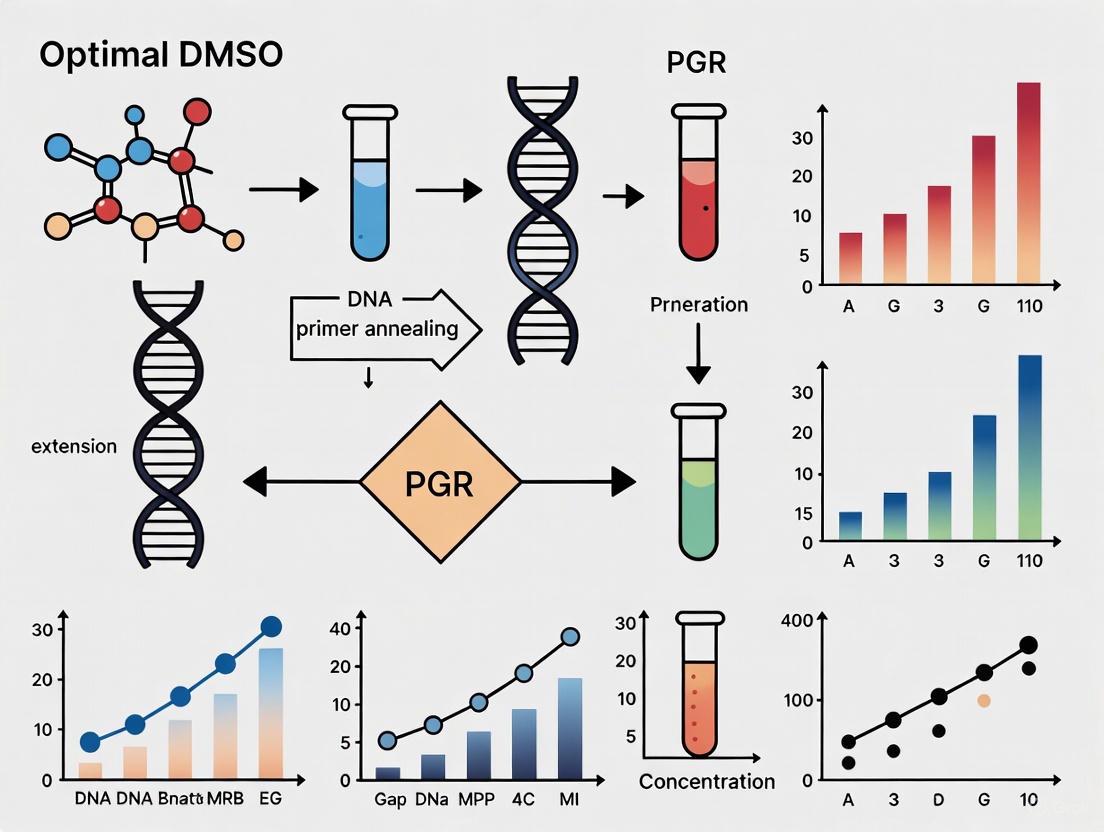
Optimizing DMSO Concentration for GC-Rich PCR: A Complete Guide for Researchers
Amplifying GC-rich DNA sequences is a common challenge in molecular biology, often leading to PCR failure due to stable secondary structures and high melting temperatures.
Optimizing DMSO Concentration for GC-Rich PCR: A Complete Guide for Researchers
Abstract
Amplifying GC-rich DNA sequences is a common challenge in molecular biology, often leading to PCR failure due to stable secondary structures and high melting temperatures. This article provides a comprehensive, evidence-based guide for researchers and drug development professionals on using Dimethyl Sulfoxide (DMSO) to overcome these obstacles. We explore the foundational science behind DMSO's mechanism, present optimized methodological protocols with specific concentration guidelines (typically 2-10%), detail systematic troubleshooting approaches, and validate strategies through comparative analysis with alternative additives. The synthesized protocols enable reliable amplification of critical GC-rich targets, including promoter regions and key pharmacogenetic markers, thereby supporting advancements in biomedical research and clinical diagnostics.
Understanding the GC-Rich PCR Challenge and How DMSO Helps
What Defines a GC-Rich Template?
In molecular biology, a DNA template is considered GC-rich when 60% or more of its nucleotide bases are guanine (G) or cytosine (C) [1] [2]. While only about 3% of the human genome falls into this category, these regions are critically important as they are often found in the promoter regions of genes, including those for housekeeping and tumor suppressor genes [1].
The table below summarizes the core definition and fundamental properties of GC-rich DNA sequences.
| Feature | Description |
|---|---|
| Formal Definition | A DNA sequence where ≥ 60% of the bases are Guanine (G) or Cytosine (C) [1]. |
| Key Property | Increased thermostability (higher melting temperature, Tm) compared to AT-rich DNA [1] [2]. |
| Primary Reason for Stability | Base stacking interactions are the main stabilizing factor, with the three hydrogen bonds of GC base pairs providing additional stability over the two bonds in AT pairs [1] [2]. |
Why Are GC-Rich Sequences Problematic in PCR?
GC-rich templates present several well-characterized challenges that can lead to PCR failure, resulting in no product, a DNA smear, or multiple non-specific bands on a gel [1]. The core difficulties stem from the inherent stability of the DNA and its propensity to form complex structures.
The diagram above illustrates the logical pathway from a GC-rich template to PCR failure. The primary challenges can be broken down as follows:
- Thermal and Structural Stability: The strong base stacking and triple hydrogen bonds in GC pairs make the DNA duplex exceptionally stable [1] [2]. This stability requires higher energy (temperature) to separate the strands during the denaturation step. If the DNA is not fully denatured, the polymerase cannot access the template, leading to incomplete or failed amplification.
- Formation of Stable Secondary Structures: GC-rich regions, particularly in single-stranded DNA, readily form stable secondary structures like hairpin loops [1] [2]. These structures can physically block the progression of the DNA polymerase, resulting in truncated, incomplete PCR products [1].
- Challenges with Primer Annealing: Primers designed for GC-rich regions themselves tend to be GC-rich and can form self-dimers or cross-dimers [2]. Furthermore, the high stability of the template can prevent primers from binding efficiently, especially if the annealing temperature is not optimized [1].
Troubleshooting Guide for GC-Rich PCR Amplification
Overcoming the challenges of GC-rich PCR requires a systematic approach to reaction optimization. The following table outlines the most effective strategies and their underlying principles.
| Troubleshooting Strategy | Specific Recommendations | Mechanism of Action |
|---|---|---|
| Polymerase & Buffer Choice | Use polymerases specifically optimized for GC-rich templates (e.g., Q5 High-Fidelity, OneTaq) and their proprietary GC Enhancers [1] [3]. | Specialized enzymes have higher processivity to push through secondary structures. GC Enhancer additives help destabilize secondary structures and increase primer stringency [1]. |
| Thermal Cycling Adjustments | Increase denaturation temperature (up to 95°C) for the first few cycles [2]. Optimize annealing temperature using a gradient, typically 3–5°C below primer Tm [1] [4]. | Higher denaturation temperature ensures complete separation of the stable duplex. A higher, optimized annealing temperature promotes specific primer binding and reduces non-specific products [1]. |
| Mg²⁺ Concentration Optimization | Test a concentration gradient of MgCl₂, typically in 0.5 mM increments between 1.0 and 4.0 mM [1] [3]. | Mg²⁺ is a crucial cofactor for polymerase activity and primer binding. The optimal concentration is a balance between maximizing enzyme activity and minimizing non-specific priming [1]. |
| Use of PCR Additives | Incorporate additives like DMSO (1-10%), betaine, or glycerol [1] [2] [4]. | These compounds are thought to destabilize DNA secondary structures by interfering with base stacking, making it easier for the polymerase to unwind and traverse the template [1] [2]. |
Optimizing DMSO Concentration for GC-Rich PCR
Within the context of optimizing DMSO for GC-rich PCR research, it is critical to understand its safe and effective concentration range. While DMSO is a common additive to disrupt secondary structures, its concentration must be carefully controlled.
- Function in PCR: DMSO alters the DNA melting temperature and disrupts secondary structures by interfering with base pairing, thereby facilitating the amplification of GC-rich templates [1] [2].
- Recommended Concentration: For PCR applications, DMSO is typically used at final concentrations ranging from 1% to 10% [4]. It is advisable to start with a lower concentration (e.g., 3-5%) and optimize using a gradient.
- Toxicity Considerations: Independent research on cell cultures and zebrafish embryos indicates that DMSO concentrations up to 1% are generally safe for biological systems without causing significant toxicity or developmental abnormalities [5]. This data provides a safety benchmark for researchers, though the optimal concentration for PCR efficacy may be higher.
The Scientist's Toolkit: Key Research Reagent Solutions
Successful amplification of GC-rich templates often relies on using the right reagents. The following table details essential materials and their functions.
| Reagent / Material | Function in GC-Rich PCR |
|---|---|
| High-Processivity Polymerase (e.g., Q5, OneTaq, AccuPrime) | DNA polymerase engineered to remain bound to the template and efficiently unwind stable secondary structures [1] [2]. |
| Specialized GC Buffer & Enhancer | A proprietary buffer mixture containing additives that help denature GC-rich DNA and inhibit the formation of secondary structures like hairpins [1]. |
| DMSO (Dimethyl Sulfoxide) | A common additive that destabilizes DNA secondary structures, facilitating the denaturation of GC-rich regions during the PCR cycling [1] [2]. |
| Betaine | An additive that can help in neutralizing the base composition bias, making the DNA more uniformly accessible to the polymerase. |
| 7-deaza-dGTP | A dGTP analog that can be incorporated into the PCR product, reducing the stability of secondary structures and improving amplification yield [1] [2]. |
| MgCl₂ Solution | A source of magnesium ions, an essential cofactor for DNA polymerase activity. Its concentration must be optimized for each GC-rich target [1] [3]. |
For researchers amplifying GC-rich sequences, the sight of a failed PCR—a blank gel or a smear of non-specific products—is a common frustration. This failure often stems from the DNA template's intrinsic ability to form stable secondary structures, such as hairpins, G-quadruplexes (G4s), and i-motifs, which impede the progression of DNA polymerase [6] [2] [7]. These structures are exceptionally stable in GC-rich sequences (typically defined as >60% GC content) due to the three hydrogen bonds in G-C base pairs and strong base-stacking interactions [8] [2]. When the polymerase enzyme stalls at these points, the result is truncated products, failed amplification, or a high error rate, constraining critical research in areas like promoter analysis, gene regulation, and drug target development [9] [7] [10]. This guide, framed within the context of optimizing dimethyl sulfoxide (DMSO) concentration for GC-rich PCR, provides a structured troubleshooting resource to overcome these challenges.
The Mechanism: How Secondary Structures Stall Polymerases
FAQ: What happens at the molecular level when my polymerase "stalls"?
DNA polymerase synthesizes new DNA strands by processively moving along a single-stranded DNA template. However, regions with high GC content or repetitive sequences can fold into complex secondary structures because the single-stranded template is exposed during the PCR denaturation and annealing steps [7].
- Helicase-Polymerase Uncoupling: In replication, the replicative helicase (CMG) continues to unwind the DNA double helix ahead of the polymerase. When the polymerase stalls at a secondary structure, the helicase can continue unwinding, leading to a uncoupled fork and extensive single-stranded DNA regions [6].
- Direct Blockage of Synthesis: In PCR, which lacks accessory helicases, the DNA polymerase directly encounters these structures. The enzyme's progression is physically blocked, leading to incomplete synthesis, reduced yield, or the polymerase falling off the template entirely [2] [7]. Studies using reconstituted eukaryotic replisomes have confirmed that the DNA template alone is sufficient to cause this stalling, which is mechanistically similar to encountering a leading strand DNA lesion [6].
The following diagram illustrates the logical relationship between GC-rich templates, secondary structure formation, and the consequent PCR failure mechanisms.
Troubleshooting Guide: Overcoming PCR Failure
FAQ: My PCR for a GC-rich target has failed. What should I do first?
A systematic approach is crucial. Begin by verifying your template quality and primer design. Then, focus on optimizing reaction components and cycling conditions. The following table summarizes the core strategies to address problems amplifying GC-rich regions.
Table 1: Comprehensive Troubleshooting Guide for GC-Rich PCR
| Problem Area | Specific Issue | Recommended Solution | Rationale & Practical Notes |
|---|---|---|---|
| Polymerase & Buffer | Standard polymerase stalls at structures. | Use a specialized high-processivity polymerase (e.g., Q5, OneTaq, AccuPrime) [8] [2]. | These enzymes have higher affinity for structured templates and often come with specialized buffers. |
| Non-specific amplification. | Use hot-start DNA polymerases [4]. | Prevents non-specific primer extension during reaction setup. | |
| Reaction Additives | General secondary structure formation. | Add DMSO (1-10%); optimal often 3.75-5% [11] [12]. | Destabilizes secondary structures by interfering with base stacking. Central to thesis research on optimization. |
| Stable hairpins and G-quadruplexes. | Add Betaine (0.5 M - 2.5 M) [11] [9]. | Equalizes the stability of GC and AT base pairs, promoting uniform melting. | |
| Persistent secondary structures. | Add 7-deaza-dGTP (50 μM) [11] [2]. | A dGTP analog that disrupts G-quadruplex formation. Note: may affect downstream sequencing. | |
| Mg²⁺ Concentration | Non-specific bands or low yield. | Optimize MgCl₂ concentration (test 1.0-4.0 mM in 0.5 mM increments) [4] [8]. | Mg²⁺ is a essential cofactor; its concentration critically affects enzyme processivity and fidelity. |
| Thermal Cycling | Inefficient denaturation of template. | Increase denaturation temperature (up to 98°C) or time [4] [2]. | GC-rich duplexes and structures require more energy to melt. |
| Non-specific primer binding. | Increase annealing temperature (Tₐ) in 1-2°C increments [4] [8]. | Higher Tₐ increases stringency, improving specificity. Use a gradient cycler. | |
| Inefficient amplification of long targets. | Increase extension time [4]. | Polymerases move slower through structured regions. | |
| Competitive binding at incorrect sites. | Use shorter annealing times (3-6 seconds) [10]. | Minimizes the opportunity for primers to bind to non-specific, incorrect sites. |
Experimental Protocol: Optimizing DMSO Concentration for GC-Rich PCR
This protocol is designed specifically for testing the effect of DMSO concentration, a key variable in the thesis context.
1. Materials and Reagents
- DNA template (GC-rich target of interest)
- High-fidelity, GC-enhanced DNA polymerase (e.g., Q5 High-Fidelity, OneTaq) with corresponding standard buffer [8]
- Primer set specific to your target
- dNTP mix
- Sterile, PCR-grade water
- DMSO (Molecular Biology Grade)
2. Reaction Setup Prepare a master mix for all components except the template to minimize pipetting error. Then aliquot and add DMSO to create a concentration gradient.
Table 2: Pipetting Scheme for DMSO Concentration Gradient (50 µL Reaction)
| Reagent | Final Concentration/Amount | Master Mix (for 6 reactions) | Volume per Tube (µL) |
|---|---|---|---|
| 10X PCR Buffer | 1X | 60 µL | 10 |
| dNTP Mix | 200 µM | 6 µL | 1 |
| Forward Primer | 0.5 µM | 15 µL | 2.5 |
| Reverse Primer | 0.5 µM | 15 µL | 2.5 |
| DNA Polymerase | 1.25 U | 15 U | 2.5 |
| Template DNA | 1-100 ng | - | X |
| Sterile Water | - | Variable | Variable |
| DMSO | Varying | - | See below |
Varying DMSO Additions:
- Tube 1 (Control): 0 µL DMSO (0%)
- Tube 2: 1.25 µL DMSO (2.5%)
- Tube 3: 1.875 µL DMSO (3.75%)
- Tube 4: 2.5 µL DMSO (5.0%)
- Tube 5: 3.75 µL DMSO (7.5%)
Note: Bring the total volume of each reaction to 50 µL with sterile water.
3. Thermal Cycling Conditions
- Initial Denaturation: 98°C for 30 s
- 35 Cycles:
- Denaturation: 98°C for 5-10 s
- Annealing: Optimized Tₐ (e.g., 60-72°C) for 5 s [10]
- Extension: 72°C for 30 s/kb
- Final Extension: 72°C for 2 min
4. Analysis
- Analyze PCR products by agarose gel electrophoresis.
- Assess for yield, specificity, and the presence of smearing.
- The optimal DMSO concentration is the one that gives the strongest specific band with the least background.
The Scientist's Toolkit: Essential Reagents for GC-Rich PCR
This table details key reagents used in the featured experiments and their specific functions in overcoming polymerase stalling.
Table 3: Research Reagent Solutions for GC-Rich PCR
| Reagent | Function / Mechanism of Action | Typical Working Concentration | Key Research Findings |
|---|---|---|---|
| DMSO (Dimethyl Sulfoxide) | Destabilizes DNA secondary structures by reducing the melting temperature of GC-rich DNA. | 3.75% - 5% (v/v) [11] [12] | In one study, 5% DMSO increased PCR success rate from 42% to 91.6% for plant ITS2 barcodes [11]. Another showed it preferentially enhances amplification of larger fragments, reducing "ski-slope" effects [12]. |
| Betaine | Acts as a chemical chaperone; equalizes the thermal stability of GC and AT base pairs, preventing DNA from forming secondary structures. | 0.5 M - 2.5 M [9] | Can be used as an alternative to DMSO. One study reported a 75% PCR success rate with 1 M betaine [11]. Combining DMSO and betaine is not always additive [11]. |
| 7-deaza-dGTP | A guanine analog that is incorporated into DNA but prevents the formation of G-quadruplex structures by disrupting Hoogsteen base pairing. | 50 µM [11] | Effective for extremely stable G4 structures but may require adjustments for downstream applications like sequencing. |
| Q5 High-Fidelity DNA Polymerase | A high-fidelity, high-processivity polymerase engineered for amplifying difficult targets, including GC-rich sequences. | As per mfr. (NEB #M0491) [8] | More than 280x the fidelity of Taq polymerase. Can be supplemented with a proprietary GC Enhancer for targets up to 80% GC. |
| OneTaq GC Buffer | A specialized buffer system formulated with enhancers to denature GC-rich templates and inhibit secondary structure formation. | Used as the reaction buffer [8] [2] | Designed specifically for problematic amplicons, providing a ready-to-use solution without the need for additive optimization. |
| MgCl₂ / MgSO₄ | Essential cofactor for DNA polymerase activity. Required for catalytic function and primer binding. | 1.5 - 4.0 mM (optimization required) [4] [8] | Concentration must be carefully titrated, as excess leads to non-specific bands and too little reduces enzyme activity. |
Fundamental Mechanisms: How DMSO Affects DNA
How does DMSO lower the melting temperature (Tm) of DNA?
DMSO lowers the melting temperature of DNA by directly interfering with the molecular forces that stabilize the double helix. Its polar aprotic nature allows DMSO molecules to disrupt the water network surrounding the DNA backbone and compete with the hydrogen bonds between complementary base pairs, particularly the strong triple hydrogen bonds of G-C pairs [13]. This destabilizes the double-stranded structure, requiring less thermal energy (a lower temperature) for strand separation [14] [13]. Research has quantified that the bending persistence length of DNA decreases linearly by approximately 0.43% for every 1% increase in DMSO concentration (up to 20%) [14].
How does DMSO disrupt secondary structures like hairpins in GC-rich DNA?
GC-rich sequences readily form stable, intrastrand secondary structures such as hairpin loops due to strong base stacking interactions [15] [2]. DMSO disrupts these structures by reducing the stability of base stacking and hydrogen bonding. This action "relaxes" the DNA, preventing these regions from folding back on themselves [15] [13]. By doing so, DMSO provides easier access for DNA polymerase and primers to the template, which is critical for the success of techniques like PCR [16] [13].
Table 1: Quantitative Effects of DMSO on DNA Mechanics
| DMSO Concentration | Effect on DNA Persistence Length | Effect on DNA Conformation |
|---|---|---|
| ~0.1% | Persistence length decreases from ~50 nm to ~12 nm (in 3% DMSO) [17] | Local denaturation ("bubbles" and kinks) begins [17] |
| ≤20% | Linear decrease by (0.43 ± 0.02%) per %-DMSO [14] | Moderate compaction; mean-squared end-to-end distance decreases by 1.2% per %-DMSO [14] |
| >30% | Significant structural flexibility and denaturation [17] | Conformational shift from common B-form to a more compact A-form [13] |
Troubleshooting Guide for GC-rich PCR with DMSO
Why did my PCR fail even after adding DMSO?
While DMSO is a powerful additive, PCR failure can occur if its concentration is not optimized. High concentrations of DMSO (e.g., >10%) can significantly inhibit DNA polymerase activity [18]. Furthermore, a single optimization step is often insufficient for challenging GC-rich templates. Failure may stem from other unaddressed factors, such as suboptimal annealing temperature, inappropriate Mg²⁺ concentration, or a polymerase not suited for GC-rich amplicons [15] [2].
I am getting non-specific PCR products (multiple bands) with DMSO. How can I fix this?
Non-specific amplification indicates that primers are binding to off-target sites. DMSO can sometimes reduce stringency. To address this:
- Increase Annealing Temperature: Perform a gradient PCR to determine the optimal annealing temperature. A higher temperature promotes specific primer binding [15] [19].
- Optimize DMSO Concentration: Titrate DMSO in 1-2% increments. High concentrations can reduce reaction stringency [18].
- Check Mg²⁺ Concentration: High Mg²⁺ levels can facilitate non-specific binding. Titrate MgCl₂ in 0.5 mM increments between 1.0 and 4.0 mM to find the optimal concentration [15].
- Use a Hot-Start Polymerase: This prevents polymerase activity at room temperature, reducing the chance of primer-dimer formation and non-specific amplification during reaction setup [19].
What is the optimal concentration range for DMSO in PCR?
The optimal concentration of DMSO is typically between 2% and 10% [18]. However, for most applications, a concentration of 5% is a standard starting point [16]. It is critical to note that at concentrations higher than 5%, DNA polymerase activity may begin to decline, and 10% DMSO can be strongly inhibitory [18]. Therefore, careful titration is recommended.
Table 2: DMSO Concentration Guide for PCR
| DMSO Concentration (v/v) | Effect on PCR | Recommendation |
|---|---|---|
| 1-5% | Reduces secondary structures; lowers Tm; generally enhances specificity and yield [13] [19]. | Optimal and safe range for most applications. Start at 5% [16]. |
| 6-8% | Further lowers Tm; may begin to inhibit some polymerases. | Use for very difficult templates; requires validation [18]. |
| >10% | Significant inhibition of DNA polymerase; can increase error rate; leads to PCR failure [18]. | Avoid. |
Advanced Experimental Protocols
Protocol: Systematic Optimization of DMSO for GC-rich PCR
This protocol provides a step-by-step method for empirically determining the best DMSO conditions for a specific GC-rich target.
- Prepare a Master Mix: Create a standard master mix for your PCR, excluding DMSO and the template. Use a polymerase known for handling GC-rich templates (e.g., Q5 High-Fidelity, OneTaq GC-rich, or similar) [15].
- Set Up DMSO Titration: Aliquot the master mix into multiple tubes. Add DMSO to each tube to create a concentration gradient (e.g., 0%, 2%, 4%, 6%, 8%). Keep the template and water volume constant across all reactions.
- Run PCR: Use a thermal cycler program with a gradient annealing temperature across the block. This allows you to test the interaction of DMSO concentration and annealing temperature simultaneously [15].
- Analyze Results: Resolve the PCR products on an agarose gel. Identify the condition that yields the brightest, specific band with the least background smearing or non-specific bands.
- Further Optimization: If needed, fine-tune the Mg²⁺ concentration or incorporate other additives like betaine (0.5-2 M) based on the initial results [16] [18].
Protocol: Direct Observation of DMSO-Induced DNA Conformational Changes via AFM
Atomic Force Microscopy (AFM) can visually demonstrate DMSO's effect on DNA structure [17].
- Sample Preparation: Dilute plasmid DNA (e.g., pBR322) to a final concentration of ~1 ng/μL in a buffer containing 1 mM Tris-HCl (pH 7.8) with varying concentrations of DMSO (e.g., 0%, 0.1%, 1%, 5%, 10%). Incubate at room temperature for 30 minutes [17].
- Surface Deposition: Deposit a 40 μL aliquot of each DNA solution onto APTES-treated mica. Incubate for 3 minutes at room temperature [17].
- Rinsing and Drying: Remove the excess solution and rinse the mica surface gently with ~200 μL of deionized water. Dry with a gentle stream of nitrogen gas [17].
- AFM Imaging: Scan the prepared samples using an AFM operated in AC mode in air. Use software like ImageJ to analyze the images, measuring parameters such as contour length, end-to-end distance, and the appearance of kinks or denaturation bubbles [17].
- Expected Outcome: With increasing DMSO concentration, you will observe a measurable decrease in DNA persistence length (increased flexibility) and the direct appearance of local denaturation sites, even at concentrations as low as 0.1% [17].
Visualizing the Mechanism
The following diagram illustrates the molecular mechanism by which DMSO destabilizes DNA double helices and disrupts secondary structures.
DMSO DNA Destabilization Mechanism
The Scientist's Toolkit: Essential Reagents for DMSO Research
Table 3: Key Research Reagents and Materials
| Reagent / Material | Function in DMSO/DNA Research |
|---|---|
| High-Purity DMSO (≥99.9%) | Ensures experimental consistency and avoids confounding results from impurities [13]. |
| Proofreading DNA Polymerases (e.g., Q5, Phusion) | High-fidelity enzymes are less prone to stalling at GC-rich secondary structures, often used with GC Enhancers [15]. |
| GC-Rich PCR System Kits | Commercial kits (e.g., from Roche, NEB) provide optimized buffers with DMSO and detergents for challenging amplicons [18]. |
| Betaine | An alternative or complementary additive to DMSO that equalizes the thermal stability of G-C and A-T base pairs [16]. |
| MgCl₂ Solution | A necessary cofactor for DNA polymerase; its concentration often requires re-optimization when adding DMSO [15]. |
| APTES-Treated Mica | Provides a positively charged, atomically flat surface necessary for AFM imaging of DNA molecules [17]. |
Frequently Asked Questions (FAQs)
Can DMSO cause DNA damage in my samples?
The effect of DMSO on DNA integrity is concentration and context-dependent. At low concentrations (≤10%) typically used in PCR, DMSO is generally safe and can even have antioxidant properties that protect DNA from oxidative damage [13]. However, prolonged exposure to high concentrations of DMSO has been linked to increased chromosomal aberrations in some in vivo studies, and it can make DNA more vulnerable to strand breaks in vitro [13]. For routine PCR, this is not a concern.
Should I use DMSO or betaine for GC-rich PCR?
Both are effective, and the choice can be template-specific. DMSO works primarily by disrupting base stacking and hydrogen bonding, while betaine (also known as trimethylglycine) acts as a stabilizing osmolyte that prevents DNA from forming secondary structures without significantly altering its Tm [16] [15]. In many cases, using a combination of both can be synergistic for extremely difficult templates [16]. It is recommended to test both individually and in combination.
How does DMSO concentration affect DNA polymerase fidelity?
While DMSO can improve yield for difficult targets, it's important to note that adding DMSO can influence the error rate of the PCR [18]. The precise impact depends on the polymerase and the specific DMSO concentration. For applications requiring ultra-high fidelity (e.g., cloning), it is advisable to use a proofreading polymerase and validate the amplified sequence.
Frequently Asked Questions (FAQs)
Q1: Why is amplifying GC-rich regions like certain promoters and tumor suppressor genes so challenging in PCR? GC-rich DNA sequences (typically >60% GC content) pose two major challenges. First, the three hydrogen bonds between guanine and cytosine create stronger base pairing than A-T pairs, leading to higher melting temperatures and making it difficult to fully denature the DNA template under standard PCR conditions [16] [2]. Second, these regions readily form stable secondary structures, such as hairpin loops and knots, which can block the progression of the DNA polymerase enzyme, resulting in failed reactions or truncated products [16] [2].
Q2: How does DMSO help in the amplification of GC-rich targets? DMSO (Dimethyl sulfoxide) is an organic solvent that aids PCR by reducing the formation of secondary DNA structures [20]. It does this by lowering the overall melting temperature of the DNA, which facilitates better strand separation during the denaturation step and improves primer access to the template [16] [21]. This is particularly useful for resolving complex structures in GC-rich sequences.
Q3: What is a safe and effective concentration of DMSO to use? While concentrations from 2% to 10% can be effective, many protocols recommend starting with a final concentration of 5% [21]. However, it is critical to note that DMSO can inhibit DNA polymerase activity at higher concentrations, with 10% often being strongly inhibitory [21]. Furthermore, research indicates that even low concentrations (e.g., 0.1%) can induce large-scale changes in the cellular transcriptome and epigenome [22]. Therefore, using the lowest effective concentration is advised, especially when working with sensitive downstream applications.
Q4: Are there other additives that can be combined with DMSO? Yes, a multipronged approach is often most successful. Betaine (0.5 M to 2.5 M) is another highly effective additive that can be used alone or in combination with DMSO to destabilize secondary structures [16] [21]. Other options include formamide, glycerol, and specialized commercial "GC-rich resolution" solutions [21] [20].
Q5: My PCR still isn't working. What other critical factors should I check? Beyond additives, consider these key parameters:
- DNA Polymerase: Switch to a polymerase specifically engineered for high GC content or high processivity [16] [2].
- Mg²⁺ Concentration: Titrate the Mg²⁺ concentration, as it is a crucial cofactor for DNA polymerase. Both too little and too much can cause reaction failure [2] [20].
- Thermal Cycling Profile: Implement a "touchdown" or "slow-down" PCR protocol, and consider using a higher denaturation temperature (up to 98°C) for the first few cycles to ensure complete template denaturation [2] [20].
Troubleshooting Guide: Optimizing DMSO for GC-rich PCR
This guide provides a structured approach to troubleshooting, with a focus on integrating DMSO effectively into your protocol.
Workflow for Systematic Optimization
The following diagram outlines a logical, step-by-step workflow to diagnose and resolve common issues when amplifying GC-rich targets.
Quantitative Data for Experimental Design
The tables below summarize key quantitative data from the literature to guide your optimization experiments.
Table 1: Optimal Concentrations of Common PCR Additives for GC-rich Templates [16] [21]
| Additive | Common Working Concentration | Key Function | Important Considerations |
|---|---|---|---|
| DMSO | 2% - 5% (v/v) | Disrupts secondary structures, lowers DNA Tm [16] [20] | >5% can inhibit polymerase; may increase error rate [21] |
| Betaine | 0.5 M - 2.5 M | Destabilizes GC base pairs, equalizes Tm [16] | Often used in combination with DMSO for synergistic effect [16] |
| Formamide | 1.25% - 10% (v/v) | Denaturant, lowers DNA Tm [23] | Can be used as an alternative to DMSO |
| Glycerol | 5% - 25% (v/v) | Stabilizes enzymes, can lower DNA Tm [21] | Higher viscosity can affect reaction kinetics |
Table 2: Summary of Alternative Strategies for GC-rich PCR Amplification
| Strategy | Protocol Adjustment | Rationale |
|---|---|---|
| Specialized Polymerases | Use enzymes from Pyrococcus or Thermus thermophilus species [2] | Enhanced processivity and stability for traversing complex secondary structures. |
| Temperature Adjustments | Increase denaturation temperature to 95-98°C; use touchdown PCR [2] [20] | Ensures complete separation of DNA strands with high melting temperatures. |
| Primer Design | Longer primers (25-30 bp); avoid GC clamps at 3' end [20] | Increases binding specificity and minimizes mispriming or dimer formation. |
Experimental Protocol: A Multipronged Optimization Workflow
This protocol is adapted from recent research on amplifying GC-rich nicotinic acetylcholine receptor subunits and standard GC-rich PCR methodologies [16] [23].
Materials and Reagents
The Scientist's Toolkit: Key Research Reagents
| Item | Function/Explanation |
|---|---|
| High-Fidelity/GC-Rich DNA Polymerase | Engineered for high processivity and efficiency on difficult templates (e.g., Platinum SuperFi, Phusion) [16]. |
| DMSO (Molecular Biology Grade) | Primary additive to disrupt DNA secondary structures. Must be high purity to avoid contaminants [16] [21]. |
| Betaine (Molecular Biology Grade) | Co-additive that works synergistically with DMSO to homogenize melting temperatures [16]. |
| 10x PCR Buffer (with or without Mg²⁺) | Provides optimal ionic conditions and pH for the reaction. A buffer without Mg²⁺ allows for precise titration. |
| dNTP Mix | Building blocks for DNA synthesis. |
| Template DNA & Primers | High-quality, intact DNA and optimally designed primers are critical. |
Step-by-Step Procedure
Step 1: Prepare the Master Mix with Additive Titration Prepare reactions on ice. For a 50 µL final reaction volume, combine the following components, creating a matrix to test different DMSO and betaine conditions [16]:
- 5 µL of 10x PCR Buffer (provided with polymerase)
- 1 µL of 10 mM dNTP mix
- 0.5-2.5 µL of 25 mM MgCl₂ (if not in buffer; final conc. 0.5-5.0 mM) [23]
- 1 µL of 20 µM Forward Primer
- 1 µL of 20 µM Reverse Primer
- 1-10 ng of Template DNA
- Varying volumes of DMSO and 5M Betaine stock (see table below for final concentrations)
- Sterile Nuclease-Free Water to 49 µL
- 1 µL of DNA Polymerase (add last)
Step 2: Set Up the Additive Test Matrix It is highly recommended to test a range of DMSO and betaine concentrations to find the optimal combination for your specific template. A sample test matrix for a 50 µL reaction is shown below.
Table 3: Example Additive Test Matrix for a 50 µL PCR
| Tube | DMSO (100%) | 5M Betaine Stock | Final [DMSO] | Final [Betaine] |
|---|---|---|---|---|
| A | 0 µL | 0 µL | 0% | 0 M |
| B | 1.0 µL | 0 µL | 2% | 0 M |
| C | 2.5 µL | 0 µL | 5% | 0 M |
| D | 0 µL | 5.0 µL | 0% | 0.5 M |
| E | 1.0 µL | 5.0 µL | 2% | 0.5 M |
| F | 2.5 µL | 5.0 µL | 5% | 0.5 M |
Step 3: Execute the Thermal Cycling Protocol Use the following adjusted thermal cycling conditions in your thermocycler [16] [2]:
- Initial Denaturation: 98°C for 2 minutes (or as recommended for your polymerase).
- Amplification (35-40 cycles):
- Denaturation: 98°C for 20 seconds.
- Annealing: Use a gradient to test temperatures 3-5°C above the calculated Tm of your primers.
- Extension: 72°C for 30-60 seconds per kb.
- Final Extension: 72°C for 5-10 minutes.
- Hold: 4°C.
Step 4: Analyze Results Analyze 5-10 µL of each PCR product by agarose gel electrophoresis to identify the condition that yields the strongest, most specific amplification band with the lowest background.
Experimental Workflow Visualization
The end-to-end workflow for this optimization experiment, from setup to analysis, is summarized in the following diagram.
Protocol Development: Implementing DMSO in Your GC-Rich PCR Workflow
What makes GC-rich sequences so challenging to amplify? GC-rich DNA sequences (typically >60% GC content) present a formidable challenge in Polymerase Chain Reaction (PCR) due to their intrinsic biophysical properties. The primary issues are thermal stability and secondary structure formation. The high GC content leads to stronger base-stacking interactions and more hydrogen bonds, resulting in a significantly higher melting temperature (Tm) [24] [2]. This stability promotes the formation of rigid secondary structures, such as hairpin loops and stem-loop structures, which do not denature efficiently at standard PCR temperatures (e.g., 94–95°C) [25] [2]. These stable structures physically impede the progress of the DNA polymerase, leading to premature termination of extension and resulting in truncated amplicons or complete amplification failure [25].
How does DMSO help? Dimethyl sulfoxide (DMSO) is a pivotal chemical additive used to overcome these hurdles. It functions primarily as a helix-destabilizing agent [20]. By interfering with the DNA's base-stacking interactions, DMSO effectively lowers the melting temperature of the double-stranded DNA, facilitating more complete strand separation during the denaturation step [20] [2]. This action helps to melt stable secondary structures, providing the DNA polymerase with better access to the template and thereby enabling efficient primer binding and elongation through regions that would otherwise be inaccessible [20].
Establishing the Gold Standard: DMSO Concentration Ranges
Recommended DMSO Concentration Ranges
Based on aggregated experimental data from published research and technical protocols, the effective and safe concentration range for DMSO in GC-rich PCR is 2.5% to 10% (v/v) [25] [26]. The optimal concentration within this window is template- and reaction-dependent.
Table 1: Summary of Recommended DMSO Concentrations and Their Applications
| Concentration Range (v/v) | Typical Use Case | Key Considerations |
|---|---|---|
| 2.5% - 5% | Standard optimization range for most GC-rich templates; recommended starting point [25]. | Balances effectiveness in destabilizing secondary structures with minimal risk of polymerase inhibition. |
| 5% - 10% | For exceptionally challenging templates with very high GC content or complex secondary structures [26]. | Increased risk of inhibiting DNA polymerase activity. Requires careful optimization and potentially increased enzyme concentration [4]. |
DMSO in a Multipronged Optimization Strategy
Successful amplification of GC-rich targets rarely relies on DMSO alone. A combined approach is essential, as demonstrated in a study optimizing PCR for GC-rich nicotinic acetylcholine receptor subunits, which highlighted the "importance of a multipronged approach" [24].
Table 2: Complementary PCR Additives for GC-Rich Amplification
| Additive | Mechanism of Action | Common Concentration |
|---|---|---|
| Betaine | Equalizes the stability of GC and AT base pairs, reduces secondary structure formation, and stabilizes DNA polymerase [24] [20]. | 0.5 M - 2 M [26] |
| Formamide | Lowers the melting temperature of DNA, aiding in the denaturation of stable duplexes [20]. | Concentration requires optimization. |
Experimental Protocol: Optimizing DMSO in GC-Rich PCR
Workflow for Systematic Optimization
The following diagram outlines a logical, step-by-step workflow for integrating and optimizing DMSO in your GC-rich PCR experiments.
Step-by-Step Methodology
This protocol provides a detailed guide for a key experiment to determine the optimal DMSO concentration for a specific GC-rich target.
1. Primer and Polymerase Selection:
- Primer Design: Design primers with a higher-than-standard melting temperature (Tm > 68°C) to permit annealing at higher temperatures, which enhances specificity [25]. Avoid GC clamps (more than three consecutive G or C bases) at the 3' end to prevent mispriming [27] [2].
- DNA Polymerase: Choose a polymerase engineered for high processivity and GC-rich templates, such as PrimeSTAR Max or CloneAmp HiFi [25]. These enzymes often have a higher resistance to additives like DMSO.
2. Reaction Setup with DMSO Titration:
- Prepare a master mix containing all standard PCR components: buffer, dNTPs (typically 200 µM each), primers (0.1–1 µM), template DNA (amount optimized for complexity), and the selected DNA polymerase (1–2 units/50 µL reaction) [27] [26].
- Aliquot the master mix into five separate tubes.
- Add DMSO to the tubes to create the following final concentration series:
- Tube 1: 0% (Negative Control)
- Tube 2: 2.5%
- Tube 3: 5%
- Tube 4: 7.5%
- Tube 5: 10%
- Mix the reactions thoroughly but gently to avoid creating bubbles or shearing the DNA.
3. Thermal Cycling Conditions:
- Initial Denaturation: 98°C for 2 min [25].
- PCR Cycles (30–35 cycles):
- Denaturation: 98°C for 10 sec. A higher denaturation temperature improves melting of GC-rich structures [25].
- Annealing: Use a temperature gradient, starting at 3–5°C below the primer Tm. A short annealing time (5–15 sec) is recommended to minimize mispriming [25].
- Extension: 68°C for 15–30 sec/kb, depending on the polymerase's speed [25].
- Final Extension: 72°C for 5–10 min.
4. Post-PCR Analysis:
- Analyze the PCR products by agarose gel electrophoresis.
- Evaluate the results for:
- Specificity: A single, sharp band of the expected size.
- Yield: Intensity of the band relative to the ladder and other conditions.
- Background: Presence of smearing or non-specific bands.
The optimal DMSO concentration is the lowest one that produces a high yield of the specific product with minimal background.
Troubleshooting and FAQ
Frequently Asked Questions
Q1: Can I use DMSO with any DNA polymerase? While many DNA polymerases tolerate DMSO, the level of tolerance varies. Always consult the manufacturer's specifications. Some specialized polymerases, like PrimeSTAR Max, are tested and confirmed to work robustly with DMSO [25]. Using DMSO with non-validated enzymes may lead to unexpected inhibition.
Q2: What should I do if high DMSO concentrations (e.g., 10%) completely inhibit my reaction? Complete inhibition at 10% DMSO indicates that the polymerase's activity has been compromised. The recommended action is to:
- Reduce the DMSO concentration to the 2.5–5% range.
- Increase the amount of DNA polymerase in the reaction to counteract the inhibitory effect [4].
- Consider using a different polymerase known for higher tolerance to co-solvents.
Q3: Is it beneficial to combine DMSO with other additives like betaine? Yes, combining additives is a common and often effective strategy. Research on amplifying GC-rich nicotinic acetylcholine receptor subunits successfully employed a tailored protocol that incorporated both DMSO and betaine [24]. These additives can act through different mechanisms (helix-destabilization vs. base-pair equalization) to synergistically overcome amplification barriers.
Q4: Why might my reaction still fail even with 10% DMSO? If DMSO alone is insufficient, a more comprehensive optimization is required. Consider these factors:
- Thermal Cycling Parameters: Further increase the denaturation temperature (if the enzyme allows) or duration [4] [2].
- Alternative Polymerases: Switch to a polymerase specifically designed for GC-rich templates [25] [2].
- Primer Re-design: The primers themselves may be forming stable secondary structures. Re-design them to avoid self-complementarity and high GC content at the 3' end [27] [20].
- Magnesium Concentration: Titrate the Mg²⁺ concentration, as it is a critical cofactor for polymerase activity and can influence reaction specificity and efficiency [4] [26].
Troubleshooting Guide
Table 3: Common Problems and Solutions When Using DMSO
| Problem | Potential Cause | Recommended Solution |
|---|---|---|
| No Product | DMSO concentration too high, inhibiting the polymerase. | Titrate DMSO downward (e.g., to 2.5-5%) and/or increase polymerase amount [4]. |
| Non-specific Bands/Smearing | Annealing temperature is too low, especially in the presence of DMSO which can weaken primer binding. | Increase the annealing temperature in a stepwise manner (1-2°C increments) [4]. |
| Low Yield | Incomplete denaturation of the GC-rich template. | Increase denaturation temperature to 98°C and ensure denaturation time is short but sufficient [25] [4]. Consider combining DMSO with betaine [24]. |
The Scientist's Toolkit: Essential Reagents for GC-Rich PCR
Table 4: Key Research Reagent Solutions for GC-Rich PCR
| Reagent / Solution | Function | Example Products / Notes |
|---|---|---|
| Specialized DNA Polymerase | High-processivity enzymes designed to read through complex secondary structures and tolerate additives. | PrimeSTAR GXL, Advantage GC2, AccuPrime GC-Rich DNA Polymerase [25] [2]. |
| DMSO (PCR Grade) | Helix-destabilizing agent that lowers DNA Tm and disrupts secondary structures. | Use high-purity, sterile grades. Standard concentration range: 2.5-10% (v/v) [25] [26]. |
| Betaine | Additive that equalizes base-pair stability, prevents secondary structure formation, and stabilizes enzymes. | Often used at 0.5-2 M final concentration. Can be used in combination with DMSO [24] [26]. |
| GC Buffer | Proprietary reaction buffers optimized to enhance amplification of GC-rich targets, often containing undisclosed enhancers. | OneTaq GC Buffer (NEB), GC Enhancer (Thermo Fisher) [2]. |
| High-Quality dNTPs | Balanced deoxynucleotide solution providing the building blocks for new DNA strands. | Use balanced mixtures at 200 µM each to maintain polymerase fidelity [27] [26]. |
Technical FAQ: Overcoming GC-Rich PCR Challenges
Q1: Why is the EGFR promoter region particularly difficult to amplify using standard PCR?
The epidermal growth factor receptor (EGFR) promoter region is an exceptionally challenging template for PCR due to its extremely high GC content. Bioinformatic analysis reveals this region has a GC content of 75.45% across a 660 bp sequence, with a CpG island region spanning 558 bp. This high stability leads to formation of stable secondary structures that block DNA polymerase activity, resulting in ineffective amplification with standard protocols [28].
Q2: What was the key additive used to successfully amplify the EGFR promoter?
Dimethyl sulfoxide (DMSO) at a concentration of 5% was identified as critically necessary for successful amplification. Separate PCR reactions testing 1%, 3%, and 5% DMSO concentrations demonstrated that only 5% DMSO provided the desired amplicon yield without nonspecific amplification [28].
Q3: How does DMSO work to improve GC-rich PCR amplification?
DMSO improves GC-rich PCR amplification through multiple mechanisms:
- Reduces DNA secondary structure stability by interacting with water molecules on the DNA strand, thereby reducing hydrogen bonding [29]
- Lowers the melting temperature (Tm) of DNA, facilitating primer binding to template DNA at lower temperatures [30] [29]
- Decreases DNA persistence length by approximately 0.43% per percent DMSO, making the DNA more flexible [30]
- Moderately compacts DNA conformations, reducing mean-squared end-to-end distance by 1.2% per percent DMSO [30]
Q4: What other optimization parameters were crucial for success?
Successful amplification required a multifaceted optimization approach:
- MgCl₂ concentration: Optimal range of 1.5-2.0 mM [28]
- Annealing temperature: 63°C, which was 7°C higher than the calculated temperature [28]
- DNA template concentration: At least 2 μg/ml was necessary [28]
- DNA polymerase selection: Use of polymerases specifically designed for GC-rich templates [31]
Experimental Protocol: Optimized EGFR Promoter Amplification
Materials and Reagents
Table: Key Research Reagent Solutions
| Reagent/Item | Function/Role in Protocol | Specifications/Notes |
|---|---|---|
| Taq DNA Polymerase | Enzymatic amplification | Standard polymerase sufficient when combined with DMSO [28] |
| DMSO (5%) | GC-rich enhancer | Critical concentration; reduces secondary structures [28] |
| MgCl₂ | Cofactor | Optimal at 1.5-2.0 mM concentration [28] |
| dNTPs | Building blocks | 0.25 mM of each dNTP [28] |
| Primers | Target specificity | Designed for EGFR promoter region [28] |
| Genomic DNA | Template | From FFPE tissue; ≥2 μg/ml concentration [28] |
Step-by-Step Optimized Protocol
Reaction Setup
- Prepare 25 μl reaction volume containing:
- 1 μl genomic DNA (concentration ≥2 μg/μl)
- 0.2 μM of each primer
- 0.25 mM of each dNTP
- 1.5-2.0 mM MgCl₂
- 5% DMSO (v/v)
- 0.625 U Taq DNA polymerase
- 1× PCR buffer [28]
- Prepare 25 μl reaction volume containing:
Thermal Cycling Conditions
- Initial denaturation: 94°C for 3 minutes
- 45 cycles of:
- Denaturation: 94°C for 30 seconds
- Annealing: 63°C for 20 seconds
- Extension: 72°C for 60 seconds
- Final extension: 72°C for 7 minutes [28]
Product Analysis
- Detect 197 bp PCR products by gel electrophoresis on 2% agarose gel
- Use SYBR Safe DNA Gel Stain for visualization [28]
Troubleshooting Guide
Table: Common Issues and Solutions for GC-Rich PCR
| Problem | Possible Cause | Solution | Supporting Evidence |
|---|---|---|---|
| No amplification | Incomplete denaturation of secondary structures | Increase DMSO to 5%; use higher denaturation temperature (98°C) | Required 5% DMSO for EGFR [28] |
| Non-specific bands | Annealing temperature too low | Increase annealing temperature gradient (7°C above calculated Tm) | Optimal annealing at 63°C vs. calculated 56°C [28] |
| Weak yield | Insufficient template or enzyme inhibition | Ensure DNA concentration ≥2 μg/ml; titrate Mg²⁺ (1.0-4.0 mM) | DNA <1.86 μg/ml gave no amplification [28] |
| Smeared products | Polymerase stalling at secondary structures | Add combination of DMSO + betaine; use specialty polymerases | Betaine eliminates base composition dependence [29] |
Mechanism of Action: How DMSO Enables GC-Rich Amplification
Advanced Optimization Strategies for Challenging Templates
For researchers facing even more challenging GC-rich targets, consider these advanced strategies:
Alternative and Combination Additives
- Betaine (0.5-2.0 M): Works synergistically with DMSO by eliminating base composition dependence during DNA melting [29] [32]
- 7-deaza-dGTP: dGTP analog that improves yield of GC-rich regions, though it doesn't stain well with ethidium bromide [31]
- Specialized commercial systems: GC-RICH PCR System (Roche) containing pre-optimized enzyme mixes, buffers with detergents/DMSO, and resolution solutions [32]
Polymerase Selection
Specialized polymerases can provide significant advantages:
- OneTaq GC Buffer with High GC Enhancer (NEB): Can amplify up to 80% GC content [31]
- Q5 High-Fidelity DNA Polymerase with GC Enhancer: Provides high fidelity for GC-rich targets up to 80% GC [31]
- AccuPrime GC-Rich DNA Polymerase (ThermoFisher): Originates from Pyrolobus fumarius with increased processivity [2]
Thermal Cycling Modifications
- Higher denaturation temperatures: 98°C instead of 94-95°C for complete denaturation [33]
- Touchdown PCR: Start with higher annealing temperature and reduce by 2°C per cycle [33]
- Slow-down PCR: Incorporates 7-deaza-dGTP with lowered ramp rates and additional cycles [2]
Experimental Workflow: From Problem to Solution
This case study demonstrates that successful amplification of challenging GC-rich targets like the EGFR promoter requires systematic optimization of multiple parameters, with DMSO concentration serving as a critical factor. The 5% DMSO concentration proved essential for disrupting stable secondary structures that would otherwise prevent amplification.
This technical support center addresses a key challenge in molecular biology: the amplification of GC-rich DNA sequences. Within the broader thesis research on optimal DMSO concentration for GC-rich PCR, this guide provides targeted troubleshooting and FAQs. GC-rich templates (with over 60% GC content) pose significant challenges due to their formation of stable secondary structures and high melting temperatures, which can lead to polymerase stalling, mispriming, and complete amplification failure [34] [35]. The synergistic combination of chemical enhancers, particularly DMSO and betaine, provides a powerful strategy to overcome these obstacles, enabling robust and reliable amplification of difficult targets.
Frequently Asked Questions (FAQs)
1. Why is a combination of DMSO and betaine particularly effective for GC-rich PCR? DMSO and betaine operate through complementary mechanisms to facilitate the amplification of GC-rich sequences. DMSO acts by disrupting inter and intrastrand re-annealing of DNA, thereby preventing the formation of secondary structures like hairpins that can halt polymerase progression [34]. Betaine, an amino acid analog, functions as an isostabilizing agent by equilibrating the melting temperature between AT and GC base pairings [34]. This combination effectively reduces the energy required to denature the DNA template and inhibits the reformation of secondary structures during the PCR cycling, leading to significantly improved specificity and yield of the target amplicon [34] [36].
2. What are the recommended starting concentrations for these additives? For initial optimization experiments, the following concentration ranges are recommended. Note that the optimal concentration is often target-specific and may require fine-tuning.
Table: Recommended Concentration Ranges for Common PCR Additives
| Additive | Recommended Working Concentration | Key Considerations |
|---|---|---|
| DMSO | 2% - 10% (v/v) [37] | Concentrations >5% can reduce polymerase activity; 10% is often inhibitory [37]. |
| Betaine | 0.5 M - 2.0 M [37] | Often used at a concentration of 1.5 M [34]. |
| GC-RICH Resolution Solution | 0.5 M - 2.5 M [37] | Titrate in steps of 0.25 M for difficult templates. |
| 7-deaza-dGTP | (Partial substitution for dGTP) [36] | Does not stain well with ethidium bromide [35]. |
3. Can I use these enhancers with any DNA polymerase? While DMSO and betaine are broadly compatible with many PCR systems, their effectiveness and optimal concentration can vary with the polymerase. Many modern polymerases are specifically optimized for GC-rich amplification and are supplied with proprietary enhancer solutions [35]. It is crucial to consult the manufacturer's instructions, as some polymerases are sensitive to specific additives. For instance, the GC-RICH PCR System includes a specialized enzyme mix and buffer pre-formulated with detergents and DMSO [37]. When using standalone polymerases like Q5 High-Fidelity or OneTaq, the supplied GC Enhancers are designed for optimal performance with these enzymes [35].
4. What other factors should I optimize when troubleshooting GC-rich PCR? Amplifying GC-rich sequences often requires a multi-pronged approach. Beyond additives, consider optimizing the following:
- Polymerase Choice: Use polymerases specifically designed for GC-rich or difficult templates [35].
- Mg2+ Concentration: Titrate MgCl2 in 0.5 mM increments between 1.0 and 4.0 mM, as it is a critical cofactor for polymerase activity and primer binding [35].
- Annealing Temperature (Ta): Use a temperature gradient to find the optimal Ta. A higher Ta can increase primer specificity and help denature secondary structures [35]. The NEB Tm Calculator is a helpful tool for this.
- Thermal Cycling Parameters: Using a "hot start" or a higher denaturation temperature (e.g., 98°C) can be beneficial. A touchdown PCR protocol for the first few cycles may also improve specificity [35].
Troubleshooting Guide
Table: Common GC-rich PCR Problems and Solutions
| Problem | Possible Causes | Recommended Solutions |
|---|---|---|
| No Amplification | - Severe secondary structures- Polymerase stalling- Inefficient denaturation | - Implement a combination strategy with 2% DMSO and 1.5 M betaine [34].- Use a polymerase/high-fidelity enzyme mix designed for GC-rich templates [35] [37].- Increase denaturation temperature and time. |
| Smear or Multiple Bands | - Non-specific priming- Primer-dimer formation- Mg2+ concentration too high | - Increase the annealing temperature [35].- Titrate MgCl2 concentration downward [35].- Include additives that increase primer stringency (e.g., TMAC) [35]. |
| Faint Target Band | - Additive concentration suboptimal- Enzyme activity inhibited- Low primer annealing efficiency | - Titrate the concentration of DMSO, betaine, or GC-RICH Resolution Solution [37].- Ensure DMSO concentration does not exceed 5% if polymerase inhibition is suspected [37].- Check primer design and optimize annealing temperature. |
| Inconsistent Results | - Template quality and secondary structure | - For plasmid templates, linearize with a restriction enzyme to reduce supercoiling [37]. |
Detailed Experimental Protocols
Protocol 1: PCR Amplification Using a DMSO and Betaine Mixture
This protocol is adapted from a study on the de novo synthesis of GC-rich gene fragments and is an excellent starting point for optimization [34].
Research Reagent Solutions & Materials Table: Essential Reagents for DMSO/Betaine PCR
| Item | Function / Specification |
|---|---|
| High-Fidelity DNA Polymerase | e.g., Advantage HF Polymerase mix [34]. |
| 10X Reaction Buffer | As supplied with the polymerase. |
| dNTP Mix | Standard PCR grade. |
| DMSO | Molecular biology grade, sterile-filtered. |
| Betaine | Molecular biology grade (5M stock solution). |
| Template DNA | Genomic DNA, plasmid, or assembled construct. |
| Oligonucleotide Primers | Designed for the GC-rich target, resuspended and normalized. |
| PCR Tubes & Thermal Cycler | Standard equipment. |
Methodology:
- Prepare Master Mix: Assemble the following reaction components on ice in a sterile PCR tube:
- 2.0 µL 10X High-Fidelity PCR Reaction Buffer
- 2.0 µL of a 5M Betaine stock solution (for a final concentration of ~1.5 M)
- 0.4 - 1.0 µL DMSO (for a final concentration of 2% - 5% v/v)
- 0.4 µL dNTP Mix (10 mM each)
- 0.2 µL High-Fidelity DNA Polymerase (e.g., 1 unit/µL)
- Forward and Reverse Primers (to final concentration per manufacturer's guidance)
- Template DNA (10 - 100 ng for genomic DNA)
- Nuclease-free water to a final volume of 20 µL
Thermal Cycling: Run the following program on your thermal cycler:
- Initial Denaturation: 94°C for 5 minutes
- Amplification (25-35 cycles):
- Denature: 94°C for 15 - 30 seconds
- Anneal: 55°C - 65°C (optimize based on primer Tm) for 30 seconds
- Extend: 68°C for 1 minute per kb of amplicon
- Final Extension: 68°C for 5 - 10 minutes
- Hold: 4°C
Analysis: Analyze 5-10 µL of the PCR product by agarose gel electrophoresis.
Protocol 2: Powerful Three-Component Additive Mixture
For extremely challenging targets (GC content >70%), a triple combination of betaine, DMSO, and 7-deaza-dGTP has proven essential for successful amplification [36].
Methodology:
- Prepare Master Mix: Set up the reaction as in Protocol 1, including DMSO and betaine at their standard concentrations.
- Modify Nucleotides: Substitute the standard dGTP in your dNTP mix partially or completely with 7-deaza-2'-deoxyguanosine (7-deaza-dGTP). Note that 7-deaza-dGTP does not stain well with ethidium bromide, so consider using alternative DNA stains like SYBR Green or GelRed [35].
- Thermal Cycling: Follow a standard cycling protocol suitable for your polymerase and amplicon length. The presence of these additives generally allows the use of standard cycling conditions without further modification.
Workflow and Decision Pathway
The following diagram outlines a logical workflow for troubleshooting and optimizing PCR amplification of GC-rich sequences using synergistic buffer formulations.
Key Technical Notes
- DMSO Caution: While DMSO is highly effective, be aware that at concentrations higher than 5%, it can begin to inhibit polymerase activity, and a 10% concentration is typically inhibitory [37]. It can also influence the error rate of the PCR [37].
- Compatibility: DMSO and betaine are highly compatible with all standard reaction components of gene synthesis and do not typically require additional protocol modifications, making them easy to integrate into existing workflows [34].
- Commercial Kits: For consistency and convenience, consider using specialized commercial systems like the GC-RICH PCR System, which provides a pre-optimized blend of enzymes, buffer with DMSO, and a resolution solution for titrating enhancers [37].
This technical support resource is framed within a broader thesis investigating optimal DMSO concentration for GC-rich PCR research. It provides targeted troubleshooting guides and FAQs for researchers, scientists, and drug development professionals.
Troubleshooting Guide: Denaturation and Annealing Temperature Adjustments
The following table outlines common issues, their causes, and solutions related to denaturation and annealing steps during PCR amplification, with a focus on challenging templates like GC-rich sequences.
| Observation | Possible Cause | Recommended Solution |
|---|---|---|
| No Product | Denaturation temperature too low for GC-rich template [38] | Increase denaturation temp to 98°C [38]. Use longer initial denaturation (3-5 min) [38]. |
| Annealing temperature too high or too low [39] [4] | Use a gradient thermal cycler. Start optimization 3-5°C below the primer Tm, then adjust in 2-3°C increments [38] [4]. | |
| Complex (GC-rich) template secondary structures [16] [4] | Include 3-10% DMSO, 1M betaine, or other additives [16] [28] [40]. Use a polymerase/buffer system with a GC enhancer [40]. | |
| Multiple or Non-Specific Bands | Annealing temperature too low [38] [39] | Increase annealing temperature stepwise (1-2°C increments) [4]. |
| Excessive Mg2+ concentration [39] [40] | Optimize Mg2+ concentration in 0.2-1.0 mM increments; 1.5-2.0 mM is typical [41] [40]. | |
| Non-specific primer binding [4] | Use hot-start DNA polymerase. Employ touchdown PCR protocols [4]. | |
| Smear of Bands on Gel | Incomplete denaturation of GC-rich DNA [38] | Increase denaturation time and/or temperature [38] [4]. |
| Excessive cycle numbers leading to by-product accumulation [38] | Reduce the number of cycles (typically 25-35 is sufficient); avoid >45 cycles [38]. |
Experimental Protocols for Key Cited Experiments
Protocol: Optimization for GC-rich EGFR Promoter Amplification
This detailed methodology is adapted from a study that successfully amplified an EGFR promoter sequence with a GC content of 75.45% [28].
- DNA Template: Formalin-fixed paraffin-embedded (FFPE) lung tumor tissue. DNA concentration of at least 2 μg/ml was found to be necessary for successful amplification [28].
- Primer Design: Tm calculated using the formula: Tm = 4(G + C) + 2(A + T). The annealing temperature (Ta) was determined using the formula: Ta = 0.3 x (Tm of primer) + 0.7 x (Tm of product) - 25 [28].
- PCR Reaction Setup:
- Thermal Cycling Conditions (Techne Genius Thermocycler):
Protocol: Multipronged Optimization for GC-rich Nicotinic Acetylcholine Receptor Subunits
This protocol summarizes a multipronged optimization approach for amplifying GC-rich nAChR subunits [16].
- Template: cDNA reverse-transcribed from RNA of Ixodes ricinus and Apis mellifera [16].
- Polymerase Selection: Test high-fidelity, proofreading enzymes such as Platinum SuperFi II and Phusion High-Fidelity DNA Polymerase, which are often supplied with specialized buffers or GC enhancers [16] [40].
- Additive Strategy: Evaluate the effects of DMSO (1-10%) and Betaine (1M), both individually and in combination, added to the PCR mixture. A combination is often more effective [16].
- Primer and Annealing: Design primers of varying lengths. Use a thermal cycler with a precise temperature gradient to determine the optimal annealing temperature, which may differ from the calculated Tm [16].
Workflow Diagram: GC-Rich PCR Optimization Strategy
The diagram below illustrates a logical workflow for troubleshooting and optimizing PCR protocols for GC-rich templates, integrating thermal cycler parameters and reagent adjustments.
The Scientist's Toolkit: Research Reagent Solutions
The following table details key reagents and materials essential for optimizing integrated thermal cycler protocols, particularly for challenging PCR applications like amplifying GC-rich sequences.
| Item | Function & Application |
|---|---|
| DMSO (Dimethyl Sulfoxide) | A polar aprotic solvent that reduces DNA melting temperature (Tm) and helps denature stable secondary structures in GC-rich templates by moderately reducing the DNA's bending persistence length [40] [30]. |
| Betaine | An isostabilizing compound that can be used alone or in combination with DMSO to help amplify GC-rich regions by reducing the formation of secondary structures [16]. |
| GC Enhancer | A proprietary solution supplied with specific DNA polymerases (e.g., OneTaq, Q5) that contains a mixture of additives designed to inhibit secondary structure formation and increase primer stringency for difficult amplicons [40]. |
| High-Fidelity DNA Polymerase | Engineered enzymes with proofreading activity (e.g., Q5, Phusion, Platinum SuperFi) that offer high processivity and fidelity, making them suitable for amplifying long, complex, or GC-rich targets [16] [39] [40]. |
| Hot-Start DNA Polymerase | A modified enzyme that remains inactive at room temperature, preventing non-specific primer binding and primer-dimer formation during reaction setup, thereby improving specificity and yield [38] [4]. |
| Gradient Thermal Cycler | An instrument that allows a linear temperature gradient to be set across the reaction block, enabling rapid and precise empirical determination of the optimal annealing temperature for a primer pair in a single run [38] [42]. |
Frequently Asked Questions (FAQs)
Q1: Why is my thermal cycler's temperature uniformity critical for PCR success? A: Temperature uniformity across the thermal block ensures that every sample in a run experiences the same denaturation, annealing, and extension conditions. Poor uniformity can result in variable amplification efficiency and yield between samples, compromising experimental reproducibility and reliability [43].
Q2: How does DMSO improve PCR amplification of GC-rich sequences? A: GC-rich sequences form stable secondary structures due to stronger hydrogen bonding. DMSO interferes with these bonds, effectively lowering the melting temperature (Tm) of the DNA and helping to keep the template in a single-stranded state, thus making it more accessible for primer binding [40] [30]. This is a key consideration for thesis research on optimal DMSO concentrations.
Q3: What is the primary functional difference between a standard thermal cycler and a qPCR machine? A: A standard thermal cycler is designed solely for precise temperature cycling to amplify DNA. A qPCR machine (or real-time PCR machine) incorporates all the functions of a thermal cycler but also includes an optical detection system to monitor the accumulation of PCR product in real-time during each cycle, allowing for quantification [43].
Q4: When optimizing a new PCR assay, which parameter should I adjust first? A: The annealing temperature is often the most critical first step. Using a thermal cycler with a verifiable temperature gradient allows you to test a range of annealing temperatures simultaneously to find the optimal condition for specific primer binding, which is more efficient than sequential optimization [38] [42].
Troubleshooting Guide: Fine-Tuning DMSO for Maximum Amplification Efficiency
Within the critical research on optimal DMSO concentration for GC-rich PCR, the accurate interpretation of results is paramount. Agarose gel electrophoresis is the fundamental method for this analysis, but researchers frequently encounter diagnostic issues such as blank gels, smearing, and non-specific bands. These problems can obscure valuable data and hinder experimental progress. This guide addresses these common challenges within the context of GC-rich amplification, providing targeted troubleshooting advice and methodologies to ensure reliable and reproducible results for researchers and drug development professionals.
Troubleshooting Common Gel Electrophoresis Issues
The table below summarizes the primary issues, their potential causes, and recommended solutions.
| Observation | Possible Causes | Recommended Solutions |
|---|---|---|
| Blank Gel (No Bands) | Poor PCR amplification [44], incorrect thermocycler programming [44], insufficient template DNA [45], degraded DNA template [44], missing reaction components [44]. | Verify PCR program and component pipetting [44]. Check DNA quality/concentration and use fresh template if degraded [44] [45]. Run a positive control to confirm reagent activity. |
| Smeared Bands | Too much template DNA [45], excessive number of PCR cycles [45], degraded DNA template [46] [47], non-specific priming due to low annealing temperature [45] [47], high voltage during electrophoresis [46]. | Reduce template amount and cycle number (keep within 20-35 cycles) [45]. Increase annealing temperature and/or reduce extension time [45]. Re-isolate DNA to prevent degradation [45] [46]. Ensure electrophoresis voltage is 110-130V [46]. |
| Non-Specific Bands | Annealing temperature too low [44] [48], mispriming due to poor primer design [44], high Mg2+ concentration [44] [47], non-specific polymerase activity during setup [47]. | Increase annealing temperature; use a gradient to find optimum [44] [49]. Use hot-start polymerase to prevent premature priming [44] [47]. Optimize Mg2+ concentration in 0.2-1 mM increments [44]. Redesign primers to avoid self-complementarity and non-target binding [44]. |
| Primer-Dimer | High primer concentration [47], primers with self-complementary sequences [44] [47], low annealing temperature [47]. | Lower primer concentration (0.05–1 µM range) [44]. Redesign primers to avoid 3'-end complementarity [44]. Increase annealing temperature [47]. |
Diagram 1: A systematic workflow for diagnosing and resolving common agarose gel issues.
Essential Research Reagent Solutions for GC-Rich PCR
Success in amplifying GC-rich templates, a core challenge in this field, often relies on using specialized reagents designed to overcome obstacles like strong secondary structures and high thermostability.
| Reagent Type | Specific Examples | Function in GC-Rich PCR |
|---|---|---|
| Specialized Polymerases | OneTaq DNA Polymerase with GC Buffer [49], Q5 High-Fidelity DNA Polymerase [49], PrimeSTAR GXL DNA Polymerase [48]. | These enzymes are optimized to resist stalling at complex secondary structures common in GC-rich sequences, improving yield and accuracy [48] [49]. |
| PCR Additives | DMSO (2.5%-5%) [48] [28], Betaine [24], Q5 High GC Enhancer [49]. | These additives help denature stable GC-rich secondary structures by reducing hydrogen bonding, facilitating primer annealing and polymerase progression [24] [49]. |
| Magnesium Ions (Mg²⁺) | MgCl₂ (typical range 1.0-4.0 mM) [49] [28]. | An essential polymerase cofactor. Optimal concentration is critical for enzyme activity and primer binding; requires titration for GC-rich targets [49]. |
| High-Fidelity Buffers | GC Buffer [49], GC Enhancer [49]. | Pre-formulated solutions containing a mix of additives and optimized salt conditions to specifically enhance amplification of difficult, GC-rich templates [49]. |
Frequently Asked Questions (FAQs)
1. My negative control shows bands, indicating contamination. How do I resolve this? PCR contamination, often from aerosolized amplicons, is a serious issue. To address it, first decontaminate your workspace and equipment with a 10% bleach solution or DNA-away [50]. You must use dedicated lab coats and pipettes for PCR setup that never come into contact with amplified PCR products [50]. Always prepare aliquots of your reagents to minimize freeze-thaw cycles and avoid cross-contamination. Systematically replace each old reagent with a new, unopened aliquot to identify and eliminate the contaminated source [50].
2. What are the specific optimization strategies for PCR amplification of GC-rich regions? Amplifying GC-rich targets (>60% GC content) requires a multi-pronged approach. Key strategies include using specialized polymerases like OneTaq or Q5 with their respective GC Enhancers, which contain additives to disrupt secondary structures [24] [49]. Incorporating DMSO at 2.5-5% or betaine can significantly improve yields by helping to denature stable templates [24] [28]. Empirically optimizing the annealing temperature using a gradient (often 5-7°C higher than calculated) and titrating MgCl₂ concentration (e.g., testing 1.5-2.0 mM) are also critical steps [49] [28]. Furthermore, using a higher denaturation temperature (98°C) and shorter annealing times can improve results [48].
3. How can I differentiate between primer-dimer and non-specific amplification on a gel? Primer-dimer appears as a very bright, low molecular weight band at the bottom of the gel, typically between 20-60 bp, and is the result of primers amplifying themselves [51]. Non-specific amplification, on the other hand, can manifest as multiple discrete bands of unexpected sizes (both smaller and larger than your target) or as a "smear" of DNA extending across a range of sizes [51] [47]. Primer-dimers are often a product of high primer concentration or low annealing stringency, while non-specific bands are more frequently addressed by increasing the annealing temperature or optimizing Mg²⁺ concentration [44] [47].
Experimental Protocol: Optimizing DMSO Concentration for GC-Rich PCR
The following protocol is adapted from a study that successfully amplified an extremely GC-rich (75.45%) promoter region of the EGFR gene [28]. This provides a concrete methodological example for your research.
1. Background and Objective: To establish the optimal concentration of Dimethyl Sulfoxide (DMSO) required for the efficient and specific amplification of a high GC-content DNA target, using the EGFR promoter as a model.
2. Experimental Setup:
- Template DNA: Genomic DNA extracted from formalin-fixed paraffin-embedded (FFPE) lung tumor tissue. DNA concentration was quantified using a fluorometer [28].
- PCR Reaction Mixture: The 25 µL reaction contained 1 µL genomic DNA, 0.2 µM of each primer, 0.25 mM of each dNTP, 0.625 U of Taq DNA Polymerase, and 1X PCR buffer [28].
- Variable Parameter: DMSO was added to separate reaction tubes at final concentrations of 1%, 3%, and 5% [28].
- PCR Cycling Conditions:
- Initial Denaturation: 94°C for 3 minutes.
- 45 Cycles of:
- Denaturation: 94°C for 30 seconds.
- Annealing: 63°C for 20 seconds.
- Extension: 72°C for 60 seconds.
- Final Extension: 72°C for 7 minutes [28].
3. Analysis and Results:
- The PCR products were analyzed via gel electrophoresis on a 2% agarose gel [28].
- Result Interpretation: In this study, only the reaction with 5% DMSO produced the desired 197 bp amplicon with high yield and no non-specific amplification. The 1% and 3% conditions were insufficient for successful amplification of this particularly difficult template [28].
- Conclusion: A final concentration of 5% DMSO was determined to be necessary for the robust amplification of this specific GC-rich target. This protocol underscores the importance of empirically testing additive concentrations.
Why is amplifying GC-rich DNA so challenging, and how can DMSO help?
GC-rich DNA sequences (typically defined as having >60% GC content) pose a significant challenge for PCR amplification due to their intrinsic stability and propensity to form stable secondary structures, such as hairpin loops [2]. The three hydrogen bonds in a G-C base pair, compared to two in an A-T pair, create a more thermostable structure that is resistant to complete denaturation at standard PCR temperatures [52]. These secondary structures can block DNA polymerase progression, leading to truncated products or complete amplification failure [9] [53].
Dimethyl sulfoxide (DMSO) is a common PCR additive that acts as a destabilizing agent. It facilitates the amplification of GC-rich templates by interfering with the formation of these stable secondary structures, thereby helping to keep the DNA single-stranded and accessible to primers and polymerase [54]. Its inclusion can be the critical factor for successful amplification.
What is the recommended DMSO concentration for GC-rich PCR?
The effective concentration of DMSO can vary depending on the specific target sequence, the polymerase used, and the overall reaction conditions. The table below summarizes concentration ranges and combinations reported in the literature for successful amplification of GC-rich targets.
| Reported DMSO Concentration | Context / Combination | Target / Application |
|---|---|---|
| 5% | Used alone [28] | EGFR promoter (GC-rich) [28] |
| 5% | Combined with 1M Betaine [55] | FMR1 gene (GC-rich 5' UTR) [55] |
| 10% | Used in a protocol with 7-deaza-dGTP [55] | FMR1 gene [55] |
| 2.5% - 5% | Recommended for use with specific commercial polymerases [53] | General GC-rich templates [53] |
| 1% - 10% | Listed as a typical concentration range for PCR additives [23] | General PCR optimization [23] |
How do I perform a DMSO concentration titration?
A DMSO titration is a straightforward experiment to empirically determine the optimal concentration for your specific PCR assay.
Materials and Reagents
| Research Reagent Solution | Function in GC-rich PCR |
|---|---|
| DMSO (Molecular Biology Grade) | Disrupts secondary structures (hairpins) in GC-rich DNA, facilitating denaturation and primer annealing [2] [54]. |
| Betaine | An isostabilizing agent that equalizes the melting temperature of AT and GC base pairs, helping to prevent secondary structure formation [9] [55]. |
| High-Fidelity or GC-Rich Optimized DNA Polymerase | Specialized enzymes are less prone to stalling at stable secondary structures. Many are supplied with proprietary GC enhancers [52]. |
| MgCl₂ Solution | A required cofactor for DNA polymerases. Its concentration can be optimized alongside DMSO to improve specificity and yield [52]. |
| dNTPs | The building blocks for DNA synthesis. Consistent concentration is key for reliable PCR [23]. |
Experimental Protocol
- Prepare the Master Mix: Create a master mix containing all standard PCR components—buffer, dNTPs, primers, template DNA, polymerase, and water—for all your reactions. Omit DMSO at this stage.
- Aliquot the Reactions: Dispense equal volumes of the master mix into five (or more) PCR tubes.
- Add DMSO: Add DMSO to each tube to create a final concentration gradient. A typical range to test is 0%, 2.5%, 5%, 7.5%, and 10%. For example, to create a 5% DMSO concentration in a 50 µL reaction, you would add 2.5 µL of 100% DMSO.
- Run PCR: Perform amplification using your established thermal cycling protocol.
- Analyze Results: Resolve the PCR products on an agarose gel. The optimal condition is the one that produces the strongest, most specific band of the expected size with the least non-specific amplification or smearing.
The workflow for this optimization process is outlined below.
What are other common considerations when using DMSO?
- Synergy with Betaine: DMSO is often used in combination with 1M betaine, as the two additives can have a synergistic effect in improving the amplification of extremely GC-rich targets [55] [54].
- Polymerase Compatibility: Always check the manufacturer's recommendations. Some polymerases are inhibited by DMSO, while others are tolerant or even supplied with their own proprietary enhancers that may contain similar agents [52].
- Excessive Concentrations: Using too much DMSO (e.g., >10%) can significantly inhibit Taq DNA polymerase activity, leading to reduced yield or complete PCR failure [23].
Troubleshooting FAQ
What should I do if my DMSO titration shows no amplification in any condition?
The problem may extend beyond DMSO concentration. Verify the quality and concentration of your DNA template. Consider using a polymerase specifically designed for GC-rich targets. Re-evaluate your primer design and annealing temperature, as these may need optimization for the high Tm of GC-rich regions [53] [52].
I get non-specific bands even with DMSO. How can I improve specificity?
Try increasing the annealing temperature in conjunction with using DMSO. DMSO helps by lowering the effective melting temperature (Tm) of the DNA, allowing you to use a higher, more stringent annealing temperature to reduce mis-priming [2] [52]. A gradient PCR combining DMSO concentration and annealing temperature can be highly effective.
Can I use DMSO with other PCR enhancers?
Yes, DMSO is frequently and successfully used in combination with other additives. The most common and effective combination is with betaine [55] [54]. It is generally not recommended to mix multiple destabilizing agents (like DMSO, formamide, and glycerol) without careful optimization, as they can have additive inhibitory effects on the polymerase.
Frequently Asked Questions (FAQs)
1. How do Mg2+ concentration and primer design interact in a PCR reaction? Mg2+ acts as a critical cofactor for DNA polymerase activity and facilitates primer binding by stabilizing the interaction between the primer and the DNA template. Well-designed primers with appropriate melting temperatures (Tm) and minimal secondary structures allow for optimal function at standard Mg2+ concentrations (1.5-2.0 mM). Poorly designed primers, especially those with high GC content or self-complementarity, often require elevated Mg2+ to force binding, which frequently leads to non-specific amplification. Proper primer design creates a foundation where Mg2+ concentration can be finely tuned for specificity rather than used to compensate for design flaws [23] [27].
2. Why is the synergy between Mg2+ and primer design especially critical for GC-rich templates? GC-rich DNA sequences (≥60% GC content) form stable secondary structures and have a higher overall melting temperature due to the three hydrogen bonds in G-C base pairs. This resistance to denaturation can block polymerase progression and prevent primers from annealing efficiently [56] [28]. Consequently, primers for GC-rich targets must be designed with careful attention to their own GC content and Tm. Furthermore, Mg2+ concentration often needs to be optimized in tandem, as it stabilizes not only the desired primer-template duplex but also the unwanted secondary structures within the template itself. This makes finding the right balance particularly delicate [4] [56].
3. What are the specific consequences of imbalanced Mg2+ concentration and primer design? The table below summarizes the common issues arising from poor synergy between these two factors [4] [57] [47].
| Observation | Possible Cause Related to Mg2+ | Possible Cause Related to Primer Design |
|---|---|---|
| No Product or Low Yield | Insufficient Mg2+ concentration, leading to reduced polymerase activity and poor primer binding [4]. | Poor primer specificity; primer secondary structures; Tm too high; low primer concentration [57]. |
| Multiple or Non-Specific Bands | Excess Mg2+ concentration, which stabilizes non-specific primer binding to off-target sites [4] [57]. | Annealing temperature too low; primers complementary to multiple sites; primer-dimer formation due to 3' complementarity [23] [4]. |
| Primer-Dimer Formation | Can be exacerbated by incorrect Mg2+ levels, though it is primarily a primer design issue [47]. | High complementarity between the 3' ends of the forward and reverse primers [23] [58]. |
4. What is a systematic approach to troubleshoot a failed PCR by balancing these two factors? Begin by verifying your primer design using software tools to ensure they have appropriate length, Tm, and lack secondary structures. If problems persist, undertake a sequential optimization:
- Optimize Annealing Temperature: Use a gradient thermal cycler to find the optimal Ta, which is often critical for specificity [57] [58].
- Optimize Mg2+ Concentration: With the improved Ta, perform a Mg2+ titration in 0.2-0.5 mM increments around the standard starting point (e.g., 1.0 mM to 4.0 mM) to find the concentration that gives the highest yield and specificity [56] [57]. For difficult templates like GC-rich sequences, you may need to iterate between these steps and consider specialized polymerases or additives like DMSO [56] [28].
Troubleshooting Guides
Problem 1: No Amplification or Low Yield
Scenario: You run a PCR and see no band or a very faint band of your desired product on an agarose gel.
Step-by-Step Solution:
- Confirm Primer Quality and Design:
- Check that primers are resuspended correctly and are not degraded.
- Use an online tool (e.g., NCBI Primer-BLAST) to verify primer specificity to the target sequence [23].
- Re-calculate the Tm of both primers. Ensure they are within 5°C of each other and that the annealing temperature (Ta) is set about 3-5°C below the lowest Tm [23] [27]. If possible, use a gradient cycler to test a range of annealing temperatures [57].
- Check Mg2+ Concentration:
- Confirm that your PCR buffer contains Mg2+. If it does not, it must be added separately.
- If amplification is weak, try increasing the Mg2+ concentration in 0.5 mM increments. Insufficient Mg2+ can dramatically reduce polymerase activity [4] [56]. A titration from 1.0 mM to 4.0 mM is a good empirical test [56].
- Verify Template DNA:
Problem 2: Multiple Bands or Non-Specific Amplification
Scenario: Your gel shows the correct product band, but also several other bands of unexpected sizes.
Step-by-Step Solution:
- Increase Reaction Stringency:
- Increase Annealing Temperature: This is the most effective first step. Raise the Ta by 2-3°C increments to promote only specific primer binding [4] [57].
- Reduce Mg2+ Concentration: High Mg2+ stabilizes weak, non-specific primer binding. Titrate down the Mg2+ concentration in 0.2-0.5 mM steps [4] [57].
- Re-evaluate Primer Design:
- Check for and avoid long runs of a single base, direct repeats, and complementarity at the 3' ends of the primers [23] [27].
- Consider using a hot-start DNA polymerase, which remains inactive until a high-temperature step, preventing primer-dimer formation and non-specific extension during reaction setup [4] [47].
- Adjust Thermal Cycler Protocol:
Experimental Protocols
Protocol 1: Mg2+ Titration for Reaction Optimization
Purpose: To empirically determine the optimal Mg2+ concentration for a specific primer-template pair, balancing yield and specificity [56] [57].
Materials:
- PCR reagents: DNA polymerase, corresponding 10X PCR buffer (without Mg2+), dNTP mix, primers, template DNA.
- MgCl2 solution (e.g., 25 mM or 50 mM).
- Nuclease-free water.
- PCR tubes and thermal cycler.
Methodology:
- Prepare a Master Mix for all common components for n+1 reactions (where n is the number of Mg2+ conditions). Include water, buffer, dNTPs, primers, template, and polymerase.
- Aliquot the Master Mix equally into n different PCR tubes.
- Add MgCl2 to each tube to create a concentration series. A recommended range is 1.0 mM, 1.5 mM, 2.0 mM, 2.5 mM, 3.0 mM, and 4.0 mM [56].
- Run the PCR using your standard cycling protocol.
- Analyze the results by agarose gel electrophoresis. The condition with the brightest correct band and the least background smearing or non-specific bands represents the optimal Mg2+ concentration.
Expected Outcomes:
- Low Mg2+ (<1.5 mM): Faint or no specific band due to poor polymerase activity and primer binding.
- Optimal Mg2+ (e.g., 1.5-2.5 mM): A strong, specific band with a clean background.
- High Mg2+ (>3.0 mM): Multiple non-specific bands and/or a smeared background due to stabilized mispriming [4] [57].
Protocol 2: Optimization for GC-Rich Amplicons (within DMSO context)
Purpose: To amplify a challenging GC-rich template (>60% GC) by synergistically employing a PCR enhancer (DMSO), optimized Mg2+, and elevated annealing temperatures [56] [28].
Materials:
- Specialized polymerase recommended for GC-rich templates (e.g., Q5 High-Fidelity DNA Polymerase, OneTaq DNA Polymerase).
- PCR additives: DMSO, Betaine, or commercial GC Enhancers.
- Other standard PCR reagents.
Methodology:
- Primer Design: Design primers with a Tm near or above 60°C if possible. Avoid GC-rich stretches at the 3' ends [56].
- Set Up Optimization Reactions: Test the following conditions in parallel:
- Control: Standard PCR protocol without additives.
- Additive Test: Standard protocol with 3-10% DMSO or 1X GC Enhancer [56] [28].
- Mg2+ & Additive: Combine the optimal additive with a Mg2+ titration (e.g., 1.5 mM, 2.0 mM).
- Annealing Temperature Gradient: Using the best conditions from above, run a gradient PCR with annealing temperatures from 60°C to 72°C [28].
- Analysis: Analyze all reactions by gel electrophoresis. The most robust amplification with a single clean band indicates the successful synergistic condition.
Key Quantitative Data from Literature (GC-rich EGFR promoter amplification):
| Optimization Parameter | Tested Range | Optimal Value Found |
|---|---|---|
| DMSO Concentration | 1% to 5% | 5% [28] |
| Annealing Temperature | 61°C to 69°C | 63°C (7°C higher than calculated) [28] |
| MgCl2 Concentration | 0.5 mM to 2.5 mM | 1.5 mM [28] |
| DNA Template Concentration | 0.25 to 28.20 µg/mL | At least 2 µg/mL [28] |
Workflow and Relationship Diagrams
Diagram: Troubleshooting PCR via Mg2+ and Primer Balance
Research Reagent Solutions
The following table lists key reagents essential for the synergistic optimization of Mg2+ and primer design in PCR, particularly for challenging applications like amplifying GC-rich sequences.
| Item | Function in Optimization | Example & Notes |
|---|---|---|
| Specialized DNA Polymerases | Engineered for high processivity and affinity for difficult templates (e.g., GC-rich, long amplicons). Often supplied with optimized buffers. | OneTaq (NEB): Ideal for routine/GC-rich PCR. Q5 (NEB): >280x fidelity of Taq, ideal for long or difficult amplicons [56]. |
| Magnesium Salts | Source of Mg2+ cofactor. Required for fine-tuning concentration when the standard buffer is suboptimal. | MgCl₂: Most common salt. MgSO₄: Preferred for some polymerases (e.g., Pfu) [4]. |
| PCR Additives/Enhancers | Assist in denaturing GC-rich templates and suppressing secondary structures, reducing the need for extreme Mg2+ concentrations. | DMSO (1-10%): Disrupts secondary structures [23] [28]. Betaine (0.5-2.5 M): Equalizes DNA stability [23]. Commercial GC Enhancer: Pre-mixed formulations for specific polymerases [56]. |
| High-Quality dNTPs | Building blocks for new DNA strands. Equimolar concentrations are critical for fidelity. Excess dNTPs chelate Mg2+ [27]. | Use at final conc. of 0.2-0.4 mM each dNTP. Unbalanced concentrations increase error rate [4] [59]. |
| Hot-Start Enzymes | Minimize non-specific amplification and primer-dimer formation by remaining inactive until a high-temperature denaturation step [4]. | Available in formats activated by heat (e.g., antibody-based or aptamer-based). Essential for improving specificity during reaction setup [47]. |
FAQs on Polymerase and DMSO Compatibility
What is the role of DMSO in PCR and why is it used with GC-rich templates?
DMSO is added to PCR reactions to assist in the amplification of difficult templates, particularly those with high GC content (>65%). GC-rich sequences tend to form stable secondary structures and hairpins that can prevent the DNA polymerase from efficiently denaturing the DNA and extending the primer. DMSO acts as a duplex-destabilizing agent by weakening hydrogen bonds between base pairs, which helps to denature these stubborn regions and facilitates smoother polymerase progression, thereby improving amplification yield and specificity [60] [61] [62].
Can all high-fidelity DNA polymerases tolerate DMSO?
Not all high-fidelity DNA polymerases tolerate DMSO equally. While many can function in the presence of DMSO, the compatibility and optimal concentration can vary significantly between different enzyme formulations. It is crucial to consult the manufacturer's specifications for the particular polymerase you are using. For instance, Takara Bio notes that their PrimeSTAR MAX DNA Polymerase and CloneAmp HiFi PCR Premix are compatible with DMSO, with recommended concentrations between 2.5% and 5% [63]. Using DMSO with non-compatible polymerases can lead to a substantial loss of enzyme activity.
How does DMSO affect the fidelity of a DNA polymerase?
The presence of DMSO can potentially influence the fidelity of a DNA polymerase, though the primary effect is often related to its impact on reaction specificity. DMSO helps reduce nonspecific amplification by preventing mispriming, which can indirectly improve the overall fidelity of the reaction by ensuring that only the correct target is amplified [4]. However, the direct impact on the intrinsic error rate (e.g., substitutions per base per duplication) can be polymerase-specific. High-fidelity enzymes are engineered for low error rates, and their performance in DMSO-containing buffers should be validated by the manufacturer. Excessive DMSO concentration can inhibit polymerase activity, so optimization is key [4] [23].
What are the key considerations when optimizing DMSO concentration for a high-fidelity polymerase?
Optimizing DMSO concentration involves balancing enhanced amplification of difficult templates against potential inhibition of the polymerase. Key considerations include:
- Start with Low Concentrations: Begin testing at 2.5% DMSO and increase in increments of 2% [62] [63].
- Adjust Annealing Temperature: DMSO lowers the effective melting temperature (Tm) of the primers. A concentration of 10% DMSO can decrease the annealing temperature by 5.5–6.0°C [62]. You may need to lower your annealing temperature accordingly.
- Titrate with Mg²⁺: DMSO can affect the availability of Mg²⁺, a critical cofactor for polymerases. You might need to re-optimize the Mg²⁺ concentration when adding DMSO [4] [61].
- Monitor Polymerase Activity: High concentrations of DMSO (>5-10%) can inhibit some DNA polymerases. Adhere to the manufacturer's recommended maximum concentration [4].
What specific high-fidelity polymerases are known to be compatible with DMSO?
Several high-fidelity polymerases from commercial manufacturers have been verified for use with DMSO. The table below summarizes some known compatible enzymes and their recommended DMSO concentrations.
Table 1: High-Fidelity DNA Polymerases Compatible with DMSO
| DNA Polymerase | Manufacturer | Recommended DMSO Concentration | Key Feature |
|---|---|---|---|
| PrimeSTAR MAX DNA Polymerase | Takara Bio | 2.5% - 5% [63] | High fidelity, designed for difficult amplifications |
| CloneAmp HiFi PCR Premix | Takara Bio | 2.5% - 5% [63] | High fidelity, pre-mixed master mix |
| Phusion High-Fidelity DNA Polymerase | New England Biolabs | Often used with DMSO in published protocols [60] | Exceptionally high fidelity |
Troubleshooting Guide: DMSO and High-Fidelity PCR
Problem: No Amplification Product
Possible Cause 1: Excessive DMSO Inhibition
- Solution: Titrate the DMSO concentration downward. Start with the manufacturer's recommended lower limit (e.g., 2.5%) and test different levels. Avoid exceeding the maximum tolerated concentration [63].
Possible Cause 2: Annealing Temperature Too High
Problem: High Background or Non-specific Bands
- Possible Cause: Insufficient Specificity due to Low Annealing Temperature
- Solution: While DMSO often improves specificity, an excessively low annealing temperature (adjusted for DMSO) can cause mispriming. Try a slight increase in annealing temperature or use a hot-start high-fidelity polymerase to prevent activity at room temperature [4].
Problem: Poor Fidelity (Unexpected Mutations in Product)
- Possible Cause 1: Unbalanced dNTP or Mg²⁺ Concentrations
- Solution: Ensure equimolar concentrations of all four dNTPs. Re-optimize the Mg²⁺ concentration when adding DMSO, as excessive Mg²⁺ can reduce fidelity [4].
- Possible Cause 2: Too Many PCR Cycles
- Solution: Reduce the number of amplification cycles. A high number of cycles can increase the probability of accumulating polymerase errors, especially when the reaction conditions (like with DMSO) are at the edge of optimality [4].
Experimental Protocol: Optimizing DMSO Concentration for Your Assay
This protocol provides a methodology for empirically determining the optimal DMSO concentration for your specific high-fidelity PCR assay.
Research Reagent Solutions
Table 2: Essential Reagents for DMSO Optimization Experiments
| Item | Function | Notes |
|---|---|---|
| High-Fidelity DNA Polymerase & Buffer | Catalyzes DNA synthesis with low error rate. Buffer provides optimal ionic conditions. | Use an enzyme known for DMSO compatibility, e.g., PrimeSTAR MAX [63]. |
| GC-Rich DNA Template | The target DNA for amplification. | Integrity is critical; evaluate by gel electrophoresis if necessary [4]. |
| Primers | Specific oligonucleotides that define the start and end of the amplicon. | Designed for specificity to the target with minimal homology to other regions [4]. |
| Molecular Grade DMSO | Additive to denature GC-rich secondary structures. | Use high-purity, sterile DMSO. Aliquot to prevent contamination [23]. |
| dNTP Mix | Building blocks for new DNA strands. | Use an equimolar mixture of dATP, dCTP, dGTP, and dTTP [4]. |
| Mg²⁺ Solution (if required) | Essential cofactor for DNA polymerase activity. | Concentration may need re-optimization with DMSO [61]. |
Methodology
Prepare the Master Mix: Create a master mix containing all standard PCR components—buffer, dNTPs, primers, template, water, and high-fidelity polymerase—sufficient for all test reactions. Keep the mix on ice.
Aliquot and Add DMSO: Dispense equal volumes of the master mix into several PCR tubes. Add varying volumes of molecular-grade DMSO to each tube to create a concentration gradient (e.g., 0%, 2%, 4%, 6%, 8%). Adjust the volume of water in the master mix to compensate so that all final reaction volumes are equal [62] [23].
Thermal Cycling: Run the PCR using your standard cycling protocol, but consider incorporating an annealing temperature gradient if your thermal cycler allows it. This will let you test DMSO and annealing temperature interactions simultaneously [4].
Analyze Results: Analyze the PCR products using agarose gel electrophoresis.
- Optimal Concentration: Identify the DMSO concentration that yields the strongest, single band of the expected size.
- Inhibition: Concentrations that result in no product or a sharp drop in yield indicate polymerase inhibition.
- Non-specificity: Concentrations that show smears or multiple bands may be too low or may require an adjusted annealing temperature.
This workflow outlines the logical process for testing and determining the optimal DMSO conditions for your specific PCR assay.
The table below consolidates quantitative data on DMSO usage from the cited literature and manufacturer recommendations for easy comparison and experimental planning.
Table 3: Summary of Quantitative DMSO Guidance for PCR
| Aspect | Recommended Range / Value | Context & Notes | Source |
|---|---|---|---|
| Standard Concentration | 3% - 10% | A typical starting range for general use. | [62] |
| Specific Polymerase Use | 2.5% - 5% | Recommended for specific enzymes like PrimeSTAR MAX. | [63] |
| Optimization Increment | 2% | Suggested increment for testing different concentrations. | [62] |
| Tm Reduction (by 10% DMSO) | 5.5°C - 6.0°C | The effect of DMSO on primer annealing temperature. | [62] |
| Sensitivity Improvement | 2–5 fold | Increase in mutation detection sensitivity for HRM scanning. | [60] |
Evidence and Alternatives: Validating DMSO Against Other GC-Rich Enhancers
The Challenge of GC-Rich PCR Amplification
The polymerase chain reaction (PCR) is a fundamental technique in molecular biology, yet amplification of guanine-cytosine (GC)-rich DNA sequences presents significant challenges. GC-rich templates, typically defined as sequences with 60% or greater GC content, are refractory to standard amplification due to their propensity to form stable secondary structures such as hairpins and loops. These structures arise because G-C base pairs form three hydrogen bonds compared to the two in A-T pairs, creating more thermostable DNA regions that resist complete denaturation. Approximately 3% of the human genome consists of GC-rich regions, which are frequently found in promoter regions of housekeeping genes, tumor suppressor genes, and other critical regulatory domains. When these templates fold into complex secondary structures, they cause DNA polymerases to stall during elongation and prevent specific primer annealing, resulting in non-specific amplification, reduced yield, or complete PCR failure [64] [55].
Chemical Additives as Solutions
To overcome these challenges, researchers have developed several chemical additives that improve PCR amplification of GC-rich sequences through distinct mechanisms. This technical guide provides a comparative analysis of four key additives—DMSO, betaine, formamide, and 7-deaza-dGTP—focusing on their mechanisms, optimal concentrations, and practical applications. Understanding how these additives function individually and in combination is essential for researchers working with challenging templates, particularly in diagnostic applications and gene expression studies where GC-rich promoter regions are frequently encountered [65] [66].
Comparative Data Tables of PCR Additives
Individual Additive Properties and Mechanisms
Table 1: Characteristics and Mechanisms of Common PCR Additives for GC-Rich Templates
| Additive | Common Working Concentration | Primary Mechanism of Action | Effect on DNA Structure | Compatibility with Standard PCR |
|---|---|---|---|---|
| DMSO | 5-10% (v/v) | Reduces DNA melting temperature; disrupts secondary structures | Prevents formation of hairpins and stable GC-paired regions | Excellent; commonly included in commercial PCR mixes |
| Betaine | 0.5-1.5 M | Equalizes template melting temperatures; destabilizes GC-rich regions | Prevents DNA reassociation; reduces secondary structure stability | Excellent; may require optimization with specific polymerases |
| Formamide | 1-5% (v/v) | Denaturant that lowers melting temperature; increases primer stringency | Disrupts hydrogen bonding; prevents stable secondary structures | Good; higher concentrations may inhibit some polymerases |
| 7-deaza-dGTP | 50-150 μM (partial substitution for dGTP) | Reduces hydrogen bonding capacity of guanine residues | Incorporates into DNA preventing Hoogsteen base pairing | Moderate; requires adjustment of dNTP ratios; not compatible with all applications |
Additive Combination Strategies
Table 2: Effective Additive Combinations for Challenging GC-Rich Templates
| Additive Combination | Recommended Concentrations | Reported GC Content Successfully Amplified | Template Examples | Key Considerations |
|---|---|---|---|---|
| Betaine + DMSO | 1 M Betaine + 5% DMSO | Up to 80% GC | FMR1 gene (fragile X syndrome) | Cost-effective solution for many applications; suitable for routine diagnostics [55] |
| Betaine + DMSO + 7-deaza-dGTP | 1.3 M Betaine + 5% DMSO + 50 μM 7-deaza-dGTP | 67-79% GC | RET promoter, LMX1B, PHOX2B genes | Powerful combination for extremely refractory templates; essential for some disease genes [65] |
| DMSO + Formamide | 5% DMSO + 1-3% Formamide | 70-75% GC | General GC-rich templates | May increase primer specificity; requires concentration optimization |
Experimental Protocols and Methodologies
Standardized PCR Protocol with Additives
The following protocol is adapted from established methodologies for amplifying GC-rich sequences and can be modified based on specific template requirements [65] [55]:
Reaction Setup:
- Prepare a 25 μL reaction mixture containing:
- 1× PCR buffer (supplemented with 1.5-2.5 mM MgCl₂)
- 200 μM of each dNTP (or 150 μM 7-deaza-dGTP + 50 μM dGTP when using analog)
- 0.1-1 μM of each forward and reverse primer
- 0.5-1.25 units of thermostable DNA polymerase
- 50-100 ng of genomic DNA template
- Additives according to Table 1 and 2 recommendations
Thermal Cycling Parameters:
- Initial denaturation: 94-95°C for 3-5 minutes
- 25-40 cycles of:
- Denaturation: 94-95°C for 30-60 seconds
- Annealing: Temperature optimized for primers (60-68°C) for 30 seconds
- Extension: 72°C for 1 minute per kb of expected product
- Final extension: 72°C for 5-7 minutes
- Hold at 4°C
Critical Notes:
- For extremely GC-rich templates (>75% GC), a "slowdown PCR" approach with extended annealing and elongation times may be beneficial
- Hot-start polymerases are recommended to minimize non-specific amplification
- The annealing temperature may need incremental increases (2-5°C above calculated Tm) to improve specificity [64]
Optimization Strategy for Refractory Templates
When initial attempts at GC-rich PCR fail, follow this systematic optimization approach:
- Begin with single additives: Start with 5% DMSO or 1 M betaine alone
- Evaluate results: Check for specific amplification, non-specific bands, or no product
- Proceed to combinations: If single additives are insufficient, try betaine + DMSO
- Incorporate 7-deaza-dGTP: For the most challenging templates, add 50 μM 7-deaza-dGTP with 150 μM dGTP
- Optimize cycling parameters: Adjust annealing temperature and time based on results
- Consider polymerase selection: Specialized polymerases with GC buffers may provide better results [64]
Troubleshooting Guides and FAQs
Frequently Asked Questions
Q: What is the optimal DMSO concentration for GC-rich PCR? A: The optimal DMSO concentration typically ranges from 5-10% (v/v). Higher concentrations may inhibit polymerase activity, so we recommend starting at 5% and increasing incrementally if needed. Studies have successfully used 5% DMSO in combination with other additives to amplify templates with up to 79% GC content [65].
Q: Can I combine multiple additives in a single PCR reaction? A: Yes, combining additives often produces synergistic effects. The most powerful combination reported is 1.3 M betaine with 5% DMSO and 50 μM 7-deaza-dGTP, which successfully amplified DNA sequences with GC content ranging from 67% to 79% that were refractory to amplification with single additives [65].
Q: How does betaine improve GC-rich amplification? A: Betaine (N,N,N-trimethylglycine) acts as a chemical chaperone that equalizes the contribution of GC and AT base pairs to DNA stability. It penetrates DNA secondary structures and disrupts the base stacking, thereby reducing the formation of stable secondary structures that impede polymerase progression [65] [55].
Q: When should I consider using 7-deaza-dGTP? A: 7-deaza-dGTP is particularly valuable for extremely GC-rich templates (>80% GC) that resist amplification with other additives. It functions as a dGTP analog that lacks the nitrogen at position 7 of the purine ring, reducing Hoogsteen base pairing and thus minimizing stable secondary structure formation without disrupting normal Watson-Crick base pairing [65] [55].
Q: Are there any detection limitations when using 7-deaza-dGTP? A: Yes, 7-deaza-dGTP-containing PCR products may stain poorly with ethidium bromide, requiring alternative detection methods such as SYBR Green or silver staining. Additionally, these products may not be suitable for certain restriction enzyme digests or downstream applications [64].
Troubleshooting Common Problems
Problem: No amplification product
- Possible causes: Additive concentration too high, insufficient Mg²⁺, polymerase inhibition
- Solutions: Reduce DMSO concentration to 2-3%; increase MgCl₂ concentration to 2.5-4 mM; use a polymerase specifically designed for GC-rich templates
Problem: Smeared bands or multiple non-specific products
- Possible causes: Additive concentration too low, annealing temperature too low, primer dimers
- Solutions: Increase DMSO to 7-10% or betaine to 1.5 M; increase annealing temperature by 2-5°C; use touchdown PCR; optimize primer design
Problem: Faint target band with high background
- Possible causes: Insufficient additive benefit, suboptimal cycling conditions
- Solutions: Implement combination approach (betaine + DMSO); increase annealing temperature; reduce extension time; use fewer cycles
Problem: Inconsistent results between replicates
- Possible causes: Improper mixing of viscous additives, temperature gradients in thermal cycler
- Solutions: Prepare master mixes with additives; ensure thorough mixing; verify thermal cycler calibration and block temperature uniformity
Research Reagent Solutions
Essential Materials for GC-Rich PCR
Table 3: Key Research Reagents for GC-Rich PCR Optimization
| Reagent Category | Specific Examples | Function/Application |
|---|---|---|
| PCR Additives | DMSO, Betaine, Formamide, 7-deaza-dGTP | Disrupt secondary structures, improve amplification efficiency |
| Specialized Polymerases | OneTaq GC Buffer, Q5 High-Fidelity DNA Polymerase with GC Enhancer | Enhanced capability to amplify difficult templates |
| Buffer Components | MgCl₂, GC Enhancers, Commercial GC Buffers | Optimization of reaction conditions for specific templates |
| Template Preparation | Commercial DNA extraction kits, Buccal cell collection kits | High-quality template DNA free of inhibitors |
Workflow and Decision Pathways
Additive Selection Workflow
Experimental Optimization Pathway
Troubleshooting Guides & FAQs
Gene Synthesis & PCR Amplification Troubleshooting
Q: My PCR amplification of GC-rich targets is inefficient or has failed entirely. What should I do?
A: GC-rich sequences (over 65%) pose a common challenge due to stable secondary structures that block polymerase progression. A multi-pronged optimization strategy is recommended [67] [28].
- Use PCR Additives: Incorporate DMSO at concentrations between 1% and 10%, or betaine at 0.5 M to 2.5 M. These agents help denature stable secondary structures by equilibrating the melting temperature between AT and GC base pairs [34] [23] [28].
- Optimize Thermal Cycling: Increase the denaturation temperature to 98°C and/or use a highly processive DNA polymerase. Optimize the annealing temperature; it may be 5-10°C higher than the calculated Tm [67] [28].
- Validate Template Quality and Concentration: Ensure you are using a sufficient amount of high-quality DNA template. For difficult samples like FFPE tissue, concentrations of at least 2 µg/ml may be necessary [28].
Q: I get no product or many nonspecific bands in my PCR. What are the common causes?
A: This is often due to non-specific priming or suboptimal reaction conditions [23].
- Employ Hot-Start PCR: This technique prevents polymerase activity during reaction setup at room temperature, reducing primer-dimer formation and mispriming [67] [68].
- Optimize Primer Design: Ensure primers are 15-30 bases long with a GC content of 40-60%. The Tm for both primers should be within 5°C of each other. Avoid repeats and self-complementary sequences [23].
- Check Mg²⁺ Concentration: Mg²⁺ is a essential cofactor for Taq polymerase. The optimal concentration typically ranges from 1.5 mM to 2.5 mM and may require empirical testing [23] [28].
- Apply Touchdown PCR: Start with an annealing temperature higher than the calculated Tm and decrease it incrementally in subsequent cycles. This promotes the amplification of the specific target in the early cycles [67].
Q: I am getting few or no colonies after a cloning transformation. How can I troubleshoot this?
A: A lack of transformants can stem from issues with the cells, DNA, or ligation step [69].
- Verify Cell Competence: Transform an uncut, known plasmid (e.g., pUC19) to check cell viability and transformation efficiency.
- Check Antibiotic Selection: Confirm you are using the correct antibiotic and that the concentration in your plates is appropriate.
- Assess Ligation Efficiency: Ensure at least one fragment (typically the insert) has a 5' phosphate moiety. Vary the molar ratio of vector to insert from 1:1 to 1:10. Use fresh ligation buffer, as ATP degrades with freeze-thaw cycles [69].
- Consider DNA Toxicity: If the DNA fragment is toxic to the cells, try incubating the plates at a lower temperature (25–30°C) or use a specialized E. coli strain with tighter transcriptional control [69].
Pharmacogenetic Testing FAQs
Q: How reliable is pharmacogenetic testing for guiding psychotropic medication selection?
A: Major medical bodies advise caution. The American Academy of Child and Adolescent Psychiatry (AACAP) recommends that clinicians avoid using pharmacogenetic testing to select psychotropic medications in children and adolescents due to a lack of high-quality evidence [70]. Large randomized controlled trials (e.g., the GUIDED and PRIME Care trials) found that using pharmacogenetic test results to guide medication choices for Major Depressive Disorder did not lead to better symptom remission compared to treatment as usual [70].
Q: What are the limitations of current pharmacogenetic tests?
A: Several key limitations exist [70]:
- Limited Evidence: Many commercially available tests have not undergone rigorous randomized controlled trials, and existing studies often have conflicts of interest, small sample sizes, and short follow-up periods.
- Focus on Metabolism: Testing primarily provides information on drug metabolism (pharmacokinetics) but offers very limited data on how a patient will respond to a drug (pharmacodynamics).
- Multi-drug Interactions: The tests provide little meaningful information when a patient is taking two or more medications concurrently.
Table 1: Optimal PCR Additive Concentrations for GC-Rich Amplification
| Additive | Final Concentration Range | Function | Key Experimental Findings |
|---|---|---|---|
| DMSO | 1% - 10% [23] [28] | Disrupts secondary structures and inter/intrastrand re-annealing [34]. | 5% DMSO was necessary for specific amplification of an 88% GC-rich EGFR promoter region; lower concentrations (1%, 3%) were ineffective [28]. |
| Betaine | 0.5 M - 2.5 M [23] | Equilibrates Tm differential between AT and GC base pairs; isostabilizing agent [34]. | Greatly improved product specificity and yield in de novo synthesis of GC-rich genes (e.g., IGF2R, BRAF) when used in PCR amplification [34]. |
Table 2: Critical PCR Parameters for GC-Rich Template Amplification
| Parameter | Recommended Range / Value | Experimental Context & Optimization |
|---|---|---|
| DNA Template | ≥ 2 µg/ml [28] | Required for successful amplification from formalin-fixed paraffin-embedded (FFPE) tissue samples [28]. |
| Annealing Temperature | Calculated Tm + 5-10°C [67] [28] | For a calculated Tm of 56°C, the optimal empirical annealing temperature was found to be 63°C for a GC-rich EGFR fragment [28]. |
| MgCl₂ Concentration | 1.5 - 2.5 mM [23] [28] | Optimal concentration for the EGFR promoter was determined to be 1.5 mM via gradient testing (0.5 - 2.5 mM) [28]. |
Experimental Protocols
Detailed Protocol: Optimization of PCR for GC-Rich EGFR Promoter
This protocol is adapted from a study that successfully amplified a ~200 bp region of the EGFR promoter with up to 88% GC content [28].
1. Reagent Setup:
- Primers: 0.2 µM each [28]
- dNTPs: 0.25 mM each [28]
- Taq DNA Polymerase: 0.625 U per 25 µL reaction [28]
- MgCl₂: 1.5 mM (optimized from a tested range of 0.5 - 2.5 mM) [28]
- DMSO: 5% (v/v) (1% and 3% were found to be insufficient) [28]
- Template DNA: 1 µL genomic DNA (concentration ≥ 2 µg/µL for FFPE samples) [28]
2. Thermal Cycling Conditions:
- Initial Denaturation: 94°C for 3 minutes [28]
- Amplification (45 cycles):
- Denaturation: 94°C for 30 seconds
- Annealing: 63°C for 20 seconds (Note: This was optimized via gradient PCR from 61-69°C) [28]
- Extension: 72°C for 60 seconds
- Final Extension: 72°C for 7 minutes [28]
3. Analysis:
- Detect PCR products on a 2% agarose gel stained with SYBR Safe [28].
- For SNP genotyping, the 197 bp product can be subjected to restriction fragment length polymorphism (RFLP) analysis or direct sequencing [28].
Methodology for De Novo Synthesis of GC-Rich Genes
This methodology compares polymerase-based (PCA) and ligase-based (LCR) assembly for constructing GC-rich gene fragments [34].
1. Oligodeoxynucleotide (ODN) Design:
- Use a tool like Gene2Oligo to divide the gene sequence into overlapping 40-mer ODNs with 20 bp of hybridizable overlap [34].
2. Assembly Methods:
- Polymerase Chain Assembly (PCA):
- Pool all unmodified + and - strand ODNs.
- Perform two iterations of assembly PCR: 20 cycles of 94°C for 15 sec, 55°C for 30 sec, 68°C for 60 sec [34].
- Ligase Chain Reaction (LCR):
- Pool + and - strand ODNs separately and phosphorylate them using T4 Polynucleotide Kinase.
- Desalt and pool phosphorylated ODNs.
- Perform ligation assembly with Ampligase: 21 cycles of 95°C for 1 min, 70°C for 4 min (decreasing by 1°C per cycle) [34].
3. Amplification:
- Use the assembly product as a template for a final PCR with outside primers.
- Critical Step: Add DMSO or betaine to the PCR mix to achieve specific amplification of the full-length, GC-rich construct. The study found LCR assembly generated a more stable template for subsequent amplification [34].
Workflow Visualization
Diagram 1: GC-Rich PCR Optimization Workflow
Diagram 2: DMSO Mechanism in GC-Rich PCR
The Scientist's Toolkit
Table 3: Essential Research Reagent Solutions
| Reagent / Material | Function / Application | Specific Example & Notes |
|---|---|---|
| DMSO (Dimethyl Sulfoxide) | PCR additive that disrupts secondary structures in GC-rich templates, facilitating DNA denaturation and polymerase progression [67] [34] [28]. | Used at 5% final concentration for amplifying the GC-rich EGFR promoter; critical for success [28]. |
| Betaine | An isostabilizing PCR additive that equilibrates the melting temperature between AT and GC base pairs, reducing the formation of secondary structures [34] [23]. | Used at 0.5-2.5 M for de novo synthesis of GC-rich genes (IGF2R, BRAF); improves specificity and yield [34]. |
| Hot-Start DNA Polymerase | A modified polymerase inactive at room temperature, preventing non-specific amplification and primer-dimer formation during reaction setup [67] [68]. | Available as antibody-bound, aptamer-blocked, or chemically modified enzymes (e.g., Platinum II Taq, GoTaq G2 Hot Start) [67] [68]. |
| High-Fidelity Polymerase Blends | A mixture of a non-proofreading polymerase (for speed) and a proofreading polymerase (for accuracy), essential for long-range PCR and reducing mutation rates [69] [68]. | Recommended for amplifying targets >5kb and for cloning applications where sequence accuracy is critical (e.g., Q5 High-Fidelity Polymerase) [69] [68]. |
| T4 DNA Ligase | Joins DNA fragments by catalyzing the formation of phosphodiester bonds, a critical enzyme in cloning workflows for inserting fragments into vectors [69]. | Used with appropriate buffer containing ATP. Concentrated versions (e.g., NEB M0202) can improve efficiency for difficult ligations (e.g., single base overhangs) [69]. |
Frequently Asked Questions
What are proprietary GC enhancers and how do they work? Proprietary GC enhancers are specialized reagent mixtures developed by manufacturers to facilitate the amplification of GC-rich DNA sequences. They work primarily by reducing the formation of stable DNA secondary structures, such as hairpins, and by increasing primer annealing stringency. This destabilizes the strong hydrogen bonding in GC-rich regions, making the template more accessible to DNA polymerases [71] [72].
When should I consider using a proprietary GC enhancer? You should consider a GC enhancer when amplifying templates with a GC content of 60% or higher, especially if you observe failed amplification, smeared bands, or multiple non-specific products on an agarose gel [9] [71]. These are classic symptoms of difficulties associated with GC-rich sequences.
Can I use a GC enhancer with any master mix? No, it is crucial to follow the manufacturer's guidelines. Many GC enhancers are specifically formulated for use with their corresponding standalone polymerase and buffer systems. For instance, the Q5 High GC Enhancer is designed for use with specific Q5 enzyme formulations but is not recommended for use with Q5 Master Mixes or Q5U [72]. Always check the product documentation for compatibility.
How does the concentration of a proprietary enhancer compare to standard DMSO? Proprietary enhancers are often pre-optimized cocktails. While a common starting concentration for pure DMSO is 2-10% [73] [2], the effective concentration of active components in a proprietary enhancer is determined by the manufacturer. The required amount can also be target-specific; for example, one amplicon might need 10% of an enhancer while another requires 20% for optimal results [71].
What is the role of DMSO in the context of these enhancers? Within the broader research on GC-rich PCR, DMSO is a benchmark additive. It functions by disrupting base pairing, which helps prevent the formation of secondary DNA structures and lowers the overall melting temperature of the DNA template [9] [73] [74]. This makes it a key comparator in assessments of proprietary formulation efficacy.
Troubleshooting Guide
| Observation | Possible Cause | Recommended Solution |
|---|---|---|
| No Product | • Polymerase blocked by secondary structures• Denaturation temperature too low | • Incorporate a GC enhancer [75]• Use a polymerase designed for GC-rich templates (e.g., Q5, OneTaq) [71] [75] |
| Smear of DNA | • Non-specific priming• Poor primer specificity due to low stringency | • Increase annealing temperature [71]• Use an enhancer that increases primer stringency [71]• Optimize Mg2+ concentration [71] [2] |
| Multiple Bands | • Primer-dimer formation• Mispriming on off-target sites | • Increase annealing temperature [71] [75]• Use a hot-start polymerase [75]• Ensure enhancer is compatible with your master mix [72] |
| Weak Band | • Low reaction efficiency• Additive concentration suboptimal | • Titrate the amount of GC enhancer (e.g., test 10% vs. 20%) [71]• Increase the number of PCR cycles [75] |
| Inconsistent Results | • Suboptimal enhancer concentration for specific target | • Systematically test a gradient of enhancer concentrations to find the optimum for your specific amplicon [71] |
Experimental Protocol: Evaluating Enhancer Performance
This protocol provides a framework for systematically comparing the efficacy of proprietary GC enhancers against standard additives like DMSO.
1. Primer and Template Design
- Primers: Design primers targeting your GC-rich sequence of interest. Calculate the Tm using a calculator that accounts for the enzyme and buffer, such as the NEB Tm Calculator [71].
- Template: Use a validated template containing the target GC-rich region (e.g., nicotinic acetylcholine receptor subunits) [9].
2. Reaction Setup Prepare a series of PCR reactions with the following components, varying the additive condition as shown in the table below.
| Component | Volume (µL) - Condition A (Control) | Volume (µL) - Condition B (DMSO) | Volume (µL) - Condition C (Proprietary Enhancer) |
|---|---|---|---|
| 2X Master Mix (or standalone polymerase + buffer) | 12.5 | 12.5 | 12.5 |
| Forward/Reverse Primer (10 µM) | 1.0 each | 1.0 each | 1.0 each |
| Template DNA | 1.0 (variable ng) | 1.0 (variable ng) | 1.0 (variable ng) |
| DMSO | - | 1.25 (5% final) | - |
| Proprietary GC Enhancer | - | - | As per mfr. instructions |
| Nuclease-free Water | to 25 µL | to 25 µL | to 25 µL |
3. Thermal Cycling Use the following cycler conditions, which incorporate a higher denaturation temperature to help with GC-rich templates [2]:
- Initial Denaturation: 98°C for 30 seconds
- 35 Cycles:
- Final Extension: 72°C for 2 minutes
4. Analysis
- Analyze 5 µL of each PCR product by agarose gel electrophoresis.
- Compare the yield, specificity, and band intensity across the different conditions and annealing temperatures.
Research Reagent Solutions
| Item | Function in GC-Rich PCR |
|---|---|
| High-Fidelity DNA Polymerase (e.g., Q5) | Engineered for robustness and high fidelity; often compatible with GC Enhancers for difficult amplicons [71] [72]. |
| OneTaq DNA Polymerase with GC Buffer | A specialized system that includes a buffer formulated for the amplification of GC-rich templates [71] [75]. |
| Betaine | A common additive in many proprietary enhancers; it destabilizes secondary structures by acting as a stacking disruptor [9] [73] [74]. |
| DMSO (Dimethyl Sulfoxide) | A standard additive that aids in denaturing GC-rich DNA by disrupting hydrogen bonding [9] [73] [2]. |
| 7-deaza-dGTP | A dGTP analog that can be incorporated into DNA to reduce secondary structure formation and improve yield [71] [2]. |
| MgCl₂ | A essential cofactor for DNA polymerases; its concentration often requires optimization (typically 1.0-4.0 mM) for GC-rich PCR [71] [75]. |
Optimization Workflow Diagram
The following diagram illustrates a logical workflow for troubleshooting and optimizing PCR amplification of GC-rich templates.
Mechanism of Action Diagram
This diagram visualizes how GC enhancers and additives like DMSO work at the molecular level to facilitate the amplification of GC-rich DNA.
FAQ: Why is DMSO used in PCR, especially for GC-rich targets?
DMSO is added to PCR reactions to improve the amplification of difficult DNA templates, particularly those with high guanine-cytosine (GC) content. GC-rich sequences (generally >60%) form stable secondary structures and have high melting temperatures due to strong base-stacking interactions [9] [2]. These stable structures can block the progression of the DNA polymerase, leading to failed or inefficient amplification [28]. DMSO interferes with the formation of these secondary structures by reducing DNA thermostability, which facilitates primer binding and polymerase processivity [76]. This makes it a primary choice for amplifying complex targets like promoter regions of genes [28].
FAQ: When should I prioritize DMSO over other PCR additives?
Choosing DMSO as your primary additive is most beneficial in the following scenarios:
- Amplifying GC-rich Templates: When your target DNA has a GC content exceeding 60%, DMSO is often the first-line additive to prevent secondary structure formation [9] [2].
- Supercoiled Plasmid Templates: DMSO helps relax supercoiled plasmid DNA before amplification, making it more accessible [76].
- Presence of Nonspecific Products: If your PCR results in smears or multiple bands, DMSO can enhance specificity by promoting proper primer annealing [4].
For other situations, consider that Betaine may be preferred for extremely high GC content, and specialized commercial buffers are available as a convenient alternative [9] [2].
FAQ: What is the optimal concentration of DMSO for GC-rich PCR?
The optimal concentration of DMSO must be determined empirically, but it typically falls within a range of 1% to 10% of the total reaction volume [23]. A final concentration of 5% is frequently reported as effective and is a recommended starting point for optimization [28]. The table below summarizes quantitative data from key studies.
Table 1: Optimal DMSO Concentrations for GC-Rich PCR from Experimental Data
| GC-Rich Target / Context | Optimal DMSO Concentration | Key Experimental Findings | Source |
|---|---|---|---|
| EGFR Promoter (GC-rich, ~88%) | 5% | 5% DMSO was necessary for successful amplicon yield without nonspecific amplification. [28] | |
| Invertebrate nAChR Subunits (GC: 58-65%) | Tailored concentration used | Protocol incorporated DMSO and betaine as part of a multi-pronged optimization strategy. [9] | |
| General GC-Rich Templates | 1-10% | Recommended standard range for conventional PCR experiments. [23] |
FAQ: What are the potential drawbacks or risks of using DMSO?
While beneficial, DMSO requires careful handling:
- Cellular Toxicity: In cell-based assays, DMSO is not biologically inert. Concentrations as low as 0.1% can induce significant changes in the transcriptome and epigenome, while concentrations >0.5% can cause substantial cell death [77] [22]. For cell cultures, concentrations must be kept below 0.1% to be considered safe [77].
- Inhibition of Taq Polymerase: High concentrations of DMSO can inhibit the activity of some standard Taq DNA polymerases. It is crucial to use polymerases that are tolerant to DMSO or to adjust the enzyme concentration accordingly [4].
- Lowered Primer Annealing Temperature: DMSO decreases the melting temperature ((T_m)) of DNA. For every 10% of DMSO added, the primer annealing temperature should be reduced by 5.5–6.0 °C [76].
Experimental Protocol: Optimizing DMSO in GC-Rich PCR
This protocol provides a step-by-step methodology for testing DMSO to amplify a GC-rich target.
Objective: To determine the optimal DMSO concentration for the efficient and specific amplification of a GC-rich DNA template.
Materials (The Scientist's Toolkit):
Table 2: Essential Research Reagents and Materials
| Item | Function / Explanation |
|---|---|
| DNA Template | The GC-rich target DNA for amplification. Integrity and concentration are critical. [28] |
| DMSO (Molecular Biology Grade) | High-purity, sterile DMSO to prevent contamination and nuclease activity. |
| High-Affinity DNA Polymerase | A polymerase with high processivity is better suited for amplifying difficult templates, even with additives. [4] |
| dNTPs | Deoxynucleotides provide the building blocks for new DNA strands. |
| MgCl₂ Solution | Magnesium ions are a essential cofactor for DNA polymerase. Its concentration often requires optimization alongside DMSO. [28] [23] |
| Primers | Specifically designed oligonucleotides for the GC-rich target. The 3' end should contain a G or C to increase priming efficiency. [23] |
| Thermal Cycler with Gradient Function | Allows testing of multiple annealing temperatures simultaneously, which is crucial when using DMSO as it affects the (T_m). [28] |
Methodology:
- Prepare the Reaction Master Mix: Create a master mix containing all standard PCR components: buffer, dNTPs, primers, DNA polymerase, template DNA, and water. Aliquot this master mix into several PCR tubes.
- Add DMSO: Add DMSO to the individual tubes to create a concentration gradient (e.g., 0%, 1%, 3%, 5%, 7%). Ensure thorough mixing by pipetting.
- Thermal Cycling: Use the following generalized cycling conditions, adjusting the annealing temperature based on your primers and the presence of DMSO [28] [23]:
- Initial Denaturation: 94–98°C for 2–3 minutes.
- Amplification (35–40 cycles):
- Denaturation: 94–98°C for 15–30 seconds.
- Annealing: Gradient from 55°C to 70°C for 20–30 seconds. Note: The calculated optimal temperature may be 7°C higher than the theoretical value for GC-rich targets. [28]
- Extension: 72°C for 1 minute per kb.
- Final Extension: 72°C for 5–10 minutes.
- Analyze Results: Run the PCR products on an agarose gel. The optimal DMSO condition will be the one that produces a single, bright band of the expected size with minimal to no background or nonspecific products.
Workflow Diagram:
Troubleshooting Guide: DMSO-Related PCR Issues
Table 3: Troubleshooting Common DMSO and GC-Rich PCR Problems
| Problem | Possible Cause | Recommended Solution |
|---|---|---|
| No PCR Product | DMSO concentration is too high, inhibiting the polymerase. | Titrate DMSO to a lower concentration (1-3%). Increase the amount of DNA polymerase to compensate for mild inhibition. [4] |
| Nonspecific Bands/Smearing | Annealing temperature is too low, especially since DMSO lowers the (T_m). | Increase the annealing temperature in 1-2°C increments. Use a hot-start DNA polymerase to improve specificity. [4] |
| Poor Yield | Mg²⁺ concentration is suboptimal when used with DMSO. | Re-optimize Mg²⁺ concentration (e.g., 1.5-2.0 mM) in the presence of your chosen DMSO concentration. [28] |
| Cell Death in Downstream Assays | Residual DMSO from the PCR product is toxic to cells. | Purify the PCR product (e.g., column purification, ethanol precipitation) to remove DMSO before using in cell cultures. [77] |
Conclusion
Optimizing DMSO concentration is a powerful, cost-effective strategy for successful amplification of GC-rich DNA templates, which are prevalent in functionally significant genomic regions. A multipronged approach—combining 2-10% DMSO with tailored thermal cycling parameters, optimized Mg2+ levels, and high-fidelity polymerases—proves most effective. While DMSO stands out for its ability to disrupt secondary structures and lower melting temperatures, betaine often provides synergistic benefits. Future directions should focus on standardizing these optimized protocols for clinical diagnostics, particularly for amplifying challenging targets like the EGFR promoter in cancer research, and developing integrated commercial solutions to improve reproducibility in drug development and personalized medicine applications.
References
- 1. Four tips for PCR amplification of GC-rich sequences [https://www.neb.com/en-us/nebinspired-blog/four-tips-for-pcr-amplification-of-gc-rich-sequences?srsltid=AfmBOor6PLHD_1tSIFDVGCPgbKIH1D_C-FIjf0eQ4VpAoZH2Izoknnmp]
- 2. 5 easy tips to address problems amplifying GC-rich regions [https://bitesizebio.com/24002/problems-amplifying-gc-rich-regions-problem-solved/]
- 3. PCR Troubleshooting Guide [https://www.neb.com/en-us/tools-and-resources/troubleshooting-guides/pcr-troubleshooting-guide?srsltid=AfmBOoo7okz7OnN6mi31qu-IO_0-SO_R-katQ82amzi44l09UjbXRlNB]
- 4. PCR Troubleshooting Guide | Thermo Fisher Scientific - US [https://www.thermofisher.com/us/en/home/life-science/cloning/cloning-learning-center/invitrogen-school-of-molecular-biology/pcr-education/pcr-reagents-enzymes/pcr-troubleshooting.html]
- 5. DMSO Concentrations up to 1% are Safe to be Used in the ... [https://www.frontiersin.org/journals/toxicology/articles/10.3389/ftox.2021.804033/full]
- 6. The mechanism of replication stalling and recovery ... - PMC [https://pmc.ncbi.nlm.nih.gov/articles/PMC9296464/]
- 7. DNA polymerase stalling at structured DNA constrains the ... [https://genomebiology.biomedcentral.com/articles/10.1186/s13059-020-02124-x]
- 8. Four tips for PCR amplification of GC-rich sequences [https://www.neb.com/en-us/nebinspired-blog/four-tips-for-pcr-amplification-of-gc-rich-sequences?srsltid=AfmBOoqgzvEBoupHKIecMVU_6CH-pKK6iwiZ7TBIZPQjeSqsge77zKyg]
- 9. Optimizing PCR amplification of GC-rich nicotinic ... [https://pubmed.ncbi.nlm.nih.gov/40486497/]
- 10. A Fundamental Study of the PCR Amplification of GC-Rich ... [https://pmc.ncbi.nlm.nih.gov/articles/PMC2727727/]
- 11. DMSO and betaine significantly enhance the PCR ... [https://pubmed.ncbi.nlm.nih.gov/32433893/]
- 12. DMSO Improves the Ski-Slope Effect in Direct PCR [https://www.mdpi.com/2076-3417/11/4/1943]
- 13. What are the effects of dimethylsulfoxide on DNA? - Blog [https://www.cheezhengchem.com/blog/what-are-the-effects-of-dimethylsulfoxide-on-dna-4410.html]
- 14. The effects of DMSO on DNA conformations and mechanics [https://pubmed.ncbi.nlm.nih.gov/40605268/]
- 15. Four tips for PCR amplification of GC-rich sequences [https://www.neb.com/en-us/nebinspired-blog/four-tips-for-pcr-amplification-of-gc-rich-sequences?srsltid=AfmBOooQ_eKScgR_tNfk3LytpnKCBG5EV_dgrEVZVOgoDnhvOV8gJNDH]
- 16. Optimizing PCR amplification of GC-rich nicotinic ... [https://pmc.ncbi.nlm.nih.gov/articles/PMC12145843/]
- 17. The incipient denaturation mechanism of DNA - PMC [https://pmc.ncbi.nlm.nih.gov/articles/PMC9383117/]
- 18. GC-RICH PCR System Troubleshooting [https://www.sigmaaldrich.com/US/en/technical-documents/technical-article/genomics/pcr/gc-rich-pcr-system?srsltid=AfmBOoqv_IwGStjoc-hoPpCDer-AQ2M2GgDVRixjKbuz3Jup4TrtlXCP]
- 19. Optimizing PCR: Proven Tips and Troubleshooting Tricks [https://www.the-scientist.com/optimizing-pcr-proven-tips-and-troubleshooting-tricks-71660]
- 20. How to optimize PCR conditions for GC-rich regions? [https://synapse.patsnap.com/article/how-to-optimize-pcr-conditions-for-gc-rich-regions]
- 21. GC-RICH PCR System Troubleshooting [https://www.sigmaaldrich.com/US/en/technical-documents/technical-article/genomics/pcr/gc-rich-pcr-system?srsltid=AfmBOoqxTYLU58lewehnFU2iU5CGs_3AS2eKKWxC7gni_PtZYePPB39U]
- 22. DMSO induces drastic changes in human cellular ... [https://www.nature.com/articles/s41598-019-40660-0]
- 23. Polymerase Chain Reaction: Basic Protocol Plus ... [https://pmc.ncbi.nlm.nih.gov/articles/PMC4846334/]
- 24. Optimizing PCR amplification of GC-rich nicotinic ... [https://www.sciencedirect.com/science/article/pii/S2405580825001396]
- 25. Optimizing your PCR [https://www.takarabio.com/learning-centers/pcr/faq/optimization?srsltid=AfmBOopzJx7rV8fyNlsHXP55XmpA24htxyltVmfy-5iDwXHvp82ZFVO0]
- 26. Working with PCR [https://www.sigmaaldrich.com/US/en/technical-documents/technical-article/genomics/pcr/working-with-pcr?srsltid=AfmBOoq_v-gevpdkDAyuAjIKyEBcMGqoVX9A1QOt-jb-mAOi2PUaEGtx]
- 27. PCR Setup—Six Critical Components to Consider [https://www.thermofisher.com/us/en/home/life-science/cloning/cloning-learning-center/invitrogen-school-of-molecular-biology/pcr-education/pcr-reagents-enzymes/pcr-component-considerations.html]
- 28. Optimization of PCR Conditions for Amplification of GC‐ ... [https://pmc.ncbi.nlm.nih.gov/articles/PMC6807403/]
- 29. Enhancement of DNA amplification efficiency by using ... [https://www.bocsci.com/blog/enhancement-of-dna-amplification-efficiency-by-using-pcr-additives/?srsltid=AfmBOorR9t3wCygvH9gYJRfA102vst5WcvhXoPSx5ffymwFi2czroO35]
- 30. The effects of DMSO on DNA conformations and mechanics [https://www.sciencedirect.com/science/article/pii/S0006349525004187]
- 31. Four tips for PCR amplification of GC-rich sequences [https://www.neb.com/en-us/nebinspired-blog/four-tips-for-pcr-amplification-of-gc-rich-sequences?srsltid=AfmBOoptmD8MWAkMvxHP0JWn0wyrB2HowCGsQEAj5ssGaw0sDjv1S--K]
- 32. GC-RICH PCR System Troubleshooting [https://www.sigmaaldrich.com/US/en/technical-documents/technical-article/genomics/pcr/gc-rich-pcr-system?srsltid=AfmBOopZ24sjF4aSOmVnJXO_wSqw2KlR71pwvrg6bV6NHQIHSjh_ZzU-]
- 33. Optimizing your PCR [https://www.takarabio.com/learning-centers/pcr/faq/optimization?srsltid=AfmBOopQU1lkX1b2LAWXLrUqwjA2UgWukWtX27i4vUnIApIYMBDMMdG5]
- 34. DMSO and Betaine Greatly Improve Amplification of GC- ... [https://pmc.ncbi.nlm.nih.gov/articles/PMC2883997/]
- 35. Four tips for PCR amplification of GC-rich sequences [https://www.neb.com/en-us/nebinspired-blog/four-tips-for-pcr-amplification-of-gc-rich-sequences?srsltid=AfmBOorjvkX4-KRzj8hpL_NKLROpDsrX_amlfXdRnJlO_UYY30dKjZMj]
- 36. Betaine, Dimethyl Sulfoxide, and 7-Deaza-dGTP, a ... [https://www.sciencedirect.com/science/article/abs/pii/S1525157810603431]
- 37. GC-RICH PCR System Troubleshooting [https://www.sigmaaldrich.com/MX/en/technical-documents/technical-article/genomics/pcr/gc-rich-pcr-system?srsltid=AfmBOoooBZOGRN275Eup16r45gNpzmIHzpOwlk6agpmio9jM70COxIkZ]
- 38. PCR Cycling Parameters—Six Key Considerations for ... [https://www.thermofisher.com/us/en/home/life-science/cloning/cloning-learning-center/invitrogen-school-of-molecular-biology/pcr-education/pcr-reagents-enzymes/pcr-cycling-considerations.html]
- 39. PCR Troubleshooting Guide [https://www.neb.com/en-us/tools-and-resources/troubleshooting-guides/pcr-troubleshooting-guide?srsltid=AfmBOorV42Wa1ajjRZenR1J9UPoihlCcU41ZxD0JfGhQcJiDwE_fiJ2T]
- 40. Four tips for PCR amplification of GC-rich sequences [https://www.neb.com/en-us/nebinspired-blog/four-tips-for-pcr-amplification-of-gc-rich-sequences?srsltid=AfmBOopllUZROPpxJ_qdrr7J0iinnvOGZg37VDp-WMAL1EMM0F0UcwIC]
- 41. Guidelines for PCR Optimization with Taq DNA Polymerase [https://www.neb.com/en-us/tools-and-resources/usage-guidelines/guidelines-for-pcr-optimization-with-taq-dna-polymerase?srsltid=AfmBOoppYNlAeqvi9-xNu1Rj6aSMfEG8XKYHAsNKest67XAXvSykUyzH]
- 42. PCR Thermal Cyclers Education [https://www.thermofisher.com/us/en/home/life-science/cloning/cloning-learning-center/invitrogen-school-of-molecular-biology/pcr-education/pcr-thermal-cyclers.html]
- 43. PCR and Thermal Cycler Technology: Fundamentals, ... [https://www.labx.com/resources/pcr-and-thermal-cycler-technology-fundamentals-applications-and-emerging-trends-in-dna/5577]
- 44. PCR Troubleshooting Guide [https://www.neb.com/en-us/tools-and-resources/troubleshooting-guides/pcr-troubleshooting-guide?srsltid=AfmBOoqLXV0qqUoZxgAnv7oB9PpEcXX3Lmw7sO1w3EBRTs3bjdifjRBD]
- 45. Weak PCR Band Results or Smearing - What To Do [https://www.goldbio.com/blogs/articles/troubleshooting-pcr-part-three-solutions-for-weak-bands-and-smearing?srsltid=AfmBOopI2KAjajE4QYygoggAq2rnoLxZ_Zb3ZJAA9P-dnKy__kCu3QYQ]
- 46. Troubleshooting Nucleic Acid Gel Electrophoresis—Solved in ... [https://www.yeasenbio.com/blogs/molecular-biology/troubleshooting-nucleic-acid-gel-electrophoresis-solved-in-one-go]
- 47. PCR Troubleshooting: Common Problems and Solutions [https://www.mybiosource.com/learn/pcr-troubleshooting-common-problems-and-solutions/]
- 48. Optimizing your PCR [https://www.takarabio.com/learning-centers/pcr/faq/optimization?srsltid=AfmBOoo3IHcujQDX6KD_8PvPZmZd0cAzJk3qUqpGjYNwMyhfbYiGQl4D]
- 49. Four tips for PCR amplification of GC-rich sequences [https://www.neb.com/en-us/nebinspired-blog/four-tips-for-pcr-amplification-of-gc-rich-sequences?srsltid=AfmBOopANpCIt3baB101E1JWvJ2r2CXE_kj5m1X78opcw_r5FiG1ramL]
- 50. Clean Up Your Act! How To Clean Up PCR Contamination [https://bitesizebio.com/20773/clean-up-your-act-how-to-clean-up-pcr-contamination/]
- 51. Troubleshooting Non-Specific Amplification [https://bento.bio/resources/bento-lab-advice/troubleshooting-non-specific-amplification-with-bento-lab/]
- 52. Four tips for PCR amplification of GC-rich sequences [https://www.neb.com/en-us/nebinspired-blog/four-tips-for-pcr-amplification-of-gc-rich-sequences?srsltid=AfmBOopvroWUzyVMuoZmNwaLiabHH9NXoMTDi7WwrCi8UyRzhokpp2R9]
- 53. Optimizing your PCR [https://www.takarabio.com/learning-centers/pcr/faq/optimization?srsltid=AfmBOoqpaF0BK3-BdnpzGnneaaRQJZC91uqYzEJvBis9ugkFX24XsCA0]
- 54. DMSO and Betaine Greatly Improve Amplification of GC-Rich ... [https://journals.plos.org/plosone/article?id=10.1371/journal.pone.0011024]
- 55. Polymerase chain reaction optimization for amplification of ... [https://pmc.ncbi.nlm.nih.gov/articles/PMC3722634/]
- 56. Four tips for PCR amplification of GC-rich sequences [https://www.neb.com/en-us/nebinspired-blog/four-tips-for-pcr-amplification-of-gc-rich-sequences?srsltid=AfmBOor-Mw6DQmgHTAxuenr7OTcN0r8srq-pz-EnB7dvuUIPAze3dKrZ]
- 57. PCR Troubleshooting Guide [https://www.neb.com/en-us/tools-and-resources/troubleshooting-guides/pcr-troubleshooting-guide?srsltid=AfmBOoqcHI2OMxsVgnd0wXe8pU75P7Kp3x0WZ3PQg1nzKdsGV5JlV-ZH]
- 58. PCR Assay Optimization and Validation [https://www.sigmaaldrich.com/US/en/technical-documents/technical-article/genomics/pcr/assay-optimization-and-validation]
- 59. General Guidelines for PCR Optimization [https://primerdigital.com/pcr.html]
- 60. DMSO increases mutation-scanning detection sensitivity in ... [https://pmc.ncbi.nlm.nih.gov/articles/PMC5156568/]
- 61. Optimizing your PCR [https://www.takarabio.com/learning-centers/pcr/faq/optimization?srsltid=AfmBOorccBBqMh-qSUIt-f-0UqDiKbn6c530Ocve0eTb-z5zZOwDHK7l]
- 62. How much DMSO do I add to PCR? [https://www.aatbio.com/resources/faq-frequently-asked-questions/how-much-dmso-do-i-add-to-pcr]
- 63. Can DMSO be added to improve amplification of GC-rich ... [https://help.takarabio.com/36168/kb/article/110032/can-dmso-be-added-to-improve-amplification-of-gcrich-templates]
- 64. Four tips for PCR amplification of GC-rich sequences [https://www.neb.com/en-us/nebinspired-blog/four-tips-for-pcr-amplification-of-gc-rich-sequences?srsltid=AfmBOoq2pdzO69v_moKuQRew_SlvCz5vU755CICkk9moirnd9d25eQoT]
- 65. Betaine, Dimethyl Sulfoxide, and 7-Deaza-dGTP, a Powerful ... [https://pmc.ncbi.nlm.nih.gov/articles/PMC1876170/]
- 66. A fundamental study of the PCR amplification of GC-rich ... [https://www.sciencedirect.com/science/article/abs/pii/S1476927108000881]
- 67. PCR Methods - Top Ten Strategies [https://www.thermofisher.com/us/en/home/life-science/cloning/cloning-learning-center/invitrogen-school-of-molecular-biology/pcr-education/pcr-reagents-enzymes/pcr-methods.html]
- 68. PCR Amplification | An Introduction to PCR Methods [https://www.promega.com/resources/guides/nucleic-acid-analysis/pcr-amplification/]
- 69. Troubleshooting Guide for Cloning [https://www.neb.com/en-us/tools-and-resources/troubleshooting-guides/troubleshooting-guide-for-cloning?srsltid=AfmBOor6pREQ6kNmBtxlt_ce0fFnTnEqiyliO3z8eiCDZopLH_j-eaRD]
- 70. Pharmacogenetic Testing FAQs [https://mc3michigan.org/clinical-pearls-faqs-pharmacogenetic-testing/]
- 71. Having trouble when amplifying GC-rich sequences? [https://bionordika.no/science-hub/blog-articles/having-trouble-when-amplifying-gc-rich-sequences-here-are-four-tips-on-how-to-troubleshoot]
- 72. When and how should I use the Q5® High GC Enhancer? [https://www.neb.com/en-us/faqs/2023/08/02/when-and-how-should-i-use-the-q5-high-gc-enhancer?srsltid=AfmBOorL9ZhWUy8sf_hf8XiQ-48OE5byS20YZ2j0QWC71a5qLZQPb2pa]
- 73. DMSO and betaine greatly improve amplification of GC-rich ... [https://pubmed.ncbi.nlm.nih.gov/20552011/]
- 74. Types, mechanisms, and applications in long-range PCR [https://www.sciencedirect.com/science/article/abs/pii/S0300908422000499]
- 75. PCR Troubleshooting Guide [https://www.neb.com/en-us/tools-and-resources/troubleshooting-guides/pcr-troubleshooting-guide?srsltid=AfmBOopQwtKbM0VLSfOwQYHnDb1e_ZvylC2w9-5kw4uxj4FXkcXQHby6]
- 76. Dimethyl sulfoxide [https://en.wikipedia.org/wiki/Dimethyl_sulfoxide]
- 77. Dimethyl Sulfoxide: A Bio-Friendly or Bio-Hazard Chemical ... [https://pmc.ncbi.nlm.nih.gov/articles/PMC9318029/]