Native-PAGE vs. Denaturing PAGE: A Comprehensive Guide for Life Science Researchers
This article provides a definitive guide for researchers and drug development professionals on the critical differences between native (non-denaturing) and denaturing (SDS-PAGE) polyacrylamide gel electrophoresis.
Native-PAGE vs. Denaturing PAGE: A Comprehensive Guide for Life Science Researchers
Abstract
This article provides a definitive guide for researchers and drug development professionals on the critical differences between native (non-denaturing) and denaturing (SDS-PAGE) polyacrylamide gel electrophoresis. It covers foundational principles, methodological protocols, and application-specific guidelines to inform experimental design. The content addresses troubleshooting common issues, explores advanced hybrid techniques like NSDS-PAGE, and validates method selection through comparative analysis of protein complexes, enzymatic activity, and molecular weight determination. This resource enables scientists to optimize their electrophoretic approaches for structural biology, proteomics, and therapeutic development.
Core Principles: How Native and Denaturing Gels Work at the Molecular Level
Defining the Fundamental Separation Mechanisms
This technical guide provides an in-depth examination of the core separation mechanisms underlying denaturing and non-denaturing polyacrylamide gel electrophoresis (PAGE). Within the broader context of protein research methodology, these techniques represent fundamentally divergent approaches to biomolecular separation. Denaturing PAGE disrupts native protein structure to separate polypeptides based primarily on molecular mass, while non-denaturing PAGE preserves higher-order structure and biological function, enabling separation based on charge, size, and shape. This whitepaper details the theoretical principles, methodological protocols, and practical applications of both techniques to guide researchers and drug development professionals in selecting appropriate electrophoretic methods for specific research objectives.
Protein electrophoresis is a standard laboratory technique by which charged protein molecules are transported through a solvent by an electrical field [1]. The mobility of a molecule through an electric field depends on several factors: field strength, net charge on the molecule, size and shape of the molecule, ionic strength, and properties of the matrix through which the molecule migrates [1]. Polyacrylamide gel electrophoresis (PAGE) represents one of the most powerful analytical tools for protein separation, with denaturing and non-denaturing configurations serving distinct purposes in biochemical analysis [2] [3].
The fundamental divergence between these approaches lies in their treatment of protein structure. Denaturing PAGE methods deliberately disrupt non-covalent interactions and secondary structure, while non-denaturing PAGE maintains the native conformation and biological activity of proteins [4] [5]. This core distinction dictates their separation mechanisms, applications, and limitations within research environments.
Theoretical Foundations of Separation Mechanisms
Denaturing PAGE Separation Mechanism
In denaturing PAGE (typically SDS-PAGE), the separation mechanism relies on the uniform denaturation of proteins to create linear polypeptides that migrate based primarily on molecular weight [2] [1]. The anionic detergent sodium dodecyl sulfate (SDS) plays a critical role in this process by binding to proteins in a constant weight ratio (approximately 1.4 g SDS per 1 g of polypeptide) [1]. This SDS coating confers a uniform negative charge density that masks the proteins' intrinsic charge [3]. Simultaneously, reducing agents such as dithiothreitol (DTT) or β-mercaptoethanol break disulfide bonds, while heat treatment further disrupts secondary and tertiary structure [6].
The result is a population of SDS-polypeptide complexes that assume essentially identical rod-like shapes with equivalent charge-to-mass ratios [1]. Consequently, separation occurs principally according to polypeptide size as molecules navigate the porous polyacrylamide matrix [2]. Smaller proteins migrate more rapidly through the gel, while larger proteins experience greater frictional resistance and migrate more slowly [2]. This relationship enables accurate molecular weight determination when samples are compared to appropriate protein standards [1].
Non-Denaturing PAGE Separation Mechanism
Non-denaturing PAGE (native PAGE) employs a fundamentally different separation mechanism that preserves protein structure and function. Without denaturants, proteins maintain their native conformation, including secondary, tertiary, and quaternary structures [5]. Separation depends on three interdependent factors: intrinsic charge, size, and shape [4] [1].
In native PAGE, proteins carry their inherent net charge at the running buffer pH, which determines their electrophoretic mobility direction and velocity [1]. Most proteins possess a net negative charge in basic pH buffers and migrate toward the anode [2]. However, the gel matrix simultaneously exerts a sieving effect based on protein size and three-dimensional structure [1]. Smaller, more compact proteins navigate the porous network more readily than larger proteins or complex assemblies [4]. Additionally, protein shape influences mobility, as globular proteins typically migrate differently than fibrous proteins of equivalent mass [1]. The combined effect results in separation according to both charge-to-mass ratio and molecular geometry [3].
Comparative Analysis of Separation Characteristics
Table 1: Fundamental Characteristics of Denaturing and Non-Denaturing PAGE
| Parameter | Denaturing PAGE | Non-Denaturing PAGE |
|---|---|---|
| Protein State | Denatured to linear chains [5] | Native conformation preserved [5] |
| Separation Basis | Molecular mass primarily [1] | Charge, size, and shape [1] |
| Detergent Use | SDS present [6] | No SDS [6] |
| Sample Treatment | Heating with reducing agents [6] | No heating; non-denaturing buffers [7] |
| Structural Level Analyzed | Primary structure only [5] | All four structural levels [5] |
| Biological Activity | Destroyed [3] | Often preserved [3] |
| Molecular Weight Determination | Accurate [2] | Not reliable [2] |
| Protein Complex Analysis | Subunits separated [6] | Complexes maintained [6] |
Table 2: Applications and Limitations of Electrophoresis Methods
| Aspect | Denaturing PAGE | Non-Denaturing PAGE |
|---|---|---|
| Primary Applications | Molecular weight estimation [2], purity assessment [2], western blotting [4], protein sequencing preparation [4] | Enzyme isolation [4] [6], protein complex analysis [6], quaternary structure study [2], aggregation state determination [2] |
| Key Advantages | High resolution [2], simple interpretation [2], broad applicability [1], accurate mass determination [1] | Functional activity preservation [3], protein-protein interaction studies [3], metal cofactor retention [8] |
| Major Limitations | Loss of native structure [8], destruction of function [3], inability to study complexes [3] | Lower resolution for complex mixtures [8], unpredictable migration [2], potential protein aggregation [1] |
Experimental Methodologies
Denaturing SDS-PAGE Protocol
The following protocol describes a standard SDS-PAGE procedure based on the Laemmli discontinuous buffer system for optimal protein separation [7].
Sample Preparation:
- Combine protein sample with 2X Tris-Glycine SDS Sample Buffer to achieve final 1X concentration [7].
- Add reducing agent (DTT or β-mercaptoethanol) to final concentration of 50 mM for reduced conditions [7].
- Heat samples at 85°C for 2-5 minutes to ensure complete denaturation [7].
- Centrifuge briefly to collect condensed sample before loading [7].
Gel Electrophoresis:
- Prepare precast Tris-Glycine polyacrylamide gel (appropriate percentage based on target protein size) [7].
- Rinse wells with 1X SDS Running Buffer to remove residual acrylamide and storage buffer [7].
- Load prepared samples and molecular weight markers into wells [7].
- Assemble electrophoresis apparatus with inner (upper) and outer (lower) buffer chambers filled with appropriate running buffer [7].
- Run gels at constant voltage (125 V for mini-gels) until dye front reaches bottom of gel (approximately 90 minutes) [7].
Buffers and Reagents:
- SDS Sample Buffer (2X): 125 mM Tris-HCl (pH 6.8), 4% SDS, 20% glycerol, 0.02% bromophenol blue [7].
- SDS Running Buffer (10X): 250 mM Tris, 1.92 M glycine, 1% SDS (pH 8.3) [7].
- Reducing Agent: 500 mM dithiothreitol (DTT) in stable liquid form [7].
Non-Denaturing PAGE Protocol
This protocol outlines native PAGE methodology for separating proteins while maintaining biological activity and complex structure.
Sample Preparation:
- Mix protein sample with 2X Tris-Glycine Native Sample Buffer to achieve final 1X concentration [7].
- Do not heat samples or include reducing agents [7] [6].
- Keep samples on ice to prevent degradation or denaturation before loading [1].
Gel Electrophoresis:
- Select appropriate percentage polyacrylamide gel based on protein size and complexity [1].
- Rinse wells with 1X Native Running Buffer [7].
- Load prepared samples carefully to avoid well overflow [7].
- Fill buffer chambers with Native Running Buffer (without SDS) [7].
- Run gels at constant voltage (125 V for mini-gels) for 1-12 hours depending on protein size and gel percentage [7].
- Maintain cool temperature during electrophoresis to prevent denaturation [1].
Buffers and Reagents:
- Native Sample Buffer (2X): 125 mM Tris-HCl (pH 6.8), 20% glycerol, 0.02% bromophenol blue (no SDS) [7].
- Native Running Buffer (10X): 250 mM Tris, 1.92 M glycine (no SDS) (pH 8.3) [7].
Advanced Methodology: Native SDS-PAGE
Recent methodological developments have yielded hybrid approaches such as Native SDS-PAGE (NSDS-PAGE), which modifies traditional SDS-PAGE conditions to retain certain functional properties while maintaining high resolution [8]. This technique eliminates EDTA from buffers, reduces SDS concentration in the running buffer to 0.0375%, and omits the heating step during sample preparation [8]. Research demonstrates that NSDS-PAGE preserves bound metal ions in metalloproteins and maintains enzymatic activity in seven of nine model enzymes tested, while achieving resolution comparable to standard SDS-PAGE [8].
Research Reagent Solutions
Table 3: Essential Reagents for PAGE Experiments
| Reagent | Function | Denaturing PAGE | Non-Denaturing PAGE |
|---|---|---|---|
| SDS (Sodium Dodecyl Sulfate) | Denatures proteins; confers uniform negative charge [1] | Required [6] | Omitted [6] |
| DTT or β-mercaptoethanol | Reduces disulfide bonds [6] | Required [6] | Omitted [6] |
| Polyacrylamide | Forms porous gel matrix for molecular sieving [1] | Required | Required |
| Tris-Glycine Buffer | Maintains pH and conducts current [7] | With SDS [7] | Without SDS [7] |
| Glycerol | Increases sample density for well loading [7] | Included | Included |
| Tracking Dye | Visualizes migration progress [7] | Bromophenol blue | Bromophenol blue |
| Ammonium Persulfate (APS) & TEMED | Catalyzes acrylamide polymerization [1] | Required | Required |
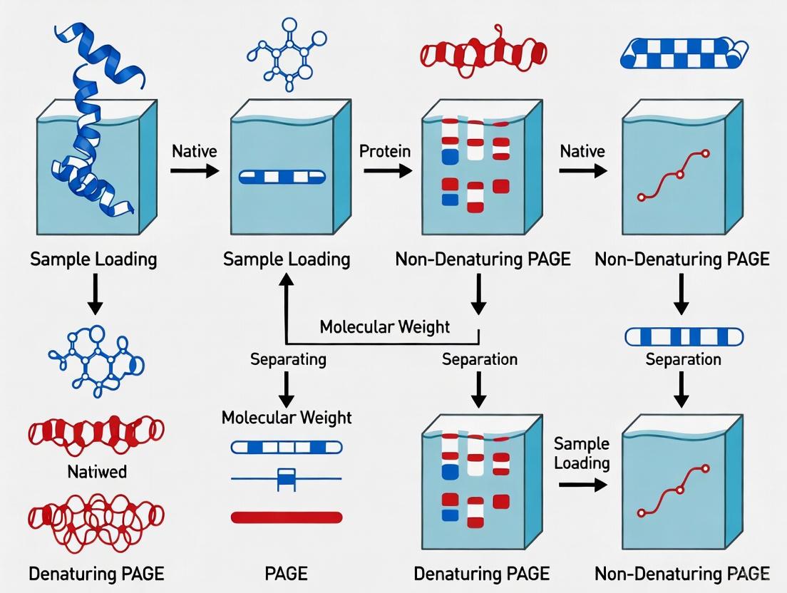
Workflow Visualization
Denaturing and non-denaturing PAGE represent complementary approaches with fundamentally distinct separation mechanisms tailored to specific research objectives. Denaturing PAGE provides high-resolution separation based primarily on molecular mass, making it ideal for molecular weight determination, purity assessment, and western blotting. Conversely, non-denaturing PAGE preserves native protein structure and function, enabling studies of protein complexes, enzymatic activity, and quaternary structure. The selection between these techniques should be guided by experimental goals, with denaturing methods preferred for structural analysis and non-denaturing methods chosen for functional studies. Advanced hybrid approaches such as NSDS-PAGE offer promising alternatives that balance resolution with preservation of certain functional properties, particularly valuable in metalloprotein research and drug development applications.
In the analysis of biomolecules via polyacrylamide gel electrophoresis (PAGE), the fundamental distinction between denaturing and non-denaturing (native) systems lies in the preservation of molecular structure. Non-denaturing PAGE maintains proteins and nucleic acids in their native, folded conformations, enabling the study of functional complexes, quaternary structures, and enzymatic activity [4] [2]. In contrast, denaturing PAGE deliberately disrupts the higher-order structure of these molecules to separate them based primarily on molecular weight [4]. This structural disruption is achieved through specific chemical conditions employing agents such as sodium dodecyl sulfate (SDS), reducing agents, and urea. These chemicals systematically target the non-covalent interactions and covalent disulfide bonds that maintain molecular structure, thereby unfolding the biomolecules into linear chains. The intentional application of these denaturants represents a cornerstone technique in molecular biology and biochemistry, providing researchers with tools to dissect complex biological systems. This technical guide explores the mechanisms, applications, and protocols associated with these critical chemical agents, framing them within the broader methodological context of PAGE-based research.
Core Chemical Mechanisms in Denaturing PAGE
Sodium Dodecyl Sulfate (SDS)
Sodium dodecyl sulfate (SDS) is an anionic detergent that plays a pivotal role in protein denaturation for SDS-PAGE. Its mechanism involves two primary actions. First, SDS binds quantitatively to proteins, with approximately one SDS molecule binding per two amino acid residues [2]. This extensive binding confers a uniform negative charge to all proteins in the sample, effectively masking their intrinsic charge and creating a consistent charge-to-mass ratio [2]. Second, SDS disrupts hydrophobic interactions and hydrogen bonds that stabilize protein tertiary and quaternary structures [9]. This disruption occurs as the hydrophobic tail of SDS interacts with hydrophobic regions of the protein, while the ionic head group interacts with the aqueous environment. The combined effect of charge masking and structural disruption transforms complex globular proteins into linear, rod-like polypeptides [2]. Consequently, separation during electrophoresis becomes dependent almost exclusively on molecular weight rather than native charge or shape, enabling accurate molecular weight determination [2].
Reducing Agents
Reducing agents specifically target disulfide bonds, the covalent linkages between cysteine residues that stabilize tertiary and quaternary protein structures. Common reducing agents include dithiothreitol (DTT), β-mercaptoethanol (BME), and tris(2-carboxyethyl)phosphine (TCEP) [10]. These compounds work through thiol-disulfide exchange reactions, where the thiol groups (-SH) of the reducing agent nucleophilically attack the disulfide bonds (-S-S-) in proteins, reducing them to free thiol groups [10]. DTT and BME operate through this mechanism, requiring careful handling due to their susceptibility to oxidation. In contrast, TCEP represents an advance in reducing agent technology as it reduces disulfide bonds through a non-thiol-based, phosphine-mediated mechanism, making it more stable in aqueous solutions and effective over a wider pH range [10]. The application of reducing agents is particularly crucial for analyzing multimetric proteins or proteins with extensive disulfide bonding, as it ensures complete unfolding into monomeric polypeptide chains prior to separation [2].
Urea
Urea serves as a potent denaturant primarily used in nucleic acid electrophoresis and specialized protein applications. It functions at high concentrations (typically 6-8 M) as a chaotropic agent that disrupts hydrogen bonding and hydrophobic interactions [9] [11]. Urea molecules form hydrogen bonds with the peptide backbone and polar side chains more effectively than water, thereby competing with the intramolecular hydrogen bonds that stabilize secondary structures like α-helices and β-sheets [9]. For RNA analysis, urea is particularly valuable because it eliminates secondary structures formed by intramolecular base pairing, ensuring that migration through the polyacrylamide gel depends solely on nucleotide chain length rather than structural conformation [11] [12]. The effectiveness of urea is temperature-dependent, with optimal denaturation occurring when gels are run at 45-55°C [11]. This temperature range maintains urea in solution while facilitating the complete unfolding of biomolecules.
Table 1: Key Denaturing Agents and Their Properties
| Denaturing Agent | Primary Mechanism of Action | Common Concentrations | Primary Applications |
|---|---|---|---|
| SDS (Sodium Dodecyl Sulfate) | Binds proteins, conferring negative charge; disrupts hydrophobic interactions | 0.1-2% in buffers; 1% in sample buffer | SDS-PAGE for protein separation and molecular weight determination [2] |
| Urea | Disrupts hydrogen bonds; chaotropic effect | 6-8 M in gel and buffers | Denaturing DNA/RNA PAGE; protein unfolding studies [11] [12] |
| DTT (Dithiothreitol) | Reduces disulfide bonds via thiol-disulfide exchange | 1-100 mM (typically 10-20 mM in sample buffer) | Reducing SDS-PAGE; protein denaturation [7] [10] |
| TCEP (Tris(2-carboxyethyl)phosphine) | Reduces disulfide bonds via phosphine mechanism | 1-50 mM (typically 5-10 mM in sample buffer) | Stable reduction for SDS-PAGE; wide pH range applications [10] |
Molecular Interactions Targeted by Denaturing Agents
The denaturing agents used in PAGE methodologies systematically disrupt specific molecular interactions that maintain biomolecular structure. Disulfide bonds, with the highest bond strength among protein interactions at approximately -230 kJ/mol, are specifically targeted by reducing agents such as DTT and TCEP [9]. These covalent linkages are cleaved through reduction-oxidation (redox) reactions. Electrostatic interactions, possessing a bond strength of about -21 kJ/mol, are disrupted by SDS, which masks intrinsic charges and imposes a uniform negative charge density [9]. Hydrogen bonds, with bond strengths of approximately -15 kJ/mol, are effectively broken by both urea and SDS [9]. Although individually weak, hydrophobic interactions are collectively significant for protein folding and are primarily disrupted by SDS through its amphiphilic properties [9]. The coordinated application of these agents enables researchers to selectively dismantle the structural integrity of biomolecules in a controlled manner, facilitating analysis based primarily on molecular dimensions rather than structural complexity.
Comparative Analysis: Denaturing vs. Non-Denaturing PAGE
Fundamental Methodological Differences
The distinction between denaturing and non-denaturing PAGE systems extends beyond the simple presence or absence of denaturing agents to encompass fundamental differences in methodology, information output, and application. Non-denaturing PAGE, performed without SDS, urea, or reducing agents, preserves the native structure and biological activity of macromolecules [4] [2]. In this system, separation depends on a combination of intrinsic charge, molecular size, and three-dimensional shape, making it ideal for studying functional biomolecular complexes [5]. In contrast, denaturing PAGE employs chemical agents to unfold biomolecules into linear chains, with separation based primarily on molecular weight due to the charge-homogenizing effect of SDS or the structure-disrupting properties of urea [4] [5]. This fundamental difference in separation principles dictates their respective applications in research, with native PAGE illuminating functional complexes and denaturing PAGE providing precise molecular weight and purity information.
Table 2: Applications of Denaturing vs. Non-Denaturing PAGE
| Application | Denaturing PAGE | Non-Denaturing PAGE |
|---|---|---|
| Molecular Weight Determination | Yes - accurate determination due to linearized structures [2] | No - migration depends on multiple factors beyond size [2] |
| Structure Analysis | Primary structure only [5] | All four levels of structure (primary, secondary, tertiary, quaternary) [5] |
| Enzyme Activity Studies | Not possible (proteins denatured) | Possible - activity often preserved after electrophoresis [2] |
| Protein Complex Analysis | Separates complexes into individual subunits [2] | Preserves and separates intact complexes [4] |
| Binding Studies | Not suitable | Suitable for protein-protein or protein-ligand interactions [4] |
| Purity Assessment | Excellent for establishing sample purity [2] | Limited due to complex migration patterns |
Chemical Conditions and Buffer Composition
The chemical conditions for denaturing and non-denaturing PAGE differ significantly in their composition, particularly regarding detergents, reducing agents, and chaotropes. Denaturing systems for proteins typically include SDS (0.1-1%) in both sample buffers and running buffers, often combined with reducing agents like DTT (10-100 mM) or β-mercaptoethanol (0.1-1%) [7]. Sample preparation for denaturing PAGE involves heating (85-100°C for 2-5 minutes) to ensure complete denaturation [7]. For nucleic acids, denaturing conditions employ 6-8 M urea in the gel matrix and running buffers, with formamide often included in loading buffers [11]. Non-denaturing systems deliberately exclude these denaturing agents, instead using mild buffers at neutral or slightly basic pH to maintain protein structure and activity [7]. Non-denaturing sample preparation occurs without heating to preserve native conformations [7]. The running buffers for native PAGE typically lack SDS, though they maintain similar ion systems (e.g., Tris-Glycine) to facilitate proper electrophoresis [7].
Experimental Protocols and Methodologies
SDS-PAGE for Protein Separation
The SDS-PAGE protocol represents a standardized method for protein separation based on molecular weight. Begin with sample preparation by mixing protein samples with 2X SDS sample buffer (125 mM Tris-Cl, pH 6.8, 4% SDS, 20% glycerol, 0.004% bromophenol blue) containing 100 mM DTT or 5% β-mercaptoethanol for reducing conditions [7]. Heat samples at 85°C for 2-5 minutes to ensure complete denaturation [7]. For non-reducing SDS-PAGE, omit the reducing agent but maintain SDS and heating. Assemble the gel apparatus according to manufacturer instructions, using pre-cast or freshly poured polyacrylamide gels with appropriate percentages for the target protein size range [7]. Fill the upper and lower buffer chambers with 1X Tris-Glycine-SDS running buffer (25 mM Tris, 192 mM glycine, 0.1% SDS, pH 8.3) [7]. Load prepared samples and molecular weight markers into wells. Run electrophoresis at constant voltage (100-150 V) until the dye front reaches the bottom of the gel [7]. Following electrophoresis, process gels for staining, western blotting, or further analysis as required by the experimental design.
Urea-PAGE for RNA Analysis
Urea-PAGE provides high-resolution separation of RNA molecules by eliminating secondary structure. Begin by preparing the gel solution according to Table 1, using ultrapure urea and appropriate acrylamide concentration based on the target RNA size range [11]. For a standard 15% gel, mix 24 g urea, 18.75 mL of 40% acrylamide solution (29:1), 5 mL of 10X TBE buffer (890 mM Tris, 890 mM boric acid, 20 mM EDTA), and deionized water to 50 mL total volume [11]. Heat the solution briefly to dissolve urea completely, then add 166 μL of 10% ammonium persulfate and 20 μL TEMED to initiate polymerization [11]. Pour the gel immediately between assembled glass plates, insert an appropriate comb, and allow 30-60 minutes for complete polymerization [11]. Assemble the gel apparatus, add 1X TBE running buffer to both chambers, and pre-run the gel for 30 minutes at 15-25 W to reach the optimal temperature of 45-55°C [11]. For sample preparation, mix RNA samples with 2X loading buffer (90% formamide, 0.5% EDTA, 0.1% xylene cyanol, 0.1% bromophenol blue), heat at 70-90°C for 2-3 minutes to denature secondary structures, then immediately place on ice [11]. Load denatured samples and run electrophoresis at constant power maintaining 45-55°C gel temperature until adequate separation is achieved [11]. Post-electrophoresis, stain with appropriate dyes (e.g., ethidium bromide, SYBR Gold) or process for transfer to membranes.
Diagram 1: Urea-PAGE workflow for RNA analysis
Native PAGE for Protein Complexes
Non-denaturing PAGE preserves protein structure and function during electrophoresis. Begin with sample preparation using native sample buffer (typically containing Tris, glycerol, and tracking dyes but no SDS or reducing agents) [7]. Crucially, do not heat samples before loading [7]. Prepare native running buffer (e.g., Tris-Glycine without SDS, pH ~8.3) [7]. Cast polyacrylamide gels without SDS or other denaturants, using the same percentage considerations as denaturing gels but noting that migration will depend on both size and native charge [7]. Load samples and run electrophoresis at constant voltage (typically 125 V) with lower current compared to SDS-PAGE due to the absence of SDS [7]. Running times may be longer than SDS-PAGE as proteins migrate more slowly in their native conformation [7]. Following electrophoresis, process gels for activity staining, western blotting under native conditions, or other detection methods compatible with preserved protein function.
The Researcher's Toolkit: Essential Reagents and Materials
Successful implementation of denaturing and non-denaturing PAGE requires specific reagents carefully selected for their properties and applications. The following table summarizes essential components for PAGE experiments, their functions, and considerations for use.
Table 3: Essential Reagents for Denaturing and Non-Denaturing PAGE
| Reagent/Category | Specific Examples | Function & Mechanism | Application Notes |
|---|---|---|---|
| Detergents | Sodium Dodecyl Sulfate (SDS) | Disrupts hydrophobic interactions; confers uniform charge | Core component of SDS-PAGE; use at 0.1-1% concentration [2] |
| Chaotropic Agents | Urea, Guanidine HCl | Disrupts hydrogen bonding; unfolds macromolecules | Use 6-8 M for RNA/DNA denaturation; handle at controlled temperatures [11] [10] |
| Reducing Agents | DTT, β-mercaptoethanol, TCEP | Reduces disulfide bonds; linearizes proteins | DTT/BME require fresh preparation; TCEP more stable [10] |
| Gel Matrix Components | Acrylamide/bis-acrylamide, TEMED, APS | Forms cross-linked polymer network for separation | Adjust percentage based on target size range; TEMED catalyzes polymerization |
| Buffer Systems | Tris-Glycine, TBE, TAE | Provides conducting medium; maintains pH | Tris-glycine for proteins; TBE for nucleic acids; concentration affects resolution |
| Tracking Dyes | Bromophenol blue, Xylene cyanol | Visualize migration front; monitor run progress | Different migration rates based on matrix; may interfere with fluorescence |
Troubleshooting and Optimization
Common Challenges in Denaturing PAGE
Several technical challenges may arise when performing denaturing PAGE, often manifesting as poor resolution, aberrant migration, or artifacts. In SDS-PAGE, incomplete denaturation frequently results from insufficient heating or inadequate SDS concentration, leading to curved bands or multiple bands for a single protein [7]. This can be addressed by ensuring sample heating at 85°C for 2-5 minutes in the presence of at least 1% SDS [7]. Oxidation of reducing agents, particularly DTT and β-mercaptoethanol, causes reappearance of disulfide bonds and improper unfolding, which can be prevented by preparing fresh reducing agent solutions for each use or switching to more stable alternatives like TCEP [10]. In urea-PAGE for RNA analysis, incomplete denaturation often stems from incorrect urea concentration (deviations from 6-8 M) or suboptimal temperature during electrophoresis [11] [12]. Maintaining gel temperature at 45-55°C throughout the run is critical for consistent results [11]. RNase contamination represents another common issue in RNA work, requiring strict RNase-free conditions including use of certified reagents, DEPC-treated water, and dedicated equipment [12].
Optimization Strategies
Systematic optimization of denaturing PAGE methods enhances resolution and reproducibility. For protein separation via SDS-PAGE, acrylamide concentration should be matched to protein size range, with lower percentages (8-12%) optimal for high molecular weight proteins and higher percentages (12-20%) better for smaller proteins [2]. Gel thickness affects resolution, with thinner gels (0.75-1.0 mm) typically providing sharper bands than thicker gels (1.5 mm) [11]. For urea-PAGE of nucleic acids, sample loading volume significantly impacts band sharpness, with ideal volumes between 1-5 μL providing optimal resolution [11]. Including 10% glycerol in loading buffers can improve sample settling in wells without affecting denaturation [11]. "Gel smiling" (uneven migration across the gel) results from uneven heat distribution and can be mitigated by using proper electrophoresis apparatus with temperature regulation or attaching metal plates to distribute heat evenly [11]. Voltage optimization is also crucial, with lower voltages at the beginning of runs sometimes improving band sharpness by allowing smooth entry of samples into the gel matrix [11].
Diagram 2: Common PAGE issues and solutions
The strategic application of specific chemical conditions—employing SDS, reducing agents, and urea—enables researchers to manipulate biomolecular structure during polyacrylamide gel electrophoresis to address distinct research questions. Denaturing conditions that disrupt native structure facilitate molecular weight determination, purity assessment, and analysis of individual macromolecular components. In contrast, non-denaturing conditions that preserve native conformation enable the study of functional complexes, enzymatic activity, and higher-order structures. The informed selection between these approaches, along with careful optimization of chemical conditions, represents a fundamental methodological decision in biomolecular research. As electrophoretic techniques continue to evolve, particularly in drug development and structural biology, the precise control over denaturing conditions remains essential for generating reproducible, interpretable data across diverse applications. Understanding these chemical principles provides researchers with a powerful framework for experimental design and data interpretation in the broader context of biomolecular analysis.
In polyacrylamide gel electrophoresis (PAGE), the separation of proteins is governed by the complex interplay of their fundamental physical properties: size, charge, and shape. The extent to which each property influences migration fundamentally depends on whether the experiment is conducted under denaturing or non-denaturing (native) conditions [4] [1]. In denaturing PAGE, the inherent charge and shape of proteins are masked, making molecular mass the primary determinant of mobility. In contrast, native PAGE leverages the protein's intrinsic charge, its three-dimensional structure, and its mass, allowing for the separation of functional complexes [1] [13]. This technical guide explores how these properties govern electrophoretic migration, framed within the core distinction between denaturing and non-denaturing PAGE methodologies, which is pivotal for researchers and drug development professionals to select the appropriate technique for their analytical goals.
Core Principles of Electrophoretic Migration
Fundamental Factors Influencing Mobility
The electrophoretic mobility of a molecule in a gel matrix is a function of the force exerted by the electric field and the retarding frictional forces encountered during migration. The general factors affecting this mobility include [14] [15]:
- Net Charge: The overall charge of the molecule at the buffer's pH determines the direction and magnitude of the force from the electric field. Mobility is directly proportional to the net charge [15].
- Size and Mass: Larger molecules experience greater frictional drag within the gel matrix, leading to slower migration. Mobility is inversely proportional to size [15].
- Molecular Shape: The three-dimensional conformation affects the frictional coefficient. Compact, globular proteins migrate faster than elongated, fibrous proteins of similar molecular weight [15].
- Gel Matrix Pore Size: The concentration of polyacrylamide determines the pore size, which acts as a molecular sieve. Higher percentage gels have smaller pores, providing better resolution for smaller proteins [1] [13].
- Buffer Conditions: The pH of the buffer determines the ionization state of the protein, thus its net charge. The ionic strength affects the conductivity of the medium and the sharpness of the separated bands [14] [15].
- Field Strength: A higher voltage increases the rate of migration but can also generate heat, leading to diffusion of bands and potential protein denaturation in native gels [15].
The Gel Matrix as a Molecular Sieve
Polyacrylamide gels are created through the polymerization of acrylamide monomers cross-linked by bisacrylamide [1] [13]. The resulting meshwork provides a porous medium through which molecules travel. The pore size is controlled by the total concentration of acrylamide (%T) and the degree of cross-linking (%C) [13]. This matrix is critical for separating molecules based on size, as smaller molecules can navigate the pores more easily than larger ones [14] [1].
Denaturing vs. Native PAGE: A Mechanistic Comparison
The decision to use denaturing or native PAGE dictates which molecular properties govern the separation. The table below summarizes the core differences between these two fundamental approaches.
Table 1: Core Differences Between Denaturing and Native PAGE
| Parameter | Denaturing PAGE (e.g., SDS-PAGE) | Native PAGE |
|---|---|---|
| Primary Separation Basis | Molecular mass (kDa) [1] [13] | Combined effect of net charge, size, and native shape [1] [13] |
| Protein Structure | Disrupted; proteins are linearized [4] [13] | Preserved in its native, folded state [4] [5] |
| Key Reagents | SDS (sodium dodecyl sulfate), reducing agents (e.g., β-mercaptoethanol) [1] [13] | No denaturing agents; may use milder detergents for membrane proteins [1] |
| Sample Preparation | Heating (∼100°C) in sample buffer containing SDS and reductant [13] | No heating; mixed with non-denaturing loading dye [13] |
| Charge Manipulation | SDS confers a uniform negative charge, masking intrinsic charge [1] | Relies on the protein's intrinsic charge at the running buffer's pH [1] |
| Information Obtained | Polypeptide chain molecular mass, sample purity [4] [1] | Oligomeric state (quaternary structure), protein-protein interactions, enzymatic activity [4] [1] |
Denaturing PAGE: Separation by Mass
In denaturing PAGE, most commonly performed as SDS-PAGE, the goal is to separate proteins based almost exclusively on the mass of their polypeptide chains [1] [13].
- Role of SDS: The anionic detergent SDS binds to the hydrophobic regions of proteins in a constant weight ratio (approximately 1.4 g SDS per 1 g of protein) [1]. This extensive coating confers a uniform negative charge per unit mass, effectively masking the protein's intrinsic charge [1] [13].
- Role of Reducing Agents: Agents like β-mercaptoethanol or dithiothreitol (DTT) break disulfide bonds that hold protein subunits together, ensuring complete denaturation into individual polypeptide chains [13].
- Effect on Molecular Properties: The combination of SDS, heat, and reductant linearizes the proteins, destroying their secondary, tertiary, and quaternary structures [4] [5]. This process eliminates the influence of both native charge and shape on migration. Consequently, the SDS-polypeptide complexes migrate through the gel as linear chains with identical charge densities, and their mobility depends solely on their molecular mass through the sieving effect of the gel [1].
Native PAGE: Separation by Charge, Size, and Shape
Native PAGE is performed without denaturants to preserve the protein's biological activity and higher-order structure [1] [13].
- Preservation of Properties: Under these conditions, a protein's intrinsic net charge (dictated by its amino acid composition and the buffer pH), its native size (including its oligomeric state), and its three-dimensional shape all contribute to its electrophoretic mobility [1] [13].
- Complex Migration: A highly charged protein will migrate faster than a less charged one of the same mass. Similarly, a compact, globular protein will migrate faster than an elongated, fibrous protein of the same mass and charge due to differences in frictional drag [15]. This allows native PAGE to separate proteins based on their functional states and complexes [4].
The following diagram illustrates the distinct migration mechanisms in these two techniques.
Experimental Protocols and Methodologies
Standard SDS-PAGE (Denaturing) Protocol
This protocol is adapted from common laboratory practices as detailed across multiple sources [1] [13].
1. Gel Preparation:
- Resolving Gel: First, a resolving gel with an appropriate acrylamide percentage (typically 8-15%) is cast at pH ~8.8. The percentage is chosen based on the target protein's size [1] [13].
- Stacking Gel: After polymerization, a stacking gel with a lower percentage of acrylamide (∼4%) and a lower pH (~6.8) is cast on top. The discontinuity in pH and gel pore size helps concentrate all protein samples into a sharp band before they enter the resolving gel, greatly improving resolution [1] [13].
2. Sample Preparation:
- Protein samples are mixed with an SDS-PAGE loading buffer containing SDS (for denaturation and charge), a reducing agent (e.g., β-mercaptoethanol to break disulfide bonds), glycerol (to weigh down the sample), and a tracking dye (e.g., bromophenol blue) [13].
- The mixture is heated at 95-100°C for 3-5 minutes to fully denature the proteins [13].
3. Electrophoresis:
- The prepared samples and a molecular weight marker (protein ladder) are loaded into the wells.
- The gel is run in an electrophoresis tank filled with a running buffer (e.g., Tris-Glycine-SDS) at a constant voltage until the tracking dye front reaches the bottom of the gel [13].
Standard Native PAGE Protocol
The setup for native PAGE is similar but omits denaturing agents [1] [13].
1. Gel Preparation:
- The gel is cast without SDS. The choice of acrylamide percentage and buffer system (e.g., Tris-Glycine, Tris-Borate) is critical and must be optimized to maintain protein stability and activity [13].
2. Sample Preparation:
- Samples are mixed with a non-denaturing loading buffer that lacks SDS and reductants. The buffer typically contains glycerol and a tracking dye, but the sample is not heated [13].
3. Electrophoresis:
- The gel is run in a running buffer without SDS. It is often advisable to run native PAGE at lower voltages or with cooling to prevent heat-induced denaturation during the run [1] [13].
The Scientist's Toolkit: Essential Reagents and Materials
Table 2: Key Research Reagent Solutions for PAGE
| Reagent/Material | Function | Key Consideration |
|---|---|---|
| Acrylamide/Bis-acrylamide | Forms the cross-linked polymer matrix (gel) that acts as a molecular sieve. | Total concentration (%T) dictates pore size and resolution range [1] [13]. |
| Ammonium Persulfate (APS) & TEMED | APS (initiator) and TEMED (catalyst) are required to polymerize the acrylamide solution into a gel. | Freshly prepared APS solutions are critical for efficient polymerization [1] [13]. |
| SDS (Sodium Dodecyl Sulfate) | Ionic detergent that denatures proteins and confers a uniform negative charge. | Essential for SDS-PAGE; must be omitted for native PAGE [1] [13]. |
| β-Mercaptoethanol or DTT | Reducing agents that cleave disulfide bonds between cysteine residues. | Ensures complete denaturation into monomeric subunits in SDS-PAGE [13]. |
| Tris-based Buffers | Provide the necessary ions to conduct current and maintain a stable pH during electrophoresis. | A discontinuous system (different pH in stacking vs. resolving gel) is key for sharp bands in SDS-PAGE [1] [13]. |
| Molecular Weight Markers | A set of pre-stained or unstained proteins of known sizes run alongside samples to estimate molecular mass. | Critical for calibrating and interpreting SDS-PAGE results [1]. |
| Coomassie Blue/Silver Stain | Dyes used to visualize separated protein bands on the gel post-electrophoresis. | Coomassie is common for general use; silver stain offers higher sensitivity [15]. |
Advanced Techniques and Applications
Two-Dimensional (2D) PAGE
2D-PAGE combines two orthogonal separation techniques to achieve extremely high resolution. In the first dimension, proteins are separated by their native isoelectric point (pI) using isoelectric focusing (IEF). In the second dimension, the same proteins are separated by their molecular mass using SDS-PAGE [1] [15]. This method allows for the resolution of thousands of proteins from a single sample and is a powerful tool in proteomics for analyzing complex protein mixtures, post-translational modifications, and changes in protein expression [1].
Capillary Electrophoresis (CE)
Capillary electrophoresis is a modern evolution of traditional gel electrophoresis. It uses narrow-bore capillaries filled with a polymer matrix or buffer. The high surface-area-to-volume ratio allows for efficient heat dissipation, enabling the use of very high voltages for fast separations with exceptional resolution [14] [15]. CE can be coupled with sophisticated detectors like mass spectrometers, enhancing its analytical capabilities for both proteins and nucleic acids [14].
The migration of proteins in polyacrylamide gel electrophoresis is a physical process dictated by the triumvirate of molecular properties: size, charge, and shape. The fundamental choice between denaturing and native PAGE determines which of these properties becomes the dominant factor for separation. SDS-PAGE, by homogenizing charge and destroying native structure, simplifies analysis to molecular mass, making it an indispensable tool for estimating purity and polypeptide size. In contrast, native PAGE embraces the complexity of proteins in their functional state, allowing researchers to probe quaternary structure, interactions, and activity. A deep understanding of how these properties affect migration under different conditions is essential for designing robust experiments, accurately interpreting electrophoretic data, and selecting the optimal strategy to answer specific biological questions in basic research and drug development.
Preservation vs. Disruption of Protein Structure and Function
The fundamental principle underlying many analytical techniques in biochemistry and molecular biology is the deliberate choice between preserving or disrupting the native structure of proteins. This distinction is particularly pronounced in polyacrylamide gel electrophoresis (PAGE), where researchers must select between native (non-denaturing) and denaturing conditions based on their experimental objectives. The strategic decision to preserve or disrupt protein structure directly impacts the type of information obtained, ranging from molecular weight determination to functional activity assessment and complex formation [4] [2].
Within the context of a broader thesis on denaturing versus non-denaturing PAGE research, this technical guide explores the fundamental principles, methodological considerations, and practical applications of both approaches. For researchers, scientists, and drug development professionals, understanding these distinctions is crucial for designing appropriate experiments, particularly when studying protein complexes that serve as important drug targets or when analyzing conformational changes implicated in neurodegenerative diseases [16].
Fundamental Principles of Protein Electrophoresis
Molecular Basis of Separation
Gel electrophoresis separates proteins through a matrix under the influence of an electric field. The migration behavior of proteins depends on their physical properties, which are manipulated differently in native versus denaturing conditions.
In native PAGE, proteins maintain their higher-order structure (secondary, tertiary, and quaternary), allowing separation based on a combination of size, shape, and intrinsic charge [4] [5]. The gel matrix serves as a molecular sieve through which compact, highly charged proteins migrate faster than larger or less charged counterparts. This method preserves protein function and activity, enabling subsequent enzymatic assays or analysis of protein complexes [2].
In denaturing SDS-PAGE, the anionic detergent sodium dodecyl sulfate (SDS) binds to hydrophobic regions of proteins at a relatively constant ratio (approximately 1.4g SDS per 1g protein), conferring a uniform negative charge that masks the protein's intrinsic charge [2]. Reducing agents like dithiothreitol (DTT) or β-mercaptoethanol break disulfide bonds, destroying tertiary and quaternary structures [2]. This linearizes proteins into rod-like chains, creating a near-uniform charge-to-mass ratio across different proteins [2]. Consequently, separation occurs primarily by molecular weight, with smaller polypeptides migrating faster through the gel matrix [2].
Table 1: Key Characteristics of Native Versus Denaturing PAGE
| Parameter | Native (Non-Denaturing) PAGE | Denaturing (SDS) PAGE |
|---|---|---|
| Protein Structure | Maintains native conformation; preserves secondary, tertiary, and quaternary structure | Disrupts higher-order structure; proteins unfolded into linear chains |
| Separation Basis | Size, shape, and intrinsic charge | Primarily molecular mass |
| Sample Preparation | Non-denaturing, non-reducing buffers; no SDS | Heated with SDS and reducing agents (DTT) |
| Charge-to-Mass Ratio | Variable | Uniform (due to SDS binding) |
| Molecular Weight Determination | Not accurate due to structural and charge variables | Accurate for polypeptide chain size |
| Functional Preservation | Enzymatic activity often preserved | Activity destroyed |
| Applications | Studying protein complexes, isolation of enzymes, analysis of quaternary structure | Molecular weight estimation, purity assessment, Western blotting, protein sequencing preparation |
Quantitative Analysis in Protein Research
Beyond simple separation, both native and denaturing electrophoresis can be incorporated into quantitative proteomic approaches. Quantitative protein profiling is essential for understanding cellular processes, with methods ranging from label-free techniques to those using isotopic labelling with amino acids (SILAC), isobaric tags (iTRAQ), or isotope-coded affinity tag reagents [17] [18]. These techniques enable researchers to quantify changes in protein abundance across different biological states, providing crucial information for systems biology and drug development [17].
Methodological Approaches
Native (Non-Denaturing) PAGE Protocols
Basic Native PAGE for Protein Complex Analysis
Objective: To separate and analyze functionally active protein complexes while preserving their native state.
Sample Preparation:
- Prepare protein samples in non-denaturing, SDS-free buffer (e.g., Tris-glycine or Tris-borate at neutral to slightly basic pH)
- Avoid heating or using reducing agents
- Centrifuge at 12,000 × g for 10 minutes to remove insoluble material
- Maintain samples at 4°C throughout preparation to preserve stability
Gel Preparation:
- Prepare resolving gel (typically 6-12% acrylamide depending on protein size)
- Composition: Acrylamide/bis-acrylamide (29:1), 0.375 M Tris-HCl (pH 8.8), 0.1% ammonium persulfate (APS), and 0.1% TEMED
- Stacking gel: 4% acrylamide, 0.125 M Tris-HCl (pH 6.8), 0.1% APS, and 0.1% TEMED
- Pour gel and allow to polymerize for 30-60 minutes
Electrophoresis Conditions:
- Running buffer: 25 mM Tris, 192 mM glycine (pH 8.3)
- Load 10-50 μg protein per lane alongside native molecular weight standards
- Run at constant voltage (100-150V) for 1.5-2 hours at 4°C to prevent heat denaturation
- Monitor migration of pre-stained standards or tracking dye
Post-Electrophoresis Analysis:
- Proteins can be visualized with Coomassie Brilliant Blue, silver stain, or specific activity stains
- For functional assays, proteins can be electroeluted or transferred to suitable membranes under native conditions
- Alternatively, gel slices can be excised and used directly in activity assays
Advanced Native Techniques for Structural Biology
Recent advances in native separation techniques include capillary electrophoresis-mass spectrometry (CE-MS), which enables structural analysis of large protein complexes under near-physiological conditions [16]. This method:
- Uses minimal sample amounts (10,000-fold lower than conventional techniques)
- Provides results in less than 30 minutes
- Maintains proteins at near-physiological conditions in solution
- Allows real-time monitoring of conformational changes and dynamic interactions [16]
Denaturing (SDS-PAGE) Protocols
Standard SDS-PAGE for Molecular Weight Determination
Objective: To separate protein subunits by molecular weight after disruption of higher-order structures.
Sample Preparation:
- Dilute protein samples in 2× Laemmli buffer: 125 mM Tris-HCl (pH 6.8), 4% SDS, 20% glycerol, 0.02% bromophenol blue
- Add 100 mM dithiothreitol (DTT) or 5% β-mercaptoethanol as reducing agent
- Heat at 95-100°C for 5-10 minutes to ensure complete denaturation
- Centrifuge briefly to collect condensed sample
Gel Preparation:
- Discontinuous system with stacking (4-5%) and resolving (8-20%) gels
- Resolving gel: Acrylamide/bis-acrylamide (29:1 or 37.5:1), 0.375 M Tris-HCl (pH 8.8), 0.1% SDS, 0.1% APS, and 0.1% TEMED
- Stacking gel: 4-5% acrylamide, 0.125 M Tris-HCl (pH 6.8), 0.1% SDS, 0.1% APS, and 0.1% TEMED
- Allow complete polymerization (30-60 minutes)
Electrophoresis Conditions:
- Running buffer: 25 mM Tris, 192 mM glycine, 0.1% SDS (pH 8.3)
- Load 10-100 μg protein per lane alongside pre-stained molecular weight markers
- Run at constant voltage (100-200V) until dye front reaches bottom
- Maintain cooling for consistent band patterns
Post-Electrophoresis Analysis:
- Fix proteins in gel with 40% ethanol/10% acetic acid
- Visualize with Coomassie Blue, silver stain, or fluorescent dyes
- For Western blotting, transfer to PVDF or nitrocellulose membranes
- For protein sequencing, electroblot to appropriate membranes
Variations and Specialized Denaturing Protocols
Urea-PAGE for Nucleic Acid-Protein Complexes:
- Uses 6-8 M urea to denature nucleic acids while maintaining protein denaturation
- Particularly useful for analyzing RNA-protein interactions [4]
Gradient Gels for Enhanced Resolution:
- Linear or nonlinear acrylamide gradients (e.g., 4-20%) improve resolution across broad molecular weight ranges
- Allow better separation of large and small proteins on the same gel
Two-Dimensional Electrophoresis:
- Combines isoelectric focusing (first dimension) with SDS-PAGE (second dimension)
- Enables high-resolution separation of complex protein mixtures [18]
Comparative Analysis and Applications
Strategic Selection Guide
The decision to use native versus denaturing PAGE depends fundamentally on the research question and desired outcomes. The table below summarizes key application scenarios for each method.
Table 2: Application Guide for Native Versus Denaturing PAGE
| Research Objective | Recommended Method | Rationale | Key Considerations |
|---|---|---|---|
| Molecular Weight Determination | Denaturing SDS-PAGE | Eliminates structural and charge variables; separation by polypeptide chain size only | Use appropriate molecular weight markers; linear range of gel should match protein size |
| Enzyme Activity Analysis | Native PAGE | Preserves tertiary structure and active site conformation | Maintain cool temperatures; use specific activity stains; avoid fixatives that denature proteins |
| Protein Complex/ Oligomeric State Study | Native PAGE | Maintains quaternary structure and subunit interactions | Vary gel concentration to assess size and shape; cross-linking may stabilize weak complexes |
| Western Blotting | Denaturing SDS-PAGE | Improves antibody accessibility to linear epitopes; standard transfer protocols | Reduction required for disulfide-linked proteins; confirm antibody recognizes denatured epitopes |
| Sample Purity Assessment | Denaturing SDS-PAGE | Reveals individual polypeptide chains; detects proteolytic fragments | Multiple staining methods available; Coomassie for major bands, silver for trace contaminants |
| Protein-Protein Interactions | Native PAGE | Preserves binding interfaces and complex formation | Vary running conditions to detect weak interactions; combine with cross-linking for stability |
| Protein Sequencing | Denaturing SDS-PAGE | Isolates individual polypeptide chains for sequencing | Electroblot to suitable membranes; minimal staining to avoid N-terminal blockage |
| Isoenzyme Separation | Native PAGE | Separates isoforms with subtle structural differences | Optimize pH and buffer systems; activity staining often required for specific detection |
| Binding Studies | Native PAGE | Maintains binding pockets and ligand interactions | May incorporate ligands in gel or buffer; mobility shifts indicate binding |
Quantitative Data from Method Applications
The effects of various processing methods on protein properties provide valuable quantitative insights for researchers selecting appropriate electrophoretic techniques.
Table 3: Quantitative Effects of Processing Methods on Protein Properties
| Processing Method | Affected Protein Properties | Quantitative Changes | Functional Implications |
|---|---|---|---|
| Ohmic Heating | Structure, solubility, emulsifying and foaming properties | Increased particle size and turbidity; enhanced water and oil holding capacity [19] | Improved bioactivity in sheep milk via proteolysis; increased bioactive peptides [19] |
| High-Pressure Processing (HPP) | Particle size, secondary structure, coagulation properties | Significant modification of structural parameters [19] | Alters functional properties for specific food applications |
| Pulsed Electric Fields (PEF) | Solubility, structure | Enhanced protein solubility; structural modifications [19] | Improves technological applications in food systems |
| Enzymatic Hydrolysis | Texture, proteolytic activity, degree of hydrolysis, solubility | Breakdown of proteins into smaller peptides [19] | Improved digestibility and absorption of amino acids [19] |
| Novel CE-MS Method | Structural analysis, conformational changes | 10,000-fold lower sample requirement; analysis in <30 minutes [16] | Enables study of protein complexes in near-native state; potential for drug development |
The Scientist's Toolkit: Essential Research Reagents
Successful implementation of native and denaturing electrophoresis requires specific reagents optimized for each method. The table below details essential solutions and their functions.
Table 4: Essential Research Reagent Solutions for Protein Electrophoresis
| Reagent Solution | Composition | Function | Method Application |
|---|---|---|---|
| Laemmli Sample Buffer | 125 mM Tris-HCl (pH 6.8), 4% SDS, 20% glycerol, 0.02% bromophenol blue, with reducing agent | Denatures proteins, provides uniform charge, adds density for loading, provides visible migration marker | Denaturing SDS-PAGE |
| Tris-Glycine Running Buffer | 25 mM Tris, 192 mM glycine (pH 8.3) with or without 0.1% SDS | Maintains pH during electrophoresis, provides conducting medium | Both native and denaturing PAGE (with/without SDS) |
| Non-Denaturing Sample Buffer | Tris-HCl or Tris-borate (pH 7-8), glycerol, tracking dye | Maintains native state, provides density for loading, visible migration marker | Native PAGE |
| Acrylamide/Bis Solution | 29:1 or 37.5:1 acrylamide:bis-acrylamide in water | Forms cross-linked polymer network for size-based separation | Both methods (gel matrix) |
| Ammonium Persulfate (APS) | 10% solution in water | Free radical source for acrylamide polymerization | Both methods (gel formation) |
| TEMED | N,N,N',N'-Tetramethylethylenediamine | Catalyzes acrylamide polymerization by generating free radicals | Both methods (gel formation) |
| Coomassie Staining Solution | 0.1% Coomassie Brilliant Blue R-250, 40% methanol, 10% acetic acid | Visualizes protein bands by binding to amino acids | Both methods (post-electrophoresis) |
| Destaining Solution | 40% methanol, 10% acetic acid | Removes background stain while retaining protein-bound dye | Both methods (post-electrophoresis) |
| Transfer Buffer | 25 mM Tris, 192 mM glycine, 20% methanol | Facilitates protein transfer from gel to membrane for Western blotting | Primarily denaturing PAGE |
| Sodium Cholate Solution | 10% (w/v) sodium cholate in Tris-EDTA buffer | Non-denaturing detergent for tissue clearing; preserves native protein state [20] | Specialized native applications |
Emerging Technologies and Future Perspectives
Innovations in Native State Analysis
The field of protein analysis continues to evolve with emerging technologies that enhance our ability to study proteins in their native states. Capillary electrophoresis-mass spectrometry (CE-MS) represents a significant advancement, enabling researchers to:
- Analyze protein complexes under near-physiological conditions [16]
- Detect conformational changes in real-time [16]
- Study interactions with small molecules, nucleotides, and metal ions [16]
- Identify point mutations within large protein complexes [16]
This method substantially minimizes sample consumption and simplifies analytical workflows while maintaining proteins at near-physiological conditions, making it particularly valuable for drug development where understanding protein-ligand interactions is crucial [16].
Novel Applications in Tissue Clearing
Innovative approaches to protein preservation are emerging in fields beyond traditional electrophoresis. The development of OptiMuS-prime, a passive tissue clearing method using sodium cholate and urea, offers enhanced protein preservation compared to traditional SDS-based methods [20]. This technique:
- Replaces SDS with sodium cholate, a non-denaturing detergent with smaller micelles [20]
- Better preserves proteins in their native state while enabling tissue transparency [20]
- Combines urea to disrupt hydrogen bonds and induce hyperhydration for enhanced probe penetration [20]
- Allows 3D imaging of immunolabeled structures while maintaining protein integrity [20]
Such advancements highlight the continuing importance of the fundamental choice between preservation and disruption of protein structure across multiple scientific disciplines.
The deliberate choice between preserving or disrupting protein structure represents a fundamental strategic decision in biochemical research. Native and denaturing electrophoresis methods provide complementary information that, when selected appropriately, enables comprehensive protein characterization. Native PAGE excels at maintaining functional activity and studying protein complexes, while denaturing SDS-PAGE provides precise molecular weight determination and purity assessment.
For researchers engaged in drug development, where protein complexes serve as critical therapeutic targets, or for those studying conformational diseases, the ability to analyze proteins under near-native conditions becomes particularly valuable. Emerging technologies that enhance our capability to study proteins with minimal structural disruption while providing high sensitivity and rapid analysis will continue to advance our understanding of protein function and facilitate therapeutic development.
The continuing evolution of both preservation and disruption techniques ensures that researchers will have increasingly sophisticated tools to address the complex questions in protein science, each method providing unique insights based on the fundamental principles outlined in this technical guide.
Understanding the Impact on Quaternary Structures and Complexes
Core Principles of PAGE: Denaturing vs. Non-Denaturing Environments
In protein research, the choice of electrophoretic method is critical, as it directly dictates the level of structural information that can be obtained. Polyacrylamide Gel Electrophoresis (PAGE) separates biological molecules based on their physical properties as they migrate through a gel matrix under an electrical field. [1] The fundamental division in these techniques lies in the use of denaturing or non-denaturing conditions, which have a profound and direct impact on the preservation of a protein's quaternary structure and native complexes. [4] [2]
In a denaturing environment, such as SDS-PAGE, proteins are unfolded into linear chains. [5] This is achieved using anionic detergents like sodium dodecyl sulfate (SDS) and reducing agents like beta-mercaptoethanol or dithiothreitol (DTT). SDS denatures the protein and coats the polypeptide backbone with a uniform negative charge, while the reducing agent breaks disulfide bonds. [2] [1] [21] This process destroys the protein's secondary, tertiary, and quaternary structures, meaning multi-subunit complexes are dissociated into their individual components. Consequently, separation occurs based almost solely on molecular mass, as all proteins have a similar charge-to-mass ratio. [21] [22]
In contrast, a non-denaturing or native environment deliberately avoids these disruptive agents. [2] Without SDS or reducing agents, the protein's native conformation—including its secondary, tertiary, and critically, its quaternary structure—is preserved throughout the electrophoresis. [5] This means that protein complexes remain intact. Separation in native PAGE depends on a combination of the protein's intrinsic charge, size, and three-dimensional shape, allowing for the analysis of functional, folded proteins and their interactions. [1]
Table 1: Fundamental Characteristics of Denaturing and Non-Denaturing PAGE
| Characteristic | Denaturing (SDS-)PAGE | Non-Denaturing (Native) PAGE |
|---|---|---|
| Gel Conditions | Denatured (contains SDS & reducing agents) [23] | Non-denatured (no SDS or reducing agents) [23] |
| Protein State | Unfolded, linear chains [5] | Folded, native conformation [2] |
| Impact on Quaternary Structure | Destroyed; complexes dissociated [2] | Preserved; complexes remain intact [2] [1] |
| Basis of Separation | Molecular mass only [21] [22] | Size, shape, and intrinsic net charge [2] [23] |
| Protein Recovery & Function | Proteins are denatured and functional activity is typically lost [23] | Proteins can often be recovered with functional/ enzymatic activity retained [2] [1] |
Experimental Methodologies: A Detailed Guide
The practical application of these techniques involves specific protocols tailored to their respective goals. Below are detailed methodologies for standard SDS-PAGE and Native PAGE, highlighting the key differences in sample preparation and running conditions.
Denaturing SDS-PAGE Protocol
This protocol is designed for complete protein denaturation and separation by mass. [1]
Sample Preparation:
- Denaturation: Mix the protein sample with an SDS-based sample loading buffer (e.g., LDS or Laemmli buffer). A common 4X buffer may contain 106 mM Tris HCl, 141 mM Tris Base, 2% LDS (lithium dodecyl sulfate), and 10% glycerol at pH 8.5. [8]
- Reduction: Include a reducing agent, such as 50-100 mM dithiothreitol (DTT) or 5% beta-mercaptoethanol, in the sample buffer to break disulfide bonds. [2]
- Heating: Heat the sample at 70-100°C for 10 minutes to ensure complete denaturation. [1] [8]
Gel Composition and Electrophoresis:
- Gel Casting: Use a polyacrylamide gel (e.g., 12% Bis-Tris) cast in a buffer containing 0.1% SDS. [8] The percentage of acrylamide should be chosen based on the target protein size (low percentage for large proteins, high percentage for small proteins). [1]
- Running Buffer: The running buffer typically contains 50 mM MOPS, 50 mM Tris Base, 0.1% SDS, and 1 mM EDTA, pH 7.7. [8] The SDS in the running buffer maintains the denatured state of the proteins during the run.
- Electrophoresis: Load the denatured samples and run at a constant voltage (e.g., 200V for 45-60 minutes) until the dye front reaches the bottom of the gel. [8]
Non-Denaturing PAGE Protocol
This protocol is designed to maintain proteins in their native, functional state. [1]
Sample Preparation:
- Native Conditions: Mix the protein sample with a non-denaturing sample buffer. This buffer is typically SDS-free and non-reducing. A example formulation is 50 mM BisTris, 50 mM NaCl, 10% glycerol, and 0.001% Ponceau S, pH 7.2. [8] The sample is not heated. [1] [8]
Gel Composition and Electrophoresis:
- Gel Casting: Use a polyacrylamide gel cast in a buffer without SDS or other denaturants. [2]
- Running Buffer: The running buffer is also free of denaturing agents. A common system uses a cathode buffer (50 mM BisTris, 50 mM Tricine, 0.02% Coomassie G-250, pH 6.8) and an anode buffer (50 mM BisTris, 50 mM Tricine, pH 6.8). [8]
- Electrophoresis: Load the native samples and run at a constant voltage (e.g., 150V for 90-95 minutes). It is crucial to keep the apparatus cool to minimize denaturation during the run. [1] [8]
The following workflow diagram illustrates the key decision points and procedural steps in selecting and executing the appropriate PAGE method.
Advanced Techniques and Hybrid Approaches
The binary choice between fully native and fully denaturing conditions has been expanded by advanced and hybrid methodologies that offer nuanced insights into protein complexes.
Two-Dimensional (2D) PAGE
This high-resolution technique combines two separate electrophoresis principles to resolve complex protein mixtures. In the first dimension, proteins are separated by their native isoelectric point (pI) using isoelectric focusing (IEF). In the second dimension, the same proteins are separated by their molecular mass using standard SDS-PAGE. [1] This allows for the separation of thousands of proteins on a single gel, making it a powerful tool in proteomic research. [1]
Native SDS-PAGE (NSDS-PAGE)
A hybrid approach has been developed to bridge the gap between the high resolution of SDS-PAGE and the functional preservation of native PAGE. In this method, the standard SDS-PAGE protocol is modified by removing SDS and EDTA from the sample buffer and omitting the heating step. The SDS in the running buffer is also significantly reduced (e.g., from 0.1% to 0.0375%). [8] This results in a technique that achieves excellent protein resolution while allowing many proteins to retain their enzymatic activity and bound metal cofactors. In one study, this method retained Zn²⁺ in proteomic samples much more effectively than standard SDS-PAGE. [8]
Combination EMSA and Denaturing PAGE
For studying protein-DNA interactions, a combined method uses a native gel for an Electrophoretic Mobility Shift Assay (EMSA) to isolate DNA-protein complexes, followed by a denaturing gel to identify the bound DNA fragments. After isolating shifted complexes from the native gel, the fragments are eluted and heated in formamide to denature them. They are then run on a denaturing polyacrylamide gel (e.g., 6% gel with 7 M urea) alongside reference markers to identify the specific DNA sequences involved in the binding. [24]
Table 2: Key Reagent Solutions for PAGE-Based Research
| Research Reagent | Function in Electrophoresis |
|---|---|
| Sodium Dodecyl Sulfate (SDS) | Anionic detergent that denatures proteins and confers a uniform negative charge, enabling separation by mass. [2] [1] |
| Dithiothreitol (DTT) / Beta-mercaptoethanol | Reducing agents that break disulfide bonds within and between polypeptide chains. [2] |
| Acrylamide/Bis-acrylamide | Monomer and crosslinker that polymerize to form the porous gel matrix, which acts as a molecular sieve. [1] |
| Ammonium Persulfate (APS) & TEMED | Catalysts that initiate and accelerate the free-radical polymerization of acrylamide gels. [1] |
| Coomassie Blue G-250 | A dye used in native buffer systems (like BN-PAGE) to confer a negative charge on proteins and visualize migration. [8] |
| Poly (dI-dC) | A non-specific competitor DNA used in EMSA experiments to reduce background protein binding to the labeled probe. [24] |
The impact of electrophoretic conditions on quaternary structures is definitive and fundamental. Denaturing PAGE is an indispensable tool for analyzing the primary building blocks of proteins—their polypeptide subunits—by mass. In contrast, non-denaturing PAGE provides a window into the functional, higher-order architecture of proteins, allowing researchers to study complexes intact. The choice between them is not a matter of superiority but of strategic alignment with the research question, whether it is determining molecular weight or probing the intricate interactions that define a protein's biological activity. Advanced techniques like 2D-PAGE and NSDS-PAGE further empower researchers to dissect complex proteomes with increasing precision and functional insight.
Protocols and Applications: Choosing the Right Method for Your Research Goal
In the realm of protein analysis, polyacrylamide gel electrophoresis (PAGE) stands as a fundamental technique for separating and characterizing proteins based on their physical properties. The dichotomy between denaturing and non-denaturing PAGE represents a fundamental methodological divide, each approach preserving or disrupting different aspects of protein structure to serve distinct analytical purposes. Denaturing PAGE, most commonly implemented as SDS-PAGE (Sodium Dodecyl Sulfate-PAGE), deliberately dismantles the higher-order structure of proteins to separate them based primarily on molecular weight [1] [2]. In contrast, native PAGE (non-denaturing PAGE) maintains proteins in their natural, folded conformation, enabling separation based on a combination of size, charge, and shape [4] [25]. The critical distinction between these techniques is established at the very beginning of the experimental process: sample preparation. Specifically, the composition of loading buffers and the application of heat during sample preparation determine whether proteins will be denatured into uniform linear chains or preserved in their native functional states, thereby dictating the type of information that can be extracted from the electrophoretic analysis [2] [13]. This technical guide examines the crucial differences in buffer composition and heating protocols that define these two electrophoretic approaches, providing researchers with the foundational knowledge needed to select and optimize preparation methods for their specific experimental objectives.
Core Principles: Denaturing Versus Non-Denaturing Electrophoresis
Denaturing PAGE (SDS-PAGE)
The primary objective of denaturing PAGE is to eliminate the influence of protein tertiary and quaternary structure, as well as inherent charge differences, thereby enabling separation based almost exclusively on molecular weight [1] [26]. This is achieved through a sample preparation regime that employs powerful denaturing and reducing agents. Sodium dodecyl sulfate (SDS), an anionic detergent, plays the central role by binding to hydrophobic regions of proteins at a relatively constant ratio of approximately 1.4 g SDS per 1 g of polypeptide [1]. This SDS coating confers a uniform negative charge to all proteins, effectively masking their intrinsic charge properties [2]. The process is typically augmented by heating samples to 70-100°C for several minutes, which further disrupts hydrogen bonds that stabilize secondary and tertiary structures [1] [13].
A critical component of denaturing sample buffers is the inclusion of reducing agents such as dithiothreitol (DTT) or β-mercaptoethanol, which cleave disulfide bonds that covalently stabilize tertiary and quaternary structures [25] [13]. The combined action of SDS, heat, and reducing agents transforms complex three-dimensional protein structures into linear, rod-like polypeptides with equivalent charge-to-mass ratios [2]. Consequently, during electrophoresis, these denatured proteins migrate through the polyacrylamide gel matrix at rates inversely proportional to the logarithm of their molecular weights, with smaller polypeptides moving faster than larger ones [1] [25]. This predictable relationship enables accurate molecular weight estimation when samples are run alongside standardized protein markers.
Non-Denaturing PAGE (Native PAGE)
In direct contrast to the denaturing approach, non-denaturing PAGE aims to preserve the native conformation, biological activity, and multimetric state of proteins throughout the separation process [2] [25]. Sample preparation for native PAGE deliberately omits SDS and reducing agents, and avoids heating, thereby maintaining the intricate structural features that define protein function [5] [13]. Without SDS to impart uniform charge, proteins in native PAGE migrate based on their intrinsic charge, size, and three-dimensional shape [1]. The net charge of a protein in native PAGE depends on the pH of the running buffer relative to the protein's isoelectric point (pI), with proteins carrying a net negative charge in alkaline running buffers migrating toward the anode [1].
The preservation of native structure means that multimeric proteins maintain their subunit interactions, allowing researchers to study quaternary structure and protein complexes [2]. Additionally, many proteins retain enzymatic activity following native PAGE separation, enabling functional assays directly from gel slices [1] [25]. This preservation of structure and function comes at the cost of resolution and straightforward molecular weight determination, as migration depends on multiple factors beyond mass alone [26] [8]. The frictional force experienced by proteins during electrophoresis through the gel matrix is influenced by both size and shape, resulting in complex migration patterns that reflect the overall bulk or cross-sectional area of the native macromolecule rather than simply the mass of its polypeptide chains [4] [5].
Critical Differences in Sample Preparation
Buffer Composition
The composition of sample buffers represents the most fundamental distinction between denaturing and non-denaturing PAGE methodologies. These buffers contain specific reagents that determine whether proteins will maintain their native structure or be unfolded into linear polypeptides.
Table 1: Key Components of Denaturing vs. Non-Denaturing Sample Buffers
| Component | Denaturing (SDS-PAGE) Buffer | Non-Denaturing (Native) Buffer | Function/Purpose |
|---|---|---|---|
| Detergent | SDS (0.5-2%) [1] [13] | Absent [25] | Denatures proteins; confers uniform negative charge |
| Reducing Agent | DTT or β-mercaptoethanol (50-100 mM) [25] [13] | Absent [25] | Cleaves disulfide bonds |
| Heating Step | 70-100°C for 3-10 minutes [1] [13] | Omitted [25] | Disrupts hydrogen bonds; completes denaturation |
| Buffer Base | Tris-HCl (pH 6.8) [13] | Tris-based (varies) [13] | Maintains pH environment |
| Glycerol | 5-10% [8] | 5-10% [8] | Increases density for well loading |
| Tracking Dye | Bromophenol blue [13] | Bromophenol blue or similar [8] | Visualizes migration front |
In denaturing buffers, SDS serves as the primary denaturant by binding to polypeptide chains in a constant weight ratio, effectively overwhelming the protein's intrinsic charge with negative sulfate groups [1]. The reducing agents work synergistically with SDS by breaking covalent disulfide linkages that might otherwise maintain elements of tertiary structure [13]. This combination ensures complete unfolding of proteins into linear chains that can be separated strictly by molecular weight.
Non-denaturing buffers lack these disruptive components, instead focusing on maintaining physiological conditions that preserve protein structure [25]. The buffer typically includes glycerol to add density for sample loading and a tracking dye to monitor electrophoretic progress, but deliberately excludes detergents and reducing compounds [8]. Some specialized native buffers may include cofactors, substrates, or allosteric effectors specifically designed to stabilize certain protein conformations during separation.
Heating Protocols
The application of heat during sample preparation constitutes another critical differentiator between denaturing and non-denaturing approaches. In denaturing PAGE, heating samples to 70-100°C for several minutes is an essential step that works synergistically with SDS and reducing agents to complete the protein unfolding process [1] [13]. This thermal treatment disrupts hydrogen bonds and hydrophobic interactions that maintain secondary and tertiary structures, facilitating complete linearization of polypeptide chains and optimal SDS binding [13].
The temperature and duration of heating must be carefully controlled to ensure complete denaturation without promoting protein aggregation or excessive degradation. Most protocols recommend heating at 95-100°C for 3-5 minutes, though some sensitive proteins may require lower temperatures (70-85°C) to prevent aggregation [1]. After heating, samples are typically briefly centrifuged to consolidate condensation before loading into gel wells.
In stark contrast, native PAGE protocols explicitly omit any heating step to preserve the delicate three-dimensional structure of proteins [25] [13]. Samples are simply mixed with native loading buffer and kept at low temperatures (typically 4°C) to maintain stability [25]. The entire electrophoresis process for native PAGE is often performed in a cold room or using a cooling apparatus to prevent thermal denaturation during separation [25]. This temperature control is essential for preserving labile protein-protein interactions, enzymatic activity, and the binding of non-covalent cofactors that would be disrupted by heating.
Experimental Protocols
Standard SDS-PAGE (Denaturing) Sample Preparation Protocol
Principle: Completely denature proteins into linear polypeptides with uniform charge-to-mass ratios for separation primarily by molecular weight [1] [13].
Materials:
- Protein sample (cell lysate, purified protein, etc.)
- 2X SDS-PAGE sample loading buffer: 100 mM Tris-HCl (pH 6.8), 4% SDS, 20% glycerol, 0.02% bromophenol blue, 200 mM DTT or β-mercaptoethanol [13]
- Heating block or water bath (95-100°C)
- Microcentrifuge tubes
- Microcentrifuge
Procedure:
- Sample Dilution: Mix protein sample with an equal volume of 2X SDS-PAGE sample loading buffer [13]. For typical mini-gel wells, use 10-20 μL total volume containing 5-50 μg protein, depending on detection method.
- Denaturation: Cap tubes securely and heat at 95-100°C for 3-5 minutes [1] [13]. Ensure complete immersion of tube bottoms in water bath or good contact with heating block.
- Cooling and Clarification: Briefly centrifuge heated samples (10-15 seconds at >10,000 × g) to collect condensation and any insoluble material [13].
- Loading: Load supernatant directly into SDS-PAGE gel wells, avoiding precipitation if present.
- Electrophoresis: Run gel using standard SDS-containing running buffers (e.g., Tris-glycine-SDS) at constant voltage [1].
Technical Notes:
- Incomplete heating may result in anomalous migration due to persistent secondary structure.
- Fresh DTT or β-mercaptoethanol is critical for complete reduction of disulfide bonds.
- For membrane proteins, additional SDS (up to 4%) and longer heating may improve solubilization.
- Avoid over-heating (>10 minutes at 100°C) to prevent protein degradation and glycation artifacts.
Standard Native PAGE Sample Preparation Protocol
Principle: Preserve native protein structure, function, and complex formation during electrophoretic separation [2] [25].
Materials:
- Protein sample (in non-denaturing buffer)
- 2X Native sample loading buffer: 125 mM Tris-HCl (pH 6.8), 30% glycerol, 0.02% bromophenol blue [8] [13]
- Ice bath
- Microcentrifuge tubes
- Microcentrifuge
Procedure:
- Sample Preparation: Mix protein sample with an equal volume of 2X native sample loading buffer [8]. Keep samples on ice throughout preparation.
- Clarification: Centrifuge samples at 4°C for 5-10 minutes at >10,000 × g to remove aggregates or insoluble material [13].
- Loading: Carefully load supernatant into native PAGE gel wells without disturbing pellet.
- Electrophoresis: Run gel using non-denaturing running buffers (e.g., Tris-borate, Tris-glycine without SDS) at constant voltage, typically in a cold room (4°C) or with cooling apparatus [25] [13].
Technical Notes:
- Maintain samples at 4°C throughout the process to preserve native structure.
- Optimize pH of running buffer based on protein stability and desired charge state.
- For basic proteins, reverse polarity or use alternative buffer systems.
- Include native molecular weight standards for approximate size estimation.
- Consider specialized native variants (BN-PAGE, CN-PAGE) for membrane protein complexes [8].
Advanced Applications and Hybrid Approaches
NSDS-PAGE: A Hybrid Methodology
Recent methodological advances have blurred the traditional dichotomy between denaturing and native approaches. Native SDS-PAGE (NSDS-PAGE) represents a hybrid technique that modifies standard SDS-PAGE conditions to preserve certain functional properties while maintaining high resolution separation [8]. This method reduces SDS concentration in running buffers from 0.1% to 0.0375%, eliminates EDTA from buffers, and omits the heating step during sample preparation [8]. Despite these modifications, NSDS-PAGE maintains excellent resolution of complex protein mixtures while dramatically increasing the retention of bound metal ions (zinc retention increased from 26% to 98% in one study) and preserving enzymatic activity in seven of nine model enzymes tested [8].
The sample buffer for NSDS-PAGE contains 100 mM Tris HCl, 150 mM Tris base, 10% glycerol, 0.0185% Coomassie G-250, and 0.00625% Phenol Red at pH 8.5, while the running buffer consists of 50 mM MOPS, 50 mM Tris base, and 0.0375% SDS at pH 7.7 [8]. This innovative approach demonstrates that a continuum of denaturing conditions exists between fully native and completely denaturing states, allowing researchers to fine-tune their protocols to balance structural preservation with separation resolution based on specific experimental needs.
Two-Dimensional and EMSA Applications
The strategic combination of native and denaturing techniques enables sophisticated protein analysis in specialized applications. Two-dimensional PAGE separates proteins by native isoelectric point in the first dimension followed by mass determination using SDS-PAGE in the second dimension, providing exceptionally high resolution of complex protein mixtures [1]. Electrophoretic mobility shift assays (EMSA) utilize native PAGE to detect protein-nucleic acid interactions, with the option of subsequent denaturing PAGE to characterize complex components [24].
In one innovative approach, researchers combined native EMSA with denaturing PAGE to identify protein-binding regions in genomic DNA [24]. This method involves incubating radiolabeled DNA fragments with nuclear proteins, separating complexes by native PAGE, excising shifted bands, and then identifying bound fragments by denaturing PAGE alongside original restriction fragments [24]. This powerful combination allows detection of both stable DNA-protein complexes and unstable complexes that dissociate during electrophoresis, demonstrating how strategic application of denaturing and non-denaturing techniques can expand experimental capabilities.
The Scientist's Toolkit: Essential Research Reagent Solutions
Table 2: Key Reagents for Denaturing and Non-Denaturing PAGE
| Reagent | Specifications | Function in PAGE | Denaturing Application | Native Application |
|---|---|---|---|---|
| SDS | Ultra-pure, >99% | Denatures proteins; confers negative charge | Essential component [1] | Omitted [25] |
| DTT | Molecular biology grade | Reduces disulfide bonds | Critical for complete denaturation [13] | Generally omitted [25] |
| Tris Buffer | Ultrapure, pH-specific | Maintains pH environment | Standard for both techniques [13] | Standard for both techniques [13] |
| Glycerol | Molecular biology grade | Increases sample density | Loading aid (5-10%) [8] | Loading aid (5-10%) [8] |
| Tracking Dye | Bromophenol blue or similar | Visualizes migration | Included in both [8] [13] | Included in both [8] [13] |
| Coomassie Dyes | G-250, R-250 | Protein staining/visualization | Post-electrophoresis [8] | May be included in buffers [8] |
| Acrylamide/Bis | Electrophoresis grade, 29:1 or 37.5:1 ratio | Gel matrix formation | Varying percentages (8-15%) [1] | Varying percentages (6-12%) [13] |
| APS/TEMED | Electrophoresis grade | Gel polymerization catalysts | Standard for both [1] | Standard for both [1] |
Workflow Visualization
Diagram 1: Comparative Workflow of Denaturing vs. Native PAGE Sample Preparation. This visualization illustrates the critical divergence in sample preparation protocols, highlighting the decisive role of buffer composition and heating steps in determining electrophoretic outcomes.
The strategic decisions regarding buffer composition and heating protocols during sample preparation fundamentally determine the analytical capabilities and limitations of polyacrylamide gel electrophoresis. Denaturing approaches utilizing SDS, reducing agents, and heat provide unparalleled resolution and molecular weight determination at the cost of structural and functional information. Conversely, non-denaturing methods preserve native conformation, biological activity, and protein complexes while sacrificing straightforward size determination and resolution. The emerging hybrid technique of NSDS-PAGE demonstrates that these approaches exist on a continuum rather than as a strict dichotomy, offering researchers opportunities to fine-tune conditions for specific applications. By understanding the profound impact of sample preparation on electrophoretic outcomes, researchers can make informed methodological choices that align with their experimental objectives, whether those prioritize structural characterization, functional analysis, or a balanced approach incorporating elements of both paradigms.
Polyacrylamide Gel Electrophoresis (PAGE) serves as a fundamental tool for researchers, scientists, and drug development professionals seeking to separate and analyze complex protein mixtures. The core distinction in PAGE methodologies lies in the choice between denaturing and non-denaturing (native) systems, a decision that fundamentally dictates the type of information obtained about the protein sample. This technical guide provides an in-depth, step-by-step comparison of these two systems, focusing on their gel composition, electrophoresis conditions, and experimental protocols. The selection between these methods hinges on the experimental objective: denaturing PAGE separates proteins based primarily on molecular weight, while non-denaturing PAGE separates proteins based on a combination of their native charge, size, and shape, thereby preserving their higher-order structure and biological activity [2] [1].
In a denaturing gel, the ionic detergent sodium dodecyl sulfate (SDS) and heat are used to unfold all proteins into linear chains. SDS binds uniformly to the polypeptide backbone, conferring a net negative charge that masks the protein's intrinsic charge [1]. Consequently, separation occurs almost exclusively based on polypeptide size, as all SDS-polypeptide complexes have a similar charge-to-mass ratio and shape [2] [1]. In contrast, a non-denaturing gel omits denaturants like SDS and reducing agents. Proteins remain in their native, folded conformation, and their migration through the gel matrix depends on their intrinsic net charge at the running pH, their size, and their three-dimensional shape [4] [5]. This preserves protein complexes, subunit interactions, and enzymatic activity, making it ideal for studying functional protein assemblies [2] [27].
Table 1: Core Principle Comparison of Denaturing and Non-Denaturing PAGE.
| Feature | Denaturing PAGE (SDS-PAGE) | Non-Denaturing PAGE (Native-PAGE) |
|---|---|---|
| Protein State | Denatured into linear polypeptides | Native, folded structure |
| Key Reagents | SDS, Reducing Agent (e.g., DTT) | Non-denaturing buffers, no SDS |
| Separation Basis | Molecular mass of polypeptides | Net charge, size, & shape of native protein |
| Quaternary Structure | Disrupted; subunits separate | Preserved; complexes remain intact |
| Enzymatic Activity | Typically lost | Often retained |
| Primary Application | Molecular weight determination, purity checks | Enzyme assays, study of protein complexes & oligomeric states |
Gel Composition: A Detailed Breakdown
The composition of the polyacrylamide gel is critical for achieving optimal separation. While both systems use a matrix of acrylamide and bisacrylamide cross-linked by ammonium persulfate (APS) and catalyzed by TEMED, the specific buffers and additives differ significantly.
Denaturing (SDS) Polyacrylamide Gels
Denaturing gels, or SDS-PAGE, utilize a discontinuous buffer system and include SDS at every stage to ensure complete denaturation. The gel is typically composed of a stacking gel and a resolving gel (separation gel) [1]. The stacking gel has a lower percentage of acrylamide (e.g., 4-5%) and a lower pH (~6.8), which serves to concentrate all protein samples into a sharp band before they enter the resolving gel. The resolving gel has a higher percentage of acrylamide (e.g., 8-20%) and a higher pH (~8.8), which is responsible for separating the proteins based on size [1]. The pore size in the gel is inversely related to the polyacrylamide percentage; lower percentages (e.g., 8%) are used to resolve high molecular weight proteins, while higher percentages (e.g., 15%) are optimal for lower molecular weight proteins [1]. Gradient gels, which have an increasing acrylamide concentration from top to bottom, can be used to resolve a broader range of protein sizes simultaneously [1].
Table 2: Typical Gel and Buffer Compositions for SDS-PAGE [7] [1].
| Component | Stacking Gel | Resolving Gel | Function |
|---|---|---|---|
| Acrylamide | Low percentage (e.g., 4%) | Higher percentage (e.g., 10-12%) | Forms the porous matrix for sieving |
| Buffer | Tris-HCl, pH ~6.8 | Tris-HCl, pH ~8.8 | Creates pH discontinuity for stacking |
| Additive | SDS | SDS | Denatures proteins and confers uniform charge |
| Running Buffer | Tris-Glycine-SDS, pH ~8.3 | Tris-Glycine-SDS, pH ~8.3 | Conducts current and maintains pH during run |
Non-Denaturing (Native) Polyacrylamide Gels
Non-denaturing gels are cast without SDS or other denaturing agents. The gel buffer system must be carefully chosen based on the isoelectric points (pI) of the target proteins to ensure they carry a net charge for electrophoretic migration [28]. For most proteins, which are acidic and have a pI below 7, a slightly basic buffer system (e.g., Tris-Glycine, pH ~8.8) is used. In this environment, these proteins gain a net negative charge and will migrate toward the anode [28] [1]. Conversely, to separate basic proteins (pI >7), the setup may require a slightly acidic buffer system and reversal of the electrodes to ensure the proteins migrate into the gel [28]. Unlike SDS-PAGE, samples for native PAGE are not heated prior to loading, as heat would denature the proteins and defeat the purpose of the technique [7] [28].
Table 3: Key Differences in Sample Preparation for Denaturing vs. Non-Denaturing PAGE [7] [2] [28].
| Step | Denaturing (SDS-PAGE) | Non-Denaturing (Native-PAGE) |
|---|---|---|
| Sample Buffer | Contains SDS and often a reducing agent (DTT/β-mercaptoethanol) | No SDS or reducing agents |
| Heat Treatment | Required (85-100°C for 2-5 minutes) | Not recommended, to preserve native structure |
| Charge Manipulation | SDS provides uniform negative charge | Relies on protein's intrinsic net charge at running pH |
Electrophoresis Conditions and Protocols
The conditions under which electrophoresis is performed are tailored to the specific requirements of each method, particularly concerning temperature and buffer systems.
Step-by-Step: Denaturing SDS-PAGE Protocol
The following protocol is adapted for standard mini-gel formats [7] [1].
- Gel Casting: Prepare the resolving gel mixture according to the desired percentage, containing acrylamide/bis-acrylamide, Tris-HCl (pH 8.8), SDS, APS, and TEMED. Pour between glass plates and overlay with water or isopropanol to ensure a flat surface. Once polymerized, pour off the overlay and prepare the stacking gel mixture (acrylamide, Tris-HCl pH 6.8, SDS, APS, TEMED). Pour on top of the resolving gel and immediately insert a comb.
- Sample Preparation: Dilute protein samples in a 2X SDS sample buffer. For reduced samples, include a reducing agent like DTT to a final concentration of 50 mM. Heat the samples at 85°C for 2-5 minutes to ensure complete denaturation [7].
- Apparatus Setup: Mount the polymerized gel cassette in the electrophoresis chamber. Fill the inner and outer chambers with Tris-Glycine SDS running buffer.
- Loading and Run: Load equal amounts of protein and molecular weight markers into the wells. Apply a constant voltage of 125-150 V. The run should continue until the dye front (bromophenol blue) reaches the bottom of the gel.
- Post-Electrophoresis Analysis: After the run, the gel cassette is opened, and the gel is carefully removed. It can then be fixed and stained (e.g., with Coomassie Brilliant Blue or silver stain) or used for downstream applications like western blotting [7].
Step-by-Step: Non-Denaturing PAGE Protocol
This protocol highlights the critical differences from SDS-PAGE [7] [28].
- Gel Casting: Prepare the native gel mixture using acrylamide/bis-acrylamide, Tris-Glycine or Tris-HCl buffer (pH 8.8 for acidic proteins), APS, and TEMED. SDS is omitted. A stacking gel may or may not be used.
- Sample Preparation: Mix the protein sample with a non-denaturing sample buffer (typically containing glycerol and a tracking dye like bromophenol blue). Do not heat the sample [7].
- Apparatus Setup: Mount the gel in the chamber and fill it with Tris-Glycine Native running buffer (without SDS).
- Loading and Run: Load the samples. Run the gel at a constant voltage (e.g., 125 V) but typically at a lower current and for a longer duration than SDS-PAGE to minimize heat generation, which could denature proteins. The run is often performed in a cold room or with a cooling apparatus [7] [28].
- Post-Electrophoresis Analysis: The gel is carefully removed and stained. Because protein activity is preserved, specific activity stains (zymography) can be applied for enzymatic proteins. Proteins can also be recovered from native gels by passive diffusion or electro-elution for functional studies [1].
The Scientist's Toolkit: Essential Reagents and Materials
Successful execution of PAGE experiments requires specific reagents. The table below catalogues key solutions and their functions.
Table 4: Essential Research Reagent Solutions for PAGE.
| Reagent / Kit | Function / Description | Key Considerations |
|---|---|---|
| Acrylamide/Bis-acrylamide (e.g., 30:1 or 29:1 ratio) | Forms the cross-linked polymer matrix of the gel. The ratio and total concentration determine gel pore size [1]. | Potent neurotoxin in powder form; pre-mixed solutions or pre-cast gels are safer alternatives [29]. |
| Ammonium Persulfate (APS) & TEMED | APS (persulfate radical source) and TEMED (catalyst) initiate and drive the polymerization reaction of acrylamide [1]. | Use fresh APS aliquots stored at -20°C for efficient and complete polymerization [29]. |
| Tris-Glycine Buffers | Standard buffer system for both gel and running buffers in discontinuous PAGE. Provides the ionic environment for electrophoresis [7]. | pH is critical (e.g., stacking gel pH 6.8, resolving gel pH 8.8) for proper protein stacking and separation [1]. |
| SDS Sample Buffer (2X) | Contains SDS to denature proteins and glycerol to density-load samples into wells. Includes a tracking dye (bromophenol blue) [7]. | Must be used with a reducing agent (DTT) for reducing conditions to break disulfide bonds. |
| Non-Denaturing Sample Buffer (5X) | Contains glycerol and tracking dye but lacks SDS and denaturants, preserving native protein structure [28]. | Samples are not heated prior to loading. |
| Molecular Weight Markers | A set of pre-stained or unstained proteins of known molecular weights, run alongside samples to estimate protein size (in SDS-PAGE) [1]. | Essential for calibrating SDS-PAGE gels. Not directly applicable for molecular weight determination in native PAGE. |
| Coomassie Brilliant Blue Staining Kit | Standard protein stain for visualizing separated protein bands in the gel post-electrophoresis [28]. | Destructive technique; compatible with both denaturing and non-denaturing gels. |
| Silver Staining Kit | A more sensitive protein detection method than Coomassie Blue, capable of detecting nanogram levels of protein [28]. | More complex, multi-step protocol; also a destructive technique. |
Advanced Application: Blue Native PAGE (BN-PAGE)
A powerful extension of non-denaturing PAGE is Blue Native PAGE (BN-PAGE), a technique specifically designed for the analysis of membrane protein complexes and mitochondrial oxidative phosphorylation complexes in their native state [27]. In BN-PAGE, the anionic dye Coomassie Blue G-250 is used to impart a negative charge to the native protein complexes proportionally to their surface, allowing for separation based on size without disrupting the complex integrity [27]. The protocol involves several key stages:
- Sample Preparation: Mitochondria or membranes are solubilized with a mild detergent like n-dodecyl-β-D-maltopyranoside (lauryl maltoside) to extract protein complexes without dissociating them [27].
- Gel Electrophoresis: A linear gradient gel (e.g., 6-13% acrylamide) is typically used to resolve a wide range of complex sizes. The cathode buffer contains Coomassie dye, which binds to the proteins during the initial phase of the run [27].
- Second Dimension (2D) Separation: For ultimate resolution, the entire lane from the first-dimension BN-PAGE gel can be excised, soaked in SDS buffer to denature the complexes, and then laid horizontally on a second SDS-PAGE gel. This 2D separation resolves the individual protein subunits that make up each complex, providing information on both the intact complex size and its subunit composition [27].
Workflow Visualization and Decision Pathway
The following diagrams summarize the key procedural workflows and the logical decision process for selecting the appropriate PAGE method.
Gel electrophoresis is a fundamental tool for separating proteins based on their physical properties. The choice between native (non-denaturing) and denaturing polyacrylamide gel electrophoresis (PAGE) systems is critical and determines the type of information obtained. This guide details the applications of Native-PAGE, framed within a broader methodological comparison for life science research.
In a native-PAGE system, proteins are separated in their folded, native state based on their size, shape, and inherent net charge [4] [2]. This technique uses non-denaturing buffers without sodium dodecyl sulfate (SDS) to preserve higher-order protein structure, enzymatic activity, and protein-protein interactions. In contrast, a denaturing gel (SDS-PAGE) destroys the complex structure of protein molecules by using anionic detergent (SDS) and reducing agents (like DTT), linearizing proteins and masking their native charge so they separate based almost solely on molecular mass [4] [2].
Table 1: Core Differences Between Native-PAGE and Denaturing PAGE
| Characteristic | Native-PAGE | Denaturing (SDS-)PAGE |
|---|---|---|
| Protein State | Native, folded structure | Denatured, linearized |
| Separation Basis | Size, shape, & net charge | Primarily molecular mass |
| Activity Preservation | Yes, enzymatic activity often retained | No, activity is destroyed |
| Information Provided | Oligomeric state, protein complexes, isozymes | Polypeptide molecular weight, sample purity |
| Typical Applications | Enzyme assays, analysis of protein complexes, isozyme separation | Western blotting, protein sequencing, establishing sample purity |
Key Applications of Native-PAGE
Enzymatic Activity Assays
A unique advantage of Native-PAGE is the ability to detect enzymatic activity directly within the gel matrix after electrophoresis. Because the protein's native conformation and function are preserved, in-gel activity assays can be performed, requiring only micrograms of protein [30]. This is particularly valuable for studying multi-subunit enzyme complexes.
Experimental Protocol for In-Gel Enzymatic Assays: The general workflow involves separating the protein complexes via Native-PAGE, followed by incubation in a specific reaction buffer to detect activity.
- Complex IV (Cytochrome c Oxidase) Assay: The in-gel activity is measured via the oxidative polymerization of 3,3'-diaminobenzidine (DAB). DAB is directly oxidized by cytochrome c, which is in turn oxidized by Complex IV. This reaction produces an insoluble, colored polymer precipitate at the location of the active enzyme band [30] [31]. The reaction is catalytic and can be monitored kinetically.
- Complex V (ATP Synthase) Assay: Activity is monitored by detecting the release of inorganic phosphate (Pi) from hydrolyzed ATP. The gel is incubated in a reaction mixture containing ATP and lead nitrate (Pb(NO₃)₂). The released phosphate reacts with lead to form an insoluble lead phosphate precipitate within the gel at the site of ATPase activity [30].
Advanced methodologies allow for continuous kinetic monitoring of these in-gel activities using time-lapse digital imaging and custom reaction chambers that filter out turbid by-products, providing detailed topological and kinetic information [30] [31].
Analysis of Protein Complexes and Quaternary Structure
Native-PAGE is the method of choice for analyzing the oligomeric state and stoichiometry of protein complexes, as it maintains subunit interactions [2] [32]. A prominent variant is Blue-Native PAGE (BN-PAGE), which uses the dye Coomassie Blue G250 to confer a negative charge on protein complexes, allowing their separation by apparent molecular mass under mild conditions [32]. This technique is indispensable for studying membrane protein complexes, such as those in the mitochondrial respiratory chain.
Experimental Protocol for BN-PAGE:
- Sample Preparation: Membrane samples are solubilized using mild non-ionic detergents. The choice of detergent is critical and depends on the complexes being studied.
- n-Dodecylmaltoside: Commonly used, tends to solubilize individual complexes.
- Digitonin: A milder detergent, often used to preserve weaker interactions and isolate supercomplexes (e.g., associations between multiple respiratory complexes) [32].
- Electrophoresis: The solubilized complexes are bound by Coomassie Blue dye and run on a polyacrylamide gradient gel (e.g., 4-16% Bis-Tris) to resolve complexes of different sizes [30] [32]. The gradient (e.g., 3-5% to 13-16% acrylamide) maximizes resolution across a wide molecular weight range [32].
- Downstream Analysis: BN-PAGE is frequently coupled with a second dimension run. The entire lane from the BN-PAGE gel is excised, laid on a second SDS-PAGE gel, and electrophoresed. This 2D separation (BN-PAGE/SDS-PAGE) allows for the resolution of individual polypeptide components within each native complex [32].
Analysis of Isozymes and Charge Variants
Since Native-PAGE separation depends on both net charge and size, it is highly effective for resolving different isoforms of an enzyme (isozymes) or other charge variants that would co-migrate on an SDS-PAGE gel. The preserved native charge allows for the distinction of these closely related protein forms.
The Scientist's Toolkit: Essential Reagents for Native-PAGE
Successful Native-PAGE experiments rely on specific reagents designed to solubilize and separate native complexes without disrupting their structure.
Table 2: Research Reagent Solutions for Native-PAGE
| Reagent | Function | Key Considerations |
|---|---|---|
| Coomassie Blue G250 | Imparts negative charge for electrophoresis; minimizes protein aggregation [30] [32] | Core of BN-PAGE; can interfere with some enzymatic reactions [30]. |
| n-Dodecylmaltoside | Mild non-ionic detergent for solubilizing membrane protein complexes [30] [32] | Effective for solubilizing individual complexes. |
| Digitonin | Mild non-ionic detergent for solubilizing membrane protein complexes [32] | Preserves weaker protein-protein interactions; used to isolate supercomplexes [32]. |
| Triton X-100 | Mild non-ionic detergent for solubilizing membrane protein complexes [32] | Another common option for native solubilization. |
| 6-Aminocaproic Acid | Low-ionic-strength salt | Supports the solubilization process of membrane complexes [32]. |
| 3,3'-Diaminobenzidine (DAB) | Chromogenic substrate for in-gel Complex IV (Cytochrome c Oxidase) activity [30] | Oxidizes to form an insoluble precipitate at the site of enzyme activity. |
| Lead Nitrate (Pb(NO₃)₂) | Precipitating agent for in-gel Complex V (ATP Synthase) activity [30] | Forms insoluble lead phosphate with Pi released from ATP hydrolysis. |
Experimental Workflow and Decision Framework
The following diagram summarizes the key decision points and experimental workflow for a Native-PAGE experiment, from sample preparation to analysis.
Native-PAGE Experimental Workflow
Native-PAGE is an indispensable technique when the goal is to understand proteins in their functional, native state. Its power lies in preserving protein-protein interactions and biological activity, enabling applications from functional enzyme assays to the dissection of massive membrane-bound supercomplexes. The choice between Native-PAGE and Denaturing PAGE is foundational, dictated by the fundamental question of whether to study a protein's structure and function or its denatured composition.
Sodium dodecyl sulfate-polyacrylamide gel electrophoresis (SDS-PAGE) represents a fundamental technique in biochemical research for separating proteins based on their molecular mass. This method employs the anionic detergent sodium dodecyl sulfate (SDS) to denature proteins, conferring a uniform negative charge that enables separation primarily by size during electrophoresis [33] [2]. Within the broader context of electrophoretic techniques, SDS-PAGE is classified as a denaturing approach, which deliberately disrupts higher-order protein structure. This stands in direct contrast to non-denaturing (native) PAGE, which preserves protein structure, function, and interactions [2] [7]. The strategic decision between these methodologies hinges on the specific research objectives: whether determining molecular weight, assessing purity, or preparing samples for immunodetection, SDS-PAGE offers distinct advantages that make it indispensable in modern laboratories, particularly in pharmaceutical and biotechnology industries where protein characterization is critical [34].
Core Principles and Comparative Advantages
Fundamental Mechanism of SDS-PAGE
SDS-PAGE operates on the principle that SDS binding masks proteins' intrinsic charges, creating a uniform charge-to-mass ratio. When samples are heated with SDS and reducing agents like dithiothreitol (DTT) or β-mercaptoethanol, protein complexes disassemble into individual polypeptides [2]. The SDS molecules bind to the polypeptide backbone at a consistent ratio of approximately 1.4g SDS per 1g protein, rendering all proteins with a similar negative charge density [34]. This transformation allows proteins to be separated based almost exclusively on molecular weight as they migrate through the polyacrylamide gel matrix under an electric field, with smaller proteins moving faster than larger ones [33] [35].
SDS-PAGE Versus Native PAGE: A Strategic Comparison
Understanding when to deploy SDS-PAGE requires clear differentiation from its native counterpart. The table below summarizes the key technical distinctions that inform application-specific selection:
Table 1: Comparative Analysis of Denaturing versus Non-Denaturing PAGE
| Parameter | SDS-PAGE (Denaturing) | Native PAGE |
|---|---|---|
| Sample Treatment | Heated with SDS and reducing agents [2] [7] | No heating or denaturants [7] |
| Protein State | Denatured, linearized polypeptides [2] | Native conformation preserved [2] |
| Separation Basis | Molecular weight/size [2] | Size, charge, and shape [2] |
| Charge Properties | SDS confers uniform negative charge [34] | Intrinsic protein charge maintained [2] |
| Functional Retention | Enzymatic activity typically destroyed [8] | Activity often preserved [8] [2] |
| Quaternary Structure | Disrupted into subunits [2] | Intact complexes maintained [2] |
| Molecular Weight Determination | Accurate estimation possible [2] | Not reliable due to charge/shape influence [2] |
| Typical Applications | Purity assessment, Western blotting, molecular weight estimation [2] [36] | Enzyme activity assays, protein complex analysis [2] |
This comparison highlights the complementary nature of these techniques. While native PAGE excels at preserving biological function, SDS-PAGE provides superior resolution for analytical separation based primarily on molecular dimensions, making it particularly valuable for the applications detailed in subsequent sections.
Key Applications of SDS-PAGE
Molecular Weight Determination
SDS-PAGE enables accurate estimation of protein molecular weight by comparing protein migration distances against standard curves generated with proteins of known molecular weights [35] [2]. The relationship between the logarithm of molecular weight and migration distance is typically linear within a certain separation range, allowing for reliable interpolation of unknown protein sizes [36]. This application is particularly valuable for verifying recombinant protein expression, identifying proteolytic fragments, and characterizing protein modifications that alter apparent molecular weight.
The effectiveness of molecular weight separation depends on appropriate gel concentration selection:
Table 2: Gel Concentration Guidelines for Optimal Separation
| Acrylamide Percentage | Effective Separation Range | Typical Applications |
|---|---|---|
| 15% | 10-50 kDa | Small proteins, peptides |
| 12% | 40-100 kDa | Medium-sized proteins |
| 10% | 70 kDa and larger | Large proteins, complexes |
For proteins with extremely high molecular weights (exceeding 200 kDa), specialized buffer systems or alternative gel matrices like agarose may be necessary for optimal resolution [35].
Protein Purity and Integrity Analysis
SDS-PAGE serves as a critical quality control tool across biotechnology and pharmaceutical development. By visualizing a protein sample as distinct bands after staining, researchers can rapidly assess sample homogeneity and identify contaminants or degradation products [33] [36]. A pure protein preparation typically manifests as a single prominent band at the expected molecular weight, while multiple bands suggest the presence of impurities, proteolytic fragments, or heterogeneous complexes [33].
In antibody drug development, SDS-PAGE purity analysis takes on particular significance. The technique effectively resolves antibody fragments under both reducing and non-reducing conditions, enabling detection of breakdown products or improperly assembled molecules that could impact therapeutic efficacy [34]. When higher sensitivity and quantitative precision are required, capillary electrophoresis SDS (CE-SDS) provides an automated, highly reproducible alternative to traditional gel-based approaches, though the fundamental separation principles remain similar [34].
Western Blotting
SDS-PAGE constitutes the essential first separation step in western blotting (immunoblotting) [37] [38]. Following electrophoretic separation, proteins are transferred from the gel onto a membrane support, typically nitrocellulose or PVDF, where they are probed with specific antibodies for target detection [37]. The denaturing conditions of SDS-PAGE are particularly advantageous for western blotting because linearized proteins expose epitopes that might otherwise be buried in native conformations, thereby enhancing antibody accessibility [38].
This combination of size-based separation with specific immunodetection enables researchers to not only confirm protein presence but also identify proteolytic fragments, alternative splicing isoforms, or post-translational modifications that alter electrophoretic mobility [38]. The technique's versatility supports diverse applications from basic research to clinical diagnostics, including HIV testing where viral proteins are separated by SDS-PAGE before detection with patient antibodies [36].
SDS-PAGE Experimental Framework
Essential Reagent Solutions
Table 3: Key Research Reagents for SDS-PAGE
| Reagent | Function | Technical Considerations |
|---|---|---|
| SDS (Sodium Dodecyl Sulfate) | Denatures proteins, confers negative charge [2] [34] | Critical for uniform charge-to-mass ratio; typically 0.1-1% in buffers |
| Acrylamide/Bis-acrylamide | Forms cross-linked gel matrix [35] | Concentration determines pore size and resolution range [33] |
| Reducing Agents (DTT, β-mercaptoethanol) | Breaks disulfide bonds [2] [7] | Essential for complete unfolding; add fresh before heating |
| Tris-Glycine Buffer | Discontinuous buffer system [7] | Creates stacking effect for sharp bands; pH critical |
| Coomassie Brilliant Blue | Protein staining [33] [36] | Standard detection (~100ng sensitivity); destaining required |
| Molecular Weight Markers | Size calibration [33] [35] | Prestained versions allow tracking migration |
Core Methodology
Sample Preparation
Proper sample preparation is critical for reproducible SDS-PAGE results. Protein samples should be mixed with SDS-containing sample buffer (e.g., Laemmli buffer) and reducing agent, then heated at 85-100°C for 2-10 minutes to ensure complete denaturation [36] [7]. For difficult-to-solubilize proteins, especially membrane proteins, additional detergents like Triton X-100 or zwitterionic CHAPS may be incorporated into the lysis buffer [38]. To prevent protein degradation during preparation, protease inhibitors (e.g., PMSF, leupeptin) and phosphatase inhibitors (e.g., sodium fluoride, β-glycerophosphate) should be included, particularly for complex biological samples [38].
Gel Preparation and Electrophoresis
While pre-cast gels offer convenience, laboratory-cast gels provide flexibility in acrylamide concentration and format. A standard protocol involves preparing resolving and stacking gel solutions, with the latter containing lower acrylamide concentration (typically 4-5%) to concentrate proteins before they enter the resolving gel [35] [36]. Electrophoresis is typically performed at constant voltage (125-150V for mini-gels) until the dye front approaches the gel bottom [36] [7]. Monitoring current and temperature prevents overheating, which can cause band distortion or "smiling" effects [35].
Detection and Analysis
Following electrophoresis, proteins are visualized using stains such as Coomassie Brilliant Blue (detection limit ~100ng) or more sensitive options like silver staining (detection limit ~1ng) [33]. For quantitative assessment, densitometry analysis of stained gels enables comparison of band intensities, allowing researchers to estimate protein purity by comparing the intensity of the target band to the total protein signal [33]. When proceeding to western blotting, proteins are transferred to membranes using electrophoretic transfer systems, with efficiency influenced by factors such as gel composition, transfer time, field strength, and protein characteristics [37].
Advanced Technical Considerations
Methodological Variations and Modifications
The standard SDS-PAGE protocol can be modified to address specific research needs. For example, non-reducing SDS-PAGE omits reducing agents, preserving disulfide bonds and providing information about protein multimers or complexes [7]. A specialized variant known as native SDS-PAGE (NSDS-PAGE) reduces SDS concentration in running buffers, eliminates sample heating and EDTA, and enables retention of enzymatic activity and metal cofactors in some proteins while maintaining high resolution separation [8]. This innovative approach represents a hybrid methodology that bridges purely denaturing and fully native techniques.
Troubleshooting Common Issues
Several technical challenges may arise during SDS-PAGE experiments:
- Smiling bands: Caused by uneven heating; resolve by decreasing voltage or improving heat dissipation [35]
- Vertical band streaking: Often results from incomplete denaturation; ensure fresh reducing agents and adequate heating [35]
- Horizontal band distortion: May indicate salt overload in samples or improperly polymerized gels [35]
- Poor resolution: Can stem from incorrect gel percentage, buffer pH issues, or excessive protein loading [35]
Systematic optimization of these parameters ensures robust, reproducible results across experiments.
SDS-PAGE remains an indispensable tool in the molecular biology toolkit, particularly when research objectives require protein separation based primarily on molecular weight, assessment of sample purity, or preparation for downstream immunodetection applications like western blotting. Its denaturing nature, which destroys native protein structure and function, stands in deliberate contrast to native PAGE approaches, with each method serving distinct but complementary purposes in protein characterization. As electrophoretic technologies continue to evolve, with innovations like capillary electrophoresis-SDS providing enhanced quantitation [34] and native SDS-PAGE bridging the gap between denaturing and native techniques [8], the fundamental principles of SDS-PAGE maintain their relevance across basic research, biotechnology, and pharmaceutical development. Mastery of both standard and modified SDS-PAGE protocols empowers researchers to extract maximum information from protein samples, supporting advances from fundamental biological understanding to therapeutic development.
Visual Guide: SDS-PAGE Workflow and Applications
Figure 1: SDS-PAGE Method Selection and Workflow Guide
Figure 2: Separation Mechanisms and Application Outcomes
Polyacrylamide gel electrophoresis (PAGE) serves as a fundamental tool in protein research, with the dichotomy between denaturing and non-denaturing techniques framing a critical methodological choice. Standard denaturing SDS-PAGE employs sodium dodecyl sulfate to unfold proteins into uniform negative charges, separating polypeptides primarily by molecular weight while obliterating native structure and function [26] [1]. In contrast, native PAGE preserves protein higher-order structure, functionality, and interactions by separating proteins based on their combined molecular size, shape, and intrinsic charge [4] [25]. This preservation enables research on active enzymes, protein-protein complexes, and oligomeric states that would be destroyed under denaturing conditions.
Despite its advantages, traditional native PAGE faces significant limitations, particularly with challenging protein classes. For membrane proteins, the native lipid environment is essential for maintaining structure and function, yet conventional methods often disrupt this environment [39]. Similarly, for metalloproteins, retaining non-covalently bound metal cofactors is crucial for function but difficult to achieve [8] [40]. These challenges have driven innovation, leading to advanced techniques that address these specific needs while maintaining the core principles of native electrophoresis.
Two significant methodological advancements have emerged to address these limitations: SMA-PAGE for membrane protein complexes and NSDS-PAGE for metalloproteins. These techniques represent specialized adaptations within the native electrophoresis paradigm, offering researchers powerful tools to investigate proteins in their functional states while overcoming traditional technical barriers. This technical guide examines the principles, methodologies, and applications of these advanced techniques, positioning them within the broader context of native versus denaturing electrophoretic research.
SMA-PAGE: Native Analysis of Membrane Protein Complexes
Principle and Technological Basis
SMA-PAGE represents a groundbreaking integration of styrene maleic acid lipid particle (SMALP) technology with native gel electrophoresis for studying membrane proteins in their native lipid environment [41] [42]. The foundation of this method lies in the unique properties of styrene maleic acid (SMA) copolymers, which spontaneously solubilize membrane proteins directly from cellular membranes while preserving their native lipid annulus and oligomeric states [39].
Unlike conventional detergents that disrupt and replace native lipid membranes, SMA copolymers "scoop out" sections of the lipid bilayer containing the protein of interest, forming discrete nanodiscs typically 10-30 nm in diameter [39]. These nanodiscs, known as SMALPs (SMA lipid particles), maintain the membrane protein in a native-like environment, preserving protein-protein interactions and functional characteristics that are typically lost during detergent-based extraction [41]. The SMA copolymer circumvents the need for detergent-based membrane protein purification, which often strips away the essential lipid environment and disrupts protein complexes [39].
When integrated with native PAGE, this SMA-based extraction enables researchers to separate intact membrane protein complexes based on their native molecular weight, oligomeric state, and lipid composition while maintaining functional properties [42]. The resulting technique provides an elegant solution to one of the most persistent challenges in membrane protein biochemistry: maintaining the native context during analysis.
Detailed Experimental Methodology
SMALP Extraction Protocol
The initial critical step involves extracting membrane proteins into SMALPs. Begin with preparing cellular membranes from your source material (tissue, cultured cells, or organelles). Resuspend the membrane fraction in an appropriate buffer (typically 50 mM Tris-HCl, 150 mM NaCl, pH 8.0) to a protein concentration of 1-5 mg/mL. Add SMA copolymer (typically as a 5% w/v solution in the same buffer) to a final concentration of 1.5-2.5% w/v [39]. The exact optimal concentration can be determined using high-throughput screening approaches that quantify extraction efficiency for specific membrane proteins [39].
Incubate the mixture with gentle agitation for 2-4 hours at 4°C or room temperature, depending on protein stability. Following incubation, remove insoluble material by ultracentrifugation at 100,000 × g for 30 minutes at 4°C. The supernatant containing the target membrane protein encapsulated in SMALPs is now ready for analysis or further purification. For proteome-wide applications, recent advances enable spatially resolved extraction of target MPs directly from specific organellar membranes using optimized membrane-active polymers identified through quantitative screening platforms [39].
SMA-PAGE Electrophoresis Conditions
For the electrophoretic separation, prepare native PAGE gels with appropriate acrylamide concentrations based on the target protein complex size. The running buffer typically consists of 50 mM Bis-Tris, 50 mM Tricine, pH 6.8, without denaturing agents [41]. Mix the SMALP extract with native sample buffer (50 mM Bis-Tris, 50 mM NaCl, 10% glycerol, 0.001% Ponceau S, pH 7.2) in a 3:1 ratio [41] [42]. Load samples and run electrophoresis at constant voltage (150V) for 90-95 minutes at 4°C to maintain complex integrity [41]. Include appropriate native molecular weight standards for size reference.
Table 1: Key Buffers and Compositions for SMA-PAGE
| Component | Composition | Function |
|---|---|---|
| Extraction Buffer | 50 mM Tris-HCl, 150 mM NaCl, pH 8.0 | Provides physiological conditions for membrane protein stability during extraction |
| SMA Copolymer | 1.5-2.5% (w/v) in extraction buffer | Forms nanodiscs by solubilizing membrane proteins with native lipids |
| Native Sample Buffer | 50 mM BisTris, 50 mM NaCl, 10% glycerol, 0.001% Ponceau S, pH 7.2 | Maintains native state during loading; glycerol increases density |
| Running Buffer | 50 mM BisTris, 50 mM Tricine, pH 6.8 | Provides appropriate ionic strength and pH for native separation |
Post-Electrophoresis Analysis
Following separation, multiple analytical approaches can be employed. For immunoblotting, transfer proteins to membranes using standard techniques—the SMALP encapsulation typically does not interfere with antibody recognition [41]. For lipid analysis, excise protein bands and extract lipids for mass spectrometry to characterize the native lipid environment [41]. For structural studies, extract intact protein-SMALP complexes from gel bands by electroelution or diffusion for visualization by electron microscopy [41] [42]. For functional assays, directly test enzymatic activity from excised gel bands after appropriate extraction and reconstitution.
Applications and Significance
SMA-PAGE enables researchers to address previously intractable questions in membrane protein biology. The method provides an excellent measure of protein quaternary structure under native conditions, revealing authentic oligomeric states that may be disrupted by detergent extraction [41]. By maintaining the native lipid environment, it allows investigation of how specific lipid compositions influence protein function and stability [41] [39]. The technique facilitates studies of protein-protein interactions within membranes, identifying endogenous complex partners that might dissociate in denaturing conditions [42]. Additionally, the compatibility with electron microscopy enables direct visualization of native membrane protein complexes, bridging the gap between functional studies and structural biology [41].
NSDS-PAGE: High-Resolution Native Separation for Metalloproteins
Principle and Technological Basis
Native SDS-PAGE (NSDS-PAGE) represents a sophisticated modification of traditional SDS-PAGE that balances the high resolution of denaturing electrophoresis with the functional preservation of native techniques [8] [40]. This method addresses a critical limitation of standard SDS-PAGE: the complete denaturation of proteins and consequent loss of non-covalently bound cofactors, particularly metal ions essential for metalloprotein function [8].
The fundamental innovation of NSDS-PAGE lies in the strategic reduction of SDS concentration and elimination of denaturing steps. While standard SDS-PAGE uses 0.1% SDS in running buffers and includes EDTA and heating steps that chelate and strip metal ions, NSDS-PAGE employs dramatically reduced SDS concentrations (0.0375%) and omits both EDTA and heating [8] [40]. This careful adjustment maintains sufficient SDS to provide resolution approaching conventional SDS-PAGE while preserving enough native structure to retain metal cofactors and enzymatic activity.
The mechanism likely involves partial but not complete protein denaturation, allowing the SDS to impart charge for electrophoretic mobility while maintaining the structural integrity of metal-binding pockets. This balance enables separation primarily by molecular weight while preserving functionally essential metal-protein interactions that would be disrupted under fully denaturing conditions [8].
Detailed Experimental Methodology
NSDS-PAGE Protocol
Begin with preparing the protein sample in a non-denaturing buffer (e.g., 20 mM Tris-Cl, pH 7.4). For NSDS-PAGE sample buffer, combine 100 mM Tris HCl, 150 mM Tris base, 10% glycerol, 0.0185% Coomassie G-250, and 0.00625% Phenol Red, pH 8.5 [8]. Notably, this formulation contains no SDS or EDTA, unlike denaturing sample buffers. Mix the protein sample with NSDS sample buffer in a 3:1 ratio (7.5 μL sample to 2.5 μL 4X buffer) and do not heat the mixture [8].
Prepare or obtain standard polyacrylamide gels (e.g., 12% Bis-Tris). Before sample loading, pre-run the gel in double-distilled water at 200V for 30 minutes to remove storage buffers and unpolymerized acrylamide [8]. For the running buffer, prepare 50 mM MOPS, 50 mM Tris Base with only 0.0375% SDS (compared to 0.1% in standard SDS-PAGE), pH 7.7 [8]. Load samples and run electrophoresis at constant voltage (200V) for approximately 45 minutes at room temperature until the dye front reaches the gel bottom.
Table 2: Buffer Composition Comparison: SDS-PAGE vs. NSDS-PAGE vs. BN-PAGE
| Component | SDS-PAGE | NSDS-PAGE | BN-PAGE |
|---|---|---|---|
| SDS in Sample Buffer | 2% LDS | None | None |
| SDS in Running Buffer | 0.1% | 0.0375% | None |
| Reducing Agent | Present (DTT/BME) | None | None |
| Heating Step | 70-100°C, 10 min | None | None |
| EDTA | Present (0.51 mM) | None | None |
| Coomassie Dye | SERVABlue G-250 | Coomassie G-250 | Coomassie G-250 |
| Metal Retention | 26% | 98% | >98% |
| Enzyme Activity Retention | None of 9 tested | 7 of 9 tested | 9 of 9 tested |
Post-Electrophoresis Analysis
Following NSDS-PAGE separation, multiple analytical approaches can be employed. For metal detection, use laser ablation-inductively coupled plasma-mass spectrometry (LA-ICP-MS) to directly quantify metal ions in gel bands, or employ specific fluorescent probes like TSQ for zinc detection [8]. For enzyme activity assays, incubate the gel in appropriate substrate solutions to detect functional enzymes directly in the gel matrix [8]. For protein recovery, excise protein bands and extract proteins using electroelution or diffusion for downstream applications. The preserved native properties enable functional studies that are impossible with standard SDS-PAGE.
Applications and Significance
NSDS-PAGE addresses a critical niche in protein analysis where both high resolution and functional preservation are required. The method dramatically improves metal retention, increasing bound zinc preservation from 26% in standard SDS-PAGE to 98% as demonstrated for zinc-metalloproteins like alcohol dehydrogenase and carbonic anhydrase [8] [40]. It maintains enzymatic activity post-electrophoresis, with seven of nine model enzymes retaining function after separation compared to complete inactivation in denaturing conditions [8]. The technique enables high-resolution metalloproteinomics, allowing researchers to separate complex metalloprotein mixtures while maintaining metal cofactors for subsequent analysis [8]. It serves as a powerful bridge between high-resolution separations and functional studies, particularly for metal-dependent biological processes.
Comparative Technical Analysis: Method Selection Guidelines
Technique Comparison and Application Scenarios
Each advanced electrophoresis technique addresses specific research needs with characteristic strengths and limitations. The following workflow diagram illustrates the decision process for selecting the appropriate method based on research objectives and protein characteristics:
Complementary Technical Profiles
Each method occupies a distinct position in the resolution-function preservation continuum. SMA-PAGE uniquely addresses membrane protein complexity by maintaining the native lipid environment, offering moderate resolution but exceptional biological relevance for membrane proteins [41] [42]. NSDS-PAGE occupies a middle ground, providing high resolution approaching traditional SDS-PAGE while preserving metal binding and enzymatic activity for many proteins [8] [40]. BN-PAGE offers excellent native state preservation but with lower resolution, making it ideal for studying large protein complexes when ultimate resolution is less critical [8]. Standard native PAGE provides a versatile option for routine native separations of soluble proteins without special requirements for lipid environments or metal retention [4] [1].
Table 3: Technical Comparison of Advanced Electrophoresis Methods
| Parameter | SMA-PAGE | NSDS-PAGE | BN-PAGE | Standard Native PAGE |
|---|---|---|---|---|
| Primary Application | Membrane proteins in native lipids | Metalloproteins & enzymes | Large protein complexes | General native separation |
| Resolution | Moderate | High | Moderate | Moderate |
| Structural Preservation | Quaternary structure + lipid environment | Metal binding sites + activity | Quaternary structure + activity | Quaternary structure |
| Key Additive | SMA copolymer | Limited SDS | Coomassie dye | None |
| Sample Processing | SMALP extraction | No heating, no EDTA | No denaturants | No denaturants |
| Downstream Applications | EM, Lipid MS, Activity assays | Activity assays, Metal analysis, MS | Activity assays, 2D-PAGE | Activity assays, Blotting |
| Limitations | Polymer-specific efficiency | Not fully native conditions | Lower resolution | Limited for membranes |
Essential Research Reagents and Materials
Successful implementation of these advanced electrophoretic techniques requires specific reagents and materials optimized for each method's unique requirements. The following table catalogizes key research solutions and their functions:
Table 4: Essential Research Reagent Solutions for Advanced Electrophoresis
| Reagent/Material | Function/Purpose | Technique |
|---|---|---|
| SMA Copolymer (2.5-5% w/v solution) | Forms nanodiscs by solubilizing membrane proteins with native lipids | SMA-PAGE |
| Membrane-Active Polymer Library | Enables screening for optimal extraction conditions for specific membrane proteins | SMA-PAGE |
| Limited SDS Running Buffer (0.0375% SDS) | Provides separation resolution while preserving metal binding and activity | NSDS-PAGE |
| SDS-Free Sample Buffer (with Coomassie) | Maintains native state during loading without stripping metals | NSDS-PAGE |
| Coomassie-Based Native Buffer | Imparts charge for separation without denaturation | BN-PAGE |
| High-Purity Tris/MOPS Buffers | Maintains precise pH control without metal contamination | All techniques |
| Protease Inhibitor Cocktails (metal-free) | Prevents proteolysis during native separations | All techniques |
| Metal-Free Electrophoresis Equipment | Prevents sample contamination during separation | NSDS-PAGE |
| Fluorescent Lipid Probes (e.g., for dithionite assays) | Quantifies membrane solubilization efficiency | SMA-PAGE |
| Metal-Specific Fluorescent Probes (e.g., TSQ for Zn²⁺) | Detects metals in gel-resolved proteins | NSDS-PAGE |
SMA-PAGE and NSDS-PAGE represent significant advancements in the ongoing effort to balance high-resolution protein separation with preservation of native structural and functional properties. These techniques address specific critical limitations of conventional electrophoretic methods—for membrane proteins and metalloproteins, respectively—while maintaining the practical utility essential for laboratory implementation.
The emerging trend in protein electrophoresis points toward increasingly specialized techniques tailored to specific biological questions rather than one-size-fits-all approaches. Future developments will likely build on these foundations, with techniques such as the high-throughput quantitative platforms for membrane protein extraction [39] enabling more targeted method selection. Similarly, the refinement of partially denaturing conditions following the NSDS-PAGE paradigm may yield additional specialized techniques for other challenging protein classes.
For researchers navigating the choice between denaturing and non-denaturing approaches, these advanced methods offer intermediate solutions that balance competing priorities of resolution and biological relevance. By selecting the appropriate technique based on protein characteristics and research goals—using the decision guidelines provided—scientists can extract more meaningful biological insights from their electrophoretic separations, particularly for the challenging protein classes that have traditionally resisted conventional analysis methods.
Solving Common Problems: A Troubleshooting Guide for Optimal Band Separation
The fundamental difference between denaturing and non-denaturing polyacrylamide gel electrophoresis (PAGE) dictates distinct sample preparation requirements, with denaturation efficiency and protein load representing two pivotal optimization parameters. Within the broader thesis context of distinguishing between denaturing and non-denaturing PAGE research, sample preparation emerges as the decisive factor determining the success of subsequent electrophoretic separation and analysis [4] [2]. Denaturing methods, particularly SDS-PAGE, deliberately disrupt native protein structures through detergent binding and heat treatment, creating uniformly charged linear polypeptides that separate based primarily on molecular mass [2] [26] [35]. Conversely, non-denaturing (native) PAGE preserves higher-order protein structures, enzymatic activities, and protein complexes by omitting denaturants, enabling separation based on combined factors of size, shape, and intrinsic charge [4] [8].
This technical guide provides researchers, scientists, and drug development professionals with comprehensive methodologies for optimizing sample preparation parameters, with specific focus on denaturation efficiency and protein loading considerations. The selection between these electrophoretic approaches fundamentally shapes experimental outcomes: denaturing conditions provide accurate molecular weight estimation and purity assessment at the cost of functional information, while native conditions preserve biological activity while offering more complex separation profiles [2] [26]. Understanding these core distinctions enables informed decisions when designing experiments for characterizing therapeutic proteins, analyzing protein-protein interactions, or validating drug targets.
Fundamental Principles: Denaturing Versus Non-Denaturing PAGE
Core Methodological Differences
The distinction between denaturing and non-denaturing PAGE begins at the sample preparation stage and extends throughout the electrophoretic process. Denaturing SDS-PAGE employs anionic detergents (primarily sodium dodecyl sulfate, SDS) and reducing agents (such as dithiothreitol or β-mercaptoethanol) to dismantle protein secondary, tertiary, and quaternary structures [2] [35]. This treatment, typically accompanied by heat denaturation (95-100°C for 5-10 minutes), unfolds proteins into linear chains that bind SDS in a constant ratio (approximately 1.4g SDS per 1g protein), imparting a uniform negative charge density [26] [35]. Consequently, separation occurs almost exclusively based on molecular mass rather than intrinsic charge or shape, with smaller polypeptides migrating faster through the gel matrix [2] [23].
In contrast, non-denaturing PAGE (native PAGE) eliminates denaturing agents from all preparation and running buffers, preserving proteins in their biologically active conformations [4] [8]. Without SDS coating, proteins maintain their native charge distributions, folding patterns, and subunit interactions [2]. Separation depends on a combination of molecular size, three-dimensional shape, and isoelectric point, with more negatively charged and compact proteins migrating faster toward the anode [26]. This preservation enables post-electrophoresis functional analyses, including enzyme activity assays and protein-protein interaction studies, which are impossible with denatured samples from SDS-PAGE [4] [8].
Application-Specific Method Selection
The choice between denaturing and non-denaturing electrophoretic methods depends fundamentally on experimental objectives, each offering distinct advantages for particular applications. SDS-PAGE applications include molecular weight estimation, assessment of sample purity and homogeneity, western blotting analysis, protein quantitation, and preparative isolation for downstream sequencing or mass spectrometry [2] [35]. The denaturing conditions effectively dissociate non-covalent complexes, reveal proteolytic degradation fragments, and provide reliable molecular mass standards for comparison [26]. The technique's reproducibility, high resolution, and standardization make it ubiquitous in molecular biology and biochemistry laboratories for routine protein characterization.
Native PAGE applications focus predominantly on functional analyses that require preserved protein structures, including identification of protein quaternary structures and oligomeric states, detection of isozymes with different charge characteristics, isolation of enzymatically active proteins for kinetic studies, investigation of protein-ligand and protein-metal interactions, and analysis of macromolecular complexes without dissociation into subunits [4] [8]. Recent methodological advances like NSDS-PAGE (native SDS-PAGE) demonstrate hybrid approaches that maintain partial denaturing conditions while preserving metal binding and enzymatic activity in many proteins [8]. This emerging methodology reduces SDS concentration (0.0375% versus standard 0.1%) and eliminates EDTA and heating steps, enabling high-resolution separation with approximately 98% zinc retention in metalloproteins compared to 26% in standard SDS-PAGE [8].
Table 1: Comparative Analysis of Denaturing versus Non-Denaturing PAGE
| Parameter | Denaturing SDS-PAGE | Non-Denaturing PAGE |
|---|---|---|
| Sample Treatment | Boiling with SDS and reducing agents | No heating or denaturants |
| Protein Structure | Linearized polypeptides | Native conformation preserved |
| Separation Basis | Molecular mass only | Size, charge, and shape |
| Molecular Weight Determination | Accurate estimation possible | Not reliable due to multiple factors |
| Enzymatic Activity Post-Electrophoresis | Not preserved | Often maintained |
| Protein Complex Analysis | Dissociates complexes | Preserves quaternary structure |
| Typical Applications | Western blotting, purity assessment, molecular weight estimation | Enzyme activity assays, protein-protein interaction studies, complex isolation |
| Protein Recovery | Generally not functional | Functional proteins can be recovered |
Denaturation Efficiency: Methods and Optimization
Chemical Denaturants and Their Mechanisms
Achieving complete denaturation represents a critical step in SDS-PAGE sample preparation, directly impacting separation quality and molecular weight accuracy. Sodium dodecyl sulfate (SDS) serves as the primary denaturant in most protocols, functioning through multiple mechanisms: disruption of hydrophobic interactions within protein cores, binding to polypeptide chains at approximately one SDS molecule per two amino acids, and imparting strong negative charge that overwhelms intrinsic charge differences [2] [35]. This SDS coating creates uniform charge-to-mass ratios across different proteins, enabling separation based primarily on molecular size rather than charge [26] [23]. The reducing agent β-mercaptoethanol or dithiothreitol (DTT) complements this process by breaking disulfide bonds between cysteine residues, further unraveling tertiary structures [35] [23].
For specialized applications requiring alternative denaturation approaches, urea and guanidine hydrochloride represent powerful chaotropic agents that disrupt hydrogen bonding networks and solubilize hydrophobic regions without employing ionic detergents [43] [44]. Recent Design of Experiments (DOE) optimization revealed urea significantly improves surrogate peptide responses in bottom-up LC-MS/MS workflows, while guanidine hydrochloride suppressed them despite its stronger denaturing capability [43]. Denaturing mass photometry protocols have standardized 5-minute denaturation in 5.4M urea or 6M guanidine hydrochloride, achieving >95% denaturation efficiency across diverse protein systems including alcohol dehydrogenase, glutamate dehydrogenase, and 20S proteasome [44].
Physical Parameters and Denaturation Protocols
Heat treatment (typically 95-100°C for 5-10 minutes) represents an essential denaturation step that cooperates with chemical denaturants to ensure complete unfolding [35]. The combination of thermal energy and SDS binding facilitates rapid disruption of secondary and tertiary structures, preventing incomplete denaturation that manifests as band broadening, multiple bands for single proteins, or aberrant migration [35]. Recent innovations in microwave-assisted extraction and ultrasound-assisted extraction demonstrate alternative physical approaches for rapid protein denaturation and extraction from complex matrices like dried plasma spots, achieving efficient recovery with significantly reduced processing times [45].
Native SDS-PAGE (NSDS-PAGE) presents a modified approach that eliminates heating steps and reduces SDS concentrations while maintaining reasonable separation resolution [8]. This method preserves enzymatic activity in seven of nine model enzymes tested, including zinc metalloproteins like alcohol dehydrogenase and carbonic anhydrase, which remained functional after electrophoresis despite the presence of minimal SDS [8]. The protocol employs specialized sample buffer (100mM Tris HCl, 150mM Tris base, 10% glycerol, 0.0185% Coomassie G-250, 0.00625% Phenol Red, pH 8.5) and running buffer with reduced SDS (0.0375% versus standard 0.1%) without EDTA, enabling metal retention increasing from 26% to 98% for zinc-containing proteins compared to standard SDS-PAGE [8].
Table 2: Denaturation Efficiency Optimization Parameters
| Denaturation Method | Optimal Conditions | Impact on Efficiency | Applications |
|---|---|---|---|
| SDS + Heat | 1-2% SDS, 95-100°C, 5-10 min | Complete denaturation, uniform charge | Standard SDS-PAGE, western blotting |
| Urea Denaturation | 5-8M urea, room temperature, 5 min to 16h | >95% denaturation, no charge manipulation | Mass photometry, LC-MS sample prep |
| Guanidine HCl | 6M GdnHCl, room temperature, variable time | Strong denaturation, may suppress signals | Protein solubilization, refolding studies |
| NSDS-PAGE | 0.0375% SDS, no heat, no EDTA | Partial denaturation, metal retention | Metalloprotein analysis, activity studies |
| Microwave Assistance | 10μL methanol, short irradiation | Rapid processing, minimal solvents | Dried plasma spots, drug analysis |
| Ultrasound Assistance | Specific frequency, short duration | Efficient extraction, greener alternative | Bioanalysis, clinical diagnostics |
Protein Load Optimization: Principles and Practices
Determining Optimal Loading Concentrations
Protein load significantly impacts electrophoretic resolution, band sharpness, and detection sensitivity, requiring careful optimization based on sample characteristics and analytical goals. Insufficient protein loading results in faint bands that may escape detection, particularly for low-abundance proteins, while excessive loading causes over-saturation, band broadening, smearing, and distorted migration patterns [35]. General guidelines recommend loading 20-50μg of total protein for Coomassie brilliant blue staining and 5-20μg for silver staining per mini-gel lane, though these values require adjustment based on target abundance and detection method sensitivity [35].
Accurate protein quantification before electrophoresis represents an essential prerequisite for optimal loading. Common colorimetric assays include Bradford, Lowry, and bicinchoninic acid (BCA) methods, each with distinct advantages and limitations regarding compatibility with detergents, reducing agents, and buffer components [35]. For western blotting applications, preliminary titration experiments determine the optimal load for specific antibody detection, balancing signal intensity against background noise [35]. Loading controls, particularly housekeeping proteins like β-actin, GAPDH, or tubulin, verify consistent loading across samples and enable normalization for semi-quantitative comparisons [35].
Troubleshooting Protein Load Issues
Common electrophoretic artifacts frequently originate from improper protein loading or sample preparation. "Smiling" bands (curved migration patterns) indicate uneven heating during electrophoresis, often remedied by reducing running voltage or improving heat dissipation [35]. "Bulging" bands suggest protein concentration exceeding gel capacity, requiring sample dilution or decreased loading volume [35]. Vertical "orphan" bands appearing between lanes typically result from well overfilling or spillage during loading, prevented by ensuring buffer levels above wells and using colored loading buffers for visualization [35].
Multiple unexpected bands may indicate protein degradation (addressed with protease inhibitors), incomplete denaturation (resolved with fresh reducing agents and extended heating), or post-translational modifications (identified through specific enzymatic treatments or control experiments) [35]. Sample preparation artifacts from high ionic strength buffers (>500mM salts) or incompatible additives (guanidine hydrochloride) cause smeared bands remedied by buffer exchange or precipitation protocols [35]. Systematic optimization using experimental design (DOE) approaches has demonstrated 2- to 50-fold improvements in peptide responses for LC-MS/MS analysis through multidimensional parameter adjustment, highlighting the value of structured optimization protocols [43].
Advanced Methodologies: Emerging Techniques and Applications
Innovative Denaturation and Detection Platforms
Denaturing mass photometry (dMP) represents an emerging technological innovation that combines rapid denaturation protocols with single-molecule detection, overcoming several limitations inherent to SDS-PAGE analysis [44]. This method employs a robust two-step denaturation protocol (5 minutes in urea or guanidine hydrochloride) followed by mass determination through interferometric scattering microscopy, achieving accurate mass identification across a broad range (30kDa-5MDa) with single-molecule sensitivity [44]. Compared to SDS-PAGE, dMP requires significantly less material (20-100× reduction), provides results within minutes rather than hours, enables direct label-free quantification of coexisting species, and accommodates extremely large complexes that cannot enter standard polyacrylamide gels [44].
On-spot protein denaturation techniques miniaturize and streamline sample preparation for bioanalytical applications, particularly in pharmaceutical and clinical research [45]. This approach deposits tiny plasma volumes (10-20μL) on paper discs, implements protein denaturation with minimal solvent volumes (10μL methanol), and extracts target analytes using microwave or ultrasound assistance [45]. The method significantly reduces organic solvent consumption, eliminates sample dilution requirements, and enables high-throughput processing with excellent recovery rates for drugs like favipiravir in plasma samples [45]. Comparative studies demonstrate ultrasound-assisted extraction provides higher recovery rates and represents a greener alternative, while microwave-assisted extraction offers better practicality for simultaneous processing of multiple samples [45].
Research Reagent Solutions for Electrophoresis
Table 3: Essential Reagents for Denaturing and Native Electrophoresis
| Reagent/Category | Function/Purpose | Application Examples | Technical Notes |
|---|---|---|---|
| SDS (Sodium Dodecyl Sulfate) | Denatures proteins, imparts uniform negative charge | SDS-PAGE, western blotting | Critical for mass-based separation [2] [35] |
| DTT (Dithiothreitol) | Reduces disulfide bonds | Denaturing electrophoresis | Fresh preparation recommended [35] |
| β-Mercaptoethanol | Reducing agent for disulfide bond cleavage | Protein denaturation | Alternative to DTT [23] |
| Urea | Chaotropic denaturant, disrupts hydrogen bonding | Mass photometry, LC-MS samples | 5.4M concentration for dMP [44] |
| Guanidine HCl | Strong chaotropic denaturant | Protein solubilization | May suppress MS signals [43] |
| Protease Inhibitors | Prevents protein degradation during preparation | All protein work | Essential for labile samples [35] |
| Phosphatase Inhibitors | Preserves phosphorylation states | Phosphoprotein analysis | Required for signaling studies [35] |
| Glycerol | Increases sample density for gel loading | Sample loading buffer | Prevents diffusion from wells [23] |
| Coomassie Dyes | Protein staining and tracking | Native PAGE, NSDS-PAGE | Also in loading buffers [8] |
| Tris-Based Buffers | pH maintenance during electrophoresis | Running and sample buffers | Various compositions [8] [35] |
Optimizing sample preparation parameters—specifically denaturation efficiency and protein load—requires systematic approaches tailored to specific research objectives within the denaturing versus non-denaturing PAGE paradigm. The fundamental distinction between these methodologies dictates not only preparation protocols but also the nature of information obtainable from electrophoretic analyses [4] [2] [26]. Denaturing SDS-PAGE remains the gold standard for molecular weight determination, purity assessment, and western blotting, relying on complete denaturation through synergistic SDS and heat treatment [35] [23]. Non-denaturing PAGE preserves structural and functional characteristics, enabling studies of protein complexes, enzymatic activities, and native interactions impossible under denaturing conditions [4] [8].
Emerging technologies like denaturing mass photometry and miniaturized on-spot denaturation platforms address limitations of traditional methods, offering rapid, sensitive alternatives with reduced sample requirements [44] [45]. Hybrid approaches such as NSDS-PAGE demonstrate that intermediate conditions can balance separation resolution with functional preservation, particularly valuable for metalloprotein research [8]. Regardless of the specific methodology selected, systematic optimization using quantitative approaches like Design of Experiments significantly enhances analytical outcomes, transforming electrophoresis from a qualitative tool to a robust quantitative technique [43]. Through strategic attention to denaturation efficiency and protein loading parameters, researchers can extract maximum biological insight from electrophoretic separations, advancing both basic research and drug development applications.
In protein electrophoresis, the clarity with which individual protein bands are resolved is a direct reflection of the technique's precision. Poor band separation manifests as smeared, distorted, or poorly defined bands, compromising the accuracy of molecular weight determination and downstream analysis. This challenge must be understood within the fundamental dichotomy of polyacrylamide gel electrophoresis (PAGE) methodologies: denaturing versus non-denaturing (native) systems [4] [2] [5].
In denaturing SDS-PAGE, proteins are unfolded and coated with the anionic detergent sodium dodecyl sulfate (SDS), giving them a uniform negative charge. Under these conditions, separation is based almost exclusively on polypeptide chain length, allowing for precise molecular weight estimation [2] [46]. In contrast, non-denaturing PAGE separates proteins in their native, folded state. Here, migration depends on a complex interplay of the protein's intrinsic charge, size, and shape, providing information on native structure, aggregation state, and enzymatic activity but not on molecular weight alone [4] [2]. The strategies for optimizing band resolution are, therefore, highly specific to the chosen method. This technical guide provides an in-depth exploration of three critical, tunable parameters—gel percentage, buffer freshness, and voltage—to diagnose and resolve poor band separation, with a primary focus on the more commonly used denaturing SDS-PAGE system.
Core Principles: The Discontinuous SDS-PAGE System
The standard SDS-PAGE setup is a discontinuous buffer system, a design that is pivotal for achieving sharp initial bands. This system comprises two distinct gel layers and a running buffer, each with different pH and ionic compositions [7] [46].
- Stacking Gel (pH ~6.8): The purpose of this low-concentration, low-pH gel is to "stack" or concentrate all protein samples into a narrow, sharp line before they enter the separating gel. This process is orchestrated by the differential mobility of ions. Chloride ions (Cl⁻) from the gel buffer act as fast "leading" ions, while glycine from the running buffer becomes a zwitterion with low mobility in the stacking gel pH, acting as slow "trailing" ions. The proteins, with mobilities between these two fronts, are compressed into a thin zone [46].
- Separating/Resolving Gel (pH ~8.8): This gel has a higher acrylamide concentration. Upon reaching it, the pH increases, causing glycine to gain a strong negative charge and race ahead. The proteins, now in a uniform pore size matrix and no longer stacked, are separated based on their size [46].
- Role of SDS and Reducing Agents: SDS denatures proteins and confers a uniform negative charge, ensuring separation is by size. Reducing agents like DTT or β-mercaptoethanol break disulfide bonds, ensuring complete unfolding [2] [47]. Failure to properly denature or reduce samples is a primary cause of smearing and poor separation [48] [49].
The following diagram illustrates the step-by-step troubleshooting workflow for poor band separation, connecting observations to their likely causes and solutions.
Optimizing the Three Key Parameters
Gel Percentage: Selecting the Correct Pore Size
The percentage of polyacrylamide in the resolving gel determines the pore size of the matrix, which is the primary factor governing the resolution of proteins of different sizes [48] [47].
- High-Percentage Gels (e.g., 12-15%) have small pores. They are ideal for resolving low molecular weight proteins (<50 kDa), as they slow down the migration of small proteins, allowing for better separation. Using a low-percentage gel for small proteins causes them to migrate too quickly and co-migrate as an unresolved band [48] [47].
- Low-Percentage Gels (e.g., 8-10%) have large pores. They are necessary for the separation of high molecular weight proteins (>100 kDa). A high-percentage gel will impede the progress of large proteins, causing them to cluster near the top of the gel with poor resolution [48] [47].
Table 1: Optimizing Acrylamide Percentage for Target Protein Size
| Gel Acrylamide Concentration (%) | Linear Separation Range (kDa) | Primary Application |
|---|---|---|
| 15 | 12 – 43 | Optimal resolution of very small proteins and peptides. |
| 12 | 16 – 68 | Standard range for small to medium-sized proteins. |
| 10 | 36 – 94 | Standard range for a broad set of medium-sized proteins. |
| 7.5 | 57 – 212 | Standard range for medium to large-sized proteins. |
| 5 | 212+ | Optimal for very large protein complexes. |
Buffer Freshness: Ensuring Consistent Ionic Environment
The running and sample buffers are critical for maintaining a stable ionic environment and pH during electrophoresis. Overused or improperly formulated buffers are a frequent, yet easily overlooked, cause of poor separation [48] [49].
- Consequences of Old Buffer: Repeated use depletes the buffering capacity, leading to pH shifts. This can disrupt the delicate ion fronts in the discontinuous system, preventing proper stacking and leading to diffuse bands. Altered ionic strength can also increase electrical resistance, generating excessive heat and causing band smiling and smearing [48] [49].
- Best Practices: It is good practice to prepare fresh running buffer before each run or as frequently as possible [48]. For the sample buffer, ensure it contains the correct concentrations of SDS and reducing agent, and that the sample is heated sufficiently (typically 85-98°C for 2-5 minutes) to achieve complete denaturation [48] [7].
Voltage and Temperature: Controlling Electrophoretic Conditions
The voltage applied during a run directly influences heat generation (Joule heating), which is a major source of band distortion and poor resolution [49].
- High Voltage Effects: Running a gel at excessively high voltage generates significant heat. If this heat is not dissipated evenly, the center of the gel becomes hotter than the edges. This temperature gradient causes proteins in the center lanes to migrate faster than those on the edges, resulting in "smiling" or "frowning" bands. Excessive heat can also denature samples mid-run and cause band diffusion [49].
- Optimization Strategy: Reducing the voltage and increasing the run time is the most effective way to minimize Joule heating and improve resolution [48] [49]. Using a power supply with a constant current mode can also help maintain a more uniform temperature. For heat-sensitive samples, running the gel in a cold room or using a gel apparatus with a cooling unit can be beneficial [48].
Table 2: Troubleshooting Guide for Poor Band Separation
| Problem Observation | Primary Associated Parameter | Root Cause | Corrective Action |
|---|---|---|---|
| Smearing/Fuzzy Bands | Buffer Freshness / Voltage | Sample degradation; Incomplete denaturation; Overloaded protein; Old buffer [48] [50] [49]. | Use fresh protease inhibitors; Ensure complete denaturation (SDS, DTT, heat); Load less protein; Make fresh buffer [48] [50]. |
| 'Smiling' or 'Frowning' Bands | Voltage | Uneven heat distribution across the gel (Joule heating) [49]. | Run at a lower voltage or constant current; Ensure buffer level is even; Use a cooling apparatus [48] [49]. |
| Poor Resolution (Bands too close) | Gel Percentage | Incorrect acrylamide % for protein size range; Gel run too long/short [48] [50] [49]. | Use appropriate gel % (see Table 1); Optimize run time; Load less protein [48] [47]. |
| No Bands or Faint Bands | Buffer Freshness / General | Protein concentration too low; Electrodes connected incorrectly; Power supply failure [50] [49]. | Increase protein load; Confirm electrode polarity; Check power supply settings and connections [50] [49]. |
The Scientist's Toolkit: Essential Reagents and Materials
Successful SDS-PAGE relies on a set of key reagents, each with a specific function.
Table 3: Essential Reagents for SDS-PAGE
| Reagent / Material | Function / Purpose |
|---|---|
| Sodium Dodecyl Sulfate (SDS) | Anionic detergent that denatures proteins and confers a uniform negative charge, enabling separation by size rather than native charge [2] [46]. |
| Acrylamide/Bis-acrylamide | Monomers that polymerize to form the porous gel matrix, which acts as a molecular sieve [47]. |
| TEMED & Ammonium Persulfate (APS) | Catalysts that initiate the polymerization reaction of acrylamide [47]. |
| Tris-Glycine Buffers | Provides the ionic environment and pH for the discontinuous buffer system in both stacking and separating gels [7] [46]. |
| DTT or β-Mercaptoethanol | Reducing agents that break disulfide bonds in proteins, ensuring complete unfolding and denaturation [7] [2]. |
| Coomassie Brilliant Blue | A dye used for staining proteins after electrophoresis, allowing for visualization of separated bands [47]. |
Advanced Experimental Protocols
Protocol: Standard SDS-PAGE Using a Pre-cast Gel System
This protocol is adapted for use with commercial pre-cast gels and mini-cell systems [7].
- Gel Preparation: Remove the pre-cast gel from its pouch and rinse the cassette with deionized water. Gently pull the comb straight out and rinse the wells thoroughly with 1X running buffer.
- Apparatus Assembly: Place the gel cassette into the electrophoresis chamber according to the manufacturer's instructions, ensuring a tight seal to prevent buffer leaks between the upper and lower chambers.
- Buffer Addition: Fill the inner (upper) and outer (lower) buffer chambers with the recommended volume of fresh 1X Tris-Glycine SDS Running Buffer.
- Sample Preparation: Mix the protein sample with an equal volume of 2X Laemmli Sample Buffer. For reduced samples, add a reducing agent like DTT to a final concentration of 50 mM. Heat the mixture at 85-98°C for 2-5 minutes to denature the proteins [48] [7].
- Sample Loading: Centrifuge the heated samples briefly to collect condensation. Carefully load the specified volume (e.g., 10-20 µL) into the wells. Load an appropriate protein molecular weight marker in one lane.
- Electrophoresis: Connect the electrodes to the power supply (red to +, black to -). Run the gel at a constant voltage of 125-150 V until the bromophenol blue tracking dye front reaches the bottom of the gel (approximately 90 minutes for a mini-gel) [7].
- Post-Run Analysis: Turn off the power, disassemble the apparatus, and carefully open the cassette. Proceed with protein transfer for Western blotting or place the gel in a staining solution like Coomassie Blue for visualization [47].
Protocol: TCA Precipitation for Dilute or Salt-Containing Samples
Protein samples that are too dilute or contain high concentrations of interfering salts can be concentrated and cleaned by Trichloroacetic Acid (TCA) precipitation prior to SDS-PAGE [47].
- Add 100 µl of 10% TCA to 100 µl of the dilute protein sample.
- Incubate on ice for 20 minutes to precipitate the proteins.
- Centrifuge at maximum speed in a microcentrifuge for 15 minutes to pellet the precipitated protein.
- Carefully decant the supernatant.
- Wash the pellet with 100 µl of ice-cold ethanol to remove residual TCA.
- Air-dry the pellet briefly to evaporate the ethanol.
- Resuspend the dried pellet in 1X SDS-PAGE sample buffer. Vortex and heat at 95°C for 5-7 minutes to fully redissolve and denature the proteins before loading onto the gel [47].
Achieving impeccable band separation in PAGE is a cornerstone of reliable protein analysis. The interplay between gel percentage, buffer freshness, and voltage is not merely procedural but foundational. As detailed in this guide, a methodical approach to optimizing these parameters—selecting the correct pore size for the target proteins, using fresh buffers to maintain ionic stability, and controlling voltage to manage heat—is essential for diagnostic clarity and reproducible results. While the principles discussed are universal, their application remains context-dependent, hinging on the initial strategic choice between probing a protein's size via denaturing PAGE or its native state via non-denaturing PAGE. Mastering this optimization empowers researchers to transform gel electrophoresis from a source of frustration into a robust and precise analytical tool.
In the realm of protein research using polyacrylamide gel electrophoresis (PAGE), the integrity of experimental data hinges on the prevention of two significant artifacts: protein aggregation and incomplete polymerization. These artifacts introduce substantial variability, compromising the accuracy of protein separation, quantification, and analysis. Understanding and mitigating these issues is particularly critical when selecting between denaturing and non-denaturing PAGE systems, as each system serves distinct research objectives and is vulnerable to unique artifact pathways.
Denaturing PAGE, typically SDS-PAGE, disrupts non-covalent protein interactions and confers a uniform charge-to-mass ratio, enabling separation based primarily on molecular weight [4] [2]. Non-denaturing (native) PAGE preserves the protein's higher-order structure—its secondary, tertiary, and quaternary conformations—allowing separation based on size, shape, and intrinsic charge [2] [5]. The choice between these methods dictates the nature of the artifacts researchers must guard against. In native PAGE, the preservation of native structure makes proteins susceptible to aggregation, which can obscure separation and analysis. In denaturing PAGE, while aggregation is reduced, the system is susceptible to inconsistencies in gel polymerization itself. This guide provides a detailed technical framework for identifying, understanding, and preventing these artifacts within the context of PAGE-based research.
Protein Aggregation: Mechanisms and Prevention
Understanding the Aggregation Mechanism
Protein aggregation is a phenomenon wherein proteins self-associate, forming insoluble, non-functional complexes. In the context of native PAGE, where the native structure is maintained, aggregation is a primary concern as it can lead to smeared bands, high-molecular-weight artifacts, and a loss of resolution [51]. The propensity for aggregation is often driven by the exposure of hydrophobic patches on the protein surface, which can occur due to stress from factors like concentration, temperature, pH, and ionic strength [51]. These exposed hydrophobic regions interact with similar regions on other protein molecules, leading to the formation of large, insoluble aggregates that cannot enter the gel matrix properly or create misleading bands.
Comprehensive Prevention Strategies
Preventing aggregation requires a multi-faceted approach that addresses the various stressors proteins encounter during sample preparation and analysis.
Table 1: Strategies for Preventing Protein Aggregation
| Strategy | Recommended Implementation | Mechanism of Action |
|---|---|---|
| Optimize Protein Concentration | Maintain low concentration during lysis and purification; increase volume if needed [51]. | Reduces the probability of molecular collisions and self-association. |
| Control Temperature | Store purified proteins at -80°C with cryoprotectants (e.g., 10-20% glycerol); avoid repeated freeze-thaw cycles [51]. | Minimizes kinetic energy that drives unfavorable interactions and prevents cold denaturation. |
| Adjust pH | Adjust buffer pH to be at least 1 unit above or below the protein's pI [51]. | Increases the protein's net charge, enhancing electrostatic repulsion between molecules. |
| Modulate Salt Concentration | Determine the optimal ionic strength for the specific protein; test different salts [51]. | Shields or disrupts electrostatic interactions that can lead to aggregation; effect is salt-specific. |
| Utilize Additives | Include additives in the buffer system [51]: | |
| ∙ Osmolytes | Glycerol (5-20%), sucrose, TMAO. | Stabilizes the native protein state by preferentially hydrating the protein surface. |
| ∙ Amino Acids | L-arginine and L-glutamate mixture (e.g., 0.1-0.5 M). | Binds to charged and hydrophobic regions, preventing improper interactions. |
| ∙ Reducing Agents | DTT, TCEP, ß-mercaptoethanol (e.g., 1-5 mM). | Breaks disulfide bonds that may form incorrectly, preventing covalent aggregation. |
| ∙ Non-denaturing Detergents | CHAPS, Tween-20 (e.g., 0.01-0.1%). | Solubilizes hydrophobic patches without denaturing the protein. |
| ∙ Ligands | Specific co-factors or substrates. | Stabilizes the native conformation, reducing the exposure of hydrophobic patches. |
| Polyanion Shielding | Dextran sulfate, heparin, hyaluronic acid (e.g., 0.1-1% w/v) [52]. | Electrostatically "shields" the protein from reactive species during encapsulation or in solution. |
Experimental Protocol: Assessing and Preventing Aggregation in Native PAGE
Objective: To prepare a native protein sample while minimizing aggregation artifacts. Materials:
- Protein of interest in a purified or crude lysate.
- Non-denaturing lysis and electrophoresis buffers (e.g., Tris-Glycine, pH 8.3-8.8).
- Stock solutions of additives: 1M DTT/TCEP, 80% glycerol, 10% CHAPS, 2M arginine-glutamate.
- Microcentrifuge and cooling centrifuge.
Method:
- Sample Preparation: Keep all samples and buffers on ice throughout the procedure.
- Additive Screening: Aliquot a constant volume of protein extract into separate tubes. Add a different potential stabilizing additive to each tube (e.g., one with 10% glycerol, another with 1mM TCEP, another with 0.1% CHAPS, and a control with no additives).
- Incubation: Incubate the samples on ice for 15-30 minutes.
- Clarification: Centrifuge all samples at 14,000-16,000 x g for 10-15 minutes at 4°C to pellet any insoluble aggregates.
- Analysis: Carefully transfer the supernatant to a new tube. Load equal volumes of each supernatant onto a native polyacrylamide gel.
- Interpretation: Compare the gel bands. A reduction in smearing or high-molecular-weight species in the treated samples compared to the control indicates effective suppression of aggregation. The clearest, sharpest banding pattern identifies the optimal additive condition.
Incomplete Polymerization: Causes and Resolution
Fundamentals of Gel Polymerization
A homogeneous and complete polyacrylamide gel matrix is fundamental to achieving high-resolution protein separation. Incomplete polymerization results in gels with inconsistent pore sizes, leading to distorted migration, poor band sharpness, and unreliable molecular weight estimation, which is particularly critical in denaturing SDS-PAGE [2]. The polymerization reaction is a free radical chain process initiated by ammonium persulfate (APS) and catalyzed by tetramethylethylenediamine (TEMED). Factors that disrupt this reaction can cause incomplete gels.
Troubleshooting Polymerization Problems
Table 2: Common Causes and Solutions for Incomplete Gel Polymerization
| Problem | Potential Causes | Solutions and Preventive Measures |
|---|---|---|
| Slow/No Polymerization | Degraded or inactive APS. | Prepare fresh APS solution weekly or use a fresh aliquot from a desiccated stock. |
| Insufficient TEMED. | Add TEMED last; increase concentration slightly (e.g., by 10-20%) in cold environments. | |
| Oxygen inhibition. | Degas the acrylamide solution for 5-10 minutes before adding TEMED to remove dissolved oxygen. | |
| Low temperature. | Allow the gel mixture to warm to room temperature before pouring. | |
| Soft or Brittle Gels | Incorrect acrylamide:bis ratio. | Accurately weigh components; use a standardized recipe for desired gel strength and pore size. |
| Inaccurate weighing or pipetting. | Calibrate pipettes; use high-quality volumetric equipment. | |
| Inconsistent Polymerization | Inadequate mixing after TEMED addition. | Mix the solution thoroughly and gently but swiftly to avoid introducing air bubbles. |
| Old or impure acrylamide. | Use high-purity, electrophoretic-grade acrylamide; store as recommended. |
Experimental Protocol: Standardized SDS-PAGE Gel Casting
Objective: To reproducibly cast a denaturing polyacrylamide gel with complete and uniform polymerization. Materials:
- 30% Acrylamide/Bis-acrylamide solution (29:1 or 37.5:1 ratio).
- 1.5M Tris-HCl, pH 8.8 (resolving gel buffer).
- 0.5M Tris-HCl, pH 6.8 (stacking gel buffer).
- 10% Sodium dodecyl sulfate (SDS) in water.
- 10% Ammonium persulfate (APS) in water (prepare fresh).
- Tetramethylethylenediamine (TEMED).
- Water-saturated isobutanol or commercial gel sealant.
Method:
- Prepare Resolving Gel Mix: In a small flask, combine water, Tris-HCl (pH 8.8), SDS, and acrylamide solution in the volumes specified for your desired gel percentage. Mix gently.
- Degas (Recommended): Place the flask under a vacuum for 5-10 minutes to remove dissolved oxygen, which inhibits polymerization.
- Initiate Polymerization: Add the recommended amount of fresh 10% APS and TEMED. Swirl the flask gently but thoroughly to mix. Do not vortex, as this can introduce oxygen.
- Cast the Gel: Immediately pipette the solution into the gel cassette, leaving space for the stacking gel. Carefully overlay the gel surface with water-saturated isobutanol or a commercial sealant to create a flat, airtight interface.
- Polymerize: Let the gel stand undisturbed for 20-30 minutes. Polymerization is complete when a distinct schlieren line is visible between the set gel and the overlay.
- Prepare and Cast Stacking Gel: After pouring off the overlay, prepare the stacking gel mixture (water, Tris-HCl pH 6.8, SDS, acrylamide). Add APS and TEMED, pour on top of the resolving gel, and immediately insert a clean comb. Allow to polymerize for 15-20 minutes.
- Quality Control: A properly polymerized gel should be firm and elastic, with well-defined, straight wells after comb removal.
The Scientist's Toolkit: Essential Reagents for Artifact Prevention
Table 3: Key Research Reagent Solutions and Their Functions
| Reagent | Primary Function in Artifact Prevention |
|---|---|
| Glycerol | Osmolyte that stabilizes native protein conformation, prevents aggregation, and serves as a cryoprotectant [51]. |
| Dithiothreitol (DTT) / TCEP | Reducing agents that break disulfide bonds to prevent covalent cross-linking and aggregation [51]. TCEP is more stable in aqueous solutions. |
| CHAPS | Zwitterionic, non-denaturing detergent that solubilizes membrane proteins and prevents hydrophobic interactions without disrupting native structure [51]. |
| Dextran Sulfate | A polyanion that electrostatically shields proteins from reactive chemical species (e.g., during hydrogel cross-linking), preserving activity and preventing conjugation [52]. |
| L-Arginine/L-Glutamate | Amino acid mixture that increases protein solubility by binding to aggregation-prone regions [51]. |
| Fresh Ammonium Persulfate (APS) | Source of free radicals to initiate complete and consistent acrylamide gel polymerization [2]. |
| TEMED | Catalyst that accelerates the polymerization reaction of acrylamide gels by decomposing APS to form free radicals [2]. |
Visualizing Artifact Pathways and Prevention Strategies
The following diagram illustrates the critical decision points in PAGE experimentation where interventions are required to prevent artifacts of aggregation and incomplete polymerization.
The reliability of data derived from polyacrylamide gel electrophoresis is fundamentally dependent on the proactive prevention of technical artifacts. Protein aggregation and incomplete polymerization represent two pervasive challenges that can be systematically mitigated through a rigorous understanding of their underlying causes. For native PAGE, the strategic use of buffer additives, pH optimization, and protective polyanions is essential to maintain protein solubility and native state [51] [52]. For denaturing PAGE, meticulous attention to the gel polymerization chemistry—using fresh reagents and standardized protocols—ensures a consistent matrix for accurate separation [2]. By integrating these detailed strategies and protocols into their workflow, researchers can significantly enhance the reproducibility, clarity, and scientific validity of their protein analyses, thereby strengthening the foundation of subsequent research and development efforts.
Within the broader methodology of polyacrylamide gel electrophoresis (PAGE), the fundamental distinction between denaturing and non-denaturing techniques dictates the preservation of protein structure and function. Denaturing PAGE, such as SDS-PAGE, dismantles higher-order structures to separate polypeptides by molecular weight alone. In contrast, non-denaturing or Native PAGE maintains the protein's native conformation, quaternary structure, and crucially, its biological activity. This technical guide details the core principles and optimized protocols for controlling temperature and pH during Native PAGE, parameters that are vital for successful separations of functional proteins for applications in enzymology, drug development, and the analysis of protein complexes.
Polyacrylamide gel electrophoresis (PAGE) is a cornerstone technique for protein analysis, existing primarily in two forms: denaturing and non-denaturing. Sodium dodecyl sulfate-PAGE (SDS-PAGE) is the most common denaturing method, where proteins are unfolded into linear chains coated with negative charges, allowing separation based almost exclusively on molecular mass [1] [25]. This process destroys the protein's native structure, subunit interactions, and biological activity.
Native PAGE, however, is performed without denaturing agents like SDS or reducing agents like DTT [25] [2]. Separation depends on the protein's intrinsic charge, size, and three-dimensional shape, preserving its native conformation [1] [5]. This allows researchers to gain information about quaternary structure, isolate enzymatically active proteins, and analyze protein complexes directly from biological samples [4] [53]. The successful application of Native PAGE hinges on maintaining the protein's native state throughout the process, making precise control of the biochemical environment, specifically temperature and pH, not just beneficial but essential.
Core Principles of Temperature and pH Control
The integrity of a protein's native structure during electrophoresis is exceptionally sensitive to its environment. Two of the most critical parameters to control are temperature and pH, as they directly influence protein stability, charge, and mobility.
The Role of Temperature
In SDS-PAGE, heat is deliberately used to denature samples. In Native PAGE, the opposite is true: heat is an adversary that must be managed. High temperatures can cause protein denaturation, aggregation, and proteolysis, leading to smeared bands, poor resolution, and loss of activity [53] [28].
- Preventing Denaturation: To minimize heat-induced denaturation, Native PAGE is often run at 4°C [25]. This low temperature stabilizes proteins, slows down enzymatic degradation, and reduces disruptive effects from Joule heating generated by the electrical current.
- Managing Joule Heating: The "smiling effect," where bands in outer lanes curve upwards, is caused by uneven heating across the gel. Maintaining a constant, cool temperature between 10°C and 20°C is paramount for even protein migration [54]. This can be achieved by using a cooling apparatus, running the gel in a cold room, or ensuring efficient buffer circulation in the electrophoresis tank.
The Role of pH
The pH of the running buffer determines the net charge of a protein, which is a primary driver of its migration in Native PAGE. A protein's isoelectric point (pI) is the pH at which its net charge is zero.
- Charge and Migration Direction: In slightly alkaline running buffers (e.g., pH 8.3-9.5), most proteins carry a net negative charge and will migrate toward the positive anode [53] [28]. However, basic proteins (pI > 7) may carry a net positive charge in these buffers. For such proteins, the cathode and anode may need to be reversed to ensure migration into the gel [28].
- Protein Stability: Exposing proteins to pH extremes can lead to irreversible denaturation or aggregation [1] [53]. Therefore, selecting a buffer system with a compatible pH range is critical for preserving protein structure and function. Different gel chemistries offer varying pH operating ranges to suit different protein types, as detailed in Table 1.
Table 1: Native PAGE Gel Chemistries and Their Operating Parameters
| Gel System | Operating pH Range | Key Features and Ideal For |
|---|---|---|
| Tris-Glycine [53] | 8.3 - 9.5 | Traditional system; ideal for studying smaller proteins (20-500 kDa) and maintaining native net charge. |
| Tris-Acetate [53] | 7.2 - 8.5 | Provides better resolution for larger molecular weight proteins (>150 kDa). |
| Bis-Tris [53] | ~7.5 (with Coomassie G-250) | Near-neutral pH; resolves proteins by molecular weight regardless of pI; ideal for membrane/hydrophobic proteins. |
The following workflow diagram summarizes the key decision points and steps for maintaining protein activity during Native PAGE.
Experimental Protocols for Parameter Optimization
This section provides detailed methodologies for setting up and running a Native PAGE experiment with a focus on temperature and pH control.
Protocol: Native PAGE with Temperature Control
Objective: To separate a mixture of proteins under non-denaturing conditions while maintaining biological activity through temperature control.
Materials:
- Pre-cast Native PAGE gel (e.g., Tris-Glycine or Bis-Tris) [53]
- Native running buffer (e.g., Tris-Glycine, pH 8.3-8.8) [53] [28]
- Non-denaturing sample buffer (e.g., containing glycerol and tracking dye, but no SDS or reducing agents) [53] [28]
- Protein samples
- Ice bath or refrigerated circulator
- Standard vertical gel electrophoresis apparatus
Method:
- Sample Preparation: Mix protein samples with non-denaturing sample buffer. Do not heat the samples [25] [28]. Centrifuge briefly to collect contents at the bottom of the tube.
- Apparatus Setup: Assemble the gel electrophoresis apparatus according to the manufacturer's instructions. Fill the inner and outer chambers with the appropriate native running buffer, pre-cooled to 4°C if possible.
- Loading and Run: Load the prepared samples into the wells. Place the entire electrophoresis apparatus in an ice bath or cold room (4°C), or connect it to a refrigerated circulator. Run the gel at the recommended constant voltage (e.g., 100-150V for mini-gels). The lower temperature may extend the run time compared to an SDS-PAGE run.
- Completion: Stop the run once the tracking dye front has reached the bottom of the gel. Proceed with staining, activity assays, or western blotting using PVDF membrane (recommended for Bis-Tris systems with Coomassie G-250) [53].
Protocol: Buffer System Selection for pH-Sensitive Proteins
Objective: To select the appropriate Native PAGE buffer system based on the isoelectric point (pI) of the target protein to ensure correct migration and stability.
Materials:
- Information on the protein's isoelectric point (pI)
- Pre-cast Native PAGE gels in different chemistries (Tris-Glycine, Tris-Acetate, Bis-Tris) [53]
- Corresponding native running buffers
Method:
- pI Determination: Obtain the theoretical pI of your protein of interest from protein databases or literature.
- Buffer Selection:
- For acidic proteins (pI < 7), use a Tris-Glycine system (pH 8.3-9.5). At this alkaline pH, the protein will be negatively charged and migrate towards the anode [53] [28].
- For basic proteins (pI > 7), a Bis-Tris system (pH ~7.5) is advantageous. The Coomassie G-250 dye in this system binds to the protein, conferring a net negative charge and allowing it to migrate towards the anode regardless of its native pI [53]. Alternatively, in traditional systems without charge-shifting dyes, the electrode polarity may need to be reversed for basic proteins to enter the gel [28].
- Validation: Run the gel following the temperature control protocol above. A single, tight band after staining indicates successful separation under stable pH conditions.
Table 2: Troubleshooting Common Issues in Native PAGE
| Problem | Potential Cause | Solution |
|---|---|---|
| Smeared Bands [55] | Protein aggregation or denaturation; incorrect pH. | Ensure run is performed at 4°C; optimize buffer pH; use compatible detergents for membrane proteins [53]. |
| Poor Resolution | Gel percentage inappropriate for protein size. | Use lower % gel for large proteins/complexes; use gradient gels for a broader size range [1] [53]. |
| No Migration/Protein Ran Off Gel | Incorrect net charge. | For basic proteins, use Bis-Tris system with G-250 or reverse polarity [53] [28]. |
| "Smiling" Bands [54] | Uneven heating across the gel. | Improve heat dissipation by using a magnetic stirrer in the outer buffer chamber or run at a lower constant current. |
The Scientist's Toolkit: Essential Reagents for Native PAGE
Successful Native PAGE requires reagents that maintain the native state of proteins. The following table lists key solutions and their functions.
Table 3: Essential Reagents for Native PAGE Experiments
| Item | Function | Key Considerations |
|---|---|---|
| Non-Denaturing Sample Buffer [28] | Prepares protein load for wells; contains glycerol for density and a tracking dye. | Must lack SDS, urea, DTT, or β-mercaptoethanol. Sample should not be heated [25]. |
| Tris-Glycine Native Running Buffer [53] | Conducts current and maintains pH (8.3-9.5) during electrophoresis. | Ideal for acidic proteins. Pre-cool before use. |
| NativePAGE Bis-Tris Running Buffer & G-250 Additive [53] | Conducts current at near-neutral pH (~7.5); Coomassie G-250 dye confers uniform negative charge. | Essential for Bis-Tris system; allows separation of basic proteins without denaturation. |
| Coomassie Brilliant Blue Staining Kit [28] | Visualizes separated protein bands post-electrophoresis. | Compatible with native proteins; alternative to fluorescent or silver staining. |
| Protease/Phosphatase Inhibitor Cocktails [28] | Added to sample during preparation to prevent proteolysis and maintain post-translational modifications. | Critical for preserving labile proteins in crude lysates. |
The strategic control of temperature and pH is what transforms Native PAGE from a simple separation technique into a powerful tool for functional protein analysis. By understanding and applying the principles outlined in this guide—running gels at low temperatures, selecting buffers with appropriate pH, and using non-denaturing reagents—researchers can reliably preserve the native structure and activity of their proteins. This capability is indispensable within the broader context of PAGE research, bridging the gap between simple molecular weight analysis and the study of active, complex biomolecules, thereby directly supporting advanced research in structural biology, complexomics, and drug development.
Gel Percentage Selection for Different Protein Size Ranges
The selection of an appropriate gel percentage is a fundamental aspect of experimental design in polyacrylamide gel electrophoresis (PAGE), with critical implications for separation efficiency and data interpretation. This selection is intrinsically linked to the choice between denaturing and non-denaturing (native) PAGE systems, which serve distinct philosophical approaches in biochemical research. Denaturing SDS-PAGE seeks to reduce proteins to their primary polypeptide components, separating them based almost exclusively on molecular mass [1] [2]. In contrast, native PAGE preserves the intricate higher-order structure of proteins, enabling separation based on a combination of mass, charge, and shape, thereby maintaining functional integrity [4] [8]. This guide provides a comprehensive framework for selecting optimal gel percentages across different protein size ranges within this broader methodological context, equipping researchers with the tools to align their electrophoretic strategy with their experimental objectives.
Fundamental Principles of PAGE Separation
Core Mechanism of Molecular Sieving
Polyacrylamide gels function as a porous matrix through which proteins migrate under the influence of an electric field. The pore size of this matrix is determined by the concentration of acrylamide and bisacrylamide; higher percentages create smaller pores, providing greater resistance and thus better resolution for smaller proteins [1]. The relationship is inverse: low-percentage gels (e.g., 4-8%) with larger pores are optimal for resolving high molecular weight proteins, while high-percentage gels (e.g., 12-20%) with smaller pores are ideal for separating low molecular weight proteins [1] [56]. This molecular sieving effect is the primary physical basis for separation in both denaturing and native systems, though its interaction with other protein properties differs significantly between the two.
Denaturing vs. Non-Denaturing PAGE: A Comparative Analysis
The choice between denaturing and native PAGE dictates what protein properties influence their migration and, consequently, what biological information can be gleaned from the experiment.
Denaturing SDS-PAGE utilizes the anionic detergent sodium dodecyl sulfate (SDS) and heat to unfold proteins, break disulfide bonds, and confer a uniform negative charge density. This process masks the protein's intrinsic charge and destroys its native conformation, resulting in separation based primarily on molecular mass [1] [2] [35]. This makes SDS-PAGE indispensable for determining polypeptide molecular weight, assessing sample purity, and for downstream applications like western blotting [4] [2].
Non-Denaturing (Native) PAGE omits denaturants and reductants. Proteins remain in their native state, meaning their migration is governed by a combination of their intrinsic net charge, size, and three-dimensional shape [1] [2]. This technique preserves protein function, including enzymatic activity, and maintains non-covalent interactions, allowing for the analysis of protein complexes and quaternary structure [4] [8].
Table 1: Applications of Denaturing vs. Non-Denaturing PAGE
| Application | Denaturing (SDS-)PAGE | Non-Denaturing (Native) PAGE |
|---|---|---|
| Molecular Weight Determination | Yes, high accuracy [2] | No, migration depends on charge and shape [2] |
| Analysis of Quaternary Structure | No, complexes are dissociated | Yes, multimeric states are preserved [2] [8] |
| Enzymatic Activity Assay | Not possible post-separation | Possible post-separation [2] [8] |
| Protein Purity & Integrity | Ideal for establishing purity [4] [2] | Less effective, complexes appear as single species |
| Western Blotting | Standard preparatory step [4] [56] | Rarely used |
| Protein-Protein Interactions | Disrupts interactions | Reveals interacting complexes [4] |
| Metal Cofactor Retention | No, metals are lost during denaturation | Yes, native metal binding can be preserved [8] |
Optimizing Gel Percentage for Protein Size Ranges
Gel Percentage Recommendations for SDS-PAGE
In SDS-PAGE, where proteins are separated by mass, choosing the correct acrylamide percentage is critical for achieving high-resolution separation. The following table provides standard guidelines for resolving proteins within specific size ranges.
Table 2: Recommended Gel Percentages for Different Protein Sizes in SDS-PAGE
| Protein Size Range | Recommended Gel Percentage (Acrylamide) | Separation Principle |
|---|---|---|
| 4 - 40 kDa | Up to 20% [56] | High % gel for small pore size to resolve small proteins. |
| 12 - 45 kDa | 15% [56] | |
| 10 - 70 kDa | 12.5% [56] | Medium-high % gel for a broad low-mid range. |
| 15 - 100 kDa | 10% [56] | Standard % gel for a common range of proteins. |
| 50 - 200 kDa | 8% [56] | Low % gel for large pore size to resolve large proteins. |
| >200 kDa | 4 - 6% [56] | Very low % gel for very large pore size. |
For samples containing proteins of widely varying molecular weights, gradient gels (e.g., 4-20%) are highly advantageous. These gels have a continuously increasing acrylamide concentration from top to bottom, creating a pore size gradient that allows a broader range of protein sizes to be resolved effectively on a single gel, often producing sharper bands [57] [1] [35].
Gel Structure: Stacking and Resolving Gels
A standard SDS-PAGE gel is cast in two distinct layers:
- Stacking Gel: A low-concentration gel (typically ~4% acrylamide) at a lower pH (e.g., 6.8) [57] [1]. Its function is to concentrate all protein samples into a sharp, unified band before they enter the resolving gel, which greatly improves final resolution.
- Resolving Gel: Also known as the separating gel, this is the higher-concentration gel (e.g., 8-20% acrylamide) at a higher pH (e.g., 8.8) where the actual separation of proteins by size occurs [57] [1]. In gradient gels, the resolving gel itself has a continuously changing concentration.
Experimental Methodologies and Protocols
Standard SDS-PAGE Protocol
The following is a detailed methodology for performing denaturing SDS-PAGE, a cornerstone technique for protein analysis [56] [58].
Step 1: Sample Preparation
- Mix protein sample with 2X or 4X SDS-PAGE sample loading buffer. A typical loading buffer contains SDS (for denaturation and charge), a reducing agent (DTT or β-mercaptoethanol to break disulfide bonds), glycerol (to add density for well-loading), and a tracking dye (e.g., bromophenol blue) [1] [35].
- Heat the samples at 70-100°C for 5-10 minutes to ensure complete denaturation [1] [35].
- Briefly centrifuge to collect condensation.
Step 2: Gel Preparation and Loading
- Prepare or select a pre-cast polyacrylamide gel with an appropriate percentage for the target proteins (see Table 2) [56].
- Assemble the gel cassette in the electrophoresis tank and fill the buffer chambers with 1X running buffer (e.g., 25 mM Tris, 192 mM glycine, 0.1% SDS, pH 8.3-8.8) [56] [35].
- Load equal amounts of protein (e.g., 10-50 µg for cell lysate) into the wells. Include one well for a molecular weight marker (protein ladder) [56] [58].
Step 3: Electrophoresis
- Apply a constant voltage. To optimize separation and band sharpness, start at a lower voltage (e.g., 80 V) until the samples have moved through the stacking gel and condensed into a thin line, then increase the voltage (e.g., 120 V) for the remainder of the run [58].
- Continue electrophoresis until the tracking dye front reaches the bottom of the gel. Typical run times are 60-90 minutes for a mini-gel [58].
Step 4: Post-Electrophoresis Analysis
- Once separation is complete, the gel can be stained (e.g., with Coomassie Blue, silver stain) for total protein visualization, or the proteins can be transferred to a membrane for western blotting analysis [1].
Native PAGE (Non-Denaturing) Protocol
The protocol for native PAGE shares similarities with SDS-PAGE but with critical modifications to preserve protein structure [2] [8].
- Sample Preparation: The sample buffer must lack SDS, urea, and other denaturing agents. It is typically composed of a mild buffer (e.g., Tris-HCl), glycerol for density, and a non-ionic or mild ionic detergent if needed for solubility [8]. The sample is not heated prior to loading [8].
- Gel Composition: The polyacrylamide gel is cast without SDS. Both the gel and the running buffer are formulated to maintain a pH that preserves protein native state (often milder pH than SDS-PAGE) [8] [35].
- Running Buffer: The running buffer does not contain SDS or other denaturants. Specialized buffers like Tris-Glycine or Tris-Borate are commonly used [8].
- Electrophoresis Conditions: The process is typically performed at lower temperatures (e.g., 4°C) to minimize denaturation and proteolysis during the run [1]. The voltage may also be adjusted to prevent heating.
The Scientist's Toolkit: Essential Reagents and Materials
Successful execution of PAGE experiments requires a suite of reliable reagents and instruments. The following table details key solutions and their specific functions within the workflow.
Table 3: Essential Research Reagent Solutions for PAGE
| Reagent/Material | Function/Description | Application |
|---|---|---|
| Acrylamide/Bis-acrylamide | Monomer and cross-linker that polymerize to form the porous gel matrix. The ratio and total concentration determine pore size [1]. | Universal for all PAGE |
| Ammonium Persulfate (APS) | Catalyst that provides free radicals to initiate the polymerization reaction of acrylamide [1]. | Universal for all PAGE |
| TEMED | Stabilizer that promotes the formation of free radicals from APS, accelerating the polymerization process [1]. | Universal for all PAGE |
| SDS (Sodium Dodecyl Sulfate) | Ionic detergent that denatures proteins and confers a uniform negative charge, masking intrinsic charge [1] [2]. | Denaturing SDS-PAGE only |
| Reducing Agents (DTT, β-Mercaptoethanol) | Breaks disulfide bonds between cysteine residues, ensuring complete protein unfolding into subunits [1] [35]. | Denaturing SDS-PAGE only |
| Tris-Glycine Buffer | A common discontinuous buffer system; the pH difference between stacking (∼pH 6.8) and resolving (∼pH 8.8) gels enables sample stacking [1] [56]. | Common for both SDS and Native PAGE |
| Coomassie G-250 | A dye used in some native PAGE buffers (e.g., BN-PAGE and NSDS-PAGE) to confer a slight negative charge and improve resolution without full denaturation [8]. | Specific Native PAGE methods |
| Molecular Weight Markers | A set of pre-stained or unstained proteins of known sizes, run in parallel to estimate the molecular weight of unknown proteins [1] [35]. | Primarily SDS-PAGE |
Advanced Considerations and Troubleshooting
The Hybrid Approach: Native SDS-PAGE (NSDS-PAGE)
A advanced methodological development, termed Native SDS-PAGE (NSDS-PAGE), demonstrates the evolving nuance in this field. This technique uses very low concentrations of SDS in the running buffer and omits SDS and heating from the sample preparation [8]. The result is a system that can, for many proteins, achieve the high resolution of traditional SDS-PAGE while retaining enzymatic activity and bound metal cofactors, effectively bridging the gap between the two classical approaches [8].
Troubleshooting Common Electrophoresis Issues
- Smiling Bands (bands curve upward at edges): Caused by uneven heating across the gel. Ensure the running buffer adequately covers the gel and consider running at a lower voltage or with cooling [35].
- Smeared Bands: Often a result of incomplete denaturation. Ensure fresh reducing agent is used in the sample buffer and that boiling is sufficient. Overloading the protein sample can also cause smearing [35] [58].
- Unexpected or Multiple Bands: Can indicate protein degradation (use protease inhibitors), aggregation, or the presence of post-translational modifications. In native PAGE, multiple bands can represent different oligomeric states of the same protein [35].
- Weak/Faint Bands: Typically caused by too little total protein loaded. Accurately determine protein concentration before loading [35].
Data Interpretation and Method Validation: Ensuring Accurate Results
In protein research, the journey from sample preparation to result interpretation requires a fundamental understanding of the electrophoresis principles employed. The choice between denaturing and non-denaturing polyacrylamide gel electrophoresis (PAGE) represents a critical methodological crossroads that directly determines what your resulting gel band patterns can reveal about your proteins of interest. Within the context of a broader thesis on the differences between denaturing and non-denaturing PAGE research, this technical guide provides researchers, scientists, and drug development professionals with the analytical framework necessary to accurately interpret electrophoretic data. Mastery of these interpretation skills enables not only appropriate experimental design but also prevents misinterpretation of protein characteristics across various applications from basic research to pharmaceutical development.
Fundamental Principles of Denaturing Versus Non-Denaturing PAGE
Mechanism of Separation
The core distinction between denaturing and non-denaturing electrophoresis lies in their treatment of protein structure during separation, which fundamentally dictates the type of information obtainable from band patterns.
Denaturing SDS-PAGE employs sodium dodecyl sulfate (SDS), an anionic detergent that comprehensively disrupts protein higher-order structure. SDS binds to hydrophobic regions of proteins in a constant weight ratio (approximately 1.4 g SDS per 1 g protein), unfolding secondary and tertiary structures into linear polypeptide chains [1] [59]. This process is typically augmented by heating samples to 70-100°C and including reducing agents like dithiothreitol (DTT) or β-mercaptoethanol (BME) that break disulfide bonds [59] [60]. The resulting SDS-polypeptide complexes carry essentially identical negative charge densities, ensuring migration through the polyacrylamide gel matrix depends solely on molecular mass rather than intrinsic charge or shape [1] [26]. Consequently, smaller polypeptides migrate faster through the gel's molecular sieve, while larger ones migrate more slowly [59].
Non-denaturing (Native) PAGE maintains proteins in their native, functional conformation by eliminating denaturing agents from all buffers [4] [5]. Separation occurs based on the combined influence of the protein's intrinsic charge, size, and three-dimensional shape [1] [2]. In alkaline running buffers, most proteins carry a net negative charge and migrate toward the anode, with higher charge density increasing migration rate [1]. Simultaneously, the gel matrix exerts a sieving effect where smaller, more compact proteins navigate pores more efficiently than larger proteins or multi-subunit complexes [2]. This preservation of native structure allows retention of enzymatic activity and protein-protein interactions, enabling functional analyses post-electrophoresis [1] [26].
Table 1: Fundamental Characteristics of Denaturing and Non-Denaturing PAGE
| Characteristic | Denaturing SDS-PAGE | Non-Denaturing PAGE |
|---|---|---|
| Separation Basis | Molecular mass only [1] [25] | Size, charge, and shape [1] [25] |
| Protein State | Denatured to primary structure [59] [60] | Native conformation preserved [4] [5] |
| Detergent | SDS present [59] [25] | No SDS [25] [2] |
| Sample Preparation | Heating with reducing agents [59] [25] | No heating, no reducing agents [25] [2] |
| Functional Retention | Function destroyed [26] [60] | Function typically preserved [1] [26] |
| Molecular Weight Determination | Accurate estimation possible [1] [2] | Not accurate due to multiple influencing factors [2] |
Methodological Workflows
The following workflows visualize the key procedural differences and molecular-level interactions in denaturing versus non-denaturing PAGE:
Interpreting Band Patterns in Denaturing Gels
Expected Band Patterns and Anomalies
In SDS-PAGE, properly prepared samples typically produce sharp, well-defined bands where migration distance correlates inversely with molecular mass [1]. Comparison with molecular weight standards enables accurate mass estimation [1] [47]. Several anomalous patterns, however, provide diagnostic information about sample quality and preparation:
Smiling or Frowning Bands: Bands that curve upward ("smiling") or downward ("frowning") typically indicate uneven heating during electrophoresis, often resulting from running gels at too high current [60]. This generates temperature gradients across the gel, with warmer areas exhibiting faster migration.
Diffuse or Fuzzy Bands: Poorly resolved bands suggest incomplete denaturation, frequently occurring with membrane proteins or samples containing excessive salt [47]. Inadequate heating during sample preparation or insufficient reducing agent can also cause broadening, as proteins may retain secondary structures that impede uniform migration [59] [60].
Multiple Bands for a Single Protein: Additional bands at molecular weights different from expected may indicate protein degradation (lower molecular weight bands) or presence of modified forms such as glycosylation, phosphorylation, or other post-translational modifications (higher or shifted molecular weight bands) [60].
Unexpected Migration (Too Fast or Slow): Certain proteins migrate anomalously in SDS-PAGE due to unusual amino acid composition. Highly acidic or basic proteins, heavily glycosylated proteins, or transmembrane proteins may bind SDS differently than standard proteins, resulting in inaccurate molecular weight estimation [1].
Quantitative Analysis
Table 2: Troubleshooting Denaturing Gel Band Anomalies
| Band Pattern | Potential Causes | Solutions |
|---|---|---|
| Vertical Smiling/Frowning | Uneven heating during run [60] | Reduce voltage; use cooling apparatus; ensure buffer circulation |
| Horizontal Smiling | Improper buffer level or loose connections | Check buffer levels; secure electrical connections |
| Diffuse Bands | Incomplete denaturation [60] | Increase heating time/temperature; ensure fresh reducing agents |
| Multiple Bands | Protein degradation or modification [60] | Use protease inhibitors; fresh samples; analyze for known modifications |
| No Bands | Insensitive detection; transfer issues | Optimize staining; check sample preparation; verify power supply |
| Straight Front Line | Salt contamination [47] | Desalt samples; use appropriate sample buffer |
Interpreting Band Patterns in Non-Denaturing Gels
Expected Band Patterns and Complex Interpretation
Native PAGE separation produces band patterns influenced by multiple protein properties, making interpretation more complex than SDS-PAGE [1] [2]. The following patterns provide specific information about native protein characteristics:
Charge Ladders: Multiple regularly spaced bands for a single protein may indicate charge heterogeneity, often resulting from post-translational modifications like phosphorylation or acetylation that alter net charge without disrupting quaternary structure [2].
Diffuse Zones: Broad, poorly resolved regions rather than sharp bands often suggest conformational heterogeneity, where a protein exists in multiple stable folding states with slightly different migration characteristics [1].
Multiple Discrete Bands: Several distinct bands may represent different oligomeric states of the same protein (monomer, dimer, tetramer) or stable complexes with different subunit compositions [4] [2]. Unlike SDS-PAGE, where subunits separate, native PAGE preserves these interactions.
Activity-Stainable Bands: When gels are developed with activity-specific stains (e.g., for enzymes), bands at particular positions confirm functional integrity and identify active species among multiple bands [1].
Quaternary Structure Analysis
The preservation of protein-protein interactions in native PAGE makes it particularly valuable for studying quaternary structure. Comparison between native and denaturing gels of the same sample reveals interaction profiles:
- A single band in native PAGE corresponding to multiple subunits in SDS-PAGE indicates a stable multimeric complex [4] [2]
- Changes in native migration under different conditions (pH, ligands, mutations) can reveal binding events or conformational changes [26]
- Band shifts in the presence of specific ligands indicate binding and can be used to determine dissociation constants [26]
Table 3: Comparative Band Interpretation in Denaturing vs. Native Gels
| Observation | Interpretation in Denaturing SDS-PAGE | Interpretation in Native PAGE |
|---|---|---|
| Single sharp band | Homogeneous polypeptide of specific mass | Homogeneous charge and size species |
| Multiple discrete bands | Different polypeptides or proteolysis | Different oligomeric states or complexes |
| Diffuse/smeared bands | Incomplete denaturation or degradation | Conformational heterogeneity |
| Band at gel top | Very large protein or aggregate | Large complex or protein aggregate |
| Unexpected migration | Abnormal SDS binding | Altered charge or conformation |
| Charge ladder | Not typically observed | Post-translational modifications |
Advanced Applications and Hybrid Approaches
Two-Dimensional Electrophoresis
Two-dimensional (2D) PAGE combines the separation powers of both native and denaturing techniques, providing exceptionally high resolution for complex protein mixtures [1]. The first dimension utilizes isoelectric focusing (IEF), which separates proteins according to their native isoelectric point (pI) [1]. The second dimension then applies SDS-PAGE to separate these focused proteins by molecular mass [1]. This orthogonal approach can resolve thousands of proteins in a single gel, making it invaluable for proteomic studies where comprehensive protein profiling is required [1]. Spots that deviate from expected diagonal patterns in 2D gels can reveal post-translational modifications or proteolytic processing events that alter either charge or mass independently.
Native SDS-PAGE (NSDS-PAGE)
A hybrid approach called Native SDS-PAGE (NSDS-PAGE) has been developed to address the limitations of both conventional methods [8]. This technique utilizes minimal SDS concentrations (0.0375% versus 0.1% in standard SDS-PAGE) and eliminates both EDTA and the heating step from sample preparation [8]. The modified conditions represent a compromise that maintains high resolution separation while preserving functional properties for many proteins [8]. Research demonstrates that Zn²⁺ retention in metalloproteins increases from 26% in standard SDS-PAGE to 98% in NSDS-PAGE, with seven of nine model enzymes retaining activity after separation [8]. This approach is particularly valuable for metalloprotein analysis, where metal cofactor retention is essential for functional studies.
The Researcher's Toolkit: Essential Reagents and Materials
Table 4: Essential Reagents for Protein Electrophoresis
| Reagent/Material | Function | Application Notes |
|---|---|---|
| Acrylamide/Bis-acrylamide | Forms porous gel matrix for molecular sieving [1] [59] | Concentration determines pore size (5-15% typical); higher % for smaller proteins [1] [47] |
| SDS (Sodium Dodecyl Sulfate) | Denatures proteins and confers uniform negative charge [59] [60] | Critical for denaturing PAGE; omitted in native PAGE [59] [2] |
| TEMED & APS | Catalyzes acrylamide polymerization [1] [59] | APS generates free radicals; TEMED accelerates polymerization [59] |
| DTT or β-Mercaptoethanol | Reducing agents that break disulfide bonds [59] [60] | Essential for complete denaturation; omitted in native PAGE [59] [2] |
| Tris Buffers | Maintains pH during electrophoresis [1] [47] | Different pH for stacking (pH 6.8) and resolving (pH 8.8) gels in SDS-PAGE [59] |
| Coomassie Brilliant Blue | Protein stain for visualization [47] | Detects ~50-100 ng protein; compatible with both PAGE types [47] |
| Molecular Weight Markers | Reference standards for size determination [1] | Essential for SDS-PAGE; limited utility for native PAGE [1] [2] |
Accurate interpretation of gel band patterns demands a fundamental understanding of the electrophoresis principles employed in each method. Denaturing SDS-PAGE provides reliable molecular weight estimation and purity assessment by eliminating conformational influences, while native PAGE reveals information about quaternary structure, functional complexes, and enzymatic capabilities by preserving native protein architecture. Researchers must align their choice of method with experimental objectives, selecting denaturing conditions for mass-based separation and native conditions for functional studies. The expanding repertoire of hybrid approaches like NSDS-PAGE and 2D electrophoresis continues to enhance our capacity to extract comprehensive protein information from electrophoretic separations. Through careful attention to band patterns and their diagnostic significance, researchers can transform simple gel images into meaningful biological insights that advance both basic research and drug development efforts.
Gel electrophoresis is a foundational technique in molecular biology and biochemistry for separating proteins based on their physical properties. When determining the molecular weight of a protein, researchers must choose between two primary electrophoretic methods: denaturing polyacrylamide gel electrophoresis (SDS-PAGE) and non-denaturing polyacrylamide gel electrophoresis (native-PAGE). This technical guide examines the principles, applications, and critical considerations for molecular weight determination accuracy using both approaches within the broader context of protein research methodologies.
The fundamental distinction lies in the treatment of the protein's structure. Denaturing conditions disrupt non-covalent interactions and secondary structure, while native conditions preserve the protein's higher-order structure and biological activity. Understanding which method to apply and how to interpret the results is essential for accurate molecular weight determination and meaningful scientific conclusions [2] [1].
Fundamental Principles of Separation
Denaturing SDS-PAGE
In denaturing SDS-PAGE, the anionic detergent sodium dodecyl sulfate (SDS) plays a crucial role. Proteins are denatured by heating in the presence of SDS and a reducing agent (e.g., β-mercaptoethanol or DTT), which disrupts disulfide bonds [2] [1]. SDS binds to the polypeptide backbone at a constant ratio of approximately 1.4 g SDS per 1 g of protein, conferring a uniform negative charge density that masks the protein's intrinsic charge [1].
Under these conditions, proteins assume a rod-like shape and migrate through the polyacrylamide gel matrix based primarily on molecular mass, with smaller proteins migrating faster than larger ones [2]. The gel matrix acts as a molecular sieve, and the relationship between migration distance and molecular weight becomes predictable when appropriate standards are used [1].
Native PAGE
In contrast, native PAGE separates proteins under conditions that preserve their tertiary and quaternary structures, biological activity, and enzyme function [61] [2] [1]. Without denaturing agents, protein migration depends on three factors: intrinsic net charge, size, and shape [4] [1].
In native PAGE, proteins are separated based on their charge-to-mass ratio and complex three-dimensional structure [2] [1]. The gel matrix exerts a frictional force that creates a sieving effect, where smaller, more compact proteins migrate faster, while larger proteins or complexes with more extensive cross-sectional areas migrate more slowly [4] [5]. This preservation of structure allows researchers to analyze protein complexes in their functional oligomeric states [2].
Comparative Separation Principles
Table 1: Fundamental Differences in Separation Principles
| Parameter | Denaturing SDS-PAGE | Native PAGE |
|---|---|---|
| Protein State | Denatured to primary structure | Native conformation preserved |
| Separation Basis | Molecular mass | Mass/charge ratio, size, and shape |
| Charge Properties | Uniform negative charge from SDS | Intrinsic charge at running pH |
| Quaternary Structure | Disrupted into subunits | Maintained intact |
| Biological Activity | Typically lost | Often preserved |
| Molecular Weight Standards | Denatured proteins | Native proteins |
Accuracy in Molecular Weight Determination
Accuracy in Denaturing SDS-PAGE
Denaturing SDS-PAGE provides highly accurate molecular weight estimates for polypeptide chains under reducing conditions. The method typically achieves accuracy within 5-10% of the true molecular weight when properly calibrated with appropriate standards [1]. This precision stems from several factors:
- Linear relationship between logarithm of molecular weight and migration distance
- Elimination of conformational effects through complete denaturation
- Charge uniformity provided by SDS binding
- Well-characterized protein ladders available as references
The accuracy can be affected by atypical amino acid composition, as proteins with unusual SDS-binding capacities (e.g., membrane proteins) may migrate anomalously. Glycosylated proteins often appear as diffuse bands due to heterogeneous glycosylation patterns rather than discrete sharp bands [1].
Accuracy in Native PAGE
Native PAGE provides less accurate molecular weight determination compared to denaturing methods, with potential deviations of 20% or more from the true molecular weight [2]. This reduced accuracy stems from several inherent limitations:
- Influence of protein's intrinsic charge at the running buffer pH
- Impact of three-dimensional shape on migration through the gel matrix
- Variable charge-to-mass ratios among different proteins
- Lack of reliable native molecular weight standards
In native PAGE, proteins can migrate toward either electrode depending on their net charge at the running pH, making molecular weight determination challenging without additional characterization [2]. The technique is most reliable for comparing proteins with similar shapes and charge characteristics.
Quantitative Comparison of Accuracy
Table 2: Accuracy Comparison for Molecular Weight Determination
| Aspect | Denaturing SDS-PAGE | Native PAGE |
|---|---|---|
| Primary Determination Basis | Polypeptide chain length | Hydrodynamic size & charge |
| Typical Accuracy Range | 5-10% | 15-25%+ |
| Key Influencing Factors | Gel concentration, buffer system, standards | Protein charge, shape, buffer pH & composition |
| Standards Requirement | Denatured protein ladder | Native protein markers |
| Shape Dependence | Minimal | Significant |
| Charge Dependence | Minimal (masked by SDS) | Significant |
| Best Applications | Precise polypeptide MW, purity assessment | Oligomeric state, functional studies, complex analysis |
Experimental Protocols and Methodologies
Denaturing SDS-PAGE Protocol
Sample Preparation:
- Dilute protein samples in SDS-PAGE sample buffer (typically containing: 50-100 mM Tris-HCl pH 6.8, 2% SDS, 10% glycerol, 0.02% bromophenol blue) [1]
- Add reducing agent (5% β-mercaptoethanol or 100 mM DTT) to break disulfide bonds [2]
- Heat samples at 70-100°C for 5-10 minutes to ensure complete denaturation [1]
- Centrifuge briefly to collect condensed sample
Gel Preparation:
- Prepare resolving gel with appropriate acrylamide concentration (typically 8-15% depending on target protein size)
- Pour stacking gel (lower acrylamide concentration, typically 4-5%) over polymerized resolving gel
- Use Tris-glycine or Bis-Tris buffer systems with 0.1% SDS [1]
Electrophoresis:
- Load samples and molecular weight markers (5-25 μg protein per lane) [8]
- Run at constant voltage (150-200V) until dye front reaches bottom
- Maintain temperature with cooling if necessary to prevent heat artifacts
Detection:
- Stain with Coomassie Blue, silver stain, or fluorescent dyes
- For western blotting, transfer to membrane before detection [2]
Native PAGE Protocol
Sample Preparation:
- Dilute protein samples in non-denaturing sample buffer (typically containing: 50 mM Tris-HCl pH 6.8, 10% glycerol, 0.02% bromophenol blue) [2]
- Omit SDS, reducing agents, and heating steps to preserve native structure [8]
- Keep samples on ice to maintain stability
Gel Preparation:
- Prepare acrylamide gels without SDS or other denaturants
- Use appropriate buffer system (e.g., Tris-glycine pH 8.3-8.8 or Bis-Tris pH 6.0-7.0) depending on protein stability requirements [1]
- Consider gradient gels for broader separation range
Electrophoresis:
- Load samples and native molecular weight markers
- Run at constant voltage (100-150V) with cooling to maintain protein stability
- Use appropriate electrode polarity based on protein net charge at running pH
Detection:
- Use non-denaturing stains like Coomassie Blue
- For enzyme detection, use activity stains in specific incubation buffers [61]
- Electroelute for functional protein recovery if needed
Modified Approaches
Recent methodologies have attempted to bridge the gap between these techniques. Native SDS-PAGE (NSDS-PAGE) uses reduced SDS concentrations (0.0375% vs standard 0.1%) and omits heating and EDTA to partially preserve protein structure and function while maintaining reasonable separation resolution [8]. This approach has shown significantly improved metal retention in metalloproteins (increasing from 26% to 98% for zinc proteins) while maintaining enzymatic activity in 7 of 9 tested enzymes [8].
Research Reagent Solutions
Successful molecular weight determination requires appropriate selection of reagents and standards tailored to each method.
Table 3: Essential Research Reagents for Molecular Weight Determination
| Reagent Category | Specific Examples | Function & Importance |
|---|---|---|
| Denaturing Agents | SDS, urea, DMSO, glyoxal | Disrupt protein structure, confer uniform charge [4] [62] |
| Reducing Agents | DTT, β-mercaptoethanol, TCEP | Break disulfide bonds for complete subunit separation [62] [2] |
| Gel Matrix Components | Acrylamide, bis-acrylamide, APS, TEMED | Form porous polyacrylamide network for molecular sieving [1] |
| Buffer Systems | Tris-glycine, Bis-Tris, MOPS | Maintain pH, conduct current, influence resolution [8] [1] |
| Molecular Weight Standards | Pre-stained & unstained protein ladders, native protein markers | Calibrate gels for molecular weight estimation [1] |
| Detection Reagents | Coomassie Blue, silver stain, SYPRO Ruby, SimplyBlue SafeStain | Visualize separated protein bands [1] |
| Stabilizing Compounds | Glycerol, Coomassie G-250 (in BN-PAGE) | Improve protein stability, maintain native state [8] |
Applications in Drug Discovery and Research
The choice between denaturing and native conditions has significant implications for downstream applications, particularly in drug discovery and development.
Target Identification and Validation
In affinity-based pull-down approaches for target identification, denaturing SDS-PAGE is routinely used after the pull-down to separate and identify target proteins that interact with small molecule drugs [63]. The method provides clean separation of protein complexes into individual components for mass spectrometry analysis [63].
Photoaffinity labeling combined with SDS-PAGE allows researchers to covalently cross-link drug targets with photoreactive groups (e.g., phenylazides, phenyldiazirines, benzophenones) before separation and identification [63]. This approach provides high specificity for detecting protein-ligand interactions despite the denaturing conditions.
Protein Complex and Oligomeric State Analysis
Native PAGE excels in applications requiring analysis of protein quaternary structure and complexes. The technique can:
- Resolve different oligomeric states (monomers, dimers, trimers) [61]
- Analyze protein-protein interactions [2]
- Separate isozymes and protein isoforms [2]
- Study protein aggregation and stability [61]
For example, research on bovine serum albumin (BSA) using native PAGE revealed multiple oligomeric states (monomer, dimer, trimer) and how these states change under thermal stress or in the presence of stabilizing compounds like gallic acid [61].
Metalloprotein Studies
The development of NSDS-PAGE has created new opportunities for studying metalloproteins while maintaining reasonable separation resolution. This approach preserves metal-protein interactions that would be disrupted under fully denaturing conditions, enabling researchers to study zinc retention in proteins like alcohol dehydrogenase, alkaline phosphatase, and carbonic anhydrase [8].
Molecular weight determination accuracy differs significantly between denaturing and native electrophoretic conditions. Denaturing SDS-PAGE provides precise molecular weight estimation (5-10% accuracy) based primarily on polypeptide length, making it ideal for assessing protein purity, expression analysis, and preliminary characterization. Native PAGE, while less accurate for molecular weight determination, preserves protein structure and function, enabling studies of oligomeric states, protein complexes, and functional interactions.
Researchers must select the appropriate method based on their specific objectives, recognizing that the choice involves trade-offs between resolution, structural preservation, and quantitative accuracy. For applications requiring both high resolution and maintenance of protein function, modified approaches like NSDS-PAGE offer promising alternatives that bridge the gap between these fundamental techniques. Understanding these principles ensures appropriate experimental design and accurate interpretation of molecular weight data in biological research and drug development.
Within the framework of protein research, the fundamental distinction between denaturing and non-denaturing polyacrylamide gel electrophoresis (PAGE) dictates the type of biological information one can extract. Denaturing techniques, such as SDS-PAGE, dismantle the intricate structure of proteins to analyze their fundamental building blocks, primarily revealing information about molecular weight and purity [4] [2]. In contrast, non-denaturing (native) PAGE preserves the protein's higher-order structure—its secondary, tertiary, and quaternary conformations—allowing researchers to investigate function, interaction, and activity [5] [28]. This guide focuses on two powerful applications of native PAGE that leverage this preservation: activity stains for detecting enzymatic function and metal retention assays for studying metalloproteins. For researchers and drug development professionals, these techniques are indispensable for validating protein function beyond mere presence or size, providing critical insights for mechanistic studies and therapeutic targeting.
The core principle of native PAGE is the separation of proteins based on their size, shape, and intrinsic charge, as all these properties remain active during the electrophoretic process [2] [25]. Because the protein's native conformation is maintained, functional properties are retained post-separation. This makes native PAGE the method of choice for isolating enzymes and isozymes, analyzing protein complexes and their quaternary structure, and studying proteins where the preservation of non-covalent interactions with cofactors, such as metal ions, is essential [4] [28]. This stands in stark opposition to SDS-PAGE, where the use of anionic detergents and reducing agents denatures proteins, destroys functional properties, and strips away weakly bound metal ions [8] [64].
Core Principles: Native vs. Denaturing PAGE
A deep understanding of the methodological differences between native and denaturing PAGE is crucial for selecting the appropriate technique for functional validation. The table below summarizes the key distinctions.
Table 1: Fundamental Differences Between Native-PAGE and Denaturing SDS-PAGE
| Feature | Native-PAGE | Denaturing SDS-PAGE |
|---|---|---|
| Gel Condition | Non-denaturing [25] | Denaturing [25] |
| Key Reagents | No SDS or reducing agents [28] | SDS and reducing agents (e.g., DTT) present [2] [64] |
| Sample Preparation | Not heated [25] | Heated (typically 70-100°C) [25] [64] |
| Protein State | Native, folded conformation [2] [5] | Denatured, linearized [2] [5] |
| Separation Basis | Size, shape, & intrinsic charge [2] [25] | Molecular weight only [2] [64] |
| Protein Function | Retained [2] [25] | Destroyed [2] [25] |
| Metal Cofactors | Often retained [8] | Stripped away [8] |
| Primary Applications | Studying function, activity, complexes, and quaternary structure [4] [2] | Determining molecular weight, purity, and expression levels [2] [64] |
The presence of sodium dodecyl sulfate (SDS) is the pivotal factor. In SDS-PAGE, SDS binds uniformly to the protein backbone, masking its intrinsic charge and conferring a uniform negative charge-to-mass ratio [2] [64]. Combined with reducing agents that break disulfide bonds, this process unravels the protein into a random coil, allowing separation based almost exclusively on molecular weight [64]. Conversely, native PAGE deliberately omits these denaturants. The resulting migration is more complex, depending on the protein's overall charge at the running buffer pH, its size, and its shape or cross-sectional area [4] [5]. This complexity, however, is what enables the analysis of functional, native proteins.
Activity Stains: Visualizing Enzymatic Function In-Gel
Activity staining, or in-gel zymography, is a technique that allows for the direct visualization of enzymatic activity following native PAGE. This powerful method bypasses the need for protein elution and enables the correlation of a specific protein band with its biological function.
Methodological Workflow
The general workflow for an activity stain after native PAGE separation is outlined in the diagram below.
Diagram 1: Activity Stain Workflow after Native-PAGE
Key Experimental Protocols
The specific protocol for activity staining varies significantly depending on the enzyme of interest. The core principle involves incubating the gel after electrophoresis in a solution containing the enzyme's specific substrate and any necessary cofactors under optimal reaction conditions (pH, temperature). The formation of an insoluble, detectable product at the location of the enzyme band reveals its activity.
- For Dehydrogenases: The gel is incubated in a reaction mixture containing the enzyme's substrate (e.g., alcohol for alcohol dehydrogenase), NAD⁺, and a tetrazolium salt like Nitro-Blue Tetrazolium (NBT). The reduction of NAD⁺ to NADH by the active enzyme, in turn, reduces NBT to an insoluble, dark blue formazan precipitate, staining the active band [8].
- For Phosphatases (e.g., Alkaline Phosphatase): The gel is incubated with a substrate such as BCIP (5-Bromo-4-chloro-3-indolyl phosphate). Active phosphatase cleaves the phosphate group, and the resulting product, in the presence of NBT, forms an insoluble purple precipitate.
- General Considerations: The entire process post-electrophoresis must be conducted under non-denaturing conditions. The gel is typically rinsed gently after electrophoresis to remove the running buffer and then submerged in the specific reaction buffer with mild agitation. Development time can range from minutes to hours and should be monitored to prevent excessive background staining.
Metal Retention Assays: Probing the Metalloproteome
A significant fraction of proteins, including many enzymes, require metal ions for their structural integrity and catalytic activity. Native PAGE is uniquely suited for studying these metalloproteins, as it preserves the non-covalent interactions between the protein and its metal cofactor.
The Pitfall of Denaturing Gels and the Native Solution
Standard SDS-PAGE is notoriously destructive for metalloproteins. The denaturing conditions, including the chelating agent EDTA often present in standard loading and running buffers, efficiently strip metal ions from the protein [8]. This leads to loss of activity and erroneous conclusions about the metal-binding status of a protein. To address this, a modified method called Native SDS-PAGE (NSDS-PAGE) has been developed. This technique offers a compromise, providing the high resolution of traditional SDS-PAGE while significantly improving the retention of metal ions and function.
Table 2: Key Modifications in NSDS-PAGE for Metal Retention
| Component | Standard SDS-PAGE | Native SDS-PAGE (NSDS-PAGE) |
|---|---|---|
| Sample Buffer | Contains SDS & EDTA; sample is heated [8] [25] | No SDS or EDTA; sample is not heated [8] |
| Running Buffer | Contains SDS (e.g., 0.1%) and EDTA [8] | Greatly reduced SDS (e.g., 0.0375%); no EDTA [8] |
| Key Outcome | Metal ions stripped; function destroyed [8] | Up to 98% metal retention; function often preserved [8] |
Research has demonstrated the dramatic effectiveness of this approach. In one study, zinc retention in proteomic samples increased from 26% with standard SDS-PAGE to 98% using NSDS-PAGE conditions. Furthermore, seven out of nine model enzymes, including four zinc-dependent proteins, retained their activity after NSDS-PAGE, whereas all were denatured during standard SDS-PAGE [8].
Workflow and Detection Methods for Metal Retention
Validating metal retention involves not just the modified electrophoresis but also subsequent detection.
Diagram 2: Metal Retention Assay and Detection Workflow
- Detection via Laser Ablation-Inductively Coupled Plasma-Mass Spectrometry (LA-ICP-MS): This is a highly sensitive and quantitative method for elemental mapping. The entire polyacrylamide gel is scanned with a laser, which ablates the material from the gel at specific coordinates. The ablated particles are transported to the ICP-MS, which identifies and quantifies the metal ions present. This allows for the direct correlation of a protein band with its specific metal content [8].
- Detection via Metal-Specific Fluorophores: Fluorophores that selectively chelate specific metals can be used for in-gel staining. A prime example is TSQ (N-(6-Methoxy-8-quinolyl)-p-toluenesulfonamide), a zinc-specific fluorophore. After electrophoresis, the gel is incubated in a TSQ solution, and upon UV illumination, zinc-containing proteins fluoresce, revealing their position [8].
- Validation via Activity Stain: For a metalloenzyme, the most biologically relevant validation of metal retention is the demonstration of enzymatic activity post-electrophoresis using the activity stain methods described in Section 3. A positive activity stain confirms that the metal ion is not only physically present but also functionally incorporated in the active site.
The Scientist's Toolkit: Essential Reagents and Materials
Successful execution of these functional assays relies on a carefully selected set of reagents.
Table 3: Research Reagent Solutions for Functional Native PAGE
| Reagent / Material | Function / Description | Key Consideration |
|---|---|---|
| Non-Denaturing Loading Buffer | Prepares protein sample for loading without denaturation [28]. | Contains no SDS or reducing agents; often includes glycerol and a visible dye like phenol red [8] [28]. |
| Tris-Glycine Buffer | A common electrophoresis running buffer for native PAGE [28]. | Provides the ionic environment and pH for separation; no denaturants added. |
| Specialized Substrates | Enzyme-specific compounds for activity staining (e.g., NBT/BCIP, specific alcohols). | Must be chosen to yield an insoluble, colored product at the site of activity. |
| Metal-Specific Probes (e.g., TSQ) | Fluorophores for direct in-gel detection of specific metal ions [8]. | Confirms metal retention; requires a fluorescence gel imager. |
| Coomassie & Silver Staining Kits | General protein stains for total protein visualization post-assay [65] [28]. | Used after functional stains to map activity against total protein profile. |
| Protease/Phosphatase Inhibitors | Added to protein extraction buffers to preserve native state [28]. | Prevents proteolysis and dephosphorylation during sample preparation, maintaining function. |
Within the broader thesis of protein research methodologies, the dichotomy between denaturing and non-denaturing electrophoresis is fundamental. Denaturing PAGE excels at analytical tasks centered on protein identity and composition. However, for a holistic understanding of protein function—enzymatic activity, protein-metal interactions, and complex formation—native PAGE is an indispensable tool. The techniques of activity staining and metal retention assays empower researchers to move beyond a simple cataloging of proteins and to validate their biological roles directly. As the field of proteomics and drug discovery continues to emphasize functional understanding, these applications of native electrophoresis will remain critical for bridging the gap between a protein's presence and its purpose.
Polyacrylamide gel electrophoresis (PAGE) serves as a fundamental tool in biochemical research for separating and analyzing complex protein mixtures. The core distinction in PAGE methodologies lies in whether they maintain proteins in their native, functional state or denature them to linearized polypeptides. This dichotomy creates a critical trade-off: denaturing methods like SDS-PAGE offer high resolution based primarily on molecular mass, while non-denaturing methods preserve protein structure and function at the potential cost of separation clarity. Within this framework, Blue Native-PAGE (BN-PAGE) has emerged as a sophisticated non-denaturing technique for analyzing protein complexes, while Native SDS-PAGE (NSDS-PAGE) represents a hybrid approach attempting to balance resolution with native state preservation. Understanding the resolution characteristics—the ability to distinguish between protein species with minimal band broadening—of these three techniques is essential for selecting the appropriate method for specific research applications in drug development and basic biological research.
The resolution of a protein separation technique determines its utility for various applications, from assessing protein purity in pharmaceutical development to studying multi-protein complexes in basic research. This technical analysis provides a comparative evaluation of SDS-PAGE, BN-PAGE, and NSDS-PAGE, focusing on their separation principles, resolution capabilities, and optimal applications within the broader context of denaturing versus non-denaturing protein analysis.
Fundamental Principles and Separation Mechanisms
SDS-PAGE: Denaturing Separation by Mass
Sodium dodecyl sulfate-polyacrylamide gel electrophoresis (SDS-PAGE) operates on the principle of complete protein denaturation and uniform charge conferment. The anionic detergent SDS binds to hydrophobic regions of proteins in a constant weight ratio (approximately 1.4 g SDS per 1 g of protein), unfolding them into linear chains and masking their intrinsic charges [66]. This SDS-protein complex assumes a rod-like shape with a net negative charge roughly proportional to polypeptide length rather than amino acid composition. During electrophoresis, these complexes migrate through a polyacrylamide gel matrix toward the anode, with separation determined primarily by molecular size due to the sieving effect of the gel [1]. The discontinuous buffer system, typically employing Tris-glycine buffers at pH 8.3-9.5, creates a stacking effect that concentrates samples into sharp bands before entering the separating gel, thereby enhancing resolution [7]. The procedure requires sample heating to 70-100°C in the presence of SDS and reducing agents like DTT or β-mercaptoethanol to break disulfide bonds and ensure complete denaturation [66]. This fundamental process provides excellent resolution based almost exclusively on polypeptide molecular weight, making it ideal for mass determination but unsuitable for studying native protein properties.
BN-PAGE: Native Separation of Protein Complexes
Blue Native-PAGE (BN-PAGE) preserves protein complexes in their native, folded state during separation. Developed by Schägger and von Jagow, this technique employs the dye Coomassie Blue G-250 which binds non-specifically to protein surfaces through hydrophobic interactions, imparting a negative charge without causing significant denaturation [27] [53]. This charge conferment allows even basic proteins with intrinsically positive charges to migrate toward the anode. Unlike SDS-PAGE, separation depends on multiple factors including protein size, charge, and three-dimensional shape [25] [1]. The technique utilizes a near-neutral pH (approximately 7.5) in Bis-Tris-based buffer systems to maintain protein stability and complex integrity during electrophoresis [53]. BN-PAGE typically employs mild detergents like digitonin or dodecyl maltoside for membrane protein solubilization and is often performed at 4°C to preserve labile complexes [67]. Critical to its function is the Coomassie dye present in the cathode buffer, which provides a continuous flow of charge molecules during separation [53]. This preservation of native structure enables BN-PAGE to resolve functional protein complexes, study protein-protein interactions, and analyze oligomeric states—capabilities beyond the scope of denaturing methods.
NSDS-PAGE: Hybrid Approach for Balanced Resolution
Native SDS-PAGE (NSDS-PAGE) represents an innovative hybrid approach designed to address limitations in both conventional techniques. This method modifies standard SDS-PAGE conditions by dramatically reducing SDS concentration in the running buffer from 0.1% to 0.0375% while eliminating EDTA and the sample heating step [8]. Unlike BN-PAGE, NSDS-PAGE utilizes Coomassie G-250 in the sample buffer rather than the running buffer, creating a different charge-conferment mechanism [8]. These specific modifications create conditions where many proteins maintain elements of their native conformation while still achieving separation with resolution approaching that of traditional SDS-PAGE. The fundamental advancement of NSDS-PAGE lies in its ability to preserve non-covalently bound metal ions in metalloproteins while providing high-resolution separation—addressing a critical limitation in metalloprotein research [8]. Studies demonstrate that Zn²⁺ retention in proteomic samples increases from 26% with standard SDS-PAGE to 98% with NSDS-PAGE conditions, with seven of nine model enzymes retaining activity post-electrophoresis compared to complete denaturation in standard SDS-PAGE [8]. This positions NSDS-PAGE as a valuable compromise when both resolution and native property retention are desired.
Comparative Analysis of Resolution Characteristics
Direct Comparison of Separation Properties
The resolution capabilities of SDS-PAGE, BN-PAGE, and NSDS-PAGE stem from their fundamental differences in protein treatment and separation mechanisms. The table below summarizes key comparative characteristics:
Table 1: Comparative Analysis of SDS-PAGE, BN-PAGE, and NSDS-PAGE Separation Characteristics
| Parameter | SDS-PAGE | BN-PAGE | NSDS-PAGE |
|---|---|---|---|
| Separation Basis | Molecular mass only [25] | Size, charge, and 3D structure [25] | Mass with native feature retention [8] |
| Protein State | Denatured, linearized polypeptides [66] | Native, folded complexes [27] | Partially denatured, native-like [8] |
| Resolution Capacity | High (based solely on mass) [8] | Moderate (multiple separation parameters) [8] | High (approaching SDS-PAGE) [8] |
| Molecular Weight Determination | Accurate for polypeptide chains [1] | Approximate for complexes [8] | Accurate for partially folded proteins [8] |
| Complex Preservation | No (subunits dissociated) [25] | Yes (quaternary structure maintained) [1] | Partial (some complexes maintained) [8] |
| Charge Modification | SDS (denaturing) [66] | Coomassie G-250 (non-denaturing) [53] | Coomassie G-250 with minimal SDS [8] |
| Metal Cofactor Retention | Minimal (26% Zn²⁺ retention) [8] | High (native preservation) | High (98% Zn²⁺ retention) [8] |
| Enzymatic Activity Post-Separation | No [25] | Yes [1] | Yes (7 of 9 enzymes active) [8] |
Practical Performance and Limitations
The practical resolution of these techniques manifests differently based on application requirements. SDS-PAGE provides sharp, discrete bands with excellent resolution for polypeptide chains, allowing distinction between proteins differing by less than 5% in molecular weight under optimal conditions [66]. However, this high resolution comes at the cost of complete structural denaturation. BN-PAGE typically shows broader bands due to multiple separation parameters but successfully resolves protein complexes that would dissociate in denaturing conditions [8]. NSDS-PAGE achieves resolution nearly comparable to SDS-PAGE while preserving significant enzymatic activity and metal binding capacity [8]. A critical limitation of BN-PAGE involves its difficulty with membrane proteins and hydrophobic proteins without optimized detergent conditions, whereas NSDS-PAGE shows better performance with these challenging protein classes [8] [53]. For high-resolution separation of complex protein mixtures, SDS-PAGE remains superior, but for studies requiring native protein function, BN-PAGE and NSDS-PAGE offer compelling alternatives with differing balance points between resolution and native state preservation.
Experimental Protocols and Methodologies
SDS-PAGE Standard Protocol
The widely adopted SDS-PAGE protocol based on the Laemmli system involves distinct steps for optimal resolution [7]:
- Gel Preparation: Discontinuous gel system with stacking gel (pH 6.8, 4-6% acrylamide) and separating gel (pH 8.8, 8-15% acrylamide depending on target protein size) [66]. Polymerization is catalyzed by ammonium persulfate (APS) and TEMED [1].
- Sample Preparation: Protein samples are mixed with SDS sample buffer containing 2% SDS, 10% glycerol, 50 mM DTT or β-mercaptoethanol, and bromophenol blue tracking dye [7]. Samples are heated to 85-100°C for 2-5 minutes to ensure complete denaturation [7].
- Electrophoresis Conditions: Using Tris-glycine running buffer (pH 8.3) with 0.1% SDS, gels are run at constant voltage (100-150 V) for 60-90 minutes until the dye front reaches the gel bottom [7].
- Post-Electrophoresis Analysis: Proteins are visualized by staining with Coomassie Blue, silver stain, or transferred to membranes for western blotting [66].
Critical considerations for optimal resolution include using fresh APS and TEMED for proper gel polymerization, avoiding overloading wells (typically 10-50 μg protein per lane), and ensuring consistent temperature during runs to prevent "smiling" bands [7].
BN-PAGE Detailed Methodology
BN-PAGE requires specific modifications to preserve native complexes [27]:
- Sample Preparation: Isolated mitochondria or membrane fractions are solubilized with mild detergents (0.5-1% digitonin or dodecyl maltoside) on ice for 30 minutes [67]. Supernatant is collected after centrifugation (72,000 × g, 30 min) and mixed with Coomassie G-250 dye (5% solution in 0.5 M aminocaproic acid) [27].
- Gel System: Gradient gels (4-16% acrylamide) in Bis-Tris or imidazole buffer (pH 7.0) provide optimal separation range for protein complexes [67] [27]. The stacking gel contains 4% acrylamide in Bis-Tris buffer (pH 6.8) [27].
- Electrophoresis Conditions: Cathode buffer (50 mM Tricine, 15 mM Bis-Tris, 0.02% Coomassie G-250, pH 7.0) and anode buffer (50 mM Bis-Tris, pH 7.0) are used [27]. Gels are run at 4°C with initial conditions of 100 V for 1 hour using blue cathode buffer, then switching to colorless cathode buffer at 12-15 mA for 1-2 hours [67].
- Two-Dimensional Analysis: For higher resolution of complex components, a second dimension can be performed by excising BN-PAGE lanes, denaturing in SDS buffer, and embedding horizontally on SDS-PAGE gels [67].
Key resolution factors include optimizing detergent-to-protein ratio (typically 3-4 g/g), maintaining cold temperatures throughout, and using fresh Coomassie dye [67].
NSDS-PAGE Modified Protocol
NSDS-PAGE modifies traditional SDS-PAGE conditions to preserve native characteristics [8]:
- Sample Preparation: Protein samples are mixed with NSDS sample buffer (100 mM Tris HCl, 150 mM Tris base, 10% glycerol, 0.0185% Coomassie G-250, 0.00625% Phenol Red, pH 8.5) without heating [8].
- Gel System: Standard Bis-Tris or Tris-glycine gels (8-12% acrylamide) can be used after pre-running with ddH₂O for 30 minutes to remove storage buffers [8].
- Electrophoresis Conditions: Running buffer contains reduced SDS concentration (0.0375% instead of 0.1%) in MOPS/Tris base (pH 7.7) without EDTA [8]. Electrophoresis proceeds at constant voltage (200 V) for standard run times.
- Activity Staining: Following electrophoresis, enzymatic activity can be assessed by specific in-gel activity assays or metal retention measured by LA-ICP-MS [8].
Critical modifications include the complete elimination of sample heating, reduced SDS concentration, and replacement of SDS with Coomassie in sample buffer [8].
Research Reagent Solutions: Essential Materials
Successful implementation of these electrophoretic techniques requires specific reagent systems optimized for each method:
Table 2: Essential Research Reagents for PAGE Techniques
| Reagent Category | Specific Examples | Function & Importance |
|---|---|---|
| Detergents | SDS (anionic, denaturing) [66], Dodecyl maltoside (mild, non-denaturing) [27], Digitonin (mild, for membrane proteins) [67] | Solubilize proteins while either denaturing (SDS) or preserving native state (mild detergents) |
| Charge Modifiers | Coomassie Blue G-250 (BN-PAGE) [53], Coomassie G-250 with minimal SDS (NSDS-PAGE) [8] | Impart negative charge while maintaining native structure (Coomassie) or with partial denaturation (SDS mixtures) |
| Buffer Systems | Tris-glycine (pH 8.3-9.5, SDS-PAGE) [7], Bis-Tris (pH ~7.0, BN-PAGE) [53], MOPS/Tris with reduced SDS (NSDS-PAGE) [8] | Maintain appropriate pH and conductivity during electrophoresis while optimizing protein stability and migration |
| Reducing Agents | Dithiothreitol (DTT) [7], β-mercaptoethanol [7], Tris(2-carboxyethyl)phosphine (TCEP) [66] | Break disulfide bonds for complete denaturation in SDS-PAGE; typically omitted in native methods |
| Gel Matrix Components | Acrylamide/bis-acrylamide (29:1, 37.5:1 ratios) [1], Ammonium persulfate (APS) [1], TEMED [1] | Form porous polyacrylamide network with controlled pore sizes for molecular sieving |
| Protease Inhibitors | PMSF, leupeptin, pepstatin [27] | Prevent protein degradation during sample preparation, especially critical for native methods |
Applications in Drug Development and Research
The distinct resolution characteristics of these techniques determine their applications in pharmaceutical and basic research:
SDS-PAGE Applications: Primarily used for protein purity assessment in biopharmaceutical production, molecular weight determination of therapeutic proteins, quality control of recombinant protein drugs, and analysis of protein expression levels in drug screening assays [25]. Its high resolution for polypeptide chains makes it ideal for monitoring protein integrity and degradation in pharmaceutical formulations.
BN-PAGE Applications: Particularly valuable for studying protein-protein interactions targeted by therapeutic drugs, analyzing oligomeric states of receptor complexes, investigating mitochondrial oxidative phosphorylation complexes in metabolic diseases, and characterizing assembly intermediates of multi-subunit proteins [68] [27]. BN-PAGE enables monitoring drug effects on complex formation and stability.
NSDS-PAGE Applications: Optimal for metalloprotein drug targeting, studying metal-binding therapeutics, analyzing enzymes where catalytic activity must be assessed post-separation, and investigating proteins where both resolution and metal cofactor retention are required [8]. This method bridges the gap when both high resolution and native function assessment are needed in drug development pipelines.
Workflow Visualization
The following workflow diagrams illustrate the procedural relationships and key decision points in selecting and implementing these electrophoretic techniques:
Diagram 1: Method Selection Workflow
Diagram 2: Technical Principles Comparison
The comparative analysis of SDS-PAGE, BN-PAGE, and NSDS-PAGE reveals a sophisticated landscape of protein separation techniques with complementary strengths. SDS-PAGE remains the gold standard for high-resolution separation based exclusively on molecular mass, making it indispensable for routine protein analysis where preservation of native structure is not required. BN-PAGE provides unique capabilities for studying intact protein complexes and functional interactions, though with moderate resolution resulting from its multi-parameter separation mechanism. NSDS-PAGE emerges as a promising hybrid, offering resolution approaching SDS-PAGE while retaining significant native protein characteristics, particularly valuable for metalloprotein research and enzymatic studies.
The choice between these techniques fundamentally reflects the core trade-off in protein electrophoresis: the tension between maximum resolution and preservation of native structure. This balance must be strategically evaluated based on specific research goals in drug development and biological research. As protein therapeutics increase in complexity, particularly with multi-subunit biologics and metal-containing enzymes, methodologies like BN-PAGE and NSDS-PAGE that provide structural insights alongside separation will grow in importance. Future technical advances will likely continue to bridge the resolution gap between denaturing and non-denaturing methods, providing researchers with increasingly sophisticated tools for protein analysis.
Correlating Electrophoresis Data with Downstream Analytical Techniques
Within the broader thesis investigating the fundamental differences between denaturing and non-denaturing polyacrylamide gel electrophoresis (PAGE), this technical guide addresses a critical subsequent step: effectively correlating the data obtained from these distinct methods with appropriate downstream analytical techniques. The strategic choice between denaturing (SDS-PAGE) and non-denaturing (native-PAGE) systems profoundly impacts the type of biological information obtained and dictates all subsequent experimental pathways [4] [2]. Denaturing gels unravel biomolecules into linear chains, making them ideal for determining molecular weight and establishing sample purity, while non-denaturing gels preserve the intricate native structures of proteins and protein complexes, enabling the study of function, binding interactions, and quaternary assembly [4] [2] [5]. This guide provides researchers and drug development professionals with a structured framework to navigate the transition from electrophoresis to advanced analysis, ensuring that the structural and functional integrity of their samples is maintained and optimally exploited for their specific research objectives.
Core Principles of Denaturing vs. Non-Denaturing PAGE
The operational distinction between denaturing and non-denaturing PAGE lies in the treatment of the sample and the resulting information that can be gleaned from the separation.
- Denaturing PAGE (SDS-PAGE): This method employs denaturing agents like sodium dodecyl sulfate (SDS) and reducing agents like dithiothreitol (DTT) to dismantle the native structure of proteins [2] [1]. SDS binds uniformly to the polypeptide backbone, conferring a high negative charge that masks the protein's intrinsic charge [1]. The reducing agent breaks disulfide bonds, destroying tertiary and quaternary structures [2]. Consequently, proteins migrate through the gel based almost exclusively on their molecular mass, enabling accurate molecular weight determination and analysis of polypeptide composition [2] [1].
- Non-Denaturing PAGE (Native-PAGE): This technique is performed under conditions that deliberately avoid disrupting the native structure of the biomolecule [2]. No denaturing or reducing agents are used. Separation depends on a combination of the protein's intrinsic charge, size, and overall three-dimensional shape [4] [1]. This preserves protein complexes in their native oligomeric state, maintains enzymatic activity, and allows for the analysis of protein-protein interactions and quaternary structure [2] [1].
Table 1: Fundamental Differences Between Denaturing and Non-Denaturing PAGE
| Parameter | Denaturing PAGE (SDS-PAGE) | Non-Denaturing PAGE (Native-PAGE) |
|---|---|---|
| Condition of Molecule | Unfolded, linearized [5] | Native, folded structure preserved [5] |
| Key Reagents | SDS, DTT or β-mercaptoethanol [2] [7] | Non-denaturing buffers [7] |
| Separation Basis | Primarily molecular mass [2] [1] | Mass, charge, and shape [4] [1] |
| Quaternary Structure | Disrupted [2] | Retained [2] [1] |
| Enzymatic Activity | Typically lost | Often preserved [2] |
Downstream Analytical Techniques and Workflow Correlation
The data from electrophoresis is rarely an endpoint; it is a gateway to deeper analysis. The choice of gel directly determines which downstream pathways are viable and most informative.
Correlating Denaturing PAGE (SDS-PAGE) Data
SDS-PAGE is the cornerstone of protein analysis, prized for its ability to separate proteins by mass. Its data are ideally correlated with techniques that require purified, denatured proteins or specific identification.
- Western Blotting: This is a quintessential downstream application of SDS-PAGE. Following separation by mass, proteins are transferred from the gel onto a membrane and probed with antibodies specific to the protein of interest. This allows for the confirmation of a protein's identity, assessment of its presence or absence, and semi-quantitative analysis [4] [2].
- Protein Sequencing (Mass Spectrometry): The clean separation of proteins by mass via SDS-PAGE is a common preparatory step for mass spectrometry (MS) [1]. Bands of interest can be excised from the gel, digested with proteases, and the resulting peptides analyzed by MS to obtain sequence information, identify post-translational modifications, and confirm protein identity with high specificity [2].
- Purity and Integrity Analysis: SDS-PAGE provides a direct visual assessment of sample purity and integrity. A single, sharp band suggests a homogeneous preparation, while multiple bands or smearing indicate contaminants or degradation, respectively [2]. This is a critical quality control step in both research and drug development.
Correlating Non-Denaturing PAGE (Native-PAGE) Data
Native-PAGE provides a snapshot of a protein's functional state, and its data are best exploited by techniques that probe biology beyond mere molecular weight.
- Analysis of Protein Complexes and Quaternary Structure: By preserving subunit interactions, native-PAGE can reveal the stoichiometry and composition of multi-protein complexes [2]. A shift in mobility can indicate the formation of a larger complex or a change in conformation.
- Enzymatic Activity Assays: A powerful advantage of native-PAGE is the ability to recover functional proteins. Enzymes separated in a native gel can be assayed for activity directly within the gel (zymography) or eluted for further functional studies, directly linking a protein band to a biochemical function [2].
- Electrophoretic Mobility Shift Assay (EMSA): EMSA is a specialized application of native gel electrophoresis used to study biomolecular interactions, particularly protein-DNA or protein-RNA binding [24]. The formation of a complex between a protein and a nucleic acid probe results in a measurable reduction in electrophoretic mobility ("shift"), allowing for the study of binding affinity, specificity, and kinetics.
Integrated and Advanced Workflows
Sophisticated research often involves correlating data from both gel systems. A prominent example is the two-dimensional PAGE (2D-PAGE), which separates proteins first by their isoelectric point (a native property) and then, in the second dimension, by molecular weight using SDS-PAGE (a denaturing property) [1]. This provides an extremely high-resolution map of a complex protein sample like a cell lysate. Furthermore, research by G. Dvoryanchikov et al. (2008) demonstrates a powerful combined workflow where a traditional EMSA (native-PAGE) is used to select DNA-protein complexes, which are then analyzed by denaturing PAGE to precisely identify the bound DNA fragments within a long genomic sequence [24].
Essential Reagents and Experimental Protocols
Successful correlation of electrophoresis data with downstream techniques hinges on robust and reproducible experimental protocols.
The Scientist's Toolkit: Key Research Reagent Solutions
Table 2: Essential Reagents for PAGE and Downstream Analysis
| Reagent / Material | Function / Purpose | Application Context |
|---|---|---|
| SDS (Sodium Dodecyl Sulfate) | Anionic detergent that denatures proteins and confers uniform negative charge [2] [1]. | Denaturing PAGE (SDS-PAGE) |
| DTT or β-Mercaptoethanol | Reducing agent that breaks disulfide bonds within and between polypeptides [2] [7]. | Denaturing PAGE (Reducing conditions) |
| Urea | Denaturing agent that disrupts hydrogen bonding, used for nucleic acids and some proteins [4] [5]. | Denaturing Gels |
| Tris-Glycine Buffers | Discontinuous buffer system for stacking and separating proteins; common for both SDS-PAGE and Native-PAGE [7]. | Denaturing & Non-Denaturing PAGE |
| Acrylamide/Bis-Acrylamide | Monomer and crosslinker that form the porous polyacrylamide gel matrix [1]. | Denaturing & Non-Denaturing PAGE |
| APS and TEMED | Ammonium persulfate (APS) and TEMED are catalysts that initiate the polymerization of the gel [1]. | Denaturing & Non-Denaturing PAGE |
| Native Sample Buffer | Non-denaturing buffer that prepares samples while preserving native structure and activity [7]. | Non-Denaturing PAGE |
| Protein Molecular Weight Markers | Pre-stained or unstained standards of known molecular weight for estimating protein size and monitoring run progress [7] [1]. | Denaturing & Non-Denaturing PAGE |
Detailed Experimental Protocol: A Combined Native/Denaturing Workflow
The following protocol, adapted from G. Dvoryanchikov et al. (2008), provides a detailed methodology for identifying protein-binding regions in genomic DNA by combining native and denaturing PAGE [24]. This exemplifies how data from one method can be directly correlated and refined using another.
Methodology for Identification of Protein-DNA Binding Regions
A. Sample Preparation and Labeling
- Digest Genomic DNA: Digest the genomic DNA fragment of interest (e.g., a ~9.3 kb BamHI fragment) with a frequent-cutting restriction enzyme (e.g., AluI or BsuRI) [24].
- Size Selection: Separate the resultant fragments on a low gelling temperature agarose gel. Excise and purify fractions of DNA fragments based on size (e.g., 30-300 bp and 300-700 bp) [24].
- End-Labeling: Radiolabel the purified DNA fragments (400 ng) at the 3' end using Klenow polymerase and [α-32P]dCTP. Purify the labeled fragments [24].
B. Electrophoretic Mobility Shift Assay (EMSA - Native-PAGE)
- Binding Reaction: Incubate the 32P-labeled DNA fragments (80 ng) with nuclear protein extract (e.g., 1 µg from PC-12 cells) in a binding buffer containing Hepes-KOH, KCl, EDTA, DTT, glycerol, and poly (dI-dC) as a non-specific competitor for 20 minutes at room temperature [24].
- Native Gel Electrophoresis: Load the reaction mixture onto a pre-run 3% or 4% polyacrylamide gel prepared in 0.5x Tris-Borate-EDTA (TBE) or a similar non-denaturing buffer. Do not include SDS or urea [24].
- Electrophoresis Conditions: Run the gel at a constant voltage (e.g., 125 V) for approximately 2 hours at room temperature. The current will typically start between 6-12 mA and end between 3-6 mA for a mini-gel format [7] [24].
- Visualization and Excision: Visualize the wet gel using a phosphoimager. The protein-DNA complexes will appear as shifted bands with slower mobility than the free DNA probe. Excise these shifted bands from the gel [24].
C. Fragment Identification (Denaturing PAGE)
- Elution and Denaturation: Elute the radiolabeled DNA from the gel slices by diffusion into TE buffer. Precipitate the DNA and resuspend it in a denaturing sample buffer containing formamide and EDTA [24].
- Denaturing Gel Electrophoresis: Heat the samples at 95°C for 2 minutes to denature the DNA. Load them onto a 6% polyacrylamide gel containing 7 M urea (a denaturant) in TBE buffer [24].
- Electrophoresis and Analysis: Run the gel at high voltage to separate the DNA fragments purely by size. Alongside the samples, run the initial, unfractionated mixture of labeled restriction fragments as a reference "ladder." After electrophoresis, visualize the gel by phosphoimaging. By comparing the position of the eluted shifted fragments to the reference ladder, the specific DNA fragments that bound the protein can be identified based on their length and known genomic sequence [24].
The journey from an electrophoretic gel to meaningful biological insight requires a deliberate and informed strategy. The initial choice between denaturing and non-denaturing PAGE is not merely a technical preference but a foundational decision that dictates the scope and direction of all subsequent analysis. By understanding that SDS-PAGE is a gateway to identification and quantification, while native-PAGE opens the door to functional and structural analysis, researchers can design workflows that directly and powerfully correlate their separation data with the most appropriate and informative downstream analytical techniques. This systematic approach ensures that the rich data contained within each gel band is fully extracted and translated into robust, actionable scientific knowledge, a principle that is paramount in both basic research and the precise world of drug development.
Conclusion
The choice between native and denaturing PAGE is not merely a technical step but a fundamental strategic decision that directly shapes research outcomes. Native-PAGE is indispensable for studying functional protein complexes, enzymatic activity, and native interactions, while SDS-PAGE provides unmatched resolution for molecular weight determination and sample purity analysis. The emergence of hybrid techniques like NSDS-PAGE, which offers a compromise between high resolution and the retention of metal cofactors, points toward future advancements in electrophoretic methods. For biomedical and clinical research, the correct application of these techniques accelerates drug discovery by enabling the accurate characterization of therapeutic proteins, the analysis of protein-drug interactions, and the identification of disease-specific biomarkers. Mastering both methods empowers scientists to extract the maximum biological insight from their protein samples.
References
- 1. Overview of Protein Electrophoresis [https://www.thermofisher.com/us/en/home/life-science/protein-biology/protein-biology-learning-center/protein-biology-resource-library/pierce-protein-methods/overview-electrophoresis.html]
- 2. Should I Use A Native Or Denaturing Gel? [https://info.gbiosciences.com/blog/should-i-use-a-native-or-denaturing-gel]
- 3. How Is Native PAGE Different from SDS-PAGE? [https://synapse.patsnap.com/article/how-is-native-page-different-from-sds-page]
- 4. Native Versus Denaturing Gels [https://bitesizebio.com/27332/native-versus-denaturing-gels/]
- 5. What is the difference between denaturing and non- ... [https://www.aatbio.com/resources/faq-frequently-asked-questions/What-is-the-difference-between-denaturing-and-non-denaturing-native-gels]
- 6. Native or Denaturing Gel - Which Is For You? [https://advansta.com/not-all-gels-are-created-equal-native-or-denaturing-which-is-for-you/]
- 7. One-Dimensional SDS and Non-Denaturing Gel ... [https://www.thermofisher.com/us/en/home/references/protocols/proteins-expression-isolation-and-analysis/sds-page-protocol/one-dimensional-sds-and-non-denaturing-gel-electrophoresis.html]
- 8. Native SDS-PAGE: High Resolution Electrophoretic ... [https://pmc.ncbi.nlm.nih.gov/articles/PMC4517606/]
- 9. Effect of Sodium Sulfite, Sodium Dodecyl Sulfate, and Urea ... [https://pmc.ncbi.nlm.nih.gov/articles/PMC5241285/]
- 10. Protein Denaturing and Reducing Agents [https://www.thermofisher.com/us/en/home/life-science/protein-biology/protein-labeling-crosslinking/protein-modification/reducing-agents-protein-disulfides.html]
- 11. Denaturing Urea Polyacrylamide Gel Electrophoresis ... [https://pmc.ncbi.nlm.nih.gov/articles/PMC3329804/]
- 12. Common Pitfalls and Troubleshooting in Urea PAGE ... [https://www.sciqst.com/Common%20Pitfalls%20and%20Troubleshooting%20in%20Urea%20PAGE%20for%20RNA%20Analysis]
- 13. Polyacrylamide Gel Electrophoresis, How It Works, ... [https://www.technologynetworks.com/analysis/articles/polyacrylamide-gel-electrophoresis-how-it-works-technique-variants-and-its-applications-359100]
- 14. Trends development and applications on electrophoresis ... [https://www.sciencedirect.com/science/article/abs/pii/S0039914025005193]
- 15. Electrophoresis - StatPearls - NCBI Bookshelf - NIH [https://www.ncbi.nlm.nih.gov/books/NBK585057/]
- 16. Researchers Find Faster Protein Analysis Method for Treatments [https://news.northeastern.edu/2024/12/13/protein-analysis-research/]
- 17. Capture and Analysis of Quantitative Proteomic Data - PMC [https://pmc.ncbi.nlm.nih.gov/articles/PMC2260796/]
- 18. Quantitative Protein Profiling Using Two-dimensional Gel ... [https://www.sciencedirect.com/science/article/pii/S1535947620346703]
- 19. Exploring the Effects of Novel Food Processing Methods on ... [https://pmc.ncbi.nlm.nih.gov/articles/PMC12152504/]
- 20. A novel protein-preserving passive tissue clearing ... [https://www.nature.com/articles/s12276-025-01550-w]
- 21. Gel Electrophoresis, PAGE, SDS - Biochemistry [https://www.medschoolcoach.com/gel-electrophoresis-page-sds-page-mcat-biochemistry/]
- 22. What is the difference between PAGE and SDS-PAGE? [https://www.aatbio.com/resources/faq-frequently-asked-questions/What-is-the-difference-between-PAGE-and-SDS-PAGE]
- 23. SDS PAGE vs Native PAGE [https://byjus.com/biology/difference-between-sds-page-and-native-page/]
- 24. Combination of native and denaturing PAGE for the detection ... [https://pmc.ncbi.nlm.nih.gov/articles/PMC2435560/]
- 25. Differences between SDS PAGE and Native PAGE [https://www.geeksforgeeks.org/biology/differences-between-sds-page-and-native-page/]
- 26. What's the Difference Between SDS-PAGE and Native ... [https://synapse.patsnap.com/article/whatE28099s-the-difference-between-sds-page-and-native-page]
- 27. Blue native electrophoresis protocol [https://www.abcam.com/en-us/technical-resources/protocols/blue-native-electrophoresis]
- 28. One article explains Native-PAGE [https://www.absin.net/article-1491.html]
- 29. Running agarose and polyacrylamide gels [https://eu.idtdna.com/page/support-and-education/decoded-plus/running-agarose-and-polyacrylamide-gels/]
- 30. Continuous monitoring of enzymatic activity within native ... [https://pmc.ncbi.nlm.nih.gov/articles/PMC3478437/]
- 31. Continuous monitoring of enzymatic activity within native ... [https://www.sciencedirect.com/science/article/abs/pii/S0003269712004393]
- 32. Blue-native PAGE in plants: a tool in analysis of protein ... [https://plantmethods.biomedcentral.com/articles/10.1186/1746-4811-1-11]
- 33. How to Validate Protein Purity Using SDS-PAGE [https://synapse.patsnap.com/article/how-to-validate-protein-purity-using-sds-page]
- 34. Comparing SDS-PAGE and CE-SDS for Antibody Purity ... [https://www.bioprocessintl.com/qa-qc/comparing-sds-page-and-ce-sds-for-antibody-purity-analysis]
- 35. Western blotting guide: Part 2, Protein separation by SDS- ... [https://www.jacksonimmuno.com/secondary-antibody-resource/technical-tips/western-blotting-guide-part-2/]
- 36. SDS-PAGE Protocol [https://neutab.creative-biolabs.com/sds-page-analysis.htm]
- 37. Western Blotting: Products, Protocols, & Applications [https://www.thermofisher.com/us/en/home/life-science/protein-biology/protein-biology-learning-center/protein-biology-resource-library/pierce-protein-methods/overview-western-blotting.html]
- 38. Western Blot: The Complete Guide [https://www.antibodies.com/applications/western-blotting]
- 39. A proteome-wide quantitative platform for nanoscale ... [https://www.nature.com/articles/s41592-024-02517-x]
- 40. Native SDS-PAGE: high resolution electrophoretic ... [https://pubs.rsc.org/en/content/articlelanding/2014/mt/c4mt00033a]
- 41. SMA-PAGE: A new method to examine complexes of ... [https://www.sciencedirect.com/science/article/pii/S0005273619301087]
- 42. A new method to examine complexes of membrane proteins ... [https://publications.aston.ac.uk/id/eprint/39895/]
- 43. Design of experiments for the optimization of sample ... [https://pubmed.ncbi.nlm.nih.gov/40492479/]
- 44. Denaturing mass photometry for rapid optimization of ... [https://www.nature.com/articles/s41467-024-47732-4]
- 45. Comparative insights to microwave and ultrasound as ... [https://www.sciencedirect.com/science/article/abs/pii/S0021967325005746]
- 46. SDS-PAGE: The science behind all those bubbles [https://www.antibodiesinc.com/pages/sds-page-demystified?srsltid=AfmBOoplqO7QCl3XPiauxSWXNdIFqynMreoP-ECLX_2_opjRkJ0EG3So]
- 47. Protein analysis SDS PAGE [https://www.qiagen.com/us/knowledge-and-support/knowledge-hub/bench-guide/protein/protein-analysis/protein-analysis-sds-page]
- 48. 6 Best Tips for Troubleshooting Poor Band Separation on ... [https://azurebiosystems.com/blog/6-tips-for-troubleshooting-poor-band-separation-on-sds-page-gels/]
- 49. Troubleshooting Common Electrophoresis Problems and ... [https://www.labx.com/resources/troubleshooting-common-electrophoresis-problems-and-artifacts/5539]
- 50. Nucleic Acid Gel Electrophoresis Troubleshooting [https://www.thermofisher.com/us/en/home/life-science/cloning/cloning-learning-center/invitrogen-school-of-molecular-biology/na-electrophoresis-education/na-electrophoresis-troubleshooting.html]
- 51. Tips for Preventing Protein Aggregation & Loss of Protein ... [https://info.gbiosciences.com/blog/tips-for-preventing-protein-aggregation-loss-of-protein-solubility]
- 52. Polyanions effectively prevent protein conjugation and ... [https://pubmed.ncbi.nlm.nih.gov/27448442/]
- 53. Native PAGE Gels | Thermo Fisher Scientific - US [https://www.thermofisher.com/us/en/home/life-science/protein-biology/protein-gel-electrophoresis/protein-gels/specialized-protein-gels/nativepage-bis-tris-gels.html]
- 54. Tips for Optimal SDS-PAGE Separation [https://www.rockland.com/resources/tips-for-optimal-sds-page-separation/?srsltid=AfmBOorV4f4bDNy60snsyumwIGsTLOrYgmOY6LHqPPDYMchQ5G5iYp91]
- 55. Controlling the separation of native proteins with ... [https://pmc.ncbi.nlm.nih.gov/articles/PMC10033466/]
- 56. Western Blot SDS-PAGE [https://www.novusbio.com/support/support-by-application/western-blot-sds-page?srsltid=AfmBOorF0R7o0IQyznTiBy2EmYtiiJKSxwWwqj1f6BZkjfAACzkbK3Er]
- 57. Optimization of Electrophoresis Gel Concentration [https://www.aatbio.com/resources/application-notes/optimization-of-electrophoresis-gel-concentration]
- 58. Optimizing Gel Electrophoresis: Best Practices for Sample ... [https://www.gelepchina.com/news/optimizing-gel-electrophoresis-best-practices-for-sample-volume-voltage-and-timing/]
- 59. Protein Electrophoresis Using SDS-PAGE: A Detailed ... [https://www.goldbio.com/blogs/articles/protein-electrophoresis-sds-page-overview?srsltid=AfmBOoot7QlAdb5IIvF98VyOH4PaNTLmrqAv2eaqFZ3mqlcvYuNEZNwB]
- 60. The Nature of Denaturing (Protein Gels, that is!) [https://bitesizebio.com/20794/the-nature-of-denaturing-protein-gels-that-is/]
- 61. Bovine serum albumin as a model protein [https://www.sciencedirect.com/science/article/pii/S0141813018360707]
- 62. Denaturing Gel Electrophoresis - an overview [https://www.sciencedirect.com/topics/chemistry/denaturing-gel-electrophoresis]
- 63. Target identification of small molecules: an overview of the ... [https://bmcbiotechnol.biomedcentral.com/articles/10.1186/s12896-023-00815-4]
- 64. SDS-PAGE guide: Sample preparation to analysis [https://www.abcam.com/en-us/knowledge-center/western-blot/sds-page-guide]
- 65. Comprehensive Guide to Polyacrylamide Gel Staining ... [https://www.alfa-chemistry.com/resources/comprehensive-guide-to-polyacrylamide-gel-staining-techniques.html]
- 66. SDS-PAGE [https://en.wikipedia.org/wiki/SDS-PAGE]
- 67. Bench Tips: Blue Native-PAGE [https://blog.benchsci.com/bench-tips-blue-native-page]
- 68. Validation of blue- and clear-native polyacrylamide gel ... [https://pubmed.ncbi.nlm.nih.gov/40966204/]