mRNA vs. Protein Subunit Immunogens: Mechanisms and Efficacy in B Cell Priming for Next-Generation Vaccines
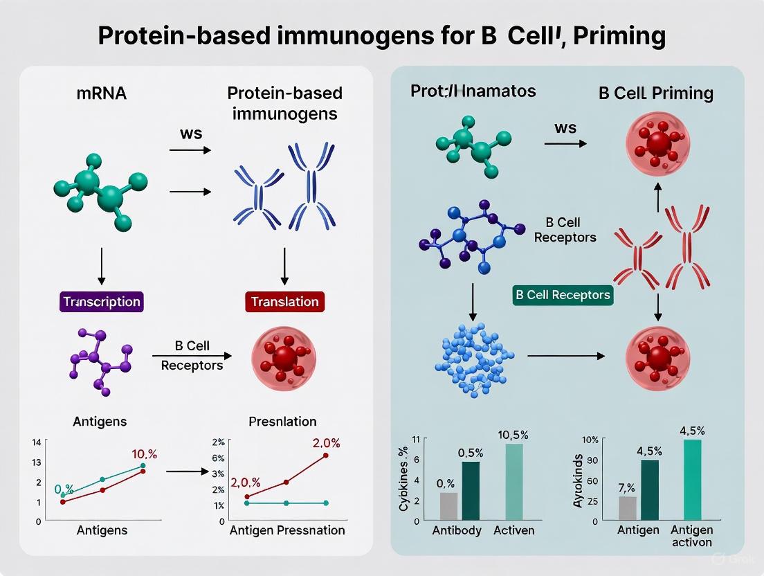
This article provides a comprehensive analysis for researchers and drug development professionals on the distinct mechanisms by which mRNA and protein-based immunogens prime B cell responses.
mRNA vs. Protein Subunit Immunogens: Mechanisms and Efficacy in B Cell Priming for Next-Generation Vaccines
Abstract
This article provides a comprehensive analysis for researchers and drug development professionals on the distinct mechanisms by which mRNA and protein-based immunogens prime B cell responses. It explores the foundational biology, including how mRNA vaccines induce robust germinal center reactions and innate immune signaling, while protein subunits offer conformational precision and benefit from advanced adjuvant systems. The content delves into methodological applications for challenging pathogens like HIV and influenza, troubleshooting limitations such as the restricted memory B cell generation by mRNA platforms and the cold-chain dependency. Finally, it presents comparative data on immunogenicity and explores future directions, including hybrid vaccination regimens that leverage the strengths of both platforms to achieve broader and more potent immunity.
Decoding the Blueprint: Fundamental Mechanisms of B Cell Activation by mRNA and Protein Immunogens
mRNA vaccines represent a transformative technological platform that integrates molecular biology and immunology to elicit protective immune responses. The basic premise involves encoding antigen sequences within mRNA molecules, delivering these transcripts to host cell cytoplasm via non-viral routes, and enabling endogenous antigen expression that stimulates robust, antigen-specific immune responses. Unlike DNA vaccines, mRNA does not interact with the genome and functions as a minimal, transient carrier of genetic information, offering an outstanding safety profile. The structural components of synthetic mRNA—including the 5' cap, 5' and 3' untranslated regions (UTRs), coding region, and poly(A) tail—collectively determine the stability, translational efficiency, and immunogenicity of the vaccine platform. This review systematically examines how optimization of these structural elements enhances protein expression and immune activation, with particular emphasis on implications for B cell priming and antibody responses compared to protein-based immunogens.
Structural Elements of mRNA Vaccines and Their Optimization
The architecture of synthetic mRNA vaccines mirrors that of mature eukaryotic mRNA, comprising several key regulatory elements that can be engineered to maximize protein expression and minimize undesirable immune recognition.
5' Cap Structure
The 5' cap consists of a 7-methylguanosine residue connected to the first nucleotide via a 5'-5' triphosphate bridge and plays crucial roles in ribosome recognition, translation initiation, and protection from exonuclease degradation [1] [2]. Two predominant capping strategies exist:
- Co-transcriptional capping utilizes cap analogs like Anti-Reverse Cap Analogs (ARCAs) during in vitro transcription to ensure proper orientation and prevent reverse incorporation, which produces untranslatable mRNA [2]. ARCAs can be further modified with phosphorothioate linkages to inhibit decapping and enhance eukaryotic initiation factor 4E binding, substantially improving translation efficiency and mRNA half-life [2].
- Post-transcriptional capping employs enzymatic systems such as the vaccinia virus capping complex, which sequentially exhibits triphosphatase, guanylyltransferase, and methyltransferase activities to generate a natural cap structure [3] [2]. This approach can be combined with 2'-O-methyltransferase to create cap-1 structures, further reducing immunogenicity and enhancing translation compared to standard cap-0 structures [3] [2].
Table 1: 5' Capping Strategies and Their Impact on mRNA Function
| Capping Method | Mechanism | Advantages | Effect on Translation |
|---|---|---|---|
| Standard Cap Analog | Incorporated during IVT with cap analog | Simple workflow | Moderate; ~33% ineffective due to reverse incorporation |
| ARCA | Modified analog prevents reverse incorporation | All cap in correct orientation | 2-fold increase vs. standard analog |
| Enzymatic Capping | Post-transcriptionally adds natural cap using VCE | Natural cap structure; no reverse incorporation | Comparable to ARCA; can be combined with 2'-O-methylation for cap-1 |
| Phosphorothioate-Modified ARCA | ARCA with sulfur substitution in triphosphate bridge | Inhibits decapping; enhances eIF4E binding | Superior to ARCA; extends half-life |
5' and 3' Untranslated Regions (UTRs)
The UTRs flanking the coding sequence significantly influence mRNA stability, subcellular localization, and translational efficiency through interactions with RNA-binding proteins and regulatory elements [1] [4]. Optimal UTR design follows several key principles:
- Avoidance of upstream start codons that disrupt open reading frame translation.
- Elimination of stable secondary structures near the 5' end that impede ribosome scanning and recruitment.
- Exclusion of destabilizing elements such as AU-rich sequences and microRNA binding sites [2].
- Implementation of shorter UTRs, which generally facilitate more efficient translation [1].
Comparative studies evaluating various 5' UTRs have demonstrated that the β-globin 5' UTR drives superior protein expression in mRNA vaccines, outperforming alternatives derived from α-globin, cytochrome B-245α chain (CYBA), albumin, and minimal synthetic sequences [4]. The enhanced performance of β-globin UTRs is attributed to their favorable secondary structure characteristics and optimal length. Similarly, the 3' UTR significantly impacts mRNA half-life, with sequences derived from α-globin and β-globin mRNAs—particularly when arranged in tandem repeats—substantially enhancing stability and protein yield [1].
Coding Region and Nucleotide Modifications
The protein-coding open reading frame represents the core of the mRNA vaccine and can be optimized through several strategies:
- Codon optimization replaces rare codons with frequently used synonymous codons to match abundant tRNA species and accelerate translation elongation [1] [5]. This approach must be balanced against potential protein misfolding that can occur when slow-translating rare codons are eliminated from regions requiring precise folding kinetics [1].
- Nucleotide modification involves incorporating modified nucleosides such as N1-methyl-pseudouridine (1mΨ), pseudouridine (ψ), and 5-methylcytidine (m5C) to reduce innate immune recognition and enhance translational efficiency [1]. These modifications enable mRNA to evade pattern recognition receptors that typically trigger antiviral responses and degrade exogenous RNA, thereby increasing both the magnitude and duration of protein expression [1].
- Algorithmic sequence design represents a recent advancement where computational tools like LinearDesign simultaneously optimize mRNA stability and codon usage by identifying sequences with minimal free energy structures [5]. This approach has demonstrated remarkable improvements, with algorithm-designed COVID-19 vaccines eliciting up to 128-fold higher antibody responses in mice compared to conventional codon-optimized benchmarks [5].
Table 2: Optimization Strategies for mRNA Coding Regions
| Strategy | Mechanism | Key Considerations | Experimental Outcome |
|---|---|---|---|
| Codon Optimization | Matches codon usage to abundant tRNA pools | May disrupt protein folding; must balance speed and accuracy | 100-fold higher translation for high GC-content sequences [1] |
| Nucleotide Modification | Incorporates 1mΨ, m5C, ψ to evade immune detection | Reduces IFN responses; increases translational efficiency | 5-fold higher Epo levels with modified vs. unmodified mRNA [1] |
| Algorithmic Design | Computationally optimizes secondary structure and codon usage | Joint optimization of MFE and CAI; handles enormous sequence space | 128x higher antibody titer in mice vs. codon-optimized benchmark [5] |
Poly(A) Tail
The 3' poly(A) tail, in conjunction with the 5' cap, forms a circularized mRNA structure through poly(A)-binding protein (PABP) interactions that promote ribosome recycling and protect both termini from exonuclease degradation [1] [3] [2]. Research demonstrates a direct correlation between poly(A) tail length and translational efficiency, with optimal performance achieved at approximately 68 adenosine residues [2]. Deleting the poly(A) region severely compromises mRNA stability, while extending it enhances both intracellular persistence and protein output [1].
Comparative Analysis: mRNA vs. Protein-Based Immunogens
Understanding the relative advantages and limitations of mRNA versus protein-based vaccines is essential for rational vaccine design, particularly in the context of B cell priming and antibody development.
Antigen Expression Kinetics and Immune Activation
Fundamental differences in antigen presentation between these platforms underlie their distinct immunological profiles:
- mRNA vaccines facilitate endogenous antigen production within host cells, enabling direct loading of peptide fragments onto both major histocompatibility complex (MHC) class I and class II molecules [2]. This promotes balanced CD8+ and CD4+ T cell responses alongside antibody production. However, mRNA-encoded antigens exhibit rapid but transient expression, with peak levels occurring within 6 hours post-immunization and declining significantly by 24 hours [6]. This brief exposure window may limit the duration of antigen availability for B cell receptor engagement and germinal center reactions.
- Protein vaccines deliver pre-formed antigens that primarily enter the exogenous presentation pathway, favoring MHC class II-restricted CD4+ T cell activation and humoral immunity [6]. While protein immunogens typically show lower initial antigen levels compared to mRNA platforms, they may offer more prolonged antigen persistence when formulated with appropriate adjuvants [6].
Table 3: Direct Comparison of mRNA and Protein Vaccine Platforms
| Parameter | mRNA Vaccine | Protein Vaccine |
|---|---|---|
| Antigen Expression | Endogenous production within host cells | Pre-formed, delivered with adjuvant |
| Expression Kinetics | Rapid (peak at 6h), transient (<24h) [6] | Lower initial levels, potentially more sustained [6] |
| Antigen Presentation | MHC I and MHC II presentation | Primarily MHC II presentation |
| Immune Response Profile | Balanced cellular and humoral immunity | Strong antibody response, CD4+ T cell help |
| Germinal Center Response | Robust GC formation and Tfh activation [7] | Varies with adjuvant |
| Memory B Cell Generation | Limited despite strong GC response [7] | Effective with appropriate adjuvants |
| Innate Immune Activation | Self-adjuvanting through PRR recognition [2] | Requires exogenous adjuvants |
Implications for B Cell Priming and Antibody Responses
The distinct antigen presentation patterns between platforms directly impact B cell activation and differentiation:
- Germinal center responses are robustly induced by mRNA vaccines, which stimulate strong T follicular helper (Tfh) cell expansion essential for B cell affinity maturation [7]. This potent GC activation reflects the platform's capacity for coordinated MHC class I and II presentation.
- Memory B cell generation presents a paradoxical limitation of mRNA vaccines—despite inducing vigorous germinal center reactions, they elicit relatively limited numbers of antigen-specific memory B cells and long-lived plasma cells compared to other platforms [7]. This discrepancy suggests that the transient antigen expression characteristic of mRNA vaccines may be insufficient to sustain the extended B cell clonal selection and differentiation required for robust memory formation.
- Immunodominance hierarchies can be favorably manipulated using mRNA platforms. For complex antigens like HIV-1 envelope glycoproteins, mRNA expression of membrane-bound gp160 fosters native antigen conformation and presentation, potentially focusing antibody responses toward functionally relevant epitopes rather than immunodominant but non-protective regions [8].
Diagram 1: Comparative Immunological Pathways of mRNA and Protein Vaccines. mRNA vaccines induce both MHC I and II presentation leading to robust germinal center formation but limited memory B cells, while protein vaccines primarily drive MHC II-restricted responses with effective memory B cell generation.
Experimental Approaches for Evaluating mRNA Vaccine Performance
Rigorous assessment of mRNA vaccine components requires standardized methodologies to quantify their impact on protein expression and immunogenicity.
In Vitro Characterization Methods
- Reporter gene systems utilizing fluorescent (e.g., EGFP) and luminescent (e.g., luciferase) proteins enable rapid, quantitative comparison of different mRNA constructs. Experimental protocols typically involve transfecting HEK293T or other relevant cell lines with candidate mRNA formulations and measuring fluorescence intensity or luminescence activity at 24, 48, and 72-hour intervals [4]. This approach directly demonstrated the superior performance of β-globin 5' UTRs compared to alternatives like α-globin, CYBA, and albumin UTRs [4].
- Western blot analysis provides direct confirmation of antigen identity and expression levels from modified mRNA sequences. For HIV gp145 mRNA vaccines, this technique verified that constructs incorporating β-globin 5' UTRs yielded the highest envelope protein expression compared to other UTR variants [4].
- Secondary structure prediction using computational platforms like Paddlehelix informs rational design by modeling thermodynamic stability of 5' UTR regions and their effects on ribosome binding and scanning efficiency [4]. These predictions help identify and eliminate unfavorable structural elements that impede translation.
In Vivo Immunogenicity Assessment
- Animal immunization studies typically administer mRNA formulations via intramuscular injection, often encapsulated in lipid nanoparticles (LNPs) to enhance cellular delivery and protect mRNA integrity. Dosing regimens vary from 1-200 µg depending on the model species and target antigen [8].
- Antigen-specific antibody quantification employs enzyme-linked immunosorbent assays (ELISAs) to measure binding antibody titers and neutralization assays to assess functional antibody responses against relevant pathogens [8].
- Germinal center and memory B cell analysis utilizes flow cytometry to quantify Tfh cells, germinal center B cells, and antigen-specific memory B cell populations in lymphoid tissues following immunization [7]. These analyses revealed the dissociation between robust GC induction and limited memory B cell formation in mRNA-vaccinated subjects [7].
- In vivo bioluminescence imaging enables real-time monitoring of antigen expression kinetics by tracking luciferase activity in living animals [6]. This technique has demonstrated the characteristically rapid but transient antigen expression profile of mRNA vaccines compared to more sustained expression from adenoviral vectors [6].
Diagram 2: Experimental Workflow for mRNA Vaccine Evaluation. Comprehensive assessment integrates in vitro characterization of protein expression with in vivo analysis of immunogenicity and antigen kinetics.
The Scientist's Toolkit: Essential Research Reagents
Successful mRNA vaccine research requires specialized reagents and systems for construct development, production, and evaluation.
Table 4: Essential Research Reagents for mRNA Vaccine Development
| Reagent/System | Function | Application Examples |
|---|---|---|
| Takara IVTpro mRNA Synthesis System | In vitro transcription template preparation | High-yield mRNA production with compatible capping approaches [3] |
| Vaccinia Capping Enzyme (VCE) | Post-transcriptional 5' capping | Generation of natural cap structures; creation of cap-1 structures when combined with 2'-O-MTase [3] |
| Anti-Reverse Cap Analogs (ARCA) | Co-transcriptional capping | Ensures proper cap orientation; enhances translation efficiency [2] |
| Lipid Nanoparticles (LNPs) | mRNA delivery and protection | Intramuscular delivery; enhances cellular uptake and protects from degradation [1] [8] |
| N1-methyl-pseudouridine | Modified nucleotide | Reduces immunogenicity; increases translation efficiency and stability [1] [4] |
| Dual-reporter constructs (NLuc-T2A-EGFP) | Assessment of UTR performance | Parallel evaluation of multiple 5' UTR designs in cell culture [4] |
| Paddlehelix computational platform | mRNA secondary structure prediction | In silico optimization of structural stability [4] |
The structural components of mRNA vaccines—5' cap, UTRs, coding region, and poly(A) tail—collectively determine their translational efficiency, intracellular stability, and ultimate immunogenicity. Strategic optimization of each element through nucleotide modification, sequence engineering, and computational design significantly enhances protein expression and immune activation. When compared to protein-based immunogens, mRNA vaccines offer distinct advantages in endogenous antigen presentation that drives robust germinal center responses and balanced T cell immunity. However, the characteristically transient antigen expression of mRNA platforms may limit memory B cell development compared to protein vaccines, presenting an important challenge for future research. The ongoing refinement of mRNA design principles, coupled with advanced delivery technologies and combinatorial approaches with other vaccine modalities, promises to unlock the full potential of this versatile platform for both infectious disease and cancer immunotherapy applications.
mRNA vaccines have revolutionized immunology by demonstrating superior efficacy in preventing symptomatic illness compared to traditional platforms. This enhanced protection stems from their unique ability to harness innate immune responses as a built-in adjuvant system. Groundbreaking research reveals that at the injection site, mRNA vaccines trigger a sophisticated cascade involving stromal cells and type I interferon signaling, particularly IFN-β, which fundamentally shapes the adaptive immune response. This review synthesizes recent single-cell transcriptomic evidence comparing mRNA vaccines against protein-based alternatives, highlighting how mRNA-induced IFN-β production by fibroblasts and subsequent dendritic cell activation creates a microenvironment conducive to robust cellular immunity and germinal center responses. These mechanistic insights provide a scientific foundation for understanding the differential immunogenicity across vaccine platforms and offer strategic guidance for future rational vaccine design, particularly for challenging pathogens requiring strong T-cell immunity.
Traditional subunit vaccines often require exogenous adjuvants to achieve sufficient immunogenicity, as they typically lack the innate immune stimulation necessary to drive robust adaptive responses. The aluminum-based adjuvants (alum) commonly used with protein vaccines primarily enhance antibody responses but are less effective at stimulating cellular immunity [9]. In contrast, mRNA vaccines incorporate a self-adjuvanting mechanism through their fundamental structure and delivery system. The two key components—the mRNA molecule itself and the lipid nanoparticles (LNPs) that deliver it—both contribute to this built-in adjuvanticity by engaging distinct innate immune pathways [10].
Recent single-cell transcriptome analyses of injection site responses have revealed that this built-in adjuvant system operates through a precisely coordinated cellular and molecular cascade. The LNP component drives pro-inflammatory responses in stromal cells, while the mRNA component specifically elicits type I interferon responses, with IFN-β emerging as a critical mediator [10] [11]. This coordinated response creates a local immunogenic environment that enhances antigen presentation and T-cell priming, providing mRNA vaccines with a distinct advantage over protein-based platforms for generating comprehensive immunity.
Comparative Immunogenicity of Vaccine Platforms
Head-to-Head Comparisons of Immune Responses
Different vaccine technologies generate markedly different immune responses, as evidenced by direct comparisons of SARS-CoV-2 vaccines. Nanoparticle and mRNA vaccines demonstrate superior immunogenicity compared to inactivated virus and recombinant protein vaccines in terms of magnitude of initial B cell responses [7]. However, each platform exhibits distinct strengths and weaknesses in the quality and longevity of the immune response they elicit.
Table 1: Comparative Immunogenicity Across Vaccine Platforms
| Vaccine Platform | Germinal Center Response | Memory B Cell Generation | T Follicular Helper Cell Induction | Antibody Class Switching Profile |
|---|---|---|---|---|
| mRNA-LNP | Robust | Limited | Strong | IgG1-dominated |
| Protein Nanoparticle | Strong | Substantial | Moderate | Balanced IgG |
| Whole Inactivated Virus | Moderate | Substantial | Weak | Enhanced IgG2a/c |
| Recombinant Protein | Weak | Moderate | Weak | IgG1-dominated |
Interestingly, despite inducing robust germinal center responses and T follicular helper (Tfh) cell activation, mRNA vaccines show a limited ability to generate memory B cells and long-lived plasma cells compared to other platforms [7]. This suggests that while mRNA vaccines excel at initiating powerful initial immune activation, other formats may offer advantages in certain aspects of immunological memory.
Efficacy Outcomes in Clinical Trials
Recent human challenge trials provide compelling evidence for the superior efficacy of mRNA vaccine platforms. An experimental mRNA influenza vaccine demonstrated 100% efficacy against both symptomatic and febrile influenza in a controlled human challenge model, compared to 85% protection against symptomatic illness provided by a conventional quadrivalent influenza vaccine (QIV) [12]. The mRNA vaccine also showed significantly better viral load control, with median differences in viral load area under the curve between vaccine and control groups of -88.66 for the mRNA vaccine versus -67.01 for QIV [12].
These clinical findings align with mechanistic studies showing that mRNA vaccines create a more immunogenic microenvironment at the injection site and in draining lymph nodes, facilitating enhanced antigen presentation and immune cell activation compared to traditional vaccine approaches.
Decoding the Injection Site Response: Single-Cell Atlas
Cellular Players and Transcriptional Dynamics
Comprehensive single-cell transcriptome profiling of mRNA vaccine injection sites has revealed a complex multicellular response involving 22 different cell types in muscle tissues, including dendritic cells, monocytes, neutrophils, endothelial cells, and fibroblasts [10]. Analysis of 83,094 single-cell profiles from murine models revealed that both empty LNP and mRNA-LNP injections provoke substantial shifts in the transcriptional landscape, with prominent increases in CD8 T cell, neutrophil, and monocyte populations 16 hours post-injection [10].
Principal component analysis of differentially expressed genes identified two major axes of transcriptional responses:
- PC1 (Stromal Inflammation): Representing responses in stromal cells (fibroblasts, endothelial cells, and mural cells) driven primarily by the LNP component
- PC2 (Antiviral/Interferon Response): Featuring type I interferon responses in migratory dendritic cells, specifically induced by the mRNA component [10]
Table 2: Key Cell Types and Their Roles in mRNA Vaccine Immune Activation
| Cell Type | mRNA Uptake | Primary Response | Key Functions | Trigger |
|---|---|---|---|---|
| Fibroblasts | High enrichment | IFN-β production | Initiate IFN cascade, support leukocyte recruitment | mRNA component |
| Migratory Dendritic Cells | Moderate | IFN-stimulated gene expression | Antigen presentation, T cell priming | mRNA-induced IFN-β |
| Monocytes/Macrophages | Moderate | Pro-inflammatory cytokine production (IL-6, TNF) | Inflammation, antigen presentation | LNP component |
| Endothelial Cells | High enrichment | Chemokine production | Immune cell recruitment, vascular permeability | LNP > mRNA |
| Lymphoid Cells | Low | Variable | Adaptive immunity execution | Indirect (via cytokines) |
Pathway enrichment analysis revealed that the stromal inflammatory axis (PC1) was associated with immune cell chemotaxis genes, while the dendritic cell antiviral axis (PC2) was linked to type I interferon response genes including Isg15, Oasl1, and Ifit3 [10]. This diversification of early innate responses between different cell types represents a fundamental characteristic of mRNA vaccine immunogenicity.
mRNA Fate and Cellular Tropism at the Injection Site
Tracking the delivered mRNA reveals fascinating tropism patterns at the injection site. Spike mRNAs are highly enriched in stromal cells—including endothelial cells, pericytes, and fibroblasts—as well as myeloid cells at 2 hours post-injection [10]. Lymphoid cells and other structural cells contain relatively low amounts of the target mRNA transcripts. The detection rates of spike mRNA decrease over time, likely due to degradation of the mRNA molecules, with 2%-46% of cells in mRNA-LNP-injected samples being spike-positive depending on the time point [10].
Fibroblasts emerge as particularly important early responders, showing high enrichment of delivered mRNA and specific expression of IFN-β in response to the mRNA component rather than the LNP component alone [10] [11]. This identifies fibroblasts as key sentinel cells in the initial immune cascade triggered by mRNA vaccination.
Experimental Models and Methodologies
Key Protocols for Studying Injection Site Immunology
Single-Cell Transcriptome Profiling of Injection Site Responses:
- Animal Model: Female BALB/c mice immunized intramuscularly with nucleoside-modified mRNA-LNP encoding SARS-CoV-2 spike protein
- Control Groups: Saline-injected and empty LNP (without mRNA) injected mice
- Tissue Processing: Anterior thigh muscles resected from 2 to 40 hours post-injection, mechanically and chemically digested to single-cell suspensions
- Sequencing: 83,094 single-cell profiles from injection site, 8,507 from draining lymph nodes
- Validation: Plaque reduction neutralization test and IFN-γ ELISpot at 2 weeks post-boost [10]
Ex Vivo Human Lymph Node Model:
- System: Precision-cut human lymph node slices maintaining architectural integrity
- Stimulation: TLR4-agonist and QS-21 saponin containing liposomal adjuvant
- Analysis: Single-cell transcriptomics and multiplexed imaging
- Key Findings: Monocytes/macrophages initiate response via TLR4 and NLRP3 inflammasome; innate lymphoid cells activated indirectly; stromal populations mediate neutrophil recruitment [13]
mRNA Modification Studies
Comparative studies evaluating different mRNA formats reveal how specific modifications alter immunogenicity:
- Experimental Design: Comparison of uridine-containing mRNA (D1-uRNA), m1Ψ-modified mRNA (D1-modRNA), and cap1-modified m1Ψ-mRNA (cC1-modRNA) in murine influenza model
- Findings: D1-uRNA upregulated Stat1 and RnaseL, increased systemic inflammation, but generated reduced antigen-specific antibodies and protection
- Conclusion: Both m1Ψ and cap1 modifications are required for optimal response by modulating innate immune recognition [14]
IFN-β as the Critical Mediator of Cellular Immunity
Mechanism of Action
IFN-β emerges as the pivotal cytokine bridging innate and adaptive immunity in mRNA vaccination. Fibroblasts at the injection site, highly enriched with delivered mRNA, express IFN-β specifically in response to the mRNA component rather than the LNP alone [10] [11]. This IFN-β then induces a specialized population of migratory dendritic cells expressing high levels of interferon-stimulated genes (mDC_ISGs) at both the injection site and draining lymph nodes.
The critical role of IFN-β signaling has been demonstrated through intervention studies. When IFN-β signaling is blocked at the injection site, mRNA vaccine-induced cellular immune responses are significantly decreased [11]. Conversely, co-injection of IFN-β with LNP-subunit vaccines substantially enhances antigen-specific cellular immune responses [10] [11]. This positions IFN-β as both necessary and sufficient for optimal T-cell priming in the context of mRNA vaccination.
Distinct Biology of Type I Interferons
The type I interferon family includes multiple members with distinct biological functions. IFN-β binds to the IFN-α/β receptor (IFNAR1/2) with 10-100 times greater affinity than IFN-α2 [15]. While IFN-α is predominantly secreted by plasmacytoid dendritic cells activated through TLR7 or TLR9, IFN-β can be secreted by essentially all nucleated cells through various pattern recognition receptors including cGAS-STING and RIG-I-like receptors [15].
This functional specialization has therapeutic implications. In humans, IFN-α drives CD8+ T cell responses and is approved for treating some cancers and viral infections, while IFN-β promotes immunomodulatory responses including IL-10 and regulatory T cells and is approved for multiple sclerosis [15]. The species-specific differences in interferon biology—with both IFN-α and IFN-β promoting strong CD8+ T cell responses in mice—highlight the importance of human model systems for translational research.
Impact on B Cell Priming and Germinal Center Responses
Comparative B Cell Responses Across Platforms
The initial priming of B cells differs substantially between mRNA and protein-based vaccines. Studies comparing SARS-CoV-2-specific B cell responses across platforms reveal that nanoparticle and mRNA vaccines exhibit superior immunogenicity compared to inactivated and recombinant protein vaccines [7]. This enhanced immunogenicity manifests in the magnitude and quality of the germinal center response, a critical determinant of antibody affinity maturation and B cell memory formation.
Despite inducing robust germinal center responses and T follicular helper cell activation, mRNA vaccines elicit a relatively limited number of memory B cells compared to other platforms [7]. This suggests that while mRNA vaccines excel at initiating powerful germinal center reactions, the differentiation of germinal center B cells into memory B cells may be less efficient than with other vaccine formats.
Modulation by mRNA Modifications
The innate immune response to mRNA vaccines can be optimized through specific modifications to the mRNA structure. Comparing uridine-containing mRNA with m1Ψ-modified mRNA reveals significant differences in gene expression profiles and inflammatory responses [14]. Uridine-containing mRNA upregulates Stat1 and RnaseL and increases systemic inflammation, but generates reduced antigen-specific antibody responses and protection compared to modified mRNA platforms [14].
The incorporation of both m1Ψ modification and cap1 structure creates an optimal balance—reducing excessive inflammation while maintaining sufficient immune activation for robust adaptive responses [14]. This fine-tuning of innate immune recognition represents a crucial advancement in mRNA vaccine technology, enabling enhanced efficacy while minimizing reactogenicity.
The Scientist's Toolkit: Essential Research Reagents
Table 3: Key Reagents for Studying mRNA Vaccine Immunology
| Reagent/Cell Type | Key Application | Research Function | Example Findings |
|---|---|---|---|
| Single-cell RNA-seq | Transcriptome profiling | Comprehensive cell-type specific response mapping | Identified IFN-β-specific mDC_ISG population [10] |
| Precision-cut human LN slices | Ex vivo human response modeling | Human-specific immunogenicity screening | Identified stromal-neutrophil recruitment axis [13] |
| IFNAR blocking antibodies | Pathway inhibition | Mechanistic studies of IFN-β function | Confirmed necessity of IFN-β signaling [11] |
| m1Ψ-modified mRNA | mRNA optimization | Reducing excessive innate activation | Enhanced antibody responses via moderated inflammation [14] |
| Barcoded LNPs | mRNA fate tracking | Cellular tropism determination | Identified fibroblast mRNA enrichment [10] |
| IFN-γ ELISpot | T cell response quantification | Cellular immunity measurement | Correlated IFN-β with enhanced T cell responses [10] |
The built-in adjuvant system of mRNA vaccines, centered on stromal cell activation and IFN-β production at the injection site, represents a fundamental advance in vaccine technology. This mechanistic understanding explains the superior efficacy of mRNA platforms compared to protein-based alternatives observed in both preclinical models and clinical trials. The coordinated cascade—initiated by mRNA uptake in fibroblasts, amplified through IFN-β secretion, and executed through mDC_ISG-mediated T cell priming—creates an immunogenic microenvironment that enhances both cellular and humoral immunity.
These insights have profound implications for future vaccine design. First, they validate the importance of the injection site as an active immunological niche rather than merely a depot for antigen presentation. Second, they identify IFN-β signaling as a potential target for modulating vaccine immunogenicity, either through enhancement in vulnerable populations or attenuation to reduce reactogenicity. Third, they provide a framework for rational combination of mRNA platforms with other technologies to leverage the respective strengths of each approach.
Future research should focus on translating these mechanistic insights from murine models to human immunology, particularly given the species-specific differences in interferon biology. The development of more sophisticated human model systems, such as precision-cut lymph node slices, will be crucial for this translational effort. Additionally, longitudinal studies examining how early innate responses correlate with the generation of long-term immunological memory will help optimize next-generation mRNA vaccines for durable protection against diverse pathogens.
The structural integrity of viral antigen proteins is a critical determinant in eliciting potent and protective immune responses. For many viral pathogens, antibodies that neutralize infection preferentially recognize the prefusion conformation of viral fusion glycoproteins, a meta-stable state that exists prior to host cell entry [16] [17]. However, recombinant expression of these antigens often results in conformational rearrangement to the more stable postfusion state, which presents different, often less protective, epitopes [17]. The field of structural vaccinology has therefore focused on developing precision engineering strategies to lock glycoproteins in their prefusion state, thereby preserving the native antigenic landscape presented to the immune system and enhancing vaccine efficacy [16] [17].
This pursuit is particularly relevant within the broader context of comparing the efficacy of mRNA and protein-based immunogens for B cell priming. While mRNA vaccines can encode antigens that are natively folded within cells, recombinant protein vaccines offer distinct advantages in stability and precise conformational control, provided the correct structure can be maintained outside a cellular environment [16] [18]. This guide provides a comparative analysis of the key strategies, experimental data, and methodologies driving innovation in prefusion-stabilized protein subunit design.
Core Principles and Strategies for Prefusion Stabilization
Structure-based design employs a suite of computational and biophysical tools to engineer antigens with enhanced stability and immunogenicity. The following table summarizes the primary stabilization strategies and their molecular mechanisms.
Table 1: Key Strategies for Engineering Prefusion-Stabilized Viral Glycoproteins
| Strategy | Molecular Mechanism | Target Regions | Key Effect |
|---|---|---|---|
| Proline Substitution [17] [19] | Introduces rigid proline residues at hinge points or loop termini; restricts backbone dihedral angles. | N-termini of α-helices, flexible loops. | Inhibits conformational rearrangements required for fusion. |
| Disulfide Bond Engineering [17] | Creates covalent crosslinks (Cys-Cys bonds) between structurally proximal elements. | Regions that undergo large conformational shifts. | Tethers domains to prevent refolding. |
| Cavity-Filling Mutations [17] | Replaces small side chains (Ala, Gly) with larger residues (Trp, Tyr, Phe) in hydrophobic cores. | Internal cavities prone to collapse during fusion. | Enhances core packing and structural rigidity. |
| Electrostatic Optimization [17] | Introduces or optimizes salt bridges; mitigates unfavorable charge repulsions. | Domain interfaces, solvent-exposed patches. | Stabilizes interfaces and reduces aggregation. |
| Glycan Shielding [17] | Adds or relocates N-linked glycosylation sites. | Variable or immunodominant non-neutralizing epitopes. | Directs immune response toward conserved epitopes; can improve stability. |
The strategic application of these methods is often guided by high-resolution structures from cryo-electron microscopy or X-ray crystallography, enabling targeted interventions to reinforce the meta-stable prefusion architecture [16] [17].
Comparative Analysis of Stabilization Approaches Across Pathogens
The principles of prefusion stabilization have been successfully applied across major respiratory viruses. The table below compares engineered antigens for SARS-CoV-2, RSV, and influenza, highlighting the specific mutations and their measurable outcomes.
Table 2: Application of Prefusion Stabilization Strategies Across Different Pathogens
| Pathogen / Target | Stabilized Antigen | Key Stabilizing Mutations | Experimental Outcomes & Advantages |
|---|---|---|---|
| SARS-CoV-2 (Spike) | HexaPro [16] | 6 Proline substitutions (e.g., F817P, A892P, A899P, A942P, K986P, V987P) | ~50-fold higher protein expression than 2P parent; increased thermal stability; preserved binding to neutralizing antibodies [16]. |
| RSV (Fusion Protein) | preF7P [19] | 7 Proline substitutions (S215P, L138P, G139P, etc.) via proline-scanning. | High-yield expression (~10 g/L in CHO cells); 1.8-fold increase in neutralizing antibody titers vs. DS-Cav2; protection in murine/cotton rat models [19]. |
| RSV (Fusion Protein) | DS-Cav1 [16] | Disulfide bonds (S155C-S290C) + cavity-filling mutations (S190F, V207L). | First major prefusion-stabilized RSV F; markedly increased neutralizing antibody responses in animal models [16]. |
| Influenza (HA) | Stem-Nanoparticles [16] | HA stem focused immunogens displayed on self-assembling nanoparticles (e.g., I53-50, mi3). | Mosaic/multivalent display promotes cross-neutralization of diverse influenza A and B strains by targeting conserved stem regions [16]. |
This comparative data demonstrates that while proline stabilization offers a streamlined, high-yield approach, classic strategies involving disulfide bonds and cavity-filling mutations have paved the way and remain highly effective. The choice of strategy often depends on the specific structural vulnerabilities of the target glycoprotein.
Experimental Protocols for Design and Validation
The development of prefusion-stabilized immunogens follows a rigorous, iterative workflow from computational design to in vivo validation. The diagram below outlines this multi-stage process.
Diagram 1: The iterative workflow for designing and validating prefusion-stabilized protein subunit vaccines, from structural analysis to immunogenicity assessment.
Detailed Methodologies for Key Experimental Steps
4.1.1 Structural Analysis and In Silico Design
- Objective: Identify flexible regions and structural vulnerabilities in the prefusion conformation using available high-resolution structures (e.g., from PDB).
- Protocol:
- Analyze protein dynamics in molecular dynamics (MD) simulations to pinpoint hinge regions and loops involved in conformational changes [20].
- Identify potential sites for proline substitution at the N-termini of α-helices and flexible loops. A systematic "proline-scanning" strategy can be employed [19].
- Use computational tools like AlphaFold2/3 or RoseTTAFold to model the effects of proposed mutations (e.g., disulfide bonds, cavity-filling mutations) on local and global structure [20] [21].
- Select a panel of mutations (single and combined) for experimental testing.
4.1.2 Recombinant Expression and Purification
- Objective: Produce high-quality, trimeric antigen protein in sufficient quantities for characterization and immunization.
- Protocol:
- Clone genes encoding the engineered variants into mammalian expression vectors (e.g., for HEK293 or CHO cells). A C-terminal trimerization domain (e.g., T4 fibritin foldon) and affinity tag (e.g., His-tag) are often included [19].
- Perform transient transfection of HEK293F cells or use stable CHO cell pools for large-scale production [18] [19].
- Purify the protein from cell culture supernatant using affinity chromatography (e.g., Ni-NTA), followed by size-exclusion chromatography (SEC) to isolate properly folded trimers and remove aggregates [19].
4.1.3 Biophysical and Structural Characterization
- Objective: Confirm the protein is in the prefusion conformation and assess its stability and homogeneity.
- Protocol:
- SEC-Multi-Angle Light Scattering (SEC-MALS): Determine the molecular weight and monodispersity of the purified trimer [19].
- Differential Scanning Calorimetry (DSC): Measure the melting temperature (Tm) to quantify thermal stability. Prefusion-stabilized variants typically show a higher Tm [19].
- Surface Plasmon Resonance (SPR) or BLI: Quantify binding affinity (KD) to a panel of conformation-specific monoclonal antibodies (e.g., prefusion-specific vs. postfusion-specific). Strong binding to prefusion-specific mAbs and weak/no binding to postfusion-specific mAbs confirms successful stabilization [19].
- Negative-Stain or Cryo-Electron Microscopy: Visualize the protein structure to directly confirm it adopts the desired prefusion conformation [16] [19].
4.1.4 Immunogenicity Assessment
- Objective: Evaluate the ability of the stabilized immunogen to elicit potent and broad neutralizing antibody responses.
- Protocol:
- Immunize animal models (e.g., mice, rats) with the purified protein, typically formulated with an adjuvant (e.g., AS01, MF59) [16] [18].
- Collect serum post-immunization and measure antigen-binding antibody titers via ELISA.
- Assess the quality of the humoral response using pseudovirus or live virus neutralization assays. Compare neutralization titers against those induced by unstabilized or postfusion antigens [18] [19].
- For B cell analysis, techniques like flow cytometry can be used to track antigen-specific B cells and memory B cell formation [7].
The Scientist's Toolkit: Essential Research Reagents and Solutions
The experimental workflow relies on a suite of specialized reagents and platforms. The following table details key solutions and their critical functions in prefusion antigen development.
Table 3: Essential Research Reagents for Prefusion-Stabilized Antigen Development
| Research Reagent / Platform | Function & Application | Relevance to Prefusion Stabilization |
|---|---|---|
| CHO (Chinese Hamster Ovary) Cell Lines [16] [19] | Mammalian expression system for recombinant protein production. | Industry standard for high-yield, clinical-grade production of complex, glycosylated proteins; enables scalable manufacturing. |
| Conformation-Specific Monoclonal Antibodies [19] | Critical tools for characterizing antigen structure via binding assays (SPR, BLI, ELISA). | Used to confirm the presence of prefusion-specific epitopes (e.g., site Ø/V on RSV F) and absence of postfusion epitopes. |
| Structure Prediction Software (AlphaFold 3, Boltz-2) [20] | AI-driven platforms for predicting protein structures and complexes. | Models the 3D structure of designed variants and predicts the impact of mutations prior to experimental testing. |
| TLR4 Agonist Adjuvants (e.g., EmT4, LiT4Q) [18] | Adjuvant formulations that enhance magnitude and breadth of immune responses to protein antigens. | When combined with stabilized protein immunogens, can significantly boost antibody responses, particularly against variants. |
| Size-Exclusion Chromatography (SEC) Columns [19] | Chromatography technique to separate proteins by size and hydrodynamic radius. | Essential for purifying correctly assembled trimeric antigens and removing misfolded or aggregated species. |
The strategic engineering of protein subunits to maintain their native prefusion conformation represents a cornerstone of modern vaccinology. Techniques such as proline substitution, disulfide bridging, and cavity-filling mutations have proven highly effective in creating stable, highly immunogenic antigens for viruses like SARS-CoV-2 and RSV, with some candidates demonstrating superior yields and potent neutralizing antibody responses in preclinical models [16] [19]. This precise control over antigen structure is a key advantage of the recombinant protein platform, enabling the rational design of vaccines that can effectively prime high-quality B cell responses against the most vulnerable sites on pathogens. As AI-driven structural prediction continues to advance [20] [21], the design process is poised to become faster and more accurate, accelerating the development of next-generation vaccines against existing and emerging viral threats.
Vaccine adjuvants are indispensable components that enhance and shape immune responses to vaccine antigens. The evolution from simple mineral salts to sophisticated adjuvant systems represents a cornerstone in the development of modern vaccinology, particularly for protein-based vaccines. The use of highly purified antigens, including recombinant proteins and synthetic peptides, has improved vaccine safety but often at the cost of reduced immunogenicity, creating an essential role for adjuvants to stimulate adequate immune protection [22]. Within the broader context of comparing vaccine platforms, understanding adjuvant function is crucial for evaluating the efficacy of mRNA versus protein-based immunogens for critical processes like B cell priming. While mRNA vaccines intrinsically incorporate lipid nanoparticles that provide self-adjuvating properties [23], protein vaccines rely exclusively on exogenous adjuvants to achieve similar potency, making adjuvant selection a pivotal factor in vaccine design.
Adjuvants are broadly classified into two categories: immunostimulants and delivery systems [24] [25]. Immunostimulants, such as pathogen-associated molecular pattern (PAMP) molecules, act as danger signals that activate innate immune cells by targeting pattern recognition receptors (PRRs) including Toll-like receptors (TLRs). This activation leads to maturation of antigen-presenting cells (APCs), enhanced antigen presentation, and production of co-stimulatory signals and cytokines that ultimately drive adaptive immunity [24]. Delivery systems, including emulsions, liposomes, and mineral salts, function as carrier materials that prolong antigen bioavailability, target antigens to lymph nodes or APCs, and facilitate antigen presentation [24] [22]. The most advanced adjuvant platforms often combine both approaches to create synergistic effects that enhance immunogenicity while maintaining favorable safety profiles.
Classification and Mechanisms of Action
Fundamental Mechanisms of Adjuvant Action
Vaccine adjuvants enhance adaptive immunity primarily by activating innate immune cells. The core mechanism involves adjuvants promoting the generation of both antigen presentation signals (Signal 1) and co-stimulatory signals (Signal 2) through the activation of antigen-presenting cells (APCs) [24]. Signal 1 consists of antigen peptides bound to major histocompatibility complexes (MHC) presented on APC surfaces after antigen uptake and processing. Signal 2 includes co-stimulatory molecules (e.g., CD40, CD80, CD86) expressed on APC surfaces and secreted inflammatory cytokines (e.g., IL-6, IL-10, IL-12, TNF-α). The production of these two signals robustly induces naive T cell activation, leading to enhanced adaptive immune responses [24].
The classification of adjuvants reflects their primary mechanisms of action. Immunostimulants act as PAMPs, damage-associated molecular patterns (DAMPs), or their synthetic analogs that interact with PRRs on APCs to trigger innate immune responses [24]. This interaction leads to APC activation and maturation, characterized by terminated phagocytic activity alongside enhanced antigen presentation capacity and increased expression of co-stimulatory signals and cytokines [24]. Different immunostimulants signal through distinct PRRs and induce different cytokine secretion profiles, which significantly determines the resulting adaptive immune response polarization [24].
Key Signaling Pathways
The following diagram illustrates the primary mechanisms by which different adjuvant classes exert their immunomodulatory effects:
The diagram illustrates how immunostimulants primarily function through Pattern Recognition Receptors (PRRs), notably Toll-like Receptors (TLRs), to activate signaling cascades that determine immune polarization. TLR4 agonists like MPL in AS04 activate both MyD88 and TRIF pathways, inducing Th1 responses through NF-κB and IRF3 [24]. In contrast, delivery systems like alum salts create antigen depots that prolong antigen exposure and recruit APCs to the injection site, while systems like MF59 enhance APC recruitment and activation, and liposomes facilitate lymph node targeting for improved antibody responses [24] [25].
Comparative Analysis of Adjuvant Platforms
Established Adjuvant Platforms
Table 1: Key Licensed Adjuvant Formulations and Their Characteristics
| Adjuvant Name | Composition | Vaccine Examples | Immune Response Profile | Mechanistic Insights |
|---|---|---|---|---|
| Alum Salts | Aluminum hydroxide, aluminum phosphate, or amorphous aluminum hydroxyphosphate sulfate [25] | Hepatitis A (HAVRIX, AVAXIM), Hepatitis B (ENGERIX B), DTaP (Boostrix) [25] | Strong Th2 response, robust antibody production, weak CD8+ T cell activation [24] | Forms antigen depot, enhances phagocytosis, activates NLRP3 inflammasome, promotes eosinophilia [22] |
| MF59 | Oil-in-water emulsion containing squalene, polysorbate 80, sorbitan trioleate [25] | Influenza (FLUAD) [25] | Enhanced antibody responses, broad cross-reactivity, strong in elderly populations | Recruits immune cells to injection site, promotes antigen uptake by APCs, induces cytokine/chemokine production |
| AS04 | Monophosphoryl lipid A (MPL, TLR4 agonist) adsorbed to aluminum salt [24] [25] | HPV (Cervarix), Hepatitis B (Fendrix) [25] | Balanced Th1/Th2 response, higher antibody titers than alum alone, enhanced CD4+ T cell responses | MPL activates TLR4 on APCs, inducing cytokine production; alum prolongs immune stimulation at injection site [24] |
| AS01 | MPL (TLR4 agonist) and QS-21 (saponin) in liposomal formulation [24] | Herpes Zoster (Shingrix), Malaria (Mosquirix) [22] | Strong CD4+ T cell responses, high antibody levels, induction of memory responses | Synergistic activation of innate immunity via TLR4 and potentially other receptors; QS-21 promotes cytotoxic T-cell responses [24] |
| AS03 | Oil-in-water emulsion containing α-tocopherol (vitamin E), squalene, polysorbate 80 [24] | Pandemic influenza vaccines | Enhanced antibody responses, antigen-sparing effect, cross-reactive immunity | α-tocopherol induces cytokine production, promotes immune cell recruitment and differentiation |
| CpG 1018 | Cytosine phosphoguanine (CpG) oligodeoxynucleotides (TLR9 agonist) [24] | Hepatitis B (Heplisav-B) [25] | Th1-skewed response, robust antibody production, strong in immunosenescent populations | Activates TLR9 in B cells and plasmacytoid dendritic cells, inducing type I interferon and proinflammatory cytokines [24] |
Immune Response Profiles Across Platforms
Table 2: Comparative Immune Response Profiles of Adjuvant Platforms
| Immune Parameter | Alum | MF59 | AS04 | AS01 | AS03 | CpG 1018 |
|---|---|---|---|---|---|---|
| Antibody Magnitude | +++ | ++++ | ++++ | ++++ | ++++ | ++++ |
| Antibody Persistence | ++ | +++ | ++++ | ++++ | +++ | ++++ |
| Th1 Response | + | ++ | +++ | ++++ | +++ | ++++ |
| Th2 Response | ++++ | +++ | ++ | ++ | ++ | + |
| CD8+ T Cell Response | + | + | ++ | +++ | ++ | +++ |
| Germinal Center Formation | ++ | +++ | +++ | ++++ | +++ | +++ |
| Cross-Reactivity | + | +++ | ++ | +++ | ++++ | ++ |
| Elderly Response | ++ | ++++ | +++ | ++++ | +++ | ++++ |
The comparative analysis reveals how adjuvant selection dramatically shapes the quality, magnitude, and duration of immune responses. Alum-adjuvanted vaccines typically elicit strong Th2-polarized responses with robust antibody production but limited cell-mediated immunity [24]. In contrast, TLR agonist-containing systems like AS04 promote more balanced Th1/Th2 profiles, while AS01 uniquely induces strong CD8+ T cell responses in addition to enhanced antibody and CD4+ T cell responses [24]. The emulsion-based systems MF59 and AS03 excel at promoting cross-reactive antibodies, which is particularly valuable for rapidly evolving pathogens [25].
Experimental Approaches and Research Tools
Core Methodologies for Adjuvant Evaluation
Research on adjuvant mechanisms and efficacy employs standardized experimental approaches that enable comparative assessment across platforms. Innate immune signaling profiling represents a foundational methodology, typically involving in vitro stimulation of human peripheral blood mononuclear cells (PBMCs) or dendritic cells with adjuvant candidates, followed by quantification of cytokine production (e.g., IL-6, TNF-α, IFNs) via ELISA or multiplex arrays, and assessment of activation markers (e.g., CD80, CD86, MHC-II) via flow cytometry [24] [26].
For comprehensive immune response characterization, researchers immunize animal models (typically mice or rats) with antigen plus adjuvant or control formulations, then analyze humoral responses through ELISA for antigen-specific antibody titers and isotype distribution (indicating Th1/Th2 bias), and pseudovirus or live virus neutralization assays. Cellular immunity is assessed via intracellular cytokine staining, ELISpot for antigen-specific T cells, and tetramer staining for T cell frequency [27]. Memory responses are evaluated by measuring tissue-resident memory T cells and long-lived plasma cells in bone marrow [28].
Advanced transcriptomic approaches have emerged as powerful tools for adjuvant evaluation. The Adjuvant Database (ADB) provides a multi-species transcriptome resource constructed using harmonized protocols, containing gene expression profiles linked to physiological measurements for 25 core adjuvants across multiple organs, time points, doses, and species [26]. Machine learning analyses of these transcriptomic signatures can predict both immunostimulatory efficacy (adjuvanticity) and potential toxicity, enabling data-driven adjuvant screening and development [26].
The Scientist's Toolkit: Essential Research Reagents
Table 3: Key Research Reagents for Adjuvant Studies
| Reagent Category | Specific Examples | Research Applications | Key Functions |
|---|---|---|---|
| TLR Agonists | MPL (TLR4), Poly(I:C) (TLR3), CpG ODN (TLR9), Imiquimod (TLR7/8) [24] [25] | Innate immune activation studies, adjuvant formulation | PRR activation, cytokine induction, APC maturation |
| Delivery Systems | Aluminum salts, MF59, liposomes, ISCOMs, PLGA nanoparticles [22] [25] | Antigen presentation studies, vaccine formulation | Antigen depot formation, APC recruitment, antigen protection |
| Cell Isolation Kits | PBMC isolation, dendritic cell isolation, CD4+/CD8+ T cell isolation | In vitro assays, adoptive transfer studies | Immune cell purification for functional assays |
| Cytokine Detection | ELISA kits, multiplex bead arrays, ELISpot kits | Immune response characterization | Quantification of innate and adaptive immune markers |
| Flow Cytometry Antibodies | CD80, CD86, CD40, MHC-II, CD4, CD8, CD19, CD38, CD27 | Immunophenotyping, intracellular cytokine staining | Cell surface and intracellular marker detection |
| Animal Models | C57BL/6, BALB/c mice, transgenic models (e.g., TCR transgenic) | In vivo efficacy and safety studies | Preclinical vaccine evaluation, immune mechanism studies |
| Transcriptomic Tools | RNA sequencing kits, gene expression microarrays, NanoString panels | Systems biology analysis, mechanism of action studies | Comprehensive immune gene expression profiling |
Recent Advances and Future Perspectives
The field of adjuvant development continues to evolve with several promising directions emerging. Novel TLR4 agonist formulations including liposomal (LiT4Q), emulsion (EmT4), alum-adsorbed (AlT4), and micellar (MiT4) systems have demonstrated significant potential in preclinical studies, particularly when used in heterologous prime-boost regimens with RNA vaccines [27]. These approaches leverage the rapid response capacity of RNA vaccines for priming followed by protein/adjuvant boosts to enhance the breadth and durability of immunity.
Systems biology approaches are increasingly illuminating the molecular networks underlying adjuvant function. The integration of transcriptomic databases like the Adjuvant Database (ADB) with toxicogenomic resources enables predictive modeling of both adjuvanticity and toxicity, facilitating more rational adjuvant design [26]. These approaches have revealed novel immunostimulatory properties of existing compounds like colchicine and identified potential hepatotoxicity of candidates like FK565 through fully data-driven analysis [26].
The surprising discovery that SARS-CoV-2 mRNA vaccines sensitize tumors to immune checkpoint blockade has opened new potential applications for existing vaccine platforms [29] [30]. Preclinical models demonstrate that mRNA vaccines induce substantial type I interferon increases, enabling innate immune cells to prime CD8+ T cells that target tumor-associated antigens [29]. This effect, which requires concomitant immune checkpoint inhibition for maximal efficacy in immunologically cold tumors, is associated with significantly improved median and three-year overall survival in retrospective clinical cohorts [29] [30]. These findings suggest that clinically available mRNA vaccines targeting non-tumor antigens may serve as potent immune modulators capable of sensitizing tumors to immunotherapy.
Future adjuvant development will likely focus on personalized approaches that account for individual factors such as age, sex, microbiota, genetics, and metabolic status [22]. Additionally, continued exploration of combination adjuvant systems that synergistically engage multiple immune pathways represents a promising strategy to tailor immune responses for specific pathogens and populations while maintaining acceptable safety profiles [24] [22]. As our understanding of immune mechanisms deepens, next-generation adjuvants will increasingly enable precise control over the quality, magnitude, duration, and specificity of vaccine-induced immune responses.
The efficacy of mRNA vaccines versus traditional protein-based immunogens is a central question in modern B cell priming research. Contrary to the long-standing paradigm that professional antigen-presenting cells (APCs) are the principal targets of vaccine components, emerging evidence reveals that somatic cells (non-APCs) abundant at vaccination sites—including fibroblasts, myocytes, and keratinocytes—demonstrate remarkable competence in both mRNA transfection and protein antigen uptake [31]. This comparison guide objectively analyzes the cellular mechanisms by which mRNA-transfected fibroblasts and professional APCs process and present antigens, providing a foundational framework for rational vaccine design. The relative contributions of these direct and indirect antigen presentation pathways ultimately shape the magnitude, quality, and durability of the resulting B cell and antibody responses.
Comparative Biology of Antigen Presenting Cells
Classification and Functional Specialization
The immune system utilizes distinct cellular subsets for antigen presentation, categorized by their functional capabilities in T cell activation.
- Professional APCs originate from hematopoietic lineages, constitutively express both Major Histocompatibility Complex (MHC) class I and II molecules, and are equipped with robust costimulatory molecules (e.g., CD80, CD86, CD40) essential for activating naïve T cells [32]. The primary professional APCs are dendritic cells (DCs), macrophages, and B cells. DCs are particularly specialized for cross-presentation, a process whereby exogenous antigens are presented on MHC class I molecules to activate CD8+ cytotoxic T cells [33] [34].
- Non-Professional APCs are typically somatic cells of non-hematopoietic origin. They may express MHC class I constitutively but generally require inflammatory signals (e.g., IFN-γ) to induce MHC class II expression. Critically, they often lack or express low levels of costimulatory molecules, limiting their ability to prime naïve T cells and favoring a role in stimulating previously activated or memory T cells [32] [35] [36]. This category includes fibroblasts, endothelial cells, and various epithelial cells.
Table 1: Fundamental Characteristics of Professional and Non-Professional APCs
| Feature | Professional APCs (e.g., Dendritic Cells) | Non-Professional APCs (e.g., Fibroblasts) |
|---|---|---|
| Origin | Hematopoietic | Mesenchymal or other non-hematopoietic lineages |
| Constitutive MHC II | Yes | No (induced by inflammation) |
| Costimulatory Molecules | High expression | Low or absent expression |
| Primary T Cell Activation | Yes, naïve T cells | Limited; primarily activated/memory T cells |
| Cross-Presentation Efficiency | High (specialized subsets) | Negligible |
| Key Role in Immunity | Initiation of adaptive immune responses | Peripheral tissue immune modulation |
Mechanisms of Antigen Acquisition and Presentation
The pathways through which antigens are processed and presented determine the nature of the ensuing immune response.
In Professional APCs: DCs internalize antigens via multiple mechanisms, including phagocytosis, receptor-mediated endocytosis, and macropinocytosis [33]. For MHC I presentation, internalized exogenous antigens can escape the endosomal compartment into the cytosol (the cytosolic pathway), where they are degraded by proteasomes. The resulting peptides are transported via TAP (Transporter Associated with Antigen Processing) into the endoplasmic reticulum or back into phagosomes for loading onto MHC I molecules, a hallmark of cross-presentation [34]. The vacuolar pathway, where antigens are degraded within endosomes and loaded onto recycled MHC I molecules, provides an alternative, TAP-independent route [33] [34].
In Non-Professional APCs: Fibroblasts and other somatic cells efficiently take up exogenous proteins and nanoparticles via endocytic pathways [31]. However, their primary role in immunity may not be direct antigen presentation to naïve T cells. Instead, when transfected with mRNA, they become factories for endogenous protein synthesis. These proteins can be processed via the standard endogenous pathway:
- The mRNA-encoded antigen is translated on ribosomes in the cytosol.
- The protein is degraded by the proteasome.
- Resulting peptides are transported into the ER via TAP and loaded onto MHC I molecules.
- The peptide-MHC I complex is transported to the cell surface for recognition by CD8+ T cells [31].
This direct presentation by non-APCs, however, often lacks sufficient costimulation, potentially leading to T cell anergy. A more immunologically significant outcome is the transfer of antigen from transfected non-APCs to nearby professional APCs. The secreted or cell-associated antigen is then acquired, processed, and cross-presented by the professional APC, leading to robust T cell activation [31].
Diagram 1: Antigen presentation pathways. This figure illustrates the distinct pathways by which transfected non-APCs and professional APCs handle antigen, culminating in effective T cell priming by professional APCs following antigen transfer.
Experimental Comparison: Uptake, Transfection, and Presentation
Quantitative Analysis of Cellular Competence
Direct experimental comparisons reveal striking differences in how these cell types handle mRNA and protein antigens. Studies using cationic nanoemulsions (CNEs) as carriers demonstrated that non-APCs, including fibroblasts, keratinocytes, and myoblasts, often outperform professional APCs like DCs in the initial uptake and transfection phases [31].
Table 2: Comparative Performance in Antigen Uptake and Expression
| Parameter | Professional APCs (DCs) | Non-Professional APCs (Fibroblasts, etc.) |
|---|---|---|
| Protein Antigen Uptake | Moderate (receptor-mediated) | High / Superior [31] |
| mRNA Transfection Efficiency | Low to Moderate (~5-30% with polymer NPs; ~80% with optimized LPNs) [37] | High / Superior [31] |
| Onset of Protein Expression (post-mRNA) | 1-2 hours [37] | Rapid (comparable or faster) |
| Peak Protein Expression (post-mRNA) | ~4 hours [37] | Data suggests early peak |
| Duration of Transgene Expression | Transient (~48 hours) [37] | Transient, influenced by secretory properties [31] |
Key Experimental Protocols and Workflows
The following methodologies are critical for generating the comparative data in this field.
Preparation of Cationic Nanoemulsion (CNE) Carriers
This protocol is adapted from studies investigating antigen delivery to both APC and non-APC populations [31].
- Lipid-Polymer Solution Preparation: Dissolve the cationic lipid 1,2-dioleoyl-3-trimethylammonium-propane (DOTAP) and the polymer PLGA (poly(lactic-co-glycolic acid)) in chloroform at a defined mass ratio.
- Primary Emulsion: Add milli-Q water to the organic lipid-polymer solution and emulsify using a probe sonicator at 30% amplitude for 30 seconds to form a water-in-oil (w/o) emulsion.
- Secondary Emulsion: Add an aqueous solution of polyvinyl alcohol (PVA) to the primary emulsion and sonicate again under the same conditions to form a stable water-in-oil-in-water (w/o/w) double emulsion.
- Solvent Evaporation & Purification: Stir the double emulsion overnight at room temperature to allow for complete evaporation of the organic solvent. Purify the resulting cationic nanoparticles using dialysis (e.g., MWCO 1 kDa membrane) or centrifugation to remove free PVA and unencapsulated materials.
- Antigen Loading: The blank CNEs are complexed with either mRNA or protein subunit antigens via electrostatic interaction. The complexation ratio (N/P ratio) must be optimized for each antigen type to ensure stability and efficient delivery.
- Characterization: The physicochemical properties of the antigen-loaded CNEs, including hydrodynamic size, polydispersity index (PDI), zeta potential, and encapsulation efficiency, are characterized using dynamic light scattering (DLS) and other relevant assays.
In Vitro Co-culture Assay for Antigen Transfer
This assay is used to quantify the functional consequence of antigen transfer from non-APCs to professional APCs [31].
- Donor Cell Preparation: Transfect non-APCs (e.g., fibroblasts, NIH/3T3 cells) with mRNA encoding a model antigen (e.g., ovalbumin) or pulse them with the protein antigen using the optimized CNEs.
- Recipient Cell Preparation: Culture professional APCs (e.g., immortalized dendritic cell line DC2.4) separately.
- Co-culture: After transfection/pulsing, wash the donor non-APCs thoroughly to remove non-internalized antigen/carriers. Co-culture the donor cells with the recipient professional APCs, allowing for direct cell contact and/or secretion-mediated antigen transfer.
- T Cell Activation Readout: Isolate T cells from a transgenic mouse model (e.g., OT-I for OVA-specific CD8+ T cells) and add them to the co-culture system. The activation of T cells can be measured by:
- Flow Cytometry: For surface activation markers (e.g., CD69, CD25) and proliferation tracking dyes (e.g., CFSE).
- ELISpot/Cytokine ELISA: To quantify antigen-specific T cell cytokine production (IFN-γ).
Diagram 2: Core experimental workflow. This flowchart outlines the key steps for preparing antigen carriers and performing the co-culture assay to evaluate antigen transfer.
Implications for B Cell Priming and Vaccine Efficacy
The interplay between direct and indirect antigen presentation has profound implications for the efficacy of mRNA versus protein-based immunogens in B cell priming.
Antigen Persistence and Deposition: The superior transfection efficiency of non-APCs at vaccination sites creates a sustained reservoir of antigen production [31]. This prolonged antigen availability is critical for driving the germinal center reactions necessary for B cell affinity maturation and the generation of long-lived plasma cells and memory B cells. Protein antigens, while effectively internalized by non-APCs, may not enjoy the same duration of presentation without sophisticated delivery systems [31].
Antigen Transfer and Immunodominance: The transfer of antigens from mRNA-transfected non-APCs to professional APCs ensures that the antigen is presented in the context of robust costimulation, preventing T cell tolerance and promoting potent T follicular helper (Tfh) cell responses [31]. These Tfh cells are indispensable for providing help to B cells in germinal centers. The efficiency of this transfer is a key determinant of the magnitude of the humoral response.
Safety Considerations: The "off-target" expression in non-APCs must be carefully managed. Intracellularly retained antigens in non-APCs presented via MHC I without costimulation could lead to CD8+ T cell anergy or deletion, a potential safety concern that can be mitigated by engineering antigens with secretory signals to promote efficient transfer to APCs [31].
The Scientist's Toolkit: Essential Research Reagents
Table 3: Key Reagents for Investigating Antigen Presentation Pathways
| Reagent / Tool | Function & Application | Experimental Context |
|---|---|---|
| Cationic Nanoemulsions (CNEs) | Lipid-polymer hybrid carrier for complexing and delivering mRNA/protein antigens to various cell types [31]. | In vitro transfection/pulsing; in vivo vaccination studies. |
| DOTAP / DOTMA Lipids | Cationic lipids forming the shell of hybrid nanoparticles, enabling mRNA complexation and enhancing cellular uptake [31] [37]. | Core component of non-viral delivery systems. |
| PLGA (Poly(lactic-co-glycolic acid)) | Biodegradable polymer core for nanoparticles, providing structural stability and controlled release [37]. | Core component of hybrid and polymer-based NPs. |
| Immortalized Cell Lines (e.g., DC2.4, NIH/3T3, RAW264.7) | Reproducible, scalable models for professional and non-professional APCs for mechanistic studies [31] [37]. | Initial in vitro screening of uptake, presentation, and transfer. |
| T Cell Receptor Transgenic Models (e.g., OT-I, OT-II) | Source of T cells with known antigen specificity for quantifying MHC I and II presentation [31]. | Readout in co-culture antigen transfer and T cell activation assays. |
| MHC Tetramers | Fluorescently labeled peptide-MHC complexes for identifying and isolating antigen-specific T cells by flow cytometry. | Confirming antigen presentation and characterizing T cell responses. |
The comparative analysis reveals a sophisticated division of labor in the immune system. While professional APCs, particularly dendritic cells, are non-negotiable for the initiation of robust, adaptive immune responses due to their unparalleled capacity for cross-presentation and provision of costimulation, the role of non-APCs like fibroblasts is far from passive. Their superior efficiency in mRNA transfection and protein uptake establishes them as critical initial antigen reservoirs and "indirect immune elicitors" via antigen transfer [31].
For researchers focused on B cell priming, this has clear strategic implications: mRNA vaccines leverage the high transfection competence of non-APCs to create a sustained antigen source, whose products are efficiently transferred to professional APCs to drive germinal center responses. Protein subunit vaccines, while reliant on professional APC uptake, can benefit from formulations that enhance deposition and prolong exposure. The future of vaccine design lies in intentionally engineering platforms that optimally orchestrate this interplay between direct and indirect antigen presentation pathways.
From Bench to Clinic: Applying mRNA and Protein Platforms for B Cell Priming Against Complex Pathogens
Developing an effective HIV-1 vaccine remains a critical global health challenge, primarily hindered by the virus's high genetic diversity, sophisticated immune evasion strategies, and the structural complexity of its Envelope (Env) glycoprotein [38]. A promising approach focuses on eliciting broadly neutralizing antibodies (bNAbs) capable of targeting conserved Env epitopes [38]. Many bNAbs, including those of the VRC01-class that target the CD4 binding site (CD4bs), exhibit unusual characteristics such as high levels of somatic hypermutation (SHM) and, in some cases, long heavy chain complementary determining region 3 (HCDR3) loops [39] [8]. Crucially, their germline precursors typically have no detectable affinity for wild-type Env, meaning conventional Env proteins are ineffective at initiating these desired B cell responses [40].
Germline-targeting vaccine design offers a potential solution. This strategy involves using a specifically engineered priming immunogen to activate rare naive B cells that possess the potential to mature into bNAb-producing cells [38] [40]. This prime is followed by a series of booster immunogens, progressively more native-like in structure, designed to shepherd the affinity maturation of these precursors toward broad neutralization capability [40] [39]. This review objectively compares two leading germline-targeting immunogens—eOD-GT8 and 426c.Mod.Core—within the context of a broader thesis examining the efficacy of mRNA versus protein-based platforms for B cell priming.
Comparative Analysis of Leading Germline-Targeting Immunogens
The table below summarizes the key characteristics of eOD-GT8 and 426c.Mod.Core, two immunogens designed to prime VRC01-class bNAb precursors.
Table 1: Comparison of Germline-Targeting Immunogens eOD-GT8 and 426c.Mod.Core
| Feature | eOD-GT8 60mer | 426c.Mod.Core |
|---|---|---|
| Immunogen Type | Self-assembling nanoparticle based on engineered outer domain of gp120 [40] | Engineered core of gp120 with modified glycosylation and stabilized structure [39] |
| Target bNAb Class | VRC01-class CD4bs antibodies [40] [41] | VRC01-class CD4bs antibodies [39] [42] |
| Prime/Boost Role | Priming immunogen [40] | Priming immunogen [39] |
| Key Clinical Trials | IAVI G001 (Phase 1, protein), IAVI G002/G003 (Phase 1, mRNA) [40] [39] | HVTN 301 (Phase 1) [39] |
| Reported Priming Efficacy | 97% response rate (35/36 participants) in IAVI G001 [40] [39] | Clinical trial ongoing; primed diverse VRC01-class precursors in pre-clinical models [39] |
| Key Delivery Platforms Tested | Protein nanoparticle with AS01B adjuvant, mRNA-LNP [40] [39] [41] | Protein nanoparticle with 3M-052-AF/Aluminum hydroxide adjuvant [39] |
mRNA vs. Protein-Based Immunogen Delivery: Platforms and Experimental Data
The choice of delivery platform—recombinant protein or mRNA encapsulated in lipid nanoparticles (mRNA-LNP)—is a critical variable in immunization. The following table synthesizes a direct comparison of these platforms based on recent research.
Table 2: Comparison of mRNA-LNP and Protein-Based Immunogen Delivery Platforms
| Aspect | mRNA-LNP Platform | Protein + Adjuvant Platform |
|---|---|---|
| Platform Description | LNP-encapsulated mRNA encoding the immunogen, which is expressed in vivo upon delivery [8] [41] | Purified, pre-formed recombinant immunogen mixed with an adjuvant [40] [8] |
| Example Adjuvants/Delivery | Lipid Nanoparticles (LNP) [41] | AS01B, SMNP, 3M-052-AF with aluminum hydroxide [40] [39] [42] |
| Reported Priming Efficiency | In mice, eOD-GT8 mRNA-LNP induced similar or higher GC B cell recruitment and higher memory B cell formation than protein, especially at low precursor frequencies [41]. | eOD-GT8 60mer protein with AS01B primed VRC01-class precursors in 97% of vaccine recipients in the IAVI G001 trial [40] [39]. |
| Key Advantages | Sustained immunogen expression; potentially enhanced germinal center responses and memory B cell formation; rapid design and production [43] [41]. | Established clinical safety profile for many adjuvants; precise control over the administered immunogen structure [8]. |
| Key Challenges/Findings | Immunogen design must ensure high-level expression of well-folded protein in vivo [8]. | Priming with a non-Env immunogen (an anti-idiotypic antibody) disfavored subsequent boosting with Env, compared to an Env-priming immunogen [42]. |
Supporting Experimental Data from Key Studies
Immunogenicity of eOD-GT8 in mRNA vs. Protein Format: A preclinical study in humanized mouse models compared eOD-GT8 60mer delivered as mRNA-LNP versus recombinant protein. The study found that a low dose (0.6 µg) of mRNA-LNP induced a comparable antigen-specific B cell response to the protein formulation. Notably, at a more stringent, physiological precursor frequency, a higher dose (10 µg) of mRNA-LNP recruited significantly more B cells to germinal centers in the spleen and induced a significantly higher percentage of class-switched IgG memory B cells than the protein immunogen [41].
Impact of Priming Immunogen on Boosting: Research in murine adoptive transfer models demonstrated that the nature of the prime is critical for effective boosting. Priming with the germline-targeting Env protein 426c.Mod.Core led to a greater expansion of VRC01-class B cells after an Env boost compared to priming with a non-Env, anti-idiotypic immunogen. B cells primed with the non-Env immunogen accumulated somatic mutations that were off-track for Env recognition, hampering their subsequent response to the boost [42].
Detailed Experimental Protocols for Key Studies
Protocol: Assessing Prime-Boost Regimens in Humanized Mouse Models
This protocol is derived from studies that evaluated the efficacy of germline-targeting immunogens in mouse models engineered to express human VRC01-class B cell precursors [40] [41].
- Mouse Model Preparation: Utilize knock-in mouse models such as the VH1-2/VK1-33 (SE09) model or the CLK series (e.g., CLK19, CLK09). These models harbor naive B cells expressing human VRC01-class B cell receptors (BCRs) with affinities and frequencies approximating those in humans [40] [41].
- Adoptive Transfer (Optional): For some models, naive B cells from donor mice are adoptively transferred into wild-type recipient mice to achieve a specific, physiologically relevant precursor frequency [41].
- Immunization:
- Prime: Administer the germline-targeting immunogen (e.g., eOD-GT8 60mer or 426c.Mod.Core) via intramuscular injection. Formulations can include:
- Boost: After 3-8 weeks, administer a booster immunization. In heterologous prime-boost regimens, this immunogen should be more native-like than the prime (e.g., core-g28v2 60mer after eOD-GT8 60mer prime). The booster can be delivered as protein or mRNA-LNP [40] [41].
- Immune Response Monitoring: At defined timepoints post-immunization (e.g., day 14, 28, 42):
- Serum Analysis: Collect blood and measure antigen-specific antibody titers via ELISA. Assess neutralization breadth and potency using pseudovirus neutralization assays [40] [41].
- B Cell Analysis: Harvest lymphoid tissues (spleen, lymph nodes). Use flow cytometry to quantify the expansion of antigen-specific B cells, their entry into germinal centers, and the formation of memory B cells [41].
- BCR Sequencing: Isolate antigen-specific B cells for single-cell BCR sequencing. This allows for tracking of somatic hypermutation (SHM) and affinity maturation, including the acquisition of key mutations associated with bnAb development [40] [41].
Protocol: Evaluating B Cell Responses in Clinical Trials
Phase 1 clinical trials for germline-targeting immunogens employ specific methodologies to deeply characterize the elicited B cell response [39].
- Trial Design: Randomized, placebo-controlled trials (e.g., IAVI G001 for eOD-GT8, HVTN 301 for 426c.Mod.Core) with healthy adult participants [40] [39].
- Vaccination Regimen: Participants receive intramuscular injections of the immunogen or placebo/control according to a set schedule (e.g., two priming vaccinations at weeks 0 and 8) [40].
- Sample Collection: Peripheral blood mononuclear cells (PBMCs) and serum are collected at multiple time points before and after vaccination.
- Immune Monitoring:
- Serum Binding Antibodies: ELISA or similar assays are used to measure the titer and specificity of serum antibodies against the immunogen and native-like Env proteins [40].
- B Cell Receptor Cloning and Analysis: Antigen-specific memory B cells are sorted from PBMCs. Their BCRs are cloned and expressed as monoclonal antibodies for functional characterization [39].
- Antibody Characterization: The isolated monoclonal antibodies are analyzed via:
- Surface Plasmon Resonance (SPR) or Biolayer Interferometry (BLI): To determine binding affinity and kinetics to the immunogen and Env [40] [39].
- In vitro Neutralization Assays: To test the ability to neutralize a panel of HIV-1 pseudoviruses [40].
- Cryo-Electron Microscopy: To visualize the structural interaction between the antibody and its target epitope on Env [39].
Visualizing the Germline-Targeting Strategy and Experimental Workflow
Germline-Targeting Vaccine Strategy
Preclinical Mouse Model Evaluation Workflow
The Scientist's Toolkit: Essential Research Reagents and Models
Table 3: Key Research Reagents for Germline-Targeting HIV Vaccine Research
| Reagent / Model | Type | Primary Function in Research |
|---|---|---|
| eOD-GT8 60mer | Recombinant Protein / mRNA | Germline-targeting priming immunogen for VRC01-class precursors; self-assembling nanoparticle to enhance B cell activation [40] [41]. |
| 426c.Mod.Core | Recombinant Protein | Germline-targeting priming immunogen for VRC01-class precursors; engineered core with modified glycosylation [39]. |
| Core-g28v2 60mer | Recombinant Protein / mRNA | First-boost immunogen designed to follow eOD-GT8 prime; more native-like to drive further maturation [40]. |
| VH1-2/VK1-33 Mouse (SE09) | Transgenic Mouse Model | Model with humanized Ig loci; provides a diverse B cell repertoire with VRC01-class precursors at near-physiological human frequency and affinity for pre-clinical testing [40]. |
| CLK Series Mice (e.g., CLK19) | Knock-in Mouse Model | Models bearing genuine, pre-rearranged human VRC01-class BCRs; used to study priming and maturation pathways of specific bnAb lineages [41]. |
| AS01B, SMNP | Adjuvant | Immune potentiators used in protein-based formulations to enhance the magnitude and quality of the B and T cell response [40] [42]. |
| Lipid Nanoparticles (LNP) | Delivery System | Carrier for mRNA vaccines; protects mRNA and facilitates its delivery into cells for in vivo immunogen production [41]. |
Prefusion-stabilization has emerged as a revolutionary structural vaccinology approach for developing effective vaccines against viral pathogens. This strategy involves engineering viral surface proteins to maintain their native conformation before host cell fusion, thereby preserving key antigenic sites that elicit the most potent neutralizing antibodies. The development of prefusion-stabilized antigens represents a cornerstone in modern vaccine design, particularly for respiratory viruses with high global burden including SARS-CoV-2, respiratory syncytial virus (RSV), and influenza.
The scientific foundation for prefusion-stabilization was largely established through work on RSV, where determining the prefusion structure of the F protein enabled targeted stabilization of key antigenic sites [44]. This breakthrough informed subsequent vaccine development for betacoronaviruses, including SARS-CoV-2, and continues to influence influenza vaccine design. The metastable nature of viral fusion proteins necessitates precise engineering to prevent conformational rearrangement to the postfusion state, which lacks the vulnerable epitopes targeted by the most effective neutralizing antibodies [45] [46].
This review examines the application of prefusion-stabilization technologies across different vaccine platforms, comparing the efficacy of mRNA versus protein-based immunogens for B-cell priming and neutralizing antibody induction. We analyze structural insights, stabilization strategies, and immunogenicity data to provide a comprehensive comparison of these approaches for next-generation vaccine development.
Prefusion-Stabilized Antigens for Major Respiratory Viruses
Respiratory Syncytial Virus (RSV) F Protein
Historical Context and Stabilization Breakthrough: The journey to RSV vaccine approval spanned over 60 years, hampered significantly by the tragic failure of a formalin-inactivated RSV (FI-RSV) candidate in the 1960s that resulted in vaccine-enhanced respiratory disease (ERD) upon natural infection [47] [45]. Decades of research revealed that the FI-RSV preparation predominantly presented the postfusion conformation of the F protein, lacking key neutralizing epitopes and eliciting non-protective immune responses [45].
The critical breakthrough came in 2013 when McLellan and colleagues successfully stabilized the RSV F protein in its prefusion conformation by identifying and utilizing monoclonal antibodies that bound exclusively to this state [44]. Structural studies revealed that the prefusion F (preF) displayed unique antigenic sites, particularly sites Ø and V, that were highly vulnerable to potent neutralizing antibodies [45]. These sites are completely absent in the postfusion conformation due to dramatic structural rearrangements during membrane fusion [46].
Stabilization Strategies and Vaccine Success: The first prefusion-stabilized subunit vaccine candidate, DS-Cav1, incorporated cavity-filling mutations and introduced a disulfide bond to lock the F protein in the prefusion state [45]. Phase I clinical trials demonstrated that DS-Cav1 elicited substantially more potent neutralizing responses than postF vaccines, providing the first proof-of-concept in humans [45]. This success paved the way for multiple RSV vaccine candidates, culminating in 2023-2024 approvals of the first RSV vaccines for older adults and pregnant individuals [47].
Table: RSV Prefusion F Stabilization Approaches
| Stabilization Strategy | Key Modifications | Immunogenicity Outcomes |
|---|---|---|
| DS-Cav1 | Cavity-filling mutations (S190F, V207L), disulfide bond (C155-C290) | >10-fold increase in neutralizing antibodies vs. postF; Phase I clinical success |
| SC-TM | Alternative disulfide bonds and cavity-filling mutations | Maintained antigenic sites Ø and V; induced potent neutralization |
| mRNA-Encoding preF | Prefusion-stabilized F encoded in lipid nanoparticles | Successful phase III trials (mRESVIA approved 2024) |
SARS-CoV-2 Spike Protein
Prefusion Stabilization for COVID-19 Vaccines: The rapid development of COVID-19 vaccines was accelerated by prior research on related coronaviruses. The SARS-CoV-2 spike protein was stabilized using a 2P mutation approach (K986P and V987P) that prevented conformational rearrangement [44]. This strategy was employed in both mRNA and protein-based COVID-19 vaccines, contributing to their unprecedented efficacy [48].
S2-Subunit Focused Approaches: More recently, researchers have developed prefusion-stabilized S2-only antigens to target more conserved regions of the spike protein. A 2024 study reported the rational design of S2-only immunogens through introduction of interprotomer disulfide bonds, successfully locking S2 in prefusion-like trimers [49]. The most promising construct, HexaPro-SS (containing S704C/K790C/Q957E substitutions), exhibited excellent expression yields and thermostability with a melting temperature increase of +7.1 to +13.7°C over baseline [49].
Table: SARS-CoV-2 Prefusion Stabilization Strategies
| Stabilization Approach | Key Features | Protection Outcomes |
|---|---|---|
| Full Spike 2P | K986P and V987P proline substitutions | >90% efficacy in initial clinical trials; basis for first-generation mRNA vaccines |
| HexaPro | 6 proline substitutions in S2 subunit | Enhanced expression and stability; used in Novavax vaccine |
| S2-only prefusion trimers | Interprotomer disulfide bonds (S704C/K790C) | Broad neutralization against sarbecoviruses; protection in lethal challenge models |
Immunization with these prefusion-stabilized S2 constructs elicited broadly neutralizing responses against several sarbecoviruses and protected mice from lethal SARS-CoV-2 challenge [49]. This approach demonstrates the potential for developing pan-coronavirus vaccines that may not require frequent reformulation against emerging variants.
Influenza Hemagglutinin
Stabilization Challenges and Approaches: Influenza vaccine development faces unique challenges due to the extensive genetic diversity of circulating strains and the metastable nature of hemagglutinin (HA). Unlike RSV and coronaviruses, influenza HA stabilization must accommodate the requirement for proteolytic cleavage of HA0 into HA1 and HA2 subunits to achieve fusion competence [50].
Recent research has identified pH-sensitive switch regions distributed across influenza B virus HA that trigger conformational changes. Stabilization strategies have targeted these regions through amino acid substitutions that prevent premature conformational changes in acidic environments such as the exocytic pathway [50]. For influenza A viruses, alternative approaches like the molecular clamp technology have successfully stabilized diverse HA subtypes in their prefusion conformation [51].
Stem-Directed Immunogens: A prominent universal influenza vaccine strategy involves developing stem-only HA immunogens that focus immune responses on the more conserved stalk region. These designs remove the highly variable head domain to elicit broadly cross-reactive antibodies. However, early stem-only antigens faced challenges with reduced immunogenicity and insufficient stabilization [51]. Recent advances have improved the stability and expression of these constructs, though complete protection against heterologous challenge remains limited [51].
Table: Influenza Prefusion HA Stabilization Techniques
| Stabilization Method | Mechanism | Breadth of Protection |
|---|---|---|
| pH-switch stabilization | Substitutions in pH-sensitive histidine clusters (e.g., HA1-His220, HA2-His74) | Enables production of cleaved, prefusion-stabilized HA |
| Molecular clamp | Trimerization domain from viral fusion proteins constrains HA in prefusion state | Protection from homologous challenge; limited cross-protection |
| Stem-only nanoparticles | Removal of head domain to focus immune response on conserved stem | Moderate cross-protection against group 1 or group 2 influenza A |
Comparative Analysis of Vaccine Platforms
mRNA versus Protein-Based Immunogens
The vaccine platform significantly influences the magnitude and quality of immune responses to prefusion-stabilized antigens. Direct comparison of adenovirus (Ad5), mRNA, and protein vaccines in mice revealed distinct immunological profiles [52].
Antigen Presentation and Innate Immune Activation: mRNA vaccines induce the most potent IFN responses and associated antigen presentation, linked to robust B-cell priming. Single-cell RNA sequencing demonstrated that mRNA vaccination caused the most pronounced transcriptional changes in draining lymph nodes, with 529 unique genes differentially expressed compared to naive mice [52]. This robust innate immune activation corresponded with enhanced antigen presentation and costimulatory molecule expression on dendritic cells.
Antigen Kinetics and Persistence: A critical difference between platforms lies in antigen expression dynamics. mRNA vaccines produce high but transient antigen levels, peaking at 6 hours and declining rapidly by 24 hours post-vaccination [52]. In contrast, Ad5 vaccines generate lower initial antigen levels that persist longer, while protein vaccines typically show the lowest antigen levels overall [52].
Impact of Pre-existing Immunity: Platform performance is significantly influenced by host immune status. In Ad5-seropositive mice (modeling the high adenovirus seroprevalence in humans), mRNA prime-boost regimens elicited more robust CD8 T-cell responses than other platforms [52]. This finding has important implications for vaccine selection in populations with high adenovirus exposure.
B-Cell Priming and Neutralizing Antibody Responses
The primary goal of prefusion-stabilized antigens is to guide B-cell responses toward broadly neutralizing epitopes. Both mRNA and protein platforms can effectively present stabilized antigens, but differences emerge in the magnitude and durability of responses.
Homologous versus Heterologous Vaccination: Studies of COVID-19 vaccination strategies demonstrate that heterologous prime-boost regimens (e.g., adenovirus vector prime followed by mRNA boost) often generate more balanced and robust humoral and cellular immune responses compared to homologous regimens [53]. The combination of distinct vaccine platforms appears to synergistically enhance both B-cell and T-cell responses.
Hybrid Immunity: Natural infection followed by vaccination (hybrid immunity) produces markedly stronger immune responses than either exposure alone [53]. This enhanced protection stems from a broader antibody repertoire capable of neutralizing diverse variants, highlighting the importance of antigen exposure history in shaping B-cell responses.
Experimental Protocols for Prefusion Antigen Evaluation
Structural Characterization Methods
Size-Exclusion Chromatography (SEC) with Multi-Angle Light Scattering (MALS): SEC-MALS analyzes the oligomeric state and monodispersity of prefusion-stabilized antigens. Proteins are purified via affinity chromatography and separated on a Superdex 200 Increase column. Monodisperse trimers typically elute as sharp, symmetrical peaks, while aggregates or misfolded proteins appear as earlier eluting peaks or shoulder peaks [49] [51].
Negative-Stain Transmission Electron Microscopy: This technique provides intermediate-resolution structural validation. Purified proteins are applied to glow-discharged carbon grids, stained with uranyl formate, and imaged using a transmission electron microscope. 2D class averages should match reprojections of known prefusion structures [51].
Differential Scanning Fluorimetry (DSF): DSF measures thermal stability by monitoring fluorescence of a dye that binds hydrophobic patches exposed during denaturation. The melting temperature (Tm) increase for stabilized constructs (e.g., +7.1 to +13.7°C for certain S2 stabilizations) indicates improved conformational stability [49] [50].
Antigenic Characterization
ELISA with Conformation-Specific Antibodies: Binding assays using monoclonal antibodies specific for prefusion epitopes (e.g., sites Ø and V for RSV F) confirm proper antigen folding. Serial antibody dilutions are incubated with immobilized antigen, and binding affinity (Kd) is calculated from the concentration yielding 50% maximal response [51]. Prefusion-stabilized antigens typically show significantly improved binding (up to 10-fold higher affinity) to conformation-specific antibodies compared to unstabilized controls [51].
Surface Plasmon Resonance (SPR): SPR provides quantitative kinetics data (ka, kd, KD) for antibody-antigen interactions, precisely measuring the stability of prefusion epitopes.
Immunogenicity Assessment
Animal Immunization: Mice or ferrets are immunized with prefusion antigens (typically 5-20 µg doses) formulated with appropriate adjuvants for protein vaccines or in lipid nanoparticles for mRNA. Prime-boost regimens are administered at 3-4 week intervals [49] [51].
Neutralization Assays: Sera collected 2-3 weeks post-immunization are evaluated for neutralizing activity using pseudotyped viruses or live virus neutralization assays. Neutralization titers (ID50 or IC50) against homologous and heterologous viruses determine breadth of response [49].
Challenge Studies: Immunized animals are exposed to lethal virus doses. Protection is assessed by monitoring survival, weight loss, and viral loads in respiratory tissues [49]. Effective prefusion antigens typically reduce viral loads by >100-fold compared to controls.
Diagram Title: Prefusion Antigen Immunization Pathway
The Scientist's Toolkit: Essential Research Reagents
Table: Key Reagents for Prefusion Antigen Research
| Reagent Category | Specific Examples | Research Application |
|---|---|---|
| Stabilizing Mutations | 2P (SARS-CoV-2), DS-Cav1 (RSV), pH-switch substitutions (Influenza) | Lock fusion proteins in prefusion conformation |
| Conformation-Specific Antibodies | D25 (RSV preF), CR6261 (Influenza HA stem), S2P6 (SARS-CoV-2 S2) | Validate structural integrity through binding assays |
| Expression Systems | ExpiCHO, Expi293F, Insect cell systems (Sf9) | Recombinant protein production with proper post-translational modifications |
| Adjuvants | AS01, Matrix-M, Alum, AdjuPhos | Enhance immunogenicity of protein subunit vaccines |
| Delivery Systems | Lipid nanoparticles (mRNA), Adenoviral vectors (ChAdOx1, Ad5) | In vivo antigen production and immune activation |
| Analytical Tools | SEC-MALS, DSF, Negative-stain EM, SPR | Assess oligomeric state, stability, and structure |
Prefusion-stabilization has fundamentally transformed vaccine development for major respiratory viruses, enabling unprecedented success against RSV and facilitating rapid COVID-19 vaccine deployment. The comparative analysis of vaccine platforms reveals that both mRNA and protein-based immunogens can effectively deliver stabilized antigens, albeit with distinct immunological profiles.
mRNA platforms excel at inducing robust innate immune activation and potent B-cell priming, while protein subunits offer precise control over antigen conformation and potentially better safety profiles. The choice between platforms involves balancing these immunological considerations with manufacturing, storage, and distribution requirements.
Future directions include improving the breadth of protection through targeting conserved epitopes (exemplified by S2-only coronavirus vaccines and stem-directed influenza immunogens) and optimizing platform selection based on target population and antigen design. The continued refinement of prefusion-stabilization strategies promises to accelerate development of effective vaccines against emerging pathogens and advance the quest for universal vaccines against variable viruses like influenza.
This guide provides a comparative analysis of two leading nanoparticle platforms in modern vaccinology: Lipid Nanoparticles (LNPs) for messenger RNA (mRNA) delivery and self-assembling protein nanoparticles for antigen display. The evaluation is framed within a critical research context: understanding the efficacy of these platforms for the specific purpose of priming B cell responses. The following data and methodologies offer researchers a foundation for selecting and optimizing nanoparticle technologies based on their specific immunogenic objectives.
Table 1: Core Technology Comparison for B Cell Priming
| Feature | Lipid Nanoparticles (LNPs) for mRNA | Self-Assembling Protein Nanoparticles |
|---|---|---|
| Core Function | Delivery vehicle for in situ antigen production [54] [55] | Multivalent scaffold for antigen display [56] [57] |
| Typical Antigen Form | Nucleic acid (mRNA) encoding the antigen [58] | Protein subunit or epitope peptides [56] [59] |
| Mechanism of B Cell Encounter | Antigen protein translated and released from host cells or presented on cell surface [58] | Direct recognition of densely arrayed, native-like antigen structures by B cell receptors [57] [59] |
| Key Advantage | Rapid development; endogenous antigen production ensures correct folding for some complex antigens [55] [18] | Repetitive, high-density antigen display can directly cross-link B cell receptors, potently activating humoral immunity [56] [57] |
| Typical Immune Profile | Strong, multifaceted response (humoral and cellular); may require boosting [18] | Potent, focused antibody response; can induce durable, long-lived immunity [57] [59] |
| Considerations | Reactogenicity concerns; cold chain storage; transient expression [58] [18] | Can require complex protein engineering to maintain nanoparticle integrity and antigen conformation [57] |
Lipid Nanoparticles (LNPs) for mRNA Delivery
LNPs are sophisticated, multi-component systems designed to protect and deliver fragile mRNA payloads into cell cytosol for in situ antigen production [54] [55]. Their efficacy hinges on a well-orchestrated intracellular trafficking process.
Table 2: Key Lipid Components and Their Functions in mRNA LNPs [54] [55]
| Lipid Component | Function in LNP | Rationale for B Cell Priming |
|---|---|---|
| Ionizable Cationic Lipid | - Complexes with mRNA (negative charge)- Drives endosomal escape via protonation in acidic endosome | Enables efficient cytosolic delivery of mRNA, leading to antigen synthesis and subsequent B cell recognition. |
| Helper Phospholipid | - Stabilizes LNP structure- Enhances membrane fusion (e.g., DOPE) | Promotes endosomal escape and LNP stability, increasing the likelihood of successful transfection and antigen presentation. |
| Cholesterol | - Enhances membrane integrity and stability- Improves transfection efficiency | Provides structural stability to the LNP, protecting the mRNA payload during transit to target cells. |
| PEG-lipid | - Reduces particle aggregation- Modulates pharmacokinetics and biodistribution | Shields LNPs from immediate immune recognition, allowing for greater uptake in target tissues. |
Quantitative Data on LNP Performance: Recent advances focus on optimizing LNP components to enhance efficacy and reduce reactogenicity. For instance, replacing traditional Poly(ethylene glycol) (PEG)-lipids with poly(carboxybetaine) (PCB)-lipids has demonstrated superior performance in preclinical models. In one study, PCB-containing LNPs (SM102-M2) yielded a >95% CAR+ expression in Jurkat T cells, which was more than twofold higher than PEG-containing LNPs, indicating significantly higher protein translation from the delivered mRNA [60]. Furthermore, PCB-LNPs mitigated the accelerated blood clearance effect associated with PEG, maintaining transfection efficiency over repeated administrations [60].
Self-Assembling Protein Nanoparticles for Antigen Display
This platform leverages proteins that naturally form symmetric, nanoscale structures, functioning as a scaffold to present antigenic epitopes in a repetitive, high-density array that mimics pathogen surfaces [57] [59].
Quantitative Data on Protein Nanoparticle Performance: The enhanced B cell priming capability of this platform is well-documented. A key example is a multi-epitope nanoparticle vaccine (Pep-Fn) developed against Grass Carp Reovirus Type II (GCRV-II). This vaccine, constructed by displaying five identified epitope peptides on a self-assembling ferritin nanoparticle, provided a protective effect against viral challenge via immersion immunization, demonstrating its ability to elicit potent neutralizing antibodies without the need for injection [56].
Another innovative approach, the "Fagy-tag" platform, engineers a small peptide with high binding affinity (34 nM) for ferritin. This allows for the precise, non-covalent conjugation of antigen domains, such as the glycoprotein domain III (GDIII) of the Rabies virus, onto ferritin nanoparticles. This GDIII-Ferritin vaccine candidate induced a potent, rapid, and durable immune response, achieving full protection in mice after a single-dose administration [59]. The high-density display was crucial for triggering robust B cell activation.
Detailed Experimental Protocols
Protocol: Formulating mRNA-LNPs via Microfluidic Mixing
This is the standard method for producing clinical-grade mRNA-LNPs [54] [55].
- Prepare Lipid Mixture: Dissolve the ionizable lipid, phospholipid (e.g., DSPC), cholesterol, and PEG-lipid (or PCB-lipid) in ethanol at a specific molar ratio (e.g., 50:10:38.5:1.5).
- Prepare Aqueous Phase: Dilute the mRNA in an acidic aqueous buffer (e.g., 10 mM citrate buffer, pH 3.0).
- Rapid Mixing: Use a microfluidic device to rapidly mix the ethanol phase with the aqueous phase at a controlled flow rate (e.g., 1:3 ratio). The change in pH and solvation conditions drives the self-assembly of lipids around the mRNA molecules.
- Buffer Exchange and Purification: Dialyze or use tangential flow filtration against a neutral pH buffer (e.g., PBS) to remove ethanol and establish a neutral pH, which is crucial for the ionizable lipid's function and LNP stability.
- Characterization: Determine particle size and polydispersity via Dynamic Light Scattering (DLS), measure RNA encapsulation efficiency using a Ribogreen assay, and verify morphology using cryogenic electron microscopy (cryo-EM).
Protocol: Constructing a Fagy-tag Ferritin Nanoparticle Vaccine
This protocol details a state-of-the-art method for creating precisely assembled protein nanoparticle vaccines [59].
- Antigen Design and Cloning: Genetically fuse the antigen gene (e.g., RABV-G DIII) to the DNA sequence encoding the engineered Fagy-tag peptide.
- Recombinant Protein Expression: Co-express the Fagy-tagged antigen and the FTH1 (ferritin heavy chain 1) subunit in a heterologous system like E. coli or HEK293F cells.
- Self-Assembly and Purification: Lyse the cells and purify the assembled 24-mer ferritin nanoparticles and the Fagy-tagged antigen separately via affinity and size-exclusion chromatography.
- In Vitro Conjugation: Mix the purified ferritin nanoparticles with the Fagy-tagged antigen. The high-affinity interaction between the Fagy-tag and FTH1 drives the spontaneous conjugation of antigens onto the nanoparticle surface.
- Characterization: Use analytical ultracentrifugation and cryo-EM to confirm structural homogeneity and correct antigen display. Validate antigenicity using ELISAs with known neutralizing antibodies.
The Scientist's Toolkit: Essential Research Reagents
Table 3: Key Reagents for Nanoparticle Vaccine Research
| Category | Reagent | Function in Research | Example Use Case |
|---|---|---|---|
| LNP Components | Ionizable Lipids (e.g., DLin-MC3-DMA, SM-102) | Core component for mRNA complexation and endosomal escape [54] [55]. | Backbone of FDA-approved siRNA and mRNA vaccines. |
| PCB-lipids | PEG-lipid alternative; enhances endosomal escape and mitigates immunogenicity [60]. | For repeated administration studies where anti-PEG antibodies are a concern. | |
| Protein Scaffolds | Ferritin (wild-type & mutants) | Naturally self-assembling protein nanoparticle scaffold [57] [59]. | Display of viral epitopes (e.g., influenza, SARS-CoV-2). |
| Computationally Designed NPs (e.g., I53-50, mi3) | Artificially engineered scaffolds with high symmetry and stability [57]. | Precision display of complex antigen trimmers. | |
| Conjugation Tools | SpyTag/SpyCatcher | Genetically encoded protein ligation system for covalent antigen attachment [57]. | Creating multiplexed vaccines with different antigens on one particle. |
| Fagy-tag | High-affinity peptide tag for non-covalent conjugation to ferritin [59]. | Simplified, high-yield assembly of antigen-ferritin conjugates. | |
| Adjuvants | TLR4 Agonists (e.g., MPLA) | Potent immune stimulator for enhancing vaccine immunogenicity [18]. | Formulated with protein nanoparticles or as part of a mixed regimen. |
| Analytical Tools | Cryo-EM | High-resolution structural analysis of nanoparticles and antigen display [59]. | Verifying nanoparticle integrity and antigen conformation. |
| BioSAXS | Analysis of nanoparticle structure and stability in solution [60]. | Studying LNP-lipid membrane interactions and endosomal escape. |
A central challenge in modern vaccinology, particularly for targets like HIV and broadly protective influenza vaccines, is the extreme rarity of B cell precursors capable of developing into broadly neutralizing antibody (bNAb) producers. These precursor B cells often occur at frequencies of less than 1 in 1 million to 1 in 50 million in the naive B cell repertoire, making their effective recruitment and maturation a significant hurdle [61] [62]. To address this challenge, two principal vaccine design philosophies have emerged: germline-targeting (lineage-based) approaches and germline-agnostic approaches. The efficacy of these strategies is further influenced by the choice of vaccine platform, with mRNA and protein-based immunogens presenting distinct advantages and limitations. This guide provides a comparative analysis of these strategies, grounded in recent experimental data, to inform researchers and drug development professionals.
Strategic Framework for Overcoming B Cell Rarity
The two main strategies for engaging rare B cell precursors differ fundamentally in their initial approach but share the ultimate goal of guiding B cells toward broad neutralization capacity through sequential immunization.
Figure 1: Strategic pathways for engaging rare B cell precursors. Germline-targeting approaches (yellow) use engineered immunogens to specifically engage predefined precursors, while germline-agnostic approaches (green) use native-like immunogens to engage diverse B cells recognizing conserved epitopes.
Comparative Analysis of Strategic Approaches
Table 1: Core characteristics of germline-targeting versus germline-agnostic vaccine strategies
| Feature | Germline-Targeting (Lineage-Based) | Germline-Agnostic |
|---|---|---|
| Primary Goal | Activate predefined rare B cell lineages with bNAb potential [61] | Engage any naive B cell recognizing conserved bNAb targets [61] |
| Immunogen Design | Structure-based reverse engineering; modified to bind germline BCRs (e.g., glycan removal) [61] [63] | Native-like Env trimers or epitope scaffolds presenting natural epitopes [61] |
| Target Specificity | High specificity for particular B cell receptor sequences [61] [63] | Broader specificity for epitope regions rather than specific BCRs [61] |
| Key Examples | eOD-GT8 60mer (VRC01-class), 426c.Mod.Core (VRC01-class), N332-GT5 (BG18-class) [61] [62] | BG505 SOSIP GT1.1, heterologous native-like trimers [61] |
| Clinical Stage | Multiple Phase I trials (HVTN301, IAVI G001/G002) [61] | Preclinical and early clinical development [61] |
mRNA vs. Protein Platform Performance
The vaccine platform—mRNA or recombinant protein—significantly influences the magnitude and quality of the primed B cell response, regardless of the strategic approach.
Table 2: Comparative immunogenicity of mRNA versus protein platforms for B cell priming
| Parameter | mRNA-LNP Platform | Recombinant Protein Platform |
|---|---|---|
| Priming Efficiency | 97% response rate for VRC01-class B cells (IAVI G002) [61] | 97% response rate for VRC01-class B cells (IAVI G001) [61] |
| Somatic Hypermutation | Higher SHM in IGHV1-2-using VRC01-class antibodies [61] | Lower SHM compared to mRNA platform [61] |
| Antigen Valency Effects | Can be formulated with multivalent designs (e.g., nanoparticle) [63] | Dependent on protein design (e.g., ferritin nanoparticles, C4b-based NPs) [63] |
| Dose-Sparing Potential | Effective with fractional doses [61] | Requires full bolus doses for optimal responses [61] |
| Mucosal Immunity | Limited data on mucosal immunity when used alone [64] | Enhanced mucosal immunity when administered intranasally with adjuvant [64] |
Key Experimental Protocols and Methodologies
Protocol 1: Assessing Rare B Cell Priming in Non-Human Primates
Objective: Evaluate recruitment of rare BG18-class precursor B cells (<1-in-50 million frequency) using germline-targeting immunogen N332-GT5 [62].
Immunization Groups:
- Animals: 36 rhesus macaques split into 6 groups (n=6/group)
- Priming: Week 0 with 100μg total N332-GT5 protein
- Boosting: Week 10 homologous bolus boost
- Delivery Variations:
- Bolus vs. escalating dose (ED) regimens
- Adjuvant comparison: SMNP (saponin/MPLA nanoparticles) vs. alum vs. nano-alum vs. Amph-CpG
- Antigen presentation: pSer-tagged trimers on alum particles
Endpoint Measurements:
- Flow cytometry: Germinal center (GC) B cells (CD38-CD71+) and GC-T follicular helper cells (PD-1HiCXCR5+) from lymph node fine needle aspirates
- Antigen-specific B cells: Stained with fluorophore-labeled N332-GT5 probes
- Memory B cell formation: Proportion of animals with BG18-like memory B cells
- Somatic hypermutation: BCR sequencing post-boost
Protocol 2: Evaluating Immunogen Valency Impact on B Cell Maturation
Objective: Determine how immunogen valency affects VRC01-class antibody maturation [63].
Experimental Design:
- Immunogens: 426c.Mod.Core (germline-targeting) and HxB2.WT.Core (boost) in different oligomerization forms:
- Ferritin-based nanoparticles (24meric, high valency)
- C4b-binding protein-based NPs (5-7meric, low valency)
- Adjuvant: TLR7/8 agonist 3M-052-AF + Alum
- Assessment: ELISA, B cell sorting, BCR sequencing, BLI, neutralization assays
Protocol 3: Heterologous Prime-Boost for Mucosal Immunity
Objective: Compare heterologous mRNA prime/protein boost against homologous regimens for respiratory pathogens [64].
Vaccine Design: Chimeric antigen containing H1N1 HA stem and SARS-CoV-2 Delta RBD
Immunization Schedule:
- Prime: Intramuscular mRNA-LNP (10μg)
- Boost (2 weeks later):
- Homologous: IM mRNA-LNP
- Heterologous: Intranasal protein subunit ± PICKCa adjuvant
- Control: IM protein subunit ± adjuvant
Challenge Model:
- Virus: Influenza A/Brisbane/02/2018 (H1N1) at 3 weeks post-boost
- Metrics: Survival, weight loss, lung viral titers (TCID50), mucosal IgA (nasal wash, BALF)
Critical Factors Influencing B Cell Priming Success
Immunogen Valency and Delivery Strategy
Higher valency immunogens (24meric ferritin nanoparticles) preferentially select for VRC01-class antibodies with somatic mutations present in broadly neutralizing antibodies, resulting in broader Env cross-binding and improved neutralizing potency compared to lower valency forms (5-7meric C4b-based NPs) [63]. Extended antigen availability through escalating dose regimens or pSer:alum tethering enhances GC responses and rare B cell recruitment compared to traditional bolus immunization [62].
Adjuvant Selection
The choice of adjuvant profoundly impacts rare B cell priming. SMNP (saponin/MPLA nanoparticles) demonstrated superior efficacy in driving germinal center responses and priming BG18-like memory B cells compared to Amph-CpG or traditional alum [62]. TLR7/8 agonist 3M-052-AF + Alum induces potent Th1-skewed responses and supports long-lived plasma cell development [63].
Host Genetic Factors
Successful priming of VRC01-class B cell precursors depends on permissive IGHV1-2 alleles, as demonstrated by the absence of responses in individuals lacking these alleles [61]. This highlights the importance of considering human immunoglobulin genetic variation in vaccine design and trial planning.
The Scientist's Toolkit: Essential Research Reagents
Table 3: Key reagents and their applications in B cell priming research
| Reagent / Tool | Function/Application | Example Use Cases |
|---|---|---|
| Germline-Targeting Immunogens | Activate rare naive B cells with bNAb potential | eOD-GT8 60mer (VRC01-class), 426c.Mod.Core (VRC01-class), N332-GT5 (BG18-class) [61] [62] |
| Native-Like Trimers | Present native epitopes for germline-agnostic approaches | BG505 SOSIP GT1.1 (binds VRC01-class and apex-specific precursors) [61] |
| Adjuvant Systems | Enhance and shape immune responses | SMNP (saponin/MPLA), 3M-052-AF (TLR7/8 agonist), PICKCa (TLR3/RIG-I/NOD agonist) [63] [62] [64] |
| B Cell Staining Reagents | Identify antigen-specific B cells | Fluorophore-labeled immunogens, click chemistry-labeled bacteria [65] |
| Delivery Platforms | Antigen presentation and stability | Ferritin nanoparticles (24meric), C4b-based NPs (5-7meric), mRNA-LNP, pSer:alum tethered antigens [63] [62] |
The strategic choice between germline-targeting and germline-agnostic approaches represents complementary rather than mutually exclusive paths to overcoming B cell precursor rarity. Germline-targeting offers precision but requires extensive knowledge of target lineages, while germline-agnostic strategies leverage natural epitope presentation but may require more extensive optimization. The platform selection—mRNA or protein—further influences outcomes, with mRNA platforms potentially driving higher somatic hypermutation and protein platforms offering advantages for mucosal delivery. Critical to success are optimization of immunogen valency, adjuvant selection, and delivery strategy, all of which significantly impact the recruitment and maturation of rare B cell precursors. As both strategies advance through clinical testing, the accumulating data will further refine our understanding of their respective strengths and optimal applications in next-generation vaccine development.
Adjuvant Selection for Polysaccharide vs. Protein Antigens in Glycoconjugate Vaccines
Glycoconjugate vaccines, which covalently link bacterial polysaccharide antigens to immunogenic carrier proteins, represent a cornerstone of modern preventive medicine against bacterial infections [66]. These vaccines have successfully reduced the global prevalence of drug-resistant bacterial infections from pathogens such as Haemophilus influenzae type b, Neisseria meningitidis, and Streptococcus pneumoniae [67]. The fundamental immunological principle underlying glycoconjugate vaccines is their ability to convert the T-cell-independent immune response elicited by pure polysaccharides into a T-cell-dependent response, thereby inducing higher-affinity antibodies, immunological memory, and enhanced protection across all age groups [67] [66].
The selection of appropriate adjuvants is a critical determinant in shaping the magnitude, quality, and durability of immune responses to glycoconjugate vaccines. While aluminum salts (Alum) remain the only authorized adjuvants for carbohydrate-based vaccines, their effectiveness is inconsistent, creating an urgent need to explore alternative adjuvants that can enhance immunogenicity [67]. This comparative guide examines how different adjuvant systems distinctly influence B-cell responses to the polysaccharide and protein components of glycoconjugate vaccines, providing experimental data and methodological details to inform vaccine development strategies.
Comparative Immunological Profiles of Adjuvant Systems
Mechanisms of Action and Key Characteristics
Table 1: Comparison of Adjuvant Systems in Licensed Vaccines and Clinical Development
| Adjuvant | Composition | Mechanism of Action | Licensed/Experimental Use | Key Immune Features |
|---|---|---|---|---|
| AS01 | MPL and QS-21 in liposomal formulation | TLR4 activation (MPL) + saponin-mediated innate signaling | Licensed (protein-based vaccines) | Enhances breadth, Fc-mediated features, avidity, and longevity of antibody response [67] |
| AS03 | α-tocopherol-containing oil-in-water emulsion | Enhanced antigen presentation and cytokine production | Licensed (prepandemic vaccines) | Most robust enhancement of high-avidity antibodies and memory B-cell populations for polysaccharide antigens [67] |
| AS04 | TLR4 agonist MPL adsorbed to Alum | TLR4 activation combined with Alum depot effect | Licensed (protein-based vaccines) | Potentiates innate immunity and CD4+ T-cell help [67] |
| AS37 | Synthetic TLR7 agonist adsorbed to Alum | TLR7 activation promoting Th1-type responses | Phase I/IIa evaluation | Induction of higher MenC-specific antibody responses relative to Alum alone [67] |
| Alum | Aluminum hydroxide | Depot effect and inflammasome activation | Only authorized adjuvant for carbohydrate-based vaccines | Excellent safety record but inconsistently effective for glycoconjugates [67] |
Differential Effects on Polysaccharide vs. Protein Antigens
A groundbreaking 2025 study directly compared the effects of five adjuvants on humoral and B-cell responses induced by a model Staphylococcus aureus glycoconjugate vaccine containing CP5 and CP8 polysaccharides conjugated to tetanus toxoid (TT) and the HlaH35L protein [67]. The research revealed profound differences in how adjuvants influence responses to polysaccharide versus protein components.
Table 2: Adjuvant Performance Against Polysaccharide vs. Protein Antigens in Mice
| Adjuvant | Effect on CP5/8-Specific IgG | Effect on Hla-Specific IgG | Antibody Avidity | GC B-Cell Expansion | Memory B-Cell Formation | Long-Lived Plasma Cells |
|---|---|---|---|---|---|---|
| AS03 | Most robust enhancement after 2 immunizations | Augmented, comparable to other adjuvants | Highest avidity maturation | Greatest expansion | Highest mature MBC numbers | Highest persistence in bone marrow |
| AS01 | Enhanced | Augmented, comparable to other adjuvants | Moderate | Moderate expansion | Moderate | Moderate |
| AS04 | Enhanced | Augmented, comparable to other adjuvants | Moderate | Moderate expansion | Moderate | Moderate |
| AS37 | Enhanced | Augmented, comparable to other adjuvants | Moderate | Moderate expansion | Moderate | Moderate |
| Alum | Moderate enhancement | Augmented, comparable to other adjuvants | Lower avidity | Limited expansion | Limited | Limited |
| Non-adjuvanted | Baseline | Baseline | Lowest | Minimal | Minimal | Minimal |
After the antigen recall, AS03 mediated stronger effects on both the avidity maturation of anti-CP antibodies and the numbers of polysaccharide-specific memory B cells, suggesting these effects were functionally linked. Importantly, while Hla-specific responses were augmented by each adjuvant, they lacked significant inter-group differences, pointing to profound differences in the adjuvants' effects on polysaccharide versus protein antigens [67].
Experimental Protocols for Adjuvant Comparison
Vaccine Formulation and Immunization
Vaccine Antigens: The model SA glycoconjugate vaccine antigen contained: (1) mutant inactivated form of SA-Hla toxin (HlaH35L; 1.38 mg/mL in KH2PO4–10 mM buffer, pH 7.2); (2) SA CP5 conjugated with tetanus toxoid (CP5-TT; 0.444 mg/mL saccharide-based in 50 mM NaCl); and (3) SA CP8 conjugated with tetanus toxoid (CP8-TT; 0.385 mg/mL saccharide-based in 50 mM NaCl) [67].
Adjuvant Formulation: The lyophilized antigen was reconstituted with either AS01 (2.5 µg each of MPL and QS-21 in liposomes), AS03 (25 µL mixed 1:1 with antigen), AS04 (10 μg MPL adsorbed on 50 μg Al3+ as aluminum hydroxide), AS37 (1 µg synthetic TLR7 agonist adsorbed on 100 μg Al3+), Alum (100 μg Al(OH)3), or phosphate-buffered saline for non-adjuvanted control [67].
Immunization Protocol: The study used 5-week-old female BALB/c mice (n=40/group) housed in individually ventilated cage systems. Animals received three intramuscular immunizations of 50 μL (25 μL/leg, containing 10 μg Hla and 2 μg each of CP5-TT and CP8-TT) four weeks apart. Control groups received PBS only or were untreated pre-immune controls for B-cell analyses [67].
Immune Response Assessment
Antibody Titers and Avidity: CP5/8-specific and Hla-specific IgG titers were quantified by ELISA. Antibody avidity was assessed using chaotropic agents (e.g., ammonium thiocyanate) in ELISA to disrupt low-affinity antigen-antibody binding, with results expressed as avidity index [67].
B-Cell immunophenotyping: Splenic germinal center (GC) B cells (B220+CD95+GL7+), mature memory B cells (MBCs) in lymph node or spleen (B220+CD38+CD73+), and long-lived plasma cells in bone marrow (B220loCD138+) were quantified by flow cytometry at multiple timepoints post-immunization [67].
Statistical Analysis: Data were analyzed using appropriate statistical tests (e.g., ANOVA with post-hoc tests for multiple comparisons) with significance set at p<0.05. Results presented as mean ± SEM with n=40 per group providing sufficient power to detect inter-group differences [67].
The Scientist's Toolkit: Essential Research Reagents
Table 3: Key Research Reagent Solutions for Glycoconjugate Vaccine Studies
| Reagent/Category | Specific Examples | Function/Application | Experimental Context |
|---|---|---|---|
| Model Glycoconjugate Antigens | CP5-TT, CP8-TT, HlaH35L (SA model) | Provide standardized antigens for evaluating adjuvant effects on polysaccharide and protein components | Preclinical mouse immunization studies [67] |
| Adjuvant Systems | AS01, AS03, AS04, AS37, Alum | Potentiate innate immunity and shape adaptive immune responses | Comparative adjuvant screening in glycoconjugate formulations [67] |
| Immunoassay Reagents | Fluorochrome-labeled anti-MHC-I-SIINFEKL, anti-B7.2, anti-4-1BBL, anti-OX40L | Quantify antigen presentation, costimulation, and immune activation | Flow cytometry analysis of dendritic cell function [52] |
| B-Cell Reagents | Anti-B220, anti-CD95, anti-GL7, anti-CD38, anti-CD73, anti-CD138 | Identify and quantify GC B cells, MBCs, and plasma cell subsets | Immunophenotyping of B-cell differentiation and memory formation [67] [7] |
| Cytokine/Chemokine Panels | IP-10, IFN-β, IFN-γ, IL-6, IL-12, TNF-α, IL-1β | Profile innate immune activation and cytokine signaling (Signal 3) | Luminex-based analysis of acute cytokine responses [52] |
Signaling Pathways in Vaccine Immunogenicity
Comparative studies between vaccine platforms have demonstrated that mRNA vaccination generates dendritic cells with higher levels of MHC class I bound to SIINFEKL peptide, suggesting more robust antigen presentation capacity (Signal 1) [52]. mRNA vaccination also induced higher expression of B7.2, 4-1BBL, and OX40L molecules on DCs, indicating greater costimulatory potential (Signal 2), along with elevated levels of various acute cytokines including IFN-related cytokines like IP-10, IFN-β, and IFN-γ (Signal 3) [52]. These enhanced signals contribute to more potent germinal center reactions, which are essential for generating high-avidity antibodies and robust B-cell memory.
Implications for Vaccine Design and Future Perspectives
The differential effects of adjuvants on polysaccharide versus protein components of glycoconjugate vaccines have profound implications for vaccine design. The finding that AS03 consistently outperforms other adjuvants in enhancing polysaccharide-specific immunity—particularly in avidity maturation and memory B-cell development—suggests it may be optimal for glycoconjugate vaccines targeting bacterial pathogens [67]. However, the comparable performance across adjuvants for protein components indicates that carrier protein immunogenicity may be less dependent on specific adjuvant selection.
Future research should focus on several key areas: (1) elucidating the molecular mechanisms underlying the differential adjuvant effects on polysaccharide versus protein antigens; (2) exploring whether these findings extend to human immune responses through clinical trials; and (3) investigating potential synergistic effects of adjuvant combinations that might further enhance glycoconjugate vaccine efficacy. As novel vaccine platforms continue to emerge, including mRNA-based technologies that demonstrate potent immunogenicity for B-cell priming [52] [7], the strategic selection of adjuvants tailored to specific antigen types will remain crucial for maximizing vaccine effectiveness against evolving bacterial threats.
Navigating Challenges and Enhancing Efficacy: Optimization Strategies for Both Vaccine Platforms
The advent of mRNA vaccine technology represents a transformative advancement in vaccinology, particularly noted for its rapid development timeline and potent immunogenicity. This platform demonstrates distinct advantages and limitations when compared to traditional protein-based immunogens, especially in the context of priming B cell responses. While mRNA vaccines have shown superior efficacy in eliciting robust neutralizing antibody responses, they face three significant challenges: thermal instability requiring stringent cold-chain logistics, inflammatory reactogenicity causing symptomatic side effects, and transient expression of encoded antigens limiting duration of immune protection. Protein-based vaccines, though often more stable and less reactogenic, may require adjuvants and multiple doses to achieve similar immunogenicity. Understanding these trade-offs is crucial for optimizing vaccine design, particularly for applications requiring durable humoral immunity and memory B cell formation. This review systematically compares these platforms, presenting experimental data to guide researchers in selecting appropriate vaccine technologies for specific immunological objectives.
Comparative Analysis of Vaccine Platforms
The table below summarizes key characteristics of mRNA, protein subunit, and adenoviral vector vaccine platforms, highlighting their relative performance in B cell priming and addressing core limitations.
Table 1: Comparative Analysis of Vaccine Platforms for B Cell Priming
| Parameter | mRNA Vaccines | Protein Subunit Vaccines | Adenoviral Vector Vaccines |
|---|---|---|---|
| Antigen Persistence | Short duration (peak: 24-48 hours; decline: 7-14 days) [68] | Short duration (dependent on formulation) | Sustained expression [52] |
| B Cell & Antibody Response | Robust germinal center and Tfh response; high initial antibody titers; may induce limited long-lived plasma cells [7] | Strong, focused antibody response; often requires adjuvants | Potent antibody and T cell responses; can be hampered by pre-existing immunity [52] |
| T Cell Response | Potent CD8+ T cells (especially with prime-boost); strong IFN responses [52] | Generally weak CD8+ T cell responses | Strong, durable CD8+ T cell response [52] |
| Thermal Stability | Low (requires ultra-cold chain) [69] | Moderate to High (often stable at 2-8°C) | Variable (often stable at 2-8°C) |
| Reactogenicity Profile | Moderate to High (local/systemic inflammation) [70] [71] | Generally Lower (can increase with adjuvants) | Moderate (can be influenced by pre-existing immunity) |
| Innate Immune Activation | High (potent IFN and ISG expression) [52] | Lower (unless specifically adjuvanted) | Moderate |
Thermal Instability and Cold Chain Challenges
The requirement for ultra-cold storage conditions presents a major logistical hurdle for global mRNA vaccine distribution. Current lipid nanoparticle (LNP)-formulated mRNA vaccines are inherently unstable at elevated temperatures, necessitating a complex cold chain from manufacturing to administration.
Experimental Data on Stability
Table 2: Thermal Stability Profiles of mRNA Vaccine Platforms
| Vaccine / Technology | Storage Temperature | Stability Duration | Key Experimental Findings |
|---|---|---|---|
| Conventional mRNA-LNP (Moderna) | 2-8°C | 30 days [69] | Shelf life reduced from 6 months to 30 days at refrigerated conditions [69] |
| Conventional mRNA-LNP (Pfizer-BioNTech) | 2-8°C | 5 days [69] | Shelf life drastically reduced to just 5 days at refrigerated conditions [69] |
| Lipid-Free, ALD-Coated mRNA | Up to 40°C | 6 months [72] | Spray-dried mRNA in alumina-coated microparticles retained immunogenicity after 6 months at 40°C [72] |
Experimental Protocol: Evaluating Thermostable Formulations
Research into thermostable mRNA formulations has yielded promising alternative strategies. One innovative protocol involves creating lipid-free, thermostable mRNA vaccines using atomic layer deposition (ALD):
- Spray-Drying: An mRNA solution is spray-dried to form glassy polysaccharide microparticles, completely embedding the mRNA and creating a dry, flowable powder.
- Atomic Layer Deposition (ALD): The spray-dried microparticles are coated with nanoscopic shells (e.g., 30-50 molecular layers) of alumina using a solvent-free ALD process.
- Characterization: The resulting alumina-coated microparticles are characterized for mRNA incorporation efficiency (e.g., via RiboGreen assay), morphology, and immunogenicity in animal models [72].
This method demonstrates that alternative formulations can overcome the cold-chain limitations of traditional LNPs. The alumina coating not only protects the mRNA from degradation but also facilitates cellular uptake and provides temporally controlled antigen release, mimicking a prime-boost response from a single injection [72].
Diagram 1: Creating thermostable mRNA vaccines.
Inflammatory Reactogenicity
Reactogenicity encompasses the expected inflammatory side effects following vaccination, such as injection-site pain, fever, myalgia, and headache. These symptoms are a physical manifestation of the innate immune response activation, which is particularly pronounced for mRNA vaccines [71].
Mechanisms of mRNA Vaccine Reactogenicity
The reactogenicity of mRNA vaccines is primarily driven by the activation of the innate immune system. The key mechanisms include:
- Pathogen-Associated Molecular Patterns (PAMPs): The mRNA molecule itself, particularly if unmodified, can be recognized as a foreign pathogen by pattern-recognition receptors (PRRs) like Toll-like receptors (TLRs) in immune cells (e.g., monocytes, macrophages) and resident stromal cells [70] [71].
- Inflammatory Cascade: This recognition triggers a signaling cascade leading to the production and release of pyrogenic cytokines such as IL-1, IL-6, and TNF-α [71].
- Local and Systemic Effects: These cytokines act as mediators of inflammation, causing local symptoms (redness, swelling, pain) and systemic symptoms (fever, fatigue, myalgia) [71]. Lipid nanoparticles (LNPs) may also contribute to this inflammatory response.
Recent research also implicates natural killer (NK) cells in reactogenicity and immunogenicity. One study found that individuals with higher pre-vaccination NK cell cytotoxicity reported higher symptom scores after their first mRNA vaccine dose [73].
Diagram 2: mRNA vaccine reactogenicity mechanism.
Experimental Protocol: Assessing Innate Immune Responses
To quantitatively compare innate immune activation across vaccine platforms, researchers typically employ the following methodology in murine models:
- Vaccination: Groups of mice (e.g., C57BL/6) are immunized intramuscularly with equivalent doses of mRNA, adenovirus (Ad5), or protein-based vaccines expressing a reporter antigen like ovalbumin (OVA) or SARS-CoV-2 spike.
- Sample Collection: Draining lymph nodes and/or serum are harvested at specific time points post-vaccination (e.g., 6 hours, 24 hours, day 3).
- Analysis of Signal 1 (Antigen Presentation):
- Dendritic cells (DCs) are isolated from lymph nodes.
- Antigen presentation is quantified using fluorochrome-labeled antibodies specific for MHC class I loaded with a model peptide (e.g., SIINFEKL for OVA) via flow cytometry [52].
- Analysis of Signal 2 (Costimulation):
- Surface expression of costimulatory molecules (e.g., B7.2, 4-1BBL, OX40L) on DCs is measured by flow cytometry [52].
- Analysis of Signal 3 (Cytokines):
- Serum or tissue homogenates are analyzed for cytokine levels (e.g., IP-10, IFN-β, IFN-γ, IL-6) using multiplex assays like Luminex [52].
- Transcriptomic Analysis:
- Single-cell RNA-Seq (scRNA-Seq) on immune cells from draining lymph nodes can reveal global transcriptional changes and enrichment of pathways related to viral sensing and interferon responses [52].
Studies using such protocols have consistently shown that mRNA vaccination induces a more robust and acute innate immune signature compared to protein and adenovirus platforms, including higher levels of antigen presentation, costimulatory molecule expression, and IFN-related cytokines [52].
Transient Expression and Durability of Immunity
A defining characteristic of first-generation mRNA vaccines is the transient nature of protein expression, which presents both an advantage for safety and a challenge for maintaining durable immune protection.
Kinetic Profile and Impact on Immunity
The protein expression kinetics of LNP-mRNA vaccines follow a predictable pattern: rapid onset within 2-6 hours, peak expression at 24-48 hours, and an exponential decline over 7-14 days [68] [74]. This transient expression contributes to the relatively rapid waning of humoral immunity observed with mRNA vaccines compared to some long-lived vaccines like yellow fever [70]. While robust germinal center and T follicular helper (Tfh) cell responses are induced, some studies indicate that this may not efficiently translate into a high frequency of long-lived plasma cells and memory B cells compared to other platforms [7].
Strategies for Sustained Efficacy
Research is focused on engineering solutions to extend the duration of protein expression, which is critical for therapeutic applications beyond prophylactic vaccines.
Table 3: Engineering Strategies to Prolong mRNA Therapeutic Efficacy
| Strategy | Mechanism | Experimental Evidence |
|---|---|---|
| mRNA Engineering | Enhances intrinsic stability and translational efficiency. | Use of nucleoside modifications, optimized 5' caps, codon optimization, and regulatory elements in UTRs [70] [74]. |
| Self-Amplifying RNA (saRNA) | Encodes a replicase, enabling intracellular RNA amplification. | Significantly lower doses required; extends protein expression duration [74]. |
| Circular RNA (circRNA) | Lacks free ends, conferring resistance to exonuclease degradation. | Demonstrates extended half-life and prolonged protein production in vitro and in vivo [74]. |
| Biomaterial-Integrated LNPs | Creates a depot for controlled release of mRNA. | LNPs integrated with polymers or hydrogels can sustain mRNA delivery over weeks [74]. |
Diagram 3: Strategies to overcome transient expression.
Experimental Protocol: Measuring Protein Expression Kinetics
Determining the kinetics of antigen expression is fundamental for vaccine and therapeutic development. A standard protocol involves:
- Reporter System: Utilizing mRNA encoding a reporter protein such as luciferase (Luc) or green fluorescent protein (GFP).
- In Vivo Imaging: Vaccinating mice (e.g., BALB/c) intramuscularly with the reporter mRNA.
- Longitudinal Quantification:
- For luciferase: Injecting the substrate luciferin at various time points (e.g., 6h, 24h, 48h, 7 days) and quantifying bioluminescence using an in vivo imaging system (IVIS). This allows non-invasive tracking of the location and intensity of antigen expression over time [52].
- Data Analysis: Plotting the bioluminescence signal over time reveals the characteristic kinetic profile: rapid rise, sharp peak, and rapid decline for conventional mRNA [52]. This protocol can be used to compare different mRNA platforms (e.g., conventional vs. saRNA) or delivery systems.
The Scientist's Toolkit: Key Research Reagents
The table below catalogues essential reagents and their functions for investigating mRNA vaccine limitations, based on cited experimental approaches.
Table 4: Research Reagent Solutions for mRNA Vaccine Development
| Research Reagent / Technology | Function / Application | Experimental Context |
|---|---|---|
| Spray Dryer with ALD Capability | Produces thermostable, lipid-free mRNA microparticles [72] | Enables formulation of vaccines stable at 40°C for 6 months [72] |
| N1-methylpseudouridine | Modified nucleoside; reduces innate immune recognition and enhances translation [70] | Standard component in Moderna and Pfizer-BioNTech COVID-19 vaccines [70] |
| Ionizable Cationic Lipids (e.g., SM-102) | Key component of LNPs; enables mRNA encapsulation and endosomal escape [70] [72] | Critical for efficient in vivo mRNA delivery and immunogenicity [70] |
| Luciferase-encoding mRNA | Reporter system for quantifying antigen expression kinetics in vivo [52] | Used to demonstrate rapid onset and short duration of protein expression [52] |
| MHC Class I Tetramers (e.g., SIINFEKL) | Flow cytometry reagent to detect antigen-specific CD8+ T cells [52] | Used for comparing T cell responses across vaccine platforms [52] |
| scRNA-Seq Platform | Transcriptomic analysis of immune cells from draining lymph nodes [52] | Identified potent IFN pathway activation by mRNA vaccines [52] |
mRNA vaccines represent a powerful platform with distinct advantages in speed of development and ability to elicit potent B and T cell responses. However, their comparative profile against protein-based and other immunogens reveals specific limitations: thermal instability, significant reactogenicity, and transient expression that can impact the durability of immunity. Ongoing research is actively addressing these challenges through innovative formulations, RNA engineering, and advanced delivery systems. The choice between mRNA and protein-based immunogens for B cell priming will ultimately depend on the specific application, balancing the need for rapid, potent immunity with considerations of logistics, tolerability, and long-term protection. The experimental data and methodologies presented herein provide a framework for researchers to systematically evaluate these technologies and contribute to the development of next-generation vaccine platforms.
A primary hurdle in modern vaccinology is overcoming immunodominance, where immune responses preferentially target variable, strain-specific epitopes on a pathogen, leaving conserved, broadly protective sites largely unrecognized. For rapidly evolving viruses, this is a primary mechanism of immune evasion [75]. The goal of advanced immunogen design is to subvert this natural hierarchy and refocus humoral responses on conserved, often functionally critical, sites that cannot mutate without compromising viral fitness. Within the broader thesis comparing the efficacy of mRNA and protein-based immunogens, this guide objectively compares three key protein immunogen optimization strategies—epitope masking, conserved site exposure, and multivalent display—detailing their experimental protocols, quantitative outcomes, and reagent requirements to inform strategic decisions in B cell priming research.
Core Strategies and Comparative Experimental Data
The following strategies employ distinct mechanisms to overcome immunodominance. Their core principles and direct experimental comparisons are summarized in the table below.
Table 1: Comparative Analysis of Protein Immunogen Optimization Strategies
| Strategy | Core Principle | Key Experimental Model | Reported Outcome | Key Advantage |
|---|---|---|---|---|
| Epitope Masking | Physically occluding variable or "distracting" epitopes to direct responses elsewhere. | Hyperglycosylation of influenza Hemagglutinin (HA) to mask variable head epitopes [75]. | Increased antibody responses to the conserved "interface" epitope [75]. | Suppresses responses to specific, well-defined immunodominant regions. |
| Conserved Site Exposure (Epitope Grafting/Resurfacing) | Transplanting a conserved epitope onto a heterologous, antigenically distant protein scaffold. | Resurfaced HAs (rsHAs) grafting an H1 RBS onto avian H4/H14 scaffolds [75] [76]. | Isolated RBS-directed antibodies with historical H1 breadth, but did not significantly increase their frequency [75]. | Presents a conserved epitope in isolation, free from its native immunodominant context. |
| Multivalent Display (Epitope Enrichment) | Creating an immunogen with a higher copy number of a conserved epitope relative to other B cell epitopes. | RsHA trimeric chimera (rsHAtCh)—a heterotrimer of three different rsHAs (H14, H3, H4), each presenting the same H1 RBS [75] [76]. | >99% of H1-specific memory B cells were RBS-directed after boost; serum response highly focused on the RBS [75] [76]. | Actively expands B cell clones targeting the enriched epitope through avidity and frequency advantages. |
Detailed Experimental Protocols and Workflows
Protocol for Epitope-Enriched Multivalent Immunogen Construction
The development of the rsHAtCh immunogen provides a robust protocol for creating and testing an epitope-enriched vaccine [75] [76].
Step 1: Epitope Grafting and Scaffold Selection
- Methodology: Identify a conserved target epitope (e.g., the H1 SI-06 RBS). Analyze sequence identity across heterologous viral strains (e.g., group 1 and 2 influenza HAs) to select antigenically distant scaffolds (e.g., H4, H14, H3). Graft the exact residues of the target epitope onto these heterologous head-only scaffolds.
- Validation: Confirm correct epitope presentation via binding affinity assays (e.g., Biolayer Interferometry - BLI) using a panel of known broadly neutralizing antibodies (bnAbs) specific to the target epitope. Affinity should be comparable to the wild-type antigen [75].
Step 2: Heterotrimerization and Stabilization
- Methodology: To form a stable heterotrimer, engineer cysteines at positions 212 and 216 (H3 numbering) at the interfaces between protomers to create disulfide bonds and "lock" the conformation [75]. Use a heterotrimerization tag (e.g., NC2 domain of human collagen IX) to position the rsHAs in the optimal order for disulfide formation. A polycistronic DNA cassette with P2A peptides ensures stoichiometric expression of all three components in mammalian cells [75].
- Validation: Analyze assembly via SDS-PAGE under non-reducing (single band ~105 kDa) and reducing (three distinct bands) conditions. Confirm retention of bnAb binding to the grafted epitope via BLI [75].
Step 3: In Vivo Immunization and Response Analysis
- Methodology: Prime and boost a relevant murine model (e.g., IGHV1-2 HC2 mice). Compare cohorts boosted with the wild-type immunogen versus the epitope-enriched immunogen (rsHAtCh).
- Readouts:
- Serum ELISA: Measure endpoint titers against the target epitope (e.g., RBS) versus the full antigen.
- Memory B Cell Analysis: Isolate antigen-specific memory B cells and characterize their specificity. For rsHAtCh, >99% of H1-specific B cells were RBS-directed [76].
- Single B Cell Sequencing & Structural Biology: Isolate monoclonal antibodies and determine their structures via cryo-Electron Microscopy (cryoEM) to confirm epitope engagement and reveal germline-encoded contacts [75] [76].
Figure 1: A simplified workflow for developing an epitope-enriched immunogen, from design to validation.
Protocol for Evaluating Scaffold Immunodominance
A critical consideration for scaffold-based designs is whether immune responses will be directed against the scaffold itself, potentially outcompeting the antigen. A systematic study evaluated this using protein nanoparticle scaffolds [77].
- Methodology: Design a series of immunogens displaying four different antigens (including HIV-1 Env) on the same nanoparticle scaffold. Compare the immune response to the antigen versus the scaffold.
- Key Experimental Groups:
- Standard Immunization: Animals receive the nanoparticle immunogen.
- Scaffold Masking: Pre-complex the immunogen with scaffold-specific antibodies before administration to physically mask the scaffold.
- Readouts: Measure antigen-specific and scaffold-specific antibody titers via ELISA.
- Findings: Physical masking of the scaffold did not enhance antigen-specific antibody responses. For most antigens, the antigen itself was immunodominant over the scaffold. However, HIV-1 Env was a notable exception, being subdominant to the scaffold. The study also demonstrated that pre-existing anti-scaffold immunity could competitively inhibit antigen-specific responses if the scaffold was provided in excess [77].
The Scientist's Toolkit: Essential Research Reagents
Table 2: Key Reagent Solutions for Immunogen Optimization Research
| Reagent / Tool | Critical Function in Research | Example from Literature |
|---|---|---|
| Resurfaced Antigen Scaffolds | Presents a conserved epitope from a pathogen on a non-native, antigenically distant background to avoid distracting immune responses. | H4, H14, H3 head domains resurfaced to present an H1 influenza RBS [75] [76]. |
| Heterotrimerization Tags | Directs the assembly of three different protein monomers into a single, stable heterotrimeric complex. | NC2 domain of human collagen IX used to form the rsHAtCh heterotrimer [75]. |
| Polycistronic Expression Vectors | Ensures co-expression and equal stoichiometry of multiple immunogen components within a single cell. | Vectors with P2A "self-cleaving" peptides used for recombinant expression of the three rsHA components [75]. |
| BLI (Biolayer Interferometry) | Label-free analysis of binding kinetics and affinity between the engineered immunogen and bnAbs or receptors. | Used to validate that grafted RBS epitopes maintained high-affinity binding to a panel of human bnAbs [75]. |
| IGHV1-2 HC2 Mouse Model | A partially humanized murine model that uses a human-relevant immunoglobulin gene segment, allowing study of specific B cell lineages. | Used to evaluate the expansion of RBS-directed B cell responses upon rsHAtCh immunization [75] [76]. |
| Single B Cell Sorting and Sequencing | Isolates and sequences the antibody genes from individual antigen-specific B cells to track clonal expansion and lineage. | Identified multiple expanded B cell lineages producing RBS-directed antibodies after rsHAtCh boost [76]. |
Comparative Analysis and Strategic Outlook
The experimental data demonstrates that while simple grafting of a conserved epitope (resurfacing) can yield cross-reactive antibodies, it may be insufficient to reprogram immunodominance hierarchies. In contrast, the epitope enrichment strategy, which presents a target epitope at a higher valency and in multiple structural contexts, proved highly effective at expanding otherwise rare B cell clones targeting a conserved site [75] [76]. This suggests that providing a competitive advantage through avidity and frequency is more effective than merely presenting an epitope in isolation.
A critical finding for the field is that scaffold immunodominance is antigen-dependent. While most tested antigens on nanoparticles were immunodominant over their scaffold, the HIV-1 Env glycoprotein was an exception, highlighting a unique challenge in HIV vaccine design [77]. This underscores the need for empirical testing for each antigen-scaffold combination.
These protein-based strategies, which often rely on precise structural engineering, offer a high degree of control over the antigenic landscape presented to the immune system. When framed within the broader thesis on B cell priming, this contrasts with some mRNA vaccine approaches, where antigen expression occurs in host cells and the precise presentation of structurally refined immunogens can be more challenging. The choice between platforms will therefore depend on the specific pathogen and whether the primary goal is to present a perfectly engineered protein structure or to elicit a broad, de novo response against a native antigen. The continued integration of computational epitope prediction [78] [79] and high-resolution structural validation will be paramount for the next generation of both protein and nucleic acid-based vaccines.
mRNA vaccines represent a transformative platform in vaccinology, demonstrating remarkable efficacy during the COVID-19 pandemic. These vaccines leverage lipid nanoparticles (LNPs) to deliver nucleoside-modified mRNA encoding antigenic proteins, enabling host cells to produce the target antigen and elicit robust immune responses [80]. The platform offers significant advantages, including rapid development cycles, high programmability, and strong activation of both cellular and humoral immunity [80]. However, despite inducing powerful germinal center (GC) reactions and high initial antibody titers, evidence suggests that mRNA vaccines face challenges in generating durable B cell memory and long-lived plasma cells (LLPCs) compared to other vaccine modalities [81] [7].
The germinal center response is a critical determinant of long-term humoral immunity, where B cells undergo somatic hypermutation, affinity maturation, and differentiation into memory B cells and LLPCs [82]. LLPCs primarily reside in specialized bone marrow niches and provide sustained antibody production, which is essential for lasting protection against pathogens [82]. Understanding how different vaccine platforms influence these processes is fundamental to improving future vaccine design, particularly against rapidly evolving pathogens like SARS-CoV-2 and HIV.
This review systematically compares the capacity of mRNA vaccines against other platforms—particularly protein nanoparticles and inactivated viruses—to generate durable B cell memory and LLPCs. We synthesize recent findings from immunological studies in both human and animal models, provide detailed experimental methodologies for key findings, and discuss emerging strategies to enhance the longevity of mRNA vaccine-induced immunity.
Comparative Performance of Vaccine Platforms
Quantitative Comparison of Immune Responses
Table 1: Comparative immunogenicity of COVID-19 vaccine platforms
| Vaccine Platform | GC B Cell Response | Memory B Cell Induction | LLPC Establishment | Neutralizing Antibody Breadth | Antibody Durability |
|---|---|---|---|---|---|
| mRNA-LNP | Robust, prolonged GC reactions [83] | Limited memory B cells despite strong GC [7] | Significantly impaired LLPC formation in bone marrow [81] | Superior against homologous strain, moderate breadth [84] | Rapid waning (3-6 months) [81] |
| Protein Nanoparticle | Strong GC activation [84] | Substantial memory B cell generation [84] | Better LLPC potential than mRNA [84] | Enhanced breadth against variants with mosaic designs [84] | Longer-lasting than mRNA platforms [84] |
| Inactivated Virus (WIV) | Moderate GC response [7] | Intermediate memory B cell levels [7] | Not specifically reported | Limited neutralization breadth [7] | Wanes but with different kinetics [7] |
| Recombinant Protein | Weaker GC formation [7] | Reduced memory B cell generation [7] | Not specifically reported | Strain-specific, limited breadth [7] | Variable durability [7] |
Table 2: SARS-CoV-2-specific bone marrow plasma cell responses after mRNA vaccination [81]
| BM Plasma Cell Compartment | Influenza-Specific IgG ASCs (%) | Tetanus-Specific IgG ASCs (%) | SARS-CoV-2-Specific IgG ASCs (%) | Interpretation |
|---|---|---|---|---|
| PopA (CD19+CD38hiCD138-) | 1.0 ± 0.66 | 0.17 ± 0.17 | 0.89 ± 1.3 | Early-minted ASCs, present for SARS-CoV-2 |
| PopB (CD19+CD38hiCD138+) | 3.43 ± 1.68 | 0.77 ± 0.87 | 3.13 ± 2.82 | Intermediate ASCs, present for SARS-CoV-2 |
| PopD (CD19-CD38hiCD138+) | 7.31 ± 3.51 | 2.14 ± 1.70 | 0.14 ± 0.23 | LLPC compartment, nearly absent for SARS-CoV-2 |
| Non-LLPC:LLPC Ratio | 0.61 | 0.44 | 29.07 | SARS-CoV-2 responses dominated by short-lived cells |
Key Insights from Comparative Studies
Several critical insights emerge from direct comparisons of vaccine platforms. First, mRNA vaccines excel at initiating robust germinal center reactions but exhibit a disconnect between GC activity and memory output. One study directly comparing vaccine formats found that despite inducing strong GC responses and T follicular helper (Tfh) cell activation, mRNA vaccines generated limited memory B cells and LLPCs [7]. This suggests potential inefficiencies in the differentiation pathways from GC B cells to long-lived memory compartments.
Second, protein nanoparticle vaccines demonstrate distinct advantages in generating broad and durable immunity. In a mouse study comparing SARS-CoV-2 mRNA vaccines with protein nanoparticles (both homotypic and mosaic designs), the mosaic nanoparticle vaccine elicited memory B cells producing antibodies with greater breadth against SARS-CoV-2 variants and SARS-CoV [84]. While serum neutralizing titers against the homologous strain were highest after mRNA vaccination, the quality of the memory B cell repertoire appeared superior with nanoparticle immunization.
Third, the bone marrow LLPC compartment reveals fundamental differences in durability. A critical human study examining bone marrow aspirates after mRNA vaccination found that SARS-CoV-2-specific antibody-secreting cells (ASCs) were largely excluded from the LLPC compartment (CD19-CD38hiCD138+), in stark contrast to established vaccines like tetanus and influenza [81]. The non-LLPC:LLPC ratio was 29.07 for SARS-CoV-2 compared to 0.44 for tetanus and 0.61 for influenza, indicating that rapid antibody waning after mRNA vaccination results from failure to establish durable LLPC populations [81].
Experimental Approaches and Methodologies
Assessing Bone Marrow Plasma Cell Longevity
Study Design: A longitudinal analysis of bone marrow aspirates from 19 healthy adults collected 2.5-33 months after SARS-CoV-2 mRNA vaccination, with comparison to influenza and tetanus responses [81].
Key Methodology:
- BM Aspirate Processing: Bone marrow mononuclear cells were isolated via Ficoll density gradient centrifugation.
- Flow Cytometry Sorting: ASC subsets were sorted based on surface markers:
- PopA (CD19+CD38hiCD138-): Early-minted ASCs
- PopB (CD19+CD38hiCD138+): Intermediate ASCs
- PopD (CD19-CD38hiCD138+): LLPC compartment
- In Vitro Culture: Sorted ASCs were maintained in a specialized human plasma cell survival system [85] to preserve viability.
- ELISpot Assay: Cells were tested for antigen-specific IgG secretion using optimized antigens:
- SARS-CoV-2: Prefusion-stabilized spike trimer (S2P)
- Influenza: Quadrivalent vaccine preparation
- Tetanus: Tetanus toxoid antigen
- Serological Correlation: Serum IgG titers were measured and correlated with ASC frequencies across compartments.
Critical Finding: SARS-CoV-2-specific ASCs were predominantly found in PopA and PopB (non-LLPC compartments) but were nearly absent from PopD (LLPC compartment), explaining rapid antibody waning after mRNA vaccination [81].
Tracking Germinal Center and Memory B Cell Responses
Study Design: Comparison of antigen-specific B cell responses across COVID-19 vaccine platforms in murine models [7].
Key Methodology:
- Immunization Groups: Mice received WIV, recombinant protein (RBD-dimer), mRNA-LNP, or nanoparticle (PLA-RBD) vaccines.
- Germinal Center Analysis:
- Flow cytometry for GC B cells (B220+FAS+GL7+) in spleen and lymph nodes
- Tfh cell quantification (CD4+CXCR5+PD-1+)
- Memory B Cell Assessment:
- ELISpot for antigen-specific ASCs
- Flow cytometry for memory B cell markers
- Longitudinal tracking of B cell responses
- Serological Analysis:
- ELISA for antigen-specific IgG titers and subclasses
- Neutralization assays against live virus and variants
Critical Finding: The mRNA vaccine induced robust GC responses but limited memory B cells, whereas nanoparticle vaccines generated stronger memory B cell responses despite similar GC activity [7].
The Scientist's Toolkit: Essential Research Reagents
Table 3: Key reagents for studying B cell memory and plasma cell longevity
| Reagent/Category | Specific Examples | Research Application |
|---|---|---|
| Cell Surface Markers | CD19, CD38, CD138, CD27, B220 | Identification and sorting of B cell subsets, plasma cell compartments [81] [86] |
| Activation/Memory Markers | FAS, GL7, CXCR5, PD-1, TIGIT, integrin β7 | Germinal center B cells, Tfh cells, LLPC precursors [7] [82] |
| Cytokines/Chemokines | IL-6, APRIL, CXCL12, IL-2, IFN-γ, TNF-α | Plasma cell survival niche modeling, T cell polarization assays [81] [85] |
| Assay Systems | ELISpot, multiplex cytokine assays, surrogate virus neutralization | Quantification of antigen-specific B cells, antibody secretion, functional immunity [81] [86] |
| Specialized Media/Culture Systems | Human plasma cell survival system [85] | Maintaining ASC viability for functional assays [81] |
| Model Antigens | S2P spike trimer, NP-KLH, RBD, quadrivalent influenza vaccine | Standardized antigen-specific response measurement across studies [81] [82] |
Mechanisms Underlying Differential Immune Durability
Cellular Trafficking and Bone Marrow Homing
The generation of LLPCs requires not only effective differentiation but also proper homing to and retention in bone marrow survival niches. Recent research has identified key regulators of this process:
Figure 1. Developmental trajectory of long-lived plasma cells from induction to effector sites
KLF2-Regulated Egress: The transcription factor KLF2 identifies bone marrow-tropic plasma cell precursors in secondary lymphoid organs (SLOs) [82]. KLF2hi plasma cells upregulate migration molecules including integrin β7, CD11b, S1PR1, and TIGIT, enabling their egress from SLOs into circulation [82].
TIGIT-Mediated Expansion: TIGIT+ plasma cells in the spleen exhibit enhanced proliferative capacity and effectively seed the bone marrow [82]. TIGIT deficiency impairs both splenic and bone marrow plasma cell generation, indicating its crucial role in LLPC development.
S1P-Guided Migration: S1PR1 expression, regulated by KLF2, is essential for plasma cell egress following the sphingosine-1-phosphate (S1P) gradient from SLOs into blood [82]. Subsequent S1PR1 internalization is required for bone marrow entry.
CXCL12-Mediated Homing: Once in circulation, plasma cells home to bone marrow niches in response to CXCL12, where they receive survival signals including IL-6 and APRIL [81] [82].
The efficiency of this trafficking pathway may vary between vaccine platforms, potentially explaining differential LLPC establishment.
Innate Immune Activation and Adaptive Response Modulation
The mRNA component of LNP-mRNA vaccines triggers potent IFNAR-dependent innate immune activation that can paradoxically attenuate adaptive immunity:
Figure 2. IFNAR-dependent immune modulation by mRNA vaccines
mRNA-Driven Innate Activation: The mRNA component itself, rather than the LNP carrier, is essential for triggering robust innate immune responses characterized by dendritic cell activation, monocyte recruitment to draining lymph nodes, and systemic cytokine production [87]. This response is dependent on type I interferon receptor (IFNAR) signaling.
Adaptive Response Attenuation: This potent IFNAR-dependent innate activation paradoxically attenuates subsequent adaptive immune responses [87]. Even nucleoside-modified mRNA, considered "immuno-silent," triggers this effect in vivo.
Therapeutic Enhancement: Transient IFNAR blockade significantly enhances LNP-mRNA vaccine immunogenicity, increasing frequencies of antigen-specific CD8+ T cells and elevating antigen-specific antibody titers [87]. This suggests the strong innate activation by mRNA vaccines may partially limit their adaptive immunogenicity.
This mechanism may contribute to the differential performance between mRNA and other vaccine platforms, as protein nanoparticles and inactivated viruses may engage innate immunity through different pathways with distinct consequences for adaptive response quality and durability.
Strategies for Enhancing mRNA Vaccine Durability
Antigen Engineering and Display
Enveloped Virus-Like Particle (eVLP) Technology: Engineering antigens to form eVLPs represents a promising approach to enhance mRNA vaccine durability. For RSV vaccines, inserting the ESCRT/ALIX-binding region (EABR) into the prefusion F (PreF) cytoplasmic tail enables self-assembly into eVLPs [85]. This strategy significantly improved neutralizing antibody persistence, germinal center B cell responses, and memory B cell formation compared to conventional PreF mRNA in murine models [85].
Nanoparticle Display: Presenting antigens in multivalent nanoparticle formats enhances B cell receptor cross-linking and immune recognition. Mosaic nanoparticles displaying multiple sarbecovirus RBDs elicited memory B cells producing antibodies with greater breadth than homotypic nanoparticles or mRNA vaccines [84]. Combining mRNA with nanoparticle display technologies may leverage the strengths of both platforms.
Innate Immune Modulation
Transient IFNAR Inhibition: As demonstrated in murine studies, brief IFNAR blockade around the time of vaccination enhances adaptive immune responses to LNP-mRNA vaccines without compromising safety [87]. This approach could be particularly valuable for prime-boost regimens where durable memory is essential.
Optimized Nucleoside Modifications: Further refinement of nucleoside modifications and purification processes may fine-tune innate immune recognition while maintaining translational efficiency. Different modification patterns and elimination of dsRNA contaminants can modulate the balance between innate activation and antigen production [87].
Rational Vaccination Regimens
Prime-Boost Strategies: Heterologous prime-boost regimens combining different vaccine platforms may optimize different aspects of the immune response. Homologous mRNA prime-boost promotes greater clonal continuity between primary and secondary GC reactions compared to adenovirus vector prime followed by mRNA boost [83]. This clonal continuity is associated with broader antibody coverage of SARS-CoV-2 variants.
Dosing Interval Optimization: Longer intervals between prime and boost vaccinations may allow for better maturation of the immune response and generation of LLPC precursors. The mRNA-1273 vaccine, with its higher mRNA dose and longer prime-boost interval, elicited stronger and more durable humoral and memory B cell immunity compared to BNT162b2 [86].
The mRNA vaccine platform represents a monumental advancement in vaccinology with exceptional capacity to induce rapid, potent immune responses. However, evidence from comparative studies reveals distinct challenges in generating durable B cell memory and long-lived plasma cells compared to protein nanoparticle and other established platforms. The dissociation between robust germinal center responses and limited LLPC establishment points to inefficiencies in the differentiation, trafficking, or niche occupancy of plasma cells generated by mRNA vaccines.
Key mechanistic insights include the role of KLF2-regulated egress pathways for bone marrow homing, the impact of mRNA-induced IFNAR signaling on adaptive immunity, and the importance of antigen display formats in shaping memory B cell quality. Emerging strategies combining mRNA with nanoparticle technologies, optimizing vaccination regimens, and fine-tuning innate immune activation hold promise for enhancing the longevity of mRNA vaccine-induced immunity.
Future research should focus on elucidating the precise molecular checkpoints governing plasma cell longevity and translating these findings into next-generation mRNA vaccine designs. By addressing these challenges, the field can fully realize the potential of mRNA technology for providing durable protection against a broad spectrum of infectious diseases.
The pursuit of enhanced vaccine safety through the use of highly purified recombinant protein subunits has inadvertently resulted in reduced immunogenicity, creating a critical need for advanced adjuvant systems to stimulate robust and durable immune responses [22]. Adjuvants have evolved from simple mineral salts to sophisticated molecular agents that precisely orchestrate innate and adaptive immunity. Among the most promising are Toll-like Receptor (TLR) agonists, which mimic pathogen-associated molecular patterns (PAMPs) to activate antigen-presenting cells (APCs) and enhance vaccine efficacy [24]. This review examines how TLR agonist-based adjuvants can elevate protein vaccines to achieve immune responses comparable to, and in some cases surpassing, those elicited by mRNA platforms, with particular emphasis on their application in B cell priming and the generation of broad, cross-reactive antibody responses.
The fundamental mechanism underlying TLR agonists involves their interaction with pattern recognition receptors (PRRs) on immune cells, leading to the production of antigen presentation signals (Signal 1) and co-stimulatory signals (Signal 2) necessary for robust T cell and B cell activation [24]. This targeted immunostimulation is particularly valuable for protein-based vaccines, which lack the intrinsic self-adjuvanting properties of nucleic acid-based platforms like mRNA vaccines. By incorporating specific TLR agonists, protein vaccines can overcome limitations related to immune breadth and durability while maintaining advantages in stability and manufacturing [18].
Mechanisms of TLR Agonists in Immune Potentiation
TLR Signaling Pathways and Immune Polarization
TLR agonists exert their adjuvant effects through precise engagement of specific Toll-like receptors, which are strategically localized within immune cells. Cell surface TLRs (TLR1, TLR2, TLR4, TLR5, and TLR6) primarily recognize microbial membrane components such as lipids, lipoproteins, and proteins, while intracellular TLRs (TLR3, TLR7, TLR8, and TLR9) are expressed in endosomal compartments and respond to nucleic acids [24]. This differential localization directly influences the resulting adaptive immune response through distinct signaling pathways.
The engagement of surface TLRs (e.g., TLR4) typically triggers the MyD88-dependent pathway, leading to NF-κB activation and the production of pro-inflammatory cytokines such as IL-1β, TNF-α, and IL-6, which predominantly drive T helper type 1 (Th1) or Th2 responses [24]. In contrast, intracellular TLRs (e.g., TLR3) often signal through the TRIF pathway, activating IRF3 and stimulating APCs to produce type I interferons that promote Th1 differentiation and cytotoxic T lymphocyte (CTL) responses [24]. The specific TLR targeting thus allows vaccine designers to tailor immune polarization according to the pathogen-specific requirements.
Enhancing B Cell Responses Through TLR Activation
TLR agonists significantly enhance B cell priming through multiple mechanisms that collectively improve the magnitude, breadth, and durability of antibody responses. By activating dendritic cells and other antigen-presenting cells, TLR agonists promote: (1) enhanced antigen uptake and processing; (2) upregulated expression of co-stimulatory molecules (CD80, CD86) and MHC class II molecules; (3) production of B cell-activating cytokines; and (4) formation of germinal centers where B cells undergo affinity maturation and class switching [88] [24].
Research on influenza vaccines demonstrates that specific TLR agonist combinations can induce remarkably broad antibody cross-reactivity. In one systematic screen, the combination of CpG (TLR9 agonist), MPLA (TLR4 agonist), and AddaVax emulsion (termed 'IVAX-1') induced antibodies with enhanced cross-reactivity against heterosubtypic influenza variants, a significant advancement for combating rapidly evolving pathogens [88]. This increased breadth is attributed to the ability of TLR agonists to promote B cell responses targeting conserved epitopes rather than immunodominant variable regions.
Comparative Analysis of TLR Agonist Platforms
TLR4 Agonists: From MPLA to Novel Synthetic Compounds
TLR4 agonists represent the most clinically validated class of TLR-based adjuvants, with monophosphoryl lipid A (MPLA) incorporated into licensed adjuvant systems such as AS01 (used in Shingrix) and AS04 (used in Cervarix and Fendrix) [89]. These adjuvants have enabled the development of first-generation vaccines against malaria and respiratory syncytial virus, along with improved vaccines for hepatitis B, human papilloma virus, and shingles [89]. The success of MPLA has spurred development of next-generation synthetic TLR4 agonists designed to overcome limitations of heterogeneity, complex synthesis, and dose-dependent toxicity associated with natural derivatives.
Recent advances have identified novel small-molecule TLR4 agonists with promising preclinical profiles. In one structure-based virtual screening study, the compound NSF-951 demonstrated strong TLR4 agonist activity, inducing proinflammatory cytokine responses (IL-6, TNF-α), upregulating CD80 and CD86 expression, and promoting macrophage maturation [90]. When formulated with Alum (as AF007), NSF-951 significantly enhanced ovalbumin-specific antibody and T-cell responses in murine immunization studies without observable toxicity, presenting a cost-effective alternative to MPLA [90].
Table 1: Comparison of TLR4 Agonist Adjuvants in Preclinical and Clinical Development
| Adjuvant Name | Composition | Development Stage | Key Characteristics | Vaccine Applications |
|---|---|---|---|---|
| AS04 | MPLA + aluminum salt | Licensed | First licensed TLR agonist, enhances antibody and memory responses | HPV (Cervarix), Hepatitis B (Fendrix) |
| AS01 | MPLA + QS-21 + liposomes | Licensed | Strong CD4+ T cell and antibody responses | Shingles (Shingrix), Malaria (Mosquirix) |
| IVAX-1 | CpG + MPLA + AddaVax emulsion | Preclinical | Rapid, broad antibody cross-reactivity | Influenza [88] |
| NSF-951 | Synthetic small molecule + Alum (AF007) | Preclinical | Cost-effective, ease of synthesis, favorable toxicity profile | Model antigen (OVA) [90] |
| EmT4TM/LiT4QTM | Proprietary TLR4 agonist formulations | Preclinical | Enhanced breadth against variants | SARS-CoV-2 [18] |
Intracellular TLR Agonists: TLR7/8 and TLR9 Activators
Intracellular TLR agonists offer distinct immune polarization profiles biased toward Th1 responses and cytotoxic T cell activation. The TLR7/8 agonist INI-4001 has demonstrated exceptional potency in enhancing the immunogenicity of virus-like particle (VLP) vaccines. In a Powassan virus VLP vaccine study, INI-4001 outperformed both alum and the TLR4 agonist INI-2002, increasing the magnitude and breadth of antibody response, inducing neutralizing antibodies, and conferring 100% protection from lethal challenge [91]. Notably, the protection elicited by INI-4001-adjuvanted POW-VLP vaccine was unaffected by either CD4+ or CD8+ T cell depletion and could be passively transferred to unvaccinated mice, indicating that protection is mediated primarily through humoral immunity [91].
Similarly, TLR9 agonists targeting endosomal nucleic acid sensing pathways have shown promise in combination approaches. CpG oligonucleotides, when combined with MPLA in emulsion systems, drive Th1-skewed responses characterized by IgG2c>IgG1 antibody ratios in mice and significant increases in IFNγ expression by both CD4+ and CD8+ T cells [88]. This specific immune polarization is particularly valuable for intracellular pathogens and cancers where cellular immunity is crucial for protection.
Experimental Evidence: TLR Agonists Enhancing Protein Vaccine Performance
Hybrid Prime-Boost Regimens: RNA Priming with Protein/Adjuvant Boosting
Recent investigations into hybrid vaccination regimens have revealed striking synergies between RNA priming and protein/adjuvant boosting. A 2025 study demonstrated that priming with RNA vaccines followed by boosting with protein antigens adjuvanted with novel TLR4 agonists (EmT4TM, LiT4QTM, MiT4TM, and AlT4TM) significantly improved immune response strength and breadth—especially against distant SARS-CoV-2 variants such as Omicron [18]. This approach leverages the rapid response capabilities of RNA vaccine technology while harnessing the enhanced stability and adjuvant compatibility of protein antigens for boosting.
The study employed a comprehensive methodological approach, producing full-length SARS-CoV-2 spike protein in HEK293F cells through expression-optimized plasmids and purifying the antigen via nickel affinity chromatography [18]. Mice were primed with RNA vaccines and subsequently boosted with adjuvanted protein or peptide formulations. Results demonstrated that all selected adjuvants boosted the strength and breadth of the antibody response, with the highest efficacy observed with protein/adjuvant boost rather than peptide/adjuvant combinations [18]. This hybrid strategy represents a promising approach for future pandemic preparedness, integrating the strengths of multiple vaccine platforms.
Table 2: Immune Response Comparison Between Vaccination Regimens in Murine Models
| Vaccination Regimen | Antibody Magnitude | Antibody Breadth | Cross-Neutralization | T-cell Response |
|---|---|---|---|---|
| RNA prime + Protein/Adjuvant boost | High | Broad | Strong against variants (including Omicron) | Robust [18] |
| Homologous RNA vaccination | High | Narrow | Limited against distant variants | Moderate [18] |
| Protein antigen alone | Low | Limited | Weak | Weak [22] |
| Protein + TLR4 agonist | Moderate-High | Moderate-Broad | Enhanced against related strains | Enhanced [18] |
| Protein + Alum | Moderate | Narrow | Limited | Th2-skewed [91] |
Direct Comparison of Adjuvant Formulations in Protein Vaccines
Head-to-head comparisons of adjuvant formulations provide critical insights for rational adjuvant selection. In the POW-VLP vaccine study, researchers systematically evaluated alum, INI-2002 (TLR4 agonist), and INI-4001 (TLR7/8 agonist) at different antigen doses [91]. The experimental protocol involved prime-boost vaccination of C57BL/6 mice two weeks apart with VLP adjuvanted with precisely quantified amounts of each adjuvant (1 or 10 nmol), followed by lethal challenge with Powassan virus and comprehensive immune monitoring.
Results demonstrated that INI-4001 generated 100% protection at a 106 FFUe antigen dose, outperforming both alum (50% protection) and INI-2002 (75% protection) [91]. The INI-4001 adjuvanted vaccine also induced more durable immunity, with 80% of mice remaining protected from lethal challenge nine months post-vaccination. This enhanced durability addresses a significant limitation of many protein subunit vaccines and highlights the potential of TLR7/8 agonists to generate long-lasting protective immunity.
Research Reagent Solutions: Essential Tools for Adjuvant Research
Table 3: Key Research Reagents for TLR Agonist and Vaccine Studies
| Reagent Category | Specific Examples | Research Application | Key Characteristics |
|---|---|---|---|
| TLR4 Agonists | MPLA, INI-2002, EmT4TM, LiT4QTM, NSF-951 | Protein vaccine formulation | MyD88/TRIF signaling, Th1/Th2 balance enhancement |
| TLR7/8 Agonists | INI-4001 | VLP and subunit vaccines | Strong Th1 bias, cytotoxic T cell activation |
| TLR9 Agonists | CpG ODN | Combination adjuvants | Type I interferon induction, Th1 polarization |
| Delivery Systems | Liposomes (AS01), Emulsions (AS03, AddaVax), Alum | Antigen presentation enhancement | Antigen depot effect, targeted immune cell delivery |
| Model Antigens | OVA, SARS-CoV-2 spike protein, Influenza HA trimers | Vaccine platform validation | Standardized immunogenicity assessment |
| Assessment Tools | Pseudovirus neutralization assays, ELISpot, single-cell transcriptomics | Immune response evaluation | Comprehensive humoral and cellular immunity profiling |
The strategic integration of TLR agonists into protein vaccine formulations represents a sophisticated approach to overcoming the inherent limitations of recombinant protein immunogens. By harnessing specific pathogen recognition pathways, these advanced adjuvant systems can significantly enhance the magnitude, breadth, and durability of immune responses, positioning protein vaccines as competitive alternatives to nucleic acid-based platforms for primary immunization and boosting.
Future directions in adjuvant development will likely focus on several key areas: (1) refined combination strategies that synergistically engage multiple PRR pathways while maintaining favorable safety profiles; (2) personalized adjuvant approaches that account for individual factors such as age, sex, microbiota, and genetics that influence vaccine responsiveness [22]; and (3) application of systems biology and artificial intelligence to predict adjuvant mechanisms and optimize formulations [22]. As these innovations mature, TLR agonist-adjuvanted protein vaccines are poised to play an increasingly prominent role in addressing emerging infectious diseases, cancer, and other therapeutic challenges requiring precise immunological control.
The rapid development of vaccines during the COVID-19 pandemic created an unprecedented opportunity to explore hybrid vaccination regimens combining different technological platforms. Among the most promising approaches is priming with mRNA vaccines followed by boosting with adjuvanted protein subunits, a strategy that integrates the strengths of both platforms to potentially overcome their individual limitations. mRNA vaccines, exemplified by the Pfizer-BioNTech and Moderna COVID-19 vaccines, offer advantages in rapid development and production, along with robust induction of both humoral and cellular immunity [92]. However, challenges remain regarding the durability of immune responses and their breadth against emerging variants [18]. Conversely, protein subunit vaccines, such as Novavax's COVID-19 vaccine, benefit from well-established manufacturing processes, favorable stability profiles, and the ability to incorporate adjuvants that can shape and amplify immune responses [93].
The immunological rationale for combining these platforms lies in their complementary mechanisms of immune activation. mRNA vaccines efficiently transduce host cells to produce antigen internally, favoring direct MHC class I presentation and robust CD8+ T cell responses [92]. When followed by adjuvanted protein boosters, this regimen can leverage the potent antibody-class switching and memory B cell differentiation capabilities of quality adjuvants, potentially enhancing both the magnitude and breadth of humoral immunity [18] [24]. This review synthesizes current experimental evidence supporting hybrid mRNA prime/protein boost regimens, with particular focus on their application within B cell immunology research and future vaccine development.
Comparative Immune Profiles of Vaccine Platforms
Platform Mechanisms and Immunological Characteristics
Vaccine platforms differ fundamentally in their antigen production, presentation pathways, and consequent immune activation profiles. Understanding these differences is essential for rational combination.
Table 1: Comparative Characteristics of Vaccine Platforms
| Feature | mRNA Vaccines | Protein Subunit Vaccines | Adenovirus Vectors |
|---|---|---|---|
| Antigen Production | In vivo translation by host cells | In vitro production, purified | In vivo expression via viral vector |
| Antigen Persistence | Short duration (days) [6] | Rapid clearance (hours-days) | Prolonged expression (weeks) [6] |
| CD8+ T Cell Induction | Moderate | Weak without cross-presentation | Strong [6] |
| CD4+ T Cell Induction | Th1-skewed | Adjuvant-dependent | Strong, Th1-skewed |
| Antibody Quality | High neutralizing titers | Adjuvant-dependent, high affinity maturation | High titers |
| Response Kinetics | Rapid immunity | Booster-enhanced | Rapid after single dose |
| Key Advantages | Rapid development, potent IFN response [6] | Established safety, flexible adjuvants | Single-dose efficacy, durable T cells [6] |
Antigen Kinetics and Innate Immune Activation
Direct comparison of antigen expression kinetics reveals platform-specific profiles with important implications for prime-boost strategies. Studies in mouse models show that mRNA vaccination induces high but transient antigen expression, peaking within hours and declining rapidly by 24 hours post-administration. In contrast, adenovirus vectors demonstrate slower onset but more sustained antigen expression over weeks. Protein subunit vaccines show the most rapid clearance unless formulated with adjuvants that create antigen depots [6].
The innate immune responses also differ significantly. mRNA vaccines, particularly those formulated in lipid nanoparticles (LNPs), induce potent type I interferon responses associated with enhanced antigen presentation and costimulation [6]. Protein vaccines typically lack intrinsic innate immune activation unless coupled with adjuvants containing pathogen-associated molecular patterns (PAMPs) or other immunostimulants. These differential innate activation patterns influence the quality of adaptive immunity, with mRNA vaccines favoring Th1 responses and protein vaccines exhibiting greater adjuvant-dependent flexibility in T helper polarization [94] [24].
Experimental Evidence for mRNA Prime/Protein Boost Regimens
Enhanced Humoral Immunity and Neutralization Breadth
Multiple preclinical studies demonstrate the superior antibody responses generated by hybrid regimens compared to homologous vaccination. A pivotal study investigating influenza vaccination in mouse models found that priming with mRNA encoding hemagglutinin (HA) followed by boosting with adjuvanted HA protein (R-P regimen) induced balanced IgG1/IgG2a responses and significantly higher hemagglutination inhibition (HI) and microneutralization (MN) titers compared to the reverse sequence (P-R) or homologous protein vaccination [94]. This balanced isotype profile indicates the induction of both Th1 and Th2 helper responses, which may provide more comprehensive protection.
In the SARS-CoV-2 context, research shows that novel TLR4 agonist adjuvants (including EmT4, LiT4Q, MiT4, and AlT4) combined with protein boosters after RNA priming significantly improved immune response strength and breadth—particularly against antigenically distant variants such as Omicron [18]. The strategic combination leverages the rapid response capability of RNA vaccines for priming, followed by protein/adjuvant-based vaccines to enhance the breadth of immunity, addressing a key limitation of first-generation COVID-19 vaccines.
Table 2: Antibody Responses in mRNA Prime/Protein Boost Regimens
| Study Model | Prime | Boost | Key Humoral Response Findings | Reference |
|---|---|---|---|---|
| Influenza (Mouse) | mRNA-HA | Protein-HA (Quil-A) | Balanced IgG1/IgG2a; Highest HI and MN titers | [94] |
| SARS-CoV-2 (Mouse) | mRNA-Spike | Protein-Spike (TLR4 adjuvant) | Enhanced cross-neutralization of Omicron variants | [18] |
| SARS-CoV-2 (Hamster) | - | Protein-Spike (Alhydrogel/Quil-A) | Reduced lung virus titers, less body weight loss after XBB challenge | [93] |
| Influenza (Mouse) | Protein-HA | mRNA-HA | IgG1-biased response; Lower HI and MN titers vs. R-P | [94] |
Cellular Immunity and Germinal Center Responses
The order of vaccination in heterologous regimens critically influences T-cell responses. In influenza vaccine studies, the mRNA prime/protein boost (R-P) regimen induced higher frequencies of antigen-specific IFN-γ producing CD4+ T cells compared to protein prime/mRNA boost (P-R) or homologous protein vaccination [94]. This enhanced cellular immunity correlated with improved protection upon viral challenge, with the R-P group showing lower virus loads and reduced inflammation in lungs.
Research on SARS-CoV-2 vaccination reveals that homologous mRNA/mRNA vaccination promotes greater clonal continuity in germinal center reactions compared to adenovirus vector prime/mRNA boost regimens [83]. Clonal continuity—the participation of individual B cell clones in durable germinal center reactions across prime and boost vaccinations—is associated with broader neutralizing antibody coverage against variants of concern. This superior clonal continuity in mRNA-primed responses suggests a mechanistic basis for the enhanced performance of mRNA prime-based regimens.
Protection Efficacy Against Challenge
Protection against live virus challenge provides the most compelling evidence for vaccine efficacy. In SARS-CoV-2 hamster models, recombinant spike protein vaccines adjuvanted with Alhydrogel or Quil-A effectively induced neutralizing antibodies that significantly suppressed viral replication in respiratory organs and reduced body weight loss upon challenge [93]. Notably, even when serum neutralizing antibody titers against the Omicron XBB variant were barely detectable in vitro, vaccinated animals still showed reduced lung virus titers and less severe disease, suggesting that additional immune mechanisms contribute to protection.
Transcriptome analysis of splenic tissue from differently vaccinated mice reveals that heterologous prime-boost groups activate distinct immune response pathways according to immunization sequence. The R-P regimen showed enriched pathways related to Th2 differentiation and CD8+ T-cell activation, creating a balanced immune environment conducive to both antibody production and cellular immunity [94].
Molecular and Cellular Mechanisms
B Cell Responses and Repertoire Remodeling
Different vaccine platforms exert distinct effects on B cell repertoire remodeling, with important implications for clonal selection and antibody quality. Comparative studies in rainbow trout models—which offer unique insights through isogenic lines—reveal that mRNA vaccines can profoundly remodel the B cell repertoire in some individuals, while protein vaccines typically induce more focused responses [95]. Attenuated vaccines drive protection through a small number of highly shared public clonotypes encoding neutralizing antibodies, whereas mRNA vaccines induce protective neutralization titers with different repertoire perturbation patterns [95].
In human responses to SARS-CoV-2 vaccination, mRNA prime-boost vaccination promotes clonal continuity in germinal center reactions, enabling sustained affinity maturation of B cell responses against evolving viral variants [83]. This continuous germinal center engagement allows for refinement of antibody specificity, potentially explaining the enhanced variant cross-reactivity observed in hybrid regimens incorporating mRNA priming.
Adjuvant Mechanisms in Protein Boosts
Adjuvants play indispensable roles in shaping immune responses to protein vaccines by providing critical maturation signals to antigen-presenting cells. In hybrid regimens, the selection of adjuvant for the protein boost component significantly influences both the magnitude and quality of the enhanced response. Adjuvants can be categorized as immunostimulants (e.g., TLR agonists, saponins) that directly activate innate immunity, and delivery systems (e.g., emulsions, nanoparticles) that enhance antigen presentation [24].
TLR4 agonists—such as the proprietary formulations EmT4, LiT4Q, MiT4, and AlT4 tested in SARS-CoV-2 vaccine studies—promote APC maturation and activation through the MyD88 and TRIF signaling pathways, leading to enhanced production of pro-inflammatory cytokines and type I interferons that drive Th1-type responses [18] [24]. Alternatively, saponin-based adjuvants like Quil-A activate the inflammasome and promote cross-presentation, enhancing cytotoxic T cell responses [93]. The mechanism of action of different adjuvant classes must be considered when designing the protein component of hybrid vaccination strategies.
Detailed Experimental Protocols
Murine Model Immunization and Immune Analysis
Vaccine Formulation:
- mRNA Vaccine: Formulate mRNA encoding target antigen (e.g., SARS-CoV-2 spike, influenza HA) in lipid nanoparticles (LNPs) at concentration of 0.1-1.0 mg/mL. For influenza studies, utilize mRNA designed with 3′-UTR and 5′-UTR from muscle cells to enhance expression [94].
- Protein Vaccine: Recombine protein antigen with selected adjuvant. Common adjuvants include Alhydrogel (500 μg Al3+/mL), Quil-A (10-25 μg/dose), or TLR4 agonist formulations (e.g., EmT4, LiT4Q at 1:10-1:50 antigen:adjuvant ratio) [18] [93].
Immunization Schedule:
- Prime 6-8 week old BALB/c or C57BL/6 mice intramuscularly with 5-10 μg mRNA-LNP vaccine in 50 μL PBS.
- Boost at 2-4 week intervals with 1-5 μg recombinant protein + adjuvant in 50-100 μL total volume.
- Include control groups: homologous mRNA/mRNA, homologous protein/protein, and reverse sequence (protein prime/mRNA boost).
Immune Monitoring:
- Serum Collection: Obtain blood via retro-orbital bleeding or submandibular vein at 7-14 days post-each immunization.
- Antibody Measurement: Determine antigen-specific IgG, IgG1, IgG2a titers by ELISA using serial serum dilutions. Perform virus neutralization assays (FRNT, PRNT) against relevant wild-type and variant viruses [94] [96].
- Cellular Immunity: Isolate splenocytes 7 days post-boost. Perform ELISpot for IFN-γ, IL-4. Conduct intracellular cytokine staining flow cytometry to quantify antigen-specific CD4+ and CD8+ T cells [94].
Challenge Studies for Protective Efficacy
Virus Challenge Model:
- Anesthetize vaccinated mice/hamsters 2-4 weeks post-final immunization.
- Challenge intranasally with 10^3-10^5 PFU of relevant virus (e.g., influenza A/PR/8/34, SARS-CoV-2 WA1 or variant) in 50 μL volume.
- Monitor daily for body weight loss, clinical symptoms, and mortality for 14 days [94] [93].
Tissue Analysis:
- Euthanize subset of animals at day 3-5 post-challenge for virological analysis.
- Collect lung tissue, homogenize in PBS, titrate virus by plaque assay or TCID50.
- Preserve lung tissue in formalin for histopathology—assess inflammation, cellular infiltration, and damage by H&E staining [93].
Immune Correlates:
- Compare pre-challenge antibody titers with lung viral loads to establish correlates of protection.
- Analyze T cell responses in bronchoalveolar lavage fluid and lung-draining lymph nodes.
The Scientist's Toolkit: Essential Research Reagents
Table 3: Key Reagents for Hybrid Vaccination Studies
| Reagent Category | Specific Examples | Research Application | Key Considerations |
|---|---|---|---|
| mRNA Platform | LNP-formulated mRNA encoding spike/HA; CleanCap technology [92] | Antigen-specific immune priming | Optimize 5' cap (Cap 1), UTRs (α-globin), codon usage for enhanced translation |
| Protein Antigens | Recombinant spike trimer; HA glycoprotein | Booster immunization with adjuvants | Ensure proper folding, glycosylation; purity >90% |
| TLR4 Agonist Adjuvants | EmT4, LiT4Q, MiT4, AlT4 [18] | Enhance cellular immunity in boosts | Activate MyD88/TRIF pathways; induce Th1 bias |
| Saponin-Based Adjuvants | Quil-A, AS01 [93] | Promote antibody and CTL responses | Form immune-stimulating complexes (ISCOMs) |
| Alum Adjuvants | Alhydrogel, AdjuPhos [93] | Th2-biased antibody responses | Depot effect; weak cellular immunity |
| Delivery Systems | Lipid nanoparticles (LNPs) [92] | mRNA stabilization and delivery | Particle size (80-100 nm), PEGylation, ionizable lipids |
| Assay Kits | ELISA kits (Ig isotyping), ELISpot kits (IFN-γ, IL-4) | Immune response quantification | Validate for species reactivity (murine/human) |
| Neutralization Assays | Focus reduction neutralization test (FRNT) [96] | Functional antibody assessment | Biosafety level requirements for live virus |
The accumulated evidence from multiple model systems indicates that hybrid vaccination regimens employing mRNA priming followed by adjuvanted protein boosting represent a promising strategy for enhancing both the magnitude and breadth of vaccine-induced immunity. The immunological advantages of this approach include: (1) leveraging the robust germinal center and T follicular helper responses initiated by mRNA vaccines; (2) exploiting the potent antibody maturation capabilities of adjuvanted protein subunits; and (3) potentially overcoming limitations of immune imprinting by diversifying the modes of antigen presentation [96].
Future research should focus on optimizing several parameters: the interval between prime and boost vaccinations, which significantly influences antibody responses [96]; the selection of adjuvants tailored to desired immune outcomes (Th1 vs. Th2 bias, mucosal immunity, etc.); and the application of this strategy to other pathogen targets beyond influenza and SARS-CoV-2. Additionally, a deeper understanding of the B cell repertoire remodeling induced by such regimens will provide critical insights for designing next-generation vaccines against rapidly evolving pathogens. As vaccine technology continues to advance, hybrid approaches offer a flexible and powerful framework for addressing future pandemic threats and improving existing vaccines for global health.
Head-to-Head: Validating Immune Responses and Comparative Performance of mRNA and Protein Vaccines
The strategic priming of B cells to generate potent and durable humoral immunity is a cornerstone of modern vaccinology. The choice of vaccine platform directly influences the quality, magnitude, and breadth of the resulting B cell response. This guide provides a direct comparison of the immunogenicity of next-generation platforms—namely, nanoparticle (NP) and mRNA vaccines—against established technologies such as inactivated and recombinant protein vaccines. The evaluation is framed within a critical research thesis: understanding how these platforms differ in their efficacy for priming B cells, from initial antigen recognition to the development of long-lived plasma cells and memory B cells. Key differentiators include the precision of antigen presentation, the density and organization of B cell epitopes, and the engagement of T follicular helper (Tfh) cells, all of which are crucial for affinity maturation and class switching. Advances in structural biology, computational design, and biomaterial engineering for delivery systems like lipid nanoparticles (LNPs) are pushing the frontiers of what is achievable, enabling vaccines that elicit antibodies with superior neutralizing potency and breadth against challenging pathogens such as SARS-CoV-2, influenza, RSV, and MPXV [16] [97] [98].
Comparative Immunogenicity Data at a Glance
The following tables summarize key immunogenicity and functional data from preclinical and clinical studies, providing a high-level comparison of the vaccine platforms.
Table 1: Comparative Immunogenicity of Vaccine Platforms Against Respiratory Viruses
| Vaccine Platform | Pathogen | Key Antigen/Design | Reported Immunogenicity Outcomes | Cross-Reactive Immunity |
|---|---|---|---|---|
| NP (Ferritin) | SARS-CoV-2 | JN.1-4S1158 (S protein displayed on ferritin NP) | Robust S-specific IgG; potent neutralizing antibodies; strong Th2-biased T-cell response in mice [99] | Effective protection against Omicron BA.5, XBB, and JN.1 variants; reduced viral loads in lungs [99] |
| NP (Ferritin) | Monkeypox Virus (MPXV) | Multivalent NP displaying A29 & M1 antigens | Robust humoral/cellular immune responses; high neutralizing antibody titers targeting conserved M1 epitopes in mice [100] | Protection against lethal MPXV and vaccinia virus challenge; single dose conferred protection in lungs [100] |
| mRNA (LNP) | SARS-CoV-2 | ABO1020 (monovalent, BA.4/5-based) | Vaccine efficacy (VE) of 66.18% against symptomatic COVID-19 in Phase 3 trial; robust nAb against BA.5 & XBB.1.5 [101] | Conferred protection against more variants, including XBB.1.5, despite being BA.4/5-designed [101] |
| Recombinant Protein | SARS-CoV-2 | LiKang V-01 (dimeric RBD) | Favorable safety; broad-spectrum protection in Phase III trials; enhanced neutralizing antibody responses [16] | Broad-spectrum protection against circulating variants [16] |
| Recombinant Protein | RSV | Prefusion F (DS-Cav1) | Markedly increased neutralizing antibody responses in animal models [16] | Foundation for recent FDA-approved RSV vaccines (GSK's RSVPreF3, Pfizer's RSVpreF) [16] |
Table 2: Comparison of Innate Immune Activation and Key Features
| Platform Attribute | mRNA/LNP Vaccines | Nanoparticle Vaccines | Recombinant Protein Vaccines | Inactivated Vaccines |
|---|---|---|---|---|
| Antigen Presentation to B Cells | Native-like protein produced in vivo; can form dense arrays on LNP membrane [97] | High-density, repetitive antigen array on scaffold (e.g., ferritin, mi3) [100] [102] | Typically monomeric or low-valency antigens; requires adjuvants [16] | Presentation of entire inactivated virus; may not optimize key epitopes |
| Tfh Cell Response | Potent induction, crucial for germinal center reactions [97] | 3-4x higher Tfh responses than subunit vaccines [99] | Can be induced, but often less potent than NP/mRNA [16] | Varies; may not be as focused on protective antigens |
| Typical Antibody Profile | High titer nAb; IgG4 subclass reported after repeated doses [103] | High titer nAb; broad cross-reactivity due to epitope focusing [100] | Strong nAb; profile can be shaped with adjuvants [16] | Broader antibody response, but may lack potency for key neutralization sites |
| Key Advantage for B Cell Priming | Rapid, endogenous production of correctly folded antigen | Dense, repetitive antigen display strongly cross-links BCRs | Proven safety profile; precise antigen design (e.g., prefusion stabilization) | Presents a wide array of viral antigens |
Detailed Experimental Protocols and Methodologies
Nanoparticle Vaccine Development and Testing
A. Ferritin Nanoparticle Vaccine Construction (SARS-CoV-2 Example) The development of a ferritin-based vaccine involves a multi-step process of design, expression, and purification to create a stable, immunogenic nanoparticle [99] [104].
- Antigen Design and Gene Construction: The spike (S) protein gene of the target variant (e.g., Delta or JN.1) is engineered with stabilizing mutations (e.g., HexaPro, or "6P") to lock the protein in its prefusion conformation. This gene is then fused to the C-terminus of the Helicobacter pylori ferritin gene via a linker sequence, often including a trimerization tag like T4 fibritin to ensure proper S protein trimer formation.
- Protein Expression and Purification: The constructed gene is transfected into a mammalian expression system like CHO-S cells. The expressed fusion protein self-assembles into a 24-subunit spherical nanocage in the culture. The supernatant is harvested, and the ferritin-S nanoparticle is purified using affinity chromatography (e.g., Strep-TactinXT gravity-flow column). The final product is resuspended in PBS and characterized by techniques like Western blot to confirm identity and integrity [99] [104].
- Animal Immunization and Challenge: Mice (e.g., C57BL/6J) are immunized with the purified nanoparticle vaccine, typically formulated with an adjuvant like alum, in a multi-dose schedule (e.g., weeks 3, 6, and 9). Sera are collected periodically to measure antigen-specific IgG and neutralizing antibody titers. To assess protection, vaccinated mice are challenged intranasally with a lethal dose of the live virus. Key endpoints include viral genomic loads and infectious virus titers in lungs and trachea, as well as lung pathology, measured a few days post-challenge [99].
B. mi3 Nanoparticle Platform for Multivalent Display (TB Example) The mi3 nanoparticle, a 60-mer self-assembling dodecahedron, offers a high-capacity scaffold for displaying multiple antigens. A recent tuberculosis vaccine study utilized the SpyTag/SpyCatcher bioconjugation system for modular antigen display [102].
- Scaffold and Antigen Preparation: The mi3 scaffold is genetically fused to the SpyTag peptide. The target antigens (e.g., Ag85A, ESAT-6, CFP10 for TB) are produced separately, each fused to the SpyCatcher protein.
- Conjugation and Assembly: The SpyTag-mi3 and SpyCatcher-antigen components are mixed in vitro. A spontaneous isopeptide bond forms between SpyTag and SpyCatcher, covalently and irreversibly conjugating the antigens to the nanoparticle surface. This creates a multivalent vaccine with high-density antigen display.
- Immune Response Evaluation: Mice immunized with these antigen-displaying mi3 nanoparticles showed stronger Th1-biased immune responses compared to recombinant protein vaccines alone. This was evidenced by increased frequencies of polyfunctional CD4⁺ T cells simultaneously producing IFN-γ, IL-2, and TNF-α—a key correlate of protection against intracellular pathogens like M. tuberculosis [102].
mRNA Vaccine Formulation and In Vivo Evaluation
A. mRNA Vaccine Design and Synthesis The immunogenicity of mRNA vaccines is heavily dependent on the optimization of the mRNA molecule itself [97] [98].
- In Vitro Transcription (IVT): mRNA is synthesized from a linearized DNA template using a phage RNA polymerase (e.g., T7). The resulting mRNA must include several key structural elements:
- 5' Cap (Cap 1 structure): Critical for ribosome binding, translation initiation, and avoiding detection by innate immune sensors.
- 5' and 3' Untranslated Regions (UTRs): Flanking the open reading frame (ORF), these regions are optimized to enhance mRNA stability and translational efficiency.
- Open Reading Frame (ORF): The sequence encoding the target antigen (e.g., SARS-CoV-2 spike protein). Codon optimization is used to enhance translation. Nucleoside modifications (e.g., pseudouridine) are incorporated to reduce innate immunogenicity and increase stability.
- Poly(A) Tail: A long poly(A) tail stabilizes the mRNA and further enhances translation.
- Lipid Nanoparticle (LNP) Formulation: The purified mRNA is encapsulated in LNPs, which protect the mRNA from degradation and facilitate its delivery into host cells. The LNP composition is crucial for efficiency and reactogenicity.
B. Efficacy and Immunogenicity Assessment in Clinical Trials The ABO1020 monovalent mRNA vaccine trial exemplifies a rigorous clinical evaluation [101].
- Study Design: A multinational, randomized, placebo-controlled, double-blind Phase 3 trial. Participants were randomly assigned to receive two doses of either ABO1020 (15 μg) or a placebo, 28 days apart.
- Endpoint Analysis:
- Primary Efficacy Endpoint: Vaccine efficacy (VE) in preventing symptomatic COVID-19 cases occurring ≥14 days after the second dose. VE is calculated as
VE = (1 - Relative Risk) * 100%. - Immunogenicity Secondary Endpoint: Neutralizing antibody (nAb) titers against circulating variants (e.g., Omicron BA.5 and XBB.1.5) were measured in a subset of participants.
- Safety Assessment: Participants were monitored for local and systemic adverse events (e.g., injection site pain, fever, headache).
- Primary Efficacy Endpoint: Vaccine efficacy (VE) in preventing symptomatic COVID-19 cases occurring ≥14 days after the second dose. VE is calculated as
Visualizing Signaling Pathways and Experimental Workflows
The following diagrams illustrate the core experimental workflow for nanoparticle vaccine development and the key immunological pathway of B cell priming in the germinal center.
Diagram 1: NP Vaccine Development Workflow.
Diagram 2: B Cell Priming and Germinal Center Reaction.
The Scientist's Toolkit: Key Research Reagents and Materials
Table 3: Essential Reagents for Vaccine Immunogenicity Research
| Reagent / Material | Function in Research | Specific Examples from Literature |
|---|---|---|
| Self-Assembling Protein Nanoparticles | Scaffold for high-density, repetitive antigen display to enhance B cell receptor cross-linking. | Ferritin (24-mer) [99], mi3 (60-mer) [102], I53-50 (designed) [16]. |
| SpyTag/SpyCatcher System | Covalent, irreversible bioconjugation system for coupling antigens to nanoparticle scaffolds. | Used to display Mtb antigens on mi3 nanoparticles [102]. |
| Stabilized Viral Antigens | Engineered immunogens that maintain the native pre-fusion conformation to target potent neutralizing antibodies. | SARS-CoV-2 Spike (2P, HexaPro) [16], RSV F protein (DS-Cav1) [16]. |
| Adjuvants | Compounds that enhance and modulate the immune response to the co-administered antigen. | Alum, AS01 series (e.g., AS01E), AS03 [16] [102]. |
| Specialized Cell Lines | Used for protein expression (expiCHO, expi293F) or virus neutralization assays (Vero E6). | CHO-S cells for ferritin NP expression [99]; Vero E6 for SARS-CoV-2/MPXV nAb assays [100] [99]. |
| Animal Models | In vivo models for evaluating immunogenicity and protective efficacy against pathogen challenge. | C57BL/6J mice for immunization/challenge [99] [102]; golden hamsters for SARS-CoV-2 [16]. |
The direct comparisons outlined in this guide substantiate a clear trend in modern vaccinology: next-generation platforms offer distinct and powerful advantages for B cell priming. Nanoparticle vaccines excel by presenting antigens in a dense, repetitive, ordered array that is highly efficient at cross-linking B cell receptors, leading to potent germinal center reactions, strong Tfh cell responses, and the induction of broad, cross-reactive neutralizing antibodies [100] [99]. mRNA/LNP vaccines leverage the host's cellular machinery to endogenously produce antigens that can adopt native-like conformations and post-translational modifications, enabling rapid induction of robust, T cell-dependent antibody responses, as demonstrated by their high real-world efficacy [97] [101].
While recombinant protein vaccines remain highly valuable, especially with structure-based designs that stabilize key epitopes, they often require potent adjuvants to match the immunogenicity of NP and mRNA platforms [16] [103]. The future of vaccine development appears to be moving toward rational, structure-based design. This will likely involve a combination of these platforms, such as using mRNA to deliver instructions for NP immunogens or employing NP scaffolds to display multiple antigenic targets from a single pathogen or even different viruses. The ultimate goal is the design of universal vaccines that can elicit broad and durable protection against families of rapidly mutating viruses, a feat that is increasingly within reach due to the mechanistic advantages of nanoparticle and mRNA technologies.
The efficacy of a vaccine is fundamentally determined by its ability to orchestrate a robust and durable humoral immune response. This response hinges on three critical B cell outcomes: the germinal center (GC) reaction, the generation of memory B cells (MBCs), and the formation of long-lived plasma cells (LLPCs) [105] [106]. With the advent of diverse vaccine platforms during the COVID-19 pandemic, particularly mRNA and protein-based immunogens, a unique opportunity has emerged to quantitatively compare their performance in priming B cell immunity. LLPCs, which reside in the bone marrow, are responsible for constitutive humoral memory, secreting antibodies for years or even a lifetime. MBCs provide reactive humoral immunity, poised to rapidly differentiate into antibody-secreting cells upon re-exposure to the antigen, thereby offering a second layer of defense [106]. The germinal center is a key microanatomical site where B cells undergo affinity maturation and are directed toward these fates [105]. This guide objectively compares the efficacy of mRNA and protein-based vaccines in driving these pivotal processes, synthesizing head-to-head experimental data to inform future vaccine design and research.
Comparative Immunogenicity of Vaccine Platforms
Direct comparative studies in murine models reveal that the platform technology significantly influences the quantity and quality of the ensuing B cell and antibody response.
Table 1: Head-to-Head Comparison of Key B Cell Metrics Between Vaccine Platforms
| Immunological Parameter | mRNA Vaccine | Protein Vaccine (with Adjuvant) | Adenovirus (Ad5) Vaccine | Key Findings |
|---|---|---|---|---|
| Antigen Presentation (Signal 1) | High levels of MHC-I peptide presentation on Dendritic Cells (DCs) [52] | Lower antigen levels and presentation [52] | Low initial antigen levels, but more sustained expression [52] | mRNA vaccination leads to more robust antigen presentation by DCs [52] |
| GC B Cell Response | Robust GC formation and T follicular helper (Tfh) cell induction [7] | Not specified in results | Not specified in results | mRNA format induces strong GC reactions [7] |
| Memory B Cell Generation | Limited ability to induce MBCs and LLPCs despite strong GCs [107] [7] | Not specified in results | Not specified in results | A disconnect exists between strong mRNA-induced GCs and the number of resulting MBCs [7] |
| Antibody Persistence | Waning humoral immunity; evanescent antibody responses [107] | Not specified in results | Not specified in results | mRNA vaccines demonstrate a problem with durability, linked to a lack of LLPCs [107] |
| CD8+ T Cell Response | Potent after prime-boost in Ad5-seropositive hosts [52] | Low, near detection limit [52] | High after single dose, but hindered by pre-existing immunity [52] | Protein vaccines are weak inducers of CD8+ T cell responses [52] |
Table 2: Humoral Immune Response Profile
| Response Characteristic | mRNA Vaccine | Protein Vaccine (with Adjuvant) | Experimental Context |
|---|---|---|---|
| IgG Isotype Profile (Prime) | IgG2a-biased (Th1-skewed) [94] | IgG1-biased (Th2-skewed) [94] | Heterologous immunization study in mice [94] |
| IgG Isotype Profile (Homologous) | Balanced IgG1/IgG2a [94] | Not specified in results | Heterologous immunization study in mice [94] |
| Immune Breadth | Narrower; enhanced by protein/adjuvant boost [27] | Broader; especially against variants like Omicron when used as boost [27] | Study on adjuvanted protein boosts after RNA prime [27] |
| Hemagglutination Inhibition (HI) Titers | High (in homologous R-R regimen) [94] | Lowest (in homologous P-P regimen) [94] | Influenza vaccine study in mice [94] |
Experimental Protocols for Quantifying B Cell Outcomes
To generate the comparative data presented above, researchers employ a suite of sophisticated in vivo and ex vivo methodologies.
Murine Immunization and Sample Collection
Objective: To assess the immunogenicity of different vaccine platforms in a controlled model system.
- Procedure:
- Groups: C57BL/6 or BALB/c mice are divided into groups receiving intramuscular injections of mRNA, protein (commonly formulated with adjuvants like AdjuPhos, AddaVax, or novel TLR4-agonists), or adenoviral vector vaccines expressing a model antigen (e.g., SARS-CoV-2 spike, OVA, or Influenza HA) [52] [94].
- Schedule: Animals typically receive a prime vaccination followed by a booster dose 2-4 weeks later.
- Sample Collection: At defined timepoints (e.g., day 3 for antigen presentation, day 7-10 post-boost for effector responses, or weeks later for memory), tissues are harvested. Key samples include serum (for antibody analysis), draining lymph nodes (dLNs), and spleens (for cellular immune analysis) [52] [94].
Key Analytical Workflow
The following diagram outlines the core experimental workflow for evaluating B cell responses, from immunization to final data analysis.
Detailed Methodologies for Core Assays
Quantifying Antigen Presentation and Early Innate Signals:
- Method: Mice are vaccinated, and dLNs are harvested ~3 days later. Single-cell suspensions are stained with antibodies for DC markers (e.g., CD11c) and fluorochrome-labeled antibodies specific for peptide-MHC complexes (e.g., for OVA, antibody against SIINFEKL-H2Kb). Levels are quantified via flow cytometry [52].
- Application: This assay directly demonstrated that mRNA vaccination generates DCs with higher levels of MHC-I bound to antigenic peptide compared to Ad5 or protein vaccines, indicating superior Signal 1 [52].
Profiling Germinal Center and Memory B Cell Responses:
- Method: dLNs/spleens are collected 1-2 weeks post-boost. Cells are stained with antigen-specific tetramers and a panel of antibodies against markers like GL7 and CD95 (to identify GC B cells), CD38, and CD80/PD-L2/CD73 (to distinguish MBC subsets). Advanced studies use single-cell RNA-Seq to dissect transcriptional pathways [52] [105] [7].
- Application: This approach revealed that while mRNA vaccines induce robust GC B cell and Tfh responses, they show a limited ability to induce MBCs and LLPCs compared to other platforms [7].
Evaluating Long-Lived Humoral Immunity and Antibody Function:
- Method: Serum is collected at various timepoints. Antigen-specific antibody titers (IgG, subclasses) are measured by ELISA. Antibody functionality is assessed via pseudovirus or microneutralization assays, and Hemagglutination Inhibition (HI) assays for influenza [52] [94].
- Application: These assays confirm the evanescence of humoral immunity after mRNA vaccination and show that boosting with an adjuvanted protein vaccine can significantly enhance the breadth of neutralizing antibodies against viral variants [107] [27] [94].
The Scientist's Toolkit: Essential Research Reagents
Table 3: Key Reagents for Investigating B Cell Immunity
| Reagent / Tool | Function in Experimental Design | Specific Example / Target |
|---|---|---|
| Antigen-Specific Tetramers | Identification and isolation of rare, antigen-specific B cells from a polyclonal response for phenotyping and functional analysis [105]. | SARS-CoV-2 RBD tetramers, NP-hapten tetramers. |
| Fluorochrome-Labeled MHC-I Tetramers | Quantification of antigen-specific CD8+ T cell responses by flow cytometry [52]. | SIINFEKL-H2Kb for OVA model systems. |
| Recombinant Protein + Adjuvant | Serves as the immunogen for protein subunit vaccine groups; adjuvants are critical for enhancing immunogenicity [52] [27] [94]. | SARS-CoV-2 Spike protein with AdjuPhos; Influenza HA protein with AddaVax. |
| LNP-formulated mRNA | The core component of mRNA vaccines; LNPs protect the mRNA and facilitate cellular delivery and expression of the encoded antigen [107] [94]. | mRNA encoding SARS-CoV-2 Spike or Influenza HA. |
| Flow Cytometry Antibody Panels | Multiplexed phenotyping of immune cell populations, differentiation states, and functional markers. | Antibodies against B220, CD38, GL7, CD95 (GC B cells); CD80, CD73, PD-L2 (MBC subsets); CXCR5, PD-1 (Tfh cells). |
| Single-Cell RNA-Seq | Unbiased transcriptional profiling of immune cells at single-cell resolution to uncover differential pathway activation and cellular heterogeneity [52] [105]. | Analysis of dLN cells to reveal IFN-pathway activation post-mRNA vaccination. |
The direct comparison of vaccine platforms reveals a nuanced landscape for B cell priming. mRNA vaccines excel at delivering Signal 1, potently activating dendritic cells and initiating robust germinal center reactions and Th1-skewed T cell help [52]. However, a significant drawback appears to be their limited capacity to generate stable memory B cells and long-lived plasma cells, leading to waning antibody titers [107] [7]. Protein subunit vaccines, while potentially less potent at initiating these responses, can induce more balanced immunity and, when used as a booster following an mRNA prime, can significantly broaden antibody responses [27] [94]. The choice of platform and vaccination regimen should therefore be guided by the desired immune outcome: mRNA platforms may be superior for rapid induction of cytotoxic T cells and strong initial GC responses, while protein platforms and heterologous prime-boost strategies may offer advantages for achieving durable and broad-spectrum humoral immunity.
For researchers and drug development professionals, the assessment of vaccine efficacy or therapeutic antibody candidates has moved beyond simple quantification of total immunoglobulin levels. The functional quality of an antibody response, characterized by its neutralizing potency, avidity maturation, and breadth against variants, is now a critical focus. This is particularly true in the context of developing next-generation immunogens, where a key challenge is to understand how different vaccine platforms—specifically, mRNA versus protein-based antigens—prime B cells to elicit these high-quality responses. Antibody breadth, the ability to recognize and neutralize diverse viral variants, is a cornerstone of protection against rapidly evolving pathogens like SARS-CoV-2 and HIV. Similarly, avidity maturation, the process by which the functional affinity of the antibody pool increases over time, is a hallmark of a mature, durable immune response and is influenced by the initial B cell priming event [108] [109]. This guide provides a comparative framework, complete with experimental data and methodologies, to objectively evaluate these critical parameters of antibody function.
Comparative Analysis of mRNA and Protein-Based Immunogens
The choice of vaccine platform profoundly influences the resulting B cell response. The following comparison synthesizes data from recent clinical and preclinical studies to highlight key differences in the antibody profiles elicited by mRNA and protein-based immunogens, with a focus on SARS-CoV-2 and HIV vaccine research.
Table 1: Platform Comparison Based on Clinical & Preclinical Evidence
| Feature | mRNA Platform (e.g., BNT162b2, mRNA-1273) | Protein + Adjuvant Platform (e.g., CoronaVac, Adjuvant Systems) |
|---|---|---|
| Neutralizing Antibody Titer | Induces high initial neutralizing titers [108] [110]. | Can induce robust titers, heavily dependent on adjuvant choice (e.g., AS01, AS03) [111]. |
| Antibody Avidity Maturation | Shows clear evidence of avidity maturation over time, broadening neutralization breadth [108] [109]. | Failure of avidity maturation noted after natural infection; data for vaccinated subjects is adjuvant-dependent [108]. |
| Cross-Reactive Breadth | Elicits antibodies with broadening cross-neutralization capacity against Variants of Concern (VoCs) over months [109]. | Breadth is a function of adjuvant. AS01B/AS01E/AS03 induce robust Fc-profiles and cross-reactive features [111]. |
| Fc-Effector Functions | Robust functional antibody responses reported. | Can be finely tuned by adjuvantation. AS01B/E induce strong ADCP, ADNP, and ADCD [111]. |
| B Cell Priming Efficiency | High response rates in priming rare B cell precursors (e.g., ~97% with eOD-GT8 60-mer) [61]. | Effective priming depends on germline-targeting immunogen design and adjuvant [61]. |
Key Experimental Findings
- mRNA Immunogen Superiority in Avidity: A prospective study in Sweden directly compared immune responses after SARS-CoV-2 infection and vaccination. It found that the avidity index was significantly higher in the vaccine cohort (which received the mRNA vaccine BNT162b2) than in the infection cohort. The study concluded that natural infection failed to yield high-avidity antibodies, whereas vaccination with the mRNA platform did [108].
- Adjuvant-Driven Functional Polarization in Protein Vaccines: A systems serology study comparing licensed adjuvants (AS01B, AS01E, AS03, AS04, Alum) with a model protein antigen (HBsAg) revealed a clear dichotomy. AS01B, AS01E, and AS03 induced robust, high-quality antibody responses with strong Fc-effector functions (e.g., antibody-dependent cellular phagocytosis - ADCP, and neutrophil phagocytosis - ADNP). In contrast, AS04 and Alum induced weaker and more restricted functional profiles. This demonstrates that with protein antigens, the adjuvant system is a critical lever for tuning antibody quality [111].
- Priming Rare B Cell Clones: Research on HIV vaccine development shows both platforms can prime rare B cell precursors. The eOD-GT8 60-mer immunogen, delivered as either a protein or via mRNA, effectively primed B cell precursors for VRC01-class broadly neutralizing antibodies (bNAbs). The mRNA platform in the IAVI G002 trial induced a number of mutations in the targeted B cell lineage that was at least comparable to, or greater than, protein immunization [61].
Essential Experimental Protocols for Assessment
To generate the comparative data outlined above, researchers employ a suite of sophisticated assays. The following section details key methodologies for assessing antibody quality and breadth.
Measuring Neutralizing Antibody Breadth and Potency
Objective: To quantify the potency and cross-reactive breadth of neutralizing antibodies against a panel of viral variants.
Protocol:
- Sample Preparation: Heat-inactivate serum samples or purify monoclonal antibodies.
- Virus Preparation: Use authentic live virus or pseudoviruses bearing the spike proteins of relevant variants (e.g., Wuhan-Hu-1, Alpha, Beta, Delta, Omicron for SARS-CoV-2). Use of live virus requires BSL-3 facilities [108] [110].
- Microneutralization Assay:
- Incubate serial dilutions of the serum/antibody with a standardized amount of virus (e.g., 100 TCID50) for 1 hour at 37°C [108].
- Add the serum-virus mixture to confluent Vero E6 cells in a 96-well plate and incubate for 5-7 days.
- Score wells for cytopathic effect (CPE) via optical microscopy. The neutralization titer (e.g., NT50 or NT90) is the reciprocal of the highest serum dilution that protects 50% or 90% of the wells from CPE [108] [110].
- Data Analysis: Report geometric mean titers (GMT) for each variant. Breadth is often reported as the number or proportion of variants effectively neutralized above a pre-defined threshold titer.
Determining Antibody Avidity Index
Objective: To evaluate the functional affinity (avidity) of the antigen-specific antibody pool.
Protocol:
- Standard ELISA: Perform a standard quantitative ELISA (e.g., using the Anti-SARS-CoV-2 QuantiVac ELISA) for the target antigen (e.g., Spike protein) according to the manufacturer's instructions to determine IgG concentration [108].
- Chaotropic Agent Elution:
- For each sample, set up duplicate wells.
- After the serum incubation and washing steps, add a dissociation agent (e.g., 4M urea) to one well and a standard buffer solution to the duplicate well.
- Incubate for 10 minutes, then wash thoroughly to remove dissociated, low-avidity antibodies.
- Completion of ELISA: Complete the remaining ELISA steps (detection antibody, substrate) for both wells.
- Calculation: The Avidity Index (AI) is calculated as:
(OD with urea / OD without urea) × 100%. A higher AI indicates a higher proportion of high-avidity antibodies in the sample [108].
Profiling Fc-Effector Functions via Systems Serology
Objective: To comprehensively characterize the ability of antibodies to recruit innate immune effector functions.
Protocol:
- Fc-Binding Protein Array: Profile antigen-specific antibodies (e.g., HBsAg- or Spike-specific) for various isotypes (IgG, IgM, IgA) and subclasses (IgG1-4) using an Fc-binding protein array. Simultaneously, measure their ability to bind to various Fc gamma receptors (FcγR) and complement protein C1q [111].
- Functional Assays:
- Antibody-Dependent Cellular Phagocytosis (ADCP): Use fluorescently labeled antigen-coated beads incubated with sera/antibodies and then with THP-1 monocyte cells. Phagocytosis is measured by flow cytometry [111].
- Antibody-Dependent Neutrophil Phagocytosis (ADNP): As above, but using primary human neutrophils as effector cells [111].
- Antibody-Dependent Complement Deposition (ADCD): Measure the deposition of complement components (e.g., C3) on antigen-coated surfaces in the presence of serum and complement source via fluorescence [111].
- Antibody-Dependent NK Cell Activation (ADNKA): Co-culture antigen-coated targets with sera/antibodies and primary NK cells. Measure NK cell activation by surface expression of CD107a or cytokine production via flow cytometry [111].
Table 2: Key Assays for a Comprehensive Antibody Quality Assessment
| Assay | Parameter Measured | Key Readout | Interpretation |
|---|---|---|---|
| Microneutralization | Neutralizing function & breadth | NT50 / NT90 | Potency and variant coverage of neutralizing antibodies. |
| Avidity Index (AI) | Functional affinity of antibody pool | AI (%) | Maturity and durability of the humoral response. |
| ADCP/ADNP | Phagocytic activity | Phagocytic Score | Ability to clear opsonized particles via monocytes or neutrophils. |
| ADCD | Complement system activation | Complement Deposition | Ability to initiate the complement cascade. |
| ADNKA | NK cell degranulation/activation | % CD107a+ NK cells | Ability to engage cytotoxic innate immune cells. |
Visualizing B Cell Priming and Antibody Maturation
The following diagram illustrates the core pathways of B cell priming and maturation leading to high-quality antibody responses, a process central to the efficacy of both mRNA and protein-based immunogens.
Diagram Title: B Cell Maturation to Quality Antibodies
The Scientist's Toolkit: Key Research Reagents and Assays
Successfully profiling antibody quality and breadth requires a combination of specialized reagents, assay kits, and analytical tools. The following table details essential solutions for the field.
Table 3: Research Reagent Solutions for Antibody Profiling
| Research Solution | Function / Application | Example Use-Case |
|---|---|---|
| Fc-Binding Protein Array | Multiplexed profiling of antigen-specific antibody isotypes/subclasses and FcγR binding. | Systems serology to compare adjuvant-driven Fc-profiles [111]. |
| Chaotropic Agent ELISA Kits | Addition of urea elution step to standard ELISA for avidity index calculation. | Differentiating mature, high-avidity responses from simple high-titer responses [108]. |
| Pseudovirus Neutralization Kits | Safe, high-throughput measurement of Nab without BSL-3 requirements. | Screening antibody breadth against a panel of SARS-CoV-2 VoCs [112]. |
| Recombinant Antigen Panels | Panels of variant-specific recombinant proteins (e.g., RBDs from sarbecoviruses). | Profiling antibody binding breadth using ELISA or bead-based arrays [112]. |
| Flow Cytometry Assay Kits (e.g., for ADCP, ADNKA) | Standardized kits with reagents (effector cells, fluorescent beads/targets) to measure Fc-effector functions. | Quantifying the non-neutralizing, protective functions of vaccine-induced antibodies [111]. |
| Antibody Comparison Sites (e.g., CiteAb, Antibodypedia) | Online databases to identify and select validated antibodies for research based on citations and data. | Finding antibodies specific to rare epitopes or for assay development [113]. |
The rigorous assessment of antibody quality and breadth is non-negotiable for modern vaccinology and therapeutic antibody development. As the data demonstrates, the choice of immunization platform—mRNA or protein-based—has distinct and significant impacts on the resulting antibody profile. The mRNA platform shows a strong capability to drive avidity maturation and broadening of neutralization. In contrast, protein-based immunogens offer a high degree of tunability, where the selection of an adjuvant system can be leveraged to deliberately shape Fc-effector functions and overall response quality. For researchers aiming to prime B cells for broad and potent responses, particularly against challenging pathogens like HIV and future pandemic threats, a deep understanding of these platforms and the application of the comprehensive experimental toolkit outlined here will be essential for success.
The strategic induction of potent B cell responses is a cornerstone of modern vaccinology. This review, framed within a broader thesis on efficacy of mRNA vs protein-based immunogens for B cell priming, examines the clinical trial correlates and immune mechanisms revealed through two distinct yet informative avenues: Discovery Medicine Phase I Clinical Trials (DMCTs) for an HIV vaccine and large-scale efficacy studies of COVID-19 vaccines in diverse populations, including people living with HIV (PLWH). The development of a protective HIV vaccine necessitates the precise elicitation of broadly neutralizing antibodies (bNAbs), a process that is being meticulously mapped in DMCTs using engineered protein immunogens [61]. In parallel, the massive global rollout of mRNA-based COVID-19 vaccines has provided real-world insights into how this novel platform performs in priming humoral and cellular immunity, including in immunocompromised individuals [114] [115]. By synthesizing data from these two fronts, we can objectively compare the performance of these platforms, focusing on their ability to initiate and shape B cell responses, a critical determinant of vaccine success.
Comparative Performance of Vaccine Platforms: Quantitative Data Synthesis
The table below summarizes key quantitative findings on the immunogenicity of different vaccine platforms from recent clinical studies, highlighting their performance in B cell priming.
Table 1: Comparison of Vaccine Platform Immunogenicity from Clinical Studies
| Vaccine Platform / Immunogen | Study Population | Key Immunogenicity Readout | Result | Citation |
|---|---|---|---|---|
| eOD-GT8 60-mer (Protein & mRNA) | Healthy adults (IAVI G001, G002, G003 trials) | Priming of VRC01-class B cell precursors | 97% response rate (35/36) with protein; mRNA platform at least as effective, induced greater SHM [61] | |
| 426c.Mod.Core (Protein Nanoparticle) | Healthy adults (HVTN 301 trial) | Activation of VRC01-class bNAb precursors | 38 monoclonal antibodies isolated; characterization shows similarities to VRC-01 reactivity [61] | |
| SARS-CoV-2 mRNA Vaccines | People Living with HIV (PLWH) | Anti-SARS-CoV-2 IgG seroprevalence | 94.3% (481/510) after vaccination; response correlated with CD4+ count [114] | |
| SARS-CoV-2 mRNA Vaccines | PLWH vs. HIV-negative | Vaccine Effectiveness (VE) against infection (7-59 days post-dose 2) | PLWH: 61.20%; HIV-negative: 67.09%; faster waning in PLWH [116] | |
| SARS-CoV-2 mRNA Vaccines | PLWH vs. HIV-negative | VE against severe outcomes (complete series) | PLWH: 69.06%; HIV-negative: 60.63% [116] | |
| AZD1222 (ChAdOx1) vs. BNT162b2 | Health Care Workers (3-year study) | Antibody titers & neutralizing capacity | Homologous BNT162b2 regimen outperformed AZD1222 long-term; deficit not compensated by mRNA boosting [117] |
Insights from HIV DMCTs: Engineering B Cell Pathways to bNAbs
Experimental Protocols and Key Findings
HIV DMCTs represent a paradigm shift from classical empirical testing to a rational, iterative process designed for rapid biologic insight. A key objective is to deeply characterize vaccine-induced immune responses to enable the elicitation of bNAbs [61]. The experimental workflow typically involves:
- Administration of Germline-Targeting Immunogens: Candidates like the eOD-GT8 60-mer or the 426c.Mod.Core nanoparticle are administered to volunteers.
- In-Depth Immune Monitoring: Longitudinal blood samples are collected to isolate and characterize antigen-specific B cells and monoclonal antibodies.
- Analysis Techniques: Techniques include B cell repertoire sequencing, biolayer interferometry (BLI) for binding affinity, in vitro neutralization assays, and Cryo-electron microscopy to map antibody-epitope interactions [61].
These trials have yielded critical correlates. The IAVI G001 trial demonstrated that the eOD-GT8 protein immunogen could prime naive B cell precursors in 97% of recipients, a success attributed to its design for binding to B cell receptors (BCRs) with VRC01-class genetic properties [61]. Subsequent trials (IAVI G002/G003) using an mRNA platform to deliver eOD-GT8 showed this method was at least as effective as protein and, notably, induced a greater number of somatic hypermutations (SHMs)—a crucial process for bNAb maturation [61]. Similarly, the HVTN 301 trial of the 426c.Mod.Core nanoparticle immunogen successfully activated a range of VRC01-class B cell precursors, with isolated monoclonal antibodies showing key similarities to known bNAbs [61].
Signaling Pathways in B Cell Priming and Germinal Center Reaction
The diagram below illustrates the critical signaling pathways involved in B cell priming and maturation initiated by HIV vaccine immunogens, a process central to the DMCT strategy.
COVID-19 Vaccine Efficacy: Real-World Correlates of Protection in Diverse Populations
Efficacy in Immunocompromised States and the Impact of Platform
Large-scale observational studies and retrospective cohorts have provided rich data on COVID-19 vaccine performance. Research consistently shows that mRNA vaccines elicit strong antibody and T-cell responses in most people living with HIV (PLWH), particularly those with higher CD4 counts (>500 cells/μL) [115]. However, a key correlate of suboptimal response is immunosuppression. PLWH with CD4+ T cell counts below 200 cells/μL demonstrate weaker and faster-waning antibody responses, underscoring the critical role of CD4+ T cell help in sustaining humoral immunity [114] [115]. This finding directly informed health policy, with guidelines now recommending a third primary dose for immunocompromised individuals [115].
Quantitative data from a test-negative design study shows that while the peak vaccine effectiveness (VE) against SARS-CoV-2 infection 7-59 days after the second dose was high in both PLWH (61.20%) and people without HIV (PWoH, 67.09%), it waned faster in PLWH [116]. Crucially, VE against severe outcomes was robust and similarly high in both groups (PLWH: 69.06%; PWoH: 60.63%), demonstrating the vaccines' fundamental importance in preventing serious disease [116].
The choice of vaccine platform also has long-term implications. A three-year longitudinal study in healthcare workers found that a homologous regimen of BNT162b2 (mRNA) induced superior antibody titers and neutralizing capacity compared to a homologous regimen of AZD1222 (adenovirus-vectored). This deficit from the initial protein-based prime was not fully compensated by subsequent boosting with mRNA vaccines, indicating that the initial priming immunogen exerts a long-term effect on the humoral immune response, potentially due to immune imprinting [117].
Unanticipated Cross-Talk: mRNA Vaccination and Cancer Immunotherapy
An unexpected and groundbreaking correlate from COVID-19 mRNA vaccination emerged in oncology. A large retrospective cohort study found that patients with non-small cell lung cancer (NSCLC) or melanoma who received a SARS-CoV-2 mRNA vaccine within 100 days of initiating immune checkpoint blockade (ICI) had significantly improved median overall survival (37.3 vs. 20.6 months) and 3-year survival (55.7% vs. 30.8%) compared to unvaccinated patients [29]. Mechanistic studies in preclinical models revealed that the vaccine induces a systemic type I interferon (IFN) response, which enhances antigen-presenting cell (APC) priming of CD8+ T cells. This resets the tumor microenvironment, making previously "cold" tumors sensitive to ICI [29]. This represents a profound "off-target" effect of mRNA vaccination, highlighting its potent capacity to modulate systemic immunity beyond the intended antigenic target.
The Scientist's Toolkit: Essential Research Reagents and Models
The following table details key reagents, models, and technologies that are foundational to the research discussed in this review.
Table 2: Key Research Reagent Solutions in Vaccine Immunology
| Research Tool / Reagent | Primary Function / Application | Specific Example / Role |
|---|---|---|
| Engineered Immunogens | Structure-based antigens to prime specific B cell precursors | eOD-GT8 60-mer, 426c.Mod.Core, BG505 SOSIP GT1.1 [61] |
| Adjuvant Systems (AS) | Potentiate innate immunity to enhance magnitude/quality of adaptive response | AS01 (MPL+QS-21), AS03 (α-tocopherol emulsion), AS04 (MPL+Alum) [118] |
| mRNA-LNP Platform | Delivery of in vivo antigen production; highly immunogenic | Pfizer-BioNTech BNT162b2, Moderna mRNA-1273 COVID-19 vaccines [29] [115] |
| Biolayer Interferometry (BLI) | Label-free analysis of binding kinetics and affinity | Characterize binding of isolated monoclonal antibodies to HIV Env [61] |
| Cryo-Electron Microscopy | High-resolution structural biology | Mapping epitope-paratope interactions of vaccine-induced antibodies [61] |
| Flow Cytometry Panels | High-dimensional phenotyping of immune cells | Quantifying GC B cells, Tfh cells, MBCs, polyfunctional T cells [114] [118] |
| Mycobacterial Growth Inhibition Assay (MGIA) | Functional in vitro assessment of vaccine-induced immunity | Measure the ability of splenocytes/lung cells to inhibit M. tb growth [119] |
Integrated Discussion: Synthesis of Correlates Across Platforms
The collective data from HIV DMCTs and COVID-19 studies reveal a multi-layered understanding of vaccine efficacy. DMCTs for HIV have proven that rational immunogen design can successfully engage rare, naive B cell precursors, setting them on a desired maturation path toward bNAbs. The initial priming immunogen is critical, and the mRNA platform shows promise in potentially enhancing the affinity maturation process, as indicated by higher SHM in the IAVI G002 trial [61]. In contrast, COVID-19 vaccine studies have highlighted that the in vivo-expressed mRNA antigen is highly effective at priming robust B cell and antibody responses in the general population, though these responses are contingent on the host's immunocompetence, particularly CD4+ T cell help [114] [115].
A unifying theme is the central role of CD4+ T helper cells, especially T follicular helper (Tfh) cells, in supporting germinal center reactions for both protein and mRNA platforms. The success of glycoconjugate vaccines and the failure of plain polysaccharides further underscore this principle [118]. The recent finding that mRNA vaccines can synergize with cancer immunotherapy by inducing a potent innate immune response (type I IFN) that enhances cross-priming of CD8+ T cells [29] opens a new frontier. It suggests that the innate immune stimulation by mRNA-LNP is a powerful, and previously underappreciated, feature that could be harnessed in future vaccine design against persistent infections and cancer. The experimental and reagent toolkit, now advanced by both fields, provides the means to dissect these complex correlates and engineer the next generation of precision vaccines.
The success of vaccination strategies, particularly those designed to prime specific B cell responses, is fundamentally influenced by host genetic factors. Among these, variation in the Immunoglobulin Heavy Chain Variable (IGHV) gene repertoire represents a critical and often overlooked determinant of vaccine efficacy. The emergence of germline-targeting vaccine approaches, designed to engage rare precursors of broadly neutralizing antibodies (bnAbs), has brought the significance of IGHV allelic variation into sharp focus [120] [121]. These precision immunogens are engineered to activate B cells expressing B cell receptors (BCRs) with specific genetic signatures, making their success inherently dependent on the presence and frequency of permissive IGHV alleles within an individual's repertoire [61].
This review examines the impact of IGHV allelic variation on B cell responses, framing the discussion within the ongoing comparison of two prominent vaccine platforms: mRNA and recombinant protein immunogens. As the field advances toward rationally designed vaccines against challenging pathogens like HIV and influenza, understanding how platform selection interacts with host genetics becomes paramount for designing inclusive and effective vaccination strategies [122] [94]. We synthesize evidence from recent clinical trials, repertoire sequencing studies, and mechanistic investigations to provide researchers and drug development professionals with a comprehensive analysis of how inherited immunoglobulin gene variations shape B cell responses to modern vaccine modalities.
IGHV Allelic Variation: A Primer for Vaccine Research
Genetic Architecture and Functional Consequences
The human immunoglobulin heavy chain locus on chromosome 14 contains numerous variable (V), diversity (D), and joining (J) genes that recombine to generate antibody diversity. IGHV genes exhibit significant allelic variation within human populations, with single nucleotide polymorphisms (SNPs) and occasional insertions or deletions contributing to this diversity [123] [124]. These variations are not merely silent polymorphisms; they can alter amino acid sequences in structurally important regions of the antibody variable domain, thereby influencing antigen binding affinity and specificity [123].
The functional impact of IGHV allelic variation is particularly evident in antibodies targeting viral pathogens. For example, specific allelic variants of IGHV1-69 and IGHV1-2 have been associated with differential responses to influenza, HIV, and SARS-CoV-2 vaccines [121] [123] [125]. These associations underscore the importance of compiling comprehensive allelic atlases and understanding population-level distributions of IGHV variants to inform vaccine design and evaluation [123] [124].
Documentation and Nomenclature Challenges
Accurate annotation of IGHV alleles in Adaptive Immune Receptor Repertoire (AIRR) sequencing studies remains challenging due to incomplete reference databases. The International ImMunoGeneTics (IMGT) information system manages the official nomenclature, but AIRR-seq studies consistently reveal previously undocumented polymorphisms [124]. To address this, the Inferred Allele Review Committee (IARC) was established by the AIRR Community to streamline the process for identifying and naming inferred allelic sequences [124]. This effort is crucial for improving the accuracy of mutational analysis and gene utilization frequency calculations in vaccine studies.
Case Study: IGHV1-2 Alleles in HIV Germline-Targeting Vaccines
Clinical Evidence from the IAVI G001 Trial
The IAVI G001 phase 1 clinical trial of the eOD-GT8 60mer germline-targeting immunogen provided compelling evidence for the role of IGHV allelic variation in vaccine responses. This immunogen was designed to prime B cell precursors of VRC01-class bnAbs, which target the CD4-binding site of HIV Env and typically utilize IGHV1-2*02 or *04 alleles [120] [121]. While the trial initially appeared to show a dose-effect, with higher VRC01-class B cell frequencies in the high-dose group, subsequent genotyping revealed this difference was primarily explained by an imbalance in IGHV1-2 allele distribution between dose groups [121].
Table 1: IGHV1-2 Allele Distribution and Response in IAVI G001 Trial
| Parameter | IGHV1-2*02 Allele | IGHV1-2*04 Allele | IGHV1-205/06 Alleles |
|---|---|---|---|
| Frequency in High Dose Group | 72% (13/18 participants) | 44% (8/18 participants) | Similar between groups |
| Frequency in Low Dose Group | 28% (5/18 participants) | 94% (17/18 participants) | Similar between groups |
| mRNA Expression in Naive Repertoire | ~3.2% (both homozygous and heterozygous) | ~0.8% (both homozygous and heterozygous) | *05: 0.09%; *06: 2.4% |
| Unique Precursor B Cell Frequency | Higher | ~4-fold lower than *02 | Very low (*05) |
| VRC01-class Response | Strong | Present but weaker | No detectable response |
Personalized genotyping of all 48 trial participants revealed nine different IGHV1-2 genotypes, with the single participant who did not mount a VRC01-class response being the only one lacking either the *02 or *04 allele [121]. Quantification of IGHV1-2 allele usage in the naive repertoire demonstrated that *02 alleles had approximately four-fold higher mRNA expression frequencies and correspondingly higher frequencies of unique precursor B cells compared to *04 alleles [121]. This difference in precursor frequency, rather than vaccine dose, primarily explained the observed response patterns.
Mechanistic Insights: From Genotype to Phenotype
The association between specific IGHV alleles and vaccine response can be explained by several mechanistic factors:
- Precursor Frequency: Individuals carrying IGHV1-2*02 alleles have higher frequencies of naive B cells expressing this allele in their repertoire, increasing the probability of activating VRC01-class precursors [121].
- Binding Affinity: While analyses determined that surface BCR density was similar for *02 and *04 alleles, differences in affinity for the germline-targeting immunogen may contribute to response variation [121].
- Structural Constraints: Variations in amino acid sequences between alleles can affect complementarity-determining region (CDR) loop conformations and their ability to engage the targeted epitope [123].
These findings demonstrate that human genetic variation can significantly modulate the strength of vaccine-induced bnAb precursor B cell responses, with important implications for dose selection and efficacy interpretation in clinical trials [120].
Comparative Analysis: mRNA vs. Protein-Based Immunogens for B Cell Priming
Platform Characteristics and Immune Profiling
Direct comparisons of mRNA and recombinant protein vaccines reveal distinct immune response patterns that may interact differently with host IGHV genotypes:
Table 2: Immune Response Profiles of mRNA vs. Protein Vaccines
| Immune Parameter | mRNA Vaccines | Recombinant Protein Vaccines | References |
|---|---|---|---|
| IgG Subclass Bias | Th1-biased (higher IgG2a) | Th2-biased (higher IgG1) | [122] [94] |
| Neutralizing Antibody Dynamics | Lower initial titers, higher after boost | Higher initial titers, lower after boost | [122] |
| Cellular Immune Responses | Stronger T cell activation, particularly CD8+ | Weaker T cell responses | [122] [23] |
| Germline-Targeting Priming Efficiency | Effective priming of VRC01-class precursors; potentially enhanced somatic hypermutation | Effective priming of VRC01-class precursors | [61] |
| Stability and Storage | Requires cold chain storage (-20°C to -80°C) | Superior stability, reduced cold chain requirements | [122] [16] |
Heterologous Prime-Boost Strategies
The order of immunization with different platform types significantly influences the quality of immune responses. In studies comparing mRNA and protein HA vaccines against influenza, mRNA priming followed by protein boosting (R-P) elicited balanced IgG1/IgG2a responses and higher hemagglutination inhibition titers compared to the reverse order (P-R) [94]. The R-P regimen also induced stronger T cell responses and provided better protection against viral challenge, demonstrating that sequence optimization can leverage the unique advantages of each platform [94].
Transcriptome analysis revealed that vaccination sequence directs distinct immune response pathways. mRNA priming activated cytotoxic T-cell differentiation and CD8+ T-cell activation pathways, while protein priming preferentially activated mast cell and neutrophil degranulation pathways [94]. These findings suggest that the initial exposure to antigen sets the trajectory for subsequent immune responses, with implications for engaging specific B cell clones based on their IGHV repertoire.
Molecular Mechanisms: How IGHV Alleles Shape Antibody Responses
Structural and Energetic Considerations
Research on SARS-CoV-2 antibodies has revealed how specific amino acid variations in IGHV alleles can dramatically impact antigen binding and neutralization capacity. For IGHV1-69 alleles, antibodies carrying G50 and L55 residues exhibited enhanced binding affinity and neutralizing potency against SARS-CoV-2 variants containing the L452R mutation, whereas antibodies with R50 and F55 residues showed reduced activity [123]. Similarly, for IGHV2-5 alleles, the presence of D56 (in *02) versus N56 (in *01) affected interactions with K444 on the receptor-binding domain (RBD) of Omicron subvariants, leading to differences in neutralizing breadth [123].
These structural insights demonstrate that IGHV polymorphisms can directly influence antibody function by altering key residues in CDR regions that contact antigenic epitopes. This has profound implications for vaccine design, as immunogens must be optimized to engage multiple allelic variants to achieve population-level coverage.
Population Genetics and Vaccine Coverage
Large-scale genetic studies have identified IGHV variants associated with COVID-19 vaccine immunogenicity. A genome-wide association study of 180,580 vaccinated individuals from the UK Biobank identified significant SNPs near IGHV1-69 associated with vaccine-induced antibody responses [125]. Mendelian randomization analysis indicated that these SNPs likely influence immunogenicity by modulating IGHV1-69 expression levels [125].
The population frequency of specific IGHV alleles varies across ethnic groups, potentially contributing to disparities in vaccine efficacy. This highlights the importance of considering human genetic diversity during vaccine immunogen design and evaluation, particularly for germline-targeting approaches [121] [123].
Experimental Approaches and Research Tools
Methodologies for Assessing IGHV-Mediated Responses
Personalized IGHV Genotyping: The IAVI G001 trial implemented a comprehensive genotyping approach using IgM libraries from each participant and the germline allele inference tool IgDiscover to determine IGHV1-2 genotypes with nucleotide-level precision [121]. This method allowed researchers to account for individual variation in the naive B cell repertoire when analyzing vaccine responses.
B Cell Receptor Repertoire Sequencing: High-throughput AIRR sequencing enables quantitative analysis of IGHV allele usage, somatic hypermutation, and clonal expansion following vaccination [61] [124]. Bioinformatic pipelines such as MiXCR facilitate the annotation of raw sequencing data against IMGT reference databases [123].
Antigen-Specific B Cell Sorting and Characterization: Memory B cells or plasmablasts induced by vaccination can be isolated using fluorophore-labeled antigen probes, followed by single-cell RNA sequencing and recombinant antibody expression to determine IGHV usage, affinity, and neutralization capacity [121] [61].
The Scientist's Toolkit: Essential Research Reagents
Table 3: Key Reagents for Studying IGHV Allelic Variation in Vaccine Responses
| Reagent/Category | Specific Examples | Function/Application | References |
|---|---|---|---|
| Germline-Targeting Immunogens | eOD-GT8 60mer, 426 c.Mod.Core | Prime naive B cells expressing specific IGHV alleles | [120] [61] |
| Adjuvant Systems | AS01B, 3M-052-AF + aluminum hydroxide | Enhance immunogenicity of protein subunit vaccines | [121] [61] |
| mRNA Delivery Platforms | Lipid nanoparticles (LNPs) | Protect and deliver mRNA encoding vaccine antigens | [122] [61] |
| Antigen-Labeling Reagents | Fluorophore-conjugated recombinant proteins | Identify and sort antigen-specific B cells | [121] |
| Reference Databases | IMGT/GENE-DB, Coronavirus Antibody Database | Annotate IGHV genes and compare antibody sequences | [123] [124] |
Research Workflow and Signaling Pathways
The following diagram illustrates the experimental workflow for evaluating IGHV allelic impacts on B cell responses to different vaccine platforms:
Research Workflow for IGHV Allelic Impact Studies
The diagram above outlines a comprehensive approach for investigating how IGHV allelic variation influences B cell responses to different vaccine platforms. This workflow begins with detailed genotyping of study participants, proceeds through controlled vaccination and longitudinal sampling, and culminates in multi-faceted immune response analysis.
Discussion and Future Perspectives
The evidence reviewed herein establishes IGHV allelic variation as a critical factor modulating B cell responses to vaccination, with particular relevance for next-generation germline-targeting immunogens. The differential engagement of B cell precursors based on their IGHV genotype has emerged as a fundamental consideration for vaccine design, especially as the field advances toward precision approaches against challenging pathogens like HIV [121] [61].
When comparing mRNA and protein vaccine platforms, each offers distinct advantages that may be strategically employed based on the target population and their genetic composition. mRNA vaccines appear particularly effective at priming responses and driving somatic hypermutation, potentially advantageous for engaging rare bnAb precursors [61]. Protein vaccines offer superior stability and may be preferable for boosting previously primed responses [122] [16]. The emerging strategy of heterologous prime-boost regimens represents a promising approach to leverage the unique benefits of each platform [94].
Future research should prioritize several key areas:
- Comprehensive Allele Atlases: Expanding documentation of IGHV variation across diverse populations to inform vaccine design [123] [124].
- Platform Optimization: Delineating the mechanisms by which different vaccine modalities engage B cell precursors and drive their maturation [122] [61].
- Personalized Approaches: Developing strategies to account for human genetic diversity in vaccination regimens, potentially through multivalent immunogens designed to engage multiple allelic variants [121] [125].
In conclusion, the integration of immunoglobulin genetics into vaccine research represents a crucial step toward developing broadly effective vaccines against current and emerging pathogens. By accounting for how IGHV allelic variation shapes B cell responses, researchers can design more inclusive vaccination strategies that overcome the limitations imposed by human genetic diversity.
Conclusion
The development of next-generation vaccines necessitates a nuanced understanding of the complementary strengths of mRNA and protein-based immunogens. mRNA vaccines excel at rapid deployment and inducing robust germinal center reactions and potent T cell immunity, largely driven by their built-in adjuvant effect. Protein subunits offer superior stability, precise conformational control, and a proven ability to be enhanced by advanced adjuvants to direct immune responses. Future research should focus on hybrid prime-boost strategies that leverage mRNA for rapid initial priming and adjuvanted proteins for durable, broad-spectrum boosting. Overcoming challenges related to the durability of mRNA-induced B cell memory, the cold-chain requirements, and optimizing adjuvant pairing for protein antigens will be critical. The ultimate path forward lies in a platform-agnostic approach, rationally selecting or combining these technologies based on the specific immunological requirements of the target pathogen to achieve potent and protective B cell immunity.
References
- 1. Modifications of mRNA vaccine structural elements for ... [https://pmc.ncbi.nlm.nih.gov/articles/PMC8450916/]
- 2. Developing mRNA-vaccine technologies - PMC [https://pmc.ncbi.nlm.nih.gov/articles/PMC3597572/]
- 3. 5-prime capping of mRNA [https://www.takarabio.com/learning-centers/mrna-and-cdna-synthesis/mrna-synthesis/5-prime-capping-of-mrna?srsltid=AfmBOoqdtS0G0TaIq4-aDGflFgGs3AhHGySED-E7ffiqiiKBvC5M8Dd9]
- 4. Optimization of the 5ʹ untranslated region of mRNA vaccines [https://www.nature.com/articles/s41598-024-70792-x]
- 5. Algorithm for optimized mRNA design improves stability ... [https://www.nature.com/articles/s41586-023-06127-z]
- 6. Comparative analysis of adenovirus, mRNA, and protein ... [https://insight.jci.org/articles/view/198069]
- 7. Comparison of antigen-specific B cell responses reveals ... [https://www.sciencedirect.com/science/article/pii/S2949928324000804]
- 8. Comparison of the immunogenicity of mRNA-encoded and ... [https://pmc.ncbi.nlm.nih.gov/articles/PMC11406964/]
- 9. Vaccine Adjuvants: Putting Innate Immunity to Work - PMC [https://pmc.ncbi.nlm.nih.gov/articles/PMC3420356/]
- 10. Innate immune responses against mRNA vaccine promote ... [https://www.nature.com/articles/s41467-024-51411-9]
- 11. Innate immune responses against mRNA vaccine promote ... [https://pubmed.ncbi.nlm.nih.gov/39191748/]
- 12. Experimental mRNA flu vaccine shows superior efficacy ... [https://www.cidrap.umn.edu/influenza-vaccines/experimental-mrna-flu-vaccine-shows-superior-efficacy-against-symptomatic]
- 13. Ex vivo model of functioning human lymph node reveals ... [https://www.sciencedirect.com/science/article/pii/S2211124725007090]
- 14. Reducing cell intrinsic immunity to mRNA vaccine alters ... [https://www.sciencedirect.com/science/article/pii/S2162253123002639]
- 15. New insights into the role of IFN-α/β and TLR7/8/9 in cancer ... [https://pmc.ncbi.nlm.nih.gov/articles/PMC12458789/]
- 16. Advances in the structure-based design of protein vaccines for ... [https://pmc.ncbi.nlm.nih.gov/articles/PMC12554891/]
- 17. Precision vaccine design targeting the prefusion state of ... [https://www.sciencedirect.com/science/article/abs/pii/S0006295225006148]
- 18. Adjuvanted Protein Vaccines Boost RNA-Based ... [https://pmc.ncbi.nlm.nih.gov/articles/PMC12389852/]
- 19. Highly scalable prefusion-stabilized RSV F vaccine with ... [https://www.nature.com/articles/s41467-025-63084-z]
- 20. 2025 Guide to AI-Driven Protein Design & Machine Learning ... [https://nanohelix.ai/blog/ai-protein-design-guide-2025]
- 21. High-accuracy protein complex structure modeling based ... [https://www.nature.com/articles/s41467-025-65090-7]
- 22. Advances in vaccine adjuvant development and future ... [https://pmc.ncbi.nlm.nih.gov/articles/PMC12180328/]
- 23. Cross-Priming and Cross-Tolerance After Intramuscular ... [https://pmc.ncbi.nlm.nih.gov/articles/PMC12565069/]
- 24. Vaccine adjuvants: mechanisms and platforms [https://www.nature.com/articles/s41392-023-01557-7]
- 25. An Overview of Vaccine Adjuvants: Current Evidence and ... [https://pmc.ncbi.nlm.nih.gov/articles/PMC9147349/]
- 26. An adjuvant database for preclinical evaluation of vaccines ... [https://www.sciencedirect.com/science/article/pii/S2451945625002284]
- 27. Adjuvanted Protein Vaccines Boost RNA-Based ... [https://www.mdpi.com/2076-393X/13/8/797]
- 28. Long-lasting antibody B-cell responses to SARS-CoV-2 ... [https://www.sciencedirect.com/science/article/pii/S2211124725002694]
- 29. SARS-CoV-2 mRNA vaccines sensitize tumours to immune ... [https://www.nature.com/articles/s41586-025-09655-y]
- 30. ESMO 2025: mRNA-based COVID vaccines generate ... [https://www.mdanderson.org/newsroom/research-newsroom/-esmo-2025--mrna-based-covid-vaccines-generate-improved-response.h00-159780390.html]
- 31. Antigen transfer from non-APCs to APCs impacts the ... [https://www.sciencedirect.com/science/article/abs/pii/S1748013221002516]
- 32. Antigen-Presenting Cells: Potential of Proven und New ... [https://pmc.ncbi.nlm.nih.gov/articles/PMC7768096/]
- 33. Understanding the Biology of Antigen Cross-Presentation ... [https://pmc.ncbi.nlm.nih.gov/articles/PMC3986565/]
- 34. Cross-presentation [https://en.wikipedia.org/wiki/Cross-presentation]
- 35. Antigen presenting cells: Professionals, amateurs, and ... [https://www.sciencedirect.com/science/article/pii/S1357272522001765]
- 36. Comparative study of the role of professional versus ... - PubMed [https://pubmed.ncbi.nlm.nih.gov/8665146/]
- 37. Kinetics of mRNA delivery and protein translation in ... [https://pmc.ncbi.nlm.nih.gov/articles/PMC6145106/]
- 38. Germline-targeting Strategies to Induce bNAbs against HIV-1 [https://pubmed.ncbi.nlm.nih.gov/40511646/]
- 39. Optimizing human B cell repertoire analyses to interpret ... [https://pmc.ncbi.nlm.nih.gov/articles/PMC12271457/]
- 40. Heterologous prime-boost vaccination drives early ... [https://pmc.ncbi.nlm.nih.gov/articles/PMC11233128/]
- 41. mRNA-LNP prime–boost evolves precursors toward ... [https://pmc.ncbi.nlm.nih.gov/articles/PMC11488661/]
- 42. Priming VRC01-precursor B cells with non-envelope ... [https://www.nature.com/articles/s41541-025-01235-5]
- 43. Health Technology Assessment of mRNA Vaccines [https://pmc.ncbi.nlm.nih.gov/articles/PMC12568131/]
- 44. RSV vaccines: the latest success story [http://www.iavi.org/iavi-report/rsv-vaccines-the-latest-success-story/]
- 45. The road to approved vaccines for respiratory syncytial virus [https://www.nature.com/articles/s41541-023-00734-7]
- 46. All Eyes on the Prefusion-Stabilized F Construct, but Are ... [https://www.mdpi.com/2076-393X/12/1/97]
- 47. From setbacks to success: lessons from the journey of RSV ... [https://pmc.ncbi.nlm.nih.gov/articles/PMC11660060/]
- 48. Comparing Moderna's mRNA-1083 and Pfizer's dual-target ... [https://www.nature.com/articles/s41541-025-01145-6]
- 49. - Prefusion - stabilized - SARS S2-only CoV provides... 2 antigen [https://www.nature.com/articles/s41467-024-45404-x?error=cookies_not_supported&code=17168c4b-1ab0-4b8f-a1f3-226f47625610]
- 50. Engineering a cleaved, prefusion-stabilized influenza B virus ... [https://pmc.ncbi.nlm.nih.gov/articles/PMC11497598/]
- 51. Development of molecular clamp stabilized hemagglutinin ... [https://www.nature.com/articles/s41541-021-00395-4]
- 52. Comparative analysis of adenovirus, mRNA, and protein ... [https://pmc.ncbi.nlm.nih.gov/articles/PMC12643482/]
- 53. T and B cell responses in different immunization scenarios ... [https://www.frontiersin.org/journals/immunology/articles/10.3389/fimmu.2025.1535014/full]
- 54. Recent Advances in the Lipid Nanoparticle-Mediated ... [https://pmc.ncbi.nlm.nih.gov/articles/PMC10059764/]
- 55. Lipid nanoparticles for mRNA delivery [https://www.nature.com/articles/s41578-021-00358-0]
- 56. Self-assembled nanoparticle vaccine comprised of multiple ... [https://pmc.ncbi.nlm.nih.gov/articles/PMC12403303/]
- 57. Review Self-Assembling protein nanoparticle platform for ... [https://www.sciencedirect.com/science/article/abs/pii/S037851732500434X]
- 58. Deciphering the biological fate of mRNA-LNP-based ... [https://www.sciencedirect.com/science/article/pii/S2211383525007737]
- 59. Self-assembling nanoparticle engineered from the ... [https://www.nature.com/articles/s41467-024-52908-z]
- 60. Poly(carboxybetaine) lipids enhance mRNA therapeutics ... [https://pmc.ncbi.nlm.nih.gov/articles/PMC12354250/]
- 61. Optimizing human B cell repertoire analyses to interpret ... [https://www.nature.com/articles/s41541-025-01168-z]
- 62. Diverse priming outcomes under conditions of very rare ... [https://pmc.ncbi.nlm.nih.gov/articles/PMC12060733/]
- 63. Increased immunogen valency improves the maturation of ... [https://pmc.ncbi.nlm.nih.gov/articles/PMC11952507/]
- 64. Heterologous sequential immunization using chimeric ... [https://www.nature.com/articles/s41598-025-22414-3]
- 65. Agnostic B cell selection approach identifies antibodies ... [https://www.nature.com/articles/s41467-024-52372-9]
- 66. Recent advances in the biosynthesis of polysaccharide ... [https://www.frontiersin.org/journals/microbiology/articles/10.3389/fmicb.2024.1457908/full]
- 67. Distinct effects of adjuvants on B cell responses to protein ... [https://pmc.ncbi.nlm.nih.gov/articles/PMC12411546/]
- 68. mRNA therapeutics beyond vaccines: dosing precision ... [https://pubs.rsc.org/en/Content/ArticleLanding/2026/PM/D5PM00159E]
- 69. mRNA vaccine cold chain challenges threaten widespread ... [https://www.worldhealthexpo.com/insights/medical-laboratory/mrna-vaccine-cold-chain-challenges-threaten-widespread-access-across-african-healthcare-systems]
- 70. Knife's edge: Balancing immunogenicity and reactogenicity ... [https://pmc.ncbi.nlm.nih.gov/articles/PMC10394010/]
- 71. The how's and what's of vaccine reactogenicity [https://www.nature.com/articles/s41541-019-0132-6]
- 72. Lipid-free, thermostable mRNA vaccines prepared using ... [https://www.sciencedirect.com/science/article/abs/pii/S0022354925005209]
- 73. Natural killer cells and BNT162b2 mRNA vaccine ... [https://www.frontiersin.org/journals/immunology/articles/10.3389/fimmu.2023.1225025/full]
- 74. Unlock the sustained therapeutic efficacy of mRNA [https://www.sciencedirect.com/science/article/abs/pii/S0168365925004572]
- 75. An epitope-enriched immunogen expands responses to a ... [https://pmc.ncbi.nlm.nih.gov/articles/PMC9883670/]
- 76. An epitope-enriched immunogen expands responses to a ... [https://www.sciencedirect.com/science/article/pii/S2211124722014991]
- 77. Article Antigen- and scaffold-specific antibody responses to ... [https://www.sciencedirect.com/science/article/pii/S2666379122003354]
- 78. Immunoinformatic-driven design and evaluation of multi ... [https://pmc.ncbi.nlm.nih.gov/articles/PMC12106336/]
- 79. AI-driven epitope prediction: a systematic review ... [https://pmc.ncbi.nlm.nih.gov/articles/PMC12398602/]
- 80. mRNA Vaccines: Current Applications and Future Directions [https://pmc.ncbi.nlm.nih.gov/articles/PMC12572956/]
- 81. SARS-CoV-2-specific plasma cells are not durably ... [https://www.nature.com/articles/s41591-024-03278-y]
- 82. Developmental trajectory of long-lived plasma cells [https://www.frontiersin.org/journals/immunology/articles/10.3389/fimmu.2025.1684210/full]
- 83. mRNA prime-boost vaccination promotes clonal continuity ... [https://www.sciencedirect.com/science/article/pii/S1525001625006380]
- 84. Memory B cell Development in Response to mRNA SARS ... [https://pubmed.ncbi.nlm.nih.gov/41256446/]
- 85. Improved mRNA-based RSV vaccine with PreF forming ... [https://www.nature.com/articles/s41541-025-01205-x]
- 86. Humoral and Memory B Cell Responses Following SARS ... [https://pmc.ncbi.nlm.nih.gov/articles/PMC12390443/]
- 87. The mRNA component of LNP-mRNA vaccines triggers ... [https://www.frontiersin.org/journals/immunology/articles/10.3389/fimmu.2025.1670350/full]
- 88. Magnitude and breadth of antibody cross-reactivity induced ... [https://www.nature.com/articles/s41598-022-12727-y]
- 89. The success of toll-like receptor 4 based vaccine adjuvants [https://www.sciencedirect.com/science/article/pii/S0264410X25007108]
- 90. Adjuvant activity of a small molecule TLR4 agonist ... [https://pmc.ncbi.nlm.nih.gov/articles/PMC12480558/]
- 91. The TLR7/8 agonist INI-4001 enhances ... [https://www.nature.com/articles/s41541-025-01215-9]
- 92. Revolutionizing immunization: a comprehensive review of ... [https://pmc.ncbi.nlm.nih.gov/articles/PMC11900334/]
- 93. Recombinant spike protein vaccines coupled with ... [https://www.sciencedirect.com/science/article/pii/S0264410X23010083]
- 94. Analyzing immune responses to varied mRNA and protein ... [https://www.nature.com/articles/s41541-023-00684-0]
- 95. Divergent B-cell repertoire remodelling by mRNA, DNA ... [https://www.nature.com/articles/s41541-025-01232-8]
- 96. Immune imprinting and vaccine interval determine antibody ... [https://www.nature.com/articles/s43856-025-00898-4]
- 97. mRNA vaccines in disease prevention and treatment [https://www.nature.com/articles/s41392-023-01579-1]
- 98. Research progress of mRNA vaccines for infectious diseases [https://eurjmedres.biomedcentral.com/articles/10.1186/s40001-025-03060-x]
- 99. Ferritin nanoparticle vaccine displaying optimized spike ... [https://www.frontiersin.org/journals/cellular-and-infection-microbiology/articles/10.3389/fcimb.2025.1676592/full]
- 100. A multivalent nanoparticle vaccine elicits potent ... [https://www.sciencedirect.com/science/article/abs/pii/S0168365925009538]
- 101. Efficacy, immunogenicity, and safety of a monovalent ... [https://www.sciencedirect.com/science/article/pii/S2666634024002599]
- 102. A novel nanoparticle vaccine displaying multistage ... [https://www.nature.com/articles/s41541-025-01216-8]
- 103. Challenges to the Effectiveness and Immunogenicity of ... [https://www.mdpi.com/2076-393X/13/8/789]
- 104. Ferritin nanoparticle vaccine displaying optimized spike ... [https://pmc.ncbi.nlm.nih.gov/articles/PMC12623353/]
- 105. Advances in understanding the formation and fate of B-cell ... [https://pmc.ncbi.nlm.nih.gov/articles/PMC8499879/]
- 106. B cell memory: from generation to reactivation [https://www.nature.com/articles/s41420-024-01889-5]
- 107. mRNA COVID-19 Vaccines and Long-Lived Plasma Cells [https://pmc.ncbi.nlm.nih.gov/articles/PMC8703557/]
- 108. A Prospective Study of Immune Response After COVID-19 ... [https://pmc.ncbi.nlm.nih.gov/articles/PMC12185213/]
- 109. Immunization route-mediated differences in long-term ... [https://www.frontiersin.org/journals/immunology/articles/10.3389/fimmu.2025.1634187/full]
- 110. A 22 month prospective assessment of neutralizing and ... [https://www.nature.com/articles/s41598-025-05377-3]
- 111. Systems serology-based comparison of antibody effector ... [https://pmc.ncbi.nlm.nih.gov/articles/PMC9992919/]
- 112. SARS-CoV-2 RBD antibodies that maximize breadth and ... [https://www.nature.com/articles/s41586-021-03807-6]
- 113. Antibody Comparison Sites: How to Find Your Perfect ... [https://www.ptglab.com/news/blog/antibody-comparison-sites-how-to-find-your-perfect-antibody-faster/?srsltid=AfmBOop7XwIq31oIbUZTA-llENlRS64YW1_yplP_IXugpQVIW6gZGyr7]
- 114. Efficacy of COVID-19 Vaccination in People Living with HIV ... [https://pmc.ncbi.nlm.nih.gov/articles/PMC11945875/]
- 115. COVID-19 vaccines in people with HIV [https://www.aidsmap.com/about-hiv/covid-19-vaccines-people-hiv]
- 116. Real-world effectiveness of COVID-19 vaccine in people ... [https://pubmed.ncbi.nlm.nih.gov/39581372]
- 117. The type of the first prime/boost vaccine against SARS-CoV ... [https://www.sciencedirect.com/science/article/pii/S1521661625000981]
- 118. Distinct effects of adjuvants on B cell responses to protein ... [https://www.frontiersin.org/journals/immunology/articles/10.3389/fimmu.2025.1574941/full]
- 119. A heterologous prime-boost regimen using BCG and an ... [https://www.sciencedirect.com/science/article/abs/pii/S0006291X25011234]
- 120. Human immunoglobulin gene allelic variation impacts ... [https://pubmed.ncbi.nlm.nih.gov/36993183/]
- 121. Human immunoglobulin gene allelic variation impacts ... [https://www.nature.com/articles/s41541-024-00811-5]
- 122. Comparison of Immune Responses Elicited by SARS-CoV-2 ... [https://pmc.ncbi.nlm.nih.gov/articles/PMC9161160/]
- 123. An allelic atlas of immunoglobulin heavy chain variable ... [https://pmc.ncbi.nlm.nih.gov/articles/PMC11746035/]
- 124. Inferred Allelic Variants of Immunoglobulin Receptor Genes [https://www.frontiersin.org/journals/immunology/articles/10.3389/fimmu.2019.00435/full]
- 125. Associations between genetic variations of HLA and IGHV ... [https://pmc.ncbi.nlm.nih.gov/articles/PMC12623965/]