MOB2: An Emerging Master Regulator of DNA Damage Response and Therapeutic Target in Cancer
This article synthesizes current knowledge on the multifaceted role of MOB2 in the DNA Damage Response (DDR) network.
MOB2: An Emerging Master Regulator of DNA Damage Response and Therapeutic Target in Cancer
Abstract
This article synthesizes current knowledge on the multifaceted role of MOB2 in the DNA Damage Response (DDR) network. Targeting researchers and drug development professionals, it explores MOB2's foundational biology, from its interactions with the MRN complex and RAD51 in homologous recombination to its recently characterized tumor suppressor functions. We detail methodological approaches for studying MOB2, analyze challenges in targeting its pathways, and evaluate its emerging role as a predictive biomarker for DDR-targeted therapies, particularly PARP inhibitors. The review concludes with a forward-looking perspective on translating MOB2 biology into novel cancer therapeutic strategies, addressing key unanswered questions and future research directions.
Unraveling MOB2: From Basic Cellular Physiology to DNA Damage Sentinel
MOB2 Protein Structure and Evolutionary Conservation in Eukaryotes
The MOB (Mps one binder) family represents a group of highly conserved, non-catalytic scaffold proteins that function as critical regulators of essential signaling pathways in eukaryotes. Among these, MOB2 stands out for its unique role as a co-regulatory partner for NDR (Nuclear Dbf2-related) kinases and its recently discovered functions in maintaining genomic integrity. This technical guide provides a comprehensive analysis of MOB2 protein structure, evolutionary conservation across eukaryotic species, and its emerging significance in DNA damage response (DDR) pathways. As research continues to elucidate MOB2's multifaceted functions, understanding its structural basis and evolutionary trajectory provides crucial insights for therapeutic targeting in cancer and other diseases associated with genomic instability.
MOB2 Protein Structure
MOB2 proteins adopt a conserved globular fold characterized by a core structure consisting of a four alpha-helix bundle [1]. This structural motif, often referred to as the "Mob family fold," forms the foundation for MOB2's protein-protein interaction capabilities. Unlike enzymatic proteins, MOB2 functions primarily as a protein interaction hub, leveraging this conserved three-dimensional structure to engage with various binding partners, most notably NDR/LATS family kinases [2] [1].
The MOB2 structure contains distinct surfaces that mediate specific molecular interactions. One surface facilitates binding to NDR kinases through a conserved N-terminal regulatory (NTR) domain, while other regions may interact with additional regulatory partners [1]. Structural analyses reveal that MOB2 proteins lack enzymatic activity, consistent with their role as adaptor proteins that organize signaling complexes [2].
Structural Complexes with NDR Kinases
High-resolution crystal structures of MOB2 in complex with its kinase partners have provided exceptional insights into its mechanistic function. The Cbk1-Mob2 complex from Saccharomyces cerevisiae has been particularly well-characterized, with multiple structures deposited in the Protein Data Bank (Table 1) [3] [4] [5].
Table 1: Experimentally Determined Structures of MOB2-Kinase Complexes
| PDB ID | Resolution | Organism | Complex Components | Key Findings |
|---|---|---|---|---|
| 5NCM | 2.80 Å | S. cerevisiae | Cbk1(NTR)-Mob2 complex | Revealed novel kinase-coactivator system; Mob2 organizes Cbk1 NTR to interact with hydrophobic motif [3] |
| 4LQS | 3.30 Å | S. cerevisiae | Full-length Cbk1-Mob2 | First structure of NDR/LATS kinase-Mob complex; revealed substrate docking mechanism [4] |
| 5NCL | 3.15 Å | S. cerevisiae | Cbk1-Mob2 with SSD1 peptide | Demonstrated how Mob2 facilitates substrate recruitment through docking interaction [5] |
These structures demonstrate that MOB2 binding induces structural rearrangements in its kinase partners, particularly in the N-terminal regulatory (NTR) region of NDR/LATS kinases. The interface between MOB2 and Cbk1 forms a distinctive kinase-coactivator system where MOB2 organizes the kinase NTR to interact with the C-terminal hydrophobic motif (HM), which is involved in allosteric regulation [3] [4]. This interaction facilitates the association of the HM with an allosteric site on the N-terminal kinase lobe, representing a unique regulatory mechanism among AGC family kinases.
Structural Basis of Specificity and Regulation
MOB2 exhibits precise binding specificity for particular NDR kinase family members. Structural studies indicate that cofactor specificity is restricted by discrete sites rather than being broadly distributed across the interaction surface [3]. For instance, in S. cerevisiae, MOB2 specifically associates with Cbk1 kinase but not with Dbf2, which preferentially binds MOB1 [3] [6].
Key structural features governing MOB2 function include:
- Conserved Interface Residues: Specific amino acid residues at the MOB2-kinase interface determine binding specificity and affinity [3]
- Allosteric Regulation: MOB2 binding induces conformational changes that modulate kinase activity and substrate access [4]
- Substrate Docking Sites: MOB2 contributes to novel substrate docking mechanisms that enhance phosphorylation specificity [4] [5]
The structural insights gleaned from these complexes provide a foundation for understanding how MOB2 functions as a critical regulatory component in eukaryotic signaling pathways.
Evolutionary Conservation
MOB2 Across Eukaryotic Lineages
MOB2 proteins demonstrate remarkable evolutionary conservation across the eukaryotic kingdom, with homologs identified in all major eukaryotic lineages. Comprehensive phylogenetic analyses of MOB domain-containing proteins from 43 sequenced eukaryotic genomes confirm the universal distribution of this protein family, underscoring its fundamental biological importance [6].
Table 2: Evolutionary Conservation of MOB2 Proteins Across Selected Eukaryotes
| Organism | MOB2 Homolog | Key Functions | Interacting Kinases |
|---|---|---|---|
| Saccharomyces cerevisiae | Mob2p | Cell morphogenesis, polarized growth | Cbk1p [6] |
| Schizosaccharomyces pombe | Mob2p | Maintenance of cell polarity | Orb6p [6] |
| Drosophila melanogaster | dMOB2 | Wing hair morphogenesis, photoreceptor development | Tricornered [2] |
| Homo sapiens | hMOB2 | DDR, cell cycle regulation, cell survival | NDR1/NDR2 [7] [6] |
| Arabidopsis thaliana | Mob2A/Mob2B | Unknown | Unknown [6] |
The MOB family has undergone progressive expansion from unicellular to multicellular organisms, reaching its highest diversity in mammals [6]. While yeast genomes typically encode two MOB proteins (Mob1 and Mob2), mammalian genomes encode at least six different MOB family members [2]. This diversification reflects the increasing complexity of MOB-mediated regulatory networks in multicellular organisms.
Phylogenetic Classification
MOB proteins across eukaryotes cluster into distinct phylogenetic classes. MOB2 proteins belong to Class II of the MOB family, which is clearly distinguishable from Class I (MOB1 proteins), Class III, and Class IV members [1]. This classification is supported by sequence identity comparisons and phylogenetic tree analyses (Figure 1).
Figure 1: Evolutionary relationships of MOB protein classes across eukaryotes. MOB2 proteins constitute a distinct phylogenetic class (Class II) that is conserved from fungi to mammals.
The sequence identity between MOB2 proteins across species is significant but not as high as that observed for MOB1 proteins. For example, human MOB2 shows moderate sequence similarity with both dMOB1 and dMOB2 from Drosophila melanogaster [2]. This pattern suggests that MOB2 has evolved specialized functions in different evolutionary lineages while maintaining its core structural and functional characteristics.
Functional Evolution
The evolutionary trajectory of MOB2 reveals a consistent theme: association with morphogenesis and cell polarity pathways. In yeast, MOB2 partners with Cbk1 (in S. cerevisiae) or Orb6 (in S. pombe) to regulate cellular morphogenesis and polarized growth [6]. Similarly, in Drosophila, dMOB2 participates in wing hair morphogenesis and photoreceptor development, likely through its interaction with the Tricornered kinase [2].
In mammals, MOB2 has retained its association with NDR kinases but has also acquired additional functions, particularly in cell cycle regulation and DNA damage response [7]. This functional expansion represents an evolutionary adaptation to the more complex regulatory requirements of multicellular organisms.
MOB2 in DNA Damage Response
MOB2 and DDR Signaling
Recent research has uncovered a critical role for human MOB2 (hMOB2) in the DNA damage response network. hMOB2 promotes DDR signaling, cell survival, and cell cycle arrest following exogenously induced DNA damage [7]. Under normal growth conditions, hMOB2 functions to prevent the accumulation of endogenous DNA damage, thereby maintaining genomic stability.
Notably, many of hMOB2's functions in DDR are independent of its canonical partners, the NDR kinases. Knockdown experiments demonstrate that hMOB2 depletion causes accumulation of DNA damage and triggers a p53/p21-dependent G1/S cell cycle arrest - phenotypes not observed upon manipulation of NDR1 or NDR2 [7]. This indicates that MOB2 possesses NDR-independent functions in genome maintenance.
Mechanism of Action in DDR
Mechanistic studies have revealed that hMOB2 interacts directly with RAD50, a core component of the MRE11-RAD50-NBS1 (MRN) complex [7]. The MRN complex serves as a primary sensor of DNA double-strand breaks and plays crucial roles in initiating DNA damage signaling and repair.
Yeast two-hybrid screens identified RAD50 as a direct binding partner of hMOB2, with all four RAD50 hits occurring in-frame, suggesting a biologically relevant interaction [7]. This MOB2-RAD50 interaction facilitates the recruitment of the complete MRN complex and activated ATM (ataxia-telangiectasia mutated) kinase to sites of DNA damage (Figure 2).
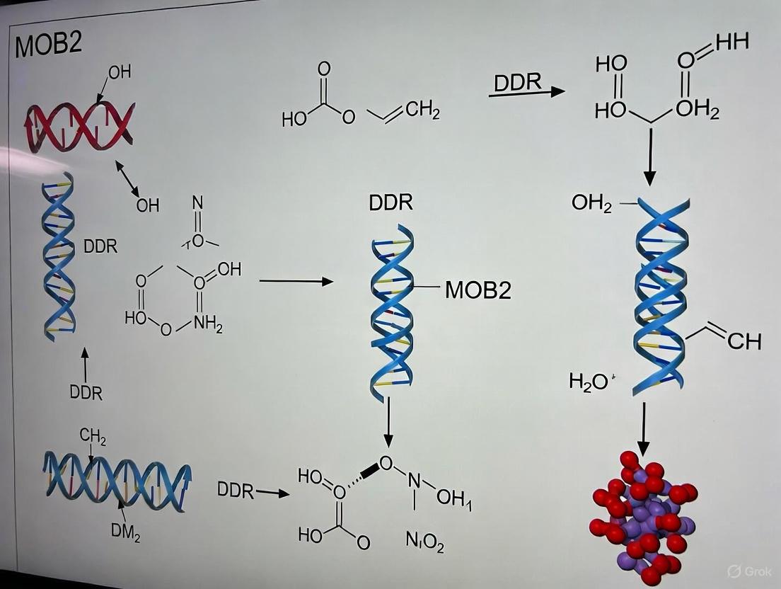
Figure 2: MOB2's role in DNA damage response. MOB2 interacts with RAD50 to facilitate recruitment of the MRN complex and activated ATM to DNA damage sites, promoting DDR signaling and cellular survival.
This mechanism explains how hMOB2 supports DDR signaling and cellular survival after DNA damage induction. Through its interaction with RAD50, MOB2 enhances the efficiency of DNA damage sensing and repair, thereby promoting genomic stability.
Cancer Relevance
The significance of MOB2 in maintaining genomic integrity directly links it to cancer biology. The human MOB2 gene displays loss of heterozygosity (LOH) in more than 50% of bladder, cervical, and ovarian carcinomas according to The Cancer Genome Atlas (TCGA) [7]. This frequent loss in cancers suggests that MOB2 may function as a tumor suppressor, possibly through its roles in preventing DNA damage accumulation and ensuring proper DNA repair.
The DDR functions of MOB2, particularly its interaction with the MRN complex, position it as a potential biomarker for cancer therapies targeting DNA repair pathways, such as PARP inhibitors or other DDR-targeting agents currently in development [7] [8].
Experimental Protocols
Structural Characterization Methods
The molecular characterization of MOB2 has relied heavily on X-ray crystallography to determine high-resolution structures of MOB2 in complex with its binding partners. Key methodological considerations include:
Protein Expression and Purification:
- Heterologous expression in Escherichia coli systems [3] [4]
- Affinity chromatography followed by size-exclusion chromatography
- Selenomethionine incorporation for phasing [4]
Crystallization and Data Collection:
- Vapor diffusion methods with commercial screening kits
- Cryo-cooling with appropriate cryoprotectants
- X-ray diffraction data collection at synchrotron facilities
- Typical resolutions ranging from 2.80 Å to 3.30 Å for MOB2 complexes [3] [4]
Structure Determination:
- Molecular replacement using existing structures as search models
- Iterative model building and refinement
- Validation using MolProbity and related tools [3] [4]
Interaction Studies
Yeast Two-Hybrid Screening:
- Normalized universal human tissue cDNA library screening
- Bait construction: pLexA-N-hMOB2 (full-length)
- Screening of 1×10^6 transformants yielding 59 bait-dependent hits [7]
- Identification of 28 putative interactors, with RAD50 identified in-frame in all four hits [7]
Co-immunoprecipitation and Immunoblotting:
- Cell lysis in appropriate buffer conditions
- Antibody-coupled bead incubation for pull-down
- Western blotting with enhanced chemiluminescence detection [7]
Chromatin-Cytosol Separation:
- Cell fractionation using differential centrifugation
- Buffer A (cytosolic extraction) and Buffer B (chromatin extraction) [7]
- Validation of chromatin-enriched fractions using marker proteins
Functional Assays
DNA Damage Response Assays:
- Induction of DNA damage using doxorubicin or ionizing radiation
- Clonogenic survival assays to assess cell viability
- Comet assays to detect DNA strand breaks [7]
- Immunofluorescence for γH2AX foci quantification
Cell Cycle Analysis:
- Flow cytometry with propidium iodide staining
- Assessment of G1/S arrest following MOB2 depletion [7]
- p21 and p53 activation measurements
Research Reagent Solutions
Table 3: Essential Research Reagents for MOB2 Studies
| Reagent Category | Specific Examples | Applications | Key Features |
|---|---|---|---|
| Structural Biology | 5NCM, 4LQS, 5NCL PDB entries [3] [4] [5] | Molecular modeling, structure-function studies | High-resolution crystal structures of MOB2-kinase complexes |
| Cell Line Models | RPE1-hTert, BJ-hTert fibroblasts, U2-OS cells [7] | DDR functional studies, signaling analysis | hTert-immortalized non-cancerous models |
| Stable Cell Lines | Tetracycline-inducible (Tet-on) RPE1 cells [7] | Inducible knockdown or overexpression | Controlled gene expression, minimal basal leakage |
| Interaction Screening | pLexA-N-hMOB2 bait construct [7] | Yeast two-hybrid screening | Full-length hMOB2 as bait |
| DNA Damage Inducers | Doxorubicin, ionizing radiation [7] | DDR pathway activation | Controlled DNA damage induction |
| Chromatin Fractionation | Buffer A/B extraction system [7] | Subcellular localization studies | Separation of chromatin-bound proteins |
These research reagents represent essential tools for investigating MOB2 structure, function, and its role in DNA damage response pathways. The structural data available in the PDB provides a foundation for structure-guided experiments, while the cell models and biochemical tools enable functional characterization in physiological contexts.
MOB2 represents a phylogenetically conserved regulatory protein with a uniquely organized globular structure that facilitates specific protein-protein interactions, particularly with NDR family kinases. Its evolutionary conservation from yeast to humans underscores its fundamental importance in eukaryotic cell biology, primarily in morphogenesis and cell polarity pathways. The recent discovery of MOB2's role in DNA damage response through its interaction with the MRN complex expands our understanding of its functional repertoire and provides a mechanistic link to cancer biology, where MOB2 is frequently lost. The structural insights into MOB2-kinase complexes, coupled with emerging understanding of its DDR functions, position MOB2 as a potential therapeutic target or biomarker in cancer treatment strategies, particularly those leveraging synthetic lethal approaches against DNA repair-deficient cancers. Future research will likely focus on elucidating how MOB2 coordinates its canonical functions in morphogenesis with its more recently discovered roles in genome maintenance, potentially revealing novel nodes for therapeutic intervention.
MOB2 Interactions with NDR1/2 Kinases and Competition with MOB1
The Mps one binder (MOB) family of proteins represents a class of highly conserved eukaryotic scaffold proteins that function as crucial signal transducers in essential intracellular pathways, primarily through their regulatory interactions with serine/threonine protein kinases [9] [10]. Mammalian genomes encode at least six different MOB proteins (MOB1A, MOB1B, MOB2, MOB3A, MOB3B, and MOB3C), indicating significant functional diversification from unicellular to complex multicellular organisms [7] [11]. MOB proteins typically lack enzymatic activity and instead operate as globular scaffold proteins that influence cellular signaling through specific protein-protein interactions [10].
The Nuclear Dbf2-related (NDR) kinases (NDR1/STK38 and NDR2/STK38L in mammals) belong to the NDR/LATS subfamily of AGC kinases and function as essential regulators of diverse cellular processes including cell cycle progression, transcription, apoptosis, and stem cell differentiation [9] [12]. These kinases serve as core components of the evolutionarily conserved Hippo signaling pathway, which controls organ size and tissue homeostasis by regulating cell proliferation and apoptosis [12] [10]. The functional interplay between MOB proteins and NDR kinases represents a critical regulatory node in cellular signaling networks with implications for cancer biology, DNA damage response, and neuronal development.
Table: Core Components of MOB-NDR Signaling Network
| Component | Classification | Key Functions | Representative Family Members |
|---|---|---|---|
| MOB Proteins | Scaffold proteins | Regulate NDR/LATS kinase activity | MOB1A/B, MOB2, MOB3A/B/C |
| NDR Kinases | Serine/Threonine kinases | Cell cycle regulation, DNA damage response, morphogenesis | NDR1/STK38, NDR2/STK38L |
| LATS Kinases | Serine/Threonine kinases | Core Hippo pathway components, tumor suppression | LATS1, LATS2 |
Molecular Mechanism of MOB2-NDR1/2 Interaction and MOB Competition
Structural Basis of MOB2-NDR1/2 Binding
MOB2 interacts specifically with NDR1/2 kinases through a conserved N-terminal regulatory domain shared by NDR/LATS kinases [9] [13]. This interaction is highly specific, as MOB2 binds exclusively to NDR kinases but not to the related LATS kinases in mammalian cells [9] [7]. Structural analyses reveal that MOB proteins adopt a conserved globular fold that enables them to dock onto the N-terminal domain of NDR kinases, thereby modulating their catalytic activity and substrate accessibility [10]. The binding interface between MOB2 and NDR kinases involves conserved hydrophobic patches and electrostatic interactions that ensure specificity within this signaling module.
MOB2-MOB1 Competition Mechanism
The competitive relationship between MOB2 and MOB1 for NDR binding represents a crucial regulatory switch in NDR kinase signaling. Both MOB1 and MOB2 target the same binding domain on NDR1/2 kinases, creating a mutually exclusive binding scenario [9] [13]. This competition has significant functional consequences, as the MOB1-NDR and MOB2-NDR complexes are associated with opposing effects on kinase activity:
- The MOB1/NDR complex is characterized by enhanced NDR kinase activity and promotes downstream signaling associated with cell cycle progression and Hippo pathway regulation [9] [7].
- In contrast, the MOB2/NDR complex is associated with diminished NDR kinase activity, effectively blocking NDR activation under certain cellular contexts [9] [7] [13].
This competitive binding establishes a yin-yang relationship between MOB1 and MOB2, allowing cells to fine-tune NDR kinase signaling in response to varying physiological conditions and cellular stresses.
Diagram: Competitive Binding of MOB1 and MOB2 to NDR Kinases
MOB2 in DNA Damage Response and Cell Cycle Regulation
MOB2's Role in Genome Stability Maintenance
Beyond its regulatory function in NDR kinase signaling, MOB2 plays a crucial and independent role in maintaining genome stability through the DNA damage response (DDR) pathway. Under normal growth conditions, MOB2 prevents the accumulation of endogenous DNA damage, thereby avoiding undesired activation of cell cycle checkpoints [9] [7]. When MOB2 expression is knocked down, cells exhibit significant accumulation of DNA damage even in the absence of exogenously induced genotoxic stress, leading to activation of the ATM-CHK2 DNA damage signaling pathway [9]. This accumulation of DNA damage triggers a p53/p21-dependent G1/S cell cycle arrest, effectively halting cellular proliferation until damage can be repaired [7] [11].
The functional significance of this role becomes particularly evident when comparing MOB2 depletion with NDR kinase manipulations. While MOB2 knockdown induces a robust G1/S cell cycle arrest, similar effects are not observed upon individual knockdown of NDR1 or NDR2, suggesting that MOB2's function in DDR operates through mechanisms largely independent of its regulation of NDR kinases [9] [7].
MOB2 in DNA Damage Sensing and Repair
Upon exposure to exogenous DNA-damaging agents such as ionizing radiation (IR) or doxorubicin, MOB2 becomes essential for optimal cellular response to genotoxic stress. MOB2 promotes cell survival following DNA damage, supports appropriate cell cycle arrest at G1/S checkpoint, and facilitates efficient DNA damage signaling [7] [11]. These functions are mediated through a novel interaction between MOB2 and RAD50, a critical component of the MRE11-RAD50-NBS1 (MRN) complex that serves as a primary sensor of DNA double-strand breaks [7] [11].
This MOB2-RAD50 interaction facilitates the recruitment of the MRN complex and activated ATM (ataxia-telangiectasia mutated) kinase to sites of DNA damage on chromatin, enhancing the initial detection and signaling cascade following genotoxic insult [7]. Recent research has further elucidated that MOB2 specifically regulates homologous recombination (HR)-mediated DNA repair by supporting the phosphorylation and accumulation of the RAD51 recombinase on resected single-strand DNA overhangs [14].
Table: MOB2 Functions in DNA Damage Response
| Cellular Context | MOB2 Function | Key Interactors | Downstream Consequences |
|---|---|---|---|
| Normal Conditions | Prevents endogenous DNA damage accumulation | RAD50, MRN complex | Maintains genome stability, prevents aberrant cell cycle arrest |
| Exogenous DNA Damage | Promotes damage sensing and signaling | ATM, MRN complex | Facilitates cell survival, appropriate cell cycle checkpoints |
| DNA Repair | Supports homologous recombination | RAD51, MRN complex | Ensures accurate DNA double-strand break repair |
Experimental Approaches for Studying MOB2-NDR Interactions
Methodologies for Protein-Protein Interaction Analysis
The investigation of MOB2 interactions with NDR kinases and other binding partners employs a multifaceted experimental approach combining biochemical, genetic, and cellular techniques. Yeast two-hybrid (Y2H) screening has been instrumental in identifying novel MOB2 binding partners, including RAD50 [7]. This method involves cloning full-length MOB2 into a pLexA bait vector and screening against a normalized universal human tissue cDNA library (typically with complexity >2.8×10⁶ clones), followed by validation of bait-dependent interactions through auxotrophic selection and β-galactosidase assays [7].
Co-immunoprecipitation (co-IP) assays provide critical validation of interactions in mammalian cells. The standard protocol involves transfection of epitope-tagged MOB2 and potential binding partners (e.g., NDR1/2, RAD50) into appropriate cell lines (such as COS-7, 293T, or U2-OS), followed by lysis in non-denaturing buffers (typically containing 50 mM Tris-HCl pH 7.5, 150 mM NaCl, 1 mM EDTA, 0.5% NP-40, and protease/phosphatase inhibitors) [7] [13]. Immunoprecipitation using tag-specific or protein-specific antibodies coupled with protein A/G beads is followed by extensive washing and immunoblotting analysis to detect co-precipitated proteins.
Chromatin fractionation assays have been particularly valuable for studying MOB2's role in DNA damage response. This technique involves sequential extraction of cellular components using differential detergent-containing buffers to separate cytosolic, nucleoplasmic, and chromatin-bound protein fractions, allowing researchers to monitor recruitment of MOB2 and associated proteins (MRN complex, ATM) to DNA damage sites [7].
Functional Characterization Techniques
RNA interference approaches utilizing sequence-specific siRNAs or shRNAs have been extensively employed to dissect MOB2 functions. Stable knockdown cell lines can be generated using lentiviral delivery of shRNA constructs followed by puromycin selection (typically 1.0 μg/ml for 2 weeks) [13] [15]. For CRISPR/Cas9-mediated knockout, single-guide RNAs targeting MOB2 (e.g., 5'-AGAAGCCCGCTGCGGAGGAG-3') are cloned into lentiCRISPRv2 vectors and transduced into target cells [13].
Clonogenic survival assays assess the functional consequences of MOB2 manipulation on cellular response to DNA-damaging agents. Cells with modulated MOB2 expression are treated with ionizing radiation or chemotherapeutic agents (e.g., doxorubicin), plated at low density, and allowed to form colonies for 10-14 days before fixation, staining (with crystal violet or methylene blue), and counting [7] [11].
Cell cycle analysis through flow cytometry and immunoblotting of cell cycle regulators (p53, p21, cyclin D1) has been crucial for establishing MOB2's role in G1/S checkpoint control [7]. Additional functional assays including wound healing, Transwell migration/invasion, and comet assays for DNA strand break detection provide comprehensive assessment of MOB2's cellular functions [13] [15].
Diagram: Experimental Approaches for MOB2-NDR Research
Research Reagent Solutions Toolkit
Table: Essential Research Reagents for MOB2-NDR Studies
| Reagent/Category | Specific Examples | Application/Function | Experimental Context |
|---|---|---|---|
| Cell Lines | RPE1-hTert, U2-OS, BJ-hTert fibroblasts, SMMC-7721 | Model systems for DDR studies, transformation assays | [7] [11] [13] |
| Expression Vectors | pT-Rex-HA-NDR1-PIF, pTER shRNA, pLXSN, lentiCRISPRv2 | Ectopic expression, inducible knockdown/knockout | [7] [13] [15] |
| Antibodies | Anti-MOB2, anti-NDR1/2, anti-RAD50, anti-phospho-ATM | Detection, immunoprecipitation, chromatin recruitment | [7] [11] |
| Chemical Inhibitors/Activators | Doxorubicin, Forskolin (cAMP activator), H89 (PKA inhibitor) | Induce DNA damage, modulate signaling pathways | [7] [15] |
| Selection Agents | Puromycin (1.0 μg/ml), Blasticidin, G418 | Maintain stable cell lines, select for transduced cells | [7] [13] |
The interaction between MOB2 and NDR1/2 kinases, and particularly its competition with MOB1 for NDR binding, represents a sophisticated regulatory mechanism for controlling NDR kinase activity and its diverse cellular functions. While the MOB2-NDR interaction clearly modulates kinase signaling, MOB2 has additionally evolved NDR-independent functions, most notably in DNA damage response through its interaction with the RAD50 component of the MRN complex.
The dual functionality of MOB2 positions it as a critical node at the intersection of cell cycle regulation, DNA damage response, and cancer biology. Evidence suggests that MOB2 expression is frequently lost in various cancers, including glioblastoma, ovarian, bladder, and cervical carcinomas, supporting its potential role as a tumor suppressor [7] [15]. The recent discovery that MOB2 deficiency sensitizes cancer cells to PARP inhibitors further highlights the translational potential of understanding MOB2 functions in DNA repair pathways [14].
Future research directions should focus on obtaining high-resolution structural data of MOB2 in complex with NDR kinases and RAD50, elucidating the precise molecular mechanisms by which MOB2 regulates homologous recombination repair, and exploring the therapeutic potential of targeting MOB2-mediated pathways in cancer treatment. The competitive binding relationship between MOB1 and MOB2 for NDR kinases may represent a tunable regulatory switch that could be exploited for therapeutic intervention in cancers with dysregulated DNA damage response pathways.
The MRE11-RAD50-NBS1 (MRN) complex serves as a primary sensor for DNA double-strand breaks (DSBs), orchestrating critical early responses to genomic insult. Recent research has uncovered that Mps one binder 2 (MOB2), a protein previously studied primarily in cell cycle and morphological contexts, forms a critical partnership with RAD50, a core component of the MRN complex. This in-depth technical review examines the mechanistic basis and functional consequences of the MOB2-RAD50 interaction, framing it within the broader DNA damage response (DDR) role of MOB2. We synthesize key findings from foundational studies, present quantitative data in structured formats, detail experimental methodologies, and visualize signaling pathways to provide researchers and drug development professionals with a comprehensive resource on this emerging DDR axis. The partnership represents a potentially targetable interface for therapeutic intervention in cancers displaying MRN complex dysfunction.
MOB proteins constitute an evolutionarily conserved family of signal transducers that regulate serine/threonine kinases of the NDR/LATS family [7] [9]. While MOB1 has established roles in Hippo signaling and MOB3 interacts with the pro-apoptotic kinase MST1, MOB2 has remained somewhat enigmatic [7]. Initial biochemical characterization revealed that human MOB2 (hMOB2) specifically interacts with NDR1/2 kinases, competing with MOB1 for NDR binding and potentially antagonizing NDR activation [7] [9]. However, biological functions extending beyond this biochemical activity were poorly defined until recent investigations positioned MOB2 within the DDR landscape.
A genome-wide screen for novel DDR factors initially identified MOB2 as a candidate protein [7] [11], prompting systematic investigation into its potential roles in genome maintenance. Subsequent work has established that MOB2 promotes DDR signaling, cell survival, and cell cycle arrest following exogenously induced DNA damage [7]. Under basal conditions, MOB2 prevents accumulation of endogenous DNA damage and consequent p53/p21-dependent G1/S cell cycle arrest [7] [9]. Surprisingly, these functions appear independent of NDR kinase signaling, suggesting MOB2 operates through alternative mechanisms in DDR contexts [7] [9].
The pivotal mechanistic insight emerged from the discovery that MOB2 directly interacts with RAD50, a core component of the MRN complex [7]. This partnership facilitates recruitment of the complete MRN complex and activated ATM to DNA damaged chromatin, positioning MOB2 as a novel regulator of initial DNA damage sensing and signaling [7].
The MRN Complex: Architecture and DSB Response Functions
Structural Organization
The MRN complex is a hetero-hexameric assembly comprising two subunits each of MRE11, RAD50, and NBS1 [16]. Its architecture underlies diverse functions in DNA damage sensing, signaling, and repair:
- MRE11: Contains N-terminal nuclease motifs, a RAD50-binding domain, and DNA-binding domains (DBDs) that confer 3'-5' exonuclease and endonuclease activities essential for DNA end resection [16].
- RAD50: A structural maintenance of chromosomes (SMC) family protein with Walker A/B nucleotide-binding domains forming ABC-ATPase modules. These domains connect via long antiparallel coiled-coils terminating in a CxxC "zinc-hook" motif that mediates inter-complex tethering [17] [16].
- NBS1: An eukaryotic adaptor subunit containing forkhead-associated (FHA) and BRCT domains that mediate phospho-protein interactions, plus MRE11- and ATM-binding motifs that coordinate downstream signaling [16].
The complex exhibits ATP-dependent conformational dynamics, transitioning between open and closed states that regulate DNA access and nuclease activities [16]. Recent evidence indicates that MRN components form higher-order oligomers mediated through RAD50 head domain interactions, facilitating focal accumulation at DSBs and enabling processive resection [18].
Functional Roles in DSB Response
The MRN complex executes multiple critical functions in DSB response pathways:
- Damage Sensing and ATM Activation: MRN is recruited rapidly to DSBs, where it activates ATM through NBS1-mediated interactions. ATM then phosphorylates numerous downstream targets to initiate cell cycle checkpoints and amplify damage signaling [17] [16].
- DNA End Resection: MRE11 nuclease activity, stimulated by RAD50 and ATP, processes DNA ends to generate 3' single-stranded DNA overhangs required for homologous recombination repair [17] [16].
- DNA Tethering and Bridging: RAD50 coiled-coil domains mediate intra- and inter-complex interactions that bridge DNA ends over distances up to 1200 Å, maintaining genomic integrity during repair [18] [16].
Table 1: MRN Complex Components and Their Functions
| Component | Key Domains | Primary Functions | Associated Human Diseases |
|---|---|---|---|
| MRE11 | Nuclease domain, DNA-binding domains, RAD50-binding domain | DNA end resection (exonuclease/endonuclease), DNA binding | Ataxia-telangiectasia-like disorder (ATLD) |
| RAD50 | Walker A/B ATPase domains, coiled-coil region, zinc-hook | ATP hydrolysis, DNA tethering, complex scaffolding | Nijmegen breakage syndrome-like disorder |
| NBS1 | FHA domain, BRCT domains, MRE11-binding, ATM-binding | Protein recruitment, ATM activation, signaling integration | Nijmegen breakage syndrome (NBS) |
MOB2-RAD50 Partnership: Molecular Mechanism and Functional Impact
Discovery of Direct Interaction
The connection between MOB2 and RAD50 was established through a yeast two-hybrid (Y2H) screen employing full-length hMOB2 as bait against a normalized human tissue cDNA library [7]. This screen identified multiple specific interactors, with RAD50 emerging as a statistically significant hit with all four isolated RAD50 clones appearing in-frame [7]. Follow-up co-immunoprecipitation experiments validated the interaction between exogenously and endogenously expressed proteins, confirming the physiological relevance of this partnership [7] [9].
Interaction mapping revealed that MOB2 binds two functionally relevant domains within RAD50, suggesting a regulatory rather than scaffolding role [9]. However, identification of discrete point mutations that specifically disrupt MOB2-RAD50 binding while preserving other MOB2 functions has proven challenging, limiting definitive functional assignment [9].
Functional Consequences for MRN Complex Activity
MOB2 depletion impairs multiple MRN-dependent processes, positioning MOB2 as a facilitator of MRN complex functionality:
- MRN and ATM Recruitment: MOB2 supports recruitment of both the MRN complex and activated ATM to DNA damaged chromatin. MOB2-deficient cells show defective ATM activation and impaired accumulation of MRN components at damage sites [7] [9].
- DSB Signaling and Repair: MOB2 promotes cell survival following DSB-inducing agents like ionizing radiation and doxorubicin. MOB2-depleted cells display heightened sensitivity to these treatments, indicating compromised DSB repair [7] [11].
- Endogenous Genomic Stability: Under unperturbed conditions, MOB2 prevents accumulation of endogenous DNA damage, as evidenced by increased γH2AX foci and activation of the ATM-CHK2 pathway in MOB2-knockdown cells [7] [9].
Table 2: Phenotypic Consequences of MOB2 Manipulation in Cellular Models
| Experimental Condition | Cell Survival | DDR Signaling | Cell Cycle Progression | Endogenous DNA Damage |
|---|---|---|---|---|
| MOB2 Knockdown (unperturbed) | Unaffected | ATM-CHK2 activation | p53/p21-dependent G1/S arrest | Increased γH2AX foci |
| MOB2 Knockdown + DNA damage | Decreased | Impaired ATM activation, reduced γH2AX | Defective damage-induced G1/S arrest | Not applicable |
| MOB2 Overexpression | Unaffected or slightly enhanced | Enhanced MRN/ATM recruitment | Unaffected under normal conditions | Reduced baseline damage |
The following diagram illustrates the molecular relationship between MOB2 and the MRN complex in the DNA damage response:
Figure 1: MOB2-MRN Pathway in DNA Damage Response. MOB2 interacts with RAD50 and facilitates MRN complex recruitment to DNA double-strand breaks, promoting ATM activation and subsequent DDR signaling.
Experimental Approaches and Research Reagent Solutions
Key Methodologies for Investigating MOB2-RAD50 Interaction
Yeast Two-Hybrid Screening
The initial MOB2-RAD50 interaction was identified using a comprehensive Y2H approach:
- Bait Construction: Full-length hMOB2 was cloned into pLexA vector to create a DNA-binding domain fusion.
- Library Screening: A normalized universal human tissue cDNA library (complexity: 2.8×10^6 clones) in pGADT7-recAB was screened against the MOB2 bait.
- Hit Validation: Screening of 1×10^6 transformants yielded 59 bait-dependent hits, with RAD50 identified repeatedly among 28 putative interactors (all four RAD50 hits were in-frame) [7].
Functional DDR Assays
Clonogenic Survival Assays:
- Cells are plated at fixed densities and treated with DNA damaging agents (doxorubicin or ionizing radiation).
- Following treatment, cells are allowed to form colonies for 10-14 days before staining and quantification.
- MOB2-depleted U2-OS cells show significantly reduced survival following DNA damage [7] [11].
Immunofluorescence Microscopy:
- Cells are fixed and stained with antibodies against γH2AX, RAD50, MRE11, or phosphorylated ATM.
- Focus formation is quantified manually or using automated image analysis.
- MOB2 deficiency reduces MRN and activated ATM foci at damage sites [7].
Chromatin Fractionation:
- Cells are lysed in hypotonic Buffer A (10 mM Pipes, 100 mM NaCl, 300 mM sucrose, 3 mM MgCl2, 0.1% Triton X-100).
- Cytosolic and chromatin fractions are separated by differential centrifugation.
- Chromatin-bound proteins are solubilized in high-pH Buffer B (3 mM EDTA, 0.2 mM EGTA).
- MOB2 depletion reduces RAD50 and MRE11 association with chromatin following damage [7].
Essential Research Reagents
Table 3: Key Reagents for MOB2-MRN Research
| Reagent/Cell Line | Specific Application | Function/Utility | Source/Reference |
|---|---|---|---|
| RPE1-hTert Tet-on cells | Generation of inducible knockdown | Near-diploid untransformed retinal pigment epithelial cells for DDR studies | [7] |
| U2-OS cells | Clonogenic survival assays | Osteosarcoma cell line with robust colony-forming ability | [7] [11] |
| pLexA-N-hMOB2(FL) | Yeast two-hybrid bait | Full-length MOB2 fusion with DNA-binding domain for interaction screening | [7] |
| pT-Rex-HA-NDR1-PIF | NDR kinase studies | Tetracycline-inducible expression of hyperactive NDR1 | [7] [9] |
| Anti-RAD50 antibodies | Immunoprecipitation, Western blot, immunofluorescence | Detection of endogenous RAD50 expression and localization | [7] |
| Anti-γH2AX antibodies | DNA damage quantification | Marker for DNA double-strand breaks | [7] [9] |
MOB2 in Cancer Biology and Therapeutic Implications
MOB2 as a Tumor Suppressor
Emerging evidence supports a tumor suppressive role for MOB2 beyond its DDR functions. Bioinformatic analyses of The Cancer Genome Atlas (TCGA) data reveal loss of heterozygosity (LOH) at the MOB2 locus in >50% of bladder, cervical, and ovarian carcinomas [7] [15]. In glioblastoma (GBM), MOB2 expression is significantly downregulated at both mRNA and protein levels compared to low-grade gliomas and normal brain tissue [15]. Clinically, low MOB2 expression correlates with poor prognosis in glioma patients [15].
Functional studies demonstrate that MOB2 overexpression suppresses, while its depletion enhances, malignant phenotypes in GBM models, including clonogenic growth, migration, invasion, and in vivo metastasis [15]. These effects involve MOB2-mediated regulation of FAK/Akt and cAMP/PKA signaling pathways, indicating pleiotropic tumor-suppressive mechanisms [15].
Therapeutic Opportunities
The MOB2-MRN interface presents several potential therapeutic avenues:
- Synthetic Lethality: Tumors with underlying MRN complex deficiencies may exhibit heightened dependence on MOB2 function, creating potential synthetic lethal interactions.
- Predictive Biomarker: MOB2 expression status may predict response to DNA-damaging chemotherapies and radiotherapy, enabling treatment stratification.
- Targeted Combination Therapies: Small molecules modulating MOB2-RAD50 interaction could sensitize tumors to conventional genotoxic treatments.
The partnership between MOB2 and RAD50 represents a significant expansion of our understanding of both MOB protein biology and MRN complex regulation. MOB2 facilitates MRN recruitment to DNA damage sites and supports subsequent ATM activation, positioning it as a novel regulator of initial DNA damage sensing. This function appears genetically separable from MOB2's previously described role in NDR kinase regulation, suggesting context-dependent protein interactions.
Key outstanding questions merit further investigation:
- Structural characterization of the MOB2-RAD50 interface to guide targeted therapeutic development.
- Identification of specific post-translational modifications regulating MOB2-RAD50 complex formation.
- In vivo validation of MOB2's tumor suppressive functions in genetically engineered models.
- Comprehensive analysis of MOB2 mutations and expression across cancer types to establish clinical relevance.
The MOB2-MRN connection exemplifies how systematic investigation of novel protein interactions can uncover unexpected regulatory nodes in core biological processes. As research continues to delineate this partnership, opportunities for therapeutic exploitation in cancer and other genomic instability disorders will likely emerge.
MOB2 in Cell Cycle Regulation and G1/S Checkpoint Activation
Mps one binder 2 (MOB2) is an emerging critical regulator of genomic integrity, functioning at the nexus of cell cycle progression and DNA damage response (DDR) signaling. Recent research has established that MOB2 plays dual roles in maintaining cell cycle progression under normal conditions and activating checkpoint signaling following DNA damage. Through its interaction with the MRE11-RAD50-NBS1 (MRN) complex, MOB2 facilitates the recruitment of key DDR components to DNA lesions, thereby promoting efficient damage sensing and repair. Loss of MOB2 function results in accumulated endogenous DNA damage, triggering p53/p21-dependent G1/S cell cycle arrest. This in-depth technical review synthesizes current understanding of MOB2's molecular mechanisms, experimental approaches for its study, and implications for therapeutic development in cancer and other diseases associated with genomic instability.
The MOB (Mps one binder) protein family represents highly conserved regulators of essential signaling pathways from yeast to humans. While MOB1 has established roles in Hippo signaling and tumor suppression, MOB2 has remained more enigmatic despite its conservation throughout evolution. Human MOB2 (hMOB2) was initially characterized biochemically as an inhibitor of NDR (Nuclear Dbf2-related) kinases through competitive binding with hMOB1. However, the physiological relevance of this interaction remained unclear until recent investigations revealed MOB2's significant functions in maintaining genomic stability [7] [9].
A genome-wide screen for novel DDR factors identified MOB2 as a potential player in DNA damage management, prompting detailed investigation into its cellular functions. Subsequent research has established that MOB2 promotes DDR signaling, cell survival, and cell cycle arrest following exogenously induced DNA damage. Under normal growth conditions, MOB2 prevents the accumulation of endogenous DNA damage and subsequent activation of p53/p21-dependent G1/S cell cycle checkpoints. These functions appear independent of its previously characterized NDR regulatory role, suggesting novel mechanisms of action [7].
Molecular Mechanisms of MOB2 Action
MOB2 Structure and Binding Partners
MOB proteins function primarily as signal transducers through regulatory interactions with serine/threonine protein kinases of the NDR/LATS family. Mammalian genomes encode at least six different MOB genes (MOB1A, MOB1B, MOB2, MOB3A, MOB3B, MOB3C), indicating functional diversification in higher organisms. While MOB1 interacts with both LATS and NDR kinases, MOB2 displays specific binding to NDR kinases only. MOB3 proteins associate with the pro-apoptotic kinase MST1 rather than NDR/LATS kinases [7] [9].
Biochemically, MOB2 competes with MOB1 for NDR binding, with the MOB1/NDR complex associated with increased NDR kinase activity, while MOB2 binding to NDR blocks NDR activation. This competition mechanism initially suggested that MOB2 might function primarily as a negative regulator of NDR signaling. However, recent evidence indicates that MOB2's roles in DDR and cell cycle regulation operate through NDR-independent pathways [7] [9].
MOB2-RAD50 Interaction and MRN Complex Recruitment
A critical breakthrough in understanding MOB2's DDR functions came from a yeast two-hybrid screen that identified RAD50 as a novel MOB2 binding partner. RAD50 is a central component of the MRN (MRE11-RAD50-NBS1) DNA damage sensor complex, which is essential for the recruitment and activation of the ataxia-telangiectasia mutated (ATM) kinase at DNA double-strand breaks [7].
Table 1: MOB2 Protein Interactions and Functional Consequences
| Binding Partner | Interaction Type | Functional Outcome | Dependence |
|---|---|---|---|
| NDR1/2 kinases | Competitive with MOB1 | Inhibits NDR activation | Not essential for DDR role |
| RAD50 | Direct interaction | Facilitates MRN complex recruitment | Essential for DDR signaling |
| MRN complex | Indirect via RAD50 | Promotes ATM activation at damage sites | Critical for G1/S checkpoint |
| UBR5, KPNB1, KIAA0226L | Yeast two-hybrid hits | Potential novel functions | Under investigation |
The MOB2-RAD50 interaction facilitates the recruitment of the entire MRN complex and activated ATM to DNA damaged chromatin. This mechanism explains how MOB2 supports DDR signaling and checkpoint activation. Mapping studies have identified two functionally relevant domains on RAD50 that mediate interaction with MOB2, though specific point mutations disrupting this interaction have proven difficult to generate, limiting definitive functional validation [7] [9].
MOB2 in Cell Cycle Checkpoint Control
MOB2 plays distinct yet complementary roles in cell cycle regulation under normal conditions versus following DNA damage:
- Under normal conditions: MOB2 prevents accumulation of endogenous DNA damage, thereby avoiding inappropriate activation of p53/p21-dependent G1/S cell cycle checkpoints.
- After exogenous DNA damage: MOB2 promotes proper activation of cell cycle checkpoints, particularly G1/S arrest, and supports cell survival.
The G1/S arrest observed in MOB2-depleted cells is functionally dependent on p53 and p21, as co-knockdown of p53 or p21 together with MOB2 abrogates the cell cycle arrest and restores proliferation capacity. This places MOB2 upstream of p53 activation in the DDR signaling cascade [7] [9].
Experimental Evidence and Methodologies
Key Experimental Approaches
Investigating MOB2's roles in DDR and cell cycle regulation requires multidisciplinary approaches. The following methodologies have proven essential in characterizing MOB2 functions:
Yeast Two-Hybrid Screening for Novel Binding Partners A normalized universal human tissue cDNA library was screened using pLexA-N-hMOB2 (full-length) as bait. The pGADT7-recAB-based cDNA library had a complexity of 2.8 × 10^6 with an average insert size of 1.58 kb. Screening of 1 × 10^6 transformants yielded 59 bait-dependent hits, identifying 28 putative interactors. RAD50 was identified as a novel MOB2 binding partner with all four RAD50 hits in-frame, confirming a direct protein-protein interaction [7].
Chromatin-Cytosol Fractionation Studies To examine MOB2's role in recruiting DDR components to chromatin, researchers developed a protocol for separating chromatin-bound from cytosolic proteins. Cells were harvested with ice-cold PBS, resuspended in buffer A (10 mM Pipes, 100 mM NaCl, 300 mM sucrose, 3 mM MgCl₂, 5 mM EDTA, 1 mM EGTA, 50 mM NaF, 0.1 mM Na₃VO₄, 0.1% Triton X-100, and protease inhibitors at pH 6.8), incubated for 10 minutes, then centrifuged at 1,300 × g for 5 minutes at 4°C. The supernatant was collected as the cytosolic fraction. The pellet was washed with buffer A, then lysed in buffer B (3 mM EDTA, 0.2 mM EGTA, and protease inhibitors at pH 8.0) for 10 minutes at 4°C, followed by centrifugation at 1,700 × g for 5 minutes at 4°C to collect the chromatin-containing supernatant [7].
Single Cell Network Profiling (SCNP) for DDR Assessment SCNP using multiparametric flow cytometry enables quantitative measurement of DNA damage repair functionality at single-cell resolution. This method involves exposing cells to genotoxic agents (e.g., etoposide, PARP inhibitors) and measuring activation of multiple DDR readouts through intracellular staining of phosphorylated DDR proteins (p-H2AX, p-ATM, p-DNA-PKcs, p-53BP1, p-RPA2/32, p-BRCA1, p-p53) and cell cycle markers (CyclinA2 for S/G2/M phases). Cells are stained with amine aqua viability dye, fixed with 1.6% paraformaldehyde, permeabilized with 100% ice-cold methanol, then stained with antibody cocktails for flow cytometry analysis. This approach can distinguish functional deficiencies in HRR and NHEJ pathways and detect haploinsufficiency as in BRCA1+/- cells [19].
Quantitative Data on MOB2 Knockdown Effects
Table 2: Functional Consequences of MOB2 Depletion in Human Cells
| Experimental Condition | Phenotype Observed | Molecular Markers Altered | Rescue Approach |
|---|---|---|---|
| MOB2 knockdown (no induced damage) | Accumulated endogenous DNA damage | Increased γH2AX, p-ATM, p-CHK2 | p53/p21 co-knockdown |
| MOB2 knockdown + IR/doxorubicin | Reduced cell survival | Impaired ATM activation, defective MRN recruitment | Expression of wild-type MOB2 |
| MOB2 knockdown | G1/S cell cycle arrest | Increased p53, p21; decreased Cyclin D1, c-myc | p53 or p21 co-knockdown |
| MOB2 overexpression | Enhanced DDR signaling | Improved MRN recruitment, ATM activation | Not applicable |
The quantitative data demonstrate that MOB2 is required for efficient DDR and proper cell cycle progression. Notably, MOB2 manipulations produce phenotypes distinct from NDR1/2 manipulations, supporting NDR-independent functions for MOB2 in DDR and cell cycle regulation [7] [9].
Signaling Pathways and Molecular Relationships
The following diagram illustrates the molecular relationships between MOB2, the MRN complex, and DDR activation:
MOB2-dependent DDR activation pathway
The Scientist's Toolkit: Essential Research Reagents
Table 3: Key Research Reagents for MOB2 and DDR Studies
| Reagent/Tool | Specific Example | Application | Function in Experiment |
|---|---|---|---|
| siRNA/shRNA | Qiagen siRNAs, pTER shRNA constructs | MOB2 knockdown | Loss-of-function studies to determine MOB2 requirements |
| Stable cell lines | RPE1-hTert Tet-on inducible systems | Controlled gene expression | Doxycycline-inducible expression of wild-type or mutant MOB2 |
| DNA damage agents | Doxorubicin, Etoposide, IR | Induce DNA damage | Activate DDR pathways to study MOB2 role in response |
| DDR markers | p-H2AX, p-ATM, p-CHK2 antibodies | Immunoblotting, immunofluorescence | Quantify DNA damage and DDR activation |
| Cell cycle markers | Cyclin A2, p21, p53 antibodies | Flow cytometry, immunoblotting | Assess cell cycle position and checkpoint activation |
| Yeast two-hybrid | pLexA-N-hMOB2, pGADT7-recAB library | Protein interaction screening | Identify novel MOB2 binding partners |
| Chromatin fractionation | Buffer A/B extraction protocol | Subcellular localization | Determine protein recruitment to damaged chromatin |
Discussion and Research Implications
The emerging role of MOB2 in DDR and cell cycle regulation has significant implications for both basic biology and translational applications. MOB2 represents a novel class of DDR regulator that functions at the interface between damage sensing and cell cycle control. Its interaction with the MRN complex positions MOB2 as a critical facilitator of early DDR signaling, with particular importance for G1/S checkpoint maintenance.
From a therapeutic perspective, MOB2's functions suggest potential utility as a biomarker for cancer prognosis or prediction of response to DNA-damaging therapies. The observation that MOB2 gene displays loss of heterozygosity in more than 50% of bladder, cervical, and ovarian carcinomas (The Cancer Genome Atlas data) underscores its potential relevance in human cancer. Furthermore, understanding MOB2 status may help stratify patients for therapies targeting DDR pathways, such as PARP inhibitors [7] [9].
Several important questions remain unresolved. The precise molecular mechanism by which MOB2 enhances MRN recruitment to damage sites requires further elucidation. The potential coordination between MOB2's NDR-regulatory and DDR functions deserves investigation in physiological contexts. Additionally, cell-type specific functions of MOB2 and its regulation during development and disease progression represent fertile ground for future research.
MOB2 has emerged as a significant regulator of genomic stability through its dual functions in promoting efficient DNA damage response and maintaining normal cell cycle progression. Through its interaction with RAD50 and facilitation of MRN complex recruitment to DNA damage sites, MOB2 plays a crucial role in early DDR signaling that ultimately influences G1/S checkpoint activation. The experimental approaches and reagents outlined in this review provide researchers with the necessary tools to further investigate MOB2's functions and mechanisms. As research progresses, MOB2 may offer new opportunities for therapeutic intervention in cancer and other diseases characterized by genomic instability.
The Mps one binder 2 (MOB2) protein, a highly conserved component of eukaryotic signaling networks, plays a fundamental role in maintaining genomic integrity by preventing the accumulation of endogenous DNA damage. While initially characterized as an inhibitor of Nuclear Dbf2-related (NDR) kinases, recent research has uncovered DNA damage response (DDR) functions that operate independently of this canonical signaling pathway [7]. Under normal growth conditions without exogenously induced DNA damage, hMOB2 deficiency triggers accumulation of DNA damage and subsequent p53/p21-dependent G1/S cell cycle arrest [7] [20]. This baseline protective function establishes hMOB2 as a crucial guardian against endogenous genotoxic stress, with profound implications for cancer development and therapeutic targeting.
The significance of hMOB2 in human pathology is underscored by genomic analyses revealing loss of heterozygosity (LOH) in more than 50% of bladder, cervical, and ovarian carcinomas documented in The Cancer Genome Atlas (TCGA) [7] [15]. This whitepaper examines the molecular mechanisms through which hMOB2 prevents endogenous DNA damage accumulation, details experimental methodologies for investigating these functions, and explores the translational potential of these findings for cancer drug development.
Molecular Mechanisms of hMOB2 in DNA Damage Prevention
MRN Complex Recruitment and ATM Activation
hMOB2 directly interacts with RAD50, a core component of the MRE11-RAD50-NBS1 (MRN) DNA damage sensor complex [7]. This interaction facilitates the recruitment of both the MRN complex and activated ATM (ataxia-telangiectasia mutated) kinase to DNA damaged chromatin, initiating early DDR signaling cascades [7]. The MRN complex serves as a primary sensor for DNA double-strand breaks (DSBs), and hMOB2-mediated enhancement of its recruitment represents a crucial mechanism for maintaining genomic stability under basal conditions.
Figure 1: hMOB2-dependent DDR pathway. hMOB2 interacts with RAD50 to facilitate MRN complex recruitment and ATM activation at DNA damage sites, initiating downstream signaling for cell cycle checkpoint activation and DNA repair.
Homologous Recombination Repair Regulation
Beyond its role in damage sensing, hMOB2 directly regulates homologous recombination (HR) repair of DNA double-strand breaks [21] [20]. hMOB2 supports the phosphorylation and accumulation of the RAD51 recombinase on resected single-strand DNA (ssDNA) overhangs, a critical step in HR-mediated repair [20]. This function explains how hMOB2 protects cells from endogenous DNA damage accumulation, as defective HR repair leads to persistent DSBs that trigger cell cycle arrest and genomic instability.
NDR Kinase-Independent DDR Functions
Interestingly, many DDR functions of hMOB2 operate independently of its canonical role as an NDR kinase regulator [7]. While hMOB2 can inhibit NDR kinases by competing with hMOB1 for NDR binding, the molecular and cellular phenotypes observed in DNA damage accumulation and repair are not recapitulated by NDR manipulations alone [7]. This indicates that hMOB2 possesses kinase-independent functions in genome maintenance, potentially through its interactions with core DNA repair machinery like RAD50 and regulation of RAD51 nucleofilament formation.
Quantitative Analysis of hMOB2 Deficiency Phenotypes
Table 1: Cellular phenotypes associated with hMOB2 deficiency under normal growth conditions
| Phenotype | Experimental Readout | Magnitude of Effect | Functional Consequence |
|---|---|---|---|
| Endogenous DNA damage accumulation | γH2AX foci, comet assay | Significant increase | Persistent DNA double-strand breaks |
| DDR activation | p-ATM Ser1981, p-CHK2 | Elevated levels | Constitutive DDR signaling |
| Cell cycle arrest | p53/p21 upregulation | G1/S phase blockade | Impaired cell proliferation |
| Apoptotic sensitivity | Caspase activation, Annexin V | Enhanced apoptosis | Reduced cell viability |
| Chromatin recruitment of repair factors | RAD51 foci formation | Impaired accumulation | Defective homologous recombination |
Table 2: hMOB2-dependent DNA repair pathway activities
| Repair Pathway | hMOB2 Role | Key Molecular Interactions | Functional Outcome |
|---|---|---|---|
| Homologous Recombination (HR) | Promotes RAD51 loading on ssDNA | RAD51, RAD50, MRN complex | Error-free DSB repair |
| ATM Signaling Activation | Facilitates MRN complex recruitment | RAD50, ATM | Enhanced DSB sensing and signaling |
| Cell Cycle Checkpoints | Supports G1/S arrest after damage | p53, p21 | Cell cycle arrest for repair |
| NHEJ | Limited direct evidence | Not fully characterized | Potential regulatory role |
Experimental Protocols for hMOB2 DDR Analysis
hMOB2 Loss-of-Function Approaches
RNA Interference (RNAi) Knockdown
- Transfection: Use Lipofectamine RNAiMax with Qiagen-derived siRNAs at 10-50 nM concentration [7] [20]
- Validation: Confirm knockdown efficiency by immunoblotting 48-72 hours post-transfection with anti-hMOB2 antibodies [20]
- Controls: Include non-targeting siRNA and rescue experiments with siRNA-resistant hMOB2 expression constructs
Stable Knockdown Cell Lines
- Vector System: Utilize pSuper.retro.puro or pMKO.1 puro retroviral vectors for shRNA expression [7] [20]
- Selection: Apply puromycin (1-2 μg/mL) for 3-5 days post-infection to select stable pools [7]
- Validation: Verify persistent hMOB2 reduction by immunoblotting and quantitative RT-PCR
DNA Damage Assessment Methodologies
Immunofluorescence Analysis of DNA Damage Foci
- Fixation: Use 4% paraformaldehyde for 15 minutes at room temperature
- Permeabilization: Treat with 0.5% Triton X-100 in PBS for 10 minutes
- Staining: Incubate with primary antibodies against γH2AX (1:1000), RAD51 (1:500), and 53BP1 (1:1000) overnight at 4°C [20]
- Quantification: Count foci per nucleus in minimum 100 cells across three independent experiments
Comet Assay for DNA Strand Breaks
- Embedding: Suspend 1×10⁴ cells in 0.7% low-melting-point agarose
- Lysis: Immerse in alkaline lysis buffer (2.5M NaCl, 100mM EDTA, 10mM Tris, 1% Triton X-100, pH 10) for 1 hour at 4°C
- Electrophoresis: Run at 25V for 30 minutes in alkaline electrophoresis buffer (300mM NaOH, 1mM EDTA, pH >13)
- Analysis: Score tail moment using automated comet assay imaging software
Clonogenic Survival Assays
- Seeding: Plate 200-10,000 cells per dish depending on expected survival fraction
- Treatment: Expose to DNA damaging agents (bleomycin, mitomycin C, PARP inhibitors) for 24 hours [20]
- Incubation: Culture for 10-14 days to allow colony formation
- Staining and Counting: Fix with methanol:acetic acid (3:1), stain with 0.5% crystal violet, count colonies >50 cells
Chromatin Recruitment Studies
Chromatin-Cytosol Fractionation
- Harvesting: Collect cells with ice-cold PBS, centrifuge at 1,000 × g for 2 minutes at 4°C [7]
- Extraction: Resuspend in Buffer A (10 mM Pipes, 100 mM NaCl, 300 mM sucrose, 3 mM MgCl₂, 5 mM EDTA, 1 mM EGTA, 0.1% Triton X-100) for 10 minutes [7]
- Separation: Centrifuge at 1,300 × g for 5 minutes at 4°C; collect supernatant as cytosolic fraction [7]
- Chromatin Isolation: Lyse pellet in Buffer B (3 mM EDTA, 0.2 mM EGTA) for 10 minutes at 4°C, centrifuge at 1,700 × g for 5 minutes [7]
- Analysis: Process chromatin fractions for immunoblotting against RAD50, MRE11, NBS1, and hMOB2
The hMOB2-DNA Repair Interaction Network
Figure 2: hMOB2 interaction network in DNA damage prevention. hMOB2 coordinates with multiple DNA repair proteins (RAD50, RAD51) and complexes (MRN) to maintain genomic stability through enhanced damage sensing, repair, and signaling.
Research Reagent Solutions
Table 3: Essential research reagents for studying hMOB2 in DNA damage prevention
| Reagent Category | Specific Examples | Application & Function | Experimental Notes |
|---|---|---|---|
| hMOB2 Targeting siRNAs | Qiagen-designed sequences [7] | Loss-of-function studies | Sequences available upon request from original publications |
| hMOB2 Antibodies | Rabbit monoclonal anti-hMOB2 (Epitomics) [20] | Immunoblotting, immunofluorescence | Custom-produced for specific research |
| DNA Damage Markers | γH2AX, p-ATM Ser1981, p-CHK2 [7] [20] | Damage detection and signaling | Commercial antibodies available from multiple vendors |
| Repair Protein Antibodies | RAD50, MRE11, NBS1, RAD51 [7] [20] | Complex recruitment studies | Verify specificity for chromatin fractionation |
| DDR Inhibitors | KU-55933 (ATM), NU-7441 (DNA-PK) [20] | Pathway perturbation | Use at calibrated concentrations for specific inhibition |
| Cell Line Models | RPE1-hTert, U2OS, HCT116, ovarian cancer panels [7] [20] | In vitro functional studies | Include both transformed and non-transformed models |
Therapeutic Implications and Translational Potential
The role of hMOB2 in preventing endogenous DNA damage accumulation has significant implications for cancer therapy development. hMOB2-deficient cells show enhanced sensitivity to PARP inhibitors (olaparib, rucaparib, veliparib), similar to BRCA-deficient cells [21] [20]. This synthetic lethal relationship suggests that hMOB2 status could serve as a predictive biomarker for PARP inhibitor response, particularly in ovarian carcinomas where reduced MOB2 expression correlates with improved overall survival [20].
Additionally, hMOB2 deficiency sensitizes cancer cells to DNA crosslinking agents like mitomycin C and cisplatin [20], indicating that hMOB2 status may inform therapeutic strategies across multiple DNA-damaging chemotherapeutics. The development of standardized assays to evaluate hMOB2 functional status in clinical samples could enable better patient stratification and personalized treatment approaches.
hMOB2 serves a critical baseline role in preventing endogenous DNA damage accumulation through coordinated regulation of DNA damage sensing, signaling, and repair pathways. Its interactions with the MRN complex and regulation of RAD51 activity in homologous recombination provide mechanistic insight into how hMOB2 maintains genomic integrity under normal growth conditions. The experimental methodologies outlined herein provide a framework for further investigation of hMOB2 in DNA damage prevention, with significant potential for translation into biomarker development and targeted therapeutic strategies.
Decoding MOB2 Function: Techniques and Translational Applications in Oncology
1. Introduction
Mps one binder 2 (MOB2) is a highly conserved signal transducer protein. While initially characterized as a regulator of Nuclear Dbf2-related (NDR) kinases, recent research has uncovered its critical, NDR-independent roles in fundamental cellular processes, including the DNA damage response (DDR), cell cycle progression, and cell migration [7] [9] [15]. Deficiencies in MOB2 function have been linked to genomic instability, impaired DDR signaling, and the development of pathological states such as cancer and neurodevelopmental disorders [22] [15]. This technical guide provides a comprehensive overview of the experimental models and functional assays utilized to dissect the phenotypic consequences of MOB2 deficiency, with a particular focus on its role in maintaining genomic integrity.
2. Established MOB2-Deficiency Models
Researchers employ various models to probe MOB2 function, each with distinct advantages. The table below summarizes the key in vitro and in vivo models.
Table 1: Experimental Models for Studying MOB2 Deficiency
| Model System | Organism/Cell Type | Method of Depletion | Key Phenotypes Observed | Primary Research Context |
|---|---|---|---|---|
| Stable Knockdown (shRNA) | Human GBM cells (e.g., LN-229, T98G) [15] | Lentiviral delivery of shRNA | Enhanced migration, invasion, clonogenic growth, and focal adhesion formation [15] | Cancer cell migration and invasion |
| Stable Knockdown (shRNA) | Human untransformed cells (e.g., hTert-immortalized fibroblasts, RPE1) [7] [9] | Lentiviral or retroviral delivery of shRNA | Accumulation of endogenous DNA damage, p53/p21-dependent G1/S cell cycle arrest, sensitivity to DNA-damaging agents [7] [9] | DNA damage response and cell cycle regulation |
| CRISPR/Cas9 Knockout | HEK293 cells [23] | CRISPR/Cas9-mediated gene ablation | Generation of a constitutive MOB2-null model for functional studies [23] | General molecular and cellular function |
| In Utero Electroporation | Mouse developing cortex [22] | In utero electroporation of shRNA constructs | Disrupted neuronal migration, leading to periventricular heterotopia; defects in cilia positioning and number [22] | Neurodevelopment and neuronal migration |
3. Functional Consequences of MOB2 Deficiency
MOB2 deficiency triggers profound cellular defects across multiple pathways. The core signaling relationships and phenotypic outcomes are summarized in the diagram below.
Diagram 1: Core signaling pathways and phenotypic outcomes associated with MOB2 deficiency. MOB2 plays a central role in promoting DDR and maintaining genome integrity, while its loss leads to cell cycle arrest and cancer hallmarks.
3.1. DNA Damage Response and Cell Cycle Regulation
A primary function of MOB2 is its NDR-independent role in the DDR. Key experimental findings and protocols are outlined below.
Accumulation of Endogenous DNA Damage: MOB2 knockdown in untransformed human cells (e.g., BJ-hTert fibroblasts, RPE1-hTert) leads to the accumulation of spontaneous DNA damage, as detected by an increase in γH2AX and 53BP1 foci, markers of DNA double-strand breaks [7] [9].
- Assay: Immunofluorescence for DNA Damage Foci
- Protocol: Seed cells on coverslips. Fix with 4% paraformaldehyde, permeabilize with 0.5% Triton X-100, and block. Incubate with primary antibodies against γH2AX (Ser139) and/or 53BP1, followed by fluorophore-conjugated secondary antibodies. Counterstain nuclei with DAPI and mount. Quantify the number of foci per nucleus using fluorescence microscopy [7].
- Assay: Immunofluorescence for DNA Damage Foci
Sensitivity to Exogenous DNA Damage: MOB2-deficient cells exhibit hypersensitivity to DNA-damaging agents like ionizing radiation (IR) and doxorubicin.
- Assay: Clonogenic Cell Survival Assay
- Protocol: Treat MOB2 knockdown and control cells (e.g., U2-OS) with varying doses of IR or doxorubicin. After treatment, seed a low number of cells and allow them to grow for 7-14 days to form colonies. Fix and stain colonies with crystal violet or methylene blue. Count colonies containing >50 cells and plot the surviving fraction relative to untreated controls [7] [11].
- Assay: Clonogenic Cell Survival Assay
Impaired DDR Signaling and Checkpoint Activation: The molecular basis for the DDR defect involves impaired recruitment of the MRN complex and activated ATM to sites of DNA damage.
- Assay: Chromatin Fractionation and Immunoblotting
- Protocol: After inducing DNA damage (e.g., with IR), fractionate cells to isolate chromatin. Lyse cells in a buffer containing Triton X-100 and separate the soluble (cytosolic/nucleoplasmic) fraction from the chromatin-bound pellet by centrifugation. Analyze the chromatin fraction by immunoblotting for components of the MRN complex (RAD50, MRE11, NBS1) and activated ATM (pATM Ser1981) [7].
- Assay: Chromatin Fractionation and Immunoblotting
p53/p21-Dependent G1/S Arrest: The accumulation of DNA damage in MOB2-deficient cells triggers a canonical DDR, resulting in a p53/p21-mediated cell cycle arrest.
- Assay: Cell Cycle Analysis by Flow Cytometry
- Protocol: Harvest MOB2 knockdown and control cells. Fix in 70% ethanol, treat with RNase A, and stain DNA with propidium iodide (PI). Analyze cell cycle distribution (G1, S, G2/M phases) using a flow cytometer. Confirm the mechanism by co-knockdown of p53 or p21, which should rescue the G1/S arrest and restore cell proliferation [7] [9].
- Assay: Cell Cycle Analysis by Flow Cytometry
3.2. Cancer Hallmarks: Migration, Invasion, and Tumor Growth
MOB2 acts as a tumor suppressor in several cancers, most notably in glioblastoma (GBM). Its loss promotes aggressive cellular behaviors in vitro and in vivo.
Enhanced Migration and Invasion:
- Assay: Transwell Migration/Invasion Assay
- Protocol: For migration, seed serum-starved MOB2 knockdown or overexpressing GBM cells into the upper chamber of a Transwell insert with a porous membrane. Place complete growth medium in the lower chamber as a chemoattractant. After incubation (e.g., 24-48 hours), remove non-migrated cells from the top surface, fix and stain migrated cells on the bottom surface. For invasion, pre-coat the membrane with Matrigel to simulate the extracellular matrix. Count migrated/invaded cells under a microscope [15].
- Assay: Transwell Migration/Invasion Assay
Activation of FAK/Akt Signaling: MOB2 negatively regulates the pro-migratory FAK/Akt pathway.
- Assay: Immunoblotting for Phosphorylated Signaling Proteins
- Protocol: Lyse MOB2-manipulated and control cells. Resolve proteins by SDS-PAGE and transfer to a membrane. Probe with antibodies against total and phosphorylated forms of FAK (e.g., p-FAK Tyr397) and Akt (p-Akt Ser473). GAPDH or actin serves as a loading control. Densitometric analysis quantifies pathway activation [15].
- Assay: Immunoblotting for Phosphorylated Signaling Proteins
In Vivo Tumor Growth and Metastasis:
- Assay: Chick Chorioallantoic Membrane (CAM) Model
- Protocol: implant MOB2 knockdown GBM cells (e.g., LN-229-shMOB2) and control cells onto the CAM of 8-10 day-old chick embryos. After several days of incubation, harvest the tumors and analyze them. Assess local invasion into the chick mesoderm histologically and quantify metastatic spread to distant organs (e.g., chick liver) using human-specific genomic PCR [15].
- Assay: Chick Chorioallantoic Membrane (CAM) Model
4. The Scientist's Toolkit: Key Research Reagents
The table below catalogues essential reagents for constructing and analyzing MOB2-deficient models.
Table 2: Essential Research Reagents for MOB2 Studies
| Reagent / Tool | Function / Purpose | Example Use-Case / Explanation |
|---|---|---|
| MOB2-specific shRNAs | Stable knockdown of MOB2 expression. | Lentiviral shRNAs used to generate stable MOB2-deficient cell lines (e.g., LN-229-shMOB2) to study long-term phenotypic consequences [15]. |
| CRISPR/Cas9 KO Cell Line | Complete, constitutive ablation of the MOB2 gene. | HEK293 MOB2-KO cells serve as a clean genetic background for functional rescue experiments or to study fundamental MOB2 biology [23]. |
| Anti-RAD50 Antibody | Immunoprecipitation and immunofluorescence. | Used to validate the novel MOB2-RAD50 protein-protein interaction, a key finding in the DDR role of MOB2 [7] [9]. |
| Anti-p-FAK (Tyr397) Antibody | Readout of FAK/Akt pathway activity. | Critical for demonstrating that MOB2 loss leads to hyperactivation of the FAK/Akt signaling axis, driving migration and invasion [15]. |
| Anti-γH2AX (Ser139) Antibody | Marker for DNA double-strand breaks. | Used in immunofluorescence to quantify the accumulation of endogenous DNA damage in MOB2-depleted cells under normal growth conditions [7] [9]. |
| Doxorubicin / Ionizing Radiation | Exogenous DNA damage inducers. | Used in clonogenic survival assays and DDR signaling experiments to test the functional integrity of the DDR in MOB2-deficient models [7] [11]. |
5. Standard Experimental Workflow
A typical project investigating MOB2 deficiency follows a logical sequence from model generation to mechanistic insight, as illustrated below.
Diagram 2: A generalized workflow for assessing MOB2 deficiency, from model creation to phenotypic and mechanistic analysis.
6. Conclusion
MOB2 deficiency, modeled through robust knockdown and knockout systems, manifests in functionally critical phenotypes, most notably a compromised DDR and enhanced oncogenic properties. The assays detailed herein—from clonogenic survival and chromatin fractionation to Transwell migration and in vivo CAM models—provide a rigorous experimental framework for researchers to quantify these defects. The growing evidence of MOB2's NDR-independent functions, particularly its partnership with the MRN complex in DDR and its regulation of the FAK/Akt pathway, solidifies its status as a multifaceted genome guardian and tumor suppressor. Future research exploiting these models and assays will be vital for fully delineating MOB2's mechanism of action and exploring its potential as a therapeutic target.
The DNA Damage Response (DDR) is a sophisticated safeguarding mechanism essential for maintaining genomic integrity. Homologous recombination (HR) represents a critical, high-fidelity pathway for repairing DNA double-strand breaks (DSBs), with its efficiency serving as a pivotal biomarker for cellular health, disease predisposition, and therapeutic response. This technical guide focuses on two principal methodologies for quantifying HR efficiency: RAD51 foci formation analysis and GFP reporter assays. Within the broader scope of DDR research, emerging evidence highlights the significance of the Mps one binder 2 (MOB2) protein. MOB2 has been identified as a novel DDR factor that interacts with the MRE11-RAD50-NBS1 (MRN) complex, a primary DNA damage sensor, and supports the recruitment of activated ATM to damaged chromatin [9] [7]. Understanding the interplay between MOB2 and HR efficiency is crucial, as MOB2 depletion leads to accumulated DNA damage and sensitizes cells to genotoxic stress, positioning it as a potential regulator of HR pathway functionality [7].
Fundamentals of Homologous Recombination and Key Quantifiable Parameters
Homologous recombination (HR) is an error-free DNA repair pathway predominantly active in the S and G2 phases of the cell cycle, where a sister chromatid is available as a repair template. The core HR machinery involves the coordinated action of numerous proteins, with the RAD51 recombinase playing a central role. Following DNA damage, RAD51 is loaded onto resected single-stranded DNA (ssDNA) to form a nucleoprotein filament, which facilitates the critical steps of homology search and strand invasion [24] [25].
The quantitative assessment of HR efficiency typically centers on two measurable events:
- RAD51 Foci Formation: The visualization and enumeration of subnuclear RAD51 foci serve as a direct, functional biomarker of HR activity. Each focus represents a site of ongoing HR repair, and their presence indicates functional HR pathway status [26].
- GFP Reporter Assay Readouts: Engineered GFP-based reporter systems (e.g., DR-GFP) measure the outcome of a site-specific DSB repair event via HR, resulting in a functional GFP gene that can be quantified by flow cytometry [25].
Table 1: Core Components of HR Quantification Assays
| Component | Role in HR Assay | Significance in MOB2 Research |
|---|---|---|
| RAD51 Protein | Forms nucleoprotein filaments on ssDNA; foci are a surrogate marker for ongoing HR [26]. | MOB2 prevents endogenous DNA damage; its loss may impair RAD51 focus formation indirectly [9]. |
| MRN Complex | Initial sensor for DSBs; recruits and activates ATM [27]. | MOB2 directly interacts with RAD50, facilitating MRN complex recruitment to damage sites [7]. |
| γH2AX | Phosphorylated histone variant marking sites of DSBs; co-localizes with but is not specific to HR [28]. | Used to quantify total DSBs. MOB2 depletion increases γH2AX foci, indicating accumulated damage [7]. |
| BRCA1/BRCA2 | Facilitates RAD51 loading onto DNA; essential for HR competence [26]. | MOB2's role in HR is hypothesized to be independent of but parallel to BRCA1/2 function [7]. |
| Cell Cycle Markers (e.g., Cyclin A2, Geminin) | Identify S/G2 phase cells where HR is active, enabling phase-specific analysis [26] [19]. | Crucial for contextualizing HR measurements, as MOB2 knockdown induces a G1/S arrest [9]. |
RAD51 Foci Formation Assay
Principles and Workflow
The RAD51 foci formation assay is a powerful immunofluorescence (IF)-based technique that quantifies HR functionality at the single-cell level. The principle relies on the fact that functional HR repair requires the assembly of RAD51 molecules into discrete, microscopically visible nuclear foci at sites of DSBs. The presence of these foci correlates directly with HR proficiency, while their absence indicates HR deficiency [26]. This assay has proven clinically relevant; for instance, RAD51 foci are a functional biomarker for predicting sensitivity to PARP inhibitors in germline BRCA-mutated cancers, where foci-negative tumors respond to treatment, and foci-positive tumors exhibit resistance [26].
Detailed Experimental Protocol
1. Cell Preparation and DNA Damage Induction:
- Seed cells onto glass coverslips in culture dishes and allow to adhere.
- Induce DSBs using a chosen genotoxic agent. Common inducers include:
- Include untreated controls and, if available, HR-deficient cell lines (e.g., BRCA1/2 mutant) as negative controls.
2. Cell Fixation and Immunostaining:
- At appropriate post-damage timepoints (typically 4-6 hours for RAD51 foci), rinse cells with PBS and fix with 4% paraformaldehyde for 15 minutes at room temperature.
- Permeabilize cells with 0.5% Triton X-100 in PBS for 10-15 minutes.
- Block non-specific binding with 5% bovine serum albumin (BSA) in PBS for 1 hour.
- Incubate with primary antibodies diluted in blocking buffer overnight at 4°C. Essential antibodies include:
- The following day, wash coverslips and incubate with fluorophore-conjugated secondary antibodies (e.g., Alexa Fluor 488, 568, 647) for 1 hour at room temperature.
- Counterstain nuclei with DAPI (0.1-1 µg/mL) and mount coverslips onto slides.
3. Image Acquisition and Analysis:
- Acquire images using a high-resolution fluorescence or confocal microscope. Take z-stacks to capture all foci within the nucleus.
- For each condition, analyze a minimum of 50-100 cells.
- Use image analysis software (e.g., ImageJ, CellProfiler) to:
- Identify nuclei based on DAPI staining.
- Gate on S/G2 phase cells using Cyclin A2 positivity or geminin staining [26].
- Count the number of discrete RAD51 foci within each nucleus. A threshold (e.g., ≥5 foci) is often used to define RAD51-positive cells.
Table 2: Quantitative RAD51 Foci Data from Selected Studies
| Cell/Model System | Treatment/Condition | RAD51 Foci Quantification | Biological/Clinical Interpretation |
|---|---|---|---|
| gBRCA1 PDX Models [26] | Olaparib (PARPi) treatment | Low RAD51 foci count | Associated with objective response to PARPi; indicates HR deficiency. |
| gBRCA1 PDX Models [26] | Olaparib (PARPi) resistance | High RAD51 foci count | Functional HR restoration; biomarker for PARPi resistance. |
| CP70 (eEOC) Cells [28] | Etoposide + LCK Knockout (KO) | Significant suppression of foci | LCK kinase is essential for RAD51 foci formation and HR repair. |
| CP70 (eEOC) Cells [28] | Etoposide + LCK Overexpression (OE) | Significant increase in foci | LCK stabilizes RAD51 protein and promotes HR repair. |
| U2OS Cells [29] | Bleomycin (5h post-treatment) | Accumulation on chromatin | RAD51 mobilizes to chromatin in response to damage without total protein level changes. |
GFP Reporter Assays for Homologous Recombination
Principles and Workflow
GFP reporter systems are genetically engineered tools that quantitatively measure HR repair efficiency by restoring a functional GFP gene through successful HR events. The most common system is the DR-GFP reporter, where a mutant, non-functional GFP gene (SceGFP) is stably integrated into the cellular genome. This mutant GFP contains an recognition site for the I-SceI endonuclease. Upon I-SceI expression, a specific DSB is created. Repair of this break via gene conversion using a downstream truncated GFP fragment as a template (iGFP) results in a functional GFP gene, which is then quantifiable by flow cytometry [25]. The percentage of GFP-positive cells serves as a direct measure of HR efficiency.
Detailed Experimental Protocol
1. Cell Line Development:
- Generate a cell line (e.g., U2OS) with stable integration of the DR-GFP reporter construct. Alternatively, validated commercial cell lines are available.
2. DSB Induction and HR Measurement:
- Transfect the reporter cell line with an expression plasmid encoding the I-SceI meganuclease to induce a site-specific DSB. A transfection efficiency control plasmid (e.g., dsRED) should be co-transfected.
- Allow 48-72 hours for the cells to repair the DSB and express GFP.
- Harvest cells by trypsinization, wash with PBS, and resuspend in flow cytometry buffer.
- Analyze cells using a flow cytometer equipped with 488-nm and 561-nm lasers. Gate on viable, transfected cells (dsRED-positive) and measure the percentage of GFP-positive cells within this population.
3. Data Analysis and Normalization:
- HR efficiency is calculated as the percentage of GFP-positive cells among the successfully transfected (dsRED-positive) cells.
- Normalize the HR efficiency of test conditions (e.g., siRNA knockdown of a gene of interest like MOB2) to the control (scrambled siRNA) condition.
- A significant reduction in the normalized HR efficiency indicates that the targeted gene is required for efficient HR.
Table 3: Key Reagent Solutions for HR Efficiency Studies
| Reagent / Resource | Function/Principle | Example Application |
|---|---|---|
| Anti-RAD51 Antibody | Primary antibody for detecting RAD51 nuclear foci via immunofluorescence. | Determining HR status in patient-derived tumor samples [26]. |
| Anti-γH2AX (Ser139) Antibody | Marks sites of DNA double-strand breaks; used to confirm damage induction and quantify total DSBs. | Co-staining with RAD51 to confirm damage induction and calculate repair ratio [28]. |
| Anti-Cyclin A2 Antibody | Cell cycle marker to identify S/G2 phase cells, where HR is active. | Gating on CyclinA2+ cells for accurate RAD51 foci quantification in primary AML samples [19]. |
| DR-GFP Reporter Cell Line | Stable cell line with integrated HR reporter construct to measure HR efficiency via flow cytometry. | Genome-wide siRNA screens for HR regulators [25]. |
| I-SceI Expression Plasmid | Expresses the meganuclease to induce a specific DSB within the integrated reporter construct. | Initiating the HR repair process in the DR-GFP assay [25]. |
| PARP Inhibitors (e.g., Olaparib) | Induce replication-associated DSBs that require HR for repair; used to challenge the HR pathway. | Functional testing of HR proficiency and synthetic lethality [26] [24]. |
| Surface Plasmon Resonance (SPR) | Measures binding affinity (KD) between proteins (e.g., RAD51 and potential inhibitors or partners) [24]. | Characterizing interaction of MOB2 with RAD50 or other novel binding partners [7]. |
Integrating MOB2 Research with HR Quantification
The integration of MOB2 functional studies with HR quantification assays provides a powerful approach to elucidate its precise role in the DDR network. Initial genome-wide siRNA screens for regulators of RAD51 foci formation can identify MOB2 as a potential candidate [25]. Subsequent validation requires a direct experimental workflow to confirm its role and mechanism.
Key experimental strategies include:
- Functional Validation: Demonstrating that MOB2 depletion reduces RAD51 foci formation and GFP reporter-based HR efficiency, while increasing persistent γH2AX foci and sensitivity to DNA-damaging agents like PARP inhibitors or platinum drugs [9] [7].
- Mechanistic Insight: Utilizing co-immunoprecipitation (Co-IP) and yeast two-hybrid screens to confirm the interaction between MOB2 and the RAD50 component of the MRN complex. This supports the model where MOB2 facilitates the recruitment of the MRN complex and activated ATM to DSB sites, thereby promoting efficient HR initiation [7].
- Phenotypic Confirmation: Showing that MOB2 loss triggers a p53/p21-dependent G1/S cell cycle arrest, consistent with the accumulation of endogenous DNA damage and persistent DDR signaling [9]. This underscores the critical role of MOB2 in maintaining genomic stability through supporting DNA repair.
The precise quantification of HR efficiency through RAD51 foci formation and GFP reporter assays is indispensable for advancing our understanding of DNA repair mechanisms, cancer biology, and the development of targeted therapies like PARP inhibitors. These techniques provide direct, functional readouts of HR pathway status, surpassing the limitations of static genomic analyses. The integration of these assays with the study of emerging DDR proteins like MOB2 opens new avenues for discovery. Elucidating how MOB2, through its interaction with the MRN complex, influences RAD51 focus formation and overall HR efficiency will not only refine our models of the DDR network but may also reveal novel synthetic lethal relationships and biomarker-driven therapeutic strategies for cancers where MOB2 function is compromised.
This technical guide provides a comprehensive framework for analyzing MOB2 expression within The Cancer Genome Atlas (TCGA) datasets, contextualized within its emerging role in DNA damage response (DDR) pathways. MOB2 (MOB kinase activator 2) has recently been identified as a significant tumor suppressor in multiple cancers, with particular relevance in glioblastoma (GBM). This whitepaper details bioinformatic methodologies for extracting and interpreting MOB2 expression data, correlates these findings with clinical outcomes, and integrates experimental protocols for functional validation. With accumulating evidence positioning MOB2 as a regulator of both DDR and FAK/Akt signaling, understanding its expression patterns in TCGA datasets provides critical insights for therapeutic development and biomarker discovery.
MOB2 functions as a conserved regulator of essential signaling pathways with dual roles in cancer biology. Initially characterized as an inhibitor of NDR kinases, recent investigations have revealed its broader functions in maintaining genomic integrity through DDR regulation. Evidence from PMC articles demonstrates that MOB2 interacts with RAD50, facilitating recruitment of the MRE11-RAD50-NBS1 (MRN) DNA damage sensor complex and activated ATM to damaged chromatin [7]. This DDR function operates independently of its established role in NDR kinase regulation, revealing a multifaceted tumor suppressor mechanism.
Analysis of TCGA data reveals that MOB2 exhibits frequent loss of heterozygosity (LOH) in more than 50% of bladder, cervical, and ovarian carcinomas, suggesting its potential role as a novel tumor suppressor across multiple cancer types [30] [7]. In glioblastoma, MOB2 is significantly downregulated at both mRNA and protein levels compared to low-grade gliomas and normal brain tissue, establishing its particular relevance in neuro-oncology [30]. The integration of MOB2 expression data with clinical outcomes enables researchers to stratify patient populations and identify potential therapeutic vulnerabilities.
MOB2 Expression Analysis in TCGA Datasets
Data Retrieval and Processing
Data Sources: MOB2 expression data can be extracted from TCGA using the following identifiers:
- Gene Symbol: MOB2
- Ensembl ID: ENSG00000182208 [31]
- Platforms: RNA-Seq (Illumina HiSeq), methylation arrays (Illumina Infinium 450k), and copy number variation (Affymetrix SNP 6.0) data
Preprocessing Pipeline:
- Quality Control: Remove probes with >20% missing values; exclude probes corresponding to SNP and sex chromosomes for methylation analysis [32]
- Normalization: Apply YuGene transformation for cross-platform consistency when integrating microarray and RNA-Seq data [33]
- Batch Effect Correction: Utilize preprocessCore R package to remove technical artifacts [32]
- Expression Quantification: For RNA-Seq data, employ RSEM (RNA-Seq by Expectation Maximization) for transcript quantification; log2 transform and normalize expression matrices [33]
MOB2 Expression Patterns Across Cancers
Analysis of TCGA data reveals distinct MOB2 expression patterns across cancer types:
Table 1: MOB2 Expression and Prognostic Significance in TCGA Datasets
| Cancer Type | Expression Pattern | Prognostic Significance | Statistical Validation |
|---|---|---|---|
| Glioblastoma (GBM) | Significant downregulation in tumor vs. normal | Low expression correlates with poor survival (p=0.00999) | TCGA cohort: n=690 (low:173, high:517) [30] |
| Renal Cancer (KIRC) | Not specified | Validated prognostic - favorable (p<0.001) | TCGA analysis [31] |
| Pan-Cancer | Low cancer specificity; detected in all | Varies by cancer type | RNA-seq data across 17 cancer types [31] |
The protein expression data from the Human Protein Atlas indicates that MOB2 shows weak to moderate cytoplasmic staining in most hepatocellular carcinomas, as well as several urothelial, thyroid, pancreatic, and ovarian cancers, while renal cancers display both cytoplasmic and membranous positivity [31].
Methodologies for MOB2-DDR Clinical Correlation Analysis
Survival Analysis Implementation
Kaplan-Meier Survival Analysis:
Multivariate Cox Regression:
- Adjust for covariates: age, tumor stage, gender, and cancer subtype
- Calculate hazard ratios (HR) with confidence intervals (CI)
- Assess proportional hazards assumption [30]
Integration with DDR Deficiency Markers
DDR Deficiency Scoring:
- Mutation Signature Analysis: Utilize mutSignatures R package to identify DDR-deficient tumors [33]
- Homologous Recombination Deficiency (HRD) Metrics:
- Number of genomic segments with loss of heterozygosity (LOH)
- Fraction of genome altered
- Aneuploidy score [32]
- DDR Pathway Activation: Calculate single-sample GSEA (ssGSEA) enrichment scores for DDR pathways [34]
Table 2: Key DDR Markers for Correlation with MOB2 Expression
| DDR Marker Category | Specific Assays | Analytical Tools | Interpretation |
|---|---|---|---|
| Mutation Burden | Tumor mutational burden (TMB), non-silent mutation rate | MutationalPatterns, maftools | Higher TMB suggests DDR deficiency [33] |
| Copy Number Alterations | Fraction of genome altered, number of segments, focal amplifications/deletions | GISTIC 2.0 (q-value=0.05, confidence=0.95) [33] | Chromosome instability patterns |
| Mutational Signatures | COSMIC signatures (e.g., Signature 3 - HR deficiency) | mutSignatures R package [33] | Specific DDR pathway defects |
| Gene Expression Profiles | DDR pathway scores (ssGSEA) | clusterProfiler, GSVA | Pathway activation states [34] |
Experimental Validation Protocols
Functional Assays for MOB2 in DDR
Immunoprecipitation and Chromatin Fractionation:
Colony Formation Assay:
- Seed GBM cells (LN-229, T98G) at clonal density (500-1000 cells/well)
- Transfer MOB2 constructs: wild-type (WT) or MOB2-H157A (NDR-binding defective mutant)
- Culture for 10-14 days with regular medium changes
- Fix with methanol and stain with 0.5% crystal violet
- Quantify colony numbers and sizes using ImageJ software [30]
In Vivo Metastasis Models
Chick Chorioallantoic Membrane (CAM) Assay:
- Implant MOB2-manipulated GBM cells (SF-539, SF-767) on 10-day-old fertilized chick eggs
- Incubate for 7 days at 37°C with 60% humidity
- Resect tumors and process for histology (H&E staining)
- Assess invasion by measuring tumor strands penetrating chick tissue
- Perform IHC for Ki67 proliferation marker [30]
MOB2 Signaling Pathways in Cancer
The molecular mechanisms through which MOB2 exerts its tumor suppressor functions involve multiple interconnected signaling pathways:
MOB2 Signaling Pathway Overview: This diagram illustrates the multifaceted role of MOB2 in regulating key cancer-relevant pathways, including DDR, PKA signaling, and FAK/Akt pathway, explaining its tumor suppressor mechanisms.
Research Reagent Solutions
Table 3: Essential Research Reagents for MOB2 Functional Studies
| Reagent/Cell Line | Application | Key Features/Specifications |
|---|---|---|
| LN-229 GBM cells | MOB2 loss-of-function studies | Relatively high endogenous MOB2 expression; suitable for shRNA knockdown [30] |
| SF-539 GBM cells | MOB2 gain-of-function studies | Low/undetectable endogenous MOB2; ideal for ectopic expression [30] |
| shMOB2 lentiviral constructs | Stable MOB2 knockdown | Two distinct targeting sequences; scramble shRNA as control [30] |
| pCDH-MOB2-V5 vector | MOB2 overexpression | V5-tagged for detection; empty vector control (pCDH-VEC) [30] |
| Anti-MOB2 antibodies | IHC, immunoblotting | HPA046313 for IHC; validation required for specific applications [31] |
| Forskolin | cAMP pathway activation | Increases MOB2 expression in GBM cells (10-20 μM treatment) [30] |
| H89 | PKA inhibition | Decreases MOB2 expression (specific PKA inhibitor) [30] |
| Chick embryo model | In vivo invasion assays | CAM model for metastasis studies; 7-day incubation post-implantation [30] |
Data Integration and Multi-Omics Approaches
Multi-Omics Subtyping Integration
Advanced analysis of MOB2 should incorporate multi-omics integration methods:
iCluster Algorithm Implementation:
MOFA+ Framework Application:
- Integrate transcriptomic, proteomic, and metabolomic data [35]
- Identify latent factors associating MOB2 with clinical outcomes
- Extract feature loadings to interpret MOB2's multi-omics impact [35]
Pan-Cancer Hierarchical Association Mapping
The Integrated Hierarchical Association Structure (IHAS) approach enables placement of MOB2 within broader cancer networks:
- Association Model Construction: Identify molecular alterations associated with MOB2 expression
- Super Module Placement: Contextualize MOB2 within co-regulated gene groups
- Cross-Cancer Validation: Validate associations in >300 external datasets [36]
Clinical Translation and Therapeutic Implications
The analysis of MOB2 in TCGA datasets reveals several clinically actionable insights:
* Prognostic Stratification*:
- Low MOB2 expression identifies high-risk GBM patients (HR=1.18, p=0.045) [30]
- MOB2 expression combined with DDR deficiency markers may enhance patient stratification
Therapeutic Opportunities:
- FAK inhibitors (PF562271, VS-4718) in clinical trials may benefit MOB2-low patients [30]
- cAMP activators (Forskolin) could potentially restore MOB2 expression in deficient tumors
- DDR-deficient, MOB2-low tumors may exhibit enhanced sensitivity to immune checkpoint inhibitors [34]
MOB2 represents a functionally significant tumor suppressor with particular importance in glioblastoma and potentially other cancers. Its dual role in regulating both DDR pathways and FAK/Akt signaling makes it a compelling biomarker and potential therapeutic target. Systematic analysis of MOB2 expression in TCGA datasets, integrated with functional validation using the experimental protocols outlined in this guide, provides a robust framework for advancing our understanding of MOB2 in cancer biology. Future research directions should focus on elucidating the precise molecular mechanisms connecting MOB2's DDR functions with its effects on cell migration and invasion, potentially identifying novel combinatorial therapeutic approaches for aggressive cancers.
The DNA Damage Response (DDR) is a critical barrier against tumorigenesis, and its core components present attractive targets for cancer therapy. Emerging research has established the Mps one binder 2 (MOB2) protein as a novel and significant regulator of DDR, functioning independently of its previously known role in NDR kinase signaling. This whitepaper delineates the molecular mechanisms by which hMOB2 facilitates homologous recombination (HR) repair and promotes the recruitment of key DDR complexes to damaged chromatin. We synthesize recent findings demonstrating that cancer cells with low MOB2 expression exhibit heightened sensitivity to DNA-damaging agents and PARP inhibitors. The subsequent sections provide a detailed experimental framework for validating MOB2's role in HR, a compendium of essential research reagents, and a forward-looking perspective on the translational potential of MOB2 modulation as a strategy for synergistic cancer therapy.
The family of Mps one binder (MOB) proteins are evolutionarily conserved signal transducers. While MOB1 is a recognized regulator of LATS kinases in the Hippo pathway, and MOB3 interacts with the pro-apoptotic kinase MST1, MOB2 has been biochemically characterized as a specific binder of NDR1/2 kinases, which it inhibits [7] [9]. However, biological functions of endogenous MOB2 remained enigmatic until genome-wide screens identified it as a potential novel DDR factor [7]. Subsequent investigations have revealed that MOB2 plays a critical role in maintaining genomic integrity, preventing the accumulation of endogenous DNA damage, and enabling proper cellular response to exogenously induced DNA lesions [7] [20] [9].
A pivotal discovery was that MOB2 deficiency triggers a p53/p21-dependent G1/S cell cycle arrest in untransformed human cells, which is a consequence of accumulated DNA damage and subsequent activation of the ATM/CHK2 DDR pathway, even in the absence of externally applied genotoxic stress [7] [9]. This function appears to be independent of its interaction with NDR kinases, as NDR1/2 manipulations did not phenocopy these effects [7] [9]. This placed MOB2 squarely within the DDR network and prompted a search for its mechanistic role, leading to the identification of its interaction with the MRN complex and its essential function in homologous recombination.
Molecular Mechanisms of MOB2 in DNA Damage Response
Interaction with the MRN Complex and ATM Activation
A key mechanistic insight into MOB2's DDR function came from a yeast two-hybrid screen that identified RAD50 as a novel direct binding partner of MOB2 [7]. RAD50 is a core component of the MRE11-RAD50-NBS1 (MRN) complex, the primary sensor for DNA double-strand breaks (DSBs) that is crucial for the recruitment and activation of the master DDR kinase ATM [7].
Follow-up studies confirmed this interaction with both exogenous and endogenous proteins. It was demonstrated that MOB2 is required for the efficient recruitment of both the MRN complex and activated ATM (phosphorylated on Ser1981) to sites of damaged chromatin [7]. This provides a clear molecular mechanism for MOB2's role in early DDR signaling. By facilitating the function of the MRN sensor complex, MOB2 ensures robust ATM activation and the subsequent phosphorylation of its downstream targets, which orchestrate cell cycle checkpoints and DNA repair (Figure 1).
Figure 1. MOB2 in Early DNA Damage Signaling. The model illustrates how hMOB2 interacts with the RAD50 component of the MRN complex, facilitating its recruitment to DNA double-strand breaks. This interaction supports the full activation of the ATM kinase, which then propagates the DNA damage signal to initiate repair and cell cycle checkpoints. This mechanism is independent of MOB2's known role in NDR kinase regulation.
Critical Role in Homologous Recombination Repair
Beyond its role in initial damage signaling, MOB2 is essential for the error-free repair of DSBs via the homologous recombination (HR) pathway. Research has shown that hMOB2 deficiency specifically impairs HR-mediated DSB repair, while leaving non-homologous end joining (NHEJ) largely unaffected [20].
The defect lies in a critical step after the initial DNA end resection. In MOB2-deficient cells, the RAD51 recombinase—which forms the vital nucleoprotein filament on resected single-stranded DNA (ssDNA) to initiate strand invasion—fails to accumulate and stabilize on damaged chromatin [20]. Consequently, the formation of RAD51 foci, a key marker of productive HR, is severely compromised. This indicates that MOB2 supports the phosphorylation and stable loading of RAD51 onto ssDNA overhangs, a process essential for the successful completion of HR (Figure 2).
Figure 2. MOB2 is Required for Homologous Recombination. The diagram outlines the role of hMOB2 in the homologous recombination pathway. hMOB2 promotes the stabilization and accumulation of the RAD51 recombinase on resected single-stranded DNA, a critical step for strand invasion and error-free repair. Deficiency in hMOB2 leads to a failure in RAD51 focus formation, resulting in impaired HR.
MOB2 as a Therapeutic Target and Biomarker
MOB2 Deficiency Sensitizes Cancer Cells to DNA-Damaging Agents
The central role of MOB2 in DDR translates directly into a significant impact on cancer cell survival following genotoxic stress. Experimental data confirms that MOB2 expression supports cancer cell survival in response to a range of DSB-inducing anti-cancer compounds [20].
Table 1: Sensitivity of MOB2-Deficient Cancer Cells to DNA-Damaging Agents
| Drug Class | Specific Agent | Primary Damage Induced | Observed Effect in MOB2-Deficient Cells |
|---|---|---|---|
| Radiomimetics | Bleomycin | DNA double-strand breaks | Increased sensitivity [20] |
| Crosslinking Agents | Mitomycin C (MMC) | DNA interstrand crosslinks | Increased sensitivity [20] |
| Crosslinking Agents | Cisplatin | DNA intra-/interstrand crosslinks | Increased sensitivity [20] |
| Topoisomerase II Inhibitor | Doxorubicin | DNA double-strand breaks | Increased sensitivity [7] |
The increased vulnerability of MOB2-deficient cells to these agents underscores the therapeutic potential of targeting MOB2 function or exploiting its natural loss in certain cancers.
Synergy with PARP Inhibitors
A therapeutically highly relevant finding is that MOB2 deficiency sensitizes cancer cells to FDA-approved PARP inhibitors (PARPi), including olaparib, rucaparib, and veliparib [20]. This phenomenon is consistent with the mechanism of PARPi toxicity, which exploits a synthetic lethal interaction with HR-deficient cells, as famously observed in BRCA1/2-mutant cancers.
PARP inhibition leads to the accumulation of single-strand breaks, which are converted to DSBs during DNA replication. Cells rely on functional HR to repair these replication-associated DSBs. In MOB2-deficient cells, the HR repair pathway is compromised, mimicking a BRCA-like state and rendering them highly vulnerable to PARP inhibition [20]. This synthetic lethality provides a strong rationale for using PARP inhibitors in cancers with low MOB2 expression.
Clinical Correlations and Biomarker Potential
Analysis of data from The Cancer Genome Atlas (TCGA) reveals that the MOB2 gene displays loss of heterozygosity (LOH) in more than 50% of bladder, cervical, and ovarian carcinomas [7] [20]. Furthermore, in ovarian carcinoma, reduced MOB2 expression correlates significantly with increased overall patient survival [20]. These clinical observations, combined with the functional data, strongly suggest that MOB2 expression may serve as a candidate stratification biomarker for identifying patients who are most likely to benefit from HR-deficiency targeted therapies, such as PARP inhibitor treatments [20] [37].
Experimental Guide: Validating MOB2 Function in HR
This section provides a detailed protocol for researchers to assess the functional impact of MOB2 modulation on homologous recombination efficiency in cancer cells.
Methodology for DR-GFP HR Repair Assay
The DR-GFP assay is a widely used, quantitative method for measuring HR repair efficiency in living cells [20].
Principle: The assay uses a U2OS cell line containing a stably integrated DR-GFP reporter construct. This construct has two mutant GFP genes: an upstream SceGFP gene, which is inactive due to an inserted I-SceI recognition site, and a downstream GFP fragment (iGFP) that serves as a donor for repair. Transfection with an I-SceI endonuclease expression plasmid creates a specific DSB within the SceGFP gene. Successful repair via HR using the downstream iGFP as a template restores a functional GFP gene, which is quantified by flow cytometry.
Procedure:
- Cell Culture: Maintain U2OS DR-GFP reporter cells in DMEM without sodium pyruvate, supplemented with 10% fetal calf serum (FCS).
- MOB2 Knockdown: Seed cells at a consistent confluence and transfect with validated siRNAs targeting MOB2 (e.g., Qiagen) using Lipofectamine RNAiMax, according to the manufacturer's instructions. Include a non-targeting siRNA as a negative control.
- DSB Induction: 24-48 hours post-siRNA transfection, transfect cells with the I-SceI expression plasmid using Fugene 6 or Lipofectamine 2000.
- HR Efficiency Quantification: 48-72 hours after I-SceI transfection, harvest cells and analyze by flow cytometry to determine the percentage of GFP-positive cells, which represents successful HR events.
- Validation: Confirm MOB2 knockdown efficiency via immunoblotting using a specific anti-hMOB2 antibody (e.g., rabbit monoclonal from Epitomics) [20].
Expected Outcome: MOB2-deficient cells will show a significant reduction in the percentage of GFP-positive cells compared to control cells, indicating impaired HR efficiency.
Supporting assays:
- Immunofluorescence for RAD51 Foci: Seed cells on chamber slides. After inducing DNA damage (e.g., with 5-10 Gy ionizing radiation or 1µM camptothecin), fix and stain cells with antibodies against γH2AX (damage marker) and RAD51. MOB2 depletion will result in a significant reduction in cells with >5 RAD51 foci per nucleus, despite persistent γH2AX foci [20].
- Clonogenic Survival Assays: After MOB2 modulation, treat cells with serial dilutions of DNA-damaging agents (e.g., bleomycin, mitomycin C, PARP inhibitors). Seed a known number of cells and allow them to form colonies for 10-14 days. Stain and count colonies to generate survival curves. MOB2-deficient cells will exhibit reduced survival, particularly after treatment with HR-inducing agents [20].
The Scientist's Toolkit: Key Research Reagents
Table 2: Essential Reagents for MOB2 and DDR Research
| Reagent / Tool | Function / Application | Example & Source |
|---|---|---|
| siRNA/shRNAs | Knockdown of MOB2 gene expression | Validated siRNA sequences (Qiagen); pSuper.retro.puro shRNA plasmids [20] [9] |
| Anti-hMOB2 Antibody | Detection of MOB2 protein by immunoblot/IF | Rabbit monoclonal anti-hMOB2 (Epitomics) [20] |
| DR-GFP U2OS Cell Line | Quantifying HR repair efficiency | Commercially available or through academic collaborations [20] |
| I-SceI Expression Plasmid | Inducing a specific DSB in the DR-GFP assay | Standard reagent in DDR research labs [20] |
| PARP Inhibitors | Assessing synthetic lethality in HR-deficient models | Olaparib (AZD-2281), Rucaparib, Veliparib (ABT-888) [20] |
| RAD51 Antibody | Visualizing HR progression via immunofluorescence (foci formation) | Commercial antibodies (e.g., Abcam, Santa Cruz) [20] |
The accumulated evidence firmly establishes MOB2 as a pivotal player in the DNA Damage Response, with a non-redundant function in the homologous recombination repair pathway. Its interaction with the MRN complex and its role in stabilizing RAD51 on chromatin provide a mechanistic basis for its tumor-suppressive functions. The synthetic lethality between MOB2 deficiency and PARP inhibition opens a promising avenue for personalized cancer therapy.
Future research should focus on:
- Developing MOB2 Inhibitors: The identification of small molecules that can disrupt the MOB2-RAD50 interaction or otherwise inhibit MOB2's function could create a new class of drugs to induce synthetic lethality in HR-proficient cancers.
- Biomarker Validation: Large-scale retrospective and prospective clinical trials are needed to formally validate MOB2 protein or mRNA expression levels as a robust biomarker for predicting response to PARP inhibitors and other DNA-damaging therapies.
- Exploring Combinatorial Regimens: Combining MOB2 modulation (either via inhibition or by exploiting its loss) with immunotherapy, based on evidence that DDR defects can alter the tumor immune microenvironment, represents an exciting frontier.
In conclusion, integrating MOB2 status into the diagnostic and therapeutic framework for cancer represents a rational and promising strategy to enhance the efficacy of existing DNA-damaging agents and to better select patients for targeted therapies.
The DNA damage response (DDR) network represents a critical defense mechanism that maintains genomic integrity by detecting and repairing DNA lesions, with its dysfunction being a hallmark of cancer development and progression [7] [38]. Among the intricate web of DDR proteins, Mps one binder 2 (MOB2) has emerged as a significant regulator of genome stability, with recent research illuminating its essential role in homologous recombination (HR) repair [37] [21]. This whitepaper examines the evolving understanding of MOB2's molecular functions in DDR and explores its promising potential as a novel biomarker for stratifying patients for poly(ADP-ribose) polymerase (PARP) inhibitor therapy.
PARP inhibitors have revolutionized cancer treatment for homologous recombination-deficient tumors, particularly those harboring BRCA1/2 mutations, through the principle of synthetic lethality [38] [39]. However, a significant clinical challenge remains in accurately identifying patients who will benefit from these therapies, as current biomarkers beyond BRCA1/2 mutations provide incomplete predictive value [40] [38]. The discovery that MOB2 deficiency impairs HR repair and sensitizes cancer cells to PARP inhibitors positions this protein as a potential stratification tool that could expand the population of patients eligible for these targeted therapies and help overcome resistance mechanisms [37] [21].
Molecular Mechanisms of MOB2 in DNA Damage Repair
MOB2 and the MRN Complex in DNA Damage Sensing
MOB2 plays a pivotal role in the early stages of DNA double-strand break (DSB) sensing and repair pathway choice. Biochemical and functional studies have revealed that human MOB2 (hMOB2) interacts directly with RAD50, a core component of the MRE11-RAD50-NBS1 (MRN) complex, which serves as a primary sensor of DSBs [7]. This interaction facilitates the recruitment of the complete MRN complex and activated ATM (ataxia-telangiectasia mutated) to damaged chromatin, initiating the DDR signaling cascade [7]. Interestingly, these functions of MOB2 in DDR appear to be independent of its previously characterized role in regulating NDR kinases, suggesting a specialized function in genome maintenance [7].
Table 1: Key Protein Interactions and Functional Roles of MOB2 in DNA Damage Response
| Interacting Partner/Component | Functional Consequence | Cellular Outcome |
|---|---|---|
| RAD50 (MRN complex) | Facilitates MRN recruitment to damaged chromatin | Enhanced DNA damage sensing and ATM activation |
| NDR kinases | Regulation of cell cycle progression | Cell cycle checkpoint control |
| RAD51 | Stabilization on resected single-strand DNA | Efficient homologous recombination repair |
MOB2 in Homologous Recombination Repair
Beyond its role in damage sensing, MOB2 is critically involved in the execution of homologous recombination, a high-fidelity repair pathway for DNA double-strand breaks. Research demonstrates that MOB2 supports the phosphorylation and accumulation of the RAD51 recombinase on resected single-strand DNA (ssDNA) overhangs, a fundamental step in the HR process [37] [21]. This function enables the effective strand invasion and DNA synthesis necessary for error-free repair during the S and G2 phases of the cell cycle. Cancer cells deficient in MOB2 display impaired HR-mediated repair, leading to accumulation of DNA damage and increased genomic instability [37].
Under normal growth conditions without exogenously induced DNA damage, MOB2 plays a crucial role in preventing the accumulation of endogenous DNA damage, thereby avoiding a subsequent p53/p21-dependent G1/S cell cycle arrest [7]. This protective function highlights the continuous requirement for MOB2 in maintaining genomic integrity during routine cellular replication and metabolism.
Figure 1: MOB2's Role in the Homologous Recombination Repair Pathway. MOB2 facilitates MRN complex recruitment to DNA double-strand breaks and stabilizes RAD51 loading on resected single-strand DNA, promoting efficient homologous recombination repair.
MOB2 as a Predictive Biomarker for PARP Inhibitor Response
Synthetic Lethality Between MOB2 Deficiency and PARP Inhibition
The concept of synthetic lethality—where simultaneous disruption of two pathways leads to cell death, while impairment of either alone is viable—provides the fundamental rationale for using PARP inhibitors in HR-deficient cancers [38] [39]. MOB2-deficient cells exhibit marked sensitivity to PARP inhibitors due to their underlying HR defect, which creates a dependency on PARP-mediated repair pathways for survival [37] [21]. When PARP function is pharmacologically inhibited in MOB2-deficient cells, the cumulative repair deficit leads to an accumulation of unrepaired DNA damage, replication fork collapse, and ultimately, cell death.
This synthetic lethal interaction has significant implications for cancer therapy, as it suggests that tumors with low MOB2 expression may be uniquely vulnerable to PARP inhibition, regardless of their BRCA1/2 status. Research demonstrates that loss of MOB2 renders ovarian and other cancer cells more vulnerable to FDA-approved PARP inhibitors, suggesting that MOB2 expression may serve as a candidate stratification biomarker for HR-deficiency targeted cancer therapies [37] [21].
Clinical Evidence Supporting MOB2 as a Biomarker
Emerging clinical evidence supports the potential utility of MOB2 as a predictive biomarker for PARP inhibitor response. Analysis of patient data has revealed that reduced MOB2 expression correlates with increased overall survival in patients suffering from ovarian carcinoma who received PARP inhibitor therapy, suggesting that MOB2 deficiency identifies a patient population that derives particular benefit from these agents [37]. This correlation holds significant promise for refining patient selection beyond the current reliance on BRCA1/2 mutations and homologous recombination deficiency (HRD) scores.
Table 2: Functional Consequences of MOB2 Deficiency in Cancer Cells
| Cellular Process | MOB2-Proficient Cells | MOB2-Deficient Cells |
|---|---|---|
| Homologous Recombination | Efficient RAD51 loading and HR repair | Impaired RAD51 stabilization and defective HR |
| PARP Inhibitor Sensitivity | Relatively resistant | Markedly sensitive |
| Endogenous DNA Damage | Minimal accumulation | Significant accumulation |
| Cell Cycle Progression | Normal cell cycle progression | p53/p21-dependent G1/S arrest |
| Genomic Stability | Maintained | Compromised |
The predictive capacity of MOB2 appears to extend beyond ovarian cancers, with evidence suggesting potential utility in multiple cancer types. As PARP inhibitors gain approval in diverse malignancies including breast, pancreatic, and prostate cancers, the development of robust biomarkers like MOB2 becomes increasingly important for optimizing therapeutic application across tumor types [40] [38] [39].
Experimental Approaches for Evaluating MOB2 Function and Biomarker Potential
Methodologies for Assessing MOB2-Mediated HR Function
The evaluation of MOB2's role in homologous recombination and its potential as a biomarker requires a multifaceted experimental approach. Key methodologies include:
Yeast Two-Hybrid Screening and Co-Immunoprecipitation
- Protocol: Yeast two-hybrid screening using pLexA-N-hMOB2 (full-length) as bait against a normalized universal human tissue cDNA library (complexity: 2.8 × 10^6 with average insert size of 1.58 kb) [7].
- Validation: Co-immunoprecipitation assays in human cell lines (e.g., RPE1-hTert, U2-OS) using antibodies against MOB2 and potential binding partners (RAD50, RAD51) [7] [37].
- Application: Identification and confirmation of novel MOB2 binding partners, particularly components of the DDR machinery.
Chromatin Fractionation and Recruitment Assays
- Protocol: Cell fractionation into cytosolic and chromatin-bound fractions using differential extraction buffers (Buffer A: 10 mM Pipes, 100 mM NaCl, 300 mM sucrose, 3 mM MgCl2, 5 mM EDTA, 1 mM EGTA, 0.1% Triton X-100; Buffer B: 3 mM EDTA, 0.2 mM EGTA) followed by centrifugation steps [7].
- Analysis: Immunoblotting of fractionated samples for MOB2, RAD50, MRE11, NBS1, and γH2AX to assess recruitment to damaged chromatin.
- Application: Evaluation of MOB2's role in facilitating MRN complex recruitment to DNA damage sites.
RAD51 Foci Formation and Immunofluorescence
- Protocol: Immunofluorescence staining for RAD51 in cells with MOB2 knockdown or overexpression, with or without DNA damage induction (e.g., ionizing radiation, radiomimetic drugs) [37] [21].
- Quantification: Measurement of RAD51 foci number per nucleus at various time points post-DNA damage.
- Application: Assessment of functional HR capacity in MOB2-deficient cells.
Functional Assays for PARP Inhibitor Sensitivity
Clonogenic Survival Assays
- Protocol: Treatment of MOB2-proficient and deficient cells with titrated doses of PARP inhibitors (olaparib, niraparib, rucaparib) for 14-21 days, followed by crystal violet staining and colony counting [37].
- Analysis: Determination of IC50 values and survival fractions to quantify differential sensitivity.
- Application: Direct measurement of PARP inhibitor sensitivity in the context of MOB2 status.
Comet Assays for DNA Damage Assessment
- Protocol: Single-cell gel electrophoresis under alkaline conditions to detect DNA strand breaks in MOB2-manipulated cells with and without PARP inhibitor treatment [7].
- Analysis: Tail moment quantification as a measure of DNA damage.
- Application: Evaluation of cumulative DNA damage resulting from combined MOB2 deficiency and PARP inhibition.
Cell Cycle Analysis
- Protocol: Propidium iodide staining and flow cytometry analysis of MOB2-deficient cells with and without PARP inhibitor treatment [7].
- Analysis: Cell cycle profile determination, with particular attention to G1/S arrest and sub-G1 population (indicative of apoptosis).
- Application: Assessment of cell cycle checkpoint activation and cell death in response to PARP inhibition in MOB2-deficient backgrounds.
Figure 2: Experimental Workflow for Validating MOB2 as a Predictive Biomarker. A comprehensive approach combining molecular analysis of MOB2 status, functional homologous recombination assays, drug sensitivity testing, and clinical correlation.
Research Reagent Solutions for MOB2 Investigation
Table 3: Essential Research Reagents for MOB2 and DNA Damage Response Studies
| Reagent/Cell Line | Application | Key Utility |
|---|---|---|
| RPE1-hTert Tet-on cells | Generation of inducible MOB2 knockdown/overexpression | Enables controlled manipulation of MOB2 expression levels |
| U2-OS osteosarcoma cells | DDR studies with easy transfection and imaging | Ideal for RAD51 foci formation and immunofluorescence assays |
| Anti-MOB2 antibodies | Immunoblotting, immunofluorescence, IHC | Detection of MOB2 expression and subcellular localization |
| Anti-RAD50 antibodies | Co-immunoprecipitation, chromatin fractionation | Assessment of MRN complex formation and recruitment |
| Anti-RAD51 antibodies | Immunofluorescence for HR proficiency | Evaluation of functional homologous recombination |
| PARP inhibitors (olaparib, niraparib) | Clonogenic survival, synergy assays | Direct testing of PARP inhibitor sensitivity |
| siRNA/shMOB2 constructs | Transient or stable MOB2 knockdown | Functional analysis of MOB2 deficiency |
| pCDH-MOB2 expression vectors | MOB2 overexpression studies | Rescue experiments and gain-of-function analysis |
Clinical Implications and Future Directions
The emerging role of MOB2 as a regulator of homologous recombination and biomarker for PARP inhibitor response arrives at a critical juncture in cancer therapeutics. While PARP inhibitors have demonstrated significant clinical benefit in BRCA-mutated cancers, resistance remains a substantial challenge, often mediated by HR restoration through secondary mutations or alternative repair pathways [38]. MOB2 expression analysis may help identify patients likely to develop resistance and guide combination strategies.
The integration of MOB2 assessment with existing biomarkers like BRCA1/2 mutations, HRD scores, and RAD51 functional assays promises to refine patient stratification paradigms [40]. For instance, tumors classified as HRD-positive by genomic scarring algorithms but retaining MOB2 expression might exhibit reduced PARP inhibitor sensitivity due to residual HR capacity, highlighting the potential of MOB2 as a complementary biomarker that captures the functional HR status more accurately.
Future research directions should focus on validating MOB2 as a biomarker in prospective clinical trials, developing standardized assays for MOB2 detection in clinical samples, and exploring the therapeutic implications of MOB2 status across different cancer types. As the field moves toward more personalized cancer therapy, molecular markers like MOB2 will be increasingly crucial for matching the right patients with the most effective treatments, ultimately improving outcomes and minimizing unnecessary toxicities.
MOB2 has emerged as a significant player in the DNA damage response network, with particular importance in homologous recombination repair through its dual functions in facilitating MRN complex recruitment and stabilizing RAD51 on damaged chromatin. The synthetic lethal interaction between MOB2 deficiency and PARP inhibition provides a strong mechanistic rationale for utilizing MOB2 as a predictive biomarker to identify patients most likely to benefit from PARP inhibitor therapy. While further validation is needed, current evidence positions MOB2 as a promising stratification tool that could enhance precision medicine approaches in oncology and expand the population of patients who can derive meaningful benefit from PARP-targeted therapies.
Navigating Complexities: Challenges and Refinement of MOB2-Targeted Strategies
Distinguishing NDR-Dependent vs. NDR-Independent MOB2 Functions
MOB2 is a multifaceted regulator of essential cellular processes, functioning through both NDR kinase-dependent and NDR-independent mechanisms. This whitepaper delineates the distinct molecular pathways through which MOB2 operates, providing a technical framework for researchers investigating its roles in cell cycle regulation, DNA damage response, and cancer biology. We synthesize current biochemical and functional evidence to establish clear experimental criteria for distinguishing these mechanisms, with particular emphasis on MOB2's recently discovered functions in genome maintenance that operate independently of its classical NDR kinase regulation. The comprehensive analysis presented herein aims to equip drug development professionals with the mechanistic understanding necessary to target MOB2 pathways therapeutically.
MOB proteins constitute an evolutionarily conserved family of signal transducers that regulate crucial cellular processes through their interactions with serine/threonine kinases. The human genome encodes six distinct MOB proteins (MOB1A, MOB1B, MOB2, MOB3A, MOB3B, and MOB3C), with MOB2 emerging as a particularly intriguing member due to its dual functionality in both NDR kinase-dependent and independent pathways [41]. MOB2 serves as a critical node in cellular signaling networks, integrating information to regulate cell proliferation, DNA damage response, and cell motility.
Originally identified through its homology to yeast Mob proteins, MOB2 has evolved complex regulatory mechanisms in higher eukaryotes. In Saccharomyces cerevisiae, Mob2 forms an essential complex with the NDR kinase Cbk1 to regulate cell polarity and morphogenesis, while in Drosophila melanogaster, dMOB2 contributes to neuromuscular junction and photoreceptor morphology [9]. Mammalian MOB2 exhibits more diverse functions, acting as both a regulator of NDR kinases and performing critical roles outside this classical pathway. This functional diversification positions MOB2 as a potential tumor suppressor and DNA damage response coordinator, with implications for cancer therapy and genome stability maintenance.
Molecular Mechanisms of MOB2 Function
NDR-Dependent Functions
Competitive Binding and NDR Kinase Regulation
The most characterized function of MOB2 is its role as a regulator of NDR1/2 kinases through direct binding competition with MOB1. Both MOB1 and MOB2 bind to the same N-terminal regulatory domain of NDR kinases, yet they exert opposing effects on kinase activity [41]. MOB1 binding stimulates NDR kinase autophosphorylation and activation, while MOB2 binding maintains NDR in an inactive state [41] [42]. This competitive interaction creates a molecular switch that determines NDR signaling output.
Biochemical studies have revealed that MOB2 preferentially binds to unphosphorylated NDR, effectively sequestering the kinase in an inactive conformation [41]. The functional consequence of this competition is evident in cellular assays where MOB2 overexpression interferes with NDR-mediated processes including death receptor signaling and centrosome duplication [41]. Conversely, RNA interference-mediated depletion of MOB2 results in increased NDR kinase activity, further supporting its role as a negative regulator [41].
Structural Basis of MOB-NDR Interactions
The molecular determinants governing MOB-NDR interactions involve conserved structural motifs within both proteins. MOB2 binds to the N-terminal region of NDR1, though evidence suggests the binding modes differ significantly from MOB1-NDR interactions [41]. Structural analyses indicate that an insert within the catalytic domain of NDR kinases between subdomains VII and VIII, characterized by high basic amino acid content, serves an autoinhibitory function [43]. MOB1 binding to the N-terminal domain of NDR induces release of this autoinhibition, while MOB2 binding fails to relieve this inhibitory constraint.
Table 1: Key Differences Between MOB1 and MOB2 Interactions with NDR Kinases
| Feature | MOB1 | MOB2 |
|---|---|---|
| Binding site on NDR | N-terminal regulatory domain | N-terminal regulatory domain |
| Effect on NDR activity | Stimulates autophosphorylation and activation | Maintains inactive state; competitive inhibitor |
| NDR phosphorylation state preference | Phosphorylated NDR | Unphosphorylated NDR |
| Specificity for NDR/LATS | Binds both NDR1/2 and LATS1/2 | Binds specifically to NDR1/2, not LATS1/2 |
| Biological consequence | Promotes NDR functions in apoptosis, proliferation | Antagonizes MOB1-dependent NDR activation |
NDR-Independent Functions
DNA Damage Response Through RAD50 Interaction
A significant breakthrough in understanding MOB2 function was the discovery of its NDR-independent role in DNA damage response. A yeast two-hybrid screen identified RAD50, a core component of the MRE11-RAD50-NBS1 (MRN) DNA damage sensor complex, as a novel MOB2 binding partner [7]. This interaction provides a mechanistic basis for MOB2's involvement in DNA damage repair independent of NDR signaling.
MOB2 directly interacts with RAD50, facilitating the recruitment of the MRN complex and activated ATM (ataxia-telangiectasia mutated) kinase to DNA damaged chromatin [7]. This function is particularly important for efficient DDR signaling, as MOB2 depletion impairs ATM activation and the subsequent phosphorylation of downstream targets like CHK2. The interaction domains map to two functionally relevant regions of RAD50, though specific point mutations that disrupt this interaction have been challenging to generate [9].
Cell Cycle Regulation
MOB2 plays a critical role in cell cycle progression through mechanisms that extend beyond NDR regulation. Under normal growth conditions, MOB2 prevents the accumulation of endogenous DNA damage, thereby avoiding undesired activation of cell cycle checkpoints [9]. When MOB2 is depleted, cells accumulate DNA damage and activate a p53/p21-dependent G1/S cell cycle arrest [7]. This phenotype is not observed upon manipulation of NDR1 or NDR2, providing strong evidence for NDR-independent functionality [9].
The cell cycle regulatory function of MOB2 becomes particularly important following exogenous DNA damage, where MOB2 promotes cell survival and appropriate cell cycle arrest [7]. This suggests that MOB2 serves as a guardian of genome integrity through both its NDR-dependent and independent mechanisms, with the latter being primarily responsible for its cell cycle checkpoint functions.
Experimental Distinction of MOB2 Functions
Biochemical Approaches
Protein-Protein Interaction Assays
Determining whether a specific MOB2 function operates through NDR-dependent or independent mechanisms requires rigorous biochemical characterization. Co-immunoprecipitation experiments should be performed to assess MOB2 interactions with both NDR kinases and alternative binding partners like RAD50.
Protocol for Differential Co-immunoprecipitation:
- Transfect cells with plasmids expressing tagged MOB2 (e.g., myc-MOB2)
- Treat cells with appropriate DNA damaging agents (e.g., doxorubicin, ionizing radiation) or control conditions
- Lyse cells in modified RIPA buffer (50 mM Tris-HCl pH 7.4, 150 mM NaCl, 1% NP-40, 0.5% sodium deoxycholate, 1 mM EDTA) supplemented with protease and phosphatase inhibitors
- Incubate lysates with anti-tag antibody-conjugated beads for 2-4 hours at 4°C
- Wash beads extensively with lysis buffer
- Elute bound proteins and analyze by immunoblotting for NDR1/2, RAD50, and other relevant partners
Interpretation of results should consider that NDR-binding and RAD50-binding may not be mutually exclusive, and contextual factors (e.g., DNA damage) may influence interaction preferences.
Kinase Activity Assays
Direct measurement of NDR kinase activity in the presence of MOB2 wild-type and mutant forms provides critical evidence for NDR-dependent functions.
Protocol for NDR Kinase Activity Assessment:
- Immunoprecipitate NDR1 or NDR2 from cell lysates
- Perform in vitro kinase reactions using appropriate substrates (e.g., histone H1 or synthetic peptides) in kinase buffer (25 mM HEPES pH 7.4, 10 mM MgCl2, 1 mM DTT, 100 μM ATP)
- Incorporate [γ-32P]ATP for radioactive detection or use phospho-specific antibodies for non-radioactive detection
- Quantify phosphorylation levels to determine NDR activity
- Compare activity in cells expressing MOB2 wild-type, MOB2 mutants defective in NDR binding (e.g., H157A), and MOB2 knockdown conditions
Functional Assays
Phenotypic Rescue Experiments
A definitive approach to distinguish NDR-dependent versus independent functions involves phenotypic rescue experiments with MOB2 mutants specifically defective in NDR binding.
Key Experimental Workflow:
- Establish MOB2 knockdown phenotype (e.g., enhanced migration, DNA damage sensitivity)
- Re-express wild-type MOB2 and confirm phenotypic rescue
- Re-express MOB2-H157A (NDR-binding defective mutant) and assess rescue capacity
- Compare results to NDR1/2 knockdown or overexpression
The MOB2-H157A mutant, which is defective in binding NDR1/2 but retains other functions, serves as a crucial tool for this determination [15]. If the mutant rescues the phenotype, the function is likely NDR-independent.
Table 2: Guide for Distinguishing NDR-Dependent vs. Independent MOB2 Functions
| Experimental Approach | NDR-Dependent Function | NDR-Independent Function |
|---|---|---|
| MOB2-H157A mutant rescue | Fails to rescue phenotype | Rescues phenotype |
| NDR1/2 manipulation | Phenocopies MOB2 manipulation | No similar phenotype |
| Co-localization studies | Co-localizes with NDR1/2 | No co-localization with NDR1/2 |
| Kinase activity correlation | NDR activity inversely correlates with MOB2 expression | No change in NDR activity |
| RAD50 interaction | Not applicable | Co-immunoprecipitation with RAD50 |
DNA Damage Response Assays
Specific protocols for assessing MOB2's NDR-independent role in DNA damage response:
Protocol for Monitoring MRN Complex Recruitment:
- Transfect cells with MOB2-targeting siRNAs or non-targeting control
- Induce DNA damage (e.g., 10 Gy ionizing radiation)
- Perform chromatin fractionation at various time points post-irradiation
- Ispacehicate chromatin-bound fractions for MRN components (MRE11, RAD50, NBS1) and activated ATM (pS1981)
- Quantify recruitment efficiency compared to control cells
Comet Assay for DNA Damage Accumulation:
- Embed cells in low-melting-point agarose on microscope slides
- Lyse cells in neutral or alkaline conditions
- Perform electrophoresis to allow DNA migration
- Stain with DNA-binding dye (e.g., SYBR Gold)
- Score DNA damage by tail moment analysis
Research Reagent Solutions
Table 3: Essential Research Reagents for Investigating MOB2 Functions
| Reagent | Type | Key Application | Considerations |
|---|---|---|---|
| MOB2-H157A mutant | Point mutant | Distinguishing NDR-dependent functions | Defective in NDR binding but maintains other interactions [15] |
| shMOB2 plasmids | Knockdown vectors | Assessing loss-of-function phenotypes | Multiple targets recommended to rule off-target effects |
| Anti-MOB2 antibodies | Immunodetection | Immunoblot, immunofluorescence, IHC | Validation in KO cells essential for specificity |
| pT444-NDR1 antibody | Phospho-specific antibody | Monitoring NDR kinase activity | Recognizes hydrophobic motif phosphorylation site |
| Anti-RAD50 antibodies | Immunodetection | Studying MOB2-RAD50 interaction | Co-IP quality antibodies required |
| CRISPR/Cas9 MOB2 KO | Gene knockout | Complete functional ablation | Clonal validation essential due to potential compensation |
Signaling Pathway Diagrams
Diagram 1: MOB2 Signaling Pathways. This diagram illustrates the distinct mechanisms of NDR-dependent (top) and NDR-independent (bottom) MOB2 functions, highlighting key molecular interactions and cellular outcomes.
Diagram 2: Experimental Framework for Distinguishing MOB2 Functions. This workflow provides a systematic approach to determine whether a specific MOB2 function operates through NDR-dependent or independent mechanisms.
Implications for Therapeutic Development
The dual functionality of MOB2 presents both challenges and opportunities for therapeutic targeting. In glioblastoma, MOB2 acts as a tumor suppressor by regulating FAK/Akt and cAMP/PKA signaling pathways, with its downregulation associated with poor patient prognosis [15]. These tumor-suppressive functions involve both NDR-dependent and independent mechanisms, suggesting that MOB2 restoration strategies could have therapeutic benefit.
In hepatocellular carcinoma, MOB2 suppresses migration and invasion through regulation of the Hippo-YAP pathway [44]. The mechanistic understanding that MOB2 regulates the alternative interaction of MOB1 with NDR1/2 and LATS1 provides specific nodal points for therapeutic intervention. Small molecules that modulate MOB2 expression or function could potentially reactivate the Hippo tumor suppressor pathway in contexts where it is disabled.
The DNA damage response function of MOB2 suggests potential applications in cancer therapy, particularly in combination with DNA-damaging agents or PARP inhibitors. MOB2 status could serve as a predictive biomarker for response to these therapies, similar to other DDR pathway components [34]. Furthermore, the NDR-independent nature of this function means that targeting MOB2 in DDR could avoid potential side effects associated with disrupting NDR signaling.
MOB2 represents a paradigm of functional complexity within signaling networks, operating through both classical NDR kinase regulation and novel NDR-independent mechanisms. The experimental framework presented herein enables systematic distinction between these pathways, providing researchers with validated approaches to dissect context-specific MOB2 functions. As drug development efforts increasingly focus on targeted therapies, understanding the nuanced functionality of key regulators like MOB2 becomes essential for designing effective treatment strategies that account for both primary and alternative signaling pathways. Future research should aim to identify small molecule modulators of MOB2 and further elucidate the structural basis of its diverse protein interactions, potentially opening new avenues for therapeutic intervention in cancer and other diseases.
Overcoming Technical Hurdles in Mapping MOB2-RAD50 Protein Interactions
The Mps one binder 2 (MOB2) protein has emerged as a significant player in the maintenance of genomic integrity, operating within the critical DNA Damage Response (DDR) network. Its function is particularly vital in preventing the accumulation of endogenous DNA damage and ensuring proper activation of cell cycle checkpoints [9]. Initially characterized as a specific regulator of NDR1/2 kinases, competing with MOB1 for NDR binding to form complexes associated with diminished NDR kinase activity, MOB2's biological significance remained partially enigmatic until its connection to DDR was established [9] [7]. A pivotal advancement in understanding MOB2's mechanism came with the identification of RAD50, a core component of the MRE11-RAD50-NBS1 (MRN) DNA damage sensor complex, as a novel binding partner through yeast two-hybrid screening [7]. This interaction functionally supports the recruitment of the MRN complex and activated ATM to DNA damaged chromatin, positioning MOB2 as a facilitator of early DDR signaling [9] [7]. However, the precise mapping and characterization of the MOB2-RAD50 interaction present substantial technical challenges that this guide aims to address for researchers and drug development professionals working at the intersection of DDR and cancer therapeutics.
Technical Challenges in MOB2-RAD50 Interaction Studies
Molecular Complexity of the Interaction
The MOB2-RAD50 interaction is characterized by several intrinsic complexities that complicate its experimental mapping. Biochemically, the interaction has been mapped to two functionally relevant domains within the large, multi-domain RAD50 protein, but creating MOB2 variants with single point mutations that specifically disrupt this binding has proven unsuccessful despite considerable effort [9]. This suggests the interaction interface may be discontinuous or involve multiple contact points that are not easily disrupted by single amino acid substitutions. Furthermore, RAD50 functions as part of the essential MRN complex, and its interaction with MOB2 likely occurs within the context of this higher-order assembly, adding layers of complexity to binding studies [7] [45]. The functional outcome of this interaction—facilitation of MRN and ATM recruitment to damaged chromatin—indicates that the complex may be transient or conformation-specific, responding to DNA damage states [7].
Limitations of Conventional Assay Systems
Standard protein-protein interaction assays often fall short when applied to the MOB2-RAD50 system due to these complexities. Standard yeast two-hybrid (Y2H) systems, while useful for initial discovery, require nuclear localization of both proteins and may lack necessary post-translational modifications, co-factors, or binding partners present in the native cellular environment [46]. This is particularly relevant for interactions involving the MRN complex, which is regulated by phosphorylation and other modifications in response to DNA damage. Additionally, the standard Y2H relies on overexpression, which can produce non-specific interactions and false positives [46]. For membrane-associated proteins or complexes, the Membrane Yeast Two-Hybrid (MYTH) system can be adapted, but this is less ideal for nuclear-centric DDR proteins like RAD50 [46]. Standard co-immunoprecipitation (co-IP) approaches may struggle to capture transient interactions that occur specifically at DNA damage sites or in response to specific cellular signals.
Table 1: Key Technical Challenges in MOB2-RAD50 Interaction Mapping
| Challenge Category | Specific Technical Hurdles | Impact on Research |
|---|---|---|
| Interaction Interface | Discontinuous epitopes; inability to create disruptive point mutations; conformational dependence | Difficult to define precise binding sites and develop specific inhibitors |
| Cellular Context | Dependence on MRN complex integrity; potential regulation by post-translational modifications; DNA damage state dependence | In vitro binding studies may not recapitulate physiological interactions |
| Assay Limitations | Standard Y2H lacks DDR-relevant cellular environment; co-IP may miss chromatin-associated complexes; transient nature of interaction | High false-negative rates; incomplete understanding of interaction dynamics |
| Functional Validation | Phenotypes not fully recapitulated by NDR kinase manipulations; potential compensatory mechanisms | Difficult to establish direct functional consequences of specific interaction disruption |
Experimental Strategies and Methodologies
Advanced Yeast Two-Hybrid Screening
The initial identification of RAD50 as a MOB2 binding partner was achieved through a comprehensive yeast two-hybrid screen [7]. To overcome the technical hurdles in this system, researchers employed a normalized universal human tissue cDNA library with high complexity (2.8 × 10^6 clones) and average insert size of 1.58 kb to ensure adequate coverage of RAD50 fragments [7]. Screening approximately 1 × 10^6 transformants yielded 59 bait-dependent hits, ultimately identifying 28 putative interactors, with RAD50 being one of only four proteins identified in multiple independent hits [7]. This approach demonstrates the importance of screening depth and validation through redundant hits. To adapt Y2H for challenging interactions like MOB2-RAD50, consider using specialized systems such as the next-generation Y2H that incorporates modified transcription factors with reduced auto-activation potential or systems designed specifically for nuclear proteins. Additionally, using both full-length proteins and defined domains as baits can help isolate interacting regions while avoiding toxic effects of full-length expression.
Complementary Interaction Assays
Given the limitations of individual methods, a multi-pronged approach using complementary techniques is essential for verifying and characterizing the MOB2-RAD50 interaction:
Co-immunoprecipitation (Co-IP) Under DNA Damage Conditions: Perform co-IP experiments in human cell lines (such as RPE1-hTert or U2-OS) with and without induction of DNA damage using agents like ionizing radiation (IR) or doxorubicin [7]. This context-dependent approach is crucial for detecting interactions that may be DNA damage-responsive. Chromatin fractionation prior to co-IP can further isolate interactions occurring specifically on chromatin, which is relevant for the MRN complex's function [7].
Bimolecular Fluorescence Complementation (BiFC): This technique splits fluorescent protein fragments between MOB2 and RAD50, with fluorescence reconstitution occurring only upon interaction [46]. BiFC is particularly valuable for visualizing the subcellular localization of the interaction and its dynamics in live cells, potentially revealing damage-induced recruitment to nuclear foci.
Proximity Ligation Assay (PLA): PLA allows in situ detection of protein interactions with high specificity and spatial resolution in fixed cells [46]. This method can visualize endogenous MOB2-RAD50 complexes without overexpression artifacts and quantify changes in interaction frequency following DNA damage.
Diagram 1: Experimental workflow for comprehensive MOB2-RAD50 interaction analysis (Width: 760px)
Functional Validation in Cellular Models
Establishing the functional relevance of the MOB2-RAD50 interaction requires robust cellular assays that measure DDR endpoints:
Chromatin Recruitment Assays: Isolate chromatin fractions from cells with MOB2 knockdown or overexpression after inducing DNA damage. Monitor recruitment of RAD50, MRE11, NBS1, and phosphorylated ATM (pS1981) to chromatin fractions over time using immunoblotting [7]. This approach directly tests the proposed mechanism by which MOB2 supports MRN complex function.
DNA Damage Sensitivity Profiling: Perform clonogenic survival assays in isogenic cell lines with modulated MOB2 expression (knockdown, knockout, or overexpression) treated with DNA damaging agents like IR or cisplatin [7] [47]. Expected results based on published findings include increased sensitivity to these agents in MOB2-deficient cells, supporting its role in promoting survival after DNA damage [7].
DNA Damage Foci Analysis: Quantify formation and resolution of γH2AX, 53BP1, and RAD50 foci in MOB2-manipulated cells using immunofluorescence and high-content microscopy. Co-staining with MOB2 can reveal potential co-localization with DDR markers.
Table 2: Research Reagent Solutions for MOB2-RAD50 Studies
| Reagent Category | Specific Examples | Function & Application |
|---|---|---|
| Cell Lines | RPE1-hTert (untransformed), U2-OS, BJ-hTert fibroblasts, MCF10A | Model systems for studying endogenous MOB2 function in DDR; provide relevant cellular context [7] |
| DNA Damage Inducers | Ionizing radiation (IR), Doxorubicin, Cisplatin | Activate DDR pathways; test functional relevance of MOB2-RAD50 interaction under genotoxic stress [9] [7] [47] |
| Knockdown Systems | siRNA/shRNAs against MOB2, RAD50, NDR1/2; Tetracycline-inducible systems | Specific protein depletion to study loss-of-function phenotypes; assess functional relationships [7] |
| Expression Constructs | pLexA-N-hMOB2 (for Y2H), pTER shRNA vectors, pT-Rex-HA-NDR1-PIF | Protein expression and mutagenesis studies; structure-function analysis of MOB2-RAD50 interaction [7] |
| Interaction Assay Systems | Yeast Two-Hybrid (Y2H), Co-IP reagents, Proximity Ligation Assay (PLA) kits | Detect and validate protein-protein interactions through complementary methodologies [7] [46] |
| DDR Readout Tools | γH2AX antibodies, pATM (S1981) antibodies, comet assay reagents, clonogenic survival assay materials | Measure functional outcomes of DNA damage and repair efficiency [7] |
Computational and Structural Approaches
Bioinformatics and Molecular Modeling
Computational methods provide valuable complementary approaches for understanding the MOB2-RAD50 interaction. For RAD50, which has known structural domains but whose full structure has been challenging to resolve, AI-based protein structure prediction tools like AlphaFold, RoseTTAFold, and I-TASSER can generate reliable models of both wild-type and mutant proteins [45]. These models can then be used for protein-protein docking studies to predict interaction interfaces. Molecular dynamic simulations further assess the stability of predicted complexes and the impact of mutations on binding [45]. When applied to MOB2, these approaches could help identify potential binding surfaces that mediate the interaction with RAD50. For analyzing genetic variants, tools like CADD, PolyPhen, SIFT, and PANTHER can predict the pathogenicity and functional impact of nsSNPs in both MOB2 and RAD50, prioritizing variants for experimental study [45].
Analysis of Conserved Regions and Domains
Evolutionary conservation analysis using tools like ConSurf can identify highly conserved amino acid residues in MOB2 and RAD50 that may represent critical functional or interaction domains [45]. For RAD50, specific mutations (such as A73P, V117F, L518P, L1092R, N1144S, and A1209T) have been computationally predicted to alter protein stability and interactions with MRE11, providing a framework for similar analyses with MOB2 [45]. Identification of conserved regions in MOB2 across species may reveal domains beyond those involved in NDR binding that could mediate RAD50 interaction, potentially explaining why MOB2 functions in DDR independently of its role in NDR regulation [9] [7].
Integration with Broader DDR Research and Therapeutic Applications
Connecting MOB2-RAD50 to DDR Pathways and Cancer Therapy
The functional interaction between MOB2 and RAD50 places MOB2 within the broader network of DDR proteins that represent promising targets for cancer therapy. The MRN complex, where RAD50 operates, initiates DSB repair and activates ATM signaling, serving as a critical barrier against genomic instability and tumorigenesis [7] [45]. MOB2's role in supporting MRN function suggests it may influence responses to DNA-damaging chemotherapeutics like cisplatin, similar to how RAD50 disruption sensitizes tumor cells to such treatments [47]. Indeed, targeted impairment of RAD50 function has been shown to sensitize human squamous cell carcinoma cells to cisplatin, causing significant tumor regression in xenograft models [47]. This principle may extend to MOB2, positioning it as a potential target for chemosensitization strategies. Furthermore, with the growing importance of DDR deficiencies in predicting response to immune checkpoint inhibitors (ICIs), understanding MOB2's role in DDR may have implications for immunotherapy [34].
Diagram 2: MOB2-RAD50 interaction in DDR pathway and therapeutic context (Width: 760px)
Future Directions and Translational Potential
Future research on the MOB2-RAD50 interaction should explore several translational avenues. First, determining whether MOB2 expression or localization status has prognostic value for responses to DNA-damaging agents (radiotherapy, chemotherapy) could yield clinically useful biomarkers [9]. Second, developing specific disruptors of the MOB2-RAD50 interaction could provide a novel chemosensitization approach, similar to strategies targeting RAD50 itself [47]. Third, investigating potential crosstalk between MOB2 and other DDR pathways, particularly those with known predictive value for immunotherapy (such as homologous recombination deficiency), may reveal additional therapeutic applications [34]. As combination therapies involving DDR inhibitors and ICIs advance, understanding how MOB2 influences the tumor immune microenvironment through its DDR functions will become increasingly important [34].
Mapping the MOB2-RAD50 protein interaction represents both a technical challenge and a significant opportunity to advance our understanding of DDR mechanisms. The approaches outlined here—employing complementary interaction assays, contextualizing experiments within DNA damage conditions, leveraging computational predictions, and focusing on functional outcomes—provide a roadmap for overcoming the specific technical hurdles in this system. Successfully characterizing this interaction will not only elucidate MOB2's mechanism in DDR but may also reveal novel therapeutic targets for enhancing cancer treatment efficacy. As DDR-targeted therapies continue to evolve and combine with immunotherapies, fundamental research on interactions within the DDR network, such as that between MOB2 and RAD50, will remain crucial for developing next-generation cancer treatments.
The DNA damage response (DDR) network represents a critical therapeutic target in oncology, with its components exhibiting distinct tumor-type-specific functionalities. This review examines the multifaceted role of MOB2, a conserved DDR regulator, in glioblastoma (GBM) and ovarian cancer, highlighting how its context-dependent mechanisms influence malignant progression and therapeutic susceptibility. In GBM, MOB2 functions as a tumor suppressor by regulating cell migration and invasion through FAK/Akt and cAMP/PKA signaling, independently of its DDR functions. Conversely, in ovarian cancer, MOB2's primary significance emerges through its DDR regulatory capacity, where it stabilizes RAD51 on damaged chromatin and promotes homologous recombination repair. This functional dichotomy underscores the necessity for tissue-specific therapeutic targeting strategies. We synthesize clinical, molecular, and experimental evidence to illustrate how MOB2 exemplifies the complex interplay between DDR pathways and tumor-type-specific vulnerabilities, providing a framework for developing precision medicine approaches targeting DDR deficiencies across cancer types.
The DNA damage response (DDR) comprises an intricate network of signaling pathways that detect, signal, and repair DNA lesions to maintain genomic integrity [48]. This system includes sensors, transducers, and effectors that coordinate responses to various DNA damages, with specific pathways addressing distinct lesion types: base excision repair (BER) for single-strand breaks, homologous recombination (HR) and non-homologous end joining (NHEJ) for double-strand breaks (DSBs), nucleotide excision repair (NER) for bulky adducts, and mismatch repair (MMR) for replication errors [48]. DDR pathways are frequently altered in cancer, contributing to genomic instability while simultaneously creating therapeutic vulnerabilities that can be exploited through synthetic lethal approaches [49] [48].
MOB2 (Mps one binder 2) has emerged as a biologically significant yet understudied component of the DDR machinery. Initially identified as a regulator of NDR1/2 kinases, MOB2 has since been recognized as a DDR protein that interacts with key DNA repair complexes [7] [11]. This review analyzes how MOB2 exhibits tumor-type-specific functionalities in GBM and ovarian cancers—two malignancies characterized by distinct DDR alteration patterns and clinical challenges. Understanding these context-dependent roles provides critical insights for developing effective DDR-targeted therapies.
MOB2 in Glioblastoma (GBM)
MOB2 Expression Patterns and Clinical Correlations
GBM, the most aggressive primary brain tumor, demonstrates consistently downregulated MOB2 expression across multiple clinical datasets. Immunohistochemical analyses reveal abundant MOB2 protein in normal brain tissues and low-grade gliomas (LGGs) but markedly reduced expression in GBM samples [15]. Bioinformatic analyses of The Cancer Genome Atlas (TCGA) data confirm significant downregulation of MOB2 mRNA in GBM samples (n=165) compared to LGG samples (n=525; p=3.94e-05) [15]. This expression pattern carries clinical significance, as low MOB2 levels significantly correlate with poor prognosis in glioma patients (p=0.00999) [15].
Table 1: MOB2 Expression in GBM Clinical Specimens and Model Systems
| Sample Type | MOB2 Expression Status | Clinical/Experimental Correlation |
|---|---|---|
| Normal brain tissue | High expression | Baseline reference |
| Low-grade glioma (LGG) | High expression | Better prognosis |
| Glioblastoma (GBM) | Significantly downregulated | Poor patient survival |
| GBM cell lines | Reduced protein levels | Enhanced malignant phenotypes |
Tumor Suppressor Mechanisms in GBM
MOB2 exerts tumor suppressor functions in GBM through regulation of distinct signaling pathways that control malignant phenotypes. Functional studies demonstrate that MOB2 depletion enhances, while its overexpression suppresses, GBM cell proliferation, clonogenic growth, migration, and invasion [15]. These effects are mediated through two primary mechanisms:
FAK/Akt Pathway Regulation: MOB2 negatively regulates the integrin-mediated FAK/Akt signaling axis. Depletion of MOB2 enhances formation of focal adhesions and confers resistance to anoikis, thereby promoting invasive capabilities [15]. This regulation occurs independently of MOB2's interaction with NDR1/2 kinases, as evidenced by rescue experiments showing that both wild-type MOB2 and the MOB2-H157A mutant (defective in NDR binding) can reverse malignant phenotypes [15].
cAMP/PKA Signaling Modulation: MOB2 interacts with and promotes protein kinase A (PKA) signaling in a cAMP-dependent manner. The cAMP activator Forskolin increases MOB2 expression, while the PKA inhibitor H89 decreases it, establishing a feedback loop wherein MOB2 contributes to cAMP/PKA-mediated inactivation of the FAK/Akt pathway [15].
Table 2: Functional Consequences of MOB2 Manipulation in GBM Models
| Experimental Manipulation | In Vitro Phenotypes | In Vivo Consequences |
|---|---|---|
| MOB2 knockdown | Enhanced proliferation, migration, invasion, clonogenic growth, anoikis resistance | Increased tumor invasion in CAM model |
| MOB2 overexpression | Suppressed proliferation, migration, invasion, clonogenic growth | Decreased tumor growth in xenograft models, reduced invasion in CAM model |
Experimental Approaches for MOB2 Functional Analysis in GBM
Cell Line Models: Utilize GBM cell lines with varying endogenous MOB2 expression levels (e.g., LN-229 and T98G with relatively high MOB2; SF-539 and SF-767 with low/undetectable MOB2) [15].
Genetic Manipulation:
- Knockdown: Employ lentiviral shRNA constructs (e.g., LN-229-shMOB2, T98G-shMOB2) with scramble shRNA controls (LN-229-shCON, T98G-shCON)
- Overexpression: Use stable expression of V5-tagged MOB2 (e.g., SF-539-pCDH-MOB2, SF-767-pCDH-MOB2) with empty vector controls (SF-539-pCDH-VEC, SF-767-pCDH-VEC) [15]
Functional Assays:
- Migration: Transwell migration assays
- Invasion: Transwell invasion assays with Matrigel coating
- Proliferation: BrdU incorporation assays
- Clonogenic growth: Colony formation assays
- In vivo invasion: Chick chorioallantoic membrane (CAM) model
- Tumor growth: Mouse xenograft models [15]
MOB2 in Ovarian Cancer
DDR Alterations in Ovarian Cancer Subtypes
Ovarian cancer encompasses multiple histological subtypes with distinct molecular characteristics and DDR alteration patterns. High-grade serous carcinoma (HGSC), the most prevalent and aggressive subtype, exhibits frequent alterations in HR repair pathways, including BRCA1/2 mutations [48]. Approximately 50% of HGSCs harbor defects in homologous recombination repair, while emerging evidence suggests that probably all HGSCs have a defect in at least one DDR pathway [48]. Other subtypes, including endometrioid and clear cell carcinomas, frequently harbor ARID1A mutations, which also confer DNA repair deficiencies [48].
Table 3: DNA Damage Response Alterations in Ovarian Cancer Subtypes
| Ovarian Cancer Subtype | Prevalence | Key DDR Alterations | Therapeutic Implications |
|---|---|---|---|
| High-grade serous (HGSC) | 70% | TP53 mutations (>96%), BRCA1/2 mutations (5-15%), HR deficiency in ~50% | Sensitivity to platinum-based chemotherapy, PARP inhibitors |
| Endometrioid carcinoma | 10% | ARID1A mutations (30%), PTEN, PIK3CA, KRAS alterations | Potential HR deficiency with ARID1A loss |
| Clear cell carcinoma | 10% | ARID1A mutations (50%), PTEN, PIK3CA, KRAS alterations | Potential HR deficiency with ARID1A loss |
| Low-grade serous | <5% | BRAF, KRAS mutations | Lower response to conventional chemotherapy |
| Mucinous carcinoma | 5% | KRAS, TP53, HER2/Neu alterations | Lower response to conventional chemotherapy |
MOB2 Function in Ovarian Cancer DDR
In ovarian cancer, MOB2 plays a specialized role in regulating homologous recombination repair through direct effects on RAD51 function. Mechanistically, MOB2 is required for the stabilization of RAD51 on resected single-strand DNA overhangs at DNA damage sites, facilitating efficient HR repair [21]. This function has significant therapeutic implications, as reduced MOB2 expression renders ovarian cancer cells more vulnerable to FDA-approved PARP inhibitors [21]. Clinically, lower MOB2 expression correlates with increased overall survival in ovarian carcinoma patients, suggesting its potential utility as a stratification biomarker for HR-targeted therapies [21].
The Cancer Genome Atlas data indicate loss of heterozygosity (LOH) for MOB2 in more than 50% of ovarian carcinomas, further supporting its potential tumor suppressor role in this malignancy [7] [11]. This frequency of LOH positions MOB2 as a significant DDR component in ovarian cancer pathogenesis and treatment response.
Experimental Approaches for MOB2 Functional Analysis in Ovarian Cancer
DDR-Specific Assays:
- RAD51 focus formation: Immunofluorescence staining to quantify RAD51 nuclear foci after DNA damage induction
- Clonogenic survival assays: Post-treatment with PARP inhibitors (e.g., olaparib), platinum drugs, or other DNA-damaging agents
- Comet assays: Neutral comet assays to quantify DNA double-strand breaks
- HR proficiency assays: DR-GFP reporter or similar HR-specific reporter systems [21] [7]
Molecular Interaction Studies:
- Co-immunoprecipitation: Validate MOB2 interactions with RAD50 and components of the MRN complex
- Chromatin fractionation: Assess recruitment of MRN complex and activated ATM to damaged chromatin
- Immunofluorescence co-localization: Visualize MOB2 localization with DNA damage markers (γH2AX) and repair factors [7] [11]
Comparative Analysis: MOB2 Mechanisms Across Tumor Types
The functional roles of MOB2 in GBM versus ovarian cancer reveal striking tumor-type-specific mechanisms. While both cancer types exhibit evidence of MOB2 tumor suppressor activity, the pathways through which this suppression occurs differ substantially.
Table 4: Comparative Analysis of MOB2 Functions in GBM versus Ovarian Cancer
| Functional Aspect | Glioblastoma (GBM) | Ovarian Cancer |
|---|---|---|
| Primary role | Regulation of migration/invasion via FAK/Akt and cAMP/PKA signaling | Regulation of homologous recombination via RAD51 stabilization |
| DDR involvement | Indirect connection; not primary mechanism | Direct involvement in HR repair pathway |
| Key interacting partners | Integrins, PKA | RAD50 (MRN complex), RAD51 |
| Therapeutic implications | Potential biomarker for FAK/Akt-targeted therapies | Predictive biomarker for PARP inhibitor response |
| Expression pattern | Downregulated in majority of tumors | LOH in >50% of tumors |
| Clinical correlation | Low expression correlates with poor survival | Low expression correlates with improved survival |
Research Reagents and Methodological Toolkit
Table 5: Essential Research Reagents for MOB2 and DDR Studies
| Reagent/Category | Specific Examples | Application/Function |
|---|---|---|
| Cell line models | GBM: LN-229, T98G, SF-539, SF-767; Ovarian: OVCAR series, CAOV series, primary cells | Tumor-type-specific functional studies |
| Genetic manipulation | Lentiviral shRNAs (e.g., shMOB2), overexpression constructs (V5-tagged MOB2), mutant variants (MOB2-H157A) | MOB2 loss- and gain-of-function studies |
| Antibodies | Anti-MOB2, anti-RAD50, anti-RAD51, anti-γH2AX, anti-phospho-ATM, anti-V5 tag | Detection, localization, and interaction studies |
| DDR inhibitors | PARPi (olaparib), DNA-PKi (VX-984, M3814), ATRi, ATMi | Targeting specific DDR pathways |
| DNA damaging agents | Doxorubicin, ionizing radiation, cisplatin, temozolomide | Inducing specific DNA lesion types |
| Functional assays | Clonogenic survival, transwell migration/invasion, comet, HR reporter | Quantifying DNA repair capacity and malignant phenotypes |
| In vivo models | Chick chorioallantoic membrane (CAM), mouse xenografts | Studying invasion, metastasis, and tumor growth |
Signaling Pathway Diagrams
MOB2 in DNA Damage Response and Homologous Recombination
Diagram 1: MOB2 role in DNA damage response and homologous recombination. MOB2 interacts with RAD50 of the MRN complex and stabilizes RAD51 on resected DNA, facilitating homologous recombination repair of double-strand breaks.
MOB2 Tumor Suppressor Functions in GBM
Diagram 2: MOB2 tumor suppressor mechanisms in glioblastoma. MOB2 inhibits GBM cell migration, invasion, and proliferation through regulation of FAK/Akt and cAMP/PKA signaling pathways.
The tumor-type-specific functions of MOB2 in GBM and ovarian cancers illustrate the complex interplay between DDR pathways and tissue context. In GBM, MOB2 primarily acts as a migration and invasion suppressor through regulation of cytoskeletal and adhesion signaling networks. In ovarian cancer, its predominant role involves HR pathway regulation through RAD51 stabilization. This functional divergence has profound implications for therapeutic development, suggesting that MOB2's clinical utility will depend on tumor-specific context: as a potential biomarker for FAK-targeted therapies in GBM versus PARP inhibitor response in ovarian cancer.
Future research should prioritize developing tumor-type-specific molecular profiles that integrate MOB2 status with other DDR alterations to better predict therapeutic responses. Additionally, the exploration of MOB2 in other cancer types may reveal further functional specializations. As DDR-targeted therapies continue to advance, understanding context-dependent roles of individual DDR components like MOB2 will be essential for realizing the promise of precision medicine in oncology.
Mitigating Compensatory DDR Pathway Activation in MOB2-Deficient Cells
The Mps one binder 2 (MOB2) protein represents an emerging critical regulator of genomic integrity, with recent research illuminating its fundamental role in DNA damage response (DDR) pathways. MOB2 is a highly conserved signal transducer that initially gained attention for its regulatory interactions with NDR1/2 (STK38/STK38L) kinases [9]. However, subsequent investigations have revealed that MOB2 possesses crucial DDR functions that operate independently of its role in NDR kinase regulation [7]. The protein has been implicated in preventing endogenous DNA damage accumulation, facilitating appropriate cell cycle checkpoint activation, and supporting multiple DNA repair mechanisms [9] [7]. Understanding how cells compensate for MOB2 deficiency and developing strategies to mitigate these compensatory pathways holds significant promise for advancing cancer therapeutics, particularly in the context of synthetic lethal approaches.
MOB2 in DNA Damage Response and Repair Pathways
Molecular Functions of MOB2 in Genome Maintenance
MOB2 performs dual roles in protecting genome stability through both pre-repair damage response signaling and direct involvement in repair mechanisms. Under normal growth conditions without exogenous DNA damage, MOB2 prevents the accumulation of endogenous DNA damage, thereby avoiding inappropriate activation of cell cycle checkpoints [7]. When DNA damage occurs, MOB2 promotes DDR signaling, cell survival, and proper cell cycle arrest following exposure to damaging agents such as ionizing radiation and topoisomerase II poisons [7]. Molecularly, MOB2 depletion causes accumulation of DNA damage and consequent activation of DDR kinases ATM and CHK2, indicating its importance in the early DNA damage recognition phase [9].
Table 1: Key DNA Damage Response Functions of MOB2
| Function | Mechanistic Basis | Consequence of MOB2 Loss |
|---|---|---|
| Endogenous DNA Damage Prevention | Prevents accumulation of spontaneous DNA lesions | Activation of p53/p21-dependent G1/S cell cycle arrest [7] |
| Damage Signaling | Supports ATM activation and recruitment to damage sites | Impaired DDR signaling through ATM kinase [7] |
| MRN Complex Function | Interacts with RAD50 component of MRN complex | Defective recruitment of MRN and activated ATM to damaged chromatin [7] |
| Homologous Recombination | Promotes RAD51 phosphorylation and accumulation on ssDNA | Impaired HR-mediated repair of double-strand breaks [14] |
| Cell Cycle Checkpoints | Facilitates proper G1/S arrest after DNA damage | Defective checkpoint activation despite DNA damage presence [7] |
MOB2 Interaction with the MRN Complex and HR Repair
A critical mechanistic insight into MOB2's DDR function came from the identification of its direct interaction with RAD50, a central component of the MRE11-RAD50-NBS1 (MRN) DNA damage sensor complex [7]. This complex serves as the primary sensor of DNA double-strand breaks and is crucial for the recruitment and activation of the DDR kinase ATM at DNA lesions [7]. MOB2 supports the recruitment of both the MRN complex and activated ATM to DNA damaged chromatin, positioning it as a key facilitator of early DNA damage recognition [7]. Beyond its role in damage sensing, MOB2 also functions in homologous recombination (HR) repair by supporting the phosphorylation and accumulation of the RAD51 recombinase on resected single-strand DNA overhangs [14]. This dual involvement in both initial damage sensing and subsequent repair highlights the multifaceted nature of MOB2 in maintaining genomic stability.
Compensatory DDR Pathway Activation in MOB2-Deficient Cells
Alternative Repair Pathway Utilization
In response to MOB2 deficiency, cells activate compensatory DNA repair mechanisms to maintain viability despite impaired HR functionality. Research indicates that MOB2-deficient cells demonstrate increased reliance on non-homologous end joining (NHEJ) and alternative end-joining pathways as a compensatory mechanism for their HR deficiencies [14]. This pathway switching represents a fundamental adaptive response that allows cancer cells to survive despite compromised HR repair. Additionally, upregulation of base excision repair (BER) and nucleotide excision repair (NER) pathways has been observed in MOB2-deficient contexts, potentially compensating for increased single-strand DNA damage accumulation [9] [14]. The therapeutic implication of this compensation is significant, as it creates a vulnerability that can be exploited through targeted inhibition of these backup repair pathways.
Cell Cycle and Checkpoint Adaptations
MOB2-deficient cells undergo substantial cell cycle reprogramming to accommodate their DDR deficiencies. These cells exhibit prolonged S-phase duration and reduced replication fork speed, allowing more time for damage repair before genome duplication completion [7]. There is also enhanced G2/M checkpoint activation, providing additional opportunity for repair before mitosis entry, and increased utilization of translesion synthesis (TLS) polymerases to bypass persistent DNA lesions [9] [14]. These adaptations demonstrate the remarkable plasticity of cellular responses to DNA repair deficiencies and highlight the multiple layers of regulation that must be considered when developing therapeutic strategies.
Table 2: Compensatory Mechanisms in MOB2-Deficient Cells
| Compensatory Mechanism | Molecular Basis | Therapeutic Implications |
|---|---|---|
| NHEJ Upregulation | Increased Ku70/Ku80 and DNA-PKcs activity | Sensitivity to DNA-PK inhibitors [14] |
| PARP Activation | Increased PARP1/2 recruitment to damage sites | Synthetic lethality with PARP inhibitors [14] |
| Replication Fork Protection | Enhanced SMARCAL1 and BRCA2 recruitment | Vulnerability to ATR inhibition [14] |
| p53 Signaling Modulation | Alterations in p53 activation and downstream signaling | Combined targeting with MDM2 inhibitors [7] |
| Cell Cycle Redistribution | Increased G1 phase population | Enhanced sensitivity to cell cycle-specific chemotherapeutics [7] |
Experimental Approaches for Characterizing Compensatory Pathways
Protocol for Assessing DNA Repair Pathway Dynamics
Objective: Quantitatively measure alterations in DDR pathways in MOB2-deficient cells using Single Cell Network Profiling (SCNP) [19].
Materials and Reagents:
- MOB2-deficient cell lines (genetically modified using CRISPR/Cas9 or RNAi)
- Isogenic wild-type control cells
- DDR modulators: Etoposide (TOP2 inhibitor), PARP inhibitors (olaparib, veliparib)
- ATM/ATR inhibitors (KU-55933, VE-821)
- DNA-PKcs inhibitor (NU7441)
- Antibodies for phospho-epitopes: p-H2AX, p-ATM, p-DNA-PKcs, p-53BP1
Methodology:
- Cell Preparation: Culture MOB2-deficient and control cells under standardized conditions. Harvest during logarithmic growth phase.
- Genotoxin Treatment: Aliquot cells (50,000-100,000 per condition) and treat with titrated doses of DNA damaging agents (e.g., 5-20 μM etoposide) with or without pathway-specific inhibitors.
- Incubation and Fixation: Incubate for 2-24 hours at 37°C. Stain with viability dye, fix with 1.6% paraformaldehyde, and permeabilize with 100% ice-cold methanol.
- Multiparametric Staining: Stain with antibody cocktails containing fluorochrome-conjugated antibodies against DDR markers and cell cycle markers (Cyclin A2).
- Flow Cytometry Acquisition: Acquire data on LSR II flow cytometer using FACS Diva software.
- Data Analysis: Analyze using FlowJo or WinList software. Gate on viable, CyclinA2+ cells to focus on replicating population. Quantify drug-induced changes in phosphorylation markers.
Key Readouts:
- NHEJ activity: p-DNA-PKcs and 53BP1 foci formation
- HR efficiency: RAD51 and BRCA1 foci formation
- Overall DNA damage: γ-H2AX intensity
- Cell cycle position: Cyclin A2 expression
Protocol for Synthetic Lethality Screening
Objective: Identify synthetic lethal interactions in MOB2-deficient cells using targeted inhibitor libraries.
Materials and Reagents:
- MOB2-isogenic cell line pairs
- Targeted inhibitor library (96-well format)
- CellTiter-Glo viability assay kit
- High-content imaging system for foci analysis
- siRNA library for DDR genes
Methodology:
- Cell Seeding: Seed MOB2-deficient and proficient cells in 96-well plates (1,000-3,000 cells/well).
- Compound Treatment: Add titrated concentrations of targeted inhibitors (e.g., PARPi, ATRi, DNA-PKi) using automated liquid handling.
- Viability Assessment: After 5-7 days, measure cell viability using CellTiter-Glo assay.
- High-Content Analysis: In parallel plates, fix cells at 24h and 48h, stain with DDR markers (γ-H2AX, 53BP1, RAD51) and analyze foci formation using high-content imaging.
- Data Analysis: Calculate differential viability between MOB2-proficient and deficient cells. Z-score > 2 indicates significant synthetic lethality.
Therapeutic Targeting Strategies
PARP Inhibitor Sensitivity in MOB2-Deficient Cells
The most promising therapeutic approach for targeting MOB2-deficient cells involves PARP inhibition. Research has demonstrated that MOB2 deficiency impairs homologous recombination-mediated DNA repair and sensitizes cancer cells to PARP inhibitors [14]. This synthetic lethal interaction occurs because PARP inhibition creates DNA lesions that require functional HR for repair, creating a vulnerability specifically in HR-deficient MOB2-compromised cells. Multiple PARP inhibitors (olaparib, rucaparib, niraparib) have shown efficacy in MOB2-deficient models across ovarian, breast, and other cancer types [14]. The degree of PARPi sensitivity correlates with the extent of HR deficiency, making MOB2 expression a potential biomarker for PARPi response stratification.
Combination Therapy Approaches
Based on the compensatory pathway activation observed in MOB2-deficient cells, rational combination therapies have been developed:
PARP Inhibitor + ATR Inhibitor Combination: Simultaneously targets the primary HR defect (via PARPi) and the compensatory cell cycle checkpoint activation (via ATRi). This approach prevents G2/M checkpoint-mediated survival of MOB2-deficient cells, increasing mitotic catastrophe.
DNA-PKcs Inhibitor + Radiation Therapy: Exploits the NHEJ upregulation in MOB2-deficient cells. DNA-PKcs inhibition prevents the compensatory NHEJ activation, enhancing radiation sensitivity in MOB2-deficient tumors.
PARP Inhibitor + Cell Cycle Checkpoint Inhibitors: Combined targeting of the HR defect and adapted cell cycle regulation creates multiple synergistic vulnerabilities in MOB2-deficient cancer cells.
Research Toolkit for MOB2-DDR Investigations
Table 3: Essential Research Reagents for MOB2-DDR Studies
| Reagent/Category | Specific Examples | Research Application |
|---|---|---|
| MOB2 Modulators | shMOB2 lentiviruses, CRISPR/Cas9 MOB2 KO constructs, V5-tagged MOB2 expression vectors | Genetic manipulation of MOB2 expression [15] |
| DDR Inhibitors | PARPi (olaparib), ATRi (VE-822), DNA-PKi (NU7441), ATMi (KU-55933) | Pathway-specific inhibition to assess compensatory mechanisms [14] [19] |
| DNA Damaging Agents | Etoposide, doxorubicin, ionizing radiation, UV-C | Induce specific types of DNA damage to probe repair capacity [7] |
| Detection Antibodies | p-H2AX (Ser139), p-ATM (Ser1981), RAD51, 53BP1, p-DNA-PKcs (Thr2609) | Assess DDR activation and repair protein recruitment [19] |
| Cell Line Models | Isogenic MOB2-KO lines, BRCA1/2-mutated lines with MOB2 modulation, Patient-derived GBM models | Context-specific functional studies [15] [14] |
| In Vivo Models | Chick CAM model, Mouse xenografts with MOB2-modulated cells, Patient-derived xenografts | Assess therapeutic responses in physiological contexts [15] |
Signaling Pathway Diagrams
Diagram 1: MOB2 Deficiency in DNA Damage Response and Therapeutic Targeting Strategies. This diagram illustrates the molecular consequences of MOB2 deficiency (red) and the resulting compensatory pathway activation (green) that creates therapeutic vulnerabilities.
Diagram 2: Experimental Workflow for Characterizing Compensatory DDR in MOB2-Deficient Cells. This workflow outlines the key methodological steps from model generation to therapeutic strategy development.
MOB2 deficiency creates a unique cellular state characterized by impaired homologous recombination repair and consequent activation of compensatory DDR pathways. The strategic inhibition of these backup mechanisms, particularly through PARP inhibition in HR-compromised cells, represents a promising therapeutic approach. The translational potential of targeting MOB2-deficient cancers is substantial, with MOB2 expression status potentially serving as a biomarker for patient stratification in clinical trials of PARP and DNA-PK inhibitors. Future research should focus on elucidating the precise molecular mechanisms by which MOB2 regulates RAD51 function and MRN complex activity, developing more specific MOB2-targeting approaches, and exploring the tissue-specific variations in compensatory pathway activation. As our understanding of MOB2's multifaceted roles in genome maintenance deepens, so too will our ability to therapeutically exploit the vulnerabilities created by its deficiency.
Optimizing Drug Combinations to Leverage MOB2 Deficiency Therapeutically
The Mps one binder 2 (MOB2) protein is an evolutionarily conserved signal transducer that has emerged as a critical, yet complex, player in maintaining genomic integrity. Its functions intersect with essential cellular processes, most notably the DNA damage response (DDR) and cell cycle regulation. Initially characterized as a regulator of the NDR1/2 kinases, recent research has revealed that MOB2 possesses important, NDR-independent functions in DDR signaling [7] [9]. This whitepaper provides an in-depth technical guide for researchers and drug development professionals aiming to therapeutically exploit MOB2 deficiency, particularly through optimized drug combinations. The core thesis is that MOB2-deficient cells display specific vulnerabilities in their DNA repair machinery, creating a therapeutic window that can be targeted with precision oncology approaches. A comprehensive understanding of MOB2's molecular interactions and the resultant synthetic lethal relationships is paramount for designing effective combination therapies aimed at cancers with compromised MOB2 function.
Molecular Mechanisms of MOB2 in Genome Stability
Key Functional Roles and Binding Partners
MOB2 contributes to genome stability through multiple interconnected mechanisms, functioning as a molecular scaffold that facilitates critical protein-protein interactions within the DDR network.
Regulation of the MRN Complex and ATM Activation: A pivotal discovery revealed that MOB2 directly interacts with RAD50, a core component of the MRE11-RAD50-NBS1 (MRN) complex, which serves as a primary sensor for DNA double-strand breaks (DSBs) [7] [9]. This interaction facilitates the recruitment of the MRN complex and activated ATM kinase to sites of DNA damage. Consequently, MOB2-deficient cells exhibit impaired ATM signaling and defective DDR activation upon exposure to DSB-inducing agents [7].
Promotion of Homologous Recombination (HR): MOB2 plays a direct role in the HR repair pathway. Research demonstrates that MOB2 is required for the stabilization of the RAD51 recombinase on resected single-strand DNA (ssDNA) overhangs at DSB sites [21]. This function is essential for the successful execution of the strand invasion step, a critical phase of error-free HR. Therefore, loss of MOB2 creates an HR-deficient cellular state.
Prevention of Endogenous DNA Damage: Under normal growth conditions, MOB2 functions to prevent the accumulation of endogenous DNA damage. MOB2 depletion triggers a p53/p21-dependent G1/S cell cycle arrest, a response to replication stress or spontaneous DNA lesions [7] [9]. This indicates a proactive role for MOB2 in managing baseline genomic stress, independent of exogenous insults.
The following table summarizes the primary molecular functions of MOB2 and the consequences of its loss:
Table 1: Core Molecular Functions of MOB2 and Phenotypes of its Deficiency
| Molecular Function | Key Binding Partners/Pathways | Consequence of MOB2 Deficiency |
|---|---|---|
| DSB Sensor Recruitment | RAD50, MRN Complex, ATM | Impaired MRN/ATM recruitment to damage sites; defective DDR signaling [7] [9] |
| Homologous Recombination | RAD51 | Defective RAD51 focus formation; impaired HR repair [21] |
| Cell Cycle Progression | p53, p21 | Accumulation of endogenous DNA damage; G1/S cell cycle arrest [7] |
| Kinase Signaling | NDR1/2, FAK/Akt | Altered cell morphology, migration, and invasion; context-dependent tumor suppressive roles [30] |
MOB2 in Cancer: A Putative Tumor Suppressor
Evidence positions MOB2 as a putative tumor suppressor in multiple cancer types. Bioinformatic analyses of The Cancer Genome Atlas (TCGA) data reveal a loss of heterozygosity (LOH) for the MOB2 gene in over 50% of bladder, cervical, and ovarian carcinomas [7] [30]. In glioblastoma (GBM), MOB2 expression is markedly downregulated at both the mRNA and protein levels compared to low-grade gliomas and normal brain tissue [30]. Functionally, low MOB2 expression correlates with poor patient prognosis in glioma, and ectopic MOB2 expression suppresses malignant phenotypes in GBM cells, including clonogenic growth, migration, and invasion [30]. Mechanistically, in this context, MOB2 acts as a tumor suppressor by negatively regulating the FAK/Akt signaling pathway and participating in cAMP/PKA signaling [30].
Therapeutic Exploitation of MOB2 Deficiency
The specific DDR defects associated with MOB2 loss present clear opportunities for targeted therapy, primarily through the principle of synthetic lethality.
Synergy with DNA-Damaging Agents
MOB2 deficiency sensitizes cancer cells to a range of DNA-damaging chemotherapeutics. Studies show that MOB2 knockdown promotes cellular sensitivity to agents such as the topoisomerase II poison doxorubicin and ionizing radiation [7] [9]. This is a direct result of the compromised ability of MOB2-deficient cells to efficiently detect and repair the induced DSBs, leading to increased cell death.
Synthetic Lethality with PARP Inhibition
The most promising therapeutic strategy leveraging MOB2 deficiency involves PARP inhibitors (PARPi). Given that MOB2 is required for proficient HR repair, its loss creates a molecular phenotype akin to deficiencies in BRCA1/2. This establishes a synthetic lethal interaction with PARP inhibition [21]. Preclinical data confirms that MOB2-deficient ovarian and other cancer cells show heightened vulnerability to FDA-approved PARP inhibitors. Consequently, low MOB2 expression correlates with increased overall survival in ovarian carcinoma patients, likely reflecting a better response to therapy, and suggests MOB2 expression can serve as a predictive biomarker for patient stratification in PARPi treatments [21].
Table 2: Therapeutic Vulnerabilities and Candidate Biomarkers in MOB2-Deficient Cancers
| Therapeutic Vulnerability | Class of Agent | Mechanistic Rationale | Potential Biomarker |
|---|---|---|---|
| HR Deficiency | PARP Inhibitors (e.g., Olaparib, Rucaparib) | Synthetic lethality due to defective RAD51 loading and HR repair [21] | Low MOB2 mRNA/protein expression |
| DSB Repair Defect | DNA-Damaging Agents (e.g., Doxorubicin, IR) | Impaired repair of agent-induced double-strand breaks [7] | High yH2AX, p53 activation post-treatment |
| FAK/Akt Signaling | FAK Inhibitors (e.g., VS-4718) | MOB2 loss enhances FAK/Akt pathway activity in some cancers [30] | Phospho-FAK, Phospho-Akt levels |
| Cell Cycle Checkpoints | ATR/CHK1 Inhibitors | Exploit reliance on ATR/CHK1 for cell cycle arrest after damage | p21 expression, G1/S arrest profile |
Experimental Framework for Validating MOB2 Function
Core Assays for Assessing MOB2 Status and DDR Proficiency
To effectively research MOB2 and validate its role in model systems, the following experimental protocols are essential.
Protocol 1: Evaluating HR Proficiency via RAD51 Foci Formation
- Objective: To quantify HR efficiency by measuring the formation of RAD51 nuclear foci in response to induced DNA damage.
- Methodology: Seed cells onto coverslips. Treat with a DSB-inducing agent (e.g., 10 Gy IR or 1µM Doxorubicin). At fixed time points (e.g., 2, 4, 8 hours) post-treatment, perform immunostaining.
- Fixation: 4% Paraformaldehyde (PFA) for 15 min.
- Permeabilization: 0.5% Triton X-100 in PBS for 10 min.
- Staining: Incubate with anti-RAD51 primary antibody (e.g., Abcam ab133534) and a fluorescent secondary antibody. Counterstain with DAPI.
- Analysis: Image using a fluorescence microscope and quantify the percentage of cells with >5 distinct RAD51 foci. MOB2-deficient cells will show a significant reduction in RAD51-positive cells [21].
Protocol 2: Clonogenic Survival Assay Post-DNA Damage
- Objective: To determine the long-term survival and proliferative capacity of MOB2-deficient cells after DNA damage.
- Methodology: Seed a low, known number of cells (200-1000) into dishes. After 24 hours, treat with a range of doses of a DNA-damaging agent (e.g., 0-4 Gy IR or 0-100 nM Doxorubicin).
- Culture: Allow cells to grow for 10-14 days to form colonies.
- Fixing and Staining: Aspirate media, fix with methanol/acetic acid (3:1), and stain with crystal violet (0.5% w/v).
- Analysis: Count colonies containing >50 cells. Plot the surviving fraction against drug dose or radiation dose. MOB2-deficient cells are expected to show reduced survival relative to controls [7] [21].
Protocol 3: Chromatin Fractionation for MRN Complex Recruitment
- Objective: To biochemically assess the recruitment of the MRN complex and ATM to chromatin after DNA damage.
- Methodology: Harvest cells (e.g., RPE1-hTert) before and after DNA damage (e.g., 10 Gy IR). Use a subcellular fractionation protocol.
- Cytosolic Fraction: Lyse cells in Buffer A (10 mM Pipes, 100 mM NaCl, 300 mM sucrose, 3 mM MgCl2, 0.1% Triton X-100, protease inhibitors) and collect supernatant [7].
- Chromatin-Bound Fraction: Pellet the nuclei and extract chromatin-associated proteins with Buffer B (3 mM EDTA, 0.2 mM EGTA) [7].
- Analysis: Perform immunoblotting on the chromatin fraction for RAD50, NBS1, MRE11, and phospho-ATM (S1981). MOB2 deficiency results in reduced levels of these proteins in the chromatin fraction post-damage [7].
The Scientist's Toolkit: Essential Research Reagents
Table 3: Key Research Reagents for Investigating MOB2 Biology
| Reagent / Tool | Function / Specificity | Example Use Case | Key Experimental Readout |
|---|---|---|---|
| siRNA/shMOB2 | Knockdown of endogenous MOB2 expression | Validating MOB2 loss-of-function phenotypes [7] [30] | DDR impairment (reduced pATM, γH2AX); G1/S arrest |
| Anti-MOB2 Antibody | Detection of MOB2 protein (IHC, WB) | Assessing MOB2 expression in patient samples or cell lines [30] | Low MOB2 levels as a potential biomarker |
| Anti-RAD51 Antibody | Staining for RAD51 nuclear foci | Quantifying homologous recombination proficiency [21] | Number of RAD51 foci per nucleus after IR |
| Anti-RAD50 Antibody | Co-immunoprecipitation; chromatin fractionation | Confirming MOB2-RAD50 interaction and MRN recruitment [7] | Co-IP efficiency; RAD50 in chromatin fraction |
| PARP Inhibitors (e.g., Olaparib) | Inducing synthetic lethality in HRD cells | Testing sensitivity in MOB2-deficient models [21] | Clonogenic survival; apoptosis assays |
Visualizing Signaling and Experimental Logic
The diagrams below illustrate the core signaling pathways and experimental rationale for targeting MOB2 deficiency.
MOB2 in DNA Damage Response and Repair Pathways
Drug Combination Optimization Workflow
MOB2 deficiency represents a therapeutically actionable molecular state characterized by defective HR repair and DDR signaling. The strategic combination of PARP inhibitors with DNA-damaging agents or agents targeting complementary vulnerabilities (e.g., ATR/CHK1 inhibitors) holds significant promise for treating MOB2-deficient cancers. Future work must focus on refining patient stratification biomarkers, understanding mechanisms of resistance in this context, and integrating MOB2 status into composite genomic instability scores to better guide combination therapy. The experimental frameworks and strategic concepts outlined herein provide a roadmap for researchers and drug developers to optimize drug combinations that leverage MOB2 deficiency, ultimately advancing personalized cancer treatment.
Bench to Bedside: Validating MOB2's Role and Comparing its Therapeutic Potential
The efficacy of Poly (ADP-ribose) polymerase inhibitors (PARPi) in cancer therapy is primarily limited to tumors with homologous recombination (HR) deficiencies, most commonly through BRCA1/2 mutations. However, a significant challenge in the field is both innate and acquired resistance to these agents. Emerging research has identified Mps one binder 2 (MOB2) as a novel and critical regulator of the DNA damage response (DDR) and HR repair. This whitepaper consolidates preclinical evidence demonstrating that loss of MOB2 function induces HR deficiency, disrupts RAD51-mediated repair, and consequently sensitizes cancer cells to PARP inhibition. The data synthesized herein position MOB2 as a promising biomarker for patient stratification and a potential therapeutic target to overcome PARPi resistance.
The DNA damage response (DDR) is a complex signaling network essential for maintaining genomic integrity, with homologous recombination (HR) representing a high-fidelity pathway for repairing DNA double-strand breaks (DSBs). The concept of synthetic lethality between HR deficiency and PARP inhibition has revolutionized the treatment of BRCA-mutated cancers [50] [51]. PARP inhibitors (PARPi) trap PARP enzymes on DNA, blocking the repair of single-strand breaks (SSBs), which collapse into replication-associated DSBs. In HR-proficient cells, these DSBs are faithfully repaired. However, in HR-deficient cells (e.g., BRCA1/2 mutated), the concurrent loss of both repair pathways leads to genomic instability and cell death [51] [52].
Despite the success of PARPi, their application is constrained by resistance, often mediated by the restoration of HR function [51] [52]. This underscores the necessity to identify and characterize additional regulators of HR that can expand the population of patients who may benefit from PARPi. MOB2, a conserved component of intracellular signaling pathways, has recently been implicated in the DDR. While initially studied for its role in regulating NDR1/2 kinases, recent findings have revealed MOB2-specific functions in promoting HR repair and maintaining genome stability independent of NDR signaling [53] [20] [9]. This whitepaper details the preclinical validation of MOB2 loss as a sensitizer to PARP inhibitors, framing it within the broader context of targeting the DDR for cancer therapy.
MOB2: Mechanism of Action in HR Repair
Molecular Functions of MOB2 in DDR
MOB2 plays a multi-faceted role in the DDR, primarily through its interaction with core components of the HR machinery. The key mechanisms include:
- Interaction with the MRN Complex: MOB2 directly binds to RAD50, a core component of the MRE11-RAD50-NBS1 (MRN) complex, which is the primary sensor for DSBs. This interaction facilitates the recruitment of the activated MRN complex and the apical kinase ATM to sites of DNA damage [53] [9]. This early-step function is critical for initiating the DDR signaling cascade.
- Stabilization of RAD51 Nucleofilaments: A pivotal function of MOB2 is its role in the stabilization of the RAD51 recombinase on resected single-strand DNA (ssDNA) overhangs. RAD51 loading is a rate-limiting step in HR, and MOB2 is required for the proper phosphorylation and accumulation of RAD51 at damage sites, thereby promoting the formation of the nucleoprotein filament essential for strand invasion [20].
- Prevention of Endogenous DNA Damage: Under normal growth conditions, MOB2 is essential for preventing the accumulation of spontaneous DNA damage. Depletion of MOB2 leads to increased levels of endogenous DNA damage, triggering a p53/p21-dependent G1/S cell cycle arrest [53] [9].
The following diagram illustrates the central role of MOB2 in the HR pathway and the consequences of its loss:
Preclinical Data: MOB2 Deficiency and PARPi Sensitivity
Key Quantitative Findings
A 2021 study provided direct evidence that MOB2 deficiency disrupts HR and sensitizes cancer cells to PARP inhibitors [20]. The following table summarizes the core quantitative findings from this and related preclinical studies:
Table 1: Summary of Key Preclinical Findings on MOB2 Loss and PARPi Sensitivity
| Experimental Readout | Experimental Model | Key Finding with MOB2 Loss | Biological Significance |
|---|---|---|---|
| HR Repair Efficiency | U2OS DR-GFP reporter assay [20] | Significant impairment of HR-directed DNA repair | Creates a BRCA-like, HR-deficient state |
| RAD51 Foci Formation | HCT116 cells post-cisplatin treatment [20] | Significant reduction in RAD51 nuclear foci | Confirms disruption of a critical HR step |
| Cell Survival Post-PARPi | Clonogenic assays in ovarian cancer lines (e.g., HOC7, OVCA429) [20] | Increased sensitivity to olaparib, rucaparib, and veliparib | Demonstrates synthetic lethality |
| Sensitivity to DSB Agents | Cell survival assays [20] | Increased sensitivity to bleomycin and mitomycin C | Confirms broader HR defect phenotype |
| Endogenous DNA Damage | γH2AX foci analysis in MOB2-knockdown cells [53] [9] | Increased accumulation of DNA damage in absence of external insult | Highlights role in genome stability |
Clinical Correlation
An analysis of data from The Cancer Genome Atlas (TCGA) revealed that the MOB2 gene displays loss-of-heterozygosity in over 50% of ovarian carcinomas [20]. Furthermore, reduced MOB2 expression was found to correlate with increased overall survival in patients suffering from ovarian carcinoma, suggesting that MOB2 deficiency may define a subset of cancers with distinct biological behavior and treatment response [20].
Experimental Protocols for Validating MOB2 Loss
To rigorously validate the role of MOB2 in sensitizing to PARPi, a series of standardized experimental approaches can be employed. The workflow below outlines the logical progression from establishing a MOB2-deficient model to assessing the functional and phenotypic outcomes:
Detailed Methodologies
Generating MOB2-Deficient Models
- Gene Knockdown: Transfect cells with validated siRNAs targeting MOB2 using lipofectamine RNAiMAX. Use a non-targeting siRNA as a negative control. Confirm knockdown efficiency 72-96 hours post-transfection by immunoblotting [20].
- Stable Knockdown/Knockout: Use lentiviral or retroviral delivery of shRNAs or CRISPR-Cas9 constructs for stable MOB2 depletion. For in vivo studies, generate stable pools or clones from relevant cancer cell lines (e.g., ovarian cancer lines HOC7, OVCA429) [20].
Assessing Homologous Recombination Efficiency
- RAD51 Foci Immunofluorescence: Seed cells on coverslips. Induce DSBs with gamma-irradiation (e.g., 5-10 Gy) or cisplatin (e.g., 10-20 µM). Fix cells 4-6 hours post-treatment, permeabilize, and immunostain with anti-RAD51 and anti-γH2AX antibodies. Counterstain with DAPI. Score the percentage of nuclei with >5 RAD51 foci in γH2AX-positive cells using fluorescence microscopy [20].
- DR-GFP Reporter Assay: Utilize U2OS DR-GFP cells, which contain an integrated HR substrate. Transfect with an I-SceI expression plasmid to induce a site-specific DSB. Analyze the percentage of GFP-positive cells by flow cytometry 48-72 hours post-transfection. HR efficiency is calculated relative to control cells [20].
Evaluating PARPi Sensitivity In Vitro
- Clonogenic Survival Assay: Plate a defined number of cells (e.g., 500-1000) and treat with a range of PARPi concentrations (e.g., Olaparib: 1 nM - 10 µM). Refresh media with drugs every 3-4 days. After 10-14 days, fix and stain colonies with crystal violet. Count colonies (>50 cells) and calculate the surviving fraction relative to the untreated control [20].
- Real-Time Cell Proliferation: Use systems like the IncuCyte Live-Cell Imaging System to monitor confluency every two hours over several days in the presence or absence of PARPi. This provides kinetic data on cell proliferation and death [20].
In Vivo Validation in Xenograft Models
- Tumor Implantation: Subcutaneously inject 1-5 million MOB2-deficient or control cancer cells (suspended in Matrigel) into the flanks of immunocompromised mice (e.g., NOD/SCID).
- Treatment and Monitoring: Once tumors reach a palpable size (e.g., 100-150 mm³), randomize animals into treatment groups. Administer PARPi (e.g., Olaparib, 50-100 mg/kg via oral gavage) or vehicle control daily. Measure tumor dimensions 2-3 times weekly with calipers.
- Endpoint Analysis: At the end of the study, harvest tumors. Weigh and measure them. A portion of the tumor should be formalin-fixed and paraffin-embedded for immunohistochemical analysis of HR biomarkers (e.g., RAD51 foci) and DNA damage (γH2AX) [20].
The Scientist's Toolkit: Essential Research Reagents
Table 2: Key Research Reagent Solutions for Investigating MOB2 and PARPi Sensitivity
| Reagent / Tool | Function / Application | Example Source / Citation |
|---|---|---|
| siRNAs targeting MOB2 | Transient knockdown of MOB2 mRNA to assess acute functional loss. | Qiagen [20] |
| Anti-MOB2 Antibody | Detection of MOB2 protein levels via immunoblotting or immunofluorescence. | Rabbit monoclonal (Epitomics) [20] |
| Anti-RAD51 Antibody | Staining for RAD51 nuclear foci, a direct functional readout of HR proficiency. | Commercial antibodies (e.g., Abcam, Millipore) [20] |
| Anti-γH2AX Antibody | Detection of DNA double-strand breaks as a measure of genomic instability and drug efficacy. | Commercial antibodies (e.g., Millipore) [20] |
| PARP Inhibitors (Olaparib, Rucaparib, Veliparib) | Small molecules to induce synthetic lethality in HR-deficient models. | Selleckchem, Enzo Life Sciences [20] |
| U2OS DR-GFP Cell Line | A validated reporter system for quantitatively measuring HR repair efficiency. | [20] |
| HOC7, OVCA429 Cell Lines | Ovarian cancer cell lines used to model PARPi sensitivity in a relevant genetic background. | [20] |
The collective preclinical evidence robustly validates that MOB2 loss induces HR deficiency and sensitizes cancer cells to PARP inhibitors. MOB2 plays a non-redundant role in the DDR by facilitating the MRN complex function and stabilizing RAD51 at damaged chromatin. The loss of MOB2 creates a "BRCAness" phenotype, making otherwise HR-proficient cells vulnerable to PARPi through synthetic lethality [20].
The translational implications of these findings are significant. Assessing MOB2 expression levels could serve as a predictive biomarker to stratify patients for PARPi therapy, potentially extending the use of these drugs beyond BRCA-mutated cancers. Furthermore, for tumors that have acquired resistance to PARPi through mechanisms such as HR restoration, targeting MOB2 or its associated pathways could represent a novel strategy to re-sensitize them. Future research should focus on:
- In vivo validation using genetically engineered mouse models to confirm the synthetic lethal interaction.
- Exploring the structural basis of the MOB2-RAD50 interaction to identify potential sites for therapeutic intervention.
- Conducting retrospective analyses of patient tumor samples from PARPi clinical trials to correlate MOB2 status with treatment response.
In conclusion, MOB2 emerges as a crucial component of the HR machinery, and its deficiency presents a therapeutically exploitable vulnerability, paving the way for new strategies to enhance the efficacy of PARP inhibitor therapy.
Within the framework of DNA damage response (DDR) research, the identification of robust biomarkers for Homologous Recombination Deficiency (HRD) is a cornerstone of precision oncology. HRD renders cancer cells susceptible to targeted therapies, such as PARP inhibitors (PARPi), and accurately stratifying patients is paramount for treatment success [54] [55]. While BRCA1 and BRCA2 mutations are the most established HRD biomarkers, and alterations in genes like ATM are incorporated into clinical testing, the landscape of HRD biomarkers is evolving. This review provides a comparative analysis of a novel candidate, hMOB2, against the canonical biomarkers BRCA1/2 and ATM, situating their roles within the broader DDR context and evaluating their potential for clinical application [21].
Molecular Functions and Mechanisms in the DDR Pathway
The homologous recombination (HR) pathway is a high-fidelity system for repairing DNA double-strand breaks (DSBs). Understanding the distinct roles of various proteins within this pathway is critical for appreciating their value as biomarkers.
The Core HR Mechanism
The HR repair process initiates with the recognition of a DSB by the MRN complex (MRE11-RAD50-NBS1), which recruits the ATM kinase [55]. Subsequent end resection creates 3' single-stranded DNA (ssDNA) overhangs. The recombinase RAD51 is then loaded onto these ssDNA tails, a step facilitated by BRCA2 and its partner PALB2, to form a nucleoprotein filament. This filament invades a homologous DNA template to direct accurate repair [56] [55]. Deficiencies in any critical component of this pathway can lead to a state of HRD, characterized by genomic instability.
Comparative Analysis of Biomarker Functions
The following table summarizes the distinct molecular functions of each biomarker within the HR pathway.
Table 1: Molecular Functions of HRD Biomarkers
| Biomarker | Primary Molecular Function | Stage of HR Pathway Involvement | Consequence of Loss |
|---|---|---|---|
| hMOB2 | Stabilization of RAD51 on resected ssDNA [21] | Late; post-resection, during RAD51 nucleofilament formation | Impaired RAD51 accumulation on chromatin, failure to complete repair [21] |
| BRCA1 | End resection initiation; recruitment of repair factors to damage sites [55] | Early; damage recognition and resection | Defective end resection, impaired RAD51 loading [55] |
| BRCA2 | Direct loading of RAD51 onto ssDNA [55] | Late; mediator of RAD51 loading | Failure to form RAD51 nucleofilament [55] |
| ATM | DSB sensing; phosphorylation of key substrates (e.g., CHEK2, BRCA1) to activate cell cycle checkpoints and repair [55] | Early; initial damage signaling and checkpoint activation | Defective damage signaling, impaired cell cycle arrest, and reduced repair efficiency [55] |
The diagram below illustrates the specific points of action for these biomarkers within the sequential HR pathway.
Clinical Prevalence and Associations
The prevalence of HRD and the frequency of specific biomarker alterations vary significantly across cancer types. This variability influences the utility of biomarker testing in different clinical contexts.
Table 2: HRD and Biomarker Prevalence Across Cancers
| Cancer Type | Overall HRD Prevalence | BRCA1/2 Mutation Prevalence | ATM Mutation Prevalence | Notes on hMOB2 |
|---|---|---|---|---|
| High-Grade Serous Ovarian Cancer (HGSOC) | ~50% [56] [57] | ~15% germline [54] | Part of broader HR-DDR mutation rate (e.g., 23% in gastroesophageal adenoca.) [58] | Low hMOB2 correlates with improved survival; predictive for PARPi [21] |
| Triple-Negative Breast Cancer (TNBC) | 50-70% [56] [57] | 10-22% germline [55] | Up to 2x increased lifetime risk with mutations [59] | hMOB2 deficiency sensitizes cancer cells to PARPi [21] |
| Luminal Breast Cancer | Lowest among subtypes [55] | Lower than in TNBC | Associated with HRD in luminal cancer [55] | Research ongoing |
| Gastroesophageal Adenocarcinoma | HR-DDR mutations in 23% [58] | Part of overall HR-DDR rate | Part of overall HR-DDR rate | Research ongoing |
Methodologies for Detection and Analysis
Accurate assessment of HRD status relies on a diverse toolkit of molecular techniques, each with strengths and limitations.
Detection Modalities for Different Biomarkers
- BRCA1/2 and ATM: Typically identified via next-generation sequencing (NGS) of DNA to detect germline or somatic pathogenic variants (single-nucleotide variants, small insertions/deletions) [60]. Gene-targeted deletion/duplication analysis is also critical for identifying larger genomic rearrangements [60].
- hMOB2: The foundational study utilized functional assays centered on immunofluorescence microscopy to visualize RAD51 foci formation in response to DNA damage [21]. This indicates that hMOB2 deficiency is characterized by a reduced number of RAD51 foci. Expression levels of hMOB2 can also be quantified via immunoblotting or qPCR.
HRD Assessment: Beyond Single Genes
For a broader "genomic scar" analysis, which captures the historical footprint of HRD regardless of the causative gene, several assays are employed:
- Genomic Scar Scores: FDA-approved tests like Myriad myChoice CDx calculate a composite score based on three metrics: Loss of Heterozygosity (LOH), Telomeric Allelic Imbalance (TAI), and Large-Scale State Transitions (LST) [54]. A score above a specific threshold (e.g., 42 for myChoice) defines HRD-positivity.
- Functional Assays: The RAD51 foci assay is a functional readout of HR capacity. After ex vivo irradiation of a tumor sample, proficient HR is indicated by the formation of numerous RAD51 nuclear foci, while HRD is indicated by their absence [56] [54].
- Emerging Methods: Newer approaches include epigenetic profiling (e.g., for BRCA1 promoter methylation), multigene panels, and multi-omics integration to improve detection sensitivity [56] [57].
Experimental Protocol: Key Functional Assay for hMOB2 and HR
The following workflow details a central methodology for evaluating HR functionality, which is directly relevant to assessing the impact of hMOB2 loss.
Diagram Title: RAD51 Foci Formation Assay Workflow
The Scientist's Toolkit: Research Reagent Solutions
Table 3: Key Reagents for HRD Biomarker Research
| Reagent / Assay | Function in Research | Specific Application |
|---|---|---|
| Anti-RAD51 Antibody | Detect RAD51 protein localization and foci formation via immunofluorescence. | Functional HR capacity assay; validates hMOB2 role in RAD51 stabilization [21] [54]. |
| Anti-hMOB2 Antibody | Detect hMOB2 protein expression and subcellular localization via immunoblotting/IF. | Correlate hMOB2 levels with RAD51 function and PARPi sensitivity [21]. |
| PARP Inhibitors (e.g., Olaparib) | Induce synthetic lethality in HRD cells; used for in vitro sensitivity assays. | Test cellular vulnerability post-hMOB2 knockdown or in BRCA/ATM-mutant models [21] [54]. |
| NGS Panels (HRD-focused) | Identify mutations and copy number variations in a curated set of HR-related genes. | Detect pathogenic variants in BRCA1/2, ATM, and other HR genes [60] [54]. |
| Genomic DNA Microarrays | Analyze genome-wide copy number variants for LOH, TAI, and LST scoring. | Generate genomic scar-based HRD scores (e.g., for Myriad myChoice) [54]. |
Clinical Implications and Therapeutic Predictive Value
The primary clinical application of HRD biomarkers is to predict response to DNA-damaging agents and targeted therapies.
- PARP Inhibitor and Platinum Sensitivity: Tumors with alterations in BRCA1/2 show robust responses to PARP inhibitors and platinum-based chemotherapy [60] [56]. A "BRCAness" phenotype, which includes tumors with hMOB2 deficiency or other HRD causes, also predicts sensitivity to these agents [21] [54]. For example, hMOB2 loss potentiates the anti-tumor effects of PARPi in ovarian cancer cells [21].
- Prognostic Value: The prognostic impact varies. In ovarian carcinoma, reduced hMOB2 expression is associated with increased overall survival, likely reflecting better treatment response [21]. Conversely, in metastatic castration-sensitive prostate cancer, BRCA1/2 positivity is linked to worse outcomes (shorter time to next treatment and castration resistance) compared to HRR-negative disease [61].
- Resistance Mechanisms: A significant challenge is acquired resistance to PARPi. A key mechanism is BRCA reversion mutations, which restore the open reading frame and HR function, making the biomarker status obsolete despite the presence of genomic scars [54] [57]. Other resistance pathways involve SETD1A/EME1 and SOX5 [57].
The comparative analysis of hMOB2 with BRCA1/2 and ATM reveals a complex but complementary landscape of HRD biomarkers. While BRCA1/2 mutations are foundational biomarkers with established clinical utility, and genes like ATM contribute to polygenic risk models, hMOB2 emerges as a critical regulator of a late, decisive step in HR—the stabilization of the RAD51 nucleofilament [21]. Its function provides a mechanistic explanation for a subset of HRD tumors that are BRCA1/2-wildtype but still exhibit RAD51 dysfunction and PARPi sensitivity.
Future research directions should focus on the integration of these biomarkers into a unified diagnostic framework. This includes:
- Standardization of hMOB2 assessment in clinical samples, potentially using the RAD51 foci assay or quantitative expression analysis.
- Multi-omic approaches that combine mutation profiling, genomic scar scoring, epigenetic analysis, and functional assays like RAD51 foci to capture the dynamic nature of HRD [56] [57].
- Liquid biopsy applications to track the emergence of resistance mechanisms, such as BRCA reversion, in real-time [56].
In conclusion, hMOB2 represents a promising and mechanistically distinct biomarker within the DDR nexus. Its incorporation into future HRD testing strategies holds significant potential to refine patient stratification for PARP inhibitor therapy and improve outcomes in cancer treatment.
Emerging clinical and experimental evidence positions the Mps one binder 2 (MOB2) protein as a significant factor in cancer biology. Contrary to typical tumor suppressors, research indicates that low MOB2 expression correlates with improved patient survival in specific cancer contexts, particularly following treatments that induce DNA damage. This paradigm is linked to MOB2's crucial role in regulating the DNA damage response (DDR) and promoting double-strand break (DSB) repair via homologous recombination. This whitepaper synthesizes clinical survival data and delineates the molecular mechanisms, focusing on how MOB2 deficiency sensitizes cancer cells to DNA-damaging agents and PARP inhibitors. The presented data underscore MOB2's potential as both a predictive biomarker for patient stratification and a promising therapeutic target.
The MOB family of proteins are evolutionarily conserved scaffold proteins that regulate essential signaling pathways, primarily by interacting with NDR/LATS kinases [2]. While its family member MOB1 is a well-established tumor suppressor in the Hippo pathway, the biological functions of MOB2 have remained enigmatic until recently. A growing body of evidence now implicates MOB2 as a core regulator of genomic integrity [7] [21].
Initial investigations revealed that hMOB2 is required to prevent the accumulation of endogenous DNA damage under normal growth conditions, thereby avoiding undesired activation of p53/p21-dependent G1/S cell cycle arrest [7] [62]. Subsequently, a genome-wide screen identified hMOB2 as a novel player in the DDR [7]. Mechanistically, hMOB2 was found to interact with RAD50, a key component of the MRE11-RAD50-NBS1 (MRN) complex, which is the primary sensor for DNA double-strand breaks [7]. This interaction facilitates the recruitment of the MRN complex and activated ATM to sites of DNA damage, establishing MOB2 as an early responder in DDR signaling [7]. Furthermore, hMOB2 supports the phosphorylation and stabilization of the RAD51 recombinase on resected single-strand DNA overhangs, a critical step in homologous recombination (HR)-mediated repair [21]. It is through these intricate roles in DDR that MOB2 expression impacts cancer cell survival and patient outcomes in response to genotoxic therapies.
Clinical and Survival Data: Correlating Low MOB2 with Improved Outcomes
The functional role of MOB2 in DNA damage repair suggests that its expression levels could significantly influence patient survival, especially after DNA-damaging therapies. Clinical evidence supporting this correlation is summarized in the table below.
Table 1: Clinical Evidence Linking MOB2 Expression to Patient Survival and Treatment Response
| Cancer Type | Study Type / Model | Key Finding on Low MOB2 / MOB2 Deficiency | Proposed Mechanism | Citation |
|---|---|---|---|---|
| Ovarian Carcinoma | Patient survival analysis | Correlated with increased overall survival | Sensitization to endogenous DNA damage and impaired repair; Potential biomarker for PARP inhibitor response. | [21] |
| Various Cancers (via TCGA) | Bioinformatic Analysis | Loss of heterozygosity (LOH) in >50% of bladder, cervical, and ovarian carcinomas. | Suggests a tumor-suppressive role; however, LOH may render tumors more vulnerable to specific therapies. | [7] [30] |
| Glioblastoma (GBM) | In vitro and in vivo (xenograft) models | Suppressed tumor growth and metastasis when MOB2 was overexpressed. | Inhibition of FAK/Akt signaling pathway and activation of cAMP/PKA signaling. | [30] |
| Cancer Cell Lines (Ovarian, etc.) | Pre-clinical (in vitro) | Rendered cancer cells more vulnerable to PARP inhibitors and other DSB-inducing compounds. | Critical impairment of Homologous Recombination (HR) repair due to failed RAD51 stabilization. | [21] |
The evidence reveals a nuanced picture. In Glioblastoma (GBM), MOB2 acts as a classical tumor suppressor, where its low expression is associated with poor prognosis and its overexpression suppresses malignant phenotypes [30]. However, in the context of DNA-damaging therapies, low MOB2 levels create a therapeutically exploitable vulnerability. For instance, in ovarian cancer, reduced MOB2 expression correlates with increased overall survival, likely because these tumors have inherent HR deficiencies, making them more susceptible to cell death upon further DNA insult by treatments [21].
Molecular Mechanisms: MOB2 in DNA Repair Pathway Regulation
The correlation between low MOB2 and improved survival is mechanistically grounded in its direct functions within the DNA damage repair machinery. MOB2 operates at multiple nodes to ensure efficient DDR.
The MRN Complex and ATM Recruitment
The MRN complex is the primary sensor of DNA double-strand breaks. A yeast two-hybrid screen identified RAD50 as a novel direct binding partner of hMOB2 [7]. This interaction is crucial for the early stages of DDR. Experimental evidence demonstrates that hMOB2 facilitates the recruitment of the MRN complex and activated ATM (ataxia-telangiectasia mutated) kinase to chromatin after DNA damage induction [7]. ATM is a master regulator of the DDR, phosphorylating a plethora of substrates to initiate cell cycle arrest, apoptosis, and DNA repair. By promoting MRN-ATM recruitment, MOB2 is essential for the amplification of the DNA damage signal.
RAD51 Stabilization and Homologous Recombination
Beyond initial damage sensing, MOB2 is critically involved in the downstream HR repair pathway. HR is a high-fidelity repair mechanism for DSBs during the S and G2 phases of the cell cycle. A key step in HR is the formation of a RAD51 nucleoprotein filament on resected single-strand DNA, which enables strand invasion into the homologous DNA template.
Research has uncovered that hMOB2 is required for the stabilization of RAD51 on damaged chromatin [21]. In MOB2-deficient cells, the accumulation and retention of RAD51 at DNA damage sites is severely impaired. This failure to stabilize RAD51 leads to a defective HR process, leaving DSBs unrepaired or forcing the cell to utilize more error-prone repair mechanisms like non-homologous end joining (NHEJ). Consequently, cancer cells with low MOB2 levels exhibit a functional HR deficiency (HRD).
Diagram: MOB2's Role in the DNA Damage Response (DDR) Pathway
Synthetic Lethality and PARP Inhibitor Sensitization
The concept of synthetic lethality is leveraged when two genetic defects together cause cell death, while either defect alone is viable. Cancers with inherent HR deficiencies (e.g., BRCA1/2 mutations) are highly sensitive to PARP inhibition. This is because PARP enzymes are involved in base excision repair, and their inhibition leads to an accumulation of single-strand breaks that collapse into DSBs during replication. In a HR-proficient cell, these DSBs are repaired; however, in a HR-deficient cell, they prove lethal.
As illustrated in the pathway diagram, low MOB2 levels create a HR-deficient cellular state. This state is synthetically lethal with PARP inhibition. Pre-clinical studies confirm that loss of hMOB2 renders ovarian and other cancer cells more vulnerable to FDA-approved PARP inhibitors [21]. This provides a direct mechanistic link between low MOB2 expression and improved therapeutic response, explaining the correlation with increased patient survival.
Experimental Methodologies for Investigating MOB2
To validate the role of MOB2 in DDR and survival, robust experimental protocols are essential. Below are detailed methodologies for key assays cited in the foundational research.
Assessing MOB2-Dependent Cell Survival and DNA Damage Sensitivity
Objective: To determine the impact of MOB2 modulation (knockdown or overexpression) on cell survival after induction of DNA damage, typically using clonogenic assays.
- Protocol Details:
- Cell Line Selection: Use relevant cancer cell lines (e.g., RPE1-hTert, U2-OS, or patient-derived GBM lines [7] [30]).
- MOB2 Modulation:
- Knockdown: Transfect cells with validated siRNAs or transduce with lentiviral vectors expressing shRNAs targeting MOB2. Use non-targeting shRNA/siRNA as a control [7] [30].
- Overexpression: Stably transfect cells with plasmids expressing V5- or HA-tagged wild-type MOB2. Use empty vector as a control [30].
- DNA Damage Induction: 24-48 hours post-modulation, seed cells at a fixed density and treat with DNA-damaging agents:
- Clonogenic Assay: Allow cells to grow for 7-14 days to form colonies. Fix colonies with methanol/acetic acid and stain with crystal violet. Count colonies containing >50 cells.
- Data Analysis: Calculate the surviving fraction relative to the untreated control. Compare the dose-response curves between MOB2-deficient and control cells to demonstrate enhanced sensitivity.
Analyzing HR Repair Efficiency via RAD51 Foci Formation
Objective: To quantify the efficiency of Homologous Recombination by measuring the formation and persistence of RAD51 foci at sites of DNA damage.
- Protocol Details:
- Cell Preparation and Damage Induction: Grow MOB2-modulated and control cells on glass coverslips. Induce DNA damage by irradiating cells with 5-10 Gy of IR or by adding a radiomimetic drug like Neocarzinostatin.
- Immunofluorescence Staining: At specific time points post-damage (e.g., 2, 6, 24 hours), perform staining:
- Fix cells with 4% paraformaldehyde.
- Permeabilize with 0.5% Triton X-100.
- Block with 5% BSA.
- Incubate with primary antibody against RAD51 (e.g., mouse monoclonal, 1:500), followed by a fluorescent secondary antibody (e.g., Alexa Fluor 488, 1:1000).
- Counterstain DNA with DAPI.
- Microscopy and Quantification: Image cells using a high-resolution fluorescence microscope. Score at least 50 damaged cells (identified by co-staining with γH2AX, a marker for DSBs) for the presence of clear RAD51 foci. The percentage of γH2AX-positive cells that contain >5 distinct RAD51 foci is a direct measure of HR proficiency [21].
Protein-Protein Interaction: Co-Immunoprecipitation (Co-IP) of MOB2 and RAD50
Objective: To confirm the direct physical interaction between MOB2 and the MRN complex component RAD50.
- Protocol Details:
- Cell Lysis: Harvest MOB2-overexpressing or control cells. Lyse in a mild, non-denaturing lysis buffer (e.g., containing 20 mM Tris-HCl pH 7.5, 150 mM NaCl, 1% Triton X-100, and protease/phosphatase inhibitors).
- Immunoprecipitation: Clear the cell lysate by centrifugation. Incubate the supernatant with an antibody against MOB2 (or the tag, e.g., anti-V5) or a control IgG, coupled to Protein A/G beads.
- Washing and Elution: After overnight incubation at 4°C, wash the beads extensively with lysis buffer to remove non-specifically bound proteins.
- Immunoblotting: Elute the bound proteins by boiling in SDS sample buffer. Separate the proteins by SDS-PAGE and transfer to a PVDF membrane.
- Detection: Probe the membrane with antibodies against RAD50 and MOB2. The presence of RAD50 in the MOB2 immunoprecipitate, but not in the control, confirms the interaction [7].
The Scientist's Toolkit: Key Research Reagents
Table 2: Essential Reagents for MOB2 and DNA Damage Response Research
| Reagent / Assay | Function / Purpose | Example & Brief Explanation |
|---|---|---|
| MOB2 shRNA/siRNA | Knockdown studies | Validated Qiagen siRNAs or lentiviral pMKO.1-puro shRNAs [7] [30] to deplete endogenous MOB2 and study loss-of-function phenotypes. |
| MOB2 Expression Plasmid | Overexpression studies | pT-Rex or pCDH vectors for tetracycline-inducible or stable expression of tagged (e.g., HA, V5) wild-type or mutant MOB2 [7] [30]. |
| Anti-MOB2 Antibody | Detection & IP | Specific antibody for immunoblotting to confirm knockdown/overexpression, and for immunofluorescence to assess localization [7] [30]. |
| Anti-RAD50 Antibody | Interaction studies | Critical for co-immunoprecipitation (Co-IP) and chromatin fractionation assays to validate the MOB2-RAD50 interaction [7]. |
| Anti-RAD51 Antibody | HR efficiency readout | Used in immunofluorescence to quantify RAD51 foci formation, a direct functional marker for homologous recombination repair capacity [21]. |
| Anti-γH2AX Antibody | DSB marker | Phospho-specific antibody to detect and quantify DNA double-strand breaks, used in tandem with RAD51 staining [21]. |
| PARP Inhibitors | Therapeutic sensitization | FDA-approved agents (e.g., Olaparib) to test the synthetic lethality approach in MOB2-deficient cells in clonogenic and apoptosis assays [21]. |
| Clonogenic Assay Kit | Cell survival metric | Reagents for fixing (methanol/acetic acid) and staining (crystal violet) colonies to determine long-term cell survival post-DNA damage [7] [30]. |
The compilation of clinical and molecular evidence firmly establishes a correlation between low MOB2 expression and improved patient survival in the context of DNA-damaging therapies and PARP inhibition. This is mechanistically driven by MOB2's non-redundant roles in promoting the MRN-ATM axis and stabilizing RAD51 to ensure efficient homologous recombination repair. A deficiency in MOB2 creates a therapeutically exploitable HR-deficient phenotype.
Future research should focus on:
- Validating MOB2 as a Biomarker: Conducting large-scale retrospective and prospective clinical trials to validate MOB2 expression levels as a robust biomarker for stratifying patients for PARP inhibitor therapy or platinum-based chemotherapy.
- Exploring MOB2 in Cancer Types: Expanding research beyond ovarian cancer and GBM to other malignancies to understand the pan-cancer relevance of MOB2 in DDR.
- Targeting MOB2 Therapeutically: Investigating strategies to therapeutically inhibit MOB2's function or its interaction with RAD50/RAD51 to deliberately induce HR deficiency and sensitize a broader range of tumors to existing genotoxic therapies.
In conclusion, MOB2 has emerged from obscurity to become a key player in the DNA damage response network. Understanding its function provides not only deeper insight into the maintenance of genomic stability but also a clear path toward translating this knowledge into improved cancer therapies and patient outcomes.
The DNA Damage Response (DDR) is a highly coordinated signaling network essential for maintaining genome integrity, with defects in this system strongly linked to carcinogenesis [63]. Within this complex network, Mps one binder 2 (MOB2) has been identified as a novel and multifaceted DDR protein [9] [7]. Initially recognized for its role in cell cycle regulation and as a regulator of NDR1/2 kinases, recent evidence has positioned MOB2 as a significant contributor to DDR mechanisms, creating functional overlaps with several established DDR proteins [9] [7] [21]. This technical guide examines MOB2's integration within the DDR landscape, detailing its synergistic relationships with key DDR complexes, its specific functions in homologous recombination repair, and the experimental approaches used to characterize these interactions. Understanding MOB2's position in the DDR network provides crucial insights for developing novel cancer therapeutic strategies, particularly in leveraging synthetic lethality approaches and patient stratification for DNA-damaging agents [21].
Molecular Functions of MOB2 in DNA Damage Response
MOB2 Structure and Fundamental Interactions
MOB2 is an evolutionarily conserved signal transducer that exhibits specific binding preferences within cellular signaling networks. Biochemically, MOB2 interacts specifically with NDR1/2 kinases but not with LATS kinases, forming complexes that are associated with diminished NDR kinase activity [9] [11]. This competitive binding dynamic, where MOB2 displaces MOB1 from NDR binding sites, creates a regulatory switch controlling NDR pathway activity [9]. Beyond its established role in NDR kinase regulation, MOB2 has been found to possess NDR-independent functions in the DDR, indicating a more complex functional profile than initially recognized [7] [11].
The critical breakthrough in understanding MOB2's DDR role came from the discovery of its direct interaction with RAD50, a core component of the MRE11-RAD50-NBS1 (MRN) complex [7] [11]. This interaction was identified through systematic yeast two-hybrid screening of a normalized universal human tissue cDNA library, which revealed RAD50 as a novel binding partner among 28 putative interactors [7]. The binding sites for MOB2 on RAD50 were mapped to two functionally relevant domains, though specific point mutations disrupting this interaction have been challenging to generate [9]. This MOB2-RAD50 interaction provides a mechanistic basis for MOB2's observed effects on DDR signaling, connecting it directly to a fundamental DNA damage sensor complex.
Functional Roles in DSB Repair and Checkpoint Activation
MOB2 plays a critical role in maintaining genome stability under both normal growth conditions and following genotoxic stress. Under normal conditions, MOB2 prevents the accumulation of endogenous DNA damage, thereby avoiding undesired activation of cell cycle checkpoints [9]. When MOB2 is depleted, cells accumulate DNA damage and activate a p53/p21-dependent G1/S cell cycle arrest even without exogenously induced DNA damage [7] [11]. This baseline function highlights MOB2's essential role in genome maintenance during routine cellular proliferation.
Following exogenous DNA damage induction, MOB2 becomes crucial for efficient DDR signaling and cell cycle regulation. MOB2 supports cell survival, proper cell cycle checkpoint activation, and optimal DDR signaling upon exposure to DNA-damaging agents such as ionizing radiation (IR) and doxorubicin [7] [11]. MOB2-depleted cells display heightened sensitivity to these agents, impaired cell proliferation, defective cell cycle checkpoints, and suboptimal ATM activation—phenotypes resembling those observed in MRN-deficient cells [11]. These cumulative findings position MOB2 as a significant supporter of MRN complex functionality in DNA damage detection, DDR signaling, and cell cycle checkpoint activation.
Table 1: Key Functional Roles of MOB2 in DNA Damage Response
| Functional Context | MOB2's Role | Cellular Outcome | Experimental Evidence |
|---|---|---|---|
| Normal Growth Conditions | Prevents accumulation of endogenous DNA damage | Maintains proliferation without checkpoint activation | MOB2 depletion causes p53/p21-dependent G1/S arrest [7] |
| Response to Exogenous DSBs | Supports DDR signaling and checkpoint activation | Promotes cell survival after DNA damage | Increased sensitivity to IR/doxorubicin in MOB2-deficient cells [11] |
| HR Repair | Stabilizes RAD51 on resected DNA | Facilitates error-free DSB repair | Reduced RAD51 foci and HR efficiency in MOB2-deficient cells [21] |
| MRN Complex Function | Enhances MRN recruitment to damage sites | Improves DNA damage sensing and ATM activation | Impaired MRN and activated ATM recruitment to chromatin after damage [7] |
Functional Overlap with Key DDR Proteins and Pathways
Interaction with the MRN Complex and ATM Signaling
MOB2 exhibits significant functional overlap with the MRN complex (MRE11-RAD50-NBS1), a primary sensor for DNA double-strand breaks (DSBs). The direct interaction between MOB2 and RAD50 facilitates the recruitment of the entire MRN complex to damaged chromatin, thereby enhancing DNA damage recognition [7] [11]. This MOB2-MRN relationship creates a functional partnership where MOB2 supports MRN's essential activities rather than performing redundant functions. Consequently, MOB2 depletion results in impaired accumulation of both MRN components and activated ATM at DNA damage sites, mirroring aspects of MRN deficiency [7].
The functional relationship between MOB2 and ATM signaling extends beyond mere complex recruitment. MOB2 is required for optimal ATM activation following DNA damage, as MOB2-depleted cells show reduced phosphorylation of ATM substrates [9] [7]. This positions MOB2 as a modulator of the early DDR kinase cascade, influencing the amplitude of the DNA damage signal transduction. The interplay between MOB2 and the MRN-ATM axis represents a crucial functional overlap that enhances the cell's ability to detect and respond to DSBs, with MOB2 acting as a supportive factor that boosts the efficiency of this fundamental DDR pathway.
Figure 1: MOB2's Role in MRN Complex Recruitment and ATM Activation. MOB2 enhances the recruitment of the MRN complex to DNA damage sites, facilitating optimal ATM kinase activation and subsequent DDR signaling.
Role in Homologous Recombination and RAD51 Regulation
MOB2 plays a significant role in homologous recombination (HR), one of the two major pathways for DSB repair, by directly influencing RAD51 recombinase function. MOB2 supports the phosphorylation and stable accumulation of RAD51 on resected single-strand DNA (ssDNA) overhangs, a critical step in the formation of nucleoprotein filaments that catalyze strand invasion during HR [21]. This regulatory function positions MOB2 as an important facilitator of the HR machinery, creating functional overlap with other HR mediators such as BRCA1, BRCA2, and PALB2.
The relationship between MOB2 and RAD51 represents a specialized functional interaction where MOB2 enhances the efficiency of RAD51-mediated repair without directly replacing RAD51's canonical regulators. MOB2 deficiency results in impaired RAD51 focus formation and decreased HR efficiency, rendering cells more dependent on alternative repair pathways [21]. This functional overlap has significant therapeutic implications, as MOB2-deficient cells show increased vulnerability to PARP inhibitors, similar to cells with defects in other HR components [21]. The MOB2-RAD51 axis thus represents a vulnerable node in HR repair that can be exploited therapeutically in cancers with compromised MOB2 function.
Coordination with NDR Kinases in Cell Cycle Regulation
MOB2 exhibits complex functional relationships with NDR1/2 kinases in coordinating cell cycle progression with DNA damage status. While MOB2 was initially characterized as an NDR-binding protein that competes with MOB1 for NDR interaction, several DDR-related functions of MOB2 appear to operate independently of NDR signaling [9] [7]. For instance, MOB2 depletion triggers a p53/p21-dependent G1/S cell cycle arrest that is not observed upon manipulation of NDR1 or NDR2 alone [7]. This suggests that MOB2 has both NDR-dependent and NDR-independent functions in DDR and cell cycle regulation.
The functional overlap between MOB2 and NDR kinases creates a multidimensional regulatory network that integrates DNA damage status with cell cycle progression decisions. NDR kinases themselves have been linked to G1/S progression control through regulation of c-myc and p21/Cip1 levels, and they participate in mitotic processes [9]. MOB2 appears to modulate these functions while also exercising additional NDR-independent roles through its interactions with the MRN complex and HR machinery. This complex network allows for fine-tuned coordination between DDR activation and cell cycle progression, with MOB2 serving as a connector between these fundamental cellular processes.
Table 2: MOB2's Functional Overlap with Key DDR Proteins and Complexes
| DDR Component | Type of Functional Overlap | Joint Biological Function | Therapeutic Implications |
|---|---|---|---|
| MRN Complex | Direct protein interaction (RAD50) | Enhanced DNA damage sensing and ATM activation | Potential biomarker for ATM/ATR inhibitor response |
| RAD51 | Regulation of stability and recruitment | Facilitation of homologous recombination | MOB2 loss increases sensitivity to PARP inhibitors [21] |
| NDR1/2 Kinases | Complex formation and pathway modulation | Integration of DDR with cell cycle control | Possible target for combination therapies |
| ATM/ATR Kinases | Support for optimal activation | Amplification of DDR signaling | May influence response to DNA-damaging chemotherapy |
Experimental Evidence and Research Approaches
Key Methodologies for Studying MOB2-DDR Interactions
The investigation of MOB2's role in DDR has employed a diverse array of molecular and cellular techniques. Yeast two-hybrid screening against a normalized human tissue cDNA library served as the foundational discovery approach, identifying RAD50 as a direct MOB2 binding partner among 28 putative interactors [7]. This protein-protein interaction was subsequently validated through co-immunoprecipitation assays using both exogenous and endogenous proteins, confirming the physiological relevance of the MOB2-RAD50 complex [7] [11].
Functional characterization of MOB2 in DDR has heavily relied on loss-of-function approaches using RNA interference. siRNA- or shRNA-mediated MOB2 knockdown in various cell lines, including untransformed human cells and cancer models, has consistently demonstrated its requirement for proper DDR activation [7] [11] [21]. These knockdown approaches have been combined with clonogenic survival assays following treatment with DNA-damaging agents (e.g., ionizing radiation, doxorubicin, PARP inhibitors) to quantify the impact of MOB2 deficiency on cell survival [7] [11]. Additional functional assessments include immunofluorescence microscopy for DDR markers (γH2AX, RAD51 foci, activated ATM), comet assays to measure DNA damage levels, and chromatin fractionation studies to evaluate protein recruitment to damaged chromatin [7] [11].
Research Reagent Solutions for MOB2-DDR Studies
Table 3: Essential Research Reagents for Investigating MOB2 in DDR
| Reagent Category | Specific Examples | Experimental Application | Key Findings Enabled |
|---|---|---|---|
| Knockdown Systems | siRNA/shRNAs targeting MOB2 | Loss-of-function studies | MOB2 depletion causes G1/S arrest and DDR defects [7] [11] |
| Expression Constructs | V5-tagged MOB2, HA-NDR1-PIF | Gain-of-function and rescue experiments | MOB2 overexpression effects on cancer phenotypes [15] |
| Cell Line Models | U2-OS, RPE1-hTert, BJ-hTert, GBM lines | DDR and transformation assays | Cell type-specific MOB2 functions [7] [15] |
| DNA Damage Agents | Ionizing radiation, doxorubicin, PARP inhibitors | Inducing genotoxic stress | MOB2 role in survival and checkpoint activation [7] [21] |
| Detection Antibodies | Anti-MOB2, RAD50, γH2AX, pATM | Protein localization and activation | Impaired MRN/ATM recruitment in MOB2-deficient cells [7] |
Clinical Implications and Therapeutic Opportunities
MOB2 as a Potential Biomarker for Targeted Therapies
The functional overlap between MOB2 and canonical HR proteins positions MOB2 as a potential biomarker for targeted cancer therapies, particularly PARP inhibitor treatment. Reduced MOB2 expression correlates with increased sensitivity to PARP inhibitors in ovarian and other cancer cells, mimicking the synthetic lethality observed in BRCA-deficient tumors [21]. This sensitivity pattern suggests that MOB2 deficiency creates a functional HR impairment that can be therapeutically exploited. Additionally, analysis of clinical datasets reveals that low MOB2 expression correlates with increased overall survival in ovarian carcinoma patients, further supporting its potential utility as a stratification biomarker for HR-directed therapies [21].
Beyond PARP inhibitors, MOB2 expression may influence responses to DNA-damaging chemotherapeutic agents. MOB2-deficient cells show heightened sensitivity to ionizing radiation and doxorubicin, indicating that MOB2 status could inform the selection of conventional DNA-damaging treatments [7] [11]. The relationship between MOB2 and the MRN-ATM axis also suggests potential implications for ATM or ATR inhibitor responses, though these connections require further validation. The development of reliable MOB2 detection methods, including IHC and mRNA expression assays, will be crucial for translating these findings into clinical biomarker applications.
MOB2 in Cancer Development and Progression
MOB2 demonstrates characteristics of a tumor suppressor in specific cancer contexts, particularly glioblastoma (GBM). MOB2 is significantly downregulated at both mRNA and protein levels in GBM patient specimens compared to normal brain tissues and low-grade gliomas [15]. Functional studies demonstrate that MOB2 overexpression suppresses malignant phenotypes in GBM cells, including clonogenic growth, migration, and invasion, while its depletion enhances these characteristics [15]. These tumor-suppressive functions involve MOB2's regulation of the FAK/Akt and cAMP/PKA signaling pathways, indicating that MOB2 influences cancer progression through both DDR-dependent and DDR-independent mechanisms.
Genomic analyses further support MOB2's potential tumor suppressor role across multiple cancer types. The MOB2 gene displays loss of heterozygosity (LOH) in more than 50% of bladder, cervical, and ovarian carcinomas documented in The Cancer Genome Atlas (TCGA) [7] [11]. This frequent genomic loss across diverse malignancies suggests a broad tumor-suppressive function for MOB2 that may extend beyond its documented roles in specific cancer types. The combination of DDR-related functions and additional cancer-relevant activities positions MOB2 as a multifaceted regulator of genome stability and cancer progression.
Figure 2: Clinical Implications of MOB2 in Cancer. MOB2 status informs multiple clinical applications, including predicting PARP inhibitor response, guiding conventional therapy selection, and providing prognostic information.
MOB2 represents a functionally interconnected component of the DDR network, with significant overlaps involving the MRN complex, RAD51, and NDR kinases. Through these relationships, MOB2 contributes to multiple aspects of DNA damage sensing, repair pathway choice, and cell cycle regulation. The experimental evidence demonstrates that MOB2 supports MRN complex recruitment to damage sites, facilitates RAD51 stability during homologous recombination, and participates in damage-responsive cell cycle checkpoints through both NDR-dependent and independent mechanisms. These functional interactions position MOB2 as an important modulator of genome stability with direct relevance to cancer development and treatment.
Future research should focus on elucidating the structural basis of MOB2's interactions with RAD50 and other DDR components, developing more precise models of its NDR-independent functions, and validating its utility as a predictive biomarker in clinical settings. The expanding toolkit for DDR research, including advanced screening methods and targeted inhibitors, provides exciting opportunities to further delineate MOB2's position within the broader DDR landscape. As our understanding of MOB2's molecular functions continues to grow, so too will its potential applications in cancer diagnosis, patient stratification, and therapeutic development.
The Mps one binder 2 (MOB2) protein, a member of the highly conserved MOB family, has emerged as a significant regulator of essential intracellular signaling pathways, particularly those involving the NDR/LATS kinase family. Initially characterized as an inhibitor of Nuclear Dbf2-related (NDR) kinases, recent research has uncovered MOB2's crucial functions in the DNA damage response (DDR) and cell cycle regulation. This whitepaper synthesizes current evidence establishing MOB2 as a tumor suppressor across multiple human carcinomas, with particular emphasis on its mechanistic roles in maintaining genomic integrity. The downregulation of MOB2 expression represents a common event in tumorigenesis, correlating with advanced disease stages and poor patient outcomes, thereby positioning MOB2 as both a biomarker and potential therapeutic target in cancer biology.
MOB2 Expression and Clinical Correlations in Human Carcinomas
Downregulation in Glioblastoma and Other Cancers
Substantial clinical evidence demonstrates that MOB2 is frequently downregulated in human carcinomas. Analysis of MOB2 expression in glioma patient specimens revealed marked downregulation at both mRNA and protein levels in glioblastoma (GBM) compared to low-grade gliomas (LGGs) and normal brain tissues [30]. Immunohistochemical analysis showed MOB2 expression was largely undetected in examined GBM samples (n=19) while abundant in LGG samples (n=16) and normal brain samples [30].
Bioinformatic analyses of The Cancer Genome Atlas (TCGA) data further confirmed that MOB2 mRNA levels were significantly downregulated in GBM samples (n=165) compared to LGG samples (n=525) [30]. This pattern extends beyond central nervous system cancers, with TCGA datasets revealing loss of heterozygosity (LOH) for MOB2 in more than 50% of bladder, cervical, and ovarian carcinomas, and in at least 30% of tested cancer cell lines [7].
Clinical Prognostic Significance
The clinical relevance of MOB2 downregulation is substantiated by survival analysis. Kaplan-Meier survival analyses of MOB2 mRNA expression data from TCGA (n=690) demonstrated that low MOB2 expression significantly correlated with poor prognosis for glioma patients [30]. This correlation between reduced MOB2 expression and adverse clinical outcomes underscores its potential importance as a prognostic biomarker across multiple carcinoma types.
Table 1: MOB2 Downregulation in Human Carcinomas - Clinical Evidence
| Cancer Type | Expression Status | Clinical Correlation | Data Source |
|---|---|---|---|
| Glioblastoma (GBM) | Significantly downregulated at mRNA and protein levels | Poor prognosis | TCGA, IHC of patient specimens [30] |
| Bladder Carcinoma | LOH in >50% of cases | Not specified | TCGA [7] |
| Cervical Carcinoma | LOH in >50% of cases | Not specified | TCGA [7] |
| Ovarian Carcinoma | LOH in >50% of cases | Not specified | TCGA [7] |
| Various Cancer Cell Lines | LOH in ≥30% | Not specified | TCGA [7] |
MOB2 Functions in Tumor Suppression Mechanisms
Regulation of Malignant Phenotypes in Experimental Models
Functional studies across multiple cancer models have established MOB2's role in suppressing key malignant phenotypes:
- Clonogenic Growth: Stable depletion of MOB2 in LN-229 and T98G GBM cell lines significantly potentiated colony formation capacity, while MOB2 overexpression in SF-539 and SF-767 cells suppressed it [30].
- Migration and Invasion: MOB2 knockdown enhanced transwell migration and invasion capabilities in GBM cells, with complementary overexpression experiments showing opposing effects [30]. Similar findings were reported in hepatocellular carcinoma (HCC) cell lines SMMC-7721 and HepG2 [44].
- Anoikis Resistance: Depletion of MOB2 conferred resistance to anoikis (detachment-induced apoptosis), promoting survival under non-adherent conditions [30].
- In Vivo Metastasis: In chick chorioallantoic membrane (CAM) models, MOB2-depleted GBM cells displayed enhanced invasion into chicken host tissue, while MOB2-overexpressing cells showed decreased invasion potential [30]. Mouse xenograft models confirmed that MOB2-overexpressing SF-767 cells exhibited significantly decreased tumor growth compared to controls [30].
Table 2: Functional Consequences of MOB2 Manipulation in Cancer Models
| Malignant Phenotype | Effect of MOB2 Knockdown | Effect of MOB2 Overexpression | Experimental Models |
|---|---|---|---|
| Cell Proliferation | Enhanced | Suppressed | Brdu assay in GBM cells [30] |
| Migration | Enhanced | Suppressed | Transwell migration assay in GBM/HCC cells [30] [44] |
| Invasion | Enhanced | Suppressed | Transwell invasion assay in GBM/HCC cells [30] [44] |
| Clonogenic Growth | Enhanced | Suppressed | Colony formation assay in GBM cells [30] |
| Anoikis Resistance | Enhanced | Not reported | Detachment assays in GBM cells [30] |
| In Vivo Metastasis | Enhanced | Suppressed | Chick CAM model, mouse xenografts [30] |
MOB2 in DNA Damage Response and Genomic Stability
Beyond regulating malignant behaviors, MOB2 plays a fundamental role in maintaining genomic stability through the DNA damage response (DDR). Under normal growth conditions, MOB2 prevents accumulation of endogenous DNA damage, thereby avoiding undesired activation of cell cycle checkpoints [7] [9]. When DNA damage occurs, MOB2 promotes cell survival and appropriate cell cycle arrest following exposure to damaging agents such as ionizing radiation (IR) or doxorubicin [7].
Mechanistically, MOB2 interacts with RAD50, a central component of the essential MRE11-RAD50-NBS1 (MRN) DNA damage sensor complex [7] [9]. This interaction facilitates recruitment of both the MRN complex and activated ATM (ataxia-telangiectasia mutated) kinase to damaged chromatin [7]. Consequently, MOB2 supports optimal DDR signaling through the ATM pathway, explaining why its depletion causes accumulation of DNA damage and sensitizes cells to genotoxic stress [7] [9].
Notably, these DDR functions appear to operate independently of MOB2's regulation of NDR kinases, as NDR manipulations did not phenocopy the cell cycle/DDR defects observed in MOB2-depleted cells [9].
Molecular Mechanisms and Signaling Pathways
Regulation of FAK/Akt and Integrin Signaling
In GBM models, MOB2 has been demonstrated to negatively regulate the FAK/Akt pathway involving integrin signaling [30]. Ectopic MOB2 expression led to inactivation of FAK/Akt signaling, resulting in inhibition of GBM cell migration and invasion. Conversely, MOB2 depletion enhanced FAK/Akt activation, promoting malignant phenotypes [30].
This pathway regulation has significant therapeutic implications, as small compounds targeting FAK are currently under investigation in clinical trials. MOB2 expression may therefore serve as a potential predictor of response to these targeted therapies [30].
Interaction with cAMP/PKA Signaling
MOB2 participates in cAMP/PKA signaling-mediated inhibition of cell migration and invasion in GBM cells [30]. The cAMP activator Forskolin increased MOB2 expression, while the PKA inhibitor H89 decreased it in GBM cells [30]. Functionally, MOB2 contributed to cAMP/PKA signaling-regulated inactivation of the FAK/Akt pathway, establishing a novel connection between MOB2 and this key signaling axis [30].
Modulation of Hippo Pathway Components
In hepatocellular carcinoma models, MOB2 regulates the alternative interaction of MOB1 with NDR1/2 and LATS1 kinases, resulting in increased phosphorylation of LATS1 and MOB1 [44]. This leads to subsequent inactivation of YAP (Yes-associated protein) and consequently inhibition of cell motility [44]. MOB2 knockout via CRISPR/Cas9 promoted migration and invasion, induced phosphorylation of NDR1/2, and decreased phosphorylation of YAP in SMMC-7721 cells, while MOB2 overexpression produced opposite effects [44].
Diagram 1: MOB2 Signaling Pathways in Tumor Suppression. MOB2 (center) regulates three major pathways: DNA Damage Response (green), FAK/Akt signaling (red), and Hippo/YAP pathway (blue), ultimately influencing cancer phenotypes.
Experimental Protocols for MOB2 Research
Gene Manipulation Methodologies
Knockdown Approaches
- Lentiviral shRNA: Two distinct shRNAs targeting MOB2 were delivered via lentiviral vectors in LN-229 and T98G GBM cells. Control cells received scramble shRNA. Selection was performed with appropriate antibiotics (e.g., puromycin) for stable cell line generation [30].
- siRNA Transfection: For transient knockdown, cells were transfected with MOB2-specific siRNAs using Lipofectamine RNAiMax according to manufacturer's instructions [7].
Overexpression Strategies
- Lentiviral Expression: Full-length MOB2 with V5-tag was cloned into pCDH vectors and delivered via lentivirus to SF-539 and SF-767 GBM cells expressing low endogenous MOB2. Empty vector served as control [30].
- CRISPR/Cas9 Knockout: For MOB2 knockout, single-guide RNA (sgRNA: 5'-AGAAGCCCGCTGCGGAGGAG-3') was cloned into lentiCRISPRv2 vector and transfected into 293T cells with packaging vectors pSPAX2 and pCMV-VSV-G. Viral particles were harvested and used to infect SMMC-7721 cells [44].
Functional Assays for Tumor Suppressor Phenotypes
Migration and Invasion assays
- Transwell Migration: Serum-starved cells were seeded in upper chambers of Boyden chambers (6.5 mm diameter, 8.0 μm pore size). Complete medium in lower chamber served as chemoattractant. After 24-48 hours, migrated cells on lower surface were fixed with methanol, stained with 0.1% crystal violet, and counted from six random fields [30] [44].
- Transwell Invasion: Similar to migration assay but chambers were pre-coated with Matrigel to simulate extracellular matrix barrier [30] [44].
- Wound Healing: Cell monolayers were wounded with sterile pipette tip, washed, and photographed immediately (0h) and after 48h culture in 1% FBS medium. Wound closure was quantified [44].
DNA Damage Response assays
- Clonogenic Survival: After DNA damage induction (e.g., IR, doxorubicin), cells were seeded at low density, allowed to form colonies for 10-14 days, then fixed, stained, and counted. Survival fractions were calculated relative to untreated controls [7].
- Comet Assay: Single-cell gel electrophoresis was performed to detect DNA strand breaks. Cells were embedded in agarose on slides, lysed, subjected to electrophoresis, stained with DNA-binding dye, and analyzed for tail moment [7].
- Immunofluorescence for Foci Formation: Cells were fixed and stained for DDR markers (γH2AX, 53BP1, RAD51) following DNA damage. Foci were counted per nucleus to quantify DDR activation [7].
In Vivo Models
- Chick Chorioallantoic Membrane (CAM) Assay: GBM cells were implanted on CAM of embryonic day 10 chicken eggs. After 7-10 days, tumors were analyzed for invasion into chick host tissue. Histological examination with H&E staining and IHC for Ki67 was performed [30].
- Mouse Xenografts: MOB2-overexpressing or control cells were inoculated subcutaneously into nude mice. Tumor growth was measured regularly with calipers, and volume calculated using standard formula [30].
Diagram 2: Experimental Workflow for MOB2 Functional Studies. A systematic approach for investigating MOB2 tumor suppressor functions from initial genetic manipulation to in vivo validation.
The Scientist's Toolkit: Research Reagent Solutions
Table 3: Essential Research Reagents for MOB2 Investigations
| Reagent/Category | Specific Examples | Function/Application | Key Considerations |
|---|---|---|---|
| Knockdown Vectors | shRNA lentiviral constructs (pLKO.1-based), siRNA | MOB2 loss-of-function studies | Use multiple distinct targets to rule off-effects; include scramble controls [30] |
| Expression Vectors | pCDH-MOB2-V5 lentivirus, pT-Rex-HA-NDR1 | MOB2 gain-of-function studies | Tagged constructs allow detection; empty vector controls essential [30] |
| CRISPR Tools | lentiCRISPRv2 with MOB2 sgRNA | MOB2 knockout models | Verify knockout at protein level; control with empty vector [44] |
| Cell Lines | LN-229, T98G (GBM); SF-539, SF-767 (GBM); SMMC-7721 (HCC) | Model systems for functional assays | Select lines based on endogenous MOB2 expression [30] [44] |
| DDR Inducers | Ionizing radiation, Doxorubicin, Etoposide | Activate DNA damage response pathways | Titrate concentration for appropriate response [7] |
| Pathway Modulators | Forskolin (cAMP activator), H89 (PKA inhibitor) | Investigate signaling interactions | Use specific concentrations validated in literature [30] |
| Antibodies | Anti-MOB2, anti-V5, anti-phospho-Akt, anti-γH2AX, anti-Ki67 | Detection and validation in assays | Verify specificity with knockdown/knockout controls [30] [7] |
| In Vivo Models | Chick CAM, nude mice | Study metastasis and tumor growth | Follow ethical guidelines; include sufficient sample size [30] |
The collective evidence firmly establishes MOB2 as a significant tumor suppressor across multiple human carcinomas. Its downregulation contributes to tumorigenesis through multiple mechanisms, including impaired DNA damage response, enhanced FAK/Akt signaling, and dysregulated Hippo pathway activity. The consistency of MOB2 downregulation in clinical specimens, coupled with functional data from experimental models, underscores its fundamental role in maintaining genomic integrity and suppressing malignant progression.
Future research should prioritize several key areas:
- Therapeutic Targeting: Developing strategies to restore MOB2 function or target its downstream effectors, particularly in combination with DNA-damaging agents or immunotherapy.
- Biomarker Development: Validating MOB2 as a predictive biomarker for therapy response, especially for DDR-targeting agents and immune checkpoint inhibitors.
- Mechanistic Elucidation: Further characterizing the molecular details of MOB2/RAD50 interaction and its crosstalk with other tumor suppressor pathways.
Given the expanding recognition of DDR deficiencies in cancer therapy response, understanding and targeting MOB2-related pathways holds significant promise for improving cancer treatment strategies.
Conclusion
MOB2 has firmly emerged as a pivotal regulator of genome integrity, operating at the nexus of the DNA Damage Response by facilitating the MRN complex function and homologous recombination repair. Its deficiency creates a therapeutically exploitable vulnerability, particularly sensitizing cancer cells to PARP inhibition. The cumulative evidence positions MOB2 as a promising predictive biomarker for patient stratification and a novel tumor suppressor. Future research must focus on developing direct MOB2-targeting compounds, elucidating its complete interactome, and validating its biomarker potential in prospective clinical trials. Integrating MOB2 status into diagnostic DDR panels could ultimately enable more personalized and effective cancer therapies, expanding the benefits of DDR-targeted agents beyond currently defined genetic subgroups.
References
- 1. Mob Family Proteins: Regulatory Partners in Hippo and ... [https://www.frontiersin.org/journals/cell-and-developmental-biology/articles/10.3389/fcell.2020.00161/full]
- 2. MOB (Mps one Binder) Proteins in the Hippo Pathway and ... [https://www.mdpi.com/2073-4409/8/6/569]
- 3. 5NCM: Crystal structure Cbk1(NTR)-Mob2 complex [https://www.rcsb.org/structure/5NCM]
- 4. 4LQS: Crystal structure of the Cbk1-Mob2 kinase ... [https://www.rcsb.org/structure/4LQS]
- 5. 5NCL: Crystal structure of the Cbk1-Mob2 kinase- ... [https://www.ncbi.nlm.nih.gov/Structure/pdb/5NCL]
- 6. Characterization and Evolution of the Cell Cycle-Associated ... [https://pmc.ncbi.nlm.nih.gov/articles/PMC2684140/]
- 7. Regulation of DNA damage responses and cell cycle ... [https://pmc.ncbi.nlm.nih.gov/articles/PMC4276419/]
- 8. DNA Damage Response (DDR) Consortium [https://braintumor.org/research/initiatives/ddr-consortium/]
- 9. The Possible Crosstalk of MOB2 With NDR1/2 Kinases in Cell ... [https://pmc.ncbi.nlm.nih.gov/articles/PMC5321467/]
- 10. MOB (Mps one Binder) Proteins in the Hippo Pathway and ... [https://pmc.ncbi.nlm.nih.gov/articles/PMC6627106/]
- 11. Regulation of DNA damage responses and cell cycle ... [https://www.sciencedirect.com/science/article/abs/pii/S0898656814003702]
- 12. The NDR family of kinases: essential regulators of aging [https://www.frontiersin.org/journals/molecular-neuroscience/articles/10.3389/fnmol.2024.1371086/full]
- 13. Monopolar spindle-one-binder protein 2 regulates the ... [https://www.spandidos-publications.com/10.3892/ol.2018.7952]
- 14. hMOB2 deficiency impairs homologous recombination- ... [https://ora.ox.ac.uk/objects/uuid:622303b0-f369-4fe4-ae5d-93c9938e0357]
- 15. MOB2 suppresses GBM cell migration and invasion via ... [https://www.nature.com/articles/s41419-020-2381-8]
- 16. MRE11-RAD50-NBS1 complex alterations and DNA damage ... [https://molecular-cancer.biomedcentral.com/articles/10.1186/s12943-019-1100-5]
- 17. RAD50, an SMC family member with multiple roles in DNA ... [https://pmc.ncbi.nlm.nih.gov/articles/PMC4494100/]
- 18. Mre11-Rad50 oligomerization promotes DNA double ... [https://www.nature.com/articles/s41467-022-29841-0]
- 19. Quantitative measurement of alterations in DNA damage ... [https://translational-medicine.biomedcentral.com/articles/10.1186/1479-5876-12-184]
- 20. hMOB2 deficiency impairs homologous recombination ... [https://pmc.ncbi.nlm.nih.gov/articles/PMC8514680/]
- 21. hMOB2 deficiency impairs homologous recombination- ... [https://www.sciencedirect.com/science/article/pii/S0898656821001959]
- 22. Mob2 Insufficiency Disrupts Neuronal Migration in the ... [https://pmc.ncbi.nlm.nih.gov/articles/PMC5857600/]
- 23. HEK293 - MOB2 (KO) Cell Line [https://neurost.creative-biolabs.com/hek293-mob2-ko-cell-line-984.htm]
- 24. New RAD51 Inhibitors to Target Homologous ... [https://pmc.ncbi.nlm.nih.gov/articles/PMC8235719/]
- 25. A genome-wide IR-induced RAD51 foci RNAi screen ... [https://www.nature.com/articles/celldisc201534]
- 26. RAD51 foci as a functional biomarker of homologous ... [https://pmc.ncbi.nlm.nih.gov/articles/PMC5961353/]
- 27. DNA damage response and cell fate decisions across the ... [https://cellandbioscience.biomedcentral.com/articles/10.1186/s13578-025-01442-6]
- 28. LCK facilitates DNA damage repair by stabilizing RAD51 ... [https://pmc.ncbi.nlm.nih.gov/articles/PMC10294509/]
- 29. Determination of the number of RAD51 molecules in different ... [https://pmc.ncbi.nlm.nih.gov/articles/PMC6927726/]
- 30. MOB2 suppresses GBM cell migration and invasion via ... [https://pmc.ncbi.nlm.nih.gov/articles/PMC7156523/]
- 31. Expression of MOB2 in cancer - Summary [https://www.proteinatlas.org/ENSG00000182208-MOB2/cancer]
- 32. Multi-omics integrative analysis and survival risk model ... [https://pmc.ncbi.nlm.nih.gov/articles/PMC7435128/]
- 33. Uncovering molecular features driving lung adenocarcinoma ... [https://translational-medicine.biomedcentral.com/articles/10.1186/s12967-024-05437-8]
- 34. The role of DNA damage repair (DDR) system in response to ... [https://jeccr.biomedcentral.com/articles/10.1186/s13046-022-02469-0]
- 35. Comprehensive multi-omics analysis of breast cancer ... [https://www.nature.com/articles/s41389-024-00521-6]
- 36. An integrated analysis of the cancer genome atlas data ... [https://pmc.ncbi.nlm.nih.gov/articles/PMC9931374/]
- 37. hMOB2 deficiency impairs homologous recombination- ... [https://pubmed.ncbi.nlm.nih.gov/34363951/]
- 38. Clinical approaches to overcome PARP inhibitor resistance [https://molecular-cancer.biomedcentral.com/articles/10.1186/s12943-025-02355-1]
- 39. A decade of clinical development of PARP inhibitors in ... [https://pmc.ncbi.nlm.nih.gov/articles/PMC6771225/]
- 40. Predictive biomarkers for the efficacy of PARP inhibitors in ... [https://pmc.ncbi.nlm.nih.gov/articles/PMC11897386/]
- 41. Differential NDR/LATS Interactions with the Human MOB ... [https://pmc.ncbi.nlm.nih.gov/articles/PMC2937529/]
- 42. Human Mob Proteins Regulate the NDR1 and NDR2 ... [https://www.sciencedirect.com/science/article/pii/S0021925820665796]
- 43. Mechanism of Activation of NDR (Nuclear Dbf2-related) ... [https://www.sciencedirect.com/science/article/pii/S0021925820730991]
- 44. Monopolar spindle-one-binder protein 2 regulates the ... [https://pmc.ncbi.nlm.nih.gov/articles/PMC5840656/]
- 45. Frontiers | Comprehensive structural and functional analyses of RAD ... 50 [https://www.frontiersin.org/journals/bioinformatics/articles/10.3389/fbinf.2025.1535482/full]
- 46. Fundamentals of protein interaction network mapping - PMC [https://pmc.ncbi.nlm.nih.gov/articles/PMC4704491/]
- 47. JCI - Molecular disruption of RAD sensitizes human tumor cells to... 50 [https://www.jci.org/articles/view/33816]
- 48. DNA Damage Response Alterations in Ovarian Cancer [https://pmc.ncbi.nlm.nih.gov/articles/PMC9856346/]
- 49. The promise of DNA damage response inhibitors for the ... [https://pmc.ncbi.nlm.nih.gov/articles/PMC7954093/]
- 50. PARP Inhibitors: Clinical Relevance, Mechanisms of Action ... [https://pmc.ncbi.nlm.nih.gov/articles/PMC7509090/]
- 51. PARP inhibitor resistance: the underlying mechanisms and ... [https://molecular-cancer.biomedcentral.com/articles/10.1186/s12943-020-01227-0]
- 52. Mechanism of PARP inhibitor resistance and potential ... [https://pmc.ncbi.nlm.nih.gov/articles/PMC10425807/]
- 53. Regulation of DNA damage responses and cell cycle ... [https://pubmed.ncbi.nlm.nih.gov/25460043/]
- 54. Homologous recombination deficiency (HRD) testing landscape [https://biomarkerres.biomedcentral.com/articles/10.1186/s40364-025-00740-y]
- 55. DNA damage response in breast cancer and its significant ... [https://biomarkerres.biomedcentral.com/articles/10.1186/s40364-024-00653-2]
- 56. Homologous Recombination Deficiency in Ovarian and Breast ... [https://pmc.ncbi.nlm.nih.gov/articles/PMC12384633/]
- 57. Homologous Recombination Deficiency in Ovarian and ... [https://pubmed.ncbi.nlm.nih.gov/40864792/]
- 58. Association of Homologous Recombination–DNA Damage ... [https://pmc.ncbi.nlm.nih.gov/articles/PMC9506445/]
- 59. Other Breast Cancer Gene Mutations [https://www.bcrf.org/about-breast-cancer/other-breast-cancer-gene-mutations/]
- 60. BRCA1- and BRCA2-Associated Hereditary Breast and ... - NCBI [https://www.ncbi.nlm.nih.gov/books/NBK1247/]
- 61. Comparison of Real-World Outcomes between Patients ... [https://pubmed.ncbi.nlm.nih.gov/40528125/]
- 62. The Possible Crosstalk of MOB2 With NDR1/2 Kinases in ... [https://pubmed.ncbi.nlm.nih.gov/28239681/]
- 63. Insights into the DNA damage response and tumor drug resistance | Cancer Biology & Medicine [https://www.cancerbiomed.org/content/early/2025/03/18/j.issn.2095-3941.2025.0020]