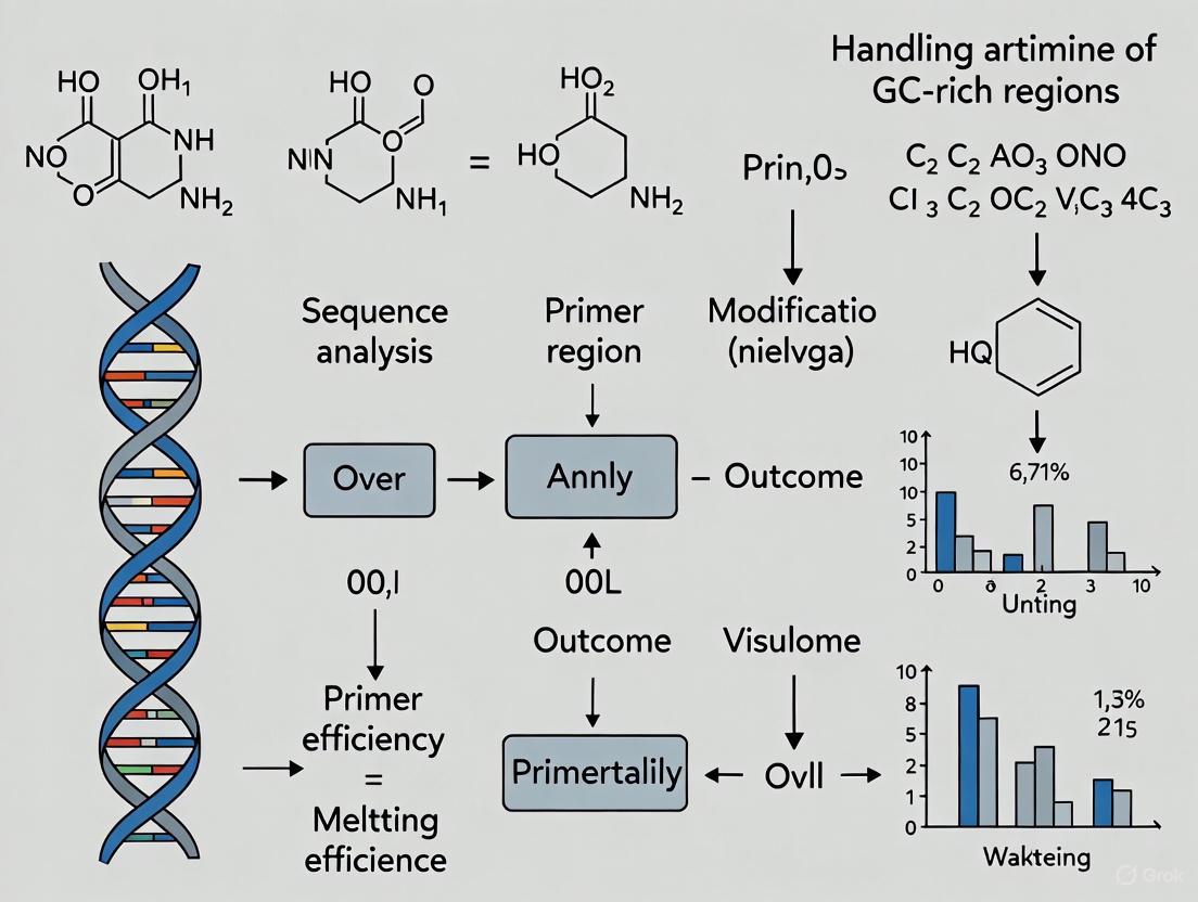
Mastering GC-Rich Primer Design: Strategies for Robust PCR Amplification in Biomedical Research
Amplifying GC-rich DNA sequences is a common yet formidable challenge in molecular biology, often leading to PCR failure due to stable secondary structures and high melting temperatures.
Mastering GC-Rich Primer Design: Strategies for Robust PCR Amplification in Biomedical Research
Abstract
Amplifying GC-rich DNA sequences is a common yet formidable challenge in molecular biology, often leading to PCR failure due to stable secondary structures and high melting temperatures. This article provides a comprehensive guide for researchers and drug development professionals, detailing the fundamental principles behind these challenges and presenting a multi-faceted approach to overcome them. We explore advanced primer design strategies, systematic troubleshooting protocols for reaction optimization, and rigorous validation techniques to ensure specificity and efficiency. By synthesizing foundational knowledge with practical application, this resource equips scientists to successfully handle even the most difficult GC-rich targets, such as gene promoters, thereby accelerating research in genomics, biomarker discovery, and therapeutic development.
Why GC-Rich DNA Poses a Unique Challenge: Understanding the Fundamentals
Defining GC-Rich Regions and Their Biological Significance in Gene Promoters and Housekeeping Genes
Core Definition and Characteristics
GC-rich regions are DNA sequences characterized by a high proportion of guanine (G) and cytosine (C) nucleotides. In molecular biology, a region is typically considered GC-rich when its GC content is 60% or greater [1]. These regions are structurally distinct because GC base pairs form three hydrogen bonds, compared to the two hydrogen bonds in AT base pairs, resulting in higher thermostability and melting temperatures [2].
From a functional genomic perspective, a prominent subset of GC-rich regions are CpG islands (CGIs). These are defined as genomic regions greater than 500 base pairs with a cytosine/guanine content of more than 55% [3] [4]. They are characterized by a higher frequency of CpG dinucleotides than the genomic average. Approximately 60% of CpG islands are found in the 5′ regulatory (promoter) regions of genes, where they play crucial roles in gene regulation and epigenetic modification [4].
Biological Significance and Genomic Distribution
GC-rich regions, particularly CpG islands, are fundamental genomic elements associated with more than 50% of mammalian gene promoters, including both housekeeping and tissue-specific genes [4]. In the human genome, GC-content can range from 35% to 60% across 100-Kb fragments, creating a mosaic-like formation with islet regions called isochores [2]. Gene-rich regions tend to have higher GC-content, and the length of a gene's coding region is often directly proportional to its GC-content [2].
Table: Genomic Distribution and Characteristics of GC-Rich Regions
| Feature | Description | Biological Significance |
|---|---|---|
| Structural Stability | GC base pairs form three hydrogen bonds | Increased thermostability and higher melting temperature (Tm) of DNA [2] |
| Promoter Association | >50% of mammalian gene promoters are GC-rich/CpG islands [4] | Facilitates open chromatin conformation and regulates transcription initiation [5] |
| Housekeeping Genes | Extremely high GC-content in 5'-UTR; CGG tri-SSRs dominate (74-79%) [6] | May contribute to stable, ubiquitous expression profiles [6] |
| Epigenetic Regulation | Target for DNA methylation | Methylation of promoter CGIs leads to transcriptional repression [4] |
Technical Challenges in Experimental Work
Fundamental Experimental Hurdles
The strong triple-bond structure of GC-rich regions that provides biological stability also creates significant technical challenges for molecular biology techniques. The primary issues researchers encounter include:
- Resistance to Denaturation: The increased thermostability makes it difficult to fully separate DNA strands during PCR denaturation steps, preventing primer access [1].
- Formation of Secondary Structures: GC-rich regions are highly "bendable" and readily form stable secondary structures such as hairpins and G-quadruplexes. These structures can cause polymerases to stall, resulting in truncated or incomplete amplification products [1] [4].
- Non-Specific Amplification: The higher annealing energy can lead to primer-dimer formation and off-target binding, especially when standard PCR protocols are used without optimization [1] [7].
Impact on Specific Techniques
These challenges manifest concretely in experimental workflows:
- In PCR: Failed reactions, smeared bands on gels, or significantly lower yield compared to AT-rich targets.
- In Sequencing: Technologies like Illumina sequencing have documented difficulties reading high-GC-content sequences, potentially leading to "missing genes" in genomic assemblies [2].
- In Primer Design: Standard parameters for melting temperature (Tm) calculation become less accurate, complicating the design of effective primers.
FAQs and Troubleshooting Guides
Frequently Asked Questions
Q1: Why is amplifying GC-rich templates so challenging in PCR? GC-rich templates (≥60% GC content) present three main challenges: First, the strong triple hydrogen bonds between G and C bases require more energy to denature, often necessating higher denaturation temperatures. Second, these regions readily form stable secondary structures (e.g., hairpins) that block polymerase progression. Third, the primers themselves can form dimers or adopt secondary structures, further reducing amplification efficiency [1].
Q2: How do GC-rich promoters differ between housekeeping and tissue-specific genes? Housekeeping and tissue-specific genes show distinct features in their GC-rich regions, particularly in the 5'-Untranslated Region (5'-UTR). Housekeeping genes have SSR densities in 5'-UTRs about 1.7 times higher than those in tissue-specific genes. Furthermore, tri-nucleotide SSRs in housekeeping genes are more GC-rich, with the CGG repeat type accounting for 74-79% of tri-SSRs, compared to 42-57% in tissue-specific genes [6].
Q3: What is the relationship between CpG islands, GC-content, and nucleosome positioning? In mammals, GC content and CpG islands are major promoter elements that govern open chromatin conformation. High GC content and wider CpG islands directly correlate with nucleosome depletion both in vivo and in vitro, creating an accessible chromatin environment that facilitates transcription factor binding and supports paused RNA polymerase, which is critical for rapid gene activation [5].
Q4: What defines a CpG island, and what is the best algorithm to identify them? CpG islands are traditionally defined as DNA regions >500 bp with a GC content >55% and an observed-to-expected CpG ratio >0.65. A systematic comparison of identification algorithms concluded that the Takai and Jones algorithm is overall more appropriate for identifying promoter-associated CpG islands in vertebrate genomes, as it effectively excludes false positives like Alu repeats and better aligns with gene markers. In contrast, the CpGcluster algorithm identifies many shorter, CpG-richer regions (CGCs) that are predominantly located in intergenic regions and less associated with known promoters [3].
Troubleshooting PCR for GC-Rich Sequences
This guide addresses the most common symptoms of failed GC-rich PCR amplifications.
Table: Troubleshooting Guide for GC-Rich PCR
| Symptom | Potential Cause | Solution | Underlying Principle |
|---|---|---|---|
| No Product or Faint Bands | Incomplete denaturation of template; polymerase stalling at secondary structures. | 1. Use a specialized polymerase (e.g., Q5 High-Fidelity).2. Add a GC Enhancer (e.g., betaine/DMSO).3. Increase denaturation temperature (e.g., 98°C).4. Use a touchdown PCR protocol. | GC Enhancers destabilize secondary structures. Specialized polymerases are engineered to navigate complex templates [1]. |
| Multiple Non-Specific Bands | Reduced primer stringency due to stable GC-primer interactions. | 1. Optimize MgCl2 concentration (test 1.0-4.0 mM in gradients).2. Increase annealing temperature (use a gradient).3. Use a hot-start polymerase.4. Check primers for self-complementarity. | Higher annealing temperature and optimized Mg2+ increase binding stringency, reducing off-target priming [1] [7]. |
| Primer-Dimer Formation | Primers with high GC content, especially at 3'-end, form stable hybrids with each other. | 1. Design primers with a GC clamp (G or C at the 3' end) but avoid long G/C runs.2. Increase annealing temperature.3. Use software to check for inter-primer homology. | A 3' GC clamp improves specificity and initiation efficiency, but overdoing it promotes self-dimerization [7]. |
Optimized Experimental Protocols
Robust PCR Protocol for GC-Rich Templates
This protocol is adapted from recommendations for using polymerases like Q5 High-Fidelity DNA Polymerase and includes critical steps for success [1].
Reagent Setup:
- Polymerase: Q5 High-Fidelity DNA Polymerase (NEB #M0491) or OneTaq DNA Polymerase (NEB #M0480)
- GC Enhancer: Use the manufacturer's supplied GC Enhancer at a starting concentration of 10% (v/v).
- Primers: Designed according to the guidelines in Section 4.2. Resuspend and dilute to 10 µM working stock.
- Template DNA: 10-100 ng genomic DNA or 1-10 ng plasmid DNA.
- dNTPs: 10 mM each dNTP.
- Mg2+: The buffer is typically supplied with 1.5-2 mM MgCl2; have a separate MgCl2 stock (25 mM) available for optimization.
Thermocycling Protocol:
- Initial Denaturation: 98°C for 30 seconds.
- Amplification (35 cycles):
- Denaturation: 98°C for 5-10 seconds. (Note: A higher temperature and shorter time can help).
- Annealing: Temperature gradient from 65°C to 75°C for 20 seconds. Determine optimal Ta empirically.
- Extension: 72°C for 20-30 seconds per kb.
- Final Extension: 72°C for 2 minutes.
- Hold: 4°C.
Troubleshooting Notes:
- If amplification fails, titrate the GC Enhancer concentration from 5% to 20%.
- If non-specific bands persist, try a "Touchdown" PCR: start with an annealing temperature 5-10°C above the calculated Tm and decrease by 0.5-1°C per cycle for the first 10-15 cycles, then continue at the lower temperature for the remaining cycles.
Primer Design for GC-Rich Targets
Effective primer design is the most critical factor for successful amplification of GC-rich regions. Follow these guidelines [1] [7]:
- GC Content: Aim for 40-60% GC content for the primer itself.
- GC Clamp: Ensure the 3'-end of the primer ends in at least one G or C base to strengthen binding, but avoid runs of more than 3 G/Cs.
- Length: Optimal primer length is 18-30 bases.
- Melting Temperature (Tm): Design primers with a Tm between 65°C and 75°C, and ensure forward and reverse primers are within 5°C of each other.
- Avoid Secondary Structures: Use tools to check for hairpins, self-dimers, and cross-dimers. Avoid runs of 4 or more of the same base and dinucleotide repeats (e.g., ACCCC or ATATAT).
- Validation: Always use a primer analysis tool (e.g., NEB's Tm Calculator) that accounts for the enzyme and buffer system you are using.
The following workflow diagram summarizes the logical process for designing and optimizing primers for GC-rich targets:
Research Reagent Solutions
Successful experimentation with GC-rich regions requires a carefully selected toolkit. The following table catalogs essential reagents and their functions.
Table: Essential Reagents for Working with GC-Rich Regions
| Reagent / Tool | Function / Application | Example Products / Notes |
|---|---|---|
| Specialized Polymerases | High-processivity enzymes engineered to navigate stable secondary structures and GC-rich templates. | Q5 High-Fidelity DNA Polymerase (NEB), OneTaq DNA Polymerase (NEB) [1]. |
| GC Enhancers | Additives that destabilize secondary structures, lower the melting temperature of GC-rich DNA, and increase yield. | Betaine, DMSO, Formamide. Often supplied as a proprietary "GC Enhancer" with polymerases [1]. |
| Mg2+ Solution | Cofactor essential for polymerase activity and primer binding; concentration requires optimization for GC-rich targets. | Supplied with polymerase buffer; separate 25 mM MgCl2 stock for titration (1.0-4.0 mM) [1]. |
| Bioinformatics Tools | Algorithms for identifying CpG islands and analyzing primer characteristics for GC-rich targets. | Takai and Jones algorithm for CGI identification [3]; NEB Tm Calculator for primer design [1]. |
| Methylation Analysis Kits | For investigating the epigenetic status of CpG islands in promoter regions. | Bisulfite conversion kits, Methylation-specific PCR kits. |
| Antisense Oligonucleotides | Emerging therapeutic and research tools for targeting pathogenic GC-rich repeat expansions (e.g., in C9orf72). | Used to bind repeat expansions and disrupt toxic RNA foci [4]. |
Fundamental Biochemical Forces in DNA Recognition
The exceptional specificity of DNA replication and PCR amplification is governed by key physical forces that enable DNA polymerases to select and incorporate the correct nucleotide. Understanding these forces is crucial for troubleshooting experimental challenges, particularly with difficult templates like GC-rich sequences.
Hydrogen Bonding
- Function: Forms specific base pairs between complementary strands (A-T and G-C) through directional interactions.
- Role in Specificity: Watson-Crick hydrogen bonding provides the primary mechanism for distinguishing correct versus incorrect nucleotides during DNA synthesis. The three hydrogen bonds in a G-C pair contribute to its higher thermal stability compared to the two bonds in an A-T pair [8] [9].
- Experimental Impact: In GC-rich regions, the increased hydrogen bonding contributes to stronger template-primer binding but also promotes formation of inhibitory secondary structures.
Base Stacking
- Function: Provides stabilizing interactions between adjacent aromatic bases in the DNA helix through hydrophobic interactions and van der Waals forces.
- Role in Stability: Base stacking contributes significantly to the overall stability of the DNA double helix and influences polymerase processivity during strand extension [8].
- Experimental Impact: GC-rich sequences exhibit enhanced stacking interactions that further increase template stability and resistance to denaturation.
Steric Effects
- Function: Governs the precise geometric fit of nucleotides within the polymerase active site.
- Role in Specificity: The shape and size complementarity between natural bases and the enzyme's active site provide a secondary mechanism for excluding incorrect nucleotides, working in concert with hydrogen bonding recognition [8].
Diagram 1: Biochemical forces and their experimental impacts.
Troubleshooting GC-Rich Amplification: FAQs
What defines a GC-rich template and why is it problematic?
A template is considered GC-rich when 60% or more of its bases are guanine (G) or cytosine (C) [9]. These regions present significant challenges because:
- Enhanced Stability: G-C base pairs form three hydrogen bonds compared to two in A-T pairs, creating higher thermostability that requires more energy to denature [9].
- Secondary Structures: GC-rich regions are 'bendable' and readily form stable secondary structures like hairpins that block polymerase progression [9].
- Primer Challenges: Primers designed for GC-rich targets tend to form dimers and exhibit non-specific binding [9].
Why does my GC-rich PCR reaction show no amplification or low yield?
This common issue typically stems from the combined effects of hydrogen bonding and base stacking:
| Primary Cause | Underlying Biochemistry | Recommended Solution |
|---|---|---|
| Polymerase stalling | Strong base stacking promotes secondary structures that physically block polymerase progression [9] | Use polymerases specifically optimized for GC-rich templates (e.g., Q5 High-Fidelity, OneTaq) [9] [10] |
| Incomplete denaturation | Excessive hydrogen bonding in GC-rich regions resists standard denaturation conditions [11] [9] | Increase denaturation temperature (up to 98°C) and/or time; add GC enhancers [11] [9] |
| Poor primer binding | High primer Tm leads to insufficient binding at standard annealing temperatures [12] | Design primers with higher Tm (>79.7°C) and use higher annealing temperatures (>65°C) [12] |
| Enzyme inhibition | Secondary structures may sequester polymerase or cofactors | Increase polymerase concentration; use additives to destabilize structures [11] [9] |
How can I eliminate non-specific products in GC-rich PCR?
Non-specific amplification occurs when primers bind to unintended regions due to compromised specificity:
- Increase Annealing Temperature: Raise temperature by 2-5°C above calculated Tm to enhance stringency [9] [10]. GC clamp design (3' ending in G or C) improves binding specificity but requires optimized temperatures [7].
- Optimize Mg²⁺ Concentration: Test concentrations between 1.0-4.0 mM in 0.5 mM increments [9]. Excessive Mg²⁺ promotes non-specific binding [10].
- Use Hot-Start Polymerases: These enzymes remain inactive until high-temperature activation, preventing spurious initiation during reaction setup [11] [10].
- Employ Touchdown PCR: Begin with higher annealing temperatures and decrease incrementally to favor specific amplification [11].
What causes smeared bands or primer-dimer formation?
- Smeared Bands: Result from gradual accumulation of "amplifiable DNA contaminants" specific to your primers or degraded DNA template creating multiple fragment sizes [13].
- Primer-Dimer Formation: Occurs when primers anneal to each other through complementary sequences, promoted by high primer concentrations and suboptimal annealing temperatures [13].
Diagram 2: Systematic troubleshooting workflow for GC-rich PCR.
Advanced Primer Design Strategies for GC-Rich Templates
Specialized Design Parameters
Conventional primer design rules often fail with GC-rich templates. Implement these evidence-based strategies:
- Elevated Melting Temperature: Design primers with Tm >79.7°C to withstand the high stability of GC-rich targets [12].
- Minimized Tm Differential: Maintain ΔTm <1°C between forward and reverse primers to ensure balanced annealing [12].
- GC Clamp Implementation: Include G or C bases at the 3' end to strengthen binding through enhanced hydrogen bonding, but avoid excessive repeats that promote dimerization [7].
- Length Optimization: Use longer primers (22-30 bases) to increase specificity despite high GC content [10].
Sequence Composition Guidelines
- Avoid Secondary Structures: Screen for self-complementarity and hairpin formation, particularly problematic in GC-rich designs [7].
- Balance Distribution: Maintain relatively even distribution of GC-rich and AT-rich domains rather than concentrated GC stretches [7].
- Prevent Repeats: Eliminate runs of 4+ identical bases or dinucleotide repeats (e.g., ACCCC or ATATATAT) that complicate synthesis and promote mispriming [7].
Research Reagent Solutions for GC-Rich Experiments
| Reagent Category | Specific Examples | Function & Mechanism |
|---|---|---|
| Specialized Polymerases | Q5 High-Fidelity (NEB #M0491), OneTaq DNA Polymerase (NEB #M0480) [9] | Enhanced processivity to overcome stalling at secondary structures; some include proprietary GC enhancers |
| PCR Additives | DMSO, Betaine, Glycerol, Formamide [9] | Reduce secondary structure formation by interfering with hydrogen bonding and base stacking stability |
| GC Enhancers | OneTaq High GC Enhancer, Q5 High GC Enhancer [9] | Proprietary formulations that combine multiple additives to specifically address GC-rich challenges |
| Magnesium Salts | MgCl₂, MgSO₄ (preferred for some polymerases like Pfu) [11] | Cofactor essential for polymerase activity; concentration optimization critical for balancing specificity and yield |
| Hot-Start Enzymes | OneTaq Hot Start DNA Polymerase, Q5 Hot Start High-Fidelity [11] [10] | Prevent non-specific amplification during reaction setup through antibody-mediated or chemical inhibition |
Quantitative Data Guidelines for GC-Rich Experiments
Template and Primer Requirements
| Template Type | Optimal Quantity | Special Considerations for GC-Rich Templates |
|---|---|---|
| PCR Product | ||
| ∟ 100-200 bp | 1-3 ng | May require increased amount due to secondary structures |
| ∟ 200-500 bp | 3-10 ng | Standard quantification often insufficient |
| ∟ 500-1000 bp | 5-20 ng | Quality critical; verify absence of degradation |
| ∟ 1000-2000 bp | 10-40 ng | Purity essential to prevent inhibition |
| ∟ >2000 bp | 40-100 ng | Maximum amounts may be necessary |
| Plasmid DNA | 250-500 ng | Higher purity standards required |
| Genomic DNA | 2-3 µg | Consider fragmentation from extended denaturation |
Data adapted from Stony Brook DNA Sequencing Facility guidelines [14]
Thermal Cycling Modifications for GC-Rich Templates
| Cycling Parameter | Standard Conditions | GC-Rich Optimized Conditions |
|---|---|---|
| Initial Denaturation | 94-95°C for 30-60 sec | 98°C for 2-5 minutes |
| Denaturation Cycles | 94-95°C for 15-30 sec | 98°C for 20-30 sec |
| Annealing Temperature | Calculated Tm -5°C | Calculated Tm -3°C to Tm +2°C |
| Extension Time | 60 sec/kb | 90-120 sec/kb |
| Cycle Number | 25-35 | 35-40 |
| Final Extension | 5-10 minutes | 10-15 minutes |
Experimental Protocols for Challenging Templates
Standardized Optimization Protocol
This systematic approach methodically addresses the biochemical challenges of GC-rich amplification:
Initial Setup:
- Prepare master mix containing 1X GC buffer, 200 μM dNTPs, and 10-100 ng template DNA
- Add 5-10% GC enhancer according to manufacturer recommendations [9]
Magnesium Titration:
- Prepare reactions with Mg²⁺ concentrations from 1.0-4.0 mM in 0.5 mM increments
- Use constant annealing temperature 5°C below primer Tm [9]
Temperature Optimization:
- After identifying optimal Mg²⁺ concentration, test annealing temperatures from 60-72°C
- Use gradient cycler if available [11]
Additive Screening:
- Test individual additives: DMSO (2-10%), betaine (0.5-2M), or formamide (1-5%)
- Compare against proprietary GC enhancer formulations [9]
Cycle Adjustment:
- Extend cycle number to 40 if product yield remains low after other optimizations [11]
Primer Design Validation Protocol
- Tm Calculation: Use algorithms accounting for nearest-neighbor thermodynamics rather than simple Wallace rule
- Secondary Structure Analysis: Employ software tools to identify hairpins with ΔG < -3 kcal/mol
- Dimer Evaluation: Screen for primer-dimer potential with emphasis on 3' complementarity
- Empirical Testing: Validate with temperature gradient PCR using 50-100 ng control template
The successful amplification of GC-rich sequences requires a comprehensive understanding of the underlying biochemistry of hydrogen bonding and base stacking interactions. By systematically addressing these fundamental forces through optimized reagent selection, primer design, and reaction conditions, researchers can overcome the challenges posed by these difficult templates and achieve reliable, specific amplification for their experimental needs.
In molecular biology, particularly in research involving GC-rich regions, researchers often encounter experimental hurdles caused by the intrinsic properties of nucleic acids. A significant challenge is the formation of stable secondary structures, such as hairpin loops and G-quadruplexes, within DNA templates and oligonucleotide primers. These structures arise from predictable molecular interactions: Guanine (G) and cytosine (C) base pair with three hydrogen bonds, making them more thermostable than adenine (A) and thymine (T) pairs, which have only two [15] [16]. This increased stability allows GC-rich sequences (typically defined as over 60% GC content) to readily fold into complex, stable secondary structures [15].
The primary experimental consequence of these structures is polymerase stalling. During PCR or DNA synthesis, the DNA polymerase enzyme can pause or stall completely when it encounters these physical barriers, leading to failed amplification, truncated products, or non-specific bands on a gel [15] [17]. For researchers working on promoter regions of housekeeping or tumor suppressor genes, which are often GC-rich, understanding and overcoming these pitfalls is essential for successful experimentation and accurate data in drug development projects [15].
Frequently Asked Questions (FAQs) & Troubleshooting
Q1: My PCR of a GC-rich promoter region shows either a blank gel or a smeared product. What are my first steps for troubleshooting?
This is a classic symptom of polymerase stalling due to secondary structures [15]. Your systematic troubleshooting approach should focus on four key areas:
- Polymerase Choice: Standard Taq polymerase often struggles with structured templates. Switch to a polymerase specifically engineered for GC-rich or difficult templates, such as NEB's OneTaq or Q5 High-Fidelity DNA Polymerase, which are often supplied with specialized GC enhancer buffers [15] [16].
- Annealing Temperature: A gradient PCR with increased annealing temperature can help disrupt secondary structures that prevent primer binding and increase specificity [15].
- Additives: Include additives like DMSO, betaine, or formamide in your reaction. These compounds help denature stable secondary structures and promote specific primer annealing [15].
- Mg²⁺ Concentration: Magnesium is a critical cofactor. Titrating MgCl₂ concentration (e.g., testing 0.5 mM increments between 1.0 and 4.0 mM) can optimize enzyme processivity and primer binding specificity [15] [16].
Q2: How can I design primers to minimize the risk of secondary structure formation from the start?
Careful primer design is the most effective preventive measure. Adhere to the following guidelines [7] [18]:
- GC Content: Aim for a GC content between 40% and 60%.
- GC Clamp: Ensure the 3' end of the primer ends in a G or C residue (a "GC clamp") to promote strong binding, but avoid runs of more than three G or C bases in a row at the 3' end, as this can promote non-specific binding [7] [18].
- Length: Design primers between 18 and 30 nucleotides long. Shorter primers bind more efficiently but must be long enough for specificity.
- Melting Temperature (Tm): Aim for a Tm between 65°C and 75°C, and ensure the forward and reverse primers have Tms within 5°C of each other.
- Avoid Self-Complementarity: Check that primers do not contain regions of self-complementarity (more than 3 bases) or complementarity to each other (inter-primer homology), as this leads to hairpin formation and primer-dimers [7].
Q3: What is the molecular mechanism behind polymerase stalling at hairpin loops?
Polymerase stalling is a direct physical blockage. When a DNA template folds into a hairpin or other secondary structure, it creates a stable, double-stranded-like region that the polymerase cannot unwind or traverse efficiently. Research using high-throughput assays has demonstrated that this stalling occurs at the single-nucleotide level and is a major constraint on the evolution and abundance of structured repeats in genomes [17]. The stalled polymerase is blocked from progression, leading to incomplete synthesis [19]. In the context of a replication fork, the helicase can continue unwinding the DNA ahead of the stalled polymerase, a phenomenon known as helicase-polymerase uncoupling, which exposes large stretches of single-stranded DNA and can lead to genomic instability [20].
Experimental Protocols & Optimization
Standardized Protocol for Amplifying GC-Rich Targets
The following protocol is adapted from recommendations by New England Biolabs and is an excellent starting point for amplifying challenging, structure-prone sequences [15] [16].
Materials:
- DNA template
- High-Fidelity Polymerase (e.g., NEB Q5 or OneTaq)
- Compatible Reaction Buffer (including any supplied GC Enhancer)
- dNTPs
- Optimized primers
- MgCl₂ (if adjusting concentration)
- Additives (e.g., DMSO, Betaine)
Procedure:
- Reaction Setup: Prepare a 50 µL reaction mixture on ice.
- Use a polymerase master mix tailored for GC-rich targets, or for a standalone polymerase, use the accompanying GC buffer.
- Add GC Enhancer: If using a standalone polymerase like Q5 or OneTaq, add the supplied GC Enhancer to the recommended final concentration (e.g., 5-10%).
- Include Additives: If problems persist, include an additive like DMSO. A good starting concentration is 3-5% (v/v). Note: Some additives can inhibit the polymerase, so titration is advised.
- Thermocycling:
- Initial Denaturation: 98°C for 30 seconds.
- Amplification (35 cycles):
- Denaturation: 98°C for 10 seconds.
- Annealing: Use a temperature gradient to determine the optimal temperature. Start with a Ta 5°C below the calculated Tm of your primers.
- Extension: 72°C for 30 seconds per kb.
- Final Extension: 72°C for 2 minutes.
- Hold: 4°C.
Troubleshooting Notes:
- If non-specific amplification occurs, increase the annealing temperature in 2°C increments.
- If yield is low, consider performing a "Touchdown PCR," where the annealing temperature is gradually decreased over the first few cycles.
- If the product is still not amplified, refer to the optimization tables below for systematic adjustment of reaction components.
Quantitative Data for Reaction Optimization
The tables below consolidate key optimization data from technical resources to guide your experimental adjustments.
Table 1: Optimization of PCR Additives for Secondary Structures
| Additive | Common Working Concentration | Primary Mechanism of Action | Considerations |
|---|---|---|---|
| DMSO | 3-10% (v/v) | Disrupts base pairing, reduces DNA melting temperature [15] | Can be inhibitory at high concentrations (>10%) [15] |
| Betaine | 0.5 - 1.5 M | Equalizes the stability of AT and GC base pairs, prevents secondary structure formation [15] | |
| Formamide | 1-5% (v/v) | Increases primer annealing stringency, reducing off-target binding [15] | |
| 7-deaza-dGTP | (Partial substitution for dGTP) | dGTP analog that disrupts Hoogsteen base pairing in G-quadruplexes [15] | Does not stain well with ethidium bromide [15] |
Table 2: Magnesium and Temperature Optimization Guide
| Parameter | Standard Condition | GC-Rich Optimization | Effect of Deviation |
|---|---|---|---|
| MgCl₂ Concentration | 1.5 - 2.0 mM [16] | Titrate in 0.5 mM steps from 1.0 to 4.0 mM [15] [16] | Too low: Reduced polymerase activity. Too high: Non-specific binding [15] |
| Annealing Temperature (Ta) | 5°C below primer Tm | Test a gradient from 5°C below Tm to 5°C above Tm [15] | Too low: Primer-dimer and non-specific bands. Too high: Weak or no yield [15] |
| Initial Denaturation | 95°C for 30 sec | 98°C for 30-60 sec | Incomplete denaturation of GC-rich templates leads to failure |
Visualizing the Stalling and Recovery Mechanism
The following diagram illustrates the molecular events when a replication fork encounters a DNA secondary structure, leading to polymerase stalling and the pathways to recovery, as elucidated by in vitro studies with eukaryotic replisomes [20].
The Scientist's Toolkit: Research Reagent Solutions
Table 3: Essential Reagents for Managing Secondary Structure Pitfalls
| Reagent / Tool | Supplier Example | Function & Application |
|---|---|---|
| Q5 High-Fidelity DNA Polymerase | New England Biolabs (NEB #M0491) | High-fidelity enzyme ideal for long or difficult amplicons; GC Enhancer improves amplification up to 80% GC content [15] |
| OneTaq Hot Start 2X Master Mix with GC Buffer | New England Biolabs (NEB #M0480) | Convenient master mix format with optimized buffer for routine and GC-rich PCR [15] [16] |
| Q5 Blood Direct 2X Master Mix | New England Biolabs (NEB #M0500) | Specialized formulation for direct amplification from blood samples, resistant to inhibitors and works for amplicons up to 75% GC [15] |
| PCR Additives (DMSO, Betaine) | Various (e.g., Sigma-Aldrich) | Chemical helpers to destabilize secondary structures in the DNA template [15] |
| NEB Tm Calculator | New England Biolabs (Online Tool) | Critical for primer design and determining the optimal annealing temperature, accounting for enzyme and buffer choice [15] |
| Pif1 Helicase | N/A (Recombinant) | An extrinsic accessory helicase demonstrated in research to be essential for unwinding G-quadruplex structures to allow replication fork progression [20] |
Troubleshooting Guides
Guide 1: Troubleshooting Inefficient Denaturation of GC-Rich Templates
Problem: Amplification failure or low yield of GC-rich DNA targets due to incomplete separation of DNA strands.
Underlying Cause: GC-rich DNA sequences (≥60% GC content) have higher thermostability due to three hydrogen bonds in G-C base pairs and strong base-stacking interactions, making them resistant to standard denaturation temperatures [21] [22].
Solutions:
- Increase Denaturation Temperature: Temporarily increase denaturation temperature to 95–98°C for the first few PCR cycles. Avoid prolonged exposure to >95°C to prevent polymerase damage [22].
- Use PCR Additives: Incorporate DMSO (2–10%), betaine (1–2 M), or glycerol to disrupt secondary structures and lower the effective melting temperature of GC-rich DNA [23] [21].
- Extend Denaturation Time: Increase denaturation time during cycling to ensure complete separation of stubborn double-stranded regions [11].
Guide 2: Troubleshooting Non-Specific Annealing and Primer-Dimer Formation
Problem: Multiple bands, smearing on gels, or short, smeary products around 100 bp indicating primer-dimer artifacts [24].
Underlying Causes:
- Low Annealing Temperature: Permits primers to bind to non-target sequences with partial complementarity [23].
- Primer Complementarity: Regions of complementarity, especially at the 3' ends of primers, lead to self-dimers or cross-dimers [24] [25].
- High Primer Concentration: Excess primers increase the likelihood of primer-to-primer interactions [25] [26].
Solutions:
- Optimize Annealing Temperature: Use a gradient thermal cycler to determine the optimal temperature. Start at 3–5°C below the primer Tm and increase incrementally for specificity [23] [11].
- Use Hot-Start Polymerases: Employ hot-start enzymes that remain inactive until a high-temperature activation step, preventing primer-dimer formation during reaction setup [23] [24].
- Reduce Primer Concentration: Titrate primer concentration (typically 0.1–1 μM) to find the lowest level that supports efficient amplification [11] [25].
- Improve Primer Design: Utilize software to design primers with minimal 3' end complementarity, appropriate length (18–24 bases), and closely matched Tm values (within 1–2°C) [23] [12].
Frequently Asked Questions (FAQs)
FAQ 1: What are the primary reasons for non-specific amplification in standard PCR?
The most common cause is an annealing temperature set too low, reducing binding stringency and allowing primers to anneal to off-target sites. Excess magnesium ions (Mg²⁺) or problematic primer design with self-complementary regions can also promote non-specific products [23] [11].
FAQ 2: How does a high-fidelity polymerase differ from standard Taq polymerase?
High-fidelity polymerases (e.g., Pfu, Q5) possess 3'→5' exonuclease (proofreading) activity, which corrects base misincorporation. This results in an error rate up to 280 times lower than non-proofreading enzymes like standard Taq, which is crucial for cloning and sequencing applications [23] [21].
FAQ 3: When should I use a buffer additive like DMSO or betaine?
Consider additives when amplifying templates with high GC content (above 65%) or complex secondary structures. DMSO (typically 2–10%) helps denature stable DNA structures, while betaine (1–2 M) homogenizes the melting stability of DNA, improving polymerase progression and yield [23] [21] [22].
FAQ 4: Why is Mg²⁺ concentration so critical for PCR?
Magnesium ions are an essential cofactor for DNA polymerase activity. Too little Mg²⁺ leads to reduced enzyme activity and poor yield, while too much promotes non-specific amplification and decreases fidelity. The optimal concentration is typically between 1.5 and 2.5 mM but requires titration for specific reactions [23] [21] [26].
FAQ 5: How can I prevent primer-dimer formation in my PCR assays?
Prevention strategies include designing primers with low 3' end complementarity, using lower primer concentrations, increasing the annealing temperature, and employing hot-start DNA polymerases. Always include a no-template control to identify primer-dimer artifacts [24] [25].
Experimental Protocols
Protocol 1: Systematic Optimization of Annealing Temperature and Mg²⁺ Concentration
Objective: To determine the optimal annealing temperature (Ta) and Mg²⁺ concentration for specific amplification of a GC-rich target.
Materials:
- Thermal Cycler with Gradient Function
- Test Primers and GC-Rich Template DNA
- MgCl₂ or MgSO₄ Stock Solutions (concentration varies with polymerase)
- PCR Additives (e.g., DMSO, Betaine, GC Enhancer)
Methodology:
- Prepare a Master Mix excluding Mg²⁺ and template. Aliquot the master mix into multiple tubes.
- Spike each aliquot with a different volume of Mg²⁺ stock solution to create a concentration series (e.g., 0.5 mM, 1.0 mM, 1.5 mM, 2.0 mM, 2.5 mM, 3.0 mM, 3.5 mM, 4.0 mM) [21].
- Add template DNA to each tube and run PCR using the thermal cycler's gradient function across a temperature range (e.g., 55°C to 70°C) [23].
- Analyze the PCR products using agarose gel electrophoresis. The optimal conditions will produce a single, intense band of the expected size.
Protocol 2: Evaluating Polymerase Performance on Difficult Templates
Objective: To compare the efficacy of different DNA polymerases in amplifying a GC-rich target with known secondary structures.
Materials:
- Standard Taq Polymerase
- High-Fidelity/Proofreading Polymerases (e.g., Q5, Pfu)
- Specialized Polymerase Kits with GC buffers (e.g., OneTaq with GC Buffer)
- GC-Rich Control Template
Methodology:
- Set up identical PCR reactions differing only in the type of polymerase and buffer used according to manufacturer protocols.
- For polymerases with optional enhancers, include reactions with and without the GC Enhancer additive [21].
- Use a universal annealing temperature if using polymerases formulated for this purpose (e.g., 60°C for Platinum polymerases), or the calculated optimal Ta for others [27].
- Analyze results by gel electrophoresis for product yield and specificity, and consider downstream sequencing to assess fidelity for cloning applications [23] [26].
Data Presentation
Table 1: Optimal Ranges for Critical PCR Components
| Component | Standard Range | Optimal for GC-Rich Targets | Effect of Low Concentration | Effect of High Concentration |
|---|---|---|---|---|
| Mg²⁺ | 1.5 - 2.0 mM [26] | 2.0 - 3.5 mM (Requires titration) [21] | Reduced enzyme activity; poor or no yield [23] | Non-specific amplification; lower fidelity [23] [11] |
| Primers | 0.1 - 1.0 µM [26] | Lower end of standard range (0.1 - 0.5 µM) | Low reaction yield [11] | Primer-dimer formation; non-specific products [25] [26] |
| Annealing Temperature (Ta) | 3 - 5°C below Tm [11] | Higher end of range (e.g., 65 - 72°C) [12] | Non-specific binding; multiple bands [23] | Reduced primer binding; low or no yield [23] |
| Denaturation Temperature | 94 - 95°C | 95 - 98°C (for initial cycles) [22] | Incomplete denaturation; poor efficiency | Polymerase denaturation over time [22] |
Table 2: Research Reagent Solutions for GC-Rich PCR
| Reagent | Function | Example Products |
|---|---|---|
| High-Fidelity Polymerase with Proofreading | Reduces error rate for accurate amplification of complex templates; often has higher processivity [23]. | Q5 High-Fidelity DNA Polymerase (NEB), Pfu Polymerase [21] [26] |
| Specialized GC Buffer | Formulated to destabilize secondary structures and increase primer-binding stringency [21]. | OneTaq GC Buffer (NEB), GC-Rich DNA Polymerase (ThermoFisher) [21] [22] |
| PCR Enhancer/Additive | Disrupts stable DNA structures, homogenizes DNA melting temperature [23] [21]. | DMSO, Betaine, Q5/OneTaq High GC Enhancer (NEB) [23] [21] |
| Hot-Start Polymerase | Prevents enzymatic activity during setup, reducing primer-dimer and non-specific amplification [23] [24]. | Platinum Taq DNA Polymerase (ThermoFisher), Hot Start Taq (multiple vendors) [23] [27] |
Workflow Visualization
Designing Primers for Success: A Strategic Methodology for GC-Rich Targets
This guide provides targeted troubleshooting and FAQs for researchers facing challenges in PCR primer design, specifically within the context of handling GC-rich regions. GC-rich templates present unique difficulties due to their high melting temperatures and stable secondary structures, which can impede DNA polymerase progression and lead to amplification failure [28]. The following sections detail core principles and solutions to optimize your experimental outcomes.
Troubleshooting Guides
Troubleshooting Primer Design for Standard Templates
| Observation | Possible Cause | Recommended Solution |
|---|---|---|
| No Amplification | Primer Tm too high/low | Recalculate Tm; test annealing temperature gradient starting 5°C below primer Tm [29]. |
| Poor primer specificity | Verify primer uniqueness with BLAST; avoid secondary structures and primer dimers (ΔG > -9.0 kcal/mol) [30]. | |
| Low primer concentration | Optimize concentration, typically 0.1-1 µM; standard is 0.4-0.5 µM [31] [29]. | |
| Non-Specific Bands/Multiple Products | Annealing temperature too low | Increase temperature; optimal Ta is typically 3-5°C below the primer Tm [30] [11]. |
| Poor primer design | Avoid GC-rich 3' ends; ensure primers are non-complementary; increase primer length [7] [29]. | |
| Excess primer or Mg2+ | Optimize primer concentration (0.1-1 µM); titrate Mg2+ concentration in 0.2-1 mM increments [29]. | |
| Low Yield/Weak Product | Primer Tm mismatch | Ensure forward and reverse primer Tms are within 2°C of each other [30]. |
| Incorrect Tm calculation | Use nearest-neighbor method with reaction-specific buffer conditions (e.g., [Na+], [Mg2+]) [30] [32]. | |
| Suboptimal GC content | Design primers with GC content between 40-65%; ideal is 50% [30] [7]. |
Troubleshooting Amplification of GC-Rich Regions
| Observation | Possible Cause | Recommended Solution |
|---|---|---|
| No Product from GC-Rich Template | Stable secondary structures | Use specialized polymerase (e.g., Q5 High-Fidelity); add co-solvents like DMSO (3-10%), betaine (1-1.3 M), or formamide [28] [33] [29]. |
| Insufficient denaturation | Increase denaturation temperature (up to 98°C) and/or time [11] [33]. | |
| High primer ΔG | Redesign primers using codon optimization (wobble position changes) to disrupt secondary structures [28]. | |
| Smear or Truncated Products | Polymerase stalling | Use polymerases with high processivity; add GC enhancers; include a final extension step of 5-15 minutes [11] [33]. |
| Mispriming at high Tm | Utilize touchdown PCR; start with high annealing temperature, decrease incrementally in subsequent cycles [33]. |
Frequently Asked Questions (FAQs)
Q1: What are the ideal ranges for primer length, Tm, and GC content?
The following table summarizes the core quantitative principles for general primer design:
| Parameter | Ideal Range | Special Considerations |
|---|---|---|
| Primer Length | 18-30 bases [30] [7] | Shorter primers within this range anneal more efficiently [7]. |
| Melting Temperature (Tm) | 60-75°C [30] [7] | Primer pairs should be within 2°C of each other [30]. |
| GC Content | 40-60% [30] [7] | Aim for 50% as an ideal target; avoid long G/C runs [30]. |
| Annealing Temperature (Ta) | 3-5°C below primer Tm [30] [32] | Must be optimized based on calculated Tm and experimental conditions. |
Q2: Which method should I use to calculate Tm accurately?
The choice of method depends on your requirements:
- Basic Method (
2(A+T) + 4(G+C)): Suitable for quick estimates of short primers (<14 nt) [34]. - Nearest-Neighbor Method: Most accurate for most applications, as it considers the sequence context and stacking interactions [30] [32].
- Salt-Adjusted/Buffer-Specific Methods: Essential for high accuracy. Always use an online calculator (e.g., NEB Tm Calculator) and input your specific reaction conditions, including salt and Mg2+ concentrations, for a reliable Tm [30] [32].
Q3: How should I modify my primers for GC-rich targets?
- Codon Optimization: Modify the wobble (third) position of codons in the primer sequence to replace G/C bases with A/T without altering the encoded amino acid. This reduces the local GC content and disrupts secondary structures [28].
- Avoid GC Clamps: Contrary to standard advice, for GC-rich targets, avoid long stretches of Gs or Cs at the 3' end to prevent non-specific binding [33].
- Check Secondary Structures: Always analyze primers using tools like OligoAnalyzer to ensure hairpins and self-dimers have a ΔG weaker than -9.0 kcal/mol [30] [28].
Q4: What are the key steps in a protocol for amplifying a GC-rich gene?
The following workflow outlines a combination strategy for successful amplification of difficult GC-rich templates:
Detailed Protocol:
- Primer Design: Design primers following the strategies in FAQ #3. Use tools like IDT's OligoAnalyzer to analyze and select primers with minimal secondary structures [30] [28].
- Reaction Setup:
- Use a specialized DNA polymerase, such as Q5 High-Fidelity or OneTaq DNA Polymerase, which are robust for GC-rich templates [29].
- Prepare a master mix containing a final concentration of 1X polymerase buffer, dNTPs, and primers.
- Add Enhancers: Include DMSO (3-10%), betaine (1-1.3 M), or a commercial GC enhancer [28] [33].
- Optimize Mg2+: Titrate Mg2+ concentration, as it is critical for polymerase activity. Start with the manufacturer's recommendation and test in 0.2-1 mM increments [29].
- Thermal Cycling:
- Denaturation: Use a higher temperature (e.g., 98°C) and/or a longer duration than standard protocols [11] [33].
- Annealing: Employ a touchdown PCR protocol or set the annealing temperature 5-10°C above what you would use for a standard template [33].
- Extension: Use a standard extension temperature (e.g., 72°C) but consider a longer duration to help the polymerase navigate through complex structures [11].
- Validation: Analyze the PCR product on an agarose gel. For definitive confirmation, sequence the amplified product [28].
Q5: What reagents are essential for troubleshooting PCR of GC-rich regions?
| Reagent Category | Example Products | Function in GC-Rich Amplification |
|---|---|---|
| Specialty Polymerases | Q5 High-Fidelity (NEB), OneTaq (NEB), Hieff Ultra-Rapid II (Yeasen) [31] [29] | High processivity and affinity to denature and replicate stable secondary structures. |
| PCR Additives/Enhancers | DMSO, Betaine, Commercial GC Enhancers [33] [29] | Reduce DNA melting temperature, disrupt secondary structures, and stabilize polymerase. |
| Magnesium Salts | MgCl2, MgSO4 [11] [29] | Cofactor for DNA polymerase; optimal concentration is critical for efficiency and specificity. |
| High-Qucleotide Mixes | Balanced dNTP Mixes [11] | Unbalanced concentrations can increase error rates; fresh, equimolar mixes are crucial. |
| Primer Design Tools | IDT OligoAnalyzer, NEB Tm Calculator [30] [32] | Accurately calculate Tm and analyze potential secondary structures before ordering. |
What are self-dimers and hairpins, and why are they problematic in primer design?
Self-dimers and hairpins are secondary structures that can form within primers, significantly compromising PCR efficiency and specificity.
Self-dimers occur when two primer molecules anneal to each other instead of to the target DNA template. This can happen through two mechanisms: self-dimerization (two identical primers hybridizing) or cross-dimerization (forward and reverse primers hybridizing together) [18]. When these primer-dimers form, they create unintended templates that DNA polymerase can extend, consuming reaction components and generating amplified primer artifacts rather than your target amplicon [35].
Hairpins (or stem-loop structures) form through intramolecular interactions when regions within a single primer are complementary to each other [18]. When these regions anneal, they create a looped structure. Hairpins are particularly problematic when they involve the 3' end of the primer, as this can lead to self-amplification even without the intended DNA template [35].
Both structures reduce primer availability for the intended target, decrease amplification efficiency, and can cause complete PCR failure or misleading results through non-specific amplification [18] [35].
What thermodynamic parameters indicate problematic secondary structures?
The stability of secondary structures is quantified by Gibbs free energy (ΔG). More negative ΔG values indicate more stable, problematic structures. The table below summarizes key thermodynamic thresholds to monitor during primer design and analysis.
Table 1: Thermodynamic Parameters for Problematic Secondary Structures
| Structure Type | Problematic ΔG Threshold | Critical Concern | Calculation Method |
|---|---|---|---|
| Self-Dimer | < -9 kcal/mol [36] | Strong stability leading to amplifiable dimers[ccitation:6] | Nearest-neighbor method [35] |
| Cross-Dimer | < -9 kcal/mol [36] | Primer-primer interaction depleting both primers | Nearest-neighbor method [35] |
| Hairpin | Varies; competitive with binding | 3' complementarity enabling self-amplification [35] | Nearest-neighbor method [35] |
| Stable Hairpin Loop | Optimal: 4-5 DNA residues [37] | Maximum stability for DNA hairpin loops | Experimental measurement [37] |
For LAMP assays, which use longer primers, even hairpins with complementarity one or two bases away from the 3' end can self-amplify, requiring careful screening [35].
How can I screen my primer sequences for potential self-dimers and hairpins?
A robust screening workflow combines computational tools and manual verification to identify primers prone to secondary structure formation.
Table 2: Experimental Reagents and Tools for Secondary Structure Analysis
| Tool or Reagent | Primary Function | Key Features |
|---|---|---|
| Thermo Fisher Multiple Primer Analyzer [38] | Analyzes multiple primers for dimers & basic parameters | Detects possible primer-dimers; provides Tm, GC% |
| NCBI Primer-BLAST [39] | Designs & checks primer specificity | Integrates Primer3 design with BLAST specificity checking |
| OligoAnalyzer Tool (IDT) [35] [36] | Evaluates hairpins & self-dimers | Provides thermodynamic ΔG values for structures |
| mFold Tool [35] | Predicts nucleic acid folding | Models secondary structure formation |
| Chemical Cross-linkers (e.g., EGS) [40] | Stabilizes dimers for detection | Converts non-covalent dimers to covalent for analysis |
| Microchip Electrophoresis [40] | High-throughput separation | Rapidly separates cross-linked dimers from monomers |
Experimental Protocol: In Silico Secondary Structure Screening
- Input Sequences: Obtain your forward and reverse primer sequences in 5' to 3' format.
- Hairpin Analysis: Use the mFold tool or equivalent software to predict intramolecular folding. Examine the output for structures where the 3' end is involved in the hybridized stem [35].
- Self-Dimer and Cross-Dimer Analysis: Use the Multiple Primer Analyzer tool. Input all your primers simultaneously. The tool will report potential dimer pairs based on complementarity [38].
- Thermodynamic Validation: For any flagged dimers or hairpins, use OligoAnalyzer to calculate the Gibbs free energy (ΔG). Discard or redesign primers with ΔG values stronger than -9 kcal/mol [36].
- Specificity Check: Finally, run your sequences through NCBI Primer-BLAST to confirm they bind uniquely to your intended target and do not have significant complementarity to other genomic regions [39].
The following workflow diagram illustrates this screening process:
What design strategies effectively prevent secondary structures?
Implement these specific design strategies during the primer creation phase to proactively avoid secondary structures.
- Optimize Primer Length and Sequence: Design primers between 18-24 nucleotides for standard PCR [18]. Avoid long stretches of a single nucleotide (e.g., AAAA) or dinucleotide repeats (e.g., ATATAT), which promote mispriming and slippage [36].
- Manage GC Content and Distribution: Maintain GC content between 40-60% [18] [36]. Ensure a relatively uniform distribution of G and C bases; avoid clustering many G/C bases at one end. A "GC clamp" (1-2 G/C bases at the 3' end) can help with binding, but more than 3 G/C bases in the last five nucleotides can promote non-specific binding [18] [36].
- Eliminate 3' End Complementarity: This is critical. Ensure the 3' ends of your primers lack self-complementarity and cannot form stable duplexes with other primers. Extensions from primer-dimers are especially efficient if the 3' ends are complementary [35].
- Adjust Primer Location: If your initial primers form problematic structures, shift their binding sites a few nucleotides upstream or downstream on the template sequence. Even a small shift can eliminate complementarity responsible for dimer or hairpin formation while maintaining target specificity.
How do I troubleshoot PCR experiments failing due by suspected secondary structures?
If your PCR results show a blank gel, smearing, or multiple non-specific bands, follow this systematic troubleshooting guide.
Table 3: Troubleshooting Guide for PCR Failure Due to Secondary Structures
| Symptom | Potential Cause | Corrective Actions |
|---|---|---|
| Low or no yield | Hairpins blocking polymerization; primers sequestered in dimers | - Increase annealing temperature (Ta)\n- Use PCR additives (DMSO, betaine) [41] [22]\n- Redesign primers |
| Multiple bands or smearing | Non-specific binding; primer-dimer artifacts | - Increase Ta for greater stringency [41]\n- Optimize Mg²⁺ concentration [41] [22]\n- Redesign primers |
| Primer-dimer artifacts on gel | Strong 3' complementarity between primers | - Screen for and eliminate 3' complementarity [35]\n- Increase Ta [18]\n- Adjust primer concentration [18] |
| Slow amplification or high Cq | Stable secondary structures in template or primers | - Use a polymerase mix optimized for GC-rich templates [41] [22]\n- Add GC enhancers [41]\n- Use slow-down PCR protocols [22] |
Experimental Protocol: Optimizing Annealing Temperature and Reagents
- Annealing Temperature Gradient: Set up a thermal cycler program with an annealing temperature gradient spanning at least 5°C above and below the calculated Tm of your primers. Higher temperatures favor specific binding and can melt secondary structures [41].
- Magnesium Concentration Titration: Test Mg²⁺ concentrations in 0.5 mM increments between 1.0 and 4.0 mM. Excessive Mg²⁺ can cause non-specific binding, while too little reduces polymerase activity [41].
- Additive Enhancement: Prepare reaction mixtures containing potential enhancers:
- Polymerase Selection: If problems persist, switch to a DNA polymerase specifically engineered for challenging templates. These often come with specialized buffers that can significantly improve results with structured sequences [41] [22].
FAQs: Demystifying the GC Clamp in Primer Design
Q1: What is a GC clamp, and why is it considered important in primer design?
A GC clamp refers to the presence of one or more guanine (G) or cytosine (C) bases within the last five nucleotides at the 3' end of a primer [42] [18]. It is considered important because the bonding between G and C bases involves three hydrogen bonds, which is stronger than the two hydrogen bonds in A-T base pairs [22] [16]. This stronger bonding helps promote specific binding at the 3' end, "clamping" the primer to the template DNA and preventing the ends from "breathing" or fraying, thereby increasing priming efficiency [43] [42].
Q2: What are the potential pitfalls of having a GC clamp that is too strong?
A clamp that is too strong can lead to several issues:
- Non-specific binding and mispriming: A very strong clamp can cause the primer to bind to non-target sequences that are not a perfect match, leading to false-positive results and amplification of incorrect products [44] [18].
- Formation of primer-dimers: Excessive G or C residues, especially at the 3' end, can increase the likelihood of primers annealing to each other instead of the template DNA, resulting in primer-dimer formation [42] [18].
- High melting temperature (Tm): A high GC content increases the overall Tm of the primer. If the Tm is too high, it can make it difficult to find an appropriate annealing temperature that allows for specific binding [44].
Q3: What is the recommended number of G or C bases in the last five bases of the 3' end?
The widely recommended guideline is to have 1 or 2 G or C bases in the last five nucleotides at the 3' end [42] [44] [43]. Most sources advise avoiding more than 3 G or C bases in this region to prevent the pitfalls associated with an overly strong clamp [42] [18].
Q4: Is a GC clamp always necessary for a successful PCR?
No, a GC clamp is not an absolute requirement for PCR success [42]. It is one of several factors that can improve the specificity and efficiency of amplification. Primers without a perfect GC clamp can and do work successfully [42] [44]. One empirical account suggests that a single G or C at the 3' end is often sufficient to keep "the PCR gods" happy [42]. The primary parameters for primer design remain specificity, the absence of self-complementarity, and an appropriate melting temperature [42] [30].
Q5: How does the need for a GC clamp relate to amplifying GC-rich templates?
Amplifying GC-rich templates (typically >60% GC content) presents unique challenges, such as stable secondary structures and high overall Tm [45] [22]. While a GC clamp on the primer is still beneficial, the broader strategy for GC-rich PCR involves more than just primer design. This includes using specialized polymerases, buffer additives (e.g., DMSO, betaine), and optimized thermal cycling conditions to help denature these stable regions [45] [22] [16].
Troubleshooting Guide: PCR Amplification of GC-Rich Regions
GC-rich DNA sequences (≥60% GC content) are notoriously challenging to amplify due to their formation of stable secondary structures (e.g., hairpins) and their higher thermodynamic stability, which resists complete denaturation [45] [22]. The following guide addresses common symptoms and solutions.
Symptom 1: No PCR product (blank gel) or a smear of non-specific products.
Potential Causes and Solutions:
Cause: Non-optimal Mg²⁺ concentration. Magnesium is a crucial cofactor for polymerase activity, but incorrect concentrations can lead to failure or spurious bands [45].
Cause: Inefficient denaturation of template and/or non-specific primer annealing.
- Solution 1: Increase denaturation temperature. Temporarily increase the denaturation temperature to 95-98°C for the first few cycles to better melt stable GC-rich structures. Avoid prolonged use of very high temperatures to prevent polymerase damage [22].
- Solution 2: Increase annealing temperature. Use a temperature gradient to find a higher, more stringent annealing temperature (Ta) to reduce non-specific priming [45] [16]. The Tm of primers for GC-rich targets should be high, and one strategy suggests using primers with a Tm >79.7°C and an annealing temperature >65°C to prevent secondary structure formation [12].
Cause: Standard polymerase and buffer are insufficient.
- Solution: Switch to a polymerase system specifically designed for GC-rich templates. Many manufacturers offer polymerases (e.g., Q5 High-Fidelity, OneTaq) supplied with specialized GC buffers and GC enhancers. These enhancers often contain a cocktail of additives like DMSO, glycerol, or betaine that help denature secondary structures [45] [16].
Symptom 2: PCR product yield is low, despite a visible band.
Potential Causes and Solutions:
Cause: Polymerase stalling at secondary structures.
- Solution: Incorporate PCR additives. Reagents like DMSO (1-10%), formamide (1.25-10%), or betaine (0.5 M to 2.5 M) can help reduce secondary structure formation by interfering with base pairing, allowing the polymerase to read through difficult regions [45] [22] [43]. Using a polymerase with high processivity is also recommended [22].
Cause: The 3' end of the primer is suboptimal.
Experimental Protocols and Workflows
Protocol 1: Standardized Workflow for Primer Design and Validation
The diagram below outlines a logical workflow for designing and validating primers, incorporating strategic decisions regarding the GC clamp.
Protocol 2: Optimizing a PCR for a GC-Rich Target
This protocol provides a detailed methodology for setting up a PCR reaction, with specific considerations for GC-rich targets [43].
Key Reagent Solutions:
| Reagent | Function | Final Concentration (50 µl reaction) |
|---|---|---|
| 10X PCR Buffer | Provides optimal salt (K⁺) and pH conditions for the polymerase. May contain Mg²⁺. | 1X (e.g., 5 µl) |
| dNTP Mix | Building blocks (dATP, dCTP, dGTP, dTTP) for new DNA synthesis. | 200 µM (50 µM each) |
| MgCl₂ Solution | Essential cofactor for polymerase activity. Critical for optimization. | 1.5-4.0 mM (titrate) |
| Forward & Reverse Primers | Bind specifically to the target sequence to define the amplicon. | 20-50 pmol each (0.1-1 µM) |
| DNA Polymerase | Enzyme that synthesizes new DNA strands. Use specialized polymerases for GC-rich targets. | 0.5-2.5 units |
| Template DNA | The target GC-rich DNA to be amplified. | 1-1000 ng (10^4-10^7 molecules) |
| PCR Additives (e.g., DMSO) | For GC-rich targets: Reduces secondary structure, increases specificity. | 1-10% (v/v) |
| Sterile Water | Brings the reaction to the final volume. | Q.S. to 50 µl |
Methodology:
- Reaction Setup: Thaw all PCR reagents on ice. Prepare a Master Mix in a sterile 1.8 ml microcentrifuge tube to minimize pipetting errors and ensure consistency between reactions. Add reagents in the following order: sterile water, 10X PCR buffer, dNTP mix, MgCl₂, primers, template DNA, and polymerase (if not hot-start). Gently mix by pipetting up and down 20 times [43].
- Thermal Cycling: Place the tubes in a thermal cycler and run a program similar to the following, optimizing the annealing temperature (Ta) as needed:
- Initial Denaturation: 94-98°C for 2-5 minutes (use higher end for GC-rich templates).
- Amplification (25-35 cycles):
- Denature: 94-98°C for 20-30 seconds.
- Anneal: 5°C below the primer Tm (or higher for specificity) for 20-30 seconds. Use a gradient function to test a range of temperatures.
- Extend: 72°C for 1 minute per kb of amplicon.
- Final Extension: 72°C for 5-10 minutes.
- Hold: 4-10°C ∞.
- Analysis: Analyze the PCR products by agarose gel electrophoresis alongside an appropriate DNA molecular weight standard to confirm the size and specificity of the amplicon [43].
Research Reagent Solutions for GC-Rich PCR
The following table details key reagents that are essential for successful amplification of GC-rich DNA sequences.
| Reagent / Product | Function / Rationale | Example Products |
|---|---|---|
| Specialized Polymerases | Engineered for high processivity and ability to read through stable secondary structures that cause stalling in standard polymerases like Taq. | OneTaq DNA Polymerase (NEB), Q5 High-Fidelity DNA Polymerase (NEB), AccuPrime GC-Rich DNA Polymerase (ThermoFisher) [45] [22] [16]. |
| GC Buffer / Enhancer | Proprietary buffer formulations containing additives that destabilize DNA secondary structures, promoting full denaturation of the GC-rich template and improving primer binding stringency. | OneTaq GC Buffer, Q5 High GC Enhancer [45] [16]. |
| PCR Additives | Chemicals that interfere with hydrogen bonding or stabilize DNA in a single-stranded state, helping to melt secondary structures like hairpins. | DMSO, Betaine, Glycerol, Formamide [45] [22] [43]. |
| Magnesium Chloride (MgCl₂) | A critical cofactor for DNA polymerase activity. Its concentration can significantly impact yield and specificity, making titration essential for GC-rich PCR optimization [45] [16]. | Typically supplied with polymerase buffer. |
Engaging with GC-rich DNA sequences is a common challenge in molecular biology, particularly in primer design and gene synthesis. The strong hydrogen bonding of G-C base pairs (three versus two in A-T pairs) leads to stable secondary structures that can hinder polymerase progression during PCR and reduce translation efficiency in protein expression. This technical support center details a refined strategy—strategic codon optimization via base substitution at the wobble position—to overcome these hurdles, enhance experimental success, and optimize heterologous protein production.
FAQs and Troubleshooting Guides
FAQ 1: What is codon optimization and how does it help with GC-rich sequences?
Answer: Codon optimization is the process of modifying a DNA sequence to enhance its expression in a host organism without altering the amino acid sequence of the resulting protein. This is achieved by introducing synonymous mutations—changes to the DNA sequence that do not change the encoded protein [46].
For GC-rich sequences, a primary difficulty is the formation of stable secondary structures like hairpins, which can cause polymerases to stall during amplification [28] [47]. By substituting bases at the wobble position (the third base in a codon), you can reduce the local GC content. This disrupts these problematic secondary structures, facilitates easier primer binding, and enables more efficient PCR amplification and protein translation [46] [28].
FAQ 2: I cannot amplify my GC-rich target gene. What should I do?
Answer: Failed amplification of GC-rich targets is a frequent issue. Follow this systematic troubleshooting guide.
| Observation | Possible Cause | Solution |
|---|---|---|
| No product or very faint band on gel | Polymerase stalling due to strong secondary structures | - Use a polymerase specifically designed for GC-rich templates (e.g., Q5 High-Fidelity, OneTaq DNA Polymerase) [48] [47].- Incorporate a GC Enhancer or additives like DMSO, betaine, or glycerol into your PCR mix [48] [47]. |
| Primer annealing temperature is too high | Perform a temperature gradient PCR to determine the optimal annealing temperature [48]. | |
| Primers themselves have high GC content and form secondary structures | Redesign primers by introducing synonymous mutations at the wobble position to lower GC content [28]. | |
| Multiple or non-specific bands | Non-specific primer binding | Increase the annealing temperature to improve stringency [48] [47].Use a hot-start polymerase [48]. |
| Mg²⁺ concentration is suboptimal | Test a Mg²⁺ gradient (e.g., 0.5 mM increments between 1.0 and 4.0 mM) to find the ideal concentration [47]. |
FAQ 3: How does base substitution at the wobble position affect protein expression levels?
Answer: While synonymous, these substitutions are not neutral. Codons that are efficiently translated by the host organism's abundant tRNAs are considered "optimal." Using these codons can significantly increase protein yield by preventing ribosome stalling [46].
Conversely, the use of rare codons, which may be decoded by inefficient GU wobble pairing, can reduce translation efficiency and protein levels [49]. Therefore, effective codon optimization involves not only disrupting DNA secondary structures but also matching the codon usage frequency of your expression host to ensure efficient translation elongation.
Core Concepts and Experimental Protocols
The Science of the Wobble Position
The genetic code is degenerate, meaning most amino acids are encoded by more than one codon. This redundancy is primarily at the third, or "wobble," position of the codon [46]. The wobble hypothesis explains that the pairing between the codon and the tRNA anticodon is less strict at this position, allowing a single tRNA to recognize multiple codons for the same amino acid [46]. This flexibility is what makes synonymous codon optimization possible.
Experimental Protocol: Codon Optimization for Amplification of GC-Rich Genes
This protocol is adapted from a successful study that amplified refractory GC-rich genes from Mycobacterium tuberculosis [28].
1. Primer Redesign with Wobble Base Substitution
- Identify Target Codons: Analyze the sequence of your gene, focusing on the terminal regions where primer binding occurs. Identify codons with high GC content, especially those ending in G or C.
- Implement Synonymous Changes: Systematically change the third base of these codons to an A or T. For example:
- Change a CGG codon (Arginine) to CGA (also Arginine).
- Change a CGT codon (Arginine) to CGA (Arginine) [28].
- Verify Primer Quality: Use primer analysis software to ensure the modified primers have:
2. PCR Amplification with Optimized Conditions
- Polymerase Selection: Choose a high-fidelity polymerase known for handling complex templates, such as Q5 or OneTaq DNA Polymerase [48] [47].
- PCR Setup:
- Reaction Mix: Use the buffer supplied with your polymerase. Include 5% DMSO (v/v) or the manufacturer's GC Enhancer.
- Thermocycling Parameters:
- Initial Denaturation: 98°C for 30 seconds.
- Amplification (30 cycles):
- Denaturation: 98°C for 5-10 seconds.
- Annealing: Use the calculated Tm for your modified primers. A gradient PCR is highly recommended.
- Extension: 72°C for 30 seconds/kb.
- Final Extension: 72°C for 2 minutes.
- Analysis: Verify the amplification product on an agarose gel and confirm the sequence by Sanger sequencing.
Flowchart: Overcoming GC-Rich PCR Failure
The Scientist's Toolkit: Research Reagent Solutions
The following reagents are critical for implementing the strategies discussed in this guide.
| Item | Function | Example Use Case |
|---|---|---|
| High-Fidelity DNA Polymerase for GC-Rich Templates | Engineered to resist stalling on complex DNA structures; often includes specialized buffers. | Amplifying promoter regions or other high-GC genomic loci. Q5 High-Fidelity or OneTaq DNA Polymerase are common choices [47]. |
| GC Enhancer / PCR Additives | Chemical additives that destabilize DNA secondary structures, improving polymerase processivity. | Added to the PCR mix when amplifying targets with >60% GC content. Often supplied with the polymerase or can be added separately (e.g., DMSO, betaine) [47]. |
| Codon Optimization Software/Algorithm | Computational tools that automate the design of gene sequences for optimal expression in a target host. | Designing a synthetic gene for high-yield expression of a human protein in E. coli. Platforms like GenScript's OptimumGene consider codon bias, mRNA structure, and other factors [46]. |
| Site-Directed Mutagenesis Kit | Enables precise introduction of base substitutions at the wobble position for functional studies. | Systematically testing the impact of different synonymous codons on protein expression levels in a heterologous system [50]. |
Visualizing Codon-Anticodon Interaction and Optimization Impact
Understanding the molecular basis of the wobble position is key to applying this strategy effectively.
Codon-Anticodon Wobble Pairing
The strategic introduction of synonymous mutations at the wobble position has cascading benefits for experimental workflows, as shown in the following logic.
Effects of Wobble Position Substitution
Frequently Asked Questions (FAQs)
FAQ 1: What is the most accurate method for calculating melting temperature (Tm) for GC-rich primers, and why?
The nearest-neighbor thermodynamics method is the most accurate for calculating Tm, especially for GC-rich sequences. Unlike simple GC percentage methods, which can have an accuracy of ±5-10°C, the nearest-neighbor method accounts for the sequence context of each dinucleotide pair and specific experimental conditions, achieving an accuracy of ±1-2°C [51] [52]. This method, based on SantaLucia's (1998) unified parameters, is considered the gold standard because it incorporates dinucleotide stacking energies, providing superior predictions for the stable structures often formed by GC-rich oligonucleotides [51].
FAQ 2: Which online tools are recommended for predicting and preventing primer secondary structures?
For comprehensive secondary structure prediction, use tools that integrate multiple analysis functions. IDT's OligoAnalyzer tool is a key resource for screening primers for hairpins and self-dimers by calculating thermodynamic ΔG values [52] [36]. It is recommended to avoid primers with strong intramolecular folding, particularly those with hairpin ΔG values < -2 kcal/mol or dimer ΔG < -5 kcal/mol, as these can prevent binding to the target template [51]. For in-silico primer design and validation, NCBI Primer-BLAST is highly recommended as it combines the Primer3 design engine with BLAST-based specificity checking [36].
FAQ 3: How do salt concentrations and common PCR additives affect the Tm of my primers, and how can I account for this?
Salt concentrations and additives significantly impact Tm and must be factored into calculations. Divalent cations like Mg²⁺ have a much stronger effect than monovalent ions like Na⁺; changing Mg²⁺ from 0 mM to 2 mM can increase Tm by +5 to +8°C [51] [52]. Conversely, additives like DMSO lower Tm by approximately 0.5-0.7°C per 1% concentration [51]. For accurate results, always use a Tm calculator that allows you to input these specific conditions, such as the Owczarzy (2008) salt correction model used in modern tools, which accounts for mixed ion solutions and competitive binding (e.g., dNTPs chelating Mg²⁺) [51] [52].
Troubleshooting Guides
Issue 1: Poor or No Amplification of GC-Rich Targets
This is a common issue caused by the high stability of GC-rich templates, which leads to inefficient denaturation and pronounced secondary structures that block primer access.
Troubleshooting Steps:
Verify Tm Calculation Parameters:
Incorporate PCR Additives:
Optimize Thermal Cycler Conditions:
Select a Specialized Polymerase:
Issue 2: Multiple or Non-Specific PCR Products
This often occurs when primers bind to off-target sites, exacerbated by suboptimal annealing stringency or primer design flaws.
Troubleshooting Steps:
Check Primer Specificity:
- Use NCBI Primer-BLAST to verify that your primer sequences are unique to the intended target and do not have significant homology to other regions in the genome [36].
Increase Annealing Stringency:
Use a Hot-Start Polymerase:
Optimize Mg²⁺ Concentration:
Issue 3: Primer-Dimer Formation
Primer-dimer artifacts are caused by complementarity between primers, especially at their 3' ends, and can consume reagents, reducing the yield of the desired product.
Troubleshooting Steps:
Redesign Primers:
Adjust Reaction Components:
Apply Laboratory Techniques:
- Set up PCR reactions on ice and use a hot-start polymerase to prevent low-temperature activity that facilitates dimer formation [54].
Data Presentation: Tm Calculation Methods & Parameters
Table 1: Comparison of DNA Melting Temperature (Tm) Calculation Methods
| Method | Accuracy | Complexity | Key Parameters Accounted For | Best For |
|---|---|---|---|---|
| Nearest-Neighbor Thermodynamics | Highest (±1–2°C) [51] | High | Dinucleotide stacking, sequence context, salt effects (Na⁺, Mg²⁺), oligo concentration [51] | All applications requiring high accuracy, especially GC-rich primers and complex templates [51] |
| Salt-Corrected Models (e.g., Owczarzy 2008) | High [51] | Medium | Mixed ion solutions (Mg²⁺, Na⁺), competitive binding (e.g., dNTPs chelating Mg²⁺) [51] [52] | Standard PCR conditions with Mg²⁺ present [51] |
| GC% Approximation | Low (±5–10°C) [51] | Low | Only GC base pair percentage [51] | Quick estimates only; not recommended for experimental design [51] |
Table 2: Critical Experimental Parameters for Accurate Tm Calculation
| Parameter | Typical Range | Standard PCR Condition | Impact on Tm |
|---|---|---|---|
| [Mg²⁺] Concentration | 0–5 mM [51] | 1.5–2.5 mM [51] | Strongest effect; +5 to +8°C for 0→2 mM change [51] [52] |
| [Na⁺] Concentration | 50–200 mM [51] | 50 mM [51] | +3 to +5°C for 50→100 mM change (logarithmic effect) [51] |
| DMSO Percentage | 0–10% [51] | 0–5% [51] | Lowers Tm by ~0.5–0.7°C per 1% [51] |
| Oligo Concentration | 50–500 nM [51] | 200–500 nM [51] | Slight Tm increase at higher concentrations [51] [52] |
| GC Content | 40–60% (Optimal) [51] [36] | 45–55% | Higher GC content increases Tm and stability, but >70% risks secondary structures [51] |
Experimental Protocols
Protocol 1: In-Silico Primer Design and Tm Calculation for GC-Rich Templates
This protocol uses a combination of free, online tools to design and validate primers with accurate Tm values for challenging GC-rich sequences.
Methodology:
Define Target and Retrieve Sequence:
- Obtain the precise FASTA sequence of your target region from a curated database like NCBI RefSeq [36].
Design Primer Pairs:
- Use NCBI Primer-BLAST with the following key parameters [36]:
- Product size: 200–500 bp.
- Primer Tm: 58–62°C (aim for a "sweet spot").
- Max Tm difference: ≤2°C for the primer pair.
- GC content: 40–60%.
- Select organism for specificity checking.
- Use NCBI Primer-BLAST with the following key parameters [36]:
Validate Primer Specificity:
- Examine the Primer-BLAST report and select primer pairs with no significant off-target matches in the genome [36].
Calculate Condition-Specific Tm:
- Input the chosen primer sequences into IDT's OligoAnalyzer tool.
- In the tool's settings, input your exact experimental conditions: primer concentration (e.g., 0.5 µM), Monovalent cation concentration (e.g., 50 mM K⁺), Divalent cation concentration (e.g., 1.5–2.5 mM Mg²⁺), and DMSO percentage (if used) [51] [52] [53].
Screen for Secondary Structures:
Protocol 2: Empirical Validation and Optimization of Annealing Temperature (Tₐ)
The calculated Tm is a theoretical starting point. Empirical validation is crucial for success.
Methodology:
Prepare Master Mix:
- Prepare a standard PCR reaction mix using your designed primers and a high-fidelity polymerase suitable for GC-rich templates.
Perform Gradient PCR:
Analyze Results:
- Run the PCR products on an agarose gel.
- Identify the annealing temperature that produces the single, brightest band of the expected size with the least non-specific product or primer-dimer.
Sequencing Verification:
- Purify the amplicon from the optimal condition and confirm the sequence via Sanger sequencing to ensure fidelity [36].
Mandatory Visualization
Primer Design and Validation Workflow
Research Reagent Solutions for GC-Rich PCR
| Reagent / Material | Function in GC-Rich PCR | Brief Explanation |
|---|---|---|
| DMSO (Dimethyl Sulfoxide) | Destabilizing Agent | Disrupts hydrogen bonding in GC-rich duplexes, lowering Tm and reducing secondary structures [51] [11]. |
| Betaine | Destabilizing Agent / Solute Equalizer | Reduces the gap in thermal stability between GC-rich and AT-rich DNA regions, promoting uniform amplification [11]. |
| High-Processivity DNA Polymerase | Enzyme | Engineered for high affinity to template DNA, improving amplification efficiency through stable secondary structures [11] [54]. |
| dNTP Mix (Balanced) | Nucleotide Substrates | Unbalanced dNTP concentrations increase PCR error rates; equimolar mixes are essential for high-fidelity amplification [54]. |
| Mg²⁺ Solution (MgCl₂/MgSO₄) | Cofactor | Essential divalent cation for polymerase activity; its concentration must be carefully optimized as it significantly stabilizes DNA and increases Tm [51] [11] [54]. |
Beyond Primer Design: Troubleshooting and Optimizing the PCR Reaction
Amplifying GC-rich DNA templates (typically >60% GC content) is a common challenge in molecular biology, often leading to PCR failure, low yield, or non-specific amplification. This guide provides a detailed comparison of specialized polymerases and master mixes, along with troubleshooting protocols, to help you successfully amplify these difficult targets.
The primary challenges with GC-rich templates are:
- Formation of stable secondary structures (e.g., hairpins) due to stronger hydrogen bonding in G-C base pairs [56] [22].
- Higher thermostability, requiring higher denaturation temperatures [22].
- Reduced polymerase processivity as enzymes stall at stable secondary structures [57].
Polymerase & Master Mix Comparison Table
The table below compares several commercially available polymerases and master mixes specifically designed or enhanced for GC-rich PCR.
| Product Name | Manufacturer | Key Features / Enzyme Type | Optimal GC Range | Recommended Additives | Fidelity (vs. Taq) |
|---|---|---|---|---|---|
| Q5 High-Fidelity DNA Polymerase [58] [16] | New England Biolabs (NEB) | High-fidelity, proofreading | Up to 80% (with GC Enhancer) | Q5 High GC Enhancer | >280x [16] |
| OneTaq DNA Polymerase [16] | New England Biolabs (NEB) | Blend for routine/GC-rich PCR | Up to 80% (with GC Enhancer) | OneTaq High GC Enhancer | 2x [16] |
| Platinum SuperFi II DNA Polymerase [59] | Thermo Fisher Scientific | Engineered, high-fidelity, hot-start | Robust for GC-rich targets | Buffer formulated for 60°C annealing | >300x [59] |
| Advantage GC 2 Polymerase Mix [60] | Takara Bio | Titanium Taq + proofreading polymerase | Up to 90% | Proprietary GC-Melt Reagent, DMSO | High (blend) |
| PCRBIO Ultra Polymerase [57] | PCR Biosystems | Engineered for demanding templates | Up to 80% | Proprietary buffer system | Not specified |
Frequently Asked Questions (FAQs)
What defines a "GC-rich" template and why is it problematic?
A template is generally considered GC-rich when its guanine-cytosine (GC) content exceeds 60-65% [56] [16] [61]. The strong triple hydrogen bonding between G and C bases (versus double bonding in A-T pairs) creates highly stable DNA. This results in:
- Incomplete denaturation at standard temperatures (e.g., 94-95°C).
- Formation of persistent secondary structures like hairpins and stem-loops.
- Stalling of DNA polymerase during extension, leading to poor yield or product failure [56] [57] [22].
When should I use a specialized GC-enhanced master mix versus a standalone polymerase?
- Master Mixes offer convenience, reduced pipetting errors, and are ideal for high-throughput workflows when your targets are within the mix's validated GC range [16].
- Standalone Polymerases provide greater flexibility for optimization. They are recommended when dealing with extremely high GC content (>80%), very long amplicons, or when initial attempts with master mixes have failed, as they allow you to tweak buffer composition, magnesium concentration, and additives [16].
What is the role of GC enhancers and how do they work?
GC enhancers are additive cocktails that work through two primary mechanisms [58] [16]:
- Reducing Secondary Structures: Additives like DMSO, betaine, and glycerol destabilize hydrogen bonding, effectively lowering the melting temperature (Tm) of DNA and preventing the formation of hairpins and other stable structures.
- Increasing Primer Stringency: Additives like formamide help reduce non-specific primer binding, thereby improving the specificity of the amplification.
My GC-rich PCR shows no product. What should I adjust first?
- Verify Polymerase and Additives: Confirm you are using a polymerase known to handle high GC content and that the recommended GC enhancer is included in the reaction [16].
- Increase Denaturation Temperature: Try a higher denaturation temperature (e.g., 98°C) for the first few cycles. Be cautious, as temperatures above 95°C can accelerate enzyme denaturation over many cycles [22].
- Check Primer Design: Ensure primers have a balanced GC content (40-60%), are of sufficient length (20-30 nucleotides), and do not have GC-rich 3' ends that promote mispriming [62].
My gel shows smearing or multiple bands. How can I improve specificity?
- Increase Annealing Temperature: Use a temperature gradient to find the highest possible annealing temperature that still yields product. A higher temperature promotes specific primer binding [16].
- Optimize Mg2+ Concentration: Titrate MgCl2 in 0.5 mM increments between 1.0 and 4.0 mM. Too much Mg2+ can cause non-specific amplification, while too little reduces yield [16].
- Use a Hot-Start Polymerase: Enzymes like Platinum SuperFi II and OneTaq Hot Start remain inactive until the initial denaturation step, preventing primer-dimer formation and non-specific amplification at lower temperatures [16] [59].
Troubleshooting Guide: Common Scenarios
| Problem | Potential Causes | Recommended Solutions |
|---|---|---|
| No Amplification | - Stable secondary structures- Insufficient denaturation- Polymerase inhibited | 1. Use a specialized GC-rich polymerase [57] [60].2. Add GC enhancers (e.g., DMSO, betaine) [56] [16].3. Increase denaturation temp to 98°C for first 5 cycles [22]. |
| Smear or Multiple Bands | - Non-specific priming- Mg2+ concentration too high- Annealing temperature too low | 1. Optimize MgCl2 concentration (try 1.0-2.0 mM) [16].2. Increase annealing temperature in 1-2°C increments [16].3. Use a hot-start, high-fidelity polymerase [59]. |
| Weak or Faint Bands | - Polymerase stalling at secondary structures- Low primer binding efficiency- Inhibitors present | 1. Increase concentration of GC enhancer (e.g., 10% final) [16].2. Increase polymerase concentration by 1.5-2x [56].3. Increase number of PCR cycles (e.g., 35-40 cycles). |
| Primer-Dimer Formation | - Primer self-complementarity- Low annealing temperature- Excessive primer concentration | 1. Re-design primers to avoid 3' complementarity [62].2. Use a primer design tool to check for secondary structures.3. Ensure primer concentration is between 0.05-1.0 µM [62]. |
The following workflow diagram outlines a systematic approach to troubleshooting a failed GC-rich PCR experiment.
Experimental Protocol: Optimizing GC-Rich PCR
This protocol is adapted from research on amplifying GC-rich nicotinic acetylcholine receptor subunits and manufacturer recommendations [56] [16] [60].
Materials & Reagents
- DNA Template: 10-100 ng of genomic DNA or cDNA.
- Specialized Polymerase: e.g., Q5 High-Fidelity DNA Polymerase (NEB #M0491) or Advantage GC 2 Polymerase Mix (Takara #639114).
- GC Enhancer: Provided with polymerase (e.g., Q5 High GC Enhancer) or 100% DMSO.
- Primers: Desalted or HPLC-purified, 20-30 nt, Tm within 5°C of each other [62].
- dNTPs: 10 mM mixture.
- Nuclease-free Water.
Step-by-Step Procedure
Reaction Setup (50 µL)
Thermal Cycling Conditions
- Initial Denaturation: 98°C for 30 seconds (for polymerases like Q5) or 95°C for 2 minutes (for Taq-based blends).
- Amplification (35 cycles):
- Denaturation: 98°C (or 95°C) for 5-10 seconds.
- Annealing: Use a temperature 5°C below the primer Tm or 60°C if using a universal polymerase like Platinum SuperFi II [59]. A temperature gradient is recommended for optimization.
- Extension: 72°C for 20-30 seconds per kb.
- Final Extension: 72°C for 2 minutes.
- Hold: 4°C.
Analysis
- Analyze 5 µL of the PCR product by agarose gel electrophoresis.
- If amplification is unsuccessful, proceed through the troubleshooting workflow.
Key Research Reagent Solutions
| Reagent | Function in GC-Rich PCR |
|---|---|
| DMSO (Dimethyl Sulfoxide) | Disrupts base pairing, reduces DNA secondary structure, and lowers the template's melting temperature [56] [16]. |
| Betaine | Equalizes the contribution of GC and AT base pairs to DNA stability, facilitating more uniform denaturation [56]. |
| 7-deaza-dGTP | A modified dGTP analog that incorporates into DNA but disrupts Hoogsteen base pairing, preventing secondary structure formation [22] [61]. |
| GC-Melt Reagent (Takara) | A proprietary formulation that, in combination with DMSO, enables amplification of templates with up to 90% GC content [60]. |
| Q5 High GC Enhancer (NEB) | A supplemental reagent that reduces secondary structures and increases primer stringency for use with Q5 polymerase [58]. |
Frequently Asked Questions
What are secondary structures and why are they a problem in PCR? Secondary structures, such as hairpins and loops, form when GC-rich regions in DNA templates fold onto themselves due to strong base stacking interactions and hydrogen bonding [22]. These structures are exceptionally stable and do not melt well at standard PCR denaturation temperatures. They can physically block the DNA polymerase, leading to incomplete or truncated PCR products, failed reactions, or nonspecific amplification [63] [64].
How do additives like DMSO help with GC-rich PCR? Additives work through different mechanisms to destabilize secondary structures. DMSO (Dimethyl Sulfoxide) interferes with hydrogen bonding between DNA strands and water molecules, effectively lowering the melting temperature (Tm) of the DNA. This helps the DNA strands separate more easily, facilitating primer binding and polymerase elongation [64] [65].
Can I use multiple additives together? Yes, combining additives can sometimes be more effective than using a single one. Research on amplifying challenging nicotinic acetylcholine receptor subunits demonstrated that a multipronged approach involving various organic molecules was key to success [63] [56]. However, their effects can be highly variable, so optimization is necessary [22].
What is a common mistake when first trying these additives? A common error is testing additives at a single concentration. Their effectiveness and potential for inhibition are concentration-dependent. For example, while DMSO can be beneficial, it also reduces Taq polymerase activity, and too high a concentration will inhibit the PCR reaction [65]. It is crucial to test a range of concentrations.
Additive Properties and Usage
The table below summarizes the mechanisms and optimal use of common PCR additives.
| Additive | Primary Mechanism of Action | Suggested Concentration | Key Considerations |
|---|---|---|---|
| DMSO | Disrupts hydrogen bonding, lowers DNA melting temperature (Tm) [64] [65]. | 2% - 10% [65] | Reduces DNA polymerase activity; requires balance between structure disruption and enzyme inhibition [65]. |
| Betaine | Reduces formation of secondary structures, eliminates base pair composition dependence during denaturation [65]. | 1 M - 1.7 M [65] | Use betaine or betaine monohydrate; hydrochloride form may affect reaction pH [65]. |
| Formamide | Destabilizes DNA double helix, increases primer annealing stringency, reduces non-specific amplification [64] [65]. | 1% - 5% [65] | Binds to DNA grooves, disrupting hydrogen bonds and hydrophobic interactions [65]. |
| 7-deaza-dGTP | dGTP analog that incorporates into DNA, reducing secondary structure stability [22] [64]. | Varies (as dGTP substitute) | May not stain well with ethidium bromide; used in specialized protocols like "slow-down PCR" [22] [64]. |
Experimental Protocol: Optimizing PCR with Additives for a GC-Rich Target
This protocol is adapted from a study that successfully amplified GC-rich nicotinic acetylcholine receptor subunits [63].
1. Reaction Setup
- Polymerase Selection: Begin with a polymerase known for robust performance on GC-rich templates, such as OneTaq or Q5 High-Fidelity DNA Polymerase [64]. These are often supplied with specialized GC buffers and enhancers.
- Master Mix:
- If using a standalone polymerase, prepare a master mix according to the manufacturer's instructions but omit the additives.
- Divide the master mix into several tubes.
- Additive Testing: To different tubes, add:
- DMSO at final concentrations of 2%, 5%, and 8%.
- Betaine at a final concentration of 1 M.
- A combination of 5% DMSO and 1 M Betaine.
- Keep one tube as a no-additive control.
- Template and Primers: Use your GC-rich template (e.g., cDNA reverse-transcribed from RNA) and designed primers [63].
2. Thermocycling Conditions
- Initial Denaturation: 98°C for 30 seconds.
- Amplification Cycles (35 cycles):
- Denaturation: 98°C for 10-15 seconds.
- Annealing: Use a temperature gradient, for example, from 60°C to 72°C, for 20-30 seconds. The optimized study used temperatures adjusted based on polymerase and primer recommendations [63].
- Extension: 72°C for 1 minute per kb.
- Final Extension: 72°C for 5 minutes.
3. Analysis
- Analyze the PCR products on an agarose gel.
- Compare the yield and specificity of the amplification from the different additive conditions against the no-additive control.
- The successful condition from the study yielded clear, specific bands for their target genes [63].
The Scientist's Toolkit: Research Reagent Solutions
| Item / Reagent | Function / Application |
|---|---|
| OneTaq DNA Polymerase with GC Buffer & Enhancer | A specialized system for amplifying difficult, GC-rich amplicons (up to 80% GC) [64]. |
| Q5 High-Fidelity DNA Polymerase | Ideal for long or difficult amplicons including GC-rich DNA; offers very high fidelity [64]. |
| Phusion High-Fidelity DNA Polymerase | A proofreading enzyme with accompanying GC enhancers, designed for challenging amplifications [63]. |
| Hot-Start Polymerases | Reduces non-specific amplification and primer-dimer formation by inhibiting polymerase activity until high temperatures are reached [66]. |
| dNTP Mix | The building blocks for DNA synthesis. Maintaining optimal concentration is crucial for efficient amplification [66]. |
| MgCl₂ Solution | A critical cofactor for DNA polymerases. Its concentration can be optimized (e.g., 1.0-4.0 mM in 0.5 mM steps) to improve yield and specificity in GC-rich PCR [64]. |
Workflow for Additive Selection and PCR Optimization
The following diagram illustrates a logical pathway for troubleshooting PCR amplification of GC-rich sequences using the discussed strategies.
We hope this guide helps you overcome the challenges of amplifying GC-rich sequences. Should you have any further questions, please contact our technical support team.
For researchers targeting GC-rich regions, PCR optimization can be a significant hurdle. The stability of these sequences often leads to inefficient amplification and non-specific products. Among the critical parameters to control, the concentration of magnesium chloride (MgCl₂) stands out as a foundational cofactor that directly influences the success of your experiments. This guide provides targeted troubleshooting advice and evidence-based protocols to help you precisely adjust MgCl₂, thereby enhancing both the yield and specificity of your amplifications, especially for challenging GC-rich templates.
Core Concepts: MgCl₂ in PCR
Why is MgCl₂ so critical? Magnesium ion (Mg²⁺) serves as an essential cofactor for DNA polymerase activity [67]. It catalyzes the formation of phosphodiester bonds between nucleotides and stabilizes the interaction between primers and the DNA template by neutralizing the negative charges on their phosphate backbones [67]. An incorrect MgCl₂ concentration is a common source of PCR failure.
What happens when concentration is suboptimal?
- Too High (>4.5 mM): Promotes non-specific primer binding, leading to erroneous amplification and primer-dimer formation [68] [67].
- Too Low (<1.5 mM): Impedes primer annealing and polymerase activity, resulting in weak or no amplification [68].
FAQs & Troubleshooting Guides
FAQ 1: What is the recommended starting range for MgCl₂ concentration in a standard PCR?
A systematic meta-analysis of 61 peer-reviewed studies established an optimal range of 1.5 to 3.0 mM for efficient PCR performance [69] [70]. This range serves as an excellent starting point for initial optimization experiments.
FAQ 2: How does MgCl₂ concentration specifically affect PCR when amplifying GC-rich regions?
GC-rich templates present two main challenges that are directly influenced by Mg²⁺:
- Thermal Stability: GC-rich DNA has a higher melting temperature (Tm) due to stronger base-stacking interactions [22]. Mg²⁺ further stabilizes DNA duplexes, and every 0.5 mM increase within the 1.5-3.0 mM range raises the DNA melting temperature by approximately 1.2 °C [69] [70]. This can make it difficult to fully denature the template and secondary structures.
- Secondary Structures: Stable hairpin loops can form within GC-rich sequences, which polymerases cannot traverse, leading to truncated products [22]. Excessive Mg²⁺ can over-stabilize these structures, while too little can prevent specific primer-template binding.
FAQ 3: What are the specific symptoms of MgCl₂-related problems in my PCR results?
The table below outlines common gel electrophoresis results and their likely MgCl₂-related causes.
| Observation on Gel | Potential MgCl₂-Related Cause | Other Contributing Factors |
|---|---|---|
| No amplification band | Concentration too low [68]. | Template quality, primer design, polymerase inactivity. |
| Smear of non-specific products | Concentration too high [68] [67]. | Annealing temperature too low, primer concentration too high. |
| Faint target band | Suboptimal concentration (slightly too low or high) [71]. | Template amount, insufficient cycle number. |
| Primer-dimer formation | Concentration too high [68] [67]. | Primer design with 3'-complementarity, low annealing temperature. |
Troubleshooting Guide: Addressing MgCl₂ Issues with GC-Rich Templates
Problem: A faint target band is accompanied by a smeared background.
- Action: Perform a MgCl₂ titration. Set up a series of reactions with MgCl₂ concentrations spanning from 0.5 mM to 5.0 mM in 0.5 mM increments [22]. This is the most direct method to find the optimal concentration for your specific primer-template combination.
Problem: Amplification fails despite a well-designed primer set.
- Action: Combine lower MgCl₂ with additives. Start at the lower end of the optimal range (e.g., 1.5 mM) and include PCR enhancers like DMSO (5-10%) or glycerol (1-5%) [71] [22]. These additives help denature GC-rich secondary structures, reducing the need for high Mg²⁺ that can promote non-specificity.
Problem: Persistent failure with a known difficult GC-rich target.
- Action: Switch your polymerase system. Use a polymerase-buffer kit specifically engineered for GC-rich templates [22]. These specialized buffers often contain proprietary additives and may require a different optimal MgCl₂ range, so consult the manufacturer's protocol and perform a titration.
Experimental Protocols & Data
Protocol 1: MgCl₂ Titration for Optimization
This is the definitive experiment for determining the perfect MgCl₂ concentration for any new PCR assay.
Materials:
- Research Reagent Solutions:
- 10X PCR Buffer (without MgCl₂): Provides the core reaction environment.
- MgCl₂ Stock Solution (25 mM): For creating concentration gradients.
- dNTP Mix (10 mM each): DNA building blocks.
- Forward and Reverse Primers (10 µM each): Designed for high specificity.
- DNA Polymerase (e.g., Taq, 5 U/µL): Thermostable enzyme.
- Nuclease-Free Water: Reaction solvent.
- Template DNA: High-quality, GC-rich target.
- PCR Enhancers (e.g., DMSO): For difficult templates [22].
Method:
- Prepare a master mix for n+1 reactions, containing all components except MgCl₂ and template.
- Aliquot the master mix into n PCR tubes.
- Add MgCl₂ stock solution to each tube to create your desired concentration gradient (e.g., 0.5, 1.0, 1.5, 2.0, 2.5, 3.0, 4.0, 5.0 mM).
- Add template DNA to each tube.
- Run PCR using optimized cycling conditions.
- Analyze results via agarose gel electrophoresis. The tube with the brightest target band and cleanest background indicates the optimal MgCl₂ concentration.
Quantitative Data for Experimental Design
The following table summarizes key quantitative relationships derived from a recent meta-analysis, providing a theoretical basis for your optimization [69] [70].
| Factor | Relationship with MgCl₂ Concentration | Quantitative Effect |
|---|---|---|
| DNA Melting Temperature (Tm) | Positive, logarithmic | Increase of ~1.2 °C per 0.5 mM within 1.5-3.0 mM range |
| Template Complexity | Higher complexity requires more Mg²⁺ | Genomic DNA requires higher [Mg²⁺] than plasmid DNA |
| Optimal Performance Range | Balance between specificity and efficiency | 1.5 mM to 3.0 mM for standard templates |
Visualization of Workflows
MgCl₂ Optimization Logic
PCR Component Interdependence
Precise optimization of MgCl₂ is not a one-time task but a vital step in developing robust PCR assays, especially for demanding applications involving GC-rich regions. By leveraging the systematic titration protocols and troubleshooting frameworks outlined in this guide, researchers can transform a finicky reaction into a reliable and efficient tool, saving valuable time and resources in the drug development pipeline.
Framed within primer design research for GC-rich regions, optimizing temperature cycling is not merely a technical step but a fundamental requirement for success. GC-rich sequences (those with a guanine-cytosine content greater than 65%) pose a significant challenge in polymerase chain reaction (PCR) due to their propensity to form stable secondary structures and their higher thermodynamic stability [72]. These factors often lead to inefficient primer annealing, premature polymerase stalling, and ultimately, PCR failure. This guide details advanced thermal cycling strategies—Touchdown, Slow-Down, and Gradient PCR—designed to overcome these obstacles by enhancing amplification specificity and yield, thereby ensuring the reliability of your experimental results in drug development and basic research.
Troubleshooting Guides & FAQs
FAQ: Why do my PCRs with GC-rich targets often fail or show smeared bands?
PCR amplification of GC-rich sequences often fails due to the formation of stable secondary structures, such as hairpin loops, within the DNA template or the primers themselves [73]. These structures prevent the DNA polymerase from reading through the template and synthesizing the full-length product. The strong hydrogen bonding between G and C bases also makes it difficult to fully denature the double-stranded DNA during the cycling process, leading to inefficient priming and non-specific amplification, which manifests as smeared bands on a gel [11] [13].
FAQ: How does Touchdown PCR improve specificity in complex reactions?
Touchdown PCR enhances specificity by starting with an annealing temperature that is higher than the calculated melting temperature (Tm) of the primers [74] [75]. This initial high stringency ensures that only the most perfectly matched primer-template pairs can anneal, selectively enriching the reaction with the correct amplicon in the early cycles. The annealing temperature is then gradually lowered in subsequent cycles (e.g., by 1°C per cycle) until it reaches the optimal Tm. At this point, the desired product, which is now the dominant DNA species, is amplified efficiently, outcompeting any potential non-specific products that might form at the lower, more permissive temperatures [74].
FAQ: When should I consider using a Slow-Down PCR approach?
While not as commonly defined as Touchdown PCR, a "Slow-Down" approach can be beneficial when dealing with exceptionally difficult templates, such as those with very high GC content or long amplicons. This strategy involves increasing the duration of the extension step to give the DNA polymerase more time to navigate through regions of complex secondary structure [11]. It can also involve a gradual reduction in annealing temperature over fewer, larger steps (a technique sometimes called Stepdown PCR [75]), allowing for empirical determination of the optimal annealing condition without prior precise knowledge of the primer Tm.
FAQ: What is the most common cause of non-specific bands in PCR, and how can a thermal gradient help?
Non-specific amplification is frequently caused by an annealing temperature that is too low for the primer-template pair, allowing primers to bind to off-target sequences with partial complementarity [11] [13]. A thermal gradient cycler is an invaluable tool for troubleshooting this issue. It allows you to run a single PCR experiment where the annealing temperature varies across the block. By analyzing the results, you can quickly identify the temperature that produces the highest yield of your specific target band with the least background, effectively optimizing the reaction in a single run [11].
Thermal Cycling Methodologies
Touchdown PCR Protocol
Touchdown PCR is a primary strategy for increasing specificity, particularly when primer optimal annealing temperatures are approximate [74].
Detailed Protocol:
- Reaction Setup: Assemble your PCR master mix on ice, including a hot-start DNA polymerase to prevent non-specific amplification during setup [74].
- Initial Denaturation: A standard initial denaturation at 95°C for 3 minutes.
- Touchdown Phase: Begin the first set of cycles (e.g., 10 cycles) with an annealing temperature approximately 10°C above the calculated Tm of your primers.
- Denaturation: 95°C for 30 seconds.
- Annealing: Start at Tm+10°C (e.g., 67°C if Tm is 57°C) for 45 seconds. Reduce the annealing temperature by 1°C in each of the following cycles (e.g., Cycle 1: 67°C, Cycle 2: 66°C, ..., Cycle 10: 58°C).
- Extension: 72°C for 45 seconds (adjust based on amplicon length and polymerase speed) [74].
- Standard Amplification Phase: Continue with an additional 20-25 cycles using the final annealing temperature reached in the touchdown phase (e.g., 57°C in the example above).
- Final Extension: 72°C for 5-15 minutes to ensure all amplicons are fully extended [74].
The logical workflow and phase transition of this method are illustrated below.
Slow-Down PCR for GC-Rich Targets
This protocol is adapted for amplifying stubborn GC-rich templates by incorporating additives and adjusted cycling parameters [73] [72].
Detailed Protocol:
- Reaction Setup: Include a PCR enhancer such as betaine (0.5 M to 2.5 M final concentration) or DMSO (1-10%) in the master mix to help denature GC-rich secondary structures [43] [72]. Use a highly processive and thermostable DNA polymerase.
- Cycling Conditions:
- Denaturation: Use a higher denaturation temperature of 98°C for 5-10 seconds if your polymerase permits it [72].
- Annealing & Extension: Employ a two-step PCR protocol. Anneal at a temperature optimized for your primers (potentially determined by a gradient). Follow with a slowed-down extension at 68-72°C, allowing 15-30 seconds per kb of amplicon, which is longer than standard protocols. This gives the polymerase extra time to resolve through complex structures.
- Cycle Number: Run for 30-35 cycles.
- Final Extension: A prolonged final extension of 10 minutes is recommended.
Gradient PCR for Annealing Temperature Optimization
Gradient PCR is not a standalone amplification strategy but a powerful one-time optimization tool.
Detailed Protocol:
- Setup: Prepare a single, large master mix containing all components for your PCR reaction and aliquot it evenly across multiple tubes.
- Programming the Cycler: On your thermal cycler, set a gradient for the annealing step that spans a wide temperature range (e.g., 50°C to 70°C). The cycler will then run identical reactions simultaneously, each at a different annealing temperature within the specified range.
- Analysis: Analyze all reactions by gel electrophoresis. The optimal annealing temperature is the one that produces the strongest band of the correct size with the least or no non-specific products.
Strategy Comparison and Data Presentation
The choice of thermal cycling strategy depends on your specific experimental context and challenges. The following table provides a structured comparison to guide your selection.
Table 1: Comparative Analysis of Advanced PCR Temperature Cycling Strategies
| Strategy | Primary Application | Key Mechanism | Advantages | Limitations |
|---|---|---|---|---|
| Touchdown PCR | Increasing specificity; Preventing non-specific amplification & primer-dimers [74] [72] | Annealing temperature starts high and is incrementally decreased over cycles. | - Enriches correct product early [75]- Less sensitive to inaccurate Tm calculation [74]- No special reagents required | - Requires programmable thermal cycler- Slightly more complex protocol design |
| Slow-Down PCR | Amplifying difficult templates (e.g., GC-rich, long amplicons) [11] | Longer extension times and/or use of PCR enhancers. | - Aids polymerase through secondary structures- Can be combined with other strategies like Touchdown | - Longer total run time- May require optimization of additive concentration |
| Gradient PCR | Empirical optimization of annealing temperature [11] | Simultaneously tests a range of annealing temperatures in one run. | - Rapidly identifies optimal annealing temperature- Saves time and reagents during optimization | - An optimization tool, not a standard amplification protocol- Requires a thermal cycler with gradient functionality |
Table 2: Troubleshooting Common Issues in PCR for GC-Rich Targets
| Problem | Potential Causes | Recommended Solutions |
|---|---|---|
| No Amplification | - Overly stable secondary structures- Insufficient denaturation- Primer degradation | - Add betaine or DMSO [73] [72]- Increase denaturation temperature to 98°C [72]- Use highly processive DNA polymerase [72]- Check primer integrity and design [76] |
| Non-Specific Bands / Smearing | - Annealing temperature too low- Primer-dimer formation | - Implement Touchdown PCR [74] [72]- Use hot-start DNA polymerase [11] [72]- Optimize Mg2+ concentration [11] [13]- Perform gradient PCR to find optimal Ta [11] |
| Low Yield | - Polymerase inhibited by secondary structures- Inefficient priming | - Apply Slow-Down PCR with longer extension [11]- Include GC-rich enhancers [72]- Ensure primer GC content is 40-60% and 3' end is clamped with G/C [7] [76] |
The Scientist's Toolkit: Essential Reagents and Materials
Success in amplifying difficult targets relies on a combination of smart strategies and quality reagents. The following table lists key materials for your research.
Table 3: Essential Research Reagent Solutions for GC-Rich PCR
| Reagent / Material | Function | Considerations for GC-Rich Targets |
|---|---|---|
| Hot-Start DNA Polymerase | Inhibits polymerase activity at room temperature, reducing primer-dimer and non-specific product formation [11] [72]. | Essential for maintaining specificity in Touchdown and complex PCRs. |
| Betaine | PCR additive that equalizes the thermodynamic stability of GC and AT base pairs, aiding in the denaturation of secondary structures [72]. | Typically used at 0.5 M to 2.5 M final concentration. May require adjustment of annealing temperature [43]. |
| DMSO | A co-solvent that disrupts hydrogen bonding, helping to denature GC-rich DNA [72]. | Use at 1-10% (v/v). Higher concentrations can inhibit polymerase; titration is required [43]. |
| dNTPs | The building blocks (dATP, dCTP, dGTP, dTTP) for DNA synthesis. | Use balanced, high-quality dNTP solutions. Unbalanced concentrations can increase error rate [11]. |
| Mg2+ (MgCl₂ or MgSO₄) | A crucial cofactor for DNA polymerase activity. | Concentration must be optimized; excess can lead to non-specific bands, while too little reduces yield [11] [43]. |
| GC-Rich Specific Kits | Commercial kits often contain specialized polymerase blends and optimized buffers. | Can provide a quick and reliable solution, often incorporating multiple enhancers [72]. |
The synergistic relationship between primer design, reagent selection, and temperature cycling is critical for successful amplification of GC-rich targets, as summarized in the following workflow.
Core Concepts: Why PCR Amplification Fails
Q: What are the most common fundamental reasons for PCR failure?
A: PCR failure can typically be traced to a few core issues related to the template DNA, primer design, reaction components, or cycling conditions. The most common causes include:
- Template DNA Issues: Poor DNA quality or integrity (degraded or sheared DNA), insufficient quantity, or the presence of co-purified inhibitors (such as phenol, EDTA, or heparin) can prevent amplification [11] [77]. The template's sequence complexity, especially high GC content (>60%), presents a major challenge due to strong hydrogen bonding and formation of stable secondary structures that hinder polymerase progression [63] [78] [79].
- Primer Design Flaws: Primers that are non-specific, have a high tendency to form dimers, or contain secondary structures like hairpins will fail to anneal correctly to the target [11] [36]. Suboptimal melting temperature (Tm) or significant Tm mismatch between a primer pair also leads to failure.
- Suboptimal Reaction Conditions: The use of an inappropriate DNA polymerase for the specific template (e.g., a standard polymerase for a GC-rich target), incorrect Mg²⁺ concentration, or unbalanced dNTPs can derail the reaction [11] [80] [81].
- Incorrect Thermal Cycling Parameters: An annealing temperature that is too high or too low, insufficient denaturation time or temperature (critical for GC-rich templates), or an inadequate number of cycles are frequent culprits [11] [77].
Systematic Troubleshooting: A Step-by-Step Diagnostic Table
Follow this sequential checklist to diagnose and resolve your amplification issue.
| Step | Problem Area | Specific Issue to Check | Diagnostic & Fix Actions |
|---|---|---|---|
| 1 | Template DNA | Integrity, Purity, & Quantity |
|
| 2 | Primers | Design, Quality, & Concentration |
|
| 3 | Polymerase & Buffer | Enzyme Choice & Mg²⁺ Concentration |
|
| 4 | Additives | Need for Denaturing Agents |
|
| 5 | Thermal Cycling | Denaturation, Annealing, & Extension |
|
Special Focus: Troubleshooting Amplification of GC-Rich Templates
Q: What specific strategies are required for successful amplification of GC-rich regions?
A: GC-rich sequences (>60% GC) require a specialized, multi-pronged approach because their strong secondary structures resist denaturation and cause polymerase stalling [63] [79]. A single adjustment is rarely sufficient.
Research Reagent Solutions for GC-Rich Templates
| Reagent Category | Specific Examples | Function & Application Notes |
|---|---|---|
| Specialized Polymerases | Q5 High-Fidelity, OneTaq, Phusion, PrimeSTAR GXL, Platinum SuperFi [63] [78] [80] | High-processivity enzymes with strong affinity for complex templates; often supplied with proprietary enhancers. |
| Chemical Additives | DMSO (1-10%), Betaine (1-1.5 M), Formamide (1.25-5%) [63] [78] [33] | Disrupt hydrogen bonding, lower effective Tm, and reduce secondary structure formation. Can be used in combination. |
| GC Enhancers | Q5 High GC Enhancer, OneTaq High GC Enhancer [78] | Proprietary reagent mixes formulated to inhibit secondary structure and increase primer stringency for specific polymerases. |
Experimental Protocol for GC-Rich Amplification:
- Polymerase Selection: Begin with a polymerase validated for GC-rich amplification, such as Q5 or OneTaq, and use it with its supplied GC buffer and enhancer [78].
- Additive Titration: If problems persist, titrate DMSO or betaine into the reaction. A common starting point is 5% DMSO or 1 M betaine [63] [79].
- Cycling Modifications:
- Use a higher denaturation temperature (98°C) and a longer initial denaturation time (2-5 minutes) [11] [33].
- Employ a 2-step PCR protocol, combining annealing and extension at 68-72°C, which can be performed at a slower ramp rate to facilitate amplification through difficult structures [79].
- Implement touchdown PCR to increase specificity in the early cycles [33] [79].
- Primer Re-design: As a last resort, redesign primers to bind to less GC-dense regions of the template, if possible [63].
The following workflow diagram summarizes the logical decision process for troubleshooting a failed PCR, integrating the steps outlined above.
FAQ: Addressing Common Scenarios
Q: I see multiple bands or a smear on my gel. How do I increase specificity?
A: Multiple bands or smearing indicate non-specific amplification [11] [77].
- Increase Annealing Temperature: Raise the temperature in 2°C increments. Use a gradient cycler to find the optimal temperature [11] [80].
- Use Hot-Start Polymerase: This prevents enzyme activity during setup at low temperatures, reducing primer-dimer formation and non-specific priming [11] [81].
- Reduce Cycle Number: Too many cycles can lead to accumulation of non-specific products. Try reducing to 25-30 cycles [11] [77].
- Check Primer Design: Ensure primers are specific to the target and do not have complementary regions, especially at their 3' ends [11] [36].
Q: My negative control shows a product. What should I do?
A: This indicates contamination with template DNA or previous PCR products [77].
- Decontaminate Workspace and Equipment: Clean pipettes and work surfaces with 10% bleach or UV irradiation [77].
- Establish Separate Pre- and Post-PCR Areas: Use dedicated equipment, reagents, and lab coats for each area. Never bring post-PCR materials into the pre-PCR area [77].
- Prepare Fresh Reagents: Use new aliquots of water, buffer, and dNTPs [80] [77].
Q: I get a product, but sequencing reveals errors. How do I improve fidelity?
A: Sequence errors result from polymerase misincorporation [80] [77].
- Switch to a High-Fidelity Polymerase: Use an enzyme with proofreading (3'→5' exonuclease) activity, such as Q5 or Phusion [80] [81].
- Reduce Mg²⁺ Concentration: High Mg²⁺ can increase error rates. Titrate to the lowest effective concentration [80] [77].
- Ensure Balanced dNTPs: Use fresh, equimolar dNTP mixtures [80] [77].
- Avoid Over-Cycling: Reduce the number of PCR cycles to minimize the accumulation of errors in later cycles [80].
Ensuring Specificity and Efficiency: Validation and Comparison of Methods
Within the broader research on primer design for GC-rich regions, confirming successful and specific amplification is a critical step. This guide provides targeted troubleshooting support for researchers using gel electrophoresis to analyze PCR products, with a special focus on challenges arising from amplifying GC-rich templates. The following FAQs and protocols will help you diagnose common issues, optimize your experiments, and ensure the purity of your amplification products for downstream applications in drug development and diagnostic research.
Frequently Asked Questions (FAQs)
Q1: My gel shows a DNA smear instead of a sharp band when amplifying a GC-rich promoter region. What is the main cause? The primary cause is often the formation of stable secondary structures, such as hairpins, due to the high GC content. These structures can cause the polymerase to stall, resulting in incomplete or fragmented products that appear as a smear [15]. Other common causes include sample degradation, running the gel at an excessively high voltage, or overloading the sample [82] [83].
Q2: How can I improve the amplification of a GC-rich template from the start? Begin by selecting a polymerase specifically engineered for GC-rich or difficult amplicons. Many such polymerases are supplied with a dedicated GC Enhancer or specialized buffer containing additives like DMSO, betaine, or glycerol, which help denature secondary structures and increase yield [15] [16].
Q3: I get no bands on my gel. My PCR reagents are fresh. What should I check? First, verify your electrophoresis setup. Ensure the power supply was turned on, electrodes were connected correctly (gel wells at the negative cathode), and the running buffer was fresh [82] [83]. If the setup was correct, the issue likely lies in the PCR itself. For GC-rich targets, consider optimizing the annealing temperature using a gradient and confirming primer design parameters, as higher annealing temperatures can improve specificity [15] [12].
Q4: What does it mean if I see multiple bands instead of one specific product? Multiple bands typically indicate non-specific primer binding. The most straightforward remedy is to increase the annealing temperature to enhance stringency [15] [16]. You can also optimize the MgCl₂ concentration, as too much can reduce fidelity and promote mis-priming [15].
Troubleshooting Common Gel Electrophoresis Artifacts
The table below summarizes common problems, their potential causes, and solutions specific to analyzing PCR products from GC-rich sequences.
Table 1: Troubleshooting Guide for Gel Electrophoresis of GC-Rich Amplicons
| Gel Artifact | Possible Causes | Recommended Solutions |
|---|---|---|
| DNA Smear | • Secondary structures in GC-rich DNA [15].• Sample degradation by nucleases [82] [83].• Excessive voltage during electrophoresis [84] [83].• Overloaded sample well [82]. | • Use a polymerase with a GC Enhancer[b] [15] [16].• Use fresh, sterile reagents and keep samples on ice [83].• Run the gel at a lower voltage for a longer duration [84].• Reduce the amount of DNA loaded per well [82]. |
| Faint or No Bands | • PCR amplification failure (e.g., polymerase stalling on GC-structures) [15].• Insensitive nucleic acid stain or insufficient staining [82] [84].• Incorrect electrode connection in gel tank [82].• Primer design unsuitable for high GC content [12]. | • Optimize PCR with additives and temperature cycling [15].• Prepare fresh stain; ensure adequate staining time [82].• Confirm gel is oriented with wells at the cathode (negative terminal) [82].• Design primers with high Tm and low ΔTm (<1°C) for high annealing temperatures [12]. |
| Poor Band Resolution | • Gel percentage not optimal for fragment size [82] [83].• Gel run time too short or too long [83].• Sample contained high salt concentration [82]. | • Use a higher agarose % for small fragments, lower % for large fragments [84].• Adjust run time; bands should be sufficiently separated but not over-diffused [83].• Dilute or desalt the sample before loading [83]. |
| "Smiling" or "Frowning" Bands | • Uneven heat distribution across the gel (Joule heating), often from high voltage [83].• High salt concentration in specific samples [83]. | • Run the gel at a lower voltage [84] [83].• Use a power supply with constant current mode [83].• Ensure uniform buffer level and gel thickness [83]. |
Experimental Protocols
Protocol: Optimizing PCR for GC-Rich Templates
This protocol provides a systematic approach to achieve specific amplification of GC-rich DNA sequences (≥60% GC).
1. Reagent Setup:
- Polymerase Selection: Choose a high-fidelity polymerase designed for GC-rich templates, such as Q5 or OneTaq DNA Polymerase [15].
- GC Enhancer: Use the corresponding GC Enhancer supplied by the manufacturer. A starting concentration of 1X is recommended, which may be optimized from 5-20% [15] [16].
- Mg²⁺ Stock: Prepare a stock solution of MgCl₂ for optimization. The standard concentration is typically 1.5-2.0 mM [15].
2. Thermal Cycling Conditions:
- Initial Denaturation: 98°C for 30 seconds.
- Amplification (35 cycles):
- Denaturation: 98°C for 5-10 seconds.
- Annealing: Use a temperature gradient from 65°C to 72°C to determine the optimal temperature [15] [12]. A higher Ta (e.g., 70°C) is often necessary to prevent secondary structure formation and improve specificity [12].
- Extension: 72°C (use polymerase-specific time per kb).
- Final Extension: 72°C for 2 minutes.
- Hold: 4°C.
3. Optimization Steps:
- If smearing or multiple bands occur, incrementally increase the annealing temperature by 1-2°C [15].
- If yield is low, titrate MgCl₂ in 0.5 mM increments from 1.0 mM to 4.0 mM [15] [16].
- If optimization fails, consider using a 7-deaza-dGTP dNTP analog to reduce secondary structure stability [15].
Protocol: Agarose Gel Electrophoresis for Product Purity Analysis
This protocol ensures clear visualization and accurate interpretation of PCR results.
1. Gel Preparation (1.5-2.0% Agarose):
- Weigh the appropriate amount of agarose and add it to 1X TAE or TBE buffer.
- Heat until the agarose is completely dissolved and the solution is clear.
- Cool the flask to about 50-60°C, then add a fluorescent nucleic acid stain (e.g., GelRed) as per manufacturer's instructions [84].
- Pour the gel into a casting tray with a well comb and allow it to solidify completely.
2. Sample and Run Setup:
- Mix 5-10 µL of PCR product with 1/6 volume of DNA loading dye.
- Load the mixture into the well. Always include a DNA ladder/marker in one well for size reference [83].
- Fill the electrophoresis tank with the same 1X buffer used to cast the gel.
- Run the gel at 5-8 V/cm (distance between electrodes) until the dye front has migrated ⅔ to ¾ of the gel length. Avoid high voltages (>10 V/cm) to prevent band smearing and distortion [84] [83].
3. Visualization and Analysis:
- Visualize the gel on an appropriate transilluminator.
- A successful, pure amplification is indicated by a single, sharp band at the expected molecular size.
- The presence of multiple bands suggests non-specific amplification, while a smear indicates non-specific products or degradation [15] [82].
The Scientist's Toolkit: Research Reagent Solutions
Table 2: Essential Reagents for Amplifying and Analyzing GC-Rich DNA
| Reagent / Material | Function / Application | Key Considerations |
|---|---|---|
| High-Fidelity DNA Polymerase (e.g., Q5) | Robust amplification of long or difficult amplicons with high GC content. High fidelity reduces mutation rates [15]. | More than 280x the fidelity of Taq polymerase. Often sold with a proprietary GC Enhancer [15]. |
| GC Enhancer / Additives | Disrupts secondary structures (e.g., hairpins) in GC-rich DNA, improving polymerase processivity and yield [15] [16]. | Common additives include DMSO, betaine, and glycerol. Pre-formulated enhancers simplify optimization [15]. |
| dNTP Mix (with 7-deaza-dGTP) | Contains a dGTP analog that base-pairs with dCMP but forms fewer hydrogen bonds, reducing DNA duplex stability [15]. | Can be challenging to stain with some intercalating dyes. May require specialized staining protocols [16]. |
| Agarose (High Sieving) | Matrix for separating DNA fragments by size. High-sieving agarose provides superior resolution for small fragments (20-800 bp) [84]. | Comparable to polyacrylamide gel resolution for many applications. Ideal for checking primer-dimer formation. |
| DNA Ladder / Marker | Provides molecular weight standards for estimating the size of amplified DNA fragments on a gel [84]. | Critical for confirming the target amplicon size and assessing product purity. Always include a ladder on every gel [83]. |
| Fluorescent Nucleic Acid Stain | Binds to DNA for visualization under UV or blue light. Safer alternatives to ethidium bromide are available [84]. | Stains like GelRed and GelGreen are less mutagenic and offer high sensitivity. Ensure compatibility with your imaging system [84]. |
Workflow Diagrams
The following diagram illustrates the logical decision-making process for troubleshooting failed amplification or impure products, integrating both PCR optimization and gel analysis.
Diagram 1: A logical workflow for troubleshooting PCR amplification of GC-rich templates, from initial gel analysis to targeted optimization steps.
This second diagram outlines the key experimental steps for analyzing PCR product purity via gel electrophoresis, from sample preparation to final interpretation.
Diagram 2: A standardized workflow for analyzing PCR product purity using agarose gel electrophoresis, from gel casting to result interpretation.
Frequently Asked Questions (FAQs)
Q1: Why is Sanger sequencing considered the "gold standard" for confirming amplicon fidelity?
Sanger sequencing is renowned for its high single-base accuracy, with an error rate as low as 0.01% when using bidirectional sequencing. It provides long read lengths (typically 500-800 base pairs) and single-molecule resolution, making it an ideal and reliable method for verifying the sequence of PCR amplicons, especially in critical applications like gene editing validation and clone checking [85].
Q2: What are the most common reasons for a completely failed Sanger sequencing reaction (e.g., a chromatogram full of N's)?
The primary causes and their fixes are [86]:
- Low template concentration (#1 cause): Ensure DNA concentration is between 100-200 ng/µL. Use a fluorometric method (e.g., Qubit) for accurate quantification, as spectrophotometers can be inaccurate at low concentrations.
- Poor DNA quality: Contaminants like salts or phenol can inhibit the sequencing reaction. Check that your 260/280 OD ratio is 1.8 or greater and clean up your DNA sample.
- Too much DNA: Excessive template can kill the reaction. Adhere to the recommended concentration range.
- Bad or incorrect primer: Verify primer quality, sequence, and that the correct primer was used.
Q3: My sequencing data is good but suddenly stops. What causes this "hard stop" in the chromatogram?
This is frequently a sign of secondary structures in the template, such as hairpins formed by GC-rich regions. These stable structures can block the progress of the sequencing polymerase. Solutions include [86]:
- Using a specialized "difficult template" sequencing chemistry offered by some core facilities.
- Designing a new sequencing primer that sits directly on or just after the problematic region.
- Sequencing from the opposite direction.
Q4: How do I troubleshoot a mixed or "double" sequence from the beginning of the read?
A mixed sequence indicates more than one template is being sequenced. Causes include [86]:
- Colony contamination: Accidentally picking more than one bacterial colony when preparing plasmid DNA.
- Multiple priming sites: The sequencing primer is binding to more than one location on your template.
- Incomplete purification of a PCR product: Residual primers from the amplification reaction can act as sequencing primers. Ensure your PCR product is thoroughly cleaned up before sequencing.
Q5: My amplicon is from a GC-rich region (>60%). What specific challenges does this pose for Sanger sequencing?
GC-rich sequences are notoriously difficult due to their stable secondary structures and high melting temperatures. This can lead to [87] [22]:
- Polymerase stalling: The enzyme cannot navigate through the tight hairpins and loops, causing sequencing reactions to terminate early.
- Poor denaturation: The DNA strands may not separate completely during the sequencing reaction cycle, preventing primer binding and polymerization.
- Low yield or complete reaction failure.
Troubleshooting Guide: Common Sanger Sequencing Issues
The table below outlines common Sanger sequencing problems, their visual identifiers in the chromatogram, and recommended solutions.
| Problem & Identification | Possible Causes | Corrective Actions |
|---|---|---|
| Failed ReactionTrace is messy or contains mostly "N"s [86] | - Template concentration too low or too high- Poor DNA quality/purity- Bad primer [86] | - Re-quantify DNA with a fluorometer; adjust to 100-200 ng/µL.- Re-purify DNA; check 260/280 and 260/230 ratios.- Redesign or re-synthesize primer [86]. |
| High Background NoiseDiscernable peaks but high baseline noise [86] | - Low signal intensity- Poor amplification due to low template or inefficient primer binding [86] | - Increase template concentration within the optimal range.- Check primer design for hairpins or dimers; ensure it is not degraded [86]. |
| Early Termination (Hard Stop)Good data that ends abruptly [86] | - Secondary structures (e.g., hairpins) in GC-rich regions blocking polymerase [86] | - Use a "difficult template" sequencing protocol (if available).- Design a new primer closer to or within the problematic region.- Sequence from the reverse direction [86]. |
| Double Peaks / Mixed SequenceTwo or more peaks per position from the start [86] | - Multiple templates (colony contamination)- Multiple primer binding sites- Unpurified PCR primers in sample [86] | - Re-isolate plasmid DNA from a single colony.- Verify primer specificity for a single site on the template.- Perform thorough PCR clean-up before sequencing [86]. |
| Poor ResolutionPeaks are broad, blobby, and not sharp [86] | - Unknown contaminants in the DNA sample- Degraded sequencing polymer (instrument issue) [86] | - Try a different DNA clean-up method or dilute the template.- Contact your sequencing core facility; the run may need to be repeated [86]. |
Special Focus: Troubleshooting GC-Rich Amplicons
GC-rich regions require special consideration for both the initial PCR amplification and the subsequent Sanger sequencing. The diagram below outlines a strategic workflow for handling these challenging templates.
Experimental Protocol for Optimizing GC-Rich PCR
The following detailed methodology is adapted from proven strategies for amplifying GC-rich targets [87] [22].
1. Reagent Setup:
- Polymerase: Choose a polymerase specifically optimized for GC-rich templates, such as OneTaq DNA Polymerase (NEB #M0480) with its GC Buffer, or Q5 High-Fidelity DNA Polymerase (NEB #M0491) [87].
- Additives: Prepare stock solutions of potential additives. A common starting point is to add 3-10% DMSO or 1M Betaine to the PCR reaction [87] [22].
- Mg2+ Gradient: Prepare a master mix without MgCl2 and aliquot it. Then add MgCl2 to create a concentration gradient from 1.0 mM to 4.0 mM in 0.5 mM increments [87].
2. PCR Amplification Protocol:
- Initial Denaturation: 98°C for 2 minutes.
- Cycling (35 cycles):
- Denaturation: 98°C for 20-30 seconds. A higher denaturation temperature helps melt stable GC structures.
- Annealing: Use a temperature gradient PCR machine to test annealing temperatures 5°C above and below the calculated Tm of your primers.
- Extension: 72°C for 1 minute per kb of product.
- Final Extension: 72°C for 5 minutes.
- Hold: 4°C forever.
3. Post-PCR Analysis:
- Run 5 µL of the PCR product on a 1-2% agarose gel to check for amplification success, specificity, and yield [88].
- Purify the successful PCR product using a standard PCR clean-up kit before submitting it for Sanger sequencing.
Research Reagent Solutions
The table below lists key reagents and their functions for successfully sequencing difficult amplicons, particularly GC-rich targets.
| Reagent / Kit | Function / Application |
|---|---|
| OneTaq DNA Polymerase with GC Buffer & Enhancer | A specialized system for amplifying difficult amplicons, including those with up to 80% GC content. The GC Enhancer contains additives that help disrupt secondary structures [87]. |
| Q5 High-Fidelity DNA Polymerase with GC Enhancer | A high-fidelity enzyme ideal for long or difficult amplicons. The GC Enhancer improves amplification robustness for targets with high GC content [87]. |
| DMSO (Dimethyl Sulfoxide) | A common additive that reduces secondary structure formation by interfering with hydrogen bonding, making GC-rich templates more accessible [87] [22]. |
| Betaine | An additive that equalizes the stability of AT and GC base pairs, helping to prevent polymerase stalling and promoting uniform amplification across the template [56]. |
| AccuPrime GC-Rich DNA Polymerase | A polymerase derived from a thermophilic archaeon that remains stable at high temperatures (up to 95°C), aiding in the denaturation of stable GC-rich structures [22]. |
| 7-deaza-2'-deoxyguanosine | A dGTP analog that can be incorporated during PCR. It reduces the formation of secondary structures without compromising base-pairing, improving yield in "Slow-down PCR" protocols [87] [22]. |
Troubleshooting Guide: FAQs on Polymerase Performance
FAQ 1: Why does my PCR reaction for a GC-rich target show no product or a DNA smear on the gel?
GC-rich templates (sequences with 60% or greater GC content) are challenging due to their high thermostability and tendency to form complex secondary structures, such as hairpins, which can cause the polymerase to stall. This results in incomplete or truncated products. The strong hydrogen bonding in GC-rich regions also resists complete denaturation, preventing primers from annealing effectively [89] [22].
FAQ 2: How can I improve the yield and specificity of my PCR when using standard Taq polymerase fails?
Several strategies can be employed:
- Polymerase Choice: Switch to a polymerase specifically engineered for high GC content or high fidelity, such as Q5 High-Fidelity DNA Polymerase or OneTaq DNA Polymerase, which are often supplied with specialized GC buffers and enhancers [89].
- Additives: Incorporate additives like DMSO, betaine, or formamide into your reaction. These work by reducing secondary structures or increasing primer annealing stringency [89] [90].
- Optimize Mg2+ Concentration: Titrate the MgCl2 concentration, as it is a critical cofactor for polymerase activity. Testing a gradient from 1.0 mM to 4.0 mM can help find the optimal concentration for your specific target [89].
- Adjust Annealing Temperature: Use a temperature gradient to optimize the primer annealing temperature (Ta). A higher Ta can increase specificity and help denature secondary structures [89] [22].
FAQ 3: What defines a "high-fidelity" polymerase, and when is it necessary?
High-fidelity DNA polymerases possess proofreading activity (3'→5' exonuclease activity), which corrects base-pairing errors during DNA synthesis, resulting in significantly lower error rates than non-proofreading enzymes like Taq. They are essential for applications where sequence accuracy is critical, such as cloning, sequencing, and functional genomics. For example, while Taq polymerase has an error rate of approximately 1-20 x 10⁻⁵ errors per base pair per duplication, proofreading enzymes like Pfu and Phusion have error rates over 10 times lower [91].
FAQ 4: How do I handle PCR inhibition from complex sample types like blood or forensic specimens?
Inhibitors in complex samples can be counteracted by:
- Polymerase Selection: Using inhibitor-resistant polymerases, such as KAPA2G Robust or KAPA3G Plant, which have demonstrated superior performance in the presence of inhibitors like ammonium nitrate and humic acids compared to standard Taq [92].
- Additives: Adding Bovine Serum Albumin (BSA) to the reaction can bind to and neutralize certain inhibitors [92].
- Dilution: Diluting the DNA extract reduces the concentration of co-purified inhibitors, though this may not be suitable for low-concentration samples [92].
Polymerase Performance Data and Protocols
Comparative Analysis of DNA Polymerases
The following table summarizes key performance characteristics of various polymerases based on published data and vendor specifications.
Table 1: Polymerase Performance Characteristics
| Polymerase | Proofreading Activity | Typical Error Rate (errors/bp/duplication) | Relative Fidelity (vs. Taq) | Key Applications & Notes |
|---|---|---|---|---|
| Taq | No | 1.0 - 20 x 10⁻⁵ [91] | 1x [91] | Routine PCR, genotyping. Low fidelity. |
| OneTaq | Yes | ~2x higher than Taq [89] | ~2x higher than Taq [89] | Ideal for routine and GC-rich PCR; supplied with GC Buffer. |
| AccuPrime Taq, HF | No | N/A | ~9x better than Taq [91] | Good for high AT- and GC-rich targets. |
| Q5 High-Fidelity | Yes | 4.0 x 10⁻⁷ (HF buffer) [89] [91] | >50x better than Taq [91] | Demanding applications (long, GC-rich amplicons); high fidelity. |
| Pfu | Yes | 1.0 - 2.0 x 10⁻⁶ [91] | 6-10x better than Taq [91] | High-fidelity PCR for cloning. |
| Phusion Hot Start | Yes | 4.0 x 10⁻⁷ (HF buffer) [91] | >50x better than Taq [91] | High-fidelity PCR; robust for GC-rich targets. |
| KAPA2G Robust | No | N/A | N/A | High resistance to PCR inhibitors found in forensic and plant samples [92]. |
Experimental Protocol: Standard PCR with GC-Rich Enhancements
This protocol is adapted for amplifying difficult, GC-rich templates using a high-fidelity polymerase system.
Materials:
- Template DNA (e.g., genomic DNA, plasmid)
- High-fidelity DNA polymerase (e.g., Q5 or OneTaq) with supplied buffer
- GC Enhancer (if available, e.g., Q5 High GC Enhancer)
- 10 mM dNTP mix
- Forward and Reverse primers (designed per guidelines in Section 3)
- Nuclease-free water
- Thermocycler
Methodology:
- Reaction Setup: Assemble the following components on ice in a sterile PCR tube:
- Nuclease-free water: to a final volume of 50 µL
- 2X High-Fidelity Master Mix or GC Buffer: 25 µL
- GC Enhancer: 5-10% of the final reaction volume (if required) [89]
- Forward Primer (10 µM): 2.5 µL
- Reverse Primer (10 µM): 2.5 µL
- Template DNA: 10-100 ng (mass should be optimized)
- Thermocycling: Place tubes in a thermocycler and run the following program:
- Initial Denaturation: 98°C for 30 seconds (for hot-start activation)
- Amplification (30-35 cycles):
- Denaturation: 98°C for 5-10 seconds
- Annealing: Optimized temperature (Ta) for 5-15 seconds (Use a gradient to determine the optimal Ta) [89]
- Extension: 72°C for 15-30 seconds per kb
- Final Extension: 72°C for 2 minutes
- Hold: 4°C ∞
- Analysis: Analyze 5-10 µL of the PCR product by agarose gel electrophoresis.
Troubleshooting Note: If non-specific amplification persists, consider a "touchdown" PCR approach or systematically test additives like DMSO (3-10%) or betaine (1-1.5 M) [89] [90] [22].
Primer Design and Optimization for Specificity
Guidelines for Primer Design
Effective primer design is fundamental to PCR success, especially for challenging targets. Key principles include [7]:
- Length: 18-30 bases.
- GC Content: Between 40% and 60%.
- Melting Temperature (Tm): Aim for 65°C to 75°C for both primers, with Tm values within 5°C of each other.
- GC Clamp: The 3'-end of the primer should ideally end with a G or C base to strengthen binding.
- Specificity: Avoid long runs of a single base (>4) and intra- or inter-primer homology to prevent primer-dimer and secondary structure formation.
Decision Workflow for Polymerase Selection
The following diagram outlines a logical workflow for selecting the appropriate polymerase and optimization strategy based on template characteristics.
The Scientist's Toolkit: Essential Research Reagents
Table 2: Key Reagents for PCR Optimization
| Reagent | Function | Example Use Case |
|---|---|---|
| DMSO (Dimethyl Sulfoxide) | Disrupts base pairing, helping to denature GC-rich secondary structures [89] [22]. | Add at 3-10% (v/v) to improve amplification of high-GC targets. |
| Betaine | Equalizes the contribution of GC and AT base pairs to DNA stability, reducing the melting temperature of GC-rich regions [89] [90]. | Use at a concentration of 1-1.5 M for GC-rich templates. |
| BSA (Bovine Serum Albumin) | Binds to and neutralizes a range of PCR inhibitors commonly found in biological samples [92]. | Add to reactions (e.g., 400 ng/µL) when amplifying from blood, soil, or plant material. |
| GC Enhancer | Proprietary formulations (e.g., from NEB) containing a mix of additives to improve yield and specificity for difficult amplicons [89]. | Supplement the provided GC buffer with 5-20% enhancer. |
| 7-deaza-dGTP | A dGTP analog that incorporates into DNA and disrupts Hoogsteen base-pairing, preventing secondary structure formation [89] [22]. | Used in "slow-down PCR" protocols; can be partially substituted for dGTP. |
| TMA Oxalate | A salt that increases primer annealing stringency, thereby reducing non-specific amplification and improving yield of the specific product [90]. | Effective at low concentrations (e.g., 2 mM) to enhance specificity. |
FAQs: Understanding and Tackling GC-Rich Challenges
Q1: Why is the EGFR promoter region particularly challenging for PCR amplification?
The core challenge lies in its exceptionally high guanine-cytosine (GC) content, which can be as high as 88% in some areas [93]. This leads to two main problems:
- Thermal and Structural Stability: GC base pairs form three hydrogen bonds, compared to two in AT pairs, resulting in stronger interactions and a higher melting temperature (Tm) for the DNA duplex [94] [22]. This makes it difficult to fully denature the template strands during the PCR cycling.
- Formation of Stable Secondary Structures: GC-rich sequences readily form complex secondary structures, such as hairpin loops and stem-loops, which can physically block the progression of the DNA polymerase enzyme during amplification [93] [22]. This often results in failed reactions, smeared bands on gels, or truncated PCR products.
Q2: What are the first parameters I should optimize when my GC-rich PCR fails?
A systematic approach is recommended. The initial optimization should focus on three key parameters, as demonstrated in the EGFR promoter case study [93] [95]:
- Annealing Temperature (Ta): Due to the high stability of GC-rich templates, the optimal annealing temperature is often significantly higher (e.g., 7°C higher in the EGFR study) than the calculated Tm of the primers [93]. A gradient PCR is essential to determine the ideal temperature.
- Magnesium Chloride (MgCl₂) Concentration: Mg²⁺ is a critical cofactor for DNA polymerase. The optimal concentration for GC-rich targets often falls within a narrow range; for the EGFR promoter, it was found to be between 1.5 mM and 2.0 mM [93].
- Use of PCR Additives: Additives like Dimethyl Sulfoxide (DMSO) can be crucial. They help by disrupting the secondary structures and lowering the overall Tm of the DNA. A concentration of 5% DMSO was necessary for the successful amplification of the EGFR promoter [93] [95].
Q3: How should primer design strategy differ for GC-rich targets?
Standard primer design tools are often insufficient. An effective strategy involves designing primers with a higher melting temperature (Tm >79.7°C) and a very low difference in Tm (ΔTm <1°C) between the forward and reverse primers [12]. This allows you to use a higher annealing temperature (>65°C), which helps prevent the formation of secondary structures in both the template and the primers themselves, thereby increasing amplification specificity and yield [12].
Q4: My PCR product looks good on a gel, but Sanger sequencing fails or shows a rapid drop in signal. What could be the cause?
This is a classic symptom of persistent secondary structures that the sequencing polymerase cannot resolve. GC-rich templates can cause the sequencing reaction to begin strongly but then rapidly lose signal, resulting in short read lengths or abrupt stops [96]. This occurs because the polymerase is unable to melt through stable hairpins and other structures during the linear sequencing process. Using additives like DMSO or betaine in the sequencing reaction, or employing specialized sequencing kits designed for difficult templates, can help mitigate this issue.
Q5: How does GC bias in high-throughput sequencing affect the analysis of genomes or metagenomes containing GC-rich regions?
GC bias can lead to severe inaccuracies in quantitative analyses. Different sequencing platforms and library prep protocols exhibit distinct GC bias profiles [97]. For example, Illumina's MiSeq and NextSeq workflows can show a major under-representation of sequences outside the 45-65% GC range, leading to a falsely low coverage for both GC-rich and GC-poor regions [97]. This can create gaps in genome assemblies and skew abundance estimates in metagenomic studies, making it critical to understand the bias profile of your chosen workflow or use platforms like Oxford Nanopore, which was shown to be less afflicted by this bias [97].
Troubleshooting Guide: Common Scenarios and Solutions
| Symptom | Possible Causes | Recommended Solutions |
|---|---|---|
| No amplification (blank gel) |
|
|
| Smear of non-specific bands |
|
|
| Weak or low-yield product |
|
|
Experimental Protocol: Amplification of the Human EGFR Promoter Region
This protocol is adapted from the optimized methodology used to genotype the -216G>T and -191C>A SNPs in the GC-rich EGFR promoter [93] [95].
Materials and Reagents
- Template DNA: Genomic DNA (e.g., from formalin-fixed paraffin-embedded (FFPE) tissue). Concentration should be ≥ 2 μg/ml [93].
- Primers: Specific primers for the EGFR promoter region (see sequence in Liu et al., as referenced in [93]).
- Polymerase: Standard Taq DNA polymerase or, for better results, a polymerase optimized for GC-rich templates (e.g., OneTaq or Q5) [94].
- PCR Buffers and Reagents: 1X PCR buffer, dNTPs (0.25 mM each).
- Critical Additives: DMSO (5% final concentration) [93] [95].
- MgCl₂: Stock solution for titration (optimal range 1.5-2.0 mM) [93].
Step-by-Step Procedure
- Reaction Setup: Prepare a 25 μl reaction mixture containing:
- 1X PCR Buffer
- 0.2 μM of each primer
- 0.25 mM of each dNTP
- 1.5 - 2.0 mM MgCl₂ (optimize within this range)
- 5% DMSO (v/v)
- 0.625 U of DNA Polymerase
- 1 μl of genomic DNA (≥ 2 μg/ml concentration)
- Thermal Cycling: Use the following cycling conditions on your thermocycler:
- Initial Denaturation: 94°C for 3 minutes.
- Amplification (45 cycles):
- Denaturation: 94°C for 30 seconds.
- Annealing: 63°C for 20 seconds (Note: This was 7°C higher than the calculated Tm for this specific assay) [93].
- Extension: 72°C for 60 seconds.
- Final Extension: 72°C for 7 minutes.
- Analysis: Analyze 5-10 μl of the PCR product by electrophoresis on a 2% agarose gel stained with a DNA-safe dye. A specific band of the expected size (197 bp in the original study) should be visible [93].
Data Presentation: Optimization Parameters
Table 1: Quantitative Optimization of PCR Conditions for GC-Rich EGFR Promoter Amplification [93]
| Parameter | Tested Range | Identified Optimal Value | Key Observation |
|---|---|---|---|
| DMSO Concentration | 1% to 5% | 5% | 5% DMSO provided the desired amplicon yield without nonspecific amplification. |
| Annealing Temperature | 61°C to 69°C (gradient) | 63°C | The optimal temperature was 7°C higher than the calculated Tm of the primers. |
| MgCl₂ Concentration | 0.5 mM to 2.5 mM | 1.5 mM | Adequate concentration ranged from 1.5 to 2.0 mM for specific amplification. |
| DNA Template Concentration | 0.25 to 28.20 μg/ml | ≥ 2 μg/ml | Samples with DNA concentration below 1.86 μg/ml failed to amplify. |
Workflow and Conceptual Diagrams
Diagram 1: A systematic troubleshooting workflow for GC-rich PCR amplification, outlining key optimization strategies.
Diagram 2: The molecular challenges posed by GC-rich DNA sequences and their impact on experimental outcomes.
The Scientist's Toolkit: Essential Reagents for GC-Rich PCR
Table 2: Key Research Reagent Solutions for GC-Rich Amplification
| Reagent Category | Specific Examples | Function & Rationale |
|---|---|---|
| Specialized Polymerases | Q5 High-Fidelity DNA Polymerase (NEB), OneTaq DNA Polymerase (NEB), AccuPrime GC-Rich DNA Polymerase (ThermoFisher) | These enzymes are specifically engineered or sourced from extremophiles to have high processivity and to stall less frequently at complex secondary structures [94] [22]. |
| PCR Additives | DMSO (5-10%), Betaine (1-1.5 M), GC Enhancer (commercial) | These compounds help denature stable GC-rich DNA by disrupting base stacking and hydrogen bonding, effectively lowering the melting temperature and preventing the re-formation of secondary structures [93] [94] [22]. |
| Optimized Buffers | GC Buffer (e.g., supplied with OneTaq), Q5 High GC Enhancer | These are specially formulated buffers that often contain a proprietary mix of additives designed to enhance the amplification of difficult targets, providing a convenient starting point for optimization [94] [16]. |
| dNTP Analogs | 7-deaza-2'-deoxyguanosine | This analog can be incorporated in place of dGTP. It base-pairs with cytosine but does not form the same stable hydrogen-bonding network, which helps reduce the stability of secondary structures and can improve yield in a method known as "slow-down PCR" [94] [22]. |
Frequently Asked Questions (FAQs)
Q: Why is my PCR assay for a GC-rich target producing smeared or no bands on the gel?
A: Amplifying GC-rich regions (typically >60% GC content) is challenging due to the formation of stable secondary structures and higher thermostability of the template [98] [99]. To address this:
- Polymerase Choice: Use a polymerase specifically designed for GC-rich templates, such as those supplied with a GC Enhancer or buffer [98] [22].
- Annealing Conditions: Optimize the annealing temperature and, critically, use shorter annealing times (e.g., 3-6 seconds) to minimize mispriming and the formation of incorrect products [99].
- Additives: Incorporate additives like DMSO, glycerol, or betaine into your reaction mix. These help destabilize secondary structures and can significantly improve yield [98] [99] [22].
Q: My assay works but gives highly variable results from day to day. What are the key areas to investigate?
A: Day-to-day variability often stems from inconsistencies in reagents or liquid handling.
- Reagent Quality: Implement quality control (QC) testing for your reagents and consumables before use in critical experiments [100]. For protein reagents, perform minimal QC tests, including assessing purity, homogeneity, and identity [101].
- Liquid Handling: Manual pipetting is a major source of variability and error [102]. Consider using automated liquid handling systems to ensure precision, accuracy, and repeatability across all your assay runs [102].
- Environmental Control: For plate-based assays, ensure consistent temperature and humidity across the entire plate to avoid edge effects [102] [103].
Q: What quantitative metrics should I use to confirm my assay is robust enough for high-throughput screening (HTS)?
A: For HTS, the Z'-factor is the gold standard statistical metric for assessing assay quality [103].
- Z' > 0.5: Indicates an excellent assay suitable for HTS.
- Z' between 0.5 and 0.7: Acceptable for pilot screens.
- Z' < 0.4: Suggests the assay requires further optimization [103]. Other critical parameters include the signal-to-background ratio (S/B) and the coefficient of variation (CV%), which should ideally be below 10% [103].
Troubleshooting Guides
PCR Amplification of GC-Rich Regions
GC-rich templates can form stable secondary structures that cause polymerases to stall, leading to poor yield or non-specific amplification [98] [22]. The following table summarizes common issues and solutions.
| Problem | Possible Cause | Solution |
|---|---|---|
| No Product | Polymerase stalling; incomplete denaturation of template | Use a polymerase optimized for GC-rich targets (e.g., with GC buffer/enhancer) [98]. Increase denaturation temperature (but do not exceed 95°C) [22]. |
| Smeared Bands | Non-specific binding; mispriming due to long annealing times | Shorten annealing time to 3-6 seconds [99]. Increase annealing temperature [98]. Titrate MgCl₂ concentration in 0.5 mM increments (1.0-4.0 mM) [98]. |
| Low Yield | Additives not used; suboptimal reagent concentrations | Add DMSO (e.g., 11% v/v), glycerol, or betaine [99] [98]. Perform a matrix experiment to optimize enzyme and substrate concentrations [103]. |
Experimental Protocol: Optimizing Annealing for GC-Rich PCR This protocol is based on fundamental research demonstrating that shorter annealing times are critical for GC-rich amplification [99].
- Prepare Master Mix: Create a standard PCR reaction using a hot-start polymerase like KOD Hot-Start. Include potential additives like DMSO.
- Program Thermocycler: Use the following cycle conditions after an initial hot start:
- Denaturation: 94°C for 2 seconds.
- Annealing: Test a temperature gradient (e.g., 56°C to 64°C) with a short, fixed time of 3 seconds.
- Extension: 72°C for 4 seconds (sufficient for polymerses with fast extension rates).
- Repeat for 35-38 cycles.
- Analyze Results: Run products on an agarose gel. A specific, clean band at the expected size indicates successful optimization. If smear persists, test an annealing time gradient (1-20 seconds) at the best temperature from the first run.
Improving Assay Reproducibility and Sensitivity
Reproducibility is compromised by manual errors, reagent instability, and suboptimal assay conditions [102] [101]. Sensitivity is a measure of the lowest level of target an assay can reliably detect [104].
| Problem | Possible Cause | Solution |
|---|---|---|
| High Background / Low Specificity | Non-specific antibody binding; reagent degradation | Titrate antibody concentrations. Use quality control tests (e.g., SDS-PAGE, MS) to check reagent purity and identity [101]. |
| Low Signal-to-Noise Ratio | Suboptimal reagent concentrations; detection interference | Titrate enzyme and substrate to find the kinetic window for linear product formation [103]. Use a homogeneous, "mix-and-read" assay format to minimize interference [103]. |
| Poor Z'-Factor | High variability; low dynamic range | Use automated liquid handling to minimize pipetting errors [102]. Ensure plate uniformity by controlling for evaporation (e.g., using plate seals and humidity control) [103]. |
Experimental Protocol: Determining Z'-Factor for HTS Assay Readiness The Z'-factor is a statistical measure of assay quality that takes into account both the dynamic range and the data variation [103].
- Run Control Plates: On a minimum of two plates, set up positive controls (e.g., enzyme reaction with no inhibitor) and negative controls (e.g., no enzyme or fully inhibited reaction) in alternating wells. A 384-well format is typical.
- Calculate Means and Standard Deviations: For each plate, calculate the average (mean) signal (μ) and the standard deviation (σ) for both the positive and negative controls.
- Apply the Z'-Factor Formula:
- Z' = 1 - [ 3(σpositive + σnegative) / |μpositive - μnegative| ]
- A value above 0.5 is generally sought for robust screening assays [103].
Research Reagent Solutions
The following table lists key reagents and tools essential for developing robust and sensitive assays, particularly when dealing with challenging targets like GC-rich sequences.
| Item | Function/Benefit |
|---|---|
| Specialized Polymerases (e.g., OneTaq, Q5) | Optimized for amplifying difficult templates like GC-rich regions; often include proprietary GC buffers and enhancers [98]. |
| PCR Additives (DMSO, Betaine) | Destabilize secondary structures in GC-rich DNA, improving polymerase processivity and yield [98] [99] [22]. |
| Automated Liquid Handler | Provides precise, non-contact dispensing; reduces human error and variability, enhancing reproducibility [102]. |
| GC Enhancer Buffer | A proprietary solution containing a cocktail of additives that help inhibit secondary structure formation and increase primer stringency [98]. |
Workflow Diagrams
Assay Development and Optimization Pathway
GC-Rich PCR Troubleshooting Logic
Conclusion
Successfully navigating the complexities of GC-rich primer design requires a holistic strategy that integrates intelligent in silico design with meticulous wet-lab optimization. The key takeaways are that there is no single universal solution; rather, success hinges on a synergistic approach combining purpose-designed primers, specialized enzymes and buffers, and tailored thermal cycling conditions. Mastering these techniques is not merely a technical exercise but a critical enabler for biomedical research. As we delve deeper into the human genome and pursue personalized medicine, the ability to reliably amplify and study GC-rich regulatory regions—many of which control tumor suppressor genes and drug targets—will be paramount for future discoveries in molecular diagnostics, pharmacogenomics, and novel therapeutic development.
References
- 1. Four tips for PCR amplification of GC-rich sequences [https://www.neb.com/en-us/nebinspired-blog/four-tips-for-pcr-amplification-of-gc-rich-sequences?srsltid=AfmBOoo5wpM0jnJeD1CWpIyEb4HlBDHVoH-Q0WVcjy0NygODjPE6ixSb]
- 2. GC-content [https://en.wikipedia.org/wiki/GC-content]
- 3. CpG islands or CpG clusters: how to identify functional GC - rich ... [https://bmcbioinformatics.biomedcentral.com/articles/10.1186/1471-2105-10-65]
- 4. GC Rich Sequence - an overview [https://www.sciencedirect.com/topics/neuroscience/gc-rich-sequence]
- 5. CpG islands and GC content dictate nucleosome depletion in ... [https://pmc.ncbi.nlm.nih.gov/articles/PMC3514669/]
- 6. Housekeeping and tissue-specific genes differ in simple ... [https://pubmed.ncbi.nlm.nih.gov/17964742/]
- 7. PCR Primer Design Tips - Behind the Bench [https://www.thermofisher.com/blog/behindthebench/pcr-primer-design-tips/]
- 8. Hydrogen bonding, base stacking, and steric effects in dna ... [https://pubmed.ncbi.nlm.nih.gov/11340050/]
- 9. Four tips for PCR amplification of GC-rich sequences [https://www.neb.com/en-us/nebinspired-blog/four-tips-for-pcr-amplification-of-gc-rich-sequences?srsltid=AfmBOooI8u2_6tRn_A67KdRu4bscqY15NvyaUmnpfQpi8pQs7pldlDSk]
- 10. PCR Troubleshooting Guide [https://www.neb.com/en-us/tools-and-resources/troubleshooting-guides/pcr-troubleshooting-guide?srsltid=AfmBOooTsrZaS_dxHh0BMlTWYir558JnFclkKNLLKHPdYAhrf6ezYQlO]
- 11. PCR Troubleshooting Guide | Thermo Fisher Scientific - US [https://www.thermofisher.com/us/en/home/life-science/cloning/cloning-learning-center/invitrogen-school-of-molecular-biology/pcr-education/pcr-reagents-enzymes/pcr-troubleshooting.html]
- 12. A primer design strategy for PCR amplification of GC-rich DNA sequences - PubMed [https://pubmed.ncbi.nlm.nih.gov/21315705/]
- 13. PCR Troubleshooting: Common Problems and Solutions [https://www.mybiosource.com/learn/pcr-troubleshooting-common-problems-and-solutions/]
- 14. DNA Sequencing FAQs | Office of Scientific Affairs [https://osa.stonybrookmedicine.edu/research-core-facilities/dna-sequencing/faqs]
- 15. Four tips for PCR amplification of GC-rich sequences | NEB [https://www.neb.com/en-us/nebinspired-blog/four-tips-for-pcr-amplification-of-gc-rich-sequences]
- 16. Having trouble when amplifying GC-rich sequences? [https://bionordika.no/science-hub/blog-articles/having-trouble-when-amplifying-gc-rich-sequences-here-are-four-tips-on-how-to-troubleshoot]
- 17. DNA polymerase stalling at structured DNA constrains the ... [https://pmc.ncbi.nlm.nih.gov/articles/PMC7441554/]
- 18. Primer design guide - 5 tips for best PCR results [https://the-dna-universe.com/2022/09/05/primer-design-guide-the-top-5-factors-to-consider-for-optimum-performance/]
- 19. DNA Secondary Structure Effects on DNA Synthesis ... [https://www.sciencedirect.com/science/article/pii/S0021925819596661]
- 20. The mechanism of replication stalling and recovery within ... [https://www.nature.com/articles/s41467-022-31657-x]
- 21. Four tips for PCR amplification of GC-rich sequences [https://www.neb.com/en-us/nebinspired-blog/four-tips-for-pcr-amplification-of-gc-rich-sequences?srsltid=AfmBOopHVvRDU7uNZb86kxI61pCtiOIh1zvafU1T_Dkku6bt2r2N9XeR]
- 22. 5 easy tips to address problems amplifying GC-rich regions [https://bitesizebio.com/24002/problems-amplifying-gc-rich-regions-problem-solved/]
- 23. Optimizing PCR Reaction Conditions for High Fidelity and ... [https://www.labx.com/resources/optimizing-pcr-reaction-conditions-for-high-fidelity-and-yield/5579]
- 24. Troubleshooting primer dimer in PCR [https://www.minipcr.com/primer-dimer-pcr/]
- 25. How can I avoid primer-dimer formation during PCR ... [https://www.qiagen.com/us/resources/faq/544]
- 26. PCR Optimization: Factors Affecting Reaction Specificity ... [https://www.mybiosource.com/learn/pcr-optimization-factors-affecting-reaction-specificity-and-efficien/]
- 27. How to Simplify PCR Optimization Steps for Primer Annealing [https://www.thermofisher.com/us/en/home/brands/thermo-scientific/molecular-biology/molecular-biology-learning-center/molecular-biology-resource-library/spotlight-articles/pcr-annealing-optimization-universal-annealing.html]
- 28. Primer Based Approach for PCR Amplification of High GC ... [https://pmc.ncbi.nlm.nih.gov/articles/PMC3982478/]
- 29. PCR Troubleshooting Guide [https://www.neb.com/en-us/tools-and-resources/troubleshooting-guides/pcr-troubleshooting-guide?srsltid=AfmBOoqP8I_kSVDckcUMmjXT1naxiIV_1L4cdM17jwc508kU70WQH5LN]
- 30. Rules and Tips for PCR & qPCR Primer Design | IDT [https://www.idtdna.com/page/support-and-education/decoded-plus/how-to-design-primers-and-probes-for-pcr-and-qpcr/]
- 31. Free Guide: Top 5 PCR Optimization Tips - Yeasen [https://www.yeasenbio.com/blogs/molecular-biology/free-guide-top-5-pcr-optimization-tips]
- 32. NEB TM Calculator | Accurate 2025 Primer Melting Temperature [https://cdcalculators.com/neb-tm-calculator/]
- 33. How to optimize PCR conditions for GC - rich ? regions [https://synapse.patsnap.com/article/how-to-optimize-pcr-conditions-for-gc-rich-regions]
- 34. to Methods (Tm) for Calculate Melting Temperature Primers [https://www.reneshbedre.com/blog/melting-temp-calculation.html]
- 35. Impact of Primer Dimers and Self-Amplifying Hairpins on ... [https://pmc.ncbi.nlm.nih.gov/articles/PMC5922443/]
- 36. How to Design Primers for DNA Sequencing | Practical Lab ... [https://www.cd-genomics.com/resource-how-to-design-primers-for-dna-sequencing.html]
- 37. Hairpin formation in synthetic oligonucleotides [https://pubmed.ncbi.nlm.nih.gov/4084598/]
- 38. Multiple Primer Analyzer | Thermo Fisher Scientific - US [https://www.thermofisher.com/us/en/home/brands/thermo-scientific/molecular-biology/molecular-biology-learning-center/molecular-biology-resource-library/thermo-scientific-web-tools/multiple-primer-analyzer.html]
- 39. Primer designing tool - NCBI - NIH [https://www.ncbi.nlm.nih.gov/tools/primer-blast/]
- 40. A high throughput dimer screening assay for monoclonal ... [https://www.sciencedirect.com/science/article/abs/pii/S1570023209005431]
- 41. Four tips for PCR amplification of GC-rich sequences [https://www.neb.com/en-us/nebinspired-blog/four-tips-for-pcr-amplification-of-gc-rich-sequences?srsltid=AfmBOoqia2oPe8-C72W7EkN5rDWSMi9ohIfNvbaIVJuGp0Y1Cr9521O6]
- 42. pcr - When designing primers how important is the GC clamp? - Biology Stack Exchange [https://biology.stackexchange.com/questions/10843/when-designing-primers-how-important-is-the-gc-clamp]
- 43. Polymerase Chain Reaction: Basic Protocol Plus ... [https://pmc.ncbi.nlm.nih.gov/articles/PMC4846334/]
- 44. Primer design - GC clamp - Molecular Biology [http://www.protocol-online.org/biology-forums-2/posts/9572.html]
- 45. Four tips for PCR amplification of GC-rich sequences [https://www.neb.com/en-us/nebinspired-blog/four-tips-for-pcr-amplification-of-gc-rich-sequences?srsltid=AfmBOoqoZS_cQr4WTouXoVa8_7-4ZkekYdHXmJP87HBlzg3qubMd32MJ]
- 46. Codon Optimization for Increased Protein Expression [https://www.genscript.com/codon-optimization-for-increased-protein-expression.html]
- 47. Four tips for PCR amplification of GC-rich sequences [https://www.neb.com/en-us/nebinspired-blog/four-tips-for-pcr-amplification-of-gc-rich-sequences?srsltid=AfmBOoo3emDphFOUzP8PqY0FTvgavT3PxkY77vWzv9He6kHZ3UvWMTzU]
- 48. PCR Troubleshooting Guide [https://www.neb.com/en/tools-and-resources/troubleshooting-guides/pcr-troubleshooting-guide?srsltid=AfmBOop7djW4X61zzgPapOmG-uQd5Aar46yXb_a80WnvlnbfJPBjPl5t]
- 49. Frequent GU wobble pairings reduce translation efficiency ... [https://www.nature.com/articles/s41598-017-00801-9]
- 50. designing primers for site-directed mutagenesis [https://www.takarabio.com/about/bioview-blog/tips-and-troubleshooting/designing-primers-for-site-directed-mutagenesis?srsltid=AfmBOorNGdUI87hLU9ua3Y_hxPHeECuu_GF4AcYtSydw0HA994MKWNph]
- 51. 2025 Oligonucleotide Design Guide: Complete User Manual [https://oligopool.com/resources/user-guide]
- 52. How to Calculate DNA Melting Temperature (Tm) | IDT [https://www.idtdna.com/page/support-and-education/decoded-plus/understanding-melting-temperature-t-sub-m-sub/]
- 53. Tm Calculator for Primers [https://www.thermofisher.com/us/en/home/brands/thermo-scientific/molecular-biology/molecular-biology-learning-center/molecular-biology-resource-library/thermo-scientific-web-tools/tm-calculator.html]
- 54. PCR Troubleshooting Guide [https://www.neb.com/en-us/tools-and-resources/troubleshooting-guides/pcr-troubleshooting-guide?srsltid=AfmBOoo741TD-SgMh7w_QBKF-yIoOADOyjFRT9zUq_o7cKtWofbxng86]
- 55. How to Design PCR Primers in Geneious Prime [https://www.geneious.com/tutorials/primer-design]
- 56. Optimizing PCR amplification of GC-rich nicotinic ... [https://www.sciencedirect.com/science/article/pii/S2405580825001396]
- 57. Polymerases for GC-Rich PCR [https://pcrbio.com/applications/pcr/gc-rich-pcr/]
- 58. When and how should I use the Q5® High GC Enhancer? [https://www.neb.com/en-us/faqs/2023/08/02/when-and-how-should-i-use-the-q5-high-gc-enhancer?srsltid=AfmBOoo5PSubQwY_3SSJW9vgflDQkM_SFRHnqaBt8qWSpOdSe353ZOMd]
- 59. Platinum SuperFi II DNA Polymerase-High Fidelity PCR ... [https://www.thermofisher.com/fm/en/home/life-science/pcr/pcr-enzymes-master-mixes/platinum-high-fidelity-pcr-enzyme.html.html]
- 60. Advantage GC 2 polymerase mix & PCR kit [https://www.takarabio.com/products/pcr/gc-rich-pcr/advantage-gc-2-polymerase?srsltid=AfmBOorq-m6KThebvDDEeddQ5oPmo78EUlyd3Uh-Kdpv2fUOzE4loDVV]
- 61. PCR enhancers. IV. Amplification of GC-rich templates - Biomics [https://biomicsj.ru/en/archive-of-issues/2025/volume-17,-no-2/articles/932.html]
- 62. Proven tips for PCR primer design [https://www.neb.com/en-us/nebinspired-blog/proven-tips-for-pcr-primer-design?srsltid=AfmBOopkmfLys-phrR4790DGrhinZrxxLDqXoDaj0nNV7EoPotdO4fwV]
- 63. Optimizing PCR amplification of GC-rich nicotinic ... [https://pmc.ncbi.nlm.nih.gov/articles/PMC12145843/]
- 64. Four tips for PCR amplification of GC-rich sequences [https://www.neb.com/en-us/nebinspired-blog/four-tips-for-pcr-amplification-of-gc-rich-sequences?srsltid=AfmBOoqlg_xl25W-bxv1pW_EhsTR-S9lqgLrLz3NmDsO5W-k_kl-gJIs]
- 65. Enhancement of DNA amplification efficiency by using ... [https://www.bocsci.com/blog/enhancement-of-dna-amplification-efficiency-by-using-pcr-additives/?srsltid=AfmBOopOBp2t_Cvx6BVchbDtCGDxDyb0dXHqPSkBuP1bcPEbu2VSVsZ7]
- 66. PCR Amplification | An Introduction to PCR Methods [https://worldwide.promega.com/resources/guides/nucleic-acid-analysis/pcr-amplification/]
- 67. PCR Setup—Six Critical Components to Consider [https://www.thermofisher.com/us/en/home/life-science/cloning/cloning-learning-center/invitrogen-school-of-molecular-biology/pcr-education/pcr-reagents-enzymes/pcr-component-considerations.html]
- 68. How does magnesium concentration affect PCR? [https://www.aatbio.com/resources/faq-frequently-asked-questions/how-does-magnesium-concentration-affect-pcr]
- 69. Comprehensive review and meta-analysis of magnesium ... [https://pubmed.ncbi.nlm.nih.gov/40436087/]
- 70. Investigating concentration effects on reaction efficiency ... [https://www.sciencedirect.com/science/article/abs/pii/S0003269725001472]
- 71. Some Optimizing Strategies for PCR Amplification Success [https://www.creative-biogene.com/blog/some-optimizing-strategies-for-pcr-amplification-success]
- 72. PCR Methods - Top Ten Strategies [https://www.thermofisher.com/us/en/home/life-science/cloning/cloning-learning-center/invitrogen-school-of-molecular-biology/pcr-education/pcr-reagents-enzymes/pcr-methods.html]
- 73. A primer design strategy for PCR amplification of GC-rich ... [https://www.sciencedirect.com/science/article/abs/pii/S0009912011000634]
- 74. Touchdown PCR: Easy Intro Plus 5 Expert Tips [https://bitesizebio.com/2203/touchdown-pcr-a-primer-and-some-tips/]
- 75. Touchdown and Stepdown PCR [https://bento.bio/resources/thoughts-notes-and-ideas/touchdown-and-stepdown-pcr/]
- 76. Proven tips for PCR primer design [https://www.neb.com/en-us/nebinspired-blog/proven-tips-for-pcr-primer-design?srsltid=AfmBOoqwL7ta8Rmlmyc9Euy7OFEiPvUNI3JPBLsZWnkzmBV3GoRQTEdN]
- 77. Troubleshooting your PCR [https://www.takarabio.com/learning-centers/pcr/faq/troubleshooting?srsltid=AfmBOoorXKA9QuA007fstdkAaZFj8Kps_XY8T17VJ6_9vfP-Lm29CoaS]
- 78. Four tips for PCR amplification of GC-rich sequences [https://www.neb.com/en-us/nebinspired-blog/four-tips-for-pcr-amplification-of-gc-rich-sequences?srsltid=AfmBOorgZ9uH_n4CSvRUBdh0wbGAfXavSnc64cGarAaiFIv-ueD1sQ3e]
- 79. PCR procedures to amplify GC-rich DNA sequences of ... [https://www.sciencedirect.com/science/article/abs/pii/S016770122030837X]
- 80. PCR Troubleshooting Guide [https://www.neb.com/en-us/tools-and-resources/troubleshooting-guides/pcr-troubleshooting-guide?srsltid=AfmBOoo_RCFjbnnZHiTH-JZ0HfpTrkP6Klk87OuaEzK4ypY4JZyQp0FI]
- 81. Optimizing PCR: Proven Tips and Troubleshooting Tricks [https://www.the-scientist.com/optimizing-pcr-proven-tips-and-troubleshooting-tricks-71660]
- 82. Nucleic Acid Gel Electrophoresis Troubleshooting [https://www.thermofisher.com/us/en/home/life-science/cloning/cloning-learning-center/invitrogen-school-of-molecular-biology/na-electrophoresis-education/na-electrophoresis-troubleshooting.html]
- 83. Troubleshooting Common Electrophoresis Problems and ... [https://www.labx.com/resources/troubleshooting-common-electrophoresis-problems-and-artifacts/5539]
- 84. Troubleshooting Nucleic Acid Gel Electrophoresis—Solved in ... [https://www.yeasenbio.com/blogs/molecular-biology/troubleshooting-nucleic-acid-gel-electrophoresis-solved-in-one-go]
- 85. The Future of Sanger Sequencing: Technical Development ... [https://www.cd-genomics.com/blog/sanger-sequencing-technology-innovation/]
- 86. Sanger sequencing troubleshooting guide - Research at Yale [https://research.yale.edu/sanger-sequencing-troubleshooting-guide]
- 87. Four tips for PCR amplification of GC-rich sequences [https://www.neb.com/en-us/nebinspired-blog/four-tips-for-pcr-amplification-of-gc-rich-sequences?srsltid=AfmBOorxjDkdY1PG52J8XxD-JgEtLiJ16DPVtFJoqN9_wqa8jvE7DOeX]
- 88. Amplicon Sequencing using the Long-Read ... [https://www.jove.com/t/68370/amplicon-sequencing-using-the-long-read-sequencing-technologies]
- 89. Four tips for PCR amplification of GC-rich sequences [https://www.neb.com/en-us/nebinspired-blog/four-tips-for-pcr-amplification-of-gc-rich-sequences?srsltid=AfmBOoqJ57WASvoeLPAhlR0JfioFEh0Twmxvt8eWADdWTSyoFqfa67dB]
- 90. New specificity and yield enhancer of polymerase chain ... [https://pmc.ncbi.nlm.nih.gov/articles/PMC102717/]
- 91. Error Rate Comparison during Polymerase Chain Reaction ... [https://pmc.ncbi.nlm.nih.gov/articles/PMC4150459/]
- 92. Comparison of DNA polymerases for improved forensic ... [https://www.sciencedirect.com/science/article/abs/pii/S1872497316300953]
- 93. Optimization of PCR Conditions for Amplification of GC‐Rich EGFR Promoter Sequence - PMC [https://pmc.ncbi.nlm.nih.gov/articles/PMC6807403/]
- 94. Four tips for PCR amplification of GC-rich sequences [https://www.neb.com/en-us/nebinspired-blog/four-tips-for-pcr-amplification-of-gc-rich-sequences?srsltid=AfmBOopvzpRwjCCrTA4wWcBLtX2JBdzuFQp9gqx3AH8YFE4mtB4nZfZK]
- 95. Optimization of PCR conditions for amplification of GC-Rich EGFR promoter sequence - PubMed [https://pubmed.ncbi.nlm.nih.gov/24218132/]
- 96. Sanger Sequencing Services | Troubleshooting Guide [https://mugenomicscore.missouri.edu/difficulttemplates.html]
- 97. GC bias affects genomic and metagenomic reconstructions ... [https://pmc.ncbi.nlm.nih.gov/articles/PMC7016772/]
- 98. Four tips for PCR amplification of GC-rich sequences [https://www.neb.com/en-us/nebinspired-blog/four-tips-for-pcr-amplification-of-gc-rich-sequences?srsltid=AfmBOoor9qsmJ4VrMC3dyg5XFvnOgDPI9tOEnVpkAeSWsIBXulsu1AmY]
- 99. A Fundamental Study of the PCR Amplification of GC-Rich ... [https://pmc.ncbi.nlm.nih.gov/articles/PMC2727727/]
- 100. Quality Control Testing of Reagents and Consumables [https://nij.ojp.gov/nij-hosted-online-training-courses/dna-amplification/contamination/prevention/quality-control-testing-reagents-and-consumables]
- 101. Quality control of protein reagents for the improvement ... [https://www.nature.com/articles/s41467-021-23167-z]
- 102. Assay Optimization 101: Enhancing Sensitivity, Specificity, ... [https://dispendix.com/blog/assay-optimization-101-enhancing-sensitivity-specificity-and-reproducibility]
- 103. Optimizing Assay Performance for High-Throughput Screens [https://bellbrooklabs.com/optimizing-assay-performance-for-hts/?srsltid=AfmBOoq_u-ejbJrAu5-CynV1sr-_mGJiMYVVJktHaTzwJ2PmzJW8ivTd]
- 104. Analytical Sensitivity, Reproducibility of Results, and ... [https://pmc.ncbi.nlm.nih.gov/articles/PMC86981/]