Magnesium Concentration in PCR: A Comprehensive Guide to Overcoming Inhibition and Optimizing Amplification
This article provides a systematic framework for researchers and drug development professionals to troubleshoot PCR inhibition through magnesium concentration adjustment.
Magnesium Concentration in PCR: A Comprehensive Guide to Overcoming Inhibition and Optimizing Amplification
Abstract
This article provides a systematic framework for researchers and drug development professionals to troubleshoot PCR inhibition through magnesium concentration adjustment. It covers the foundational role of Mg2+ as a critical cofactor, outlines step-by-step methodological optimization protocols, presents advanced strategies for complex samples, and validates approaches through comparative analysis of alternative techniques. The guidance synthesizes current best practices to enhance assay reliability in biomedical and clinical research applications, addressing common challenges like nonspecific amplification and complete reaction failure.
The Fundamental Role of Magnesium in PCR: Why Concentration Matters
The Catalytic Mechanism: How Magnesium Ions Drive DNA Synthesis
Q: What is the fundamental role of magnesium in the DNA synthesis reaction catalyzed by DNA polymerases?
A: Magnesium ions (Mg²⁺) are not merely facilitators; they are essential catalytic cofactors that directly participate in the nucleotidyl transferase reaction. DNA polymerases require two divalent metal ions to catalyze the addition of nucleotides to a growing DNA chain. Structural studies, particularly on DNA polymerase β, have provided a clear atomic-level view of this process. These metal ions are held in place by a set of conserved aspartate residues within the enzyme's active site (the palm subdomain) and are crucial for achieving the correct geometry for catalysis [1].
The mechanism involves two specific metal ions occupying distinct sites, known as Metal A (Catalytic metal) and Metal B (Nucleotide-binding metal) [1] [2]:
- Metal A (Catalytic): This ion is coordinated by the conserved aspartate residues, water molecules, and critically, the 3'-OH group of the primer terminus. Its primary function is to lower the pKa of this hydroxyl group, thereby activating it to serve as a potent nucleophile. This activated O3' then launches an in-line nucleophilic attack on the α-phosphorus (αP) of the incoming deoxynucleoside triphosphate (dNTP) [1] [2].
- Metal B (Nucleotide-binding): This ion is also coordinated by the same aspartate residues and the non-bridging oxygen atoms of the incoming dNTP's triphosphate moiety. It aids in stabilizing the negative charge of the triphosphate group, facilitates the binding of the correct dNTP, and assists in the eventual release of the pyrophosphate (PPi) byproduct after the reaction is complete [1] [2].
Both metal ions work in concert to stabilize the pentavalent transition state of the α-phosphorus during the reaction. The presence of both the 3'-OH and the catalytic Mg²⁺ is required for the active site to achieve a proper octahedral geometry, positioning the reactants correctly for efficient and accurate catalysis [1].
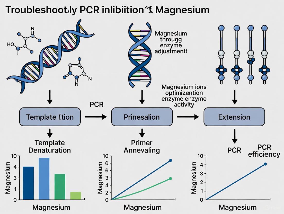
The diagram below illustrates this two-metal-ion mechanism and the logical process for troubleshooting related PCR issues.
Magnesium Optimization in PCR: Quantitative Guidelines
Q: How does magnesium chloride (MgCl₂) concentration specifically affect PCR efficiency and specificity, and what are the optimal ranges?
A: In the context of Polymerase Chain Reaction (PCR), Mg²⁺ serves as an essential cofactor for the DNA polymerase enzyme. Its concentration is a critical parameter that profoundly influences reaction success by affecting both enzyme activity and DNA duplex stability [3].
A comprehensive meta-analysis of PCR optimization studies has quantified the relationship between MgCl₂ concentration and PCR performance. The following table summarizes the key quantitative findings [3] [4]:
| Parameter | Effect of Increasing [MgCl₂] | Optimal Range | Key Quantitative Relationship |
|---|---|---|---|
| DNA Melting Temperature (Tₘ) | Increases | N/A | Every 0.5 mM increment within 1.5–3.0 mM range raises Tₘ by ~1.2°C [3] [4] |
| Reaction Efficiency | Increases to an optimum, then may decline | 1.5 – 3.0 mM (typical) | Highest efficiency observed within this logarithmic relationship [3] [4] |
| Specificity | Lower is generally better | Start at lower end (e.g., 1.5 mM) | High [MgCl₂] reduces stringency, promoting non-specific priming [5] [6] |
| Fidelity | Lower is better | Lower end of optimum | Excess Mg²⁺ increases misincorporation error rate [6] |
The optimal MgCl₂ concentration is not universal and is significantly influenced by template DNA characteristics [3] [4]:
- Template Complexity: Genomic DNA templates generally require higher Mg²⁺ concentrations compared to simple plasmid DNA.
- GC Content: GC-rich templates, which have higher intrinsic melting temperatures, often require more precise optimization of Mg²⁺, sometimes benefiting from concentrations in the higher end of the range to ensure efficient denaturation and primer annealing [3] [5].
- Presence of Chelators: If the reaction buffer or template sample contains EDTA, a higher concentration of MgCl₂ will be needed to compensate for the chelated ions [5].
Troubleshooting FAQs: Directly Addressing Experimental Challenges
Q: I am getting no amplification or a very low yield of my PCR product. Could magnesium be the issue?
A: Yes, insufficient Mg²⁺ concentration is a common cause of PCR failure. Mg²⁺ is a direct cofactor for the DNA polymerase; without enough, the enzyme cannot function efficiently [5] [7].
- Solution: Systematically increase the MgCl₂ concentration in 0.5 mM increments starting from a baseline (e.g., 1.5 mM) up to around 4.0-5.0 mM. Ensure that you are using the correct type of magnesium salt (usually MgCl₂, but some polymerases like Pfu may perform better with MgSO₄) [5] [6]. Also, verify that no chelating agents (like EDTA) are present in your template DNA solution, as they can sequester free Mg²⁺ [5].
Q: My PCR results in multiple bands or smeared products on the gel. How can adjusting magnesium help?
A: This is a classic symptom of low reaction stringency, often caused by excessive Mg²⁺ concentration. High Mg²⁺ levels stabilize DNA duplexes to the point where primers can anneal to non-target, partially complementary sequences on the template DNA [5] [6] [7].
- Solution: Systematically decrease the MgCl₂ concentration in 0.2-0.5 mM increments. Begin optimization from the lower end of the typical range (e.g., 1.0 mM) [6]. This increases stringency, forcing the primers to bind only to their perfect complements.
Q: Why does high magnesium concentration lead to increased errors in my PCR product sequence?
A: High fidelity is crucial for applications like cloning and sequencing. Excessive Mg²⁺ can compromise the base-selection fidelity of the DNA polymerase. While the exact structural rationale is complex, it is observed that high Mg²⁺ levels reduce the enzyme's ability to discriminate against incorrectly paired nucleotides (misincorporation), leading to a higher error rate in the final amplified product [6] [2].
- Solution: For high-fidelity applications, use a lower concentration of MgCl₂ within the polymerase's functional range and always ensure that the dNTPs are present in balanced, equimolar concentrations [6].
Research Reagent Solutions: Essential Materials for Experimentation
The following table details key reagents and their specific functions in studying and optimizing magnesium-dependent PCR.
| Reagent / Material | Function in Experimentation |
|---|---|
| MgCl₂ or MgSO₄ Solution | Provides the essential divalent cation cofactor (Mg²⁺) for DNA polymerase activity. The choice and concentration are primary optimization variables [5] [6]. |
| Hot-Start DNA Polymerase | A modified enzyme inactive at room temperature. Prevents non-specific amplification and primer-dimer formation during reaction setup, which can be exacerbated by Mg²⁺, leading to cleaner results [5] [7]. |
| dNTP Mix (Equimolar) | The balanced building blocks for DNA synthesis. Unbalanced dNTP concentrations can increase error rates, and the total dNTP concentration can affect free Mg²⁺ availability as they bind the ion [6]. |
| GC Enhancer / PCR Additives | Additives like betaine or DMSO help denature GC-rich secondary structures. This alters template requirements for Mg²⁺ and can improve amplification of difficult targets when used in combination with Mg²⁺ optimization [5] [6]. |
| Non-Hydrolyzable dNTP Analogs | Crucial research tools (e.g., dTMPPCP) used in X-ray crystallography to "trap" the catalytic intermediate for structural studies, allowing visualization of Mg²⁺ ions in the active site [1]. |
FAQs: Magnesium's Role in PCR
Q1: How does Mg²⁺ concentration specifically affect primer binding to the DNA template?
Mg²⁺ acts as a crucial cofactor that stabilizes the primer-template interaction by shielding the negative charges on the phosphate backbone of both DNA strands. This shielding reduces electrostatic repulsion, allowing the primer to anneal more efficiently to its complementary sequence. The ion's positive charge facilitates hydrogen bonding between complementary bases by neutralizing the repulsive forces that would otherwise prevent close association. Optimal Mg²⁺ concentrations (typically 1.5-2.5 mM for standard PCR) create the ideal ionic environment for stable primer binding without promoting non-specific interactions [5] [8].
Q2: What are the consequences of insufficient Mg²⁺ for DNA template stability?
Insufficient Mg²⁺ leads to several observable experimental failures:
- Weak or no amplification due to poor primer annealing and unstable template structure
- Reduced product yield as polymerase activity is suboptimal without its essential cofactor
- Increased non-specific binding when Mg²⁺ is too low to properly stabilize specific primer-template interactions
- Incomplete amplification of complex templates, particularly GC-rich regions that require additional stabilization [5] [9]
Q3: How does excessive Mg²⁺ concentration impact PCR specificity?
Excess Mg²⁺ promotes non-specific amplification through multiple mechanisms:
- Reduced stringency by stabilizing mismatched primer-template interactions
- Enhanced primer-dimer formation by facilitating annealing between complementary primer sequences
- Decreased fidelity by altering the polymerase's proofreading capability and promoting misincorporation
- Multiple bands or smearing on gels due to spurious amplification products [5] [9]
Troubleshooting Guides
Problem 1: No Amplification or Weak Band Intensity
| Possible Cause | Diagnostic Clues | Recommended Action |
|---|---|---|
| Insufficient Mg²⁺ | Faint or absent bands; may affect all samples equally | Increase Mg²⁺ concentration in 0.5 mM increments up to 5.0 mM [5] [8] |
| Mg²⁺ chelation | Previously working protocol fails with new template | Increase Mg²⁺ concentration to compensate for EDTA in template prep; ensure no residual chelators [5] |
| Incorrect annealing | Empty lanes or inconsistent results across primers | Optimize annealing temperature; verify primer Tm calculations account for Mg²⁺ concentration [9] |
Problem 2: Non-specific Amplification or Multiple Bands
| Possible Cause | Diagnostic Clues | Recommended Action |
|---|---|---|
| Excess Mg²⁺ | Primer-dimers, multiple bands, or smeared products | Decrease Mg²⁺ concentration in 0.2-1.0 mM increments; use hot-start polymerase [9] |
| Primer issues | Specific pattern of non-specific products | Redesign primers to avoid secondary structures; optimize concentration (0.1-1 μM) [9] [8] |
| Low annealing | Multiple bands of varying intensity | Increase annealing temperature stepwise (1-2°C increments); use gradient cycler [5] |
Problem 3: Inconsistent Results Between Replicates
| Possible Cause | Diagnostic Clues | Recommended Action |
|---|---|---|
| Uneven Mg²⁺ distribution | Variable results across identical reactions | Thoroughly mix Mg²⁺ solution and buffer before aliquotting; avoid freeze-thaw cycles [9] |
| Template contaminants | Inconsistent amplification with same template | Repurify template via alcohol precipitation or column cleanup; assess 260/280 ratio [9] |
| Inhibitor carryover | Specific template preparations fail | Use polymerases with high inhibitor tolerance; add BSA (10-100 μg/mL) to bind inhibitors [5] |
Magnesium Optimization Data
Table 1: Mg²⁺ Concentration Effects on PCR Performance
| Mg²⁺ Concentration (mM) | Amplification Efficiency | Specificity | Common Applications |
|---|---|---|---|
| <1.0 | Very low to none | High (if any product) | Not recommended; typically results from miscalculation |
| 1.5-2.0 | High | High | Standard targets; routine cloning; diagnostic PCR |
| 2.5-3.5 | High | Moderate | Complex templates (GC-rich, secondary structure) |
| 4.0-5.0 | Variable | Low | Special applications requiring enhanced processivity |
| >5.0 | Unpredictable | Very low | Not recommended; promotes significant artifacts |
Table 2: Magnesium-Dependent Protocol Adjustments for Challenging Templates
| Template Challenge | Mg²⁺ Adjustment | Complementary Modifications |
|---|---|---|
| GC-rich sequences | Increase to 3.0-4.0 mM | Add co-solvents (DMSO 1-10%, formamide 1.25-10%); increase denaturation temperature [5] [8] |
| Long amplicons (>5 kb) | Increase to 2.5-3.5 mM | Extend extension time; use polymerases with high processivity; reduce annealing temperature [5] |
| Low template copy number | Optimize 1.5-2.5 mM | Increase cycle number (up to 40); use high-sensitivity polymerases; add BSA (10-100 μg/mL) [5] [9] |
| High fidelity requirements | Strictly control 1.5-2.0 mM | Use high-fidelity polymerases; balance dNTP concentrations; minimize cycle number [9] |
Experimental Protocols
Protocol 1: Systematic Mg²⁺ Titration for Method Development
Purpose: To empirically determine the optimal Mg²⁺ concentration for a new PCR assay.
Reagents:
- 10X PCR Buffer (without Mg²⁺)
- 25 mM MgCl₂ solution
- dNTP mix (10 mM each)
- Forward and reverse primers (20 μM each)
- DNA template (10-100 ng/μL)
- DNA polymerase (0.5-2.5 units/50 μL reaction)
- Sterile distilled water
Methodology:
- Prepare a master mix containing all components except Mg²⁺ and template
- Aliquot equal volumes into 8 PCR tubes
- Add MgCl₂ to achieve final concentrations of: 0.5, 1.0, 1.5, 2.0, 2.5, 3.0, 4.0, and 5.0 mM
- Add template DNA to each tube
- Run thermal cycling using predetermined conditions
- Analyze products by agarose gel electrophoresis
- Select concentration yielding strongest specific product with minimal background [8]
Expected Results: A clear optimum concentration where specific product is maximal and non-specific amplification is minimal.
Protocol 2: Magnesium Rescue for Problematic Amplifications
Purpose: To salvage PCR reactions showing poor yield or specificity through targeted Mg²⁺ adjustment.
Diagnostic Steps:
- Analyze failed reaction on agarose gel to characterize failure mode
- For no amplification: Increase Mg²⁺ by 0.5-1.0 mM increments
- For non-specific bands: Decrease Mg²⁺ by 0.2-0.5 mM increments
- For primer-dimer formation: Combine Mg²⁺ reduction with hot-start activation
- Test optimized condition alongside original for direct comparison
Validation:
- Sequence products from optimized conditions to verify specificity
- Compare yield using quantitative methods if available
- Establish reproducibility across multiple template preparations [5] [9]
Advanced Magnesium Mechanisms
Structural Role of Mg²⁺ in DNA Polymerization
The essential function of Mg²⁺ extends beyond general charge shielding to specific structural roles in the polymerase active site. The two-metal-ion mechanism is conserved across DNA polymerases, where one Mg²�+ (Metal A) activates the 3'-OH nucleophile for attack on the α-phosphate, while the second Mg²⁺ (Metal B) stabilizes the negative charge on the pyrophosphate leaving group [10] [11].
Recent research has revealed a third catalytic Mg²⁺ ion that appears transiently during phosphodiester bond formation. This third metal ion (Metal C) coordinates with the DNA product and pyrophosphate after bond formation and provides the final energetic push for catalysis. The binding site for this third Mg²⁺ only becomes accessible after thermal activation of the enzyme-substrate complex, creating an opening for metal entry [12].
Mg²⁺ Coordination Chemistry in Template Stabilization
Mg²⁺ stabilizes the DNA template through specific coordination geometries:
- Inner-sphere coordination directly to phosphate oxygens
- Outer-sphere coordination through water molecules
- Sequence-dependent affinity with variation across different template regions
The following diagram illustrates the complete magnesium-dependent workflow for PCR optimization and troubleshooting:
The Scientist's Toolkit: Research Reagent Solutions
Table 3: Essential Reagents for Magnesium Optimization Studies
| Reagent | Function | Technical Considerations |
|---|---|---|
| MgCl₂ solutions | Most common Mg²⁺ source for PCR | Hygroscopic; prepare fresh aliquots; concentration must be verified [8] |
| MgSO₄ solutions | Alternative for some polymerases | Preferred for certain proofreading enzymes like Pfu; check polymerase specificity [5] |
| Mg-free buffers | Enable precise concentration control | Essential for systematic titration; verify absence of contaminating Mg²⁺ [8] |
| Chelator resins | Remove contaminating metals | Useful for establishing baseline; can be included in template purification [9] |
| Hot-start polymerases | Limit non-specific amplification | Reduce primer-dimer formation; particularly valuable when using higher Mg²⁺ [5] [13] |
| PCR additives | Modulate Mg²⁺ effectiveness | DMSO, formamide, betaine can affect Mg²⁺ availability and requirement [8] |
The critical relationship between Mg²⁺ and DNA template stability represents a fundamental parameter in PCR optimization. Through systematic investigation of magnesium concentration effects and implementation of targeted troubleshooting protocols, researchers can overcome amplification challenges and develop robust, reproducible genetic analyses. The dual role of Mg²⁺ as both catalytic cofactor and structural stabilizer underscores its unique position as the most impactful adjustable parameter in PCR optimization.
FAQs on MgCl2 in PCR
Q1: What is the fundamental role of MgCl2 in a PCR reaction? Magnesium chloride (MgCl2) is an essential cofactor for DNA polymerase activity. It stabilizes the primer-template complexes by neutralizing the negative charges on the DNA backbone, thereby facilitating proper enzyme function and influencing the overall thermodynamics and kinetics of DNA denaturation and annealing [3].
Q2: How does MgCl2 concentration quantitatively affect DNA melting temperature (Tm)? Recent meta-analyses have demonstrated a significant logarithmic relationship between MgCl2 concentration and DNA melting temperature. Within the critical range of 1.5 to 3.0 mM, every increment of 0.5 mM in MgCl2 concentration consistently raises the melting temperature by approximately 1.2°C [3] [4]. This quantitative relationship is crucial for predicting and controlling PCR stringency.
Q3: What are the consequences of using a suboptimal MgCl2 concentration?
- Insufficient MgCl2: Reduces PCR yield and can lead to complete amplification failure due to inefficient primer binding and poor DNA polymerase activity [5] [14].
- Excessive MgCl2: Promotes non-specific amplification, resulting in multiple unwanted bands or smears on a gel. It also reduces PCR fidelity by increasing the misincorporation of nucleotides [5] [7] [14].
Q4: How do I determine the optimal MgCl2 concentration for my specific PCR assay? The optimal concentration is template-dependent. The table below summarizes general guidelines, but empirical optimization is recommended.
| Template Characteristic | Recommended MgCl2 Starting Range | Rationale for Adjustment |
|---|---|---|
| Standard/Simple Templates | 1.5 - 2.0 mM | Provides sufficient cofactor activity without significant nonspecific binding. |
| Genomic DNA Templates [3] [14] | 2.0 - 4.0 mM | Higher complexity and potential presence of chelators require more Mg2+ ions. |
| GC-Rich Templates [3] | 2.0 - 4.0 mM | Higher Mg2+ helps stabilize the stronger double-stranded DNA against denaturation. |
| Reactions with EDTA Contamination [14] | Increase as needed | EDTA chelates Mg2+ ions; concentration must be increased to compensate. |
| Reactions with High dNTPs [14] | Increase as needed | dNTPs bind Mg2+; the Mg2+ concentration should generally be 1-2 mM higher than the total dNTP concentration. |
Q5: My PCR shows no product. Could MgCl2 be the issue? Yes. No amplification or low yield is a common problem that can often be resolved by optimizing MgCl2 concentration alongside other parameters like annealing temperature and template quality [7] [14]. A systematic troubleshooting approach is outlined in the experimental protocol section below.
Troubleshooting Guide: Common PCR Problems and Magnesium-Based Solutions
Problem 1: No Amplification or Low Yield
Possible Causes & Solutions:
- Cause: Insufficient Mg2+ concentration. Mg2+ ions are a necessary cofactor for DNA polymerase; without enough, the enzyme cannot function efficiently [5] [14].
- Solution: Perform a MgCl2 titration experiment, testing concentrations in the range of 1.0 mM to 4.0 mM in 0.5 mM increments [14].
- Other checks: Confirm template quality and quantity, and verify primer design and annealing temperature [5] [15].
Problem 2: Non-Specific Products or High Background
Possible Causes & Solutions:
- Cause: Excessive Mg2+ concentration. High Mg2+ concentrations stabilize weak, non-specific primer-template interactions, leading to amplification of incorrect sequences [5] [7].
- Solution: Titrate down the MgCl2 concentration. Start from the manufacturer's recommended concentration and decrease in 0.25 mM steps [5].
- Other checks: Increase the annealing temperature, switch to a hot-start DNA polymerase, and verify primer specificity [5] [7] [15].
Problem 3: Primer-Dimer Formation
Possible Causes & Solutions:
- Cause: While often a primer design issue, high Mg2+ concentrations can exacerbate primer-dimer formation by stabilizing the short, self-complementary sequences between primers [7].
- Solution: Optimize MgCl2 concentration and primer concentration (typically 0.1-1 μM). Using hot-start enzymes can also prevent low-temperature artifacts [5] [7] [14].
Experimental Protocols for MgCl2 Optimization
Protocol 1: Standard MgCl2 Titration
This protocol is the first step in systematically optimizing a new PCR assay.
Research Reagent Solutions:
| Item | Function in Experiment |
|---|---|
| MgCl2 Stock Solution (e.g., 25 mM or 50 mM) | To create a gradient of Mg2+ concentrations in the final PCR reaction. |
| DNA Polymerase with Separate Mg-free Buffer | Essential for being able to adjust the Mg2+ concentration freely. |
| Template DNA (optimized quantity) | The target DNA to be amplified; its quality is critical. |
| Primers (well-designed, high purity) | Short sequences that define the region to be amplified. |
| dNTP Mix | The building blocks (nucleotides) for new DNA strands. |
| Nuclease-Free Water | To bring the reaction to the final volume without degrading components. |
Methodology:
- Prepare a Master Mix: Combine all common reaction components (buffer, dNTPs, primers, polymerase, template, water) for all reactions except MgCl2.
- Aliquot the Master Mix into multiple PCR tubes.
- Add MgCl2: Add the MgCl2 stock solution to each tube to create a final concentration series (e.g., 1.0, 1.5, 2.0, 2.5, 3.0, 3.5, 4.0 mM). Include a control with no MgCl2 to confirm the necessity of the cofactor.
- Run PCR: Use standard cycling conditions for your template.
- Analyze Results: Separate the PCR products on an agarose gel. The optimal condition is the one that produces the highest yield of the desired specific product with the least background.
Protocol 2: Combined Annealing Temperature and MgCl2 Optimization
For challenging assays, a two-dimensional optimization of both annealing temperature and MgCl2 concentration is the most robust approach. The following workflow visualizes this systematic process.
Troubleshooting Guide: Common PCR Inhibition Scenarios
This guide helps diagnose and resolve common PCR inhibition issues encountered with biomedical samples.
| Observation | Possible Cause | Recommended Solution |
|---|---|---|
| No or weak amplification [16] [17] | PCR inhibitors in template sample (e.g., heparin, hemoglobin, urea) [17] | Dilute template 100-fold or re-purify using a clean-up kit [17]. Use polymerases resistant to impurities (e.g., Terra PCR Direct) [17]. |
| Suboptimal Mg²⁺ concentration [16] | Test a Mg²⁺ gradient from 0.5 mM to 4.0 mM in 0.5 mM increments [18]. | |
| Incorrect annealing temperature [16] | Perform an annealing temperature gradient, starting 5°C below the primer's Tm [16]. | |
| Non-specific bands or multiple products [16] [17] | Presence of metal ion inhibitors (e.g., Zn²⁺, Fe²⁺, Cu²⁺, Ca²⁺) [19] | Add chelating agents like EGTA for Ca²⁺ inhibition [19]. Use a hot-start polymerase to prevent premature replication [16]. |
| Annealing temperature too low [16] [17] | Increase annealing temperature in 2°C increments [17]. | |
| Too much template or primer [16] [20] | Reduce template amount by 2-5 fold [17]. Ensure primer concentration is between 0.05-1 µM [16]. | |
| Smearing on the gel [17] | Overcycling or excessive template [17] | Reduce the number of PCR cycles. Use less template DNA [17]. |
| Contamination (e.g., previous PCR products) [17] | Run a negative control (no template). Decontaminate workspace and equipment with 10% bleach or UV light [17]. | |
| Errors in sequence [16] | Unbalanced dNTP concentrations [16] | Prepare fresh dNTP mixes to ensure balanced concentrations [16]. |
| Low-fidelity polymerase [16] | Switch to a high-fidelity polymerase with proofreading activity (e.g., Q5 or Phusion) [16]. |
Frequently Asked Questions (FAQs)
1. My PCR failed and I suspect a common inhibitor. What are the most likely culprits? Inhibitors are often sample-specific. Common offenders include:
- Blood-derived samples: Heparin, hemoglobin, lactoferrin, and immunoglobulin G (IgG) [17] [21].
- Tissues: Collagen, myoglobin, and melanin [17].
- Plant materials: Polyphenols, polysaccharides, and pectin [17].
- Forensic or environmental samples: Humic acids, tannic acids, and metal ions (e.g., from soil or metal surfaces) [17] [19]. A significant dilution of the template (e.g., 100-fold) can often reduce inhibition sufficiently for amplification [17].
2. How do I optimize magnesium concentration for a difficult PCR? Magnesium is a critical cofactor for polymerase activity, and its optimal concentration is often template and enzyme-specific [18].
- Standard Range: Most PCRs use 1.5 to 2.0 mM Mg²⁺ [18] [21].
- Optimization Strategy: If your PCR is failing, test a concentration gradient. For GC-rich templates, try increments of 0.5 mM between 1.0 and 4.0 mM to find the ideal concentration [18]. Remember that too much Mg²⁺ can cause non-specific binding, while too little reduces polymerase activity [18].
3. What can I do if my template has high GC content? GC-rich templates (>60%) form stable secondary structures that hinder amplification [18]. A multi-pronged approach is best:
- Specialized Reagents: Use a polymerase and buffer system specifically designed for GC-rich templates (e.g., OneTaq DNA Polymerase with GC Buffer or Q5 High-Fidelity DNA Polymerase with GC Enhancer) [18].
- Additives: Incorporate additives like DMSO (1-10%), formamide (1.25-10%), or betaine, which help reduce secondary structure formation [18] [21].
- Protocol Adjustments: Use a higher denaturation temperature (98°C) and a "hot-start" technique to increase specificity [21].
4. My negative control shows a product, indicating contamination. How do I resolve this? Contamination requires strict laboratory practices:
- Physical Separation: Maintain separate pre-PCR and post-PCR areas. Never bring reagents or equipment from the post-PCR area back to the clean pre-PCR area [17].
- Dedicated Equipment: Use separate sets of pipettes, tips with aerosol filters, lab coats, and consumables for each area [17].
- Decontamination: Clean workstations and pipettes with 10% bleach and expose them to UV light in a laminar flow cabinet [17].
- Aliquot Reagents: Prepare and store small aliquots of all PCR reagents to avoid contaminating bulk stocks [17].
Experimental Protocol: Systematic Optimization of Magnesium Chloride Concentration
1. Objective To empirically determine the optimal MgCl₂ concentration for efficient and specific amplification of a target DNA sequence.
2. Background Mg²⁺ is an essential cofactor for DNA polymerase activity. It facilitates primer binding and catalyzes the phosphodiester bond formation between nucleotides [18]. The presence of inhibitors in a sample can chelate Mg²⁺, reducing its effective concentration. Conversely, supra-optimal concentrations can reduce fidelity and specificity [16] [18]. This protocol outlines a method to test a range of Mg²⁺ concentrations.
3. Materials
- Template DNA
- Forward and reverse primers
- 10X PCR buffer (without MgCl₂)
- 25 mM MgCl₂ stock solution
- dNTP mix
- DNA polymerase
- Nuclease-free water
- Thermal cycler
4. Procedure
- Prepare Master Mix: Create a master mix for all reactions, excluding MgCl₂ and template DNA.
- Dilute MgCl₂ Stock: Prepare the MgCl₂ stock solution to facilitate accurate pipetting of small volumes.
- Set Up Reactions: Label thin-wall PCR tubes. Aliquot the master mix into each tube.
- Create Mg²⁺ Gradient: Add different volumes of the MgCl₂ stock to each tube to create a final concentration series (e.g., 0.5, 1.0, 1.5, 2.0, 2.5, 3.0, 3.5, 4.0 mM) in a 50 µL reaction.
- Add Template and Amplify: Add template DNA to each tube and run the PCR using predetermined cycling conditions.
- Analyze Results: Separate PCR products by agarose gel electrophoresis. Identify the Mg²⁺ concentration that yields the strongest specific product with the least background.
Research Reagent Solutions for PCR Inhibition
This table lists key reagents used to overcome PCR inhibition.
| Reagent | Function / Rationale | Example Use Case |
|---|---|---|
| High-Fidelity Polymerases [16] [21] | Enzymes with 3'→5' exonuclease (proofreading) activity for high accuracy and processivity on complex templates. | Cloning, sequencing, amplifying long or difficult templates (e.g., Q5 High-Fidelity DNA Polymerase) [16] [21]. |
| Hot-Start Polymerases [16] [21] | Polymerase is inactive at room temperature, preventing non-specific priming and primer-dimer formation before the PCR begins. | Reactions with high primer complexity or prone to mispriming (e.g., OneTaq Hot Start DNA Polymerase) [16]. |
| GC Enhancer / Additives [18] | A proprietary mixture of reagents that disrupt secondary structures in GC-rich DNA and increase primer stringency. | Amplification of GC-rich targets (>60% GC); used with specific polymerases like OneTaq or Q5 [18]. |
| BSA (Bovine Serum Albumin) [21] | Acts as a stabilizer and competitor, binding to inhibitors present in the sample (e.g., phenolic compounds, humic acids). | Amplification from complex biological samples like blood or plant extracts [21]. |
| DMSO (Dimethyl Sulfoxide) [18] [21] | Disrupts secondary DNA structures by reducing the melting temperature (Tm) of DNA. | Standard aid for amplifying GC-rich templates; typical final concentration of 1-10% [21]. |
| EGTA (Ethylene glycol-bis...) [19] | A calcium-specific chelator. Binds and inactivates Ca²⁺ ions, which competitively inhibit Taq polymerase by binding in place of Mg²⁺. | Reversing PCR inhibition from bone samples or other sources high in calcium [19]. |
Mechanism of Metal Ion Inhibition and Chelator Rescue
Metal ions inhibit PCR through multiple mechanisms. Divalent cations like Ca²⁺ can compete with the essential cofactor Mg²⁺ for the polymerase's active site, directly impairing enzyme function [19]. Other ions, such as Cu²⁺ and Al³⁺, can bind directly to DNA bases or the phosphate backbone, creating crosslinks that block polymerase progression [19]. The diagram below illustrates this pathway and the restorative action of chelators.
Magnesium ions (Mg²⁺) are fundamental cofactors in molecular biology, especially in polymerase chain reaction (PCR). Mg²⁺ is essential for DNA polymerase activity, influencing enzyme fidelity, processivity, and the overall efficiency of nucleic acid amplification [7] [3]. However, the effective concentration of Mg²⁺ available for PCR is not simply the amount added to the reaction mix; it is a dynamic value subject to reduction by various chelators and competitors present in the sample or reagents. Understanding this delicate balance is crucial for troubleshooting PCR inhibition and achieving consistent, reliable amplification results.
Core Scientific Principles: The Chemistry of Mg²⁺ Binding
The Chelation of Mg²⁺ by Metabolites and Reagents
In a cellular context, the vast majority of Mg²⁺ is not free in solution but is bound to a variety of molecules. The total cellular Mg²⁺ concentration can be remarkably high (estimated at 20-80 mM), yet the concentration of free, hydrous Mg²⁺ ions is much lower, typically between 0.5-3.0 mM in both eukaryotic and bacterial cells [22]. This difference is due to chelation.
Mechanisms of Chelation: Chelation occurs through two primary types of interactions between divalent Mg²⁺ ions and organic molecules [23]:
- Electrostatic Interactions: Positively charged Mg²⁺ ions interact with negatively charged carboxyl groups (–COO⁻) found in the side chains of amino acids like glutamic acid (Glu) and aspartic acid (Asp).
- Coordination Covalent Interactions: Mg²⁺ ions pair with atoms possessing unshared electrons, such as those in carbonyl groups (C=O), amino groups (–NH₂) at the N-terminus and in side chains of lysine (Lys) and arginine (Arg), and the imidazole ring of histidine (His).
The table below summarizes common laboratory substances known to chelate Mg²⁺, many of which can be introduced into a PCR via sample carryover or buffer components.
Table 1: Common Mg²⁺ Chelators and Competitors in a Laboratory Setting
| Compound Category | Specific Examples | Mechanism of Action |
|---|---|---|
| Nucleotides | ATP, dNTPs | Phosphate groups chelate Mg²⁺; the Mg²⁺-ATP complex is the predominant form in cells [24] [25] [26]. |
| Amino Acids | Glutamate, Aspartate, Histidine, Arginine, Lysine | Carboxylate, imidazole, and amino groups act as binding sites for Mg²⁺ [23] [22]. |
| Chelating Agents | EDTA, EGTA | Purpose-built to tightly bind divalent cations like Mg²⁺ and Ca²⁺, effectively removing them from solution. |
| Organic Acids | Citrate, Isocitrate | Carboxyl groups chelate Mg²⁺, a principle used in some methods to measure free Mg²⁺ [24]. |
| Inorganic Ions | K⁺, Na⁺ | Can indirectly affect Mg²⁺-dependent processes by altering the electrostatic environment and DNA duplex stability [5]. |
The Critical Role of Free vs. Bound Mg²⁺ in PCR
In PCR, the DNA polymerase enzyme strictly requires free Mg²⁺ ions as a cofactor. The Mg²⁺ ions facilitate the binding of the dNTPs to the enzyme's active site and stabilize the transition state during the phosphoryl transfer reaction [3]. When chelators are present, they bind a portion of the Mg²⁺, reducing the free concentration available for the polymerase. This can lead to:
- A direct reduction in polymerase activity.
- Altered enzyme fidelity.
- Changes in the melting temperature (Tm) of the DNA template, which can affect primer annealing and overall amplification efficiency [3].
The following diagram illustrates how various factors compete for and influence the pool of free Mg²⁺ in a typical PCR.
Diagram 1: Factors influencing the free Mg²⁺ pool in PCR.
FAQs & Troubleshooting Guides
Q1: My PCR shows no amplification or very low yield after I added my purified DNA template. I suspect Mg²⁺ chelation. What is the first step I should take?
A: The most effective first step is to quantify the purity of your DNA template. Use spectrophotometry (A260/A280 and A260/A230 ratios) to check for contaminants. Common chelators like phenol, EDTA, or residual salts from the purification process can be carried over with your DNA [5]. If purity is low, re-purify your DNA template using a method proven to remove these contaminants, such as ethanol precipitation with 70% ethanol washes [5].
Q2: How can I determine the optimal MgCl₂ concentration for my specific PCR assay, especially when working with complex samples?
A: We recommend performing a MgCl₂ titration experiment. Set up a series of identical PCR reactions, varying only the MgCl₂ concentration. A typical range is 1.0 mM to 4.0 mM in increments of 0.5 mM [7] [3]. Using a thermal cycler with a gradient function for the annealing temperature can further refine optimization. The goal is to identify the concentration that yields the highest amount of specific product with the least background.
Table 2: MgCl₂ Optimization Guide Based on PCR Symptom
| PCR Symptom | Possible Cause | Recommended MgCl₂ Adjustment | Additional Actions |
|---|---|---|---|
| No/Low Yield | Free [Mg²⁺] too low; chelation by dNTPs/template | Increase concentration in 0.5 mM steps [7] | Verify template quality/purity; check dNTP concentrations [5] |
| Non-specific Bands/High Background | Free [Mg²⁺] too high; reduced reaction stringency | Decrease concentration in 0.5 mM steps [5] | Increase annealing temperature; use hot-start polymerase [7] [5] |
| Poor Efficiency with GC-Rich Templates | Inadequate denaturation; stable secondary structures | Increase concentration (e.g., up to 4-5 mM) [3] | Use PCR additives (e.g., DMSO, betaine); increase denaturation temp [5] |
| Smeared Bands | Multiple causes, including Mg²⁺ imbalance | Optimize via titration | Check for contaminating nucleases; ensure primer specificity; separate pre- and post-PCR areas [7] |
Q3: I am using a pre-made PCR master mix. Do I still need to worry about Mg²⁺ optimization?
A: While master mixes are convenient and robust, they are not universal solutions. If you are amplifying a challenging template (e.g., high GC-content, long amplicon, or from a complex sample like blood or plant tissue) or if your amplification efficiency is suboptimal, you should still consider optimizing Mg²⁺. Many master mixes are supplied with a separate vial of MgCl₂ solution for this exact purpose. Consult your manufacturer's protocol for guidance on supplemental Mg²⁺.
Essential Protocols & Research Reagent Solutions
Protocol: Standard MgCl₂ Titration for PCR Optimization
Principle: To empirically determine the optimal free Mg²⁺ concentration for a specific primer-template combination by testing a range of concentrations.
Materials:
- 10X PCR Buffer (without MgCl₂)
- 50 mM MgCl₂ stock solution
- dNTP mix (e.g., 10 mM each)
- Forward and Reverse Primers (10 μM each)
- DNA Template
- DNA Polymerase (e.g., hot-start Taq polymerase)
- Nuclease-free Water
Method:
- Prepare a master mix for 8 reactions, containing 1X PCR buffer, 0.2 mM dNTPs, 0.5 μM of each primer, 1.25 U of DNA polymerase, and a constant amount of DNA template in a total volume of 24 μL per reaction.
- Aliquot 24 μL of the master mix into each of eight 0.2 mL PCR tubes.
- Add MgCl₂ to each tube to achieve the following final concentrations: 1.0 mM, 1.5 mM, 2.0 mM, 2.5 mM, 3.0 mM, 3.5 mM, 4.0 mM, and 4.5 mM. For example, to make a 25 μL reaction with 2.0 mM MgCl₂, add 1.0 μL of the 50 mM MgCl₂ stock.
- Run the PCR using your standard thermal cycling parameters.
- Analyze the results by agarose gel electrophoresis. Identify the MgCl₂ concentration that produces the strongest, cleanest band of the expected size.
The Scientist's Toolkit: Key Reagents for Investigating Mg²⁺ Effects
Table 3: Essential Research Reagents for Mg²⁺-Related Studies
| Reagent / Tool | Function / Explanation |
|---|---|
| MgCl₂ / MgSO₄ | The source of free Mg²⁺ ions. The choice of salt can depend on polymerase preference (e.g., Pfu polymerase often works better with MgSO₄) [5]. |
| EDTA / EGTA | Chelating agents used to create low/no Mg²⁺ conditions in control experiments or to study metal dependence. They can also be contaminants that inhibit PCR [5] [26]. |
| Hot-Start DNA Polymerase | A modified polymerase inactive at room temperature, preventing non-specific primer extension and primer-dimer formation that can waste Mg²⁺ and dNTPs during reaction setup [7] [5]. |
| PCR Additives (e.g., BSA, Betaine) | Help overcome inhibition. BSA can bind inhibitors carried over from samples. Betaine can destabilize secondary structures in GC-rich templates, reducing Mg²⁺ demands for denaturation [7] [5]. |
| Mg²⁺-Sensitive Dyes / Assays | Tools like 4-(2-pyridylazo) resorcinol can be used to quantitatively measure free Mg²⁺ concentration in solution [23]. |
Advanced Concepts & Research Perspectives
Emerging research continues to reveal the nuanced roles of Mg²⁺ beyond its function as a simple enzyme cofactor. Studies on ion channels like MgtE and RyR1 have shown that Mg²⁺ binding to specific sites with different affinities can induce allosteric changes, regulating gating and function [27] [26]. Furthermore, the concept that weakly chelated Mg²⁺ (e.g., in complexes with metabolites like ATP and glutamate) can positively influence biochemical processes, such as RNA folding and stability, is gaining traction [22]. This suggests that the traditional binary view of "free" vs. "bound" Mg²⁺ is an oversimplification, and the activity of Mg²⁺-dependent enzymes like DNA polymerases may be influenced by a more complex equilibrium of Mg²⁺ species in solution. For PCR troubleshooting, this underscores the importance of considering the complete chemical environment of the reaction, not just the gross concentration of added MgCl₂.
Systematic Optimization of Magnesium Concentration: A Step-by-Step Protocol
FAQ: Polymerase and Magnesium Concentration Basics
Why is magnesium concentration so critical for PCR?
Magnesium ions (Mg²⁺) are an essential cofactor for DNA polymerase activity [28]. They directly facilitate the formation of phosphodiester bonds between nucleotides during DNA synthesis [28]. The concentration of Mg²⁺ also influences the melting and annealing temperatures of DNA by stabilizing the double helix through interactions with the phosphate backbone [3]. An incorrect concentration can lead to a complete failure of the reaction or the production of non-specific products [29] [30].
How do I choose a starting magnesium concentration?
The optimal magnesium concentration depends on your specific polymerase, buffer composition, and template. A good starting point is often 1.5 mM to 2.0 mM for standard Taq DNA Polymerase [29]. However, you should always consult the manufacturer's protocol for your specific enzyme, as recommendations can vary. For instance, Pfu DNA polymerase may perform better with MgSO₄ than with MgCl₂ [5]. From there, optimization should be performed by testing a range of concentrations, typically in 0.5 mM increments up to 4 mM [29] [31].
What happens if the magnesium concentration is too high or too low?
The following table summarizes the effects of suboptimal magnesium concentrations:
| Magnesium Condition | Consequences for PCR |
|---|---|
| Concentration Too Low | - Greatly reduced or no PCR yield [31] [30]- Poor polymerase activity due to insufficient cofactor [28] |
| Concentration Too High | - Accumulation of non-specific PCR products [29] [31]- Increased formation of primer-dimers [30]- Reduced fidelity (higher error rate) [5] |
Recommended Starting Concentrations for Common Polymerases
The table below provides a general guideline for the starting concentrations of key PCR components. Always verify against the specific manufacturer's instructions for the enzyme you are using.
Table 1: Baseline Reaction Component Concentrations
| Reaction Component | Standard Taq Polymerase [29] | High-Fidelity Polymerases (e.g., Q5, Phusion) [31] | Notes |
|---|---|---|---|
| DNA Polymerase | 0.5 - 2.0 units/50 µL | As per manufacturer | Enzyme amounts are typically given in units. Adjust if inhibitors are present [28]. |
| Mg²⁺ (Final Conc.) | 1.5 - 2.0 mM | Varies by enzyme | MgCl₂ is common; some polymerases prefer MgSO₄ [5]. This is a primary optimization variable. |
| Primers (Each) | 0.1 - 0.5 µM | 0.3 - 0.5 µM | Higher concentrations can promote mispriming [28]. |
| dNTPs (Each) | 200 µM | 200 µM | Use equimolar concentrations for high fidelity [5]. Higher concentrations may require more Mg²⁺ [28]. |
| Template DNA | Genomic: 1 ng - 1 µg; Plasmid: 1 pg - 10 ng | Genomic: 1 ng - 1 µg; Plasmid: 1 pg - 10 ng | Higher DNA concentrations can decrease specificity [29]. |
Experimental Protocol: Systematic Optimization of Magnesium Chloride
This protocol provides a detailed methodology for empirically determining the optimal MgCl₂ concentration for your specific PCR assay.
Objective
To identify the MgCl₂ concentration that yields the highest amount of the specific target product with minimal background in a 25 µL PCR reaction.
Materials
- DNA Template: Your target DNA (e.g., genomic, plasmid).
- Primers: Forward and reverse primers specific to your target.
- PCR Master Mix: A core mix containing buffer, dNTPs, and polymerase. Alternatively, individual components: 10X reaction buffer (without MgCl₂), dNTP mix, Taq or other DNA polymerase.
- MgCl₂ Stock Solution: 25 mM or 50 mM stock.
- Nuclease-free Water
- Thermal Cycler
- Agarose Gel Electrophoresis equipment
Procedure
Prepare a Master Mix: Create a master mix for all reactions to minimize pipetting error. For n reactions, prepare a mix for n+1. The master mix should contain, per reaction:
- 1X Reaction Buffer (without MgCl₂)
- 200 µM of each dNTP
- 0.2 µM of each forward and reverse primer
- 1 unit of DNA Polymerase
- Template DNA (e.g., 10-50 ng genomic DNA)
- Nuclease-free water to a final volume of 23 µL (after adding MgCl₂).
Aliquot and Supplement with MgCl₂: Aliquot 23 µL of the master mix into each PCR tube. Then, add MgCl₂ from a stock solution to achieve the desired final concentration in a 25 µL total volume. A suggested gradient is shown below.
Run the PCR: Place the tubes in a thermal cycler and run using your standard cycling program.
Analyze the Results: Separate the PCR products using agarose gel electrophoresis. Visualize the bands under UV light.
Table 2: Example MgCl₂ Optimization Test Gradient
| Tube Number | Volume of 50 mM MgCl₂ Stock to Add (µL) | Final MgCl₂ Concentration (mM) |
|---|---|---|
| 1 | 0.5 | 1.0 |
| 2 | 0.75 | 1.5 |
| 3 | 1.0 | 2.0 |
| 4 | 1.25 | 2.5 |
| 5 | 1.5 | 3.0 |
| 6 | 1.75 | 3.5 |
| 7 | 2.0 | 4.0 |
Data Interpretation
Identify the reaction tube that produces a single, intense band of the expected size. A concentration that yields a strong specific product with no or faint non-specific bands is optimal. If no product is visible across the entire range, consider a wider concentration range or troubleshoot other parameters like primer design and annealing temperature [31].
The Scientist's Toolkit: Research Reagent Solutions
Table 3: Essential Reagents for PCR Optimization
| Reagent | Function in PCR | Key Considerations |
|---|---|---|
| Hot-Start DNA Polymerase | Reduces non-specific amplification and primer-dimer formation by remaining inactive at room temperature [5] [31]. | Critical for complex templates. Available in antibody-based or chemically modified formats. |
| Magnesium Salts (MgCl₂, MgSO₄) | Essential cofactor for polymerase activity; stabilizes DNA double helix [28] [3]. | Concentration is critical. MgCl₂ is standard; MgSO₄ is preferred for some high-fidelity polymerases [5]. |
| PCR Enhancers/Additives | Help amplify difficult templates (e.g., GC-rich, with secondary structure) and mitigate inhibition [5] [32]. | Common examples: DMSO, formamide, BSA, Tween-20, betaine, and glycerol. Must be optimized as they can inhibit PCR at high concentrations [32] [33]. |
| dNTP Mix | The building blocks (dATP, dCTP, dGTP, dTTP) for new DNA strand synthesis [28]. | Use equimolar concentrations to maintain fidelity. High concentrations can chelate Mg²⁺, making it unavailable for the polymerase [28] [29]. |
| Nuclease-Free Water | The solvent for the reaction, ensuring no enzymatic degradation of primers or template. | A critical quality control measure; always use certified nuclease-free water. |
Troubleshooting Logic: Addressing PCR Inhibition via Magnesium Optimization
The following diagram outlines a logical workflow for troubleshooting PCR inhibition, with a focus on assessing and adjusting magnesium concentration.
How does magnesium concentration affect PCR?
Magnesium (Mg²⁺) is an essential cofactor for thermostable DNA polymerases. Its concentration directly influences enzyme activity, fidelity, and specificity. The Mg²⁺ ions form a soluble complex with the dNTPs, which is the actual substrate the polymerase recognizes. An incorrect Mg²⁺ concentration is a common cause of PCR failure and can manifest as no product, non-specific products, or smeared bands on a gel.
- Too low: Insufficient Mg²⁺ can result in low enzyme activity or no PCR product at all [34].
- Too high: Excessive Mg²⁺ can reduce specificity, leading to non-specific amplification and incorrect products [34] [5]. It can also increase the error rate, reducing the fidelity of the amplified product [35] [5].
What are the recommended starting ranges and adjustment increments for Mg²⁺?
The optimal Mg²⁺ concentration must be determined empirically for each primer-template combination. The table below summarizes the general guidance for optimization.
Table 1: Mg²⁺ Concentration Optimization Guidelines
| Parameter | Recommended Range & Increments | Notes & Considerations |
|---|---|---|
| Typical Starting Range | 1.5 - 2.0 mM [34] | This is the standard for Taq DNA Polymerase. Always refer to the specific polymerase's manufacturer guidelines. |
| Broad Optimization Range | 0.5 - 5.0 mM [8] | A wider range may be needed for specialized applications or problematic templates. |
| Optimization Increments | 0.2 - 1.0 mM [35] [34] | Fine-tuning in 0.2 mM increments is often effective. Larger 0.5 - 1.0 mM steps can be used for an initial broad search. |
| Critical Cofactors | dNTPs (200 µM typical) [34] | Mg²⁺ concentration must be optimized relative to dNTP concentration, as Mg²⁺ chelates dNTPs [5]. |
How do I set up an experiment to optimize Mg²⁺ concentration?
This protocol provides a detailed methodology for determining the optimal Mg²⁺ concentration for your PCR assay.
Materials and Reagents
- PCR-grade water
- 10X PCR Buffer (without MgCl₂)
- MgCl₂ or MgSO₄ solution (e.g., 25 mM or 50 mM stock)
- dNTP Mix (e.g., 10 mM)
- Forward and Reverse Primers
- DNA Template
- Thermostable DNA Polymerase (e.g., Taq polymerase)
- Thin-walled PCR tubes or plates
- Thermal Cycler
Table 2: Key Research Reagent Solutions
| Reagent | Function in the Experiment |
|---|---|
| MgCl₂ / MgSO₄ Stock Solution | The variable being tested; provides the Mg²⁺ cofactor for the DNA polymerase. |
| 10X PCR Buffer (Mg-free) | Provides the stable pH and ionic environment for the reaction, allowing for precise Mg²⁺ titration. |
| dNTP Mix | The building blocks for DNA synthesis; their concentration directly influences the required Mg²⁺ level. |
| Hot-Start DNA Polymerase | Reduces non-specific amplification and primer-dimer formation during reaction setup, leading to cleaner optimization results [35] [5]. |
Experimental Workflow
The following diagram illustrates the logical workflow for the Mg²⁺ optimization experiment.
Step-by-Step Procedure
Prepare a Master Mix: Calculate the required reactions, including extras to account for pipetting error. In a sterile tube, combine the following components for all reactions except the Mg²⁺ and DNA template:
- PCR-grade water
- 10X PCR Buffer (without MgCl₂)
- dNTP Mix
- Forward and Reverse Primers
- DNA Polymerase Mix the Master Mix thoroughly by pipetting up and down or gentle vortexing.
Aliquot the Master Mix: Dispense equal volumes of the Master Mix into individual PCR tubes or a PCR plate.
Add Magnesium Chloride: Add a different volume of Mg²⁺ stock solution to each tube to create your desired concentration gradient. Include a negative control (no template) for each Mg²⁺ level being tested. For example, to test 0.5 mM increments from 1.0 to 4.0 mM in a 50 µL reaction using a 25 mM MgCl₂ stock, you would add 1, 1.5, 2, 2.5, 3, 3.5, and 4 µL of stock to the respective tubes.
Add DNA Template: Add a consistent amount of DNA template to all experimental tubes. Add an equivalent volume of sterile water or elution buffer to the negative control tubes.
Run PCR: Place the tubes in a thermal cycler and start the appropriate PCR program.
Analyze Results: After cycling, analyze the PCR products by agarose gel electrophoresis. Identify the Mg²⁺ concentration that produces a single, strong band of the expected size with the least background or non-specific product.
What are common issues and advanced troubleshooting tips for Mg²⁺ optimization?
No product across all Mg²⁺ concentrations: The issue may not be Mg²⁺. Check primer design, template quality and quantity, and thermal cycling conditions [35] [8].
Persistent non-specific bands even at optimal Mg²⁺:
- Increase annealing temperature: Use a gradient thermal cycler to find the optimal annealing temperature [35] [5].
- Use a Hot-Start polymerase: This prevents enzyme activity during reaction setup at low temperatures, reducing non-specific priming [35] [5].
- Add enhancers: For complex templates (e.g., GC-rich), additives like DMSO, formamide, or BSA can be helpful [8] [36]. BSA is particularly effective at countering PCR inhibitors found in some sample types [36].
Interference from contaminants: Ensure your template DNA is pure. Residual EDTA from purification can chelate Mg²⁺, effectively lowering its available concentration [5].
Relationship with dNTPs: Remember that Mg²⁺ binds dNTPs. If you significantly change the dNTP concentration in your protocol, you must re-optimize the Mg²⁺ concentration [5].
Frequently Asked Questions (FAQs)
Q1: Why does my GC-rich template consistently fail to amplify, and what specific steps can I take? GC-rich templates (typically >60% GC content) are challenging due to their stable secondary structures and high melting temperatures. The strong hydrogen bonding in GC-rich regions prevents complete DNA denaturation, and the templates often form hairpins or other structures that cause polymerases to stall [37]. To resolve this:
- Use a specialized polymerase: Choose enzymes specifically formulated for GC-rich templates, such as Q5 High-Fidelity or OneTaq DNA Polymerase, which are often supplied with a proprietary GC Enhancer [37] [38].
- Employ additives: Incorporate DMSO, formamide, or betaine into your reaction. These additives help denature secondary structures and increase primer stringency [37] [21]. A recommended starting concentration for DMSO is 1-10% [21].
- Adjust thermal cycling: Increase the denaturation temperature and/or time to ensure full separation of the DNA strands [5].
Q2: How can I tell if my PCR failure is due to poor sample purity, and how do I fix it? Common PCR inhibitors include phenol, EDTA, heparin, heme, humic acids, and polysaccharides [5] [39]. Signs of inhibition are low yield, complete amplification failure, or smeared bands on a gel. To address this:
- Re-purify your template: Use ethanol precipitation (70%) or a commercial PCR cleanup kit to remove salts and inhibitors [5] [38].
- Dilute the template: A simple 10- to 100-fold dilution of your DNA sample can reduce the concentration of inhibitors to a level that no longer affects the reaction [39].
- Use a robust polymerase: Select polymerases with high processivity and tolerance to inhibitors, which are particularly useful when working with samples directly from blood, soil, or plants [5].
Q3: What are the best practices for amplifying long or complex DNA targets? Long targets (>10 kb) and those with complex secondary structures require polymerases with high processivity and fidelity.
- Polymerase selection: Use DNA polymerases specially designed for long-range PCR. These enzymes combine high processivity with proofreading activity to efficiently and accurately amplify long fragments [5] [21].
- Optimize extension time: Prolong the extension time according to the length of your amplicon [5].
- Adjust temperatures: Slightly reduce the annealing and extension temperatures to aid primer binding and maintain enzyme stability during longer extension phases [5].
Troubleshooting Guides
Guide 1: Troubleshooting Based on Template Quality and Purity
| Observation | Possible Cause | Recommended Solution |
|---|---|---|
| No amplification or weak yield | PCR inhibitors present (e.g., phenol, EDTA, salts, heparin) | Re-purify template via ethanol precipitation or column-based cleanup kit; dilute template 1:10 to 1:100; use inhibitor-tolerant polymerases [5] [39] [38]. |
| Insufficient template quantity or degraded DNA | Evaluate DNA integrity by gel electrophoresis; increase template amount within recommended range (e.g., 10-100 ng genomic DNA); use polymerases with high sensitivity [5] [21]. | |
| High background or nonspecific bands | Excess template input | Reduce the quantity of template DNA to minimize nonspecific priming [5] [39]. |
| Carryover contamination from previous PCRs | Use a dedicated pre-PCR workspace; employ UDG (uracil-DNA glycosylase) treatment with dUTP in PCR to degrade carryover amplicons [28]. |
Guide 2: Troubleshooting Amplification of GC-Rich and Complex Templates
| Observation | Possible Cause | Recommended Solution |
|---|---|---|
| No product or very low yield | Stable secondary structures (hairpins) | Use a GC-enhanced polymerase; add co-solvents like DMSO (1-10%), glycerol, or betaine [37] [21]. |
| Incomplete denaturation of GC-rich regions | Increase denaturation temperature (up to 98°C) and/or duration [5]. | |
| Multiple bands or smearing | Non-specific priming due to low annealing stringency | Increase annealing temperature stepwise (1-2°C increments); use a thermal gradient; employ touchdown PCR [5] [39]. |
| Suboptimal Mg2+ concentration | Test a Mg2+ gradient from 0.5 mM to 4.0 mM in 0.5 mM increments to find the optimal concentration [37]. | |
| Errors in sequencing results | Low fidelity of DNA polymerase | Switch to a high-fidelity polymerase with proofreading activity (3'→5' exonuclease); balance dNTP concentrations; reduce cycle number [5] [21]. |
Experimental Protocols
Protocol 1: Systematic Workflow for PCR Troubleshooting
This workflow provides a step-by-step methodology for diagnosing and resolving common PCR issues related to the DNA template.
Protocol 2: Optimizing Magnesium Concentration for Inhibitor-Rich and Complex Templates
This protocol details the experimental setup for determining the optimal Mg²⁺ concentration, a critical factor when troubleshooting inhibition or amplifying difficult templates.
Background: Magnesium ions (Mg²⁺) are an essential cofactor for DNA polymerase activity. They facilitate primer binding by neutralizing the negative charge on DNA backbones and catalyze the formation of phosphodiester bonds during nucleotide incorporation [28]. The presence of PCR inhibitors like EDTA or high concentrations of dNTPs can chelate Mg²⁺, making it unavailable for the polymerase. Furthermore, complex templates may require a different Mg²⁺ optimum for efficient amplification [5] [37].
Materials:
- Standard PCR components (template, primers, dNTPs, polymerase, reaction buffer)
- 25 mM MgCl₂ stock solution
- Nuclease-free water
Method:
- Prepare a master mix containing all PCR components except the MgCl₂ and the template. Distribute equal volumes into 8 PCR tubes.
- Add the template to each tube.
- Spike the tubes with the MgCl₂ stock solution to create a final concentration gradient. A typical range is 0.5 mM to 4.0 mM.
- Tube 1: 0.5 mM Mg²⁺
- Tube 2: 1.0 mM Mg²⁺
- Tube 3: 1.5 mM Mg²⁺
- Tube 4: 2.0 mM Mg²⁺ (standard starting point for many buffers)
- Tube 5: 2.5 mM Mg²⁺
- Tube 6: 3.0 mM Mg²⁺
- Tube 7: 3.5 mM Mg²⁺
- Tube 8: 4.0 mM Mg²⁺
- Run the PCR using your standard cycling program.
- Analyze the results by agarose gel electrophoresis.
- Identify the Mg²⁺ concentration that yields the strongest specific product with the least background. Use this concentration for future reactions with this specific primer-template combination.
Research Reagent Solutions
The following table lists key reagents and their specific functions for overcoming template-related PCR challenges.
| Reagent Category | Example Products | Function in PCR | Application Context |
|---|---|---|---|
| High-Performance Polymerases | Q5 High-Fidelity DNA Polymerase, OneTaq DNA Polymerase, Platinum DNA Polymerases | High fidelity, processivity, and tolerance to inhibitors; often available in hot-start formulations to prevent nonspecific amplification [5] [37] [38]. | GC-rich templates, long amplicons, and samples with potential inhibitors (blood, plant tissues). |
| PCR Additives & Co-solvents | DMSO, Betaine, Formamide, GC Enhancer | Destabilize DNA secondary structures, lower template Tm, increase primer stringency, and improve polymerase processivity on complex templates [5] [37] [21]. | Essential for amplifying GC-rich regions (>60% GC) or templates prone to forming hairpins and secondary structures. |
| Nucleic Acid Purification Kits | MagMAX DNA Multi-Sample Ultra 2.0, GeneJET Plant Genomic DNA Purification Kit, NucleoSpin Gel and PCR Clean-up | Remove common PCR inhibitors (phenols, polysaccharides, heme, EDTA) and isolate high-integrity DNA from complex biological samples [5] [40] [39]. | Critical first step for samples from blood, plants, soil, or formalin-fixed tissues. |
| Magnesium Salt Solutions | MgCl₂, MgSO₄ | Cofactor for DNA polymerase; optimal concentration is critical for enzyme activity, primer annealing, and reaction specificity [5] [28]. | Requires optimization via concentration gradients, especially with complex templates or when inhibitors are suspected. |
In Polymerase Chain Reaction (PCR) optimization, the interaction between magnesium ions (Mg²⁺) and deoxynucleoside triphosphates (dNTPs) is a critical, yet often overlooked, determinant of success. Mg²⁺ is an essential cofactor for thermostable DNA polymerases, while dNTPs are the fundamental building blocks for new DNA strands [21]. The necessity for precise balancing arises from the fact that Mg²⁺ forms a soluble complex with dNTPs to create the actual substrate recognized by the DNA polymerase [41]. Consequently, the concentration of free Mg²⁺—which is what the enzyme requires for activity—is directly determined by the total dNTP concentration in the reaction mix. An imbalance can lead to a range of issues, from total amplification failure and non-specific products to reduced fidelity, all of which represent forms of PCR inhibition that can halt critical research and drug development pipelines [5] [7] [41]. This guide provides a structured troubleshooting framework to help researchers systematically diagnose and resolve these specific inhibition challenges.
Quantitative Reference Tables
Table 1: Standard Concentration Ranges for Key Reagents in a 50 μL PCR
| Reagent | Stock Solution Concentration | Final Concentration in Reaction | Primary Function |
|---|---|---|---|
| Mg²⁺ (as MgCl₂ or MgSO₄) | 25 mM [21] | 1.0 - 5.0 mM [21] [41] | DNA polymerase cofactor; stabilizes DNA double-helix [21] |
| dNTPs (balanced mixture) | 10 mM (each dNTP at 2.5 mM) [21] | 20 - 200 μM (each dNTP) [21] [41] | Building blocks for DNA synthesis [21] |
| DNA Polymerase (e.g., Taq) | 5 U/μL [21] | 0.5 - 2.5 U per 50 μL reaction [21] [41] | Enzymatic synthesis of new DNA strands [21] |
| Primers (forward & reverse) | 20 μM [21] | 0.1 - 1.0 μM (each primer) [21] [5] [41] | Specific binding to template to initiate synthesis [21] |
Table 2: Troubleshooting the Mg²⁺-dNTP Relationship
| Observed Problem | Potential Imbalance | Recommended corrective Action | Expected Outcome |
|---|---|---|---|
| No or low amplification [7] | Excess dNTPs chelate all available Mg²⁺, leaving no free cofactor for the polymerase [41]. | Increase Mg²⁺ concentration in increments of 0.5 mM [5] [7]. | Restoration of polymerase activity and product yield. |
| Non-specific amplification/background smearing [5] [7] | Excess free Mg²⁺ reduces reaction stringency, promoting primer binding to non-target sequences [5] [41]. | Decrease Mg²⁺ concentration in increments of 0.5 mM [5]. | Increased specificity and a cleaner, single-band product. |
| High error rate (low fidelity) [5] | Excess Mg²⁺ concentration and/or unbalanced dNTP concentrations [5]. | Optimize Mg²⁺ for specificity and ensure equimolar dNTP concentrations [5] [41]. | Higher fidelity amplification, critical for cloning and sequencing. |
FAQs on Mg²⁺ and dNTP Interactions
Q1: Why does increasing my dNTP concentration sometimes cause amplification to fail?
Amplification fails because dNTPs act as Mg²⁺ chelators. When you increase the total dNTP concentration without adjusting Mg²⁺, you effectively reduce the concentration of free Mg²⁺ ions available to act as an essential cofactor for the DNA polymerase. Without sufficient free Mg²⁺, polymerase activity is drastically reduced or halted completely [41]. To resolve this, you must increase the Mg²⁺ concentration proportionally to compensate for the higher dNTP load.
Q2: How can high Mg²⁺ levels lead to non-specific PCR products?
High levels of free Mg²�+ can reduce the reaction's stringency by stabilizing the binding of primers to sequences that are not perfectly complementary to the target. This stabilization of mismatched primer-template complexes leads to the amplification of non-specific products, which appear as multiple bands or a smear on a gel [5] [41]. Lowering the Mg²⁺ concentration increases stringency, forcing primers to bind only to their perfect complementary sites.
Q3: My PCR buffer already contains Mg²⁺. Do I need to adjust it?
Yes, most commercial PCR buffers are supplied with a standard concentration of Mg²⁺ (often 1.5 mM). However, this is a starting point. The optimal Mg²⁺ concentration is dependent on your specific primer-template system and, crucially, your dNTP concentration [41]. Therefore, empirical optimization of Mg²⁺ is required for every new assay to achieve the best specificity and yield, even when using a pre-formulated buffer.
Q4: How do I systematically optimize Mg²⁺ for a new assay?
The most effective method is to perform a Mg²⁺ titration experiment.
- Set up a series of identical PCR reactions.
- Vary the Mg²⁺ concentration across the reactions, typically from 1.0 mM to 5.0 mM in 0.5 mM increments [21] [41].
- Run the products on an agarose gel. The condition that produces the strongest specific band with the least background is your optimal Mg²⁺ concentration. Using a thermal cycler with a gradient function can further combine this optimization with annealing temperature titration.
Experimental Protocol for Optimization
Detailed Methodology: Mg²⁺ and dNTP Titration
This protocol provides a step-by-step guide to empirically determine the optimal Mg²⁺ and dNTP concentrations for any new PCR assay.
Materials Needed:
- DNA template (e.g., 50 ng human genomic DNA)
- Forward and reverse primers (20 μM stock each)
- 10X PCR Buffer (without Mg²⁺)
- MgCl₂ solution (25 mM)
- dNTP mix (10 mM total, 2.5 mM each)
- DNA Polymerase (e.g., Taq, 5 U/μL)
- Nuclease-free water
- Thin-walled PCR tubes and thermal cycler
Procedure:
- Prepare Master Mixes: Create two separate master mixes to ensure consistency. Master Mix A contains all components except Mg²⁺ and dNTPs. Master Mix B is a 2X concentrated mix of Mg²⁺ and dNTPs at your starting concentrations.
- Set Up Titration Series: For a 50 μL final reaction volume, aliquot a constant volume of Master Mix A into each tube. Then, add a varying volume of Master Mix B and compensate with water to create your desired concentration gradient. A sample setup for a Mg²⁺ titration (at a fixed dNTP concentration of 200 μM) is shown below.
- Run PCR: Place the tubes in a thermal cycler and run a standard cycling program suitable for your amplicon.
- Analyze Results: Separate the PCR products by agarose gel electrophoresis. Visualize the bands under UV light. Identify the tube with the most intense specific band and the cleanest background (least smearing or extra bands). The Mg²⁺ concentration in that tube is optimal for your assay.
- Iterate for dNTPs (Optional): Once the optimal Mg²⁺ is found, you can perform a similar titration of dNTP concentration (from 50 μM to 500 μM) while keeping Mg²⁺ at the newly determined optimal level to fine-tune the system further.
The Scientist's Toolkit: Research Reagent Solutions
Table 3: Essential Reagents for Troubleshooting PCR Inhibition
| Reagent / Solution | Function in PCR | Role in Mitigating Inhibition / Improving Balance |
|---|---|---|
| Magnesium Salts (MgCl₂/MgSO₄) | Essential cofactor for DNA polymerase; stabilizes nucleic acids [21]. | The primary variable for optimizing polymerase activity and reaction stringency in response to dNTP levels [41]. |
| Balanced dNTP Mix | Provides the four nucleotides (dATP, dCTP, dGTP, dTTP) for DNA synthesis [21]. | Prevents misincorporation errors and ensures consistent elongation. Unbalanced mixes are a common source of low fidelity and require Mg²⁺ adjustment [5] [41]. |
| Hot-Start DNA Polymerase | A modified enzyme inactive at room temperature [21]. | Prevents non-specific priming and primer-dimer formation during reaction setup, which can be exacerbated by suboptimal Mg²⁺ levels, thereby simplifying the optimization process [5] [7]. |
| PCR Additives (e.g., DMSO, BSA, Betaine) | Modifies nucleic acid melting behavior and enzyme stability [21]. | Can help amplify difficult templates (e.g., GC-rich), reducing failure modes that might be misattributed to Mg²⁺/dNTP issues. BSA can also bind PCR inhibitors [21] [7]. |
| dUTP and Uracil-DNA Glycosylase (UDG) | dUTP is used to replace dTTP in the reaction; UDG cleates uracil-containing DNA [41]. | A system to control carryover contamination from previous PCR products, which is a separate issue from Mg²⁺/dNTP inhibition but critical for robust assay design [41]. |
Logical Workflow for Troubleshooting
The following diagram outlines a systematic decision-making process for diagnosing and resolving PCR issues related to Mg²⁺ and dNTP balance.
This guide provides a systematic workflow for optimizing Polymerase Chain Reaction (PCR), with a special focus on overcoming PCR inhibition through magnesium concentration adjustment. Efficient PCR is fundamental to genetic analysis, diagnostic testing, and drug development research. However, reaction efficiency can be significantly compromised by inhibitors and suboptimal conditions. This resource offers researchers and scientists a detailed, practical approach to troubleshooting and optimization, enabling the development of robust and reliable PCR protocols.
Frequently Asked Questions (FAQs)
1. How does magnesium chloride (MgCl₂) specifically affect PCR efficiency? Magnesium chloride (MgCl₂) is a critical PCR cofactor that directly influences DNA polymerase activity and reaction stringency [3]. It acts as a cofactor for the DNA polymerase enzyme and affects the thermodynamics of DNA denaturation and annealing [3] [4]. Recent meta-analyses demonstrate a clear logarithmic relationship between MgCl₂ concentration and DNA melting temperature, with every 0.5 mM increase within the 1.5–3.0 mM range consistently raising the melting temperature by approximately 1.2°C [3] [4]. This quantitative understanding allows for precise modulation of reaction conditions based on specific template characteristics.
2. What are the primary causes of PCR inhibition and how can they be addressed? PCR inhibition can result from various organic and inorganic compounds that directly inhibit DNA polymerase or chelate essential cofactors like magnesium ions [7]. Common inhibitors include phenol, EDTA, heparin, and various salts [5]. Strategic solutions include:
- Re-purification: Precipitate and wash DNA with 70% ethanol to remove residual salts or inhibitors [5]
- Additives: Incorporate bovine serum albumin (BSA) to reduce inhibitor binding to polymerase [7]
- Magnesium Adjustment: Increase Mg²⁺ concentration to compensate for chelation by contaminants [5] [7]
3. Why do primer dimers form and how can they be minimized? Primer dimers form when primers anneal to each other instead of the target template, creating short, unintended DNA fragments [42]. This occurs due to complementarity between primer sequences and is promoted by high primer concentrations, low annealing temperatures, and long annealing times [7] [8]. Effective minimization strategies include:
- Improved Primer Design: Design primers with minimal 3' end complementarity using tools like NCBI Primer-BLAST [42] [8]
- Hot-Start Polymerases: Utilize polymerases inactive at room temperature to prevent pre-amplification mishybridization [5] [7]
- Optimized Conditions: Lower primer concentrations and increase annealing temperatures [42]
4. What constitutes an effective PCR optimization workflow? An effective optimization workflow follows a systematic approach:
- Begin with proper master mix preparation and reagent quality verification [43]
- Optimize magnesium concentration based on template characteristics [3] [4]
- Fine-tune thermal cycling parameters, particularly annealing temperature [5]
- Validate with appropriate controls (no-template and positive controls) [8]
- Implement contamination prevention measures throughout the process [43]
Troubleshooting Guides
No or Low Amplification
Possible Causes and Solutions:
| Cause | Detection Method | Solution |
|---|---|---|
| Insufficient Mg²⁺ | Systematic titration | Increase MgCl₂ concentration in 0.5 mM increments [5] [7] |
| Low Template Quality/Degradation | Gel electrophoresis (smearing) | Repurify template; use TE buffer for storage [5] |
| Suboptimal Annealing Temperature | Gradient PCR | Increase temperature 3–5°C below primer Tm [5] [8] |
| Insufficient Polymerase | Positive control failure | Increase units per reaction; consider high-processivity enzymes [5] |
Protocol: Magnesium Titration for Low Yield
- Prepare a master mix excluding MgCl₂ and aliquot into 6 tubes
- Add MgCl₂ to create final concentrations of 1.0, 1.5, 2.0, 2.5, 3.0, and 3.5 mM
- Include a no-template control for each concentration
- Run PCR with optimized cycling conditions
- Analyze results on agarose gel to determine optimal concentration [5] [8]
Non-Specific Amplification
Possible Causes and Solutions:
| Cause | Detection Method | Solution |
|---|---|---|
| Excess Mg²⁺ | Multiple spurious bands | Titrate MgCl₂ downward [5] [7] |
| Low Annealing Temperature | Ladder of non-specific products | Increase temperature incrementally (1–2°C steps) [5] |
| Excess Polymerase | High background smearing | Review manufacturer's recommendations; reduce enzyme amount [5] |
| Poor Primer Design | Primer-dimer formation | Redesign primers with specificity checks [7] [8] |
Protocol: Annealing Temperature Optimization
- Utilize a thermal cycler with gradient functionality
- Set a temperature range of 3–5°C below to 3–5°C above the calculated Tm
- Maintain all other components constant
- Analyze products by gel electrophoresis to determine the temperature providing specific single-band amplification [5]
PCR Inhibition
Possible Causes and Solutions:
| Cause | Detection Method | Solution |
|---|---|---|
| Carryover Inhibitors | No product despite positive control working | Repurify DNA; add BSA (10–100 μg/mL) [7] |
| Mg²⁺ Chelation | Inhibition correlated with sample type | Increase MgCl₂ concentration beyond standard range [5] [7] |
| Complex Templates | GC-rich secondary structures | Add co-solvents like DMSO (1–10%) or betaine (0.5–2.5 M) [5] [8] |
Protocol: Overcoming Inhibition with Magnesium and Additives
- Prepare a series of reactions with MgCl₂ concentrations from 2.0 mM to 5.0 mM in 0.5 mM increments
- Include parallel reactions with potential enhancers:
- DMSO at 1%, 5%, and 10%
- Betaine at 1.0 M and 1.5 M
- BSA at 50 μg/mL
- Compare amplification efficiency against inhibited positive controls
- Determine the combination that restores robust amplification [5] [7] [8]
Quantitative Data and Guidelines
Magnesium Concentration Optimization
Table 1: Optimal MgCl₂ Concentration Ranges Based on Template Characteristics
| Template Type | GC Content | Amplicon Size | Recommended [MgCl₂] | Special Considerations |
|---|---|---|---|---|
| Genomic DNA | 40-60% | 100-500 bp | 1.5-2.5 mM | Higher concentrations often required [3] |
| Plasmid/Viral DNA | 40-60% | 500-1000 bp | 1.5-2.0 mM | Standard optimization sufficient [8] |
| GC-Rich Templates | >60% | 100-1000 bp | 2.5-4.0 mM | May require additives (DMSO, betaine) [5] |
| Complex Templates | Variable | >1000 bp | 2.0-3.5 mM | Combine with polymerase-specific buffers [5] |
Table 2: Effect of MgCl₂ Concentration on PCR Parameters
| [MgCl₂] (mM) | Melting Temp Effect | Specificity | Efficiency | Typical Application |
|---|---|---|---|---|
| <1.5 | Reduced | High | Low | May work for simple, short amplicons |
| 1.5-2.5 | Optimal balance | High | High | Standard applications; ideal range for most templates |
| 2.5-3.5 | Increased | Medium | High | Complex templates; GC-rich regions |
| >3.5 | Significantly increased | Low | Variable | Inhibition rescue; requires careful optimization |
Master Mix Preparation Standards
Table 3: Standard 50 μL PCR Reaction Components
| Component | Stock Concentration | Final Concentration | Volume/Rxn (μL) | Notes |
|---|---|---|---|---|
| PCR Buffer | 10X | 1X | 5 | May contain 15 mM MgCl₂ [8] |
| MgCl₂ | 25 mM | 1.5-2.5 mM | 3-5 | Adjust based on optimization [43] |
| dNTPs | 10 mM | 0.2 mM | 1 | 50 μM of each nucleotide [43] [8] |
| Forward Primer | 10 μM | 0.25 μM | 1.25 | Optimize concentration (0.1-1 μM) [5] [43] |
| Reverse Primer | 10 μM | 0.25 μM | 1.25 | Optimize concentration (0.1-1 μM) [5] [43] |
| DNA Polymerase | 5 U/μL | 1.25 U | 0.25 | Follow manufacturer recommendations [43] |
| Template DNA | Variable | 1 pg-1 μg | 0.5-5 | Amount depends on template type [5] [43] |
| Water | - | - | To 50 μL | Nuclease-free, PCR-grade |
Workflow Visualization
PCR Optimization Workflow
Research Reagent Solutions
Table 4: Essential Reagents for PCR Optimization
| Reagent | Function | Optimization Considerations |
|---|---|---|
| Magnesium Chloride (MgCl₂) | DNA polymerase cofactor; affects primer annealing | Critical parameter; titrate between 1.5-4.0 mM based on template [3] [4] |
| Hot-Start DNA Polymerase | Reduces non-specific amplification; activated at high temperatures | Essential for specificity; prevents primer-dimer formation [5] [7] |
| dNTPs | Building blocks for DNA synthesis | Use balanced concentrations (200 μM total); excess can chelate Mg²⁺ [5] [8] |
| PCR Buffer | Maintains pH and salt conditions | May contain MgCl₂; check composition before additional supplementation [8] |
| DMSO | Additive for difficult templates | Helps denature GC-rich regions (1-10%); reduces annealing temperature [5] [8] |
| BSA (Bovine Serum Albumin) | Additive for inhibited reactions | Binds inhibitors (10-100 μg/mL); improves efficiency in dirty samples [7] |
Successful PCR optimization requires a systematic approach that integrates careful master mix preparation, magnesium concentration optimization based on template characteristics, and precise thermal cycling conditions. The quantitative relationships between MgCl₂ concentration and PCR efficiency parameters provide a scientific foundation for moving beyond empirical optimization. By implementing this practical workflow and utilizing the troubleshooting guides provided, researchers can develop robust, reliable PCR protocols capable of overcoming even challenging inhibition scenarios, thereby advancing their genetic analysis and diagnostic testing capabilities.
Advanced Magnesium Troubleshooting for Complex Samples and Inhibition Scenarios
In polymerase chain reaction (PCR) experiments, interpreting the results on an agarose gel is a critical first step in troubleshooting. Two common but opposing outcomes—no amplification and nonspecific bands—can often be linked to the concentration of magnesium chloride (MgCl₂), a crucial cofactor for DNA polymerases. This guide provides a structured approach to diagnosing these patterns and adjusting MgCl₂ concentrations to restore reaction specificity and efficiency.
FAQ: Common Questions on Amplification Issues
1. What does it mean if I see no amplification band on my gel? No amplification, evidenced by a blank lane or complete absence of a product band at the expected size, typically indicates a failure of the PCR reaction to initiate or proceed to completion. A common cause is insufficient free magnesium (Mg²⁺) ion concentration, which is an essential cofactor for DNA polymerase activity [44] [5]. Without adequate Mg²⁺, the polymerase enzyme cannot function, leading to a lack of product.
2. What does a smear or multiple bands indicate? A smear (a continuous ladder of DNA of varying sizes) or multiple discrete bands at unexpected sizes signifies nonspecific amplification [45]. This occurs when primers bind to incorrect, off-target sites on the DNA template. A frequent cause is an excessively high Mg²⁺ concentration, which can reduce primer annealing stringency and stabilize nonspecific primer-template interactions [5].
3. How does magnesium concentration directly cause these issues? Magnesium chloride plays a dual role:
- Essential Cofactor: Mg²⁺ is absolutely required for DNA polymerase enzymatic activity. It facilitates the binding of the polymerase to the DNA template and catalyzes the formation of phosphodiester bonds between nucleotides [44].
- Reaction Stringency: Mg²⁺ concentration influences the melting temperature (Tm) of the DNA. Higher concentrations stabilize double-stranded DNA, which can cause primers to bind less discriminately [3].
The following table summarizes how Mg²⁺ levels relate to these common PCR outcomes:
Table 1: Diagnosing PCR Results and Initial Magnesium Adjustment Strategy
| Observed Result | Possible Cause Related to MgCl₂ | Recommended Initial Action |
|---|---|---|
| No Amplification (Blank lane) | Mg²⁺ concentration too low [44] [5] | Increase MgCl₂ concentration in 0.5 mM increments [44] |
| Nonspecific Bands/Smearing | Mg²⁺ concentration too high [5] | Decrease MgCl₂ concentration in 0.5 mM increments [5] |
Experimental Protocol: Systematic MgCl₂ Optimization
This protocol provides a method to empirically determine the optimal MgCl₂ concentration for your specific PCR reaction.
Materials Required
- Template DNA
- Forward and Reverse Primers
- DNA Polymerase and its accompanying buffer (without MgCl₂)
- dNTP Mix
- Nuclease-free Water
- MgCl₂ stock solution (e.g., 25 mM)
Table 2: Research Reagent Solutions for Magnesium Optimization
| Reagent | Function | Key Considerations |
|---|---|---|
| DNA Polymerase | Enzyme that synthesizes new DNA strands. | Choice is critical; use hot-start to reduce nonspecific products; some are optimized for GC-rich targets [44] [5]. |
| MgCl₂ Stock Solution | Source of Mg²⁺ cofactor. | Typically optimized between 1.0 mM and 4.0 mM final concentration [44]. |
| dNTP Mix | Building blocks (nucleotides) for DNA synthesis. | High dNTP concentrations can chelate Mg²⁺, effectively reducing its availability [5]. |
| PCR Additives (e.g., DMSO, Betaine) | Reduces secondary structure in GC-rich templates [44]. | Can interact with Mg²⁺; may require re-optimization of its concentration [5]. |
Step-by-Step Methodology
Prepare a Master Mix: Create a master mix containing all PCR components except the template DNA and MgCl₂. This includes nuclease-free water, buffer, primers, dNTPs, and DNA polymerase.
Set Up a MgCl₂ Gradient: Aliquot the master mix into multiple PCR tubes. Add MgCl₂ from the stock solution to each tube to create a final concentration gradient. A standard range to test is from 1.0 mM to 4.0 mM in 0.5 mM increments [44] [5].
Add Template and Run PCR: Add an equal amount of template DNA to each tube. Place the tubes in a thermal cycler and run the optimized PCR program.
Analyze Results: Resolve the PCR products on an agarose gel. Identify the MgCl₂ concentration that produces a single, robust band of the expected size with minimal to no background smearing or nonspecific bands.
Diagram: MgCl₂ Troubleshooting Workflow
Advanced Considerations for Complex Templates
The optimal Mg²⁺ window can be narrower for challenging templates. GC-rich sequences (>65% GC) are particularly difficult due to their stable secondary structures [44] [46]. For such templates:
- Combine Strategies: In addition to MgCl₂ optimization, consider using specialized polymerases formulated for GC-rich targets or adding enhancers like DMSO (1-10%) or betaine to help denature stable structures [44].
- Adjust Thermal Cycling: Increase the denaturation temperature (e.g., to 98°C) and use a higher annealing temperature to improve specificity [46].
Successfully diagnosing PCR inhibition patterns hinges on a systematic approach. By methodically testing a gradient of MgCl₂ concentrations and analyzing the resulting gel profiles, researchers can precisely identify the optimal condition that balances enzyme activity with reaction stringency. This targeted adjustment of magnesium concentration is a fundamental and highly effective strategy for resolving both amplification failure and nonspecific product formation.
Frequently Asked Questions
What are the most common signs that my PCR reaction is inhibited? Key indicators include a delayed quantification cycle (Cq), reduced amplification efficiency, abnormal amplification curves (e.g., flattened or inconsistent), and complete reaction failure [47]. In digital PCR, you may observe a decrease in the number of positive partitions [48].
Which inhibitors are most common in blood samples? Hemoglobin and Immunoglobulin G (IgG) are the two major PCR inhibitors in blood [48]. Hemoglobin directly inhibits DNA polymerase activity and can quench fluorescence, while IgG binds to single-stranded DNA, preventing polymerase access [48].
How can I improve my PCR results from plant or soil-derived DNA? Inhibitors like humic acid, polysaccharides, and tannins are common in these samples [47]. Strategies include using inhibitor-resistant DNA polymerases, adding enhancers like BSA or trehalose to the reaction, and performing additional purification or dilution steps after nucleic acid extraction [5] [47].
Can adjusting magnesium concentration help overcome inhibition? Yes. Optimizing Mg²⁺ concentration can be an effective strategy, particularly if inhibitors like EDTA (a chelator) are present [5]. However, excess Mg²⁺ can also reduce specificity and fidelity, so adjustments should be made carefully, often in 0.5 mM increments [5] [49].
Troubleshooting Guide
| Observation | Possible Cause | Recommended Solution |
|---|---|---|
| No/Low Amplification | Co-purified inhibitors (e.g., hemoglobin, humic acid) | Use inhibitor-resistant polymerases; dilute template; add BSA (0.1-0.5 µg/µL); perform additional clean-up [5] [47]. |
| Inhibitors chelating Mg²⁺ | Increase Mg²⁺ concentration; ensure dNTPs are balanced [5] [49]. | |
| Fluorescence quenching (e.g., by hematin) | Use DNA binding dyes less prone to quenching; ensure passive reference dye is used in qPCR [48]. | |
| Nonspecific Products | Enzyme activity compromised by inhibitors, leading to low fidelity | Use hot-start polymerases; optimize Mg²⁺ concentration; increase annealing temperature [5] [49]. |
| Inconsistent Results | Fluorescence quenching or uneven inhibitor distribution in reaction | Mix reaction components thoroughly; use a master mix designed for inhibitor tolerance [5] [47]. |
Quantitative Effects of Common Inhibitors
The table below summarizes the concentration at which common inhibitors begin to adversely affect PCR, based on experimental data.
| Inhibitor | Source | Critical Concentration (for significant inhibition) | Primary Mechanism of Inhibition |
|---|---|---|---|
| Hemoglobin | Blood | ~160 µM [48] | Direct inhibition of DNA polymerase activity; fluorescence quenching [48]. |
| Hematin | Blood | 25-50 µM [50] [48] | Fluorescence quenching; affects DNA polymerase [48]. |
| IgG | Blood/Serum | ~27 µM [48] | Binds to single-stranded DNA, preventing polymerization [48]. |
| Humic Acid | Soil, Water | ~5 ng/µL [50] | Not specified in available sources, but known to inhibit polymerase. |
| Tannic Acid | Plants, Food | ~0.5 µM [50] | Not specified in available sources. |
Experimental Protocol: Evaluating Mg²⁺ Adjustment to Counteract Inhibition
This protocol provides a methodology to test whether adjusting magnesium concentration can restore amplification efficiency in inhibited samples, suitable for a thesis research context.
1. Sample Preparation with Inhibitors
- Prepare a stock solution of the inhibitor of interest (e.g., hemoglobin, humic acid) in molecular-grade water or an appropriate solvent [48].
- Spike a constant amount of your purified, high-quality DNA template (e.g., 10 ng/reaction) with a dilution series of the inhibitor. For example, create reactions containing 0, 1, 5, and 10 ng/µL of humic acid [50] [48].
- Include a no-inhibitor control with only DNA template.
2. Mg²⁺ Optimization Matrix
- Prepare a set of PCR master mixes that vary only in their Mg²⁺ concentration.
- A typical starting range is 1.0 mM to 4.0 mM, in 0.5 mM increments [5] [49].
- Use the manufacturer's recommended concentration as the baseline.
3. qPCR Setup and Run
- Combine the inhibited samples with the different Mg²⁺ master mixes.
- Perform qPCR amplification using standard cycling conditions for your assay.
- Include an internal PCR control (IPC) if available, to help distinguish between inhibition and other failures [47].
4. Data Analysis
- Cq Value: Compare Cq values across Mg²⁺ concentrations for each inhibitor level. A significant decrease in Cq with increased Mg²⁺ suggests mitigation of inhibition.
- Amplification Efficiency: Calculate PCR efficiency from a standard curve or using linregPCR software. Efficient reactions should be 90-110% [47].
- Standard Curve Slope: The ideal slope is between -3.1 and -3.6. A shallower slope indicates poor efficiency, potentially due to inhibition [47].
The Scientist's Toolkit: Research Reagent Solutions
| Reagent / Material | Function in Overcoming Inhibition |
|---|---|
| Inhibitor-Resistant Polymerase | DNA polymerases with high processivity and robustness are less affected by inhibitors common in blood, soil, and plants [5] [47]. |
| Bovine Serum Albumin (BSA) | Acts as a stabilizer by competing for binding sites on the polymerase and "soaking up" inhibitors [47]. |
| Trehalose | A disaccharide that stabilizes enzymes and can improve PCR performance in the presence of inhibitors [47]. |
| dUTP & UNG | While not directly an anti-inhibitor, incorporating dUTP and Uracil-N-Glycosylase (UNG) prevents carryover contamination, which is critical when working with diluted or difficult samples [5]. |
| SPRI Beads | Solid-phase reversible immobilization beads can be used for additional post-extraction clean-up to remove residual salts and inhibitors [5]. |
Inhibition Mechanisms and Mitigation Pathways
The following diagram illustrates how common inhibitors affect the PCR process and the primary strategies to counteract them.
Experimental Workflow for Inhibited Samples
This workflow provides a logical sequence for diagnosing and addressing PCR inhibition in difficult samples.
FAQs and Troubleshooting Guides
What is the fundamental role of magnesium (Mg²⁺) in a PCR reaction?
Magnesium is an essential cofactor for all DNA polymerases and plays two critical roles in the PCR process [51] [52]:
- Enzyme Cofactor: It is required for the catalytic activity of the DNA polymerase. Mg²⁺ binds to a dNTP at its α-phosphate group, enabling the removal of the β and gamma phosphates and helping catalyze the phosphodiester bond between the incoming dNMP and the 3' OH of the adjacent nucleotide [51].
- Nucleic Acid Stability: It facilitates primer binding by binding to the negatively charged phosphate backbone of DNA, thereby reducing electrostatic repulsion between the primer and template DNA strands [51] [52]. This stabilization is crucial for efficient annealing.
Why do GC-rich templates and long amplicons require special magnesium optimization?
GC-rich templates (typically defined as sequences with >60% GC content) and long amplicons present unique challenges that are highly sensitive to magnesium concentration [5] [51].
- GC-Rich Templates: Guanine and cytosine form base pairs with three hydrogen bonds, compared to the two in A-T pairs. This makes GC-rich regions more thermostable and resistant to denaturation. These regions are also prone to forming complex secondary structures (like hairpins) that can block polymerase progression. Optimal Mg²⁺ helps destabilize these secondary structures and facilitates the denaturation and annealing steps [5] [51].
- Long Amplicons: Amplifying long DNA targets requires sustained polymerase activity and processivity. Suboptimal Mg²⁺ levels can lead to reduced enzyme efficiency, resulting in incomplete or truncated products [5].
What are the typical symptoms of suboptimal magnesium concentration in PCR?
The table below summarizes the common indicators of too much or too little Mg²⁺ [5] [51] [53]:
| Observation | Potential Mg²⁺ Issue | Other Possible Causes |
|---|---|---|
| No amplification product | Too little Mg²⁺ | Inhibitors in template, degraded DNA, problematic primers, insufficient template [53] |
| Smear of DNA on agarose gel | Too much Mg²⁺ | Contamination, overcycling, poorly designed primers [53] |
| Multiple non-specific bands | Too much Mg²⁺ | Annealing temperature too low, primer concentration too high [5] [53] |
| Low yield of desired product | Too little or too much Mg²⁺ | Insufficient number of cycles, insufficient template [5] |
What is the recommended starting range for MgCl₂ concentration, and how should I optimize it?
A systematic meta-analysis of 61 peer-reviewed studies established a strong logarithmic relationship between MgCl₂ concentration and PCR efficiency, with a general optimal range between 1.5 and 3.0 mM [3] [4]. Within this range, every 0.5 mM increase in MgCl₂ is associated with a 1.2 °C increase in DNA melting temperature [3] [4].
For optimization, it is recommended to test a gradient of MgCl₂ concentrations, for example, from 1.0 mM to 4.0 mM in 0.5 mM increments [51]. It is critical to remember that the optimal concentration is influenced by template characteristics; genomic DNA templates often require higher Mg²⁺ concentrations than simpler plasmid DNA templates [3] [4].
Besides magnesium, what other strategies can help amplify difficult templates?
Magnesium optimization is a key first step, but a holistic approach is often necessary. The following table outlines other critical factors and reagents to consider [5] [51] [54]:
| Factor | Recommendation for Difficult Templates |
|---|---|
| Polymerase Choice | Use polymerases specifically engineered for high processivity and GC-rich or long-range PCR. Hot-start polymerases can improve specificity [5] [51]. |
| PCR Additives | Include additives like DMSO (1-10%), Betaine (0.5-2.5 M), or formamide (1.25-10%). These help denature GC-rich secondary structures or increase primer annealing stringency [51] [8]. |
| Thermal Cycling | Increase denaturation temperature and/or time for GC-rich templates. For long amplicons, reduce extension temperature and increase extension time [5]. |
| Primer Design | Ensure primers for GC-rich targets have appropriate Tm and avoid stable secondary structures. Consider using longer primers [5]. |
Experimental Protocols and Data
Detailed Protocol: Magnesium Titration for GC-Rich PCR Amplification
This protocol is adapted from a study optimizing the amplification of an extremely GC-rich (75.45%) promoter region of the EGFR gene [54].
1. Materials and Reagents
- Template DNA: Genomic DNA (e.g., from FFPE tissue). The study found a concentration of at least 2 μg/ml was necessary for success [54].
- Primers: Designed for the specific target.
- PCR Master Mix Components: Taq DNA polymerase, corresponding 10X PCR buffer (without MgCl₂), dNTP mix (0.25 mM each), and DMSO.
- MgCl₂ Stock Solution: 25 mM.
2. Reaction Setup Prepare a master mix for multiple reactions to ensure consistency. The final 25 μl reaction should contain [54]:
- 1X PCR Buffer
- 0.2 μM of each primer
- 0.25 mM of each dNTP
- 5% DMSO
- 1 μl genomic DNA template
- 0.625 U Taq DNA Polymerase
- Varying volumes of 25 mM MgCl₂ stock to create a final concentration gradient (e.g., 0.5, 1.0, 1.5, 2.0, 2.5 mM).
3. Thermal Cycling Conditions
- Initial Denaturation: 94°C for 3 minutes.
- Amplification (45 cycles):
- Denaturation: 94°C for 30 seconds.
- Annealing: The optimized temperature was found to be 63°C for 20 seconds, which was 7°C higher than the calculated Tm [54].
- Extension: 72°C for 60 seconds.
- Final Extension: 72°C for 7 minutes.
4. Analysis
- Analyze PCR products by gel electrophoresis (e.g., 2% agarose).
- The study determined that 1.5 mM MgCl₂ in the presence of 5% DMSO provided the desired amplicon yield without non-specific amplification [54].
The following tables consolidate key quantitative relationships and recommendations from the search results.
Table 1: Effect of MgCl₂ Concentration on PCR Performance [3] [4]
| MgCl₂ Concentration | Theoretical Impact on DNA Melting Temp (Tm) | Observed Effect on PCR Output |
|---|---|---|
| < 1.5 mM | Lower than optimal Tm | Reduced polymerase activity; weak or no amplification [51] |
| 1.5 - 3.0 mM | Optimal range; +1.2°C per 0.5 mM increase | Maximal efficiency and specificity for most templates [3] [4] |
| > 3.0 mM | Higher than optimal Tm | Increased non-specific primer binding; smearing or multiple bands [5] [51] |
Table 2: Template-Specific Magnesium and Protocol Guidance
| Template Type | Recommended Mg²⁺ Starting Point | Key Co-Optimization Strategies |
|---|---|---|
| Standard Amplicon | 1.5 mM | Standard protocols typically suffice [5]. |
| GC-Rich (>60%) | 1.5 - 2.5 mM | Use a specialized polymerase (e.g., Q5 High-Fidelity), additives like DMSO or betaine, and a higher annealing temperature [5] [51] [54]. |
| Long Amplicon (>10 kb) | 2.0 - 3.0 mM | Use a long-range polymerase, reduce annealing/extension temperatures, and significantly prolong extension time [5]. |
| Complex Genomic DNA | Higher concentrations often needed | Genomic DNA requires more Mg²⁺ than plasmid templates. Optimize via gradient [3] [4]. |
The Scientist's Toolkit: Research Reagent Solutions
| Reagent / Material | Function in Overcoming Template Challenges |
|---|---|
| High-Processivity DNA Polymerase | Engineered enzymes (e.g., OneTaq, Q5) with high affinity for templates; essential for amplifying through secondary structures and long targets [5] [51]. |
| MgCl₂ Stock Solution | A pure, precisely concentrated stock is vital for fine-tuning the reaction buffer to suit specific template demands [5] [8]. |
| PCR Enhancers/Additives | DMSO, glycerol, betaine, and formamide work by reducing DNA secondary structure formation or increasing primer stringency, directly aiding GC-rich amplification [5] [51] [8]. |
| GC Buffer | Specialty buffers provided with some polymerases that are pre-optimized with a proprietary mix of enhancers for difficult amplicons [51]. |
| Gradient Thermal Cycler | Instrumental for empirically determining the optimal annealing temperature and, by extension, the correct Mg²⁺ requirements for a given primer-template system [5] [54]. |
Optimization Workflow and Relationships
The diagram below outlines a logical workflow for troubleshooting PCR amplification of challenging templates, integrating magnesium optimization with other key strategies.
Diagram 1: A logical workflow for troubleshooting PCR challenges.
In polymerase chain reaction (PCR) optimization, magnesium ion (Mg²⁺) concentration is a critical cofactor for DNA polymerase enzyme activity, directly influencing reaction efficiency, specificity, and fidelity [55] [5]. Mg²⁺ facilitates the binding of the polymerase to the DNA template and is essential for the formation of phosphodiester bonds during nucleotide incorporation [55]. However, PCR amplification of complex templates, such as those with high GC-content or those derived from inhibitory sample matrices, often requires more than just magnesium optimization. This is where the synergistic use of PCR enhancers like Bovine Serum Albumin (BSA), Dimethyl Sulfoxide (DMSO), and Betaine becomes crucial.
These enhancers operate through distinct mechanisms—BSA binds to and neutralizes inhibitors [33] [56], DMSO reduces DNA secondary structure stability [55] [57], and Betaine equalizes the melting temperatures of GC-rich and AT-rich regions [55]. When combined with precise Mg²⁺ adjustment, they can effectively overcome recalcitrant amplification problems that neither approach alone can solve. This guide provides targeted troubleshooting and protocols centered on this synergistic strategy to rescue inhibited PCR assays.
Technical FAQs: Resolving Common Experimental Challenges
FAQ 1: Why should I combine magnesium adjustment with PCR enhancers instead of optimizing one at a time?
Optimizing Mg²⁺ and enhancers separately can overlook powerful synergistic effects. Magnesium is a fundamental cofactor for the DNA polymerase, but its optimal concentration can be shifted by the presence of enhancers. For instance, DMSO can reduce DNA melting temperature, which may alter the stringency of primer annealing and, consequently, the enzyme's magnesium dependency [55]. Similarly, BSA can bind to inhibitors that would otherwise chelate magnesium ions, effectively increasing the availability of free Mg²⁺ for the polymerase [33]. A sequential approach might miss the specific combination where an enhancer mitigates a specific problem (e.g., inhibitor presence or secondary structures) while the adjusted magnesium level perfectly supports polymerase activity under those new conditions.
FAQ 2: My PCR from blood samples shows weak or no amplification despite correct Mg²⁺ levels. What synergistic approach can help?
Blood components like immunoglobulins and heme are potent PCR inhibitors that can bind to DNA or the polymerase itself [58] [33]. In this case, a combination of BSA and adjusted Mg²⁺ is particularly effective.
- Mechanism: BSA acts as a "decoy" protein, binding to the inhibitory substances (e.g., immunoglobulins, phenolic compounds) present in the blood sample [33] [56]. This prevents the inhibitors from inactivating the DNA polymerase. Furthermore, some inhibitors can chelate Mg²⁺ ions. By neutralizing inhibitors, BSA indirectly prevents the depletion of free Mg²⁺, ensuring it remains available for the polymerase.
- Suggested Protocol:
- Prepare a master mix with a standard Mg²⁺ concentration (e.g., 1.5 mM).
- Add BSA to a final concentration of 0.8-1.0 mg/mL [55] [56].
- Run the initial PCR. If amplification remains suboptimal, titrate the Mg²⁺ concentration upwards in 0.5 mM increments (e.g., from 1.5 mM to 2.0, 2.5, 3.0 mM) while keeping the BSA concentration constant. The BSA's protective effect may require a slightly different Mg²⁺ optimum for maximal yield [5].
FAQ 3: How do I troubleshoot failed PCR of a high GC-rich template using these combined methods?
GC-rich templates tend to form stable secondary structures that prevent efficient primer binding and polymerase elongation. For this, a combination of Betaine or DMSO with Mg²⁺ optimization is the preferred strategy.
- Mechanism: Betaine (typically at 1-1.7 M) reduces the formation of secondary DNA structures and equalizes the contribution of GC and AT base pairs to DNA melting temperature [55]. DMSO (typically at 2-10%) also disrupts secondary structures by reducing DNA melting temperature [55] [56]. However, both additives can slightly inhibit DNA polymerase activity. Therefore, fine-tuning the Mg²⁺ concentration is essential to counterbalance this effect and restore maximum enzyme efficiency.
- Suggested Protocol:
- Begin with a master mix containing a mid-range Mg²⁺ concentration (e.g., 2.0 mM).
- Add either Betaine (1.0 M final concentration) or DMSO (5% v/v final concentration) [55]. Note: It is advisable to test them separately initially.
- If non-specific amplification occurs, increase the annealing temperature by 1-2°C, as these additives lower the effective melting temperature (Tm) of the primers.
- If yield is low, titrate Mg²⁺ upwards in 0.5 to 1.0 mM increments. The presence of betaine or DMSO often allows for a higher Mg²⁺ optimum, which can improve polymerase processivity on difficult templates [5].
Troubleshooting Guide: Diagnosis and Resolution of PCR Failure
Table 1: Troubleshooting Common PCR Problems Using a Combined Mg²⁺ and Enhancer Approach
| Observation | Possible Cause | Synergistic Solution |
|---|---|---|
| No Product | Inhibitors in sample (e.g., from blood, soil, plants) chelating Mg²⁺ or binding polymerase. | Add BSA (0.8-1.0 mg/mL) to bind inhibitors. Subsequently, increase Mg²⁺ concentration by 0.5-1.0 mM to compensate for any chelation and to provide optimal cofactor levels [5] [33] [56]. |
| No Product (GC-rich template) | Stable secondary DNA structures preventing denaturation or primer annealing. | Add Betaine (1.0 M) or DMSO (2-10%) to disrupt secondary structures. Optimize Mg²⁺ concentration in 0.5 mM increments, as enhancers can alter the polymerase's Mg²⁺ requirement [55] [5]. |
| Faint/Smeared Bands | Partial inhibition or suboptimal conditions leading to low yield and non-specific products. | Combine a multi-action enhancer like BSA (for inhibition) with a low concentration of DMSO (for specificity, 2-4%). Use a hot-start polymerase. Systematically titrate Mg²⁺ to find the optimum for specificity and yield [5] [59]. |
| Non-specific Bands/ Primer-dimer | Excess Mg²⁺ and/or low annealing stringency promoting mis-priming. | Reduce Mg²⁺ concentration in 0.2-0.5 mM decrements. Introduce DMSO (2-5%) to increase primer binding stringency. Avoid using betaine in this scenario, as it may exacerbate non-specific binding [55] [5] [60]. |
Experimental Protocol: A Step-by-Step Workflow for Optimization
The following workflow provides a systematic method for implementing the synergistic optimization of magnesium and PCR enhancers.
Figure 1: A logical workflow for troubleshooting PCR failure by synergistically combining magnesium adjustment with PCR enhancers.
Step-by-Step Procedure
Define the Problem and Select Enhancer: Based on the troubleshooting guide (Table 1), choose the most appropriate primary enhancer.
- Suspected Inhibition (from blood, feces, soil): Start with BSA.
- GC-rich Template or Secondary Structures: Start with Betaine or DMSO.
Prepare Master Mix with Mid-Range Mg²⁺: Assemble your PCR reactions as usual, but include your chosen enhancer at its standard starting concentration. Use a mid-range Mg²⁺ concentration (e.g., 1.5 - 2.0 mM) as a baseline [8] [5].
Run Initial PCR and Analyze: Perform amplification using your standard thermal cycling protocol. Analyze the results by gel electrophoresis.
Titrate Magnesium Chloride (MgCl₂): Based on the outcome:
Fine-tune Enhancer Concentration: If Mg²⁺ titration alone is insufficient, adjust the enhancer concentration.
Refine Thermal Cycling Parameters: As a final step, adjust thermal cycling conditions. When using DMSO or Betaine, you may need to increase the denaturation temperature by 1-2°C for GC-rich templates or adjust the annealing temperature due to the altered DNA melting behavior [5].
The Scientist's Toolkit: Key Reagents for Synergistic PCR Optimization
Table 2: Essential Reagents for Combining Magnesium and PCR Enhancers
| Reagent | Primary Function | Role in Synergistic Strategy | Typical Working Concentration |
|---|---|---|---|
| MgCl₂ / MgSO₄ | Essential cofactor for DNA polymerase activity [55]. | The foundational variable that is adjusted in response to the presence of enhancers to restore polymerase processivity and fidelity. | 1.0 - 4.0 mM (optimization required) [5]. |
| Bovine Serum Albumin (BSA) | Binds to and neutralizes a wide range of PCR inhibitors (e.g., phenolics, immunoglobulins) [33] [56]. | Protects the polymerase and prevents inhibitor-mediated chelation of Mg²⁺, thereby stabilizing the effective Mg²⁺ concentration in the reaction. | 0.1 - 1.0 mg/mL (0.8 mg/mL is common) [55] [56]. |
| Dimethyl Sulfoxide (DMSO) | Reduces DNA secondary structure by lowering melting temperature (Tm) [55] [57]. | Facilitates denaturation and primer annealing for difficult templates; its slight inhibition of Taq is counteracted by Mg²⁺ optimization. | 2 - 10% (v/v) [55] [56]. |
| Betaine | Reduces DNA secondary structure, homogenizes Tm of GC- and AT-rich regions [55]. | Enables amplification of GC-rich templates; its presence often shifts the optimal Mg²⁺ concentration, requiring co-optimization. | 0.5 - 2.5 M (1.0 - 1.7 M is common) [55]. |
| Hot-Start DNA Polymerase | Prevents non-specific amplification and primer-dimer formation by remaining inactive until the first high-temperature step [61] [5]. | Provides a stringent baseline, making the beneficial effects of Mg²⁺ and enhancer optimization on specificity more discernible. | As per manufacturer's instructions. |
| dNTP Mix | Building blocks for DNA synthesis. | The total dNTP concentration influences free Mg²⁺, as Mg²⁺ binds to dNTPs. A balanced dNTP mix is crucial to prevent misincorporation [60] [59]. | 200 μM of each dNTP [8]. |
Within the broader research on troubleshooting PCR inhibition through magnesium concentration adjustment, achieving optimal magnesium chloride (MgCl₂) concentration is a critical, yet often miscalibrated, parameter. Mg²⁺ ions act as an essential cofactor for DNA polymerase activity, influencing everything from enzyme kinetics to primer-template specificity. However, a common mistake is the use of excessive Mg²⁺, which directly compromises amplification fidelity—the accuracy of nucleotide incorporation. This guide provides a detailed framework for identifying and correcting Mg²⁺-related issues to ensure high-fidelity PCR, which is paramount for downstream applications like cloning, sequencing, and functional genetic analysis in drug development.
FAQs and Troubleshooting Guides
Q1: What are the specific consequences of using excessively high Mg²⁺ concentration in PCR?
Excessive Mg²⁺ leads to two primary negative outcomes: reduced fidelity and loss of specificity.
- Reduced Fidelity: High Mg²⁺ concentrations promote misincorporation of nucleotides by the DNA polymerase, increasing the error rate in the final amplicon [5]. This is detrimental for applications requiring precise DNA sequence replication.
- Loss of Specificity: Elevated Mg²⁺ levels stabilize non-specific primer binding, leading to the amplification of unintended products. This can manifest as multiple bands or smeared backgrounds in gel electrophoresis [7] [5] [62].
Q2: How does Mg²⁺ concentration directly affect PCR fidelity?
The fidelity of DNA polymerase is highest when the concentration of its cofactor, Mg²⁺, is optimal. Excessive Mg²⁺ concentrations disrupt the precise ionic environment required for accurate nucleotide selection and incorporation during the extension phase of PCR. This imbalance favors the formation of mismatched base pairs, thereby increasing the overall error rate of the amplification process [5].
Q3: What is the typical optimal range for MgCl₂ concentration, and what factors influence it?
Meta-analyses of PCR optimization studies have established a common optimal range for MgCl₂ between 1.5 mM and 3.0 mM [3] [4]. Within this range, every 0.5 mM increment increases the DNA melting temperature by approximately 1.2°C, which must be accounted for in reaction efficiency [3] [4]. The ideal concentration within this range is influenced by:
- Template Complexity: Genomic DNA generally requires a higher Mg²⁺ concentration than simple plasmid templates [3] [4].
- dNTP Concentration: Mg²⁺ ions form complexes with dNTPs. Atypically high dNTP concentrations will chelate more Mg²⁺, potentially requiring an increase in MgCl₂ to maintain free Mg²⁺ for the polymerase [5].
- Presence of Chelators: If the sample or buffer contains EDTA or other metal chelators, the MgCl₂ concentration may need to be increased to compensate [5].
Q4: What are the visual indicators of low, optimal, and high Mg²⁺ concentrations in gel electrophoresis?
The following table summarizes the gel electrophoresis patterns associated with different Mg²⁺ conditions:
| Mg²⁺ Condition | Gel Electrophoresis Result | Underlying Cause |
|---|---|---|
| Limiting/Low | Smearing or weak/absent bands [62] | Reduced DNA polymerase activity, leading to incomplete and aborted products [62]. |
| Optimal | A single, sharp band of the expected size | Balanced polymerase activity and high primer specificity. |
| Excessive/High | Multiple bands or a smeared background [5] [62] | Reduced fidelity and non-specific primer annealing, generating unintended products [5]. |
Q5: Besides adjusting Mg²⁺, what other strategies can improve fidelity?
- Use High-Fidelity Polymerases: Employ DNA polymerases engineered for high fidelity, which often have proofreading (3'→5' exonuclease) activity [5] [19].
- Balance dNTPs: Ensure equimolar concentrations of all four dNTPs, as unbalanced nucleotide concentrations increase the error rate [5].
- Avoid Excessive Cycle Numbers: Reduce the number of PCR cycles where possible, as a high number of cycles increases the accumulation of errors [5].
Quantitative Data and Optimization Tables
Table 1: Quantitative Relationship Between MgCl₂ Concentration and PCR Parameters
Data synthesized from meta-analysis reveals the following quantitative effects [3] [4]:
| MgCl₂ Concentration | Effect on Melting Temperature (Tₘ) | Effect on PCR Efficiency & Specificity |
|---|---|---|
| 1.5 - 3.0 mM (Optimal Range) | Increases by ~1.2°C per 0.5 mM increment. | Maximal efficiency and specificity. |
| < 1.5 mM | Below optimal, affecting duplex stability. | Reduced polymerase activity; low yield or smearing [62]. |
| > 3.0 mM | Elevated beyond optimal calculation. | Decreased fidelity and non-specific amplification [5]. |
Table 2: Troubleshooting Guide for Mg²⁺-Related PCR Issues
| Problem | Possible Cause Related to Mg²⁺ | Recommended Solution |
|---|---|---|
| No or low yield | Limiting Mg²⁺ concentration [62] | Titrate MgCl₂ upward in 0.5 mM increments from 1.5 mM [3] [4]. |
| Non-specific bands/ smearing | Excessive Mg²⁺ concentration [5] [62] | Titrate MgCl₂ downward. Use a hot-start polymerase to prevent mis-priming [7] [5]. |
| High error rate (low fidelity) | Excessive Mg²⁺ concentration [5] | Lower MgCl₂ concentration to the minimum optimal level. Use a high-fidelity polymerase [5] [19]. |
Experimental Protocols
Protocol 1: Systematic MgCl₂ Titration for Optimization
This protocol is designed to empirically determine the ideal MgCl₂ concentration for a specific primer-template system.
Materials:
- Research Reagent Solutions:
- MgCl₂ Stock Solution (25 mM): Sterile, PCR-grade.
- 10X PCR Buffer (without MgCl₂): To ensure a defined starting point.
- dNTP Mix (10 mM each): Prepared as an equimolar mixture.
- DNA Polymerase: Selected based on fidelity requirements (e.g., standard Taq for robustness, high-fidelity enzyme for cloning).
- Template DNA and Primers: Quantified and of high purity.
Method:
- Prepare a Master Mix containing all PCR components except MgCl₂ and the template DNA. Calculate for one extra reaction.
- Aliquot the master mix into 8 PCR tubes.
- Add MgCl₂ stock solution to each tube to create a concentration series. A recommended range is 0.5 mM to 5.0 mM in 0.5 mM increments.
- Add template DNA to each tube, mix thoroughly, and briefly centrifuge.
- Run the PCR using the optimized thermal cycling conditions.
- Analyze the results by agarose gel electrophoresis. Identify the Mg²⁺ concentration that produces the strongest, specific band with the least background.
Protocol 2: Assessing Fidelity Through Cloning and Sequencing
This downstream protocol validates the fidelity of the optimized PCR reaction.
Method:
- Perform PCR using the optimized MgCl₂ concentration and a high-fidelity polymerase.
- Clone the purified PCR product into a suitable vector using a standard cloning kit.
- Transform the ligation product into competent cells and plate for single colonies.
- Pick at least 10-20 colonies for plasmid purification and Sanger sequencing.
- Align the sequenced inserts with the known target sequence to calculate the error rate (mutations per base pair per duplication).
Workflow and Mechanism Diagrams
The following diagram illustrates the logical workflow for troubleshooting and optimizing Mg²⁺ concentration to achieve high-fidelity PCR.
PCR Mg2+ Optimization Path
The following diagram illustrates the molecular mechanism of how Mg²⁺ concentration impacts PCR fidelity.
Mg2+ Impact on Fidelity
The Scientist's Toolkit: Research Reagent Solutions
Table 3: Essential Reagents for Mg²⁺ Optimization and Fidelity Studies
| Item | Function in Experiment | Rationale |
|---|---|---|
| PCR-grade MgCl₂ (25 mM stock) | Provides the free Mg²⁺ ions essential for DNA polymerase cofactor function. | High purity is critical to avoid contaminating metal ions that could inhibit the reaction [19]. |
| 10X PCR Buffer (Mg-free) | Provides the core reaction environment (pH, salts) without defining Mg²⁺. | Allows for precise, incremental adjustment of Mg²⁺ concentration without interaction from a pre-made buffer [5]. |
| Hot-Start DNA Polymerase | Enzyme activated only at high temperatures, preventing activity during reaction setup. | Suppresses non-specific amplification and primer-dimer formation, which is crucial when optimizing for specificity and fidelity [7] [5]. |
| High-Fidelity DNA Polymerase | Enzyme with proofreading (3'→5' exonuclease) activity. | Significantly reduces error rates during amplification, complementing optimal Mg²⁺ conditions for high-quality results [5] [19]. |
| Bovine Serum Albumin (BSA) | Additive used in PCR master mixes. | Can help stabilize the polymerase and bind to inhibitors that might otherwise interfere with the Mg²⁺-polymerase interaction [63] [64] [65]. |
Validation and Comparative Analysis: Measuring Success and Alternative Approaches
Quantitative Metrics for PCR Assessment
To objectively determine the success of a PCR optimization, specific quantitative metrics must be measured. The table below summarizes the key parameters, their calculation methods, and optimal values for assessing reaction efficiency and specificity.
| Metric | Calculation Method | Optimal Value / Interpretation |
|---|---|---|
| Amplification Efficiency (E) | Derived from standard curve slope in qPCR: ( E = 10^{(-1/slope)} - 1 ) [66] | Ideal: 90–100% (E=0.9 to 1.0); 80% relative efficiency causes ~2x under-representation after 12 cycles [66] |
| Specificity | Gel electrophoresis band analysis, DNA melting curve analysis, or sequencing | A single, sharp band of the expected size indicates high specificity; smeared or multiple bands indicate non-specific amplification [7] |
| Threshold Cycle (Ct) | qPCR fluorescence curve crossing a defined threshold | Lower Ct values indicate higher template abundance or better efficiency; compare relative to controls |
| Coverage Skew | Measuring the change in amplicon sequence coverage after multiple cycles via sequencing [66] | Progressive broadening of coverage distribution indicates non-homogeneous amplification; a subset of sequences with efficiencies as low as 80% relative to the mean will be drowned out [66] |
Experimental Protocols for Key Metrics
Protocol 1: Determining Amplification Efficiency via qPCR
This method is considered the gold standard for obtaining a precise, numerical value for PCR efficiency.
- Serial Dilution: Prepare a logarithmic dilution series (e.g., 1:10, 1:100, 1:1000) of a known concentration of the target DNA template.
- qPCR Run: Amplify each dilution in triplicate using your optimized PCR protocol with a fluorescent DNA-binding dye or probe.
- Standard Curve: Plot the log of the initial template quantity against the Ct value for each dilution.
- Calculation: Calculate the slope of the standard curve. Use the formula ( E = 10^{(-1/slope)} - 1 ) to determine the amplification efficiency. An ideal slope of -3.32 corresponds to 100% efficiency [66].
Protocol 2: Assessing Specificity via Gel Electrophoresis
A fundamental method to confirm the amplification of a single, correct product.
- Post-PCR Analysis: After the PCR cycle is complete, mix a portion of the reaction product with a DNA loading dye.
- Gel Separation: Load the mixture onto an agarose gel (concentration appropriate for the expected amplicon size), alongside a DNA molecular weight ladder.
- Visualization: Stain the gel with an intercalating dye and visualize under UV light.
- Interpretation: The presence of a single, sharp band at the expected size indicates high specificity. Smeared bands indicate non-specific products or primer-dimers, while multiple discrete bands suggest off-target binding [7].
The Scientist's Toolkit: Research Reagent Solutions
The following reagents are critical for troubleshooting and optimizing PCR efficiency and specificity.
| Reagent / Material | Function in PCR Optimization |
|---|---|
| Hot-Start DNA Polymerase | Reduces non-specific amplification and primer-dimer formation by remaining inactive at room temperature, only activating after the initial high-temperature denaturation step [5] [7]. |
| Magnesium Chloride (MgCl₂) | Acts as an essential cofactor for DNA polymerase activity. Its concentration critically influences primer annealing, template denaturation, and reaction specificity. Optimization is required for each primer-template system [67] [3]. |
| PCR Additives (e.g., BSA, Betaine, DMSO) | Help overcome inhibition from contaminants or amplify difficult templates (e.g., GC-rich sequences) by destabilizing secondary structures [5] [7]. |
| dNTP Mix | The building blocks for new DNA strands. Unbalanced concentrations can increase the error rate of the polymerase and reduce yield [5]. |
| Optimized Primer Pairs | Well-designed primers with appropriate length, Tm, and minimal self-complementarity are fundamental for specific and efficient target binding [67] [5]. |
Frequently Asked Questions (FAQs)
Q1: My PCR has no yield. Where should I start troubleshooting?
Begin by systematically checking these three areas:
- Template DNA: Confirm the presence, quantity, and purity of your template. Re-purify if necessary to remove inhibitors like phenol or salts [5].
- Mg²⁺ Concentration: Titrate MgCl₂ concentration, as it is a critical cofactor for polymerase activity. Start testing within the 1.5–5.0 mM range [67] [3].
- Polymerase Activity: Ensure you are using an appropriate and functional DNA polymerase. Increase the amount of enzyme or switch to a more robust, high-processivity enzyme if needed [5].
Q2: How can I distinguish between non-specific products and the correct amplicon?
Use a combination of these methods:
- Gel Electrophoresis: Compare the size of your PCR product bands against a DNA ladder. The correct amplicon should correspond to the expected size [7].
- DNA Melting Curve Analysis: If using qPCR, a single, sharp peak in the melting curve indicates a specific, homogeneous product. Multiple peaks suggest different DNA products or contamination [3].
- Sequencing: The most definitive method. Sanger sequencing of the gel-purified band will confirm the exact DNA sequence [66].
Q3: My amplification is inefficient, leading to high Ct values in qPCR. What parameters should I optimize first?
Focus on reaction conditions and components known to directly impact efficiency:
- Annealing Temperature: Optimize using a gradient thermal cycler. Increase the temperature stepwise to enhance specificity, but avoid temperatures that are too high and prevent primer binding [5].
- Primer Concentration: Test concentrations between 0.1–1 μM. Low concentrations can reduce yield, while excess primer promotes non-specific binding and primer-dimer formation [67].
- MgCl₂ Concentration: Systematically test different concentrations. A meta-analysis showed a significant logarithmic relationship between MgCl₂ concentration and DNA melting temperature, which is quantitatively related to reaction efficiency [3].
Q4: After adjusting MgCl₂, my reaction shows smeared bands. What does this mean and how can I fix it?
Smeared bands typically indicate non-specific amplification or the presence of contaminants.
- Cause: This can be caused by excessive MgCl₂, which reduces stringency and allows primers to bind to incorrect sites [5]. It can also be due to degraded template DNA or contaminating DNA from previous PCRs [7].
- Solutions:
- Increase Annealing Temperature: Raise the temperature by 1–2°C increments to increase stringency [5].
- Reduce MgCl₂: Lower the concentration slightly from your current level.
- Use Hot-Start Polymerase: This prevents activity during reaction setup and reduces low-temperature artifacts [5] [7].
- Change Primers: If contaminants are the issue, switching to a new set of primers with different sequences can completely resolve the problem [7].
PCR Optimization Workflow
The following diagram outlines a logical, step-by-step workflow for diagnosing and resolving common PCR issues related to efficiency and specificity.
MgCl₂ Concentration vs. PCR Performance
Understanding the quantitative relationship between magnesium concentration and PCR outcomes is crucial. This diagram illustrates the non-linear effect of MgCl₂ on key performance metrics.
Magnesium ions (Mg²⁺) are an essential cofactor for all DNA polymerases, serving a critical dual role in the polymerase chain reaction (PCR). First, Mg²⁺ facilitates the formation of the complex between the primers and DNA templates by stabilizing the negative charges on their phosphate backbones [28]. Second, magnesium ions are directly required for catalytic activity, as they enable the incorporation of dNTPs during polymerization by catalyzing the formation of the phosphodiester bond between the 3′-OH of a primer and the phosphate group of an incoming dNTP [28]. The optimal concentration of Mg²⁺ is therefore a key determinant of PCR efficiency, specificity, and fidelity.
However, the ideal Mg²⁺ concentration is not universal; it varies significantly depending on the type of DNA polymerase used. This guide provides a comparative analysis of magnesium requirements across common enzyme types, offering troubleshooting advice and experimental protocols for researchers aiming to optimize PCR assays, particularly when confronting issues of inhibition or poor yield.
Magnesium's Mechanism and Optimal Concentration Ranges
Biochemical Role of Mg²⁺
The fundamental roles of Mg²⁺ in PCR are illustrated below:
A recent meta-analysis of 61 peer-reviewed studies established a clear quantitative relationship between MgCl₂ concentration and PCR efficiency, identifying an optimal range of 1.5 to 3.0 mM for standard PCR applications. The analysis found a logarithmic relationship between MgCl₂ concentration and DNA melting temperature, with every 0.5 mM increase in MgCl₂ within this range associated with a 1.2 °C increase in melting temperature [4]. The template complexity significantly influences the optimal Mg²⁺ requirement, with genomic DNA templates generally requiring higher concentrations than simpler templates like plasmid DNA [4].
Comparative Magnesium Requirements by Polymerase Type
Optimal Magnesium Concentration Ranges
The optimal magnesium concentration varies by polymerase type due to differences in enzyme structure and processivity. The following table summarizes typical requirements:
| Polymerase Type | Typical Optimal [Mg²⁺] Range | Magnesium Salt Form | Key Characteristics Influencing Mg²⁺ Requirement |
|---|---|---|---|
| Standard Taq | 1.5 - 2.5 mM [68] | MgCl₂ | Moderate processivity; susceptible to inhibition; common starting point for optimization. |
| Proofreading (e.g., Pfu) | 2.0 - 3.0 mM [5] | MgSO₄ (preferred for some) [5] | 3'→5' exonuclease activity; often requires higher Mg²⁺ for stability; check salt preference. |
| High-Fidelity (e.g., Q5) | 1.5 - 2.5 mM [68] | MgCl₂ | Engineered for accuracy; often sensitive to excess Mg²⁺, which can increase error rate. |
| Hot-Start Variants | Varies (see parent enzyme) | MgCl₂ or MgSO₄ | Polymerase-specific requirements; activation mechanism does not alter Mg²⁺ cofactor need. |
| Blend/Mixtures | 2.0 - 3.0 mM [61] | MgSO₄ (often) | Combination of polymerases; may require intermediate or elevated levels for balanced activity. |
Magnesium-Related Performance and Troubleshooting
Different polymerases exhibit distinct performance characteristics related to magnesium concentration:
| Polymerase Type | Impact of Low [Mg²⁺] | Impact of High [Mg²⁺] | Best for Magnesium-Sensitive Applications |
|---|---|---|---|
| Standard Taq | Drastic reduction in yield; no amplification [7] | Increased nonspecific amplification; higher error rate [5] | Routine amplification where fidelity is not critical |
| Proofreading | Reduced processivity, especially on long targets [5] | Can tolerate slightly higher levels but may still reduce fidelity | High-fidelity applications like cloning and sequencing |
| High-Fidelity | Poor yield due to suboptimal enzyme activity [68] | Marked decrease in fidelity; misincorporation [5] [68] | Applications requiring utmost accuracy (e.g., NGS library prep) |
| Long-Range | Failure to amplify long targets >5 kb [61] | Accumulation of truncated products; smeared gels [61] | Amplification of long genomic fragments |
Troubleshooting Guide: Magnesium-Related PCR Issues
Frequently Asked Questions (FAQs)
Q1: How does magnesium concentration specifically affect PCR fidelity? High Mg²⁺ concentrations can decrease fidelity by reducing the enzyme's ability to discriminate against incorrect nucleotides during incorporation. Excess Mg²⁺ also stabilizes non-specific primer-template interactions, leading to mispriming and an increased error rate. For high-fidelity applications, it is critical to use the minimum Mg²⁺ concentration that supports robust amplification [5] [68].
Q2: My PCR has no product. Should I increase or decrease Mg²⁺? Insufficient Mg²⁺ is a common cause of PCR failure. If you observe no amplification, begin troubleshooting by increasing the Mg²⁺ concentration in 0.5 mM increments from a baseline of 1.5 mM up to 4.0 mM. Conversely, if you see nonspecific bands or smearing, decrease the Mg²⁺ concentration [7] [68].
Q3: Why does my proofreading polymerase require MgSO₄ instead of MgCl₂? Some proofreading polymerases, particularly those derived from archaea like Pfu, have a different molecular structure and ion cofactor preference in their active site. These enzymes often function more efficiently with MgSO₄, which can provide better enzyme processivity and PCR yield compared to MgCl₂ [5]. Always consult the manufacturer's instructions.
Q4: How do PCR inhibitors affect magnesium availability? Common PCR inhibitors carried over from biological samples (e.g., EDTA, heparin, ionic detergents, hemoglobin) can chelate Mg²⁺ ions or interact with the DNA polymerase directly, effectively reducing the amount of free Mg²⁺ available for the reaction. In inhibitor-prone samples, increasing the Mg²⁺ concentration or using polymerases with high inhibitor tolerance can restore amplification [5] [7].
Troubleshooting Flowchart: Magnesium Optimization
The following flowchart provides a systematic approach for diagnosing and resolving common magnesium-related PCR issues:
Experimental Protocols for Magnesium Optimization
Protocol 1: Standard Magnesium Titration
This protocol is suitable for initial optimization of any new primer-template-enzyme combination.
Research Reagent Solutions:
- Template DNA: 1-10 ng genomic DNA or 0.1-1 ng plasmid DNA [28].
- 10X Reaction Buffer: Supplied with polymerase, typically without Mg²⁺.
- Mg²⁺ Stock Solution: 25 mM MgCl₂ or MgSO₄ (as appropriate for the enzyme).
- dNTP Mix: 10 mM total dNTP mix (2.5 mM each).
- Primers: 10 µM stock solutions each.
- DNA Polymerase: 1-2 units per reaction.
Methodology:
- Prepare a master mix containing all reaction components except the Mg²⁺ stock solution.
- Aliquot the master mix into 8 PCR tubes.
- Add the Mg²⁺ stock solution to achieve the following final concentrations: 0.5, 1.0, 1.5, 2.0, 2.5, 3.0, 3.5, and 4.0 mM.
- Run the PCR using the recommended thermal cycling conditions.
- Analyze 5-10 µL of each reaction on an agarose gel. Identify the concentration that yields the strongest specific product with the least background.
Protocol 2: Magnesium Optimization for Inhibitor-Prone Samples
This protocol is designed for samples known to contain PCR inhibitors, such as those derived from blood, soil, or plants.
Research Reagent Solutions:
- Sample DNA: Unpurified or crudely purified DNA extract.
- Mg²⁺ Stock Solution: 50 mM MgCl₂.
- Tolerant DNA Polymerase: A polymerase known for high processivity and inhibitor tolerance [5].
- PCR Additives: Bovine Serum Albumin (BSA) at 10 mg/mL or Betaine at 5 M.
Methodology:
- Prepare two identical master mixes, one with and one without an additive (0.5 µL BSA or 1 µL Betaine per 25 µL reaction).
- Aliquot each master mix into 5 PCR tubes.
- Add Mg²⁺ to achieve final concentrations of 2.0, 2.5, 3.0, 3.5, and 4.0 mM.
- Run PCR and analyze by gel electrophoresis. The combination of additive and higher Mg²⁺ often overcomes inhibition.
The Scientist's Toolkit: Essential Reagents
| Item | Function/Application | Notes |
|---|---|---|
| MgCl₂ Solution | Standard Mg²⁺ source for most polymerases | Use a sterile, nuclease-free stock (e.g., 25 mM). |
| MgSO₄ Solution | Preferred Mg²⁺ source for some proofreading enzymes | Check polymerase manufacturer's recommendation [5]. |
| dNTP Mix | Nucleotide substrates for DNA synthesis | Use balanced equimolar concentrations; excess can chelate Mg²⁺ [5] [28]. |
| Hot-Start Polymerase | Reduces nonspecific amplification at room temperature | Minimizes primer-dimer formation, lessening Mg²⁺-sensitive artifacts [61]. |
| BSA (Bovine Serum Albumin) | Additive to counteract inhibitors in complex samples | Binds inhibitors, freeing up Mg²⁺ and polymerase [7]. |
| Betaine | Additive for GC-rich templates | Reduces secondary structure; can alter Mg²⁺ requirements [5]. |
| GC Enhancer | Commercial additive for difficult templates | Often used with specific polymerases (e.g., Invitrogen Platinum enzymes) [5]. |
What are the most common signs that my PCR reaction is inhibited?
Inhibition of the polymerase chain reaction (PCR) is a frequent challenge that can lead to false negative results or an underestimation of target concentration. The signs can vary depending on the detection method.
- In quantitative PCR (qPCR): The primary indicators are a significant increase in the quantification cycle (Cq) value or a complete absence of amplification (no Cq value) when compared to control reactions that do not contain the sample matrix [33]. This results in a suppressed or undetectable signal, leading to an underestimation of the viral or DNA load in the sample.
- In conventional (end-point) PCR: The results are typically visualized on an agarose gel. Inhibition can manifest as a complete lack of a band, a band with very low intensity (low yield), or a smeared appearance instead of a crisp, distinct band [7]. This smearing can be caused by the gradual accumulation of amplifiable DNA contaminants that interfere with the specific amplification [7].
What substances commonly cause PCR inhibition?
PCR inhibitors are a heterogeneous class of substances that can originate from the sample itself or be introduced during the extraction process [33]. They interfere with the reaction through various mechanisms, such as degrading the polymerase, binding to the DNA template, or sequestering essential cofactors.
Table 1: Common PCR Inhibitors and Their Sources
| Inhibitor Category | Specific Examples | Common Sources |
|---|---|---|
| Organic Compounds | Humic and fulvic acids, polyphenols, tannins [33] [64] | Soil, plants, wastewater, sewage [33] [64] |
| Biological Molecules | Hemoglobin (heme), immunoglobulin G (IgG), collagen, proteases, melanin [33] | Blood, serum, fecal matter, tissues [33] |
| Metal Ions | Calcium (Ca²⁺), copper (Cu²⁺), zinc (Zn²⁺), iron (Fe²⁺/³⁺) [69] [33] | Bone, metal surfaces (bullets, wires), soil, blood [69] |
| Laboratory Chemicals | Phenol, EDTA, ethanol, ionic detergents (SDS) [5] [33] | DNA extraction and purification reagents [33] |
When should I use sample dilution to manage inhibition?
Sample dilution is a simple, fast, and cost-effective first-line strategy for mitigating PCR inhibition.
- When to Use: Dilution is most effective when the concentration of the target nucleic acid in your sample is moderately high. This approach works by reducing the concentration of the inhibitor to a level that no longer significantly affects the PCR, while ideally still leaving enough target for detection [64]. It is particularly useful when you are unsure of the inhibitor's identity, as it is a non-specific method.
- Advantages: The main advantages are its simplicity and low cost, as it requires no additional reagents [33] [64].
- Disadvantages: The primary drawback is the concomitant dilution of the target nucleic acid. This can drastically reduce the sensitivity of your assay and may lead to false negatives if the target copy number is low to begin with [33]. The optimal dilution factor often needs to be determined empirically, which can consume extra sample [64].
When should I use chemical additives to manage inhibition?
Chemical additives, also known as amplification facilitators, work by binding inhibitors, stabilizing reaction components, or altering DNA thermodynamics. This approach is preferred when dilution is not feasible.
- When to Use: Employ chemical additives when your sample has a low concentration of target nucleic acid and dilution would render it undetectable. They are also the method of choice when the inhibitor's identity is known, allowing for a targeted strategy—for instance, using Bovine Serum Albumin (BSA) to counteract inhibitors in blood or fecal samples [33] [21].
- Advantages: The key advantage is that it allows you to combat inhibition without diluting your target, thereby preserving the assay's sensitivity [70] [33].
- Disadvantages: Additives can be expensive and may require their own optimization, as some can become inhibitory at high concentrations [5] [33]. The effect can also be inhibitor-specific, meaning one additive may not work for all types of inhibition.
Table 2: Common PCR Additives and Their Applications
| Additive | Recommended Concentration | Mechanism of Action | Ideal for Inhibitors From |
|---|---|---|---|
| Bovine Serum Albumin (BSA) | ~400 ng/μL [21] | Binds to inhibitors like phenolics, humic acid, and heme; acts as a protease sink [33] [21] | Blood, fecal matter, soil, plants [70] [33] |
| T4 Gene 32 Protein (gp32) | 0.2 μg/μL [70] | Binds single-stranded DNA, preventing denaturation and inhibitor binding [70] [33] | Wastewater, complex environmental samples [70] |
| Dimethyl Sulfoxide (DMSO) | 1-10% [21] | Disrupts base pairing, lowers DNA melting temperature [5] [21] | GC-rich templates, secondary structures [5] [21] |
| Betaine | Varies | Equalizes the stability of GC and AT base pairs [33] | GC-rich templates [33] |
| Non-Ionic Detergents (e.g., Tween-20) | 0.1-1% [21] | Stabilizes DNA polymerases, prevents secondary structures [33] [21] | Various, often used in combination |
How do I choose between dilution and chemical additives?
The decision between dilution and additives is a strategic one, based on the specific context of your sample and experimental goals. The following workflow provides a systematic approach to selecting the best method.
Strategic Decision Workflow for Managing PCR Inhibition
Can I combine dilution and chemical additives?
Yes, combining strategies is not only possible but often recommended for stubborn inhibition. If initial dilution or the use of a single additive does not fully resolve the issue, a sequential approach can be highly effective [64]. For example, a moderate dilution of the sample (e.g., 1:5 or 1:10) can be performed first to lower the overall inhibitor burden. This diluted sample can then be used as the template in a PCR reaction that also contains an optimized concentration of a chemical additive like BSA or gp32. This combination can overcome inhibition that is resistant to either method alone, while a minimal dilution factor helps preserve detection sensitivity.
What other methods can help overcome PCR inhibition?
Beyond dilution and additives, two other critical approaches are selecting a robust DNA polymerase and using physical purification methods.
- Polymerase Selection: Different DNA polymerases exhibit varying degrees of tolerance to inhibitors. For instance, polymerases isolated from Thermus thermophilus (rTth) and Thermus flavus (Tfl) are significantly more resistant to inhibitors found in blood than traditional Taq polymerase [33]. Hot-start polymerases are also highly recommended as they prevent non-specific amplification and primer-dimer formation at room temperature, which can improve performance in challenging samples [5] [7].
- Physical Purification: Several kits and methods are designed to physically remove inhibitors during the nucleic acid extraction step. These include:
- Silica-column based kits [33]
- Magnetic bead-based purification [33]
- Polymeric adsorbents like Supelite DAX-8, which is particularly effective at removing humic acids from environmental water samples [64]
- Chelating agents like EGTA, which can specifically reverse calcium-induced inhibition by chelating the Ca²⁺ ions [69]
The Scientist's Toolkit: Key Research Reagent Solutions
Table 3: Essential Reagents for Overcoming PCR Inhibition
| Reagent / Kit | Primary Function | Considerations for Use |
|---|---|---|
| Bovine Serum Albumin (BSA) | Neutralizes a wide range of inhibitors by binding them. | A versatile, first-choice additive for unknown inhibitors from biological or environmental samples [70] [33] [21]. |
| T4 Gene 32 Protein (gp32) | Protects single-stranded DNA template. | Highly effective for complex environmental samples like wastewater; cost may be a factor [70] [33]. |
| Supelite DAX-8 Resin | Physically adsorbs organic inhibitors like humic acid. | Used during nucleic acid cleanup prior to PCR; requires a centrifugation step [64]. |
| PCR Inhibitor Removal Kits | Silica-column or magnetic-bead based purification. | Convenient but may not remove all inhibitor types; can sometimes lead to DNA loss [70] [64]. |
| Hot-Start DNA Polymerase | Prevents non-specific amplification at low temperatures. | A fundamental best practice for improving specificity and yield, especially in contaminated samples [5] [7] [71]. |
| DMSO | Aids in denaturing GC-rich sequences and secondary structures. | Helpful for specific template issues, but can inhibit the reaction at high concentrations (>10%) [5] [21]. |
Polymersse chain reaction (PCR) inhibition poses a significant challenge in molecular biology, particularly when working with complex samples. Inhibitory substances can co-purify with nucleic acids, leading to reduced amplification efficiency, false negatives, and inaccurate quantification. Digital PCR (dPCR) has emerged as a powerful validation tool for assessing inhibition resistance due to its unique partitioning technology and endpoint detection, which provide greater resilience to inhibitors compared to quantitative PCR (qPCR). This technical support center provides comprehensive troubleshooting guides and FAQs to help researchers overcome inhibition challenges in their experiments, with particular focus on magnesium concentration adjustment as a key optimization strategy.
FAQs: Understanding PCR Inhibition and dPCR Advantages
Q1: Why is digital PCR more resistant to PCR inhibitors than quantitative PCR?
dPCR demonstrates superior resistance to PCR inhibitors for two primary reasons. First, it utilizes end-point measurement rather than monitoring amplification kinetics, meaning quantification isn't skewed by delayed amplification curves caused by inhibitors [72]. Second, the sample partitioning process distributes both template DNA and inhibitor molecules into thousands of individual reactions, effectively reducing the local concentration of inhibitors in positive partitions [72]. This partitioning increases the likelihood that some reactions will contain DNA templates without sufficient inhibitors to prevent amplification, enabling more accurate quantification in challenging samples.
Q2: What are the common sources of PCR inhibitors in complex samples?
PCR inhibitors originate from diverse sources, including:
- Biological samples: Hemoglobin in blood, lactoferrin, immunoglobulin G, melanin in tissues, and collagen [72] [73]
- Environmental samples: Humic and fulvic acids in soil, tannins from plants, and polysaccharides [72] [73]
- Metal ions: Calcium from bones, zinc, tin, iron, and copper from metal surfaces [69]
- Laboratory reagents: Phenol from extraction procedures, EDTA, and heparin [72] [73]
Q3: How does magnesium concentration affect PCR inhibition?
Magnesium ions (Mg²⁺) serve as essential cofactors for DNA polymerase activity [74] [75]. Many inhibitors function by chelating Mg²⁺ ions, effectively reducing their availability for the polymerase enzyme [73] [7]. This competitive binding can decrease reaction efficiency or cause complete amplification failure. Increasing MgCl₂ concentration can compensate for this effect by providing additional cofactors that bypass the inhibitor binding [75]. However, excessive Mg²⁺ can reduce specificity and promote non-specific amplification [74] [76], necess careful optimization.
Troubleshooting Guides
Problem 1: Complete Amplification Failure in Complex Samples
Possible Causes and Solutions:
- Cause: High concentration of metal ion inhibitors (e.g., from soil or metal surfaces)
- Solution: Add metal chelators such as ethylene glycol-bis(2-aminoethylether)-N,N,N',N'-tetraacetic acid (EGTA) to counteract calcium-induced inhibition [69]
- Solution: Use inhibitor-tolerant DNA polymerases such as KOD polymerase, which has demonstrated superior resistance to metal inhibition compared to Taq and Q5 polymerases [69]
- Cause: Humic acid inhibition in environmental samples
- Solution: Employ specialized inhibitor removal kits with columns designed to bind polyphenolic inhibitors like humic acids [73]
- Solution: Dilute the DNA extract to reduce inhibitor concentration, though this may decrease sensitivity [72] [73]
Problem 2: Inconsistent Results Between Partitions in dPCR
Possible Causes and Solutions:
- Cause: Uneven distribution of inhibitors across partitions
- Solution: Optimize MgCl₂ concentration using a systematic gradient approach to overcome variable inhibition [75]
- Solution: Include PCR enhancers such as bovine serum albumin (BSA) or betaine to stabilize the reaction environment [7]
- Cause: Suboptimal partitioning due to inhibitor interference with droplet formation
- Solution: Further purify the DNA template using alcohol precipitation, drop dialysis, or spin-column cleanup kits [74]
Problem 3: Reduced Amplification Efficiency in Inhibited Samples
Possible Causes and Solutions:
- Cause: Mg²⁺ chelation by inhibitory substances
- Solution: Systematically optimize MgCl₂ concentration in 0.2-1.0 mM increments, testing a range typically between 1.5-4.5 mM [74] [75]
- Solution: Use master mixes specifically formulated for inhibitor resistance that may contain higher magnesium concentrations or specialized polymerases [72]
- Cause: Direct binding of inhibitors to DNA polymerase
- Solution: Increase polymerase concentration to provide more active enzyme molecules, though this may increase costs [7]
Experimental Protocols
Protocol 1: Systematic Magnesium Optimization for Inhibition Resistance
Purpose: To determine the optimal MgCl₂ concentration for reliable dPCR amplification in the presence of inhibitors.
Table 1: Recommended Magnesium Chloride Concentration Gradient
| Final MgCl₂ Concentration (mM) | Volume of 5 mM MgCl₂ (µL) | Volume of PCR Grade Water (µL) |
|---|---|---|
| 1.5 | 0 | 12 |
| 2.0 | 2 | 10 |
| 2.5 | 4 | 8 |
| 3.0 | 6 | 6 |
| 3.5 | 8 | 4 |
| 4.0 | 10 | 2 |
| 4.5 | 12 | 0 |
Procedure:
- Prepare a 1x Master Mix for 8 reactions of 20 µL each, excluding MgCl₂ [75]
- Aliquot 8 µL of Master Mix into each of 7 labelled PCR tubes
- Add the corresponding volumes of 5 mM MgCl₂ and PCR grade water according to Table 1
- Add template DNA containing known inhibitors
- Run dPCR using manufacturer-recommended cycling conditions
- Analyze results to determine the MgCl₂ concentration that provides the clearest positive/negative partition separation with minimal non-specific amplification [75]
Protocol 2: Assessing Inhibition Resistance Using dPCR
Purpose: To validate the resistance of dPCR to inhibitors compared to qPCR.
Procedure:
- Prepare identical sample aliquots containing a known quantity of target DNA
- Spike increasing concentrations of a known inhibitor (e.g., humic acid, heparin, or metal ions) into the aliquots
- Perform both dPCR and qPCR analysis on all aliquots
- Compare the quantification accuracy between the two methods across the inhibitor concentration range
- For dPCR, monitor partition failure rate and reduction in positive partitions as indicator of inhibition threshold [72]
Protocol 3: Inhibitor Removal and dPCR Validation
Purpose: To evaluate the effectiveness of inhibitor removal methods for dPCR analysis.
Procedure:
- Split inhibited sample into three portions:
- Portion 1: No treatment
- Portion 2: Dilution-based inhibitor reduction (1:5, 1:10)
- Portion 3: Column-based inhibitor removal [73]
- Extract DNA from all portions using the same method
- Perform dPCR analysis on all samples
- Compare total partition counts, positive partition percentages, and estimated DNA concentrations across treatments
- Select the method that provides the most accurate quantification with minimal DNA loss
Data Presentation
Table 2: Metal Ion Inhibition of PCR and Polymerase Resistance Profiles [69]
| Metal Ion | IC₅₀ Value | Inhibition Severity | Most Resistant Polymerase |
|---|---|---|---|
| Zinc | < 1 mM | High | KOD |
| Tin | < 1 mM | High | KOD |
| Iron (II) | < 1 mM | High | KOD |
| Copper | < 1 mM | High | KOD |
| Calcium | Variable | Moderate | KOD (with EGTA) |
| Aluminium | > 1 mM | Low-Moderate | KOD |
| Nickel | > 1 mM | Low-Moderate | KOD |
Table 3: Relationship Between MgCl₂ Concentration and PCR Performance Parameters [3]
| MgCl₂ Concentration (mM) | Effect on Melting Temperature (Tₘ) | Impact on PCR Efficiency | Effect on Specificity |
|---|---|---|---|
| 1.0 - 1.5 | Lower Tₘ | Reduced efficiency | Higher specificity |
| 1.5 - 2.0 (Optimal) | Balanced Tₘ | Optimal efficiency | Balanced specificity |
| 2.0 - 3.0 | Increased Tₘ | High efficiency | Reduced specificity |
| 3.0 - 4.5+ | Significantly increased Tₘ | Potentially reduced | Significantly reduced |
The Scientist's Toolkit: Essential Research Reagents
Table 4: Key Reagents for dPCR Inhibition Resistance Studies
| Reagent/Category | Specific Examples | Function/Application |
|---|---|---|
| Inhibitor-Tolerant Polymerases | KOD, Q5, Phusion Flash, OneTaq | Resistant to inhibition through structural stability |
| Magnesium Adjustment Solutions | 25 mM MgCl₂ stock solutions | Compensate for Mg²⁺ chelation by inhibitors |
| Inhibitor Removal Kits | OneStep PCR Inhibitor Removal Kit, | Specifically remove polyphenolics, humic substances |
| DNA Clean & Concentrator kits | ||
| PCR Enhancers | Bovine Serum Albumin (BSA), betaine | Stabilize reactions, reduce secondary structure |
| Metal Chelators | EGTA, EDTA | Counteract metal ion inhibition |
| Inhibitor Standards | Humic acid, hematin, tannic acid | Standardized inhibition for method validation |
Advanced Technical Considerations
When implementing dPCR for inhibition resistance assessment, consider these advanced factors:
Template-Specific Considerations: GC-rich templates often require higher MgCl₂ concentrations (up to 4.5 mM or more) for optimal amplification, particularly in the presence of inhibitors [3]. For longer amplicons, consider using specialized polymerase blends designed for complex templates, as standard polymerases may fail even with magnesium optimization [74].
Instrument-Specific Optimization: Different dPCR platforms may demonstrate varying sensitivity to inhibitors based on their partitioning technology and reaction volumes. Always compare results across platforms when validating methods for standardized applications. Consult manufacturer guidelines for instrument-specific recommendations regarding magnesium concentrations and reaction setup [77].
Validation Requirements: For diagnostic or regulatory applications, establish comprehensive validation protocols that include:
- Determination of limit of detection (LOD) in the presence of expected inhibitors
- Establishment of magnesium optimization criteria for different sample types
- Documentation of inhibition threshold levels for various inhibitor types
- Inter-laboratory reproducibility studies using standardized inhibitor panels
Digital PCR provides a powerful platform for assessing and overcoming PCR inhibition in complex samples. Through strategic magnesium concentration optimization, selection of inhibitor-tolerant polymerases, and appropriate sample preparation methods, researchers can achieve reliable amplification even in challenging sample matrices. The protocols and troubleshooting guides presented here offer a comprehensive framework for validating inhibition resistance in dPCR applications, supporting robust experimental design across research, clinical, and industrial settings.
FAQs: Magnesium's Role in PCR and Inhibition Troubleshooting
Q1: Why is magnesium concentration so critical in PCR, and how can it cause PCR failure?
Magnesium (Mg²⁺) is an essential cofactor for DNA polymerase activity. Its concentration directly influences reaction efficiency and specificity. Mg²⁺ facilitates the binding of the polymerase to the DNA template and is necessary for the formation of the catalytically active complex [78] [5].
Incorrect Mg²⁺ concentrations are a common source of PCR failure. Insufficient Mg²⁺ results in poor polymerase activity, leading to low or no yield [5]. Excess Mg²⁺ decreases specificity by stabilizing non-specific primer-template interactions, resulting in smeared or multiple bands on a gel, and can also increase error rates by promoting nucleotide misincorporation [78] [5]. Furthermore, Mg²⁺ can be chelated or competed against by various substances common in clinical samples, effectively reducing its free, active concentration in the reaction [78] [64].
Q2: What are common PCR inhibitors found in clinical sample matrices, and how do they interact with magnesium?
Clinical samples contain a wide array of substances that can inhibit PCR, many of which function by interfering with magnesium. The table below summarizes key inhibitors and their mechanisms [78] [64].
Table 1: Common PCR Inhibitors in Clinical Samples and Their Mechanisms
| Inhibitor Category | Example Substances | Mechanism of Inhibition |
|---|---|---|
| Organic Compounds | Hemoglobin, Lactoferrin, IgG (from blood/serum) | Form reversible complexes with DNA polymerase [78]. |
| Anticoagulants | Heparin, EDTA, Citrate | EDTA and citrate directly chelate Mg²⁺, reducing its availability. Heparin can inhibit polymerase activity [78] [79]. |
| Metabolites & Ions | Urea, Bile Salts, Ca²⁺ | Urea can denature the polymerase. Ca²⁺ and other metal ions compete with Mg²⁺ for binding sites [78] [64]. |
| Nucleic Acid Contaminants | Humic Acids (in stool), Melanin, Collagen | Interact with the template DNA and polymerase, preventing the enzymatic reaction [78] [64]. |
Q3: What specific adjustments can I make to magnesium concentration to overcome inhibition?
Optimizing Mg²⁺ is a primary strategy to mitigate inhibition. The optimal concentration is enzyme-specific but typically ranges from 1.5 mM to 5.0 mM [5] [76]. To troubleshoot, prepare a titration series.
Table 2: Magnesium Optimization Guide for Troubleshooting PCR Inhibition
| Symptom | Suggested Mg²⁺ Adjustment | Rationale |
|---|---|---|
| No/Low Yield | Increase concentration in 0.5 mM increments up to 5.0 mM. | Compensates for Mg²⁺ chelation by inhibitors or dNTPs, restoring polymerase activity [5] [7]. |
| Non-specific Bands/Smearing | Decrease concentration in 0.5 mM increments. | Increases reaction stringency, reducing non-specific primer binding stabilized by excess Mg²⁺ [78] [5]. |
| High Background (in qPCR) | Titrate between 1.5 - 4.0 mM. | Finds a balance that provides sufficient signal from the target without promoting spurious amplification [5]. |
Q4: Besides magnesium adjustment, what other strategies can help amplify difficult clinical samples?
Mg²⁺ optimization should be part of a comprehensive approach. Other effective strategies include [78] [5] [64]:
- Sample Dilution: A simple 10- to 100-fold dilution of the template can reduce inhibitor concentration below a critical threshold.
- Use of Additives: Additives like Bovine Serum Albumin (BSA) (10–100 μg/μl) can bind to and neutralize inhibitors. Betaine (0.5 M to 2.5 M) can help denature secondary structures in GC-rich templates [8] [7] [64].
- Polymerase Selection: Use polymerases with high processivity and demonstrated tolerance to inhibitors found in blood, stool, or other complex matrices [78] [5].
- Advanced Purification: Employ purification methods or adsorbents like Supelite DAX-8, which can permanently remove inhibitory substances like humic acids from nucleic acid extracts [64].
- Hot-Start PCR: Using a hot-start polymerase prevents non-specific amplification and primer-dimer formation during reaction setup, improving specificity and yield [5] [7].
Experimental Protocol: Systematic Magnesium Titration for Inhibited Samples
This protocol provides a detailed method for empirically determining the optimal Mg²⁺ concentration when amplifying targets from challenging clinical matrices.
Objective: To identify the Mg²⁺ concentration that maximizes specific product yield from a clinical sample extract.
Materials:
- DNA template extracted from the clinical sample.
- PCR master mix components: 10X PCR buffer (without Mg²⁺), dNTP mix, forward and reverse primers, high-quality water, and a thermostable DNA polymerase.
- 25 mM or 50 mM MgCl₂ stock solution.
- PCR tubes and a thermal cycler.
Methodology:
- Prepare a Master Mix: Calculate the required volumes for all components except the template and MgCl₂ for (N+1) reactions, where N is the number of Mg²⁺ conditions. This includes PCR buffer, dNTPs, primers, polymerase, and water. Mix thoroughly.
- Aliquot the Master Mix: Dispense equal volumes of the master mix into N number of PCR tubes.
- Add MgCl₂: Add a different, precise volume of the MgCl₂ stock solution to each tube to create a titration series. A recommended starting range is 1.0 mM to 5.0 mM in 0.5 mM increments. Include one tube with no added Mg²⁺ as a negative control.
- Add Template: Add an equal amount of the clinical sample DNA template to each tube. Mix gently by pipetting.
- Run PCR: Place tubes in a thermal cycler and run the optimized PCR protocol for your target.
- Analyze Results: Separate the PCR products by agarose gel electrophoresis. Identify the Mg²⁺ condition that produces the strongest target band with the least non-specific amplification or smearing.
Workflow Diagram: A Systematic Approach to Magnesium Optimization
The following diagram illustrates the logical decision-making process for troubleshooting PCR inhibition using magnesium optimization and complementary strategies.
The Scientist's Toolkit: Research Reagent Solutions
This table details key reagents and materials essential for troubleshooting PCR inhibition via magnesium optimization.
Table 3: Essential Research Reagents for Magnesium and Inhibition Troubleshooting
| Reagent / Material | Function / Purpose | Example Use Case |
|---|---|---|
| MgCl₂ or MgSO₄ Stock Solutions | Provides the Mg²⁺ cofactor for DNA polymerase. The salt choice depends on polymerase preference (e.g., Pfu works better with MgSO₄) [5]. | Creating a titration series (e.g., 1.0, 1.5, 2.0, 2.5, 3.0 mM) to find the optimal concentration. |
| Mg²⁺-Free Reaction Buffer | Allows for complete control over the final Mg²⁺ concentration in the reaction, which is not possible with pre-formulated buffers containing Mg²⁺ [76]. | Essential for performing a systematic Mg²⁺ optimization experiment. |
| Bovine Serum Albumin (BSA) | Binds to and neutralizes a range of organic PCR inhibitors, such as phenols and humic acids, that may be present in clinical extracts [8] [64]. | Added to PCR at 10-100 μg/μl to improve amplification from inhibitory samples like stool or blood. |
| Betaine | A kosmotrope that destabilizes DNA secondary structures, thereby improving the amplification of GC-rich templates that are prone to inhibition [8] [5]. | Used at a final concentration of 0.5 M to 2.5 M to aid in denaturing difficult templates. |
| Hot-Start DNA Polymerase | Remains inactive at room temperature, preventing non-specific priming and primer-dimer formation during reaction setup. This enhances specificity and yield, especially with suboptimal Mg²⁺ [5] [7]. | The preferred choice for all diagnostic and clinical PCR to maximize specificity and robustness. |
| Adsorbents (e.g., DAX-8, PVP) | Polymeric adsorbents that permanently remove specific inhibitors like humic acids from nucleic acid extracts prior to PCR setup [64]. | Pre-treatment of extracted DNA/RNA from complex environmental or clinical samples (e.g., soil, stool). |
Conclusion
Effective magnesium concentration optimization represents a cornerstone strategy for overcoming PCR inhibition in biomedical research. The evidence demonstrates that a systematic approach—beginning with understanding Mg2+'s fundamental role, implementing methodical titration protocols, applying targeted troubleshooting for specific inhibition scenarios, and validating through comparative analysis—significantly enhances assay robustness. Future directions should focus on developing standardized, template-specific magnesium adjustment protocols and integrating real-time monitoring technologies for dynamic optimization. For drug development applications, these refined PCR protocols will improve the reliability of molecular diagnostics, genetic screening, and biomarker validation, ultimately accelerating translational research outcomes.
References
- 1. Magnesium Induced Assembly of a Complete DNA ... [https://pmc.ncbi.nlm.nih.gov/articles/PMC1868394/]
- 2. Different Divalent Cations Alter the Kinetics and Fidelity of ... [https://www.sciencedirect.com/science/article/pii/S0021925820358683]
- 3. Investigating concentration effects on reaction efficiency ... [https://www.sciencedirect.com/science/article/abs/pii/S0003269725001472]
- 4. Investigating concentration effects on reaction efficiency ... [https://pubmed.ncbi.nlm.nih.gov/40436087/]
- 5. PCR Troubleshooting Guide | Thermo Fisher Scientific - US [https://www.thermofisher.com/us/en/home/life-science/cloning/cloning-learning-center/invitrogen-school-of-molecular-biology/pcr-education/pcr-reagents-enzymes/pcr-troubleshooting.html]
- 6. PCR Troubleshooting Guide [https://www.neb.com/en-us/tools-and-resources/troubleshooting-guides/pcr-troubleshooting-guide?srsltid=AfmBOop97HfY4CRtFNDJbXGx4uPbMh7B7Yneg719ZRhJKpf7zeA13niJ]
- 7. PCR Troubleshooting: Common Problems and Solutions [https://www.mybiosource.com/learn/pcr-troubleshooting-common-problems-and-solutions/]
- 8. Polymerase Chain Reaction: Basic Protocol Plus ... [https://pmc.ncbi.nlm.nih.gov/articles/PMC4846334/]
- 9. PCR Troubleshooting Guide [https://www.neb.com/en-us/tools-and-resources/troubleshooting-guides/pcr-troubleshooting-guide?srsltid=AfmBOopGJNZ2Ry1fs4e67wFgWj6afxDzo-WnBpcqhLUXMnIrJl9LfpHm]
- 10. 2 Mg++ catalysis [https://earth.callutheran.edu/Academic_Programs/Departments/BioDev/omm/jsmolnew/2mg_catalysis/2mg.html]
- 11. Distinct Roles of the Active-site Mg2+ Ligands, Asp882 and ... [https://pmc.ncbi.nlm.nih.gov/articles/PMC3030377/]
- 12. Capture of A Third Mg2+ is Essential for Catalyzing DNA ... [https://pmc.ncbi.nlm.nih.gov/articles/PMC6320252/]
- 13. Eliminating primer dimers and improving SNP detection ... [https://pmc.ncbi.nlm.nih.gov/articles/PMC7200914/]
- 14. Endpoint PCR Troubleshooting - Technical Support [https://www.biotechrabbit.com/support/technical-support/endpoint-pcr-troubleshooting.html]
- 15. Common Mistakes in PCR Experiments and How to Avoid Them [https://shop.cgenomix.com/archives/54051/common-mistakes-in-pcr-experiments-and-how-to-avoid-them/?srsltid=AfmBOoqHrziYBLdoUurjgo59XJhN3vbjqcVhhB20qOukVuUN8jgimrXt]
- 16. PCR Troubleshooting Guide [https://www.neb.com/en-us/tools-and-resources/troubleshooting-guides/pcr-troubleshooting-guide?srsltid=AfmBOoouaH0W-jOn31WsyFiIDWQ5nk1iXeauG0F3nC-cCnQ2F-_LCJpo]
- 17. Troubleshooting your PCR [https://www.takarabio.com/learning-centers/pcr/faq/troubleshooting?srsltid=AfmBOor76w2vAGvECgAVT7rRQjT8lS4TG3yQO8hF9Qf4sqQSdQkbUP5j]
- 18. Four tips for PCR amplification of GC-rich sequences [https://www.neb.com/en-us/nebinspired-blog/four-tips-for-pcr-amplification-of-gc-rich-sequences?srsltid=AfmBOooyYlJT1o-PzIUGAnPyuqxNkhQ9HJd1ctDCjGnfLt_ouKARVb_-]
- 19. Impact of metal ions on PCR inhibition and RT- ... [https://link.springer.com/article/10.1007/s00414-020-02363-4]
- 20. “Pipette, Cry, Repeat” - Advice on Material/Technical Errors ... [https://www.goldbio.com/blogs/articles/pipette-cry-repeat-advice-on-materialtechnical-errors-in-polymerase-chain-reaction?srsltid=AfmBOopeT1D9oN0GQ7cL53v07D9clThHrY4gr50jgv_QFyuoNGOs2sAj]
- 21. Optimizing PCR: Proven Tips and Troubleshooting Tricks [https://www.the-scientist.com/optimizing-pcr-proven-tips-and-troubleshooting-tricks-71660]
- 22. Functional Roles of Chelated Magnesium Ions in RNA ... [https://pmc.ncbi.nlm.nih.gov/articles/PMC8747768/]
- 23. Magnesium chelation of low molecular weight peptides ... [https://pmc.ncbi.nlm.nih.gov/articles/PMC12241970/]
- 24. Intracellular magnesium homeostasis - NCBI - NIH [https://www.ncbi.nlm.nih.gov/books/NBK507258/]
- 25. A Transient Rise in Free Mg2+ Ions Released from ATP-Mg ... [https://www.sciencedirect.com/science/article/pii/S0960982217316688]
- 26. Interplay between Mg2+ and Ca2+ at multiple sites of the ... [https://www.nature.com/articles/s41467-024-48292-3]
- 27. Functional roles of Mg2+ binding sites in ion-dependent ... [https://elifesciences.org/articles/31596]
- 28. PCR Setup—Six Critical Components to Consider [https://www.thermofisher.com/us/en/home/life-science/cloning/cloning-learning-center/invitrogen-school-of-molecular-biology/pcr-education/pcr-reagents-enzymes/pcr-component-considerations.html]
- 29. Guidelines for PCR Optimization with Taq DNA Polymerase [https://www.neb.com/en-us/tools-and-resources/usage-guidelines/guidelines-for-pcr-optimization-with-taq-dna-polymerase?srsltid=AfmBOooe24p90o4lBKrBzb0jPOY4vrLvMiPwkXzVTNekA_QVcy0gSYlQ]
- 30. How does magnesium concentration affect PCR? [https://www.aatbio.com/resources/faq-frequently-asked-questions/how-does-magnesium-concentration-affect-pcr]
- 31. PCR Troubleshooting Guide [https://www.neb.com/en-us/tools-and-resources/troubleshooting-guides/pcr-troubleshooting-guide?srsltid=AfmBOoo_PZ3DWOA6R3wvUNEN3_ME58H-QSGE8i6r-w0qUBHKJ0d9mf2A]
- 32. Evaluation of PCR-enhancing approaches to reduce ... [https://www.sciencedirect.com/science/article/pii/S0048969724069250]
- 33. Inhibit inhibition: PCR inhibition and how to prevent it [https://www.bioecho.com/blog/inhibit-inhibition-pcr-inhibition-and-how-to-prevent-it]
- 34. Guidelines for PCR Optimization with Taq DNA Polymerase [https://www.neb.com/en-us/tools-and-resources/usage-guidelines/guidelines-for-pcr-optimization-with-taq-dna-polymerase?srsltid=AfmBOopXM84lIFhCrEPI2aYQEHrPk0pkvVpqVYDR_bQ_7SgTzioS5pSM]
- 35. PCR Troubleshooting Guide [https://www.neb.com/en-us/tools-and-resources/troubleshooting-guides/pcr-troubleshooting-guide?srsltid=AfmBOoro8EbxZeEo2Rr4h7uNbwUyl7nNSYjC4SO5qglUjiAGiGUWhoK1]
- 36. PCR optimization for buccal swab-derived samples [https://bmcgenomics.biomedcentral.com/articles/10.1186/s12864-025-12350-x]
- 37. Four tips for PCR amplification of GC-rich sequences [https://www.neb.com/en-us/nebinspired-blog/four-tips-for-pcr-amplification-of-gc-rich-sequences?srsltid=AfmBOoqnTAzIINVT1IJu6o74VDjiwLyFfIv1wyhlF_PPqEuMXCcfgNXj]
- 38. PCR Troubleshooting Guide [https://www.neb.com/en-us/tools-and-resources/troubleshooting-guides/pcr-troubleshooting-guide?srsltid=AfmBOoqUAlGbh5IZm1efNnqSQQcPnrF0JJIhzwCYOY7YK7iyl0T3ajQF]
- 39. Troubleshooting your PCR [https://www.takarabio.com/learning-centers/pcr/faq/troubleshooting?srsltid=AfmBOoqu6pW7f6nSf-QlHNVsHeqVrtcyQ-mpQjvRhVGUm3AnZFUWQjN1]
- 40. DNA Extraction Guide: Mastering 7 Essential Sample Types [https://www.thermofisher.com/blog/life-in-the-lab/dna-extraction-guide-mastering-7-essential-sample-types/]
- 41. Working with PCR [https://www.sigmaaldrich.com/US/en/technical-documents/technical-article/genomics/pcr/working-with-pcr?srsltid=AfmBOorrAmS4luyeCh8WTBmG42J_lqUgwEuUo82MsOlKlO7HB9Ga9q4r]
- 42. Troubleshooting primer dimer in PCR [https://www.minipcr.com/primer-dimer-pcr/]
- 43. PCR tips and tricks (incl. master mix calculations) | INTEGRA [https://www.integra-biosciences.com/united-states/en/blog/article/pcr-tips]
- 44. Four tips for PCR amplification of GC-rich sequences [https://www.neb.com/en-us/nebinspired-blog/four-tips-for-pcr-amplification-of-gc-rich-sequences?srsltid=AfmBOoqcsoUCuLsd8y6AT6vgRHNRESR1MLVEyzGl9KrbgAMNFFwtKuxy]
- 45. Troubleshooting Non-Specific Amplification [https://bento.bio/resources/bento-lab-advice/troubleshooting-non-specific-amplification-with-bento-lab/]
- 46. Optimizing your PCR [https://www.takarabio.com/learning-centers/pcr/faq/optimization?srsltid=AfmBOoqmscbW5IAL9CbHPWnDqq8wG_1zi4cCmmOGJwqaFhkMlcMxHB0s]
- 47. Overcoming qPCR Inhibitors: Strategies for Reliable ... [https://www.promegaconnections.com/overcoming-qpcr-inhibitors-strategies-for-reliable-quantification/]
- 48. Inhibition mechanisms of hemoglobin, immunoglobulin G, and ... [https://pmc.ncbi.nlm.nih.gov/articles/PMC5857286/]
- 49. Taq PCR Kit Troubleshooting Guide [https://www.neb.com/en-us/tools-and-resources/troubleshooting-guides/taq-pcr-kit-troubleshooting-guide?srsltid=AfmBOor0mdXoNo1ilQsmUui83Ox6rQ3U8swW_JIzplQglqoEoCwfsJ8g]
- 50. Effects of PCR inhibitors on mRNA expression for human ... [https://www.sciencedirect.com/science/article/abs/pii/S1344622317304674]
- 51. Four tips for PCR amplification of GC-rich sequences [https://www.neb.com/en-us/nebinspired-blog/four-tips-for-pcr-amplification-of-gc-rich-sequences?srsltid=AfmBOoo16OM4N3bM98vWYG7mKVHJCF7KXpxN1ae29Zkgp2PEz06tF4TP]
- 52. Setup—Six Critical Components to... | Thermo Fisher Scientific - SG PCR [https://www.thermofisher.com/sg/en/home/life-science/cloning/cloning-learning-center/invitrogen-school-of-molecular-biology/pcr-education/pcr-reagents-enzymes/pcr-component-considerations.html]
- 53. Troubleshooting your PCR [https://www.takarabio.com/learning-centers/pcr/faq/troubleshooting?srsltid=AfmBOooeqZ1GuG9fCKXWbrUlQoKFQ1IhhBoy-iF_TDRYfC0vF4gJ7ocn]
- 54. Optimization of PCR Conditions for Amplification of GC‐ ... [https://pmc.ncbi.nlm.nih.gov/articles/PMC6807403/]
- 55. Enhancement of DNA amplification efficiency by using ... [https://www.bocsci.com/blog/enhancement-of-dna-amplification-efficiency-by-using-pcr-additives/?srsltid=AfmBOoqX2zqXhI17lY694jaxL2DmCdNBkQuo7hvieOEBgqAu0xwb4gAJ]
- 56. Bovine serum albumin further enhances the effects of organic ... [https://pmc.ncbi.nlm.nih.gov/articles/PMC3466135/]
- 57. Evaluation of PCR-enhancing approaches to reduce ... [https://www.sciencedirect.com/science/article/abs/pii/S0048969724069250]
- 58. The Inhibitors Haunting Your PCR [https://bitesizebio.com/35727/inhibitors-haunting-pcr/]
- 59. Troubleshooting your PCR [https://www.takarabio.com/learning-centers/pcr/faq/troubleshooting?srsltid=AfmBOoqpzh7F3JnTAwSN6-NDOzwYuJB1WEUZnX81LG8zFb6YASn3Oxl8]
- 60. PCR Troubleshooting Guide [https://www.neb.com/en-us/tools-and-resources/troubleshooting-guides/pcr-troubleshooting-guide?srsltid=AfmBOor9QBP4MOMfE2iJFNlnIupMltRfBwmj4KMe8msJQFx09iXQyu2P]
- 61. PCR Amplification | An Introduction to PCR Methods [https://www.promega.com/resources/guides/nucleic-acid-analysis/pcr-amplification/]
- 62. Smearing in PCR Amplification | NTA NET LIFE SCIENCE [https://www.letstalkacademy.com/smearing-in-pcr-amplification-due-to-low-mg-ions-concentration/]
- 63. Strategies for Overcoming PCR Inhibition in Diagnostic ... [https://www.amerigoscientific.com/resource-strategies-for-overcoming-pcr-inhibition-in-diagnostic-analysis.html?srsltid=AfmBOoo2W3PkMnzGYgZo9MbsQv3UsyMLOK7nqV-zQemzwYSRaK7GSSoT]
- 64. Assessment of PCR Inhibitor Removal Methods to Monitor ... [https://link.springer.com/article/10.1007/s11270-023-06821-8]
- 65. Tips for coping with PCR inhibitors [https://www.npdn.org/newsletter/2020/12/article/3]
- 66. Predicting sequence-specific amplification efficiency in ... [https://www.nature.com/articles/s41467-025-64221-4]
- 67. PCR Optimization: Factors Affecting Reaction Specificity ... [https://www.mybiosource.com/learn/pcr-optimization-factors-affecting-reaction-specificity-and-efficien/]
- 68. PCR Troubleshooting Guide [https://www.neb.com/en-us/tools-and-resources/troubleshooting-guides/pcr-troubleshooting-guide?srsltid=AfmBOopWE72wrTkROVPPCjqjzlAdMf5qF_PM-_XdC2sKsNF_UjumFZJc]
- 69. Impact of metal ions on PCR inhibition and RT- ... [https://pmc.ncbi.nlm.nih.gov/articles/PMC7782418/]
- 70. Evaluation of PCR-enhancing approaches to reduce ... [https://pubmed.ncbi.nlm.nih.gov/39393702/]
- 71. PCR Troubleshooting Guide [https://www.neb.com/en-us/tools-and-resources/troubleshooting-guides/pcr-troubleshooting-guide?srsltid=AfmBOor1LREuEkQZR719r0y8y6dfNV1R5Zpjze7t0OPeWXjNianETmbK]
- 72. PCR inhibition in qPCR, dPCR and MPS—mechanisms and ... [https://pmc.ncbi.nlm.nih.gov/articles/PMC7072044/]
- 73. What Are PCR Inhibitors? [https://www.zymoresearch.de/blogs/blog/what-are-pcr-inhibitors]
- 74. neb.com/en/tools-and-resources/ troubleshooting - guides / pcr ... [https://www.neb.com/en/tools-and-resources/troubleshooting-guides/pcr-troubleshooting-guide]
- 75. Magnesium Chloride (25 mM MgCl2) [https://bento.bio/resources/datasheets/magnesium-chloride-25-mm-mgcl2/]
- 76. General Guidelines for PCR Optimization [https://primerdigital.com/pcr.html]
- 77. Real-Time PCR and Digital PCR Instruments Support Center [https://www.thermofisher.com/us/en/home/technical-resources/technical-reference-library/real-time-digital-PCR-instruments-support-center.html]
- 78. Troubleshooting your PCR [https://www.takarabio.com/learning-centers/pcr/faq/troubleshooting?srsltid=AfmBOoqXWTZOO_A1ds3Cs_D8myIeCh7ONnHD3sK91AlwCSQ3vdqT9pTC]
- 79. The Laboratory and Clinical Perspectives of Magnesium ... [https://pmc.ncbi.nlm.nih.gov/articles/PMC10693313/]