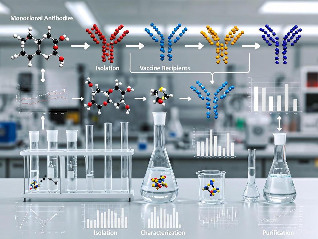
Isolation and Characterization of Monoclonal Antibodies from Vaccine Recipients: From B Cell to Clinical Candidate
This article provides a comprehensive guide for researchers and drug development professionals on the isolation and functional characterization of monoclonal antibodies (mAbs) from vaccine recipients.
Isolation and Characterization of Monoclonal Antibodies from Vaccine Recipients: From B Cell to Clinical Candidate
Abstract
This article provides a comprehensive guide for researchers and drug development professionals on the isolation and functional characterization of monoclonal antibodies (mAbs) from vaccine recipients. Covering the full scope from foundational principles to advanced applications, it explores the scientific rationale for sourcing mAbs from vaccinated donors, details cutting-edge isolation methodologies like single B cell sorting and hybridoma technology, and addresses critical troubleshooting aspects such as variant characterization and process optimization. Furthermore, it outlines rigorous validation frameworks, including comparative analyses and assessment of cross-reactivity, essential for advancing therapeutic candidates. The content synthesizes recent advances and real-world case studies, such as the development of orthopoxvirus-neutralizing mAbs, to offer a practical roadmap for transforming vaccine-elicited immune responses into potent biologic countermeasures.
The Scientific Rationale: Why Vaccine Recipients Are an Ideal Source for Therapeutic mAbs
Vaccination serves a dual purpose: protecting recipients from disease and generating a rich source of highly specific, functional human monoclonal antibodies (mAbs) for therapeutic development [1]. The isolation of mAbs from vaccinees leverages the body's refined immune response, yielding antibodies with proven neutralization capability and natural humanness, which are critical for therapeutic application [2] [3]. This approach, sometimes termed Reverse Vaccinology 2.0, interrogates the human B-cell response directly to identify protective antigens and epitopes included in a vaccine formulation [3]. These mAbs provide not only tools for combating infectious diseases but also critical insights for structure-guided vaccine design, closing the loop between natural immunity and advanced therapeutic development [1]. This protocol details methods for isolating and characterizing human mAbs from individuals vaccinated against pathogens such as monkeypox virus (MPXV) and Neisseria meningitidis (MenB) [2] [3].
Application Notes: Key Findings from Vaccine-Elicited mAbs
Cross-Reactive mAbs against Orthopoxviruses
Recent studies with the recombinant vaccinia vaccine (rTV) have successfully isolated three potent E8-specific mAbs (C5, C9, and F8) that demonstrate cross-neutralization activity against both vaccinia virus (VACV) and MPXV [2]. The C9 mAb notably targets the virion surface region of E8, showing cross-neutralization with an IC50 of 3.0 μg/mL against MPXV clade IIb and 51.1 ng/mL against VACV [2]. Complement enhanced neutralization against VACV by more than 50-fold, though no similar enhancement was observed for MPXV, highlighting pathogen-specific optimization needs [2]. In a VACV-infected mouse model, administration of these mAbs accelerated clinical recovery by 24 hours and achieved significant viral clearance with a 0.9-log reduction [2].
mAbs against Meningococcal and Gonococcal Pathogens
Investigation of the 4CMenB vaccine response identified PorB and lipooligosaccharide (LOS) as key immunogenic components in the outer membrane vesicle (OMV) responsible for eliciting cross-protective mAbs [3]. Researchers isolated 18 bactericidal PorB-specific mAbs and 1 LOS-specific mAb, with three of the PorB mAbs and the LOS-specific mAb showing bactericidal activity against gonococcus, demonstrating cross-protection potential [3]. The PorB mAbs were categorized into three functional classes through binding and in silico docking experiments, indicating this antigen serves as a multi-epitope immunogenic component enabling cross-protection across multiple MenB strains [3].
Table 1: Characteristics of Monoclonal Antibodies Isolated from Vaccine Recipients
| Vaccine Source | mAb Identifier | Target Antigen | Neutralization Potency (IC50) | Cross-Reactivity | Complement-Dependent Enhancement |
|---|---|---|---|---|---|
| Recombinant Vaccinia (rTV) | C5 | MPXV E8 / VACV D8 | VACV: 3.9 ng/mL | VACV, MPXV | >50-fold for VACV |
| Recombinant Vaccinia (rTV) | C9 | MPXV E8 / VACV D8 | MPXV: 3.0 μg/mL; VACV: 51.1 ng/mL | VACV, MPXV | >50-fold for VACV |
| Recombinant Vaccinia (rTV) | F8 | MPXV E8 / VACV D8 | VACV: 101.1 ng/mL | VACV, MPXV | >50-fold for VACV |
| 4CMenB | 18 clones | PorB | Bactericidal activity across MenB strains | Multiple MenB strains, N. gonorrhoeae | Not reported |
| 4CMenB | 1 clone | LOS | Bactericidal activity across MenB strains | Multiple MenB strains, N. gonorrhoeae | Not reported |
Table 2: Analytical Techniques for mAb Characterization
| Characterization Aspect | Key Analytical Techniques | Critical Quality Attributes Assessed |
|---|---|---|
| Structural & Physicochemical | Peptide mapping, Mass spectrometry, CD, SEC, IEX | Amino acid sequence, Post-translational modifications, Glycosylation patterns, Higher-order structure |
| Immunological Properties | ELISA, Surface Plasmon Resonance (SPR) | Antigen binding affinity/specificity, Epitope mapping, CDR identification |
| Biological Activities | ADCC/CDC assays, Viral neutralization tests, Plaque reduction assays | Effector functions, Neutralization potency, In vivo protective efficacy |
| Purity & Impurities | HPLC/UPLC, CE-SDS, icIEF | Aggregate content, Charge variants, Product-related impurities |
Experimental Protocols
Protocol 1: Antigen-Specific Single B Cell Sorting from Vaccine Recipients
Purpose: To isolate antigen-specific memory B cells from vaccinated donors for mAb discovery [2] [3].
Materials:
- PBMCs from vaccine recipients (collected 7 days post-boost immunization optimal for plasmablasts)
- Fluorescently labeled antigen (e.g., His- and Avi-tagged MPXV E8 protein)
- Antibody staining cocktail: anti-CD3-Pacific Blue, anti-CD8-Pacific Blue, anti-CD14-Pacific Blue, anti-CD19-BV510, anti-CD20-ECD, anti-CD27-APCCy7, anti-IgG-FITC, anti-IgM-PercpCy5.5
- LIVE/DEAD Fixable Dead Cell Stain (Pacific Blue)
- FACS buffer (PBS with 1-2% FBS)
- Cell sorter (e.g., FACS Aria SORP)
Procedure:
- PBMC Preparation: Thaw frozen PBMCs from vaccine recipients and wash twice with FACS buffer.
- Viability Staining: Resuspend cells in PBS containing LIVE/DEAD stain and incubate for 20-30 minutes at 4°C in the dark.
- Surface Staining: Add fluorescently labeled antigen and antibody staining cocktail. Incubate for 30 minutes at 4°C in the dark.
- Cell Sorting: Wash cells and resuspend in FACS buffer for sorting. Use the gating strategy: CD3⁻CD8⁻CD14⁻CD19⁺CD20⁺CD27⁺IgG⁺IgM⁻ antigen⁺.
- Collection: Sort single B cells into 96-well PCR plates containing lysis buffer and store at -80°C.
Protocol 2: Single B Cell RT-PCR and mAb Recombinant Expression
Purpose: To clone variable region genes of immunoglobulins and express recombinant mAbs [2] [3].
Materials:
- Single B cells in lysis buffer
- Reverse transcription reagents
- Nested PCR primers for Ig heavy and light chains
- Expression vectors for Ig heavy and light chains
- Expi293F or similar expression system
- Protein A or G affinity chromatography materials
Procedure:
- Reverse Transcription: Synthesize cDNA from single sorted B cells using gene-specific primers for Ig constant regions.
- Nested PCR: Amplify variable region genes of heavy and light chains in two rounds of PCR.
- Cloning: Clone PCR products into Ig expression vectors containing constant regions.
- Recombinant Expression: Co-transfect heavy and light chain vectors into Expi293F cells using standard protocols.
- Purification: Harvest culture supernatants after 5-7 days and purify mAbs using Protein A or G affinity chromatography.
Protocol 3: Neutralization Potency Assessment
Purpose: To evaluate the neutralization capability of isolated mAbs against live virus [2].
Materials:
- Purified mAbs
- Viruses: VACV Tiantan strain (VTT) or MPXV clade IIb
- Viral dilution medium
- Cell line: Vero cells or primary chicken embryo fibroblasts (CEFs)
- Cell culture maintenance media
Procedure:
- mAb Dilution: Prepare serial dilutions of mAbs in viral dilution medium.
- Virus-mAb Incubation: Mix equal volumes of mAb dilution and virus suspension (approximately 100 plaque-forming units). Incubate for 1-2 hours at 37°C.
- Infection: Add virus-mAb mixture to confluent cell monolayers in 24-well plates. Incubate for 1-2 hours at 37°C with occasional rocking.
- Overlay and Incubation: Remove inoculum and add semi-solid overlay medium. Incubate for 2-3 days until plaques develop.
- Plaque Counting: Fix and stain cells with crystal violet. Count plaques and calculate IC50 values using non-linear regression.
The Scientist's Toolkit: Research Reagent Solutions
Table 3: Essential Research Reagents for mAb Isolation and Characterization
| Reagent/Category | Specific Examples | Function/Application |
|---|---|---|
| Cell Isolation & Sorting | Anti-human CD19, CD20, CD27, IgG, IgM antibodies | Identification and isolation of antigen-specific B cell populations by flow cytometry |
| Antigen Probes | His/Avi-tagged recombinant proteins (e.g., MPXV E8) | Detection and sorting of antigen-specific B cells; can be labeled with streptavidin-APC/PE |
| Expression Systems | Expi293F cells, HEK293T cells | Recombinant expression of human mAbs following variable region gene cloning |
| Characterization Assays | ELISA, Surface Plasmon Resonance (SPR) | Assessment of antigen binding affinity, kinetics, and specificity |
| Functional Assays | Plaque reduction neutralization test (PRNT), Serum bactericidal assay (SBA) | Evaluation of neutralization potency against viral or bacterial pathogens |
| In Vivo Models | BALB/c mice (for VACV challenge) | Assessment of protective efficacy and viral load reduction |
Visualizing Workflows and Signaling Pathways
mAb Isolation and Characterization Workflow
mAb Isolation and Characterization Workflow
mRNA Vaccine Immune Activation Pathway
mRNA Vaccine Immune Activation Pathway
Therapeutic mAb Mechanisms of Action
Therapeutic mAb Mechanisms of Action
The monkeypox virus (MPXV), a member of the orthopoxvirus genus, has been declared a public health emergency of international concern (PHEIC) by the World Health Organization on two separate occasions, underscoring the urgent need for effective countermeasures [4] [2]. Currently, no approved targeted therapeutics exist for monkeypox, and treatment remains primarily supportive [2]. This application note details the isolation and characterization of E8-specific human monoclonal antibodies (mAbs) derived from recipients of the recombinant vaccinia vaccine (rTV). These antibodies demonstrate potent cross-neutralizing activity against orthopoxviruses, including both vaccinia virus (VACV) and authentic monkeypox virus, establishing E8 as a critical conserved target for pan-poxvirus countermeasure development [4]. The protocols and data presented herein provide a framework for researchers developing monoclonal antibody-based therapies against emerging viral pathogens.
Experimental Protocols
Donor Selection and B Cell Sorting
Objective: To isolate antigen-specific memory B cells from vaccinated donors for monoclonal antibody development.
Materials:
- Donor Samples: Peripheral blood mononuclear cells (PBMCs) from HIV-negative healthy adult donors enrolled in clinical trial (ChiCTR1900021422) who received two doses of recombinant vaccinia vaccine (rTV) [2].
- Staining Reagents: Anti-human antibodies (CD3-Pacific Blue, CD8-Pacific Blue, CD14-Pacific Blue, CD19-BV510, CD20-ECD, CD27-APCCy7, IgG-FITC, IgM-PercpCy5.5, PD-1-PECy7, CXCR5-APC-R700, CXCR3-PECy5, CD45RA-BV650, CD4-BV605).
- Sorting Probe: Recombinant MPXV E8 protein with His and Avi tags (commercially sourced), labeled with streptavidin-allophycocyanin (SA-APC) and streptavidin-R-phycoerythrin (SA-PE).
- Viability Stain: LIVE/DEAD Fixable Dead Cell Stain Kit (Pacific Blue).
Procedure:
- Donor Screening: Screen donor serum samples using ELISA for E8-binding antibodies and neutralization assays against VACV Tiantan strain. Select donors exhibiting the highest E8-binding and virus-neutralizing activity for B cell sorting [2].
- PBMC Preparation: Thaw frozen PBMCs and wash with appropriate buffer.
- Cell Staining: Resuspend PBMCs in staining buffer and incubate with the antibody mixture and viability stain for 30 minutes at 4°C in the dark.
- Probe Staining: Incubate cells with labeled MPXV E8 protein probe.
- Flow Cytometry Sorting: Using a FACS Aria SORP cell sorter, sort single, live, antigen-positive B cells using the gating strategy:
CD3-CD8-CD14-CD19+CD20+CD27+IgG+IgM-E8+into 96-well PCR plates containing lysis buffer. - Storage: Immediately store sorted plates at -80°C for subsequent analysis [2].
Critical Parameters:
- Cell viability must be maintained throughout the sorting process.
- Include fluorescence-minus-one (FMO) controls to establish accurate gating boundaries.
- The E8 probe should be freshly labeled and titrated to determine optimal staining concentration.
Antibody Gene Amplification and Recombinant Production
Objective: To recover antibody variable region genes from single sorted B cells and produce recombinant monoclonal antibodies.
Materials:
- RT-PCR Reagents: Reverse transcriptase, PCR buffer, dNTPs, gene-specific primers for human immunoglobulin genes.
- Expression Vectors: IgG expression vectors suitable for mammalian cell expression.
- Cell Line: Expi293F cells for transient antibody expression.
- Culture Medium: SMM 293-TII expression medium.
Procedure:
- Reverse Transcription: Perform reverse transcription on single B cell lysates using immunoglobulin gene-specific primers.
- Nested PCR: Amplify heavy and light chain variable region genes using nested PCR protocols with V-gene and C-gene specific primers.
- Sequence Analysis: Sequence PCR products and analyze for V(D)J gene usage and somatic hypermutation.
- Cloning: Clone heavy and light chain sequences into IgG1 expression vectors.
- Antibody Production: Co-transfect heavy and light chain plasmids into Expi293F cells using standard transfection protocols.
- Antibody Purification: Harvest culture supernatants after 5-7 days and purify antibodies using protein A or G affinity chromatography [2].
Critical Parameters:
- Use high-fidelity DNA polymerases to minimize PCR errors.
- Verify antibody sequence integrity before large-scale production.
- Determine antibody concentration and purity by spectrophotometry and SDS-PAGE.
In Vitro Neutralization Assay
Objective: To evaluate the neutralization potency of isolated mAbs against VACV and MPXV.
Materials:
- Viruses: VACV Tiantan strain (VTT), recombinant VTT expressing GFP (TT-GFP), and MPXV clade IIb.
- Cell Lines: Vero cells (for VACV) or BHK21 cells (for MPXV).
- Culture Medium: DMEM supplemented with 10% FBS and 1% penicillin-streptomycin.
- Dilutions: Serial dilutions of purified mAbs.
Procedure:
- Virus Preparation: Propagate and titrate viruses in appropriate cell lines. For VACV, use primary chicken embryo fibroblasts (CEFs); for MPXV, use approved BSL-3 facilities [2].
- Antibody Dilution: Prepare 3-fold serial dilutions of mAbs in culture medium.
- Virus-Antibody Incubation: Mix equal volumes of virus (approximately 100-200 plaque-forming units) with each antibody dilution and incubate for 1 hour at 37°C.
- Infection: Add virus-antibody mixtures to confluent cell monolayers in 96-well plates and incubate for 1-2 hours at 37°C.
- Overlay and Culture: Remove inoculum and add carboxymethylcellulose overlay medium. Incubate for 48-72 hours until plaques develop.
- Plaque Quantification: Fix and stain cells with crystal violet or monitor GFP expression (for TT-GFP). Count plaques and calculate percentage neutralization relative to virus-only controls [4] [2].
- IC50 Calculation: Determine antibody concentrations that achieve 50% neutralization using non-linear regression analysis.
Critical Parameters:
- Include appropriate controls: virus-only, cell-only, and isotype antibody controls.
- For MPXV, all procedures must be conducted in BSL-3 containment facilities.
- Perform assays in duplicate or triplicate to ensure reproducibility.
In Vivo Protection Study
Objective: To evaluate the therapeutic efficacy of mAbs in a mouse model of VACV infection.
Materials:
- Animals: Six-week-old female BALB/c mice.
- Virus: VACV Tiantan strain.
- Antibodies: Purified mAbs (individually or in combination).
- Equipment: Biosafety level 2 animal facility.
Procedure:
- Ethics Approval: Obtain appropriate institutional animal care and use committee approval (e.g., China CDC approval 2024-CCDC-IACUC-009) [2].
- Infection: Challenge mice with a lethal dose of VACV Tiantan strain via intraperitoneal injection.
- Antibody Administration: Administer mAbs (200-500 µg per mouse) via intraperitoneal injection 24 hours post-infection.
- Clinical Monitoring: Monitor mice daily for clinical signs of disease (weight loss, activity level, morbidity) for 14-21 days.
- Viral Load Assessment: At designated time points, euthanize subsets of mice and collect organs (lungs, liver, spleen) for viral titer determination by plaque assay.
- Data Analysis: Compare clinical scores, survival rates, and viral loads between treatment and control groups [4].
Critical Parameters:
- Randomize animals into experimental groups to minimize bias.
- Include positive (infected, untreated) and negative (uninfected) control groups.
- Calculate statistical significance using appropriate tests (log-rank test for survival, t-test for viral loads).
Results & Data Analysis
Neutralization Potency of E8 mAbs
The table below summarizes the in vitro neutralization activity of the three isolated E8 mAbs against VACV and MPXV:
Table 1: Neutralization potency of E8-specific monoclonal antibodies
| Antibody | VACV IC₅₀ | MPXV IC₅₀ | Complement Enhancement (VACV) |
|---|---|---|---|
| C5 | 3.9 ng/mL | Not reported | >50-fold |
| C9 | 51.1 ng/mL | 3.0 μg/mL | >50-fold |
| F8 | 101.1 ng/mL | Not reported | >50-fold |
All three mAbs demonstrated potent neutralization against VACV, with C5 showing exceptional potency (IC₅₀ = 3.9 ng/mL). Antibody C9 exhibited cross-neutralizing activity against both VACV and MPXV. Notably, the addition of complement enhanced neutralization against VACV by more than 50-fold for all antibodies, though no enhancement was observed for MPXV neutralization [4].
In Vivo Efficacy Data
Table 2: In vivo protection by E8 mAbs in VACV-infected mouse model
| Parameter | Control Group | mAb Treatment Group |
|---|---|---|
| Clinical Recovery | 96-120 hours | Accelerated by 24 hours |
| Viral Clearance | Baseline | 0.9-log reduction |
| Survival Rate | Not specified | Significant improvement |
In vivo administration of the E8 mAbs (used in combination) accelerated clinical recovery by approximately 24 hours and achieved significant viral clearance (0.9-log reduction) in a VACV-infected mouse model [4]. These results demonstrate the therapeutic potential of E8-targeting mAbs against orthopoxvirus infections.
The Scientist's Toolkit
Table 3: Essential research reagents for monoclonal antibody isolation and characterization
| Reagent/Category | Specific Examples | Function/Application |
|---|---|---|
| Cell Separation | FACS Aria SORP, anti-human CD19/CD20/CD27 | Isolation of antigen-specific memory B cells |
| Antigen Probes | His/Avi-tagged MPXV E8 protein, SA-APC, SA-PE | Fluorescent labeling and detection of target B cells |
| Expression System | Expi293F cells, SMM 293-TII medium | Recombinant monoclonal antibody production |
| Virus Stocks | VACV Tiantan strain, MPXV clade IIb | Neutralization assays and challenge studies |
| Animal Models | BALB/c mice | In vivo efficacy evaluation |
Workflow & Pathway Diagrams
Experimental Workflow for E8 mAb Discovery
Diagram 1: mAb discovery and characterization workflow.
E8 mAb Mechanism of Action
Diagram 2: E8 mAb neutralization mechanisms.
Discussion
The E8 protein of MPXV represents a highly conserved target for therapeutic antibody development against orthopoxviruses. The mAbs described in this application note—particularly C9 which shows cross-neutralizing activity against both VACV and MPXV—demonstrate the potential for broad-spectrum protection [4]. The complement-dependent enhancement observed for VACV neutralization but not for MPXV highlights important pathogen-specific differences that must be considered during therapeutic optimization [4].
These findings establish E8 as a critical conserved target for pan-poxvirus countermeasure development and provide a robust methodology for isolating and characterizing therapeutic monoclonal antibodies from vaccine recipients. The protocols outlined herein can be adapted for developing mAbs against other emerging viral pathogens, contributing to pandemic preparedness efforts.
The rapid antigenic evolution of RNA viruses like SARS-CoV-2 and the persistent emergence of novel pathogenic variants necessitate the development of potent therapeutic monoclonal antibodies (mAbs) with broad neutralizing capacity [5]. A critical strategy in this pursuit is the identification and characterization of conserved neutralizing epitopes—specific regions on viral surface proteins that are targeted by antibodies and are relatively unchanged across viral variants and even related virus species. This Application Note details the key viral surface proteins and conserved epitopes essential for developing cross-neutralizing antibodies, providing structured experimental data and detailed methodologies tailored for researchers and drug development professionals working within the context of monoclonal antibody isolation and characterization from vaccine recipients.
Key Viral Surface Proteins and Conserved Epitopes
For SARS-CoV-2 and related coronaviruses, the primary target for neutralizing antibodies is the spike (S) glycoprotein, a Class I viral fusion protein that mediates viral entry into host cells [5] [6]. The S protein is comprised of two subunits: S1, which contains the receptor-binding domain (RBD) responsible for attaching to the host receptor (ACE2), and S2, which facilitates membrane fusion [6]. The S2 subunit, in particular, is highly conserved across coronavirus genera and represents a promising target for broad neutralization [7].
Table 1: Key Conserved Epitopes for Cross-Neutralizing Antibodies
| Epitope Location | Description / Signature Motif | Representative mAbs | Neutralization Breadth | Key Structural Features |
|---|---|---|---|---|
| HR1 Domain (S2 subunit) [5] | 6-mer peptide forming a β-turn fold (950-DVVNQN-955 in SARS-CoV-2) [5] | 3D1 [5] | Pan-coronavirus (excludes Omicron Q954H) [5] | Pre-hairpin intermediate transition state [5] |
| S2 Apex / Hinge Region [7] | Conformational epitope (residues 980–1006 in SARS-CoV-2) at HR1/CH hairpin hinge [7] | RAY53 (3A3 mouse precursor) [7] | SARS-CoV-2, MERS-CoV [7] | Prefusion-specific, flexible hinge [7] |
| Conserved RBD Epitope [8] | Non-RBS epitope, highly conserved between SARS-CoV and SARS-CoV-2 [8] | COVA1-16 [8] | SARS-CoV-2, SARS-CoV [8] | Bound by long CDR H3, steric hindrance to ACE2 [8] |
| S2 Stem Helix [5] | Conserved stem helix region in S2 subunit [5] | S2P6, B6, 28D9, CC40.8 [5] | Restricted within β-Coronavirus genera [5] | Targets membrane fusion [5] |
Experimental Protocols for Epitope Characterization
A multi-faceted approach is required to thoroughly characterize neutralizing antibodies and define their target epitopes. The protocols below outline key steps from initial antibody isolation to high-resolution structural analysis.
Protocol: Isolation of Broadly Neutralizing mAbs from a Combinatorial Library
This protocol is adapted from the isolation of the 3D1 antibody, which was derived from a pre-COVID-19 naïve human combinatorial antibody library [5].
- Principle: A synthetic immune system with vast diversity (e.g., 10^11 sequences) is screened against a conserved viral antigen to identify high-affinity binders with cross-reactive potential [5].
- Procedure:
- Antigen Design: Design a peptide antigen based on a conserved consecutive motif in a stable domain like HR1. For 3D1, the intact 32-mer peptidic HR1 fusion core (HR1FC) of SARS-CoV-2 (residues 924-955) was used [5].
- Library Panning: Perform 3-4 rounds of panning the combinatorial antibody library (e.g., scFv-IgG1 format displayed on phage) against the immobilized antigen [5].
- Clone Screening: Screen output clones via ELISA for binding to the immunogen and cross-reactivity with orthologs (e.g., SARS-CoV-1 HR1FC) [5].
- Affinity Maturation Analysis: Infer germline origins using the IMGT database and analyze somatic hypermutation (SHM) levels [5].
- Applications: Rapid discovery of rare and specialized antibodies, including cross-reactive antibodies, bypassing the need for donor immune cells [5].
Protocol: Epitope Mapping and Affinity Determination
- Principle: Define the minimal epitope and binding affinity of a candidate mAb using peptide truncations and biophysical assays.
- Procedure:
- Epitope Localization: Localize binding to a subdomain (e.g., C-terminal HR1FC) via ELISA with long and truncated peptides [5].
- Minimal Epitope Mapping: Assess binding affinity to systematically truncated peptides via ELISA to define the minimal core epitope (e.g., the 6-mer DVVNQN for 3D1) [5].
- Critical Residue Identification: Use alanine scanning mutagenesis on the minimal epitope and confirm loss of binding via Bio-Layer Interferometry (BLI). For 3D1, Q954 was identified as critical [5].
- Cross-Reactivity Profiling: Evaluate binding to homologous peptides from a panel of related viruses (e.g., across α, β, γ, and δ coronavirus genera) to define the breadth of reactivity [5].
Protocol: Structural Characterization of Antibody-Epitope Complexes
- Principle: Determine the high-resolution structure of the antibody-antigen complex to understand the molecular basis of cross-reactivity and neutralization.
- Procedure:
- Complex Formation: Purify the antigen-binding fragment (Fab) of the mAb and its cognate epitope (e.g., HR1C of SARS-CoV-2) and form a stable complex for crystallography [5].
- X-ray Crystallography: Determine the crystal structure of the Fab-epitope complex. This reveals precise atomic interactions, such as the β-turn fold recognized by 3D1 [5].
- Hydrogen-Deuterium Exchange Mass Spectrometry (HDX-MS): As an alternative or complementary technique, incubate the full spike protein with and without the mAb (IgG or Fab) in deuterated buffer. Monitor decreases in deuterium uptake in the antibody-bound state to identify protected epitope peptides, as was done to map the RAY53 epitope to residues 980-1006 [7].
- Cryo-Electron Microscopy (Cryo-EM): For larger complexes or flexible proteins, perform single-particle cryo-EM of the mAb bound to the full spike trimer. This can reveal the binding stoichiometry and conformational state of the antigen, as demonstrated with COVA1-16 and the S trimer [8].
The Scientist's Toolkit: Essential Research Reagents
Table 2: Key Research Reagent Solutions for Cross-Neutralization Studies
| Reagent / Material | Function and Application | Example Usage |
|---|---|---|
| Combinatorial Antibody Library [5] | Source of diverse human antibody sequences for in vitro selection against conserved antigens. | Isolation of 3D1 bnAb from a naïve human library [5]. |
| Stabilized Prefusion S Protein [7] | Antigen for immunization and in vitro assays, maintaining native trimeric conformation. | MERS SS.V1 S2 protein for mouse immunizations [7]; SARS-CoV-2 HexaPro for HDX studies [7]. |
| Pseudovirus Neutralization Assay [5] [9] | Safe, BSL-2 method to quantify neutralizing potency and breadth against wild-type and mutant viruses. | Testing 3D1 against SARS-CoV-2 and SARS-CoV-1 pseudoviruses [5]. |
| Bio-Layer Interferometry (BLI) [5] | Label-free technology for measuring binding kinetics (kon, koff, KD) between mAbs and antigens. | Confirming Q954 as a critical residue for 3D1 binding [5]. |
Visualizing the Experimental Workflow for mAb Discovery
The following diagram illustrates the integrated workflow for isolating and characterizing cross-neutralizing monoclonal antibodies, from antigen design to functional validation.
The strategic targeting of conserved epitopes on viral surface proteins, particularly within the S2 subunit of coronaviruses, presents a viable path toward developing broadly protective antibodies and vaccines. The experimental frameworks and data summarized in this Application Note provide a roadmap for researchers aiming to isolate and characterize the next generation of cross-neutralizing monoclonal antibodies, ultimately enhancing our preparedness for future viral outbreaks.
The successful isolation of potent monoclonal antibodies (mAbs) from human B cells is a cornerstone of modern immunology and therapeutic development. This application note establishes a critical correlation between high post-vaccination serum antibody titers and the efficacy of subsequent antigen-specific B cell sorting. We provide a detailed, standardized protocol for identifying optimal vaccinee donors and for the single-cell sorting of antigen-specific B cells, specifically within the context of vaccine-induced immune responses. The methodologies outlined herein are designed to enhance the success rate of isolating high-affinity, neutralizing mAbs, thereby accelerating therapeutic antibody discovery.
The emergence of novel pathogens and the need for advanced biologics have underscored the importance of efficient monoclonal antibody (mAb) discovery pipelines. A pivotal, yet often variable, factor in this process is the initial selection of human donor subjects. The foundational hypothesis of this protocol is that vaccinated individuals exhibiting high serum antibody titers possess a peripheral blood B cell repertoire enriched for antigen-specificity and high-affinity potential, making them optimal candidates for B cell sorting campaigns [10] [11].
Following vaccination, antigen-specific B cells undergo activation, class-switch recombination, and somatic hypermutation (SHM) within germinal centers, ultimately differentiating into antibody-secreting plasmablasts (PBs) and memory B cells (MBCs) [11]. The frequency of these antigen-experienced B cells in peripheral blood transiently increases, providing a window for their isolation. This document details a workflow from donor selection based on antibody titer analysis to the single-cell sorting of B cells, forming the basis for reverse vaccinology 2.0 and therapeutic antibody development [3].
Key Correlations: B Cell Subsets and Antibody Titers
Quantitative data from studies on vaccine responses provide a rationale for focusing on specific B cell subsets. Research on the BNT162b2 mRNA COVID-19 vaccine revealed distinct correlations between pre- and post-vaccination B cell frequencies and the resulting anti-SARS-CoV-2 antibody titer.
Table 1: Correlation of B Cell Subsets with High Antibody Titers Post-Vaccination [10]
| B Cell Subset | Phenotype | Correlation with High Antibody Titer | Key Findings |
|---|---|---|---|
| Naïve B Cells | CD19+, CD20+, CD27-, IgD+ | Positive | A higher frequency before vaccination was associated with a higher antibody response. |
| Transitional B Cells | CD24hi, CD38hi | Positive | Positively correlated with a higher antibody titer. |
| Late Memory B Cells | CD19+, CD20+, CD27+, IgD- | Negative | Associated with a lower antibody titer. |
| Plasmablasts | CD19+, CD20-, CD27hi, CD38hi | Negative | Frequencies were associated with a lower antibody titer. |
| Activated CD8+ T Cells | CD8+, CD38+, HLA-DR+ | Positive | Fold change in frequency upon vaccination was correlated with high antibody titers. |
These data suggest that donors selected for high antibody titers are likely to have a B cell pool enriched with naïve and transitional B cells, which are crucial for a robust, de novo humoral response. Conversely, an abundance of late memory B cells or plasmablasts may indicate a different immunological history that is less optimal for isolating high-affinity mAbs against the vaccine antigen of interest.
Experimental Protocols
Protocol 1: Donor Selection and Serum Antibody Titer Analysis
Objective: To identify and select vaccinee donors with high serum antibody titers for B cell sorting experiments.
Materials:
- Serum samples from vaccinated donors (collected 2-3 weeks post-boost)
- Commercial ELISA or Electrochemiluminescence immunoassay (ECLIA) kit for target antigen (e.g., Elecsys Anti-SARS-CoV-2S)
- Cobas e801 analyzer or equivalent
- Phosphate-Buffered Saline (PBS)
Procedure:
- Sample Collection: Collect peripheral blood from vaccinees 14-21 days after the most recent vaccine dose. This timing corresponds with the peak of the antibody-secreting cell and serum antibody response [10] [11].
- Serum Separation: Centrifuge clotted blood at 1,000-2,000 x g for 10 minutes in a serum separation tube. Transfer the supernatant (serum) to a new tube and store at -20°C or -80°C for long-term storage.
- Antibody Titer Measurement: Quantify antigen-specific antibody levels using a standardized, quantitative immunoassay. For instance, the Elecsys Anti-SARS-CoV-2S assay on a Cobas e801 module can be used for viral spike proteins [10]. Perform all measurements according to the manufacturer's instructions.
- Donor Stratification: Rank donors based on their antibody titer. Select donors falling in the upper quartile as "high responders" for subsequent B cell sorting. Ensure that selected donors are age-matched to potential "low responders" if a control group is desired [10].
Protocol 2: Peripheral Blood Mononuclear Cell (PBMC) Isolation and Cryopreservation
Objective: To isolate and preserve mononuclear cells from donor whole blood.
Materials:
- Whole blood in heparin or EDTA tubes
- Ficoll-Paque PLUS
- Cell freezing media (e.g., 90% FBS, 10% DMSO)
- Controlled-rate freezer
Procedure:
- Dilution: Dilute whole blood 1:1 with PBS.
- Density Gradient Centrifugation: Carefully layer the diluted blood over Ficoll-Paque in a centrifuge tube. Centrifuge at 400 x g for 30-35 minutes at room temperature with the brake disengaged.
- PBMC Collection: Aspirate the buffy coat layer at the interface and transfer to a new tube.
- Washing: Wash cells twice with PBS by centrifuging at 300 x g for 10 minutes.
- Cryopreservation: Resuspend the PBMC pellet in cold freezing media. Transfer to cryovials and freeze using a controlled-rate freezer. Store vials in liquid nitrogen until sorting [10] [2].
Protocol 3: Single-Cell Sorting of Antigen-Specific B Cells
Objective: To isolate single, live, antigen-specific B cells for monoclonal antibody cloning.
Materials:
- Thawed PBMCs from high-titer donors
- Fluorescently-labeled antigen probe (e.g., biotinylated MPXV E8 with SA-APC/PE) [2]
- Antibody staining panel (see Table 2)
- LIVE/DEAD Fixable Viability Dye
- Human TruStain FcX (Fc receptor blocking solution)
- FACS Aria SORP or equivalent cell sorter
- 96-well PCR plates containing lysis buffer
Procedure:
- Thaw and Viability Stain: Rapidly thaw cryopreserved PBMCs and immediately wash in pre-warmed culture medium. Stain cells with a LIVE/DEAD viability dye (e.g., Zombie Green) to exclude dead cells [10] [2].
- Fc Receptor Blocking: Incubate cells with Human TruStain FcX to reduce non-specific antibody binding.
- Surface Staining: Stain the cells with a pre-optimized antibody cocktail and the fluorescently-labeled antigen probe for 20-30 minutes on ice. A representative panel is shown below.
Table 2: Example Flow Cytometry Panel for Antigen-Specific B Cell Sorting [2]*
| Target | Fluorochrome | Purpose |
|---|---|---|
| Live/Dead | Pacific Blue | Viability Marker |
| CD19 | BV510 | Pan-B Cell Marker |
| CD20 | ECD | Pan-B Cell Marker |
| CD3/CD8/CD14 | Pacific Blue | Lineage Exclusion (T cells, monocytes) |
| CD27 | APCCy7 | Memory B Cell Marker |
| IgG | FITC | Isotype (Class-switched) |
| IgM | PercpCy5.5 | Isotype (Naïve/IgM+ Memory) |
| Antigen Probe | APC/PE | Antigen-Specific B Cell Identification |
- Gating Strategy and Sorting:
- Single-Cell Dispensing: Sort single B cells directly into a 96-well PCR plate containing lysis buffer for subsequent RNA extraction and reverse transcription. Immediately freeze plates at -80°C [2].
The Scientist's Toolkit: Research Reagent Solutions
Table 3: Essential Reagents for B Cell Sorting and mAb Generation
| Reagent/Category | Function | Example Products/Components |
|---|---|---|
| Antigen Probes | Label target-specific B cells for sorting. | Recombinant his- and avi-tagged proteins (e.g., MPXV E8), streptavidin-APC/PE conjugates [2]. |
| Viability Dyes | Distinguish live from dead cells to improve sort efficiency. | Zombie Green, LIVE/DEAD Fixable Stain kits [10]. |
| Fc Receptor Blocker | Reduce nonspecific antibody binding, lowering background. | Human TruStain FcX [10]. |
| B Cell Phenotyping Antibodies | Identify and isolate specific B cell subsets (naïve, memory, plasmablast). | Anti-human CD19, CD20, CD27, CD38, IgG, IgM [10] [2]. |
| Cell Culture Media | Support the growth of hybridomas or for recombinant expression. | RPMI, DMEM, Expi293 Expression Medium [2]. |
| Single-Cell RNA/DNA Kits | Amplify and clone variable heavy/light chain genes from single B cells. | Smart-seq2, NEBNext Single Cell/Low Input RNA Library Prep Kit [12]. |
Workflow Visualization
Diagram 1: High-titer donor B cell sorting workflow.
Diagram 2: Flow cytometry gating for antigen-specific B cells.
The strategy of preselecting donors based on high serum antibody titers provides a powerful filter to enrich for B cell repertoires with high value for mAb discovery. The protocols outlined here offer a standardized approach to leverage this correlation, from systematic donor screening to the precise isolation of antigen-specific B cells.
It is crucial to recognize that the quality of the isolated mAbs is not solely dependent on donor titer. Downstream processes, including the efficiency of single-cell B cell receptor (BCR) cloning, recombinant expression, and functional characterization, are equally critical [12]. Furthermore, the observed dominance of pre-existing memory B cells in early antibody-secreting cell responses highlights the complexity of the human B cell memory landscape. While these pre-existing MBCs can rapidly differentiate into antibody-secreting cells, the highest-affinity mAbs often originate from naive B cell precursors that undergo robust affinity maturation in the germinal center [13].
In conclusion, integrating donor selection based on high serum antibody titers with robust single-cell sorting and cloning methodologies significantly de-risks and accelerates the pipeline for isolating high-affinity, therapeutically relevant monoclonal antibodies from vaccinated individuals.
From Blood to Biologic: Cutting-Edge Methodologies for mAb Isolation and Production
The isolation and characterization of monoclonal antibodies (mAbs) from vaccine recipients is a cornerstone of modern immunology and biopharmaceutical development. This research is critical for understanding protective immune responses, developing novel therapeutics, and creating new diagnostic tools. The process involves obtaining B cells from immunized individuals and isolating those that produce antibodies against the vaccine antigen. Over the years, several technological platforms have been developed for this purpose, each with distinct advantages, limitations, and applications. This guide provides a comprehensive comparison of the three principal mAb isolation platforms: hybridoma technology, single B cell methods, and antibody display technologies, with specific consideration for their application in research on vaccine recipients.
The following table provides a systematic comparison of the three major mAb isolation platforms, highlighting their key characteristics to aid researchers in platform selection.
Table 1: Comparative Analysis of Major mAb Isolation Platforms
| Feature | Hybridoma Technology | Single B Cell Technologies | Antibody Display Technologies |
|---|---|---|---|
| Core Principle | Fusion of antigen-specific B cells with immortal myeloma cells to create stable antibody-producing cell lines [14] [15] | High-throughput screening and isolation of single antigen-specific B cells from an immune repertoire, followed by antibody gene amplification [14] [16] |
In vitro selection of antibodies from vast genetic libraries displayed on the surface of phages, yeast, or other entities [16] [17] |
| Throughput | Low to moderate; process is complex and labor-intensive [16] | High; enables rapid screening of thousands of B cells [16] | Very High; libraries can contain >1011 unique variants [17] |
| Development Timeline | Long (several months) | Short (weeks) | Short (weeks) |
| Antibody Format | Full-length, naturally paired IgG (from the host organism) | Full-length, naturally paired IgG (from the original host) | Typically antibody fragments (scFv, Fab); requires reformatting to IgG [17] |
| Key Advantage | Preserves natural antibody pairing and innate B cell biology; established, straightforward production once clone is established [14] [17] | Directly captures native, paired antibody sequences from individual immune cells without the need for fusion [14] | Bypasses immune tolerance; allows discovery against self-antigens, toxic targets, and enables extensive in vitro engineering [17] |
| Primary Limitation | Low B cell-myeloma fusion efficiency; reliance on animal immunization; potential immunogenicity of murine antibodies [14] [15] | Requires specialized instrumentation for single-cell sorting and handling; high dependency on PCR efficiency [16] | Lacks native B cell context; antibodies may not reflect natural immune response; requires affinity maturation for high affinity [17] |
| Immune Context from Vaccine Recipients | Excellent; captures antibodies from an active, in vivo immune response in an immunized host. | Excellent; directly sequences functional antibodies from the circulating B cell repertoire of vaccinated individuals. | Limited; libraries are often synthetic or naïve, not directly reflecting the vaccine-induced immune repertoire. |
The workflow for selecting and implementing these technologies involves several key decision points, as illustrated below.
Experimental Protocols for mAb Isolation
Protocol 1: Hybridoma Technology for mAb Generation
Application Context: Ideal for generating stable, permanent cell lines producing a single mAb from splenocytes of immunized animals (e.g., mice, rats) or from human B cells obtained from vaccine recipients [15].
Materials:
- Myeloma cells (e.g., SP2/0, P3X63Ag8.653)
- Splenocytes from an immunized donor or purified peripheral blood B cells from a vaccine recipient
- Polyethylene Glycol (PEG) solution or electrofusion equipment [15]
- Hypoxanthine-Aminopterin-Thymidine (HAT) selection medium [18]
- ELISA plates coated with the vaccine antigen of interest
Procedure:
- Immunization & Cell Preparation: Immunize an animal with the target vaccine antigen following a standard immunization schedule. Confirm serum antibody titer by ELISA. Alternatively, isolate peripheral blood mononuclear cells (PBMCs) from human vaccine recipients. Harvest spleen from the immunized animal or use the PBMCs, and prepare a single-cell suspension.
- Cell Fusion: Mix splenocytes/PBMCs with myeloma cells at a ratio between 2:1 and 10:1. Perform cell fusion using either:
- Chemical Fusion: Gently add 50% PEG solution to the cell pellet over one minute, followed by a slow dilution with serum-free medium.
- Electrofusion: Use a pulsed electric field (PEF) to fuse cells, a method noted for higher efficiency [15].
- Selection and Cloning: Plate the fused cells in HAT selection medium. Only hybridoma cells will survive, as myeloma cells lack the enzyme HGPRT and cannot proliferate in HAT medium. After 7-14 days, screen supernatants from visible hybridoma colonies for antigen-specific antibodies via ELISA.
- Subcloning and Expansion: Isolate positive wells and perform limiting dilution subcloning to ensure the monoclonality of the hybridoma. Expand stable, antibody-producing clones for cryopreservation and large-scale mAb production.
Protocol 2: Single B Cell Antibody Technology
Application Context: Enables direct isolation of antibody variable region genes from individual antigen-specific B cells sourced from vaccine recipients, preserving the native heavy and light chain pairing [14] [16].
Materials:
- Fluorescently labeled vaccine antigen probes (e.g., biylated antigen with streptavidin-fluorophore)
- Flow cytometer with single-cell sorting capability (e.g., FACS)
- Single-cell RT-PCR kits
- Nested PCR primers for antibody variable heavy (VH) and variable light (VL) chains
- Expression vectors for full-length IgG or Fab fragments
Procedure:
- B Cell Staining and Sorting: Label PBMCs from a vaccine recipient with a cocktail of antibodies (e.g., anti-CD19, anti-CD20) and a fluorescently labeled vaccine antigen probe. Use flow cytometry to identify and single-cell sort antigen-binding B cells into 96-well PCR plates containing lysis buffer.
- Reverse Transcription and PCR: Perform reverse transcription in the single-cell lysate to generate cDNA. Subsequently, conduct nested PCR reactions using family-specific primers to amplify the Ig heavy and light chain variable region genes.
- Gene Cloning and Expression: Clone the paired VH and VL PCR products into IgG expression vectors. Co-transfect the heavy and light chain vectors into mammalian cells (e.g., HEK293, CHO) for transient or stable antibody expression.
- Antibody Characterization: Screen and purify the expressed antibodies from culture supernatant. Characterize specificity and affinity using ELISA and Surface Plasmon Resonance (SPR), respectively [18].
Protocol 3: Phage Display Library Selection (Biopanning)
Application Context: For discovering high-affinity antibody fragments from large synthetic, naïve, or immune libraries, independent of an ongoing B cell response [16] [17].
Materials:
- Phagemid library displaying scFv or Fab fragments
- M13K07 helper phage
- Immunotubes or magnetic beads coated with the purified vaccine antigen
- E. coli strains suitable for phage infection (e.g., TG1, XL1-Blue)
Procedure:
- Library Preparation: Amplify the phage display antibody library by infecting with helper phage to produce phage particles displaying the antibody fragments.
- Panning: Incubate the phage library with immobilized antigen (coated on a plate or magnetic beads). Wash away non-specific and weakly binding phages. Elute the specifically bound phages using an acidic buffer or competitive antigen displacement.
- Amplification and Iteration: Infect exponentially growing E. coli with the eluted phages and rescue with helper phage to amplify the enriched pool for the next round of selection. Typically, 3-4 rounds of panning are performed to enrich for high-affinity binders.
- Screening and Reformating: After the final round, plate the infected E. coli to obtain single colonies. Screen individual clones for antigen binding via phage ELISA. Sequence the positive clones, then reformat the selected scFv/Fab sequences into a full-length IgG format for downstream production and characterization.
Characterization of Isolated Monoclonal Antibodies
Once mAbs are isolated, rigorous characterization is essential. The following table outlines the key quality attributes and the standard analytical techniques used for evaluation, which is critical for both research validation and regulatory compliance [18].
Table 2: Key Analytical Techniques for mAb Characterization
| Characterization Aspect | Critical Quality Attributes | Common Analytical Techniques |
|---|---|---|
| Structural & Physicochemical | Amino acid sequence, Peptide map, Disulfide bridges, Size and charge variants, Glycosylation profile [18] | Mass Spectrometry, Chromatographic Methods (SEC, IEC, HIC), Electrophoretic Methods (SDS-PAGE, CE-SDS, IEF) [18] |
| Binding & Immunological | Specificity, Affinity (KD), Kinetics (ka, kd), Epitope binning, Immunoreactivity [18] | ELISA, Surface Plasmon Resonance (SPR), Bio-Layer Interferometry (BLI) [18] |
| Functional & Biological | Neutralization potency, Effector functions (ADCC, CDC) [18] | Cell-based neutralization assays, Reporter gene assays, Flow cytometry |
The overall workflow from isolation to characterization involves multiple parallel processes, as summarized in the following diagram.
The Scientist's Toolkit: Essential Research Reagents and Materials
Successful isolation and characterization of mAbs require a suite of specialized reagents and instruments.
Table 3: Essential Research Reagents and Solutions for mAb Isolation
| Category | Item | Primary Function in mAb Workflow |
|---|---|---|
| Cell Culture & Isolation | Myeloma Cells (e.g., SP2/0) | Fusion partner for hybridoma generation providing immortality [15]. |
| HAT Selection Medium | Selects for successfully fused hybridoma cells by eliminating unfused myeloma cells [18]. | |
| Fetal Bovine Serum (FBS) | Provides essential nutrients and growth factors for hybridoma and recombinant cell growth. | |
| Fluorescent Antigen Probes | Labels antigen-specific B cells for isolation via flow cytometry in single B cell technologies [16]. | |
| Molecular Biology | Phagemid Vectors | Allows cloning of antibody gene libraries and display on phage surface [16] [17]. |
| Single-Cell RT-PCR Kits | Amplifies antibody mRNA from single B cells for gene cloning [16]. | |
| Family-Specific VH/VL Primers | Amplifies the diverse repertoire of antibody variable region genes from cDNA. | |
| Screening & Characterization | ELISA Plates & Reagents | High-throughput screening of hybridoma supernatants or phage clones for antigen binding. |
| SPR/BLI Instruments (e.g., Biacore, Octet) | Label-free analysis of binding affinity (KD) and kinetics (ka, kd) [18]. | |
| SEC/UPLC Columns | Analyzes mAb aggregation, fragmentation, and size heterogeneity [18]. |
Emerging Technologies and Future Directions
The field of mAb isolation is rapidly evolving. DNA-encoded monoclonal antibodies (DMAbs) represent a paradigm shift, where a synthetic DNA plasmid encoding the antibody is administered in vivo, turning the patient's muscle cells into a bioreactor for sustained antibody production. A recent phase 1 trial demonstrated durable expression of a SARS-CoV-2 neutralizing mAb cocktail for over 72 weeks following intramuscular administration of the DMAb, highlighting its potential as a long-acting, cold-chain-independent platform [19].
Furthermore, high-throughput methodologies are being increasingly integrated with next-generation sequencing (NGS) and artificial intelligence (AI). NGS allows for deep mining of the antibody repertoire from single B cells or display libraries, while AI models are now being used for de novo design of antibody sequences and prediction of affinity and stability, potentially revolutionizing the discovery process [16] [17].
The isolation and characterization of monoclonal antibodies (mAbs) from vaccine recipients represent a cornerstone of modern immunology and therapeutic development. Traditional methods, such as hybridoma generation, are often plagued by low efficiency, lengthy timelines, and the loss of natural antibody chain pairing [20] [21]. Antigen-specific single B cell sorting coupled with reverse transcription-polymerase chain reaction (RT-PCR) has emerged as a powerful, high-throughput alternative. This technology enables the direct isolation of rare antigen-specific B cells from a heterogeneous population, followed by the amplification and cloning of their native, paired heavy and light chain variable genes [22] [23]. This approach is highly versatile, having been successfully applied to humans, transgenic animals, and other model organisms to generate highly specific, discriminative, and potent human mAbs for both basic research and immunotherapeutic purposes [20] [24] [4]. The following protocol provides a detailed guide for implementing this technique, with a specific focus on the context of isolating mAbs from individuals who have received vaccinations.
Materials and Reagents
Research Reagent Solutions
The following table lists essential materials and their functions for the antigen-specific single B cell sorting and cloning workflow.
Table 1: Key Research Reagents and Their Functions in the Protocol
| Reagent/Material | Function/Application | Key Details |
|---|---|---|
| Biotinylated Antigen | Primary probe for identifying antigen-specific B cells. | Tetramerized with fluorochrome-conjugated streptavidin to enhance binding avidity [20]. |
| Fluorochrome-conjugated Streptavidin | Used to multimerize biotinylated antigens, creating fluorescent probes for cell staining. | PE, APC, and BV421 are common choices for multi-color staining [20] [2]. |
| Magnetic Cell Sorting Kits | For bulk enrichment of antigen-specific B cells prior to FACS. | Improves efficiency by pre-concentrating rare cell populations [23]. |
| Viability Stain (e.g., LIVE/DEAD) | To exclude dead cells during flow cytometry. | Critical for ensuring high-quality RNA and cDNA from sorted single cells [22] [2]. |
| Anti-Immunoglobulin & B Cell Phenotyping Antibodies | To identify and isolate the desired B cell subset (e.g., memory B cells, IgG+ B cells). | Panels often include anti-CD19, CD20, CD27, IgG, and IgM [2] [21]. |
| Single-Cell RT-PCR Kits | For reverse transcription and amplification of antibody genes from single cells. | Often use gene-specific primers or template-switching mechanisms [22] [25]. |
| IgG Expression Vectors | For the cloning and recombinant expression of amplified VH and VL genes. | Vectors contain constant regions for human IgG1 are commonly used [20] [22]. |
Methods
The diagram below outlines the complete experimental workflow, from sample preparation to antibody expression and validation.
Sample Preparation and B Cell Enrichment
- Source Material: Begin with peripheral blood mononuclear cells (PBMCs) from vaccinated donors or convalescent patients [2] [21]. Alternatively, use single-cell suspensions from spleen or lymph nodes of immunized animal models [20] [23].
- PBMC Isolation: Isolate PBMCs from whole blood using density gradient centrifugation (e.g., Ficoll-Paque) per standard protocols [20] [21].
- B Cell Enrichment (Optional but Recommended): To significantly increase the efficiency of finding rare antigen-specific B cells, perform a bulk enrichment step. This is typically done using magnetic-activated cell sorting (MACS) to isolate total memory B cells or IgG+ B cells.
- Use a commercial Memory B Cell Isolation Kit, which typically involves depleting non-target cells (e.g., T cells, monocytes, IgM+ naive B cells) [21].
- This enrichment step has been shown to increase the hit rate of antigen-specific mAbs from 1-8% to 51-88% in the subsequent single-cell sorting [23].
Staining and Antigen-Specific Single B Cell Sorting
Staining Cocktail Preparation:
- Prepare a staining cocktail in a FACS buffer (PBS + 2% FBS). The cocktail should contain:
- Fluorochrome-labeled antigen probe: Use a pre-formed tetramer of your biotinylated antigen with streptavidin-conjugated fluorophores (e.g., PE, APC). Using two different colors for the same antigen (e.g., PE- and APC-conjugated tetramers) helps distinguish high-affinity, specific B cells from non-specific binders [20] [2].
- Antibody panel for B cell phenotyping: This typically includes:
- Anti-CD19 and/or anti-CD20 (B cell markers).
- Anti-CD27 (memory B cell marker).
- Anti-IgG (to isolate class-switched B cells).
- Anti-IgM (to exclude naive B cells).
- Viability dye (e.g., LIVE/DEAD fixable stain) to exclude dead cells.
- Lineage exclusion markers: Anti-CD3, anti-CD14, etc., to exclude T cells and monocytes [2].
- Prepare a staining cocktail in a FACS buffer (PBS + 2% FBS). The cocktail should contain:
Cell Staining:
Fluorescence-Activated Cell Sorting (FACS):
- Pass the stained cell suspension through a cell strainer (e.g., 70 μm) to remove clumps.
- Using a high-speed cell sorter (e.g., BD FACSAria), identify and sort the target population. The typical gating strategy is:
- FSC-A vs. SSC-A to gate on lymphocytes.
- Single cells (FSC-H vs. FSC-A).
- Live cells (Viability dye-negative).
- CD19+/CD20+/CD3-/CD14- (B cells).
- CD27+/IgG+/IgM- (Antigen-experienced, class-switched memory B cells).
- Double-positive for the two different fluorophores of the antigen tetramer (e.g., PE+ APC+) [20] [2].
- Sort single cells directly into a 96-well PCR plate containing 10-20 μL of lysis buffer (e.g., containing RNase inhibitor and DTT) [20] [22]. Immediately freeze the plate at -80°C.
Single-Cell Reverse Transcription-Polymerase Chain Reaction (RT-PCR)
This critical step involves amplifying the paired variable regions of the heavy (VH) and light (VL) chains from a single B cell. Two primary methods are commonly used.
Reverse Transcription:
- Thaw the 96-well PCR plate containing sorted single cells on ice or at room temperature.
- Perform reverse transcription to generate cDNA. The reaction mix typically includes reverse transcriptase, buffers, dNTPs, and RNase inhibitor [22].
Amplification of VH and VL Genes:
- Method A: Multiplex Nested PCR. This method uses a large set of primers designed to match the known leader sequences of immunoglobulin V genes in the species of interest.
- Method B: SMART-RACE PCR. This method is advantageous when the leader sequences are highly diverse or not fully characterized (e.g., in non-standard animal models like rhesus macaques) [25].
- During reverse transcription, the reverse transcriptase adds a universal adapter sequence to the 5' end of the cDNA (template-switching).
- Subsequent PCRs then use a universal 5' primer (binding the adapter) and a universal 3' primer (binding the constant region), simplifying the primer design and increasing amplification efficiency [25].
Analysis of PCR Products: Verify the success and size of the VH and VL PCR products using agarose gel electrophoresis (e.g., 96-well eGel system) [23]. Successful recovery rates of paired VH and VL genes are typically between 81-99% [23].
Sequencing, Cloning, and Expression
- Sequence Analysis: Sanger sequence the purified PCR products. Analyze the sequences using databases like IMGT/V-QUEST to identify the germline V, D, and J genes, and to analyze somatic hypermutation [22] [2].
- Cloning into Expression Vectors: Clone the validated VH and VL sequences into IgG expression vectors containing the desired constant regions (e.g., human IgG1) [20] [22]. Restriction enzyme-based cloning or infusion cloning are standard methods.
- Recombinant Antibody Expression: Co-transfect the heavy and light chain plasmids into an expression system, such as Expi293F cells, using a high-throughput method [22] [23]. Culture the cells for 5-7 days to allow for antibody secretion into the supernatant.
- Antibody Purification: Purify the expressed antibodies from the culture supernatant using protein A or protein G affinity chromatography.
Functional Characterization of Monoclonal Antibodies
The table below outlines key assays for characterizing the isolated mAbs.
Table 2: Functional Assays for Characterizing Monoclonal Antibodies
| Assay Type | Purpose | Example Methodology |
|---|---|---|
| Binding Affinity | To quantify the strength and kinetics of antigen binding. | Enzyme-linked immunosorbent assay (ELISA), Surface Plasmon Resonance (SPR) [21], Bio-Layer Interferometry (BLI) [22]. |
| Specificity/Discrimination | To confirm the antibody binds the intended target and can distinguish between highly homologous antigens. | ELISA against a panel of related proteins or peptide-MHC complexes [20]. |
| Neutralization Potency | To assess the antibody's ability to block viral infection in vitro. | Pseudovirus-based assays or live virus neutralization assays (e.g., for SARS-CoV-2, MPXV) [4] [21]. Reported as IC50 values (e.g., 3.0 μg/mL for MPXV) [4]. |
| In Vivo Efficacy | To evaluate protective capacity in an animal model of infection or disease. | Administer mAb to infected animals and monitor survival, clinical scores, and viral load reduction [4] [2]. |
Anticipated Results and Technical Notes
This platform consistently yields a high percentage of antigen-specific monoclonal antibodies. As demonstrated in studies with multiple antigens, the enrichment process results in 51% to 88% of expressed antibodies showing specific binding to the target antigen, a dramatic increase from the 1-8% typically found in unenriched B cell populations [23]. The entire process, from sorted cell to purified and characterized mAb, can be completed in approximately 6-8 weeks [20].
Troubleshooting:
- Low cell viability after sorting: Ensure all buffers are ice-cold and the sorting process is performed as quickly as possible. Using a bulk MACS pre-enrichment step can reduce FACS time.
- Poor PCR amplification from single cells: Include positive control cells (e.g., a known hybridoma) in the sorting and RT-PCR process to troubleshoot reagents. Optimize primer sets or switch to the SMART-RACE method to improve efficiency, especially for non-human species [25].
- High background in binding assays: The use of dual-fluorochrome tetramer staining significantly reduces the sorting of non-specific B cells [20]. Including an irrelevant protein during the staining step can also help block non-specific interactions.
Cell Line Engineering and Recombinant Expression for High-Yield mAb Production
The isolation of potent monoclonal antibodies (mAbs) from vaccine recipients, such as those recently identified against monkeypox virus E8 protein and Neisseria meningitidis, represents a crucial first step in therapeutic development [4] [2] [3]. However, translating these discoveries into clinically viable treatments requires robust, scalable production systems capable of delivering high-quality recombinant mAbs. Chinese Hamster Ovary (CHO) cells have emerged as the predominant host system, accounting for approximately 70% of recombinant therapeutic proteins currently marketed [26] [27] [28]. This protocol details optimized methodologies for engineering high-yielding CHO cell lines and presents novel approaches to enhance recombinant antibody production, specifically framed within the context of producing mAbs isolated from vaccine recipients for therapeutic applications.
Quantitative Comparison of Expression Systems
The selection of an appropriate expression system is critical for achieving both high yield and correct biological function of recombinant mAbs. Below is a systematic comparison of common platforms.
Table 1: Performance Metrics of Mammalian Expression Systems for mAb Production
| Cell Line | Peak Cell Density (10⁶ cells/mL) | Specific Productivity (pg/cell/day) | Maximum Reported Yield (g/L) | Process Type |
|---|---|---|---|---|
| CHO [26] | 23.9 - 33.5 | 35 - 57 | 13 | Fed-batch, Perfusion |
| PER.C6 [26] | 5 - >150 | 14 - 24 | 27 | Batch, Perfusion |
| HEK 293 [26] | 6 - 8 | 5 - 10 | 0.6 | Fed-batch |
| NS0 [26] | 0.6 - 2.3 | 20 - 50 | 0.2 | Batch |
Table 2: Non-Mammalian Expression Systems for Antibody Production
| System | Key Advantage | Key Limitation | Suitability for Full mAbs |
|---|---|---|---|
| Bacterial (E. coli) [27] | Rapid, cost-effective production | Lacks glycosylation machinery; often forms inclusion bodies | Poor (suitable only for antibody fragments) |
| Yeast [27] | Cost-effective; some PTM capability | Hyper-glycosylation with non-mammalian patterns | Moderate (requires glycoengineering) |
| Insect (BEVS) [27] | High protein yield; eukaryotic PTMs | Glycosylation inconsistencies; batch-based production | Moderate |
| Plant [27] | Highly scalable; eukaryotic folding | Plant-specific glycans; variable yield; difficult extraction | Emerging (with glycoengineering) |
Cell Line Development Workflow
The generation of stable, high-producing cell lines is a multi-stage process that typically requires 4-6 months from transfection to master cell bank creation [29]. The workflow below outlines the key stages for developing a clonal cell line suitable for cGMP manufacturing.
Protocol: Host Cell Line Selection and Transfection
Objective: To select an appropriate CHO host cell line and efficiently deliver expression vectors containing heavy and light chain genes from isolated mAbs.
Materials:
- CHO host cells (e.g., CHO-K1, CHO-S, CHO-DG44, or CHO-DXB11) [29]
- Bicistronic or bipromoter expression vector with optimized regulatory elements [30]
- Antibiotic or metabolic selection markers (e.g., glutamine synthetase, DHFR) [28]
- Electroporation system or lipid-based transfection reagents [28]
- Serum-free adaptation media
Method Details:
- Host Cell Preparation:
- Maintain CHO host cells in serum-free media at 37°C, 5% CO₂ with shaking for suspension adaptation.
- Subculture to maintain cells in mid-log phase (0.5-1.5 × 10⁶ cells/mL) for 3 passages prior to transfection.
Vector Design Optimization (Critical Step):
- Utilize strong viral promoters (CMV, EF1α) to drive expression of heavy and light chains [28].
- Incorporate a consensus Kozak sequence (GCCGCCACC) immediately upstream of the start codon to enhance translation initiation [31] [28].
- Include secretion signal peptides (e.g., native immunoglobulin signal sequences) for proper antibody secretion.
- For bicistronic vectors, position the heavy chain upstream of the IRES element and the light chain downstream to maximize expression of both chains [30].
Transfection:
- For electroporation: Harvest 1 × 10⁷ cells, resuspend in electroporation buffer with 10-20 µg of linearized plasmid DNA.
- Apply electrical pulse (e.g., 300V, 500µF for Bio-Rad Gene Pulser).
- Immediately transfer cells to pre-warmed recovery media.
- After 48 hours, begin selection with appropriate antibiotics (e.g., puromycin, blasticidin) or metabolic inhibitors (e.g., methionine sulphoximine for GS system) [28].
Novel Approaches for Enhanced mAb Production
Recent advances in vector optimization and cell engineering have significantly increased recombinant protein yields. The following integrated approach demonstrates a novel expression system that combines multiple enhancement strategies.
Protocol: Vector and Cell Line Optimization for High-Yield Production
Objective: To implement a novel CHO expression system combining regulatory element optimization with anti-apoptotic engineering to maximize mAb production.
Part A: Vector Optimization with Regulatory Elements [31]
Construct Design:
- Generate expression vector with Kozak sequence (GCCGCCACC) upstream of the start codon.
- Add a leader peptide sequence (e.g., Igκ signal peptide) for efficient secretion.
- Clone heavy and light chain variable regions from isolated mAbs (e.g., E8-specific mAbs from vaccine recipients) into the optimized vector.
Validation:
- Transfect CHO-S cells with regulatory element-enhanced vectors and control vectors.
- Assess transient expression at 48-72 hours post-transfection via flow cytometry (for fluorescent reporters) or ELISA (for therapeutic mAbs).
- Expected outcome: 1.4- to 2.2-fold increase in protein expression compared to control vectors [31].
Part B: Apoptosis Inhibition via Apaf1 Knockout [31]
Guide RNA Design:
- Design sgRNAs targeting conserved exons of the Apaf1 gene using CRISPR design tools.
- Co-transfect CHO cells with Cas9 expression plasmid and Apaf1-specific sgRNAs.
Clone Selection and Validation:
- Isolate single cells by limiting dilution or FACS sorting.
- Screen clones for Apaf1 knockout via PCR genotyping and Western blot.
- Validate anti-apoptotic phenotype by challenging with apoptosis inducers (e.g., staurosporine) and measuring caspase-3/7 activity.
Recombinant Protein Expression in Apaf1⁻¹⁻ Clones:
- Transfer optimized expression vectors into Apaf1-deficient CHO cells.
- Perform fed-batch cultures and compare mAb titers to parental controls.
- Expected outcome: Significantly reduced apoptosis and increased volumetric productivity in Apaf1 knockout cells [31].
The Scientist's Toolkit: Essential Research Reagents
Table 3: Key Reagents for Cell Line Development and mAb Production
| Reagent/Cell Line | Function/Application | Key Characteristics |
|---|---|---|
| CHO Host Cells [29] | Primary host for stable cell line generation | CHO-K1: Classic adherent line; CHO-S: Suspension-adapted; CHO-DG44: DHFR-deficient for selection |
| Selection Systems [28] | Selection of successfully transfected cells | Glutamine Synthetase (GS)/MSX: High-yield, amplifiable; DHFR/MTX: Historical standard; Antibiotic resistance: Rapid selection |
| Expression Vectors [30] [28] | Delivery of heavy and light chain genes | Bicistronic (IRES): Coordinated expression; Bipromoter: Independent expression; Include strong promoters (CMV, EF1α) |
| Transfection Reagents [28] | Introduction of DNA into host cells | Electroporation: High efficiency; Lipofection: Convenient for high-throughput; Calcium phosphate: Cost-effective |
| CRISPR/Cas9 System [31] | Cell line engineering | Apaf1 knockout: Reduces apoptosis; Gene editing for improved productivity |
| Analytical Tools [29] | Clone screening and characterization | ambr250 system: High-throughput screening; Flow cytometry: Clone selection; ELISA: Productivity assessment |
The integration of systematic cell line development with novel engineering approaches enables the rapid translation of discovered monoclonal antibodies into manufacturable therapeutics. By implementing the optimized protocols outlined in this application note—including vector optimization with Kozak and leader sequences, anti-apoptotic engineering, and rigorous clone selection—researchers can achieve significant improvements in recombinant mAb production. These advances are particularly valuable in the context of producing therapeutic antibodies isolated from vaccine recipients, where speed to clinical development and consistent product quality are paramount for addressing emerging infectious disease threats.
The isolation of monoclonal antibodies (mAbs) from vaccine recipients provides a rich source of naturally selected, high-affinity binders [3]. However, the therapeutic potential of these discovered antibodies is often unlocked only through strategic protein engineering. Moving beyond native structure allows researchers to tailor mAbs for enhanced efficacy, novel mechanisms of action, and improved drug-like properties. This document provides application notes and detailed protocols for three pivotal engineering strategies: creating bispecific antibodies, implementing Fc domain modifications, and constructing antibody-drug conjugates (ADCs), with specific consideration for antibodies isolated from vaccinated donors.
Engineering Bispecific Antibodies from Isolated Clones
Background and Purpose
Bispecific antibodies (bsAbs) are engineered proteins that can simultaneously bind two different antigens or epitopes. This dual targeting permits sophisticated mechanisms of action not possible with conventional monospecific antibodies, such as redirecting immune cells to tumor cells or co-engaging two different receptors on the same cell surface [32]. For researchers working with mAbs isolated from vaccine recipients, bsAb technology offers a path to enhance the natural specificity of these clones, creating multifunctional therapeutics with synergistic activities.
Key Bispecific Formats and Their Characteristics
Table 1: Common Bispecific Antibody Formats and Properties
| Format | Structure | Presence of Fc | Molecular Weight | Key Advantages | Key Challenges |
|---|---|---|---|---|---|
| IgG-like (e.g., Knobs-into-Holes) | Asymmetric IgG with two different Fabs | Yes | ~150 kDa | Long half-life, Enhanced stability, ADCC/CDC effector functions | Chain mispairing, Complex production |
| CrossMab | Domain-swapped Fab fragments | Yes | ~150 kDa | Correct heavy-light chain pairing, Reduced mispairing issues | Requires extensive engineering |
| Bispecific T-cell Engager (BiTE) | Two scFvs connected by a linker | No | ~55 kDa | Potent T-cell recruitment, Efficient tumor penetration | Short half-life, Requires continuous infusion |
| DART (Dual Affinity Re-Targeting) | Two Fv chains with interchain disulfide bond | No | ~50 kDa | High stability, Efficient heterodimer formation | Short half-life, Lack of effector functions |
Practical Protocol: Knobs-into-Holes (KiH) Bispecific Construction
Principle: The KiH technology promotes heterodimerization of two different antibody heavy chains by introducing sterically complementary mutations in their CH3 domains [33].
Materials:
- Parental monoclonal antibody heavy and light chain expression vectors
- Site-directed mutagenesis kit
- Mammalian expression system (e.g., HEK293 or CHO cells)
- Protein A affinity chromatography resin
- Cation-exchange chromatography materials
- Surface Plasmon Resonance (SPR) equipment for affinity validation
Procedure:
Vector Preparation and Mutagenesis:
- Clone the heavy and light chain sequences of the two selected mAbs into mammalian expression vectors.
- Introduce "knob" mutations (e.g., T366Y) into the CH3 domain of the first heavy chain.
- Introduce "hole" mutations (e.g., T366S, L368A, Y407V) into the CH3 domain of the second heavy chain.
- Confirm all mutations by Sanger sequencing.
Transient Transfection and Expression:
- Co-transfect Expi293 or ExpiCHO cells with four plasmids: knob heavy chain, hole heavy chain, and the two corresponding light chains.
- Use a total DNA mass of 1 mg per 100 mL culture, with a mass ratio of 1:1:1:1 for the four chains.
- Maintain cultures at 37°C, 8% CO₂ with constant shaking for 5-7 days.
- Monitor cell viability and protein expression daily.
Purification and Characterization:
- Harvest culture supernatant by centrifugation at 4,000 × g for 30 minutes.
- Load clarified supernatant onto a Protein A column equilibrated with PBS, pH 7.4.
- Wash with 10 column volumes of PBS, then elute with 0.1 M glycine, pH 3.0.
- Immediately neutralize elution fractions with 1 M Tris, pH 8.0.
- Further purify by cation-exchange chromatography to remove mispaired species.
- Confirm bsAb formation by non-reducing SDS-PAGE, size-exclusion chromatography (SEC-HPLC), and LC-MS.
- Validate dual binding specificity by ELISA or SPR against both target antigens.
Troubleshooting Notes:
- If heterodimer purity is low, consider introducing additional Fc mutations (e.g., to promote charge-pairing) or using a two-step purification strategy.
- If expression yields are unsatisfactory, optimize codon usage for mammalian systems or test different signal peptides.
- To address protein aggregation, include a SEC polishing step and formulate the final product in a stabilizing buffer.
Fc Modifications to Enhance Therapeutic Properties
Background and Purpose
The Fc region of antibodies mediates critical effector functions and determines their serum half-life. Fc engineering allows researchers to tailor these properties to specific therapeutic applications, either enhancing cytotoxic functions for oncology indications or silencing them for receptor blockade applications [34]. For mAbs isolated from vaccinees, Fc engineering represents a powerful tool to optimize the functional profile of these naturally-selected binders.
Fc Engineering Strategies and Their Applications
Table 2: Fc Engineering Strategies and Their Functional Outcomes
| Engineering Approach | Specific Modifications | Functional Consequences | Therapeutic Applications |
|---|---|---|---|
| Effector Function Enhancement | S298A/E333A/K334A (AAA), G236A/S239D/I332E (ADE) | Increased FcγRIIIa binding, Enhanced ADCC | Oncology, Infectious Diseases |
| Effector Function Silencing | L234A/L235A (LALA), L234A/L235A/P329G (LALA-PG) | Reduced FcγR binding, Minimized ADCC/ADCP | Autoimmune Diseases, Receptor Blockade |
| Half-life Extension | M252Y/S254T/T256E (YTE), H433K/N434F (HF) | Increased FcRn binding at pH 6.0, Extended serum half-life | Chronic Conditions, Reduced Dosing |
| Complement Enhancement | K326W/E333S | Increased C1q binding, Enhanced CDC | Hematological Malignancies |
| Glycoengineering | Afucosylation (FUT8 knockout) | Increased FcγRIIIa affinity, Potentiated ADCC | Oncology |
Fc-Mediated Effector Mechanisms
Figure 1: Fc-mediated effector functions. ADCC: Fc binding to FcγRIIIa on NK cells triggers release of cytotoxic granules. ADCP: Fc binding to FcγRIIa on macrophages induces phagocytosis. CDC: Fc binding to C1q initiates complement cascade and membrane attack complex (MAC) formation.
Practical Protocol: Fc Engineering for Enhanced Effector Function
Principle: Introducing specific point mutations in the Fc region can enhance binding to activating Fcγ receptors, particularly FcγRIIIa, thereby increasing antibody-dependent cellular cytotoxicity (ADCC) [35].
Materials:
- Plasmid containing the mAb heavy chain sequence
- Site-directed mutagenesis kit or synthetic gene fragment
- Mammalian expression system
- Protein A resin for purification
- FcγRIIIa (V158 and F158 allotypes) for binding assays
- ADCC reporter bioassay kit
Procedure:
Fc Mutagenesis:
- Select an Fc engineering strategy based on desired functional outcome (e.g., ADCC enhancement).
- Design mutagenic primers to introduce mutations (e.g., S298A/E333A/K334A for "AAA" variant).
- Perform site-directed mutagenesis on the heavy chain plasmid.
- Verify mutations by DNA sequencing.
Antibody Expression and Purification:
- Co-transfect the mutated heavy chain plasmid with the corresponding light chain plasmid.
- Express and purify the antibody as described in Section 2.3.
Functional Characterization:
- FcγR Binding Analysis:
- Use Surface Plasmon Resonance (SPR) to measure binding kinetics to FcγRIIIa.
- Immobilize FcγRIIIa (V158 and F158 allotypes) on a CMS chip.
- Flow purified antibody over the chip at concentrations ranging from 0.1 nM to 100 nM.
- Calculate KD values for both allotypes; enhanced variants should show 10-100 fold improved affinity.
- ADCC Reporter Bioassay:
- Use an ADCC reporter cell line expressing FcγRIIIa (V158) and an NFAT-response element driving luciferase.
- Incubate target cells expressing the antigen with serially diluted antibodies.
- Add ADCC reporter cells at an effector:target ratio of 6:1.
- After 6 hours, measure luciferase activity as a surrogate for ADCC potency.
- Calculate EC50 values; enhanced Fc variants typically show 10-50 fold improved potency.
- FcγR Binding Analysis:
Troubleshooting Notes:
- If expression yields decrease significantly after Fc mutations, check for protein aggregation by SEC-HPLC.
- If FcγR binding improvement is marginal, verify mutation incorporation by LC-MS peptide mapping.
- For comprehensive characterization, test binding to both activating (FcγRIIIa, FcγRIIa) and inhibitory (FcγRIIb) receptors to confirm enhanced activating/inhibitory ratio.
Construction and Characterization of Antibody-Drug Conjugates
Background and Purpose
Antibody-drug conjugates (ADCs) deliver highly potent cytotoxic agents specifically to target cells, minimizing systemic exposure and off-target toxicity. For therapeutic mAbs isolated from vaccine recipients, ADC technology provides a mechanism to convert targeted binders into potent cell-killing agents, particularly relevant in oncology applications [36].
ADC Configuration and Linker-Payload Strategies
Table 3: Common ADC Linker-Payload Combinations and Properties
| Linker Type | Payload | Mechanism of Action | Cleavage Mechanism | Example ADCs |
|---|---|---|---|---|
| Cleavable (VC) | MMAE | Microtubule disruption | Cathepsin B | Brentuximab vedotin |
| Cleavable (hydrazone) | Doxorubicin | DNA intercalation | Acid-sensitive | Gemtuzumab ozogamicin |
| Non-cleavable (MC) | DM1 | Microtubule disruption | Lysosomal degradation | Trastuzumab emtantine |
| Cleavable (sulfonylazide) | Calicheamicin | DNA double-strand breaks | Glutathione reduction | Inotuzumab ozogamicin |
| Enzyme-cleavable (glucuronide) | PBD | DNA minor groove alkylation | Glucuronidase | Sacituzumab govitecan |
ADC Manufacturing Workflow
Figure 2: ADC manufacturing workflow. The monoclonal antibody is partially reduced to generate free cysteine thiols, conjugated with drug-linker complex via maleimide chemistry, purified to remove unconjugated payload and aggregates, and characterized for drug-to-antibody ratio (DAR) and other critical quality attributes.
Practical Protocol: Cysteine-Based ADC Conjugation
Principle: Native cysteine residues in antibodies can be partially reduced to generate free thiol groups for site-specific conjugation with maleimide-containing drug-linker complexes, typically achieving a drug-to-antibody ratio (DAR) of 3.5-4 [36].
Materials:
- Purified monoclonal antibody (5-10 mg/mL in PBS, pH 7.0)
- Tris(2-carboxyethyl)phosphine (TCEP)
- Drug-linker complex with maleimide functionality (e.g., mc-VC-PAB-MMAE)
- N-ethylmaleimide (NEM)
- Zeba Spin Desalting Columns (7K MWCO)
- SEC-HPLC system with UV/Vis and fluorescence detectors
- LC-MS system for DAR analysis
Procedure:
Antibody Partial Reduction:
- Dialyze the antibody into conjugation buffer (50 mM Tris, 50 mM NaCl, 2 mM EDTA, pH 7.2).
- Add 1.5-3.0 molar equivalents of TCEP per antibody (typically 3-6 equivalents per interchain disulfide).
- Incubate at 37°C for 2 hours.
- Remove excess TCEP using a Zeba Spin Desalting Column pre-equilibrated with conjugation buffer.
Conjugation Reaction:
- Add 6-8 molar equivalents of drug-linker complex (dissolved in DMSO) to the reduced antibody.
- Incubate at room temperature for 1-2 hours with gentle agitation.
- Quench the reaction by adding a 2-fold molar excess of N-ethylmaleimide relative to initial TCEP.
Purification and Characterization:
- Purify the conjugated ADC using tangential flow filtration or size-exclusion chromatography.
- Determine Drug-to-Antibody Ratio (DAR) by:
- HPLC-based method: Use hydrophobic interaction chromatography (HPLC-HIC) to separate DAR species.
- Mass spectrometry: Measure intact mass by LC-MS to calculate average DAR.
- UV-Vis spectroscopy: Use the different absorbance maxima of antibody (A280) and payload (e.g., A248 for MMAE) to calculate DAR.
- Assess aggregation by SEC-HPLC with multi-angle light scattering (SEC-MALS).
- Confirm binding affinity by SPR or ELISA.
Troubleshooting Notes:
- If DAR is too low, optimize TCEP concentration and reduction time.
- If aggregation occurs during conjugation, reduce drug-linker equivalents or include stabilizing excipients.
- If unconjugated payload remains after purification, optimize purification conditions or include an additional polishing step.
The Scientist's Toolkit: Essential Research Reagents
Table 4: Key Research Reagent Solutions for Antibody Engineering
| Reagent/Category | Specific Examples | Function/Application | Considerations for Vaccine-Derived mAbs |
|---|---|---|---|
| Expression Systems | CHO, HEK293 | Recombinant antibody production | Maintain natural glycosylation patterns of human mAbs |
| Purification Resins | Protein A, Protein G, Protein L | Antibody capture and purification | Protein L useful for certain VL domain structures |
| Characterization Instruments | SPR (Biacore), BLI (Octet) | Binding kinetics and affinity measurements | Confirm retained antigen binding after engineering |
| Fcγ Receptors | FcγRIIIa (V158/F158), FcγRIIa (H131/R131) | Effector function assessment | Test against polymorphic variants for clinical relevance |
| Analytical Chromatography | SEC-HPLC, HIC-HPLC, IEX-HPLC | Purity, aggregation, and charge variant analysis | Monitor stability of engineered constructs |
| Cell-Based Assays | ADCC reporter bioassay, CDC assays | Functional characterization | Validate mechanism of action for engineered mAbs |
| Conjugation Reagents | Maleimide-linker payloads, TCEP | Site-specific ADC conjugation | Optimize for individual mAb stability properties |
| Critical Quality Attribute Assays | Peptide mapping, Glycan analysis, Intact mass LC-MS | Comprehensive characterization | Ensure consistency with native isolated antibody |
The engineering of bispecific antibodies, Fc domains, and antibody-drug conjugates represents a powerful toolkit for enhancing the therapeutic potential of monoclonal antibodies isolated from vaccine recipients. By applying these structured protocols and leveraging appropriate analytical methods, researchers can transform naturally selected binders into multifunctional therapeutics with optimized pharmacological properties. The integration of these engineering approaches with the initial antibody isolation and characterization workflow creates a comprehensive pipeline for developing next-generation biologic medicines.
Navigating Challenges: Characterization of Variants and Process Optimization
The development of monoclonal antibodies (mAbs) as therapeutic agents is a cornerstone of modern biologics, with over 120 mAbs approved globally as of 2024 [37]. A critical challenge in ensuring their safety and efficacy is managing product-related variants—heterogeneous molecular forms that arise during manufacturing and storage. These variants, including aggregates, fragments, and post-translational modifications (PTMs), can compromise drug quality by affecting stability, potency, and immunogenicity [38]. According to ICH Q6B Guidelines, variants comparable to the desired product in activity, efficacy, and safety are classified as product-related substances, while those deviating in these properties are deemed product-related impurities that require strict control [38].
The isolation and characterization of mAbs from vaccine recipients presents unique challenges due to the need for precise identification of functional antibodies while accounting for heterogeneity introduced by biological systems. For instance, in the context of poxvirus research, E8-specific human mAbs have been successfully isolated from recipients of recombinant vaccinia vaccine, demonstrating cross-neutralizing activity against orthopoxviruses [4]. This underscores the importance of robust methodologies to identify and mitigate variants that could impact therapeutic efficacy.
Characterization and Analysis of Product Variants
Types and Impact of Product Variants
Product-related variants in mAbs can be broadly categorized into three main types: size variants (aggregates and fragments), charge variants (from PTMs), and sequence variants. These variants form during various stages of the antibody lifecycle, including cell culture, downstream recovery, purification, and storage [38].
Size variants include high molecular weight (HMW) species such as dimers, trimers, and higher-order aggregates, as well as low molecular weight (LMW) species such as fragments resulting from cleavage. These variants represent one of the critical quality attributes (CQAs) for which specifications must be set for batch release [38]. Charge variants arise from chemical modifications that alter the protein's isoelectric point, common examples include deamidation, isomerization, oxidation, glycation, and C-terminal lysine processing [38].
The table below summarizes common product variants, their causes, and potential impacts on therapeutic antibodies:
Table 1: Common Product-Related Variants in Therapeutic Monoclonal Antibodies
| Variant Type | Specific Variants | Formation Causes | Potential Impact on Product Quality |
|---|---|---|---|
| Size Variants | Aggregates (HMW) | Heat stress, agitation, surface adsorption [38] | Increased immunogenicity, reduced efficacy [39] |
| Fragments (LMW) | Enzymatic cleavage in hinge region, hydrolysis [39] | Altered binding kinetics, loss of effector function | |
| Charge Variants | Acidic variants | Deamidation, isomerization, sialylation [38] | Reduced antigen binding (if in CDR) [38] |
| Basic variants | C-terminal lysine, oxidation, succinimide [38] | Altered serum half-life (Fc oxidation) [38] | |
| PTMs | Oxidation | Exposure to peroxides or light [38] | Affects FcRn binding, reducing serum half-life [38] |
| Glycosylation | Cell culture conditions, enzymatic activity [38] | Modulates effector functions (ADCC, CDC) [38] |
The location of modifications significantly determines their functional impact. Modifications in the complementarity-determining region (CDR) can directly reduce antigen-binding affinity and potency, categorizing them as product-related impurities [38]. For example, deamidation and isomerization in the CDR have been shown to reduce antigen binding affinity [38]. In contrast, modifications in the Fc region can affect neonatal Fc receptor (FcRn) binding, potentially influencing the drug's serum half-life, while modifications in highly exposed regions like C-terminal lysine or N-terminal pyroglutamate typically do not affect safety or efficacy [38].
Analytical Techniques for Variant Characterization
A comprehensive characterization strategy employs orthogonal analytical techniques to fully understand variant profiles. The selection of methods depends on the type of variant being investigated and the required sensitivity and resolution.
Table 2: Analytical Techniques for Characterizing mAb Variants
| Technique | Application | Key Information Provided | Detection Limits/Resolution |
|---|---|---|---|
| Size-Exclusion Chromatography (SEC) | Size variant analysis | Separation of monomers, aggregates, and fragments based on hydrodynamic radius [38] | Quantitative; can detect <1% variants [38] |
| Cation Exchange Chromatography (CEX) | Charge variant analysis | Separation of acidic and basic variants [38] | High resolution for deamidation, glycation, etc. |
| Capillary Electrophoresis-SDS (CE-SDS) | Fragment analysis | Size-based separation under denaturing conditions [38] | Distinguishes different cleavage products |
| Mass Spectrometry (MS) | Mass determination | Accurate molecular weight of variants, PTM localization [38] [39] | High mass accuracy; identifies modification sites |
| SEC-UV-Native MS | Aggregate characterization | Combines separation with mass identification of aggregates [39] | Identifies heterogeneous dimers (e.g., mAb-Fab/c) [39] |
| Cell-Based Assays | Functional assessment | Biological activity of isolated variants [38] | Determines impact on potency and efficacy |
SEC-UV-Native MS has emerged as a particularly powerful technique for characterizing mAb stability and aggregation. This method combines the separation capability of SEC with the accurate mass detection of native MS, allowing for identification and quantification of low-level aggregates with high specificity [39]. A recent study utilized this approach to identify heterogeneous dimers—including intact mAb dimers, mAb-Fab/c dimers, and Fab/c-Fab/c dimers—in stability samples, revealing that the presence of Fab/c fragments can influence aggregation kinetics differently between glycosylated and non-glycosylated mAbs [39].
The following workflow illustrates the comprehensive characterization of product-related variants from isolation to identification:
Experimental Protocols for Variant Isolation and Characterization
Protocol 1: Isolation of Product-Related Variants Using Semi-Preparative HPLC
Principle: Product-related variants present in drug substance (DS) and drug product (DP) are first separated and enriched using scaled-up chromatographic methods to obtain sufficient quantities for detailed characterization [38].
Materials:
- Therapeutic mAb sample (DS or DP)
- Analytical HPLC system with UV detection
- Semi-preparative HPLC system with fraction collector
- Size-exclusion (SE) or cation-exchange (CEX) columns (analytical and semi-preparative)
- Volatile mobile phase buffers (e.g., ammonium acetate) [39]
- Concentration devices (e.g., centrifugal concentrators)
- Buffer exchange columns
Procedure:
- Analytical Profiling: Analyze the mAb sample using analytical SE-HPLC or CEX-HPLC to establish the baseline variant profile and identify target variant peaks [38].
- Method Transfer: Scale up the analytical method to semi-preparative dimensions. Optimize parameters including column dimensions, particle sizes, flow rates, and gradient profiles to maintain resolution while accommodating larger sample loads [38].
- Fraction Collection: Perform multiple injections of the mAb sample and collect individual variant peaks. For low-abundance variants (<1%), pool fractions from multiple injections to obtain milligram quantities [38].
- Purity Assessment: Analyze collected fractions alongside unfractionated starting material using analytical HPLC. Overlay chromatograms to confirm enrichment and assess purity. If co-eluting species are detected, perform re-fractionation to improve purity [38].
- Concentration and Buffer Exchange: Concentrate fractions and exchange into appropriate buffers for downstream analysis. Avoid repeated freeze-thaw cycles and control pH variations to prevent introducing artifacts [38].
- Control Preparation: Collect the main peak (desired product) as a control for comparative characterization studies [38].
Troubleshooting Tips:
- If band broadening occurs during scale-up, optimize tubing lengths and components to minimize extra column volume [38].
- If variant purity is insufficient after initial fractionation, implement re-fractionation or consider enzymatic pretreatment (e.g., carboxypeptidase B for C-terminal lysine removal) before fraction collection [38].
- To control artifacts, process control and variant samples side-by-side under identical conditions [38].
Protocol 2: Characterization of Size Variants Using SEC-UV-Native MS
Principle: This protocol combines the separation power of size-exclusion chromatography with the accurate mass detection capability of native mass spectrometry to identify and quantify size variants, including aggregates and fragments, under native-like conditions [39].
Materials:
- UHPLC system compatible with MS
- TripleTOF 6600 or similar high-resolution mass spectrometer
- SEC column (e.g., 2.7 μm, 150 mm length)
- Volatile ammonium acetate buffer (25 mM, pH 6.8-7.2) [39]
- mAb samples and isolated variants
- Reference standard for mass calibration
Procedure:
- System Setup: Couple the UHPLC system to the mass spectrometer using a volatile buffer system compatible with both SEC and MS [39].
- MS Optimization: Adjust source temperature, de-clustering potential, and collision energy to achieve gentle ionization that preserves non-covalent interactions and maintains proteins in a native-like state [39].
- Sample Preparation: Dilute mAb samples to appropriate concentration (typically 1-5 mg/mL) in ammonium acetate buffer. Centrifuge to remove particulates.
- Chromatographic Separation: Inject samples onto the SEC column and separate using isocratic or shallow gradient elution with ammonium acetate buffer at flow rates of 0.1-0.3 mL/min [39].
- MS Data Acquisition: Operate MS in positive ion mode with appropriate mass range (m/z 1000-8000). Use extended fill time for improved signal-to-noise for low-abundance species.
- Data Analysis: Deconvolute mass spectra to determine molecular weights of separated species. Identify variants by comparing experimental masses with theoretical values:
- Quantification: Integrate UV chromatogram peaks for quantification of variant percentages. Correlate MS identification with retention times.
Applications:
- Identification of heterogeneous dimers (mAb-mAb, mAb-Fab/c, Fab/c-Fab/c) in stability samples [39]
- Monitoring aggregation kinetics under accelerated stability conditions
- Characterization of fragment species generated during storage
Protocol 3: Forced Degradation Studies for Variant Enrichment
Principle: Intentional exposure of mAbs to controlled stress conditions accelerates the formation of low-abundance variants, facilitating their isolation and characterization [38].
Materials:
- mAb drug substance and drug product
- Temperature-controlled incubators (2-8°C, 25°C, 40°C)
- Agitation platform (orbital shaker)
- Light exposure chamber (ICH Q1B compliant)
- Hydrogen peroxide solution (for oxidation studies)
- pH adjustment solutions (HCl, NaOH)
Procedure:
- Thermal Stress: Incubate mAb samples at elevated temperatures (25°C, 40°C) for defined periods (1-3 months). Monitor for aggregate formation as a major degradation pathway [38].
- Agitation Stress: Subject mAb solutions to mechanical agitation (e.g., orbital shaking at 200 rpm) for 24-72 hours to induce surface-mediated denaturation and aggregation.
- Oxidative Stress: Add hydrogen peroxide (0.01-0.1%) to mAb solutions and incubate at 2-8°C for 2-24 hours to generate oxidized species [38].
- pH Stress: Incubate mAbs at extreme pH conditions (pH 3-4 or pH 9-10) for short durations (hours) to induce fragmentation and deamidation.
- Light Stress: Expose mAb samples to controlled light conditions (e.g., 1.2 million lux hours) according to ICH Q1B guidelines [38].
- Analysis: Analyze stressed samples using SE-HPLC, CEX-HPLC, and SEC-UV-native MS to identify and quantify induced variants.
Note: Carefully select stress conditions to primarily modify the same sites present in naturally occurring variants while minimizing off-target modifications [38].
The Scientist's Toolkit: Key Research Reagent Solutions
Successful characterization of mAb variants requires specialized reagents and materials. The following table outlines essential solutions for implementing the protocols described in this application note:
Table 3: Research Reagent Solutions for mAb Variant Characterization
| Reagent/Material | Function/Application | Key Features |
|---|---|---|
| Semi-preparative HPLC Columns | Isolation of variant fractions | Compatible with volatile buffers, high resolution for mAbs and fragments [38] |
| Volatile Buffer Systems | SEC-UV-native MS analysis | MS-compatible (e.g., ammonium acetate), maintains protein native state [39] |
| Carboxypeptidase B (CPB) | Removal of C-terminal lysine | Specifically cleaves C-terminal lysine residues without affecting antibody structure [38] |
| Sialidase | Removal of sialic acid | Eliminates sialic acid contribution to acidic species for cleaner variant analysis [38] |
| PNGase F | Deglycosylation for MS analysis | Removes N-linked glycans, simplifying mass spectra for variant identification [38] |
| Fab/c Generation Enzymes | Production of fragment standards | Specific cleavage in upper hinge region (e.g., IgdE) to generate monovalent fragments [39] |
| Cross-linking Reagents | Stabilization of transient aggregates | Capture weak protein-protein interactions for structural analysis |
Mitigation Strategies and Computational Approaches
Mitigation of Product Variants
Understanding the formation pathways of product variants enables the development of effective mitigation strategies throughout the product lifecycle:
Formulation Optimization: Excipient selection can significantly impact variant formation. Zhang et al. demonstrated that co-formulating an IgG1 with its Fab fragment alleviated degradation of the IgG1, likely due to reduced unfolding through interaction with Fab [39].
Process Control: Manufacturing processes should be designed to minimize variant generation. This includes controlling cell culture conditions to reduce enzymatic activity that causes fragmentation, implementing purification steps that specifically remove aggregates, and maintaining proper hold times and conditions throughout downstream processing.
Storage Condition Optimization: Establishing appropriate storage temperature, container closure systems, and buffer composition can suppress variant formation during shelf life. The use of stabilizers and antioxidants can mitigate aggregation and oxidation pathways.
Computational and AI-Based Approaches
Advanced computational methods are increasingly employed to predict and mitigate variant formation:
Structure-Based Optimization: Computational design approaches simulate antibody-antigen interactions and predict the impact of mutations on binding interfaces [40]. Molecular docking serves as a fundamental technique for predicting three-dimensional structures of antibody-antigen complexes and identifying key binding residues [40].
AI and Machine Learning: Artificial intelligence methods, particularly structure-prediction tools like AlphaFold-Multimer and AlphaFold 3, have significantly advanced the ability to model antibody-antigen complexes with atomic-level accuracy [37]. These tools enable in silico assessment of how modifications might affect antibody structure and function.
Affinity Optimization: Computational approaches enable systematic improvement of antibody affinity while minimizing immunogenicity risks. Methods include point mutation analysis, saturation mutagenesis in silico, and chain shuffling simulations [40].
The following diagram illustrates the integrated approach to mAb variant mitigation spanning from discovery to manufacturing:
The comprehensive characterization and mitigation of product-related variants—aggregates, fragments, and post-translational modifications—is essential for developing safe and effective monoclonal antibody therapeutics. Through orthogonal analytical approaches, particularly advanced techniques such as SEC-UV-native MS, researchers can identify and quantify critical quality attributes that impact drug stability, efficacy, and immunogenicity.
The protocols outlined in this application note provide a framework for systematic variant analysis, from isolation using semi-preparative chromatography to detailed characterization using structural and functional assays. Integration of forced degradation studies enables proactive identification of potential degradation pathways, while computational approaches offer powerful tools for predictive assessment and mitigation strategy design.
As the field advances, the combination of experimental characterization and in silico prediction will continue to enhance our ability to control product variants, ultimately leading to safer, more effective monoclonal antibody therapeutics with improved patient outcomes.
Addressing Low Expression Yields and Amplification Efficiency in Single B Cell PCR
The isolation and characterization of monoclonal antibodies (mAbs) from vaccine recipients provides invaluable insights into protective immune responses and accelerates therapeutic antibody discovery [3]. Single B cell polymerase chain reaction (PCR) is a powerful technique that enables the direct amplification of antibody variable region genes from individual antigen-specific B cells, leading to the production of recombinant mAbs without the limitations of traditional hybridoma technology [41]. However, this approach faces significant practical challenges, particularly concerning low amplification efficiency and inconsistent expression yields of immunoglobulin genes [42] [43]. These technical hurdles can severely compromise the throughput and success of antibody discovery campaigns. This Application Note presents optimized protocols and strategic considerations to address these limitations, providing researchers with refined methodologies to enhance efficiency in the isolation and characterization of monoclonal antibodies from vaccinated donors.
Key Challenges in Single B Cell PCR
The journey from single B cell to recombinant antibody expression is fraught with technical bottlenecks that can diminish experimental outcomes. A primary challenge lies in the variable amplification efficiency across different immunoglobulin gene types. As evidenced in recent studies, amplification success rates differ substantially between heavy and light chains, with lambda light chains presenting particular difficulties [43]. This imbalance can result in incomplete antibody pairs and reduced recombinant antibody output.
Additional obstacles include the inefficient isolation of rare antigen-specific B cell populations from peripheral blood, low mRNA recovery from single cells, and suboptimal cloning strategies that fail to preserve native antibody sequences [43]. The practical implication of these challenges is directly reflected in final antibody expression, where only a fraction of successfully transfected cells typically produce antigen-binding antibodies [43]. Overcoming these interconnected limitations requires an integrated approach targeting each step of the workflow.
Optimized Experimental Protocols
Enhanced B Cell Isolation and Sorting
Principle: Pre-enrichment of B cells from peripheral blood mononuclear cells (PBMCs) prior to sorting significantly improves the recovery of antigen-specific B cells [43].
Procedure:
- Isolate PBMCs from vaccinated donors using density gradient centrifugation
- Incubate PBMCs with a cocktail of antibodies for B cell surface markers:
- CD19-BV510: Pan-B cell marker
- CD20-ECD: Mature B cells
- CD27-APCCy7: Memory B cells/plasmablasts
- IgG-FITC: Antigen-experienced B cells
- Include viability dye (e.g., LIVE/DEAD Fixable Dead Cell Stain) to exclude dead cells
- For antigen-specific sorting, label cells with fluorescently-conjugated antigen probes (e.g., MPXV E8 protein labeled with SA-APC and SA-PE) [44]
- Sort single B cells using the gating strategy: CD19+CD20+CD27+IgG+Antigen+ into 96-well PCR plates containing cell lysis buffer
- Immediately freeze sorted plates at -80°C until processing
Critical Considerations:
- Include fluorescence minus one (FMO) controls to establish proper gating boundaries [43]
- For vaccine studies, select donors with high antigen-specific antibody responses for better B cell recovery [44]
- Maintain cell viability throughout processing through careful temperature control and timely processing
Improved Reverse Transcription and PCR Amplification
Principle: Efficient cDNA synthesis and robust PCR amplification are crucial for recovering complete antibody variable regions from single cells.
Procedure:
- Prepare single-cell lysates by thawing sorted PCR plates at room temperature
- Perform reverse transcription using random hexamer primers rather than gene-specific primers to enhance coverage of all immunoglobulin isotypes [43]
- Use high-fidelity hot-start DNA polymerase (e.g., Kapa Hifi Hot Start Polymerase) for PCR amplification to minimize errors
- Amplify heavy and light chain variable regions in separate multiplex PCR reactions using consensus primers targeting framework regions
- For heavy chains: Use VH FR1 and JH consensus primers
- For kappa light chains: Use Vκ FR3-1, Vκ FR3-2 with Jκ1, Jκ2 primers [45]
- For lambda light chains: Use Vλ FR3 with Jλ1 and Jλ2 primers in separate reactions [45]
- Employ touchdown PCR cycling conditions to enhance specificity:
- Initial denaturation: 95°C for 3 min
- 5 cycles: 95°C for 30 sec, 65°C for 30 sec (-1°C/cycle), 72°C for 1 min
- 35 cycles: 95°C for 30 sec, 60°C for 30 sec, 72°C for 1 min
- Final extension: 72°C for 5 min
Critical Considerations:
- Include control reactions with known antibody sequences to verify amplification efficiency
- Use nested or semi-nested PCR approaches for difficult samples to improve yield
- Pool multiple PCR reactions per cell to overcome stochastic amplification failure
Efficient Cloning and Expression Vector Systems
Principle: Streamlined cloning strategies minimize recombination events and maintain native antibody pairings.
Procedure:
- Purify PCR products using solid-phase reversible immobilization (SPRI) beads
- Clone amplified variable regions into IgG1 expression vectors using restriction enzyme/ligase-independent methods such as Gibson Assembly or In-Fusion cloning
- Transform cloning reactions into high-efficiency bacterial cells and plate on selective media
- Screen colonies by colony PCR or restriction digest to identify correct inserts
- Isolate plasmid DNA from positive clones for sequencing and expression
- Co-transfect heavy and light chain plasmids into HEK293T/17 or Expi293F cells using polyethylenimine (PEI) or commercial transfection reagents
- Harvest cell culture supernatants after 5-7 days and purify antibodies using protein A/G affinity chromatography
Critical Considerations:
- Use specialized vectors with optimized promoters and secretion signals for enhanced antibody expression [42]
- Implement high-throughput cloning workflows to process multiple B cells in parallel
- Verify antibody sequence fidelity through Sanger or next-generation sequencing
Quantitative Performance Data
The following tables summarize typical efficiency metrics achieved with optimized protocols compared to conventional approaches:
Table 1: Amplification Efficiency by Chain Type Using Optimized Protocol
| Chain Type | Efficiency (%) | Key Improvement Strategies |
|---|---|---|
| Heavy Chain (VH) | 68% | Random hexamer RT, multiplex PCR |
| Kappa Light Chain (Vκ) | 45% | Vκ FR3-1/2 primers, optimized annealing |
| Lambda Light Chain (Vλ) | 22% | Separate Jλ reactions, enhanced cycling |
Table 2: Overall Workflow Success Rates
| Process Step | Success Rate | Critical Factors |
|---|---|---|
| Antigen-specific B cell sorting | Variable (donor-dependent) | Donor selection, probe quality |
| mRNA recovery & RT | >80% | Immediate freezing, random primers |
| Variable region amplification | 68% (VH), 22% (Vλ) | Primer design, polymerase selection |
| Recombinant antibody production | ~70% of transfections | Vector system, transfection method |
| Antigen-binding antibodies | ~7% of total expressed | Sorting strategy, donor response |
Research Reagent Solutions
Table 3: Essential Materials for Single B Cell Antibody Discovery
| Reagent/Category | Specific Examples | Function/Application |
|---|---|---|
| Cell Sorting Reagents | Anti-CD19-BV510, Anti-CD27-APCCy7, Anti-IgG-FITC | Identification of antigen-experienced B cells |
| Antigen Probes | His/Avi-tagged recombinant proteins (e.g., MPXV E8) [44] | Detection of antigen-specific B cells |
| Nucleic Acid Amplification | Kapa Hifi Hot Start Polymerase, Random hexamer primers | High-fidelity amplification of variable regions |
| Cloning Systems | IgG1 expression vectors, Gibson Assembly reagents | Recombinant antibody expression |
| Expression Systems | HEK293T/17, Expi293F cells, PEI transfection reagent | Production of full-length monoclonal antibodies |
Workflow Visualization
Optimized Single B Cell PCR Workflow
Strategic Solutions to Technical Challenges
Discussion and Future Perspectives
The optimized methodologies described herein substantially improve the reliability and throughput of single B cell antibody cloning, particularly in the context of vaccine research. By addressing critical bottlenecks in amplification efficiency and expression yields, researchers can more effectively harness the natural immune repertoire of vaccine recipients to discover therapeutic antibodies [3]. The integration of these refined wet-lab protocols with emerging technologies such as next-generation sequencing and computational analysis of antibody repertoires represents the future of high-throughput antibody discovery [41].
These advances are particularly valuable for rapidly characterizing immune responses to emerging pathogens and developing targeted biologics. As single B cell technologies continue to evolve, further improvements in automation, miniaturization, and computational integration will undoubtedly enhance our ability to mine the human antibody repertoire for research and therapeutic applications.
Strategies for Humanization and Affinity Maturation to Overcome Immunogenicity
The isolation of monoclonal antibodies from vaccine recipients provides a critical starting point for therapeutic development. However, antibodies derived from non-human sources, or even fully human antibodies with unique variable regions, carry a risk of eliciting anti-drug antibody (ADA) responses in patients, which can impact both drug safety and efficacy [46]. Immunogenicity is often driven by CD4+ T helper cells that recognize foreign peptide epitopes presented on MHC class II molecules [46]. To mitigate this risk, antibody engineering strategies focus on two complementary processes: humanization, which reduces immunogenicity by increasing sequence similarity to human germline antibodies, and affinity maturation, which enhances antigen binding while maintaining this human-like character [47] [48]. This application note provides detailed protocols for reducing immunogenicity while preserving or enhancing the bioactivity of candidate therapeutic antibodies, framed within the context of monoclonal antibody research from vaccine recipients.
Background and Rationale
The Immunogenicity Challenge in Therapeutic Antibodies
The administration of therapeutic antibodies can provoke immune responses characterized by the development of ADAs. These responses are primarily driven by the recognition of "non-self" epitopes, particularly within the idiotypic regions of the antibody. Key factors influencing immunogenicity include:
- Presence of CD4+ T cell epitopes: Linear peptide fragments from the antibody, particularly in the CDR regions, can be presented by HLA class II molecules and activate helper T cells, which are essential for B cell activation and ADA production [46].
- Structural and sequence properties: Features such as a hydrophobic CDR-H3 loop, the absence of Gly at the turn of the CDR-H2 loop, and a smaller cavity volume at the CDR region have been associated with higher immunogenicity [49].
- Extrinsic factors: Aggregates, adjuvant-like contaminants, and the patient's immunological status can further influence immunogenic potential [46].
Notably, neither "humanness scores" nor T-cell epitope prediction results alone consistently correlate with clinical immunogenicity, highlighting the need for integrated, multi-parameter optimization strategies [49].
The Role of Affinity Maturation in Antibody Development
Affinity maturation is the natural process by which B cells in the germinal center undergo iterative rounds of somatic hypermutation (SHM) and selection to produce antibodies with increased affinity and functionality against their target antigens [47]. In therapeutic development, recapitulating this process in vitro is crucial for enhancing the potency of lead candidates, particularly for countering highly variable pathogens. The goal is to guide this maturation toward antibodies that not only possess high affinity and breadth but also maintain favorable developability properties, including low immunogenicity [47].
Application Notes & Protocols
Computational Analysis and Humanness Assessment
Before undertaking humanization, computationally analyze the lead candidate to identify potential immunogenic hotspots and plan engineering strategies.
Protocol 3.1.1.: In Silico Immunogenicity Risk Profiling
- Sequence Annotation: Input the heavy and light chain variable region sequences (VH and VL) into an immunoglobulin-specific annotation tool (e.g., IgBLAST) to identify the framework regions (FRs) and complementarity-determining regions (CDRs).
- Humanness Scoring: Calculate composite humanness scores using multiple metrics:
- T20 Score: Perform a BLAST search against a database of human germline sequences. Calculate the average percentage sequence identity of the top 20 matching sequences [48].
- Human String Content (HSC): Fragment the sequence into consecutive 9-mer peptides. For each 9-mer, determine its identity to the closest human germline 9-mer. The HSC is the percentage of 9-mers that are identical to a human germline sequence [48].
- Structural Analysis (if structure is available): Use a tool like PITHA to analyze the co-crystal structure or a high-quality homology model. Pay specific attention to:
- Cavity volume at the CDR region.
- Hydrophobicity of the CDR-H3 loop.
- Presence of Gly at the turn of the CDR-H2 loop [49].
- Risk Assessment: Integrate the results from steps 2 and 3. Sequences with low T20/HSC scores, a hydrophobic CDR-H3, and absence of CDR-H2 Gly are flagged as high risk and prioritized for engineering.
Table 1: Key Computational Tools for Immunogenicity Assessment
| Tool Name | Type of Analysis | Key Output Metrics | Application in Protocol |
|---|---|---|---|
| IgBLAST | Sequence Annotation | V/D/J gene assignment, FR/CDR demarcation | Protocol 3.1.1., Step 1 |
| PITHA | Structural Risk Prediction | Cavity volume, CDR-H3 hydrophobicity, CDR-H2 Gly presence | Protocol 3.1.1., Step 3 |
| T20 Calculator | Humanness Scoring | Average identity to top 20 human germline matches | Protocol 3.1.1., Step 2 |
| HSC Calculator | Humanness Scoring | Percentage of human-like 9-mer peptides | Protocol 3.1.1., Step 2 |
Antibody Humanization Strategies
The core objective of humanization is to replace non-human sequences in the antibody with human equivalents without compromising antigen binding, which primarily resides in the CDRs.
Protocol 3.2.1.: CDR Grafting with Framework Optimization
This is the most established humanization technique, involving the transfer of non-human CDRs onto a human acceptor framework.
- Select Human Acceptor Framework: Choose a human germline framework that is highly homologous to the non-human donor antibody. High sequence identity in the framework regions is critical for preserving the canonical structure of the CDR loops [48].
- Graft CDRs: Synthesize the VH and VL sequences comprising the human acceptor frameworks and the donor CDRs.
- Identify Critical Vernier Residues: Analyze the donor antibody structure or model to identify "vernier" residues—framework residues that underlie the CDRs and can influence their conformation. If these residues are not conserved between the donor and acceptor, plan to revert them back to the donor sequence (back-mutate) in the next step [48].
- Design and Construct Back-Mutation Variants: Create a panel of variants where key vernier residues in the human framework are reverted to the original donor residue. Typically, 3-5 variants with different combinations of back-mutations are generated.
- Express and Purify Variants: Transiently transfect each variant into an expression system like HEK Expi293F cells and purify the antibodies using protein A/G affinity chromatography [2].
- Functional Characterization: Test all variants for antigen binding affinity (e.g., by Surface Plasmon Resonance) and biological activity (e.g., neutralization assay) compared to the original donor antibody.
The following workflow diagram illustrates the key decision points in the CDR grafting process:
Guided Affinity Maturation
For antibodies isolated from vaccine recipients, in vitro affinity maturation can be used to enhance potency, particularly when starting affinity is low.
Protocol 3.3.1.: Structure-Guided Affinity Maturation
This rational approach uses structural information to target specific residues for mutagenesis.
- Obtain Structural Data: Determine the co-crystal structure of the antibody-antigen complex or generate a high-quality computational model. Identify residues at the binding interface, focusing on CDR loops.
- Design Mutagenesis Libraries: Instead of random mutagenesis, target specific CDR residues for saturation mutagenesis or codon-based mutagenesis (e.g., NNK codons). Prioritize residues that make sub-optimal contacts with the antigen or have high conformational entropy.
- Library Construction and Selection: Clone the mutagenic libraries into a phage or yeast display system. Perform 2-3 rounds of selection under increasing stringency (e.g., with limited antigen concentration or short antigen exposure time) [47].
- Characterize Enriched Clones: Sequence enriched clones to identify mutations. Express and purify top candidates.
- Evaluate Binding and Function: Characterize affinity (KD, kon, koff) using Biolayer Interferometry (BLI) or SPR. Test functional potency in relevant biological assays (e.g., viral neutralization, receptor activation blockade).
Table 2: Key Parameters for Affinity Maturation of Antibodies from Vaccine Recipients
| Parameter | Typical Starting Point | In Vitro Goal | Analysis Method |
|---|---|---|---|
| Binding Affinity (KD) | 10-100 nM (from primary isolate) | < 1 nM | Surface Plasmon Resonance (SPR) |
| Somatic Mutation Level | Varies by donor/disease | Increase by 5-15% from germline | IgBLAST vs. IMGT germline database |
| Neutralization Potency (IC50) | Microgram range (e.g., 1-10 µg/mL) | Nanogram range (e.g., 0.1-0.01 µg/mL) | Microneutralization Assay |
| Cross-Reactivity | Single strain/clade | Multiple strains/clades (e.g., SARS-CoV-1 & SARS-CoV-2) [50] | Cross-neutralization Panel Assay |
De-immunization by T-cell Epitope Modification
For antibodies that remain immunogenic after humanization, a targeted de-immunization approach can be applied.
Protocol 3.4.1.: CDR-specific De-immunization
This protocol focuses on modifying immunogenic T cell epitopes within the CDRs, which are often the primary drivers of immunogenicity [46].
- Map T-cell Epitopes: Use in silico tools (e.g., from IEDB) to predict 9-mer core sequences within the antibody's CDRs that bind promiscuously to common HLA-DR alleles.
- Design Amino Acid Substitutions: For each predicted high-affinity epitope, design substitutions at the primary HLA anchor residues. Prefer conservative mutations that are unlikely to disrupt the CDR structure.
- Test Minimal Modifications: Incorporate up to two amino acid modifications per epitope, as this has been shown to sufficiently reduce immunogenic potential while often retaining full biologic function [46].
- Validate Mutants: Test the de-immunized variants in parallel with the parent antibody for:
- Antigen binding affinity.
- Biological activity (e.g., opsonophagocytic activity, viral agglutination) [51].
- In vitro immunogenicity using assays like T-cell activation assays with human PBMCs.
The Scientist's Toolkit: Research Reagent Solutions
Table 3: Essential Reagents for Humanization and Characterization Workflows
| Reagent / Material | Function / Application | Example from Literature |
|---|---|---|
| HEK Expi293F Cells | Transient expression system for high-yield production of antibody variants for initial characterization. | Used for recombinant expression of human monoclonal antibodies for functional testing [2]. |
| Surface Plasmon Resonance (SPR) | Label-free kinetic analysis of antibody-antigen interactions (measurement of KD, kon, koff). | Critical for characterizing affinity gains during maturation and confirming binding is retained after humanization. |
| Phage/Yeast Display Library | Display platform for presenting antibody libraries for selection under pressure (e.g., during affinity maturation). | Enables selection of higher-affinity clones from large mutant libraries [47]. |
| Human PBMCs | In vitro assessment of T-cell responses to candidate antibodies to gauge immunogenic potential. | Used in ELISpot assays to detect antibody-secreting cells and in T-cell activation assays [52] [3]. |
| Biolayer Interferometry (BLI) | A simpler, faster alternative to SPR for measuring binding kinetics and affinity. | Ideal for screening large numbers of clones from early-stage engineering. |
| Human Germline Database (IMGT) | Reference database for identifying most homologous human acceptor frameworks and calculating humanness scores. | Foundation for all rational humanization design strategies [48]. |
The successful development of therapeutic antibodies from vaccine recipients hinges on a balanced engineering strategy that concurrently addresses immunogenicity and function. The protocols outlined herein—ranging from computational profiling and CDR grafting to structure-guided affinity maturation and targeted de-immunization—provide a robust framework for researchers. By systematically applying these strategies and utilizing the essential tools described, scientists can effectively engineer lead candidates toward molecules with reduced immunogenic risk and enhanced therapeutic potential, thereby increasing their likelihood of success in clinical development.
In the field of monoclonal antibody (mAb) discovery and development, particularly when isolating and characterizing antibodies from vaccine recipients, process changes are inevitable. As research advances from small-scale research batches to larger volumes needed for pre-clinical and clinical studies, modifications to the production process must be accompanied by rigorous analytical comparability assessments to ensure that product consistency, safety, and efficacy remain unaffected [53]. This application note provides detailed protocols and frameworks for demonstrating analytical comparability during such process changes, with specific consideration for mAbs isolated from immune donors.
For mAbs derived from vaccine recipients—such as those targeting viruses like monkeypox or SARS-CoV-2—the functional integrity of the isolated antibody is paramount [2] [54]. Even minor alterations in glycosylation patterns or aggregation states introduced during process scaling can impact critical mechanisms of action, including antigen binding and effector functions. A structured comparability study is therefore essential to confirm that process changes do not alter the antibody's critical quality attributes (CQAs).
Strategic Framework for Comparability Studies
Establishing the Comparability Protocol
A successful comparability study begins with a well-defined protocol that outlines the study's scope, acceptance criteria, and analytical methodology. This protocol should be established prior to executing the process change.
- Define the Change: Clearly document the nature and extent of the process change, including the specific parameters being modified (e.g., bioreactor scale, purification resin, cell culture media).
- Identify Critical Quality Attributes (CQAs): CQAs are physical, chemical, biological, or microbiological properties that must be within an appropriate limit, range, or distribution to ensure the desired product quality [55] [56]. For a therapeutic mAb, these typically include:
- Physicochemical Properties: Molecular identity, size variants (aggregates and fragments), charge variants, glycosylation patterns, and post-translational modifications.
- Biological Activity: Binding affinity (KD), neutralization potency (IC50), and effector functions (e.g., ADCC, CDC) [57].
- Purity and Impurities: Product-related variants (e.g., oxidized forms) and process-related impurities (e.g., host cell proteins, DNA).
- Set Acceptance Criteria: Predefined acceptance criteria for comparability should be based on the historical data of the original process and the analytical variability of the methods used. A typical criterion is that the quality attributes of the post-change product fall within the normal variability of the pre-change product [56].
The Role of an Analytical Control Strategy
A stage-appropriate analytical control strategy is the backbone of any comparability exercise. This strategy evolves throughout the product lifecycle, from pre-clinical development to commercialization [55]. The strategy should be based on Quality by Design (QbD) principles and involve:
- Establishing a Quality Target Product Profile (QTPP).
- Identifying CQAs through risk assessment.
- Developing and validating analytical methods to monitor these CQAs.
- Defining the control strategy for drug substance and drug product [55].
The roadmap for this strategy ensures that the analytical methods used for comparability are scientifically sound and fit-for-purpose at each stage of development.
Experimental Protocols for Key Comparability Assays
The following section provides detailed methodologies for core experiments used to assess the comparability of mAbs. These protocols are essential for characterizing mAbs isolated from vaccine recipients, where subtle changes in structure can significantly impact biological function.
Protocol 1: Structural Characterization by Intact Mass Analysis
Objective: To confirm the molecular identity and detect gross structural changes in the monoclonal antibody by determining its intact molecular weight.
Principle: High-Resolution Mass Spectrometry (HRMS) accurately measures the mass-to-charge ratio of intact proteins, enabling the confirmation of the amino acid sequence and detection of major post-translational modifications [57].
Materials:
- LC-HRMS System: Ultra-performance liquid chromatography system coupled to a high-resolution mass spectrometer (e.g., Q-TOF or Orbitrap).
- Mobile Phase A: 0.1% Formic acid in water.
- Mobile Phase B: 0.1% Formic acid in acetonitrile.
- Desalting Column: Reversed-phase C4 or C8 column (1.0 mm x 100 mm, 3.5 µm).
- mAb samples: Pre-change and post-change drug substance.
Procedure:
- Sample Preparation: Dilute the mAb samples to a concentration of 1 mg/mL in a compatible buffer (e.g., 0.1% formic acid).
- LC Configuration:
- Column Temperature: 60 °C
- Flow Rate: 0.1 mL/min
- Gradient:
- 0-2 min: 5% B
- 2-15 min: 5% to 90% B
- 15-18 min: 90% B
- 18-20 min: 90% to 5% B
- MS Configuration:
- Ionization Mode: Electrospray Ionization (ESI) positive
- Source Temperature: 150 °C
- Capillary Voltage: 3.0 kV
- Mass Range: 500-4000 m/z
- Data Analysis: Deconvolute the raw mass spectra using dedicated software. Compare the measured intact mass of the pre- and post-change samples. The masses should be identical within the instrument's mass accuracy tolerance (typically < 50 ppm).
Protocol 2: Purity and Aggregation Analysis by Size-Exclusion Chromatography (SEC)
Objective: To quantify soluble aggregates (high molecular weight species) and fragments (low molecular weight species) in the mAb sample.
Principle: SEC separates molecules in solution based on their hydrodynamic size. This is a critical test as aggregates can impact product safety by increasing immunogenicity [58] [56].
Materials:
- HPLC System: With UV-Vis or multi-wavelength detector.
- SEC Column: e.g., TSKgel G3000SWxl, 7.8 mm ID x 30 cm, 5 µm.
- Mobile Phase: 50 mM Sodium phosphate, 150 mM NaCl, pH 6.8 (filtered and degassed).
- mAb samples: Pre-change and post-change drug substance.
Procedure:
- Sample Preparation: Dilute samples to 1 mg/mL in the mobile phase. Centrifuge at 14,000 x g for 10 minutes to remove insoluble particles.
- Chromatographic Conditions:
- Column Temperature: 25 °C
- Flow Rate: 0.5 mL/min
- Detection: UV at 280 nm
- Injection Volume: 10 µL
- Run Time: 30 minutes
- System Suitability: Inject a system suitability standard (e.g., a mAb with known aggregate content). The peak asymmetry factor should be between 0.8 and 1.8, and the %RSD for the retention time of the main peak should be ≤ 2.0% for five replicate injections.
- Data Analysis: Integrate the chromatogram and report the relative peak areas for the monomer, high molecular weight (HMW) aggregates, and low molecular weight (LMW) fragments. The differences in HMW and LMW species between pre- and post-change batches should be within the validated range of the method and predefined acceptance criteria (e.g., not more than ±1.0% absolute difference).
Protocol 3: Potency Assessment by Cell-Based Neutralization Assay
Objective: To measure the biological activity of a mAb by its ability to neutralize a target virus in a cell-based assay. This is particularly relevant for mAbs isolated from vaccine recipients, such as those targeting MPXV or SARS-CoV-2 [2] [54].
Principle: A constant dose of virus is incubated with serial dilutions of the mAb. The antibody-virus mixture is then added to susceptible cells. The reduction in viral infection (measured via a reporter gene, cytopathic effect, or plaque reduction) is used to calculate the neutralizing potency.
Materials:
- Cells: Virus-susceptible cell line (e.g., Vero E6 for MPXV [2]).
- Virus: Relevant virus strain (e.g., MPXV Clade IIb, SARS-CoV-2 variant).
- mAb samples: Pre-change and post-change drug substance.
- Cell Culture Media: Appropriate medium (e.g., DMEM + 10% FBS).
- 96-well Cell Culture Plate.
- Detection Reagent: Depending on the readout (e.g., plaque assay, GFP expression, cell viability dye).
Procedure:
- mAb Dilution: Prepare a 3-fold serial dilution of the mAb samples in culture medium across 8-10 wells of a 96-well plate.
- Virus Incubation: Add a predetermined, constant amount of virus (e.g., 1000 plaque-forming units) to each well containing the mAb dilution. Include a virus-only control (0% neutralization) and a cell-only control (100% neutralization). Incubate for 1 hour at 37°C.
- Cell Infection: Add a suspension of cells to each well. Incubate the plate at 37°C, 5% CO2 for a predetermined time (e.g., 48-72 hours).
- Quantification: Measure the level of viral infection. For a plaque reduction assay, this involves fixing and staining the cells to count plaques. For a reporter virus, measure fluorescence or luminescence.
- Data Analysis: Plot the dose-response curve and calculate the half-maximal inhibitory concentration (IC50) using a 4-parameter logistic curve fit. The potency of the post-change mAb, relative to the pre-change mAb, should be within predefined limits (e.g., 70-150%).
Data Presentation and Analysis
The following table outlines the primary analytical techniques used in a comprehensive comparability study and the CQAs they assess.
Table 1: Orthogonal Analytical Methods for mAb Comparability Assessment
| Analytical Technique | Critical Quality Attribute (CQA) Assessed | Key Metric | Typical Acceptance for Comparability |
|---|---|---|---|
| Size-Exclusion Chromatography (SEC) | Purity / Aggregation | % High Molecular Weight (HMW) | Δ ≤ ±1.0% (absolute) |
| Capillary Isoelectric Focusing (cIEF) | Charge Variants | % Acidic, % Main, % Basic peaks | Δ ≤ ±5.0% (absolute) |
| Hydrophobic Interaction Chromatography (HIC) | Drug-Antibody Ratio (for ADCs) | % Unconjugated Antibody | Δ ≤ ±3.0% (absolute) |
| Liquid Chromatography-Mass Spectrometry (LC-MS) | Post-Translational Modifications (e.g., oxidation, deamidation) | Relative Abundance | Profile highly similar |
| Glycan Analysis (HILIC-UPLC) | Glycosylation Profile | % G0F, % G1F, % G2F, % Man5 | Profile highly similar |
| Cell-Based Bioassay | Biological Potency | Relative IC50 | 70-150% of reference |
Case Study: Comparability Data Presentation
A side-by-side comparison of analytical data is the cornerstone of a comparability report. The following table illustrates how data from pre- and post-change batches can be summarized.
Table 2: Example Comparability Study Results for a Hypothetical Anti-Monkeypox mAb [2]
| Quality Attribute | Analytical Method | Pre-Change Batch | Post-Change Batch | Acceptance Met? |
|---|---|---|---|---|
| Protein Concentration | UV Absorbance at 280 nm | 45.2 mg/mL | 46.1 mg/mL | Yes |
| Purity (HMW) | SEC-HPLC | 1.2% | 1.5% | Yes |
| Purity (LMW) | SEC-HPLC | 0.8% | 0.9% | Yes |
| Main Isoform | cIEF | 62.5% | 61.8% | Yes |
| Potency (IC50) | Virus Neutralization Assay | 3.0 μg/mL | 3.2 μg/mL | Yes |
| Binding Affinity (KD) | Surface Plasmon Resonance | 2.1 nM | 2.3 nM | Yes |
| Primary Glycan G0F | HILIC-UPLC | 25.5% | 26.8% | Yes |
The Scientist's Toolkit: Essential Research Reagents and Materials
Successful execution of comparability studies relies on high-quality, well-characterized reagents and materials.
Table 3: Essential Research Reagent Solutions for mAb Comparability
| Reagent/Material | Function in Comparability Studies | Example & Notes |
|---|---|---|
| Well-Characterized Reference Standard | Serves as the benchmark for assessing pre- and post-change material quality [59]. | In-house or USP compendial standards. Must be thoroughly characterized and stored for long-term use. |
| System Suitability Standards | Verifies that an analytical instrument and method are performing as expected before sample analysis [59]. | USP mAb System Suitability Mixture; used to confirm resolution, retention time, and peak shape in chromatographic assays. |
| Host Cell Protein (HCP) Assay Kits | Quantifies process-related impurities that can co-purify with the mAb; critical for safety. | Platform kits (e.g., anti-CHO HCP ELISA) provide a standardized approach for impurity clearance studies. |
| Cell-Based Assay Reagents | Measures the biological potency of the mAb, a direct indicator of efficacy. | Includes susceptible cell lines, reporter viruses, and vital dyes. Assay must be validated for precision and accuracy. |
| Characterized Antigen | Used in binding affinity and functional assays to confirm the mAb's target engagement is unchanged. | e.g., Recombinant MPXV E8 protein [2] or SARS-CoV-2 Spike RBD [54]. Purity and activity are critical. |
Workflow Visualization
The following diagram illustrates the logical workflow and decision process for conducting an analytical comparability study.
Figure 1: Analytical Comparability Study Workflow. This flowchart outlines the key stages and decision points in a formal comparability exercise, from initial planning through to implementation or corrective action.
Robust analytical comparability protocols are non-negotiable for ensuring the consistent quality, safety, and efficacy of monoclonal antibodies as manufacturing processes evolve. This is especially critical for mAbs isolated from vaccine recipients, where the functional profile is directly linked to the protective immune response being harnessed. By implementing a science- and risk-based approach, leveraging orthogonal analytical methods, and adhering to structured protocols as described herein, developers can successfully navigate process changes while maintaining product integrity and building a compelling case for regulatory approval.
Establishing Therapeutic Potential: Functional Assays and Comparative Efficacy
The isolation and characterization of monoclonal antibodies (mAbs) from vaccine recipients represent a cornerstone of modern immunology and biologics development [44]. A comprehensive functional profile of a mAb extends far beyond its simple binding affinity to encompass a detailed analysis of its neutralizing potency and its Fc-mediated effector functions [1]. These functions, which include Antibody-Dependent Cellular Cytotoxicity (ADCC), Antibody-Dependent Cellular Phagocytosis (ADCP), and Complement-Dependent Cytotoxicity (CDC), are critical for eliminating infected cells and clearing pathogens, thereby shaping the overall efficacy of a vaccine-induced or therapeutic antibody [60] [61] [62].
The functional capacity of a mAb is intrinsically linked to its structure. The Fab (Fragment antigen-binding) region is responsible for antigen recognition and is the primary determinant of neutralization potency. The Fc (Fragment crystallizable) region, meanwhile, engages Fc receptors on immune cells and complement proteins, orchestrating a suite of effector functions that are vital for in vivo protection [1]. This application note provides detailed protocols and frameworks for the standardized profiling of these critical mAb functions, with a specific focus on applications in research characterizing mAbs isolated from vaccine recipients.
Core Functional Assays for mAb Profiling
A robust profiling workflow requires a panel of assays to quantify the diverse antiviral activities of mAbs. The following sections detail the key methodologies.
Neutralization Potency Assays
Purpose: To measure the ability of a mAb to directly block viral entry and infection, typically by binding to viral surface proteins and interfering with host cell receptor engagement [63] [1].
Detailed Protocol (Pseudovirus Neutralization Assay):
- Cell Line Preparation: Culture susceptible cells (e.g., HEK 293T or Vero cells) in appropriate media (e.g., DMEM with 10% FBS) to 70-90% confluence in a cell culture incubator (37°C, 5% CO2).
- Serum/mAb Incubation: Serially dilute the mAb or serum sample across a 96-well plate. Combine with a standardized volume of replication-incompetent pseudovirus (e.g., lentiviral or VSV-G pseudotyped) expressing the target viral glycoprotein (e.g., SARS-CoV-2 Spike, MPXV E8) [44] [62].
- Virus-Antibody Mixture Incubation: Incubate the antibody-virus mixture for 1 hour at 37°C to allow neutralization.
- Cell Infection: Add the mixture to the pre-plated cells and incubate for 48-72 hours to permit infection of non-neutralized virus.
- Readout and Analysis:
- Luciferase-based: Lyse cells and quantify infection by measuring luciferase activity if the pseudovirus carries a luciferase reporter gene. Calculate the half-maximal inhibitory concentration (IC50 or IC80) [60] [62].
- GFP-based: If the pseudovirus encodes GFP, analyze the percentage of GFP-positive cells using flow cytometry.
- Controls: Include a no-antibody virus control (100% infection) and a cell-only control (0% infection) to normalize data. Report neutralization potency as the half-maximal inhibitory concentration (IC50).
Antibody-Dependent Cellular Cytotoxicity (ADCC) Assay
Purpose: To quantify the ability of antibody-coated target cells to activate NK cells, leading to target cell lysis.
Detailed Protocol (Luciferase-Based ADCC Assay):
- Target Cell Preparation: Use a stably transfected target cell line (e.g., T-REx 293) expressing the viral antigen of interest (e.g., SARS-CoV-2 Spike) and a luciferase reporter. Seed these cells into a 96-well plate.
- Antibody Opsonization: Add serially diluted mAbs to the target cells and incubate to allow antibody binding.
- Effector Cell Addition: Isolate primary human Natural Killer (NK) cells from peripheral blood (PBMCs) or use an NK cell line (e.g., NK-92). Add effector cells to the target cells at a specific Effector:Target (E:T) ratio (e.g., 10:1).
- Coculture and Lysis Detection: Coculture for 4-24 hours. Following incubation, add a luciferase substrate. Upon target cell lysis, the luciferase enzyme is released into the supernatant, and its activity, measured as Relative Light Units (RLU), is proportional to the number of lysed cells [60] [62].
- Analysis: Calculate specific cytotoxicity using the formula:
(RLUtest - RLUspontaneous) / (RLUmaximum - RLUspontaneous) * 100, where spontaneous lysis is from targets with effectors but no antibody, and maximum lysis is from targets lysed with detergent.
Complement-Dependent Cytotoxicity (CDC) Assay
Purpose: To measure the ability of a mAb to activate the classical complement pathway, resulting in the formation of a membrane attack complex (MAC) and subsequent lysis of the target cell.
Detailed Protocol:
- Target Cell Preparation: As with the ADCC assay, prepare antigen-expressing target cells in a 96-well plate.
- Antibody and Complement Addition: Incubate target cells with serially diluted mAbs. Add a standardized source of complement, such as normal human serum (NHS), as a source of functional complement proteins.
- Incubation and Lysis Detection: Incubate the plate for 1-2 hours at 37°C. Cell lysis can be quantified using the same luciferase-release method described for the ADCC assay [60] or by measuring fluorescence release from pre-labeled target cells.
- Controls: Essential controls include:
- Target cells + antibody + heat-inactivated complement (negative control).
- Target cells + complement only (background control).
- Target cells + lysis buffer (maximum lysis control).
Antibody-Dependent Phagocytosis Assays (ADCP/ADNP)
Purpose: To measure the uptake of antibody-opsonized targets by professional phagocytes like macrophages (ADCP) or neutrophils (ADNP) [60] [61].
Detailed Protocol (Flow Cytometry-Based Phagocytosis):
- Target Particle Preparation: Generate fluorescently labeled virus-like particles (VLPs) or bioparticles that display the viral antigen. For instance, produce HIV-gag VLPs incorporating SARS-CoV-2 Spike protein and label the producer cells with a lipophilic fluorescent dye (e.g., DiD) before VLP harvest [60] [61].
- Opsonization: Incubate the fluorescent VLPs with serially diluted mAbs or serum. Optimization is critical, as a prozone effect (reduced activity at high antibody concentrations) can occur. A concentration of 12.5 µL/mL serum has been found optimal in some assays [61].
- Phagocytosis: Add the opsonized VLPs to phagocytic effector cells:
- For ADCP: Use a macrophage cell line like THP-1.
- For ADNP: Use a differentiated neutrophil-like cell line like HL-60.
- Flow Cytometry Analysis: After coculture, analyze the effector cells by flow cytometry. Phagocytic cells that have ingested fluorescent VLPs will be positive for the dye (e.g., DiD detected at ~670 nm). The percentage of DiD-positive effector cells quantifies phagocytic activity [60] [61].
Quantitative Data and mAb Function Comparison
To facilitate the comparison of mAb functional profiles, quantitative data from assays must be systematically compiled. The tables below summarize key metrics and a specific example from recent literature.
Table 1: Key Quantitative Metrics for mAb Functional Profiling
| Functional Assay | Primary Readout | Typical Units | Critical Experimental Parameters |
|---|---|---|---|
| Neutralization Potency | Half-maximal Inhibitory Concentration | IC50 / IC80 (µg/mL) | Pseudovirus batch, cell line susceptibility, incubation time [44] [62] |
| ADCC Activity | Specific Cytotoxicity | % Lysis or RLU | Effector:Target (E:T) ratio, NK cell source/viability [62] |
| CDC Activity | Specific Lysis | % Lysis or RLU | Complement serum source/batch, activity validation [60] |
| ADCP/ADNP | Phagocytic Uptake | % Phagocytic Cells or MFI | VLP-to-cell ratio, phagocyte differentiation state [60] [61] |
Table 2: Example mAb Functional Profiles from Recent Studies
| mAb / Serum Source | Target Antigen | Neutralization Potency (IC50) | ADCC/ADCP/CDC Activity | Research Context |
|---|---|---|---|---|
| C9 mAb [44] | MPXV E8 / VACV D8 | MPXV (Clade IIb): 3.0 µg/mLVACV: 51.1 ng/mL | Complement enhanced VACV neutralization >50-fold | mAbs isolated from recombinant vaccinia vaccine (rTV) recipients [44] |
| Post-XBB.1.5 Booster Serum [60] | SARS-CoV-2 XBB.1.5 spike | Significantly boosted against XBB.1.5 | Fc-effector antibodies (ADCC, ADCP, ADNP) showed broad cross-reactive boost | Serum from healthy adults post-monovalent XBB.1.5 COVID-19 vaccine [60] |
| Post-3rd mRNA Vaccine (PLWH) [64] | SARS-CoV-2 Spike | Comparable to PWOH in some studies | ↓ Fc capacities associated with ↑ IgG4 levels | People Living with HIV (PLWH) on ART vs. People Without HIV (PWOH) [64] |
The Scientist's Toolkit: Essential Research Reagents
A successful mAb characterization pipeline relies on a suite of high-quality reagents and tools. The following table details essential components.
Table 3: Key Research Reagent Solutions for mAb Functional Profiling
| Reagent / Solution | Critical Function | Application Examples | Considerations |
|---|---|---|---|
| Stable Antigen-Expressing Cell Lines | Provides consistent, reproducible source of target antigen for neutralization and effector function assays. | T-REx 293 cells inducibly expressing SARS-CoV-2 spike variants [60] [61]. | Ensure high surface expression and confirm antigen sequence fidelity. |
| Virus-Like Particles (VLPs) | Non-replicative particles displaying native antigen conformation for phagocytosis assays. | HIV-gag VLPs incorporating SARS-CoV-2 Wu-1, XBB.1.5, or EG.5 spike proteins [61]. | Normalize VLP quantity (e.g., by Gag p24 levels) for assay consistency [60]. |
| Luciferase-Reporter Pseudoviruses | Safe, quantifiable method for measuring viral neutralization in a BSL-2 setting. | Lentiviral pseudoviruses bearing SARS-CoV-2 spike proteins of variants [62]. | Standardize virus stocks by titer; use consistent volumes across experiments. |
| Defined Effector Cell Lines | Provide a standardized, renewable source of effector cells (NK, macrophages, neutrophils). | THP-1 (ADCP), differentiated HL-60 (ADNP), NK-92 (ADCC) [60] [61]. | Monitor cell line health, differentiation status, and receptor expression. |
| Reference Sera & Controls | Essential for assay standardization, normalization, and validation across experiments. | BEI NRH 28557 (COVID-19 vaccinated polyclonal serum) [60]. | Include positive, negative, and background controls in every assay run. |
Visualizing mAb Functional Mechanisms and Workflows
Understanding the mechanistic pathways and experimental workflows is crucial for assay design and data interpretation. The following diagrams, generated using DOT language, illustrate these concepts.
mAb Antiviral Functional Mechanisms
This diagram illustrates the two primary mechanisms of mAb action. The Fab region mediates direct virus neutralization. The Fc region engages innate immune mechanisms: ADCC via NK cells, ADCP via macrophages, and CDC via the complement system, all leading to the clearance of infected cells or virions [60] [1].
Integrated mAb Profiling Workflow
This workflow outlines the sequential application of the core assays described in this note. A candidate mAb is tested in parallel for its neutralization potency and its various Fc effector functions. The data from these discrete assays are then integrated to build a comprehensive functional profile, which is critical for understanding the mAb's overall potential in vivo [60] [62].
Concluding Remarks
A multi-faceted approach to mAb profiling is indispensable for accurately characterizing the biological activity of antibodies isolated from vaccine recipients. By implementing this standardized panel of assays—measuring neutralization, ADCC, CDC, and phagocytosis—researchers can move beyond simple binding data to generate a holistic functional profile. This comprehensive dataset is crucial for elucidating correlates of protection, informing vaccine design, and selecting the most promising therapeutic antibody candidates for further development [60] [65] [1].
The isolation and characterization of monoclonal antibodies (mAbs) from vaccine recipients represents a critical pathway for developing novel therapeutics against infectious diseases. A central challenge in this research is accurately predicting clinical efficacy through a structured pipeline that progresses from in vitro screening to in vivo assessment in animal challenge models. This process is pivotal for selecting the most promising antibody candidates for clinical development, requiring a rigorous, multi-stage evaluation framework. The recent FDA initiative to reduce animal testing and prioritize human-based research technologies underscores the need for more predictive models in therapeutic development [66]. This application note provides a detailed protocol for assessing the efficacy of isolated mAbs, focusing on the translation of in vitro findings to in vivo outcomes within the context of infectious disease research, such as malaria and influenza.
The following tables summarize key quantitative data on the use of animal models and advanced in vitro systems in translational research for monoclonal antibody development.
Table 1: Efficacy of Monoclonal Antibodies in Preclinical and Clinical Studies
| Disease Target | mAb Name | In Vivo Model / Clinical Population | Key Efficacy Readout | Result | Reference / Context |
|---|---|---|---|---|---|
| COVID-19 | Tixagevimab/Cilgavimab | Lung Transplant Recipients (PrEP) | Breakthrough Infection Rate | 8% vs. 23% in control (p=0.010) [67] | |
| COVID-19 | Tixagevimab/Cilgavimab | Lung Transplant Recipients (PrEP) | Impact of Higher Dose (300mg vs 150mg) | Lower breakthrough infection rate (log-rank p=0.025) [67] | |
| Influenza | MHAA4549A | Human Clinical Trial (H3N2) | Viral Load Reduction | 97.5% reduction [68] | |
| Malaria | - | Adults in Burkina Faso | Vaccine Safety & Efficacy | Demonstrated in Phase 2 trial [69] | |
| COVID-19 | Pemgarda (pemivibart) | Immunocompromised Adults (EUA) | Pre-exposure Prophylaxis | Authorized [70] |
Table 2: Performance and Concordance of Preclinical Models
| Model Type | Application / Context | Key Performance Metric | Concordance / Limitation | Reference / Context |
|---|---|---|---|---|
| Traditional Animal Models | General Drug Toxicology (Liver) | Positive Concordance with Human Trials | 55% (Olson et al., 2000) [71] | |
| Rat Models | General Drug Toxicology (Liver) | Positive Concordance with Human Trials | 33% (Monticello et al., 2017) [71] | |
| Dog Models | General Drug Toxicology (Liver) | Positive Concordance with Human Trials | 27% (Monticello et al., 2017) [71] | |
| Non-Human Primate Models | General Drug Toxicology (Liver) | Positive Concordance with Human Trials | 50% (Monticello et al., 2017) [71] | |
| Liver-on-a-Chip (Cross-species) | DILI prediction for drugs with known interspecies differences | Improved detection of human-relevant toxicity | Identified toxicity missed by animal models (e.g., Sitaxentan) [71] | |
| Advanced In Vitro Models | FDA's Modernization Act 2.0 (2022) | Regulatory acceptance for supplementing/replacing animal tests | Encouraged and incentivized [71] |
Experimental Protocols for Efficacy Assessment
Protocol 1: In Vitro Functional Characterization of Candidate mAbs
This protocol outlines the steps for initial potency screening and functional profiling of mAbs isolated from vaccine recipients, prior to animal challenge studies.
Part A: Binding Affinity and Specificity Assay (ELISA)
- Coating: Dilute the target antigen (e.g., recombinant viral protein like SARS-CoV-2 Spike or influenza Hemagglutinin) in carbonate-bicarbonate buffer (pH 9.6) to a concentration of 1-2 µg/mL. Add 100 µL per well to a 96-well microtiter plate and incubate overnight at 4°C.
- Blocking: Discard the coating solution and wash the plate three times with PBS containing 0.05% Tween-20 (PBST). Add 200 µL of blocking buffer (e.g., 3-5% BSA or non-fat dry milk in PBS) per well and incubate for 1-2 hours at room temperature (RT).
- Primary Antibody Incubation: Wash the plate three times with PBST. Prepare serial dilutions of the purified monoclonal antibodies in blocking buffer. Add 100 µL of each dilution to the wells and incubate for 1.5 hours at RT.
- Secondary Antibody Detection: Wash the plate three times with PBST. Add 100 µL per well of an enzyme-conjugated secondary antibody (e.g., HRP-conjugated anti-human IgG) diluted in blocking buffer. Incubate for 1 hour at RT in the dark.
- Signal Development and Readout: Wash the plate three times with PBST. Add 100 µL of substrate solution (e.g., TMB for HRP) per well. Incubate for 10-30 minutes in the dark. Stop the reaction with 50 µL of 1M H₂SO₄ and measure the absorbance immediately at 450 nm using a plate reader.
Part B: Viral Neutralization Assay (Plaque Reduction Neutralization Test - PRNT)
- Antibody-Virus Incubation: Prepare serial dilutions of the monoclonal antibody in cell culture medium (e.g., MEM). Mix an equal volume of each antibody dilution with a solution containing approximately 100 plaque-forming units (PFU) of the live, infectious virus. Include a virus-only control (no antibody) and a cell-only control. Incubate the antibody-virus mixtures for 1 hour at 37°C.
- Inoculation: Aspirate the growth medium from confluent monolayers of susceptible cells (e.g., Vero E6 cells for SARS-CoV-2) in 12-well or 24-well plates. Add the entire antibody-virus mixture to the cell monolayers in duplicate or triplicate. Incubate for 1 hour at 37°C with gentle rocking every 15 minutes to allow for viral adsorption.
- Overlay and Plaque Development: Following adsorption, remove the inoculum and carefully overlay the cells with a semi-solid medium (e.g., carboxymethylcellulose or agarose) mixed with 2X cell culture medium to prevent viral spread. Incubate the plates for the appropriate number of days until visible plaques form in the virus-only control wells.
- Plaque Visualization and Counting: Remove the overlay and fix the cells with 10% formalin for 30 minutes. Stain the cells with 0.1% crystal violet solution for 15-30 minutes to visualize the plaques. Rinse off the excess stain and count the number of plaques in each well. The neutralization titer (PRNT50) is defined as the highest antibody dilution that reduces the number of plaques by 50% compared to the virus-only control.
Protocol 2: In Vivo Efficacy Testing in an Animal Challenge Model
This protocol describes the assessment of mAb efficacy in a murine challenge model, using a pathophysiologically relevant route of infection.
- Animal Model Selection and Ethical Approval: Utilize immunocompetent or immunodeficient mouse strains (e.g., BALB/c, C57BL/6, or humanized models) appropriate for the pathogen. All procedures must be approved by the Institutional Animal Care and Use Committee (IACUC).
- Prophylactic or Therapeutic mAb Administration:
- Prophylactic Regimen: Administer the mAb candidate via intraperitoneal (IP) or intravenous (IV) injection 24 hours prior to viral challenge. A typical dose range is 5-20 mg/kg. Include control groups receiving an isotype control antibody or PBS.
- Therapeutic Regimen: Administer the mAb candidate at a specified time point (e.g., 1-4 hours) post-challenge, or upon initial observation of symptoms, using a similar dose range.
- Pathogen Challenge: Anesthetize the mice and challenge them with a predetermined lethal or sublethal dose of the pathogen via the intranasal route to model respiratory infection (e.g., influenza, SARS-CoV-2) or via intravenous injection for systemic infections (e.g., malaria).
- Clinical Monitoring and Sample Collection: Monitor the animals daily for body weight, clinical scores (e.g., ruffled fur, lethargy, hunched posture), and mortality for the duration of the study. Collect biological samples (e.g., blood, nasal washes, lung tissue) at predefined endpoints to quantify viral load (via PCR or plaque assay) and assess histopathology.
- Data Analysis: Compare survival curves using the Log-rank (Mantel-Cox) test. Analyze viral load and other quantitative data using Student's t-test or one-way ANOVA with appropriate post-hoc tests. A significant improvement in survival and reduction in viral load/organ damage in the treatment group compared to controls indicates in vivo efficacy.
The Scientist's Toolkit: Research Reagent Solutions
Table 3: Essential Reagents for mAb Isolation and Characterization
| Research Reagent | Function / Application |
|---|---|
| Fluorescence-Activated Cell Sorter (FACS) | High-throughput isolation of antigen-specific single B cells from immunized subjects or vaccine recipients for antibody gene cloning [42]. |
| Hybridoma Culture System | A classic method for the generation and stable production of monoclonal antibodies from immortalized B cells [72]. |
| mAb Isolation via Affinity Chromatography | Standardized procedure for purifying monoclonal antibodies from hybridoma culture supernatant, critical for obtaining high-quality material for functional assays [72]. |
| Cross-Species Liver-on-a-Chip | Microphysiological system (e.g., PhysioMimix) using human, rat, or dog hepatocytes to model drug-induced liver injury (DILI) and improve in vitro to in vivo translatability [71]. |
| Organoids & Tissue Chips | Advanced in vitro systems that model human disease and capture patient-specific characteristics, used as alternatives to animal models [66]. |
Workflow Diagram for mAb Assessment
The following diagram illustrates the integrated workflow for the isolation and efficacy assessment of monoclonal antibodies, from B cell sourcing to final in vivo validation.
The translational pathway from in vitro screening to in vivo efficacy in animal models is a cornerstone of monoclonal antibody development. This application note provides a structured framework for researchers to rigorously characterize the functional capacity of mAbs isolated from vaccine recipients, using a combination of quantitative binding/neutralization assays and physiologically relevant animal challenge models. The integration of advanced tools, such as single B cell sorting and cross-species organ-on-a-chip models, enhances the predictive power of this pipeline. As the field evolves with regulatory encouragement toward human-based technologies, these foundational protocols remain essential for de-risking the selection of lead therapeutic candidates, ultimately accelerating their progression into clinical trials for infectious diseases.
Network meta-analysis (NMA) serves as a powerful statistical tool for comparing the relative efficacy and safety of multiple interventions, even when direct head-to-head clinical trials are unavailable. In the field of monoclonal antibody (mAb) therapeutics, NMAs provide critical evidence to guide drug selection, health technology assessment, and clinical decision-making. This application note details the experimental and computational protocols for conducting a robust comparative NMA, using contemporary case studies from chronic rhinosinusitis with nasal polyps (CRSwNP) and COVID-19 as exemplars. Framed within broader research on isolating and characterizing mAbs from vaccine recipients, this protocol provides a standardized framework for benchmarking novel mAbs against established therapies and standards of care.
The rapid development of monoclonal antibodies (mAbs) has transformed the treatment landscape for numerous diseases, including infectious diseases, cancer, and chronic inflammatory conditions. For researchers isolating and characterizing novel mAbs from vaccine recipients, a critical step in the development pathway is to understand the potential therapeutic position of the candidate mAb relative to existing standards of care and other biologics. Comparative Network Meta-Analysis (NMA) is a sophisticated statistical methodology that enables the synthesis of evidence across a network of randomized controlled trials (RCTs) to provide estimates of the relative effects of multiple interventions.
This document provides a detailed protocol for planning, conducting, and interpreting a comparative NMA for the purpose of benchmarking mAbs. The procedures are contextualized for research teams who have isolated mAbs, such as those targeting viral pathogens or inflammatory mediators, and need to project their candidate's potential efficacy and safety profile within the existing treatment ecosystem. The protocols are illustrated with recent examples from the literature, including analyses of biologics for CRSwNP [73] and neutralising mAbs for COVID-19 [74].
Experimental & Computational Protocols
Protocol 1: Systematic Literature Review and Study Selection
Objective: To identify all relevant RCTs for inclusion in the NMA, forming the connected network of evidence.
Materials:
- Bibliographic Databases: Access to PubMed/MEDLINE, Web of Science, Cochrane Central Register of Controlled Trials, and Embase.
- Pre-print Servers (optional, with caution): medRxiv, bioRxiv.
- Citation Management Software: EndNote, Zotero, or Mendeley.
- Systematic Review Software: Rayyan, Covidence, or similar.
Procedure:
- Define the PICO Framework:
- Population: Precisely define the patient population (e.g., adults with severe CRSwNP; high-risk outpatients with COVID-19).
- Interventions: List all mAbs and relevant comparators (e.g., dupilumab, omalizumab, tezepelumab, standard of care, placebo).
- Comparators: All interventions should be connectable, directly or indirectly.
- Outcomes: Specify primary and secondary outcomes (e.g., change in Nasal Polyp Score (NPS), hospitalisation, all-cause mortality, adverse events).
Develop Search Strategy:
- Draft a comprehensive search query using a combination of keywords and controlled vocabulary (e.g., MeSH terms) related to the disease, mAbs, and RCTs.
- Test the search strategy for sensitivity and refine iteratively.
- Execute the final search across all designated databases and sources. Document the search date and the number of records retrieved from each source.
Screen Studies:
- Import all retrieved records into citation management software and remove duplicates.
- Perform a two-stage screening (title/abstract followed by full-text) against pre-defined inclusion/exclusion criteria.
- At least two independent reviewers should conduct the screening. Resolve conflicts through discussion or by a third reviewer.
- Record the number of studies included and excluded at each stage using a PRISMA flow diagram.
Data Extraction:
- Develop and pilot a standardized data extraction form.
- Extract the following information from each included study:
- Study identifiers (first author, year, trial name).
- Patient baseline characteristics and sample size.
- Details of the interventions and comparators (dose, frequency).
- Outcome data for each time point of interest (e.g., mean change from baseline with standard deviation for continuous outcomes; number of events for dichotomous outcomes).
Troubleshooting:
- If the network is disconnected (i.e., has isolated interventions), consider broadening the inclusion criteria or acknowledging the limitation.
- For studies with multiple publications, use the primary or the most complete source to avoid double-counting.
Protocol 2: Risk of Bias Assessment and Quality Evaluation
Objective: To appraise the methodological quality and certainty of evidence from the included studies.
Materials:
- Cochrane Risk of Bias (RoB 2.0) tool for randomized trials.
- GRADE (Grading of Recommendations, Assessment, Development, and Evaluations) framework for NMA.
Procedure:
- Assess Risk of Bias: Use the RoB 2.0 tool to evaluate each included study across five domains: bias arising from the randomization process, due to deviations from intended interventions, due to missing outcome data, in measurement of the outcome, and in selection of the reported result. Classify overall RoB as 'low,' 'some concerns,' or 'high.'
- Evaluate Certainty of Evidence: Use the GRADE approach for NMA to rate the certainty of evidence for each pairwise comparison in the network. Evaluate factors including within-study bias (RoB), imprecision, inconsistency, indirectness, and publication bias. Rate the evidence as high, moderate, low, or very low certainty.
Protocol 3: Statistical Analysis and Model Implementation
Objective: To synthesize the evidence and estimate the relative effects between all interventions in the network.
Materials:
- Statistical software (R, Python, or Stata).
- R packages:
BUGSnet(for Bayesian analysis in R),netmeta(for frequentist analysis in R).
Procedure:
- Network Geometry: Plot a network diagram to visualize the evidence base. Each node represents an intervention, and the edges between them represent the direct comparisons from RCTs. The thickness of edges can be proportional to the number of studies, and the size of nodes can be proportional to the total number of patients.
- Choose Analytical Model:
- Bayesian Approach: Uses Markov Chain Monte Carlo (MCMC) simulation. Requires specifying prior distributions for model parameters. Recommended for complex models and allows for probabilistic interpretation.
- Frequentist Approach: Often uses multivariate meta-analysis and meta-regression techniques. Implemented in packages like
netmeta. A recent study indicated that results from Bayesian and frequentist approaches seldom show important differences [75].
- Model Implementation:
- For dichotomous outcomes (e.g., hospitalisation), use Odds Ratios (OR) or Risk Ratios (RR) with a binomial likelihood and log link.
- For continuous outcomes (e.g., NPS, FEV1), use Mean Difference (MD) or Standardized Mean Difference (SMD) with a normal likelihood.
- Select between fixed-effect and random-effects models. The random-effects model is generally preferred to account for heterogeneity between studies. Assess statistical heterogeneity using the I² statistic or the estimated heterogeneity variance (τ²).
- Rank Treatments: Calculate ranking statistics, such as the Surface Under the Cumulative Ranking curve (SUCRA) for Bayesian analyses or P-scores for frequentist analyses, to estimate the probability of each treatment being the best, second best, etc., for a given outcome.
- Assess Inconsistency: Check for disagreement between direct and indirect evidence. Use local methods (e.g., node-splitting) or global methods (e.g., design-by-treatment interaction model) to test for inconsistency. If significant inconsistency is found, investigate potential sources.
Troubleshooting:
- If MCMC chains do not converge, increase the number of iterations, adjust tuning parameters, or check model specification.
- High heterogeneity may require subgroup analysis or meta-regression to explore effect modifiers.
Protocol 4: Data Visualization and Interpretation
Objective: To present the results of the NMA in a clear and accessible manner for researchers and clinicians.
Procedure:
- League Tables: Create a league table, which is a upper triangular matrix displaying the relative effect estimates (with 95% CrI or CI) for all pairwise comparisons.
- Forest Plots: Generate forest plots to compare all interventions against a common reference (e.g., placebo).
- Rankograms: Plot rankograms or cumulative ranking curves to visualize the treatment hierarchy and the associated uncertainty.
Case Study Applications
Case Study 1: Biologics for Chronic Rhinosinusitis with Nasal Polyps (CRSwNP)
An NMA of 16 RCTs (3,040 patients) compared six biologics (dupilumab, omalizumab, mepolizumab, benralizumab, tezepelumab, reslizumab) for CRSwNP [73].
Key Quantitative Findings: Table 1: Efficacy and Safety Outcomes of Biologics for CRSwNP from a Network Meta-Analysis [73]
| Biologic | Change in Nasal Polyp Score (NPS) vs. Placebo (MD, 95% CI) | Improvement in Nasal Congestion Score (MD, 95% CI) | Serious Adverse Event Profile |
|---|---|---|---|
| Dupilumab | -2.44 (-2.85, -2.03) | Not the top-ranked | Comparable to placebo |
| Tezepelumab | -2.07 (-2.39, -1.74) | Not the top-ranked | Comparable to placebo |
| Mepolizumab | Not the top-ranked | -2.64 (-3.24, -2.04) | Comparable to placebo |
| Omalizumab | Not the top-ranked | Not the top-ranked | Lowest adverse event rate (49.6%) |
| Interpretation | Dupilumab was most effective for NPS reduction. | Mepolizumab was superior for congestion. | Omalizumab had the best safety profile. |
The analysis concluded that while dupilumab was most effective for reducing polyp size, omalizumab and tezepelumab had favourable overall profiles when considering both efficacy and safety [73].
Case Study 2: Neutralising mAbs for High-Risk COVID-19 Patients
A Bayesian NMA synthesised evidence from 8 articles on four nMABs (bamlanivimab, bamlanivimab/etesevimab, casirivimab/imdevimab, sotrovimab) in high-risk COVID-19 patients [74].
Key Quantitative Findings: Table 2: Efficacy of Neutralising mAbs in High-Risk COVID-19 Patients from a Network Meta-Analysis [74]
| Neutralising mAb | Relative Risk Reduction for Hospitalisation vs. Placebo (95% CrI) | Absolute Reduction in Hospitalisations per 1000 Patients Treated |
|---|---|---|
| Bamlanivimab / Etesevimab | 70-80% | 35-40 |
| Casirivimab / Imdevimab | 70-80% | 35-40 |
| Sotrovimab | 70-80% | 35-40 |
| Interpretation | All four nMABs showed statistically significant and comparable efficacy in reducing hospitalisation. The effect on mortality was also reduced but with greater uncertainty. |
The NMA demonstrated the class-effect of nMABs in reducing hospitalisation, though pairwise comparisons between individual mAbs were uncertain due to broad credible intervals [74].
The Scientist's Toolkit: Research Reagent Solutions
Table 3: Essential Materials for mAb Isolation and Characterization Experiments
| Research Reagent / Material | Function / Application | Example Context |
|---|---|---|
| Fluorescently Labeled Antigen Probes | To identify and sort antigen-specific B cells from PBMCs via flow cytometry. | MPXV E8 protein labeled with SA-APC/PE for sorting memory B cells from vaccinees [44]. |
| Single-Cell Sorting Platform (e.g., FACS) | To isolate single antigen-specific B cells into PCR plates for subsequent cloning. | Isolation of CD19+CD20+CD27+IgG+IgM-E8+ single B cells [44]. |
| RT-PCR Reagents | To amplify the variable heavy (VH) and light (VL) chain genes of immunoglobulins from single B cells. | Generation of cDNA and PCR amplification for antibody sequence identification [44] [3]. |
| Recombinant Antibody Expression System (e.g., HEK293F cells) | To produce full-length human monoclonal antibodies for functional characterization. | Large-scale production of recombinant HumAbs for binding and neutralisation assays [44] [76]. |
| Pseudovirus Neutralisation Assay | To measure the neutralisation potency of isolated mAbs in a biosafe environment (BSL-2). | Testing mAbs against SARS-CoV-2 variants using pseudoviruses bearing spike proteins [76]. |
| Surface Plasmon Resonance (SPR) or Bio-Layer Interferometry (BLI) | To quantify the binding affinity (KD) and kinetics (kon, koff) of mAbs to their target antigen. | Characterizing the binding of anti-SARS-CoV-2 RBD mAbs [76]. |
Visualizing Workflows and Pathways
The following diagrams, generated using Graphviz DOT language, illustrate core experimental and analytical pathways.
Diagram 1: mAb Isolation and Production Workflow. This flowchart outlines the key steps from obtaining peripheral blood mononuclear cells (PBMCs) from vaccine recipients to the production and characterization of recombinant monoclonal antibodies.
Diagram 2: Network Meta-Analysis Protocol. This diagram illustrates the sequential steps for conducting a comparative network meta-analysis, from defining the research question to interpreting the final results.
This application note provides a comprehensive protocol for conducting a comparative network meta-analysis to benchmark monoclonal antibodies against existing therapies. By integrating detailed experimental methodologies for mAb characterization with robust statistical analysis frameworks, researchers can systematically evaluate the potential of candidate mAbs isolated from vaccine recipients. The provided case studies, reagent toolkit, and visual workflows offer a practical guide for generating high-quality, actionable evidence to inform the next stages of therapeutic development and clinical translation. As the biologics landscape continues to evolve, NMAs will remain an indispensable tool for positioning new mAbs within a crowded and complex therapeutic arena.
The rapid emergence of SARS-CoV-2 variants of concern (VOCs) presents a significant challenge to the efficacy of monoclonal antibody (mAb) therapies and vaccines. For researchers isolating and characterizing mAbs from vaccine recipients, evaluating the breadth of neutralization across diverse viral variants is a critical step in candidate selection. This Application Note provides detailed protocols and frameworks for assessing cross-reactive potential, enabling the identification of broadly neutralizing antibodies (bNAbs) with therapeutic promise against current and future SARS-CoV-2 variants. The methodologies are contextualized within a broader research thesis on mAb isolation from immunized individuals, focusing on the systematic profiling of antibody breadth to guide therapeutic development.
Key Concepts and Definitions
Neutralizing Breadth refers to the ability of a monoclonal antibody or serum sample to neutralize a diverse range of viral variants, not just the homologous strain used for immunization or infection [77]. Cross-reactivity describes the binding of an antibody to antigenic determinants shared across related but distinct viral strains, such as different SARS-CoV-2 VOCs or even across sarbecoviruses like SARS-CoV-1 and SARS-CoV-2 [77] [78]. A broadly neutralizing antibody (bNAb) demonstrates potent neutralization activity against multiple genetically distinct variants, often targeting conserved epitopes that are less susceptible to mutational escape [78].
Quantitative Assessment of Neutralization Breadth
Comprehensive profiling of mAbs requires quantitative neutralization data against a panel of representative VOCs. The following table summarizes findings from recent studies evaluating antibody neutralization potency across SARS-CoV-2 variants.
Table 1: Cross-Neutralization Profiles of Characterized Monoclonal Antibodies
| Antibody Name | Origin/Source | Neutralization Breadth (Variants Potently Neutralized) | Key Escape Variants | Reference |
|---|---|---|---|---|
| XG014, XG051, XG052, XG069, XG070 | SARS-CoV-2 convalescent donor (cross-reactive to SARS-CoV-1) | Alpha (B.1.1.7), Beta (B.1.351), Gamma (P.1), Delta (B.1.617.2) | Omicron (B.1.1.529) and sublineages (BA.1, BA.2, etc.) | [77] |
| 7D6, 6D6 | Mice immunized with SARS-CoV-2 S-trimer (7D6) or combination of Sarbecovirus spikes (6D6) | SARS-CoV-2 (ancestral), SARS-CoV-1; Conserved epitope suggests potential breadth | Less susceptible to early VOCs due to highly conserved cryptic epitope | [78] |
| Panel of 28 mAbs (e.g., BA.4/5-1, BA.4/5-2) | Vaccine recipients with BA.4/5 breakthrough infections | BA.4/5, some cross-neutralization of earlier variants (e.g., Victoria) | XBB sublineages, especially XBB.1.5.70 ("FLip" mutations); Regained activity against BA.2.86 | [79] |
| mAbs 6B1, 8B6, 7B10 | Mice immunized with recombinant RBD (Wuhan-Hu-1) | Wuhan-Hu-1, Alpha, Beta, Gamma, Delta | Not specified for Omicron in source | [80] |
The data reveal a common pattern of immune evasion by Omicron sublineages. For instance, a panel of SARS-CoV-1-cross-reactive mAbs isolated from a convalescent donor lost neutralizing potency against Omicron and its sublineages, despite being broad and potent against previous VOCs [77]. Furthermore, a negative correlation was observed between the neutralizing activities of bNAbs against SARS-CoV-1 and the SARS-CoV-2 Omicron variant, suggesting distinct antigenic features [77].
Table 2: Attrition of Neutralization in BA.4/5-Elicited mAbs Against Subsequent Variants
| Variant | Key RBD Mutations | mAbs Knocked Out (/28 Total) | Neutralizing mAbs Remaining |
|---|---|---|---|
| BA.4/5 | L452R, F486V, R493Q (reversion) | 0 | 28 |
| BA.2.75.2 | D339H, R346T, K356T, F486S, R493Q (reversion) | 12 | 16 |
| BQ.1.1 | K444T, N460K, F486P, R493Q (reversion) | 14 | 14 |
| XBB.1.5 | V83A, H146Q, Q183E, F486P, R493Q (reversion) | 18 | 10 |
| XBB.1.5.70 ("FLip") | L455F, F456L + XBB.1.5 background | 28 | 0 |
| BA.2.86 | Multiple (14 in RBD), ΔV483 | ~18 | ~10 (similar to XBB.1.5) |
| JN.1 | BA.2.86 + L455S | ~22 | ~6 |
Data adapted from [79]
Experimental Protocols for Evaluating mAb Breadth
Protocol 1: Single B-Cell Sorting and mAb Isolation from Donor PBMCs
This protocol is designed to isolate antigen-specific memory B cells from human peripheral blood mononuclear cells (PBMCs) for the production of recombinant monoclonal antibodies, as utilized in recent studies [77] [79].
Materials
- Bait Antigen: Biotinylated recombinant protein (e.g., SARS-CoV-1 S1, SARS-CoV-2 S trimer, or VOC-specific RBD)
- PBMCs: Isolated from convalescent or vaccinated donors
- Staining Antibodies: Anti-CD20-PECy7, streptavidin-PE, streptavidin-APC
- Cell Culture Medium: RPMI-1640 supplemented with FBS
- Sorting Equipment: FACSAria II or equivalent sorter
Procedure
- Bait Protein Preparation: Biotinylate the Avi-tagged recombinant bait protein using a BirA Biotin-protein Ligase kit. Incubate the biotinylated protein with streptavidin-fluorophore conjugates (PE/APC) overnight at 4°C to form the staining reagent.
- B Cell Enrichment: Thaw and wash PBMCs. Incubate cells with CD19 MicroBeads for positive selection of B lymphocytes using an LS column according to manufacturer's instructions.
- Surface Staining: Resuspend enriched B cells in FACS buffer (PBS + 2% FBS). Perform sequential incubations:
- Add human Fc block (15 min, 4°C).
- Add anti-CD20-PECy7 and the freshly prepared bait protein-PE/APC (30 min, 4°C, protected from light).
- Cell Sorting: Wash and resuspend cells in sorting buffer. Sort single, live, CD20+, bait protein-PE+, bait protein-APC+ memory B cells directly into 96-well PCR plates containing cell lysis buffer. Store plates at -80°C.
- Antibody Gene Amplification & Cloning: Perform nested RT-PCR on single sorted B cells to amplify heavy and light chain variable regions. Clone the amplified sequences into IgG expression vectors for recombinant antibody production in systems like HEK293F cells [77].
Protocol 2: High-Throughput Pseudovirus Neutralization Assay
This protocol measures the potency and breadth of mAb neutralization against a panel of SARS-CoV-2 VOCs using a pseudovirus system, a standard method cited in multiple studies [77] [81] [79].
Materials
- Pseudoviruses: HIV-1 or VSV-based luciferase reporter pseudoviruses bearing SARS-CoV-2 Spike proteins of different VOCs.
- Target Cells: HEK293T-ACE2 cells (constitutively expressing human ACE2 receptor).
- Assay Plates: 96-well tissue culture-treated white plates.
- Luciferase Assay System: Bright-Glo or Renilla Luciferase Assay System.
Procedure
- mAb Dilution: Serially dilute the mAb (e.g., 3- or 5-fold dilutions) in cell culture medium in a 96-well plate.
- Virus-Antibody Incubation: Add a standardized volume of pseudovirus (corresponding to a predetermined MOI) to each well containing the diluted mAb. Mix gently and incubate for 1 hour at 37°C.
- Cell Infection: Add HEK293T-ACE2 cells (e.g., 10,000 cells/well in fresh medium) to the virus-antibody mixture. Incubate the plates for 48-72 hours at 37°C, 5% CO2.
- Luciferase Readout: Following incubation, lyse cells according to the luciferase assay manufacturer's instructions. Add the luciferase substrate and measure luminescence immediately using a plate reader.
- Data Analysis: Calculate the percentage of neutralization for each dilution relative to the average luminescence of virus-only control wells (0% neutralization) and cell-only control wells (100% neutralization). Determine the half-maximal inhibitory concentration (IC50 or ID50) using a four-parameter logistic (4PL) regression model.
Protocol 3: Epitope Mapping and Competition Assays
Determining the binding epitope of a mAb is crucial for understanding its mechanism of action and potential for breadth, as epitopes targeting conserved regions are less likely to be affected by variant mutations [78].
Surface Plasmon Resonance (SPR) Competition Assay
- Sensor Preparation: Immobilize a reference mAb (with known epitope, e.g., ACE2-blocking mAb) on a CM5 sensor chip via amine coupling to a high response level (~5000 RU).
- Complex Formation: Inject the SARS-CoV-2 RBD over the reference mAb surface to form a complex.
- Test mAb Injection: Inject the test mAb over the RBD-Reference mAb complex. A lack of binding signal for the test mAb indicates that it shares an overlapping or sterically hindered epitope with the reference mAb.
- Epitope Binning: Perform this assay pairwise across a panel of mAbs to group them into competing bins, which represent distinct epitopes [78].
Structural Epitope Mapping via X-ray Crystallography
- Complex Formation and Crystallization: Incubate the Fab fragment of the mAb with the target antigen (e.g., SARS-CoV-2 RBD). Purify the complex and screen for crystallization conditions.
- Data Collection and Structure Solution: Collect X-ray diffraction data at a synchrotron source. Solve the structure by molecular replacement using known models of the RBD and an antibody Fab.
- Epitope Analysis: Analyze the solved structure to identify the specific residues on the RBD that contact the antibody (the epitope). The high conservation of this epitope across VOCs can explain broad neutralization [78].
The Scientist's Toolkit: Essential Research Reagents
Table 3: Key Reagent Solutions for mAb Breadth Evaluation
| Reagent / Solution | Function / Application | Example Specifications / Notes |
|---|---|---|
| Recombinant Antigens | Bait for B-cell sorting; binding assays (ELISA, SPR) | Biotinylated S1, RBD, or S-trimer from multiple VOCs and SARS-CoV-1. Mammalian cell expression (e.g., HEK293) ensures proper glycosylation and folding [77] [80]. |
| Pseudovirus Panel | High-throughput neutralization breadth screening | HIV-1 or VSV-based luciferase reporters pseudotyped with Spike proteins from ancestral virus and key VOCs (Alpha, Beta, Delta, Omicron sublineages) [81] [79]. |
| Authentic Virus Isolates | Confirmatory neutralization assays | Live virus isolates of VOCs for PRNT or FRNT assays. Requires BSL-3 containment [79]. |
| SPR or BLI Instrumentation | Quantifying binding affinity/kinetics and epitope binning | Systems like Biacore (SPR) or Octet (BLI). Crucial for determining affinity (KD) and mapping epitopes via competition assays [78]. |
| Crystallography/Cryo-EM | High-resolution structural definition of antibody-epitope complexes | Reveals atomic-level interactions explaining breadth and resistance. For example, defining binding to a cryptic, conserved RBD site [78]. |
Data Interpretation and Criteria for bNAb Selection
Establishing clear criteria is essential for identifying the most promising bNAb candidates from a large panel of isolated mAbs. The following framework is proposed based on the evaluated studies:
- Cross-neutralization of Pre-Omicron VOCs: Potent neutralization (IC50 < 100 ng/mL) against a minimum of Alpha, Beta, Gamma, and Delta variants demonstrates initial breadth beyond the homologous strain [77] [80].
- Activity Against Omicron Sublineages: Given its extensive mutations, retained neutralization potency against major Omicron sublineages (e.g., BA.1, BA.2, BA.4/5, and recent XBB and JN.1 variants) is a strong positive indicator. A significant drop or complete loss of activity against these variants is common and indicates VOC-sensitivity [77] [79].
- SARS-CoV-1 Cross-Reactivity: The ability to bind and/or neutralize SARS-CoV-1 is a robust marker for targeting highly conserved epitopes. However, a negative correlation with Omicron neutralization has been observed for some antibody families, suggesting Omicron may have diverged in these conserved regions. Therefore, the ideal bNAb should neutralize both SARS-CoV-1 and Omicron sublineages [77].
- Identification of Conserved Epitopes: Structural data confirming the mAb binds an epitope that is highly conserved across sarbecoviruses and not subject to immune pressure in circulating variants is a key criterion. Antibodies like 7D6 and 6D6, which bind a cryptic RBD site with 100% residue conservation among the tested VOCs, are prime examples [78].
The systematic evaluation of cross-reactivity and neutralization breadth is a cornerstone in the isolation and characterization of therapeutic monoclonal antibodies. The protocols and frameworks outlined herein provide a standardized approach for researchers to profile mAbs isolated from vaccine recipients against a constantly evolving viral landscape. By integrating high-throughput functional neutralization assays with detailed epitope mapping and structural analysis, scientists can prioritize bNAb candidates that target conserved, vulnerability sites on the SARS-CoV-2 spike protein, thereby paving the way for next-generation antibody therapies and vaccine designs with resilience against future variants.
Conclusion
The isolation and characterization of monoclonal antibodies from vaccine recipients represents a powerful and rapidly advancing strategy for developing novel therapeutics against infectious diseases, as exemplified by the successful generation of cross-neutralizing anti-E8 mAbs for monkeypox. This end-to-end process, from foundational donor selection through rigorous validation, demonstrates how the human immune response can be precisely mined to yield clinical candidates. Future directions will be shaped by the integration of artificial intelligence and machine learning into discovery platforms, the continued rise of bispecific and engineered antibody formats, and the critical need to develop pan-variant countermeasures that remain effective against evolving pathogens. This field stands to redefine our approach to outbreak response and the development of next-generation biologics.
References
- 1. Antibodies to combat viral infections: development ... [https://www.nature.com/articles/s41573-022-00495-3]
- 2. Isolation and Characterization of E8 Monoclonal ... [https://www.mdpi.com/2076-393X/13/5/471]
- 3. Isolation of human monoclonal antibodies from 4CMenB ... [https://www.frontiersin.org/journals/immunology/articles/10.3389/fimmu.2025.1565862/full]
- 4. Isolation and Characterization of E8 Monoclonal ... [https://pubmed.ncbi.nlm.nih.gov/40432083/]
- 5. A broadly neutralizing antibody recognizes a unique ... [https://www.nature.com/articles/s41467-025-63101-1]
- 6. Protective neutralizing epitopes in SARS‐CoV‐2 - PMC [https://pmc.ncbi.nlm.nih.gov/articles/PMC9348472/]
- 7. Identification of a conserved S2 epitope present on spike ... [https://elifesciences.org/articles/83710]
- 8. Cross-Neutralization of a SARS-CoV-2 Antibody to ... [https://pmc.ncbi.nlm.nih.gov/articles/PMC7687367/]
- 9. Antibody feedback regulates immune memory after SARS- ... [https://www.nature.com/articles/s41586-022-05609-w]
- 10. PMC Search Update - PubMed Central [https://pmc.ncbi.nlm.nih.gov/articles/PMC8872843/]
- 11. B-cell and antibody responses to SARS-CoV-2 [https://www.nature.com/articles/s41423-023-01095-w]
- 12. An Efficient Method to Generate Monoclonal Antibodies from ... [https://pubmed.ncbi.nlm.nih.gov/30539468/]
- 13. Affinity-independent memory B cell origin of the early ... [https://www.sciencedirect.com/science/article/pii/S1074761324003728]
- 14. Review Article Hybridoma technology: is it still useful? [https://www.sciencedirect.com/science/article/pii/S2590255521000056]
- 15. Monoclonal antibodies: Principles and applications of ... [https://pmc.ncbi.nlm.nih.gov/articles/PMC4598607/]
- 16. High-throughput strategies for monoclonal antibody screening [https://pmc.ncbi.nlm.nih.gov/articles/PMC12063422/]
- 17. Technologies for Monoclonal Antibody Discovery and ... [https://www.mdpi.com/1422-0067/26/21/10470]
- 18. Analytical Techniques for the Characterization and ... [https://pmc.ncbi.nlm.nih.gov/articles/PMC9967501/]
- 19. Safety and pharmacokinetics of SARS-CoV-2 DNA ... [https://www.nature.com/articles/s41591-025-03969-0]
- 20. Antigen-specific single B cell sorting and expression ... [https://pmc.ncbi.nlm.nih.gov/articles/PMC5234254/]
- 21. A novel high-throughput single B-cell cloning platform for ... [https://pmc.ncbi.nlm.nih.gov/articles/PMC9142369/]
- 22. Antigen-Specific Single B Cell Sorting and Monoclonal ... [https://www.frontiersin.org/journals/microbiology/articles/10.3389/fmicb.2019.00672/full]
- 23. Honing-in antigen-specific cells during antibody discovery [https://www.nature.com/articles/s42003-022-04129-7]
- 24. Isolation and Characterization of Human Monoclonal ... [https://pubmed.ncbi.nlm.nih.gov/34756090/]
- 25. A SMART method for isolating monoclonal antibodies from ... [https://www.sciencedirect.com/science/article/pii/S0022175923001849]
- 26. Advances in recombinant antibody manufacturing - PMC [https://pmc.ncbi.nlm.nih.gov/articles/PMC4803805/]
- 27. A Guide to Choosing a Recombinant Expression System [https://www.rapidnovor.com/antibody-production-guide-recombinant-expression-system/]
- 28. Cell culture processes for monoclonal antibody production [https://pmc.ncbi.nlm.nih.gov/articles/PMC2958569/]
- 29. The Science of Cell Line Development for Biologics [https://www.patheon.com/us/en/insights-resources/blog/the-science-of-cell-line-development-for-biologics.html]
- 30. Optimization of recombinant antibody production based on ... [https://www.sciencedirect.com/science/article/pii/S1687157X23010995]
- 31. Optimization of a novel expression system for recombinant ... [https://www.nature.com/articles/s41598-024-76995-6]
- 32. Design and engineering of bispecific antibodies [https://www.frontiersin.org/journals/bioengineering-and-biotechnology/articles/10.3389/fbioe.2024.1352014/full]
- 33. Production of Bispecific Antibodies: 6 steps from Design to ... [https://www.evitria.com/journal/antibodies/bispecific-antibody-production/]
- 34. Boosting therapeutic potency of antibodies by taming Fc ... [https://www.nature.com/articles/s12276-019-0345-9]
- 35. Fc-Engineered Antibodies with Enhanced Fc-Effector ... [https://pmc.ncbi.nlm.nih.gov/articles/PMC7603375/]
- 36. Best practices for manufacturing Antibody Drug Conjugates ... [https://www.parexel.com/insights/blog/best-practices-for-manufacturing-antibody-drug-conjugates-adcs]
- 37. Technological advancements in antibody-based therapeutics ... [https://jbiomedsci.biomedcentral.com/articles/10.1186/s12929-025-01190-2]
- 38. Characterization of Product Related Variants in ... [https://www.chromatographyonline.com/view/characterization-of-product-related-variants-in-therapeutic-mabs]
- 39. Evaluation of the impact of antibody fragments on ... [https://pmc.ncbi.nlm.nih.gov/articles/PMC10978026/]
- 40. Advances in Techniques for the Structure and Functional ... [https://pmc.ncbi.nlm.nih.gov/articles/PMC12467787/]
- 41. High-throughput strategies for monoclonal antibody screening [https://jbioleng.biomedcentral.com/articles/10.1186/s13036-025-00513-z]
- 42. Rapid generation of antigen-specific monoclonal ... [https://www.biophysics-reports.org/article/doi/10.52601/bpr.2025.240067]
- 43. Optimizing the method for expressing human monoclonal ... [https://www.sciencedirect.com/science/article/pii/S0022175924001327]
- 44. Isolation and Characterization of E8 Monoclonal Antibodies ... [https://pmc.ncbi.nlm.nih.gov/articles/PMC12116032/]
- 45. Improvements to B cell clonality analysis using PCR ... [https://pmc.ncbi.nlm.nih.gov/articles/PMC1187157/]
- 46. The immunogenicity of humanized and fully human antibodies [https://pmc.ncbi.nlm.nih.gov/articles/PMC2881252/]
- 47. Strategies to guide the antibody affinity maturation process [https://pmc.ncbi.nlm.nih.gov/articles/PMC4456294/]
- 48. Prospects for the computational humanization of antibodies ... [https://www.frontiersin.org/journals/immunology/articles/10.3389/fimmu.2024.1399438/full]
- 49. Prediction of immunogenicity for humanized and full human ... [https://pmc.ncbi.nlm.nih.gov/articles/PMC7458303/]
- 50. Isolation and characterization of cross-neutralizing ... [https://www.sciencedirect.com/science/article/pii/S2211124721007294]
- 51. Isolation and Characterization of Human Monoclonal ... [https://pmc.ncbi.nlm.nih.gov/articles/PMC8579928/]
- 52. A humanized mouse model identifies key amino acids for ... [https://www.nature.com/articles/s41598-017-01372-5]
- 53. Tech Transfer Strategies for Monoclonal Antibody Drug ... [https://pci.com/resources/tech-transfer-strategies-for-monoclonal-antibody-drug-products/]
- 54. Optimizing a human monoclonal antibody for better ... [https://www.nature.com/articles/s41467-025-61472-z]
- 55. Analytical control strategy for biologics. Part II: Roadmap ... [https://www.sciencedirect.com/science/article/abs/pii/S0022354925002874]
- 56. Analytical Lifecycle for Monoclonal Antibodies - Mabion [https://www.mabion.eu/science-hub/articles/full-analytical-lifecycle-for-monoclonal-antibodies/]
- 57. 5 Critical Insights You Need to Know About Antibody ... [https://precisionantibody.com/insights-about-antibody-characterization/]
- 58. Biopharmaceutical analysis — current analytical ... [https://link.springer.com/article/10.1007/s00216-025-06036-2]
- 59. mAb Therapies: Reducing Costs and Accelerating Timelines [https://www.bioprocessintl.com/monoclonal-antibodies/reducing-costs-and-accelerating-timelines-with-tools-for-physicochemical-characterization-of-mab-therapies]
- 60. Fc-effector functional antibody assays for SARS-CoV-2 ... [https://www.frontiersin.org/journals/immunology/articles/10.3389/fimmu.2025.1571835/full]
- 61. Fc-effector functional antibody assays for SARS-CoV-2 ... [https://pmc.ncbi.nlm.nih.gov/articles/PMC12130042/]
- 62. SARS-CoV-2 vaccines elicit differential Fc effector functions [https://pmc.ncbi.nlm.nih.gov/articles/PMC12309960/]
- 63. Neutralizing Antibodies vs. Viruses: Interacting ... [https://www.mdpi.com/2076-2607/13/9/2199]
- 64. Reduced Fc-mediated antibody responses after COVID-19 ... [https://www.nature.com/articles/s41598-025-26149-z]
- 65. Immune correlates analysis of mRNA-1345 RSV vaccine ... [https://pmc.ncbi.nlm.nih.gov/articles/PMC12229610/]
- 66. NIH to prioritize human-based research technologies [https://www.nih.gov/news-events/news-releases/nih-prioritize-human-based-research-technologies]
- 67. Monoclonal Antibodies in Prevention and Early Treatment of ... [https://www.frontierspartnerships.org/journals/transplant-international/articles/10.3389/ti.2025.13800/full]
- 68. Monoclonal antibodies against influenza viruses: a clinical ... [https://www.frontiersin.org/journals/immunology/articles/10.3389/fimmu.2025.1669073/full]
- 69. New Monoclonal Antibody Shows Promise for Preventing ... [https://www.medschool.umaryland.edu/news/2025/new-monoclonal-antibody-shows-promise-for-preventing-malaria-infections.html]
- 70. Monoclonal antibody maker sees opportunity as COVID-19 ... [https://cen.acs.org/pharmaceuticals/biologics/Monoclonal-antibody-maker-sees-opportunity/103/web/2025/09]
- 71. Improve In Vitro to In Vivo Translatability with OOC [https://cn-bio.com/resource/how-ooc-can-improve-in-vitro-to-in-vivo-translatability-of-preclinical-data/]
- 72. Protocol for culture, purification, and target validation of a ... [https://pubmed.ncbi.nlm.nih.gov/40465455/]
- 73. Comparative efficacy and safety of monoclonal antibodies ... [https://www.sciencedirect.com/science/article/pii/S0196070925001371]
- 74. a Bayesian network meta-analysis | Scientific Reports [https://www.nature.com/articles/s41598-022-22431-6]
- 75. Comparative efficacy of biologics for uncontrolled asthma ... [https://bmcpulmmed.biomedcentral.com/articles/10.1186/s12890-025-03911-7]
- 76. Isolation and Characterization of Neutralizing Monoclonal ... [https://www.mdpi.com/2073-4468/13/1/5]
- 77. Characterization of cross‐reactive monoclonal antibodies ... [https://pmc.ncbi.nlm.nih.gov/articles/PMC9880677/]
- 78. Cross-neutralizing antibodies bind a SARS-CoV-2 cryptic ... [https://www.nature.com/articles/s41467-021-25997-3]
- 79. Emerging variants develop total escape from potent ... [https://www.nature.com/articles/s41467-024-47393-3]
- 80. Isolation and Characterization of Mouse Monoclonal ... [https://www.frontiersin.org/journals/immunology/articles/10.3389/fimmu.2021.750386/full]
- 81. The Breadth of the Neutralizing Antibody Response to ... [https://pmc.ncbi.nlm.nih.gov/articles/PMC10754238/]