In Vivo Affinity Maturation for HIV-1 Entry Inhibitors: From B-Cell Engineering to Next-Generation Therapeutics
This article explores the cutting-edge field of in vivo affinity maturation as a powerful strategy to enhance the potency and breadth of HIV-1 entry inhibitors.
In Vivo Affinity Maturation for HIV-1 Entry Inhibitors: From B-Cell Engineering to Next-Generation Therapeutics
Abstract
This article explores the cutting-edge field of in vivo affinity maturation as a powerful strategy to enhance the potency and breadth of HIV-1 entry inhibitors. It covers the foundational principles of germinal center biology and somatic hypermutation, detailing innovative methodologies such as CRISPR-Cas12a engineering of primary B cells to express human immunoadhesins like CD4-Ig. The content provides a critical analysis of troubleshooting and optimization strategies to overcome technical hurdles, and offers a comparative validation of in vivo approaches against traditional in vitro techniques. Aimed at researchers, scientists, and drug development professionals, this review synthesizes recent breakthroughs and their profound implications for developing effective biologics against HIV-1 and other challenging pathogens.
The Biological Blueprint: Principles of Affinity Maturation and HIV-1 Entry Inhibition
{ article }
Understanding Natural Affinity Maturation: Somatic Hypermutation and Germinal Center Selection
Application Notes and Protocols for In Vivo Affinity Maturation of HIV-Entry Inhibitors
Affinity maturation is the functional evolutionary process within the adaptive immune system that produces antibodies of progressively higher affinity for a target antigen. For research on HIV-entry inhibitors, leveraging the body's own sophisticated machinery—somatic hypermutation (SHM) and germinal center (GC) selection—offers a powerful alternative to in vitro engineering methods [1] [2]. This process is critical for the development of broadly neutralizing antibodies (bnAbs) against HIV-1, which typically require a high degree of somatic mutation and take years to develop naturally [3] [4]. These application notes detail the protocols and mechanistic insights for applying in vivo affinity maturation to biologics, specifically focusing on inhibitors that target the HIV-1 envelope glycoprotein (Env).
Core Principles and Key Data
Table 1: Key Quantitative Parameters of Antibody Affinity Maturation
| Parameter | Typical Range/Value | Context & Notes | Primary Reference |
|---|---|---|---|
| Somatic Hypermutation Rate | ~10⁻³ mutations per site per B cell generation | Initiated by Activation-Induced Cytidine Deaminase (AID) | [4] |
| Evolutionary Rate of bnAb Lineages | ~2-10% substitutions/site/year | Slows over time; VRC01: ~2%, CH103: ~10% | [4] |
| Increase in Neutralization Potency | >10-fold | For matured CD4-Ig-v0 against global HIV-1 panel | [1] [2] |
| Antigen Dissociation Constant (Kd) | Decreases (affinity increases) during maturation | Binding energy (ϵ) related to Kd by ϵ = log Kd | [5] |
| Optimal Antigen Dosage | Intermediate, non-monotonic effect | Low and high doses can be suboptimal for affinity | [5] |
The Affinity Maturation Process
Affinity maturation is a Darwinian evolutionary process occurring within Germinal Centers (GCs) of lymphoid tissues. It involves iterative cycles of somatic hypermutation in the dark zone and subsequent selection in the light zone [5] [4]. Somatic hypermutation introduces random point mutations into the variable regions of antibody genes at a high rate. B-cells then migrate to the light zone, where they are selected based on their ability to acquire antigen from Follicular Dendritic Cells (FDCs) and present it as peptide-MHC complexes (pMHC) to Follicular Helper T cells (Tfh). B-cells that successfully receive Tfh survival signals recycle back to the dark zone for further proliferation and mutation [3] [4].
Figure 1: The Cyclical Germinal Center Reaction. B cells cycle between the dark zone for proliferation and mutation and the light zone for antigen-driven selection.
Structural Mechanisms of Affinity Improvement
Structural studies, particularly those enabled by Next-Generation Sequencing (NGS) of antibody lineages, reveal that somatic mutations increase affinity through several mechanisms [6]:
- Improved Shape Complementarity: A tighter steric fit between the antibody paratope and the antigen epitope.
- Increased Buried Surface Area: A larger area of contact upon complex formation.
- Additional Interfacial Interactions: Formation of new hydrogen bonds, salt bridges, and hydrophobic interactions.
- Preorganization of the Paratope: Reduced flexibility and pre-stabilization of the antigen-binding site into a favorable binding conformation, which decreases the entropic penalty upon binding.
Protocol: In Vivo Affinity Maturation of an HIV-Entry Inhibitor
This protocol details the methodology for affinity-maturing the CD4 D1D2 domains of the HIV-1 entry inhibitor CD4-Ig-v0 in mice, as described by Pan et al. [1] [2].
Research Reagent Solutions
Table 2: Essential Reagents for Native-Loci B Cell Engineering
| Research Reagent | Function and Description | Example/Catalog Consideration |
|---|---|---|
| CRISPR/Mb2Cas12a RNP | Creates a double-stranded break in the murine B-cell receptor heavy-chain locus (JH4 segment) for targeted gene insertion. | Recombinantly produced ribonucleoprotein complex. |
| AAV-DJ HDRT Vector | Recombinant adeno-associated virus serotype DJ delivering the homology-directed repair template. Enables precise insertion of the transgene. | AAV-DJ containing 5' and 3' homology arms and the D1D2-OKT3-VH insert. |
| D1D2-OKT3-VH HDRT | The repair template encoding CD4 domains 1-2 fused via a (G4S)3 linker to the OKT3 VH domain, under a native promoter. | Custom-designed homology-directed repair template. |
| mRNA-LNP Immunogen | Lipid nanoparticles containing mRNA encoding HIV-1 Env trimer (e.g., 16055-ConMv8.1 SOSIP-TM). Used for immunization. | Prepared using standard microfluidic mixing techniques. |
| Flow Cytometry Antibodies | Anti-CD4 and fluorescently labeled HIV-1 gp120. Used to detect surface expression of the engineered BCR. | Commercially available fluorescent antibodies and recombinant proteins. |
Step-by-Step Methodology
Part 1: Engineering Primary Murine B Cells
- Harvest and Isolate B Cells: Isolate naive splenic B cells from donor mice (e.g., B6 CD45.1).
- Electroporation: Electroporate the isolated B cells with pre-complexed CRISPR/Mb2Cas12a ribonucleoprotein (RNP) targeting the JH4 segment.
- Viral Transduction: Immediately transduce the electroporated cells with the recombinant AAV-DJ vector containing the HDRT with homology arms targeting the VH1-34 segment.
- Culture and Validation: Culture cells for 48-72 hours. Analyze editing efficiency by flow cytometry using anti-CD4 or fluorescent gp120 to detect surface expression of the engineered BCR (D1D2-OKT3-VH). Expect efficiencies of ~11% [2].
Part 2: Adoptive Transfer and Immunization
- Adoptive Transfer: Adoptively transfer approximately 15,000 edited B cells (constituting about 0.3% of total B cells) via intravenous injection into wild-type recipient mice (e.g., CD45.2 C57BL/6J).
- Prime Immunization: At 24 hours post-transfer, immunize mice intramuscularly with mRNA-LNP encoding the HIV-1 Env trimer.
- Booster Immunizations: Administer booster immunizations with the same mRNA-LNP immunogen at two- or four-week intervals.
Part 3: Monitoring and Analysis
- Serum Analysis: Collect serum periodically after each immunization.
- Neutralization Assay: Measure neutralization potency (ID₅₀) against a panel of HIV-1 pseudoviruses (e.g., BG505, CE1176) using a TZM-bl assay.
- Antibody Concentration: Quantify serum concentrations of D1D2-OKT3-VH-IgG via ELISA.
- Lineage Tracking: Isolate splenocytes after the final immunization. Sequence the variable regions of antibodies from sorted plasma or memory B cells to identify somatic hypermutations.
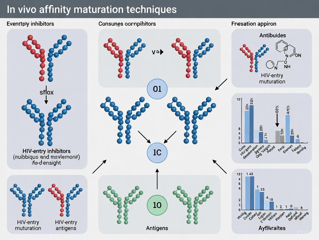
Figure 2: Workflow for in vivo affinity maturation of an HIV-entry inhibitor.
Application in HIV-1 Research and Broader Implications
Role of Tfh Cells and Antigen Availability in Selecting for Breadth
The evolution of bnAbs is not solely dependent on B-cell intrinsic factors. T follicular helper (Tfh) cells play a critical role. A mathematical model suggests that B cells with broad reactivity can outcompete narrow, strain-specific B cells if they capture a diverse set of HIV-1 proteins from the FDC network and, consequently, present a wide variety of pMHC complexes. This allows them to be rescued by a larger fraction of the diverse Tfh cell repertoire in the germinal center [3]. Furthermore, quantitative modeling shows that antigen availability directly shapes the stringency of selection in the GC, with intermediate antigen dosages often yielding the highest average affinity, highlighting a non-monotonic relationship critical for vaccine design [5].
Epitope Masking and Therapeutic Antibody Therapy
Computational models investigating bnAb therapy in people with HIV show that administered bnAbs can mask their target epitopes on the virus. This "epitope masking" gradually shifts the autologous antibody response towards less dominant, unmasked epitopes. This process can promote the evolution of a diverse "net" of autologous antibodies that collectively target multiple epitopes, which can delay viral rebound after antiretroviral therapy (ART) interruption [7]. This provides a mechanistic framework for using bnAbs to guide and shape the endogenous immune response.
Concluding Remarks
In vivo affinity maturation, leveraging the natural processes of SHM and GC selection, represents a highly efficient platform for optimizing protein biologics, including HIV-entry inhibitors. The detailed protocol for engineering B cells to express CD4 D1D2 domains demonstrates the feasibility of this approach, resulting in significant (over ten-fold) improvements in neutralization potency without compromising pharmacokinetic properties [1] [2]. For HIV research, this method, combined with insights into Tfh cell help, antigen dosing, and epitope masking, provides a powerful strategy to guide the development of next-generation bnAbs and vaccine strategies aimed at eliciting such responses. This approach surmounts limitations of in vitro methods by concurrently optimizing for affinity, stability, and bioavailability within a physiological context.
{ /article }
The Challenge of HIV-1 Diversity and the Role of Entry Inhibitors
Human Immunodeficiency Virus type 1 (HIV-1) entry into host cells represents a critical, multi-stage process that serves as a prime target for antiretroviral therapeutic intervention [8] [9]. This intricate mechanism begins with the viral envelope glycoprotein, a heterotrimer composed of three gp120 surface subunits and three gp41 transmembrane subunits, engaging the primary cellular receptor, CD4 [8]. The binding to CD4 induces substantial conformational changes in gp120, exposing a highly conserved coreceptor binding site, which subsequently interacts with a chemokine coreceptor—predominantly CCR5 or CXCR4 [8]. Following coreceptor engagement, the gp41 subunit undergoes further structural transitions, deploying its fusion peptide to insert into the host cell membrane and ultimately forming a stable six-helix bundle that drives the fusion of viral and cellular membranes [8]. The high mutation rate of HIV-1 and the resulting global diversity of envelope glycoproteins present a formidable challenge for therapy and vaccine development [8]. Entry inhibitors comprise a class of antiretroviral agents that disrupt this process at various stages, offering a strategic approach to overcome the obstacle of viral diversity by targeting conserved regions essential for host cell entry [8] [9].
The HIV-1 Diversity Problem
HIV-1's remarkable genetic diversity stems from its high replication rate, error-prone reverse transcriptase, and frequent recombination events [8]. This diversity is most pronounced in the env gene, which encodes the gp160 precursor protein that is cleaved into the gp120 and gp41 envelope subunits [8]. The V3 loop within gp120 is a primary determinant of coreceptor usage, influencing whether the virus utilizes CCR5 (R5 virus) or CXCR4 (X4 virus) for entry [8]. This coreceptor tropism has significant clinical implications; R5 viruses typically predominate during early infection and are primarily responsible for transmission, while X4 or dual-mixed (D/M) viruses emerge as disease progresses and are associated with accelerated CD4+ cell decline and poorer clinical outcomes [9]. The envelope glycoprotein's quaternary structure and its affinity for CD4 and coreceptors vary considerably among primary isolates, affecting both viral pathogenicity and susceptibility to entry inhibitors [8]. This variability necessitates sophisticated approaches to inhibitor design that can anticipate and overcome the challenge of pre-existing and treatment-emergent resistant variants.
Classes of HIV-1 Entry Inhibitors
Inhibitors of HIV-1 entry are categorized based on their specific mechanism of action and target within the multi-step entry process. The following table summarizes the key classes, their molecular targets, and representative agents.
Table 1: Classes of HIV-1 Entry Inhibitors
| Class | Molecular Target | Mechanism of Action | Representative Agent(s) | Development Status |
|---|---|---|---|---|
| Attachment Inhibitors | gp120 | Bind to gp120 and block interaction with CD4 | BMS-663068 (prodrug of BMS-626529) | Phase 2b Trials [9] |
| Post-Attachment Inhibitors | CD4 domain 2 | Binds to CD4, hindering conformational changes post-gp120 binding needed for coreceptor engagement | Ibalizumab (formerly TNX-355) | Approved [9] |
| CCR5 Antagonists | CCR5 coreceptor | Small molecule antagonists blocking interaction between gp120:CD4 complex and CCR5 | Maraviroc, Cenicriviroc, Vicriviroc, Aplaviroc | Maraviroc Approved; Cenicriviroc Phase 2b; Others discontinued [9] |
| CXCR4 Antagonists | CXCR4 coreceptor | Block interaction between gp120:CD4 complex and CXCR4 | AMD3100 (Plerixafor) | Not in clinical trials for HIV [9] |
| Fusion Inhibitors | gp41 | Prevent formation of the six-helix bundle in gp41 required for membrane fusion | Enfuvirtide (T-20) | Approved [8] [9] |
| Capsid Inhibitors | HIV-1 Capsid Protein | Disrupt capsid assembly, nuclear import, and prevent integration | Lenacapavir (GS-6207) | Approved (Treatment & Prevention) [10] |
The development trajectory of these classes highlights both the promise and the challenges of targeting HIV-1 entry. While several agents have achieved clinical success, others have been discontinued due to toxicity (e.g., aplaviroc-induced hepatitis) [9], lack of efficacy in treatment-naive populations (e.g., vicriviroc) [9], or pharmacokinetic limitations [9]. The recent approval of the capsid inhibitor lenacapavir, with its novel mechanism and long-acting profile, represents a significant advancement in the field [10].
Figure 1: HIV-1 Host Cell Entry Process. This diagram illustrates the sequential steps of HIV-1 entry, from initial gp120 binding to CD4, through coreceptor engagement, to final membrane fusion.
Application Note: In Vivo Affinity Maturation for Entry Inhibitor Optimization
Background and Principle
In vivo affinity maturation represents a novel bioengineering strategy that harnesses the body's natural immune machinery to enhance the potency of protein-based therapeutics. This approach was recently applied to improve CD4-Ig, a biologic initially developed as an entry inhibitor that functions as a soluble molecular decoy by binding to HIV-1 Env and blocking its interaction with cellular CD4 [11] [12]. Traditional in vitro engineering techniques, while successful at improving affinity, often fail to select against undesirable properties that can impair clinical efficacy, such as protease sensitivity or self-reactivity [11] [12]. The in vivo affinity maturation platform overcomes these limitations by integrating the therapeutic protein sequence into the B-cell receptor (BCR) of murine B cells, allowing for natural selection and optimization through the germinal center response [11] [12].
Experimental Protocol
Step 1: Genetic Engineering of B Cells
- Objective: Introduce genes encoding the D1D2 domains of CD4 from the half-life-enhanced CD4-Ig (CD4-Ig-v0) into the heavy-chain loci of primary murine B cells [11] [12].
- Procedure:
- Isolate primary B cells from donor mice.
- Use CRISPR/Cas9-mediated homology-directed repair to precisely insert the CD4-D1D2 sequence into the immunoglobulin heavy-chain locus, replacing the variable region while preserving regulatory elements necessary for expression and diversification [11].
- Expand the successfully engineered B cells in culture for subsequent transfer.
Step 2: Adoptive Transfer and Immunization
- Objective: Introduce the engineered B cells into a host organism where they can undergo natural selection and affinity maturation.
- Procedure:
- Adoptively transfer the engineered B cells into wild-type recipient mice [11].
- Immunize the recipient mice with HIV-1 Env antigens to stimulate a germinal center response. Multiple immunizations may be performed to drive successive rounds of affinity maturation [11] [12].
- Monitor B cell expansion, class switching, and antibody production.
Step 3: Analysis and Sorting of Affinity-Matured B Cells
- Objective: Identify B cell clones producing affinity-matured CD4-Ig variants.
- Procedure:
- Isolate splenocytes or lymph node cells from immunized mice.
- Sort individual B cells expressing high levels of the CD4-D1D2 containing BCR using flow cytometry.
- Sequence the rearranged heavy-chain loci to identify somatic hypermutations (SHMs) that have accumulated in the CD4-D1D2 encoding region [11] [12].
Step 4: Functional Characterization of Optimized Biologics
- Objective: Evaluate the impact of identified SHMs on the neutralization potency and breadth of the matured CD4-Ig.
- Procedure:
- Recombinantly produce the CD4-Ig variants incorporating the identified SHMs.
- Determine binding affinity for HIV-1 Env using surface plasmon resonance (SPR) or similar techniques [11].
- Assess neutralization potency against a global panel of HIV-1 isolates in TZM-bl or similar neutralization assays [11] [12].
- Evaluate pharmacokinetic properties, including half-life, in relevant animal models [11].
Table 2: Key Research Reagent Solutions for In Vivo Affinity Maturation
| Reagent / Material | Function / Application | Specifications / Considerations |
|---|---|---|
| Primary Murine B Cells | Platform for expression and evolution of the biologic | Requires specific genetic background compatible with genetic engineering and adoptive transfer |
| CRISPR/Cas9 System | Precision genome editing for BCR knock-in | Requires design of specific guide RNAs and homology arms targeting the IgH locus |
| HIV-1 Env Antigens | Immunogen to drive affinity maturation | Should include diverse Envs from different clades to select for broad neutralization |
| Flow Cytometry System | Analysis and sorting of engineered B cells | Requires antibodies specific for B cell markers and the expressed biologic |
| Next-Generation Sequencing | Identification of somatic hypermutations | Enables comprehensive analysis of the entire repertoire of matured BCRs |
Key Findings and Outcomes
Application of this protocol resulted in the identification of specific somatic hypermutations within the CD4-D1D2 domains that significantly enhanced the antiviral potency of CD4-Ig-v0 [11] [12]. The affinity-matured variants exhibited more than a ten-fold improvement in neutralization potency across a global panel of HIV-1 isolates without impairing pharmacokinetic properties [11] [12]. This demonstrates that in vivo affinity maturation can guide the development of more effective therapeutics against HIV-1, effectively addressing the challenge of viral diversity through a natural selection process that optimizes for both high affinity and favorable drug-like properties.
Figure 2: In Vivo Affinity Maturation Workflow. This diagram outlines the key steps in the experimental protocol for affinity maturing HIV-entry inhibitors.
Resistance Mechanisms to Entry Inhibitors
Despite their targeted mechanism of action, HIV-1 can develop resistance to entry inhibitors through several evolutionary pathways. For CCR5 antagonists like maraviroc, a primary mechanism of resistance involves viral tropism switching—the emergence of variants capable of utilizing CXCR4 instead of, or in addition to, CCR5 for entry [8] [9]. Additional proposed mechanisms for CCR5 antagonist resistance include the development of increased affinity for the CCR5 coreceptor, an enhanced rate of virus entry into host cells, and the ability to utilize the inhibitor-bound conformation of CCR5 for entry [8]. Resistance to the post-attachment inhibitor ibalizumab is associated with reduced susceptibility correlated with fewer potential N-linked glycosylation sites in the gp120 variable region 5 (V5), particularly at the V5 N-terminus [9]. These viruses with reduced susceptibility to ibalizumab surprisingly demonstrated higher levels of infectivity compared to paired, baseline viruses but remained susceptible to other entry inhibitors like maraviroc and enfuvirtide [9]. The high genetic barrier to resistance of the capsid inhibitor lenacapavir is attributed to its unique and extensive binding profile at the NTD-CTD inter-subunit interface within capsid hexamers, interacting with multiple residues on two adjacent capsid proteins [10].
The challenge posed by HIV-1 diversity necessitates continuous innovation in therapeutic strategies. Entry inhibitors provide a crucial arsenal in the fight against HIV-1, offering mechanisms of action distinct from traditional enzyme-targeting antiretrovirals. The development of advanced techniques such as in vivo affinity maturation represents a promising frontier for engineering next-generation biologics with enhanced potency and breadth against diverse HIV-1 strains. Furthermore, the success of long-acting agents like lenacapavir [10] highlights a shift toward treatment and prevention modalities that could significantly improve adherence and quality of life for individuals living with or at risk for HIV-1. Future research directions will likely focus on combining multiple entry inhibitors with complementary mechanisms, developing strategies to overcome pre-existing and emergent resistance, and further optimizing long-acting formulations for both treatment and prevention. As our understanding of the HIV-1 entry process deepens, so too will our ability to design innovative therapeutic interventions that effectively overcome the challenge of viral diversity.
CD4-Ig represents a pioneering class of biologics known as immunoadhesins. These molecules are engineered by fusing the extracellular domain of a receptor (in this case, the D1D2 domains of human CD4) to the Fc region of an immunoglobulin [13] [9]. The initial therapeutic goal was to create a soluble decoy receptor that would block HIV-1 entry into host cells by binding to the viral envelope glycoprotein gp120, thereby preventing the virus from engaging the actual CD4 receptor on T-cells [9]. Despite a sound conceptual foundation, first-generation CD4-Ig molecules faced significant clinical hurdles. Their limited half-life in vivo and inadequate potency against diverse HIV-1 isolates ultimately precluded their widespread clinical use [13] [9]. Early attempts using recombinant soluble CD4 molecules showed good in vitro activity but delivered disappointing results in clinical trials [9]. These shortcomings spurred efforts to optimize the molecule, leading to a half-life-enhanced form known as CD4-Ig-v0, which served as a backbone for further refinement [13].
Table: Evolution of CD4-Ig-Based HIV-1 Entry Inhibitors
| Molecule | Design | Key Features | Clinical Limitations |
|---|---|---|---|
| Soluble CD4 | Recombinant CD4 extracellular domain [9] | First-generation decoy receptor; binds gp120 [9] | Disappointing activity in early-phase clinical trials [9] |
| PRO 542 | Tetravalent CD4-IgG fusion protein [9] | Multivalent design for improved avidity [9] | No ongoing clinical studies reported [9] |
| CD4-Ig-v0 | D1D2 domains fused to Fc with half-life enhancements [13] | Iterative in vitro optimization for half-life and potency [13] | Served as a platform for further affinity maturation [13] |
| Affinity-Matured CD4-Ig | CD4-Ig-v0 subjected to in vivo affinity maturation [13] | Somatic hypermutations improve neutralization breadth and potency without impairing pharmacokinetics [13] | A research tool and potential therapeutic candidate; clinical efficacy not yet established [13] |
The landscape of HIV-1 treatment has been transformed by antiretroviral therapy (ART), which effectively controls viral replication. However, ART is not a cure and requires lifelong adherence, associated with cumulative toxicities, a significant pill burden, and social stigma [14]. Furthermore, the persistence of a rebound-competent viral reservoir of latently infected cells means that viral load typically rebounds swiftly after treatment interruption [14] [15]. This reality underscores the critical need for continued research into novel therapeutic and curative strategies, including the development of potent and broad neutralizing agents like improved CD4-Ig.
In Vivo Affinity Maturation of CD4-Ig: A Novel Approach
Rationale and Experimental Principle
Traditional in vitro methods for improving the affinity of protein biologics (e.g., phage display) often fail to select against undesirable properties that impair clinical efficacy, such as protease sensitivity or self-reactivity [13]. In contrast, the natural process of affinity maturation in the germinal centers of immunized animals simultaneously selects for higher affinity while eliminating variants that are unstable, self-reactive, or poorly expressed [13]. This process is continuous and highly sensitive to small affinity differences, making it a powerful tool for drug optimization [13].
A groundbreaking study demonstrated that the B-cell receptors (BCRs) of primary murine B cells could be engineered to affinity mature the human CD4 domains of CD4-Ig in vivo [13] [16] [17]. The core strategy involved replacing the variable region of the BCR with the sequence for the CD4 D1D2 domains. When these engineered B cells were transferred into mice and immunized with HIV-1 Env antigens, they underwent natural selection, leading to the development of CD4-Ig variants with superior properties [13].
Detailed Experimental Workflow and Protocol
The following diagram illustrates the key steps for engineering B cells to affinity mature CD4-Ig in vivo.
Protocol 1: Engineering Primary Mouse B Cells to Express CD4-Ig Ex Vivo
- B Cell Isolation: Harvest splenic B cells from donor mice (e.g., B6 CD45.1 strain) [13].
- CRISPR/Cas12a RNP Complex Formation: Prepare ribonucleoproteins (RNPs) using Mb2Cas12a and a guide RNA targeting the 3'-most JH segment (JH4) in the murine heavy-chain locus [13].
- Homology-Directed Repair Template (HDRT) Design: Clone an HDRT into a recombinant adeno-associated virus DJ (AAV-DJ) vector. The HDRT must contain [13]:
- 5' Homology Arm: 570 bp complementary to the 5' UTR upstream of a specific VH segment (e.g., VH1-34 or VH1-64).
- Insert Sequence: A gene encoding the D1D2-OKT3-VH fusion protein. This consists of human CD4 domains 1 and 2 (D1D2) fused via a (G4S)3 linker to the amino-terminus of the heavy-chain variable domain of the murine OKT3 antibody (OKT3-VH).
- 3' Homology Arm: 600 bp complementary to the 3' intronic region immediately downstream of the JH4 segment.
- Cell Electroporation: Co-electroporate the isolated B cells with the Cas12a RNPs and the AAV-DJ vector containing the HDRT.
- Validation of Editing: Culture edited cells for 2-3 days and assess editing efficiency via flow cytometry using fluorescently labeled anti-CD4 antibody or HIV-1 gp120. Target editing efficiencies of ~11% are achievable and sufficient [13].
Protocol 2: In Vivo Affinity Maturation and Analysis
- Adoptive Transfer: Adoptively transfer approximately 15,000 B cells expressing the D1D2-OKT3-VH fusion (constituting about 0.3% of engrafted cells) into wild-type (CD45.2) C57BL/6J recipient mice [13].
- Immunization Regimen:
- Serum Monitoring: Collect serum after each immunization to monitor the development of neutralization activity against HIV-1 pseudoviruses (e.g., BG505, CE1176) using TZM-bl assays [13].
- B Cell Isolation and Sequencing: After evidence of neutralization is detected (typically after boosts), isolate splenic B cells. Amplify and sequence the engineered heavy-chain locus to identify patterns of somatic hypermutation (SHM), particularly within the D1D2-encoding region [13].
- Functional Characterization: Clone identified variant sequences into expression vectors for the full CD4-Ig biologic. Produce and purify the antibodies, then evaluate:
- Binding Affinity: For HIV-1 Env gp120 using surface plasmon resonance (SPR).
- Neutralization Potency and Breadth: Against a global panel of HIV-1 pseudoviruses.
- Pharmacokinetic Properties: Assess half-life in murine models to ensure improvements in affinity do not impair bioavailability [13].
Key Outcomes and Research Toolkit
The in vivo affinity maturation approach yielded significant improvements over the original CD4-Ig-v0. Somatic hypermutations identified in the D1D2 domain of engrafted B cells led to variants with markedly enhanced neutralization potency, achieving levels below 1 µg/ml against a global panel of HIV-1 isolates [13]. Crucially, this enhanced potency was achieved without compromising the molecule's near-absolute breadth, high thermostability, or long in vivo half-life, demonstrating the unique ability of in vivo selection to improve efficacy while maintaining favorable biophysical and pharmacokinetic properties [13].
Table: Neutralization Potency of Affinity-Matured CD4-Ig
| Molecule/Variant | Neutralization Potency (IC50) | Neutralization Breadth | Key Characteristics | Source |
|---|---|---|---|---|
| Original CD4-Ig-v0 | Baseline (pre-maturation) | Near-absolute breadth, but limited potency | Optimized for half-life via iterative in vitro methods [13] | [13] |
| Affinity-Matured CD4-Ig | < 1 µg/mL (against a global HIV-1 panel) [13] | Retained near-absolute breadth of CD4-Ig-v0 [13] | Improved affinity without impaired pharmacokinetics [13] | [13] |
| HmAb64 (Vaccine-Elicited CD4bs mAb) | Moderate (50% neutralization of 10% of a 208-virus panel) [18] | 20/208 viruses (10%), including tier-2 strains [18] | Isolated from a human vaccine trial; demonstrates feasibility of eliciting CD4bs antibodies [18] | [18] |
The Scientist's Toolkit: Essential Research Reagents
Table: Key Reagents for B Cell Engineering and Affinity Maturation
| Research Reagent | Function in the Protocol | Specific Example / Description |
|---|---|---|
| CRISPR/Mb2Cas12a RNP | Creates a double-stranded break in the B cell genome to enable gene insertion [13] | Ribonucleoprotein complex with gRNA targeting the JH4 segment in the murine heavy-chain locus [13] |
| AAV-DJ HDRT Vector | Delivers the repair template for homology-directed repair, inserting the gene of interest [13] | Recombinant AAV-DJ with homology arms for VH1-34 and a D1D2-OKT3-VH insert sequence [13] |
| Env mRNA-LNP Immunogen | Provides the antigen for in vivo immunization and selection of high-affinity B cells [13] | Lipid nanoparticles containing mRNA encoding engineered HIV-1 Env trimers (e.g., 16055-ConM-v8.1 SOSIP-TM) [13] |
| Fluorescently Labeled gp120 / Anti-CD4 | Critical for validating surface expression of the engineered BCR via flow cytometry [13] | Used to detect successful fusion of D1D2 domains to the BCR on the surface of primary murine B cells [13] |
Clinical Limitations and Future Perspectives
Despite the promising preclinical advancements, CD4-Ig-based therapies and other entry inhibitors face persistent clinical challenges. A major limitation of CCR5-targeting strategies, including some entry inhibitors, is their inactivity against viruses that use the CXCR4 co-receptor [15]. Viral rebound from CXCR4-tropic virus has been observed in individuals who received CCR5-Δ32 stem cell transplants and stopped ART, highlighting that targeting a single entry pathway may be insufficient if viral populations are mixed [15].
Furthermore, the field of HIV cure research is increasingly focused on the complex biology of the viral reservoir. The reservoir is not static but dynamic, and is composed of latently infected cells residing in diverse anatomical sanctuaries like lymph nodes and gut-associated lymphoid tissue [14]. Recent studies using spatial transcriptomics have shown that HIV-infected cells in lymph nodes are often localized within B cell follicles, which are poorly accessed by CD8+ T cells, creating a protected niche for viral persistence [14]. This anatomical protection presents a significant barrier for any therapeutic, including biologics like CD4-Ig, that must reach and eliminate these sanctuary sites to achieve a cure.
Future research directions will likely involve combining potent, affinity-matured entry inhibitors like CD4-Ig with other therapeutic modalities. Logical partners include capsid inhibitors like lenacapavir, which disrupts a different stage of the viral life cycle [19], and gene therapy approaches such as CRISPR/Cas9 to excise the provirus or engineer HIV-resistant cells [15]. The demonstrated success of using engineered B cells as a vehicle for in vivo affinity maturation also opens the door to applying this platform to other non-antibody biologics, such as CTLA-4, SIRPα, and IL-7, for a range of diseases beyond HIV [13].
The development of broad neutralization against human immunodeficiency virus (HIV) represents a central goal in vaccinology. A key advancement in this pursuit is the identification and characterization of broadly neutralizing antibodies (bNAbs) that target conserved regions on the HIV-1 envelope glycoprotein (Env). Among these, the CD4 binding site (CD4bs) stands out as a highly conserved and functionally critical epitope, making it a premier target for both therapeutic antibody development and vaccine design [20] [21]. Antibodies against this site can block viral entry by preventing the essential interaction between the virus and the host CD4 receptor [21]. Other vulnerable epitopes include the membrane-proximal external region (MPER) of gp41, the V1V2 glycan site, the V3 glycan supersite, and the gp120-gp41 interface [20] [21]. This application note details the key epitope targets for HIV-1 bNAbs, provides quantitative comparisons of their characteristics, and outlines established experimental protocols for their study within the context of in vivo affinity maturation research.
Quantitative Profiling of Key bNAb Targets
The potency and breadth of bNAbs are quantitatively assessed against diverse, multi-clade panels of HIV-1 pseudoviruses. The following table summarizes the characteristics of prominent bNAbs targeting key vulnerable sites, with a focus on the CD4bs.
Table 1: Characteristics of Broadly Neutralizing Antibodies Targeting Key HIV-1 Epitopes
| Antibody | Target Epitope | Heavy Chain Germline | Neutralization Breadth | Geometric Mean IC50 (µg/mL) | Key Features |
|---|---|---|---|---|---|
| 04_A06 [20] | CD4 Binding Site | VH1-2 | 98.5% (332 strains) | 0.059 | 11-amino-acid insertion in FWRH1; resistant to classic CD4bs escape variants |
| FD22 [21] | CD4 Binding Site | IGHV3-30 | 82% (145 strains) | 0.27 | 20-amino-acid CDRH3; mediates robust ADCC; non-autoreactive |
| VRC01 [21] | CD4 Binding Site | IGHV1-2*02 | 88% (145 strains) | 0.25 | Classic VRC01-class antibody; high somatic hypermutation |
| MPER-Targeting bNAbs [22] | Membrane-Proximal External Region | - | - | - | Epitopes are more exposed on immature virions; some exhibit polyreactivity |
Beyond neutralization activity, the genetic composition of bNAbs reveals patterns critical for immunogen design. Somatic hypermutation (SHM) is a hallmark of most bNAbs, and the level of mutation often correlates with neutralization potency [20]. The choice of germline gene is also pivotal; while the VH1-2 gene segment is frequently observed in potent CD4bs bNAbs like VRC01 and 04_A06 [20] [21], the isolation of FD22 from the IGHV3-30 germline demonstrates that alternative genetic pathways can yield equally potent and broad neutralization [21]. This diversity is essential for designing vaccines that can engage a wider repertoire of B cell precursors.
Table 2: Genetic and Binding Properties of Featured CD4bs bNAbs
| Property | 04_A06 [20] | FD22 [21] | VRC01 [21] |
|---|---|---|---|
| Somatic Hypermutation (VH) | ~38-39% (61-63% germline identity) | 37% | High (exact value not specified) |
| CDRH3 Length | Information not specified in source | 20 amino acids | 15 amino acids (typical for VRC01-class) |
| Binding Specificity | Interprotomer contacts on adjacent gp120 | Loop D, CD4 binding loop, V5 loop | Classical CD4bs footprint |
| Autoreactivity | Not reported | Non-autoreactive (tested against HEp-2 cells and cardiolipin) | Not reported |
Experimental Protocols for bNAb Discovery and Evaluation
Protocol: Single B Cell Sorting and Cloning from Elite Neutralizers
This protocol is adapted from large-scale profiling studies of HIV-1 elite neutralizers [20].
Application: Isolation of antigen-specific memory B cells for the discovery of novel bNAbs.
Materials and Reagents:
- Source: Peripheral blood mononuclear cells (PBMCs) from HIV-1-infected donors with broad serum neutralization.
- Staining Reagents: GFP-labeled BG505SOSIP.664 and YU2gp140 trimer baits, anti-human CD20 antibody, anti-human IgG antibody, DAPI.
- Cell Culture Media: RPMI-1640 supplemented with FBS, cytokines (e.g., IL-2, IL-21), and feeder cells (e.g., CD40L-expressing cells) for B cell culture.
- Amplification & Cloning: Reverse transcription and PCR reagents, expression vectors for IgG heavy and light chains.
Procedure:
- Cell Preparation: Isolate PBMCs from donor blood samples using density gradient centrifugation.
- Enrichment and Staining: Enrich for B cells and stain with GFP-labeled Env baits, anti-CD20, anti-IgG, and the viability dye DAPI.
- Single-Cell Sorting: Using a fluorescence-activated cell sorter (FACS), single-sort live (DAPI-), CD20+, IgG+, Env-bait+ memory B cells into 96- or 384-well PCR plates.
- Antibody Gene Amplification: Perform reverse transcription and nested PCR to amplify paired heavy- and light-chain variable region genes from single cells.
- Antibody Expression: Clone the amplified variable genes into immunoglobulin expression vectors containing constant regions. Co-transfect heavy and light chain plasmids into mammalian cells (e.g., HEK 293F or Expi293F cells) for recombinant antibody production.
- Primary Screening: Harvest culture supernatants and screen for neutralization activity against a panel of 6-8 diverse HIV-1 pseudoviruses.
- Characterization: Scale up production of lead neutralizing antibodies, purify, and characterize for breadth, potency, and epitope specificity.
Protocol: In Vivo Affinity Maturation of Engineered B Cells
This protocol describes a cutting-edge technique for improving the affinity of HIV-entry inhibitors, such as CD4-Ig, by leveraging the natural germinal center reaction in mice [13] [23].
Application: Affinity maturation of non-antibody biologics, such as immunoadhesins, to enhance their antiviral potency.
Materials and Reagents:
- Primary Murine B Cells: Harvested from donor mice (e.g., C57BL/6J).
- Gene Editing System: CRISPR/Mb2Cas12a ribonucleoproteins (RNPs) targeting the murine JH4 segment.
- Homology-Directed Repair Template (HDRT): Delivered via recombinant adeno-associated virus DJ (AAV-DJ), encoding the biologic (e.g., CD4 D1D2 domains fused to an antibody variable region) flanked by homology arms.
- Immunogen: mRNA-lipid nanoparticles (mRNA-LNP) expressing an engineered HIV-1 Env trimer (e.g., 16055-ConM-v8.1 SOSIP-TM).
Procedure: 1. B Cell Engineering: - Isolate splenic B cells from donor mice. - Electroporate cells with Mb2Cas12a RNPs to create a double-strand break in the heavy-chain locus (JH4). - Simultaneously, transduce with AAV-DJ containing the HDRT to insert the gene encoding the biologic. - Confirm surface expression of the engineered biologic via flow cytometry. 2. Adoptive Transfer and Immunization: - Adoptively transfer a controlled number (e.g., ~15,000) of engineered B cells into recipient wild-type mice. - 24 hours post-transfer, immunize mice intramuscularly with the Env trimer mRNA-LNP. - Administer booster immunizations at 2- or 4-week intervals. 3. Monitoring and Analysis: - Collect serum periodically to monitor the development of neutralizing activity using TZM-bl or similar neutralization assays. - After final immunization, isolate splenic plasma cells or memory B cells. - Sequence the engineered gene region from these cells to identify accumulated somatic hypermutations (SHM). - Clone the mutated sequences, express the improved biologic variants, and evaluate their binding affinity and neutralization potency against a global panel of HIV-1 strains.
Visualizing the B Cell Engineering and Affinity Maturation Workflow
The following diagram illustrates the key steps for engineering B cells to express a protein biologic and subjecting it to in vivo affinity maturation.
Mapping bNAb Epitopes on the HIV-1 Envelope Trimer
The diagram below provides a simplified schematic of the HIV-1 Env trimer, highlighting the key vulnerable epitopes targeted by broadly neutralizing antibodies.
The Scientist's Toolkit: Essential Research Reagents
Table 3: Key Reagents for HIV-1 bNAb and Affinity Maturation Research
| Reagent / Solution | Function / Application | Examples / Specifications |
|---|---|---|
| Stabilized Env Trimers | B cell sorting baits; immunogens for animal studies; structural biology. | BG505SOSIP.664, YU2gp140, 16055-ConM-v8.1 SOSIP-TM [20] [13] |
| CRISPR-Cas System | Precision genome editing for B cell receptor engineering. | Mb2Cas12a ribonucleoproteins (RNPs) targeting the JH segment [13] |
| AAV-DJ HDR Template | Efficient delivery of homology-directed repair templates for gene insertion. | Contains homology arms for VH1-34/VH1-64 and JH4; encodes biologic fusion construct [13] |
| mRNA-LNP Immunogen | Potent in vivo delivery of antigen for immunization and affinity maturation. | LNPs encapsulating mRNA encoding engineered Env trimers [13] |
| CD4-Ig-v0 | Optimized immunoadhesin biologic; starting template for affinity maturation. | Half-life-enhanced version of CD4-Ig; scaffold for improving potency [13] [23] |
| TZM-bl Cell Line | Reporter cell line for quantifying HIV-1 neutralization potency and breadth. | Expresses CD4 and CCR5; contains a Tat-responsive luciferase reporter gene |
The Rationale for In Vivo vs. In Vitro Maturation Strategies
Affinity maturation is a cornerstone of modern biologic drug development, essential for enhancing the potency and efficacy of therapeutic proteins, including antibodies and immunoadhesins. This process refines a protein's ability to bind its target antigen with high affinity and specificity. Two principal paradigms dominate this field: in vivo affinity maturation, which harnesses the natural power of the mammalian immune system, and in vitro affinity maturation, which relies on laboratory-based display technologies and directed evolution [13] [24]. The choice between these strategies carries significant implications for the developmental trajectory, properties, and ultimate clinical success of a biologic.
Within HIV-1 research, the development of entry inhibitors such as CD4-Ig represents a critical therapeutic avenue. These biologics aim to block viral entry by targeting the highly conserved CD4 binding site (CD4bs) on the HIV-1 envelope glycoprotein [20] [25]. However, the initial versions of these proteins often lack the requisite potency to effectively neutralize diverse global HIV-1 strains. Affinity maturation is therefore indispensable for transforming these promising candidates into clinically viable therapeutics. This application note delineates the rationale for selecting in vivo versus in vitro maturation strategies, providing a structured comparison, detailed experimental protocols, and practical guidance for their application in HIV-entry inhibitor research.
Comparative Analysis of Maturation Strategies
Table 1: Strategic Comparison of In Vivo vs. In Vitro Affinity Maturation
| Feature | In Vivo Affinity Maturation | In Vitro Affinity Maturation |
|---|---|---|
| Core Principle | Leverages the natural germinal center reaction in immunized animals [13]. | Utilizes laboratory-based display systems (e.g., phage, yeast) and artificial selection [24]. |
| Selection Pressure | Holistic; favors high affinity, stability, low self-reactivity, and protease resistance [13]. | Primarily focused on enhancing target binding affinity [24]. |
| Diversification Mechanism | Continuous somatic hypermutation (SHM) in B cells [13]. | Site-saturated mutagenesis or error-prone PCR in discrete steps [24]. |
| Key Advantage | Generates variants with superior bioavailability and developmental potential [13]. | Rapid, controlled, and does not require animal immunization [24]. |
| Primary Limitation | Technically complex, lower throughput, and requires specialized B-cell engineering [13]. | May yield variants with poor solubility, aggregation, or immunogenicity [24]. |
| Typical Affinity Gain | Up to ~1000-fold neutralization improvement for CD4-Ig-v0 [25]. | Up to 100-fold for synthetic human VHs [24]. |
The comparative data reveals that in vivo maturation integrates multiple, simultaneous selection pressures that mirror the natural development of effective biologics. This process not only selects for improved affinity but also against undesirable characteristics like self-reactivity, protease sensitivity, and poor stability, which are common failure points for therapeutics in development [13]. This holistic filtering often results in candidates with a higher probability of clinical success, as demonstrated by the ability of in vivo-matured CD4-Ig to achieve enhanced neutralization breadth and potency while maintaining favorable pharmacokinetics [25].
Conversely, in vitro maturation offers unparalleled speed and direct control over the mutagenesis process. It is highly effective for achieving significant affinity gains, as seen with synthetic human VHs targeting SARS-CoV-2 [24]. However, this narrow focus can lead to "over-optimization" for affinity at the expense of other biophysical properties. For instance, high-affinity VHs derived from synthetic libraries frequently exhibit low solubility and a tendency to aggregate, necessitating additional engineering steps, such as "camelization," to make them viable for therapeutic application [24].
Application in HIV-1 Entry Inhibitor Research
The development of HIV-1 entry inhibitors exemplifies the strategic trade-offs between these maturation approaches. The CD4 binding site (CD4bs) on the HIV-1 Env glycoprotein is a highly conserved and validated target for broadly neutralizing antibodies (bnAbs) and engineered biologics like CD4-Ig [20]. The goal is to achieve exceptional breadth and potency against a diverse array of global HIV-1 strains.
Recent research highlights the success of in vivo maturation for this specific challenge. Pan et al. demonstrated that engineering murine B cells to express a CD4-based immunoadhesin (CD4-Ig-v0) on their surface, followed by adoptive transfer and immunization, led to robust affinity maturation [13] [25]. The resulting somatic hypermutations in the CD4 domains (D1D2) significantly improved the neutralization potency of CD4-Ig-v0 against a global panel of HIV-1 isolates without compromising its pharmacokinetic profile [25]. This approach leverages the immune system's unparalleled ability to navigate complex fitness landscapes and identify optimal mutations that enhance function while preserving biocompatibility.
Simultaneously, large-scale profiling of elite neutralizers—individuals who naturally produce potent bnAbs—has identified antibodies like 04_A06, which targets the CD4bs with remarkable breadth (98.5%) and potency [20]. The study of such antibodies provides a blueprint for the desired outcomes of affinity maturation, whether it occurs naturally, in vivo in model systems, or is mimicked in vitro. These bnAbs are characterized by a high degree of somatic mutation, which correlates with their antiviral activity, underscoring the importance of extensive sequence diversification and selection [20].
Table 2: Performance Metrics of Matured HIV-1 Entry Inhibitors
| Biologic / Strategy | Key Outcome Metric | Result |
|---|---|---|
| CD4-Ig-v0 (In Vivo Matured) | Neutralization Potency (geometric mean IC₅₀) | Improved to <1 µg/mL against a global HIV-1 panel [25] |
| 04_A06 bnAb (Elite Neutralizer) | Neutralization Breadth | 98.5% (against 332 multiclade virus strains) [20] |
| 04_A06 bnAb (Elite Neutralizer) | Neutralization Potency (geometric mean IC₅₀) | 0.059 µg mL⁻¹ [20] |
| Synthetic Human VH (In Vitro Matured) | Affinity Improvement (KD) | Up to 100-fold (from ~480 nM to 3 nM) [24] |
Experimental Protocols
Protocol for In Vivo Affinity Maturation of an HIV-1 Entry Inhibitor
This protocol details the methodology for affinity maturing the CD4 domains of CD4-Ig in a murine model, as established by Pan et al. [13] [25].
Key Reagent Solutions:
- Primary Murine B Cells: Isolated from splens of C57BL/6 mice.
- CRISPR/Mb2Cas12a RNP: For creating a double-stranded break in the JH4 segment of the heavy-chain locus.
- AAV-DJ HDRT: Recombinant adeno-associated virus delivering the homology-directed repair template.
- HDR Template: Encodes the D1D2-OKT3-VH fusion construct with homology arms.
- mRNA-LNP Immunogen: Lipid nanoparticles containing mRNA encoding an engineered HIV-1 Env trimer (e.g., 16055-ConM-v8.1 SOSIP-TM).
Procedure:
- B Cell Engineering:
- Harvest splenic B cells from donor mice.
- Electroporate cells with CRISPR/Mb2Cas12a ribonucleoproteins (RNPs) targeted to the JH4 segment and transduce with AAV-DJ containing the HDR template.
- The template replaces the endogenous VDJ-recombined gene with a sequence encoding the biologic (e.g., CD4 D1D2 domains) fused to a murine VH domain.
- Confirm surface expression of the fusion protein via flow cytometry using a fluorescently labeled anti-CD4 antibody or HIV-1 gp120.
Adoptive Transfer and Immunization:
- Adoptively transfer approximately 15,000 successfully edited B cells into wild-type recipient mice.
- Immunize mice intramuscularly 24 hours post-transfer with the mRNA-LNP immunogen.
- Administer booster immunizations at 2-week and 4-week intervals.
Analysis and Recovery:
- Monitor serum for neutralization activity against HIV-1 pseudoviruses after each immunization.
- Isolate splenic B cells from immunized mice.
- Sort single B cells expressing the matured biologic for single-cell RNA sequencing to recover the mutated VH genes.
- Clone the matured sequences into expression vectors for large-scale production and functional characterization.
Protocol for In Vitro Affinity Maturation of a Synthetic VH
This protocol describes the in vitro affinity maturation and subsequent solubilization of a human VH, as performed by Henry et al. [24].
Key Reagent Solutions:
- Yeast Surface Display Library: Library of VH variants created via site-saturated mutagenesis of CDRs.
- Antigen: Recombinant target antigen (e.g., SARS-CoV-2 RBD, HIV-1 gp120).
- Fluorescence-Activated Cell Sorting (FACS): For isolating high-affinity binders.
- Camelization Mutagenesis Primers: For introducing G44E and L45R mutations in FR2.
Procedure:
- Library Generation:
- Start with a lead VH clone identified from a synthetic phage display library.
- Generate a mutant library by performing site-saturated mutagenesis on the complementarity-determining regions (CDRs).
- Clone the library into a yeast surface display vector.
Selection for Affinity:
- Induce expression of the VH library on the yeast surface.
- Label yeast cells with a fluorescently tagged antigen.
- Use multiple rounds of fluorescence-activated cell sorting (FACS) to isolate yeast populations that display VHs with the highest antigen-binding affinity.
- Sequence sorted populations to identify key mutations.
Solubility Engineering (Camelization):
- Identify affinity-matured VHs that exhibit aggregation.
- Introduce camelizing mutations (e.g., G44E, L45R in FR2) into the affinity-matured VH via site-directed mutagenesis.
- Express and purify the camelized variant.
- Assess solubility and aggregation resistance using size-exclusion chromatography (SEC) and compare affinity pre- and post-camelization via surface plasmon resonance (SPR).
The Scientist's Toolkit
Table 3: Essential Research Reagent Solutions for Affinity Maturation
| Reagent / Tool | Function / Application | Example Use Case |
|---|---|---|
| CRISPR/Cas12a RNP | Precise genome editing in primary B cells to integrate transgenes. | Targeted knock-in of CD4-Ig construct into the murine BCR locus [13]. |
| AAV-DJ HDRT | High-efficiency delivery of homology-directed repair templates. | Delivery of the D1D2-OKT3-VH fusion construct for B cell engineering [13]. |
| mRNA-LNP | Potent immunization vehicle for delivering encoded antigen. | Expression of HIV-1 Env trimer in mice to drive affinity maturation [13]. |
| Yeast Surface Display | Platform for displaying protein libraries and selecting high-affinity binders. | In vitro affinity maturation of synthetic human VHs [24]. |
| Camelization Mutagenesis | Framework mutagenesis to improve solubility of human VHs. | Converting aggregation-prone, affinity-matured VHs into soluble therapeutics [24]. |
The choice between in vivo and in vitro affinity maturation is not merely a technical decision but a strategic one that shapes the fundamental properties of the resulting biologic. For HIV-1 entry inhibitor research, where the goal is to achieve exceptional breadth and potency alongside favorable in vivo stability, in vivo affinity maturation presents a compelling strategy. Its key advantage lies in its ability to concurrently optimize for multiple drug-like properties, potentially de-risking later stages of clinical development [13] [25].
However, in vitro maturation remains a powerful and efficient alternative, particularly for rapid initial affinity optimization and when animal immunization is impractical. Its limitations concerning solubility can be effectively mitigated through subsequent camelization, providing a robust pipeline for generating high-quality human VHs from synthetic libraries [24].
For research teams, the following guidance is proposed:
- Prioritize In Vivo Maturation when developing complex biologics like immunoadhesins for HIV therapy, where holistic optimization is critical for clinical success.
- Leverage In Vitro Maturation for rapid affinity optimization of antibody fragments (e.g., VHs) and when working with highly toxic or non-immunogenic targets.
- Adopt a Hybrid Approach by using in vitro methods for initial, rapid affinity gains, followed by in vivo maturation to refine stability and bioavailability, represents a promising frontier in biologic engineering.
Engineered Evolution: CRISPR B-Cell Reprogramming and In Vivo Maturation Protocols
This application note details a advanced methodology for the precise integration of human antibody variable genes into native murine B-cell loci using CRISPR-Cas12a. Developed within the context of HIV-entry inhibitor research, this protocol enables the generation of B cells producing human neutralizing antibodies that subsequently undergo in vivo affinity maturation [26] [27]. The approach provides a novel platform for evaluating HIV-1 vaccine candidates and developing advanced B cell therapies by modeling human-like antibody responses in an in vivo system [26]. Below, we present optimized protocols, key reagent specifications, and visual workflows to facilitate implementation of this technology in research settings focused on antiviral therapeutics.
CRISPR-Cas12a genome editing represents a significant advancement in precision genetic engineering of primary immune cells. Unlike Cas9, Cas12a is a type V-A CRISPR-associated nuclease that creates staggered double-stranded breaks with cohesive ends, enhancing precise gene integration [28]. This system is particularly valuable for B cell engineering as it enables direct replacement of mouse antibody variable chain genes with human versions through homology-directed repair [26] [27]. The edited primary B cells maintain normal physiological functions, including the capacity to undergo T cell-dependent affinity maturation in response to antigen exposure, making them ideal for studying HIV-1 and SARS-CoV-2 neutralizing antibody development [26] [27].
Table 1: Key Advantages of CRISPR-Cas12a for B Cell Engineering
| Feature | Benefit for B Cell Editing | Therapeutic Application |
|---|---|---|
| Staggered DNA breaks | Facilitates precise integration of large gene cassettes | Enables clean replacement of variable regions with human sequences |
| T-rich PAM recognition | Targets different genomic sites compared to Cas9 | Expands possible integration sites in immunoglobulin loci |
| RuvC domain activity | Cleaves both target and non-target strands | Creates clean breaks for precise editing [28] |
| Minimal off-target effects | Higher specificity than Cas9 systems | Reduces risk of unintended genomic alterations |
| Intrinsic RNase activity | Processes multiple crRNAs from single transcript | Enables multiplexed genome regulation [29] |
Research Reagent Solutions
Successful implementation of this technology requires carefully selected and validated reagents. The table below outlines essential components for CRISPR-Cas12a-mediated B cell engineering.
Table 2: Essential Research Reagents for CRISPR-Cas12a B Cell Editing
| Reagent Category | Specific Examples | Function & Importance |
|---|---|---|
| Cas12a Variants | LbCas12a, Flex-Cas12a, hyperCas12a | Engineered versions with expanded PAM recognition (5'-NYHV-3') or enhanced activity [30] [29] |
| crRNA Design | Target-specific crRNAs | Guides Cas12a to specific genomic loci; requires 5'-TTTV-3' PAM for wildtype [30] [28] |
| Editing Template | Human variable gene cassette with homology arms | Contains human antibody sequences flanked by regions homologous to mouse immunoglobulin loci [26] |
| Delivery System | Electroporation of ribonucleoprotein complexes | Encomes high-efficiency delivery to primary B cells with minimal toxicity |
| B Cell Culture Media | Optimized cytokine cocktails | Supports viability and proliferation of edited B cells pre- and post-transplantation |
| Validation Assays | Flow cytometry, ELISA, sequencing | Confirms successful integration and function of human antibody genes |
The following tables consolidate key performance metrics for CRISPR-Cas12a-mediated B cell engineering and subsequent affinity maturation outcomes.
Table 3: Efficacy Metrics of CRISPR-Cas12a B Cell Editing
| Parameter | Performance Value | Experimental Context |
|---|---|---|
| Editing Efficiency | High (specific values not provided in sources) | Primary mouse B cells [26] [27] |
| In Vivo Engraftment | Successful | Edited B cells transplanted into recipient mice [26] |
| Affinity Maturation | Significant improvement in antibody potency | Post-immunization in vivo maturation [26] [27] |
| Bioavailability | Maintained | No loss of antibody bioavailability after maturation [26] |
| Neutralization Breadth | Enhanced against HIV-1 and SARS-CoV-2 | For antibodies undergoing in vivo affinity maturation [26] |
Table 4: Comparison of Cas12a Variants for Genome Editing Applications
| Variant | Key Mutations | PAM Recognition | Therapeutic Advantage |
|---|---|---|---|
| Wildtype LbCas12a | None | 5'-TTTV-3' | Established specificity, lower off-target risk |
| Flex-Cas12a | G146R, R182V, D535G, S551F, D665N, E795Q | 5'-NYHV-3' | Expands targetable sites to ~25% of human genome [30] |
| hyperCas12a | D156R, D235R, E292R, D350R | 5'-TTTV-3' (with some non-canonical) | Enhanced activity, particularly at low crRNA concentrations [29] |
Experimental Protocols
Core Protocol: CRISPR-Cas12a-Mediated Variable Gene Replacement in Primary B Cells
Objective: Precise replacement of mouse antibody variable genes with human versions in primary B cells for in vivo affinity maturation studies.
Materials:
- Primary mouse B cells isolated from spleen or lymph nodes
- Recombinant LbCas12a protein (wildtype or engineered variants)
- crRNAs targeting mouse immunoglobulin variable regions
- Donor DNA template containing human variable genes flanked by homology arms
- Electroporation system (e.g., Neon Transfection System)
- B cell culture media with cytokines (IL-4, IL-21, CD40L)
- Flow cytometry antibodies for B cell markers (B220, CD19)
Procedure:
- B Cell Isolation: Isolate primary B cells from mouse spleen using magnetic bead-based separation (B cell isolation kit) to achieve >95% purity.
- crRNA Complex Formation: Pre-complex LbCas12a protein with gene-specific crRNAs (30-minute incubation at 25°C in Cas12a buffer).
- Electroporation Preparation: Mix 1×10^6 B cells with Cas12a-crRNA ribonucleoprotein complexes (30 pmol) and donor DNA template (15 pmol) in electroporation buffer.
- Electroporation: Deliver using optimized electroporation parameters (1400V, 20ms, 2 pulses for primary B cells).
- Recovery Culture: Immediately transfer cells to pre-warmed B cell media with IL-4 (10ng/mL) and IL-21 (25ng/mL). Culture for 48 hours at 37°C, 5% CO2.
- Validation: Analyze editing efficiency by flow cytometry for surface human immunoglobulin expression and PCR/sequencing of target loci.
- In Vivo Transfer: Transplant 5×10^5 to 1×10^6 edited B cells into recipient mice via intravenous injection for affinity maturation studies.
Troubleshooting Notes:
- Low editing efficiency: Optimize crRNA design to target accessible regions of immunoglobulin loci; verify Cas12a protein activity using validation plasmid.
- Poor cell viability: Reduce electroporation pulse duration; add caspase inhibitors to recovery media.
- Insufficient integration: Increase homology arm length in donor template (recommended 800-1000bp); verify donor concentration and quality.
Supporting Protocol: Affinity Maturation Analysis of Edited B Cells
Objective: Evaluate the affinity maturation process of CRISPR-edited B cells producing human antibodies in vivo.
Materials:
- Mice transplanted with edited B cells
- HIV-1 envelope protein or relevant antigen
- Adjuvant for immunization (e.g., Alum, Complete Freund's Adjuvant)
- ELISA plates coated with antigen
- Anti-human IgG detection antibodies
- Flow cytometry reagents for germinal center B cell analysis (GL7, CD95)
- Single-cell RNA sequencing reagents for B cell receptor analysis
Procedure:
- Immunization: Immunize recipient mice with HIV-1 envelope protein (10-50μg) in adjuvant via subcutaneous or intraperitoneal route at days 7 and 21 post B cell transfer.
- Serum Collection: Collect blood at weekly intervals to monitor antibody titers by ELISA.
- Germinal Center Analysis: At day 14 post-boost, isolate splenocytes and analyze germinal center B cells by flow cytometry (B220+GL7+CD95+).
- Single-Cell Sequencing: Sort single germinal center B cells for V(D)J sequencing to analyze somatic hypermutation in human variable regions.
- Antibody Potency Assay: Test serum neutralizing activity against HIV-1 pseudoviruses in TZM-bl cells.
Workflow and Mechanism Diagrams
Diagram 1: B Cell Engineering Workflow
Diagram 2: Molecular Mechanism
Application in HIV-Entry Inhibitor Research
This CRISPR-Cas12a B cell engineering platform directly advances HIV-entry inhibitor research by enabling the in vivo evaluation of human antibody candidates in a physiological context [26]. The technology facilitates study of affinity maturation pathways critical for developing broad-spectrum neutralizing antibodies against highly variable viral pathogens like HIV-1. Specifically, the platform allows researchers to:
- Introduce human antibody genes targeting conserved HIV-1 envelope regions into the native B cell development pathway
- Study how somatic hypermutation improves antibody potency against HIV-1 entry mechanisms
- Evaluate multiple antibody candidates simultaneously through multiplexed CRISPR approaches [29]
- Model human antibody responses in a controlled experimental system
The capacity for edited B cells to undergo antigen-driven affinity maturation while maintaining bioavailability makes this approach particularly valuable for both vaccine development and B cell therapy applications against HIV-1 and other viral pathogens [26] [27].
The first two domains (D1D2) of the human CD4 receptor are critical for HIV-1 entry, serving as the primary viral attachment site on the envelope glycoprotein (Env) gp120 [31] [32]. Soluble CD4 (sCD4) constructs comprising these domains have been extensively investigated as promising inhibitors and components of vaccine immunogens for over two decades [31]. Their potent inhibitory activity stems from the ability to bind gp120, induce conformational changes that prematurely expose coreceptor binding sites, and block viral attachment to cellular CD4 [31] [33]. However, early sCD4 therapeutics faced limitations including moderate potency against diverse primary isolates and unfavorable pharmacokinetics [33].
Recent advances have focused on engineering these domains into various antibody-like scaffolds to create broadly potent HIV-1 entry inhibitors. This application note details the design principles, experimental protocols, and key findings for creating and optimizing CD4 D1D2 fusion constructs, with particular emphasis on emerging in vivo affinity maturation techniques that enhance their therapeutic potential.
CD4 D1D2 Fusion Construct Designs: From Concept to Implementation
Core Scaffold Architectures
Table 1: Primary Designs for CD4 D1D2-Antibody Fusion Constructs
| Construct Design | Key Components | Structural Features | Primary Advantages |
|---|---|---|---|
| Traditional CD4-Ig | CD4 D1D2 fused to IgG Fc domain [33] | Immunoadhesin format; bivalent | Extended serum half-life; simple architecture |
| eCD4-Ig | CD4 D1D2 + CCR5-mimetic peptide + IgG Fc [33] | Targets both CD4 and coreceptor sites | Enhanced breadth and potency; dual mechanism |
| BCR-Presented D1D2 | D1D2 fused to N-terminus of BCR via (G4S)3 linker [13] | Surface-expressed for affinity maturation | Enables in vivo optimization; natural selection |
| Bispecific Formats | D1D2 combined with bNAb variable regions [33] | Multiple Env targeting | Reduced escape potential; synergistic neutralization |
Quantitative Performance of Engineered Constructs
Table 2: Comparative Performance of CD4 D1D2-Based Inhibitors
| Construct/Variant | Neutralization Potency (IC50/IC80) | Breadth (% of isolates) | Key Mutations/Features | Reference |
|---|---|---|---|---|
| D1D2 (sCD4) | Variable; often >1 µg/mL [31] | Limited [31] | Wild-type | [31] |
| Engineered mD1.1 | 50-fold improvement over D1D2 [31] | Improved across multiple clades [31] | Interface stabilization mutations | [31] |
| CD4-Ig-v0 | Moderate | Broad [13] | Half-life optimized | [13] |
| Affinity-matured CD4-Ig | >10-fold improvement (to <1 µg/mL) [13] | ~98-100% [13] | Somatic hypermutations from in vivo maturation | [13] [12] |
| eCD4-Ig | Exceptional (often <0.1 µg/mL) [33] | Near-pan-reactive [33] | Incorporated CCR5-mimetic peptide | [33] |
Experimental Protocols: Methodologies for Construct Engineering and Evaluation
Core Protocol: Engineering B Cells forIn VivoAffinity Maturation
This protocol enables the expression of CD4 D1D2 domains as part of the B-cell receptor (BCR) complex, allowing for natural affinity maturation in immunized mouse models [13] [16].
Materials and Reagents
- Cells and Animals: Splenic B cells from B6 CD45.1 mice; wild-type C57BL/6J (CD45.2) recipient mice [13]
- Engineering Components:
- Immunization: mRNA lipid nanoparticles (LNP) encoding HIV-1 Env trimer (16055-ConM-v8.1 SOSIP-TM) [13]
Step-by-Step Procedure
B Cell Isolation and Engineering
Adoptive Transfer and Immunization
Monitoring and Analysis
Protocol: Phage Library Construction for D1 Domain Optimization
For situations where in vivo maturation is not feasible, in vitro phage display offers an alternative optimization strategy [31].
Key Steps
Library Construction:
Panning and Selection:
The Scientist's Toolkit: Essential Research Reagents
Table 3: Key Research Reagent Solutions for CD4 D1D2 Fusion Construct Development
| Reagent/Category | Specific Examples | Function/Application | Experimental Notes |
|---|---|---|---|
| Expression Systems | HEK 293E cells [32], CHO cells [32], E. coli [31] | Recombinant protein production | Mammalian cells ensure proper glycosylation; E. coli for simpler D1 mutants [31] |
| HIV-1 Env Antigens | BG505 SOSIP.664 [13], YU2gp140 [20], SF162 gp120 [32] | Binding assays, immunization, neutralization testing | Well-characterized trimers mimic native Env conformation |
| Engineering Tools | CRISPR/Mb2Cas12a RNP [13], AAV-DJ [13], Phagemid pComb3X [31] | Genetic modification and library construction | CRISPR enables precise genomic integration; phage for in vitro evolution |
| Detection Reagents | Anti-CD4 antibodies [13], Soluble gp120 [13] | Flow cytometry, binding validation | Critical for quantifying surface expression and binding affinity |
| Analysis Platforms | Hydrogen/deuterium exchange MS [32], SAXS [32], Neutralization assays [13] | Structural and functional characterization | HDX-MS reveals conformational dynamics; SAXS provides solution structures |
Applications and Future Directions in HIV-1 Research
The integration of CD4 D1D2 domains with antibody scaffolds represents a powerful strategy for HIV-1 intervention, particularly when combined with advanced affinity maturation technologies. The in vivo approach has demonstrated remarkable success, achieving greater than ten-fold improvements in neutralization potency while maintaining breadth across global HIV-1 panels [13] [12]. These affinity-matured variants overcome key limitations of earlier sCD4-based inhibitors while preserving favorable pharmacokinetic properties [13].
Future applications of these engineered constructs extend beyond direct therapeutic use. They serve as valuable components of vaccine immunogens, tools for exploring HIV-1 entry mechanisms, and blueprints for developing similar strategies against other viral pathogens [31] [33]. The modular nature of these designs enables rapid adaptation to emerging viral threats, while the continuous evolution of protein engineering methodologies promises even more potent and broad-spectrum inhibitors in the future.
The protocols and design principles outlined herein provide a robust foundation for researchers developing next-generation biologics targeting HIV-1 and other infectious diseases.
This application note details a protocol for evaluating HIV-1 envelope (Env) trimer vaccines and HIV-entry inhibitors using a mouse model that combines adoptive transfer of engineered B cells with in vivo immunization. The method enables the direct study of antigen-specific B cell responses and the affinity maturation process against HIV-1 Env trimers in a live animal system. This approach is particularly powerful for the in vivo optimization of non-antibody biologics, such as CD4-based HIV-1 entry inhibitors, by leveraging the host's natural immune machinery [13] [25].
The protocol is designed within the broader research context of developing effective HIV-1 prevention strategies. It allows for the functional assessment of vaccine candidates, including the ConM SOSIP.v7 native-like trimer and mRNA-encoded membrane-anchored trimers, which have shown promise in recent clinical trials by eliciting autologous tier 2 neutralizing antibodies [34] [35]. Furthermore, the model provides a platform to investigate how different vaccine parameters—such as administration route and adjuvant use—influence the quality of antibody responses, including the induction of effector functions [36].
Experimental Design and Workflow
The overall experiment comprises two major phases: 1) the ex vivo engineering and preparation of donor B cells, and 2) the adoptive transfer of these cells into recipient mice followed by a defined immunization schedule.
The diagram below illustrates the complete experimental pathway from B cell isolation to the analysis of matured biologics.
Key Experimental Variables and Outcomes
Table 1: Key Experimental Parameters and Their Impact on Affinity Maturation Outcomes
| Experimental Variable | Protocol Specification | Impact on Immune Response / Key Outcome |
|---|---|---|
| B Cell Engineering | CRISPR/Cas12a RNP + AAV-DJ HDRT targeting VH1-34 locus [13] | Achieves ~11% editing efficiency; enables surface expression of D1D2-OKT3-VH fusion protein. |
| Cell Transfer Quantity | Adoptive transfer of ~15,000 successfully edited B cells (approx. 0.3% of total) [13] | Ensures sufficient engraftment for subsequent expansion and affinity maturation post-immunization. |
| Immunogen | mRNA-LNP encoding engineered HIV-1 Env trimer (e.g., 16055-ConM-v8.1 SOSIP-TM) [13] | Provides antigen for in vivo affinity maturation. Membrane-anchored trimers improve neutralizing antibody rates in humans [35]. |
| Immunization Schedule | Prime 24 hours post-transfer, with boosts at 2- or 4-week intervals [13] | Allows for germinal center formation, B cell proliferation, and somatic hypermutation. |
| Neutralization Potency | Measured via pseudovirus assays (e.g., against BG505 or CE1176 Envs) [13] | Key functional readout. Somatic hypermutations can improve neutralization potency to below 1 µg/ml [13] [25]. |
Detailed Methodology
Part 1: Engineering of Primary Murine B Cells
This section details the process of modifying B cells to express a biologics of interest, such as the CD4 D1D2 domain, as part of their B cell receptor (BCR).
Reagents and Equipment
- Source Animals: C57BL/6 mice (CD45.1 allele for donor cells, CD45.2 for recipients) [13].
- Isolation: Mouse splenic B cells.
- Culture Media: Appropriate B cell medium (e.g., RPMI-1640 supplemented with FBS, cytokines).
- Engineering Components:
- CRISPR/Mb2Cas12a Ribonucleoproteins (RNPs): Designed to create a double-stranded break in the JH4 segment of the heavy-chain locus [13].
- Homology-Directed Repair Template (HDRT): Delivered via recombinant Adeno-Associated Virus DJ (AAV-DJ). The HDRT should contain:
- 5' homology arm complementary to the VH1-34 upstream region.
- 3' homology arm complementary to the 600 bp intronic region downstream of JH4.
- Insert sequence encoding the biologic (e.g., D1D2-OKT3-VH), flanked by a modified heavy-chain splice donor [13].
- Validation Reagents: Fluorescently-labeled anti-CD4 antibody or HIV-1 Env gp120 for flow cytometry.
Step-by-Step Protocol
- Isolate Splenic B Cells: Harvest spleens from donor mice (B6 CD45.1) and isolate naive mature B cells using a standard negative selection kit to achieve high purity.
- Activate B Cells (Optional): Culture cells with activating stimuli (e.g., CD40L + IL-4) for 24 hours to enhance editing efficiency if necessary.
- Electroporation: Electroporate the isolated B cells with the pre-complexed Mb2Cas12a RNPs.
- Viral Transduction: Immediately following electroporation, transduce the cells with the AAV-DJ vector carrying the HDRT. Use an MOI optimized for high efficiency with minimal toxicity.
- Culture and Expression Analysis: Culture the edited cells for 3-5 days. Analyze the surface expression of the engineered BCR using flow cytometry with a fluorescently-labeled anti-CD4 antibody or recombinant HIV-1 Env gp120 protein. Expect an editing efficiency of approximately 11% [13].
Part 2: Adoptive Transfer and Immunization
This section covers the transfer of engineered cells into recipient mice and the subsequent vaccination protocol.
Reagents and Equipment
- Recipient Mice: Wild-type C57BL/6J (CD45.2) mice, 6-8 weeks old.
- Immunogen: mRNA-Lipid Nanoparticles (LNP) encoding a stabilized, native-like HIV-1 Env trimer. The 16055-ConM-v8.1 SOSIP-TM is a validated immunogen for this purpose [13].
- Adjuvants: While the mRNA-LNP itself is immunogenic, the model is compatible with co-administered adjuvants. Note that adjuvants like MPLA (a TLR4 agonist) can induce qualitatively different antibody responses and have been associated with sex-dependent immune outcomes in clinical trials [34] [36].
- Delivery Supplies: 1 mL syringes, 27-30G needles for intramuscular (IM) injection.
Step-by-Step Protocol
- Cell Preparation: Prior to transfer, count the edited B cells and resuspend them in PBS. Adjust the cell concentration so that the injection volume (e.g., 200 µL) contains approximately 15,000 cells expressing the engineered BCR. This represents about 0.3% of the total transferred B cell population [13].
- Adoptive Transfer: Intravenously inject the prepared cell suspension into the tail vein of recipient mice.
- Prime Immunization: At 24 hours post-transfer, administer the first immunization via the intramuscular route. The recommended dose is 100 µg of mRNA-LNP encoding the Env trimer [13] [35]. Intramuscular administration has been shown to induce more functional antibody responses compared to subcutaneous routes in preclinical models [36].
- Boost Immunizations: Administer at least two booster immunizations at 2- or 4-week intervals using the same immunogen and dose [13].
Part 3: Monitoring and Analysis
This section outlines the methods for evaluating the success of the affinity maturation process.
Serum Analysis
- Blood Collection: Collect serum from mice before immunization and 7-10 days after each boost.
- Neutralization Assay: Test serum for neutralizing activity against a panel of HIV-1 pseudoviruses (e.g., tier 1 and tier 2 viruses like BG505). No neutralization is typically detected after the prime immunization, with titers rising after the second and third immunizations [13].
B Cell and Biologics Analysis
- Isolation of Engineered B Cells: At endpoint, isolate splenic or lymph node B cells from recipient mice. Cells expressing the engineered biologic can be identified and sorted based on CD45.1 (donor marker) and anti-CD4 or gp120 binding.
- Sequence Analysis: Amplify and sequence the engineered heavy-chain region from sorted B cells. Analyze sequences for somatic hypermutation (SHM) frequencies and patterns. High-frequency mutations observed in multiple independent mice are strong candidates for improving biologic function [13].
- Recombinant Expression and Profiling: Clone the matured D1D2 sequences (or other biologics) back into the parent molecule (e.g., CD4-Ig-v0). Express and purify the protein for detailed characterization:
- Neutralization Potency and Breadth: Test against a global panel of HIV-1 pseudoviruses. Successful affinity maturation can yield inhibitors with geometric mean half-maximal inhibitory concentration (IC50) values below 1 µg/ml [13] [25].
- Binding Affinity: Determine the binding affinity (KD) for the HIV-1 Env glycoprotein using surface plasmon resonance (SPR) or similar techniques.
- Pharmacokinetic Properties: Assess the in vivo half-life in mice to ensure maturation has not impaired bioavailability [13].
The Scientist's Toolkit
Table 2: Essential Research Reagents for Adoptive Transfer and In Vivo Immunization Models
| Reagent / Material | Function / Role in the Protocol | Example & Notes |
|---|---|---|
| CRISPR/Cas12a System | Creates a precise double-strand break in the B cell genome to enable targeted gene insertion. | Mb2Cas12a ribonucleoproteins (RNPs) targeting the JH4 segment [13]. |
| AAV-DJ HDRT Vector | Delivers the repair template with homology arms for precise integration of the transgene. | AAV-DJ serotype offers high transduction efficiency in primary cells. Carries the D1D2-OKT3-VH insert [13]. |
| Native-like HIV-1 Env Trimer | Serves as the immunogen to drive affinity maturation of the engineered B cells in vivo. | 16055-ConM-v8.1 SOSIP-TM [13] or BG505 MD39.3 designs (soluble or membrane-anchored) [35]. |
| mRNA-LNP Platform | Delivers the genetic code for the Env trimer immunogen, enabling in vivo expression. | Mimics virion presentation of membrane-anchored trimers, improving neutralization response rates [35]. |
| Adjuvants | Enhances and shapes the immune response to the co-administered immunogen. | MPLA (TLR4 agonist) can be formulated in liposomes; induces strong, functionally diverse antibody responses [34] [36]. |
Critical Parameters and Troubleshooting
Optimization Guide
The affinity maturation process and its outcomes can be influenced by several key factors, which are summarized in the diagram below.
- Low Editing Efficiency: If HDR efficiency is below 5%, optimize the ratio of RNP to AAV, the cell activation state, and the electroporation parameters.
- Poor Serum Neutralization: If neutralization titers remain low after two boosts, confirm the quality and concentration of the mRNA-LNP immunogen. Consider increasing the number of boosts or re-evaluating the immunogen sequence.
- Lack of SHM: If few or no mutations are found in the recovered sequences, ensure the immunization schedule allows sufficient time (at least 4-6 weeks) for germinal center reactions. The use of a potent adjuvant like MPLA can enhance germinal center activity [34] [36].
- Sex-Based Discrepancies: Be aware that significant sex-associated differences in antibody responses have been observed in both clinical and preclinical studies. Female participants and animal models often show higher neutralization titers and more robust antibody-mediated effector functions [34] [36]. It is critical to include both sexes in experimental groups and analyze data accordingly.
Application to Broader Research Goals
The protocol described herein is a powerful tool for advancing the broader thesis of developing effective HIV-entry inhibitors through in vivo affinity maturation. This model moves beyond traditional in vitro display methods by leveraging the entire germinal center environment, which continuously selects for variants with not only higher affinity but also superior bioavailability and stability [13] [37].
This approach has successfully matured the CD4-Ig-v0 entry inhibitor, resulting in variants with markedly enhanced neutralization potency and breadth against a global panel of HIV-1 isolates while retaining favorable pharmacokinetics [13] [25]. Furthermore, the model is highly adaptable. It has been used to present other human proteins, such as SIRPα, CTLA-4, and IL-7, on the BCR, indicating its potential for optimizing a wide range of protein therapeutics beyond HIV [13]. By providing a controlled in vivo system to study B cell responses to specific Env trimer vaccines, this protocol serves as a critical bridge between computational design, in vitro screening, and clinical evaluation in the quest for an effective HIV-1 vaccine.
This application note details a protocol for leveraging in vivo affinity maturation to enhance the neutralization potency of HIV-1 entry inhibitors. The methodology involves engineering primary murine B cells to express human CD4 domains 1 and 2 (D1D2) as part of their B cell receptor (BCR), followed by adoptive transfer and immunization to direct somatic hypermutation (SHM) within germinal centers. This approach significantly improved the neutralization capability of the biologics against a global panel of HIV-1 isolates while maintaining favorable pharmacokinetic properties, demonstrating a powerful strategy for optimizing non-antibody protein therapeutics [13] [16] [25].
The development of effective HIV-1 entry inhibitors represents a critical frontier in antiviral therapy. While protein biologics such as immunoadhesins offer promising therapeutic potential, conventional in vitro optimization techniques often improve affinity at the expense of clinical efficacy, potentially introducing issues like protease sensitivity or self-reactivity [13]. In contrast, the natural process of affinity maturation in germinal centers performs simultaneous selection for both enhanced affinity and bioavailability [13] [38].
This protocol establishes a methodology for harnessing this sophisticated in vivo selection system to improve the CD4-Ig-v0 HIV-1 entry inhibitor. By integrating the genes encoding human CD4 D1D2 domains into the BCR of primary murine B cells, we demonstrate that subsequent germinal center reactions can drive the evolution of variants with markedly improved neutralization potency without compromising other therapeutic properties [13] [25].
The foundational technology involves reprogramming the BCR specificity through CRISPR/Cas12a-mediated genome editing to replace endogenous variable genes with sequences encoding therapeutic proteins of interest. This "native-loci" editing strategy ensures proper expression and function of the engineered BCR, enabling the modified B cells to participate in normal immune responses including T cell-dependent germinal center reactions [13] [17].
Table: Key Advantages of In Vivo Affinity Maturation Over In Vitro Methods
| Parameter | In Vitro Methods | In Vivo Affinity Maturation |
|---|---|---|
| Selection Pressure | Affinity primarily | Affinity, stability, non-self-reactivity, bioavailability |
| Diversification Process | Discrete steps | Continuous, coordinated somatic hypermutation |
| Sensitivity | Limited detection of small affinity differences | Highly sensitive to minor affinity variations |
| Clinical Failure Risks | May select properties impairing efficacy | Counterselects against undesirable characteristics |
This platform technology has been successfully extended to multiple protein biologics beyond CD4-Ig, including human SIRPα, CTLA-4, and IL-7, demonstrating its broad applicability for therapeutic protein optimization [13].
Experimental Protocols
B Cell Engineering and Construct Design
Key Reagents:
- Primary murine splenic B cells from C57BL/6 CD45.1 mice
- CRISPR/Mb2Cas12a ribonucleoproteins (RNPs)
- Recombinant adeno-associated virus DJ (AAV-DJ) delivering homology-directed repair template (HDRT)
- Fluorescently labeled anti-CD4 antibody and HIV-1 envelope glycoprotein gp120 for validation
Procedure:
- Design of BCR Construct: Fuse genes encoding human CD4 domains 1 and 2 (D1D2, residues 1–173) to the amino-terminus of the heavy-chain variable domain of murine antibody OKT3 (OKT3-VH) using a (G4S)3 linker [13].
- Prepare Targeting Vector: Clone the D1D2-OKT3-VH insert into an AAV-DJ vector containing homology arms complementary to the 5' untranslated region (570 bp upstream of VH1-34) and a 3' intronic region (600 bp downstream of JH4 segment) [13].
- Electroporation: Introduce CRISPR/Mb2Cas12a RNPs targeting JH4 into primary murine B cells to create a double-stranded break, simultaneously transducing with AAV-DJ HDRT [13].
- Validation: Assess editing efficiency 72 hours post-electroporation using flow cytometry with fluorescently labeled anti-CD4 antibody or HIV-1 gp120. Expect approximately 11% editing efficiency [13].
Adoptive Transfer and Immunization
Key Reagents:
- Wild-type (CD45.2) C57BL/6J recipient mice
- mRNA lipid nanoparticles (LNP) encoding HIV-1 envelope glycoprotein (Env) trimer (16055-ConM-v8.1 SOSIP-TM)
Procedure:
- Adoptive Transfer: Intravenously transfer approximately 15,000 engineered B cells (0.3% of total B cells) expressing D1D2-OKT3-VH into each recipient mouse [13].
- Immunization Schedule:
- Serum Monitoring: Collect serum samples after each immunization to monitor neutralization potency against HIV-1 pseudoviruses expressing BG505 or CE1176 Envs [13].
Analysis of Affinity-Matured Variants
Procedure:
- Isolate Antigen-Specific B Cells: Harvest spleens from immunized mice and sort antigen-positive B cells using fluorescently labeled Env probes [13].
- Sequence Analysis: Amplify and sequence the engineered heavy-chain regions to identify somatic hypermutations [13].
- Produce Recombinant Antibodies: Clone affinity-matured variable regions into expression vectors containing human Fc domains for large-scale production [13].
- Functional Characterization:
- Evaluate neutralization potency against global HIV-1 panels
- Assess binding affinity to HIV-1 Env gp120
- Determine pharmacokinetic properties and thermostability [13]
Key Findings and Data
The in vivo affinity maturation protocol generated CD4-Ig variants with significantly enhanced antiviral activity while maintaining the favorable properties of the original biologic.
Table: Quantitative Improvements in Neutralization Potency After Affinity Maturation
| HIV-1 Isolate | CD4-Ig-v0 Neutralization | Affinity-Matured Variants | Fold Improvement |
|---|---|---|---|
| BG505 (Tier 2) | ~1 μg/mL (IC50) | <0.1 μg/mL (IC50) | >10-fold |
| CE1176 (Tier 2) | ~1.5 μg/mL (IC50) | ~0.15 μg/mL (IC50) | ~10-fold |
| Global Panel (n=208) | 97% coverage at 10 μg/mL | 97% coverage at <1 μg/mL | >10-fold potency increase |
Additional characterization confirmed that the affinity-matured variants retained the long in vivo half-life and high thermostability of the original CD4-Ig-v0, with no evidence of increased self-reactivity or protease sensitivity [13]. Somatic hypermutations were concentrated in specific regions of the D1D2 domains, primarily enhancing binding affinity for the HIV-1 envelope glycoprotein without altering its fundamental binding characteristics [13] [25].
The Scientist's Toolkit
Table: Essential Research Reagents and Solutions
| Reagent/Solution | Function/Application | Specific Example/Source |
|---|---|---|
| CRISPR/Mb2Cas12a RNPs | Precision genome editing at immunoglobulin heavy-chain locus | Target JH4 segment in primary murine B cells [13] |
| AAV-DJ HDRT Vector | Delivery of homology-directed repair template | Contains 5' (570 bp) and 3' (600 bp) homology arms for VH1-34 targeting [13] |
| mRNA-LNP Immunogen | In vivo immunization and GC activation | Encodes engineered HIV-1 Env trimer (16055-ConM-v8.1 SOSIP-TM) [13] |
| Fluorescent Env Probes | Detection and sorting of antigen-specific B cells | Labeled HIV-1 envelope glycoprotein gp120 [13] |
Germinal Center Dynamics and T Follicular Helper Cell Requirements
Successful affinity maturation depends on properly functioning and sustained germinal center (GC) reactions. Recent research highlights the critical role of T follicular helper (Tfh) cells in maintaining productive GC responses. A longitudinal study in non-human primates demonstrated that antigen-specific Tfh clones can persist within GCs for over six months without signs of exhaustion, maintaining stable gene expression profiles [39] [40].
An escalating-dose priming regimen, compared to conventional bolus immunization, elicited higher and more sustained Tfh cell responses in lymph nodes [39] [40]. Multiple functional GC-Tfh subpopulations, including IL-4hi and IL-21hi cells, were continually present and correlated with enhanced HIV Env-specific antibody and neutralization titers [39] [40]. These findings underscore the importance of immunization strategies that sustain Tfh responses for optimal antibody maturation.
Computational Modeling of Germinal Center Permissiveness
Emerging computational models provide insights into optimizing germinal center reactions for eliciting broad neutralization. Traditional affinity-based selection models are being revised to incorporate GC permissiveness - the allowance of B cells with a range of affinities to persist, thereby promoting clonal diversity and enabling the rare emergence of broadly neutralizing antibodies [38].
Advanced simulations now integrate multifactorial processes including stochastic B cell decisions, antigen extraction efficiency influenced by probabilistic bond rupture, and avidity-driven BCR binding on multivalent antigens [38]. These models suggest that strategies promoting GC permissiveness rather than maximal stringency may be more effective for developing antibodies against highly mutable pathogens like HIV-1 [38].
Visualization of Experimental Workflow and Germinal Center Dynamics
Experimental Workflow for In Vivo Affinity Maturation
Germinal Center Selection Dynamics
This application note provides a detailed protocol for harnessing germinal centers to direct somatic hypermutation for improved HIV-1 neutralization. The methodology enables the natural immune system to optimize protein biologics through its sophisticated affinity maturation machinery, resulting in substantially improved neutralization potency while maintaining favorable pharmaceutical properties. This approach represents a paradigm shift in biologic optimization, moving beyond in vitro techniques to leverage the powerful selection capabilities of the mammalian immune system.
The development of potent HIV-1 entry inhibitors represents a crucial frontier in the fight against AIDS. While the immunoadhesin CD4-Ig, which fuses the D1D2 domains of human CD4 to an IgG Fc region, has shown promise by blocking the HIV-1 envelope glycoprotein (Env), its clinical application has been hampered by limited half-life and neutralization potency [12] [13]. Iterative in vitro optimization previously yielded CD4-Ig-v0, a variant with improved half-life and potency [2] [13]. However, in vitro techniques often fail to select against properties that impair clinical efficacy, such as self-reactivity, protease sensitivity, or poor bioavailability [12] [13].
This case study details a groundbreaking approach that bypasses these limitations by leveraging the body's own immune machinery. We describe how in vivo affinity maturation was applied to CD4-Ig-v0, resulting in a remarkable more than ten-fold enhancement of its neutralization potency to below 1 µg/ml across a global panel of HIV-1 isolates, all while maintaining its excellent pharmacokinetic properties [12] [2].
Key Quantitative Findings
The in vivo affinity maturation process led to significant improvements in the neutralization capability of the optimized CD4-Ig-v0. The table below summarizes the key quantitative outcomes from the study.
Table 1: Summary of Key Experimental Findings from In Vivo Affinity Maturation
| Parameter | Finding | Significance |
|---|---|---|
| Neutralization Potency Increase | >10-fold improvement [12] | Greatly enhanced ability to block HIV-1 infection. |
| Final Neutralization Potency | Sub-microgram per milliliter (<1 µg/ml) across a global HIV-1 panel [12] [13] | Surpassed a critical threshold for high potency. |
| In Vivo Half-Life | Maintained [12] | Retained the favorable pharmacokinetic profile of the parent CD4-Ig-v0. |
| Breadth of Neutralization | Maintained near-absolute breadth [12] | Preserved ability to neutralize a wide range of diverse HIV-1 strains. |
| B Cell Editing Efficiency | ~11% of primary murine B cells [2] [13] | High efficiency in engineering the B cell receptor (BCR). |
| Serum D1D2-IgG Concentration | 40-75 µg/ml after immunizations [2] | Successful production of the biologic in immunized mice. |
Experimental Protocols
Engineering Primary Murine B Cells to Express CD4-Ig-v0
The initial phase involved genetically modifying primary mouse B cells to express the biologic as part of their B cell receptor [2] [13].
- B Cell Source: Splenic B cells were harvested from B6 CD45.1 mice [2].
- Molecular Construct: A sequence encoding CD4 domains 1 and 2 (D1D2) was fused via a (G4S)3 linker to the amino-terminus of the heavy-chain variable domain (VH) of the murine OKT3 antibody (D1D2-OKT3-VH) [2] [13].
- Gene Editing via Native-Loci Editing:
- Validation: Successful editing and surface expression of the chimeric BCR were confirmed via flow cytometry using fluorescently labeled anti-CD4 antibodies or HIV-1 gp120 [2] [13].
In Vivo Affinity Maturation Protocol
This protocol describes the process of engrafting the engineered B cells into mice and inducing affinity maturation through immunization.
- Adoptive Transfer:
- Immunization Schedule:
- Priming: 24 hours post-transfer, mice were immunized intramuscularly with mRNA Lipid Nanoparticles (LNP) encoding an engineered HIV-1 Env trimer (16055-ConM-v8.1 SOSIP-TM) [2] [13].
- Boosting: Mice received two booster immunizations with the same Env mRNA-LNP at either two-week or four-week intervals [2] [13].
- Germinal Center Response: The immunization stimulated the engrafted B cells to proliferate, undergo class-switching to IgG, and enter germinal centers where the introduced D1D2 domain underwent somatic hypermutation (SHM) and affinity-based selection [12] [2].
In Vivo Affinity Maturation Workflow
Screening and Analysis of Matured Clones
Following affinity maturation, the output was screened and characterized to identify and validate improved variants.
- Serum Monitoring: Sera were collected post-immunization and tested for neutralization activity using pseudoviruses expressing heterologous HIV-1 Envs (e.g., BG505, CE1176) in a luciferase-based TZM-bl neutralization assay [2] [41].
- Isolation and Sequencing: B cells were isolated from germinal centers or via antigen-specific sorting. The variable region genes of the BCR were amplified and sequenced to identify accumulated somatic hypermutations [12] [13].
- Affinity and Potency Assessment:
- Binding Affinity: The impact of identified mutations on binding affinity for HIV-1 Env was quantified using techniques like Surface Plasmon Resonance (SPR) or Bio-Layer Interferometry (BLI) [12].
- Neutralization Breadth and Potency: The improved CD4-Ig-v0 variants were tested against a global panel of HIV-1 pseudoviruses to confirm enhanced potency and maintained breadth [12] [13].
- Pharmacokinetics: The in vivo half-life of the lead matured variants was assessed to ensure no impairment from the acquired mutations [12].
The Scientist's Toolkit: Key Research Reagents
The following table lists essential reagents and their applications for replicating this in vivo affinity maturation protocol.
Table 2: Key Research Reagent Solutions for In Vivo Affinity Maturation
| Research Reagent | Function and Application in the Protocol |
|---|---|
| CRISPR/Mb2Cas12a RNP | Creates a precise double-strand break in the B cell receptor locus to enable insertion of the biologic gene [2] [13]. |
| AAV-DJ HDR Vector | Delivers the homology-directed repair template for targeted integration of the D1D2-OKT3-VH construct into the native BCR locus [2] [13]. |
| Env mRNA-LNP Immunogen | mRNA lipid nanoparticles encoding a stabilized HIV-1 Env trimer used to immunize mice and drive the affinity maturation process [2] [13]. |
| TZM-bl Cell Line | An engineered cell line expressing CD4, CCR5, and a luciferase reporter gene, used in standardized assays to quantify HIV-1 neutralization [41]. |
| Reference HIV-1 Panels | Standardized global panels of HIV-1 Env-pseudotyped viruses for assessing the breadth and potency of neutralizing agents [41]. |
Visualizing the B Cell Engineering Strategy
The core of this methodology is the precise engineering of the B cell receptor to present the protein biologic, enabling its participation in natural affinity maturation pathways.
B Cell Receptor Engineering Strategy
This case study demonstrates that in vivo affinity maturation is a powerful and versatile strategy for optimizing non-antibody protein biologics. By engineering primary murine B cells to express CD4-Ig-v0 within their native BCR context and leveraging the natural germinal center response, we achieved a greater than ten-fold enhancement in neutralization potency to sub-microgram levels against a global HIV-1 panel. This approach efficiently selects for improved affinity while simultaneously counter-selecting for undesirable traits that often plague in vitro methods. The success with CD4-Ig-v0 establishes a robust paradigm that can be extended to other protein therapeutics, such as SIRPα, CTLA-4, and IL-7, accelerating the development of more effective biologics for HIV and beyond [12] [13].
Navigating Technical Hurdles: Strategies for Enhanced Efficiency and Breadth
Optimizing Homology-Directed Repair and Editing Efficiency in Primary B Cells
The application of CRISPR-Cas9-mediated homology-directed repair (HDR) in primary B cells represents a transformative approach for advancing therapeutic antibody development and functional genomics research. This technique enables precise knock-in of DNA sequences, allowing researchers to engineer B cells to secrete broadly neutralizing antibodies (bNAbs) against challenging pathogens like HIV-1 [42]. Primary B cells present unique editing challenges due to their quiescent nature and preference for the non-homologous end joining (NHEJ) repair pathway over HDR [43]. This protocol details optimized methods to overcome these biological constraints, providing a robust framework for achieving high-efficiency editing in primary human B cells for HIV research applications. When successfully implemented, this technology enables the creation of engineered B cells capable of secreting anti-HIV-1 antibodies while simultaneously disrupting endogenous gene expression through a single editing step [44].
Scientific Background and Challenges
HDR Mechanism and B Cell Relevance
Homology-directed repair is a precise DNA repair mechanism that utilizes a template to faithfully restore DNA sequences at double-strand break sites. When CRISPR-Cas9 induces a targeted double-strand break, the 5' ends are resected to create 3' single-stranded overhangs that can invade a homologous donor template [45]. The synthesis-dependent strand-annealing (SDSA) pathway is particularly relevant for genome engineering as it exclusively yields non-crossover products, ensuring the newly synthesized sequence is retained by the original DNA molecule without chromosomal rearrangements [45].
For HIV research, HDR in B cells enables the knock-in of genes encoding potent bNAbs that target conserved regions of the HIV-1 envelope, such as the V3 loop (10-1074), V1/2 loop (PGDM1400, CAP256V2LS), and CD4 binding site (3BNC117) [42]. This approach can be combined with CCR5 knockout strategies to create multilayered HIV-1 resistance in hematopoietic stem and progenitor cells (HSPCs) and their B-cell progeny [42].
Unique Challenges in Primary B Cells
Primary B cells present several intrinsic biological challenges for HDR-based editing. These non-dividing cells predominantly utilize the NHEJ pathway for DNA repair, which competes with HDR and often results in random insertions or deletions (indels) rather than precise knock-in [43]. Additionally, B cells have limited ex vivo lifespan and viability, imposing constraints on experimental timelines. The closed chromatin structure of quiescent B cells further reduces accessibility to CRISPR machinery and repair templates [43].
Critical Optimization Parameters
HDR Efficiency Enhancement Strategies
Several strategic approaches can significantly improve HDR outcomes in primary B cells. The design of the HDR template is paramount, with homology arm length being a critical determinant of success. For short single-stranded oligodeoxynucleotide (ssODN) templates, optimal homology arms range from 30-60 nucleotides, while for plasmid-based templates, 500-1000 base pair arms are recommended [45]. The modification of the cut-to-mutation distance should be minimized, ideally placing the insertion within 10 base pairs of the double-strand break site [45].
Cell cycle synchronization represents another powerful strategy, as HDR is most active in S and G2 phases. Small molecule inhibitors that suppress NHEJ components, such as nedisertib, can dramatically shift the repair balance toward HDR [43]. Additionally, the use of high-fidelity Cas9 variants and carefully designed guide RNAs with minimal off-target potential is essential for maintaining cell viability during editing.
Table 1: Optimization Parameters for HDR in Primary B Cells
| Parameter | Optimal Specification | Impact on Efficiency |
|---|---|---|
| Homology Arm Length | 30-60 nt (ssODN), 500-1000 bp (plasmid) [45] | Increases template recognition and strand invasion |
| Cut-to-Mutation Distance | <10 bp from DSB [45] | Minimizes resection effects and template usage errors |
| Template Type | ssODN for small edits (<50 bp), dsDNA for large inserts [45] | Affects cellular processing and nuclear availability |
| Cell Cycle Stage | S/G2 phase synchronization | Maximizes HDR machinery activity [43] |
| NHEJ Inhibition | Small molecule inhibitors (e.g., nedisertib) [43] | Reduces competing repair pathway activity |
sgRNA and Template Design
The design of sgRNA and donor templates requires careful consideration of multiple molecular factors. sgRNA should be designed to minimize off-target effects while maintaining high on-target efficiency, with the cut site positioned as close as possible to the intended insertion site [43]. For the HDR template, strand preference should be considered based on the location of the edit relative to the PAM site. For PAM-proximal edits, the targeting strand is preferred, while the non-targeting strand shows benefits for PAM-distal edits [43].
To prevent re-cutting after successful HDR, the donor template should be designed to disrupt either the gRNA binding site or PAM sequence through silent mutations [45]. For therapeutic applications involving antibody engineering, innovative approaches like the single-chain full immunoglobulin (scFull-Ig) cassette allow for simultaneous disruption of endogenous Ig expression and knock-in of desired specificity in a single editing step [44].
Experimental Protocol
Primary B Cell Isolation and Culture
Begin by isolating primary human B cells from peripheral blood mononuclear cells (PBMCs) using negative selection kits to maintain cell viability and function. Activate isolated B cells using a combination of CD40L (1 µg/mL) and IL-4 (50 ng/mL) to promote cell cycling and HDR competence [44]. Culture cells in optimized medium supplemented with 10% FBS and maintain at densities between 0.5-1 × 10^6 cells/mL throughout the pre-stimulation period.
RNP Complex Preparation and Electroporation
Prepare CRISPR-Cas9 ribonucleoprotein (RNP) complexes by combining high-purity Cas9 protein with synthesized sgRNA at a molar ratio of 1:2.5 (Cas9:sgRNA) and incubate at room temperature for 15-20 minutes to allow complex formation. Simultaneously, prepare the HDR donor template—either ssODN or dsDNA depending on insert size—with appropriate homology arms and purification to ensure compatibility with electroporation systems suitable for primary B cells.
For electroporation, use a specialized system such as the Neon Transfection System with the following optimized parameters: 1400V, 20ms, 2 pulses [44]. Combine 1×10^6 pre-stimulated B cells with RNP complexes (at 5µM final concentration) and HDR donor template (at 1-2µM final concentration for ssODN) in a total volume not exceeding 20µL. Immediately following electroporation, transfer cells to pre-warmed culture medium containing recovery supplements.
HDR Enhancement and Post-Editing Culture
After electroporation, add small molecule inhibitors such as nedisertib (100-500nM) or other NHEJ suppressors to the culture medium to enhance HDR efficiency [43]. Maintain cells in optimized culture conditions with appropriate cytokine support (IL-4, IL-21, CD40L) for 48-96 hours post-editing to ensure cell viability and allow for transgene expression.
Table 2: Key Research Reagent Solutions for B Cell Editing
| Reagent Category | Specific Examples | Function & Application Notes |
|---|---|---|
| NHEJ Inhibitors | Nedisertib, Romidepsin [43] | Shifts DNA repair balance toward HDR; use 48-72h post-editing |
| Cell Activation | CD40L (1 µg/mL), IL-4 (50 ng/mL) [44] | Promotes cell cycling and HDR competence; pre-stimulate 24-48h |
| Electroporation System | Neon Transfection System [44] | High-efficiency delivery with optimized B cell parameters |
| HDR Template | ssODN (for <50 bp edits), dsDNA (for large inserts) [45] | Carrier-free, high-purity templates recommended |
| Culture Supplements | IL-21, BAFF | Enhances B cell viability and supports plasma cell differentiation |
Analysis and Validation
Assess editing efficiency 72-96 hours post-electroporation using flow cytometry for surface marker expression or fluorescent reporter activation. For precise quantification of HDR rates, employ genomic DNA extraction followed by next-generation sequencing or droplet digital PCR (ddPCR) to detect allele-specific incorporation. For therapeutic antibody secretion validation, utilize ELISA to quantify antibody concentrations in culture supernatants and TZM-bl neutralization assays to confirm functional activity against HIV-1 pseudoviruses [42].
Applications in HIV Research
The optimized HDR protocol for primary B cells enables several advanced applications in HIV research and therapeutic development. A prominent approach involves engineering hematopoietic stem and progenitor cells (HSPCs) with CCR5 knockout combined with knock-in of HIV-inhibiting antibody genes, creating a multilayered resistance strategy [42]. This approach provides both cell-intrinsic protection (via CCR5 disruption) and cell-extrinsic protection (via secreted bNAbs) against diverse HIV-1 strains.
For B-cell specific applications, the scFull-Ig cassette design enables redirection of B cell receptor specificity toward HIV envelope proteins while preserving normal B cell functions, including somatic hypermutation, class switching, and differentiation into antibody-secreting plasma cells [44]. This allows for the production of a continuous supply of anti-HIV antibodies in vivo, potentially establishing long-term immunity without repeated administrations.
Troubleshooting and Quality Control
Common challenges in primary B cell editing include low viability post-electroporation and variable HDR efficiency between donors. To address viability issues, optimize electroporation parameters and ensure immediate transfer to recovery medium with appropriate cytokine support. For inconsistent HDR rates, verify cell cycle status pre-editing and consider increasing the concentration of NHEJ inhibitors while monitoring for potential toxicity.
Quality control measures should include regular assessment of guide RNA efficiency using T7E1 or ICE assays, validation of HDR template integrity through sequencing, and monitoring of chromosomal aberrations via karyotyping or off-target analysis. For therapeutic applications, comprehensive analysis of edited B cells should include in vivo engraftment potential and long-term functional persistence in immunodeficient mouse models [42].
The development of potent HIV-1 entry inhibitors represents a crucial frontier in the fight against AIDS. While in vitro affinity maturation techniques have successfully generated biologics with enhanced target binding, these approaches often fail to select against properties that impair clinical efficacy, such as protease sensitivity and self-reactivity [13]. This application note explores a paradigm-shifting approach: leveraging in vivo affinity maturation in murine models to optimize the CD4-based HIV-1 entry inhibitor CD4-Ig. We detail how this methodology simultaneously enhances neutralizing potency against diverse HIV-1 isolates while maintaining favorable bioavailability and pharmacokinetic profiles, effectively addressing the critical balance between affinity gains and therapeutic viability.
The Core Challenge: Affinity vs. Bioavailability
Traditional in vitro methods for enhancing drug affinity, including phage, yeast, and mammalian-cell display techniques, frequently introduce modifications that compromise therapeutic utility [13]. The table below summarizes key trade-offs encountered during affinity optimization.
Table 1: Common Trade-offs in Affinity Optimization of Protein Biologics
| Affinity-Enhancing Method | Potential Compromises to Bioavailability |
|---|---|
| In vitro display techniques (phage, yeast, mammalian-cell) | Increased interactions with serum or cell-surface proteins, impaired pharmacokinetics [13] |
| Structure-guided design | May introduce protease sensitivity or self-reactivity not present in native protein [13] |
| Conventional formulation strategies | May improve solubility but do not address inherent self-reactivity of the biologic [46] [47] |
In contrast, the natural process of affinity maturation in germinal centers performs continuous, coordinated diversification and selection that is highly sensitive to small affinity differences while simultaneously eliminating self-reactive, unstable, poorly expressed, or protease-sensitive variants [13] [48]. This biological filter provides a compelling solution to the limitations of in vitro methods.
In Vivo Affinity Maturation of HIV-1 Entry Inhibitors: A Protocol
The following protocol describes the methodology for leveraging murine germinal centers to affinity-mature the CD4 domains of the HIV-1 entry inhibitor CD4-Ig.
Principle
Genes encoding human CD4 domains 1 and 2 (D1D2) are introduced into the heavy-chain loci of primary murine B cells. When adoptively transferred into mice and immunized with HIV-1 envelope glycoprotein, these engineered B cells undergo natural germinal center reactions, leading to somatic hypermutation and affinity maturation of the expressed D1D2 biologic. This process selects for variants with improved HIV-1 neutralizing activity while maintaining, or even improving, bioavailability properties [13] [16].
Materials and Reagents
Table 2: Essential Research Reagent Solutions for In Vivo Affinity Maturation
| Research Reagent | Function and Application in the Protocol |
|---|---|
| Primary Murine B Cells (B6 CD45.1 mice) | Host cells for genetic engineering; source of the B-cell receptor (BCR) machinery [13] |
| CRISPR/Mb2Cas12a Ribonucleoproteins (RNPs) | Generation of double-stranded breaks in the 3'-most JH segment (JH4) of the BCR heavy-chain locus for precise gene editing [13] |
| AAV-DJ (Recombinant Adeno-Associated Virus DJ) | Delivery vector for the homology-directed repair template (HDRT) [13] |
| CD4-Ig-v0 HDRT (Homology-Directed Repair Template) | Template encoding D1D2-OKT3-VH fusion construct for insertion into the native heavy-chain locus [13] |
| mRNA-LNP (Lipid Nanoparticles encoding HIV-1 Env Trimer) | Immunization agent to stimulate affinity maturation; encodes engineered HIV-1 envelope glycoprotein (16055-ConM-v8.1 SOSIP-TM) [13] |
| Fluorescently-labeled anti-CD4 antibody / gp120 | reagents for flow cytometric detection and sorting of successfully edited B cells expressing the D1D2 construct [13] |
Experimental Workflow
The following diagram illustrates the key stages of the in vivo affinity maturation protocol:
Detailed Procedure
Engineering Primary Murine B Cells ex vivo
- Isolate splenic B cells from donor mice.
- Electroporate cells with Mb2Cas12a RNPs targeted to the JH4 segment of the immunoglobulin heavy-chain locus.
- Transduce cells with AAV-DJ containing the HDRT. The template should feature:
- 5' homology arm complementary to the VH1-34 5' UTR.
- Insert sequence encoding the D1D2-OKT3-VH fusion protein.
- 3' homology arm complementary to the intronic region downstream of JH4.
- Culture cells for 48-72 hours and confirm editing efficiency (typically ~11%) via flow cytometry using fluorescently-labeled anti-CD4 antibody or gp120 [13].
Adoptive Transfer and Immunization
- Adoptively transfer approximately 15,000 B cells expressing D1D2-OKT3-VH into wild-type recipient mice.
- Prime mice via intramuscular injection with mRNA-LNP encoding the HIV-1 Env trimer 24 hours post-transfer.
- Administer booster immunizations at 2-week and 4-week intervals using the same mRNA-LNP immunogen [13].
Monitoring Immune Responses
- Collect serum samples after each immunization.
- Assess the development of neutralizing activity using pseudovirus assays against heterologous HIV-1 strains (e.g., BG505, CE1176). Neutralizing activity is typically detectable after the second immunization [13].
Isolation and Characterization of Affinity-Matured Clones
- Isolate B cells from immunized mice and sequence the engineered heavy-chain region to identify patterns of somatic hypermutation.
- Express selected D1D2-IgG variants and evaluate:
- Binding affinity for HIV-1 Env gp120.
- Neutralization breadth and potency against a global panel of HIV-1 isolates.
- Pharmacokinetic properties (e.g., half-life in murine models).
- Self-reactivity profiles using assays for polyreactivity or binding to self-antigens [13].
Key Findings and Data Outputs
Application of this protocol to the half-life-enhanced variant CD4-Ig-v0 demonstrated that in vivo affinity maturation could significantly enhance its antiviral properties without impairing bioavailability.
Table 3: Quantitative Outcomes of In Vivo Affinity Maturation for CD4-Ig-v0
| Parameter | Pre-Maturation Profile | Post-Maturation Profile | Assay/Method |
|---|---|---|---|
| Neutralization Potency | Moderate | Enhanced to <1 µg/mL | HIV-1 pseudovirus neutralization assay [13] |
| Neutralization Breadth | High near-absolute breadth | Retained | Testing against a global panel of HIV-1 isolates [13] |
| Pharmacokinetics | Long in vivo half-life (designed) | No impairment | In vivo half-life measurement in murine models [13] |
| Thermostability | High | Retained | Stability assays [13] |
Mechanisms of Self-Reactivity Avoidance in Germinal Centers
The success of this approach hinges on the inherent ability of germinal center reactions to counter-select self-reactive variants. The following diagram outlines the key mechanisms that balance affinity for foreign antigen with avoidance of self-reactivity:
The "autoantibody redemption" process allows B cells that initially possess some self-reactivity to acquire mutations that decrease self-binding while increasing affinity for the foreign antigen [48]. Furthermore, the absolute requirement for T follicular helper (Tfh) cell stimulation provides a critical checkpoint; B cells whose increased self-reactivity compromises their ability to be effectively activated by Tfh cells are eliminated [48]. This ensures that the final output is enriched for high-affinity, non-self-reactive clones.
The in vivo affinity maturation protocol detailed herein provides a robust methodology for enhancing the potency of protein biologics like HIV-1 entry inhibitors while simultaneously safeguarding against the development of undesirable properties that impair clinical efficacy. By harnessing the sophisticated selection mechanisms of the germinal center, this approach effectively balances affinity gains with critical bioavailability considerations, notably self-reactivity avoidance and protease stability. This strategy offers a powerful tool for researchers and drug development professionals aiming to develop next-generation biologics with optimized therapeutic profiles.
Application Note
In the development of HIV-1-entry inhibitors, in vivo affinity maturation presents a powerful strategy for enhancing the potency of biologics derived from human proteins, such as CD4-Ig. Traditional in vitro engineering techniques, while effective at improving affinity for ligands, often fail to select against clinical efficacy-impairing properties like protease sensitivity or self-reactivity [12] [16]. This application note details a protocol for guiding mutational pathways during in vivo affinity maturation of the HIV-1 Env-binding domain of CD4 (D1D2), ensuring that the process enhances function without compromising the drug-like properties of the therapeutic candidate. The approach leverages the body's own immune machinery to evolve high-potency inhibitors, a method that has yielded a more than ten-fold improvement in neutralization potency against a global panel of HIV-1 isolates [12].
Key Structural Insights for Guiding Maturation
The success of affinity maturation is contingent upon a deep understanding of the structure of both the therapeutic biologic and its viral target. The following insights are critical for designing maturation strategies and interpreting outcomes:
- Targeting the CD4-Gp120 Interface: The primary interaction occurs between the CD4 D1D2 domains and a depression on the HIV-1 envelope glycoprotein gp120. The CD4 residue Phe43 inserts into a hydrophobic cavity on gp120 [49]. Affinity-matured variants should optimize contact within this pocket while preserving the overall geometry required for high-affinity binding.
- Lessons from Broadly Neutralizing Antibodies: Structural studies of patient-derived, broadly neutralizing antibodies (bnAbs) that target the CD4 binding site reveal that their heavy chains mimic the CD4 interaction with gp120 [49]. These antibodies often undergo extensive somatic hypermutation to achieve breadth and potency. The in vivo maturation process can be guided to emulate these naturally occurring, highly effective mutational pathways.
- Avoiding Undesired Outcomes: Off-track maturation can be identified by mutations that:
- Introduce steric clashes with the gp120 surface, reducing affinity.
- Disrupt the core structure of the CD4 D1D2 domains, compromising stability.
- Increase immunogenicity or self-reactivity, negatively impacting pharmacokinetics and safety [12].
- Preserving Pharmacokinetics: A key advantage of the described in vivo method is that it inherently selects for mutations that do not impair the pharmacokinetic profile of the biologic, a common challenge with purely in vitro-engineered variants [16].
Experimental Protocols
In Vivo Affinity Maturation of CD4 D1D2
This protocol describes the process of engineering murine B cells to express a human CD4-based biologic and employing the host's immune system to affinity-mature it in vivo [12] [16].
- Primary Objective: To generate and select for CD4 D1D2 variants with enhanced affinity for HIV-1 Env and improved viral neutralization potency.
- Key Materials: Refer to the "Research Reagent Solutions" table in Section 4.1.
Procedure:
B Cell Engineering: a. Isolate primary murine B cells from donor mice. b. Using CRISPR-Cas-based genome editing, introduce genes encoding the human CD4 domains 1 and 2 (D1D2) into the immunoglobulin heavy-chain (IgH) locus of the isolated B cells. This creates B cells whose B-cell receptors (BCRs) present the CD4 D1D2 biologic [12] [17]. c. Expand the successfully edited B cells in culture.
Adoptive Transfer and Immunization: a. Adoptively transfer the engineered B cells into wild-type recipient mice [16]. b. Immunize these mice with a suitable HIV-1 Env immunogen. This stimulates the germinal center reaction, initiating the natural processes of B cell proliferation, class switching, and somatic hypermutation.
In Vivo Affinity Maturation: a. Over a period of several weeks, the engineered B cells undergo multiple rounds of somatic hypermutation within their variable regions, which now encode the CD4 D1D2 domains. b. B cells expressing D1D2 variants with higher affinity for the HIV-1 Env immunogen receive stronger survival signals and are preferentially selected for expansion [12].
Output and Analysis: a. After a defined period post-immunization, isolate B cells or hybridomas from the spleen or lymph nodes of the mice. b. Sequence the variable regions of the BCRs to identify the spectrum of somatic hypermutations (SHMs) acquired in the CD4 D1D2-encoding region. c. Recombinantly produce the mutated CD4-Ig variants (e.g., as full-length immunoadhesins) for downstream functional characterization.
Characterization of Affinity-Matured Clones
This protocol outlines the key experiments to validate the functionality and quality of the affinity-matured CD4 D1D2 variants.
Procedure:
Binding Affinity Measurement: a. Use Surface Plasmon Resonance (SPR) or Bio-Layer Interferometry (BLI) to quantify the binding kinetics (KD, kon, koff) of the matured CD4-Ig variants against recombinant HIV-1 Env proteins. b. Compare the values to the original CD4-Ig-v0 construct to determine the fold-improvement in affinity [12].
Viral Neutralization Potency Assay: a. Perform neutralization assays using a panel of diverse, global HIV-1 pseudoviruses. b. Incubate serial dilutions of the purified CD4-Ig variants with the pseudoviruses before adding to susceptible target cells (e.g., TZM-bl cells). c. After a set period, measure infection rates (e.g., via luciferase activity) and calculate the half-maximal inhibitory concentration (IC50) for each variant. d. The goal is a significant reduction in IC50, indicating improved neutralization potency across multiple viral clades [12].
Pharmacokinetic Assessment: a. Administer the lead affinity-matured CD4-Ig variant to animal models (e.g., mice or non-human primates). b. Collect serial blood samples over time and measure serum concentration of the biologic using a specific ELISA or similar assay. c. Calculate key PK parameters such as terminal half-life (t½) and clearance. The matured biologic should demonstrate non-inferior PK properties compared to the original construct [12] [16].
Workflow and Pathway Visualization
The following diagram illustrates the integrated experimental workflow for the in vivo affinity maturation and subsequent validation of the CD4-based HIV-1 entry inhibitor.
In Vivo Affinity Maturation Workflow for HIV-1 Inhibitors
Research Tools and Data
Research Reagent Solutions
The following table details the essential materials and reagents required for the execution of the protocols described in this application note.
Table 1: Key Research Reagents for In Vivo Affinity Maturation
| Item | Function/Description | Application in Protocol |
|---|---|---|
| Primary Murine B Cells | Source of B lymphocytes for genetic engineering. | Isolated from donor mice and serve as the cellular chassis for BCR editing [12]. |
| CRISPR-Cas System | Genome editing tool (e.g., Cas12a/Cpf1 or Cas9). | Facilitates the precise knock-in of the CD4 D1D2 gene sequence into the immunoglobulin heavy-chain (IgH) locus [12] [17]. |
| CD4 D1D2 Knock-in Construct | DNA template encoding human CD4 Domains 1 & 2. | The genetic payload that is integrated into the B cell genome, enabling the B cell to express the CD4-based biologic on its surface as a BCR [12] [16]. |
| HIV-1 Env Immunogen | Recombinant envelope glycoprotein (e.g., gp140 trimer). | Used to immunize mice post-transfer; drives the affinity maturation process by selecting for BCRs (CD4 D1D2) with high affinity for Env [12]. |
| Wild-Type Recipient Mice | Host organism for the adoptive transfer. | Provide the intact immune environment (germinal centers) necessary for in vivo B cell expansion, class switching, and affinity maturation [16]. |
Quantitative Outcomes of Affinity Maturation
The success of the in vivo affinity maturation protocol is quantified by measuring key biochemical and functional parameters before and after the maturation process.
Table 2: Characterization Data for Affinity-Matured CD4-Ig
| Parameter | Pre-Maturation (CD4-Ig-v0) | Post-Maturation (Representative Clone) | Assay Method |
|---|---|---|---|
| Binding Affinity (KD) | Baseline (Reference) | >10-fold improvement [12] | Surface Plasmon Resonance (SPR) |
| Neutralization Potency (IC50) | Baseline against a global HIV-1 panel | >10-fold improvement across a global panel of HIV-1 isolates [12] | TZM-bl Neutralization Assay |
| Pharmacokinetic Half-life | Baseline (e.g., in murine model) | Not impaired; maintained favorable PK profile [12] [16] | ELISA on serial serum samples |
Broadly neutralizing antibodies (bNAbs) targeting the CD4 binding site (CD4bs) of the HIV-1 envelope glycoprotein represent a promising class of biologics for therapeutic and preventive applications [20] [50]. However, their clinical utility is substantially limited by the emergence of viral escape variants that evade neutralization through mutations in key epitope regions [50] [51]. This application note details experimental approaches for the design and characterization of next-generation CD4bs bNAbs with enhanced resilience against classic escape pathways, with a specific focus on structural insights derived from elite neutralizers and advanced in vitro escape models.
Recent identification of the 04_A06 antibody from a top elite neutralizer demonstrates that exceptional breadth and potency (98.5% breadth against 332 strains) can be achieved through unique structural features, including an 11-amino-acid heavy chain insertion that facilitates interprotomer contacts with highly conserved gp120 residues [20]. This antibody maintains full viral suppression in HIV-1-infected humanized mice and is not susceptible to classic CD4bs escape variants, providing a template for engineering strategies aimed at overcoming viral resistance [20].
Quantitative Profiling of CD4bs bnAb Potency and Breadth
Neutralization Metrics for Leading CD4bs bnAbs
Table 1: Comparative neutralization profiles of characterized CD4bs bnAbs
| Antibody | Class | Geometric Mean IC50 (µg mL⁻¹) | Breadth (% strains neutralized) | Viral Panel Size | Key Distinctive Features |
|---|---|---|---|---|---|
| 04_A06 | VH1-2-derived | 0.059 | 98.5% | 332 | 11-amino-acid FWRH1 insertion |
| 04_A06 (AMP trial viruses) | VH1-2-derived | 0.082 | 98.4% | 191 | High activity against circulating strains |
| VRC01 | VRC01-class | - | ~90% | Diverse | Prototypical CD4bs bnAb |
| N6 | VRC01-class | - | 98% | 181 | Exceptional breadth against diverse panels |
| 3BNC117 | VRC01-class | - | ~90% | Diverse | Clinical trial evaluation |
| IOMA-class | Non-VRC01-class | - | Broad | Diverse | Fewer somatic hypermutations |
Note: IC50 values represent half-maximal inhibitory concentration; Breadth calculated as percentage of viral strains neutralized at defined IC50 threshold; Data compiled from [20]
Clinically Relevant Resistance Profiles
Table 2: Prevalence of CD4bs bnAb resistance in clinical cohorts
| Resistance Context | Population Studied | Resistance Rate | Key Determinants |
|---|---|---|---|
| Pre-existing resistance (3BNC117+10-1074) | Chronically infected, virologically suppressed PWH | 50% | Viral diversity prior to bnAb exposure |
| VRC01 sensitivity (AMP trials) | Placebo recipients in HVTN 704/HPTN 085 | 70% | Natural prevalence of resistant strains |
| Triple bnAb therapy (PGT121+PGDM1400+VRC07-523LS) | ART-interrupted PWH | 17% (2/12 early rebound) | Baseline resistance to ≥1 bnAb in cocktail |
Note: PWH = People with HIV; Data compiled from [52] [53]
Structural Mechanisms of Escape Resistance
Conserved Region Targeting
The 04_A06 antibody achieves remarkable resistance to viral escape through structural adaptations that target highly conserved regions adjacent to the canonical CD4bs. The unusually long 11-amino-acid insertion in framework region heavy chain 1 (FWRH1) enables contacts with conserved residues on the adjacent gp120 protomer with >99% conservation across HIV-1 strains [20]. This interprotomer binding mode engages regions less tolerant to mutation, as alterations would compromise envelope function.
Structural analyses reveal that 04_A06 maintains binding affinity despite the presence of classic CD4bs escape mutations such as those in Loop D, CD4 binding loop, and V5 region [20]. This distinguishes it from VRC01-class antibodies that often show reduced neutralization capacity against strains with mutations at these positions [50] [51].
Diagram 1: Structural basis of escape-resistant CD4bs bnAb 04_A06. The unique 11-amino-acid FWRH1 insertion enables interprotomer contacts with highly conserved residues (>99% conservation), conferring resistance to classic CD4bs escape variants while imposing significant fitness costs on potential escape mutants.
Viral Escape Pathways from CD4bs bnAbs
HIV-1 utilizes diverse escape pathways from CD4bs bnAbs, with resistance-conferring mutations primarily located in the inner domain, loop D, and β23/loop V5/β24 of gp120 [50]. These mutations typically reduce bnAb binding affinity through either direct alteration of contact residues or steric hindrance imposed by bulky side chains and glycan additions [50].
Notably, some naturally occurring HIV-1 strains exhibit broad resistance against multiple CD4bs bnAbs simultaneously. For example, strains CNE6 and CNE66 demonstrate resistance to 15 of 16 tested CD4bs bnAbs, while BL01 is resistant to all 16 CD4bs bnAbs evaluated [50]. This multidrug resistance pattern highlights the importance of targeting truly conserved epitopes with limited structural plasticity.
Experimental Protocols for Characterizing bnAb Escape Resistance
In Vitro Viral Escape Assay (56-Day Protocol)
This standardized protocol enables systematic evaluation of HIV-1 escape from bnAbs under controlled conditions, facilitating the identification of resistant variants and escape pathways [54].
Week 1: Initial Setup
- Day 1: Seed TZM-bl reporter cells in 96-well plates at 1×10⁴ cells/well and incubate overnight
- Day 2: Infect cells with HIV-1 strains at MOI=1 in presence of serially diluted bnAbs (0.1-50 µg/mL)
- Day 3: Wash cells to remove unbound virus and refresh bnAb-containing medium
Weeks 2-8: Serial Passaging and Escape Selection
- Days 7, 14, 21, 28, 35, 42, 49: Collect culture supernatants for viral load quantification
- Harvest 50% of supernatant to infect fresh TZM-bl cells with same bnAb concentration
- Gradually increase bnAb concentration (2-fold increments) if complete neutralization observed
- Preserve aliquots at each time point for subsequent sequencing analysis
Endpoint Analysis
- Day 56: Extract genomic DNA/RNA from final viral population
- Amplify env gene by RT-PCR and sequence via Illumina MiSeq (minimum 10,000 reads/sample)
- Identify emerging mutations through comparison with input virus sequence
- Validate escape mutations through site-directed mutagenesis and neutralization assays
This extended-duration assay enables observation of both primary escape mutations and compensatory adaptations that may emerge during viral passage [54].
Soft-Randomization Mutagenesis for Comprehensive Escape Profiling
This advanced protocol accelerates the identification of potential escape pathways by introducing controlled diversity into bnAb epitope regions [51].
Step 1: Epitope Mapping and Primer Design
- Precisely map bnAb epitope using structural data and alanine scanning mutagenesis
- Design soft-randomization primers targeting Loop D (11 amino acids) and V5 region (17 amino acids)
- Synthesize primers with hand-mixed nucleotide ratios (88:4:4:4 for Loop D, 91:3:3:3 for V5) to introduce 1-3 mutations per region
Step 2: Library Construction
- Amplify HIV-1 env gene using soft-randomization primers in high-fidelity PCR
- Clone resulting amplicons into HIV-1 proviral backbone via Gibson assembly
- Transform library into E. coli to achieve >10⁶ unique clones coverage
- Harvest plasmid DNA for viral production
Step 3: Selection and Characterization
- Produce virus library by transfecting HEK-293T cells with proviral plasmid pool
- Passage virus library in presence of sub-neutralizing bnAb concentrations (1-5 µg/mL)
- Harvest resistant variants after 3-5 passages when cytopathic effect observed
- Sequence env genes from resistant pools and individual clones
- Characterize resistance profiles against additional bnAbs to identify cross-resistance patterns
This method enables rapid identification of escape variants resistant to entire bnAb classes, including those capable of evading all well-characterized VRC01-class antibodies [51].
The Scientist's Toolkit: Essential Reagents and Assays
Table 3: Key research reagents for CD4bs bnAb characterization
| Category | Specific Reagents | Application | Key Features |
|---|---|---|---|
| bnAb Reagents | 04_A06, VRC01, 3BNC117, N6, VRC07-523LS | Neutralization assays, structural studies | Varying neutralization breadth, distinct epitope focusing |
| HIV-1 Panels | Global 12-strain panel, AMP trial virus panel, Resistant strain panel (CNE6, CNE23, etc.) | Breadth and potency assessment | Diversity validation, clinical relevance |
| Cell Lines | TZM-bl reporter cells, HEK-293T production cells, Primary CD4+ T cells | Neutralization assays, virus production, physiologic relevance | CCR5/CXCR4 tropism, standardized readouts |
| Assay Systems | Single B cell sorting (FACS), ART-DEX for plasma separation, Soft-randomization mutagenesis | bnAb discovery, drug interference removal, escape variant identification | Functional screening, specific measurement, comprehensive profiling |
| Animal Models | Humanized mouse models (HIV-1YU2 challenge) | in vivo efficacy evaluation | Human immune system reconstitution |
Note: Compiled from multiple sources [20] [50] [51]
Discussion and Future Perspectives
The development of CD4bs bnAbs resistant to viral escape requires integrated approaches combining structural biology, deep mutational scanning, and in vitro evolution models. The exceptional breadth and potency of 04_A06 demonstrates the potential of targeting interprotomer conserved regions through specialized structural adaptations like the 11-amino-acid FWRH1 insertion [20]. This antibody represents a promising template for engineering next-generation bnAbs with reduced susceptibility to classic escape pathways.
Future efforts should focus on combination strategies that target multiple conserved envelope regions simultaneously, as demonstrated by the triple-bNAb cocktail (PGT121, PGDM1400, and VRC07-523LS) that maintained virologic suppression in 83% of ART-interrupted participants for at least 28 weeks [53]. Additionally, Fc engineering approaches including LS mutations for extended half-life (04_A06LS) show promise for enhancing clinical utility, with in silico modeling predicting >93% prevention efficacy [20].
The ongoing challenge of pre-existing resistance in natural HIV-1 populations (observed in up to 70% of viruses against VRC01 in AMP trials) underscores the need for bnAbs targeting truly conserved epitopes with minimal structural plasticity [52]. Advanced screening methods like the ART-DEX (ART dissociation and size exclusion) platform enable accurate assessment of bnAb activity in the presence of antiretroviral drugs, facilitating clinical translation [55].
As the field progresses, integration of in silico prediction models with high-throughput experimental validation will accelerate the design of bnAbs capable of overcoming viral escape while maintaining broad coverage against globally diverse HIV-1 strains.
Within the context of developing in vivo affinity maturation techniques for HIV-entry inhibitor research, the biological process of in vitro maturation (IVM) offers a powerful conceptual framework. It exemplifies how diverse starting populations, when subjected to precise selective pressures, yield optimized functional outcomes. In oocyte IVM, a heterogeneous pool of immature oocytes, each with variable developmental competence, is subjected to a controlled culture environment that selects for the most viable embryos [56]. Similarly, the discovery and optimization of HIV-entry inhibitors require screening diverse molecular libraries against stringent biological targets to identify candidates capable of blocking viral entry with high efficacy. This application note details how IVM-derived methodologies can inform experimental strategies for HIV-entry inhibitor research, with a focus on library diversity assessment and selection stringency optimization.
Quantitative Data Synthesis from IVM Studies
Key performance metrics from IVM studies provide a quantitative baseline for designing selection campaigns in drug discovery. The following table summarizes critical data on how variations in IVM protocols influence developmental outcomes, offering benchmarks for establishing selection stringency in other domains.
Table 1: Impact of IVM Protocol Variations on Oocyte Developmental Outcomes
| IVM Parameter | Experimental Group | Key Outcome Measure | Result | Research Context |
|---|---|---|---|---|
| IVM Duration [56] | Slow NMS oocytes, 24h IVM | Blastocyst Formation Rate | Lower (Improved with extended IVM) | Bovine oocytes, individual culture |
| Slow NMS oocytes, 28h IVM | Blastocyst Formation Rate | Improved (Comparable to fast NMS) | Bovine oocytes, individual culture | |
| Maturation Speed (NMS) [56] | Fast-predicted NMS oocytes | Cleavage Rate | Higher | Bovine oocytes, machine learning prediction |
| Slow-predicted NMS oocytes | Cleavage Rate | Increased with 28h IVM | Bovine oocytes, machine learning prediction | |
| Cellular Support System [57] | Traditional IVM with denuded oocytes | Blastocyst Formation Rate | 38.0 ± 16.2% | Murine oocytes, reproductive toxicology study |
| OSC IVM with COC | Blastocyst Formation Rate | 56.1 ± 19.2% | Murine oocytes, reproductive toxicology study | |
| Prediction Model [58] | LightGBM Machine Learning Model | Predictive Accuracy for Blastocyst Yield (3-class) | 67.8% (Kappa: 0.5) | Human IVF cycles, retrospective analysis |
The data demonstrates that individualized approaches, informed by predictive models and tailored culture conditions, significantly enhance the selection of competent specimens from a diverse starting pool.
Core Principles and Analogous Workflows
The synergy between a diverse input library and a stringent, well-designed selection process is fundamental to both IVM and drug discovery. The diagram below illustrates the conceptual and operational parallels between these two fields.
Principle 1: Characterizing Library Diversity
A comprehensive understanding of library diversity is a prerequisite for effective selection.
- In IVM: The initial population of cumulus-oocyte complexes (COCs) is not uniform. Machine learning models can non-invasively predict the nuclear maturation speed (NMS) of individual oocytes by analyzing morphological features such as expansion ratio and expansion patterns of the cumulus investment, allowing for classification into "fast" or "slow" NMS groups [56]. This stratification acknowledges and quantifies intrinsic diversity.
- In HIV Research: A library for discovering entry inhibitors can comprise small molecules, peptides, or engineered antibodies. Diversity here is measured by parameters such as structural variety, binding site coverage (e.g., targeting CD4, CCR5, CXCR4, or gp41), and genetic sequence variance in the case of phage display libraries. Characterizing this diversity ensures the library has the potential to yield hits against different epitopes and mechanisms, such as attachment inhibition (fostemsavir) or post-attachment inhibition (ibalizumab) [59].
Principle 2: Designing Stringent Selection Pressures
The selection environment must be meticulously crafted to identify truly superior candidates.
- In IVM: The IVM culture conditions act as the primary selection pressure. Extending the IVM duration to 28 hours provides oocytes with slower predicted NMS adequate time to complete essential cytoplasmic maturation, thereby improving their cleavage rates and developmental competence to a level comparable with fast NMS oocytes [56]. The selection pressure is not merely survival, but the attainment of specific competencies like successful fertilization and blastocyst formation.
- In HIV Research: Selection stringency is imposed through a cascade of increasingly demanding bioassays. Initial high-throughput screens against the viral envelope or host receptors are followed by secondary assays measuring potency (EC50), specificity, and the ability to neutralize a broad panel of pseudotyped viruses representing global HIV strains. The selection pressure is the demonstrable and potent blockade of HIV entry under physiologically relevant conditions.
Principle 3: Utilizing Predictive Modeling for Outcome Optimization
Advanced computational models enhance the efficiency of selecting high-quality outcomes from a diverse library.
- In IVM: Machine learning models (e.g., LightGBM, XGBoost) can quantitatively predict blastocyst yield from an IVF cycle. Feature importance analysis identified the number of extended culture embryos, mean cell number on Day 3, and the proportion of 8-cell embryos as the most critical predictors [58]. This allows for data-driven decisions on embryo culture strategies.
- In HIV Research: In silico models can predict the binding affinity of library compounds to target structures, the likelihood of resistance emergence, and pharmacokinetic properties. This computational pre-screening can prioritize a subset of candidates for experimental testing, funneling resources toward the most promising leads and accelerating the identification of clinical candidates like lenacapavir, whose design was informed by structural biology [60].
Experimental Protocols
Protocol 1: Machine Learning-Guided Prediction of Nuclear Maturation Speed (NMS) in Bovine Oocytes
This protocol enables the non-invasive stratification of a diverse oocyte library based on developmental potential [56].
Key Resources:
- Ovaries: Sourced from Japanese Black beef heifers.
- Collection Medium: Commercial Oocyte Collection Medium (OCM).
- IVM Medium: IVMD101 supplemented with FSH (0.02 AU/mL).
Procedure:
- COC Collection & Selection: Aspirate follicular fluid from medium antral follicles (2-8 mm diameter). Select COCs with homogeneous cytoplasm surrounded by compact cumulus cells in more than three layers.
- Individual Culture & Imaging: Rinse COCs and individually place into 10 µL droplets of IVM medium. Culture under standard conditions (5% CO₂, 38.5°C). Capture photomicrographs of each COC at 0, 12, 15, and 18 hours of IVM.
- Feature Extraction: Use image analysis software (e.g., Adobe Photoshop) to measure the COC area at each time point. Calculate features including:
- Expansion ratio (area at t / area at 0h)
- Expansion rate per hour (Δarea / Δtime)
- Expansion patterns at 18 h (categorized)
- NMS Prediction: Input the extracted features into a pre-validated, decision tree-based machine learning model. Classify each oocyte into "fast-" or "slow-predicted NMS" groups based on the model's output regarding the likelihood of reaching Metaphase II by 18 hours.
Application Note: This stratification allows for the application of individualized IVM durations, optimizing resource allocation and improving overall yield from a heterogeneous gamete library.
Protocol 2: High-Throughput Screening for HIV-1 Protease Autoprocessing Inhibitors
This protocol exemplifies a stringent selection campaign for identifying novel inhibitors from a diverse chemical library, targeting a viral maturation step [61].
Key Resources:
- Cell Line: Transfectable mammalian cell line (e.g., HEK293T).
- Plasmids: Expression vectors for p6*-PR fusion precursors with relevant tags (e.g., GST, FLAG).
- Assay Kit: Amplified Luminescent Proximity Homogeneous Assay (AlphaLISA) with glutathione donor and anti-FLAG acceptor beads.
- Compound Library: ~320,000 small-molecule compounds.
Procedure:
- Cell-based HTS Platform: Transfect cells with the p6*-PR fusion construct. The construct is designed such that autoprocessing at the proximal site separates the GST and FLAG tags.
- Compound Treatment: After transfection, treat cells with library compounds (e.g., at 10 µM) for a defined period.
- Cell Lysis and Detection: Lyse cells and transfer lysates to a 1536-well plate. Add AlphaLISA donor and acceptor beads. The signal is generated only if the full-length, unprocessed precursor is present, as this allows the beads to be in close proximity.
- Hit Identification: Identify hits as compounds that significantly increase the AlphaLISA signal compared to DMSO controls, indicating inhibition of autoprocessing. Confirm hits through dose-response analysis.
- Orthogonal Infectivity Assay: Validate top hits using a highly sensitive infectivity assay with TZM-bl reporter cells to confirm dose-dependent inhibition of viral infectivity.
Application Note: This functional screen selectively identifies compounds that act via a novel mechanism (precursor autoprocessing inhibition), potentially overcoming resistance associated with mature protease inhibitors. It demonstrates the power of designing a selection pressure based on a specific, critical biological function.
The Scientist's Toolkit: Research Reagent Solutions
The following table catalogues essential materials and reagents derived from the cited protocols, highlighting their critical function in managing library diversity and implementing selection stringency.
Table 2: Essential Research Reagents for Diversity and Selection Studies
| Reagent / Material | Function in Protocol | Specific Example / Context |
|---|---|---|
| IVM Medium (Supplemented) [56] [57] | Provides the essential biochemical environment for oocyte cytoplasmic and nuclear maturation, acting as the primary selection pressure. | IVMD101 with FSH; medium supplemented with human-stem-cell-derived ovarian support cells (OSC) [57]. |
| Machine Learning Model [56] [58] | Non-invasively predicts the developmental potential of individual units within a heterogeneous library, enabling stratification and tailored protocols. | Decision-tree model for NMS prediction [56]; LightGBM model for blastocyst yield prediction [58]. |
| AlphaLISA Beads [61] | Enables high-throughput, homogeneous quantification of a specific molecular event (e.g., protease autoprocessing) in a cell-based system for rapid library screening. | Glutathione (donor) and anti-FLAG (acceptor) beads used in HTS for HIV-1 PR autoprocessing inhibitors [61]. |
| Entry Inhibitor Bioassays [59] | Provide the functional readout for selecting potent HIV-entry inhibitors from diverse libraries, measuring the blockade of viral infection. | Assays using TZM-bl reporter cells or peripheral blood mononuclear cells (PBMCs) infected with CCR5-tropic, CXCR4-tropic, or dual-tropic HIV-1. |
| Stem-Cell-Derived Ovarian Support Cells (OSC) [57] | Mimics the in vivo follicular environment in vitro, enhancing cytoplasmic maturation and improving the selection stringency for high-quality oocytes. | Human-stem-cell-derived OSCs used in murine IVM to significantly improve blastocyst formation rates [57]. |
The principles derived from in vitro maturation provide a robust, biologically-validated framework for optimizing discovery campaigns for HIV-entry inhibitors. The core lesson is that success is not merely a function of initial library size, but of intelligent diversity characterization coupled with the implementation of functionally-relevant, stringent selection pressures. By adopting predictive modeling, individualized culture conditions, and high-throughput functional screens, researchers can more efficiently navigate vast molecular libraries. This leads to the identification of superior therapeutic candidates, such as broadly neutralizing antibodies or small-molecule entry inhibitors, mirroring the way a refined IVM protocol selects for the most developmentally competent embryos. Integrating these cross-disciplinary lessons will accelerate the development of novel antiviral strategies.
Benchmarking Success: Efficacy, Breadth, and Comparative Analysis of Matured Biologics
Assaying Neutralization Potency and Breadth Against Global HIV-1 Panels
The assessment of neutralization potency and breadth is a cornerstone in the development of HIV-1 entry inhibitors and broadly neutralizing antibodies (bNAbs). For researchers investigating in vivo affinity maturation techniques, these standardized assays provide the critical quantitative data needed to evaluate the success of engineered biologics in neutralizing diverse, circulating HIV-1 strains [13] [25]. The neutralization "breadth" of an antibody or biologic refers to the percentage of heterologous viruses it can neutralize, while "potency" is the concentration required to achieve neutralization, typically reported as an IC50 (50% inhibitory concentration) or IC80 (80% inhibitory concentration) value [62] [63].
A key concept in this field is the tiered categorization of HIV-1 neutralization phenotypes. Tier 1 viruses (highly sensitive, often lab-adapted) are generally easier to neutralize but are less representative of circulating strains. Tier 2 viruses (moderately sensitive) constitute the majority of circulating strains and are the primary target for vaccine-elicited responses and therapeutic development. Tier 3 viruses are the most resistant to neutralization [64]. Therefore, a rigorous assessment of novel entry inhibitors must utilize global panels of pseudoviruses that are predominantly tier 2 to accurately predict clinical efficacy [41] [64].
Key Concepts and Definitions
Quantitative Criteria for Broadly Neutralizing Antibodies
There is no single universally accepted definition for a bNAb; however, numerical criteria provide essential benchmarks for comparing candidates. The table below summarizes proposed definitions and key characteristics.
Table 1: Defining Broadly Neutralizing Antibodies
| Category | Proposed Breadth | Proposed Potency (IC50) | Key Characteristics |
|---|---|---|---|
| Standard bnAb | >30% (across a 118 multi-clade panel) | ≤ 3.6 µg/mL | Neutralizes most Tier 2 viruses [62] [63] |
| Elite bnAb | >68% (across a 118 multi-clade panel) | < 0.06 µg/mL | Exceptional potency and breadth against highly resistant strains [62] [63] |
| Therapeutically Relevant | High breadth | IC80 < 5 µg/mL | Potency threshold correlated with efficacy in prevention trials [65] |
The CAByN tool, developed by the Los Alamos HIV Databases, allows researchers to apply custom criteria for breadth and potency to identify bnAbs from the public CATNAP neutralization database, facilitating standardized comparisons [62] [63].
Successful neutralization profiling relies on standardized reagents and well-characterized viral panels.
Table 2: Essential Research Reagent Solutions
| Reagent / Resource | Function and Description | Example / Source |
|---|---|---|
| Reference Virus Panels | Multiclade panels of Env-pseudotyped viruses (predominantly Tier 2) for standardized breadth assessment. | Global Panels (e.g., 118-virus panel); Clade-specific panels [62] [41] |
| TZM-bl Cell Line | Engineered cell line expressing CD4, CCR5, and CXCR4, used in a validated luciferase reporter gene assay for neutralization. | Duke University Central Reference Laboratory [41] |
| Broadly Neutralizing Antibodies | Positive controls targeting major Env sites; used for assay validation and comparison. | VRC01 (CD4bs), PGDM1400 (V2-apex), 10-1074 (V3-glycan), 10E8 (MPER) [66] |
| CATNAP Database | Public repository (Compile, Analyze and Tally NAb Panels) of published antibody and virus neutralization data. | Los Alamos National Laboratory [62] [63] |
| CAByN Web Tool | Interactive tool to filter antibodies in CATNAP based on user-defined potency and breadth criteria. | Los Alamos National Laboratory [62] [63] |
Standardized Experimental Protocol: TZM-bl Neutralization Assay
The TZM-bl luciferase-based reporter assay is the gold standard for measuring HIV-1 neutralization and is recommended for standardized assessments of vaccine-elicited and therapeutic antibodies [41].
The following diagram illustrates the key steps of the TZM-bl neutralization assay.
Detailed Methodology
Title: TZM-bl Neutralization Assay for HIV-1 Entry Inhibitors
Key Resources:
- Cell line: TZM-bl cells (express CD4, CCR5, CXCR4, and contain a Tat-responsive luciferase reporter gene) [41]
- Virus: Env-pseudotyped viruses from a global reference panel [41]
- Positive Controls: Known bNAbs (e.g., VRC01) [66]
- Negative Controls: Pooled sera from HIV-negative individuals
Procedure:
- Plate Preparation: Serially dilute the test sample (e.g., serum, purified antibody, or affinity-matured biologic) in a 96-well cell culture plate. A standard 3-fold or 4-fold dilution series is recommended.
- Virus Incubation: Add a predetermined titer of Env-pseudotyped virus to each well. The virus inoculum is calibrated to yield a suitable luciferase signal after 48 hours. Include virus-only (no inhibitor) and cell-only (no virus) control wells.
- Neutralization: Incubate the virus-sample mixture for a specified period (e.g., 30-60 minutes) at 37°C to allow for neutralization.
- Cell Addition: Add TZM-bl cells suspended in culture medium to all wells. The cells express the HIV-1 coreceptors necessary for viral entry.
- Infection and Development: Incubate the plates for 48 hours at 37°C in a 5% CO2 atmosphere to allow for infection, gene expression, and luciferase production.
- Luciferase Measurement: Lyse the cells and develop the luciferase reaction using a commercial assay system. Measure the luminescence using a plate-reading luminometer.
- Data Analysis:
- Calculate the percent neutralization for each sample dilution relative to the virus-only control wells.
- Generate a dose-response curve and calculate the IC50 and IC80 values using a non-linear regression function (e.g., 4-parameter logistic curve fit).
Application Note: This protocol is directly applicable for evaluating the output of in vivo affinity maturation experiments. For instance, sera from mice engrafted with B cells expressing CD4-based immunoadhesins can be tested for neutralization breadth and potency against a global panel following this exact methodology [13] [25].
Case Study: Applying Neutralization Assays in Affinity Maturation Research
A 2024 study by Pan et al. serves as a prime example of integrating these standardized assays to validate an in vivo affinity maturation strategy for the HIV-1 entry inhibitor CD4-Ig [13] [25].
Experimental Workflow:
- B-cell Engineering: Primary murine B cells were engineered via CRISPR/Cas12a to express a fusion protein of human CD4 domains 1 and 2 (D1D2) on their B-cell receptors.
- In Vivo Affinity Maturation: The engineered cells were adoptively transferred into mice, which were then immunized with mRNA-LNPs encoding an engineered HIV-1 Env trimer.
- Serum Neutralization Tracking: Sera collected after each immunization were assayed using the TZM-bl neutralization assay against heterologous pseudoviruses (e.g., BG505). Neutralization potency was monitored over time, showing a marked increase after repeated immunizations, indicating successful affinity maturation [13].
- Characterization of Matured Biologics: Monoclonal antibodies derived from these matured B cells were tested against a large global panel of HIV-1 isolates. The study demonstrated that somatic hypermutations significantly improved the neutralization potency of CD4-Ig-v0 without impairing its pharmacokinetic properties [13] [25].
Advanced Applications: Bispecific Antibodies and Prepositioning
Recent advances have leveraged neutralization assays to profile next-generation biologics like bispecific antibodies (bsAbs). These molecules are designed to simultaneously bind a conserved viral epitope and a host cell receptor, "prepositioning" the inhibitor at the site of viral entry for a dramatic increase in potency.
Key Findings:
- A bsAb (iMab/D5_AR) targeting the transiently exposed gp41 N-heptad repeat (NHR) and host CD4 achieved 95% breadth against a 119-virus panel with a median IC80 of 0.52 µg/mL [65].
- An optimized bsAb (D5_AR/P140) targeting NHR and the CCR5 coreceptor demonstrated even greater potency (median IC80 of 0.082 µg/mL) and 100% breadth against the same 119-virus panel, surpassing the activity of many known bNAbs [67].
Table 3: Neutralization Performance of Advanced Bispecific Antibodies
| Antibody / Biologic | Target(s) | Breadth (Panel Size) | Median IC80 (Potency) |
|---|---|---|---|
| iMab/D5_AR [65] | gp41 NHR & CD4 | 95% (119 viruses) | 0.52 µg/mL |
| D5_AR/P140 [67] | gp41 NHR & CCR5 | 100% (119 viruses) | 0.082 µg/mL |
| VRC01 (bnAb control) [65] | CD4 binding site | ~50-70% (varies) | ~1-5 µg/mL (varies) |
The exceptional potency and breadth of these bsAbs, validated using standardized global panels, underscore the value of this assay system in pushing the boundaries of HIV-1 therapeutic design.
In the development of HIV-1 entry inhibitors, pharmacokinetic (PK) validation represents a critical gateway from candidate selection to clinical application. For biologics emerging from in vivo affinity maturation platforms, confirming long half-life and in vivo stability becomes paramount to translating improved in vitro potency into therapeutic efficacy. The unique context of HIV-1 treatment demands particularly rigorous PK assessment, as combination therapies present complex drug-drug interaction (DDI) scenarios that can dramatically alter exposure profiles [68]. Furthermore, the advancement of novel modalities—from small molecule attachment inhibitors to engineered immunoadhesins—requires sophisticated analytical and computational approaches to accurately predict human PK behavior from preclinical data [69] [13].
This Application Note establishes structured methodologies for PK validation of HIV-entry inhibitors, with emphasis on protocols specialized for long-half-life compounds and biologics refined through in vivo affinity maturation. We present integrated workflows that bridge conventional small molecule analysis with emerging techniques for biologic therapeutics, providing a comprehensive framework for researchers advancing candidates from discovery to preclinical development.
Key Methodological Considerations for Long Half-Life Compounds
Analytical Challenges and Solutions
Accurate PK assessment of long-half-life drugs presents distinctive challenges that conventional approaches may inadequately address:
- Incomplete Concentration-Time Profiles: Traditional non-compartmental analysis (NCA) systematically under-predicts DDI impact for long-half-life drugs, with demonstrated bias of 29-96% for parent compounds and 20-677% for metabolites [68]
- Study Design Limitations: Sequential or crossover designs with impractical washout periods may yield carry-over effects that compromise data integrity
- Metabolite Characterization: Active metabolites with extended half-lives require parallel quantification to fully understand exposure relationships
Table 1: Comparative Performance of PK Analysis Methods for Long Half-Life Compounds
| Analysis Method | Half-Life Drugs | Key Advantages | Documented Limitations |
|---|---|---|---|
| Non-Compartmental Analysis (NCA) | Systematically under-predicts DDI impact (29-96% bias) | Simple implementation; Minimal model assumptions | Requires complete concentration-time profiles; Poor DDI quantification |
| Model-Based Population PK | Accurate for long-half-life drugs; Unbiased DDI predictions | Handles sparse sampling; Incorporates metabolite data | Requires specialized expertise; Computational complexity |
| Parallel Study Design | Low inter-individual variability scenarios | Avoids carry-over effects; Simultaneous group comparison | Generally inferior to sequential designs; Requires larger sample sizes |
Advanced Analytical Techniques
Liquid chromatography coupled with tandem mass spectrometry (LC-MS/MS) has become the cornerstone technology for quantitative PK analysis due to its superior sensitivity, specificity, and throughput:
- Method Validation: Bioanalytical methods must demonstrate linearity (r²≥0.987), precision (%RSD<15), accuracy (85-115%), and appropriate recovery (>90%) across the relevant concentration range [70] [71]
- Matrix Considerations: Sample preparation techniques including protein precipitation (PPT), liquid-liquid extraction (LLE), and solid-phase extraction (SPE) must be optimized for specific matrices—with SPE particularly valuable for removing phospholipids that cause ion suppression in tissue matrices [71]
- Metabolite Monitoring: Simultaneous quantification of metabolites (e.g., M2 metabolite of bedaquiline) significantly improves precision in DDI predictions and provides comprehensive exposure assessment [68]
Experimental Protocols for Comprehensive PK Characterization
Protocol 1: Preclinical PK Study for Small Molecule HIV Inhibitors
Objective: Establish complete PK profile of novel HIV-1 entry inhibitors in relevant animal models
Materials:
- Test compound (e.g., NBD-14189, BMS-626529)
- Animal models: Sprague-Dawley rats (200-220g), Beagle dogs
- LC-MS/MS system with C18 column (1.7µm, 2.1×50mm)
- Mobile phases: 0.1% formic acid in water (A), acetonitrile (B)
Methodology:
- Formulation: Prepare compound in appropriate vehicle for oral (PO) and intravenous (IV) administration
- Dosing: Administer via PO (10mg/kg) and IV (1mg/kg) routes in crossover design with adequate washout
- Sample Collection: Collect serial blood samples (e.g., 0.25, 0.5, 1, 2, 4, 8, 12, 24h post-dose) into EDTA-coated tubes
- Processing: Centrifuge at 4°C (3000×g, 10min), transfer plasma to clean tubes, store at -80°C
- Analysis: Quantify using validated LC-MS/MS method with multiple reaction monitoring (MRM)
- Data Analysis: Calculate PK parameters (AUC, C~max~, T~max~, t~1/2~, CL, V~d~) using model-based approaches
Expected Outcomes: For promising candidates, expect favorable oral bioavailability (%F = 61% demonstrated for NBD-14189) and extended half-life compatible with clinical dosing regimens [72]
Protocol 2: In Vivo Metabolic Stability Assessment
Objective: Determine metabolic stability and intrinsic clearance using liver microsomes
Materials:
- Test compound
- Rat/human liver microsomes (0.5mg protein/mL)
- NADPH-regenerating system
- LC-MS/MS system with validated bioanalytical method
Methodology:
- Incubation: Combine test compound (1µM), liver microsomes, and NADPH system in potassium phosphate buffer (100mM, pH 7.4)
- Time Course: Aliquot reactions at predetermined times (0, 5, 15, 30, 45, 60min)
- Termination: Add ice-cold acetonitrile (2:1 v/v) to precipitate proteins
- Analysis: Quantify remaining parent compound by LC-MS/MS
- Calculation: Determine in vitro t~1/2~ and intrinsic clearance (CL~int~)
Interpretation: Compounds with slow intrinsic clearance (e.g., CL~int~ = 14.92 µL/min/mg protein, t~1/2~ = 92.87min for givinostat) suggest favorable metabolic stability for extended dosing intervals [70]
Diagram 1: Metabolic stability workflow
Protocol 3: PK Validation for Affinity-Matured Biologics
Objective: Confirm in vivo stability and half-life of affinity-matured HIV-1 entry inhibitors
Materials:
- Engineered biologics (e.g., CD4-Ig-v0, 10-1074 variants)
- CD-1 or C57BL/6 mice
- Detection reagents: anti-CD4 antibody, HIV-1 envelope glycoprotein gp120
- UPLC-MS/MS system for quantitative analysis
Methodology:
- Dosing: Administer single IV dose of affinity-matured biologic (e.g., 9.4mg/kg)
- Serial Sampling: Collect blood samples at extended intervals (0.25, 1, 4, 8, 24, 48, 72, 96h, 1-2 weeks)
- Bioanalysis: Quantify therapeutic concentrations using specific immunoassays or LC-MS/MS
- Data Analysis: Apply two-compartment model to estimate alpha and beta half-lives
- Stability Assessment: Monitor for protease sensitivity and aggregation tendencies
Key Parameters: Successful affinity-matured variants maintain long in vivo half-life while achieving improved potency (e.g., CD4-Ig-v0 variants with neutralization potency <1μg/ml) [13]
The Scientist's Toolkit: Essential Research Reagent Solutions
Table 2: Key Research Reagents for PK Validation of HIV-1 Entry Inhibitors
| Reagent/Category | Specific Examples | Research Application | Functional Role |
|---|---|---|---|
| LC-MS/MS Systems | Waters ACQUITY UPLC, XEVO TQS | Small molecule quantification | High-sensitivity detection and separation of analytes in biological matrices |
| Chromatography Columns | ACE Excel C18, BEH C18 | Compound separation | Reverse-phase separation with retention of polar and non-polar compounds |
| Mobile Phase Additives | Formic acid, TFA, ammonium acetate | MS signal optimization | Enhance ionization efficiency and peak shape in LC-MS analysis |
| Metabolic System | Liver microsomes, hepatocytes | In vitro metabolic stability | Prediction of in vivo clearance pathways and rates |
| Animal Models | Sprague-Dawley rats, Beagle dogs, SCID-hu mice | Preclinical PK and efficacy | In vivo PK/PD relationship establishment prior to human trials |
| CRISPR/Cas Components | Mb2Cas12a RNPs, AAV-DJ vectors | B-cell engineering for affinity maturation | Introduction of exogenous genes into native B-cell loci |
Data Analysis and Interpretation Framework
Model-Based Analysis for Long Half-Life Compounds
For HIV-1 entry inhibitors with extended half-lives, model-based analysis outperforms traditional NCA:
- Implementation: Utilize population PK modeling software (NONMEM, Monolix) with appropriate structural models
- DDI Prediction: Incorporate interaction parameters to quantitatively predict enzyme inhibition/induction effects
- Covariate Analysis: Identify patient factors (weight, organ function, genetics) influencing PK variability
- Metabolite Integration: Simultaneously model parent and metabolite data to increase DDI prediction precision [68]
Diagram 2: Model-based analysis workflow
Critical PK Parameters for HIV-1 Entry Inhibitors
Successful HIV-1 entry inhibitor candidates should demonstrate:
- Favorable Oral Bioavailability: >20% for small molecules (e.g., 61% for NBD-14189) [72]
- Extended Half-Life: Compatible with desired dosing interval (e.g., >12 hours for once-daily administration)
- Adequate Tissue Distribution: Sufficient exposure at sites of HIV replication (lymphoid tissues, gut mucosa)
- Low Variability: Consistent exposure across population to minimize efficacy/safety fluctuations
- DDI Resilience: Maintained exposure in combination ART regimens
Comprehensive pharmacokinetic validation represents an indispensable component in the development pathway of HIV-1 entry inhibitors. The methodologies outlined in this Application Note provide a structured approach to confirming long half-life and in vivo stability—critical attributes for therapeutic success. By implementing model-based analysis, specialized experimental protocols, and rigorous bioanalytical techniques, researchers can accurately predict human PK behavior and de-risk the transition from preclinical discovery to clinical application.
For HIV-1 entry inhibitors emerging from innovative platforms like in vivo affinity maturation, these PK validation strategies ensure that improvements in in vitro potency successfully translate to enhanced in vivo performance, ultimately accelerating the development of next-generation antiretroviral therapies.
The development of HIV-entry inhibitors leverages distinct yet complementary technological platforms for generating and optimizing therapeutic agents. In vivo maturation harnesses the mammalian immune system's natural selection and affinity refinement processes. In contrast, in vitro display technologies like phage and yeast display enable controlled, high-throughput screening of vast combinatorial libraries. Meanwhile, rational design employs computational and structure-based strategies to engineer molecules de novo. The selection of an appropriate method profoundly impacts the affinity, specificity, and developability of the resulting biologic. This application note provides a comparative analysis and detailed protocols for these core methodologies within the context of HIV-entry inhibitor research.
Comparative Platform Analysis
The table below summarizes the fundamental characteristics, advantages, and limitations of each key technology platform.
- Table 1: Technology Platform Comparison for HIV-Entry Inhibitor Discovery
| Feature | In Vivo Maturation | Phage Display | Yeast Display | Rational Design |
|---|---|---|---|---|
| Principle | Natural immune response in an animal host [73]. | Genotype-phenotype coupling on bacteriophage surfaces [74] [75]. | Genotype-phenotype coupling on yeast cell surfaces [76] [74]. | AI-driven or structure-based computational design [77]. |
| Typical Format | Full-length IgGs (from hybridomas or single B cells) [78]. | scFv, Fab, VHH (antibody fragments) [74] [78]. | scFv, Fab, full-length IgG [74] [78]. | Diverse (small molecules, peptides, engineered proteins) [77] [79]. |
| Library Size | Limited by host immune repertoire [78]. | 10^10 - 10^12 [78]. | 10^8 - 10^9 [78]. | Vast virtual chemical space, not library-limited [77]. |
| Affinity Maturation | Occurs naturally in vivo; can be guided by immunization [73]. | Achieved via iterative panning and chain shuffling in vitro [74]. | Achieved via mutagenesis and FACS sorting for affinity in vitro [74]. | "Maturation" is achieved through iterative computational optimization of binding affinity and properties [77]. |
| Key Advantage | Native VH-VL pairing; natural affinity maturation [78]. | Extremely large library sizes; robust and cost-effective [74] [78]. | Eukaryotic expression allows for FACS-based multiparametric screening [74] [78]. | Can target novel, engineered, or cryptic epitopes (e.g., bifunctional inhibitors) [77] [79]. |
| Key Limitation | Requires animal use; difficult for toxic/targets [73]. | Typically produces antibody fragments; prokaryotic expression may affect folding [78]. | Lower library diversity than phage; yeast-specific glycosylation [76] [78]. | High dependency on accurate structural data and model predictions [77]. |
| Therapeutic Example | Many early monoclonal antibodies. | Adalimumab (Humira) [74]. | Not yet a marketed therapeutic from yeast [78]. | AI-designed small molecules & peptides (research phase) [77] [79]. |
Application Notes & Detailed Protocols
Protocol: In Vivo Maturation via Single B-Cell Screening
This protocol outlines the process for isolating antigen-specific monoclonal antibodies from immunized hosts using single B-cell screening, which preserves the natural heavy and light chain pairing [78].
- Application: Ideal for generating a panel of high-affinity, fully human or humanized antibodies against immunogenic HIV antigens like gp120 or gp41 when using transgenic animal models.
- Workflow: The key steps are illustrated in the following diagram.
- Key Steps:
- Immunization: Immunize transgenic mice (e.g., with human immunoglobulin genes) with a purified HIV envelope antigen (e.g., soluble gp140 trimer). Use an appropriate adjuvant and a schedule that promotes robust germinal center responses [73].
- B Cell Harvest & Enrichment: Euthanize the animal and harvest spleen or lymph nodes. Create a single-cell suspension and enrich for B cells or plasma cells using magnetic-activated cell sorting (MACS) or by staining with markers like CD19+ or CD138+ [78].
- Single-Cell Sorting: Use Fluorescence-Activated Cell Sorting (FACS) or microfluidic platforms to deposit single, antigen-specific B cells into 96-well PCR plates. Antigen-specificity is typically determined using fluorescently labeled antigen probes [78].
- Gene Amplification: Lyse sorted cells and perform reverse transcription followed by nested PCR to amplify the cognate pairs of heavy (VH) and light (VL) chain variable region genes [78].
- Cloning & Expression: Clone the amplified VH and VL fragments into IgG expression vectors. Co-transfect these vectors into a mammalian cell line (e.g., HEK293 or CHO) for recombinant full-length IgG production [78].
- Functional Screening: Screen the culture supernatants for binding to the target antigen via ELISA and, crucially, for HIV-entry inhibition in cellular neutralization assays (e.g., TZM-bl assay).
Protocol: Yeast Surface Display for Affinity Maturation
This protocol details the use of yeast surface display to engineer and affinity-mature antibody fragments or other scaffolds (e.g., DARPins) targeting HIV envelope proteins [76] [74] [80].
- Application: Optimizing the affinity and stability of existing lead candidates (e.g., from phage display or immune libraries) against difficult HIV targets like the CD4 binding site or V3 loop.
- Workflow: The iterative process of library building and selection is shown below.
- Key Steps:
- Library Construction: Clone a mutagenized gene library (e.g., for an scFv) into a yeast display vector, such as the pYD1 platform, creating a fusion with the Aga2p cell wall protein. Mutagenesis can be error-prone PCR or focused on CDR regions [76] [80].
- Transformation & Induction: Electroporate the library DNA into S. cerevisiae strain EBY100. Induce surface expression by transferring cells from glucose-based to galactose-based medium and incubating at 20-30°C [76].
- Staining for Sorting: Label induced yeast cells with a biotinylated antigen. Also, use a primary antibody against a C-terminal epitope tag (e.g., c-myc) to detect full-length display. Use fluorescent streptavidin and an anti-c-myc antibody conjugate for detection [76] [74].
- FACS Sorting: Use a flow cytometer to sort a dual-positive population (high antigen binding and high surface expression). This multiparametric selection simultaneously enriches for both high affinity and good expressibility [74] [78].
- Plasmid Recovery & Iteration: Isect plasmid DNA from the sorted yeast population, transform into E. coli for amplification, and then repeat steps 1-4 for additional rounds of maturation. After 2-4 rounds, isolate individual clones for characterization.
- Lead Characterization: Sequence individual clones and produce soluble protein (e.g., by subcloning the scFv into a soluble expression vector). Evaluate binding kinetics (SPR or BLI) and antiviral potency in neutralization assays.
Protocol: Rational Design of Bifunctional HIV-Entry Inhibitors
This protocol describes a computational and structural biology approach to design novel inhibitors that simultaneously target multiple sites on the HIV envelope, such as gp120 and gp41 [77] [79].
- Application: Creating potent, next-generation inhibitors that are less prone to viral escape by engaging multiple conserved epitopes, overcoming the limitations of single-target agents.
- Workflow: The integrated computational and experimental pipeline is as follows.
- Key Steps:
- Generative Molecular Design: Train a deep learning model (e.g., an Autoencoder-LSTM network) on databases of known anti-HIV compounds. Use the model to generate novel molecular structures that are pre-filtered for drug-likeness [77].
- Multi-Target Interaction Prediction: Employ Geometric Deep Learning (GDL) or Graph Neural Networks (GNNs) to predict the potential bioactivity of the generated molecules against multiple HIV-1 targets (e.g., integrase, protease, reverse transcriptase) simultaneously [77].
- In silico Docking Validation: Perform rigorous molecular docking simulations using software like AutoDock Vina to evaluate the binding affinity and pose of the top-ranked molecules to high-resolution crystal structures of the targets (e.g., gp120-CD4 complex, gp41 pre-hairpin intermediate). Prioritize compounds with strong predicted binding energies to multiple targets [77] [79].
- ADME/Tox Prediction: Filter the final candidate list using in silico tools to predict pharmacokinetic properties (Absorption, Distribution, Metabolism, Excretion) and toxicity, ensuring a high probability of in vivo success [77].
- Chemical Synthesis & Testing: Synthesize the top-ranking in silico candidates. Validate their function in vitro through direct binding assays (SPR) and HIV-1 neutralization assays to confirm their bifunctional potency [79].
The Scientist's Toolkit: Key Research Reagents
This table lists essential materials and reagents required for executing the protocols described above.
- Table 2: Essential Reagents for HIV-Entry Inhibitor Research
| Reagent / Solution | Function / Application | Example / Note |
|---|---|---|
| Stabilized HIV Env Antigens | Immunogen for in vivo work; target for in vitro screening. | Cleavage-independent SOSIP gp140 trimers (e.g., BG505 SOSIP.664) are preferred for presenting native-like epitopes [80]. |
| Yeast Display System | Platform for eukaryotic surface display and FACS-based screening. | S. cerevisiae strain EBY100 with pYD1 vector (or similar) for Aga2p fusion display [76] [74]. |
| Mammalian Expression System | Production of full-length IgG for characterization and assay. | HEK293F or ExpiCHO cells for high-yield, transient expression of recombinant antibodies [78]. |
| FACS/Microfluidics Platform | High-throughput isolation of single B cells or yeast clones. | Instruments (e.g., BD FACS Aria) or platforms (e.g., Dolomite Bio) for sorting based on antigen binding [78]. |
| Computational Docking Software | Predicting binding modes and affinities in rational design. | AutoDock Vina, Schrödinger Suite; used with PDB structures of targets (e.g., 3J70 for SOSIP) [77]. |
| Neutralization Assay Kit | Gold-standard functional validation of HIV-entry inhibitors. | TZM-bl reporter cell line, which expresses CD4, CCR5, and a Tat-responsive luciferase reporter gene. |
The strategic integration of in vivo, in vitro, and rational design platforms creates a powerful synergy for HIV-entry inhibitor discovery. While in vivo methods yield high-quality leads with native properties, in vitro display technologies offer unparalleled control over affinity maturation. Rational and AI-driven design opens frontiers for creating novel mechanisms of action, such as potent bifunctional inhibitors. The future of the field lies in combining these approaches—for example, using naturally isolated antibodies as templates for in vitro affinity maturation or as structural blueprints for the rational design of smaller, more drug-like mimetics—to develop the next generation of antivirals capable of overcoming HIV's formidable diversity and resistance mechanisms.
Within the pursuit of broad and potent HIV-1 entry inhibitors, in vivo affinity maturation serves as a powerful engine for optimizing biologic therapeutics. This natural process, which enhances antibody binding strength through repeated antigen exposure, can be harnessed to develop inhibitors like CD4-Ig, which mimic immune system components to block viral entry [81]. However, the full value of these matured complexes is only unlocked through rigorous structural validation using high-resolution techniques. Cryo-electron microscopy (cryo-EM) and X-ray crystallography provide the critical molecular insights necessary to understand the mechanisms of neutralization, map the precise interactions between inhibitor and virus, and guide rational vaccine design [82] [83]. This Application Note details the integrated protocols for applying these structural biology techniques to validate affinity-matured antibody-antigen complexes, with a specific focus on HIV-1 research.
The selection of a structural biology technique is dictated by the scientific question, the nature of the complex, and the desired resolution. The following table summarizes the key characteristics of the primary methods used in the field.
Table 1: Comparison of Key Structural Validation Techniques
| Technique | Typical Resolution Range | Key Advantage | Primary Application in HIV-1 Inhibitor Research | Sample Requirement |
|---|---|---|---|---|
| Single-Particle Cryo-EM | 2.5 - 4.5 Å [82] [83] | Resolves native, heterogeneous complexes without crystallization [82] | Determining structures of whole virions or Env trimers complexed with Fabs/citation:1] [84] | Homogeneous, purified complex in vitreous ice |
| X-ray Crystallography | 1.5 - 3.9 Å [83] | Atomic-level detail of side chains and chemistry [83] | High-resolution epitope and paratope mapping using scaffolded antigens/citation:9] | High-quality, diffracting crystals |
| Hydrogen/Deuterium Exchange Mass Spectrometry (HDX-MS) | N/A (Dynamic Information) | Probes local structural dynamics and conformational changes [84] | Mapping epitope dynamics and allosteric effects of antibody binding [84] | Purified complex in solution |
Quantitative Benchmarking of Computational and Structural Methods
Performance of Computational Complex Prediction
Prior to experimental validation, computational methods can predict the structure of antibody-antigen complexes. A 2024 benchmark study of 57 unique interactions provides a quantitative comparison of the current state of these tools.
Table 2: Benchmarking of Antibody-Antigen Complex Prediction Methods (2024)
| Prediction Method | Type of Method | Relative Performance | Key Finding / Characteristic |
|---|---|---|---|
| AlphaFold-Multimer | General PPI Deep Learning | Best [85] | Outperformed other methods, though absolute performance has considerable room for improvement. |
| ClusPro (Ab-mode) | Rigid-Body Docking | Intermediate [85] | Success depends on accurate separate modeling of the antibody and antigen structures. |
| SnugDock | Local Refinement | Intermediate [85] | Refines globally docked poses (e.g., from ClusPro) to improve CDR loop accuracy. |
| RoseTTAFold | General PPI Deep Learning | Worst [85] | Predicted the H3 loop well but overall 3D structure was worse than other methods. |
| AbAdapt | Homology Modeling + ML-informed Docking | Worst [85] | Pipeline combining homology modeling with rigid-body docking. |
Structural Features of Broadly Neutralizing HIV-1 Antibodies
Analysis of 206 antibody-Env structures from the PDB reveals distinct structural trends that correlate with broad neutralization.
Table 3: Structural Paratope & Epitope Features of Broad HIV-1 Neutralizing Antibodies
| Structural Feature | Broad HIV-1 bnAbs | Comparison Set of Non-HIV-1 Antibodies | Statistical Significance |
|---|---|---|---|
| Number of Protruding Loops (Paratope) | Significantly Higher [83] | Lower | P < 0.0001 [83] |
| Level of Somatic Hypermutation (SHM) | Significantly Higher [83] | Lower | P < 0.0001 [83] |
| Number of Independent Sequence Segments (Epitope) | Significantly Higher [83] | Lower | P < 0.0001 [83] |
| Glycan-Component Surface Area (Epitope) | Significantly Higher [83] | Lower | P = 0.0005 [83] |
Experimental Protocols
Protocol 1: Cryo-EM Analysis of a Virus-Fab Complex
This protocol is adapted from studies of picornavirus- and flavivirus-antibody complexes [82].
I. Sample Preparation
- Complex Formation: Incubate purified, native HIV-1 Env trimer (or whole virion) with a 1.2-2x molar excess of monovalent Fab fragment. Use Fabs over intact IgG to avoid heterogeneity from bivalent cross-linking [82].
- Purification: Isulate the formed complex using size-exclusion chromatography (e.g., Superose 6 Increase column) to remove unbound Fab and aggregates.
- Vitrification: Apply 3-4 µL of the purified complex (at ~0.5-3 mg/mL) to a freshly glow-discharged cryo-EM grid. Blot away excess liquid and plunge-freeze the grid in liquid ethane using a vitrification device (e.g., Vitrobot) to embed the sample in a thin layer of amorphous ice [82].
II. Data Collection & Processing
- Imaging: Collect a dataset of ~2,000-5,000 micrographs using a 300 keV cryo-electron microscope equipped with a direct electron detector (DED). Use a defocus range of -0.5 to -2.5 µm.
- Particle Picking: Automatically select particle images from the micrographs using template-based or AI-driven picking software (e.g., cryoSPARC, RELION).
- 2D & 3D Classification: Perform multiple rounds of 2D classification to remove junk particles, followed by 3D classification to separate homogeneous complexes from damaged particles or those with partial occupancy.
- Refinement & Modeling: Refine the selected particle stack to generate a high-resolution 3D reconstruction. For icosahedral viruses, imposing symmetry will significantly enhance resolution [82]. Build an atomic model into the density map using software like Coot and refine it with Phenix or similar tools.
Protocol 2: In Vivo Affinity Maturation of a Non-Antibody Biologic
This protocol details the innovative method for maturing proteins like CD4-Ig in a mouse model, as recently demonstrated [13] [25] [17].
I. Engineering B Cells
- Vector Design: Clone a sequence encoding the protein of interest (e.g., CD4 D1D2 domains) fused to the amino-terminus of a murine antibody variable domain (e.g., OKT3-VH) via a (G4S)3 linker into an AAV-derived homology-directed repair template (HDRT) [13].
- Isolate and Edit B Cells: Harvest splenic B cells from donor mice. Electroporate these cells with CRISPR-Mb2Cas12a ribonucleoproteins (targeting the JH4 segment) and transduce with the AAV-HDRT to replace the endogenous heavy-chain variable gene with the D1D2-fusion construct [13] [17].
- Validate Editing: Confirm surface expression of the fusion B-cell receptor (BCR) using flow cytometry with a fluorescently labeled antigen (e.g., HIV-1 gp120) [13].
II. In Vivo Maturation & Analysis
- Adoptive Transfer & Immunization: Adoptively transfer ~15,000 edited B cells into wild-type recipient mice. Immunize the mice 24 hours post-transfer and boost multiple times with mRNA lipid nanoparticles (mRNA-LNP) encoding the HIV-1 Env trimer [13].
- Monitor Response: Track the development of neutralizing activity in mouse sera over time using pseudovirus neutralization assays [13].
- Isolate and Characterize: After affinity maturation, isolate B cells from germinal centers. Sequence the edited BCR locus to identify somatic hypermutations (SHMs). Recombinantly express the matured biologic (e.g., CD4-Ig-v0 with SHMs) for biochemical and structural analysis [13] [25].
The Scientist's Toolkit: Key Research Reagents & Solutions
Table 4: Essential Reagents for Affinity Maturation and Structural Studies
| Reagent / Solution | Function / Application | Example / Key Specification |
|---|---|---|
| Stabilized HIV-1 Env Trimer | Antigen for immunization and structural studies; must be in a prefusion-closed conformation [84] [83]. | BG505 SOSIP.664, 16055-ConM-v8.1 SOSIP [13] [84] |
| mRNA-LNP Immunogen | Delivery system for in vivo expression of the antigen, driving robust B cell affinity maturation [13]. | LNP encapsulating mRNA encoding engineered Env trimers [13] |
| Fab Fragments | Monovalent antibody fragments for forming homogeneous complexes for cryo-EM, avoiding cross-linking [82]. | Generated by papain digestion of IgG and purified via chromatography [82] |
| cryo-EM Grids | Supports for vitrifying the sample for imaging by cryo-electron microscopy. | Commercially available grids (e.g., Quantifoil, C-flat) |
| BLI / SPR Systems | Label-free technologies for measuring real-time binding kinetics and affinity of matured biologics [84] [81]. | Octet (BLI), Biacore (SPR) [81] |
| HADDOCK Software | Computational docking software for integrating experimental data to model antibody-antigen complexes [86]. | Integrates NMR, mutagenesis, and other data as restraints [86] |
The Antibody Mediated Prevention (AMP) trials marked a pivotal advance in evaluating broadly neutralizing antibodies (bnAbs) for HIV-1 prevention, demonstrating that the prevention efficacy of the bnAb VRC01 was directly correlated with the in vitro sensitivity of the acquired virus [87]. This established a crucial proof-of-concept: neutralization sensitivity, quantified as the 80% inhibitory concentration (IC80), can predict in vivo protection. This protocol details the subsequent methodology developed to utilize panels of viruses derived from the AMP trials to prospectively predict the prevention efficacy (PE) of novel bnAb candidates during preclinical development. This approach is particularly powerful when framed within the context of in vivo affinity maturation, the process by which B cells evolve to produce antibodies with higher affinity and breadth [38] [88]. The objective is to provide a standardized framework for researchers to benchmark next-generation bnAbs, such as the recently identified 04_A06 [20], against a clinically relevant virus panel, thereby bridging in vitro potency measurements with projected real-world performance.
Key Concepts and Definitions
- Prevention Efficacy (PE): The percentage reduction in HIV-1 acquisition incidence in a population receiving a preventive agent compared to a placebo population.
- IC80: The concentration of an antibody required to inhibit viral infection by 80% in an in vitro neutralization assay.
- PT80 (Predicted Serum Neutralization 80% Inhibitory Dilution Titer): A unitless biomarker calculated as the bnAb serum concentration divided by the IC80 of the bnAb against a specific virus. It integrates pharmacokinetic and pharmacodynamic data [87].
- Instantaneous Inhibitory Potential (IIP): A quantitative measure that integrates both IC50 and IC80 data to model the non-linear relationship between bnAb concentration and the fraction of neutralized virions. It more accurately reflects in vivo neutralization potency [87].
- AMP Trial Virus Panels: A collection of HIV-1 pseudoviruses or isolates originally acquired from participants in the AMP trials. These panels represent the diverse, circulating viruses that a bnAb would need to neutralize to provide global protection.
Materials and Reagents
Research Reagent Solutions
Table 1: Essential Research Reagents for Neutralization Assessment and Efficacy Prediction
| Item | Function/Description | Application in Protocol |
|---|---|---|
| TZM-bl Cells | Engineered cell line expressing CD4 and CCR5/CXCR4; contains a Tat-inducible luciferase reporter gene. | Target cell for HIV-1 pseudovirus neutralization assays. Viral infection is quantified via luminescence. |
| HIV-1 Pseudovirus Panels | Panels should include AMP trial-derived viruses with a range of sensitivities to the bnAb of interest, as well as a reference global panel (e.g., 12-strain global panel). | Used to assess bnAb breadth and potency in a high-throughput manner. |
| Recombinant bnAb | The bnAb candidate for testing (e.g., VRC01, 04_A06). Must be of high purity for in vitro assays and in vivo studies. | Primary agent for neutralization assays and for establishing in vivo pharmacokinetic profiles. |
| Luciferase Assay Kit | Provides reagents to lyse cells and measure luciferase activity as a surrogate for viral infection. | Quantifying the readout of the TZM-bl neutralization assay. |
| Humanized Mouse Models (e.g., NOG-hIL-4, NRG) | Immunodeficient mice engrafted with human hematopoietic stem cells or tissues, creating a susceptible model for HIV-1 infection. | In vivo validation of bnAb efficacy for both therapy and prevention. |
Experimental Protocols
Protocol 1: High-Throughput Neutralization Assay Using TZM-bl Cells
This protocol measures the in vitro neutralization potency (IC50 and IC80) of a bnAb candidate against a defined virus panel.
- Cell Preparation: Culture TZM-bl cells in appropriate growth medium (e.g., DMEM with 10% FBS). The day before the assay, harvest and seed cells into 96-well tissue culture plates at a density of 1 x 10^4 cells per well. Incubate overnight at 37°C, 5% CO₂.
- Antibody Serial Dilution: Prepare a 3- or 5-fold serial dilution series of the bnAb candidate in cell culture medium. A typical range is from 50 µg/mL to 0.001 µg/mL, spanning 8-10 concentrations.
- Virus-Antibody Incubation: Mix a standardized amount of HIV-1 pseudovirus (pre-titered to produce a robust luminescence signal) with an equal volume of each bnAb dilution. Include virus-only (no antibody) and cell-only (no virus) controls. Incubate the virus-antibody complexes for 1 hour at 37°C.
- Infection: Transfer the virus-antibody mixtures onto the pre-seeded TZM-bl cells. Incubate the plates for 48 hours at 37°C, 5% CO₂.
- Luciferase Measurement: Aspirate the medium from the wells. Lyse the cells according to the luciferase assay kit manufacturer's instructions. Measure the luminescent signal using a plate reader.
- Data Analysis: Calculate the percentage of neutralization for each bnAb concentration relative to the virus-only control (0% neutralization) and cell-only control (100% neutralization). Fit a dose-response curve (e.g., non-linear regression) to determine the IC50 and IC80 values for each bnAb-virus pair.
Protocol 2: Predicting Prevention Efficacy Using AMP Panels and Modeling
This protocol integrates in vitro neutralization data with pharmacokinetic modeling to predict in vivo prevention efficacy.
- Neutralization Profiling: Perform Protocol 1 using the bnAb candidate against a comprehensive panel of AMP trial-derived viruses (e.g., 191 strains) [20]. Calculate the geometric mean IC50 and IC80 against the entire panel.
- Determine Serum Trough Concentration: From Phase I clinical trial data or preclinical PK studies in humanized mice, determine the expected serum trough concentration (Ctrough) of the bnAb candidate for the intended dosing regimen. For example, the 04A06LS variant was designed for extended half-life [20].
- Calculate PT80 and IIP:
- For each virus in the panel, compute the PT80 as: PT80 = Ctrough / IC80virus.
- Compute the Instantaneous Inhibitory Potential (IIP) using the formula that incorporates both IC50 and IC80 [87]: IIP = log10[(Ctrough / IC50)^h / ( (Ctrough / IC50)^h + 1 ) ], where
his the Hill coefficient derived from the neutralization curve.
- Model the Dose-Response Relationship: Establish the correlation between the computed IIP (or PT80) values and the observed prevention efficacy from the AMP trials. The AMP analysis showed a strong dose-response relationship, with IIP > 1.6 correlating with significant viral load reduction (r = -0.6, p = 2e-4) [87].
- Efficacy Prediction: Apply this correlation model to the IIP/PT80 values calculated for the novel bnAb candidate. For instance, modeling based on 04_A06's high potency (geometric mean IC50 = 0.059 µg ml⁻¹) against 191 contemporaneous viruses predicted a PE of >93% for its extended half-life variant [20].
The following diagram illustrates the logical workflow from in vitro data to in vivo efficacy prediction.
Data Analysis and Interpretation
Quantitative Data from Recent bnAb Profiling
Table 2: Comparative Neutralization Potency of bnAb 04_A06 Against Benchmark VRC01
| Parameter | bnAb 04_A06 | bnAb VRC01 (from AMP Trials) | Significance |
|---|---|---|---|
| Geometric Mean IC50 (µg ml⁻¹) | 0.059 (against 332 strains) [20] | Data not specified in results | 04_A06 demonstrates superior potency. |
| Neutralization Breadth | 98.5% (332 strains) [20] | Efficacy fell to near 0% for viruses with IC80 > 5 µg/mL [87] | 04_A06 exhibits exceptional breadth, covering most circulating viruses. |
| Activity against AMP Panel | GM IC50 = 0.082 µg ml⁻¹, Breadth = 98.4% (191 strains) [20] | Variable, dependent on virus sensitivity [87] | 04_A06 maintains high activity against recently circulating viruses. |
| Modeled Prevention Efficacy | >93% (for 04_A06LS) [20] | Max efficacy ~90% for most sensitive viruses (IC80 < 0.3 µg/mL) [87] | Suggests 04_A06LS could provide high-level protection. |
Key Findings and Correlations
The analysis of the AMP trials revealed that a PT80 > 200 is required to project approximately 90% prevention efficacy, meaning the serum concentration must be 200-fold above the in vitro IC80 of the exposing virus [87]. Furthermore, the IIP metric provided a more nuanced understanding, showing a significant negative correlation with first positive viral load in breakthrough infections (p = 0.03), particularly when IIP exceeded a threshold of 1.6 [87]. This implies that bnAbs not only prevent acquisition but can also modulate disease course in breakthrough cases. It is critical to note that mathematical modeling from AMP indicated that in vivo neutralization requires roughly 600-fold higher bnAb levels than would be predicted from in vitro assays alone [87], a vital consideration for dose selection.
Connecting to Affinity Maturation in bnAb Development
The quest for bnAbs like 04A06, with the requisite potency and breadth to succeed in trials, is fundamentally linked to understanding and guiding affinity maturation. Recent research reveals that affinity maturation in Germinal Centers (GCs) is not a simple, stringently affinity-driven process but is more permissive, allowing for clonal diversity that is essential for the emergence of bnAbs [38]. Notably, high-affinity B cell lineages may employ a protective mechanism by reducing their somatic hypermutation (SHM) rate per cell division as they receive more T-follicular helper (Tfh) cell signals [88]. This "mutation throttling" safeguards high-affinity antibody blueprints from accumulating deleterious mutations during extensive proliferation, enabling the clonal expansion of highly effective variants. The 04A06 bnAb, isolated from an elite neutralizer, exemplifies a successful outcome of this process, possessing an unusually long 11-amino-acid heavy chain insertion that contributes to its remarkable breadth by engaging highly conserved residues on an adjacent gp120 protomer [20]. This structural feature is a hallmark of extensive and sophisticated affinity maturation. Therefore, the use of AMP virus panels represents the ultimate test for antibodies evolved either naturally or through vaccine strategies, directly measuring their real-world potential against a diverse viral swarm.
Conclusion
In vivo affinity maturation, particularly through CRISPR-engineered B cells, represents a paradigm shift for optimizing HIV-1 entry inhibitors. This approach successfully overcomes key limitations of in vitro methods by leveraging the natural selective power of the germinal center to concurrently improve affinity, neutralize diverse global isolates, and maintain favorable pharmacokinetic properties. The generation of exceptionally broad and potent antibodies, such as those with unique structural features like long heavy-chain insertions, highlights the ability of this technology to discover unpredictable yet highly effective solutions. Future directions will focus on refining the precision of B-cell guidance, expanding this platform to other challenging pathogen targets, and translating these highly evolved biologics into clinical candidates for next-generation HIV-1 prevention and therapy, moving us closer to a future without HIV.
References
- 1. of the In -1 Env-binding domain of CD4 vivo affinity maturation HIV [https://pubmed.ncbi.nlm.nih.gov/38405717/]
- 2. In vivo affinity maturation of the HIV-1 Env-binding domain of ... [https://pmc.ncbi.nlm.nih.gov/articles/PMC10871246/]
- 3. How Germinal Centers Evolve Broadly Neutralizing ... [https://pmc.ncbi.nlm.nih.gov/articles/PMC5660473/]
- 4. Effects of Darwinian Selection and Mutability on Rate of ... [https://journals.plos.org/ploscompbiol/article?id=10.1371/journal.pcbi.1004940]
- 5. Quantitative modeling of the effect of antigen dosage on B- ... [https://pmc.ncbi.nlm.nih.gov/articles/PMC7360369/]
- 6. Frontiers | Insights into the Structural Basis of Antibody Affinity ... [https://www.frontiersin.org/journals/immunology/articles/10.3389/fimmu.2018.00117/full]
- 7. Mechanism for evolution of diverse autologous antibodies ... [https://www.sciencedirect.com/science/article/pii/S2211124725013166]
- 8. HIV-1 Entry, Inhibitors, and Resistance - PMC [https://pmc.ncbi.nlm.nih.gov/articles/PMC3187606/]
- 9. HIV-1 Entry Inhibitors: Recent Development and Clinical Use [https://pmc.ncbi.nlm.nih.gov/articles/PMC4213740/]
- 10. Lenacapavir, a first-in-class, long-acting capsid inhibitor ... [https://link.springer.com/article/10.1007/s00044-025-03495-1]
- 11. In vivo affinity maturation of the CD4 domains of an HIV-1- ... [https://www.nature.com/articles/s41551-024-01289-1]
- 12. In vivo affinity maturation of the HIV-1 Env-binding domain of ... [https://pubmed.ncbi.nlm.nih.gov/38370774/]
- 13. In vivo affinity maturation of the Env-binding domains of the ... [https://pmc.ncbi.nlm.nih.gov/articles/PMC12067531/]
- 14. Antiretroviral Therapy (ART)-Free Control of HIV Infection in ... [https://pmc.ncbi.nlm.nih.gov/articles/PMC12553517/]
- 15. CRISPR/Cas9 for achieving postintervention HIV control - PMC [https://pmc.ncbi.nlm.nih.gov/articles/PMC12337938/]
- 16. In vivo affinity maturation of the CD4 domains of an HIV-1- ... [https://www.broadinstitute.org/publications/broad1358126]
- 17. Affinity maturation of CRISPR-engineered B cell receptors ... [https://www.nature.com/articles/s41551-024-01184-9]
- 18. Human CD4-binding site antibody elicited by polyvalent ... [https://www.nature.com/articles/s41467-024-48514-8]
- 19. New Advances in Anti-HIV-1 Strategies Targeting the ... [https://www.mdpi.com/1422-0067/26/12/5819]
- 20. Profiling of HIV-1 elite neutralizer cohort reveals a CD4bs ... [https://www.nature.com/articles/s41590-025-02286-5]
- 21. A potent and broad CD4 binding site neutralizing antibody ... [https://www.nature.com/articles/s41421-025-00808-x]
- 22. Maturation-Induced Cloaking of Neutralization Epitopes on ... [https://journals.plos.org/plospathogens/article?id=10.1371/journal.ppat.1002234]
- 23. In vivo affinity maturation of the HIV-1 Env-binding domain of ... [https://pmc.ncbi.nlm.nih.gov/articles/PMC10889057/]
- 24. Optimization of synthetic human VH affinity and solubility ... [https://pmc.ncbi.nlm.nih.gov/articles/PMC12012759/]
- 25. In vivo affinity maturation of the CD4 domains of an HIV-1- ... [https://pubmed.ncbi.nlm.nih.gov/39638875/]
- 26. Affinity maturation of CRISPR -engineered B receptors cell in vivo [https://www.nature.com/articles/s41551-024-01184-9?error=cookies_not_supported&code=b654a1d1-b1e1-440e-b853-d63e45107fa0]
- 27. Affinity maturation of CRISPR-engineered B cell receptors in ... [https://pubmed.ncbi.nlm.nih.gov/38509393/]
- 28. Optimized protocols for the characterization of Cas12a ... [https://pmc.ncbi.nlm.nih.gov/articles/PMC10403974/]
- 29. Multiplexed genome regulation in with hyper-efficient... vivo [https://pmc.ncbi.nlm.nih.gov/articles/PMC9035114/]
- 30. Directed evolution expands CRISPR–Cas12a genome- ... [https://pmc.ncbi.nlm.nih.gov/articles/PMC12266133/]
- 31. Engineered Single Human CD4 Domains as Potent HIV-1 ... [https://pmc.ncbi.nlm.nih.gov/articles/PMC3165761/]
- 32. Solution Structure, Conformational Dynamics, and CD4 ... [https://pmc.ncbi.nlm.nih.gov/articles/PMC3421722/]
- 33. Engineering antibody-like inhibitors to prevent and treat ... [https://pmc.ncbi.nlm.nih.gov/articles/PMC5389584/]
- 34. HIV-1 envelope trimer vaccine induces sex-associated ... [https://www.nature.com/articles/s41467-025-65101-7]
- 35. Vaccination with mRNA-encoded membrane-anchored HIV ... [https://pmc.ncbi.nlm.nih.gov/articles/PMC12478711/]
- 36. Induction of HIV-1-specific antibody-mediated effector ... [https://journals.plos.org/plospathogens/article?id=10.1371/journal.ppat.1013614]
- 37. Affinity maturation for an optimal balance between long- ... [https://pmc.ncbi.nlm.nih.gov/articles/PMC8872716/]
- 38. A paradigm shift in simulating affinity maturation to elicit ... [https://www.frontiersin.org/journals/immunology/articles/10.3389/fimmu.2025.1627674/full]
- 39. Highly functional and prolonged germinal center T follicular ... [https://www.sciencedirect.com/science/article/abs/pii/S1074761325004625]
- 40. Highly functional and prolonged germinal center T follicular ... [https://pubmed.ncbi.nlm.nih.gov/41205611/]
- 41. Standardized Assessments of Neutralizing Antibodies for ... [https://www.hiv.lanl.gov/content/nab-reference-strains/html/home.htm]
- 42. Multilayered HIV-1 resistance in HSPCs through CCR5 ... [https://www.nature.com/articles/s41467-025-58371-8]
- 43. Strategies for CRISPR-based knock-ins in primary human ... [https://pmc.ncbi.nlm.nih.gov/articles/PMC12122508/]
- 44. Single-hit genome editing optimized for maturation in B ... [https://www.nature.com/articles/s41598-024-74005-3]
- 45. CRISPR 101: Homology Directed Repair [https://blog.addgene.org/crispr-101-homology-directed-repair]
- 46. Formulation Strategies to Improve the Bioavailability of ... [https://pmc.ncbi.nlm.nih.gov/articles/PMC3888743/]
- 47. What are the methods used for enhancement of ... [https://synapse.patsnap.com/article/what-are-the-methods-used-for-enhancement-of-bioavailability]
- 48. B cells in the balance: Offsetting self-reactivity avoidance with ... [https://pmc.ncbi.nlm.nih.gov/articles/PMC9364820/]
- 49. The structural biology of HIV -1: mechanistic and therapeutic insights ... [https://pmc.ncbi.nlm.nih.gov/articles/PMC3588166/]
- 50. Broadly resistant HIV-1 against CD4-binding site ... [https://pmc.ncbi.nlm.nih.gov/articles/PMC6592578/]
- 51. Diverse pathways of escape from all well-characterized ... [https://pmc.ncbi.nlm.nih.gov/articles/PMC6117093/]
- 52. Future of bNAbs in HIV Treatment - PMC [https://pmc.ncbi.nlm.nih.gov/articles/PMC12116831/]
- 53. Safety and antiviral effect of a triple combination of HIV-1 ... [https://www.nature.com/articles/s41591-024-03247-5]
- 54. Development of In Vitro Assay for Viral Escape from ... [https://www.sciencedirect.com/science/article/pii/S0166093425002022]
- 55. Decoupling HIV-1 antiretroviral drug inhibition from plasma ... [https://www.sciencedirect.com/science/article/pii/S2666379124004233]
- 56. Extended In Vitro Maturation Enhances Oocyte ... [https://pmc.ncbi.nlm.nih.gov/articles/PMC12627932/]
- 57. Ovarian support cell in-vitro maturation results in healthy ... [https://www.sciencedirect.com/science/article/pii/S1472648324008848]
- 58. Development and validation of machine learning models ... [https://www.nature.com/articles/s41598-025-06998-4]
- 59. Optimising drug-resistant human immunodeficiency virus ... [https://ijpp.com/optimising-drug-resistant-human-immunodeficiency-virus-treatment-in-india-the-potential-of-entry-inhibitors/]
- 60. unveiling the new frontiers of HIV capsid research [https://www.scienceopen.com/hosted-document?doi=10.15212/AMM-2025-0012]
- 61. High-throughput screening and characterization of novel ... [https://www.nature.com/articles/s41598-025-19090-8]
- 62. Defining criteria for broadly neutralizing HIV antibodies [https://pmc.ncbi.nlm.nih.gov/articles/PMC12310620/]
- 63. Defining criteria for broadly neutralizing HIV antibodies [https://www.frontiersin.org/journals/immunology/articles/10.3389/fimmu.2025.1624020/full]
- 64. Neutralization tiers of HIV-1 - PMC [https://pmc.ncbi.nlm.nih.gov/articles/PMC5802254/]
- 65. A broad antibody with enhanced HIV-1 neutralization via ... [https://www.nature.com/articles/s41467-025-60035-6]
- 66. Current methods for detecting and assessing HIV-1 ... [https://www.frontiersin.org/journals/immunology/articles/10.3389/fimmu.2024.1443377/full]
- 67. Exceptionally broad HIV-1 neutralization via bispecific ... [https://pmc.ncbi.nlm.nih.gov/articles/PMC12519190/]
- 68. Pharmacokinetic Interactions for Drugs with a Long Half- ... [https://pmc.ncbi.nlm.nih.gov/articles/PMC4706279/]
- 69. Improving the Accuracy of Predicted Human ... [https://www.sciencedirect.com/science/article/pii/S0165614720300687]
- 70. In Vivo Pharmacokinetic and in Vitro Metabolic Stability ... [https://www.dovepress.com/uplc-msms-method-for-givinostat-in-rat-plasma-development-validation-i-peer-reviewed-fulltext-article-DDDT]
- 71. Application to In Vitro and In Vivo Pharmacokinetic Studies [https://www.mdpi.com/1420-3049/29/18/4424]
- 72. Comparative Pharmacokinetics of a Dual Inhibitor of HIV-1, ... [https://www.mdpi.com/1999-4915/14/10/2268]
- 73. . in vitro techniques for custom antibody generation In vivo vs [https://us.proteogenix.science/scientific-corner/antibody-production/in-vivo-vs-in-vitro-techniques-for-custom-antibody-generation/]
- 74. In vitro display technologies in antibody discovery and engineering... [https://pipebio.com/blog/antibody-display-technologies]
- 75. Phages and HIV-1: From Display to Interplay [https://www.mdpi.com/1422-0067/13/4/4727]
- 76. Display of the HIV envelope protein at the yeast cell ... [https://pmc.ncbi.nlm.nih.gov/articles/PMC6193675/]
- 77. Discovery of new anti-HIV candidate molecules with an AI ... [https://www.sciencedirect.com/science/article/abs/pii/S016974392500228X]
- 78. Advancements in mammalian display technology for therapeutic... [https://pmc.ncbi.nlm.nih.gov/articles/PMC11459229/]
- 79. Design of a highly potent bifunctional HIV-1 entry inhibitor ... [https://pubs.rsc.org/en/content/articlelanding/2025/md/d5md00603a]
- 80. Directed Evolution of a Yeast-Displayed HIV-1 SOSIP gp140 ... [https://journals.plos.org/plosone/article?id=10.1371/journal.pone.0117227]
- 81. Antibody Affinity Maturation: Review, Strategies, and Process [https://www.sinobiological.com/resource/antibody-technical/antibody-affinity-maturation]
- 82. Cryo-EM Studies of Virus-Antibody Immune Complexes [https://pmc.ncbi.nlm.nih.gov/articles/PMC7035235/]
- 83. Structural Survey of Broadly Neutralizing Antibodies ... [https://pmc.ncbi.nlm.nih.gov/articles/PMC6664815/]
- 84. An HIV-1 broadly neutralizing antibody overcomes ... [https://www.nature.com/articles/s44298-023-00002-4]
- 85. A comparison of antibody–antigen complex sequence ... - PMC [https://pmc.ncbi.nlm.nih.gov/articles/PMC11337930/]
- 86. Antibody-antigen modelling tutorial using a local version of ... [https://www.bonvinlab.org/education/HADDOCK3/HADDOCK3-antibody-antigen/]
- 87. High monoclonal neutralization titers reduced ... [https://www.nature.com/articles/s41467-023-43384-y]
- 88. Regulated somatic hypermutation enhances antibody ... [https://www.nature.com/articles/s41586-025-08728-2]