How to Fix Smeared Bands in Protein Gel Electrophoresis: A Complete Troubleshooting Guide
Smeared bands in protein gel electrophoresis are a common frustration that can compromise data integrity and delay research.
How to Fix Smeared Bands in Protein Gel Electrophoresis: A Complete Troubleshooting Guide
Abstract
Smeared bands in protein gel electrophoresis are a common frustration that can compromise data integrity and delay research. This comprehensive guide provides researchers, scientists, and drug development professionals with a systematic approach to diagnosing and resolving the root causes of protein smearing. Covering everything from foundational principles and optimal sample preparation to advanced troubleshooting workflows and validation techniques, the article delivers actionable solutions for achieving sharp, publication-quality bands in SDS-PAGE and related methods, ultimately enhancing experimental reproducibility and efficiency in biomedical research.
Understanding Protein Gel Smearing: From Basic Principles to Problem Identification
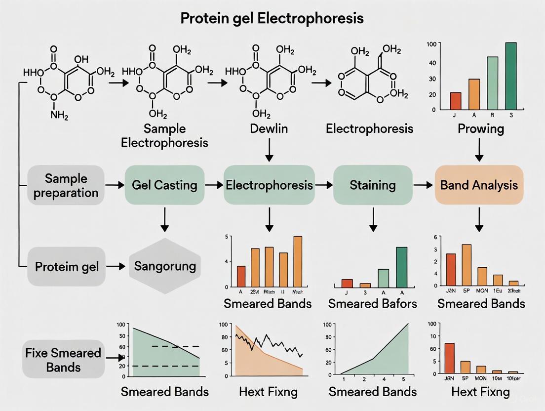
What Do Smeared Bands Look Like? Visual Identification Guide
Visual Identification of Smeared Bands
Smeared bands, also known as diffused and fuzzy bands, have a blurry appearance and are a common issue in protein gel electrophoresis. The bands are poorly resolved and often overlap with adjacent bands, making it difficult to accurately interpret your results and quantify your protein of interest. Unlike sharp, distinct bands that indicate a homogeneous protein population, smeared bands appear as a continuous, streak-like pattern running vertically down the lane [1].
There are several visual manifestations of smearing:
- Vertical Smearing: A continuous, streak-like pattern running vertically down the lane, often from the well to the bottom of the gel [2].
- Diffused and Fuzzy Bands: Bands that lack sharp, crisp boundaries and instead appear blurry and poorly defined [1].
- Trailing Smears or Warped Bands: These often manifest as U-shaped bands or bands that appear fused together, which can be a characteristic of overloaded gels [1] [3].
The table below summarizes the key visual characteristics and their general causes for quick identification.
| Visual Appearance | Description | Common Associated Cause |
|---|---|---|
| Continuous Vertical Streak | A long, smear running from the top to the bottom of the lane [2]. | Sample degradation; excessive voltage [2]. |
| Fuzzy, Poorly Resolved Bands | Bands are blurry, lack sharp edges, and may overlap [1]. | Improper sample denaturation; incorrect gel percentage [1] [2]. |
| Tailing or Trailing Smear | A "U-shaped" or "comet-tail" appearance, often behind a distinct band [1]. | Sample overloading; high salt concentration in sample [1]. |
Troubleshooting the Causes of Smeared Bands
Identifying the look of the smear is the first step; the next is to diagnose its root cause. The following workflow diagram outlines the systematic thought process for troubleshooting smeared bands, connecting the visual clues to potential experimental errors.
Detailed Analysis of Common Causes
1. Sample Preparation Errors Sample preparation is a frequent source of smearing. Key issues include:
- Sample Degradation: Proteases in the sample can digest your proteins at room temperature, creating a heterogeneous mixture of fragments that appears as a continuous vertical smear [2] [4]. To prevent this, add sample buffer and heat immediately [4].
- Improper Denaturation: Proteins must be fully denatured to linearize and coat with SDS for separation by mass. Incomplete denaturation, due to insufficient SDS, old reducing agents (DTT/β-mercaptoethanol), or incorrect boiling, leaves proteins with residual structure, causing poor resolution and smearing [3] [5]. Ensure your sample buffer is fresh and boil samples for 5 minutes at 98°C [5].
- Sample Overloading: Loading too much protein (exceeding 0.1–0.2 μg per millimeter of well width) saturates the gel, causing proteins to aggregate and trail down the lane as a smear [1] [5].
- High Salt Concentration: Excess salt in the sample increases local conductivity and heating, distorting the electric field and leading to distorted or smeared bands [1] [2].
2. Gel Run Conditions The conditions during electrophoresis are critical for sharp band formation.
- Excessive Voltage: Running the gel at too high a voltage generates excessive heat (Joule heating). This can denature proteins unevenly, soften the gel matrix, and cause band smiling and smearing [6] [2]. A standard practice is to run mini-gels at around 150V, or use a lower voltage for a longer duration [6].
- Incorrect Run Time: Running the gel for too short a time doesn't allow for proper separation, while running for too long can cause separated bands to diffuse and start to smear [1] [2]. A common standard is to stop the run when the dye front is about to reach the bottom of the gel [6].
3. Gel Quality and Composition The gel itself must be fit for purpose.
- Incorrect Gel Percentage: The polyacrylamide percentage determines the pore size. Using a gel with pores that are too small for your high molecular weight protein will impede migration, while using a gel with pores that are too large for small proteins will not provide sufficient sieving, both leading to poor resolution [5] [2].
- Incomplete Polymerization: If the polyacrylamide gel has not fully polymerized, the matrix will be weak and unstable, leading to distorted migration and smearing. This is often caused by expired or improperly stored reagents, especially TEMED and ammonium persulfate (APS) [5].
Experimental Protocols for Resolution
Protocol 1: Optimizing Sample Preparation to Eliminate Smearing
This protocol is designed to address the most common sample-related causes of smearing.
- Determine Protein Concentration: Use a standard protein assay (e.g., BCA, Bradford) to accurately determine the concentration of your samples [4].
- Prepare Fresh Sample Buffer: Ensure your SDS sample buffer containing a reducing agent (e.g., DTT or β-mercaptoethanol) is freshly prepared or thawed from a single-use aliquot [5].
- Mix Sample and Buffer: Dilute your protein sample with an appropriate volume of sample buffer to achieve a final concentration within the recommended loading range (e.g., 0.1-0.2 μg/μL per mm well width). A typical final concentration is 1X sample buffer [1] [3].
- Denature Immediately: Heat the sample-buffer mixture at 98°C for 5 minutes to fully denature proteins and inactivate proteases [5]. Note: For proteins known to be heat-sensitive or containing Asp-Pro bonds, heating at 75°C for 5 minutes may be preferable to avoid cleavage [4].
- Brief Centrifugation: Spin down the heated samples briefly (e.g., 30 seconds at 17,000 x g) to collect condensation and any insoluble material. Load the supernatant [4].
Protocol 2: Optimizing Electrophoresis Conditions
This protocol ensures the electrophoresis run itself does not introduce artifacts.
- Prepare Fresh Running Buffer: Always use fresh 1X SDS-running buffer. Overused or improperly diluted buffer can compromise separation [6] [5].
- Load Appropriate Controls: Include a pre-stained protein ladder in at least one lane to monitor the run progress and band separation.
- Set Optimal Voltage: For a standard mini-gel, set the power supply to a constant voltage of 150V. If smearing due to heat is suspected, reduce the voltage to 100-120V and extend the run time [6] [2].
- Monitor the Run: Observe the dye front. If the buffer becomes noticeably warm during the run, consider using a cooling apparatus or running in a cold room [6].
- Stop the Run: Terminate the electrophoresis when the dye front is approximately 0.5-1 cm from the bottom of the gel [6].
Research Reagent Solutions
The following table lists key reagents essential for preventing and troubleshooting smeared bands in protein gel electrophoresis.
| Research Reagent | Function in Preventing Smeared Bands |
|---|---|
| SDS (Sodium Dodecyl Sulfate) | Denatures proteins and confers a uniform negative charge, ensuring separation is based on molecular weight alone. Critical for proper linearization [5]. |
| Reducing Agents (DTT, β-mercaptoethanol) | Breaks disulfide bonds to fully unfold proteins. Must be fresh to prevent re-oxidation and incomplete denaturation, which causes smearing [3] [5]. |
| TEMED & Ammonium Persulfate (APS) | Catalyzes the polymerization of polyacrylamide gels. Essential for forming a complete and uniform gel matrix. Incomplete polymerization leads to smearing [5]. |
| Fresh Electrophoresis Buffer | Maintains correct pH and ion concentration for proper current flow and protein mobility. Old or incorrect buffer hinders separation [6] [5]. |
| Protease Inhibitor Cocktails | Added to protein extraction buffers to prevent proteolytic degradation of samples, which is a primary cause of vertical smearing [4]. |
FAQs on Smeared Bands
Q1: My bands are smeared, but my sample preparation was careful and my ladder ran perfectly. What is a likely cause? If your protein ladder is sharp but your sample bands are smeared, the issue is almost certainly specific to your sample. The most common causes are protein degradation before the sample was added to the buffer, or improper denaturation due to old or ineffective reducing agents in your sample buffer [2] [4].
Q2: I only see a smeared lane with no distinct bands. What does this mean? A continuous smear with no distinct bands typically indicates widespread and severe sample degradation [2]. Your proteins have been cleaved by proteases into a near-continuous distribution of random fragments. Re-examine your sample handling protocol, work quickly on ice, and use fresh protease inhibitors.
Q3: Can running the gel at a higher voltage fix smearing? No, in fact, the opposite is true. Running the gel at a higher voltage often causes smearing due to excessive heat generation, which can denature proteins and soften the gel matrix. To improve resolution, try running the gel at a lower voltage for a longer period [6] [2].
Q4: The bands in my gel are smiling and slightly smeared. What is the connection? Both "smiling" (curved bands) and smearing can be caused by excessive heat during the run. The smiling is due to uneven heating across the gel (the center being hotter than the edges), while the smearing is a result of the heat denaturing the proteins or affecting the gel matrix. Reducing the running voltage is the primary solution for both issues [6] [2].
Sodium Dodecyl Sulfate-Polyacrylamide Gel Electrophoresis (SDS-PAGE) is a fundamental technique in biochemistry and molecular biology laboratories worldwide. This method enables researchers to separate protein mixtures based primarily on molecular weight, providing critical information for protein analysis, purification, and characterization. The power of SDS-PAGE lies in its elegant simplification of protein physical properties—through denaturation and charge unification—allowing size-based separation in a polyacrylamide matrix. Understanding these core principles is essential not only for successfully executing the technique but also for troubleshooting common issues such as smeared bands, poor resolution, and artifactual results. This technical support resource provides comprehensive guidance on SDS-PAGE fundamentals, troubleshooting, and optimization to support researchers in obtaining clear, interpretable results.
Core Principles of SDS-PAGE
The Role of SDS in Protein Denaturation and Charge Unification
SDS-PAGE relies on two interconnected mechanisms that transform proteins into uniformly linearized, negatively charged molecules:
Protein Denaturation: SDS is an anionic detergent with a hydrophobic tail and hydrophilic head. When added to protein samples, SDS disrupts hydrogen bonds and hydrophobic interactions that maintain secondary and tertiary structures [7]. This effectively unfolds proteins into linear polypeptide chains, eliminating variations in shape that could affect migration through the gel matrix [8].
Charge Unification: The ionic sulfate group of SDS confers a strong negative charge. SDS molecules bind to polypeptide backbones in a constant weight ratio (approximately 1.4 g SDS per 1 g of protein) [9], overwhelming any intrinsic charge from amino acid side chains. This creates SDS-polypeptide complexes with essentially identical charge-to-mass ratios [9] [10].
These processes ensure that all proteins migrate toward the anode (positive electrode) when an electric field is applied, with separation determined primarily by molecular size rather than native charge or conformation [8].
The Polyacrylamide Gel as a Molecular Sieve
Polyacrylamide gels serve as a molecular sieve that differentially retards protein migration based on size. The gel forms when acrylamide monomers polymerize into long chains cross-linked by bisacrylamide, creating a porous three-dimensional network [9] [10]. The pore size depends on the concentrations of both acrylamide and bisacrylamide:
- Low-percentage gels (e.g., 8% acrylamide) have larger pores and better resolve high molecular weight proteins
- High-percentage gels (e.g., 15% acrylamide) have smaller pores and better resolve low molecular weight proteins [9]
During electrophoresis, smaller polypeptides navigate the gel matrix more easily and migrate farther, while larger polypeptides are more hindered and migrate shorter distances [8]. This results in protein bands arranged by molecular weight along the migration path.
The Discontinuous Gel System: Stacking and Resolving
SDS-PAGE typically uses a discontinuous system with two distinct gel regions:
- Stacking gel (lower acrylamide concentration, ~4%, pH ~6.8): Serves to concentrate all protein samples into a tight band before they enter the resolving gel, ensuring sharp initial bands [10].
- Resolving gel (higher acrylamide concentration, ~8-12%, pH ~8.8): Performs the actual size-based separation of proteins [9].
This two-layer system significantly enhances band sharpness and resolution compared to a single continuous gel.
Troubleshooting SDS-PAGE: FAQs and Solutions
Why am I getting smeared bands in my SDS-PAGE gel?
Smeared bands are one of the most common issues in SDS-PAGE and can arise from multiple causes. The table below summarizes the primary causes and solutions:
| Cause | Solution |
|---|---|
| Voltage too high | Decrease voltage by 25-50%; standard practice is 10-15 V/cm gel length [11]. |
| Protein overload | Reduce amount of protein loaded; 10 µg per well is often suitable [12]. |
| High salt concentration | Dialyze sample, precipitate with TCA, or use desalting column [13]. |
| Insufficient SDS | Dilute sample with more SDS solution to ensure complete denaturation [13]. |
| Protein aggregation | Add 4-8 M urea to sample (hydrophobic proteins); ensure fresh reducing agents [12] [13]. |
| Sample degradation | Prevent protease contamination; avoid freeze-thaw cycles [13]. |
Why are my protein bands poorly resolved or blurry?
Poor resolution prevents accurate molecular weight determination and quantification:
| Cause | Solution |
|---|---|
| Gel run time too short | Run gel until dye front reaches bottom; longer for high molecular weight proteins [11]. |
| Incorrect gel concentration | Use gradient gels (e.g., 4-20%) or optimize acrylamide percentage for target protein size [13]. |
| Improper running buffer | Remake running buffer with correct ion concentration to ensure proper current flow and pH [11]. |
| Old or improperly cast gel | Use fresh gels; filter gel reagents and ensure proper degassing before polymerization [13]. |
Why are my samples leaking from wells or migrating unevenly?
Well-related issues compromise sample integrity before separation begins:
- Insufficient glycerol in loading buffer: Increase glycerol concentration to help samples sink properly into wells [12].
- Air bubbles in wells: Rinse wells with running buffer before loading to displace air bubbles [12].
- Overfilled wells: Load no more than 3/4 of well capacity to prevent spillover [12].
- Delay between loading and running: Start electrophoresis immediately after loading to prevent sample diffusion [11].
- Poor well formation: Ensure stacking gel has polymerized completely (≥30 minutes) before comb removal [13].
Why do I see unusual band patterns or artifacts?
Unexpected band patterns can indicate specific issues:
- "Smile effect" (curved bands): Caused by uneven heating; run at lower voltage, use cooling, or run in cold room [11].
- Vertical streaking: Often from sample precipitation; centrifuge samples before loading [13].
- Doublet bands: Possible re-oxidation during run; use fresh reducing agents (DTT, β-mercaptoethanol) [13].
- Unexpected high molecular weight bands: May indicate protein aggregation; add reducing agents or urea [12].
Experimental Protocol for Optimal SDS-PAGE
Sample Preparation Methodology
Proper sample preparation is critical for successful SDS-PAGE:
- Sample Lysis: Use lysis buffer containing SDS (1-2%) to solubilize and denature proteins. Include protease inhibitors to prevent degradation [12].
- Reduction of Disulfide Bonds: Add fresh reducing agents (50-100 mM DTT or 5% β-mercaptoethanol) to break disulfide linkages [10].
- Heat Denaturation: Heat samples at 70-100°C for 5-10 minutes to complete denaturation [9]. Some hydrophobic proteins may aggregate at high temperatures; try 60°C if this occurs [13].
- Solubility Maintenance: For hydrophobic proteins, add 4-8 M urea to prevent aggregation [12].
- Centrifugation: Remove insoluble material by centrifuging at 10,000-15,000 × g for 10 minutes before loading [13].
Gel Preparation and Electrophoresis
Gel Composition:
- Resolving gel: 8-12% acrylamide (depending on protein size range) in Tris-HCl, pH 8.8
- Stacking gel: 4% acrylamide in Tris-HCl, pH 6.8 [10]
Polymerization:
- Add ammonium persulfate (APS) and TEMED to initiate polymerization
- Pour resolving gel, overlay with isopropanol or water for even surface
- After polymerization, pour stacking gel and insert comb [10]
Electrophoresis Conditions:
Protein Detection and Visualization
Coomassie Staining:
- Fixation step: Incubate gel in 40% methanol, 10% acetic acid for 30 minutes to prevent protein diffusion [14].
- Staining: Use colloidal Coomassie G-250 solution (0.02% CBB G-250, 5% aluminum sulfate, 10% ethanol, 2% orthophosphoric acid) for 2 hours or overnight [14].
- Destaining: Briefly destain with solution containing 10% ethanol and 2% orthophosphoric acid [14].
Alternative Detection Methods:
The Scientist's Toolkit: Essential Reagents for SDS-PAGE
| Reagent | Function | Key Considerations |
|---|---|---|
| SDS (Sodium Dodecyl Sulfate) | Denatures proteins and confers uniform negative charge | Use high-purity grade; critical for consistent charge-to-mass ratio [7] |
| Acrylamide/Bis-acrylamide | Forms porous polyacrylamide gel matrix | Adjust concentration for target protein size; neurotoxic until polymerized [9] |
| APS (Ammonium Persulfate) | Initiates polymerization reaction | Prepare fresh solution for consistent gel polymerization [10] |
| TEMED | Catalyzes polymerization reaction | Stable at 4°C; quantity affects polymerization rate [10] |
| DTT or β-mercaptoethanol | Reduces disulfide bonds | Use fresh for each experiment; prevents reoxidation artifacts [10] |
| Tris buffers | Maintain pH in stacking/resolving gels | Critical for discontinuous system function [9] |
| Coomassie Brilliant Blue | Stains proteins in gel | Colloidal CBB-G provides better sensitivity than CBB-R [14] |
| Molecular weight markers | Reference for size determination | Include both stained and unstained options for various needs [9] |
Workflow and Troubleshooting Diagrams
SDS-PAGE Experimental Workflow
Troubleshooting Smeared Bands Decision Tree
Advanced Techniques and Applications
Two-Dimensional PAGE
For complex protein mixtures, two-dimensional (2D) PAGE provides superior resolution by separating proteins based on two different properties:
- First dimension: Isoelectric focusing (IEF) separates proteins by their isoelectric point
- Second dimension: SDS-PAGE separates by molecular weight [9]
This technique can resolve thousands of proteins in a single gel and is particularly valuable for proteomic studies [16].
Native PAGE vs. SDS-PAGE
While SDS-PAGE separates denatured proteins by size, native PAGE separates proteins in their folded state based on charge, size, and shape [9]. Native PAGE preserves protein function and multimeric structures but provides different information than SDS-PAGE.
Fluorescent Protein Detection
Recent advances enable direct detection of fluorescent proteins (e.g., GFP, RFP) in gels without staining or blotting. This approach bypasses antibody-based detection, providing clearer data with less background interference while maintaining compatibility with downstream applications [15].
SDS-PAGE remains an indispensable tool for protein analysis decades after its development because of its robust principles and practical utility. The core concept—using SDS to denature proteins and confer uniform charge, then separating by size in a polyacrylamide matrix—provides a reproducible method for protein characterization. Successful implementation requires attention to both theoretical fundamentals and practical optimization, particularly when addressing common issues like smeared bands. By understanding the interplay between sample preparation, gel chemistry, and electrophoresis conditions, researchers can troubleshoot effectively and obtain high-quality results that support their scientific objectives. The troubleshooting guidelines and methodologies presented here offer a comprehensive resource for researchers seeking to optimize their SDS-PAGE experiments and produce reliable, publication-quality data.
Diagnostic Guide: Identifying the Type of Smear
Smeared bands in your protein gel can stem from distinct root causes. Use the table below to diagnose the specific issue in your experiment based on the visual characteristics of the smear.
| Smear Type | Visual Characteristics | Common Lane Pattern | Primary Underlying Cause |
|---|---|---|---|
| Degradation | Continuous, vertical streaking from the well downward; a "ladder" of smaller fragments may be visible [2]. | Often affects all samples equally, but can be sample-specific. | Protease activity breaking proteins into random-sized fragments [2]. |
| Improper Denaturation | Diffuse, fuzzy bands; general haze or background staining across the lane; proteins may not migrate according to expected molecular weight [17] [13]. | Typically affects specific samples based on preparation. | Incomplete binding of SDS, leaving proteins with folded structures and inconsistent charge/mass ratios [2] [13]. |
| Overloading | Thick, warped, or U-shaped bands at the top of the lane; intense, diffuse smearing that may concentrate in the high molecular weight region [1] [17]. | Affects only the overloaded lane(s). | Well capacity exceeded, leading to poor separation and over-saturation of the gel matrix [1] [13]. |
Step-by-Step Troubleshooting Protocols
Remedying Sample Degradation
Protein degradation is often a result of protease contamination. This protocol outlines steps to prevent and confirm proteolytic activity.
Materials Needed:
- Freshly prepared protease inhibitor cocktails
- Nuclease-free water
- Pre-cast SDS-PAGE gel or ingredients to cast one (acrylamide, bis-acrylamide, SDS, Tris-HCl, ammonium persulfate, TEMED)
- SDS-PAGE running buffer
- Sample heating block
- Centrifuge
Experimental Protocol:
- Sample Handling: Always keep samples on ice during preparation to slow protease activity [2].
- Add Inhibitors: Incorporate a broad-spectrum protease inhibitor cocktail into your lysis buffer. Ensure it is fresh and used at the recommended concentration.
- Fresh Preparation: Prepare a new sample aliquot with the fresh inhibitor cocktail. Avoid multiple freeze-thaw cycles of protein stocks [13].
- Heat Denaturation: Boil the sample in your loading buffer for 5-10 minutes to denature and inactivate proteases.
- Centrifugation: Briefly centrifuge the heated sample (e.g., 12,000 x g for 2 minutes) to pellet any insoluble debris or aggregated protein before loading the supernatant [13].
- Control Experiment: Run the new sample alongside the old, degraded sample on the same gel. A clear, sharp band in the new sample confirms degradation was the issue.
Ensuring Complete Denaturation
Incomplete denaturation prevents proteins from becoming linear, causing aberrant migration. This protocol ensures uniform SDS binding.
Materials Needed:
- Laemmli sample buffer containing SDS and a reducing agent (DTT or β-mercaptoethanol)
- Heating block (95-100°C)
- Ultrasonication bath or probe (optional, for difficult samples)
Experimental Protocol:
- Check Sample Buffer: Confirm your Laemmli buffer is fresh. SDS should be at a final concentration of ~1%, and the reducing agent (e.g., DTT) should be at 50-100 mM.
- Optimal Sample-to-Buffer Ratio: Mix your protein sample with 4X Laemmli buffer to achieve a 1X final concentration. A typical ratio is 3:1 (sample:buffer) [17].
- Heat Denaturation: Heat the sample at 95-100°C for 5-10 minutes [17]. Ensure the tube cap is securely closed to prevent evaporation.
- Cool and Centrifuge: Briefly cool the sample and centrifuge to bring down condensation and any insoluble material.
- For Problematic Samples: If smearing persists, consider:
- Longer Reduction: Extend the heating time to 10-15 minutes.
- Sonication: Sonicate the sample (on ice) for 15-30 seconds before adding the loading buffer to disrupt aggregates [17].
- Add Urea: For hydrophobic or aggregation-prone proteins, add 4-8 M urea to the sample buffer to improve solubility and denaturation [17] [13].
Correcting Sample Overloading
Overloading the well prevents the gel from resolving individual proteins. This protocol helps determine the optimal loading amount.
Materials Needed:
- Protein quantification assay (e.g., BCA, Bradford)
- SDS-PAGE gel
- Standard protein ladder
Experimental Protocol:
- Quantify Protein: Precisely determine your protein concentration using a reliable assay.
- Load an Appropriate Mass: The general recommendation is to load 10-20 µg of total protein per well for a standard mini-gel [17]. For overloading smears, significantly reduce this amount.
- Perform a Loading Gradient: Prepare a dilution series of your sample (e.g., 5 µg, 10 µg, 20 µg). Load these on the same gel to visually identify the concentration that provides the best resolution without smearing.
- Check Well Capacity: Ensure your sample volume does not exceed ~3/4 of the well's capacity to prevent spillover and leakage [17].
The Smearing Diagnostic Workflow
The following diagram outlines the logical process for diagnosing the cause of smeared bands.
Research Reagent Solutions
The following table lists key reagents essential for preventing and troubleshooting smearing in SDS-PAGE.
| Reagent | Function | Troubleshooting Consideration |
|---|---|---|
| Protease Inhibitor Cocktail | Inhibits enzymatic degradation of protein samples by proteases. | Essential for preventing degradation smears. Must be added fresh to lysis buffers [2]. |
| SDS (Sodium Dodecyl Sulfate) | A strong anionic detergent that denatures proteins and confers a uniform negative charge. | Concentration is critical (~1% final). Old or impure SDS can cause improper denaturation and smearing [2]. |
| Reducing Agents (DTT, BME) | Breaks disulfide bonds within and between protein subunits. | Prevents aggregation and ensures linearization. Use fresh DTT (50-100 mM); old stock loses efficacy [17] [13]. |
| Laemmli Sample Buffer | Contains SDS, reducing agent, glycerol, and a tracking dye for denaturing and loading samples. | The complete denaturation system. Always prepare fresh aliquots and ensure correct pH [17]. |
| Urea | A chaotropic agent that disrupts non-covalent bonds. | Added to sample buffer (4-8 M) to solubilize hydrophobic or aggregation-prone proteins and prevent precipitation [17] [13]. |
Frequently Asked Questions (FAQs)
Q1: My bands are still smeared after following the denaturation protocol. What else could it be? If degradation, denaturation, and overloading have been ruled out, consider these factors:
- Voltage Too High: Running the gel at excessively high voltage can generate heat, denature proteins in the gel, and cause smearing. Solution: Lower the voltage by 25-50% and run the gel for a longer duration [18] [13].
- High Salt Concentration: Excess salt in the sample can create a localized electric field, distorting migration. Solution: Desalt your sample using dialysis, a desalting column, or precipitation before preparation [13].
- Old or Contaminated Buffers: Always use fresh running and sample buffers to ensure proper pH and ion concentration [19].
Q2: I see a "smiling" or "frowning" effect in my bands along with smearing. What does this mean? This is a classic sign of uneven heating across the gel (Joule heating). The center of the gel becomes hotter than the edges, causing bands in the middle to migrate faster ("smiling"). Solution: Run the gel at a lower voltage, use a power supply with constant current mode, or perform the run in a cold room to dissipate heat [2].
Q3: My protein is stuck in the well. Is this related to smearing? Yes, this can be a severe form of poor migration. It is often caused by protein aggregation or precipitation in the well, or by overloading. Solution: Ensure your sample is properly solubilized. Add urea to your sample buffer, sonicate the sample, and always centrifuge it before loading to remove aggregates [17] [13].
Q4: How can I prevent smearing from happening routinely? Adopt these best practices:
- Consistent Sample Preparation: Always use fresh reagents and a standardized heating step.
- Accurate Quantification: Precisely measure protein concentration to avoid overloading.
- Optimized Running Conditions: Do not exceed 150V for standard mini-gels; lower voltages often yield sharper bands.
- Proper Gel Storage: Use freshly cast gels and avoid storing them for long periods [19] [13].
Troubleshooting Guide: Resolving Smeared Bands in Protein Gel Electrophoresis
This guide addresses the common issue of smeared bands in protein gel electrophoresis, a critical challenge that can compromise data integrity and interpretation in research and drug development. Below are targeted questions and answers to help diagnose and resolve the underlying causes.
Why are my protein bands smeared or fuzzy instead of sharp?
Smeared, fuzzy bands indicate that proteins of the same type have not migrated as a unified group. This is typically caused by issues in sample preparation, gel conditions, or the electrophoresis run parameters [1] [2].
Primary causes and solutions include:
- Sample Degradation: Proteins can be degraded by proteases, creating a mixture of full-length and fragmented proteins that appear as a smear [1] [2].
- Incomplete Denaturation: If proteins are not fully unfolded, they will not have a uniform charge and may migrate based on their native shape and charge in addition to size [20] [21].
- Protein Aggregation: Hydrophobic or over-concentrated proteins can form aggregates that do not enter the gel evenly [22] [20].
- Solution: Ensure proper sample homogenization and solubility. For hydrophobic proteins, consider adding 4-8M urea to the lysis solution. Sonicating samples can also help break up aggregates [22].
- Gel Overloading: Loading too much protein per well can overwhelm the gel's capacity, causing bands to merge and smear [1] [20] [2].
- Solution: Reduce the amount of protein loaded. A general guideline is to load 10 µg of protein per well, but this may require optimization [22].
- Incorrect Gel Percentage: Using a gel with a pore size that is not optimal for your target protein's molecular weight will lead to poor separation and smearing [2].
- Solution: Select a gel concentration appropriate for your protein's size. Higher percentage gels are better for smaller proteins [20].
How do I fix poorly separated bands that are too close together?
Poor band resolution, where bands are densely stacked and hard to distinguish, is often related to the sieving properties of the gel and the conditions of the electrophoretic run [2].
Key factors to check:
- Suboptimal Gel Concentration: The gel percentage is the most critical factor for resolution [2]. The table below summarizes recommended gel percentages for separating different protein size ranges.
| Polyacrylamide Gel Percentage (%T) | Optimal Protein Separation Range |
|---|---|
| 8% | Best for larger proteins [20]. |
| 10% | A common all-purpose percentage. |
| 12-15% | Best for smaller proteins [20]. |
| 5-20% Gradient | Resolves a wide range of protein sizes simultaneously [21]. |
- Incorrect Run Time or Voltage: Running the gel for too short a time will not allow sufficient separation, while running for too long can cause bands to diffuse. Excessively high voltage can also reduce resolution [1] [2].
- Solution: Run the gel for a longer duration at a lower voltage to improve separation. Follow recommended protocols for your specific gel system [2].
- Sample Overloading: As with smearing, overloading the well causes thick, merging bands [1].
- Solution: Load a smaller amount of sample per well [2].
What causes distorted or "smiling" bands in my gel?
Distorted bands that curve upwards ("smiling") or downwards are almost always caused by uneven heat distribution across the gel during the run [23] [2].
Causes and remedies:
- Excessive Voltage: High voltage generates excessive Joule heating, which can cause the center of the gel to become hotter than the edges. Samples in the hotter center migrate faster, creating an upward curve [23] [2].
- Solution: Reduce the voltage during the run. If available, use a power supply with a constant current mode to maintain a more uniform temperature [2].
- Improper Cooling: Lack of sufficient cooling exacerbates temperature gradients.
- Solution: Use a cooling system or run the electrophoresis in a cold room to maintain a consistent temperature [20].
- High Salt Concentration in Samples: Excess salt in a sample increases local conductivity and heating in the well, distorting the electric field [20] [2].
Why are my bands very faint or completely absent?
Faint or absent bands indicate a failure in sample detection, which can occur at multiple stages [1] [2].
Systematic troubleshooting steps:
- Insufficient Sample Concentration: The amount of protein loaded may be below the detection limit of the stain [2].
- Solution: Increase the amount of starting material or concentrate the sample. Load a minimum of 0.1–0.2 µg of nucleic acid per millimeter of gel well width as a general reference, though protein requirements may differ [1].
- Problems with Staining: The staining solution may be degraded, or the staining time may be too short [1] [2].
- Solution: Prepare fresh staining solutions and optimize the staining duration. For thick or high-percentage gels, allow more time for the stain to penetrate [1].
- Sample Degradation or Loss: The sample may have been degraded during preparation or leaked from the well [1] [2].
- Solution: Re-check sample preparation steps. Ensure the loading buffer contains enough glycerol (or similar compound) to help the sample sink into the well [22].
- Electrophoresis Setup Error: The power supply may not have been connected correctly, or the electrodes may have been reversed [1].
- Solution: Verify all power supply connections and settings. Ensure the electrodes are connected correctly (gel wells should be on the cathode/negative side) [1].
Experimental Workflow for Optimal Band Sharpness
The following diagram outlines a systematic workflow for preparing and running a protein gel to achieve sharp, well-resolved bands.
Research Reagent Solutions for Protein Electrophoresis
The following table details essential reagents and their functions for successful SDS-PAGE experiments.
| Reagent | Function |
|---|---|
| Sodium Dodecyl Sulfate (SDS) | Anionic detergent that denatures proteins and confers a uniform negative charge, masking intrinsic charge differences [21]. |
| Reducing Agents (DTT, BME) | Break disulfide bonds in proteins, ensuring complete unfolding and linearization for accurate size-based separation [20] [21]. |
| Acrylamide/Bis-acrylamide | Monomers that polymerize to form the porous polyacrylamide gel matrix, which acts as a molecular sieve [21]. |
| Ammonium Persulfate (APS) & TEMED | Catalysts that initiate and accelerate the chemical polymerization of acrylamide to form a gel [24]. |
| Tris-Glycine Buffer | A standard running buffer for SDS-PAGE; the discontinuous system (stacking vs. resolving gel) helps sharpen bands before separation [20] [21]. |
| Coomassie Blue/SYBR Stains | Dyes used to visualize proteins (Coomassie) or nucleic acids (SYBR) in the gel after electrophoresis [1] [23]. |
| Protein Molecular Weight Marker | A standard containing proteins of known sizes, allowing for estimation of the molecular weight of unknown proteins [21]. |
Frequently Asked Questions (FAQs)
What is the single most important factor for improving band resolution?
The gel concentration is the most critical factor. Selecting a gel with a pore size optimized for the molecular weight range of your target proteins is essential for achieving sharp, well-resolved bands [2].
How can I prevent smearing due to sample preparation?
Ensure complete denaturation of your proteins. This involves heating samples at 95°C for 5 minutes in a loading buffer that contains both SDS (to denature and charge) and a reducing agent like DTT or β-mercaptoethanol (to break disulfide bonds) [20] [21]. Also, avoid overloading the gel wells.
My gel has no bands, not even the ladder. What should I check first?
If no bands are visible, first verify your electrophoresis setup. Check that the power supply was turned on, the electrodes were connected correctly (black to black, red to red), and the buffer chamber was properly filled to complete the circuit [1] [2]. A missing ladder indicates a problem with the run itself, not necessarily the sample.
Mastering Sample Preparation and Electrophoresis Conditions for Crisp Bands
Core Concepts: The Principles of Effective Denaturation
Successful protein separation by SDS-PAGE relies on complete denaturation and linearization of protein samples. This process ensures proteins migrate strictly according to their molecular weight. The core components of a denaturation protocol each play a critical role.
Sodium Dodecyl Sulfate (SDS) is an ionic detergent that binds to hydrophobic regions of proteins, disrupting hydrogen bonds and van der Waals forces. It confers a uniform negative charge to the polypeptides, allowing migration toward the anode during electrophoresis. For complete saturation, a 3:1 ratio of SDS to protein (mass:mass) is often recommended to ensure all proteins are uniformly coated [4].
Dithiothreitol (DTT) is a reducing agent that breaks disulfide bonds between cysteine residues, which is crucial for separating protein subunits and achieving complete unfolding. It is important to note that DTT may be less effective at reducing buried disulfide bonds without the aid of denaturants or heat [25].
Heat (typically 95-100°C for 5 minutes) is a critical denaturation step that disrupts secondary and tertiary protein structures, facilitates the action of SDS and DTT, and helps inactivate proteases that could otherwise degrade the sample. However, some heat-sensitive proteins may precipitate upon heating, requiring protocol adjustments [4] [25].
Research Reagent Solutions
| Reagent | Primary Function | Key Considerations |
|---|---|---|
| SDS (Ionic Detergent) | Disrupts non-covalent bonds; provides uniform negative charge. | Use at a 3:1 ratio to protein mass for complete coating [4]. |
| DTT or β-Mercaptoethanol | Reduces disulfide bonds to linearize proteins. | For buried disulfides, requires strong denaturants (e.g., 8M Urea) [25]. |
| Urea (8M) | Strong denaturant; disrupts hydrogen bonding. | Alternative to heat for sensitive proteins; can form cyanate ions that carbamylate proteins over time [4]. |
| Protease Inhibitors | Prevents protein degradation during lysis. | Must be added fresh to lysis buffer; samples kept on ice [26]. |
| Glycerol/Sucrose | Adds density to sample for easy gel loading. | Prevents sample leakage from wells [27]. |
Troubleshooting Guide: Resolving Smeared Bands
Smeared bands are a common issue often traced back to problems in the lysis and denaturation steps. The table below outlines specific failures and their solutions.
Troubleshooting Smeared or Aberrant Bands
| Problem & Symptoms | Potential Cause | Recommended Solution |
|---|---|---|
| Smearing & High Background [27] [1] | Protein Aggregation/Aggregates: Incomplete denaturation causes proteins to clump. | Increase DTT concentration; Add 4-8M Urea to lysis buffer; Sonicate samples; For hydrophobic proteins, use lysis buffer with urea [27]. |
| Multiple Extra Bands [4] | Protease Degradation: Endogenous proteases active during sample prep. | Heat samples immediately after adding buffer (95-100°C, 5 min); Add fresh protease inhibitors to lysis buffer [4]. |
| Distorted, Poorly Resolved Bands [4] | Sample Overloading: Too much protein loaded per well. | Load 10-20 µg of total protein per well for analytical gels; Use a protein assay to quantify precisely [27] [4]. |
| Bands Not Entering Gel/Clumping in Well [27] | Insoluble Material: Presence of nucleic acids or cell debris. | Centrifuge lysate (e.g., 17,000 x g for 2 min) post-heating; For viscous samples, use nuclease (Benzonase) or sonication [4]. |
| Smearing with Heat-Sensitive Proteins [25] | Heat-Induced Precipitation: Protein precipitates upon heating. | Denature with 8M Urea in SDS buffer without heating; Alternatively, heat at lower temperature (e.g., 75°C) [4] [25]. |
Detailed Experimental Protocols
Standard Protocol for Cell Lysis and Denaturation
This protocol is designed for common cell culture samples and serves as a robust starting point.
Materials Needed:
- RIPA Lysis Buffer (for membrane-bound proteins) or NP-40 Lysis Buffer (for milder extraction) [26]
- 2X Laemeli Sample Buffer: 4% SDS, 10% 2-mercaptoethanol, 20% glycerol, 0.004% bromophenol blue, 0.125 M Tris HCl, pH ~6.8 [26]
- Freshly added protease inhibitors (e.g., Aprotinin, Leupeptin, PMSF) [26]
- Ice-cold Phosphate-Buffered Saline (PBS)
- Heating block or water bath (95-100°C)
Method:
- Cell Washing & Lysis: Aspirate media from cultured cells and wash once with ice-cold PBS. Aspirate completely.
- Add Lysis Buffer: Add ice-cold RIPA buffer containing fresh protease inhibitors (e.g., ~1 mL per 10⁷ cells) to the plate [26].
- Scrape & Collect: Scrape adherent cells off the plate and transfer the lysate to a microcentrifuge tube. Keep samples on ice.
- Clarify Lysate: Centrifuge the lysate at high speed (e.g., 12,000-17,000 x g) for 10 minutes at 4°C to pellet insoluble debris [26] [4].
- Transfer Supernatant: Transfer the clarified supernatant to a new tube. Discard the pellet.
- Determine Protein Concentration: Use a Bradford, BCA, or Lowry assay to determine the protein concentration of the supernatant [26].
- Prepare Sample for Loading: Mix the protein lysate with an equal volume of 2X Laemeli Sample Buffer.
- Denature: Heat the samples at 95-100°C for 5 minutes to denature proteins and inactivate proteases [4].
- Brief Spin: Centrifuge the heated samples for 1-2 minutes to pellet any insoluble material that formed during heating.
- Load and Run: Load the supernatant onto your SDS-PAGE gel.
Alternative Protocol for Heat-Sensitive Proteins
Some proteins, such as membrane proteins or specific fusion constructs like GST, can aggregate and precipitate upon heating [25]. This protocol uses chemical denaturation as an alternative.
Materials Needed:
- Standard Lysis Buffer (as in 3.1)
- Sample Buffer with Urea: 2X Laemeli Buffer supplemented with 8M Urea [25].
- DTT or β-mercaptoethanol
Method:
- Follow steps 1-6 from the standard protocol (3.1) to obtain a clarified protein lysate.
- Mix the protein lysate with an equal volume of Sample Buffer with Urea.
- Do not heat the sample. Instead, incubate the mixture at room temperature or 37°C for 10-20 minutes [25].
- Centrifuge briefly and load the supernatant onto the gel.
Critical Note: Urea in aqueous solution exists in equilibrium with ammonium cyanate, which can carbamylate lysine residues and modify protein charge and mass. To prevent this, use fresh urea solutions, or treat urea solutions with a mixed-bed resin to remove ions. Avoid storing samples in urea buffers for extended periods [4].
Workflow and Decision Pathway
The following diagram illustrates the logical process for selecting and optimizing a denaturation protocol to prevent smearing, integrating the core concepts and troubleshooting advice.
Optimized Denaturation Workflow
Frequently Asked Questions (FAQs)
Q1: Why can't I just leave my sample in the SDS buffer at room temperature instead of heating it immediately? This is a critical mistake that can lead to significant protein degradation and smearing. Even though SDS denatures most proteins, some proteases remain active in SDS at room temperature. The immediate heating step is essential to rapidly and irreversibly inactivate these proteases before they can digest your proteins of interest [4].
Q2: My protein precipitates when I heat it. What can I do? This is a classic sign of a heat-sensitive protein. The recommended approach is to avoid heating and instead use a chemical denaturant. Supplement your SDS sample buffer with 8M urea or 6M guanidine hydrochloride and incubate the sample at room temperature or 37°C for 10-20 minutes before loading [25].
Q3: I see a cluster of contaminating bands around 55-65 kDa in my silver-stained gel. What is this? This is likely keratin contamination, a common artifact introduced from skin, hair, or dander. To confirm, run a lane with sample buffer alone. If the bands appear, your buffer is contaminated. To prevent this, wear gloves, use filtered pipette tips, aliquot and store buffers at -80°C, and remake buffers if contamination is suspected [4].
Q4: How much total protein should I load per well? Overloading is a common cause of smearing and poor resolution. A good general guideline is to load 10-20 µg of total protein for a standard mini-gel with a 1.0 mm thickness when using Coomassie staining. For silver staining, which is more sensitive, load 10-100 times less. Always determine your protein concentration with a reliable assay (e.g., Bradford, BCA) to avoid over- or under-loading [27] [4].
FAQs and Troubleshooting Guides
Why are my protein bands smeared or fuzzy?
Smeared or fuzzy bands are a common issue often linked to protein aggregation, especially with hydrophobic proteins. The primary causes and solutions are summarized in the table below.
| Cause | Solution |
|---|---|
| Protein Aggregation | Add 4-8 M urea to the sample buffer to disrupt hydrophobic interactions and keep proteins solubilized [13]. |
| Incomplete Denaturation | Ensure sample buffer contains sufficient SDS and reducing agents (DTT or β-mercaptoethanol) and heat samples at 95°C for 5 minutes [28]. |
| Voltage Too High | Decrease voltage by 25-50% to minimize heating that causes band diffusion [29] [13]. |
| Protein Overloading | Reduce the amount of protein loaded per well [13]. |
| High Salt Concentration | Dialyze the sample, or use desalting columns or TCA precipitation to remove excess salt [13]. |
How can I improve the resolution of my hydrophobic membrane proteins?
Poor resolution of hydrophobic proteins often occurs due to their tendency to aggregate in standard gel systems. Consider these approaches:
- Use Specialized Gel Matrices: Incorporate N-alkylated acrylamide monomers, such as N,N'-dimethylacrylamide, into the polyacrylamide gel. This increases hydrophobic interactions between the gel matrix and membrane proteins, significantly improving their separation and identification. This method has been shown to successfully identify highly hydrophobic peptides and proteins with multiple transmembrane domains [30].
- Employ Blue Native PAGE (BN-PAGE): For analyzing native membrane protein complexes, use BN-PAGE. Effective solubilization is critical; test mild non-ionic detergents like Triton X-100 or n-dodecyl-β-D-maltoside (DDM) to find the optimal concentration that solubilizes complexes without disintegrating them. A concentration of 0.2% (w/v) Triton X-100 has proven effective for some bacterial membrane fractions [31].
- Optimize Gel Percentage: Use a gel with a pore size appropriate for your target proteins. For a wide molecular weight range or unknown sizes, a 4%-20% gradient gel is recommended [13].
My samples precipitate in the well. What should I do?
This is a classic sign of protein aggregation, particularly for hydrophobic proteins.
- Add Chaotropes: As a first-line solution, add 4-8 M urea to your sample buffer to disrupt hydrophobic interactions and prevent precipitation in the well [13].
- Avoid Over-boiling: Some protein complexes, including hydrophobic ones, can aggregate if boiled. As an alternative, try heating your samples at a lower temperature (e.g., 60°C) to denature them without inducing excessive aggregation [13].
What are the key reagents for handling challenging proteins?
The following reagents are essential for preventing aggregation and ensuring clear results.
| Reagent | Function |
|---|---|
| Urea (4-8 M) | Chaotrope that disrupts hydrophobic interactions and keeps challenging proteins solubilized in solution [13]. |
| Specialized Acrylamide Monomers (e.g., N,N'-dimethylacrylamide) | Modifies the gel matrix to increase hydrophobic interactions, improving separation of membrane proteins [30]. |
| Mild Non-Ionic Detergents (Triton X-100, DDM) | Solubilizes membrane protein complexes in their native state for techniques like BN-PAGE [31]. |
| Fresh Reducing Agents (DTT, β-mercaptoethanol) | Breaks disulfide bonds to ensure complete protein denaturation and linearization [13] [28]. |
| Glycerol | Adds density to the sample buffer to ensure the sample sinks properly to the bottom of the well [13]. |
Troubleshooting Workflow
The following diagram outlines a logical, step-by-step workflow for diagnosing and fixing smeared bands caused by protein aggregation.
Experimental Protocols
Detailed Protocol: SDS-PAGE with Urea for Challenging Proteins
This protocol is designed to handle hydrophobic and aggregation-prone proteins.
Materials:
- Lysis/Buffer: Standard RIPA buffer or other appropriate lysis buffer.
- Sample Buffer (2X): 125 mM Tris-HCl (pH 6.8), 4% (w/v) SDS, 20% (v/v) glycerol, 0.02% bromophenol blue. Just before use, add:
- Fresh DTT to a final concentration of 100-200 mM.
- Urea to a final concentration of 4-8 M from a high-purity stock.
- Polyacrylamide Gels: Standard Tris-Glycine gels or specialized gels as required.
- Running Buffer: 25 mM Tris, 192 mM glycine, 0.1% (w/v) SDS, pH ~8.3.
Method:
- Sample Preparation:
- Mix your protein sample with an equal volume of the 2X sample buffer containing DTT and urea.
- Heat denature: Incubate at 95°C for 5 minutes. For proteins known to be sensitive, test heating at 60°C for 10-15 minutes as an alternative.
- Cool briefly and centrifuge at top speed in a microcentrifuge for 1-2 minutes to pellet any insoluble debris.
- Gel Electrophoresis:
- Load the supernatant into the wells of a pre-cast or freshly poured polyacrylamide gel.
- Run the gel using a constant voltage. To prevent smearing from overheating, do not exceed 150V for a standard mini-gel. Running at a lower voltage (e.g., 100-120V) for a longer duration often improves resolution [29] [28].
- Stop the run when the dye front reaches the bottom of the gel.
Detailed Protocol: BN-PAGE for Membrane Protein Complexes
This protocol is adapted for isolating native membrane protein complexes [31].
Materials:
- Solubilization Buffer: 20-50 mM Tris-HCl (pH 7.5-8.0), 50-100 mM NaCl, 10% (v/v) glycerol. Supplement with a mild non-ionic detergent.
- Detergents: Triton X-100 or n-Dodecyl-β-D-maltoside (DDM).
- BN-PAGE Gel: A gradient polyacrylamide gel (e.g., 4-16%).
- Cathode Buffer: Light blue cathode buffer (containing 0.02% Coomassie G-250) or dark blue cathode buffer (containing 0.1% Coomassie G-250).
- Anode Buffer: Standard BN-PAGE anode buffer without dye.
Method:
- Membrane Solubilization:
- Isolate membrane fractions from your cells or tissue via differential centrifugation.
- Resuspend the membrane pellet in solubilization buffer.
- Add detergent (e.g., Triton X-100 to a final concentration of 0.2-2%). The optimal concentration must be determined empirically to balance complex integrity and solubilization efficiency [31].
- Incubate on ice for 30-60 minutes with gentle agitation.
- Centrifuge at high speed (e.g., 100,000 x g) for 30-60 minutes at 4°C to remove insoluble material.
- BN-PAGE:
- Load the supernatant (solubilized protein complexes) directly onto the BN-PAGE gradient gel.
- Run the gel at low temperatures (4°C) and follow the recommended voltage settings, typically starting at 100V and limiting to 500V, to maintain complex stability [31].
- Once the run is complete, the gel can be used for further analysis, such as Western blotting or a second dimension by SDS-PAGE.
Key Guidelines for Protein Quantity per Well
Getting the amount of protein you load into each well correct is a critical first step in avoiding smeared bands in SDS-PAGE. The optimal quantity depends on your gel's well format and the detection method you plan to use.
Table 1: Recommended Protein Loads for Mini Gels (Precast)
| Well Format | Recommended Loading Volume | Maximum Protein Load per Band |
|---|---|---|
| 1-well | 700 μL | 12 µg [32] |
| 5-well | 60 μL | 2 µg [32] |
| 10-well | 25-37 µL | 0.5 µg [32] |
| 15-well | 15-25 µL | 0.5 µg [32] |
Table 2: General Protein Load Guidelines by Application
| Application / Sample Type | Recommended Protein Load |
|---|---|
| Purified Protein (for Coomassie stain) | ≤ 2 µg [33] |
| Complex Mixture (e.g., Whole Cell Lysate for Coomassie) | ≤ 20 µg [33] |
| Western Blotting | Lower amounts than Coomassie; requires optimization [33] |
| General Good Practice | 10 µg per well [34] |
Troubleshooting FAQs: The Direct Link Between Protein Load and Smeared Bands
1. How does incorrect protein loading cause smeared bands? Loading too much protein is a primary cause of smearing. Overloading the wells can lead to protein precipitating or aggregating, which results in smears or streaks down the lane rather than sharp, distinct bands [33]. The local high concentration can also overwhelm the buffer system, causing poor resolution [2].
2. I'm loading the recommended amount, but I still get smearing. What else should I check? While load is crucial, other sample preparation issues are common culprits. Ensure your samples are properly denatured by heating at 95°C for 5 minutes [33]. After heating, spin down the samples at maximum speed for 2-3 minutes to pellet any aggregates [33]. For hydrophobic or difficult proteins, consider adding a reducing agent (DTT or β-mercaptoethanol) to your lysis buffer to break disulfide bonds, or 4-8M urea to prevent aggregation [34].
3. My bands are smiling (curved). Is this related to how I run the gel? Yes, "smiling" bands are typically caused by uneven heat distribution across the gel, which causes the center to migrate faster than the edges [35]. This is an electrophoresis running issue, not a loading issue. To fix this, run your gel at a lower voltage for a longer time, perform the run in a cold room, or use a magnetic stirrer in the outer buffer chamber to evenly distribute heat [33] [35].
4. Why do my bands appear fuzzy and poorly resolved? Poor resolution can result from several factors related to the gel itself and the run conditions:
- Gel Concentration: The gel percentage may be wrong for your protein's size. Use a lower percentage acrylamide gel for large proteins and a higher percentage for small proteins [35]. A 4-20% gradient gel is a good starting point for unknown sizes [33].
- Run Time: The gel was not run long enough for proper separation [35].
- Running Buffer: Improperly prepared or diluted running buffer can disrupt current flow and pH, leading to poor resolution. Always remake your running buffer if this issue occurs [35].
The Scientist's Toolkit: Research Reagent Solutions
Table 3: Essential Reagents for SDS-PAGE Sample Preparation
| Reagent | Function | Key Consideration |
|---|---|---|
| SDS (Sodium Dodecyl Sulfate) | Denatures proteins and confers a uniform negative charge, allowing separation based primarily on size [33]. | Ensures complete denaturation. |
| DTT (Dithiothreitol) or β-mercaptoethanol | Reducing agents that break disulfide bonds in proteins, ensuring complete unfolding [33]. | Essential for analyzing complex protein structures. DTT is less odorous but less stable [33]. |
| Glycerol | Added to the loading buffer to increase sample density, helping it sink to the bottom of the well during loading [34]. | Prevents sample from leaking out of the well. |
| Urea (4-8M) | A denaturant added to lysis buffer to help solubilize and prevent aggregation of hydrophobic proteins [34]. | Useful for problematic, aggregation-prone samples. |
Experimental Protocol: Optimized Sample Preparation to Prevent Smearing
Follow this detailed methodology to ensure your protein samples are correctly prepared for loading, minimizing the risk of smeared bands.
Workflow: Sample Preparation for SDS-PAGE
Step-by-Step Instructions:
Protein Extraction and Lysis:
Sample Denaturation:
- Mix your protein sample with SDS sample buffer. For diluted samples, use a more concentrated stock (e.g., 5X or 6X) to avoid overfilling the well [33].
- Critical Tip: Heat the samples at 95°C for 5 minutes. This completes the denaturation process, ensures the dissociation of hydrophobic interactions, and is critical for membrane proteins. Under-heating leads to incomplete denaturation, while over-heating can cause aggregation [33].
Pre-Loading Clarification:
- Critical Tip: After heating, centrifuge the samples at maximum speed for 2-3 minutes. This step is essential to pellet any insoluble aggregates or cell debris that would otherwise be loaded into the well and cause smearing [33].
Gel Loading:
- Load the recommended volume for your well format (see Table 1), taking care not to overfill. A general rule is to not load a well more than 3/4 of its capacity [34].
- Critical Tip: Use gel loading tips for better control and to avoid damaging the wells or cross-contaminating adjacent lanes [33]. Start running the gel immediately after loading to prevent samples from diffusing out of the wells [35].
For researchers, scientists, and drug development professionals, achieving crisp, well-resolved bands on a protein gel is fundamental to accurate analysis. A frequent challenge in this process is the appearance of smeared bands, which can obscure results and compromise data integrity. A critical, and often primary, step in resolving this issue is the correct selection of gel percentage, which determines the pore size and directly controls how proteins are separated by molecular weight during electrophoresis. This guide provides targeted troubleshooting and FAQs to address smearing by ensuring your gel matrix is perfectly matched to your target protein.
✓ The Scientist's Toolkit: Research Reagent Solutions
The following reagents are essential for preparing and running protein gel electrophoresis to prevent smearing and ensure clear results.
| Reagent/Item | Primary Function in Experiment |
|---|---|
| Protease/Phosphatase Inhibitors | Prevents sample degradation by inhibiting proteases and phosphatases released during cell lysis, which can cause smearing [36]. |
| RIPA Buffer | A strong denaturing lysis buffer effective for preparing whole cell, membrane-bound, and nuclear extracts; helps solubilize proteins [36]. |
| Laemmli Buffer | Standard sample buffer containing SDS and reducing agents to denature proteins and give them a uniform charge-to-mass ratio for separation by size [36]. |
| DTT or β-Mercaptoethanol | Reducing agents that break disulfide bonds in proteins, ensuring complete denaturation and unfolding to prevent aberrant migration [36]. |
| Wheat Germ Agglutinin (WGA) Beads | Useful for enriching low-abundance, heavily glycosylated proteins (like GPCRs) prior to electrophoresis, helping to concentrate the target and improve detection [36]. |
| Prestained Protein Ladder | Provides a visual reference for protein migration, separation efficiency, and transfer efficacy during western blotting [37]. |
Troubleshooting Guide: Resolving Smeared Bands
What are the primary causes of smeared bands and how can I fix them?
Smeared bands typically result from issues in sample integrity, gel composition, or electrophoresis conditions. The table below outlines common causes and their verified solutions.
| Problem Cause | Recommended Solution | Underlying Principle |
|---|---|---|
| Sample Degradation [1] [2] | Use fresh, chilled lysis buffers with protease/phosphatase inhibitors. Keep samples on ice [36]. | Inhibits enzymatic activity that randomly cleaves proteins into fragments of various sizes, creating a smear. |
| Incorrect Gel Percentage [2] | Use a higher % gel for better resolution of lower molecular weight proteins. | A higher % gel creates a smaller pore size, improving the sieving effect and separation of smaller proteins. |
| Protein Overloading [1] [2] | Reduce protein load; for mini-gels, a maximum of 0.5 µg per band or 10–15 µg of cell lysate per lane is recommended [37]. | Exceeding the gel's capacity leads to over-saturation and diffusion, causing bands to appear thick and fused. |
| Improper Denaturation [2] | Ensure sample buffer contains SDS and a reducing agent (e.g., DTT, β-ME) and heat samples adequately (typically 95°C for 5 min) [36]. | Incomplete unfolding causes proteins to migrate based on shape and charge, not just size, leading to poor resolution. |
| High Salt Concentration [1] [37] | Dialyze samples or use detergent-removal columns to ensure salt concentrations do not exceed 100 mM [37]. | High salt increases conductivity, generating localized heat that denatures proteins and distorts the electric field [2]. |
How do I select the correct gel percentage for my target protein?
Selecting the appropriate gel percentage is crucial for resolving proteins of interest. The table below provides general guidelines for separating proteins within specific molecular weight ranges.
| Target Protein Size Range | Recommended Gel Percentage | Purpose & Rationale |
|---|---|---|
| >100 kDa | 6-8% | Low-percentage gels have larger pores, allowing very large proteins to enter and migrate effectively. |
| 50 - 100 kDa | 10% | A standard, versatile concentration for resolving a broad range of average-sized proteins. |
| 30 - 50 kDa | 12% | Provides a tighter mesh for improved separation of mid-to-low molecular weight proteins. |
| <30 kDa | 15% | High-percentage gels with small pores are essential for resolving low molecular weight proteins. |
Frequently Asked Questions (FAQs)
My protein bands are smeared even though I used the recommended gel percentage. What else should I check?
If you have verified the gel percentage, investigate these other common culprits:
- Voltage too high: Running the gel at an excessively high voltage generates heat, which can denature proteins and cause smearing. Solution: Run the gel at a lower voltage for a longer duration [2].
- Sample contamination with DNA: Genomic DNA can make lysates viscous, leading to protein aggregation and smearing. Solution: Shear the DNA by sonicating the sample or passing it through a narrow-gauge needle [37].
- Non-ionic detergent interference: Detergents like Triton X-100 can interfere with SDS binding if the SDS-to-detergent ratio is too low. Solution: Maintain an SDS-to-non-ionic detergent ratio of at least 10:1, or use detergent removal columns [37].
How can I prevent smearing when working with low-abundance proteins?
For low-abundance targets, standard protocols may lead you to overload the gel to detect a signal, which causes smearing. Instead, use an enrichment step before electrophoresis:
- WGA Enrichment: Incubate your sample with Wheat Germ Agglutinin (WGA) beads for 1-2 hours at 4°C. This is particularly effective for glycosylated proteins like GPCRs [36].
- Immunoprecipitation (IP): Use a specific antibody or an epitope tag to pull down your target protein, concentrating it and removing contaminating proteins that contribute to background and smearing [36].
My bands are fuzzy and poorly resolved. Could this be related to the gel itself?
Yes, poor resolution is often directly linked to the gel's sieving properties. To improve resolution:
- Optimize Gel Concentration: This is the most important factor. Ensure the gel percentage is appropriate for the size range of your proteins (refer to the gel selection table above) [2].
- Avoid Overloading: Do not exceed the recommended amount of protein per lane. Overloading causes bands to become thick and merge [1] [2].
- Check Run Time: Running the gel for too short a time will not allow for sufficient separation. Conversely, running it for too long can cause bands to diffuse [1] [2].
What is the proper sample preparation workflow to prevent smearing from the start?
A meticulous sample preparation protocol is your first line of defense against smearing. The following workflow ensures sample integrity.
A technical guide to resolving smeared bands in protein gel electrophoresis
FAQs: Troubleshooting Overheating and Smeared Bands
1. Why do my protein bands appear smeared?
Smeared bands are a common issue often linked to excessive heat generated during the run [38]. When the gel overheats, it can cause protein denaturation and band distortion. This frequently occurs when using a voltage that is too high, which not only generates excess heat but can also cause the running buffer and the gel itself to warm up excessively [38].
2. How do I choose the correct voltage to prevent overheating?
A general recommendation is to run your gel at 10-15 Volts per centimeter (V/cm) of distance between the electrodes [38]. You can calculate the required voltage for your specific gel apparatus using this formula [39]: Voltage (V) = distance between electrodes (cm) × 5-10 V/cm For larger DNA fragments (>1.5 kb), better resolution is achieved with a lower voltage run to prevent overheating and smearing [39].
3. What are the signs that my gel is overheating during the run?
Visible signs include a "smiling" effect, where protein bands curve upwards at the edges of the gel [38]. This happens because the gel expands unevenly when too much heat is applied. Warped or U-shaped bands can also be an indicator of an overloaded gel combined with suboptimal running conditions [1].
4. How can I cool my gel during electrophoresis to prevent smearing?
Several practical methods can help manage gel temperature [38]:
- Run the gel in a cold room.
- Place ice packs inside the gel-running apparatus.
- Run the gel at a lower voltage for a longer time to reduce heat generation.
5. Could my sample preparation be causing smeared bands even with correct running conditions?
Yes, issues during sample preparation are a frequent cause of smearing. These include [1]:
- Sample Overloading: Do not overload wells; this commonly causes trailing smears.
- Sample Degradation: Use molecular biology-grade reagents and nuclease-free labware.
- Protein Contamination: Proteins in the sample can interfere with mobility. Remove them by purification or denature them by preparing the sample in a loading dye with SDS and heating before loading.
Optimizing Electrophoresis Running Conditions
The following table summarizes key parameters to optimize for preventing overheating and achieving sharp, well-resolved bands.
Table 1: Optimization Guide for Electrophoresis Running Conditions
| Parameter | Recommended Practice | Effect of Improper Setting | Troubleshooting Tip |
|---|---|---|---|
| Voltage | 5-10 V/cm for nucleic acids [39]; 10-15 V/cm for proteins [38]. | Too High: Excessive Joule heating, gel melting, smeared bands [39] [38].Too Low: Long run times, band diffusion. | For large fragments (>1.5 kb), use lower voltage for better resolution [39]. |
| Run Time | Run until the dye front is near the bottom of the gel [38]. | Too Long: Samples can run off the gel, causing loss of data and band diffusion [1] [40].Too Short: Poor separation of bands [38]. | Monitor the run and optimize time for your target protein size [38]. |
| Temperature Control | Maintain a stable, cool temperature. | Too High: "Smiling" bands, protein denaturation, loss of resolution [38].Unstable: Poor reproducibility. | Use a cold room, ice packs, or a specialized cooling apparatus [38]. |
| Gel Thickness | Keep horizontal agarose gels around 3–4 mm thick [1]. | Too Thick (>5 mm): Can result in band diffusion during electrophoresis [1]. | Use thinner gels for sharper bands. |
| Sample Load | For DNA, load 0.1–0.2 μg per millimeter of well width [1]. | Overloading: Causes trailing smears, warped or U-shaped bands [1]. | Ensure sample volume fills at least 30% of the well to avoid distortion [1]. |
Experimental Protocol: Resolving Smeared Bands
This protocol provides a step-by-step method to diagnose and fix the issue of smeared bands in protein gel electrophoresis.
Objective: To achieve sharp, well-resolved protein bands by optimizing running conditions and sample preparation.
Materials:
- Protein samples
- SDS-PAGE gel apparatus
- Power supply
- Pre-stained protein ladder
- Gel running buffer (e.g., Tris-Glycine-SDS)
- Ice bath or cold room (optional)
Methodology:
- Sample Preparation:
- Ensure your protein sample is properly mixed with an SDS-containing loading dye.
- Heat the samples at the recommended temperature and duration (e.g., 95-100°C for 5 minutes). Avoid prolonged heating, which can lead to cleavage of Asp-Pro bonds and other damage [4].
- Centrifuge heated samples briefly to pellet any insoluble material that could cause streaking [4].
Gel Setup:
- Use a gel with an appropriate acrylamide percentage for your target protein's molecular weight.
- Ensure wells are clean and undamaged. Avoid pushing the comb to the very bottom of the gel tray to prevent sample leakage [1].
- Load the recommended amount of protein. For a standard Coomassie stain, load 0.5–4.0 μg for purified proteins and 40–60 μg for crude samples [4].
Running the Gel with Optimized Conditions:
- Calculate the optimal voltage based on the distance between your gel's electrodes (e.g., 10-15 V/cm) [39] [38].
- If overheating is a known issue, run the gel at a lower voltage (e.g., at the lower end of the 5-10 V/cm range) for a longer duration [38].
- Employ active cooling by performing the run in a cold room or by placing the gel apparatus in an ice bath. For some systems, specialized cooling units are available [41].
- Monitor the run closely and stop electrophoresis as soon as the dye front approaches the bottom of the gel to prevent the samples from running off [38].
Troubleshooting Workflow: The following diagram outlines the logical process for diagnosing and correcting the causes of smeared bands.
Research Reagent Solutions
The following table lists essential materials and reagents critical for successful gel electrophoresis and preventing artifacts like smeared bands.
Table 2: Essential Reagents for Optimal Gel Electrophoresis
| Reagent/Material | Function | Key Considerations for Preventing Issues |
|---|---|---|
| Molecular Biology Grade Reagents | Used in sample prep and gel casting. | Ensures reagents are free of nuclease contamination that can degrade nucleic acids or protease contamination that can digest proteins, leading to smearing [1] [4]. |
| High-Clarity Agarose | Matrix for nucleic acid separation. | Provides minimal fluorescence background during visualization. Low electroendosmosis (EEO) value improves resolution of large nucleic acids [42]. |
| Acrylamide/Bis-acrylamide | Matrix for protein (SDS-PAGE) and high-res nucleic acid gels. | Prepare fresh solutions or use stabilized commercial stocks. Over-time breakdown to acrylic acid can affect polymerization and separation [42]. |
| Fresh Ammonium Persulfate (APS) | Initiator for polyacrylamide gel polymerization. | Prepare fresh for maximum efficiency. Stored solutions lose activity over time, leading to poorly polymerized gels that can cause band distortion [42]. |
| Appropriate Running Buffer | Carries current and maintains pH during run. | Prepare with correct salt concentration and pH. Incorrect ion concentration disrupts current flow and leads to poor band resolution [38] [40]. |
| Cooling Apparatus/Ice Packs | Actively controls gel temperature. | Prevents overheating-induced artifacts like "smiling" bands and smearing by maintaining a stable temperature [38]. |
Systematic Troubleshooting: Diagnosing and Fixing Smearing Step-by-Step
This guide provides a systematic approach to diagnosing and resolving the common issue of smeared bands in protein gel electrophoresis, a critical skill for ensuring data integrity in research and drug development.
Diagnostic Flowchart
The following flowchart provides a step-by-step method for diagnosing the cause of smeared bands in your protein gel.
Research Reagent Solutions
The following reagents are essential for preventing and resolving smeared band artifacts.
| Reagent | Function in Troubleshooting Smeared Bands | Key Considerations |
|---|---|---|
| Protease Inhibitors [44] | Prevents protein degradation by proteases during sample preparation that causes smearing. | Add to lysis buffer; use cocktails for broad-spectrum protection. |
| Fresh Reducing Agents (DTT, β-mercaptoethanol) [3] | Ensures complete protein denaturation by breaking disulfide bonds, preventing aggregation and smearing. | Prepare fresh for each use; old agents can cause re-oxidation and artifacts. |
| SDS (Sodium Dodecyl Sulfate) [5] | Linearizes proteins and confers uniform negative charge; insufficient SDS causes poor separation and smearing. | Ensure adequate concentration in sample buffer. |
| APS & TEMED [5] | Catalyzes and initiates gel polymerization; incomplete polymerization leads to distorted, smeared bands. | Use fresh solutions for complete and uniform gel polymerization. |
| DNase [44] | Degrades genomic DNA that can cause sample viscosity, protein aggregation, and smearing. | Add to lysate if sample is viscous due to high nucleic acid content. |
| Urea [13] | Helps solubilize hydrophobic or aggregating proteins that can precipitate and cause streaking. | Add to sample buffer (4-8 M) for problematic proteins. |
Frequently Asked Questions
What is the first thing I should check if I see smeared bands?
The first and most common culprit is sample overload [13] [5]. Try reducing the amount of protein loaded per lane by 25-50%. Simultaneously, verify that your samples were heated at 95-100°C for 3-5 minutes in a denaturing sample buffer to ensure complete linearization [5].
My bands are smeared even with a low protein load. What else could it be?
If sample load is optimal, investigate electrophoresis conditions. Running the gel at too high a voltage generates excessive heat, which can denature proteins and cause smearing [13] [2]. Reduce the voltage by 25-50% and run the gel for a longer duration. Also, ensure you are using fresh running buffer, as depleted buffer can alter conductivity and lead to poor resolution [5].
I see a smile effect (bands curving upward). How do I fix this?
The "smile effect" is a classic sign of uneven heat distribution across the gel, where the center is hotter than the edges [2]. To resolve this, run the gel at a lower voltage or constant current to minimize Joule heating [3] [2]. If possible, use a cooled apparatus or perform the run in a cold room [3] [5].
What does it mean if I see ghost bands or doublets?
Ghost bands or doublets often indicate issues with protein reduction or gel integrity. A common cause is that a portion of the protein has re-oxidized during the run [3] [13]. Prepare a fresh sample solution using fresh dithiothreitol (DTT) or beta-mercaptoethanol. For certain gel systems, adding an antioxidant to the running buffer can prevent this [3].
Frequently Asked Questions (FAQs)
1. What are the primary sample-related causes of smeared bands in protein gel electrophoresis? The three primary sample-related causes are:
- Sample Degradation: Proteases in the sample can partially digest proteins, leading to a smear of fragments of various sizes [4].
- Sample Overloading: Loading too much protein per well can overwhelm the gel's sieving capacity, causing proteins to aggregate and migrate as a diffuse, smeared band [1] [5].
- High Salt Concentration: Excessive salt in the sample buffer can interfere with the SDS coating of proteins, disrupt the uniform charge-to-mass ratio, and distort the electric field, resulting in distorted and smeared bands [1] [13].
2. How can I prevent protein degradation before and during sample preparation? To prevent degradation, always work on ice or at 4°C when possible. Add protease inhibitors to your lysis buffer immediately. Once your sample is mixed with SDS-PAGE loading buffer, denature it immediately by heating at 95-100°C for 5 minutes to inactivate proteases [4]. Avoid repeated freeze-thaw cycles of your protein samples [13].
3. My bands are smeared, but I confirmed my sample is not degraded. What should I check next? If degradation is ruled out, the most common culprit is overloading. Confirm you are not loading more than the recommended 0.1–0.2 μg of protein per millimeter of well width [1]. Try loading a series of dilutions of your sample to identify the optimal amount. Additionally, ensure you are using fresh running buffer and that your gel has polymerized completely [5].
4. What is a quick way to remove high salt from my protein sample? Common and effective methods for desalting samples include:
- Dialysis: Effective for large sample volumes.
- Precipitation: Using trichloroacetic acid (TCA) or acetone to precipitate the protein, followed by resuspension in a low-salt buffer [13].
- Desalting Columns: Size-exclusion chromatography columns designed for rapid buffer exchange [13].
5. Why do my protein bands appear as a single broad smear instead of discrete bands? A single broad smear often indicates that the proteins were not properly denatured or linearized. Ensure your sample buffer contains sufficient SDS and a reducing agent (like DTT or β-mercaptoethanol) to break disulfide bonds [5]. Also, verify that you heated the samples adequately (typically 5 minutes at 95-100°C) to ensure complete denaturation [5].
Troubleshooting Guide
This guide helps you diagnose and resolve the three main sample-related issues.
Diagnosing the Problem
| Observed Symptom | Most Likely Cause | Supporting Evidence |
|---|---|---|
| Faint bands of lower molecular weight than expected; multiple unexpected bands [4]. | Sample Degradation | Protein concentration may be low due to digestion; cleavage products create a smear or extra bands. |
| Bright, diffuse smearing, particularly in the high molecular weight region; bands may appear U-shaped or fused with neighboring lanes [1] [5]. | Sample Overloading | Excess protein aggregates and cannot be sieved effectively by the gel matrix. |
| Wavy, distorted, or smiling bands; streaking from the top of the gel [1] [13]. | High Salt Concentration | High ionic strength distorts the electric field and prevents proper SDS binding, leading to irregular migration. |
Solutions and Experimental Protocols
Issue 1: Sample Degradation
Root Cause: Proteolytic activity due to inadequate inhibition or delays in processing.
Protocol for Prevention and Verification:
- Immediate Inhibition: Always add a broad-spectrum protease inhibitor cocktail to your cell lysis or protein extraction buffer.
- Work Quickly and Keep Cold: Perform all pre-denaturation steps on ice or in a cold room.
- Immediate Denaturation: As soon as the sample is mixed with SDS-PAGE loading buffer (containing SDS and DTT/β-mercaptoethanol), heat it immediately at 95-100°C for 5 minutes [4]. Do not let the sample sit at room temperature in the loading buffer.
- Verification Test: If you suspect your buffer or procedure is contaminated, run a control where you load sample buffer alone onto the gel. Any bands indicate contamination (e.g., with keratin) [4].
Issue 2: Sample Overloading
Root Cause: The amount of protein loaded per well exceeds the gel's capacity for resolution.
Protocol for Determining Optimal Load:
- Quantify Your Protein: Always determine the protein concentration of your samples using a reliable assay (e.g., BCA, Bradford) before loading.
- Perform a Load Optimization Experiment: Prepare a dilution series of your sample (e.g., 5 μg, 10 μg, 20 μg, 40 μg) in a constant final volume using your loading buffer.
- Run the Gel: Load the dilution series on your SDS-PAGE gel and run as usual.
- Analyze Results: The lane with the sharpest, well-defined bands without background smearing represents the optimal loading amount. A general guideline is to load 0.1–0.2 μg of protein per millimeter of gel well width [1]. For crude samples, 40–60 μg is typical for Coomassie staining, but less is needed for more sensitive Western blotting or silver staining [4].
Issue 3: High Salt Concentration
Root Cause: Buffers from prior purification or dialysis steps (e.g., containing NaCl, KCl, or guanidine hydrochloride) increase the sample's ionic strength.
Protocol for Sample Desalting via Precipitation:
- Precipitate the Protein:
- Add 1/4 volume of 100% (w/v) Trichloroacetic acid (TCA) to your protein sample (e.g., 50 μL TCA to 200 μL sample) [13].
- Vortex and incubate on ice for 30 minutes.
- Centrifuge at maximum speed (e.g., 17,000 x g) for 10 minutes at 4°C. A protein pellet should be visible.
- Wash the Pellet:
- Carefully decant the supernatant.
- Wash the pellet with 500 μL of ice-cold acetone (or ethanol) to remove residual TCA and salt. Vortex gently.
- Centrifuge again for 10 minutes and carefully decant the acetone.
- Resuspend the Sample:
- Air-dry the pellet for 5-10 minutes to evaporate residual acetone.
- Resuspend the pellet in an appropriate volume of 1X SDS-PAGE loading buffer. You may need to briefly heat the sample at 37°C and vortex to fully resuspend the protein [4].
- Denature at 95-100°C for 5 minutes before loading.
Visual Troubleshooting Workflow
The following diagram outlines the logical decision-making process for addressing sample-related smearing.
Research Reagent Solutions
This table lists essential reagents for preventing and resolving sample-related issues in protein gel electrophoresis.
| Reagent | Function and Rationale |
|---|---|
| Protease Inhibitor Cocktail | A mixture of inhibitors that target various classes of proteases (serine, cysteine, metallo-, etc.), preventing protein degradation during and after cell lysis [4]. |
| SDS (Sodium Dodecyl Sulfate) | A strong anionic detergent that denatures proteins and confers a uniform negative charge, allowing separation primarily by molecular weight. Critical for proper linearization [5]. |
| DTT (Dithiothreitol) or β-Mercaptoethanol | Reducing agents that break disulfide bonds within and between protein subunits, ensuring complete unfolding and preventing aggregate formation [5]. |
| TCA (Trichloroacetic Acid) | Used to precipitate proteins out of solution, effectively concentrating dilute samples and removing contaminants like high salts and detergents [13]. |
| Urea | A chaotropic agent used at high concentrations (6-8 M) to help solubilize and denature difficult proteins, such as membrane proteins or those that aggregate, preventing them from precipitating in the well [4]. |
| Glycerol | A component of loading buffer that increases the density of the sample, ensuring it sinks to the bottom of the well and does not diffuse into the running buffer [45]. |
Troubleshooting Guide: Resolving Smeared Bands in Protein Gel Electrophoresis
Smeared bands are a common issue in protein gel electrophoresis that can stem from problems with gel concentration, running buffer, and sample preparation. The table below outlines the primary causes and their solutions.
| Problem Area | Specific Cause | Recommended Solution | Reference |
|---|---|---|---|
| Gel Concentration | Incorrect acrylamide percentage for target protein size. | Use a gel with a higher % acrylamide for better resolution of smaller proteins; for proteins of unknown size, use a 4%-20% gradient gel. [13] | |
| Running Buffer | Old, depleted, or improperly prepared buffer. | Prepare fresh running buffer with the correct salt concentration to ensure proper current flow and pH. [46] [13] | |
| Running Buffer | Running voltage too high. | Decrease the voltage by 25-50% and run the gel for a longer duration to minimize heating. [46] [13] | |
| Sample Preparation | Protein concentration in the sample is too high. | Reduce the amount of protein loaded on the gel. [13] | |
| Sample Preparation | High salt concentration in the sample. | Dialyze the sample, precipitate the protein with TCA, or use a desalting column. [13] | |
| Sample Preparation | Sample degradation. | Ensure there is no protease contamination and avoid repeated freeze-thaw cycles of samples. [13] | |
| Sample Preparation | Incomplete denaturation. | Ensure the sample is properly mixed with loading buffer containing SDS and a reducing agent, and heated adequately. [2] | |
| Well Formation | Poorly formed wells due to improper casting. | Ensure the comb is clean, inserted correctly, and removed carefully and steadily to prevent damaged or connected wells. [1] |
Detailed Experimental Protocols
Protocol 1: Preparing a Fresh Running Buffer For consistent results, always use fresh running buffer. Prepare Tris-Glycine-SDS buffer as follows:
- Weigh out 3.03 g Tris base, 18.8 g Glycine, and 1.0 g SDS.
- Add distilled water to a final volume of 1 liter and stir until completely dissolved.
- The final concentration should be 25 mM Tris, 192 mM Glycine, and 0.1% SDS. Verify the pH is approximately 8.3. [46]
Protocol 2: Desalting a Protein Sample via Dialysis If high salt is causing smearing, desalt your sample before electrophoresis.
- Place the protein sample in a dialysis tube with an appropriate molecular weight cut-off.
- Dialyze against a large volume (at least 100x the sample volume) of a compatible, low-salt buffer (e.g., Tris-HCl, pH 8.0) for several hours at 4°C.
- Change the dialysis buffer at least once and continue dialysis for another few hours or overnight. [13]
Frequently Asked Questions (FAQs)
Q1: My protein bands are smeared. Could this be due to the running buffer being old? Yes, old or depleted running buffer is a common cause of smearing and poor band resolution. The ions in the buffer are essential for maintaining a consistent current and pH during the run. If the buffer is old, its ionic strength may be altered, leading to irregular current flow and insufficient buffering capacity, which results in poor protein separation and smearing. Always use freshly prepared running buffer for optimal results. [46] [2]
Q2: How does an incorrect gel concentration lead to smeared bands? The concentration of the gel determines the size of the pores through which proteins migrate. If the gel percentage is too low for your protein's size, the pores are too large and will not effectively sieve the proteins, leading to poor resolution and smearing. Conversely, if the gel percentage is too high, larger proteins may not enter the gel properly. Using a gel with an appropriate acrylamide percentage, or a gradient gel, is critical for sharp, well-resolved bands. [13] [2]
Q3: What are the visual signs of poorly formed wells, and how do they cause problems? Poorly formed wells may appear torn, connected to each other, or have a dragged-down base. This damage often occurs when the comb is removed too forcefully or is pushed to the bottom of the gel cassette. Damaged wells can cause sample leakage between lanes, leading to cross-contamination and band smearing as proteins migrate irregularly from the start. [1]
Q4: My samples ran off the gel. What did I do wrong? This typically happens when the gel is run for too long. A standard practice is to stop the electrophoresis when the dye front (e.g., bromophenol blue) reaches the bottom of the gel. If you are running your gel for a much longer time, your proteins, especially lower molecular weight ones, will migrate off the gel. Reduce the run time to fix this issue. [46]
Diagnostic Flowchart: Troubleshooting Smeared Bands
The following diagram illustrates a logical workflow for diagnosing the root cause of smeared bands in your protein gel.
The Scientist's Toolkit: Research Reagent Solutions
The following table details key reagents and materials essential for successful protein gel electrophoresis and their specific functions in preventing common issues like smeared bands.
| Reagent/Material | Function in Electrophoresis | Troubleshooting Role |
|---|---|---|
| Fresh Running Buffer (e.g., Tris-Glycine-SDS) | Carries the current and maintains a stable pH during the run. | Prevents poor resolution and smearing caused by insufficient buffering capacity or incorrect ionic strength. [46] [2] |
| Appropriate Gel % (Acrylamide) | Creates a porous matrix that sieves proteins based on size. | Using the correct concentration (or a gradient) is critical for resolving target proteins and preventing smearing due to poor separation. [13] [2] |
| SDS Sample Loading Buffer | Denatures proteins, provides negative charge, and adds density to sink into wells. | Ensures proteins are linearized and have a uniform charge-to-mass ratio. Prevents smearing from incomplete denaturation and aggregation. [2] [47] |
| Reducing Agents (e.g., DTT, BME) | Breaks disulfide bonds within and between protein subunits. | Prevents band streaking and smearing caused by protein aggregation and incomplete unfolding. [13] [47] |
| Protein Molecular Weight Ladder | Provides a reference for size estimation and run progress. | A clean, well-separated ladder is a key diagnostic tool; a smeared ladder indicates systemic issues with the gel or buffer. [46] [48] |
Troubleshooting Guides
FAQ: Addressing Common SDS-PAGE Issues
1. What causes smeared bands in my SDS-PAGE gel and how can I fix it?
Smeared bands are often caused by running the gel at too high a voltage, which generates excessive heat and can disrupt uniform protein migration [49]. To resolve this, run your gel at a lower voltage for a longer time. A standard practice is 10-15 volts/cm of gel length [49]. Additionally, ensure your sample salt concentration does not exceed 50-100 mM, as high salt can cause smearing [13].
2. Why do my protein bands have a "smiling" or curved appearance?
"Smiling" bands are typically caused by uneven heat distribution during electrophoresis, where the center of the gel becomes hotter than the edges [49] [13]. To minimize this effect, run your gel at a lower voltage to reduce heat generation. You can also perform electrophoresis in a cold room or use a gel apparatus with a cooling function [49] [13].
3. My protein bands are not properly separated. What might be wrong?
Poor resolution can result from several factors: insufficient run time, incorrect gel concentration for your target protein size, or improper buffer preparation [49]. Ensure you run the gel until the dye front is nearly at the bottom. For high molecular weight proteins, you may need longer run times [49]. Also, verify that your running buffer has the correct ion concentration for proper current flow [49].
4. Why do my samples migrate out of the wells before I start electrophoresis?
This occurs when there is a significant delay between loading samples and applying current [49]. To prevent sample diffusion, start electrophoresis immediately after loading all samples. If you have many samples to load, work efficiently or process fewer samples at once [49].
5. My protein bands are distorted, especially at the edges of the gel. What causes this?
Distorted peripheral lanes often result from the "edge effect," which occurs when wells at the edges are left empty [49]. Always load protein (samples, ladder, or control protein) in all wells to ensure even current distribution across the gel [49].
Optimizing Electrical Conditions: Quantitative Guidelines
The table below summarizes key parameters for optimizing voltage and run time to minimize diffusion and improve band resolution.
Table 1: Optimization of Electrical Running Conditions for SDS-PAGE
| Parameter | Suboptimal Condition | Optimized Condition | Effect on Band Resolution |
|---|---|---|---|
| Voltage | Too high (>150V for mini-gels) [49] | 10-15 V/cm of gel [49]; 150V standard for mini-gels [49] | Prevents smearing from excessive heat; improves band sharpness [49] |
| Run Time | Too short (dye front not reaching bottom) [49] | Until dye front reaches bottom (adjust for protein size) [49] | Ensures proper separation; prevents incomplete migration [49] |
| Buffer Concentration | Too diluted [49] [13] | Correct salt concentration (e.g., 25 mM Tris, 192 mM glycine, 0.1% SDS) [3] | Maintains proper current flow; prevents too fast/erratic migration [49] |
| Temperature Management | No cooling (high heat generation) [49] | Cold room or cooling apparatus [49] [13] | Reduces "smiling" effect; prevents heat-induced diffusion [49] |
Advanced optimization can include modified running buffer formulations. Recent research indicates that a buffer containing Tris (38.1 mM), glycine (266.7 mM), HEPES (21.0 mM), and SDS (3.5 mM) at pH 8.3 enables faster separation (completed within 35 minutes at 200V) while maintaining band resolution [50].
Experimental Protocols for Optimization
Protocol 1: Systematic Voltage Optimization
- Sample Preparation: Prepare identical protein samples in 1X SDS sample buffer. Ensure salt concentration is below 100 mM [37].
- Gel Setup: Cast or use pre-cast SDS-PAGE gels of appropriate percentage for your target proteins.
- Electrophoresis: Load equal protein amounts in multiple lanes. Run gels at different voltages: 80V, 120V, 150V, and 200V [50].
- Analysis: Compare band resolution, smearing, and distortion. Identify the highest voltage that provides sharp, well-resolved bands without artifacts.
- Documentation: Record running times and buffer temperatures for each condition.
Protocol 2: Determining Optimal Run Time for Specific Protein Sizes
- Marker Selection: Use prestained protein markers with sizes spanning your proteins of interest.
- Pilot Run: Load samples and run gel at optimized voltage. Stop when dye front reaches bottom.
- Extended Run Assessment: For high molecular weight proteins (>100 kDa), continue running for 25-50% longer than standard time [49].
- Evaluation: Stain gel and assess whether target proteins have adequately separated from nearby bands.
- Standardization: Establish standardized run times for different protein size ranges in your laboratory.
The Scientist's Toolkit: Essential Research Reagents
Table 2: Key Reagents for SDS-PAGE Optimization
| Reagent | Function | Optimization Tip |
|---|---|---|
| Running Buffer (e.g., Tris-Glycine-SDS) | Maintains pH and provides ions for current conduction [49] | Check concentration; remake if bands run too fast or slow [49] [3] |
| SDS Sample Buffer | Denatures proteins and provides negative charge | Ensure fresh preparation; contains SDS for charge uniformity [13] |
| Polyacrylamide Gel | Sieving matrix for protein separation | Choose appropriate percentage (e.g., 8-10% for standard proteins) [49] |
| Protein Molecular Weight Marker | Size reference for migration distance | Include in every run to monitor electrophoresis progress [49] |
| Cooling System (cold room, cooling unit) | Regulates temperature during run | Use to prevent heat-induced artifacts like smiling bands [49] [13] |
Experimental Workflow for Troubleshooting Diffusion Issues
The diagram below illustrates a systematic approach to diagnosing and resolving diffusion-related problems in SDS-PAGE.
Systematic troubleshooting workflow for diffusion issues in SDS-PAGE.
Advanced Optimization Techniques
Recent research demonstrates that modified electrophoresis running buffers can significantly reduce run times while maintaining band integrity. A formulation containing increased Tris (38.1 mM), glycine (266.7 mM), and HEPES (21.0 mM) at pH 8.3 enables complete protein separation within 35 minutes at 200V [50]. This approach requires careful temperature management as higher voltages generate more heat.
For extreme speed requirements, protocols have been successfully tested at 300V with ice-water bath cooling, though this approach demands precise monitoring to prevent buffer overheating and gel distortion [50]. These advanced methods enable researchers to complete electrophoresis in less than half the traditional time while maintaining result quality.
Within the broader context of methodologies to resolve smeared bands in protein gel electrophoresis, advanced chemical interventions and precise staining techniques are paramount. For researchers, scientists, and drug development professionals, persistent smearing can obscure critical results related to protein purity, molecular weight determination, and sample integrity. This guide addresses these challenges through a detailed exploration of urea-based additives and optimized staining protocols, providing targeted solutions to achieve crisp, publication-ready bands.
FAQs: Addressing Smeared Bands with Additives and Staining
Q1: How can urea additives help eliminate smeared bands in my protein gels?
Urea acts as a powerful denaturant that disrupts non-covalent interactions, such as hydrogen bonding, which can cause protein aggregation and smearing. This is particularly useful for resolving several specific issues [13]:
- Hydrophobic Proteins: Proteins with extensive hydrophobic regions can exclude SDS, leading to inconsistent charge-to-mass ratios and smearing. Adding 4-8 M urea to your sample buffer helps solubilize these proteins and ensures uniform SDS binding [13].
- Protein Aggregation: Samples prone to aggregation can be treated with urea to maintain individual proteins in a denatured, monodisperse state, preventing the formation of high-molecular-weight aggregates that cause smearing [13].
- Sample Preparation Issues: If smearing is caused by protein precipitation in the wells, adding urea (e.g., 4-8 M) to the sample buffer can keep the proteins in solution throughout the loading and running process [13].
Q2: My staining protocol reveals smeared, non-distinct bands. How can I optimize it?
Smeared bands detected after staining are often a symptom of issues earlier in the process, but staining optimization is crucial for clear visualization.
- For Western Blotting: If bands appear smeary after transfer and detection, the primary or secondary antibody concentration may be too high. Follow the manufacturer’s recommended dilution or perform a dot-blot to determine the optimal concentration [3].
- For Silver Staining: Overloading protein is a common cause of smearing in highly sensitive silver stains. Ensure you are loading an appropriate amount of protein per lane [3].
- General Staining: Use a stain that fixes proteins in the gel, such as those containing methanol or acetic acid, to prevent diffusion of proteins and subsequent band smearing [13].
Q3: I've added urea, but I'm still seeing smearing. What else should I investigate?
Urea is not a universal cure. If smearing persists, you need to systematically investigate other common culprits [51] [13]:
- Protein Overload: The most frequent cause. Simply reduce the amount of protein loaded onto the gel [13].
- Excessive Voltage: Running the gel at too high a voltage generates heat, which can cause band distortion and smearing. Decrease the voltage by 25-50% and run the gel for a longer duration [51] [13].
- High Salt Concentration: Salt in the sample can interfere with electrophoresis. Desalt your sample using dialysis, a desalting column, or precipitation with trichloroacetic acid (TCA) before loading [13].
- Insufficient SDS: Ensure there is enough SDS in the sample buffer to fully coat all proteins. You can troubleshoot by adding extra SDS (0.1-0.4%) to the upper buffer chamber [3].
Troubleshooting Guide: Smeared Bands
The following table summarizes the primary causes of smeared bands and the advanced fixes associated with each.
| Primary Cause | Underlying Issue | Advanced Fix | Key Reagent(s) |
|---|---|---|---|
| Protein Aggregation & Solubility | Hydrophobic interactions; sample precipitation in wells [13]. | Add 4-8 M urea to the sample buffer [13]. | Urea |
| Protein Overload | Too much protein loaded per lane [3] [13]. | Concentrate the sample and load a smaller volume; reduce protein amount [3] [13]. | - |
| Incomplete Denaturation | Proteins not fully unfolded, leading to heterogeneous migration [3]. | Prepare fresh sample solution with fresh reducing agents (DTT or beta-mercaptoethanol) [3] [13]. | Dithiothreitol (DTT), Beta-mercaptoethanol |
| High Salt Concentration | Interferes with current flow and protein stacking [13]. | Desalt via dialysis, desalting column, or TCA precipitation [13]. | Dialysis membrane, Desalting column, Trichloroacetic Acid (TCA) |
| Excessive Voltage / Heat | Generates heat, causing band distortion and smiling [51] [13]. | Decrease voltage by 25-50%; run gel in a cold room or with a cooling apparatus [51] [13]. | - |
Experimental Protocols
Protocol 1: Incorporating Urea into Sample Preparation for Hydrophobic Proteins
This protocol is designed to resolubilize and fully denature proteins that are prone to aggregation.
Methodology:
- Prepare Urea Sample Buffer: Create a 2X sample buffer containing Tris-HCl, SDS, glycerol, and a tracking dye (e.g., bromophenol blue). To this, add solid urea to a final concentration of 8 M. Gently mix until the urea is fully dissolved. Note: Avoid heating the urea solution above room temperature to prevent cyanate formation, which can carbamylate proteins [52].
- Mix Sample: Combine your protein sample with an equal volume of the 2X urea sample buffer.
- Denature: Instead of boiling, which can promote aggregation in some samples, incubate the mixture at 37-60°C for 15-30 minutes [13].
- Centrifuge: Briefly centrifuge the denatured samples at top speed in a microcentrifuge to pellet any insoluble debris.
- Load and Run: Load the supernatant onto the gel. Ensure the running conditions (voltage) are optimized to prevent overheating [51].
Protocol 2: Optimizing Staining to Minimize Diffusion and Smearing
This protocol focuses on a standard Coomassie staining procedure with enhancements to prevent band diffusion.
Methodology:
- Fixation: Immediately after electrophoresis, transfer the gel to a container with a large volume of fixation solution (e.g., 40% methanol, 10% acetic acid). Fix for at least 30 minutes with gentle agitation. This step immobilizes the proteins in the gel matrix [13].
- Staining: Replace the fixation solution with Coomassie Brilliant Blue staining solution (e.g., 0.1% Coomassie R-250 in 40% methanol and 10% acetic acid). Stain for 1 hour to overnight with agitation.
- Destaining: To remove background stain, replace the staining solution with a destaining solution (e.g., 40% methanol, 10% acetic acid). Perform multiple destaining steps until the background is clear and bands are sharp. For small peptides (<4 kDa), which can diffuse easily, fixation with 5% glutaraldehyde before staining can be effective [13].
- Storage: Once destained, store the gel in 1% acetic acid to preserve the stained bands.
The Scientist's Toolkit: Research Reagent Solutions
The following table details key reagents used to troubleshoot smeared bands.
| Item | Function in Troubleshooting Smeared Bands |
|---|---|
| Urea | A denaturant that disrupts hydrogen bonds and hydrophobic interactions; solubilizes problematic proteins and prevents aggregation [13]. |
| Dithiothreitol (DTT) | A reducing agent that breaks disulfide bonds; ensures proteins are fully unfolded and linearized. Must be fresh to be effective [3] [13]. |
| Trichloroacetic Acid (TCA) | Used to precipitate proteins, allowing for the removal of interfering substances like high salts prior to resuspension and loading [13]. |
| Iodoacetamide | An alkylating agent that caps free cysteine thiols; prevents re-oxidation and disulfide bond shuffling during the run, which can cause artifact bands and smearing [3] [13]. |
| Glycerol | A dense agent included in sample buffer to increase the density of the sample, ensuring it sinks evenly to the bottom of the well during loading [13]. |
Troubleshooting Workflow for Smeared Bands
The following diagram outlines a logical, step-by-step decision-making process for diagnosing and fixing smeared bands.
Validating Your Results and Comparing Electrophoresis Techniques
FAQ: What are the definitive visual criteria for a successfully fixed protein band?
A successfully fixed protein band, indicating that smearing has been eliminated, will have the following visual characteristics [53] [2]:
- Sharp and Crisp Edges: The band has distinct, sharp borders against the membrane or gel background, with no fuzzy or diffuse edges [54].
- Tight and Discrete Shape: The band is a tight, well-defined horizontal line or oval. It does not appear as a vertical smear or a broad, fuzzy zone [1] [53].
- Properly Resolved from Neighbors: Individual bands are clearly separated from one another, with no overlapping or merging with bands above or below [1] [2].
- Uniform Shape Across the Gel: Bands in different lanes show consistent shape and migration, without distortions like smiling (curved bands), frowning, or dumbbell shapes [54] [53].
The diagram below outlines the logical process for diagnosing and confirming the fix for smeared bands.
FAQ: What are the most common causes of smeared bands and their specific fixes?
Smeared bands typically result from issues in sample preparation, gel composition, or electrophoresis running conditions. The table below summarizes the most common causes and their specific, actionable fixes [1] [53] [2].
Table 1: Troubleshooting Smeared Bands in Protein Gel Electrophoresis
| Problem Category | Specific Cause | Recommended Fix |
|---|---|---|
| Sample Preparation | Protein degradation by proteases | Add fresh protease inhibitors to lysis buffer; keep samples on ice [55] [2]. |
| Sample overloaded in well | Reduce total protein load; a general recommendation is 10-40 µg for a lysate [1] [54] [56]. | |
| Improper or incomplete denaturation | Ensure sample buffer contains SDS and a reducing agent (e.g., DTT); boil samples at 100°C for 10 min [53] [56]. | |
| High salt concentration in sample | Desalt or dilute the sample in nuclease-free water; purify via precipitation if needed [1]. | |
| Gel & Run Conditions | Incorrect gel percentage | Use a gel percentage appropriate for your protein's size (see Table 2) [53] [57] [56]. |
| Voltage too high | Run the gel at a lower voltage (e.g., 10-15 V/cm) for a longer duration to prevent overheating [53] [2]. | |
| Running buffer issues | Prepare fresh running buffer; ensure correct pH and ion concentration [58] [53]. |
FAQ: How do I select the right gel percentage to resolve my proteins and prevent smearing?
Choosing the correct gel percentage is critical for achieving optimal separation and preventing smearing, as the pore size of the gel acts as a molecular sieve [59] [58]. The table below provides tailored recommendations based on your protein's molecular weight.
Table 2: Optimizing Gel Percentage for Protein Size
| Target Protein Size | Recommended Gel Chemistry | Ideal Gel Percentage (Acrylamide) | Key Rationale |
|---|---|---|---|
| Low MW (2.5 - 40 kDa) | Tricine Gels [57] | 10-20% (Higher %) | A denser gel matrix with smaller pores provides better resolution of low molecular weight peptides [58] [57]. |
| Standard MW (6 - 150 kDa) | Bis-Tris or Tris-Glycine Gels [57] [56] | 4-12% (Gradient) or 10-15% (Fixed) | A versatile range for resolving a broad spectrum of standard protein sizes [57] [56]. |
| High MW (40 - 500 kDa) | Tris-Acetate Gels [57] | 3-8% (Gradient) or 8% (Fixed) | A less dense gel with larger pores allows high molecular weight proteins to migrate effectively [57] [56]. |
The Scientist's Toolkit: Key Research Reagent Solutions
The following reagents and materials are essential for preventing and troubleshooting smeared bands in protein gel electrophoresis.
Table 3: Essential Reagents for Troubleshooting Smeared Bands
| Reagent / Material | Function in Troubleshooting | Example |
|---|---|---|
| Protease Inhibitor Cocktail | Prevents protein degradation by inactivating proteases during sample preparation, eliminating smearing from degraded samples [55] [56]. | Protease Inhibitor Cocktail (100X) [55] |
| Phosphatase Inhibitors | Crucial for preserving phosphorylated proteins; prevents band shifts and smearing caused by phosphatase activity [55] [56]. | Phosphatase Inhibitor Cocktail [56] |
| SDS (Sodium Dodecyl Sulfate) | A denaturing detergent that linearizes proteins and imparts a uniform negative charge, ensuring separation is based on size alone [59] [56]. | SDS in loading buffer |
| Reducing Agent (DTT or β-mercaptoethanol) | Breaks disulfide bonds within and between protein subunits, ensuring complete denaturation and preventing aberrant migration [59] [56]. | Dithiothreitol (DTT) [56] |
| Precast Protein Gels | Offer superior lot-to-lot consistency and reliability, eliminating variability and artifacts from handcast gels [57]. | Invitrogen Bis-Tris Plus Precast Gels [57] |
In protein gel electrophoresis, the presence of smeared bands is a common frustration that can compromise data integrity and hinder research progress. A smeared appearance indicates poor resolution, often resulting from a spectrum of protein sizes migrating through the gel rather than discrete bands. Within this context, proper controls, particularly protein ladders and standard samples, transition from a routine step to an essential diagnostic tool. A protein ladder serves as a critical reference point, enabling researchers to distinguish between sample-specific issues and systemic problems with the electrophoresis process itself. By providing a known standard for molecular weight and migration patterns, these controls are the first line of defense in a systematic troubleshooting approach to resolve smearing and achieve sharp, publication-quality bands.
Troubleshooting Guides: Resolving Smeared Bands in Protein Gels
Smeared bands can arise from issues at various stages of the experimental workflow. The following guides help diagnose and fix the root cause.
FAQ: Using Protein Ladders for Problem Diagnosis
Q1: How can a protein ladder help me determine the cause of smeared bands? A protein ladder is a powerful diagnostic tool. If the ladder itself appears smeared, the problem is likely with the electrophoresis conditions or gel quality. This indicates a systemic issue, such as incorrect voltage, improper buffer, or a poorly cast gel. If the ladder is sharp but your sample lanes are smeared, the problem is specific to your sample preparation. This points to issues like protein degradation, aggregation, or overloading [60] [44].
Q2: My protein ladder ran correctly, but my samples are smeared. What does this narrow down the cause to? This result effectively narrows the cause to factors related to the sample itself. Your primary suspects should be:
- Sample Degradation: Proteases in the sample may be cleaving proteins into various-sized fragments [44] [61].
- Protein Aggregation: Incomplete denaturation can cause proteins to form aggregates that migrate irregularly [44].
- Overloading: Loading too much protein physically overwhelms the gel's sieving capacity [44] [1].
- Incomplete Solubilization: Lipids or nucleic acids can interfere with a clean migration [44].
Q3: What should I do if both my protein ladder and samples are smeared? When both the ladder and samples are smeared, the issue lies with the gel run conditions or the gel matrix. Your troubleshooting should focus on:
- Voltage: Running the gel at too high a voltage generates excessive heat, which can denature proteins and cause smearing [60] [2].
- Buffer Conditions: Using an improperly prepared or diluted running buffer can disrupt the electrical field and protein mobility [60].
- Gel Concentration: Using a gel with an incorrect acrylamide percentage for your protein's molecular weight range will lead to poor resolution [60] [2].
Step-by-Step Troubleshooting Guide for Smeared Bands
The table below outlines common causes and solutions for smeared bands, with an emphasis on how proper controls aid in diagnosis.
| Symptom | Possible Cause | Troubleshooting Solution | Role of Control (Ladder) |
|---|---|---|---|
| Smeared sample bands; sharp ladder | Protein degradation by proteases [44] | Keep samples on ice; add protease inhibitors (e.g., PMSF, cocktail) to lysis buffer; avoid freeze-thaw cycles [44] [61]. | A sharp ladder confirms the gel run was sound, isolating the problem to the sample. |
| Smeared sample bands; sharp ladder | Sample overloaded [44] [1] | Reduce the amount of total protein loaded per lane. Confirm protein concentration before loading. | The ladder's correct appearance rules out gel/run issues, pointing to a sample-specific problem. |
| Smeared sample bands; sharp ladder | Incomplete denaturation or dissociation [2] [44] | Ensure sample buffer contains sufficient SDS and reducing agent (e.g., DTT, β-mercaptoethanol); heat samples at 95-100°C for 5-10 minutes [9]. | The well-defined bands of the ladder demonstrate proper denaturation conditions during the run. |
| Smeared sample AND ladder bands | Gel run at too high a voltage [60] [2] | Lower the running voltage. A standard practice is to run mini-gels at around 100-150V. Use lower voltage for longer run times [60]. | The smeared ladder directly implicates the run conditions, as it is also affected by the excessive heat. |
| Smeared sample AND ladder bands | Improperly prepared or diluted running buffer [60] | Prepare fresh running buffer at the correct concentration and pH. Ensure the ion concentration is adequate for current flow [60]. | The distorted ladder confirms a problem with the buffer environment shared by all lanes. |
| Smeared bands at gel periphery | "Edge effect" from uneven heat dissipation [60] | Do not leave outer wells empty; load a dummy sample or ladder if necessary. Running the gel in a cold room or at lower voltage can also help [60]. | The ladder in a central lane may appear normal, helping to identify the edge-specific artifact. |
| Diffuse smearing across the entire lane | DNA contamination causing viscosity [44] | Add DNase to the lysis buffer or sonicate samples to shear genomic DNA [44] [61]. | A sharp ladder confirms the issue is not with the gel's integrity but with the sample's physical properties. |
The following workflow provides a logical pathway for diagnosing smeared bands using your protein ladder as a guide.
Research Reagent Solutions for Optimal Electrophoresis
The following table details essential reagents and materials crucial for preventing smearing and ensuring high-quality protein separation.
| Reagent/Material | Function & Importance in Preventing Smearing |
|---|---|
| Protease Inhibitor Cocktail | Prevents protein degradation by inhibiting proteases, a primary cause of smearing due to random cleavage [44] [61]. |
| Protein Molecular Weight Ladder | Serves as an essential control for diagnosing smearing causes, verifying gel performance, and determining sample protein size [60]. |
| SDS (Sodium Dodecyl Sulfate) | A strong ionic detergent that denatures proteins and confers a uniform negative charge, ensuring separation by molecular weight alone [9]. |
| Reducing Agents (DTT, β-ME) | Breaks disulfide bonds to fully denature proteins into individual subunits, preventing aggregation and smearing from complex structures [9]. |
| High-Purity Acrylamide | Forms the porous gel matrix. Inconsistent polymerization or impurities can create irregular pores, leading to distorted bands [9] [62]. |
| Fresh Electrophoresis Buffer | Provides the ions necessary for conducting current and maintaining stable pH. Old or diluted buffer causes poor resolution and smearing [60] [2]. |
Smeared bands in protein gel electrophoresis are a solvable problem when approached systematically. By incorporating proper controls, specifically a well-defined protein ladder, researchers can swiftly determine whether an issue originates from their sample preparation or the electrophoretic conditions. Adhering to optimized protocols for sample handling, denaturation, gel preparation, and running parameters, as detailed in these troubleshooting guides, will transform smeared results into sharp, reliable data. The consistent use of these controls and practices is fundamental to achieving the reproducibility and accuracy required in scientific research and drug development.
Electrophoresis is a foundational technique in molecular biology and proteomics for separating proteins based on their physical properties. While SDS-PAGE is a widely used workhorse, other methods like Native, 2D, and Capillary Electrophoresis offer unique advantages for specific research goals. Selecting the appropriate technique is critical for obtaining high-quality, interpretable data. This guide provides a comparative analysis of these methods, with a specific focus on troubleshooting a common issue—smeared bands—to help researchers optimize their experimental outcomes.
Comparison of Electrophoresis Techniques
The table below summarizes the core characteristics, applications, and strengths of the four major protein electrophoresis methods.
| Method | Separation Principle | Sample State | Key Applications | Key Advantages | Key Limitations |
|---|---|---|---|---|---|
| SDS-PAGE [9] | Molecular weight | Denatured and reduced proteins | Estimating protein molecular weight; routine protein analysis. | Simple, inexpensive; provides uniform charge-to-mass ratio. | Does not provide information on native charge, structure, or activity. |
| Native-PAGE [9] | Charge, size, and shape | Proteins in native state | Studying oligomeric structure and protein-protein interactions; analyzing enzymatic activity. | Retains protein activity and complex structure. | Complex migration pattern; not suitable for mass determination. |
| 2D Gel Electrophoresis [63] | 1st: Isoelectric point (pI)2nd: Molecular weight | Denatured proteins | Comprehensive proteomic analysis; detecting post-translational modifications. | Extremely high resolution for complex protein mixtures. | Technically complex, time-consuming, and can have limited reproducibility. |
| Capillary Electrophoresis (CE) [64] | Size and charge | Varies (can be native or denatured) | High-throughput sequencing, quantitative protein profiling, forensic analysis. | Fast analysis, high resolution, automation-friendly, very low reagent consumption. | High equipment cost; less suitable for very large protein complexes. |
Troubleshooting Guide: Resolving Smeared Bands in Protein Gels
Smeared bands are a common artifact that can occur across different electrophoresis techniques. The table below outlines the common causes and solutions, categorized by the stage of the workflow where the issue originates.
| Problem Cause | Description | Recommended Solution |
|---|---|---|
| Sample Degradation [2] | Proteases in the sample partially digest proteins, creating a population of fragments of various sizes that appear as a smear. | Keep samples on ice; use fresh, sterile buffers and protease inhibitors during sample preparation [2]. |
| Improper Sample Preparation [65] | Incomplete denaturation or reduction leaves proteins with secondary structures, leading to inconsistent migration. | Ensure sample buffer contains fresh SDS and reducing agent (DTT or β-mercaptoethanol) and heat samples adequately (typically 70-100°C) [9] [2]. |
| Protein Aggregation [65] | Hydrophobic or other interactions cause proteins to clump, preventing them from entering the gel evenly. | Add 4-8 M urea to the sample buffer; ensure proper homogenization and centrifugation to remove debris [65]. |
| Excessive Sample Loading [13] | Overloading the well exceeds the gel's capacity, causing proteins to trail down the lane. | Reduce the amount of protein loaded per well. A general guideline is to load 10-20 µg of total protein [13] [65]. |
| High Salt Concentration [13] | High ionic strength in the sample distorts the electric field locally, leading to uneven migration and smearing. | Dialyze the sample, precipitate proteins with TCA, or use a desalting column to remove excess salts [13]. |
| Gel Run Too Fast / Voltage Too High [66] | High voltage generates excessive heat, causing protein denaturation and diffusion within the gel. | Run the gel at a lower voltage (e.g., 10-15 V/cm) for a longer duration to minimize heat production [66] [13]. |
| Incorrect Gel Percentage [2] | A gel pore size that is not optimal for your target protein's size will result in poor resolution. | Use a lower acrylamide percentage for high molecular weight proteins and a higher percentage for low molecular weight proteins. Gradient gels (e.g., 4-20%) are often ideal [13]. |
Essential Research Reagent Solutions
The following table details key reagents used in protein electrophoresis and their critical functions in ensuring a successful experiment.
| Reagent / Material | Function | Key Considerations |
|---|---|---|
| SDS (Sodium Dodecyl Sulfate) [9] | Denatures proteins and confers a uniform negative charge, masking the protein's intrinsic charge. | Use in excess (e.g., in sample buffer and running buffer) to ensure complete protein coating. |
| Acrylamide/Bis-acrylamide [9] | Forms the cross-linked polyacrylamide gel matrix that acts as a molecular sieve. | The ratio and total concentration determine gel pore size. Adjust percentage based on target protein size. |
| Reducing Agents (DTT, BME) [9] | Breaks disulfide bonds within and between protein subunits, ensuring complete denaturation. | Must be fresh to be effective. Incompletely reduced samples may show artifactual bands [13]. |
| APS and TEMED [9] | Catalyze the polymerization of acrylamide to form the gel. | Use fresh reagents for consistent and complete gel polymerization. |
| Glycerol [65] | Adds density to the sample buffer, allowing the sample to sink to the bottom of the well during loading. | Check concentration if samples are leaking out of wells prior to running. |
| Coomassie/Silver Stain [63] | Binds to proteins for visualization after electrophoresis. | Coomassie is common; silver staining offers higher sensitivity for low-abundance proteins. |
Experimental Workflow and Decision-Making
The diagram below outlines a logical workflow for selecting an electrophoresis method based on research objectives and for diagnosing smeared bands.
Frequently Asked Questions (FAQs)
Q1: My protein bands are 'smiling' (curving upward at the edges). What causes this and how can I fix it? A: "Smiling" bands are caused by uneven heating across the gel, where the center becomes hotter than the edges. To resolve this, run the gel at a lower voltage to minimize heat generation, use a power supply with a constant current mode, or run the gel in a cold room or with a cooling apparatus [66] [2].
Q2: Why did my proteins run off the gel, leaving a blank region? A: This typically happens when the gel is run for too long. The tracking dye front reaching the bottom of the gel is a standard indicator to stop the run. If you are targeting very low molecular weight proteins, a shorter run time or a higher percentage gel is required to prevent them from migrating off the gel [66] [13].
Q3: I see no bands at all after staining. What is the first thing I should check? A: First, check your protein ladder or marker. If the ladder is visible, the problem lies with your sample (e.g., degradation, insufficient concentration, or incorrect sample preparation). If the ladder is also absent, the issue is with the electrophoresis setup (e.g., power supply not connected correctly, buffer issues, or incorrect staining protocol) [2].
Q4: When should I consider Capillary Electrophoresis over traditional gel methods? A: Choose Capillary Electrophoresis when you require high resolution, precise quantification, fast analysis (minutes instead of hours), automation for high-throughput applications, or when working with very small sample volumes. It is particularly valuable in sequencing, forensic analysis, and pharmaceutical quality control [64].
Q5: What is the primary purpose of the stacking gel in SDS-PAGE? A: The stacking gel, with a lower acrylamide concentration and pH, concentrates all protein samples into a very tight, sharp band before they enter the resolving gel. This initial focusing step is crucial for achieving well-resolved, sharp bands in the final result [9].
Core Concepts: Understanding Smeared Bands
Smeared bands, also known as diffused or fuzzy bands, have a blurry appearance and are poorly resolved, often overlapping with adjacent bands, which makes accurate interpretation difficult [1]. Addressing this issue is fundamental to ensuring data integrity in protein gel electrophoresis research and drug development workflows.
Troubleshooting Guides
FAQ: Common Causes and Solutions for Smeared Bands
1. Why are my protein bands smeared and poorly resolved?
Smeared bands can originate from issues in sample preparation, gel running, or transfer. The most common causes include sample overload, protein degradation, or incorrect electrophoresis conditions [1] [37]. Ensure you are not loading more than the recommended 0.5 μg per band or 10–15 μg of cell lysate per lane for mini-gels [37].
2. My lanes show streaks and are not straight. What is the cause?
This is frequently due to viscous samples, often caused by DNA contamination or excess salt (e.g., ammonium sulfate or sodium chloride) in your sample [37]. Shear genomic DNA to reduce viscosity and ensure salt concentrations do not exceed 100 mM. High detergent concentrations can also cause this issue [37].
3. How can I prevent band diffusion and smearing after electrophoresis?
Avoid storing the gel or creating a long delay between the completion of electrophoresis and visualization. Bands of smaller molecular sizes may diffuse over time [1]. For optimal results, proceed to visualization or transfer immediately after the run.
4. I see high background in my stained gel. How do I fix this?
For Coomassie-stained gels, destain the gel with water or a simple methanol:acetic acid solution to wash away excess unbound dye from the gel matrix [67]. For fluorescent stains, ensure the gel is destained properly according to the manufacturer's protocol.
Troubleshooting Table: Smeared Bands in Protein Gel Electrophoresis
The table below summarizes specific problems, their probable causes, and recommended solutions.
| Problem | Possible Cause | Recommended Solution |
|---|---|---|
| Band smearing and poor resolution [1] [37] | Sample overload | Reduce protein load to a maximum of 0.5 μg per band or 10–15 μg of cell lysate per lane for mini-gels. |
| Streaks and non-straight lanes [37] | DNA contamination / Viscous sample | Shear genomic DNA before loading. Use a small dialysis device to decrease salt concentration. |
| Dumbbell-shaped bands, lane widening [37] | Excess salt or high detergent concentration | Ensure salt concentration does not exceed 100 mM. Keep the ratio of SDS to non-ionic detergent at 10:1 or greater. |
| Protein aggregation [37] | Genomic DNA in cell lysate | Shear genomic DNA to reduce viscosity before loading the sample. |
| Band diffusion [1] | Long delay between electrophoresis and visualization | Visualize or transfer the gel immediately after the run is complete. Avoid gel storage. |
| High background (Coomassie) [67] | Incomplete destaining | Destain the gel with water or methanol:acetic acid solution to remove excess dye. |
Experimental Protocols
Detailed Methodology: Optimized Coomassie Staining Protocol
Coomassie staining is a common method for total protein detection after electrophoresis. The following protocol is designed to minimize background and produce clear, sharp bands [67].
- Post-Electrophoresis Wash: Following electrophoresis, carefully remove the gel from the apparatus. Place it in a clean tray and wash it with deionized water for 5-10 minutes. This step removes residual electrophoresis buffer and SDS, which can interfere with dye binding [67].
- Staining Incubation: Submerge the gel in a sufficient volume of ready-to-use Coomassie staining reagent (e.g., SimplyBlue SafeStain). Ensure the gel is fully covered.
- Incubation Time: Agitate the gel on an orbital shaker for at least 1 hour at room temperature. For maximum sensitivity, especially for low-abundance proteins, extend the staining time to several hours or overnight [67].
- Destaining: Pour off the staining solution. Submerge the gel in deionized water to destain. Agitate the gel, changing the water every 20-30 minutes until the background is clear and protein bands are sharply visible. The use of water (as with colloidal Coomassie stains) avoids the need for methanol and acetic acid, making the process safer and simpler [67].
- Documentation: Image the gel while it is fully submerged in water to prevent drying artifacts.
Workflow Diagram: Systematic Troubleshooting for Smeared Bands
The following diagram outlines a logical, step-by-step process to diagnose and fix the cause of smeared bands.
Systematic Troubleshooting for Smeared Bands
The Scientist's Toolkit: Key Research Reagent Solutions
The following table details essential materials and reagents used in protein gel electrophoresis to prevent artifacts like smeared bands.
| Item | Function & Importance |
|---|---|
| Protease Inhibitor Cocktails | Prevents protein degradation by endogenous proteases during sample preparation, a primary cause of smearing and loss of high-molecular-weight bands [1]. |
| Compatible Electrophoresis Buffer (e.g., Tris-Glycine, Bis-Tris) | Ensures stable pH and proper conductivity during the run. Incompatible or old buffer can cause poor resolution and band distortion [1]. |
| Precast Protein Gels | Offer consistency in polymer formation and well integrity, eliminating a major variable and preventing issues like poorly formed wells that lead to sample leakage and smearing [1]. |
| Coomassie & Fluorescent Protein Stains | For total protein visualization. Coomassie stains (sensitivity ~5-25 ng) are robust and simple. Fluorescent stains (sensitivity ~0.25-0.5 ng) offer higher sensitivity and a broader linear dynamic range for quantification [67]. |
| Appropriate Protein Ladder | Provides molecular weight references for size estimation and serves as a positive control to assess the quality of the gel run and staining process. |
| High-Purity SDS | Ensures uniform negative charge on denatured proteins. Impure or old SDS can lead to incomplete denaturation, resulting in multiple or smeared bands. |
In protein research, the quality of your gel electrophoresis is the foundation upon which all subsequent analysis is built. Smeared or distorted bands in a protein gel are not merely an aesthetic issue; they are a primary indicator of underlying problems that will compromise the accuracy and reliability of downstream applications like Western blotting. A poorly resolved gel can lead to misidentification of proteins, inaccurate quantification, failed immunodetection, and ultimately, irreproducible results. This guide is designed to help you systematically troubleshoot the root causes of smeared bands, ensuring your data is robust and interpretable for critical applications.
Troubleshooting Guide: Resolving Smeared Bands in Protein Gel Electrophoresis
The following table outlines the most common causes of smeared, fuzzy, or distorted bands in protein gels and provides targeted solutions to resolve them.
| Problem Area | Specific Cause | Recommended Solution |
|---|---|---|
| Sample Preparation | Protein Degradation [68] [69] | Always prepare samples on ice. Include protease inhibitors in the lysis buffer. Use fresh samples to minimize degradation products. |
| Overloading [68] [37] | Reduce the amount of protein loaded per lane. A common starting point is 10-50 µg for whole cell lysates; optimize from there. | |
| High Salt Concentration [37] | Ensure salt concentration does not exceed 100 mM. Use dialysis, buffer exchange, or precipitation to desalt samples. | |
| DNA Contamination [37] | Shear genomic DNA by sonication or by repeated passage through a fine-gauge needle to reduce sample viscosity. | |
| Incomplete Denaturation [68] | Ensure samples are properly reduced and denatured. Add fresh DTT or β-mercaptoethanol and heat samples appropriately (e.g., 70°C for 10 min). | |
| Gel & Electrophoresis | Incorrect Gel Percentage [68] [9] | Use a low-percentage gel (e.g., 8%) for large proteins and a high-percentage gel (e.g., 15%) for small proteins. Gradient gels (e.g., 4-20%) provide broad resolution. |
| Poorly Formed Wells [1] | Ensure combs are clean and inserted correctly. Remove combs carefully and steadily after gel polymerization to prevent well damage. | |
| Incorrect Voltage [1] | Perform electrophoresis at a constant voltage appropriate for the gel system. Too high a voltage can generate excessive heat, causing band distortion. | |
| Incompatible Buffers [37] | Ensure running buffer is fresh and correctly formulated. A high salt concentration in the sample can distort bands in adjacent lanes. | |
| Protein Characteristics | Post-Translational Modifications [68] [69] | Bands may appear as smears due to glycosylation or phosphorylation. Use enzymatic treatments (e.g., PNGase F) to confirm. |
| Protein Aggregation [68] | For membrane proteins with high aggregation propensity, avoid heating above 60°C. Heat for 20 minutes at 50°C and optimize further. |
Workflow for Systematic Troubleshooting
When you encounter a smeared gel, follow this logical pathway to diagnose and address the issue efficiently. This workflow synthesizes the troubleshooting data into a step-by-step action plan.
Frequently Asked Questions (FAQs)
1. I've fixed all my sample preparation issues, but I still get smears. What should I check next? Your gel electrophoresis conditions are the next likely culprit. First, verify that your running buffer is fresh and correctly prepared. Second, ensure you are using the appropriate voltage; excessive heat from running at too high a voltage can denature proteins mid-run and cause smearing. Finally, confirm that your gel cassette is properly assembled and that there is no leakage between lanes [1] [37].
2. My protein of interest is a high-molecular-weight membrane protein that consistently smears. What specific steps can I take? Membrane proteins and large proteins (>150 kDa) are prone to aggregation and difficult transfer. For the gel step, do not heat your samples above 60°C, as this promotes aggregation. Instead, heat for 20 minutes at 50°C and optimize from there. Use a low-percentage acrylamide gel (e.g., 6-8%) or a gradient gel to improve resolution. During transfer for Western blotting, include 0.05% SDS in the transfer buffer and consider a longer, low-voltage transfer (e.g., 30V overnight at 4°C) to help move the large protein out of the gel [68].
3. I see a smear only at the top of the gel in the well. What does this indicate? Protein or DNA aggregation that is too large to enter the gel is the most common cause. This often indicates insufficient sample denaturation or the presence of contaminating genomic DNA. Ensure your sample buffer contains fresh reducing agent (DTT or β-mercaptoethanol) and an adequate concentration of SDS. If the sample is viscous, shear the genomic DNA by sonication or by passing it repeatedly through a fine-gauge needle [37].
4. Why do my bands look smeared or like a ladder after transfer during my Western blot? This is frequently caused by glycosylation or other post-translational modifications (PTMs) that add heterogeneous mass to the protein, creating a spectrum of sizes that appears as a smear or a series of closely spaced bands. Consult protein databases for known PTM sites. To confirm glycosylation, you can treat your samples with an enzyme like PNGase F to remove N-linked glycans and see if the smear consolidates into a sharp band [68] [69].
The Scientist's Toolkit: Key Reagents for Robust Gel Electrophoresis
The following table details essential reagents and materials used in protein gel electrophoresis, along with their critical functions in ensuring clear, sharp results.
| Reagent / Material | Function in Preventing Smearing | Key Considerations |
|---|---|---|
| Protease Inhibitors | Prevents protein degradation by endogenous proteases during sample preparation, which creates fragment smears [68] [69]. | Use a commercial cocktail or common inhibitors like PMSF. Always add fresh to lysis buffer. |
| SDS (Sodium Dodecyl Sulfate) | A denaturing detergent that uniformly coats proteins with negative charge, ensuring separation by mass, not charge [9]. | Ensure the sample buffer contains a sufficient concentration (typically 1-2%) to fully denature the protein. |
| DTT / β-Mercaptoethanol | Reducing agents that break disulfide bonds to fully unfold proteins and prevent multimer formation [68] [37]. | Must be added fresh to the sample buffer as it oxidizes over time. |
| Glycerol | Adds density to the sample solution, allowing it to sink evenly to the bottom of the well without dispersing [9]. | A standard component of SDS-PAGE loading buffer. |
| Coomassie Stain / SYPRO Ruby | Used to visualize protein bands post-electrophoresis to assess gel quality before moving to Western blotting [70]. | Coomassie is cost-effective; fluorescent stains like SYPRO Ruby are more sensitive. |
| PVDF / Nitrocellulose Membrane | The solid support for Western blotting. Poor transfer due to incorrect membrane choice can appear as a smear on the blot [68] [37]. | Use 0.2 µm pore size for low MW proteins (<20 kDa) to prevent "blow-through." |
Success in downstream applications like Western blotting is directly contingent on the quality of your initial gel electrophoresis. Smeared bands are a clear signal that a variable in your process requires optimization. By methodically working through the domains of sample preparation, gel chemistry, and electrophoresis conditions—using the guidelines and tools provided—you can transform ambiguous smears into sharp, reliable bands. This rigorous approach to troubleshooting is what enables the generation of robust, publication-quality data that can withstand the scrutiny of the scientific community.
Conclusion
Achieving sharp, well-resolved bands in protein gel electrophoresis is not a matter of luck but the result of meticulous attention to sample preparation, gel conditions, and running parameters. By understanding the underlying causes of smearing—from sample degradation and improper denaturation to suboptimal electrical settings—researchers can systematically troubleshoot and prevent this common issue. Mastering these techniques is fundamental for generating reliable, reproducible data that accelerates discovery in drug development, diagnostic assay design, and basic biomedical research. As electrophoresis technology continues to evolve with advancements in microfluidics and detection sensitivity, the foundational practices outlined here will remain critical for ensuring data quality and integrity in protein analysis.
References
- 1. Nucleic Acid Gel Electrophoresis Troubleshooting [https://www.thermofisher.com/us/en/home/life-science/cloning/cloning-learning-center/invitrogen-school-of-molecular-biology/na-electrophoresis-education/na-electrophoresis-troubleshooting.html]
- 2. Common Troubleshooting Problems and Artifacts Electrophoresis [https://www.labx.com/resources/troubleshooting-common-electrophoresis-problems-and-artifacts/5539]
- 3. 1D Protein Support— Gel Electrophoresis Troubleshooting [https://www.thermofisher.com/us/en/home/technical-resources/technical-reference-library/protein-electrophoresis-western-blotting-support-center/protein-gel-1d-electrophoresis-support1/protein-gel-1d-electrophoresis-support-troubleshooting.html]
- 4. Common artifacts and mistakes made in electrophoresis [https://pmc.ncbi.nlm.nih.gov/articles/PMC7295095/]
- 5. 6 Best Tips for Troubleshooting Poor Band... - Azure Biosystems [https://azurebiosystems.com/blog/6-tips-for-troubleshooting-poor-band-separation-on-sds-page-gels/]
- 6. Troubleshooting SDS-PAGE Gel Running Issues [https://www.goldbio.com/blogs/articles/troubleshooting-sds-page-gel-running-issues?srsltid=AfmBOorsnG16q9imWQ5b82fO-BxKwI2hY6QXGS6DwPxsmZgrcbPP_Bn_]
- 7. Mechanism of SDS-PAGE in Protein Separation [https://www.mtoz-biolabs.com/mechanism-of-sds-page-in-protein-separation.html]
- 8. SDS-PAGE - Biology - Davidson College [https://www.bio.davidson.edu/movies/genomics/method/SDSPAGE/SDSPAGE.html]
- 9. Overview of Protein Electrophoresis [https://www.thermofisher.com/us/en/home/life-science/protein-biology/protein-biology-learning-center/protein-biology-resource-library/pierce-protein-methods/overview-electrophoresis.html]
- 10. Protein Electrophoresis Using SDS-PAGE: A Detailed ... [https://www.goldbio.com/blogs/articles/protein-electrophoresis-sds-page-overview?srsltid=AfmBOoqnWIQBjBB364VGxSszrZpgbDKUX8ESrjgphoVPMDFrmbigTwiw]
- 11. Troubleshooting SDS-PAGE Gel Running Issues [https://www.goldbio.com/blogs/articles/troubleshooting-sds-page-gel-running-issues?srsltid=AfmBOooYK9NCXUylVlW64TH6RERgh5f4IzGuEca3Ybn12KEtwoUALy2-]
- 12. Troubleshooting SDS-PAGE Sample Preparation Issues [https://www.goldbio.com/blogs/articles/troubleshooting-sds-page-sample-preparation-issues?srsltid=AfmBOorYOIAHpzvQDTXpfREWjezswHtys_ks2IPcv29D8JqJcaCaHulk]
- 13. Troubleshooting Sodium Dodecyl Sulfate- Polyacrylamide ... [https://www.hycultbiotech.com/sds-page/troubleshooting/]
- 14. Higher resolution protein band visualisation via improvement ... [https://chembioagro.springeropen.com/articles/10.1186/s40538-022-00297-0]
- 15. Direct observation of fluorescent proteins in gels [https://pmc.ncbi.nlm.nih.gov/articles/PMC11808229/]
- 16. A systematic evaluation of sample preparation and 2-D gel ... [https://www.sciencedirect.com/science/article/pii/S2215016124001316]
- 17. Troubleshooting SDS-PAGE Sample Preparation Issues [https://www.goldbio.com/blogs/articles/troubleshooting-sds-page-sample-preparation-issues?srsltid=AfmBOopHSiGm9KQE9sOm7nIijADi9dmJNybpeKec3K3-PTEAizIemW9N]
- 18. Troubleshooting SDS-PAGE Gel Running Issues [https://www.goldbio.com/blogs/articles/troubleshooting-sds-page-gel-running-issues?srsltid=AfmBOooYSoOvMs70zM5dD7QrsadodPqaUQCpn9Pf8g-J0HoaWIjoyKjM]
- 19. Mastering Gel Electrophoresis: 10 Tips and Tricks for ... [https://americanlaboratorytrading.com/mastering-gel-electrophoresis-10-tips-and-tricks-for-successful-results/?srsltid=AfmBOoqOL66lp_OctHl1qfqzLogaUg1KZ5lbVGyG_cMhZ1Y6OMpZPYq2]
- 20. Guide to SDS-PAGE: Tips for Better Results with NuSep ... [https://nusep.us/pages/guide-to-sds-page-tips-for-better-results-with-nusep-precast-gels?srsltid=AfmBOoqChJsjKpnRqdJlYk1zIiHHXO-7NRntzJd8U7vJopfgGK1XSUeG]
- 21. SDS-PAGE: A Cornerstone of Protein Analysis in Modern ... [https://www.metwarebio.com/sds-page-protein-analysis/]
- 22. Troubleshooting SDS-PAGE Sample Preparation Issues [https://www.goldbio.com/blogs/articles/troubleshooting-sds-page-sample-preparation-issues?srsltid=AfmBOoqmnv3MRm8_8D1QpZKV8DKU-xd00i-t00rUAqbxnaOdUD4bboIA]
- 23. Agarose Gel , How It Works... | Technology Networks Electrophoresis [https://www.technologynetworks.com/analysis/articles/agarose-gel-electrophoresis-how-it-works-and-its-uses-358161]
- 24. Five Considerations for the Nucleic Acid Gel Process Electrophoresis [https://www.thermofisher.com/sg/en/home/life-science/cloning/cloning-learning-center/invitrogen-school-of-molecular-biology/na-electrophoresis-education/na-electrophoresis-considerations.html]
- 25. If the SDS-PAGE sample can not be heated... [http://www.protocol-online.org/biology-forums/posts/20435.html]
- 26. Western Blot Protocol & Troubleshooting Guide [https://www.assaygenie.com/western-blot-protocol-troubleshooting-guide?srsltid=AfmBOooMeKrbjYDQJDO7zvFvaR8pdkrd0wKoSLAfcVIkMEIfvAckxzyT]
- 27. Troubleshooting SDS-PAGE Sample Preparation Issues [https://www.goldbio.com/blogs/articles/troubleshooting-sds-page-sample-preparation-issues?srsltid=AfmBOopj3ULDETzwwDj2mmC7fYGUbugnLQ8X11JYnC0UBnGOjWb48rxi]
- 28. SDS-PAGE Troubleshooting: Why Are My Protein Bands ... [https://synapse.patsnap.com/article/sds-page-troubleshooting-why-are-my-protein-bands-fuzzy]
- 29. Troubleshooting SDS-PAGE Gel Running Issues [https://www.goldbio.com/blogs/articles/troubleshooting-sds-page-gel-running-issues?srsltid=AfmBOoqtODF2RIdspi2Y0Nl4H_93Mw9InasT0Ue7X87puMlhr7LepKBS]
- 30. Improved gel electrophoresis matrix for hydrophobic ... protein [https://pubmed.ncbi.nlm.nih.gov/21078288/]
- 31. Combining blue native polyacrylamide gel electrophoresis with liquid... [https://bmcgenomics.biomedcentral.com/articles/10.1186/1471-2164-12-40]
- 32. Recommended Well Loading Volumes & Sample Loads [https://www.thermofisher.com/us/en/home/life-science/protein-biology/protein-gel-electrophoresis/protein-gels/recommended-well-loading-volumes-sample-loads.html]
- 33. Tips for Optimal SDS-PAGE Separation [https://www.rockland.com/resources/tips-for-optimal-sds-page-separation/?srsltid=AfmBOoqcQLlcpX-gnOfLWGaeEEI3sQipPPW2st7RTWN70g_AKnaRRSzA]
- 34. Troubleshooting SDS-PAGE Sample Preparation Issues [https://www.goldbio.com/blogs/articles/troubleshooting-sds-page-sample-preparation-issues?srsltid=AfmBOoqOjofwqv7ABiFnOAm9BocfSbDbUhpxiOyD8yXUz7GMZBSQ0HS3]
- 35. Troubleshooting SDS-PAGE Gel Running Issues [https://www.goldbio.com/blogs/articles/troubleshooting-sds-page-gel-running-issues?srsltid=AfmBOopX7MX5-FfMnvyJ9Knd3elctaeQdcOMjV0WAwNWfQ6bKqvcTJ6I]
- 36. Western Blot: The Complete Guide [https://www.antibodies.com/applications/western-blotting]
- 37. Western Blot Troubleshooting [https://www.thermofisher.com/us/en/home/life-science/protein-biology/protein-biology-learning-center/protein-gel-electrophoresis-information/western-blot-troubleshooting.html]
- 38. Troubleshooting SDS-PAGE Gel Running Issues – GoldBio [https://www.goldbio.com/blogs/articles/troubleshooting-sds-page-gel-running-issues]
- 39. Choosing the right voltage for your gel electrophoresis run [https://www.minipcr.com/choosing-the-right-voltage/]
- 40. Troubleshooting DNA Ladders – GoldBio [https://goldbio.com/articles/article/Troubleshooting-DNA-Ladders]
- 41. Cooling Units [https://azurebiosystems.com/product/cooling-units/]
- 42. Five Considerations for the Nucleic Acid Gel ... [https://www.thermofisher.com/us/en/home/life-science/cloning/cloning-learning-center/invitrogen-school-of-molecular-biology/na-electrophoresis-education/na-electrophoresis-considerations.html]
- 43. Smeared Bands Troubleshooting in Western Blots [https://www.sinobiological.com/category/wb-troubleshooting-smeared-bands]
- 44. Western Blotting Troubleshooting [https://www.southernbiotech.com/troubleshooting-tips-for-western-blotting/]
- 45. Eight Tips to Improve Gel Electrophoresis Results [https://www.thermofisher.com/us/en/home/brands/thermo-scientific/molecular-biology/molecular-biology-learning-center/molecular-biology-resource-library/spotlight-articles/8-DNA-ladder-tips.html]
- 46. Troubleshooting SDS-PAGE Gel Running Issues [https://www.goldbio.com/blogs/articles/troubleshooting-sds-page-gel-running-issues?srsltid=AfmBOoof15huvR_Um5LvkK3zDhdvQhECd1p67Ji0WUqqsMTWJSz4SIly]
- 47. Troubleshooting SDS-PAGE Sample Preparation Issues [https://www.goldbio.com/blogs/articles/troubleshooting-sds-page-sample-preparation-issues?srsltid=AfmBOooovf8yQ1LhQr6qo6_j83yu_PfUtLOk8aXC8e5jHQUVnpoA8Moe]
- 48. Troubleshooting DNA Ladders [https://www.goldbio.com/blogs/articles/troubleshooting-dna-ladders?srsltid=AfmBOoqbaQ7-Y6rPvsiislroGwnR9XxOQfJf1KKIvAUX122-Mhsu2d2Z]
- 49. Troubleshooting SDS-PAGE Gel Running Issues [https://www.goldbio.com/blogs/articles/troubleshooting-sds-page-gel-running-issues?srsltid=AfmBOoo9TJRBKyVZmWxMzjWTFy7iyPGATzHAvOuGEzULWZS84MQspDPj]
- 50. Comprehensive Optimization of Western Blotting - PMC [https://pmc.ncbi.nlm.nih.gov/articles/PMC10453944/]
- 51. Troubleshooting SDS-PAGE Gel Running Issues [https://www.goldbio.com/blogs/articles/troubleshooting-sds-page-gel-running-issues?srsltid=AfmBOoo4LsQvUzocsmdCfZSCHxsdO79mOISwiktWfuxK1ErsU264Wr7J]
- 52. Acid-Urea Gel Electrophoresis - Hancock Lab [https://cmdr.ubc.ca/bobh/method/acid-urea-gel-electrophoresis/]
- 53. Troubleshooting SDS-PAGE Gel Running Issues [https://www.goldbio.com/blogs/articles/troubleshooting-sds-page-gel-running-issues?srsltid=AfmBOoqcasc4Br0Fdwu42qziirep5oUGu2gSc-sbLRKe3ak-ssW56zCa]
- 54. All 8 Western failures and how to blot them ( prevent Guide) 2025 [https://www.linkedin.com/pulse/8-western-blot-failures-how-prevent-them-2025-guide-wildtypeone-wvvde]
- 55. Troubleshooting Guide | Cell Signaling Technology Western Blotting [https://www.cellsignal.com/learn-and-support/troubleshooting/western-blot-troubleshooting-guide]
- 56. protocol | Abcam Western blot [https://www.abcam.com/en-us/technical-resources/protocols/western-blot]
- 57. Precast Protein Gels | Thermo Fisher Scientific - US [https://www.thermofisher.com/us/en/home/life-science/protein-biology/protein-gel-electrophoresis/protein-gels.html]
- 58. Mastering Gel Electrophoresis: 10 Tips and Tricks for ... [https://americanlaboratorytrading.com/mastering-gel-electrophoresis-10-tips-and-tricks-for-successful-results/?srsltid=AfmBOooKSgZhysx3_NMQIXHWbZ8nuMY6ahH8PnqjZM16kqL9eoUBDB3T]
- 59. Mastering Electrophoresis: Techniques, Equipment, and ... [https://www.labx.com/resources/mastering-electrophoresis-techniques-equipment-and-applications-in-dna-rna-and-protei/5535]
- 60. Troubleshooting SDS-PAGE Gel Running Issues [https://www.goldbio.com/blogs/articles/troubleshooting-sds-page-gel-running-issues?srsltid=AfmBOorMyhpIyx9cw3rqMriUSqUIHfukcJjWak2vUn1O6oD_wuCs3n1-]
- 61. Western Blotting Troubleshooting Guide [https://www.cellsignal.com/learn-and-support/troubleshooting/western-blot-troubleshooting-guide?srsltid=AfmBOopqA4tt5-em6u5WnyCuF4CNPBlevp-oX5r9Ih5PeZCQDYjKk3hV]
- 62. 7 Common Gel Electrophoresis Mistakes & How to Avoid ... [https://www.igenels.com/7-common-mistakes-to-avoid-for-optimizing-gel-electrophoresis/]
- 63. Overview of Two-Dimensional Gel Electrophoresis [https://www.creative-proteomics.com/resource/overview-of-two-dimensional-gel-electrophoresis.htm]
- 64. Capillary Electrophoresis vs Gel Electrophoresis [https://www.labmanager.com/capillary-electrophoresis-vs-gel-electrophoresis-comparing-separation-techniques-33684]
- 65. Troubleshooting SDS-PAGE Sample Preparation Issues [https://www.goldbio.com/blogs/articles/troubleshooting-sds-page-sample-preparation-issues?srsltid=AfmBOoqnrD6arO-KfXVnSMXlJu4q7S_P2DFtrjBe6rXDjnJ-yQYEVQeI]
- 66. Troubleshooting SDS-PAGE Gel Running Issues [https://www.goldbio.com/blogs/articles/troubleshooting-sds-page-gel-running-issues?srsltid=AfmBOootXk5ttODxaE7S7kb1wNYXjcDUUWa4hlGuhrDeClADap6E6d4J]
- 67. Protein Gel Staining Methods [https://www.thermofisher.com/us/en/home/life-science/protein-biology/protein-biology-learning-center/protein-biology-resource-library/pierce-protein-methods/protein-gel-stains.html]
- 68. Western Blot Troubleshooting [https://www.antibodies.com/applications/western-blotting/western-blot-troubleshooting]
- 69. Western Blotting Troubleshooting Guide [https://www.cellsignal.com/learn-and-support/troubleshooting/western-blot-troubleshooting-guide?srsltid=AfmBOoqCyflexHcFPg73FLiACb6D9TzO_kfT6ySuALCh0AXuQGb5_Q_v]
- 70. Protein Detection in Gels Using Fixation UNIT 10.5 - PMC [https://pmc.ncbi.nlm.nih.gov/articles/PMC6016542/]