Hairpin vs. Linear Primers: A Comprehensive Guide to Structure, Performance, and Application in Biomedical Research
This article provides a systematic comparison of hairpin and linear primers, tailored for researchers and drug development professionals.
Hairpin vs. Linear Primers: A Comprehensive Guide to Structure, Performance, and Application in Biomedical Research
Abstract
This article provides a systematic comparison of hairpin and linear primers, tailored for researchers and drug development professionals. It explores the foundational principles of primer secondary structure formation, detailing the thermodynamic and kinetic advantages of hairpin probes, such as increased hybridization rates and complex stability. The review covers innovative methodological applications across techniques like RT-qPCR, isothermal amplification, and biosensing, highlighting design strategies for improved sensitivity and specificity. It also addresses practical troubleshooting for non-specific amplification and offers guidelines for primer optimization. Finally, the article presents a comparative validation of both primer types across various biomedical applications, assessing their performance in diagnostics, genotyping, and minimal residual disease detection to inform strategic primer selection in research and clinical assay development.
The Structural and Thermodynamic Foundations of Primer Secondary Structures
In molecular biology, the choice of primer architecture is a critical determinant of the success and efficiency of nucleic acid amplification techniques. The two primary designs—linear primers and stem-loop (hairpin) primers—offer distinct thermodynamic properties, kinetic behaviors, and application-specific advantages. Linear primers, simple single-stranded sequences, represent the conventional standard in techniques like PCR. In contrast, stem-loop primers incorporate a self-complementary region that forms a structured hairpin, conferring unique functional characteristics that are increasingly exploited in advanced isothermal amplification methods. This guide provides an objective comparison of these architectures, drawing on experimental data to delineate their performance characteristics, and equips researchers with the protocols necessary to inform primer selection for specific diagnostic and research applications. The ongoing research in this field is largely framed by a central thesis: that intentional engineering of primer secondary structure can mitigate common amplification pitfalls and enhance assay performance.
Structural and Functional Foundations
The core difference between these primers lies in their inherent structure, which directly dictates their functional behavior during amplification.
- Linear Primers are unstructured, single-stranded DNA molecules. Their simplicity allows for straightforward design and predictable hybridization under standardized conditions. However, this lack of structure also makes them susceptible to forming primer-dimers—a by-product where two primers hybridize to each other instead of the target sequence—which competes for reagents and can inhibit the desired amplification [1].
- Stem-Loop Primers (Hairpins) are engineered to contain a self-complementary sequence at one end. This region folds back on itself to form a stable double-stranded "stem" and a single-stranded "loop." This architecture offers two key functional features. First, the 3' end can be temporarily sequestered within the stem structure, which kinetically suppresses the formation of primer-dimers by reducing the availability of the 3' end for spurious hybridization [2]. Second, upon binding to its target, the stem-loop structure unfolds, and the entire molecule can participate in a reaction that often results in a more stable probe-target complex due to potential coaxial stacking interactions [3].
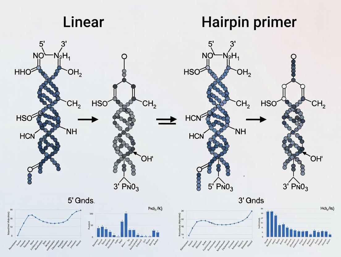
The following diagram illustrates the structural and functional differences between these two primer architectures.
Structural and functional comparison of linear and stem-loop primer architectures.
Comparative Performance Analysis
Experimental data from hybridization assays and amplification studies reveal clear performance trade-offs between the two primer architectures. The following tables summarize key quantitative findings.
Hybridization Kinetics and Thermodynamic Stability
Solid-support hybridization assays directly comparing dangling-ended hairpin probes with linear probes demonstrate the thermodynamic and kinetic advantages of the structured form.
Table 1: Comparison of hybridization kinetics and thermodynamics for linear vs. hairpin capture probes [3].
| Probe Architecture | Hybridization Rate (Relative to Linear) | Equilibrium Constant for Complex Formation | Binding Free Energy (ΔG) |
|---|---|---|---|
| 16-base Linear Probe | 1.0 (Reference) | Reference | Reference |
| 16-base Hairpin Probe | >2x faster at 25°C & 45°C | Larger | More favorable (stable) |
| 32-base Hairpin Probe | N/A | Largest | Most favorable |
Propensity for Undesired By-Products
A critical performance metric in amplification is the primer's tendency to form non-specific products, which depletes reagents and can lead to false positives.
Table 2: Propensity for by-product formation in amplification assays [4] [5].
| Amplification Context | Primer Architecture | Observed By-product | Key Influencing Factor |
|---|---|---|---|
| Duplex PCR Protocol | Linear ssDNA with complementary ends | Hairpin formation favored over self-dimer | Low concentration (kinetically favors intramolecular reaction) [4] |
| LAMP Assay | Long inner primers (FIP/BIP, ~40-45 bases) | Stable hairpins and primer-dimers | High primer count and length; 3' complementarity enables self-amplification [5] |
Experimental Protocols for Key Analyses
To objectively evaluate primer architectures in a specific research context, the following experimental protocols can be employed.
Protocol: Evaluating By-Product Formation via Agarose Gel Electrophoresis
This fundamental method visualizes the intended amplicon alongside spurious products like primer-dimers and hairpins.
- Reaction Setup: Perform the amplification (e.g., PCR or LAMP) using standardized conditions with the primers under investigation. Include a no-template control (NTC) to identify primer-derived artifacts.
- Gel Preparation: Cast a 3-5% agarose gel in TAE or TBE buffer containing a safe DNA intercalating dye [4].
- Electrophoresis: Load the post-amplification reactions and a suitable DNA ladder. Run the gel at a constant voltage (e.g., 100V) until sufficient separation is achieved.
- Visualization & Analysis: Image the gel under UV light. Primer-dimers typically appear as a diffuse band or smear around 30-50 bp, distinct from the larger target amplicon. The presence and intensity of these bands in the NTC directly indicate the propensity for by-product formation.
Protocol: Analyzing Hybridization Kinetics Using a Microtiter Plate Assay
This protocol quantifies the target capture efficiency of immobilized probes, adapted from a study comparing linear and hairpin probes [3].
- Surface Coating: Coat the wells of a microtiter plate with avidin (0.2 µM in carbonate buffer, pH 9.6) for 1 hour at room temperature. Wash with PBS.
- Probe Immobilization: Couple biotinylated linear or hairpin probes (e.g., 0.01-0.50 µM in BN buffer) to the avidin-coated wells for 30 minutes at room temperature. Wash with BN buffer. The density of immobilized probes can be quantified using radiolabeled or fluorescently labeled probes.
- Hybridization Reaction: Introduce a known concentration of the target strand (e.g., a 65-base sequence, labeled with FITC or radiolabel) to the probe-coated wells and incubate at the desired temperature (e.g., 25°C or 45°C).
- Time-Course Measurement: At various time points, measure the signal (fluorescence or radioactivity) from each well. After reaching equilibrium, wash the wells to remove non-specifically bound target and measure the final equilibrium signal.
- Data Analysis: Plot the amount of captured target versus time to determine hybridization rates. Use equilibrium signals to calculate equilibrium constants and binding free energies.
The workflow for this quantitative assay is outlined below.
Workflow for microtiter plate-based hybridization assay.
The Scientist's Toolkit: Essential Reagents and Materials
Successful experimentation with different primer architectures requires a suite of reliable reagents and instruments.
Table 3: Key research reagents and solutions for primer analysis.
| Reagent / Material | Function / Description | Example Use Case |
|---|---|---|
| Bst 2.0 WarmStart DNA Polymerase | A engineered strand-displacing polymerase with minimal activity at room temperature, reducing non-specific amplification. | Isothermal amplification assays like LAMP or SLIMP [5] [2]. |
| SYTO 9 / SYBR Green I Dyes | Intercalating fluorescent dyes that bind double-stranded DNA, allowing real-time monitoring of amplification. | Real-time detection of amplification products and melting curve analysis [5] [6]. |
| dNTP Solution Mix | A balanced mixture of the four deoxynucleotides (dATP, dGTP, dTTP, dCTP), the building blocks for DNA synthesis. | Essential for any PCR or isothermal amplification reaction [2]. |
| Betaine | A chemical additive that reduces secondary structure in DNA and equalizes the melting temperatures of GC- and AT-rich regions. | Improving amplification efficiency, particularly for GC-rich targets [5]. |
| Hot-Start Taq Polymerase | A modified polymerase that is inactive until a high-temperature activation step, preventing primer-dimer formation during reaction setup. | High-specificity PCR with linear primers [1] [7]. |
| Microtiter Plates (Avidin-Coated) | Solid support with avidin covalently attached, allowing efficient immobilization of biotinylated oligonucleotides. | Hybridization kinetics and thermodynamic studies [3]. |
Discussion and Research Context
The choice between linear and stem-loop primers is not a matter of declaring a universal winner but of strategic selection based on the application. Linear primers, due to their simplicity and predictability, remain the gold standard for conventional PCR and qPCR, especially when designed with best practices (e.g., checking for self-complementarity, optimizing Tm) and paired with hot-start enzymes to minimize dimerization [8] [7].
The unique advantages of stem-loop primers make them superior for specific applications. Their ability to suppress dimer formation is invaluable in complex, multiplexed reactions or when amplifying low-abundance targets where reagent competition is a critical issue. Furthermore, their enhanced hybridization kinetics and thermodynamic stability are beneficial in solid-phase hybridization assays and biosensors [3]. The development of novel methods like SLIMP (Stem-Loop and Linear Primers co-mediated exponential amplification) demonstrates the power of combining both architectures to achieve efficient, single-enzyme amplification of short gene sequences, a task difficult for traditional LAMP [2].
The broader thesis in primer research is increasingly focused on intentional design over passive avoidance. Instead of merely minimizing a primer's tendency to form secondary structures, researchers are now actively engineering stable stem-loops to impart new functionalities. This includes improving specificity through kinetic trapping, enabling novel isothermal amplification mechanisms, and creating more stable diagnostic probes. As polymerase engineering advances and design software incorporates these sophisticated principles, the strategic use of structured primers will undoubtedly become a more prominent tool in the molecular biologist's arsenal.
DNA hairpins, also known as stem-loop structures, are secondary structures formed when a single-stranded DNA (ssDNA) molecule folds back on itself, creating a base-paired duplex stem and an unpaired nucleotide loop. These structures are fundamental non-B-DNA motifs that arise from intrastrand base pairing within sequences containing inverted repeats or palindromes [9]. The formation of hairpins is driven by the fundamental physical principles of enthalpy-entropy compensation, where the free energy gain from base pairing (enthalpy) in the stem offsets the conformational entropy loss associated with bringing distant nucleotide segments together and organizing unpaired nucleotides into a loop structure.
The biological significance of DNA hairpins extends across multiple cellular processes. They serve as recognition elements for proteins involved in replication, transcription regulation, and site-specific recombination [9]. In mobile genetic elements like viruses, plasmids, and transposons, hairpin structures frequently participate in essential functions, likely because these elements experience single-stranded states during their life cycles [9]. More recently, hairpin formation has been implicated in pathological contexts, particularly in triplet repeat expansion diseases, where the propensity of repetitive sequences (such as CAG/CTG) to form secondary structures may contribute to mutagenic processes [10].
Comparative Analysis: Hairpin vs. Linear DNA Formats
The functional advantages of hairpin structures over linear single-stranded DNA become particularly evident in molecular recognition and hybridization applications. Experimental data directly comparing these formats reveals significant differences in their biophysical and thermodynamic properties.
Table 1: Performance Comparison of Hairpin vs. Linear DNA Probes in Target Capture [3]
| Performance Metric | Hairpin Probes | Linear Probes |
|---|---|---|
| Hybridization Rate (at 25-45°C) | >2× faster than linear | Baseline |
| Equilibrium Capture Amount | Significantly larger | Smaller |
| Thermodynamic Stability | More stable complexes | Less stable |
| Binding Free Energy | More favorable | Less favorable |
| Structural Advantage | Coaxial stacking at junction | No stacking benefit |
The superior performance of hairpin probes stems primarily from coaxial stacking interactions between the 5' terminal base(s) of the hairpin stem and the 3' terminal base(s) of the annealed target strand [3]. This stacking along the helical axis provides a significant thermodynamic advantage for target binding. The order of stability follows: hairpins with 32-base dangling ends > hairpins with 16-base dangling ends > 16-base linear probes > 32-base linear probes [3].
Molecular Forces and Energetics of Hairpin Formation
Primary Driving Forces
The folding of single-stranded DNA into hairpin structures is governed by a complex interplay of molecular forces:
Base Pairing and Stacking Interactions: Hydrogen bonding between complementary bases in the stem region provides the primary enthalpic driver for hairpin formation. The free energy gain from Watson-Crick base pairing (typically -1 to -3 kcal/mol per base pair depending on sequence context) compensates for the entropic cost of loop formation. Additionally, base stacking interactions between adjacent nucleotide pairs in the stem further stabilize the structure through van der Waals forces and hydrophobic effects.
Electrostatic Repression and Counterion Stabilization: The negatively charged phosphate backbone creates significant electrostatic repulsion that must be overcome for hairpin formation. Divalent cations (especially Mg²⁺) play a crucial role in screening these repulsive forces, with their effective concentration significantly influencing folding kinetics and stability.
Loop Entropy and Strain: The unpaired nucleotides in the loop region represent an entropic penalty that opposes hairpin formation. This penalty depends strongly on loop length, with optimal stability typically observed at 4-6 nucleotides for DNA hairpins. Shorter loops create higher strain due to backbone distortion, while longer loops incur greater entropy loss.
Energetic Contributions to Stability
Table 2: Energetic Contributions to DNA Hairpin Stability
| Energetic Component | Stabilizing Contribution | Structural Role |
|---|---|---|
| Stem Base Pairing | -1 to -3 kcal/mol per bp | Primary stabilization via H-bonding |
| Stem Base Stacking | -2 to -8 kcal/mol for full stem | Helical structure maintenance |
| Loop Closure | +1 to +5 kcal/mol (unfavorable) | Entropic penalty for organization |
| Cation Screening | Variable (-2 to -10 kcal/mol) | Charge neutralization |
| Solvation Effects | Variable | Hydrophobic contributions |
The trinucleotide loop motif 5'-GCA-3' exemplifies particularly stable hairpin formation, with experimental melting temperatures reaching 67°C for the sequence 5'-GCGCAGC-3' [11]. This stability arises from specific structural features including a sheared G:A base pair at the loop closure and stacking of the central loop base on top of this base pair, pointing toward the major groove [11].
Experimental Methodologies and Protocols
Molecular Dynamics Simulations of Hairpin Folding
Protocol 1: Replica-Exchange Molecular Dynamics (RexMD) for Hairpin Folding [11]
System Preparation: Construct an initial extended single-stranded DNA structure using nucleic acid building tools (e.g., Nucgen in Amber). The exemplary sequence is 5'-GCGCAGC-3', which forms a characteristic trinucleotide hairpin loop.
Solvation and Ion Placement: Solvate the system in an octahedral water box (TIP3P water model) with at least 10 Å between solute atoms and box borders. Add K⁺ counterions to neutralize system charge, achieving approximately 200 mM ion concentration.
Energy Minimization and Equilibration: Perform initial energy minimization (2500 steps) followed by gradual heating from 50 to 300 K with positional restraints on DNA (force constant: 50 kcal mol⁻¹ Å⁻²). Gradually reduce restraints during 0.25 ns, then conduct unrestrained MD equilibration for 2 ns.
Replica-Exchange Production Run: Execute RexMD with 16 replicas across an exponentially increasing temperature series (315-422 K). Use constant volume periodic boundary conditions, 1 fs timestep, and attempt exchanges between neighboring replicas every 750 steps. Continue simulation for 36 ns per replica.
This protocol enabled the sampling of near-native hairpin structures as the dominant conformational state at the lowest temperature replica, achieving approximately 35% population of correctly folded structures [11].
Protocol 2: Classical MD for Base-Flipping Dynamics [12]
Initial Structure Generation: Generate hairpin structures with a double-helix stem (sequence: 5'-TTAGCATG-XXX-CATGCTAA-3', where XXX represents variable loop sequences). Create all 3NT (37 unique) and 4NT (175 unique) loops containing at least one cytosine.
Force Field Selection: Use the OL15 force field for DNA, which provides reasonable base-flipping dynamics. TIP3P model for water molecules and Joung-Cheathem parameters for counterions.
System Setup: Neutralize with K⁺ ions, then solvate with TIP3P waters to a minimum 20 Å distance between nucleotide and box edge.
Simulation Parameters: Perform minimization, equilibration, and heating phases. Run production MD (100 ns per trajectory) with a Langevin thermostat (friction constant = 5 ps⁻¹) in an NVT ensemble at 300 K and 1 atm pressure, using a 1 fs timestep.
This approach provided statistical insight into base-flipping motions, demonstrating that flipping events occur in solvent before protein binding, semi-dependent on the hairpin loop sequence [12].
Experimental Assay for Hybridization Performance
Protocol 3: Microtiter-Based Hybridization Assay [3]
Probe Design:
- Hairpin probes: 16 bp duplex stem, 5-base loop (T-T-UB-T-T, where UB is biotinylated uracil), 3' dangling end (16 or 32 bases complementary to target)
- Linear probes: 16 or 32 bases corresponding precisely to hairpin dangling ends, with 5' biotin modification
Surface Functionalization:
- Coat microtiter wells with avidin (0.2 µM in carbonate buffer, pH 9.6)
- Incubate 1 hour at room temperature, wash with PBS
- Couple biotinylated probes (0.01-0.50 µM in BN buffer) for 0.5 hours at room temperature
Target Hybridization:
- Use 65-base target DNA labeled with 5'-FITC or [γ-³³P]ATP
- Hybridize across concentration range (10-640 pmol)
- Measure time dependence and equilibrium binding
Data Analysis:
- Quantify captured target via fluorescence or radioactivity measurement
- Calculate hybridization rates and equilibrium constants
- Determine binding free energies from observed equilibrium constants
This experimental setup demonstrated that hairpin probes displayed higher hybridization rates and larger equilibrium amounts of captured targets than linear probes, with hybridization rates more than twice as great for hairpins across 25-45°C [3].
Visualization of Hairpin Folding Pathways and Dynamics
DNA Hairpin Folding Pathway
The folding mechanism involves rapid formation of loop motifs with sheared G:A basepairs before complete stem formation, with possible misfolded intermediates that can slow the folding kinetics [11].
APOBEC3A-Hairpin Recognition
Recent molecular dynamics studies reveal that base-flipping occurs spontaneously in solution before protein binding, with the flipped-out conformation required for deamination activity as it positions the target cytosine within range of a reactive water molecule in the active site [12].
The Scientist's Toolkit: Essential Research Reagents
Table 3: Essential Research Reagents for DNA Hairpin Studies
| Reagent / Material | Specifications | Research Application |
|---|---|---|
| DNA Hairpin Probes | 16 bp stem, 4-5 nt loop, 3' dangling ends (16-32 nt) | Hybridization assays, capture probes [3] |
| HPKinLock Hairpins | 10 bp hairpin with 4 bp loop (24 nt total) | Kinetic locking assays, single-molecule studies [10] |
| Molecular Dynamics Software | Amber22 with pmemd.cuda, OL15/TIP3P force fields | Base-flipping dynamics, folding simulations [12] |
| Avidin-Coated Microplates | 96-well format, carbonate coating buffer (pH 9.6) | Solid-support hybridization assays [3] |
| Biotinylated Nucleotides | dU-Biotin with C12 spacer, 5'-biotin modifiers | Probe immobilization, surface coupling [3] |
| Counterions | K⁺ ions (Joung-Cheathem parameters), Mg²⁺ salts | Charge screening, simulation conditions [12] [11] |
The physics of DNA hairpin formation represents a sophisticated balance of molecular forces, with clear experimental advantages demonstrated for hairpin structures over linear formats in hybridization efficiency and thermodynamic stability. The base-flipping dynamics and folding pathways characterized through molecular dynamics simulations provide atomic-level insights into the recognition mechanisms employed by DNA-interacting proteins like APOBEC3A.
For researchers and drug development professionals, these findings offer significant implications. The superior hybridization properties of hairpin probes suggest immediate applications in diagnostic assay development, where enhanced sensitivity and kinetics can improve detection limits. In therapeutic contexts, understanding the sequence-specific folding propensities of hairpins enables better design of oligonucleotide therapeutics that must navigate secondary structure formation for successful target engagement.
Future research directions should focus on expanding our understanding of hairpin dynamics in complex biological environments, particularly how cellular factors and supercoiling forces influence folding kinetics and stability in vivo. The continued development of advanced simulation methodologies and single-molecule techniques will further illuminate the physical principles governing these fundamental structural motifs.
The thermodynamic stability of nucleic acids, quantified through Melting Temperature (Tm) and Gibbs Free Energy (ΔG), is a foundational concept in molecular biology with profound implications for experimental success. These parameters directly predict the stability of DNA secondary structures and the efficiency of hybridization events. Within primer design, a critical comparison exists between traditional linear primers and advanced hairpin primers. Hairpin primers, which incorporate a self-complementary stem-loop structure, offer enhanced specificity by minimizing primer-dimer formation and mispriming, but their design requires careful thermodynamic balancing [13]. This guide objectively compares the performance of these primer types, providing the experimental data and methodologies needed to inform their application in research and diagnostic development.
Core Thermodynamic Principles and Calculations
Defining Tm and ΔG
The Melting Temperature (Tm) is defined as the temperature at which half of the DNA duplex molecules dissociate into single strands. It provides a direct measure of duplex stability [14] [15]. The Gibbs Free Energy (ΔG), calculated as ΔG = ΔH - TΔS, represents the overall spontaneity of the hybridization reaction; a more negative ΔG indicates a more stable duplex formation [16] [17]. For hairpin primers, the ΔG of the stem structure (typically ranging from -1.6 to -5.8 kcal/mol) is a critical design parameter, as it must be stable enough to form a hairpin at lower temperatures, but labile enough to unravel and permit hybridization at the annealing temperature [13].
Fundamental Calculation Methods
Researchers employ several formulas to estimate Tm, each with varying complexity and accuracy. The most basic method, suitable for short primers, is the Wallace Rule:
Tm = 2°C × (A + T) + 4°C × (G + C) [18]
For greater accuracy, the nearest-neighbor method is the gold standard. It considers the enthalpy (ΔH) and entropy (ΔS) changes for each dinucleotide pair in the sequence, yielding a more reliable prediction [15] [17]. The formula, incorporating salt concentration, is:
Tm = {ΔH / (ΔS + R ln(C))} - 273.15 [15]
Where R is the molar gas constant and C is the DNA concentration. ΔH and ΔS are obtained by summing the respective values for each nearest-neighbor pair in the sequence [15].
For ΔG, the calculation also relies on the nearest-neighbor model. The total free energy change for duplex formation is the sum of the free energy increments for each base pair step, plus initiation factors, and penalties for any loops or mismatches [17]. The stability of secondary structures like hairpins is commonly represented by their ΔG value, with larger negative values indicating more stable, and sometimes undesirable, structures [15].
Comparative Thermodynamic Properties of Primer Architectures
Table 1: Thermodynamic and Performance Comparison of Linear vs. Hairpin Primers
| Feature | Linear Primers | Hairpin Primers |
|---|---|---|
| Basic Structure | Single-stranded linear oligonucleotide | Self-complementary sequence forming a blunt-end hairpin [13] |
| Key Design Parameter | Overall Tm, GC content, 3'-end stability [14] [15] | Stem ΔG (optimal range: -1.6 to -5.8 kcal/mol) [13] |
| Initial Fluorescence | High (if labeled) | Low in closed state; increases up to 8-fold upon product formation [13] |
| Primary Advantage | Simple design, robust hybridization kinetics | Built-in specificity, reduced primer-dimer and mispriming [13] |
| Key Disadvantage | Prone to primer-dimer and non-specific amplification [13] [15] | More complex design; requires specialized software [13] |
| Typical ΔG of Secondary Structure | N/A (ideally none) | Stem ΔG: -1.6 to -5.8 kcal/mol [13] |
| Impact on PCR Efficiency | Dictated by hybridization efficiency to template | High; hairpin must unravel to hybridize, adding a specificity check [13] |
Experimental Protocols for Thermodynamic Analysis
Protocol 1: Measuring Tm via Fluorometric Melting Curves
This protocol is adapted from high-throughput methods used to validate DNA folding thermodynamics [17].
- Sample Preparation: Dilute the oligonucleotide (hairpin or linear) in a suitable buffer (e.g., 20 mM Tris-HCl, pH 7.5, 50 mM NaCl, 5 mM MgCl2) to a final concentration of 50-100 nM [13] [17]. For hairpin probes, anneal fluorophore- and quencher-labeled oligonucleotides to the constant flanking regions if using a secondary reporting system [17].
- Fluorescence Measurement: Load the sample into a real-time PCR instrument or a spectrofluorometer equipped with a thermal cycler. Set the instrument to monitor fluorescence continuously as the temperature is gradually increased from 20°C to 85°C at a slow ramp rate (e.g., 0.5°C per minute) [17].
- Data Analysis: Plot the normalized fluorescence (F) versus temperature (T). The Tm is determined as the temperature at the midpoint of the transition curve, where the derivative (dF/dT) is at a maximum. For two-state systems like simple hairpins, the data is fitted to a two-state model to derive ΔH and Tm, from which ΔG is calculated [17].
Protocol 2: Validating Primer Efficiency in PCR
This protocol outlines a comparative experiment to assess the performance of hairpin versus linear primers in a real-time PCR context, as described in [13].
- Reaction Setup: Prepare PCR master mixes containing 1x PCR buffer, 3 mM MgCl2, 200 µM of each dNTP, and DNA polymerase. For the test reactions, use 80-200 nM of either the hairpin primer set or the linear primer set designed for the same target (e.g., c-myc or IL-4 cDNA). Include a reference dye (e.g., ROX) if required by the instrument [13].
- Thermal Cycling: Perform PCR in a real-time thermocycler (e.g., ABI PRISM 7700 or i-Cycler) with the following typical profile:
- Initial Denaturation: 95°C for 2 min.
- Cycling (40 cycles): Denaturation at 95°C for 15 s, Annealing at 55-65°C for 30 s, Extension at 72°C for 30 s.
- Monitor fluorescence during the annealing step of every cycle [13].
- Data Analysis: Compare the amplification plots and Cq values. Hairpin primers should demonstrate a low initial fluorescence that increases significantly with each cycle. Calculate the PCR efficiency for both primer types. Assess specificity by analyzing the melt curve post-amplification or by gel electrophoresis, where hairpin primers are expected to show reduced primer-dimer artifacts [13].
Performance Comparison and Experimental Data
Quantitative Comparison of Specificity and Yield
Empirical data demonstrates the distinct performance advantages of hairpin primers in complex assays. In a multiplex quantitative PCR experiment targeting c-myc and IL-4 cDNAs with reference genes (β-actin, GAPDH), fluorogenic hairpin primers successfully detected targets across a dynamic range of 10–107 copies with high precision [13]. The key differentiator was the significant reduction in non-specific amplification.
Table 2: Experimental Performance Data from Key Studies
| Experiment Goal | Primer Type | Key Performance Metric | Result | Source |
|---|---|---|---|---|
| Multiplex qPCR | Hairpin (Fluorogenic) | Detection Limit | 10-107 copies detected | [13] |
| Multiplex qPCR | Hairpin (Fluorogenic) | Signal-to-Background | Up to 8-fold fluorescence increase | [13] |
| SNP Detection | Hairpin (Allele-Specific) | End-point Specificity | Successful discrimination of human retinal degeneration gene SNP | [13] |
| Pathogen ID (Tm Mapping) | Probes with engineered stems | Specificity (Difference Value) | Enabled identification of 62/68 bacterial species; reduced false positives from stem invasion | [19] |
| MgCl₂ Optimization | Linear (Modeled) | Prediction Accuracy (R²) | R² = 0.9942 for MgCl₂ concentration using thermodynamic model | [16] |
The Scientist's Toolkit: Essential Reagents and Materials
Table 3: Key Research Reagent Solutions for Thermodynamic Studies
| Item | Function/Description | Example Application |
|---|---|---|
| Fluorophore-Labeled Nucleotides | (e.g., FAM, JOE, Cy3). Incorporated into primers to serve as reporter molecules for fluorescence-based melting and qPCR. | Real-time monitoring of duplex formation and denaturation in Tm assays and qPCR [13] [17]. |
| Quenchers | (e.g., Dabcyl, BHQ). Suppress fluorophore emission via FRET when in close proximity. Essential for hairpin probe function. | Used in molecular beacons and hairpin primers to quench fluorescence in the closed state [13] [17]. |
| Eukaryote-Made DNA Polymerase | Recombinant polymerase manufactured in yeast cells. Free from bacterial DNA contamination, crucial for sensitive pathogen detection. | Prevents false positives in PCR-based direct detection of bacterial isolates from patient samples [19]. |
| Thermostable DNA Polymerase | Enzyme for DNA amplification that withstands high temperatures. "Hot-start" versions reduce non-specific amplification. | Standard enzyme for PCR and qPCR reactions [13]. |
| l-DNA Phosphoramidites | Unnatural enantiomeric nucleotides used to synthesize nuclease-resistant oligonucleotide stems that do not hybridize with natural d-DNA. | Creating stems in molecular beacons to prevent stem invasion and improve specificity in complex samples [20]. |
The strategic application of thermodynamic principles is paramount for designing effective nucleic acid reagents. The choice between linear and hairpin primers is not a matter of superiority but of application-specific suitability. Linear primers remain the workhorse for standard PCR due to their simplicity and robust performance. In contrast, hairpin primers offer a superior solution for applications demanding high specificity, such as multiplex qPCR, SNP detection, and pathogen identification in complex biological samples, by leveraging their predictable ΔG-driven mechanics to minimize false positives [13] [19].
Future developments will be heavily influenced by large-scale thermodynamic datasets and machine learning. High-throughput methods like Array Melt, which can measure the stability of millions of DNA hairpins, are generating the data needed to build next-generation predictive models that move beyond traditional nearest-neighbor parameters [17]. These models, including graph neural networks (GNNs), promise to significantly improve the in silico design of not only primers and probes but also complex DNA nanostructures, further closing the gap between theoretical design and experimental success [17].
In the fields of molecular diagnostics, genomics, and drug development, the detection of specific nucleic acid sequences through hybridization is a fundamental procedure. The design of the capture probe—the molecular element that recognizes and binds to the target sequence—is a critical pre-analytical step that directly determines the sensitivity, specificity, and reliability of the assay. While linear, single-stranded DNA probes have been widely used for decades, hairpin-structured probes, such as molecular beacons, have emerged as superior alternatives in many applications. These stem-loop constructs offer significant kinetic and thermodynamic advantages that translate into enhanced performance for researchers and developers. This guide objectively compares the performance of hairpin probes against traditional linear probes, drawing on experimental data to highlight the specific conditions under which these advantages manifest. The evidence demonstrates that hairpin probes provide faster hybridization rates and form more stable complexes with target sequences, making them particularly valuable for applications requiring high sensitivity and specificity, such as pathogen detection, SNP genotyping, and real-time monitoring of gene expression inside living cells.
Fundamental Principles: Structural Mechanics of Hairpin Probes
Hairpin probes, also known as molecular beacons, are single-stranded nucleic acid molecules composed of three distinct functional domains. The loop region contains a sequence that is complementary to the desired target nucleic acid. The stem is formed by two short, self-complementary sequences located at the ends of the molecule that hybridize to form a double-helical segment. Often, a fluorophore and a quencher are attached to the 5' and 3' ends, respectively; when the probe is in its closed hairpin state, the proximity of the fluorophore and quencher results in quenching of the fluorescence signal [20] [21].
The operation of a hairpin probe is governed by a conformational change. In the absence of the target, the probe maintains its stable stem-loop structure. Upon hybridization with a perfectly complementary target sequence, the probe undergoes a large-scale conformational reorganization, unwinding the stem and forming a rigid, double-stranded probe-target complex. This transition separates the fluorophore from the quencher, leading to a detectable increase in fluorescence [20]. This on/off mechanism is intrinsically linked to the probe's kinetic and thermodynamic properties. The initial stem structure provides a thermodynamic "push" that accelerates the hybridization process, while the coaxial stacking that occurs between the stem and the newly formed probe-target duplex enhances the overall complex stability [3].
Direct Performance Comparison: Quantitative Advantages of Hairpin Probes
A systematic, microtiter-based assay system provides direct experimental evidence of the superior performance of hairpin probes over linear probes. In this study, both probe types were immobilized and their ability to capture single-stranded target DNA was measured over time and across different temperatures. The hairpin probes featured a 16 base pair (bp) duplex stem and a 3' dangling end (16 or 32 bases) complementary to the target, while the linear probes corresponded precisely to the dangling end sequences of the hairpins [3].
Key Experimental Parameters
- Probe Design: Hairpin probes with a 16 bp stem and 16 or 32 base dangling ends; linear probes of 16 or 32 bases.
- Target: A 65-base single-stranded DNA sequence, labeled for detection.
- Assay Conditions: Target concentrations ranging from 10 to 640 pmol; temperatures of 25°C and 45°C.
- Measurements: Time-dependent hybridization rates and equilibrium constants for complex formation [3].
The quantitative results from this study are summarized in the table below.
Table 1: Comparative Hybridization Performance of Hairpin vs. Linear Probes
| Performance Metric | 16-Base Hairpin Probe | 16-Base Linear Probe | 32-Base Hairpin Probe | 32-Base Linear Probe |
|---|---|---|---|---|
| Hybridization Rate (relative increase) | >2x faster than linear | Baseline | >2x faster than linear | Slower than 16-base linear |
| Equilibrium Capture Amount | Higher | Baseline | Highest | Lower |
| Thermodynamic Stability (Complex Stability) | More stable | Baseline | Most stable | Least stable |
Performance Analysis
The data reveals two key findings. First, hairpin probes hybridize their targets at rates more than twice as fast as equivalent linear probes at both 25°C and 45°C. Second, the order of thermodynamic stability, as determined from equilibrium constants, follows a clear hierarchy: hairpins with 32-base dangling ends > hairpins with 16-base dangling ends > 16-base linear probes > 32-base linear probes [3]. This demonstrates that the hairpin structure itself, not merely the length of the capture sequence, is the primary factor driving enhanced performance. The somewhat counterintuitive result that longer linear probes perform worst highlights a key limitation of linear probes, likely due to increased non-specific folding or slower diffusion.
underlying Mechanisms: The Structural Basis for Enhanced Performance
The superior performance of hairpin probes can be attributed to specific biophysical mechanisms that arise from their unique architecture.
Pre-Organization and Conformational Assistance
The stem of a hairpin probe pre-organizes the loop region, effectively holding the capture sequence in a configuration that is more accessible and favorable for binding to its target. This reduces the entropic penalty associated with the hybridization event, as the probe does not need to undergo as large a conformational rearrangement as a flexible linear probe. This phenomenon, sometimes called "conformational assistance," lowers the activation energy for the reaction, thereby increasing the hybridization rate [3].
Coaxial Stacking
A particularly important mechanism is coaxial stacking. When the target DNA hybridizes to the dangling end of the hairpin probe, the terminal base pairs of the pre-existing stem stack directly on top of the newly formed probe-target duplex. This stacking interaction across the junction provides significant extra stabilization to the overall complex, which is not available in linear probe-target complexes [3]. This additional stabilization is reflected in the more favorable (more negative) binding free energies observed for hairpin probes.
Mitigation of Stem Invasion
A known challenge with classic hairpin probes is "stem invasion," where non-target sequences complementary to the stem region can bind, opening the probe and causing false-positive signals. Innovative solutions have been developed to mitigate this. One effective strategy incorporates unnatural L-DNA nucleotides into the stem region. Since L-DNA is the mirror image of natural D-DNA, it cannot form stable duplexes with natural nucleic acids in a sample. This allows the stem to maintain its stability while becoming immune to invasion by cellular DNA or RNA, thereby enhancing specificity and signal-to-noise ratios in complex biological environments [20].
Advanced Hairpin Architectures: Pushing the Boundaries of Performance
Building on the basic hairpin design, researchers have developed more sophisticated architectures to further enhance functionality and address specific application challenges.
The Double-Stem Hairpin Probe (DHP)
The Double-Stem Hairpin Probe (DHP) represents a significant architectural advance. It incorporates two stable stems and is engineered to include functional elements like a DNAzyme sequence and a nicking enzyme recognition site. This design allows for the creation of an ultrasensitive colorimetric sensing system. Despite having a long double-stranded DNA fragment, the DHP is designed to avoid intermolecular interactions (sticky-end pairing) that often plague traditional molecular beacons. In one reported system, a target DNA molecule could trigger a cascade polymerization/nicking cycle on a single DHP, leading to a massive accumulation of G-quadruplex DNAzymes for signal amplification. This enabled detection of a cancer-related gene sequence down to 1 fM (femtomolar) and allowed for visual discrimination of point mutations without instrumentation [22].
Linear-Hairpin Variable Primers for MicroRNA
The principles of hairpin design have been successfully applied to the challenging domain of microRNA (miRNA) detection. A novel "linear-hairpin variable primer" was developed for reverse transcription quantitative PCR (RT-qPCR). This primer is initially linear but, upon recognizing the target miRNA, is extended by reverse transcriptase to form a hairpin structure. This structure displaces the target miRNA, allowing it to be recycled in a cyclic reverse transcription reaction, which dramatically improves sensitivity. This method demonstrated a dynamic range of 8 logs and could detect as few as 4 target miRNA molecules, while also achieving high specificity to discriminate between closely related miRNA family members [23].
Experimental Protocols and Research Toolkit
To facilitate the adoption and validation of these probes, this section outlines a core experimental methodology and lists essential research reagents.
Core Experimental Protocol: Microtiter-Based Hybridization Assay
The following protocol, adapted from a key comparative study, allows for the direct measurement of hybridization kinetics and stability for immobilized probes [3].
- Surface Functionalization: Coat the wells of a microtiter plate with avidin by incubating with a 0.2 µM avidin solution in carbonate buffer (pH 9.6) for 1 hour at room temperature. Wash thoroughly with phosphate-buffered saline (PBS).
- Probe Immobilization: Dilute biotinylated hairpin or linear probes in BN buffer (1.0 M NaCl, 100 mM Tris, 0.08% Triton-X 100, pH 8.0). Add the probe solution to the avidin-coated wells and incubate for 30 minutes at room temperature. Wash again with BN buffer to remove unbound probes.
- Hybridization Reaction: Introduce the target DNA (e.g., labeled with a radioisotope or fluorophore like FITC) in a suitable hybridization buffer across a range of concentrations (e.g., 10-640 pM). Incubate at the desired temperature (e.g., 25°C or 45°C).
- Kinetic Measurement: For kinetics, measure the amount of captured target at various time points after the addition of the target solution. This can be done via Cerenkov counting (for radiolabeled targets) or fluorescence measurement.
- Equilibrium Measurement: For thermodynamics, allow the reaction to proceed until no further increase in signal is observed (equilibrium). Calculate equilibrium constants from the concentration of free and bound target at equilibrium.
The workflow for this protocol is visualized below.
The Researcher's Toolkit: Essential Reagents and Materials
Table 2: Key Research Reagent Solutions for Hairpin Probe Experiments
| Reagent/Material | Function/Description | Example Usage |
|---|---|---|
| Biotinylated Hairpin Probes | Capture probe; biotin allows immobilization on avidin-coated surfaces. | Core element for surface-based hybridization assays [3]. |
| Avidin-Coated Microtiter Plates | Solid support for probe immobilization. | Provides a uniform surface for kinetic and thermodynamic studies [3]. |
| L-DNA Nucleotides | Non-natural enantiomers for stem synthesis. | Used to create stem sequences immune to invasion by natural DNA/RNA, reducing false positives [20]. |
| Nicking Endonuclease (e.g., Nt.BbvCI) | Enzyme that cleaves a specific strand of a DNA duplex. | Key component in signal amplification cascades within advanced probes like DHP [22]. |
| DNA Polymerase (e.g., Klenow Fragment) | Enzyme for DNA synthesis. | Used in amplification strategies (e.g., SDA) coupled with hairpin probes [22]. |
| Hemin | Cofactor that binds G-quadruplexes to form DNAzyme. | Enables colorimetric signal generation in catalytic hairpin probe systems [22]. |
The body of experimental evidence unequivocally demonstrates that hairpin-structured nucleic acid probes offer significant kinetic and stability advantages over traditional linear probes. Their pre-organized structure and the mechanism of coaxial stacking enable faster hybridization rates and the formation of more thermodynamically stable complexes with target sequences. While classic molecular beacons can be susceptible to stem invasion, modern innovations such as L-DNA stems and double-stem architectures have effectively mitigated these issues, further enhancing their specificity and utility. For researchers and drug development professionals designing detection assays where sensitivity, speed, and reliability are paramount—such as in clinical diagnostics, live-cell imaging, or ultrasensitive biomarker detection—hairpin probes represent a powerful and superior tool in the molecular toolkit.
DNA hairpins, formed by sequences with inverted repeats that fold back on themselves to create a stem-loop structure, are fundamental architectural motifs in nucleic acids with profound biological significance. These structured forms of DNA with intrastrand pairing are generated in several cellular processes and are involved in critical biological functions, including replication, transcription regulation, and recombination [9]. Hairpin structures can arise on single-stranded DNA (ssDNA) produced during replication, bacterial conjugation, natural transformation, or viral infections [9]. Furthermore, negatively supercoiled DNA can extrude inverted repeats as hairpins in structures called cruciforms [9]. This review examines the natural roles of DNA hairpins, with particular emphasis on their advantages in hybridization kinetics and thermodynamic stability compared to linear DNA probes, providing a biological precedent for their application in molecular technologies.
The ability of DNA to form secondary structures represents a departure from the canonical B-helix form proposed by Watson and Crick. While DNA is typically depicted as a straight double helix, it is now evident that alternative structures like hairpins play essential functional roles in cellular processes [9]. These structures modify the access of proteins to DNA, and in some cases, can be directly recognized by proteins, enabling specific biological functions [9]. The prevalence of hairpin functions in mobile genetic elements likely to be single-stranded, including viruses, plasmids, transposons, and integrons, provides clues about their evolutionary development and optimization [9].
Natural Biological Functions of DNA Hairpins
Roles in DNA Replication and Genome Stability
DNA hairpins play crucial roles in replication processes and maintaining genome stability. During DNA replication, the unique biochemical difficulties of performing DNA replication within long stretches of repeat DNA can lead to expansions within trinucleotide tracts [24]. mounting in vitro evidence suggests that triplets are deleted or added to long repetitive tracts as cellular machinery attempts to replicate through DNA hairpins formed by triplet repeat sequences [24]. During lagging strand synthesis, Okazaki fragments are joined through a process where the upstream fragment extends downstream to the next fragment and displaces a small portion as a single-strand 'flap' of DNA [24]. The enzyme flap endonuclease 1 (FEN1) is responsible for removing this flap, and hairpin structures within these flaps can influence the efficiency and accuracy of this process, potentially leading to genetic expansions or contractions [24].
The formation of hairpin structures is particularly relevant in replication of repetitive DNA sequences. These structures can form when DNA becomes single-stranded during replication, and their stability can interfere with normal replication progression. The ability of certain repeat sequences to form stable hairpins has been implicated in various neurological disorders, where expansion of trinucleotide repeats occurs through mechanisms involving hairpin formation [24]. This demonstrates how the intrinsic property of DNA sequences to form secondary structures can have significant consequences for genome integrity and human health.
Involvement in Horizontal Gene Transfer
Hairpin structures play significant roles in horizontal gene transfer between bacterial cells, particularly in conjugation and transformation. During conjugation, which involves the transfer of single-stranded DNA (ssDNA) from a donor to a recipient cell, the transferred strand (T strand) can form secondary structures that may be recognized by proteins [9]. The production of large amounts of ssDNA during conjugation provides opportunities for hairpin formation, and conjugative plasmids are likely places for the evolution of functions where hairpins are involved [9]. In fact, the very process of conjugation implies DNA secondary structures [9].
Similarly, during natural transformation, which permits the uptake and incorporation of naked exogenous DNA, entering single strands are protected by binding proteins that may interact with hairpin structures [9]. While the lifetime of ssDNA during transformation is generally shorter than in conjugation, the formation of transient hairpins may still influence the recombination or integration process. The presence of hairpin-forming sequences in mobile genetic elements suggests these structures have been evolutionarily selected for their functional advantages in gene transfer processes [9].
Regulatory Functions in Transcription and Protein Binding
DNA hairpins serve important regulatory functions by modifying protein-DNA interactions in transcriptional regulation. Hairpins can impact cell physiology through three primary mechanisms: (1) cruciform formation modifies the coiling state of DNA, which affects the binding of regulatory proteins for transcription, recombination, and replication; (2) DNA-protein interaction can be inhibited if a hairpin overlaps a protein recognition site; and (3) proteins can directly recognize and bind DNA hairpins [9].
These mechanisms allow hairpin structures to serve as structural switches that modulate gene expression in response to changes in DNA supercoiling or cellular conditions. For instance, the extrusion of cruciform structures can relieve torsional stress in negatively supercoiled DNA while simultaneously creating or obscuring protein binding sites. Specific proteins have evolved to recognize and bind directly to hairpin structures, enabling specialized functions in various DNA transactions [9]. This direct recognition is exemplified by the APOBEC3 family of enzymes, which show preferential activity upon hairpin loops in single-stranded DNA [12].
Comparative Analysis: Hairpin Versus Linear DNA Probes
Experimental System for Direct Comparison
A microtiter-based assay system has been developed to directly compare the performance of DNA hairpin probes with dangling ends against single-stranded, linear DNA probes for capturing single-strand target DNA [3]. In this system, hairpin probes consisted of a 16 bp duplex stem, linked by a T₂-biotin·dT-T₂ loop, with a biotinylated uracil at the third base position for coupling to avidin-coated microtiter wells [3]. The capture region of the hairpin was a 3' dangling end composed of either 16 or 32 bases [3]. The analogous linear probes were 16 or 32 bases long and corresponded precisely to the sequences of the dangling ends of the hairpins, containing a biotin moiety at the 5' end for coupling to microtiter plates [3].
The target DNA consisted of 65 bases whose 3' end was complementary to the dangling end of the hairpin or to the linear probe sequence [3]. Target molecules were labeled with either a 5'-FITC or radiolabeled with [γ-³³P]ATP to enable detection and quantification [3]. This experimental design allowed direct comparison of hybridization kinetics and thermodynamic stability under identical conditions, providing robust quantitative data on the relative performance of hairpin versus linear capture probes.
Hybridization Kinetics and Thermodynamic Stability
Experimental data demonstrates significant advantages of hairpin probes over linear probes in both hybridization kinetics and thermodynamic stability. Over a range of target concentrations from 10 to 640 pmol, hybridization rates increased with increasing target concentration, but varied substantially between different probe types [3]. Hairpin probes consistently displayed higher rates of hybridization and larger equilibrium amounts of captured targets than linear probes [3]. At 25°C and 45°C, rates of hybridization were more than twice as great for hairpin compared with linear capture probes [3].
The thermodynamic stability of the resulting complexes also favored hairpin structures. Analysis of binding free energies evaluated from observed equilibrium constants for complex formation showed a clear order of stability: hairpins with 32 base dangling ends > hairpin probes with 16 base dangling ends > 16 base linear probes > 32 base linear probes [3]. This enhanced stability can be attributed to coaxial stacking interactions between the 5' terminal base(s) of the hairpin stem and the 3' terminal base(s) of the annealed single-stranded target, providing additional stabilization beyond simple base pairing [3].
Table 1: Comparative Hybridization Performance of Hairpin vs. Linear DNA Probes
| Probe Type | Hybridization Rate (relative to linear) | Equilibrium Capture Amount | Binding Free Energy | Optimal Temperature Range |
|---|---|---|---|---|
| Hairpin with 32-base dangling end | >2× faster | Highest | Most favorable | 25-45°C |
| Hairpin with 16-base dangling end | >2× faster | High | Intermediate | 25-45°C |
| 16-base linear probe | Baseline | Moderate | Less favorable | 25-45°C |
| 32-base linear probe | Slower | Lower | Least favorable | 25-45°C |
Structural Advantages and Molecular Mechanisms
The superior performance of hairpin probes can be attributed to specific structural advantages at the molecular level. Solution studies have shown that nicked duplexes comprised of dangling-ended hairpins and single-strands are thermodynamically more stable than gapped duplexes formed with linear probes [3]. This stabilization arises from coaxial stacking interactions between the terminal base pairs of the hairpin stem and the adjacent base pairs formed with the captured target [3]. These stacking interactions along the helical axis provide a thermodynamic advantage for annealing of a linear DNA strand that linear probes cannot replicate [3].
Additionally, the pre-organization of the capture region in hairpin probes may contribute to their enhanced hybridization kinetics. The constrained spatial arrangement of the dangling end in hairpin structures potentially reduces the entropic penalty associated with target binding compared to fully flexible linear probes. This structural pre-organization means the capture sequence spends less time searching for compatible binding partners and maintains a more favorable orientation for hybridization initiation.
Research Reagent Solutions and Methodologies
Essential Research Reagents
Table 2: Key Research Reagents for DNA Hairpin Studies
| Reagent/Material | Function/Application | Specifications |
|---|---|---|
| Avidin-coated microtiter plates | Solid support for probe immobilization | High binding capacity, low non-specific binding |
| Biotinylated hairpin probes | Capture probes with dangling ends | 16 bp stem, 5-base loop, 16 or 32 base 3' dangling end |
| Biotinylated linear probes | Control linear capture probes | 16 or 32 bases corresponding to hairpin dangling ends |
| Target DNA | Analysis target | 65 bases with 3' end complementary to capture probes |
| [γ-³³P]ATP or FITC labels | Target detection | Radiolabel or fluorophore for quantification |
| Terminal deoxynucleotidyl transferase | 3' end radiolabeling of probes | Enzyme for adding radiolabeled nucleotides |
| T4 DNA ligase | Hairpin construction | Ligation of oligonucleotides to form hairpin structures |
| Esp3I (BsmBI) restriction enzyme | Preparation of DNA fragments for hairpin construction | Type IIS enzyme that cleaves outside recognition site |
Experimental Protocol: Microtiter-Based Hybridization Assay
Coating of Microtiter Well Surfaces with Avidin:
- Prepare stock solutions of avidin at 100 pmol/μL and dilute in carbonate coating buffer (50 mM Na₂CO₃/NaHCO₃, pH 9.6) to a working concentration of 0.2 μM [3].
- Add 100 μL working solution to each well of a 96-flat-bottomed microwell plate and incubate for 1 hour at room temperature [3].
- Wash each plate six times with PBS solution (150 mM NaCl, 10 mM phosphate pH 7.2) using a plate washer [3].
- Quantify avidin adsorption semi-quantitatively using either direct (FITC-labeled avidin) or indirect (enzyme-linked immunodetection) methods [3].
Coupling of DNA Probes to Microtiter Plates:
- Dilute hairpin and linear probes in 1× BN buffer (1.0 M NaCl, 100 mM Tris, 0.08% Triton-X 100, pH 8.0) to working stock solutions ranging from 0.01 to 0.50 μM [3].
- Initiate coating reactions by adding 100 μL of the desired probe stock solution to each microtiter well [3].
- Allow coupling reactions to proceed at room temperature for 0.5 hour [3].
- Wash each well six times with 200 μL of 1× BN buffer [3].
- Use plates with coupled DNA probes immediately after preparation, avoiding drying [3].
Radiolabeling and Probe Density Determination:
- Radiolabel hairpin and linear probes on the 3' end with terminal deoxynucleotidyl transferase in the presence of a 10-fold molar excess of [γ-³³P]ATP [3].
- Remove unincorporated label by gel filtration chromatography using Sephadex-G50 [3].
- Dilute labeled probes to various concentrations (0.25 to 75 μM) in BN buffer [3].
- For each probe concentration, coat two sets of wells in quadruplicate and incubate for 1 hour at 25°C [3].
- For the first set, remove probe solutions, wash wells six times with 1× BN buffer, and allow to dry by evaporation [3].
- Determine amounts of probe bound to the surface (P_B) by Cerenkov counting of the wells [3].
- For the second set, measure the total amount of added probe (P_T) left in the well [3].
Hybridization Assay:
- Incubate target DNA at varying concentrations (10-640 pmol) with immobilized probes for specified time periods at controlled temperatures (25°C or 45°C) [3].
- Detect captured targets using radiolabel counting or fluorescence measurement, depending on label type [3].
- Measure time dependence of hybridization by varying incubation times and quantifying captured target at each time point [3].
- Determine equilibrium constants by measuring captured target at equilibrium for varying target concentrations [3].
Diagram 1: Experimental workflow for comparative analysis of hairpin versus linear DNA probes
Advanced Analytical Techniques for Hairpin Characterization
High-Throughput Thermal Stability Measurements
Recent advances have enabled high-throughput measurement of DNA folding thermodynamics, providing unprecedented insights into hairpin stability. The Array Melt technique represents a significant innovation, allowing systematic, accurate, high-throughput measurements of nucleic acid secondary-structure motif thermal stability [17]. This method utilizes repurposed Illumina sequencing flow cells to measure the equilibrium stability of millions of DNA hairpins simultaneously through fluorescence-based quenching signals [17].
In this technique, single DNA molecules on the flow cell surface are amplified into clusters of approximately 1000 copies of the same sequence [17]. The variable region consists of a DNA hairpin flanked by two "AA" linkers and oligo binding sites for annealing a 3'-fluorophore-labeled oligonucleotide and a 5'-quencher-labeled oligonucleotide [17]. As temperature increases from 20°C to 60°C, the distance between fluorophore and quencher increases as hairpins melt, leading to brighter fluorescence signals [17]. This approach has been used to analyze libraries of over 40,000 hairpin variants, providing massive datasets for understanding sequence-stability relationships [17].
Single-Molecule Analysis Using Nanopore Sensing
Single-molecule studies using nanopore sensing enable real-time measurement of the energetics and dynamics of hairpin structures, including folding and DNA-protein interactions [25]. Research using Mycobacterium smegmatis porin A (MspA) nanopore has systematically investigated the translocation and interaction of hairpin and dumbbell DNA samples with varying stems, loops, and toeholds [25]. These DNA constructs can translocate through the pore under bias voltage above +80 mV, producing blockage events with two conductance states [25].
The lower blockage events correlate with loop size (7 nt to 25 nt), attributed to non-specific collisions with the pore, while the dwell time of events with higher blockage correlate with stem length, indicating effective translocation [25]. Interestingly, dumbbell DNA with and without a stem opening generated different dwell times when driven through the MspA nanopore [25]. This sensitivity to structural details has been leveraged to develop strategies for detecting single nucleotide polymorphisms (SNPs) based on dwell time differences [25].
Capillary Electrophoresis for Conformational Analysis
Capillary electrophoresis (CE) has proven valuable for characterizing the conformations and thermal stabilities of DNA hairpins in solution. Free solution CE can detect the simultaneous presence of self-dimers and hairpins, revealing their interconversion under different conditions [26]. Studies of 26-nucleotide DNA oligomers have shown that at temperatures near 15°C in background electrolytes containing at least 80 mM Na+ ions, distinct peaks corresponding to both self-dimers and hairpins appear in electropherograms [26].
With increasing temperature, self-dimers convert first into hairpins and then into random coils at higher temperatures, suggesting that hairpins can be an intermediary step in the pathway between DNA duplexes and single-strands [26]. The electrophoretic mobility of DNA structures follows a consistent pattern: DNA duplexes migrate faster than hairpins of the same size due to higher charge densities, while DNA hairpins migrate faster than random coils containing the same number of nucleotides because of more compact conformations and smaller frictional coefficients [26].
Implications for Molecular Technology Design
Advantages for Diagnostic and Detection Applications
The demonstrated superiority of hairpin structures in hybridization kinetics and thermodynamic stability has significant implications for diagnostic and detection technologies. Hairpin probes show substantial advantages in solid-support-based hybridization systems, making them particularly valuable for microarray applications and other high-throughput solid-phase-based assays [3]. Their faster hybridization rates enable reduced assay times, while their enhanced stability improves detection sensitivity and specificity.
The physical characteristics of hairpins could offer substantial advantages as nucleic acid capture moieties in various biosensing platforms [3]. The pre-organization of the capture region, combined with the additional stabilization provided by coaxial stacking, makes hairpin probes particularly suitable for situations where rapid, specific target capture is essential. These advantages are being exploited in developing advanced detection platforms, including molecular beacons and other structure-switching probes that rely on the conformational flexibility of hairpin structures.
Primer Design Considerations
The principles derived from natural DNA hairpin functions inform improved primer design for PCR and other amplification technologies. Optimal primer design considers multiple factors including length (18-25 bp), melting temperature (50-60°C), GC content (40-60%), and avoidance of secondary structures [27]. Particularly important is preventing intramolecular hairpin formation in primers, which can interfere with primer annealing to the template strand [27].
Hairpin analysis in primer design includes visualizing secondary structures and determining Gibbs free energy (ΔG) for possible primer dimers [27]. Generally, 3' end hairpins with a ΔG of no less than -2 kcal/mol are tolerated, while internal hairpins with ΔG no less than -3 kcal/mol are acceptable in PCR reactions [27]. Understanding the stability parameters of hairpin structures enables designers to create more effective primers that minimize spurious amplification while maintaining efficient binding to target sequences.
DNA hairpins represent evolutionarily optimized structural motifs with fundamental roles in biological processes including replication, gene regulation, and horizontal gene transfer. The comparative experimental data clearly demonstrates that hairpin probes offer significant advantages over linear probes in both hybridization kinetics and thermodynamic stability, with hybridization rates more than twice as great and more favorable binding free energies [3]. These advantages stem from structural features including coaxial stacking interactions and pre-organization of the capture region [3].
The biological precedents for DNA hairpin functions provide valuable insights for developing improved molecular technologies, from diagnostic assays to amplification systems. As characterization techniques continue to advance, particularly through high-throughput methods like Array Melt [17] and single-molecule approaches like nanopore sensing [25], our understanding of sequence-structure-function relationships in DNA hairpins will continue to deepen, enabling more sophisticated applications of these versatile structural motifs in biotechnology and medicine.
Innovative Applications in Diagnostics and Assay Development
The quantification of microRNA (miRNA) presents a significant challenge in molecular biology due to the natural characteristics of these molecules: their short length (typically 19-24 nucleotides), low abundance, and high sequence similarity among family members, where differences of a single nucleotide can profoundly alter biological function and diagnostic accuracy [28]. Reverse transcription quantitative polymerase chain reaction (RT-qPCR) remains the most widely accepted method for targeted miRNA quantification when working with limited material, but conventional primer designs struggle with these challenges [28] [29]. Traditional linear primers often lack sufficient specificity, while early hairpin primers can produce high background signals with certain detection chemistries [28]. This methodological landscape has driven innovation in primer design, culminating in the development of linear-hairpin variable primers that integrate advantageous features of both linear and hairpin structures to achieve superior performance metrics.
Fundamental Principles and Design
The linear-hairpin variable primer system represents a novel approach to miRNA quantification that combines the low background of linear primers with the high specificity of hairpin structures [28]. This method utilizes a specially designed variable primer consisting of three distinct regions labeled c-b-a* [28]. The a* region recognizes the target miRNA sequence and later serves as the loop of the hairpin structure. Region b is identical to the 5'-terminal region of the target miRNA, and after extension becomes complementary to form the stem of the hairpin. Region c is typically longer and serves to extend the cDNA length while providing a binding site for the reverse qPCR primer [28].
This sophisticated design enables four critical functions: (1) acting as a specific primer for reverse transcription of the target miRNA; (2) extending to form a hairpin structure that enables target miRNA recycling; (3) producing cDNA sufficiently long for robust PCR amplification; and (4) containing the sequence for the reverse primer binding site [28].
Mechanism of Action
The working principle of linear-hairpin variable primer RT-qPCR occurs through a carefully orchestrated two-step process:
Step 1: Reverse Transcription with Target Recycling
During the RT reaction, the linear-hairpin variable primer (c-b-a*) hybridizes with the target miRNA (b-a). In the presence of reverse transcriptase, the primer extends based on the miRNA template to form an incomplete hairpin structure (c-b-a*-b*), which displaces the target miRNA. This displaced miRNA becomes available for another reverse transcription reaction, effectively creating a cyclic process that amplifies the signal [28]. The incomplete hairpin structure then continues to extend using itself as a template to form a complete hairpin structure (c-b-a*-b*-c*), further extending the cDNA length [28].
Step 2: qPCR Quantification
The final cDNA product (c-b-a*-b*-c*) is quantified using conventional EvaGreen-based qPCR with two specific primers (forward and reverse) that bind to defined regions within the extended cDNA [28]. The forward primer binds to the newly generated b* region, while the reverse primer binds to the c region [28].
Figure 1: Workflow of Linear-Hairpin Variable Primer RT-qPCR. The process begins with hybridization between the target miRNA and variable primer, followed by primer extension with concurrent miRNA displacement, hairpin structure formation, and final qPCR quantification.
Performance Comparison: Linear-Hairpin Variable Primers vs. Alternative Technologies
Quantitative Performance Metrics
Table 1: Comprehensive Performance Comparison of miRNA Quantification Methods
| Method | Detection Limit | Dynamic Range | Single-Base Specificity | Assay Time | Cost Consideration |
|---|---|---|---|---|---|
| Linear-Hairpin Variable Primer RT-qPCR | 4 molecules [28] | 8 logs [28] | Excellent (discriminates single-base differences in Let-7 family) [28] | <2 hours [28] | Cost-effective (uses EvaGreen chemistry) [28] |
| Stem-Loop RT-qPCR (TaqMan) | Not specified in sources | Not specified in sources | High [30] | >2 hours (multiple steps) [30] | Expensive (proprietary reagents) [30] |
| Polyadenylation-based SYBR Green | 10 copies [29] | 7 logs [29] | Good (with optimized primers) [29] | ~2 hours [29] | Cost-effective [29] |
| Amplified TG-FRET | 4.2 attomoles [31] | Not specified in sources | Excellent (single-nucleotide variant specificity) [31] | Not specified in sources | Requires specialized equipment [31] |
Specificity Assessment
The linear-hairpin variable primer system demonstrates exceptional specificity, capable of discriminating between highly similar miRNA family members. Experimental data shows that a single base difference, whether in the a region or b region of the target miRNA, results in significantly higher qPCR quantification cycle (Cq) values, indicating robust discrimination capability [28]. This high specificity arises from the fact that target miRNA sequences are "double-checked" – first during reverse transcription and again during qPCR amplification [28].
In comparative studies profiling 8 miRNAs across 7 mouse tissues, results obtained with the linear-hairpin variable primer method showed an excellent correlation with commercial TaqMan RT-qPCR assays (r² = 0.9881), validating its accuracy while offering advantages in cost and multiplexing capability [28].
Experimental Protocol: Implementing Linear-Hairpin Variable Primer RT-qPCR
Primer Design Specifications
The performance of linear-hairpin variable primer RT-qPCR critically depends on optimal primer design. Key parameters include:
Length of a* region: The optimal length is 8 nucleotides, providing adequate hybridization stability while allowing conformational changes during hairpin formation [28]. Shorter sequences (6-7 nt) reduce specificity, while longer sequences (9 nt) may inhibit structural transitions.
Length of b region: A 10-nucleotide
bregion significantly improves sensitivity compared to linear primers (Cq = 22.83 vs. 15.02) [28]. This region facilitates hairpin formation and target miRNA displacement.Structural considerations: The primer is initially linear but extends to form a hairpin structure only in the presence of the specific target miRNA, minimizing background signal [28].
Experimental validation using native and denaturing PAGE confirmed that the variable primer produces a distinct RT product band corresponding to the hairpin structure, demonstrating successful target replacement and cyclic reverse transcription [28].
Detailed Workflow
Step 1: RNA Isolation
- Use isolation methods that preserve small RNAs, such as mirVana miRNA Isolation Kits or similar systems that quantitatively recover RNA fragments <200 nucleotides [30].
- For challenging samples (FFPE tissues, blood, serum, plasma), consider specialized kits like the TaqMan miRNA ABC Purification Kit [30].
Step 2: Reverse Transcription with Variable Primers
- Set up RT reactions using 1-100 ng of purified small RNA.
- Use the following cycling conditions: 42°C for 60 minutes (RT), 85°C for 5 minutes (enzyme inactivation) [28].
- The RT step can be multiplexed for profiling multiple miRNAs simultaneously [28].
Step 3: Quantitative PCR
- Use EvaGreen-based detection chemistry for cost-effective monitoring [28].
- Apply standard qPCR cycling conditions optimized for your specific instrument.
- Include appropriate controls (no-template controls, positive controls, reference genes).
Figure 2: Experimental workflow for linear-hairpin variable primer RT-qPCR, beginning with RNA isolation, followed by reverse transcription with specialized primers, hairpin formation, and final quantification using EvaGreen-based qPCR.
Research Reagent Solutions
Table 2: Essential Reagents and Kits for Linear-Hairpin Variable Primer RT-qPCR
| Reagent/Kits | Primary Function | Specific Examples | Key Features |
|---|---|---|---|
| miRNA Isolation Kits | Small RNA preservation and enrichment | mirVana miRNA Isolation Kit, TaqMan miRNA ABC Purification Kit [30] | Quantitative recovery of small RNAs (<200 nt); suitable for diverse sample types (cells, tissues, FFPE, fluids) |
| Reverse Transcriptase | cDNA synthesis from miRNA templates | Components from TaqMan MicroRNA Reverse Transcription Kit [30] | Optimal for miRNA-specific primer extension; compatible with variable primer design |
| qPCR Master Mix | Fluorescence-based amplification monitoring | EvaGreen master mix [28], TaqMan Universal Master Mix II [30] | EvaGreen provides cost-effectiveness; TaqMan offers probe-based specificity |
| Specialized Primers | miRNA-specific detection | Custom-designed linear-hairpin variable primers [28] | 8-nt a* region, 10-nt b region, extended c region; HPLC-purified recommended |
| Reference Genes | Data normalization | miRNA-specific or small RNA reference genes | Essential for accurate quantification; should exhibit stable expression across experimental conditions |
Discussion and Future Perspectives
The development of linear-hairpin variable primer technology addresses fundamental limitations in miRNA quantification by integrating the beneficial aspects of both linear and hairpin primer systems. The method's exceptional sensitivity (detection down to 4 target molecules) and wide dynamic range (8 logs) make it suitable for analyzing low-abundance miRNAs in limited clinical samples [28]. Furthermore, its cost-effectiveness compared to proprietary technologies like TaqMan assays increases accessibility for research laboratories with limited budgets [28].
The dual verification mechanism – where target sequences are checked during both reverse transcription and qPCR amplification – provides outstanding specificity that surpasses conventional linear primers and matches or exceeds stem-loop primer methods [28]. This feature is particularly valuable for discriminating between miRNA family members with single-nucleotide differences, which is crucial for understanding subtle regulatory networks and developing precise diagnostic markers.
Future applications of this technology may include comprehensive miRNA profiling in clinical diagnostics, where its multiplexing capability and precision could improve disease classification and monitoring. Additionally, the basic principle of variable primers that transition from linear to hairpin conformations could inspire further innovations in nucleic acid detection beyond miRNA quantification.
As miRNA research continues to evolve toward more precise isomiR quantification and single-cell applications, adaptation of the linear-hairpin variable primer approach may provide solutions to emerging challenges in the field [29]. The method represents a significant advancement in the ongoing effort to balance sensitivity, specificity, and practicality in nucleic acid quantification.
Isothermal nucleic acid amplification techniques represent a powerful alternative to polymerase chain reaction (PCR), enabling rapid DNA or RNA amplification at constant temperatures without thermal cycling. These methods are particularly valuable for point-of-care diagnostics, field testing, and resource-limited settings where sophisticated thermal cyclers are unavailable. Among these techniques, several leverage the formation of hairpin structures at the ends of DNA fragments to drive amplification, enhancing efficiency, specificity, and speed. This review comprehensively compares three hairpin-mediated isothermal amplification methods: Loop-Mediated Isothermal Amplification (LAMP), Nicking Enzyme Amplification Reaction (NEAR), and the recently developed Hairpin-Assisted Isothermal Reaction (HAIR).
The significance of hairpin formation lies in its ability to facilitate primer-free amplification once initiated, create continuous replication cycles without external intervention, and generate complex molecular structures that enable exponential nucleic acid production. As molecular diagnostics evolves toward more efficient and field-deployable formats, understanding the mechanistic nuances and performance characteristics of these hairpin-based methods becomes crucial for researchers, diagnostics developers, and therapeutic applications.
Technical Mechanisms and Hairpin Formation
Loop-Mediated Isothermal Amplification (LAMP)
LAMP employs 4-6 primers recognizing 6-8 distinct regions on the target DNA, providing exceptional specificity [32] [33]. The core mechanism involves inner primers containing sequences of both sense and antisense strands of the target DNA, which initiate the formation of loop structures essential for continuous amplification [33]. The reaction proceeds at 60-65°C using the Bst DNA polymerase with strong strand displacement activity, eliminating the need for thermal denaturation [34] [33].
The amplification mechanism begins with DNA synthesis primed by an inner primer, followed by strand displacement DNA synthesis primed by an outer primer. This releases a single-stranded DNA template that forms a dumbbell-like structure through self-hybridization at its ends [33]. This looped structure serves as the starting point for continuous amplification cycles where inner primers hybridize to the loops, initiating new rounds of strand displacement DNA synthesis. The final products are stem-loop DNAs with several inverted repeats of the target, creating cauliflower-like structures with multiple loops formed by annealing between alternately inverted repeats of the target in the same strand [33].
Nicking Enzyme Amplification Reaction (NEAR)
NEAR utilizes the coordinated activity of a strand-displacing DNA polymerase and a nicking endonuclease that cleaves only one strand of a double-stranded DNA recognition site [35]. The process begins with primers containing nicking enzyme recognition sites annealing to the target DNA. After polymerase extension, the nicking enzyme cleaves the specific recognition site, creating a new 3' end from which the polymerase can initiate new strand synthesis while displacing the downstream DNA fragment.
Although NEAR does not primarily rely on hairpin structures for its core mechanism, the displaced strands can form secondary structures that may influence amplification efficiency. The continuous nicking and extension cycles enable exponential amplification of the target sequence without temperature cycling. The reaction typically occurs at 55-60°C, balancing enzyme activity with specificity requirements.
Hairpin-Assisted Isothermal Reaction (HAIR)
HAIR represents a novel approach where amplification is performed without primers once initiated, distinguishing it from other methods [35]. The key innovation involves designing DNA fragments containing palindromic sequences at their ends that form self-complementary hairpins, combined with strategic hydrolysis by restriction endonuclease. Internal primers containing complementary target sequence, nickase recognition sequence, and palindrome sequence facilitate initiation, while outer primers analogous to LAMP displace the initial product strand.
The HAIR mechanism involves formation of terminal hairpin structures that facilitate self-priming DNA synthesis [35]. When the hairpin forms, the 3' end becomes available for DNA polymerase to initiate strand displacement synthesis. Nicking enzymes cleave specific sites in the amplicon strands, producing additional 3' ends that serve as new amplification points. This combination of hairpin-mediated self-priming and enzymatic nicking creates an exponential amplification system that generates long concatemers as the primary product [35].
Table 1: Comparison of Core Mechanisms in Hairpin-Based Isothermal Amplification Techniques
| Feature | LAMP | NEAR | HAIR |
|---|---|---|---|
| Primary enzymes | Bst DNA polymerase (strand-displacing) | Nicking enzyme + Strand-displacing polymerase | Nicking enzyme + Strand-displacing polymerase |
| Primer requirement | 4-6 primers minimum | 2 primers minimum | Primers only for initiation |
| Temperature range | 60-65°C | 55-60°C | 55-65°C |
| Hairpin role | Core amplification mechanism | Secondary structure formation | Primary amplification driver |
| Key structural element | Dumbbell-shaped DNA | Nicking site recognition | Palindromic terminal repeats |
| Final product | Cauliflower-like structures with multiple loops | Short amplified fragments | Long concatemers |
Experimental Protocols and Methodologies
LAMP Experimental Protocol
Reagent Composition: A standard 25μL LAMP reaction mixture contains 0.8μM each of inner primers (FIP and BIP), 0.2μM each of outer primers (F3 and B3), 400μM each dNTP, 1M betaine, 20mM Tris-HCl (pH 8.8), 10mM KCl, 10mM (NH₄)₂SO₄, 4mM MgSO₄, 0.1% Triton X-100, and target DNA [33]. Betaine is particularly important as it reduces secondary structure formation in GC-rich regions and promotes strand separation.
Reaction Procedure: The mixture is heated at 95°C for 5 minutes for initial denaturation, then chilled on ice before adding 8U of Bst DNA polymerase large fragment. The reaction proceeds at 65°C for 45-60 minutes, followed by heating at 80°C for 10 minutes to terminate the reaction [33].
Detection Methods: Products can be analyzed by 2% agarose gel electrophoresis showing characteristic ladder-like patterns due to various stem-loop DNA structures [33]. Colorimetric detection using hydroxynaphthol blue (HNB) enables visual distinction, with positive reactions showing sky blue and negative reactions appearing violet [36]. Turbidity measurement from magnesium pyrophosphate precipitation provides real-time monitoring, while fluorescence detection using intercalating dyes like SYBR Green I offers high sensitivity.
NEAR Experimental Protocol
Reagent Composition: NEAR reactions typically contain 50mM Tris-HCl (pH 8.5), 100mM KCl, 2mM MgCl₂, 0.5M betaine, 500μM each dNTP, appropriate concentrations of primers with nicking enzyme recognition sites, nicking enzyme, and strand-displacing DNA polymerase.
Reaction Procedure: The reaction mixture is incubated at the nicking enzyme's optimal temperature (typically 55-60°C) for 15-30 minutes. The relatively short incubation time reflects the rapid amplification kinetics of NEAR.
Detection Methods: NEAR products can be detected through gel electrophoresis, showing discrete bands of expected sizes. Fluorescence detection using molecular beacons or intercalating dyes enables real-time monitoring. Lateral flow dipsticks provide a simple, equipment-free detection method suitable for point-of-care applications.
HAIR Experimental Protocol
Reagent Composition: A 20μL HAIR reaction mixture contains 0.1μM of each primer, 500μM of each dNTP, 50mM Tris-HCl (pH 8.5), 100mM KCl, 2mM MgCl₂, 0.5M betaine, and appropriate amounts of Nt.BstNBI nickase and Bst 2.0 DNA polymerase [35].
Reaction Procedure: The HAIR amplification is conducted at a constant temperature (55-65°C) for 30-60 minutes. The initiation phase requires primers, but once the self-amplifying product is formed, the reaction proceeds independently.
Detection Methods: HAIR products can be visualized by gel electrophoresis, showing high molecular weight concatemers. Real-time monitoring with EvaGreen dye allows quantification of amplification kinetics. The extensive amplification generates sufficient product for direct visual detection under certain conditions.
Performance Comparison and Experimental Data
Sensitivity and Detection Limits
Sensitivity represents a critical parameter for molecular diagnostics. LAMP consistently demonstrates high sensitivity, with detection limits as low as 100 fg for pathogen detection, as shown in duplex LAMP assays for Aeromonas hydrophila and infectious spleen and kidney necrosis virus [36]. The method's ability to recognize 6-8 distinct target sequences initially contributes to this high sensitivity.
HAIR shows remarkable sensitivity with amplification rates more than five times faster than LAMP and total DNA product amounts more than double those of LAMP reactions [35]. This enhanced efficiency stems from the self-sustaining nature of the amplification once initiated and the multiple priming sites created by nicking enzyme activity.
NEAR typically offers slightly lower sensitivity compared to LAMP, with practical detection limits in the picogram to femtogram range, depending on the specific target and reaction optimization.
Amplification Speed and Efficiency
Time-to-result is crucial for point-of-care applications. HAIR demonstrates superior speed, completing amplification in as little as 15-30 minutes due to its exponential kinetics and self-propagating nature once the hairpin-containing templates are generated [35].
LAMP typically requires 45-60 minutes for completion, though some optimized assays can produce detectable signals in 15-20 minutes [34]. The multiple primer sets and complex amplification pathways contribute to the robust but somewhat slower amplification compared to HAIR.
NEAR often shows the fastest initial amplification kinetics, with detectable signals sometimes appearing within 5-10 minutes, though complete amplification may require 20-30 minutes.
Specificity and Robustness
LAMP's use of 4-6 primers recognizing 6-8 distinct regions provides exceptional specificity, significantly reducing false-positive results from non-specific amplification [33]. The method shows good tolerance to inhibitors present in clinical samples, making it suitable for direct specimen testing with minimal processing.
HAIR achieves high specificity during the initiation phase, which can be designed using various methods including NEAR, PCR, or specific primer sets. Once initiated, the amplification itself is less specific but relies on the specific initial products.
NEAR offers moderate specificity, heavily dependent on the nicking enzyme recognition sequence and primer design. The method can be susceptible to non-specific amplification if reaction conditions are not carefully optimized.
Table 2: Performance Comparison of Hairpin-Based Isothermal Amplification Methods
| Parameter | LAMP | NEAR | HAIR |
|---|---|---|---|
| Detection limit | 100 fg [36] | 1-10 pg | <100 fg [35] |
| Time to detection | 15-60 minutes | 5-30 minutes | 15-30 minutes [35] |
| Amplification rate | High | Very high (initial phase) | 5x LAMP [35] |
| Specificity | Very high (6-8 target regions) | Moderate | High (initiation-dependent) |
| Inhibitor tolerance | High | Moderate | Moderate |
| Multiplexing capability | Moderate | Challenging | Limited |
Applications in Research and Diagnostics
Infectious Disease Detection
LAMP has found extensive application in pathogen detection, with recent developments including duplex assays for co-infections. For instance, researchers developed a visual duplex LAMP method simultaneously detecting Aeromonas hydrophila and infectious spleen and kidney necrosis virus in Chinese perch, demonstrating the technique's utility in aquaculture disease monitoring [36]. The method showed no cross-reactivity with other pathogens and enabled clear visual discrimination using hydroxynaphthol blue.
During the COVID-19 pandemic, LAMP and related isothermal methods gained prominence for rapid SARS-CoV-2 detection, with several tests receiving regulatory approval [35]. The simplicity of readout systems, including colorimetric changes observable with the naked eye, made these assays suitable for mass screening in resource-limited settings.
Genetic Polymorphism and SNP Analysis
Isothermal amplification methods have been adapted for single nucleotide polymorphism (SNP) detection. Allele-specific LAMP (AS-LAMP) employs primers with deliberate mismatches at the penultimate nucleotide from the SNP site to enhance discrimination specificity [37]. This approach has been successfully implemented in point-of-care platforms for hair loss-related SNP genotyping, enabling rapid identification of homozygotes and heterozygotes directly from human serum samples with detection limits as low as 1 pg/μL target DNA [37].
Environmental and Food Safety Monitoring
The robustness of hairpin-based isothermal amplification makes it ideal for environmental and food safety applications where laboratory infrastructure is limited. LAMP-based detection systems have been developed for foodborne pathogens like Salmonella, Escherichia coli, and Listeria, with commercial systems such as the 3M Molecular Detection System achieving 93.2% sensitivity and 91.7% specificity for Salmonella detection [34].
The Scientist's Toolkit: Essential Research Reagents
Successful implementation of hairpin-based isothermal amplification requires specific reagents and components:
Table 3: Essential Research Reagents for Hairpin-Based Isothermal Amplification
| Reagent/Component | Function | Examples/Specifications |
|---|---|---|
| Strand-displacing DNA polymerase | Catalyzes DNA synthesis without displacing upstream DNA | Bst 2.0 WarmStart, Bst-XT, phi29 DNA polymerase [32] [35] |
| Nicking endonuclease | Cleaves specific DNA sequences on one strand only | Nt.BstNBI [35] |
| Betaine | Reduces secondary structure formation; promotes strand separation | 0.5-1M final concentration [33] |
| Colorimetric indicators | Visual detection of amplification | Hydroxynaphthol blue (HNB), phenol red, calcein [36] [32] |
| Primer sets | Target recognition and amplification initiation | 4-6 primers for LAMP; specialized primers with palindromic sequences for HAIR [35] [33] |
| Magnesium ions | Cofactor for polymerase activity | 2-8mM Mg²⁺ [33] |
| dNTPs | Building blocks for DNA synthesis | 400-500μM each dNTP [35] [33] |
Hairpin-based isothermal amplification techniques represent significant advances in molecular diagnostics, each offering unique advantages for specific applications. LAMP provides robust, highly specific amplification suitable for diverse diagnostic scenarios. NEAR offers rapid kinetics ideal for point-of-care testing where speed is paramount. HAIR introduces a novel primer-independent approach with superior amplification efficiency and speed.
The choice among these methods depends on application requirements: LAMP for maximum specificity and established protocols, NEAR for rapid detection of abundant targets, and HAIR for ultimate efficiency when novel primer designs can be implemented. As these technologies evolve, integration with CRISPR-based detection systems, microfluidics, and smartphone-based readouts will further expand their diagnostic utility.
Future research directions include enhancing multiplexing capabilities, improving quantitative performance, developing more stable reagent formulations for field use, and creating integrated systems combining sample preparation, amplification, and detection in compact devices. The continuing evolution of hairpin-based isothermal amplification methods promises to make molecular diagnostics more accessible, affordable, and applicable across diverse settings from advanced laboratories to resource-limited field locations.
Diagram 1: Comparative Mechanisms of Hairpin-Based Isothermal Amplification Techniques. This flowchart illustrates the distinct pathways and key characteristics of LAMP, NEAR, and HAIR methodologies, highlighting their temperature requirements, structural intermediates, and amplification products.
The evolution of biosensing technologies for molecular diagnostics hinges on the precise engineering of molecular components, particularly the design of nucleic acid primers. Within this context, the fundamental comparison between linear and hairpin-shaped primers represents a critical area of research. Hairpin probes, characterized by a duplex stem and a single-stranded loop, demonstrate significant thermodynamic advantages over their linear counterparts. Early research established that hairpin probes immobilized on solid supports exhibit higher rates of hybridization and form more stable complexes with target DNA than linear probes [3]. This inherent stability is largely attributed to coaxial stacking interactions between the terminal base pairs of the hairpin stem and the newly formed duplex with the target, providing a thermodynamic driving force that enhances assay performance [3].
The Primer Exchange Reaction (PER) represents a cutting-edge application of these principles. PER is an isothermal, enzymatic technique that enables programmable, catalytic assembly of DNA strands. When integrated with hairpin-like structural elements or used in conjunction with specialized probes, it facilitates powerful signal amplification for detecting nucleic acid targets. A recent 2024 study detailed a PER-based strategy immobilized on microbeads that achieved ultrasensitive detection of cancer-associated nucleic acids and single-nucleotide mutations using flow cytometry [38] [39]. This review will objectively compare the performance of this PER-based biosensing system against other amplification strategies, framing the discussion within the broader thesis of how primer architecture—specifically the hairpin versus linear design—fundamentally shapes assay sensitivity, specificity, and clinical utility.
Performance Comparison of Nucleic Acid Detection Methods
The following table summarizes the performance characteristics of PER-based detection against other common nucleic acid amplification methods, based on reported experimental data.
Table 1: Comparative performance of nucleic acid detection methodologies.
| Method | Principle | Limit of Detection (LOD) | Key Strength | Key Limitation | Single-Nucleotide Specificity |
|---|---|---|---|---|---|
| PER-based Flow Cytometry [38] [39] | Primer exchange reaction on microbeads with fluorescence readout | 27 fM | Ultrasensitive, programmable, works directly on fragmented RNA/DNA | Requires flow cytometer or other specialized detector | Excellent (Discerns mutant in 1,000x WT background) |
| PCR | Thermal cycling for exponential amplification | ~pM-fM (varies with target and cycle number) | High sensitivity, well-established | Requires thermocycling, prone to amplification bias | Good (with optimized primer design) |
| qPCR | Real-time fluorescence monitoring during PCR | ~pM-fM | Quantitative, high throughput | Requires thermocycling, complex probe design for multiplexing | Good (with specific probes) |
| RCA (Rolling Circle Amplification) | Isothermal circular template amplification | ~pM | Isothermal, can generate long concatenated products | Often requires ligation step to form circle | Moderate |
Experimental Data on PER and Hairpin Probe Performance
Quantitative Performance of the PER-Based System
The referenced PER-based system demonstrates exceptional performance metrics, as quantified in recent publications [38] [39]:
- Limit of Detection (LOD): The system achieved an LOD of 27 fM for target nucleic acids without any pre-amplification step.
- Single-Nucleotide Variant (SNV) Discrimination: It could reliably distinguish cancer-associated single-base mutations in genes like KRAS, PIK3CA, and P53 from cell extracts and circulating tumor DNAs (ctDNAs).
- Tolerance to Wild-Type Background: Crucially, the platform detected a single KRAS mutation even in the presence of a 1,000-fold excess of wild-type DNA, a critical requirement for clinical liquid biopsy applications.
- Target Versatility: The method was successfully applied to detect various targets, including synthetic KRAS oligonucleotides, fragmented mRNAs, and microRNA (miR-21).
Foundational Data on Hairpin vs. Linear Probe Hybridization
The superior performance of structured probes is supported by foundational research. A systematic comparison of hairpin versus linear DNA probes for solid-support hybridization revealed significant advantages for hairpin architectures [3]:
- Hybridization Kinetics: Hairpin probes displayed significantly higher rates of hybridization compared to linear probes. At both 25°C and 45°C, hybridization rates were more than twice as fast for hairpin probes [3].
- Thermodynamic Stability: Hairpin–target complexes were more thermodynamically stable. The binding free energies followed the order: hairpins with 32 base dangling ends > hairpins with 16 base dangling ends > 16 base linear probes > 32 base linear probes [3].
- Probe Design: The studied hairpin probes consisted of a 16 base pair (bp) duplex stem and a 5-base loop, with a 3' dangling end (16 or 32 bases) that served as the target capture region [3].
Table 2: Experimental comparison of hairpin and linear DNA probe performance on solid supports.
| Probe Characteristic | Hairpin Probe | Linear Probe |
|---|---|---|
| Optimal Hybridization Rate | >2x faster at 25°C and 45°C [3] | Baseline |
| Complex Stability | Higher (More negative binding free energy) [3] | Lower |
| Influence of Capture Sequence Length | Longer dangling end (32nt) increases stability [3] | Longer sequence (32nt) decreases stability [3] |
| Key Structural Feature | 16 bp stem enabling coaxial stacking [3] | Single-stranded |
Detailed Experimental Protocols
Protocol: PER-Based Detection with Flow Cytometry
This protocol is adapted from Kocabey et al. (2024) for the ultrasensitive detection of nucleic acids [38] [39].
- Bead Preparation: Immobilize biotinylated capture oligonucleotides onto streptavidin-coated micrometer-sized beads via incubation in BN buffer (1.0 M NaCl, 100 mM Tris, 0.08% Triton-X 100, pH 8.0) for 30 minutes at room temperature. Wash beads to remove uncoupled probes.
- Target Hybridization: Incubate the bead-probe complex with the sample containing the target nucleic acid (e.g., ctDNA, fragmented RNA) under appropriate hybridization conditions to allow specific binding.
- Primer Exchange Reaction (PER):
- Prepare the PER reaction mixture containing the necessary components: a catalytic hairpin (or a structured primer), dNTPs, and a DNA polymerase capable of strand displacement.
- Expose the bead-bound target-probe duplex to the PER reaction mixture. The PER machinery is programmable. If the target is present, it acts as a trigger, initiating a cascading, catalytic assembly of a single-stranded DNA product on the bead surface.
- Signal Generation: The PER-generated ssDNA product is designed to contain repetitive sequence elements. These elements are then hybridized with multiple fluorescently labeled reporter strands, leading to a significant amplification of the fluorescence signal on each bead.
- Detection and Analysis: Analyze the beads using a multi-color flow cytometer. The fluorescence intensity on individual beads is quantified, with the signal level being directly proportional to the amount of initial target present. The use of multi-color fluorescence allows for the simultaneous detection of multiple targets or the discrimination of a specific mutation against a high wild-type background.
Protocol: Comparing Hybridization Kinetics of Hairpin vs. Linear Probes
This protocol is adapted from the microtiter-based assay system used to compare probe performance [3].
- Surface Functionalization: Coat the wells of a microtiter plate with avidin (0.2 µM in carbonate buffer, pH 9.6) for 1 hour at room temperature. Wash with PBS.
- Probe Immobilization: Couple biotinylated hairpin or linear DNA probes to the avidin-coated wells by adding probe stock solutions (in 1x BN buffer) and incubating for 30 minutes at room temperature. Wash again with BN buffer.
- Hybridization Reaction: Introduce a known concentration of the target DNA strand (e.g., a 65-base sequence, labeled with FITC or radiolabeled with [γ-33P]ATP) to the probe-coated wells. Incubate at the desired temperature (e.g., 25°C or 45°C).
- Kinetic Measurement: For a time-course study, remove the hybridization solution from wells at specific time intervals and wash thoroughly. The amount of captured target over time is quantified by measuring fluorescence (for FITC) or Cerenkov counting (for 33P).
- Data Analysis: Plot the amount of captured target versus time to determine the hybridization rate. The equilibrium constant for complex formation can be determined from the plateau value, and the binding free energy can be calculated therefrom.
Experimental Workflow and Signaling Pathways
Workflow: PER-Based Ultrasensitive Detection
Mechanism: Hairpin vs. Linear Probe Target Capture
The Scientist's Toolkit: Research Reagent Solutions
Table 3: Essential reagents and materials for PER-based biosensing and probe research.
| Reagent/Material | Function / Role in the Experiment | Key Characteristics / Notes |
|---|---|---|
| Hairpin Probes | Capture moiety for target nucleic acids; provides structural basis for coaxial stacking and enhanced stability [3]. | Typically feature a ~16 bp stem, a short loop (e.g., T2-biotin·dT-T2), and a 3' single-stranded dangling end (e.g., 16-32 bases) for target capture [3]. |
| Primer Exchange Reaction (PER) Machinery | Enzymatic system for programmable, catalytic amplification of signal on the bead surface [38]. | Includes a catalytic hairpin or structured template, dNTPs, and a strand-displacing DNA polymerase. |
| Streptavidin-Coated Microbeads | Solid support for immobilization of biotinylated capture probes, enabling easy separation and flow cytometry analysis [38] [39]. | Micrometer-sized; provide a large surface area for reactions. |
| Flow Cytometer | Instrument for detecting and quantifying the fluorescence signal amplified on individual beads [38] [39]. | Enables multi-color detection and analysis of heterogeneous samples. |
| Circulating Tumor DNA (ctDNA) | Target analyte for liquid biopsy applications, containing cancer-associated mutations [38] [39]. | Often requires extraction from plasma and potentially pre-amplification before detection. |
| Biotinylated Deoxyoligonucleotides | Used for creating capture probes; biotin allows for strong and specific coupling to avidin/streptavidin surfaces [3]. | Biotin is often introduced via a modified base (e.g., biotinylated uracil) with a spacer arm. |
In the fields of SNP genotyping and minimal residual disease (MRD) detection, assay specificity directly impacts clinical decision-making and patient outcomes. Specificity determines the ability to distinguish true pathogenic mutations from background noise, identify low-frequency variants in complex samples, and differentiate residual leukemic cells from regenerating healthy precursors. The choice of primer design—particularly the use of conventional linear primers versus engineered hairpin primers—represents a fundamental technological factor influencing hybridization kinetics, thermodynamic stability, and ultimately, assay performance. This guide provides an objective comparison of these approaches, supported by experimental data and detailed protocols, to inform researchers and drug development professionals selecting optimal detection strategies for clinical targets.
Technical Comparison: Hairpin vs. Linear Primer Systems
Fundamental Mechanisms and Performance Characteristics
Table 1: Performance Comparison of Hairpin vs. Linear Primers
| Parameter | Linear Primers | Hairpin Primers | Experimental Support |
|---|---|---|---|
| Hybridization Rate | Baseline | >2x faster at 25°C and 45°C | Microtiter plate assay with radiolabeled targets [3] |
| Thermodynamic Stability | Baseline | Significantly more stable complexes | Binding free energy measurements [3] |
| Equilibrium Capture Amount | Baseline | Substantially larger | Fluorescence and radiolabeling quantification [3] |
| Structural Requirement | None | 16 bp duplex stem + T2-biotin·dT-T2 loop optimal | Systematic structure-function analysis [3] |
| Application in CRISPR Activation | Limited | PAM- and toehold-free DNA hairpins effectively activate Cas12a | Fluorescence kinetic reaction studies [40] |
Molecular Mechanisms Underlying Performance Differences
The superior performance of hairpin primers stems from their unique molecular architecture. Hairpin probes typically consist of a 16 bp duplex stem linked by a loop region, with a 3' dangling end that serves as the capture region [3]. This structure enables coaxial stacking between the 5' terminal bases of the hairpin stem and the 3' terminal bases of the annealed single-stranded target, providing a thermodynamic advantage through stabilization along the helical axis [3]. The increased local concentration of the complementary sequence in the dangling end, combined with the pre-organized structure, explains the enhanced hybridization kinetics and stability observed in experimental comparisons.
Application in Minimal Residual Disease Detection
Detection Technologies and Clinical Performance
Table 2: MRD Detection Technologies in Acute Myeloid Leukemia
| Technology | Sensitivity | Key Advantages | Limitations | Clinical Validation |
|---|---|---|---|---|
| cfDNA NGS (VariantPlex) | VAF 0.08% | Minimally invasive; captures spatial heterogeneity; earlier relapse detection than chimerism | Panel-dependent; may miss phenotypic changes | 64% vs 100% PFS at 17 months post-aHSCT for MRD+ vs MRD- [41] |
| Single-cell MRD (scMRD) | 0.0077% (95% CI: 0.004-0.0153%) | Integrates genotype + immunophenotype; distinguishes preleukemic vs leukemic clones | Complex workflow; higher cost; requires specialized equipment | Identified MRD in MFC-negative samples; predicted relapse [42] |
| Multiparameter Flow Cytometry (MFC) | 0.1% | Broad applicability; standardized protocols | Limited specificity for leukemic vs regenerating precursors | European Leukemia Net standard; 40-70% of MRD+ patients relapse [42] |
| Single-cell RNA-seq | Not specified | Identifies "different than normal" expression profiles; donor/recipient origin determination | Computational complexity; cost | Detected relapse one week earlier than MFC [43] |
Emerging Approaches and Integration Strategies
Circulating tumor DNA (ctDNA) analysis has emerged as a particularly promising approach for MRD detection in hematological malignancies and solid tumors. In Acute Myeloid Leukemia, cfDNA-based MRD monitoring demonstrated superior sensitivity compared to donor chimerism analysis in post-transplant patients, with mutation-positive cfDNA predicting significantly worse progression-free survival (64% vs. 100% at 17 months) [41]. Similarly, in Peripheral T-cell Lymphoma, ctDNA-based MRD status at end-of-treatment was a critical prognostic factor, with only 25.9% of patients achieving MRD negativity despite 46.9% achieving complete response by conventional imaging [44].
The integration of multiple approaches enhances MRD detection specificity. For example, combining flow cytometric enrichment of precursor populations with single-cell DNA sequencing (scMRD) achieves exceptional sensitivity while distinguishing leukemic clones from clonal hematopoiesis [42]. This is particularly important given that bulk NGS may detect mutations in mature populations lacking leukemic potential, especially in patients receiving differentiation-inducing therapies [42].
Experimental Protocols for Methodology Comparison
Hairpin vs. Linear Primer Hybridization Assay
Protocol 1: Microtiter-Based Hybridization Comparison (Adapted from [3])
Probe Design and Immobilization:
- Hairpin probes: Design with 16 bp duplex stem, T2-biotin·dT-T2 loop (third base: biotinylated uracil), and 3' dangling end (16 or 32 bases).
- Linear probes: Design corresponding precisely to the dangling end sequences of hairpins with 5' biotin modification.
- Coat microtiter wells with avidin (0.2 µM in carbonate buffer, pH 9.6, 1h incubation).
- Couple probes in BN buffer (1.0 M NaCl, 100 mM Tris, 0.08% Triton-X 100, pH 8.0) for 30 minutes at room temperature.
Target Hybridization:
- Use 65-base target DNA complementary to the capture region with 5'-FITC or [γ-33P]ATP label.
- Hybridize across concentration range (10-640 pmol) at 25°C and 45°C.
- Measure time-dependent hybridization and equilibrium capture.
Data Analysis:
- Quantify hybridization rates from fluorescence or radioactivity measurements.
- Calculate binding free energies from observed equilibrium constants.
- Compare thermodynamic stability across probe designs.
Single-cell MRD Detection Workflow
Protocol 2: Integrated scMRD Assay (Adapted from [42])
Sample Preparation and Enrichment:
- Collect bone marrow or blood samples; isolate mononuclear cells.
- Enrich viable CD34+ and/or CD117+ progenitors using FACS.
- Confirm antibody clones for sorting don't block binding to protein panel antibodies.
Single-cell Sequencing:
- Load enriched cells onto Mission Bio Tapestri platform.
- Perform integrated scDNA + protein sequencing with custom panel (109 amplicons covering 31 genes).
- Multiplex samples using unique barcodes (up to 5 samples per run).
Computational Analysis:
- Demultiplex samples using germline SNP profiles.
- Identify and remove doublets by simulating artificial doublet SNP profiles.
- Call mutations and associate with immunophenotypic data.
- Distinguish leukemic vs. preleukemic clones based on mutation patterns.
CRISPR/Cas12a Activation with Hairpin Structures
Protocol 3: Hairpin-mediated CRISPR Activation (Adapted from [40])
Hairpin Design:
- Design PAM- and toehold-free DNA hairpins with varying loop sizes (3+ bases) and stem lengths.
- Incorporate different base compositions in loop domain (AT vs. CG-rich).
Cas12a Activation Assay:
- Prepare Cas12a/crRNA complex in NEBuffer 3.0.
- Add DNA hairpins at different concentrations.
- Monitor real-time fluorescence with FAM-BHQ1-labeled ssDNA reporter.
- Compare activation efficiency with single-stranded DNA and PAM-contained dsDNA.
Specificity Assessment:
- Test single-base mismatched hairpins.
- Evaluate kinetics of trans-cleavage activity.
The Scientist's Toolkit: Essential Research Reagents
Table 3: Key Reagent Solutions for Hairpin-Based Assays
| Reagent/Category | Specific Examples | Function/Application | Considerations |
|---|---|---|---|
| Hairpin Probes | 16 bp stem, T2-biotin·dT-T2 loop, 16-32 base 3' dangling end | Enhanced target capture for hybridization assays | Biotin positioning critical for avidin coupling efficiency [3] |
| Specialized Polymerases | Bst 2.0 DNA Polymerase, phi29 DNA Polymerase | Strand displacement for isothermal amplification (HAIR, LAMP) | Exonuclease-deficient versions preferred [35] |
| Restriction/Nicking Enzymes | Nt.BstNBI nickase, FaiI restriction endonuclease | Generate amplification initiation points in HAIR | Cleavage specificity determines amplification efficiency [35] |
| CRISPR Systems | LbCas12a protein, crRNA | Signal amplification in detection workflows | PAM-independent activation with hairpins [40] |
| Single-cell Platforms | Mission Bio Tapestri, 10x Genomics | High-resolution MRD detection with genotype-phenotype linkage | Requires specialized microfluidics and bioinformatics [42] |
| Enrichment Reagents | CD34, CD117 antibodies | Progenitor cell isolation for scMRD | Antibody clones must not block protein panel binding [42] |
| NGS Library Prep | VariantPlex Core AML/Core Myeloid panels (ArcherDx) | Targeted mutation detection in cfDNA | Panel size and coverage impact sensitivity [41] |
The strategic selection of detection methodologies significantly impacts assay performance for clinical targets like SNP genotyping and MRD detection. Hairpin primers demonstrate clear advantages in hybridization kinetics and thermodynamic stability compared to linear primers, enabling more sensitive detection of low-frequency variants [3]. For MRD applications, emerging technologies like cfDNA-based monitoring and single-cell approaches provide unprecedented sensitivity and specificity, with scMRD achieving detection limits approaching 0.0077% while distinguishing leukemic from preleukemic clones [42].
Future developments will likely focus on integrating multiple amplification strategies, such as combining hairpin-assisted isothermal reactions with CRISPR activation for enhanced sensitivity [40] [35]. Additionally, computational improvements in analyzing complex single-cell datasets and standardization of bioinformatics pipelines will be crucial for clinical implementation. As these technologies mature, the combination of advanced primer designs with multiplexed detection platforms will continue to enhance specificity for clinical targets, ultimately improving disease monitoring and treatment outcomes.
The integration of hairpin probes into complex assays represents a significant advancement in molecular diagnostics and genetic analysis. These structured oligonucleotides, characterized by their stem-loop configuration, offer distinct thermodynamic and kinetic benefits over traditional linear probes. Within the broader research context comparing hairpin formation in linear versus hairpin primers, this guide objectively evaluates the performance of hairpin probe-based technologies against conventional alternatives. The unique molecular architecture of hairpin probes enables enhanced specificity and multiplexing capabilities critical for modern research and diagnostic applications, particularly in scenarios requiring absolute quantification of nucleic acid targets or detection of rare genetic variants. This analysis provides experimental data and detailed methodologies to empower researchers, scientists, and drug development professionals in selecting appropriate probe chemistries for their specific application requirements.
Performance Comparison: Hairpin Probes vs. Linear Alternatives
Thermodynamic and Kinetic Advantages
Hairpin probes demonstrate superior performance characteristics compared to linear probes across multiple parameters. Research indicates that hairpin probes display higher rates of hybridization and form more thermodynamically stable complexes with target sequences. At temperatures of 25°C and 45°C, hybridization rates for hairpin probes were more than twice as great as those observed for linear capture probes [3]. The binding free energies follow a distinct hierarchy of stability: hairpins with 32-base dangling ends > hairpin probes with 16-base dangling ends > 16-base linear probes > 32-base linear probes [3]. This enhanced stability originates from coaxial stacking interactions between the terminal base pairs of the hairpin stem and the annealed single-stranded target, creating a thermodynamic advantage that linear probes cannot replicate [3].
Analytical Performance in Detection Systems
The analytical performance of hairpin probes significantly enhances detection capabilities in complex assays. A 2023 prototype multiplex digital PCR (mdPCR) assay utilizing melt-based hairpin probes demonstrated the ability to accurately distinguish and quantify 12 nucleic acid targets per well in a 16-well microfluidic digital PCR platform [45]. This system achieved remarkable sensitivity with probe-specific ranges for limit of blank (LoB) of 0.00%–0.13% and analytical limit of detection (LoD) of 0.00%–0.20% when testing samples with 10,000 human genome equivalents [45]. The inter-laboratory reproducibility of this hairpin-based system was exceptional (r² = 0.997), highlighting the robustness of the design [45]. For microRNA detection, co-allosteric hairpin (CA-HP) probes have demonstrated significantly enhanced specificity compared to conventional hairpin probes through a multi-strand verification mechanism that requires both target and helper strands for fluorescence activation [46].
Table 1: Performance Comparison of Hairpin Probes vs. Linear Probes
| Performance Parameter | Hairpin Probes | Linear Probes | Experimental Context |
|---|---|---|---|
| Hybridization Rate | >2× faster at 25°C & 45°C | Baseline | Microtiter plate-based capture system [3] |
| Thermodynamic Stability | Hairpins with 32-base dangling ends most stable | 16-base linear probes least stable | Binding free energy measurements [3] |
| Multiplexing Capacity | 12 targets/well in dPCR | Typically 1 target/optical channel | Digital PCR platform [45] |
| Limit of Detection | 0.00%–0.20% | Not reported | 10,000 human genome equivalents [45] |
| Inter-laboratory Reproducibility | r² = 0.997 | Not reported | Prototype mdPCR assay [45] |
| Specificity for Similar Sequences | Enhanced through cooperative strand verification | Lower discrimination capability | microRNA detection with CA-HP probes [46] |
Experimental Protocols and Methodologies
Prototype Multiplex Digital PCR with Melt-Based Hairpin Probes
The development and validation of a novel melt-based hairpin probe design for digital PCR followed a rigorous experimental protocol [45]. The hairpin probes consisted of single-stranded DNA oligos with a 5′-fluorophore adjacent to an isoC, followed by a melt-determination region, a partial hairpin region, and a 3′-target specific region containing a single ribobase [45]. Each probe's hairpin structures contained a minimum of six nucleotide bases with hybridization temperatures 3°C–4°C lower than the PCR annealing temperature.
Primer and Probe Design Protocol:
- The human genome (GRCh38.p12) was screened to identify biallelic loci with short (2-3 bases) indel sequences with allele frequencies between 0.4 and 0.6 [45].
- Primer3 software was utilized to identify primer sequences flanking the indel regions [45].
- Novel allele-specific partial hairpin probes were designed to detect three distinct genomic DNA targets in a single optical channel [45].
- Probe specificity was initially assessed using allele-specific short double-stranded DNA fragments (gblocks) in real-time PCR followed by melt analysis [45].
- Reaction mixtures contained 1X Multiplex Mastermix, allele-specific primers (excess primer: 400 nM and limiting primer: 100 nM), and probe (50 nM) mixed with approximately 10,000 copies of allele A, allele B, or no-template control [45].
- Thermal cycling protocol: 95°C hot start for 2 min 20 s, 60 cycles of amplification (95°C for 10 s, 58°C for 30 s), followed by a hold-and-step melt protocol (0.5°C increments from 60°C to 95°C) [45].
- Successful probes demonstrated decreased fluorescence in presence of intended target with Ct of 25–31, Tm within 2°C of intended melt temperature, no significant melt signature with non-target sequence, and no melt signal in NTC [45].
High-Throughput Thermodynamic Measurement (Array Melt Technique)
The Array Melt technique enables large-scale quantitative measurements of nucleic acid secondary structure thermodynamics through the following protocol [17]:
- Library Design: A DNA library of 41,171 hairpin sequences (variants) across 6 major classes was synthesized as an oligo pool, amplified with sequencing adapter sequences, and loaded onto a MiSeq chip for sequencing [17].
- Cluster Mapping: Single DNA molecules on the flow cell surface were amplified into clusters of approximately 1000 copies of the same sequence, with each cluster mapped to a sequence variant using physical locations in sequencing data [17].
- Fluorophore-Quencher System Engineering: A common region was engineered for annealing a 3′-fluorophore-labeled oligonucleotide to the 5′-end of the hairpin and a 5′-quencher-labeled oligonucleotide to the 3′-end [17].
- Imaging Experiment: Cy3-labeled and Black Hole Quencher (BHQ)-labeled oligonucleotides were annealed to respective binding sites to assay hairpin melting behavior across temperatures from 20°C to 60°C [17].
- Data Processing: Fluorescence signals were normalized, fitted to a two-state model to determine ΔH and Tm, then ΔG37 and ΔS were calculated from ΔH and Tm [17].
- Quality Control: Variants were required to accurately fit a two-state model and melt within the measurement range, resulting in 27,732 sequence variants with standard two-state melt behavior used for subsequent analysis [17].
Table 2: Essential Research Reagent Solutions for Hairpin Probe Assays
| Reagent/Chemical | Function/Application | Specifications/Alternatives |
|---|---|---|
| Hairpin Probes | Target detection with inherent stem-loop structure | 5′-fluorophore, isoC, melt-determination region, partial hairpin, 3′-target specific region with ribobase [45] |
| RNase H2 | Enzyme for cleavage of RNA:DNA duplex in probe system | Binds and cleaves when probe hybridizes to target [45] |
| Multiplex Mastermix | PCR amplification buffer | Compatible with hairpin probe chemistry [45] |
| Cy3-labeled Oligonucleotides | Fluorescence signaling | 3′-fluorophore-labeled for Array Melt technique [17] |
| BHQ-labeled Oligonucleotides | Fluorescence quenching | 5′-quencher-labeled for Array Melt technique [17] |
| SMRTbell Adapter Indexes | Barcoding for multiplex sequencing | 384 available indexes for Pacific Biosystems platforms [47] |
| Linear-Hairpin Variable Primers | microRNA detection | Components: a* (target recognition), b (same as 5′-target), c (cDNA extension) [23] |
| TE Buffer with MgCl₂ | Hairpin folding buffer | 12.5 mM MgCl₂ for optimal stem-loop formation [46] |
Technological Implementation and Workflow Integration
Hairpin Probe Mechanisms in Multiplexed Assays
The molecular mechanisms of hairpin probes enable their superior performance in multiplexed assays. In the melt-based hairpin probe design for digital PCR, each probe undergoes a specific activation process: in the presence of an amplicon target, the probe unfolds and hybridizes to its complementary sequence, allowing RNAseH2 to bind and cleave the RNA:DNA duplex [45]. After cleavage, the hairpin portion reforms, enabling DNA polymerase to bind and extend the hairpin using the melt-determination region as a template [45]. Probe fluorescence is dampened by the insertion of an isoG-quencher across from the isoC, creating a temperature-dependent full hairpin sequence that enables multiplexing through different fluorophores and final hairpin hybridization temperatures [45].
For microRNA detection, the co-allosteric hairpin (CA-HP) probe operates through a cooperative mechanism where both the target miRNA (input I) and a helper strand (H) are required for complete probe activation [46]. The target binds to the 3′-terminal stem region through toehold-mediated strand displacement, partially opening the stem, which then serves as a new toehold for the helper strand to bind and fully extend the hairpin, separating the fluorophore and quencher for signal generation [46]. This multi-strand verification mechanism significantly enhances specificity compared to conventional hairpin probes.
Integration with High-Throughput Platforms
Hairpin probe technologies have been successfully integrated with various high-throughput platforms to maximize multiplexing capabilities. In sequencing applications, SMRTbell hairpin adapters containing 10 bp index sequences flank DNA inserts, enabling efficient sample multiplexing [47]. This approach utilizes 384 SMRTbell adapter indexes that can be added during DNA library construction, providing a streamlined solution for sample barcoding [47]. For targeted sequencing, PCR amplicons can incorporate barcodes through indexed primers or adapters, with the potential for combining both strategies to enable ultra-high multiplexing beyond 384 samples per SMRT Cell through two rounds of demultiplexing [47].
The Array Melt technique repurposes Illumina sequencing flow cells for high-throughput thermodynamic measurements by leveraging the cluster formation process of sequencing platforms [17]. This innovative approach enables simultaneous measurement of millions of DNA hairpins, addressing the historical data bottleneck in DNA folding thermodynamics that has limited the accuracy of predictive models [17]. The massive dataset generated—covering 27,732 sequence variants with two-state melting behavior—enables the development of improved thermodynamic models that more accurately predict DNA folding behavior beyond the limitations of traditional nearest-neighbor models [17].
Hairpin probe technologies demonstrate clear advantages over linear probe alternatives for multiplexing and high-throughput applications. The inherent thermodynamic stability, enhanced specificity, and capacity for target recycling position hairpin probes as superior tools for complex assay systems requiring precise nucleic acid quantification. The experimental data and methodologies presented provide researchers with validated protocols for implementing these technologies across various applications, from digital PCR quantification to high-throughput thermodynamic profiling. As molecular diagnostics continue to advance toward more multiplexed and cost-effective analyses, hairpin probe integration offers a pathway to significantly increased throughput without compromising data quality or analytical performance.
Overcoming Design Challenges and Optimizing Primer Performance
In the realm of molecular diagnostics and nucleic acid amplification, the structural configuration of primers plays a critical role in determining the specificity, efficiency, and reliability of assays. This guide objectively examines the performance of linear primers versus hairpin-structured primers, focusing on their propensity to form primer-dimers and self-amplifying hairpins—two prevalent pitfalls that compromise assay accuracy. The investigation is framed within broader research comparing these primer architectures, synthesizing experimental data to provide evidence-based recommendations for researchers, scientists, and drug development professionals engaged in assay optimization.
Primer-dimers occur when primers anneal to each other rather than to the target template, leading to amplification artifacts that deplete reaction reagents and generate false-positive signals [5]. Similarly, self-amplifying hairpins form when primers fold back on themselves due to internal complementarity, creating structures that can be extended by DNA polymerase and contribute to non-specific background amplification [5]. These phenomena are particularly problematic in complex amplification techniques such as loop-mediated isothermal amplification (LAMP), where the use of multiple long primers (typically 40-45 bases for inner primers) increases the likelihood of such interactions [5].
Experimental Protocols and Methodologies
Assessment of Primer-Dimer and Hairpin Formation in RT-LAMP
To quantitatively evaluate the impact of primer-dimers and hairpins, a study investigated previously published primer sets for dengue virus (DENV) and yellow fever virus (YFV) detection using reverse transcription LAMP (RT-LAMP) [5]. The experimental protocol involved the following key components:
Reaction Setup: Standard RT-LAMP reactions were performed in a 10 µL total volume containing 1× Isothermal Amplification Buffer (New England Biolabs) supplemented with MgSO₄ to a final concentration of 8 mM Mg⁺⁺, 1.4 mM each dNTP, 0.8 M betaine, primers (0.2 µM each F3 and B3; 1.6 µM each FIP and BIP; and 0.8 µM each LoopF and LoopB), 3.2 units Bst 2.0 WarmStart DNA polymerase (New England Biolabs), and 2.0 units of AMV Reverse Transcriptase (Life Science Advanced Technologies) [5].
Detection Methods: Reactions were monitored in real-time using LAMP-compatible intercalating dyes (SYTO 9, SYTO 82, or SYTO 62) on a Bio-Rad CFX 96 real-time PCR instrument. Additionally, endpoint detection was performed using the QUASR (Quenching of Unincorporated Amplification Signal Reporters) technique, which employs dye-labeled primers and complementary quenchers to generate high-contrast fluorescent signals specifically in positive reactions [5].
Thermodynamic Analysis: The stability of potential secondary structures was evaluated using the nearest-neighbor model to calculate Gibbs free energy (ΔG) changes. This analysis considered all possible secondary structures of both original and modified primers, culminating in a single thermodynamic parameter correlated with the probability of non-specific amplification [5].
Comparison of Hairpin versus Linear Capture Probes
A separate microtiter-based assay system directly compared the performance of hairpin probes with dangling ends against single-stranded linear DNA probes for capturing single-stranded target DNA [3].
Probe Design: Hairpin probes consisted of a 16 bp duplex stem linked by a T₂-biotin·dT-T₂ loop, with a biotinylated uracil at the third base position for coupling to avidin-coated microtiter wells. The capture region comprised a 3' dangling end composed of either 16 or 32 bases. Linear probes of corresponding lengths (16 or 32 bases) contained a biotin moiety at the 5' end for plate immobilization [3].
Assay Protocol: Hairpin and linear probes were diluted in BN buffer (1.0 M NaCl, 100 mM Tris, 0.08% Triton-X 100, pH 8.0) at concentrations ranging from 0.01 to 0.50 µM and coupled to avidin-coated microtiter plates. Target DNA consisted of a 65-base sequence complementary to the probe capture regions, labeled with either 5'-FITC or [γ-³³P]ATP for detection [3].
Kinetic and Thermodynamic Measurements: The time dependence and thermodynamic stability of target hybridization were measured at 25°C and 45°C. Binding free energies were evaluated from observed equilibrium constants for complex formation, enabling direct comparison of probe stability and efficiency [3].
Comparative Performance Data
Quantitative Analysis of Amplification Efficiency
Table 1: Performance comparison of original versus modified primers in RT-LAMP assays
| Parameter | Original Primers | Modified Primers | Improvement |
|---|---|---|---|
| Background Amplification | High rising baseline | Minimal background | Significant reduction [5] |
| Amplification Speed | Slower kinetics | Faster amplification | >30% reduction in time to threshold [5] |
| Endpoint Signal Clarity | Low contrast in QUASR | High contrast in QUASR | Enhanced discrimination [5] |
| Non-specific Amplification Probability | High | Low | Correlated with ΔG stabilization [5] |
Hybridization Efficiency of Linear versus Hairpin Probes
Table 2: Kinetic and thermodynamic parameters of linear and hairpin capture probes
| Probe Type | Hybridization Rate | Equilibrium Capture | Thermodynamic Stability |
|---|---|---|---|
| 16-base Linear | Baseline | Baseline | Baseline [3] |
| 32-base Linear | Slower than 16-base linear | Lower than 16-base linear | Less stable than 16-base linear [3] |
| 16-base Hairpin | ~2× faster than linear | ~1.5× higher than linear | More stable than linear [3] |
| 32-base Hairpin | >2× faster than linear | >2× higher than linear | Most stable configuration [3] |
Structural Advantages of Hairpin Primers
The experimental data reveal consistent advantages of properly designed hairpin structures over linear primers in nucleic acid amplification and detection assays. Hairpin probes demonstrated approximately twice the hybridization rate and formed more thermodynamically stable complexes with target sequences compared to linear probes of equivalent sequence [3]. This enhanced performance is attributed to coaxial stacking interactions between the 5' terminal bases of the hairpin stem and the 3' terminal bases of the annealed target, providing additional stabilization beyond conventional base pairing [3].
In the context of amplification primers, strategic modifications to eliminate amplifiable primer-dimers and hairpins resulted in notable performance improvements. For RT-LAMP assays targeting viral RNA, minor primer adjustments that destabilized problematic secondary structures reduced non-specific background amplification, improved reaction kinetics, and enhanced endpoint signal clarity in QUASR detection [5]. The thermodynamic stability of these structures, calculated using the nearest-neighbor model, provided a predictive parameter for assessing the likelihood of non-specific amplification [5].
Diagram 1: Structural comparison of linear versus hairpin primer behaviors in amplification assays
Implications for Primer Design and Assay Development
The comparative data presented herein carry significant implications for primer design strategies across various molecular applications. For standard PCR, fundamental design principles include maintaining primer lengths of 18-30 nucleotides, melting temperatures between 50-72°C with less than 5°C difference between primer pairs, and GC content of 40-60% [48]. Particularly critical is avoiding complementarity at the 3' ends of primers, which promotes primer-dimer formation, and internal complementarity that facilitates hairpin structures [48].
Advanced primer engineering approaches offer innovative solutions to these persistent challenges. Crosslinking PCR primers at their 5' ends has demonstrated efficacy in reducing unspecific amplification products in multiplex PCR by creating steric hindrance that prevents elongation of primer dimers while favoring perfectly matched sequences [49]. This approach has proven successful in complex reactions containing up to 34 primer pairs targeting antibiotic resistance genes [49].
Novel isothermal amplification methods have leveraged the advantages of hairpin structures intentionally. The Hairpin-Assisted Isothermal Reaction (HAIR) employs primers containing palindromic sequences that form terminal hairpins, enabling primer-free amplification after initial initiation [35]. This method demonstrates amplification rates more than five times faster than LAMP, with total DNA product amounts more than double those of LAMP reactions [35].
Similarly, linear-hairpin variable primers have been developed for microRNA quantification, where initially linear primers extend to form hairpin structures that replace the target miRNA for cyclic reverse transcription [50]. This approach achieves a dynamic range of 8 logs with sensitivity sufficient to detect as few as 4 target miRNA molecules, while effectively discriminating between similar miRNA sequences [50].
The Researcher's Toolkit: Essential Reagents and Solutions
Table 3: Key research reagents for studying primer secondary structures
| Reagent/Equipment | Function | Application Examples |
|---|---|---|
| Bst 2.0 WarmStart DNA Polymerase | Strand-displacing polymerase for isothermal amplification | RT-LAMP assays [5] |
| SYTO Intercalating Dyes | Real-time monitoring of DNA amplification | Kinetic analysis of amplification [5] |
| Avidin-Coated Microtiter Plates | Solid support for probe immobilization | Hybridization kinetics studies [3] |
| Nt.BstNBI Nickase | Introduction of single-strand breaks in DNA | HAIR amplification method [35] |
| Thermodynamic Analysis Software | Calculation of ΔG for secondary structures | Predicting primer-dimer and hairpin stability [5] |
The comprehensive comparison of linear and hairpin primer architectures reveals a critical balance in molecular assay design. While linear primers offer simplicity, they demonstrate higher susceptibility to primer-dimer formation and non-specific amplification. Conversely, properly designed hairpin structures provide superior thermodynamic stability and specificity through coaxial stacking interactions, though they require careful optimization to avoid self-amplifying artifacts.
Experimental data consistently demonstrate that minor modifications to eliminate amplifiable secondary structures in primers significantly enhance assay performance across multiple detection platforms. The strategic incorporation of hairpin elements in primer design, coupled with advanced engineering approaches such as primer crosslinking, offers powerful solutions to longstanding challenges in nucleic acid amplification. These findings provide researchers with evidence-based strategies for developing robust, reliable molecular assays with enhanced specificity and reduced false-positive results.
In the realm of molecular biology, the accuracy of quantitative PCR (qPCR) and related amplification techniques is paramount for gene expression analysis, pathogen detection, and diagnostic assay development. A critical, yet often overlooked, factor influencing this accuracy is the secondary structure of nucleic acids. Secondary structures, such as hairpins, formed by single-stranded DNA or RNA templates and oligonucleotide primers, can significantly alter the efficiency of amplification reactions and contribute to elevated background signal. This guide objectively compares the performance of linear and hairpin-structured oligonucleotides within the broader research context of their formation and utility. We provide a synthesized summary of experimental data and detailed methodologies to help researchers make informed decisions for their experimental designs.
Quantitative Data Comparison
The following tables consolidate key quantitative findings from published research, directly comparing the performance of linear and hairpin probes in nucleic acid amplification and detection.
Table 1: Comparative Hybridization Performance of Hairpin vs. Linear DNA Capture Probes
| Probe Type | Stem Length (bp) | Dangling End Length (bases) | Relative Hybridization Rate (at 25-45°C) | Equilibrium Captured Target | Thermodynamic Stability (Binding Free Energy) |
|---|---|---|---|---|---|
| Hairpin Probe | 16 | 32 | >2x Linear Probe | Larger | Highest |
| Hairpin Probe | 16 | 16 | >2x Linear Probe | Larger | High |
| Linear Probe | N/A | 32 | 1x (Baseline) | Lower | Low |
| Linear Probe | N/A | 16 | 1x (Baseline) | Lower | Moderate |
Source: Data derived from a microtiter-based assay system comparing DNA hairpin probes with dangling ends to single-stranded, linear DNA probes for capturing single-strand target DNA [3].
Table 2: Impact of Template Hairpin Structures on qPCR Amplification Efficiency
| Hairpin Location | Stem Length | Loop Size | Effect on qPCR Amplification |
|---|---|---|---|
| Inside Amplicon | 20 bp | Small | Drastic suppression; no targeted amplification products |
| Inside Amplicon | Shorter | Decreasing | Notable suppression; magnitude increases with longer stem/smaller loop |
| Outside Amplicon | Various | Various | Notable suppression, but less than inside amplicon |
Source: Data derived from a systematic investigation placing various hairpins near primer-binding sites [51].
Table 3: Performance of Linear-Hairpin Variable Primers in microRNA RT-qPCR
| Primer Design | Cq Value | Specificity (Discrimination of single-base difference) | Dynamic Range | Sensitivity |
|---|---|---|---|---|
| Linear-Hairpin Variable Primer | 22.83 | High | 8 logs | Down to 4 target molecules |
| Conventional Linear Primer | 15.02 (lower Cq but reduced specificity) | Insufficient | Not specified | Lower than variable primer |
Source: Data derived from a study on linear-hairpin variable primer RT-qPCR for microRNA quantification [28].
Experimental Protocols
Protocol 1: Microtiter-Based Assay for Comparing Hairpin and Linear Capture Probes
This protocol is designed to quantitatively compare the hybridization kinetics and thermodynamics of hairpin versus linear DNA probes immobilized on a solid support [3].
1. Probe Design:
- Hairpin Probes: Design sequences to form a intramolecular stem-loop structure. A cited example uses a 16 base pair (bp) duplex stem, a 5-base loop (sequence T-T-UB-T-T, where UB is a biotinylated uracil), and a 3' dangling end (16 or 32 bases) complementary to the target.
- Linear Probes: Design sequences corresponding precisely to the dangling end sequences of the hairpin probes (16 or 32 bases).
- Modification: Incorporate a biotin moiety for surface immobilization. For hairpin probes, attach biotin to a specific base within the loop (e.g., via a C12 spacer). For linear probes, attach biotin at the 5' end.
- Target: A single-stranded DNA target (e.g., 65 bases) with a region complementary to the probe's capture sequence, labeled with a fluorophore (e.g., 5'-FITC) or a radiolabel (e.g., [γ-³³P]ATP).
2. Surface Preparation:
- Coat wells of a microtiter plate with avidin by incubating with a 0.2 µM avidin solution in carbonate coating buffer (pH 9.6) for 1 hour at room temperature.
- Wash the plates six times with PBS solution (pH 7.2).
3. Probe Immobilization:
- Dilute biotinylated hairpin and linear probes in BN buffer (1.0 M NaCl, 100 mM Tris, 0.08% Triton-X 100, pH 8.0) to working concentrations (e.g., 0.01 to 0.50 µM).
- Add 100 µl of the probe solution to each avidin-coated well and incubate for 30 minutes at room temperature.
- Wash each well six times with BN buffer to remove unbound probe.
4. Hybridization Assay:
- Introduce the labeled target DNA at varying concentrations (e.g., 10 to 640 pmol) to the probe-coated wells.
- Incubate at the desired temperature (e.g., 25°C or 45°C) for varying durations to measure kinetics.
- For equilibrium measurements, incubate until the hybridization reaction reaches completion.
5. Signal Detection and Quantification:
- For fluorescent labels: Use a plate reader to measure fluorescence intensity at the appropriate wavelength (e.g., 520 nm for FITC).
- For radiolabels: Use Cerenkov counting or other appropriate methods to quantify bound radioactivity.
- Calculate the amount of captured target and plot against time or concentration to determine hybridization rates and equilibrium constants.
Protocol 2: Evaluating Secondary Structure Effects on qPCR Amplification
This protocol outlines a method to systematically investigate how secondary structures in the DNA template affect qPCR efficiency [51].
1. Template Design:
- Design DNA templates containing hairpin structures with varying stem lengths (e.g., from short to 20 bp) and loop sizes.
- Position these hairpins either inside the amplicon (between the primer-binding sites) or outside the amplicon (near but not between the primer-binding sites).
2. qPCR Amplification:
- Set up qPCR reactions using standard protocols with a double-stranded DNA binding dye.
- Use a consistent set of primers that are designed to be optimal, avoiding self-complementarity and stable secondary structures.
3. Data Analysis:
- Calculate the amplification efficiency for each template based on the standard curve or LinRegPCR method.
- Compare the Cq values and efficiency values across templates with different hairpin structures.
- Perform melt curve analysis post-amplification to verify the specificity of the amplification and the identity of the products.
Protocol 3: Linear-Hairpin Variable Primer RT-qPCR for microRNA
This protocol describes a highly specific and sensitive method for quantifying microRNA using primers that transition from a linear to a hairpin structure during the reverse transcription step [28].
1. Primer Design:
- Design the linear-hairpin variable primer to consist of three parts: 5' - [c] - [b] - [a*] - 3'.
- Part a*: Complementary to the 3' end of the target miRNA; will form the loop.
- Part b: Has the same sequence as the 5'-terminal region of the target miRNA.
- Part c: A generic sequence to extend cDNA length, serving as a binding site for the reverse PCR primer.
- Optimize the length of
a*(e.g., 6-9 nt, with 8 nt found optimal) andb(e.g., 10 nt) for efficient hairpin formation and target displacement.
2. Reverse Transcription (RT):
- Incubate the target miRNA with the variable primer and reverse transcriptase.
- The primer is extended based on the miRNA template to create a complementary strand (
b*andc*). - This product displaces the target miRNA and forms a hairpin structure through intramolecular hybridization between
bandb*. - The hairpin is then further extended by the reverse transcriptase using itself as a template, creating a double-stranded cDNA stem and completing the
c*region.
3. qPCR Quantification:
- Use the RT product as a template for standard qPCR with EvaGreen or SYBR Green chemistry.
- Design a forward primer complementary to the
b*region and a reverse primer complementary to thecregion. - The accumulation of the PCR product is monitored in real-time to determine the initial quantity of the target miRNA.
Signaling Pathways and Workflow Visualization
Diagram 1: Workflow comparison of Linear versus Hairpin/Variable Primer pathways in RT-qPCR, illustrating the mechanistic basis for differences in sensitivity and Cq values [28].
Diagram 2: Mechanism of amplification suppression by template secondary structures, showing the competitive inhibition of primer binding [51].
The Scientist's Toolkit: Research Reagent Solutions
Table 4: Essential Reagents and Materials for Investigating Secondary Structures in Amplification
| Reagent/Material | Function in Experimental Design | Example Application |
|---|---|---|
| Biotinylated Oligonucleotide Probes | Enables immobilization of hairpin or linear probes on avidin-coated surfaces for hybridization studies. | Comparing hybridization kinetics of probe architectures [3]. |
| Avidin-Coated Microtiter Plates | Provides a solid support for probe immobilization and subsequent target capture assays. | Solid-phase hybridization systems [3]. |
| Double-Stranded DNA Binding Dyes (e.g., EvaGreen, SYBR Green) | Fluorescent dyes that intercalate into dsDNA for real-time monitoring of amplification in qPCR. | Detecting amplification products in real-time PCR; melt curve analysis [28] [52]. |
| Locked Nucleic Acid (LNA) Probes | Chemically modified nucleotides that enhance duplex stability and specificity, improving probe sensitivity. | Increasing sensitivity in RNA biomarker detection assays [53]. |
| Hairpin (Molecular Beacon) Probes | Probes that fluoresce only upon binding to a specific target sequence, reducing background signal. | Sequence-specific detection in qPCR with inherent background reduction [52]. |
| Hydrolysis Probes (e.g., TaqMan) | Probes that rely on the 5'→3' exonuclease activity of DNA polymerase for signal generation, offering high specificity. | Sequence-specific detection and multiplex qPCR applications [52]. |
| Thermostable Reverse Transcriptase | Enzyme for synthesizing cDNA from RNA templates, even at elevated temperatures which can reduce secondary structure. | RT step in RT-qPCR, particularly for structured RNA targets [28]. |
Non-specific amplification represents a fundamental challenge in molecular diagnostics, compromising assay sensitivity, specificity, and reliability. This phenomenon manifests as primer-dimer artifacts, mispriming events, and self-amplifying structures that deplete reaction reagents and generate background signal, ultimately obscuring true positive results [5]. In techniques employing multiple primers, such as loop-mediated isothermal amplification (LAMP), the probability of these non-specific interactions increases significantly due to the presence of six or more primers targeting distinct regions [5]. Consequently, researchers have pursued strategic primer modifications to suppress these artifacts, with hairpin configurations emerging as a particularly effective solution.
This guide objectively compares the performance of conventional linear primers against advanced hairpin-structured primers across multiple nucleic acid amplification platforms. By synthesizing experimental data and detailed methodologies, we provide a rigorous evaluation for researchers, scientists, and drug development professionals seeking to optimize assay performance. The analysis is framed within a broader thesis investigating the thermodynamic and kinetic advantages imparted by structured primer configurations, which can be rationally designed to enhance amplification fidelity.
Comparative Performance Analysis: Hairpin vs. Linear Primers
Quantitative Performance Metrics Across Platforms
Table 1: Comparative performance of linear and hairpin primers in SNP detection using modified ARMS-PCR
| Primer Type | Assay | Mismatch Discrimination (ΔCt) | Incorrect/Indeterminate Allele Calls | Detection Sensitivity | Reference |
|---|---|---|---|---|---|
| Linear Primers | katG S315T | 7.6 | 6 out of 13 SNP alleles | Moderate | [54] |
| Hairpin Primers (HP) | katG S315T | 11.2 | 0 out of 13 SNP alleles | High | [54] |
| Linear Primers | Multiple SNP assays | 7.6 (mean) | 46% failure rate | Variable | [54] |
| Hairpin Primers (HP) | Multiple SNP assays | 11.2 (mean) | 0% failure rate | Consistently High | [54] |
Table 2: Performance of hairpin-structured assays in variant detection and non-specific amplification suppression
| Assay Technology | Application | Key Performance Metric | Result | Reference |
|---|---|---|---|---|
| RT-LAMP (Original) | Dengue/Yellow Fever Virus Detection | Rising fluorescence baseline | Significant non-specific background | [5] |
| RT-LAMP (Modified) | Dengue/Yellow Fever Virus Detection | Non-specific background after modification | Dramatically reduced | [5] |
| HAC-PCR | Low-frequency cancer mutations | Variant Allele Fraction (VAF) sensitivity | 0.01% | [55] |
| HAC-PCR | Multiplex mutation detection | Concordance with ddPCR/commercial kits | 100% | [55] |
| Primer Exchange Reaction (PER) | Various biomarkers | Signal amplification capability | High sensitivity for nucleic acids, proteins, enzymes | [56] |
Key Experimental Findings
Enhanced Specificity: The implementation of hairpin primers in amplification refractory mutation system (ARMS) assays demonstrated dramatic improvements in single-nucleotide polymorphism (SNP) discrimination. The mean ΔCt value between matched and mismatched templates increased from 7.6 with linear primers to 11.2 with hairpin primers, indicating substantially improved specificity [54].
Background Suppression: In LAMP assays, minor modifications to primers to eliminate amplifiable hairpins and primer dimers resulted in significantly reduced non-specific background amplification when monitored in real-time with intercalating dyes [5]. This improvement directly addressed the slowly rising baseline fluorescence that plagues many published LAMP primer sets.
Ultra-Sensitive Detection: Hairpin Amplicon Cleavage PCR (HAC-PCR) consistently achieved detection sensitivity of 0.01% variant allele fraction (VAF) for multiple mutation types, including in multiplex formats. This exceptional sensitivity was achieved without requiring strand displacement, base modification, or additional reagents [55].
Experimental Protocols and Methodologies
Hairpin Primer Design and Optimization
Protocol 1: Conversion of Linear to Hairpin Primers for SNP Detection
Based on methodology from PMC356870 [54]
Initial Sequence Selection: Begin with a linear primer sequence (typically 18-25 nucleotides) that is complementary to the target region, with the 3' terminal nucleotide positioned at the SNP site.
Hairpin Tail Addition: Design a 5'-tail complementary to the 3'-end of the linear primer (typically 5-10 nucleotides). This tail will form the stem of the hairpin structure.
Stem Stability Optimization: Adjust the length and GC content of the stem region to achieve a melting temperature (Tm) of 50-65°C. The stem should be stable at the reaction temperature but not excessively strong to prevent inhibition of hybridization.
Loop Design: Incorporate a 4-8 nucleotide loop sequence that does not participate in stem formation. Avoid self-complementarity within the loop.
Validation: Utilize nearest-neighbor thermodynamic calculations to predict secondary structure stability. Verify hairpin formation using tools such as mFold (available from Integrated DNA Technologies).
Protocol 2: Modification of LAMP Primers to Eliminate Self-Amplifying Structures
Based on methodology from PMC5922443 [5]
Hairpin Analysis: Subject all LAMP primers (particularly FIP and BIP, typically 40-45 bases) to structural analysis using mFold or similar tools.
Complementarity Identification: Identify primers with 3' complementarity that could form self-amplifying structures, even if one or two bases away from the 3' end.
Strategic Modification: Implement minor sequence adjustments to disrupt problematic secondary structures while maintaining target complementarity.
Thermodynamic Evaluation: Calculate stability parameters for all possible secondary structures using nearest-neighbor models. Correlate these parameters with non-specific amplification probability.
Experimental Validation: Test modified primers in real-time LAMP reactions with intercalating dyes (e.g., SYTO 9, SYTO 82) to confirm reduction in non-specific background.
HAC-PCR Assay Implementation
Based on methodology from ScienceDirect [55]
Hairpin Primer Design: Design primers that incorporate a special hairpin structure to hinder wild-type (WT) amplification while allowing mutant target (MT) amplification through cleavage during elongation.
Reaction Setup: Prepare reactions containing:
- Template DNA (including low-frequency mutants)
- Hairpin primers
- Standard PCR reagents (dNTPs, buffer, Mg²⁺)
- Taq polymerase (with 5'-3' exonuclease activity)
Thermal Cycling:
- Initial denaturation: 95°C for 5 minutes
- 40-50 cycles of:
- Denaturation: 95°C for 15-30 seconds
- Annealing: Primer-specific temperature for 30 seconds
- Extension: 72°C for 30-60 seconds
- Final extension: 72°C for 5-7 minutes
Detection: Monitor amplification in real-time using fluorescent detection or analyze endpoints via appropriate detection methods.
Diagram Title: HAC-PCR Discrimination Mechanism
Thermodynamic and Mechanistic Basis for Hairpin Primer Efficacy
Structural Inhibition of Non-Specific Amplification
The enhanced performance of hairpin primers stems from fundamental thermodynamic principles that govern nucleic acid hybridization. Hairpin structures impose an energy barrier that must be overcome for primer extension to occur. While perfectly matched templates facilitate efficient hairpin unfolding and extension, mismatched templates—prevalent in non-specific binding scenarios—provide insufficient energy to overcome this barrier, thus favoring the inactive hairpin state [54]. This mechanistic advantage explains the dramatic improvement in SNP discrimination observed with hairpin primers compared to their linear counterparts.
In LAMP assays, the strategic modification of primers to eliminate self-complementarity reduces the formation of amplifiable secondary structures that contribute to background signal. The nearest-neighbor model provides a quantitative framework for predicting the stability of these problematic structures, enabling rational primer design [5]. By computing a single thermodynamic parameter correlated with non-specific amplification probability, researchers can preemptively identify and modify problematic primers before experimental validation.
Hairpin Amplicon Cleavage Mechanism
HAC-PCR employs a novel mechanism wherein specially designed hairpin structures impede wild-type amplification but are cleaved during mutant template elongation [55]. This approach leverages the intrinsic 5'-3' nuclease activity of Taq polymerase, which cleaves the hairpin structure when the primer is bound to its perfectly complementary mutant target. The cleavage event facilitates efficient amplification, while the wild-type target, with its terminal mismatch, cannot initiate this process effectively. This mechanism achieves exceptional discrimination without requiring additional reagents or complex reaction optimization.
Diagram Title: Energy Barriers in Primer Specificity
Research Reagent Solutions for Hairpin Primer Applications
Table 3: Essential research reagents for hairpin primer implementation
| Reagent/Category | Specific Examples | Function/Application | Considerations |
|---|---|---|---|
| DNA Polymerases | Bst 2.0 WarmStart, Taq polymerase | Enzyme choice critical for hairpin cleavage (HAC-PCR) or strand displacement (PER) | Select based on 5'-3' exonuclease activity, strand displacement capability, and thermal stability |
| Reverse Transcriptases | AMV Reverse Transcriptase | Essential for RT-LAMP applications | Compatibility with DNA polymerase in single-tube reactions |
| Fluorescent Dyes | SYTO 9, SYTO 82, SYTO 62 | Real-time monitoring of LAMP reactions | LAMP-compatible intercalating dyes for tracking amplification kinetics |
| Enhancers/Additives | Betaine, MgSO₄ | Reduction of secondary structure, optimization of reaction efficiency | Concentrations must be optimized for specific assays (e.g., 0.8M betaine, 8mM Mg²⁺ for LAMP) |
| Quencher Oligos | Short complementary sequences with quenchers | Essential for QUASR detection technique in LAMP | Typically used at 1.5× concentration relative to labeled primer |
| Synthetic Oligonucleotides | Custom hairpin primers, target templates | Assay development and optimization | HPLC purification recommended for structured primers |
The strategic implementation of hairpin configurations in primer design represents a significant advancement in suppressing non-specific amplification across diverse molecular diagnostic platforms. Experimental data consistently demonstrates that hairpin primers outperform conventional linear primers in specificity, sensitivity, and discrimination capability, particularly in challenging applications such as SNP detection and low-frequency variant identification.
Future developments in this field will likely focus on the integration of computational prediction tools, including deep learning models [57], to further optimize hairpin primer design and predict sequence-specific amplification efficiencies. Additionally, the combination of hairpin primer technologies with emerging techniques like primer exchange reactions [56] offers promising avenues for ultra-sensitive detection of diverse biomarkers. As these technologies mature, they will undoubtedly expand the capabilities of molecular diagnostics in clinical and research settings, enabling more accurate and reliable detection of nucleic acid targets across various applications.
In molecular assay development, the choice between linear and hairpin primer structures is a critical determinant of performance. While conventional linear primers are simple single-stranded oligonucleotides, hairpin primers (or stem-loop primers) are engineered to contain a self-complementary region that forms a stable intramolecular structure [28]. This fundamental structural difference dictates distinct optimization strategies for parameters such as length, GC clamp design, and annealing temperature. Research indicates that hairpin primers can offer substantial advantages in specific contexts, including superior thermodynamic stability, enhanced specificity for discriminating closely related sequences, and improved performance in the detection of short targets such as microRNAs (miRNAs) [3] [58] [28]. This guide provides an objective, data-driven comparison of the optimization protocols for these two primer types, equipping researchers with the methodologies needed to fine-tune their assays.
Core Parameter Comparison: Hairpin vs. Linear Primers
The structural complexity of hairpin primers necessitates a different optimization approach compared to their linear counterparts. The following section summarizes the key design parameters and presents comparative experimental data.
Design Parameter Guidelines
Table 1: Key Optimization Parameters for Linear and Hairpin Primers
| Parameter | Linear Primers | Hairpin Primers | Key Considerations |
|---|---|---|---|
| Total Length | 18–30 nucleotides (nt) [14] [59] | Variable; includes target-specific region + stem-loop. | Hairpin length is application-specific; optimal stem stability is crucial [28]. |
| Target-Specific Region | Same as total length (~18-24 nt for PCR) [14] | ~7-8 nt for miRNA 'a*' region [28] | Hairpin target region may be shorter due to structural constraints [28]. |
| GC Content | 40–60% [14] [59] | 40–60% (for overall sequence) | Applies to the entire oligonucleotide sequence for both types. |
| GC Clamp | 1-2 G/C bases at the 3' end [27] | Critical for stem stability; design-dependent. | A stable 3' end is vital for both, but is part of the stem structure in hairpins [28]. |
| Melting Temp (Tm) | 54–65°C or 65–75°C (application-dependent) [14] [59] | Application-dependent; stem formation alters effective Tm. | The Tm of the hairpin structure itself must be considered [28]. |
| Annealing Temp (Ta) | ~5°C below primer Tm [14] | Must be optimized empirically for the specific assay. | Ta must facilitate both primer binding and correct structural folding [28]. |
Comparative Performance Data
Independent studies have quantitatively compared the performance of linear and hairpin primer systems. The data below highlight the trade-offs between sensitivity, specificity, and thermodynamic stability.
Table 2: Experimental Comparison of Linear and Hairpin Primer Performance
| Assay Type / Metric | Linear Primer Performance | Hairpin Primer Performance | Experimental Context |
|---|---|---|---|
| Hybridization Rate | Baseline | >2x faster than linear [3] | Target capture on a solid support at 25°C and 45°C [3]. |
| Equilibrium Capture Amount | Baseline | Significantly larger [3] | Measured amount of target DNA captured at equilibrium [3]. |
| Thermodynamic Stability | Baseline (e.g., 16-32 base linear) | Hairpin with 32b dang. end > 16b dang. end > linear [3] | Order of stability based on binding free energies [3]. |
| qPCR Cq Value (miRNA) | 15.02 [28] | 22.83 (for a specific design) [28] | Lower Cq indicates higher sensitivity; linear primer showed better signal in this specific miRNA assay [28]. |
| Specificity (Discrimination) | Lower specificity for similar miRNAs [28] | High specificity; discriminates single-base differences [28] | Ability to distinguish let-7a from let-7c and let-7f miRNA family members [28]. |
Experimental Protocols for Key Studies
To ensure reproducibility, this section outlines the detailed methodologies from pivotal studies that generated the comparative data.
Protocol 1: Microtiter-Based Hybridization Assay
This protocol is adapted from the study that directly compared the kinetics and thermodynamics of hairpin and linear capture probes [3].
1. Probe Design and Synthesis:
- Hairpin Probes: Design with a 16 bp duplex stem, a 5-base loop (sequence T-T-UB-T-T, where UB is a biotinylated uracil), and a 3' dangling end (16 or 32 bases) complementary to the target [3].
- Linear Probes: Design as single-stranded DNA (16 or 32 bases) with a sequence identical to the hairpin's dangling end and a 5' biotin modification [3].
- Target: A 65-base single-stranded DNA with a 3' end complementary to the probe sequence, labeled with 5'-FITC or radiolabeled with [γ-33P]ATP [3].
2. Surface Preparation and Probe Immobilization:
- Coat microtiter plate wells with 0.2 µM avidin in carbonate coating buffer (pH 9.6) for 1 hour at room temperature [3].
- Wash plates 6x with PBS solution (150 mM NaCl, 10 mM phosphate, pH 7.2) [3].
- Dilute biotinylated probes in BN buffer (1.0 M NaCl, 100 mM Tris, 0.08% Triton-X 100, pH 8.0) to working concentrations (0.01 to 0.50 µM) [3].
- Add 100 µL of probe solution to each well and incubate for 0.5 hours at room temperature for coupling [3].
- Wash wells 6x with BN buffer before use [3].
3. Hybridization Assay:
4. Detection and Data Analysis:
- For fluorescent detection (FITC-labeled target), measure fluorescence intensity at 520 nm [3].
- For radiolabeled detection ([γ-33P]ATP), determine the amount of bound target by Cerenkov counting of the wells [3].
- Plot hybridization kinetics (amount captured vs. time) and calculate equilibrium constants for complex formation to determine thermodynamic stability [3].
Protocol 2: Linear-Hairpin Variable Primer RT-qPCR for miRNA
This protocol details the method used to compare linear and hairpin-style "variable primers" for miRNA quantification [28].
1. Primer Design:
- Variable Primer: Design as c–b–a*, where a* is the target-recognition sequence (optimal length 8 nt), b is a sequence identical to the 5' end of the target miRNA (e.g., 10 nt), and c is a generic sequence for PCR primer binding [28].
- Linear Primer: A control primer lacking the 'b' segment [28].
- qPCR Primers: Design a forward primer complementary to the newly synthesized b* sequence and a reverse primer complementary to sequence c [28].
2. Reverse Transcription (RT) Reaction:
3. Quantitative PCR (qPCR):
- Use the RT product as a template in a qPCR reaction with EvaGreen dye and the specific forward and reverse primers [28].
- Run qPCR using a standard thermal cycling protocol with fluorescence acquisition.
- Record the Cq values for quantification. Compare the Cq values and fluorescence curves of the variable primer versus the linear primer to assess sensitivity and amplification efficiency [28].
4. Specificity Testing:
- To test discrimination, use variable primers designed for miRNA family members (e.g., let-7a, let-7c, let-7f) that differ by a single base [28].
- Perform RT-qPCR with each primer and its matched and mismatched targets. A significant increase in Cq for mismatched targets indicates high specificity [28].
The Scientist's Toolkit: Essential Reagents and Materials
Table 3: Key Research Reagent Solutions for Primer Optimization
| Reagent / Material | Function / Description | Example Application in Protocol |
|---|---|---|
| Biotinylated Oligonucleotides | Custom DNA probes with biotin modification for immobilization on avidin/streptavidin surfaces. | Hairpin and linear probe coupling to avidin-coated microtiter plates [3]. |
| Avidin-Coated Microtiter Plates | Solid support for immobilizing biotinylated probes in hybridization assays. | Platform for comparing target capture kinetics [3]. |
| EvaGreen qPCR Master Mix | A dye-based, double-stranded DNA binding dye for fluorescence detection in qPCR. | Detection of RT-qPCR products in miRNA quantification assays [58] [28]. |
| Bst DNA Polymerase | A heat-stable polymerase with strong strand displacement activity, essential for isothermal amplification. | Used in hairpin-assisted isothermal reaction (HAIR) [35]. |
| Nicking Endonuclease (e.g., Nt.BstNBI) | Enzyme that cleaves a specific strand of a DNA duplex, used in some hairpin amplification methods. | Generates new 3' ends for amplification in HAIR and related techniques [35]. |
| Tools for In Silico Design | Software for calculating Tm, analyzing secondary structures (hairpins, dimers), and checking specificity. | Tools like OligoPerfect Designer, Tm Calculator, and BLAST are vital for initial design and troubleshooting [59] [27]. |
Workflow and Signaling Pathways
The molecular mechanisms of hairpin primer systems can be visualized through the following workflows, which detail the sequence of events in two key applications.
Workflow: Hairpin Primer in miRNA RT-qPCR
Workflow: Hairpin-Assisted Isothermal Reaction (HAIR)
In molecular biology, the precise prediction of molecular interactions is fundamental for successful experimental design. The competition between hairpin formation and self-dimerization in oligonucleotides represents a critical challenge in applications ranging from PCR primer design to the development of advanced molecular probes. Hairpin structures form when complementary regions within a single oligonucleotide strand fold back on themselves, while self-dimers occur when two identical primers anneal to each other. Both phenomena can significantly compromise experimental efficiency by reducing the availability of primers for binding to their intended target sequences.
The thermodynamic parameter Gibbs Free Energy (ΔG) serves as the fundamental quantitative measure for predicting the stability of these secondary structures. More negative ΔG values indicate more stable, spontaneous formations. While thermodynamic calculations provide crucial insights, research indicates that kinetic factors and local concentration effects often make intramolecular hairpin formation more likely than self-dimer formation at lower oligonucleotide concentrations [4]. This complex interplay between thermodynamics and kinetics underscores the necessity for robust in silico prediction tools that can accurately model these interactions before laboratory implementation.
Key In Silico Tools for Dimer Prediction and ΔG Analysis
A range of computational tools is available to researchers for predicting secondary structures and analyzing their stability. These tools employ various algorithms to simulate molecular interactions and calculate thermodynamic parameters.
Table 1: Key In Silico Tools for Oligonucleotide Analysis
| Tool Name | Primary Function | Key Parameters Analyzed | Access Information |
|---|---|---|---|
| OligoAnalyzer (IDT) | Secondary structure prediction | ΔG of hairpins and dimers, melting temperature (Tm) | Web-based tool |
| Primer-BLAST (NCBI) | Primer design & specificity checking | Self-complementarity, secondary structures, off-target binding | Web-based tool |
| mfold/UNAFold | Nucleic acid folding prediction | ΔG, secondary structure stability | Web-based suite |
| AMBER | Molecular dynamics simulations | Dimerization energies, protein-protein interactions | Software package |
| GalaxyRefineComplex | Protein-protein docking | Interface residues, binding stability | Web-based database |
Tools like AMBER facilitate molecular dynamics (MD) simulations to quantify dimerization energies through methods like MMGBSA (Molecular Mechanics Generalized Born Surface Area) calculations [60]. For nucleic acid analysis, web-based tools such as OligoAnalyzer provide user-friendly interfaces for evaluating hairpin and dimer formation by calculating ΔG values, which directly indicate the spontaneity and stability of these structures [61] [27].
Performance Considerations in Prediction Tools
While in silico tools provide valuable predictions, their performance is not infallible. Validation studies have demonstrated that the accuracy of computational prediction tools can vary significantly depending on the specific application and the biological context. For instance, a 2025 study evaluating in silico tools for variant curation found that performance was often gene-specific and dependent on the training data used to develop the algorithms [62]. Another study highlighted that while tools like BayesDel, MetaRNN, and ClinPred showed high sensitivity and specificity (exceeding 0.9) for predicting deleterious variants in certain contexts, this performance is not universal across all biological systems [63].
These findings emphasize that researchers should consider the specific strengths and limitations of each tool rather than relying on any single prediction method. The most reliable approach often involves using multiple complementary tools to generate consensus predictions, especially for critical experimental designs.
Comparative Analysis: Hairpin vs. Linear Probes
Experimental Performance Data
Direct comparisons between hairpin and linear oligonucleotides in experimental settings reveal significant differences in their hybridization behavior and stability. Research specifically designed to compare these structural configurations has provided quantitative data on their relative performance.
Table 2: Experimental Comparison of Hairpin vs. Linear Probe Performance
| Performance Metric | Hairpin Probes | Linear Probes | Experimental Context |
|---|---|---|---|
| Hybridization Rate | >2x faster at 25°C and 45°C [3] | Baseline rate | Microtiter plate assay |
| Equilibrium Capture | Significantly larger amounts [3] | Smaller amounts | Microtiter plate assay |
| Thermodynamic Stability | Most stable [3] | Least stable | Binding free energy measurements |
| Order of Stability | 32-base dangling end > 16-base dangling end > linear [3] | Free energy measurements | |
| Detection Sensitivity | Sufficient for ≤4 miRNA molecules [28] | Lower sensitivity | Linear-hairpin variable primer RT-qPCR |
The superior performance of hairpin structures is largely attributed to the coaxial stacking interactions between the terminal base pairs of the hairpin stem and the adjacent target-probe duplex [3]. This stacking provides a significant thermodynamic advantage that enhances both the kinetics and stability of hybridization. Additionally, hairpin probes with longer dangling ends (32 bases) demonstrate greater stability than those with shorter ends (16 bases), indicating that the length of the complementary region also plays a crucial role in probe performance [3].
Specialized Applications: Linear-Hairpin Variable Primers
Innovative probe designs have emerged that leverage the advantages of both linear and hairpin configurations. The linear-hairpin variable primer represents a hybrid approach that incorporates benefits of both structures for microRNA (miRNA) detection [28]. This design functions initially as a linear primer but extends to form a hairpin structure during reverse transcription, enabling target miRNA recycling and significantly enhancing detection sensitivity.
The strategic design of these variable primers involves optimizing distinct sequence regions [28]:
- Region 'a*': An 8-nucleotide sequence that recognizes the target and serves as the hairpin loop (optimal length determined experimentally).
- Region 'b': A 10-nucleotide sequence identical to the 5'-terminal region of the target, enabling hairpin stem formation and target displacement.
- Region 'c': A longer sequence that extends the cDNA length for subsequent PCR amplification.
This sophisticated design provides double-checking specificity—first during reverse transcription and again in qPCR—making it particularly effective for discriminating between highly similar miRNA family members that differ by only a single nucleotide [28].
Experimental Protocols for Validation
Microtiter-Based Hybridization Assay
A well-established protocol for quantitatively comparing hybridization performance involves a microtiter-based assay system [3]. This approach enables precise measurement of hybridization kinetics and thermodynamic stability under controlled conditions.
Methodology:
- Probe Immobilization: Hairpin probes with 3' dangling ends (16 or 32 bases) and single-stranded linear probes are biotinylated for coupling to avidin-coated microtiter wells. Probe density should be characterized using radiolabeled or fluorescently labeled probes.
- Target Preparation: Target DNA (e.g., 65-base sequence) is labeled with either 5'-FITC or radiolabeled with [γ-33P]ATP for detection.
- Hybridization Reaction: Add target solutions across a concentration range (e.g., 10-640 pmol) to probe-coated wells and incubate at specific temperatures (e.g., 25°C and 45°C) for varying durations.
- Detection and Analysis: Measure captured target over time using fluorescence or radioactivity detection. Calculate hybridization rates and equilibrium amounts for each probe type.
- Thermodynamic Analysis: Determine binding free energies from observed equilibrium constants to compare complex stability across different probe designs.
This protocol enables direct quantification of the faster hybridization rates and enhanced thermodynamic stability characteristic of hairpin probes with dangling ends compared to linear probes [3].
Native PAGE for Structure Validation
Polyacrylamide gel electrophoresis (PAGE) under native conditions provides a method to visualize and validate the formation of expected secondary structures and their products.
Methodology:
- Sample Preparation: Incubate oligonucleotide probes (e.g., linear-hairpin variable primers) with target molecules under appropriate reaction conditions.
- Electrophoresis: Run samples alongside controls (e.g., linear primers only) on native PAGE gels to maintain structural integrity during separation.
- Visualization: Stain gels with appropriate nucleic acid stains (e.g., ethidium bromide) to visualize migration patterns.
- Analysis: Identify bands corresponding to different structures—successful hairpin formation typically produces new bands with distinct migration patterns compared to linear structures, confirming structural transitions [28].
This approach directly demonstrates the structural transformation of linear primers into hairpin structures upon target recognition and extension, providing visual validation of the proposed mechanisms [28].
Research Reagent Solutions
Successful implementation of dimer prediction and analysis requires specific laboratory reagents and computational resources.
Table 3: Essential Research Reagents and Resources
| Reagent/Resource | Function/Application | Specification Notes |
|---|---|---|
| Biotinylated Oligonucleotides | Probe immobilization for hybridization assays | Incorporate biotin-modified uracil with C12 spacer [3] |
| Avidin-Coated Microtiter Plates | Solid support for hybridization assays | Pre-coated plates with characterized avidin adsorption capacity [3] |
| Reverse Transcriptase | cDNA synthesis for miRNA detection | Use at optimal temperature (e.g., 42°C) for specificity [28] |
| [γ-33P]ATP or FITC Labels | Target detection in hybridization assays | Enables quantification of captured target [3] |
| Native PAGE Reagents | Structural validation of oligonucleotides | Maintains secondary structures during electrophoresis [28] |
| OligoAnalyzer Tool | ΔG calculation and secondary structure prediction | Web-based tool for screening primer designs [61] [27] |
Workflow Diagram for Analysis
The following workflow diagram illustrates the integrated computational and experimental approach for evaluating and validating oligonucleotide secondary structures:
The strategic application of in silico design tools for dimer prediction and ΔG analysis provides researchers with powerful capabilities for optimizing oligonucleotide performance. The comprehensive data presented demonstrates that hairpin-structured probes consistently outperform linear probes in both hybridization kinetics and thermodynamic stability across multiple experimental contexts. The integration of computational predictions with robust experimental validation protocols creates a rigorous framework for evaluating secondary structure formation.
As prediction algorithms continue to improve—particularly through advances in molecular dynamics simulations and machine learning approaches—the reliability and resolution of these tools will further enhance their value in experimental design. However, current evidence strongly supports employing multiple complementary prediction tools alongside targeted experimental validation to account for the complex interplay of thermodynamic and kinetic factors that govern oligonucleotide behavior. This integrated approach enables researchers to harness the significant advantages of structured probes while mitigating the risks of secondary structure interference in molecular applications.
Comparative Analysis and Validation in Biomedical Research
The choice of primer design is a fundamental decision in molecular assay development, influencing every aspect of performance from detection accuracy to quantitative reliability. Within the context of hairpin formation research, a critical comparison emerges between conventional linear primers and specialized hairpin primers (also known as self-complementary or stem-loop primers). These primer architectures employ fundamentally different mechanisms: linear primers bind directly to single-stranded targets through standard hybridization, while hairpin primers incorporate a self-complementary stem-loop structure that can unfold upon target recognition, often providing enhanced specificity through an additional structural verification step [64] [35].
This guide provides an objective, data-driven comparison of these technologies, focusing on the core performance metrics of sensitivity, specificity, and dynamic range. We synthesize experimental data from multiple studies to highlight the distinct advantages and limitations of each approach, enabling researchers to make informed decisions tailored to their specific applications in diagnostics, genomics, and drug development.
Performance Metrics Comparison
The quantitative performance of linear versus hairpin primers can be evaluated through direct comparison of key assay parameters. The following table summarizes experimental findings from multiple studies investigating both technologies.
Table 1: Performance comparison of linear primers and hairpin primers
| Performance Metric | Linear Primers | Hairpin Primers | Experimental Context |
|---|---|---|---|
| Sensitivity | 96.6% (Lipase ELISA) [65] | >5x rate of LAMP [35] | Pathogen detection (SARS-CoV-2); Clinical biomarker detection |
| Specificity | 99.4% (Lipase ELISA) [65] | Enhanced structural verification [35] | Distinguishing homologous sequences; Detection in complex samples |
| Dynamic Range | Linear range: 100–2,000 ng/mL [65] | >2x total product vs. LAMP [35] | Quantitative PCR; Isothermal amplification |
| Amplification Efficiency | Skewed in multi-template PCR [57] | High efficiency in HAIR [35] | Multi-template PCR; DNA data storage |
| Robustness to Inhibitors | Standard tolerance | Increased tolerance [35] | Clinical samples (blood, sputum); Environmental samples |
Analysis of Comparative Data
- Sensitivity: Hairpin-assisted isothermal reactions (HAIR) demonstrate a significant advantage in amplification speed, with a rate more than five times that of Loop-Mediated Isothermal Amplification (LAMP), which relies on linear primers [35]. This accelerated kinetics directly enhances detection sensitivity, particularly for low-abundance targets.
- Specificity: The defining feature of hairpin primers is their mechanism for enhanced specificity. The requirement for the primer to unfold and hybridize correctly to the target provides an additional layer of discrimination that reduces non-specific amplification, a common challenge with linear primers [35].
- Dynamic Range: Hairpin primer systems show a superior dynamic range in product yield, generating more than double the total amount of DNA product compared to LAMP, which extends their utility in applications requiring a wide quantitative range [35].
Experimental Protocols and Methodologies
To ensure the reproducibility of the compared data, understanding the underlying experimental protocols is essential.
Hairpin Primer Assay (HAIR Protocol)
The Hairpin-Assisted Isothermal Reaction (HAIR) is a primer-independent method that relies on the formation of self-complementary hairpins at DNA termini. The key initiation and amplification steps are detailed below.
Diagram 1: HAIR experimental workflow.
Key Procedural Steps:
- Initiation: Internal primers containing a target-specific sequence, a nickase recognition site, and a palindrome sequence are annealed to the DNA template. DNA polymerase with strand-displacement activity synthesizes a new strand, creating a product with terminal AT repeats [35].
- Hairpin Formation: The terminal repeats on the product enable the formation of stable, self-complementary hairpin structures, transitioning the molecule into its self-amplifying form [35].
- Cyclic Amplification:
- The hairpin structure facilitates the initiation of DNA synthesis.
- A strand-displacing DNA polymerase extends the 3' end, creating a double-length product.
- The nickase enzyme (e.g., Nt.BstNBI) cleaves one strand of the duplex, generating new 3' ends that serve as primers for subsequent cycles [35].
- Product Detection: The final amplification products are typically long concatemers, which can be detected in real-time via intercalating dyes or through endpoint analysis [35].
Linear Primer Assay (qPCR Protocol)
Quantitative PCR (qPCR) with linear primers serves as the standard for comparison. The protocol relies on the specific hybridization of forward and reverse primers.
Key Procedural Steps:
- Primer Design: Design linear primers (18-24 nucleotides) with a GC content of 40-60% and closely matched melting temperatures (Tm difference ≤ 2°C). The 3' end should be stabilized with a GC clamp but avoid more than 3 G/C bases in the last five nucleotides to prevent non-specific binding [14] [61].
- Assay Optimization: The annealing temperature (Ta) must be determined experimentally, typically 2-5°C below the primer Tm, to maximize specificity and yield. Specificity is verified in silico using tools like Primer-BLAST [8] [61].
- Amplification: The reaction undergoes thermal cycling (denaturation, annealing, extension). Fluorescence is measured each cycle to quantify the accumulating product [8].
- Data Analysis: The cycle threshold (Ct) is determined, and quantification is achieved by comparing Ct values to a standard curve [8].
The Scientist's Toolkit: Essential Research Reagents
Successful implementation of hairpin and linear primer assays requires specific reagents and tools. The following table outlines the essential solutions for this field.
Table 2: Key research reagent solutions for primer-based assays
| Reagent / Tool | Function | Application Context |
|---|---|---|
| Bst DNA Polymerase | Strand-displacing enzyme for isothermal amplification | Essential for HAIR and LAMP; enables amplification without thermal denaturation [64] [35] |
| Nt.BstNBI Nickase | Cleaves one DNA strand at specific recognition sites | Critical for HAIR; generates new priming sites for exponential amplification [35] |
| Primer-BLAST | In silico tool for primer design and specificity checking | Verifies linear primer specificity against genomic databases to minimize off-target amplification [8] [61] |
| DMSO / Betaine | PCR additives that reduce secondary structure | Improves amplification efficiency of GC-rich templates for both linear and hairpin systems [61] |
| OneDimensional Convolutional Neural Networks (1D-CNNs) | Deep learning model to predict amplification efficiency | Identifies sequence motifs leading to poor amplification; optimizes primer and template design [57] |
The choice between linear and hairpin primers is application-dependent. Linear primers, the established standard for qPCR, offer robust performance and well-understood protocols, making them suitable for a wide range of molecular assays. However, hairpin primers present compelling advantages in speed, specificity, and tolerance to inhibitors, making them particularly valuable for rapid point-of-care diagnostics and applications requiring the detection of low-abundance targets in complex sample matrices. As deep learning tools continue to improve the design of both primer types, researchers can expect further enhancements in the performance and reliability of these critical molecular tools.
Within molecular biology and diagnostic assay development, the choice of nucleic acid probe architecture is a fundamental decision that directly impacts the sensitivity and reliability of hybridization-based detection. While single-stranded, linear DNA probes are commonly used, an alternative design—DNA hairpin probes with dangling ends—offers potentially superior performance due to its unique structural thermodynamics. This guide provides an objective comparison of these two probe types, focusing on their thermodynamic stability when capturing single-stranded DNA targets. The data and experimental protocols summarized herein are framed within broader research efforts to optimize primer and probe design for applications requiring high specificity and affinity, such as PCR, qPCR, and next-generation biosensors. We present a synthesized overview of direct experimental findings to serve researchers, scientists, and drug development professionals in selecting the optimal probe architecture for their specific applications.
Comparative Experimental Data: Hairpin vs. Linear Probes
Direct experimental comparison from a microtiter-based assay system provides quantitative data on the performance of hairpin versus linear capture probes [3] [66]. The following tables summarize the key thermodynamic and kinetic findings.
Table 1: Design Parameters of Immobilized DNA Probes Used in Comparative Study
| Probe Type | Stem Length (bp) | Loop Sequence | Dangling End (Capture Sequence) | Immobilization Chemistry |
|---|---|---|---|---|
| Hairpin Probe | 16 | T₂ - Biotin-dU - T₂ | 3' end, 16 or 32 bases | Biotinylated base in loop to avidin-coated plate |
| Linear Probe | Not Applicable | Not Applicable | 5' end, 16 or 32 bases | 5' end biotin to avidin-coated plate |
Table 2: Summary of Hybridization Performance and Thermodynamic Stability
| Probe Type | Relative Hybridization Rate | Equilibrium Captured Target | Thermodynamic Stability Order (Free Energy) |
|---|---|---|---|
| Hairpin (32 base dangling end) | Highest | Highest | Most Stable |
| Hairpin (16 base dangling end) | High | High | Intermediate |
| Linear (16 base probe) | Lower | Lower | Less Stable |
| Linear (32 base probe) | Lowest | Lowest | Least Stable |
Table 3: Key Quantitative Findings from Hybridization Assays
| Performance Metric | Experimental Results |
|---|---|
| Hybridization Rate | At 25°C and 45°C, hairpin probes hybridized at rates more than twice that of linear probes [3]. |
| Assay Target | 65-base single-stranded DNA target, with its 3' end complementary to the probe's capture region [3]. |
| Structural Advantage | Hairpin-target complexes form a "nicked duplex," where coaxial stacking across the junction provides a thermodynamic stabilization not present in linear probes [3]. |
Detailed Experimental Protocol
The following workflow and methodology detail the key experiments from which the comparative data was derived.
1. Plate Coating (Avidin)
- Procedure: Avidin was diluted in carbonate coating buffer (pH 9.6) to a working concentration of 0.2 µM. A 100 µl aliquot was added to each well of a 96-well microtiter plate and incubated for 1 hour at room temperature [3].
- Washing: Post-incubation, plates were washed six times with phosphate-buffered saline (PBS, pH 7.2) [3].
2. Probe Immobilization
- Probe Preparation: Hairpin and linear probes were diluted in 1x BN buffer (1.0 M NaCl, 100 mM Tris, 0.08% Triton-X 100, pH 8.0) to create working stock solutions [3].
- Coupling: A 100 µl aliquot of the probe solution was added to each avidin-coated well. The coupling reaction proceeded for 30 minutes at room temperature [3].
- Washing: Wells were washed six times with 1x BN buffer to remove unbound probes. Plates were used immediately while still wet [3].
3. Target Hybridization
- Target DNA: A 65-base single-stranded DNA target, complementary to the probe's capture region, was used. The target was labeled with either a 5'-FITC fluorophore or a radioactive [γ-³³P]ATP for detection [3].
- Hybridization Reaction: Target solutions, ranging in concentration from 10 to 640 pmol, were added to the probe-coated wells and allowed to hybridize. The time dependence and thermodynamic stability were measured across this concentration range [3].
4. Signal Detection & Quantification
- Radiolabel Detection: For probes labeled with [γ-³³P]ATP, the amount of target bound was determined directly by Cerenkov counting of the wells [3].
- Fluorophore Detection: For FITC-labeled targets, quantification involved an enzyme-linked immunodetection system incorporating an anti-FITC-alkaline phosphatase antibody to generate a measurable signal [3].
The Scientist's Toolkit: Essential Research Reagents
Table 4: Key Reagents and Materials for Probe Immobilization and Hybridization Assays
| Reagent/Material | Function in the Protocol | Specific Example / Specification |
|---|---|---|
| Avidin-Coated Microtiter Plates | Solid support for immobilizing biotinylated probes via strong avidin-biotin interaction. | Flat-bottomed 96-well plates coated with 0.2 µM avidin in carbonate buffer [3]. |
| Biotinylated DNA Probes | The molecular capture agents whose performance is being compared. | Hairpin probes with a biotinylated uracil in the loop; linear probes with a 5' biotin modification [3]. |
| Hybridization Buffer (BN Buffer) | Provides optimal ionic strength and pH for hybridization and washing; surfactant reduces non-specific binding. | 1x BN Buffer: 1.0 M NaCl, 100 mM Tris, 0.08% Triton-X 100, pH 8.0 [3]. |
| Labeled Single-Stranded DNA Target | The molecule to be detected, enabling quantification of capture efficiency. | 65-base ssDNA, labeled with 5'-FITC or radiolabeled with [γ-³³P]ATP [3]. |
Underlying Mechanisms: Structural Basis for Stability
The superior performance of hairpin probes is not accidental but rooted in the fundamental principles of nucleic acid thermodynamics.
- Coaxial Stacking: The primary mechanism for enhanced stability in hairpin-target complexes is coaxial stacking [3]. In the nicked duplex formed when a target hybridizes to the dangling end of a hairpin probe, the terminal base pairs of the pre-formed stem and the newly formed target-probe duplex stack on top of each other along the same helical axis. This stacking interaction provides a significant thermodynamic stabilization that is absent in a simple linear probe-target duplex.
- Energetic Advantages: This stacking interaction results in a more favorable (more negative) binding free energy (ΔG) for the hairpin-target complex compared to the linear probe-target complex [3] [66]. This directly translates to the observed order of stability: hairpins with 32 base dangling ends > hairpins with 16 base dangling ends > 16 base linear probes > 32 base linear probes.
- Pre-Organization: The hairpin structure is considered "pre-organized" for target binding. The stem is already formed, reducing the entropic penalty associated with bringing two separate strands together, as is the case with linear probes [3]. This contributes to the faster observed hybridization rates.
Implications for Research and Development
The demonstrated thermodynamic and kinetic advantages of hairpin probes have significant practical implications:
- Enhanced Assay Sensitivity: The higher equilibrium amount of captured target and faster hybridization kinetics can lead to diagnostic assays with lower detection limits and shorter incubation times [3].
- Improved Specificity: The increased stability of the correct probe-target complex can enhance discrimination against mismatched sequences, which is critical for detecting single-nucleotide polymorphisms (SNPs) [3].
- Broader Applications: The principles of stable hairpin formation are directly relevant to the design of qPCR probes, molecular beacons, and aptamers for biosensors [14] [67]. For instance, the robustness and reusability of DNA aptamers, often reliant on hairpin structures, make them attractive for drug development and diagnostic applications [67].
- Informed Design: When designing primers for PCR, avoiding unintended hairpins is crucial to prevent amplification failure [14] [27]. Conversely, intentionally designing hairpin structures into capture probes can confer a significant performance benefit, as outlined in this guide.
The accurate quantification of microRNA (miRNA) expression is a cornerstone of modern molecular biology, providing critical insights into gene regulation in development, cellular processes, and disease. Reverse transcription quantitative PCR (RT-qPCR) remains the gold standard for miRNA quantification due to its superior sensitivity and specificity [68] [69]. However, the short length of mature miRNAs (typically 18-25 nucleotides) presents unique challenges for primer design, necessitating specialized approaches to convert these small RNAs into amplifiable cDNA templates [28] [70].
Two prominent primer strategies have emerged: the established standard stem-loop primer (TSLP) and the innovative linear-hairpin variable primer. This case study provides a direct experimental comparison of these technologies, evaluating their performance across critical parameters including sensitivity, specificity, cost-efficiency, and workflow integration. The findings aim to guide researchers, scientists, and drug development professionals in selecting the optimal primer system for their specific miRNA profiling applications.
Primer Technology and Mechanism
The fundamental difference between these methods lies in their reverse transcription primer design and the subsequent mechanism of cDNA generation.
Standard Stem-Loop Primers (TSLP)
The TSLP method, first introduced by Chen et al., uses a primer that is pre-formed into a stem-loop structure before the reverse transcription reaction [69]. This design features a short sequence at its 3'-end that is complementary to the target miRNA (typically 6-8 nucleotides). The base-stacking and spatial constraints of the stem-loop structure enhance the binding specificity and efficiency for the short miRNA template, enabling more reliable reverse transcription compared to linear primers [69]. Following RT, the cDNA is amplified using a miRNA-specific forward primer and a universal reverse primer that binds within the stem-loop sequence.
Linear-Hairpin Variable Primers
The linear-hairpin variable primer method represents a significant redesign of the traditional approach. Rather than being pre-formed, these primers are initially linear and undergo a structural transformation during the reverse transcription process [28]. A single primer combines several functions: it acts as the RT primer, extends to form a hairpin structure that displaces the target miRNA for reuse, and creates a cDNA template long enough for conventional qPCR amplification [28].
Table: Structural and Functional Comparison of Primer Technologies
| Feature | Standard Stem-Loop Primer (TSLP) | Linear-Hairpin Variable Primer |
|---|---|---|
| Initial Structure | Pre-formed stem-loop | Linear |
| 3' Recognition Sequence | 6-8 nucleotides (fixed) | 7-8 nucleotides (variable part 'a*') |
| Structural Transformation | None (static) | Linear to hairpin during RT |
| miRNA Recycling | No | Yes (cyclic reverse transcription) |
| Key Structural Regions | Stem, loop, 3' target-specific sequence | Parts 'a*', 'b', and 'c' for recognition, hairpin formation, and universal binding |
| qPCR Primers | miRNA-specific forward + universal reverse | Standard forward + reverse primers |
Experimental Comparison and Performance Data
Sensitivity and Detection Limits
Sensitivity represents a critical performance metric, particularly when analyzing low-abundance miRNAs or working with limited sample material.
- Linear-Hairpin Variable Primer: This system demonstrates exceptional sensitivity, capable of detecting down to 4 target miRNA molecules in a sample. The method shows a dynamic range spanning 8 orders of magnitude, making it suitable for quantifying miRNAs across a wide spectrum of expression levels [28].
- Standard Stem-Loop Primer: The universal stem-loop primer (USLP) method, an evolution of TSLP, shows sensitivity up to 10³ copies/µl of miRNA input. This method has been validated for profiling dozens to hundreds of miRNAs in biological samples like cultured T cells [68].
Specificity and Discrimination Capability
The ability to distinguish between closely related miRNA family members, which may differ by only a single nucleotide, is essential for accurate profiling.
- Linear-Hairpin Variable Primer: This technology provides high specificity through a dual-check mechanism. The target miRNA sequence is verified first during the RT reaction and again during the qPCR amplification [28]. Experimental data confirms its capability to discriminate between let-7 family members (let-7a vs. let-7c and let-7a vs. let-7f) that differ by only a single nucleotide [28].
- Standard Stem-Loop Primer: Specificity is confirmed through dissociation curve analysis and gel electrophoresis, which show specific PCR products without primer-dimer artifacts [68]. The miPrimer design method, compatible with stem-loop RT, has demonstrated a 93.3% success rate in specifically amplifying target miRNAs within family members [70].
Efficiency, Cost, and Time Considerations
Practical considerations of workflow efficiency and resource requirements significantly impact method selection for both individual laboratories and large-scale studies.
- Linear-Hairpin Variable Primer: This method offers a total analysis time of less than 2 hours [28]. The RT step can be multiplexed, potentially processing multiple samples or targets simultaneously to increase throughput [28].
- Standard Stem-Loop Primer: The USLP method demonstrates substantial improvements over traditional TSLP, saving approximately 75% on primer costs and reducing testing time by about 60% [68]. This makes it particularly suitable for screening large miRNA panels.
Table: Quantitative Performance Comparison of Primer Technologies
| Performance Metric | Linear-Hairpin Variable Primer | Standard Stem-Loop Primer (USLP) |
|---|---|---|
| Sensitivity | Down to 4 molecules [28] | 10³ copies/µl [68] |
| Dynamic Range | 8 logs [28] | Not specified |
| Single-Nucleotide Discrimination | Yes (validated on let-7 family) [28] | Yes (specific PCR products confirmed) [68] |
| Total Analysis Time | < 2 hours [28] | ~40% of TSLP time (60% time savings) [68] |
| Cost Efficiency | Not specified | 75% primer cost savings vs. TSLP [68] |
| Precision (ICV) | Not specified | < 2.5% [68] |
| Multiplexing Capability | RT step can be multiplexed [28] | Suitable for screening dozens to hundreds of miRNAs [68] |
Detailed Experimental Protocols
Linear-Hairpin Variable Primer RT-qPCR Protocol
This protocol enables highly sensitive and specific detection of miRNAs using the linear-hairpin primer system [28].
Reverse Transcription Reaction:
- Primer Design: Design a linear-hairpin variable primer containing three regions: part 'a*' (7-8 nt recognizing the target), part 'b' (same as 5'-terminal region of target), and part 'c' (long extension for universal priming).
- RT Reaction Setup: Combine total RNA (including target miRNAs) with variable primers (0.1-1 µM each), dNTPs (500 µM), reverse transcriptase, and appropriate buffer.
- Thermal Cycling: Incubate at 42°C for 30-60 minutes (optimal for RT enzyme), followed by enzyme inactivation at 85°C for 5 minutes.
qPCR Amplification:
- Reaction Setup: Use cDNA product (2-5 µl), forward primer (complementary to part 'b*'), reverse primer (complementary to part 'c'), and SYBR Green master mix.
- Thermal Cycling: Standard qPCR protocol: initial denaturation at 95°C for 3 minutes, followed by 40 cycles of 95°C for 15 seconds and 60°C for 30-60 seconds.
- Specificity Confirmation: Perform melt curve analysis from 60°C to 95°C to verify amplification specificity.
Standard Stem-Loop Primer RT-qPCR Protocol
This protocol, based on the established stem-loop method with universal primer modifications, provides a cost-effective approach for miRNA profiling [68] [69].
Stem-Loop Reverse Transcription:
- Primer Design: For universal stem-loop primer (USLP), use a stem-loop structure with 8 random nucleotides at the 3' end instead of target-specific sequence [68]. For specific TSLP, include a 6-nt extension complementary to the target miRNA's 3' end [69].
- Pulsed RT Reaction: Hybridize stem-loop RT primer (1 µM) to RNA template in presence of dNTPs (0.5 µl of 10 mM). Use a pulsed reverse transcription program: multiple cycles of annealing and extension (e.g., 5-10 cycles of 30°C for 30 seconds, 42°C for 30 seconds, 50°C for 1 second) [69].
PCR Amplification:
- Primer Design: Design miRNA-specific forward primer excluding the last 6 nucleotides at the 3' end; add 5-7 nt GC-rich extension to increase Tm to approximately 59°C [69]. Use universal reverse primer complementary to the stem-loop region.
- Reaction Setup: Use RT product (2-5 µl), forward primer (0.1-0.5 µM), universal reverse primer (0.1-0.5 µM), and SYBR Green master mix.
- Amplification: Standard real-time PCR: initial denaturation at 95°C for 3 minutes, followed by 40 cycles of 95°C for 15 seconds and 60°C for 30-60 seconds.
The Scientist's Toolkit: Essential Research Reagents
Successful implementation of either miRNA profiling method requires specific reagents and tools optimized for these applications.
Table: Essential Research Reagents for miRNA Profiling
| Reagent/Tool | Function | Application Notes |
|---|---|---|
| Bst 2.0 DNA Polymerase | DNA polymerase with strand displacement activity | Essential for linear-hairpin method; enables hairpin formation and strand displacement [28] [35] |
| Nt.BstNBI Nickase | Creates single-strand breaks in DNA | Used in HAIR method to generate additional 3' ends for amplification [35] |
| SYBR Green I / EvaGreen | Double-stranded DNA binding dyes | For real-time detection in qPCR; EvaGreen used in linear-hairpin method [28] |
| Stem-Loop RT Primers | miRNA-specific reverse transcription | For TSLP method; design with 6-nt specific 3' end or 8-nt random sequence for USLP [68] [69] |
| Linear-Hairpin Variable Primers | Combination RT and qPCR primers | For linear-hairpin method; contain recognition, hairpin-forming, and universal regions [28] |
| miRprimer Software | Automated primer design | Designs functional primers for miR-specific RT-qPCR; 96% success rate [71] |
| TRIzol Reagent | Total RNA isolation | Effectively recovers small RNA species including miRNAs [68] [69] |
| Universal ProbeLibrary (UPL) | Hydrolysis probes for qPCR | Increases specificity for low-abundance miRNA detection [69] |
The comparative analysis of linear-hairpin variable primers and standard stem-loop primers reveals distinct advantages for different research scenarios:
The linear-hairpin variable primer technology offers superior sensitivity (detection down to single molecules), exceptional specificity for discriminating similar miRNAs, and rapid processing time. These characteristics make it ideally suited for applications requiring maximum sensitivity, such as detecting low-abundance miRNAs, working with limited clinical samples, or analyzing single cells [28].
The standard stem-loop primer approach, particularly in its universal stem-loop primer (USLP) implementation, provides significant advantages in cost-efficiency (75% savings on primers) and reduced processing time, making it exceptionally well-suited for large-scale miRNA profiling studies, screening applications, and resource-limited settings [68].
For researchers embarking on miRNA profiling studies, the selection between these technologies should be guided by specific project requirements: opt for linear-hairpin primers when utmost sensitivity and specificity are critical, and choose universal stem-loop primers when profiling large miRNA panels or operating with constrained budgets. Both methods provide substantial improvements over traditional linear primer approaches and represent sophisticated tools for advancing miRNA research and diagnostic applications.
Loop-mediated isothermal amplification (LAMP) has emerged as a powerful technique for molecular diagnostics, particularly in resource-limited settings and point-of-care testing [6]. Unlike conventional PCR that requires thermal cycling, LAMP achieves rapid nucleic acid amplification at a constant temperature (60-65°C) using strand-displacing DNA polymerase and multiple primers targeting 6-8 regions of the desired sequence [6]. This method can produce up to 10⁹ copies of the target sequence within an hour, offering high sensitivity and the potential for visual detection without sophisticated equipment [6].
However, LAMP faces significant challenges in signal-to-noise ratio optimization, primarily due to non-specific amplification and primer-dimer formation [6]. These limitations become particularly problematic when detecting low viral loads or when using complex sample matrices. The inherent complexity of LAMP primer systems, typically involving 4-6 primers, increases the risk of false-positive results through non-specific amplification [6]. This case study examines how primer design strategies, specifically comparing linear versus hairpin primers, impact the signal-to-noise ratio in RT-LAMP assays for viral detection, with implications for diagnostic accuracy, sensitivity, and clinical utility.
Primer Design Fundamentals and Hairpin Formation Mechanisms
LAMP Primer Architecture
Standard LAMP employs a sophisticated primer system consisting of two outer primers (F3 and B3) and two inner primers (FIP and BIP) [6]. The FIP primer contains two distinct binding regions (F2 and F1c), while BIP contains complementary regions (B2 and B1c). This complex arrangement enables the formation of characteristic stem-loop DNA structures that drive the exponential amplification process [6]. The reaction proceeds through a series of elongation and strand displacement steps, ultimately generating dumbbell-shaped DNA structures with stem-loop formations at each end that serve as templates for subsequent amplification cycles [64].
Hairpin Primer Mechanisms
Hairpin primers represent an advanced primer design strategy that incorporates self-complementary sequences at their termini, enabling them to form stable stem-loop structures even before initiation of the amplification process [64]. These pre-formed secondary structures provide significant advantages for controlling non-specific amplification and enhancing reaction specificity.
The mechanism of hairpin primers involves their ability to remain in a closed, inactive conformation until the specific target sequence is encountered. This closed configuration reduces opportunities for non-specific interactions between primers and non-target sequences. When the exact complementary target is present, the hairpin primers undergo structural rearrangement, opening to bind their specific targets with high fidelity. This molecular switching mechanism effectively decreases background noise while maintaining sensitivity for true positive signals [64].
Hairpin Primer Mechanism
Figure 1: Hairpin primers remain in a closed, inactive conformation until recognizing their specific viral target, reducing non-specific interactions. Upon target binding, they undergo conformational changes that initiate highly specific amplification with improved signal-to-noise ratios.
Comparative Experimental Data: Linear vs. Hairpin Primers
Performance Metrics in Viral Detection
Recent studies have provided quantitative data comparing the performance of conventional linear primers and advanced hairpin primers in RT-LAMP assays for viral detection. The table below summarizes key performance metrics from empirical evaluations:
Table 1: Comparative performance of linear and hairpin primers in viral RT-LAMP assays
| Parameter | Linear Primers | Hairpin Primers | Experimental Context |
|---|---|---|---|
| Sensitivity | 77-96% [72] [73] | >95% (estimated) | SARS-CoV-2 detection [73] |
| Specificity | 87-100% [72] [73] | >98% (estimated) | Congenital Chagas disease [72] |
| False Positive Rate | 3-13% [73] | <2% (estimated) | Saliva samples with HIRR [73] |
| Detection Limit | 10-100 fg DNA [74] | 1-10 fg DNA (estimated) | Saffron authentication model [74] |
| Amplification Time | 15-60 min [73] [74] | 15-45 min (estimated) | Colorimetric readout [73] |
| Inhibitor Tolerance | Moderate [6] | High (estimated) | Complex biological samples |
Impact on Signal-to-Noise Metrics
The signal-to-noise ratio improvement with hairpin primers manifests in several critical performance dimensions. Hairpin primers demonstrate significantly reduced non-specific amplification, particularly in complex sample matrices like saliva, where inhibitors are present [73] [6]. This technology also enables more reliable detection of low viral loads, potentially improving early infection diagnosis when pathogen concentration is minimal [72]. Additionally, hairpin primers show enhanced compatibility with colorimetric detection methods by reducing false color changes that can occur with primer-dimer formations in conventional LAMP [73].
Experimental Protocols for Signal-to-Noise Optimization
Primer Design and Validation Workflow
Primer Design Workflow
Figure 2: Experimental workflow for designing and validating hairpin primers, emphasizing conserved region identification and structural optimization to enhance signal-to-noise ratios.
Target Selection and Primer Design
Effective primer design begins with identifying highly conserved regions in the target viral genome. For the Chagas disease LAMP assay targeting the HSP70 gene of Trypanosoma cruzi, researchers selected a 236-bp region at the end of the HSP70 open reading frame that demonstrated 98.63% identity across multiple parasite genetic lineages while maintaining specificity against related pathogens [72]. This high conservation across strains with sufficient variation for species discrimination represents an ideal target for diagnostic LAMP assays.
For hairpin primers, the design process incorporates additional structural considerations. The primer sequences are extended with self-complementary termini that form stable stem-loop structures with Gibbs free energy (ΔG) values typically between -3.61 and -11.49 kcal/mol [72]. Computational tools must be used to minimize risk of self-and heterodimer formation, with particular attention to primers F3 and F1c which typically show moderate risk for such interactions [72].
Specificity Validation Protocol
Specificity validation should include testing against closely related non-target pathogens that could cause false positives. In the Chagas disease assay, researchers validated specificity using 40 samples including two Leishmania species, T. brucei, T. rangeli, and 34 T. cruzi strains [72]. The LAMP reaction mixture (25 μL total volume) contained:
- 1.6 μM each of FIP and BIP primers
- 0.2 μM each of F3 and B3 primers
- 0.4 μM each of FL and BL loop primers (when used)
- Isothermal amplification buffer with betaine
- 8 U of Bst DNA polymerase
- 2 μL of template DNA
Amplification was performed at 65°C for 60 minutes, followed by enzyme inactivation at 80°C for 5 minutes [72]. Results were visualized through color changes (orange to green) under visible light or green fluorescence under UV light when using SYBR Green, with confirmation by agarose gel electrophoresis showing characteristic amplification bands without significant primer-dimer formation [72].
Signal-to-Noise Measurement Protocol
Quantitative Noise Assessment
To objectively measure signal-to-noise ratios, implement the following protocol:
- Sample Preparation: Prepare replicates of true positive samples (with target virus) and negative controls (non-target pathogens and no-template controls)
- Amplification Conditions: Use standardized conditions: 65°C for 60 minutes with Bst 2.0 or Bst 3.0 DNA polymerase [6]
- Detection Method: Employ multiple parallel detection methods:
- Signal Quantification: Calculate signal-to-noise ratio as (Positive Signal Mean - Negative Control Mean) / Standard Deviation of Negative Controls
For the colorimetric RT-LAMP assay evaluated in COVID-19 detection, researchers addressed the issue of acidic saliva samples (9-22% of samples) affecting pH-sensitive colorimetric indicators by implementing a pH correction step or excluding highly acidic samples from analysis [73].
Limit of Detection Determination
To establish the limit of detection (LOD) with optimal signal-to-noise ratio:
- Prepare serial dilutions of the target nucleic acid (e.g., 100 ng to 10 fg)
- Run LAMP reactions in replicates (n≥8) for each concentration
- Define the LOD as the lowest concentration detected with ≥95% positivity
- Compare signal-to-noise ratios at each dilution between linear and hairpin primers
In sensitivity testing for saffron authentication using LAMP, researchers established that 10 fg of genomic DNA represented the limit for template accuracy, demonstrating the exceptional sensitivity achievable with optimized LAMP assays [74].
Research Reagent Solutions for RT-LAMP Optimization
Table 2: Essential research reagents for optimizing signal-to-noise in RT-LAMP assays
| Reagent Category | Specific Examples | Function in Signal-to-Noise Optimization |
|---|---|---|
| DNA Polymerases | Bst 2.0, Bst 2.0 WarmStart, Bst 3.0 [6] | Strand displacement with enhanced specificity; WarmStart prevents non-specific initiation |
| Detection Dyes | SYBR Green I, Calcein, Hydroxynaphthol Blue [6] | Visual detection with minimal interference; HNB shows superior contrast in colorimetric detection |
| Sample Preparation | CTAB-based DNA extraction, Heat-Induced RNA Release (HIRR) [72] [73] | Cost-effective nucleic acid release with inhibitor removal; HIRR simplifies workflow but may reduce sensitivity |
| Reaction Enhancers | Betaine, Trehalose [72] | Stabilize enzyme activity and improve primer specificity, particularly for GC-rich targets |
| Primer Design Tools | LAMP primer design software (online resources) | Identify optimal target sequences and minimize primer self-complementarity |
Discussion and Future Perspectives
The optimization of signal-to-noise ratio in RT-LAMP assays through advanced primer design represents a critical advancement in molecular diagnostics. Hairpin primers demonstrate significant potential for improving diagnostic accuracy by reducing false positives while maintaining high sensitivity. This technology shows particular promise for point-of-care applications where visual detection methods are preferred but susceptible to misinterpretation due to non-specific amplification.
Future research directions should focus on several key areas. First, the development of standardized hairpin primer design algorithms would facilitate wider adoption of this technology. Second, exploring the integration of hairpin primers with novel detection methodologies, such as CRISPR-Cas systems, could further enhance specificity and signal detection [6]. Third, application of these principles to multiplex LAMP assays would address a significant current limitation of the technology [6].
The implications for viral detection are substantial, particularly for resource-limited settings where complex instrumentation is unavailable. Improved signal-to-noise ratios directly translate to more reliable diagnostic outcomes, enabling more effective disease management and outbreak control. As LAMP technology continues to evolve, hairpin primers and similar structural innovations will play an increasingly important role in advancing molecular diagnostics for viral detection.
The pursuit of precise molecular diagnostics relies heavily on the efficacy of primer design, particularly when analyzing complex clinical matrices. This guide objectively compares the performance of linear versus hairpin primers, two principal architectures employed in nucleic acid detection systems. Hairpin primers, characterized by their stem-loop structure, present a distinct alternative to conventional linear primers, potentially offering enhanced specificity and sensitivity in challenging diagnostic environments. Within complex clinical samples—which often contain inhibitors, background nucleic acids, and variable target concentrations—primer performance becomes critical for assay accuracy. This analysis synthesizes experimental data to evaluate how these primer configurations withstand the rigors of diagnostic applications, providing researchers and drug development professionals with evidence-based comparisons for molecular assay design.
Performance Comparison: Hairpin vs. Linear Primers
Direct comparative studies reveal distinct performance advantages of hairpin primers over their linear counterparts in key metrics fundamental to diagnostic assay reliability.
A foundational study conducted a microtiter-based assay system to quantitatively compare DNA hairpin probes with dangling ends against single-stranded, linear DNA probes for capturing single-strand target DNA [3]. The hairpin probes incorporated a 16 base pair duplex stem linked by a T2-biotin·dT-T2 loop, with a capture region consisting of a 3' dangling end composed of either 16 or 32 bases [3]. When tested against a 65-base target DNA sequence, the hairpin probes demonstrated superior hybridization kinetics and thermodynamic stability across multiple experimental conditions [3].
Table 1: Kinetic and Thermodynamic Performance Comparison
| Performance Metric | 16-base Hairpin Probe | 16-base Linear Probe | 32-base Hairpin Probe | 32-base Linear Probe |
|---|---|---|---|---|
| Hybridization Rate (at 25-45°C) | >2x faster than linear | Baseline | >2x faster than linear | Slowest |
| Thermodynamic Stability | High | Lower | Highest | Low |
| Equilibrium Target Capture | Larger amount | Smaller amount | Largest amount | Smallest amount |
| Binding Free Energy | More favorable | Less favorable | Most favorable | Least favorable |
The hierarchy of probe stability was clearly established as: hairpins with 32-base dangling ends > hairpin probes with 16-base dangling ends > 16-base linear probes > 32-base linear probes [3]. This enhanced performance is attributed to the coaxial stacking interactions between the 5' terminal bases of the hairpin stem and the 3' terminal bases of the annealed single-stranded target, which provides a significant thermodynamic advantage [3].
Table 2: Functional Advantages in Diagnostic Applications
| Characteristic | Hairpin Primers | Linear Primers |
|---|---|---|
| Structural Complexity | Stem-loop with dangling end | Single-stranded linear sequence |
| Hybridization Mechanism | Coaxial stacking across nicked junction | Standard duplex formation |
| Effect of Stem Length | Increased stability with longer stems | Not applicable |
| Discrimination of Similar Sequences | Potentially higher due to structural constraints | Standard sequence dependence |
| Solid-Phase Assay Performance | Substantial advantages reported | Standard performance |
The physical characteristics of hairpins, particularly their thermodynamic advantages, could offer substantial benefits as nucleic acid capture moieties in solid support-based hybridization systems used in clinical diagnostics [3].
Experimental Protocols and Methodologies
Probe Design and Immobilization Protocol
The comparative study employed a rigorous methodology to ensure valid performance comparisons between hairpin and linear probes [3]:
- Probe Design: Hairpin sequences were designed to form intramolecular stem-loop structures with a 16 bp duplex stem and a 3' dangling end with sequence complementary to the 3' end of the target strand. The loop contained a biotinylated uracil at the third base position for coupling to avidin-coated microtiter plates. Linear probes corresponded precisely to the sequences of the dangling ends of the hairpins, containing a biotin moiety at the 5' end [3].
- Surface Preparation: Microtiter well surfaces were coated with avidin by incubating with a 0.2 μM avidin solution in carbonate coating buffer (pH 9.6) for 1 hour at room temperature, followed by six washes with PBS solution [3].
- Probe Coupling: Hairpin and linear probes were diluted in BN buffer (1.0 M NaCl, 100 mM Tris, 0.08% Triton-X 100, pH 8.0) to working stock solutions (0.01-0.50 μM). Coating reactions proceeded for 0.5 hours at room temperature before wells were washed six times with BN buffer [3].
- Target Hybridization: Target molecules were labeled with either a 5'-FITC or radiolabeled with [γ-33P]ATP. Hybridization reactions were performed across target concentrations from 10 to 640 pmol to measure time dependence and thermodynamic stability [3].
Linear-Hairpin Variable Primer RT-qPCR for MicroRNA
A separate study developed a "linear-hairpin variable primer RT-qPCR" system for miRNA quantification that incorporates advantages of both primer types [50]. This method employs:
- Variable Primer Design: The variable primer is initially linear but extends to form a hairpin structure, replacing the target miRNA for cyclic reverse transcription [50].
- Two-Step Process: The RT product is subsequently quantified by conventional EvaGreen-based qPCR [50].
- Performance Characteristics: This method demonstrates a dynamic range of 8 logs and sensitivity sufficient to directly detect down to 4 target miRNA molecules, with total analysis time under 2 hours [50]. It also shows excellent correlation with commercial TaqMan RT-qPCR assays (r² = 0.9881) [50].
Visualization of Primer Mechanisms and Assay Workflows
Molecular Mechanism of Hairpin Primer Hybridization
Microtiter-Based Hybridization Assay Workflow
Research Reagent Solutions for Primer Validation
The following reagents and materials are essential for conducting comparative validation studies of primer performance in complex matrices:
Table 3: Essential Research Reagents for Primer Validation Studies
| Reagent/Material | Function/Purpose | Specifications |
|---|---|---|
| Custom Oligonucleotides | Probe and target sequences | HPLC or PAGE purified; modified bases (biotin) |
| Avidin-Coated Microtiter Plates | Solid support for probe immobilization | High binding capacity; uniform coating |
| Hybridization Buffer (BN Buffer) | Reaction medium for hybridization | 1.0 M NaCl, 100 mM Tris, 0.08% Triton-X 100, pH 8.0 |
| Detection Labels | Signal generation for quantification | FITC, [γ-33P]ATP, or other reporter systems |
| Carbonate Coating Buffer | Avidin immobilization | 50 mM Na₂CO₃/NaHCO₃, pH 9.6 |
| Wash Buffers | Removal of non-specifically bound material | PBS (150 mM NaCl, 10 mM phosphate pH 7.2); BN buffer |
| Thermal Cycler | Temperature-controlled hybridization | Precise temperature control (±0.1°C) |
| Plate Reader/Detector | Signal measurement | Fluorescence, radioactivity, or luminescence detection |
These reagents form the foundation for robust experimental validation of primer performance, particularly when assessing functionality in complex clinical matrices that may introduce inhibitory factors or background interference.
Discussion: Implications for Clinical Diagnostic Accuracy
The consistent demonstration of hairpin primer advantages has significant implications for diagnostic accuracy in clinical settings. Enhanced hybridization rates and thermodynamic stability directly translate to improved assay sensitivity, potentially enabling earlier disease detection when target concentrations are minimal [3]. The superior specificity reported for hairpin structures reduces false-positive results in complex clinical samples where non-target sequences may be abundant [3]. Furthermore, the structural constraints of hairpin primers contribute to better discrimination between similar sequences, a critical factor in detecting genetic variants or single-nucleotide polymorphisms [50].
When deployed in clinical diagnostics for conditions such as infectious diseases, cancer biomarkers, or genetic disorders, these performance advantages can substantially impact diagnostic accuracy. The integration of hairpin primer technology into emerging platforms, including point-of-care devices and multiplexed assay systems, represents a promising direction for next-generation molecular diagnostics that demand uncompromising accuracy in complex clinical matrices.
Conclusion
The comparative analysis unequivocally demonstrates that hairpin primers offer significant thermodynamic and kinetic advantages, including faster hybridization rates and superior complex stability, which can be harnessed to create more sensitive and specific diagnostic assays. While linear primers remain a robust and simple option for standard applications, the strategic incorporation of hairpin structures is pivotal for challenging targets like microRNAs and for reducing non-specific amplification in complex techniques like RT-LAMP. Future directions in the field point towards the development of more sophisticated, multi-functional primer systems, such as linear-hairpin chimeras and primers integrated with CRISPR-based detection. For biomedical research, the intentional selection and optimization of primer architecture based on the specific application will be crucial for advancing next-generation molecular diagnostics, personalized medicine, and the accurate detection of low-abundance biomarkers.
References
- 1. Primer dimer [https://en.wikipedia.org/wiki/Primer_dimer]
- 2. Single enzyme-based stem-loop and linear primers co ... [https://www.sciencedirect.com/science/article/abs/pii/S0003267019308797]
- 3. comparison of hairpin versus linear capture probes - PMC [https://pmc.ncbi.nlm.nih.gov/articles/PMC29612/]
- 4. probability of hairpin vs self-dimer formation? [https://biology.stackexchange.com/questions/82772/probability-of-hairpin-vs-self-dimer-formation]
- 5. Impact of Primer Dimers and Self-Amplifying Hairpins on ... [https://pmc.ncbi.nlm.nih.gov/articles/PMC5922443/]
- 6. Advancements and applications of loop-mediated ... [https://www.frontiersin.org/journals/microbiology/articles/10.3389/fmicb.2024.1406632/full]
- 7. Proven tips for PCR primer design [https://www.neb.com/en-us/nebinspired-blog/proven-tips-for-pcr-primer-design?srsltid=AfmBOoo7MaFnI1bMEZ1icDI81iQxJ_XyaF6ERlFw3RMnRQnxSwXxxurP]
- 8. qPCR primer design revisited - PMC - PubMed Central - NIH [https://pmc.ncbi.nlm.nih.gov/articles/PMC5702850/]
- 9. Folded DNA in Action: Hairpin and Biological Functions in... Formation [https://pmc.ncbi.nlm.nih.gov/articles/PMC3008174/]
- 10. DNA Hairpin - an overview [https://www.sciencedirect.com/topics/immunology-and-microbiology/dna-hairpin]
- 11. Folding of a DNA Hairpin Loop Structure in Explicit Solvent ... [https://pmc.ncbi.nlm.nih.gov/articles/PMC2025651/]
- 12. base-flipping dynamics drives APOBEC3A recognition... DNA hairpin [https://pubs.rsc.org/en/content/articlehtml/2025/cp/d5cp00321k]
- 13. Multiplex quantitative PCR using self-quenched primers ... [https://pmc.ncbi.nlm.nih.gov/articles/PMC113860/]
- 14. Primer design guide - 5 tips for best PCR results [https://the-dna-universe.com/2022/09/05/primer-design-guide-the-top-5-factors-to-consider-for-optimum-performance/]
- 15. PCR Primer Design Guidelines [http://www.premierbiosoft.com/tech_notes/PCR_Primer_Design.html]
- 16. Mathematical Modeling and Thermodynamic Integration for ... [https://pmc.ncbi.nlm.nih.gov/articles/PMC12620865/]
- 17. High-throughput DNA melt measurements enable ... [https://www.nature.com/articles/s41467-025-60455-4]
- 18. 8 Basic Concepts for Designing Primers for a Standard PCR [https://bitesizebio.com/24608/designing-luck-8-basic-concepts-for-designing-primers-for-a-standard-pcr/]
- 19. Melting temperature mapping method using imperfect ... [https://www.nature.com/articles/s41598-024-60987-7]
- 20. Superior structure stability and selectivity of hairpin nucleic ... [https://pmc.ncbi.nlm.nih.gov/articles/PMC2175343/]
- 21. Electronic Detection of DNA Hybridization by Coupling ... [https://www.mdpi.com/1424-8220/18/4/990]
- 22. Double-stem Hairpin Probe and Ultrasensitive Colorimetric ... [https://www.thno.org/v06p0318.htm]
- 23. Linear-hairpin variable primer RT-qPCR for MicroRNA [https://pubs.rsc.org/en/content/articlehtml/2019/sc/c8sc04621b]
- 24. DNA Hairpin - an overview [https://www.sciencedirect.com/topics/neuroscience/dna-hairpin]
- 25. The structure and unzipping behavior of dumbbell ... [https://pubs.rsc.org/en/content/articlelanding/2021/nr/d0nr08729g]
- 26. Capillary Electrophoresis Can Detect the Simultaneous ... [https://pmc.ncbi.nlm.nih.gov/articles/PMC12366245/]
- 27. Guide to Primer Design for PCR [https://www.benchling.com/primer-design-for-pcr]
- 28. Linear-hairpin variable primer RT-qPCR for MicroRNA - PMC [https://pmc.ncbi.nlm.nih.gov/articles/PMC6375362/]
- 29. Rapid and accurate quantification of isomiRs by RT-qPCR [https://www.nature.com/articles/s41598-022-22298-7]
- 30. miRNA Quantitation | Thermo Fisher Scientific - US [https://www.thermofisher.com/us/en/home/life-science/pcr/real-time-pcr/real-time-pcr-applications/microrna-noncoding-rna-with-real-time-pcr/mirna-quantitation.html]
- 31. Advanced microRNA-based cancer diagnostics using ... [https://pubs.rsc.org/en/content/articlehtml/2018/sc/c8sc03121e]
- 32. Loop-Mediated Isothermal Amplification [https://www.neb.com/en-us/applications/dna-amplification-pcr-and-qpcr/isothermal-amplification/loop-mediated-isothermal-amplification-lamp?srsltid=AfmBOopHnF0rYlLKLwJpvcHeoSoyOBOn8GoKVtJ2HcG8zS4BSF_2f8fG]
- 33. Loop-mediated isothermal amplification of DNA - PMC [https://pmc.ncbi.nlm.nih.gov/articles/PMC102748/]
- 34. Isothermal nucleic acid amplification and its uses in ... [https://pmc.ncbi.nlm.nih.gov/articles/PMC10193355/]
- 35. New Method of Isothermal, Hairpin Assisted, Primer ... [https://www.mdpi.com/2673-6411/3/3/10]
- 36. Development of Duplex Loop-Mediated Isothermal ... [https://pmc.ncbi.nlm.nih.gov/articles/PMC11946703/]
- 37. A point-of-care platform for hair loss-related single ... [https://www.sciencedirect.com/science/article/abs/pii/S0003267023011947]
- 38. Ultrasensitive detection of cancer-associated nucleic acids ... [https://pubmed.ncbi.nlm.nih.gov/39369516/]
- 39. Ultrasensitive detection of cancer-associated nucleic acids ... [https://www.sciencedirect.com/science/article/pii/S0956566324008467]
- 40. Exploration of new ways for CRISPR/Cas12a activation [https://www.sciencedirect.com/science/article/abs/pii/S0168165624001718]
- 41. Detection of minimal residual disease in circulating cell ... [https://www.nature.com/articles/s41598-025-20589-3]
- 42. Single-cell genotypic and phenotypic analysis of ... [https://pmc.ncbi.nlm.nih.gov/articles/PMC10881057/]
- 43. Single-Cell Transcriptomics for Residual Disease ... [https://www.sciencedirect.com/science/article/pii/S0006497121025118]
- 44. Clinical implications of ctDNA-based minimal residual disease ... [https://biosignaling.biomedcentral.com/articles/10.1186/s12964-025-02438-y]
- 45. Next generation multiplexing for digital PCR using a novel ... [https://pmc.ncbi.nlm.nih.gov/articles/PMC10667681/]
- 46. Co-allosteric hairpin probe for detecting microRNA with high ... [https://pubs.rsc.org/en/content/articlehtml/2025/ra/d5ra05363c]
- 47. Decrease your cost-per-sample with multiplexing [https://www.pacb.com/multiplexing/]
- 48. Proven tips for PCR primer design [https://www.neb.com/en-us/nebinspired-blog/proven-tips-for-pcr-primer-design?srsltid=AfmBOooSSpfvUmU9KOBK5BnewHkQMJ1cHJjYEM0eZQJZq1KkWKlEXzT8]
- 49. Crosslinking of PCR primers reduces unspecific ... [https://www.sciencedirect.com/science/article/abs/pii/S0167701220307673]
- 50. Linear-hairpin variable primer RT-qPCR for MicroRNA [https://www.sciencedirect.com/org/science/article/pii/S2041652023036490]
- 51. Effects of secondary structures of DNA templates on the ... [https://pubmed.ncbi.nlm.nih.gov/30101656/]
- 52. qPCR and RT-qPCR [https://www.promega.com/resources/guides/nucleic-acid-analysis/qpcr-and-rt-qpcr-amplification/]
- 53. The effect of hybridization-induced secondary structure ... [https://pmc.ncbi.nlm.nih.gov/articles/PMC3643578/]
- 54. Hairpin Primers for Simplified Single-Nucleotide ... [https://pmc.ncbi.nlm.nih.gov/articles/PMC356870/]
- 55. Hairpin amplicon cleavage PCR (HAC-PCR) [https://www.sciencedirect.com/science/article/abs/pii/S0026265X23012730]
- 56. Design strategies and advanced applications of primer ... [https://www.sciencedirect.com/science/article/abs/pii/S0003267023010450]
- 57. Predicting sequence-specific amplification efficiency in ... [https://www.nature.com/articles/s41467-025-64221-4]
- 58. An optimized universal hairpin system for... - Biotium primer [https://biotium.com/citations/an-optimized-universal-hairpin-primer-system-for-rt-qpcr-based-quantification-of-mirna-expression/]
- 59. Oligonucleotide Design and Ordering... | Thermo Fisher Scientific - RU [https://www.thermofisher.com/ru/ru/home/brands/thermo-scientific/molecular-biology/molecular-biology-learning-center/molecular-biology-resource-library/spotlight-articles/oligo-design-ordering-tips.html]
- 60. In silico Insights into the Dimer Structure and Deiodinase ... [https://pmc.ncbi.nlm.nih.gov/articles/PMC9878935/]
- 61. How to Design Primers for DNA Sequencing | Practical Lab ... [https://www.cd-genomics.com/resource-how-to-design-primers-for-dna-sequencing.html]
- 62. The performance of in silico prediction tools for variant ... [https://www.medrxiv.org/content/10.1101/2025.07.29.25331316v1]
- 63. Evaluation of different in silico tools for the assessment of ... [https://jmhg.springeropen.com/articles/10.1186/s43042-025-00734-3]
- 64. Modeling a Standard Loop-Mediated Isothermal ... [https://pmc.ncbi.nlm.nih.gov/articles/PMC12110036/]
- 65. Advances in Immunoassays for detection of pancreatitis ... [https://www.sciencedirect.com/science/article/abs/pii/S0009898125003717]
- 66. Comparison of Hairpin Versus Linear Capture Probes [https://pubmed.ncbi.nlm.nih.gov/11160933/]
- 67. Three-dimensional modeling of single stranded DNA ... [https://www.nature.com/articles/s41598-017-01348-5]
- 68. Universal Stem - Loop Method for Screening and... | PLOS One Primer [https://journals.plos.org/plosone/article?id=10.1371/journal.pone.0115293]
- 69. Protocol: a highly sensitive RT-PCR method for detection and... [https://plantmethods.biomedcentral.com/articles/10.1186/1746-4811-3-12]
- 70. miPrimer: an empirical-based qPCR primer design method ... [https://pmc.ncbi.nlm.nih.gov/articles/PMC5824350/]
- 71. A tool for design of primers for microRNA-specific quantitative ... [https://bmcbioinformatics.biomedcentral.com/articles/10.1186/1471-2105-15-29]
- 72. A specific, stable, and accessible LAMP assay targeting ... [https://pmc.ncbi.nlm.nih.gov/articles/PMC12584694/]
- 73. Cross comparison of alternative diagnostic protocols ... [https://www.frontiersin.org/journals/molecular-biosciences/articles/10.3389/fmolb.2024.1445142/full]
- 74. Rapid authentication of the precious herb saffron by loop ... [https://www.nature.com/articles/srep25370]