Epigenetic Modifications in Cancer Progression: Mechanisms, Therapeutic Targeting, and Clinical Frontiers
This article provides a comprehensive overview of the pivotal role epigenetic modifications play in cancer progression, from foundational mechanisms to clinical applications.
Epigenetic Modifications in Cancer Progression: Mechanisms, Therapeutic Targeting, and Clinical Frontiers
Abstract
This article provides a comprehensive overview of the pivotal role epigenetic modifications play in cancer progression, from foundational mechanisms to clinical applications. It explores how aberrant DNA methylation, histone modifications, and non-coding RNA networks drive tumorigenesis, metastasis, and therapy resistance. The content details current methodological approaches for studying the cancer epigenome and evaluates emerging epigenetic therapies, including DNMT and HDAC inhibitors, and their combination with conventional treatments. It also addresses key challenges such as tumor heterogeneity and drug resistance, while validating epigenetic biomarkers for diagnosis and patient stratification. Synthesizing the latest research, this review is tailored for researchers, scientists, and drug development professionals seeking to understand and leverage the cancer epigenome for novel therapeutic strategies.
The Epigenetic Landscape of Cancer: Unraveling Core Mechanisms and Dysregulation
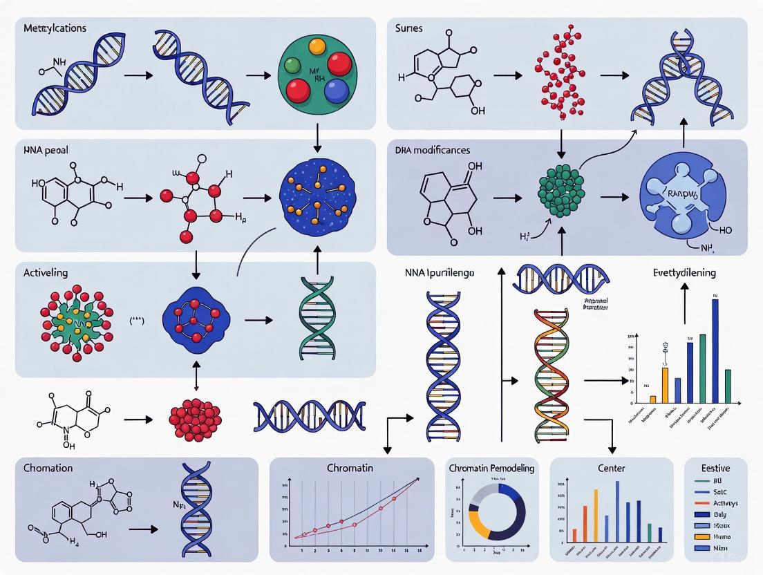
Epigenetics is commonly defined as the study of heritable changes in gene function that cannot be explained by changes in the DNA sequence [1]. These mechanisms provide a molecular framework that links an organism's genome to its environment, enabling cells with identical DNA sequences to establish and maintain distinct identities and functions [2] [1]. The concept of an "epigenetic code" refers to the complex combination of chemical modifications on DNA and histone proteins that dictate how the genetic blueprint is interpreted by the cellular machinery.
This code is written, interpreted, and erased by specialized classes of proteins often termed "writers," "readers," and "erasers" [1]. Writers introduce chemical modifications onto chromatin, erasers remove these modifications, and readers recognize specific epigenetic marks and translate them into functional outcomes by recruiting additional effector proteins [1]. This sophisticated system allows the epigenome to function as a dynamic interface between the static genome and changing environmental cues, with particular relevance in cancer biology where epigenetic dysregulation is a hallmark of disease progression [3] [4].
Core Epigenetic Mechanisms and Their Regulatory Proteins
DNA Methylation
DNA methylation involves the addition of a methyl group to the 5-position of cytosine bases, primarily within CpG dinucleotides [1]. This modification plays crucial biological roles in maintaining genomic stability, X-chromosome inactivation, and regulating gene expression during development and in response to environmental signals [1].
- Writers: DNA methyltransferases (DNMTs) catalyze the transfer of a methyl group from the cofactor S-adenosylmethionine (SAM) to cytosine residues [1]. DNMT3A and DNMT3B establish de novo methylation patterns during embryonic development, while DNMT1 functions as the maintenance methyltransferase, copying methylation patterns during DNA replication by recognizing hemi-methylated CpG sites [1].
- Erasers: The Ten-eleven translocation (TET) family of enzymes (TET1, TET2, TET3) catalyze the oxidation of 5-methylcytosine, initiating an active DNA demethylation pathway through a series of oxidized intermediates that are eventually replaced with an unmodified cytosine via the DNA repair machinery [1] [4].
- Readers: Methyl-CpG-binding domain proteins (MBDs) recognize and bind to methylated DNA. These readers then recruit additional protein complexes, including histone modifiers and chromatin remodelers, to enforce a transcriptionally repressive chromatin state [1].
The establishment and maintenance of DNA methylation patterns are visually summarized in the following diagram:
Histone Modifications
Histone proteins around which DNA is wrapped contain flexible tails that are subject to a wide array of post-translational modifications (PTMs), including acetylation, methylation, phosphorylation, and ubiquitination [1]. These modifications constitute a complex "histone code" that influences chromatin structure and gene activity.
- Writers:
- Histone acetyltransferases (HATs), such as CBP/p300, add acetyl groups to lysine residues, generally leading to a more open chromatin state and gene activation [1] [4].
- Histone methyltransferases (HMTs), such as EZH2 (which catalyzes H3K27me3), add methyl groups to lysine and arginine residues. The functional consequence depends on the specific residue modified and the degree of methylation (mono-, di-, or tri-methylation) [4].
- Erasers:
- Readers: Specific protein domains recognize distinct histone modifications.
The dynamic regulation of the histone code is illustrated below:
Non-Coding RNAs and Chromatin Remodeling
Non-coding RNAs (ncRNAs), including microRNAs (miRNAs), long non-coding RNAs (lncRNAs), and circular RNAs (circRNAs), constitute another layer of epigenetic regulation [5] [4]. They can guide epigenetic complexes to specific genomic loci, leading to targeted gene silencing or activation. Additionally, ATP-dependent chromatin remodeling complexes alter nucleosome positioning and composition, making DNA more or less accessible to the transcriptional machinery [6] [1].
Metabolic Regulation of the Epigenetic Code
Cellular metabolism is intricately linked to epigenetic regulation, as metabolic pathways generate the substrates and cofactors required for epigenetic modifications [3]. This link is particularly evident in cancer, where metabolic reprogramming directly influences the epigenome to drive tumor progression.
- S-adenosylmethionine (SAM): This metabolite is the universal methyl donor for DNA and histone methyltransferases [3]. Its synthesis is tied to one-carbon metabolism, which depends on nutrients like methionine, serine, and folate. The ratio of SAM to S-adenosylhomocysteine (SAH) is critical, as SAH is a potent feedback inhibitor of DNMTs and HMTs [3].
- Acetyl-CoA: This central metabolite serves as the essential substrate for HATs [3]. In cancer cells, pathways such as fatty acid oxidation, glutamine metabolism, and acetate uptake via ACSS2 ensure a steady nuclear supply of acetyl-CoA to support histone acetylation linked to cell growth [3].
- Oncometabolites: Metabolites such as 2-hydroxyglutarate (2-HG), fumarate, and succinate, which accumulate in certain cancers, can function as competitive inhibitors of epigenetic erasers like TET enzymes and histone demethylases, leading to a hypermethylated chromatin state and disrupted cell differentiation [3].
The connection between core metabolism and epigenetic regulation is depicted in the following pathway:
Quantitative Analysis of Epigenetic Components in Cancer
Table 1: Key Epigenetic Enzymes and Their Roles in Cancer
| Enzyme/Protein | Type | Function | Role in Cancer | Therapeutic Inhibitors |
|---|---|---|---|---|
| DNMT1 | Writer | Maintenance DNA methylation | Global hypomethylation & gene-specific hypermethylation [1] | DNMT inhibitors (DNMTi) [4] |
| EZH2 | Writer | Catalyzes H3K27me3 | Overexpressed; silences tumor suppressors [4] | EZH2 inhibitors (e.g., Tazemetostat) [4] |
| HDAC | Eraser | Removes histone acetyl groups | Overexpressed; represses tumor suppressor genes [4] | HDAC inhibitors (e.g., Vorinostat/SAHA) [4] |
| TET2 | Eraser | Initiates DNA demethylation | Frequently mutated; leads to hypermethylation [1] [4] | - |
| BET Proteins | Reader | Bind acetylated histones | Amplified; drives oncogene expression [4] | BET inhibitors [4] |
Table 2: Key Metabolites Regulating the Epigenetic Code
| Metabolite | Epigenetic Function | Linked Enzymes | Impact in Cancer |
|---|---|---|---|
| S-adenosylmethionine (SAM) | Primary methyl donor [3] | DNMTs, HMTs [3] | One-carbon metabolism upregulated; SAM levels drive hypermethylation [3] |
| Acetyl-CoA | Substrate for acetylation [3] | HATs [3] | Elevated ACLY expression increases acetyl-CoA and histone acetylation, promoting growth [3] |
| Nicotinamide adenine dinucleotide (NAD+) | Co-factor for deacetylases [3] | Sirtuins (HDAC Class III) [3] | Altered NAD+ levels affect sirtuin activity, influencing stress response and metabolism [3] |
| 2-Hydroxyglutarate (2-HG) | Competitive inhibitor [3] | TETs, KDMs [3] | "Oncometabolite" from mutant IDH; causes epigenetic block in differentiation [3] |
Experimental Methodologies for Epigenetic Analysis
Cutting-edge technologies are essential for mapping the epigenome and testing functional relationships. Key methodologies include:
- Bisulfite Sequencing (BS-seq): The gold standard for profiling DNA methylation at single-base resolution. Treatment of DNA with bisulfite converts unmethylated cytosines to uracils (read as thymines in sequencing), while methylated cytosines remain unchanged [4].
- Protocol: Extract genomic DNA -> Fragment DNA -> Bisulfite conversion -> Library preparation & Sequencing -> Alignment and analysis of C-to-T conversion rates [4].
- Chromatin Immunoprecipitation Sequencing (ChIP-seq): Identifies genome-wide binding sites for transcription factors or histone modifications. Involves cross-linking proteins to DNA, immunoprecipitating the protein-DNA complexes with a specific antibody, and sequencing the bound DNA fragments [6].
- Single-cell and Spatial Epigenomics:
- scATAC-seq: Assays Transposase-Accessible Chromatin at the single-cell level to reveal cell-to-cell variation in chromatin accessibility [4].
- Spatial Omics (e.g., DBiT-seq): Combates deterministic barcoding to map the spatial distribution of epigenetic marks within tissue contexts, preserving the architecture of the tumor microenvironment [4].
- CRISPR-based Functional Screens:
- Protocol (scCRISPR): Transduce cells with a pooled sgRNA library targeting epigenetic regulators -> Apply selective pressure (e.g., drug treatment) -> Sequence sgRNAs in surviving cells to identify genes essential for survival or drug resistance [4].
The following diagram outlines a typical workflow for integrating these advanced methodologies:
The Scientist's Toolkit: Essential Research Reagents
Table 3: Key Research Reagent Solutions for Epigenetics
| Reagent / Tool | Function / Target | Primary Application |
|---|---|---|
| 5-Azacytidine / Decitabine | DNMT inhibitor (DNMTi) [4] | Demethylating agent; used in research and therapy for AML, MDS [4] |
| Vorinostat (SAHA) | HDAC inhibitor (HDACi) [4] | Induces histone hyperacetylation; used in research and treatment of CTCL [4] |
| GSK126 | EZH2 inhibitor [4] | Suppresses H3K27me3; research tool for studying PRC2 function in cancer models [4] |
| JQ1 | BET bromodomain inhibitor [4] | Blocks reader function; disrupts oncogene expression in preclinical studies [4] |
| Illumina EPIC Array | Genome-wide CpG methylation profiling [6] | DNA methylation analysis from clinical samples (e.g., blood, tissue) [6] |
| Protein A/G Beads | Immunoprecipitation of antibody complexes | Essential for ChIP-seq protocols to pull down protein-DNA complexes [6] |
| LentiCRISPRv2 | Delivery of sgRNA and Cas9 | CRISPR-based knockout of epigenetic writers, erasers, and readers in cell lines [4] |
Concluding Perspectives
The writers, erasers, and readers of the epigenetic code collectively orchestrate a dynamic and responsive regulatory system that is fundamental to cellular identity and function. In cancer, this system is profoundly dysregulated, contributing to unchecked proliferation, immune evasion, and therapeutic resistance [4]. The intimate link between cellular metabolism and the epigenome further reveals how nutrient availability and oncogenic mutations can reshape the epigenetic landscape to favor tumor growth [3].
Future research, powered by single-cell and spatial omics technologies, will continue to decode the complexity of the epigenetic code in the context of the tumor microenvironment [4]. This will accelerate the development of novel epigenetic therapies, such as targeted degraders and epigenome editors, and guide their rational combination with immunotherapies to achieve durable responses in cancer patients [4].
Epigenetics, a term coined by Conrad Waddington in 1942, refers to the study of heritable changes in gene expression that do not involve alterations to the underlying DNA sequence [7] [8]. Among epigenetic mechanisms, DNA methylation stands as a fundamental regulator of gene expression, cellular identity, and genome stability. In the context of cancer, aberrant DNA methylation patterns are recognized as a hallmark of malignancy, contributing significantly to oncogenesis [7]. The cancer methylation landscape is characterized by a paradoxical combination of global genomic hypomethylation alongside focal hypermethylation at specific regulatory regions, particularly the promoter CpG islands of tumor suppressor genes [7] [8]. These coordinated yet opposing changes disrupt normal gene expression patterns, silencing protective tumor suppressor mechanisms while activating oncogenic pathways, thereby driving cancer initiation and progression.
This technical review examines the dual nature of DNA methylation alterations in cancer, exploring the molecular mechanisms, functional consequences, and clinical implications. We frame these concepts within the broader context of epigenetic modifications in cancer progression research, providing researchers and drug development professionals with a comprehensive analysis of current understanding and emerging therapeutic opportunities. The dynamic interplay between genetic and epigenetic alterations creates a complex regulatory network that influences tumor evolution, metastatic behavior, therapeutic resistance, and potential intervention strategies [7] [9].
Molecular Machinery of DNA Methylation
Writers, Readers, and Erasers of Methylation Marks
The establishment, maintenance, and interpretation of DNA methylation patterns are orchestrated by specialized enzymatic complexes that function as "writers," "readers," and "erasers" of epigenetic information [7] [8].
DNA Methyltransferases (DNMTs - Writers): The de novo methyltransferases DNMT3A and DNMT3B establish initial methylation patterns during embryogenesis, while DNMT1 maintains these patterns during cellular replication by copying methylation marks to the daughter DNA strand [7] [8]. These enzymes catalyze the transfer of a methyl group from S-adenosyl methionine (SAM) to the fifth carbon of cytosine bases, primarily within cytosine-guanine (CpG) dinucleotides, forming 5-methylcytosine (5mC) [7] [8].
Methyl-CpG-Binding Proteins (MBPs - Readers): Proteins including MeCP2 and MBD family members recognize and bind to methylated CpG sites, recruiting additional chromatin-modifying complexes that promote transcriptional repression [8]. UHRF1/2 plays a critical role in recruiting DNMT1 to hemimethylated DNA, and its overexpression contributes to tumor suppressor gene silencing in various cancers [8].
Ten-Eleven Translocation (TET) Enzymes (Erasers): TET enzymes catalyze the oxidation of 5mC to 5-hydroxymethylcytosine (5hmC) and further oxidized derivatives, initiating an active DNA demethylation pathway through base excision repair (BER) mechanisms [7] [8]. TET2 loss-of-function mutations impair active demethylation and are associated with various cancers [8].
Dysregulated Machinery in Oncogenesis
In cancer, components of the DNA methylation machinery are frequently dysregulated. DNMT1 is often overexpressed, leading to aberrant hypermethylation, while TET enzyme mutations result in accumulated aberrant methylation marks [8]. UHRF1/2 overexpression promotes silencing of tumor suppressor genes and enhances migratory and metastatic capabilities in cancer cells [8]. This dysregulation creates an epigenetic landscape conducive to malignant transformation by simultaneously silencing tumor suppressor genes and activating oncogenic pathways.
Global DNA Hypomethylation in Cancer
Mechanisms and Genomic Targets
Global DNA hypomethylation primarily affects repetitive elements, gene-poor regions, and intragenic areas, leading to genomic instability and aberrant gene activation [8]. This widespread loss of methylation occurs through several mechanisms:
- Passive demethylation resulting from failures in maintenance methylation during DNA replication [7].
- Active demethylation mediated by TET enzymes [7].
- DNMT1 disruption, which causes hypomethylation and chromosomal instability that can drive cancer development [8].
Hypomethylation is particularly prominent in partially methylated domains (PMDs), which are gene-poor megabase-scale regions that become hypomethylated in many cancers, similar to patterns observed during aging and long-term cell culture [10]. The extent of global hypomethylation varies across cancer types, being most pronounced in colorectal, bladder, and liver carcinomas, while virtually absent in acute leukemias, thymoma, and thyroid carcinoma [10].
Functional Consequences
The functional impacts of global hypomethylation are multifaceted and contribute significantly to oncogenesis:
- Chromosomal Instability: Hypomethylation of repetitive elements and pericentromeric regions promotes chromosomal rearrangements, translocations, and mitotic errors [8].
- Oncogene Activation: Hypomethylation can activate growth-promoting genes and proto-oncogenes. For example, androgen-response genes in prostate cancer show hypomethylation compared to normal tissues, contributing to disease progression [8].
- Loss of Imprinting: Hypomethylation can disrupt genomic imprinting, leading to biallelic expression of growth-promoting genes [8].
- Transposable Element Derepression: Hypomethylation activates transposable elements like LINE1, which can disrupt gene function and promote tumor formation [8]. LINE1 hypomethylation has been reported in multiple cancers and may serve as a biomarker [8].
Table 1: Functional Consequences of Global DNA Hypomethylation in Cancer
| Genomic Target | Functional Consequence | Example/Cancer Type |
|---|---|---|
| Repetitive Elements | Chromosomal instability | Various solid tumors |
| Pericentromeric Regions | Mitotic errors & aneuploidy | Various cancers |
| Gene Promoters | Oncogene activation | Prostate cancer (androgen-response genes) |
| Imprinted Control Regions | Loss of imprinting | Various pediatric and adult cancers |
| Transposable Elements | Retrotransposition & insertional mutagenesis | LINE1 in multiple cancers |
Focal Hypermethylation in Cancer
Targets and Patterns
In stark contrast to global hypomethylation, focal hypermethylation targets CpG-rich regions, particularly promoters of tumor suppressor genes, developmental regulators, and other cancer-relevant genes [7] [8]. This targeted hypermethylation exhibits distinct patterns:
Polycomb Target Preference: Promoters regulated by Polycomb repressive complex 2 (PRC2) in embryonic stem cells are preferentially hypermethylated in cancer [10]. These developmental regulators are often maintained in a transcriptionally poised, unmethylated state in normal adult tissues but become aberrantly methylated in malignancies [10].
Tumor Type-Specific Patterns: The specific repertoire of hypermethylated genes varies between cancer types, reflecting both the tissue of origin and the molecular subtype [10]. For example, a comprehensive analysis identified between 14 (thyroid carcinoma) and over 3000 (T-ALL) commonly hypermethylated CpG islands across 26 tumor types [10].
Pan-Cancer Hypermethylation: Despite tumor-specific patterns, 1,579 CpG islands are consistently hypermethylated across multiple cancer types, representing a "pan-cancer hyper CGI" signature enriched for neural lineage genes, possibly due to their PRC2 regulation in most non-neural tissues [10].
Functional Consequences
Promoter hypermethylation contributes to oncogenesis through several mechanisms:
Tumor Suppressor Gene Silencing: Hyper-methylation of promoters creates a repressive chromatin state that obstructs transcription factor binding and inhibits gene transcription, effectively silencing tumor suppressor genes [7] [8]. Classic examples include VHL, BRCA1, STK11, MLH1, and MGMT, which are epigenetically silenced in sporadic cancers despite being linked to familial cancer syndromes when mutated [8].
Disruption of Cellular Identity: Hypermethylation of developmental regulator genes controlled by Polycomb complexes locks cells in undifferentiated, proliferative states [10].
Interplay with Genetic Alterations: DNA methylation cooperates with genomic alterations during tumor evolution. In non-small cell lung cancer (NSCLC), hypermethylation shows parallel evolution with copy number loss, particularly affecting tumor suppressor genes in lung squamous cell carcinoma [9].
Dynamics During Multistep Carcinogenesis
DNA methylation alterations occur progressively throughout cancer development, with distinct changes characterizing different stages of oncogenesis. Recent research has delineated the timing and patterns of these epigenetic changes during colorectal cancer progression, providing insights into the dynamics of methylation alterations [11].
Temporal Patterns in Colorectal Carcinogenesis
A comprehensive analysis of methylation changes during colorectal oncogenesis revealed that nearly 12% of the colon methylome is significantly altered (q < 10^(-4)) during the transition from normal tissue to adenocarcinoma, with half of these changes representing demethylation events [11]. The majority (67.4%) of these methylation changes occur during the initial transition from non-tumor colon tissue to low-grade adenoma, highlighting the importance of early epigenetic alterations in tumor initiation [11].
Approximately 9% of DNA methylation changes are specific to low-grade and/or high-grade adenomas, representing transient epigenetic events that may facilitate early tumor development without being maintained in advanced cancers [11]. Biological pathways affected during this stepwise progression include early hypomethylation of genes involved in sensory perception of odor and stimulus, early hypermethylation of nucleic acid metabolic processes, transient hypomethylation of post-transcriptional regulation, and transient hypermethylation of mitotic cell cycle pathways [11].
Intratumoral Heterogeneity and Evolution
DNA methylation heterogeneity within individual tumors contributes to cancer evolution and adaptation. In non-small cell lung cancer, intratumoral methylation distance (ITMD) quantifies methylation heterogeneity between different regions of the same tumor [9]. This heterogeneity correlates with somatic copy number alteration heterogeneity and intratumoral expression distance, indicating coordinated genomic and epigenomic evolution [9].
Methylation heterogeneity is highest in intergenic and enhancer regions, while promoter regions show significantly lower variability, suggesting tighter regulation of promoter methylation [9]. This heterogeneity creates diverse cellular subpopulations within tumors that may differ in their malignant potential, therapeutic sensitivity, and metastatic capability.
Table 2: Dynamics of DNA Methylation Changes During Colorectal Cancer Progression
| Transition Stage | Percentage of Total Methylation Changes | Key Biological Pathways Affected | Biomarker Implications |
|---|---|---|---|
| Normal → Low-Grade Adenoma | 67.4% | Sensory perception (hypomethylation), Nucleic acid metabolic process (hypermethylation) | 21/34 known biomarkers already methylated |
| Low-Grade → High-Grade Adenoma | Progressive accumulation | Post-transcriptional regulation (transient hypomethylation), Mitotic cell cycle (transient hypermethylation) | Additional 11/34 biomarkers become methylated |
| Adenoma → Adenocarcinoma | Remaining changes | Stable maintenance of key early and intermediate changes | Full biomarker panel methylated |
Technical Approaches and Research Reagents
Methodological Considerations
Accurate assessment of DNA methylation patterns requires careful methodological considerations. The bisulfite conversion-based methods are considered the gold standard but present specific challenges:
Input DNA Quantity: Excessive DNA input (e.g., 1μg) can lead to incomplete bisulfite conversion, particularly for high-copy number regions like rDNA promoters, potentially resulting in underestimation of methylation levels [12]. Optimal conversion typically requires minimal DNA input (e.g., 1ng) [12].
Conversion Efficiency Controls: For quantitative analyses, rigorous controls for complete bisulfite conversion are essential, particularly for repetitive elements or highly methylated regions [12].
Tumor Purity Considerations: Computational deconvolution approaches like Copy number-Aware Methylation Deconvolution Analysis of Cancers (CAMDAC) help account for variable tumor purity and copy number alterations in bulk tumor samples [9].
Research Reagent Solutions
Table 3: Essential Research Reagents and Methods for DNA Methylation Analysis
| Reagent/Method | Function/Application | Technical Considerations |
|---|---|---|
| EPIC v1 Human Methylation BeadChip | Genome-wide methylation profiling (850,000 CpG sites) | Platform used for colorectal adenoma methylome analysis [11] |
| Reduced Representation Bisulfite Sequencing (RRBS) | Cost-effective genome-wide methylation analysis | Used in TRACERx NSCLC study; covers ~1-3 million CpGs [9] |
| Whole-Genome Bisulfite Sequencing (WGBS) | Comprehensive base-resolution methylation analysis | Gold standard for novel biomarker discovery; used for model benchmarking [10] |
| Quantitative Methylation-Specific PCR (qMSP) | Targeted methylation quantification | Used for rDNA promoter methylation analysis; requires careful bisulfite conversion control [12] |
| CAMDAC Algorithm | Copy number-aware deconvolution of bulk tumor methylation | Accounts for tumor purity and CN alterations in methylation analysis [9] |
| 5-azacytidine/5-aza-2'-deoxycytidine | DNMT inhibitors for functional studies | Demethylating agents used to validate methylation-dependent gene regulation [13] [8] |
Diagnostic and Therapeutic Applications
Methylation Biomarkers in Cancer
DNA methylation patterns provide valuable biomarkers for cancer detection, classification, and prognosis:
Colorectal Cancer Biomarkers: Multiple methylated genes show utility for colorectal cancer detection, including SDC2, ADHFE1, PPP2R5C, SEPT9, BMP3, KCNQ5, C9orf50, CLIP4, SNCA, FBN1, GATA5, SFRP2, HAND1, and LINC00473 [11]. These biomarkers can be detected in stool, blood, or tissue samples, offering non-invasive screening options.
Prostate Cancer Diagnostics: GSTP1 promoter hypermethylation demonstrates high diagnostic performance (AUC = 0.939) for distinguishing prostate cancer from benign tissue [13]. Panels combining multiple methylated genes (e.g., GSTP1 and CCND2) can further improve diagnostic accuracy [13].
Liquid Biopsy Applications: Methylation biomarkers are particularly suitable for liquid biopsy approaches due to the stability of DNA methylation patterns in circulating cell-free DNA [13]. This enables non-invasive cancer detection, monitoring of minimal residual disease, and assessment of treatment response [13].
Therapeutic Targeting of DNA Methylation
Several therapeutic strategies target aberrant DNA methylation in cancer:
DNMT Inhibitors: Azacitidine and decitabine inhibit DNMTs, causing DNA hypomethylation and re-expression of silenced tumor suppressor genes [7] [8]. While approved for hematological malignancies, their efficacy in solid tumors remains limited [14].
Novel Epigenetic Approaches: Emerging strategies target other components of the methylation machinery. For example, targeting UHRF1, a protein that recruits DNMT1 to sites of DNA methylation, shows promise for reactivating tumor suppressor genes [14]. The mouse STELLA protein effectively binds UHRF1 and impairs tumor growth in colorectal cancer models, suggesting a potential therapeutic avenue [14].
Combination Therapies: Epigenetic therapies may enhance the efficacy of other treatment modalities, including immunotherapy, chemotherapy, and targeted therapies, by reversing epigenetic silencing of key response mediators [7].
Experimental Workflows and Signaling Pathways
DNA Methylation Analysis Workflow
The following diagram illustrates a comprehensive workflow for analyzing DNA methylation in cancer research, integrating multiple experimental and computational approaches:
DNA Methylation Machinery in Oncogenesis
The molecular machinery governing DNA methylation patterns represents a complex network of writers, readers, and erasers that become dysregulated in cancer:
The dual nature of DNA methylation alterations in cancer—global hypomethylation coupled with focal hypermethylation—represents a fundamental aspect of oncogenesis with far-reaching basic research and clinical implications. These coordinated epigenetic changes disrupt normal gene expression patterns, promote genomic instability, and contribute to malignant transformation and progression. Understanding the dynamic interplay between genetic and epigenetic alterations, as well as the temporal patterns of methylation changes during multistep carcinogenesis, provides crucial insights into cancer biology.
Advances in methylation profiling technologies, computational deconvolution methods, and novel therapeutic approaches continue to enhance our ability to study and target these epigenetic alterations. The development of sensitive and specific methylation biomarkers for early detection, prognosis, and monitoring represents a promising avenue for improving cancer clinical management. Furthermore, emerging epigenetic therapies that target components of the methylation machinery offer new opportunities for personalized cancer treatment. As research in this field progresses, integrating DNA methylation profiling into comprehensive molecular analyses will undoubtedly yield deeper insights into cancer evolution and identify novel therapeutic vulnerabilities for precision oncology approaches.
Epigenetic dysregulation, particularly in histone modifications and chromatin remodeling, is a hallmark of cancer. These reversible alterations govern chromatin architecture and gene expression without changing the underlying DNA sequence, playing critical roles in tumor initiation, progression, and therapeutic resistance. This technical review examines the intricate mechanisms of histone acetylation, methylation, and other post-translational modifications, alongside the function of ATP-dependent chromatin remodeling complexes in oncogenesis. Within the broader thesis of epigenetic modifications in cancer progression, we explore how aberrant enzymatic activities disrupt normal cellular homeostasis, leading to malignant transformation. The review also assesses current experimental methodologies, quantitative biomarker assessments, and emerging therapeutic strategies targeting epigenetic regulators, providing a comprehensive resource for research and drug development professionals navigating this rapidly advancing field.
The concept of cancer as a disease of both genetic and epigenetic alterations has gained substantial momentum within the scientific community. Epigenetics encompasses heritable phenotypes not encoded by the DNA sequence, primarily mediated through DNA methylation, histone modifications, chromatin remodeling, and non-coding RNA interactions [15]. Histone modifications represent versatile covalent marks on histone tails that dynamically regulate chromatin structure and gene accessibility. The balance between various histone modifications is crucial for maintaining proper gene expression programs, and its disruption primes cells for malignant transformation [16] [17]. Beyond histone modifications, ATP-dependent chromatin remodeling complexes like SWI/SNF utilize energy from ATP hydrolysis to mobilize nucleosomes, exposing genomic regions for transcriptional regulation [18]. The intricate interplay between these epigenetic mechanisms creates a complex regulatory network that, when dysregulated, drives tumorigenesis through inappropriate activation of oncogenes and silencing of tumor suppressors.
Histone Modifications: Mechanisms and Biological Consequences
Histone modifications are post-translational changes to histone proteins, particularly their N-terminal tails, that alter DNA-histone interactions and serve as docking sites for reader proteins. The well-characterized modifications include:
- Methylation: Transfer of methyl groups to lysine (K) or arginine (R) residues
- Acetylation: Addition of acetyl groups to lysine residues
- Phosphorylation: Addition of phosphate groups to serine (S), threonine (T), or tyrosine (Y) residues
- Ubiquitination: Attachment of ubiquitin to lysine residues [19]
Novel modifications continue to be identified, including citrullination, crotonylation, succinylation, propionylation, butyrylation, 2-hydroxyisobutyrylation, and 2-hydroxybutyrylation [20], though their functional significance in cancer is still being elucidated.
Histone Acetylation
Histone acetylation involves the addition of acetyl groups to lysine residues, neutralizing their positive charge and reducing histone-DNA affinity. This modification is dynamically regulated by histone acetyltransferases (HATs) and histone deacetylases (HDACs). Acetylation is generally associated with active transcription, particularly at enhancers, promoters, and gene bodies [17]. Global alterations in histone acetylation patterns are linked to cancer phenotypes, with hypoacetylation of histone H4 at lysine 16 being a common feature across various cancers [17]. The p300 and CBP HATs demonstrate dual roles in cancer, functioning as tumor suppressors in hematological malignancies but potentially exhibiting oncogenic properties in certain contexts [19]. HDAC overexpression enhances tumor development and metastasis, making them attractive therapeutic targets [19].
Histone Methylation
Histone methylation occurs on both lysine and arginine residues, with lysine capable of mono-, di-, or trimethylation. Unlike acetylation, methylation does not alter histone charge but serves as recognition motifs for specific reader proteins. The functional outcome depends on the modified residue, methylation degree, and genomic context:
| Modification | Transcriptional Status | Catalytic Enzymes | Cancer Associations |
|---|---|---|---|
| H3K4me2/3 | Active | SET1A/B, MLL1-4 (KMT2 family) | SET1A promotes metastasis in breast, lung, colorectal cancers [16] |
| H3K36me3 | Active | NSD1-3 (KMT3 family) | - |
| H3K79me | Active | DOT1L (KMT4 family) | - |
| H3K9me2/3 | Repressive | SUV39H1/2, G9a, GLP, SETDB1 (KMT1 family) | G9a promotes lung cancer invasion [15] |
| H3K27me3 | Repressive | EZH1/2 (KMT6 family) | EZH2 highly expressed in prostate, breast cancers [15] [16] |
| H4K20me3 | Repressive | PR-Set7, SUV4-20H1/2 (KMT5 family) | Global loss in cancer cells [15] |
Histone demethylases (KDMs) reverse methylation marks, with at least six families identified. The KDM1 family includes LSD1 (KDM1A) and LSD2 (KDM1B), while KDM2-6 families contain JmjC domain-containing demethylases [16]. These enzymes are increasingly recognized as contributing factors in multiple cancers and represent promising therapeutic targets [21].
Other Histone Modifications
Histone phosphorylation alters chromatin structure by adding negative charges to serine, threonine, and tyrosine residues, influencing transcription regulation, cell cycle progression, and DNA damage response. H3S10 phosphorylation is associated with transcriptional activation and is considered a potential cancer biomarker [19].
Histone ubiquitination primarily affects H2A and H2B. H2AK119ub often accompanies H3K27me3 through Polycomb repressive complex activities, while H2BK120ub activates gene transcription and is a prerequisite for H3K4 and H3K79 methylation [19].
Chromatin Remodeling Complexes in Tumorigenesis
The SWI/SNF Complex
The SWI/SNF complex represents a crucial chromatin remodeling machinery that utilizes ATP hydrolysis to mobilize nucleosomes, altering chromatin accessibility for transcription factors and regulatory elements [18]. This complex exists in three distinct variants: canonical BAF (cBAF), polybromo-associated BAF (PBAF), and non-canonical BAF (ncBAF), each with unique subunit compositions and biological functions [18].
Mutations in SWI/SNF complex subunits occur at high frequencies across diverse human cancers. The complex functions as a master regulator of chromatin architecture, with specific subunits demonstrating critical tumor suppressor activities:
| SWI/SNF Subunit | Cancer Types with Frequent Mutations | Functional Consequences |
|---|---|---|
| ARID1A | Ovarian clear cell carcinoma (OCCC; >50%), gastric, breast | Endocrine therapy resistance, impaired chromatin remodeling, biomarker for immunotherapy [18] |
| SMARCB1 (SNF5/INI1) | Malignant rhabdoid tumors (MRT) | Tumor suppressor; loss drives aggressive cancers [18] [22] |
| PBRM1 | Clear cell renal cell carcinoma (ccRCC) | Associated with advanced stage, higher tumor grade [18] |
| ARID1B | ER+ breast cancer | Synthetic lethal partner with ARID1A; inactivation promotes proliferation [18] |
| SMARCA2/4 | Pancreatic cancer, DLBCL | Tumor suppressor; regulates stem cell pathways including Wnt/β-catenin [18] |
The synthetic lethal relationship between specific SWI/SNF subunits offers promising therapeutic opportunities. For instance, ARID1A and ARID1B form a synthetic lethal pair, where ARID1A loss renders cancer cells dependent on ARID1B for survival [18]. Similarly, BRM/SMARCA2 depletion induces cell cycle arrest in BRG1/SMARCA4-deficient cancer cells [18].
Experimental Methodologies for Epigenetic Analysis
Chromatin Immunoprecipitation (ChIP) Assays
Chromatin Immunoprecipitation remains the gold standard for mapping histone modifications and transcription factor binding sites genome-wide. The standard protocol involves:
- Cross-linking: Formaldehyde treatment to fix protein-DNA interactions
- Cell Lysis and Chromatin Shearing: Sonication or enzymatic digestion to fragment chromatin
- Immunoprecipitation: Incubation with specific antibodies against histone modifications
- Cross-link Reversal and DNA Purification
- Downstream Analysis: qPCR (ChIP-qPCR), microarray (ChIP-chip), or sequencing (ChIP-seq)
Recent advances utilizing ChIP-sequencing technologies have enabled genome-wide mapping of chromatin changes during tumorigenesis, revealing characteristic histone modification patterns in cancer cells [15].
Epigenetic Drug Screening Assays
Screening for epigenetic therapeutics employs various methodologies:
- Cell Viability Assays (MTT, XTT, WST-1) to assess anti-proliferative effects
- Flow Cytometry for cell cycle analysis and apoptosis detection
- Western Blotting to monitor changes in global histone modification levels
- RNA-seq to transcriptomically evaluate gene expression changes
- Combination Studies with conventional chemotherapeutics to identify synergistic effects
High-throughput screening platforms have accelerated the discovery of small-molecule inhibitors targeting epigenetic regulators, with several compounds advancing to clinical trials [21].
Quantitative Histone Modification Analysis
Global histone modification levels can be quantified using:
- Immunohistochemistry (IHC) on tissue microarrays
- Enzyme-Linked Immunosorbent Assay (ELISA)
- Mass Spectrometry for precise quantification of modification stoichiometry
These approaches have identified specific histone modification patterns as potential prognostic biomarkers in multiple cancers [15].
Quantitative Data and Clinical Correlations
Histone Modifications as Prognostic Biomarkers
Substantial clinical evidence supports the prognostic value of specific histone modifications:
| Cancer Type | Histone Modification | Prognostic Significance |
|---|---|---|
| Lung | H3K4me2, H3K9ac, H3K18ac, H4K16ac | Predictive of clinical outcomes [15] |
| Prostate | H3K4me2, H3K18ac, H4K20me1/2, H3K27me3 | Correlation with disease progression [15] |
| Breast | H3K27me3 | EZH2 overexpression associated with poor prognosis [15] |
| Multiple Cancers | H4K16ac | Global loss associated with cancer phenotype [17] |
| Leukemia | H3K79me | Aberrant patterns in fusion protein-driven leukemias [16] |
Mutation Frequencies in Epigenetic Regulators
Genomic analyses reveal significant mutation frequencies in epigenetic regulators across cancers:
- SWI/SNF complex mutations: >20% of human cancers across tumor types [19]
- ARID1A mutations: 57% of ovarian clear cell carcinomas [18]
- EZH2 mutations and dysregulation: frequent in prostate and breast cancers [15]
- DNMT3A mutations: common in hematological malignancies [7]
Visualization of Key Pathways
Histone Modification Crosstalk in Transcription Regulation
SWI/SNF Complex in Chromatin Remodeling and Cancer
Research Reagent Solutions
Essential research tools for investigating histone modifications and chromatin remodeling:
| Reagent Category | Specific Examples | Research Applications |
|---|---|---|
| Histone Modification Antibodies | Anti-H3K4me3, Anti-H3K27me3, Anti-H3K9ac, Anti-H4K16ac | ChIP, IHC, Western blot for mapping modification patterns |
| Epigenetic Inhibitors | HDACi (Vorinostat), EZH2i (Tazemetostat), LSD1i, BET inhibitors | Functional studies, therapeutic screening, combination therapies |
| Cell Line Models | SWI/SNF-mutant lines (ARID1A^-^, SMARCB1^-^), Patient-derived organoids (PDOs) | Mechanistic studies, drug screening, personalized medicine approaches |
| Sequencing Kits | ChIP-seq, ATAC-seq, Whole Genome Bisulfite Sequencing | Epigenomic profiling, chromatin accessibility mapping |
| Activity Assays | HDAC/HAT Fluorescent Activity Assays, HMT Colorimetric Kits | Enzyme activity quantification, inhibitor screening |
| Mass Spectrometry Standards | Synthetic histone peptides with specific modifications | Quantitative modification analysis, calibration |
Therapeutic Implications and Future Directions
The reversible nature of epigenetic modifications presents compelling therapeutic opportunities. Several epigenetic drugs have received FDA approval, including HDAC inhibitors (vorinostat, istodax, beleodap, panobinostat) and EZH2 inhibitors [19]. Current research focuses on developing more selective inhibitors, particularly for histone demethylases and reader domains [21]. Combination therapies represent a promising avenue, with epigenetic drugs showing potential to sensitize tumors to conventional chemotherapy, targeted therapy, and immunotherapy [20]. The application of multi-omics technologies and single-cell approaches will further refine our understanding of epigenetic heterogeneity in tumors, enabling more precise therapeutic interventions [20]. As epigenetic profiling advances, the integration of histone modification biomarkers into clinical decision-making promises to enhance patient stratification and treatment selection in oncology.
This technical review supports the broader thesis that epigenetic modifications are fundamental drivers of cancer progression, representing dynamic regulatory layers that interface with genetic alterations to shape malignant phenotypes. The continued elucidation of these mechanisms will undoubtedly yield novel diagnostic and therapeutic approaches in clinical oncology.
Non-coding RNAs (ncRNAs) have emerged as pivotal epigenetic regulators in cancer biology, orchestrating gene expression without altering the underlying DNA sequence. This review synthesizes current knowledge on how microRNAs (miRNAs), long non-coding RNAs (lncRNAs), and circular RNAs (circRNAs) contribute to epigenetic remodeling in cancer progression. We explore their roles in regulating DNA methylation, histone modifications, and chromatin architecture, highlighting the complex ceRNA networks that influence oncogene activation, tumor suppressor silencing, and therapeutic resistance. The integration of quantitative data and experimental methodologies provides a resource for researchers and drug development professionals aiming to target the epigenetic landscape of cancer.
Epigenetics, the study of heritable changes in gene expression that do not involve changes to the underlying DNA sequence, is a critical field in understanding cancer progression. Key mechanisms include DNA methylation, histone modifications, and chromatin remodeling [7]. Notably, non-coding RNAs (ncRNAs), which constitute the majority of the human transcriptome, have been identified as master regulators of these epigenetic processes [23] [20]. In cancer, the interplay between different classes of ncRNAs creates complex regulatory networks that drive tumorigenesis, metastasis, and metabolic reprogramming [23] [24]. This review focuses on three major ncRNAs—miRNAs, lncRNAs, and circRNAs—detailing their functions as epigenetic modulators, their roles in competitive endogenous RNA (ceRNA) networks, and their potential as therapeutic targets.
MicroRNAs (miRNAs) as Epigenetic Regulators
Biogenesis and Mechanism of Action
miRNAs are small non-coding RNAs, approximately 20-22 nucleotides in length, that primarily regulate gene expression post-transcriptionally. They guide the RNA-induced silencing complex (RISC) to target mRNAs, leading to translational repression or mRNA degradation [24]. A single miRNA can target hundreds of mRNAs, enabling the coordinated regulation of entire signaling pathways.
Epi-miRNAs and Their Roles in Cancer
A subset of miRNAs, known as epi-miRNAs, directly targets the expression of key epigenetic regulators, including DNA methyltransferases (DNMTs), histone deacetylases (HDACs), and histone demethylases (KDMs) [23]. The table below summarizes the functions of well-characterized epi-miRNAs in cancer.
Table 1: Key Epi-miRNAs in Cancer Epigenetic Regulation
| miRNA | Epigenetic Target | Resulting Effect | Role in Cancer |
|---|---|---|---|
| miR-29b | DNMT3A, TET enzymes | Silencing of PTEN; altered glycolytic metabolism | Tumor Suppressor [23] |
| miR-138 | KDM5B (Lysine demethylase) | Downregulation of FASN, ACLY; suppressed proliferation & migration | Tumor Suppressor [23] |
| miR-137 | LSD1 (Lysine-specific demethylase 1A) | Inhibition of Warburg effect (glycolysis) & stabilization of HIF-1α | Tumor Suppressor [23] |
| miR-155 | KDM2A (under hypoxia) | Limits ROS production, regulates mitochondrial gene expression | Context-Dependent [23] |
| miR-143 | DNMT3A | Modulates pro-glycolytic genes (e.g., HK, GLUT1) in immune cells | Tumor Suppressor [23] |
Long Non-Coding RNAs (lncRNAs) and Circular RNAs (circRNAs)
Characteristics and General Functions
lncRNAs are defined as transcripts longer than 200 nucleotides with limited or no protein-coding capacity. They function as scaffolds, decoys, or guides to regulate transcription, mRNA processing, and nuclear organization [24]. circRNAs are a more recently characterized class of ncRNA that form covalently closed, continuous loops. This structure makes them highly stable and resistant to RNase degradation [24]. Both lncRNAs and circRNAs can function as competing endogenous RNAs (ceRNAs) by sequestering miRNAs, thereby de-repressing the miRNA's downstream mRNA targets [24].
The ceRNA Hypothesis and Epigenetic Cross-talk
The ceRNA hypothesis describes a sophisticated regulatory network where lncRNAs and circRNAs act as molecular sponges for miRNAs. This competition indirectly regulates the expression of genes targeted by the sequestered miRNA, including those encoding epigenetic regulators [24]. The following diagram illustrates a core ceRNA network and its downstream epigenetic effects.
Hepatocellular carcinoma (HCC) provides a well-studied model for understanding ceRNA networks. The table below compiles specific lncRNA-miRNA-mRNA axes and their functional consequences in HCC progression.
Table 2: Selected ceRNA Networks in Hepatocellular Carcinoma (HCC) [24]
| lncRNA/circRNA | Sponged miRNA | Deregulated mRNA/Gene | Biological Function in HCC |
|---|---|---|---|
| LncRNA SNHG11 | miR-184 | AGO2 | Proliferation, migration, autophagy |
| LncRNA CCAT1 | let-7 | HMGA2, c-MYC | Proliferation, migration |
| LncRNA H19 | miR-326 | TWIST | Proliferation, metastasis |
| LncRNA MALAT1 | miR-30a-5p | Vimentin | Proliferation, metastasis |
| LncRNA NEAT1 | miR-204 | ATG3 | Autophagy process |
| LncRNA GAS5 | miR-21 | PTEN | Tumor suppression |
Experimental Protocols for Investigating ncRNA Epigenetics
Studying the epigenetic functions of ncRNAs requires a multidisciplinary approach. The following section outlines key methodologies.
Workflow for Validating a ceRNA Axis
A rigorous, multi-step workflow is essential to confirm a proposed ceRNA interaction.
Step 1: In Silico Prediction. Utilize bioinformatics tools (e.g., TargetScan, miRanda, StarBase) to predict miRNA response elements (MREs) on the candidate lncRNA/circRNA and the putative target mRNAs. Step 2: Expression Correlation. Analyze RNA-seq or qRT-PCR data from patient cohorts or cell lines to determine if a significant negative correlation exists between the lncRNA/circRNA and the miRNA, and a positive correlation between the lncRNA/circRNA and the target mRNA. Step 3: Functional Sponging Validation.
- Luciferase Reporter Assay: Clone the wild-type and MRE-mutated sequence of the lncRNA/circRNA into a reporter plasmid. Co-transfect with the miRNA mimic and measure luciferase activity. A significant reduction in activity for the wild-type, but not the mutant, confirms direct binding.
- RNA Immunoprecipitation (RIP): Use antibodies against Argonaute 2 (AGO2), a core component of RISC, to immunoprecipitate RNA-protein complexes. Enrichment of both the miRNA and the lncRNA/circRNA in the precipitate indicates they reside in the same RISC complex.
- Biotin-labeled miRNA Pulldown: Transfect cells with a biotin-tagged miRNA mimic. Use streptavidin-coated beads to pull down the miRNA and its bound RNAs. Detect the co-precipitated lncRNA/circRNA via qRT-PCR. Step 4: Phenotypic Rescue Experiments. Perform functional assays (e.g., proliferation, migration, apoptosis) after modulating the expression of the lncRNA/circRNA and the miRNA. The phenotypic effects of lncRNA/circRNA overexpression should be reversible by co-transfecting the corresponding miRNA. Step 5: Downstream Target Validation. Confirm that the expression level of the final mRNA target and its encoded protein changes as predicted by the ceRNA model, using qRT-PCR and western blotting.
Table 3: Key Research Reagent Solutions for ncRNA Epigenetics
| Reagent / Tool | Function / Application | Key Considerations |
|---|---|---|
| miRNA Mimics & Inhibitors | Functionally increase or decrease specific miRNA activity in cells. | Controls: Scrambled sequence mimics/inhibitors. Monitor off-target effects. |
| si/shRNA for lncRNA/circRNA | Knock down specific lncRNAs. | Target backsplicing junction for circRNA-specific knockdown. |
| Luciferase Reporter Vectors (e.g., pmirGLO) | Quantitatively validate direct miRNA-ncRNA binding. | Always include MRE-mutated controls. Normalize to a control reporter (e.g., Renilla). |
| AGO2 Antibodies | For RIP assays to confirm RISC complex association. | Use validated antibodies for RIP; include IgG isotype controls. |
| Biotin-labeled Probes | For pulldown of specific ncRNAs and their interacting partners. | High purity and specific activity are critical. |
| RNA-Seq & Small RNA-Seq | Transcriptome-wide discovery of differentially expressed ncRNAs and mRNAs. | Include multiple biological replicates. Plan for sophisticated bioinformatic analysis. |
The intricate interplay between miRNAs, lncRNAs, and circRNAs constitutes a critical layer of epigenetic regulation in cancer. Through mechanisms such as miRNA-directed suppression and ceRNA network activity, these ncRNAs fine-tune the expression of epigenetic writers, readers, and erasers, thereby shaping the tumor epigenome [23] [7] [20]. The inherent reversibility of epigenetic modifications makes these ncRNAs and their downstream effectors attractive therapeutic targets. Promising strategies include antisense oligonucleotides to inhibit oncogenic ncRNAs, small molecule inhibitors targeting ncRNA-disrupted epigenetic enzymes, and the use of engineered circRNA scaffolds for therapeutic delivery [25]. As multi-omics technologies continue to advance, they will further illuminate the core regulatory nodes within these complex networks, paving the way for precision oncology approaches that target the epitranscriptome to overcome therapy resistance.
The classical view of cancer as a purely genetic disease has been fundamentally expanded by the recognition that epigenetic reprogramming and metabolic dysregulation are equally critical hallmarks of cancer [26] [7]. The emerging paradigm reveals that these processes are not independent but exist in a sophisticated bidirectional relationship, forming a powerful axis that drives tumor initiation, progression, and therapeutic resistance [27] [28]. At the molecular heart of this interplay are key metabolites—most notably S-adenosylmethionine (SAM) and acetyl-coenzyme A (acetyl-CoA)—that serve as essential cofactors and substrates for epigenetic enzymes [26] [29]. The concentrations of these metabolites undergo dynamic fluctuations in response to nutrient availability, cellular energy status, and oncogenic signaling, creating a direct mechanistic link between the metabolic state of a cancer cell and its epigenetic landscape [26] [30]. This review examines how cancer cells exploit this metabolic-epigenetic circuitry to maintain proliferative advantage, promote survival, and adapt to therapeutic pressure, while also exploring the translational potential of targeting this axis for cancer intervention.
Molecular Mechanisms: Metabolites as Epigenetic Regulators
S-adenosylmethionine (SAM): The Master Methyl Donor
SAM represents the principal methyl group donor in eukaryotic cells, supplying this essential moiety for DNA methylation, histone methylation, and RNA methylation [26]. SAM is synthesized through the methionine cycle, where methionine adenosyltransferase (MAT) enzymes catalyze the addition of adenosine to methionine, with its production being intricately regulated by one-carbon metabolism involving nutrients such as folate, serine, and glycine [26]. The fundamental importance of SAM in epigenetic regulation stems from its role as the primary substrate for DNA methyltransferases (DNMTs) and histone methyltransferases (HMTs), which catalyze the transfer of methyl groups to cytosine bases in DNA and specific lysine/arginine residues on histone proteins, respectively [26] [7].
The metabolic regulation of SAM-mediated methylation exhibits remarkable complexity. Once SAM donates its methyl group, it is converted to S-adenosylhomocysteine (SAH), which functions as a potent feedback inhibitor of DNMTs and HMTs [26]. This establishes the SAM/SAH ratio as a critical metabolic checkpoint for cellular methylation capacity, with high SAM levels promoting methylation activity and SAH accumulation exerting inhibitory effects [26]. Cancer cells frequently upregulate one-carbon metabolic pathways to sustain elevated SAM levels, supporting hypermethylation of tumor suppressor genes and altered histone methylation patterns that drive oncogenic transcriptional programs [26]. In gastric cancer, SAM treatment induced hypermethylation of the VEGF-C promoter, leading to transcriptional silencing and significant inhibition of cancer growth both in vitro and in vivo [26]. Similarly, amino acid transporters LAT1 and LAT4 are overexpressed in tumors to enhance methionine uptake for SAM production, with LAT1 overexpression in lung cancer increasing SAM abundance and enhancing HMT EZH2 activity, thereby boosting oncogenic H3K27me3 levels [26].
Table 1: SAM-Dependent Methylation Reactions in Cancer Epigenetics
| Methylation Type | Enzymes Involved | Metabolic Regulators | Cancer-Specific Effects |
|---|---|---|---|
| DNA Methylation | DNMT1, DNMT3A/B | SAM/SAH ratio, folate | Tumor suppressor gene silencing, genomic instability |
| Histone Lysine Methylation | EZH2 (H3K27), SETD2 (H3K36) | SAM availability, serine/glycine flux | Oncogene activation (H3K4me3), heterochromatin formation (H3K9me3, H3K27me3) |
| Histone Arginine Methylation | PRMT family enzymes | Methionine cycle flux | Altered transcriptional coactivator recruitment |
| Non-Histone Protein Methylation | Various methyltransferases | MAT enzyme activity | Modulation of signaling pathway components |
Acetyl-Coenzyme A (Acetyl-CoA): Central to Acetylation Signaling
Acetyl-CoA serves as the indispensable acetyl group donor for histone acetylation, a fundamental epigenetic modification associated with transcriptional activation [26] [30]. This metabolite occupies a central position in cellular metabolism, being generated through multiple pathways including glucose-derived glycolysis, fatty acid β-oxidation, glutamine metabolism, and acetate metabolism via acetyl-CoA synthetase 2 (ACSS2) [26] [30]. The compartmentalization of acetyl-CoA metabolism adds another layer of regulation, with mitochondrial, nuclear, and cytosolic pools each having distinct functional implications for epigenetic control.
The primary epigenetic function of acetyl-CoA is to serve as substrate for histone acetyltransferases (HATs), which catalyze the transfer of acetyl groups to lysine residues on histone tails, neutralizing their positive charge and promoting an open chromatin configuration permissive for transcription [26] [30] [31]. The production of acetyl-CoA for epigenetic purposes involves sophisticated metabolic routing. For instance, mitochondrial acetyl-CoA generated from fatty acid oxidation cannot directly cross the mitochondrial membrane; instead, it is converted to citrate, exported to the cytosol, and reconverted to acetyl-CoA by ATP-citrate lyase (ACLY) [26]. This pathway effectively links fatty acid oxidation to histone acetylation in a glucose-independent manner. In hypoxic tumor environments, which are common in solid cancers, acetate metabolism through ACSS2 becomes increasingly important for maintaining acetyl-CoA pools and sustaining histone acetylation despite metabolic stress [26] [30].
The oncogenic implications of acetyl-CoA metabolism are profound. Elevated ACLY expression is observed in various cancers and correlates with increased histone acetylation that drives expression of oncogenes like MYC and HIF-1α [26]. In pancreatic ductal adenocarcinoma, histone H4 acetylation is elevated in pancreatic acinar cells with Kras mutations even before premalignant lesions appear, with ACLY loss inhibiting the acinar-to-ductal metaplasia critical for tumor initiation [26]. Beyond direct tumor promotion, acetyl-CoA metabolism also regulates immune evasion through upregulation of PD-L1, while ACLY inhibition can reactivate cytotoxic T cells and enhance immunotherapy efficacy [26].
Table 2: Acetyl-CoA Metabolic Pathways and Their Epigenetic Roles in Cancer
| Metabolic Pathway | Key Enzymes | Epigenetic Function | Cancer Relevance |
|---|---|---|---|
| Glucose to Acetyl-CoA | PDH, ACLY | Links glycolytic flux to histone acetylation | Upregulated in proliferating tumors; supports oncogene expression |
| Fatty Acid Oxidation | CPT1/2, ACLY | Connects lipid catabolism to chromatin state | Important in nutrient-poor environments; promotes stress adaptation |
| Acetate Metabolism | ACSS1/2 | Maintains acetyl-CoA pools under hypoxia | Enables epigenetic adaptation to tumor microenvironment stresses |
| Glutamine Metabolism | GLUD, IDH, ACLY | Supports acetylation via reductive carboxylation | Critical when glucose is limited; supports metastasis |
Quantitative Data and Experimental Evidence
Quantitative Relationships in Metabolic-Epigenetic Regulation
Rigorous quantitative analysis has revealed precise relationships between metabolite concentrations, epigenetic enzyme activities, and resulting phenotypic outcomes in cancer models. In p53 wild-type colorectal cancer cells, glycolysis contributes approximately 40% to ATP production, whereas this proportion rises to about 66% in p53 mutant cells, demonstrating how specific genetic alterations quantitatively reshape metabolic-epigenetic networks [28]. In SETD2-deficient renal cell carcinoma models, combination treatment with DNA hypomethylating agents (HMA) and PARP inhibitors demonstrated potent anti-tumor effects, providing quantitative therapeutic response data supporting the targeting of synthetic lethal interactions in epigenetically-defined cancer subtypes [27].
The quantitative regulation of SAM metabolism shows particularly precise control mechanisms. Studies have demonstrated that dietary methionine restriction decreases SAM levels, suppresses EZH2 activity, and inhibits tumor growth, while threonine metabolism reduction similarly lowers SAM synthesis and affects H3K4me3 levels and cell proliferation [26]. These findings establish a direct quantitative relationship between nutrient availability, metabolite levels, specific histone modifications, and cellular phenotypes. In hepatocellular carcinoma, methylomic and transcriptomic analyses following SAM administration revealed precise hypermethylation of metastasis-promoting genes including STMN1 and TAF15, with corresponding downregulation of oncogenic pathways [26].
Table 3: Experimentally-Determined Quantitative Relationships in Cancer Metabolic-Epigenetics
| Parameter Measured | Experimental System | Quantitative Finding | Citation Source |
|---|---|---|---|
| ATP from Glycolysis | p53 wild-type vs mutant CRC cells | 40% (wild-type) vs 66% (mutant) | [28] |
| KRAS Mutation Frequency | Multiple cancer types | Pancreatic (88%), CRC (50%), Lung (32%) | [32] |
| SAM Therapeutic Effect | Gastric cancer models | VEGF-C promoter hypermethylation and tumor growth inhibition | [26] |
| SETD2-Deficiency Therapeutic Response | RCC models | Enhanced sensitivity to HMA + PARPi combination | [27] |
Experimental Approaches and Methodologies
Investigating the metabolic-epigenetic axis requires sophisticated methodological approaches that span molecular biology, metabolomics, and epigenomics. Chromatin Immunoprecipitation followed by sequencing (ChIP-seq) represents a cornerstone technique for mapping histone modifications and transcription factor binding, with super-enhancers being identified through Med1 binding profiles and ranking of ChIP-seq intensity values [33]. For DNA methylation analysis, whole-genome bisulfite sequencing provides single-base resolution maps of 5-methylcytosine distribution, enabling identification of both hypermethylated and hypomethylated regions in cancer genomes [33] [7].
Metabolic tracing using stable isotope-labeled nutrients (e.g., ^13C-glucose, ^15N-glutamine) allows precise tracking of metabolite incorporation into epigenetic modifications, enabling researchers to determine the relative contributions of different metabolic pathways to acetyl-CoA and SAM pools [26] [30]. For functional validation, CRISPR/Cas9-mediated gene editing enables targeted knockout of metabolic enzymes (e.g., ACLY, ACSS2, MAT enzymes) to establish causal relationships between metabolic perturbations and epigenetic alterations [26]. Advanced multi-omics integration approaches, combining transcriptomic, epigenomic, and metabolomic datasets from resources like TCGA (The Cancer Genome Atlas), have proven powerful for identifying coordinated metabolic-epigenetic programs operative in human cancers [27] [28].
Diagram 1: The Metabolic-Epigenetic Regulatory Circuit in Cancer. This diagram illustrates the bidirectional relationship between metabolic pathways and epigenetic regulation, highlighting how metabolites influence chromatin state while gene expression programs simultaneously reshape metabolism.
The Scientist's Toolkit: Research Reagent Solutions
Advancing research in metabolic epigenetics requires specialized reagents and tools designed to precisely interrogate this complex interface. The following toolkit summarizes essential research solutions for investigating SAM and acetyl-CoA biology in cancer models.
Table 4: Essential Research Reagents for Investigating Metabolic-Epigenetic Regulation
| Research Tool Category | Specific Examples | Research Applications | Technical Considerations |
|---|---|---|---|
| Metabolite Analogs/Inhibitors | Sinefungin (SAM analog), Acetyl-CoA synthetase inhibitors, ACLY inhibitors (e.g., BMS-303141) | Perturbation studies to dissect metabolic requirements for epigenetic modifications | Dose optimization critical due to potential off-target effects; monitor compensatory metabolic pathways |
| Stable Isotope Tracers | ^13C-glucose, ^13C-glutamine, ^13C-acetate, deuterated methionine | Metabolic flux analysis to track nutrient contribution to epigenetic metabolites | Requires specialized mass spectrometry platforms (LC-MS, GC-MS); careful interpretation of isotope enrichment patterns |
| Epigenetic Enzyme Assays | DNMT activity assays, HAT/HDAC activity kits, HMT/demethylase activity platforms | High-throughput screening for metabolic influences on epigenetic enzyme function | Consider compartmentalization (nuclear vs. cytoplasmic enzymes); appropriate substrate concentrations |
| Metabolite Sensors/Reporters | fluorescent/acoustic SAM sensors, acetyl-CoA FRET biosensors | Real-time monitoring of metabolite dynamics in living cells | Calibration challenges in different cellular compartments; potential perturbation of native metabolism |
| Epigenome Editing Tools | dCas9-DNMT3A, dCas9-TET1, dCas9-p300 (HAT) | Causal manipulation of specific epigenetic marks at defined genomic loci | Off-target effects require careful controls; efficiency varies by genomic context |
Therapeutic Implications and Future Perspectives
Targeting the Metabolic-Epigenetic Axis
The profound interdependence of metabolic and epigenetic processes in cancer creates unique therapeutic vulnerabilities that can be exploited for cancer treatment [26] [32] [34]. Several strategic approaches have emerged, including direct targeting of metabolic enzymes that produce epigenetic cofactors, epigenetic drugs that intercept metabolite-sensitive pathways, and dietary interventions that modulate metabolite availability [26] [32] [34]. Preclinical models demonstrate that methionine restriction decreases SAM levels and suppresses tumor growth by limiting methylation reactions, while pharmacological inhibition of ACLY similarly reduces histone acetylation and inhibits proliferation in multiple cancer types [26].
The therapeutic potential of SAM itself is particularly intriguing, as it demonstrates context-dependent anti-tumor effects. In hepatocellular carcinoma, SAM selectively inhibits cancer cell growth, transformation, and invasiveness while sparing normal hepatocytes, with methylomic and transcriptomic analyses revealing SAM-mediated hypermethylation and silencing of oncogenic pathways [26]. Similar anti-tumor effects of SAM have been observed in breast cancer and osteosarcoma models, where it reduces proliferation, migration, invasion, and metastasis both in vitro and in vivo [26]. These findings highlight the therapeutic potential of modulating metabolite levels to restore normal epigenetic regulation.
Emerging combination strategies seek to simultaneously target metabolic and epigenetic pathways for enhanced efficacy. In SETD2-deficient renal cell carcinoma, the combination of DNA hypomethylating agents with PARP inhibitors demonstrates potent synthetic lethality [27]. Similarly, combining BET inhibitors (which target acetyl-lysine recognition) with statins (which impact acetyl-CoA metabolism) shows promise in preclinical models of pancreatic cancer [26]. These approaches represent a growing recognition that co-targeting both sides of the metabolic-epigenetic axis may overcome the adaptive plasticity that limits current targeted therapies.
Concluding Remarks and Future Directions
The intricate interplay between metabolism and epigenetics represents a fundamental layer of cancer biology that transcends traditional disciplinary boundaries. SAM and acetyl-CoA exemplify how metabolites function as fundamental mediators between cellular metabolic state and epigenetic information, enabling cancer cells to adapt their gene expression programs to support survival and proliferation in challenging microenvironmental conditions [26] [29] [30]. The reversible nature of epigenetic modifications and the druggability of metabolic enzymes make this axis particularly attractive for therapeutic intervention.
Significant challenges remain, particularly in understanding the spatial and temporal dynamics of metabolite-epigenome interactions and developing therapeutic strategies that selectively target cancer cells while preserving normal tissue function [26]. Advanced technologies including high-resolution structural studies, computational modeling, single-cell multi-omics, and metabolic imaging are poised to provide unprecedented insights into these dynamic processes [26] [31]. Furthermore, the influence of lifestyle factors such as diet on metabolite levels and their epigenetic consequences highlights the potential for nutritional interventions in cancer prevention and control [26] [32] [34].
As research continues to unravel the complexities of the metabolic-epigenetic axis, particularly in the context of tumor heterogeneity and therapy resistance, new opportunities will emerge for precisely targeting this circuitry. The integration of metabolic and epigenetic therapies with conventional treatments and immunotherapies represents a promising frontier in oncology that may ultimately yield more durable and personalized cancer control.
Diagram 2: Therapeutic Targeting of the Metabolic-Epigenetic Axis. This diagram outlines strategic approaches for cancer therapy, including direct metabolic intervention, epigenetic modulation, and their synergistic combination.
From Bench to Bedside: Epigenetic Analysis Tools and Emerging Therapeutic Applications
Cancer is not solely a genetic disease but also a profound epigenetic disorder. Epigenetic modifications—heritable changes in gene expression that do not alter the underlying DNA sequence—are fundamental regulators of gene expression and genomic stability, serving as a critical interface between the genome and the environment [7] [35]. In cancer, cells undergo widespread epigenetic reprogramming, characterized by global DNA hypomethylation, locus-specific CpG island hypermethylation of tumor suppressor genes, and aberrant histone modifications [36] [7] [35]. These alterations drive tumorigenesis, metastasis, and therapeutic resistance by silencing critical tumor suppressor pathways and activating oncogenic programs [36] [7].
The emergence of advanced profiling technologies has revolutionized our ability to map this epigenomic landscape. Multi-omics approaches, which integrate data from genomics, epigenomics, transcriptomics, and proteomics, provide a holistic view of tumor biology, revealing intricate molecular interactions that underlie intra-tumoral heterogeneity and cancer progression [37] [38]. Furthermore, the recent development of spatial mapping technologies enables the precise localization of epigenetic states within the tissue microenvironment, preserving critical architectural context that is lost in bulk analyses [39]. For researchers and drug development professionals, understanding these technologies is paramount for uncovering novel biomarkers and therapeutic vulnerabilities in cancer.
Core Epigenetic Mechanisms in Cancer
DNA Methylation
DNA methylation involves the addition of a methyl group to the 5-carbon position of cytosine residues, primarily within CpG dinucleotides, catalyzed by DNA methyltransferases (DNMTs) [7] [35]. In cancer, two paradoxical patterns emerge: global genomic hypomethylation, which promotes genomic instability and oncogene activation, and focal hypermethylation at CpG islands in promoter regions, which leads to the transcriptional silencing of tumor suppressor genes such as VHL, p16, and BRCA1 [36] [7] [35]. The ten-eleven translocation (TET) family of enzymes catalyzes the oxidation of 5-methylcytosine (5mC), initiating active demethylation pathways [7].
Histone Modifications
Post-translational modifications of histone tails—including acetylation, methylation, phosphorylation, and ubiquitination—act in concert to regulate chromatin structure and gene accessibility [35]. Cancer cells exhibit characteristic shifts in histone modification patterns, such as gains in activating marks (e.g., H3K4me3, H3K16Ac) and losses of repressive marks (e.g., H4K20me3) [7]. These modifications are dynamically regulated by "writer" (e.g., histone acetyltransferases, HATs), "eraser" (e.g., histone deacetylases, HDACs), and "reader" proteins, which install, remove, and interpret these epigenetic marks, respectively [7] [35]. Dysregulation of these enzymes, such as EZH2 (a histone methyltransferase) and HDACs, is a common oncogenic mechanism [35].
Table 1: Key Epigenetic Mechanisms and Their Roles in Cancer
| Epigenetic Mechanism | Catalyzing Enzymes (Examples) | Functional Consequence in Cancer | Example Therapeutic Inhibitors |
|---|---|---|---|
| DNA Methylation | DNMT1, DNMT3A, DNMT3B | Focal hypermethylation silences tumor suppressor genes; Global hypomethylation causes genomic instability. | DNMT inhibitors (Azacitidine, Decitabine) |
| Histone Acetylation | HATs (writers), HDACs (erasers) | Loss of acetylation marks (e.g., H3K9ac) typically leads to closed chromatin and gene silencing. | HDAC inhibitors (Vorinostat, Romidepsin) |
| Histone Methylation | KMTs (e.g., EZH2), KDMs (e.g., LSD1) | Can be activating (H3K4me3) or repressive (H3K27me3); dysregulation alters cell identity and proliferation. | EZH2 inhibitors (Tazemetostat), LSD1 inhibitors |
Advanced Multi-Omics Integration in Cancer Profiling
Multi-omics integration involves the coordinated analysis of multiple molecular layers—genomics, epigenomics, transcriptomics, proteomics, metabolomics—from the same patient sample to construct a comprehensive systems-level view of tumor biology [37] [38] [40]. This approach is indispensable for dissecting intra-tumoral heterogeneity (ITH), a major driver of therapeutic resistance and disease relapse [38].
Methodologies and Analytical Frameworks
The workflow begins with high-throughput data generation from individual omics layers using technologies such as next-generation sequencing (NGS) for genome and epigenome, RNA-Seq for transcriptome, and mass spectrometry for proteome [37] [38]. The subsequent integration relies on advanced computational frameworks:
- Network-Based Models: These methods model molecular features (e.g., genes, proteins) as nodes and their functional relationships as edges. This helps identify key dysregulated subnetworks and master regulatory nodes associated with disease phenotypes [37].
- Multi-View Clustering Algorithms: These algorithms identify coherent cancer subtypes by grouping patients based on patterns across multiple omics datasets simultaneously, leading to more biologically and clinically relevant classifications than single-omics analyses [40].
- Data Harmonization and Batch Effect Correction: Robust pre-processing pipelines are critical to remove technical artifacts and ensure that biological signals, not technical noise, drive the integrative analysis [38].
Applications in Translational Oncology
Integrated multi-omics analyses have revealed novel biomarkers and therapeutic targets. For instance, the TRACERx Renal study used multi-region exome sequencing to map the evolutionary trajectories of clear cell renal cell carcinoma, linking specific clonal architectures and epigenetic states to metastatic potential and prognosis [38]. Furthermore, multi-omics can resolve the cellular hierarchy and phylogenetic relationships within a tumor by correlating somatic mutations (genomics) with DNA methylation patterns (epigenomics) and gene expression profiles (transcriptomics) at single-cell resolution [38].
Breakthroughs in Spatial Epigenomic Profiling
Traditional bulk and single-cell omics methods dissociate cells from their native tissue context, losing critical spatial information about the tumor microenvironment (TME). Recent technological breakthroughs now enable spatially resolved co-profiling of the epigenome and transcriptome.
Spatial-DMT: A Workflow for Co-Profiling DNA Methylation and Transcriptome
The Spatial joint profiling of DNA methylome and transcriptome (spatial-DMT) technology enables whole-genome spatial co-profiling of DNA methylation and the transcriptome from the same tissue section at near-single-cell resolution [39].
Diagram 1: Spatial-DMT experimental workflow. The process begins with a tissue section and uses microfluidic in situ barcoding to spatially tag DNA and RNA molecules, followed by library separation and preparation for sequencing. EM-seq: enzymatic methyl-seq.
The process begins with tissue preparation. Fixed frozen tissue sections are treated with hydrochloric acid (HCl) to disrupt nucleosome structures and remove histones, thereby enhancing Tn5 transposome accessibility to genomic DNA [39]. A multi-tagmentation strategy involving two rounds of Tn5 transposition is employed to fragment the genomic DNA and insert adapters, balancing DNA yield and RNA integrity [39]. Simultaneously, mRNA is captured using biotinylated reverse transcription primers containing unique molecular identifiers (UMIs) [39].
Spatial barcoding is achieved by flowing two sets of spatial barcodes (Barcodes A1-A50 and B1-B50) perpendicularly across the tissue section in a microfluidic device. These barcodes are covalently ligated to the universal linker sequences on both the genomic DNA fragments and cDNA, creating a grid of 2,500 uniquely barcoded tissue pixels [39]. Following barcoding, the genomic DNA and cDNA are separated; cDNA is enriched using streptavidin beads, while gDNA undergoes EM-seq conversion, an enzyme-based method that is less damaging than traditional bisulfite conversion for detecting methylated cytosines [39]. Finally, separate DNA and RNA libraries are constructed and sequenced.
Insights from Spatial Epigenomic Maps
Application of spatial-DMT to mouse embryogenesis and postnatal brain has generated rich bimodal maps, revealing:
- Spatial context of methylation biology: The technology confirmed known tissue-specific methylation patterns and their interplay with gene expression, demonstrating that cell identity in developing tissues is synergistically defined by both the epigenome and transcriptome [39].
- Developmental dynamics: Integrating spatial maps from mouse embryos at different developmental stages (e.g., E11 and E13) allowed for the reconstruction of epigenomic and transcriptomic dynamics, revealing sequence-, cell-type-, and region-specific methylation-mediated transcriptional regulation [39].
Table 2: Key Quality Metrics from Spatial-DMT Profiling of Mouse Embryo (E11) and Brain (P21)
| Sample | Pixel Resolution | Total Retained Reads | Average CpGs Covered per Pixel | Average Genes Detected per Pixel | mCpG Level | mCA Level |
|---|---|---|---|---|---|---|
| E11 Embryo | 50 μm | 887.7 - 1882.6 million | 136,639 - 281,447 | 4,626 (16,709 UMIs) | 70-80% | <1% |
| P21 Brain | 20 μm | ~1.88 billion | ~281,447 | Data not specified | 70-80% | 3-4% |
The Scientist's Toolkit: Essential Reagents and Technologies
Successful implementation of advanced epigenomic profiling requires a suite of specialized reagents and platforms.
Table 3: Research Reagent Solutions for Epigenome Profiling
| Reagent / Material | Function in Experiment | Key Features / Examples |
|---|---|---|
| Tn5 Transposase | Fragments genomic DNA and inserts sequencing adapters in situ. | Key component for tagmentation-based library prep in Spatial-DMT [39]. |
| EM-seq Conversion Kit | Enzyme-based detection of methylated cytosines. | Alternative to bisulfite conversion; reduces DNA damage [39]. |
| Biotinylated dT Primer with UMIs | Captures mRNA and synthesizes cDNA while labeling molecules with unique barcodes. | Allows for mRNA capture and accurate quantification by correcting for PCR duplicates [39]. |
| Spatial Barcodes (A1-A50, B1-B50) | Uniquely labels molecules based on their 2D location in the tissue. | Microfluidic delivery creates a coordinate system for spatial reconstruction [39]. |
| Uracil-literate Polymerase | Amplifies DNA libraries after EM-seq conversion (where unmethylated C is deaminated to U). | Essential for PCR amplification of converted DNA libraries (e.g., VeraSeq Ultra) [39]. |
Clinical Translation and Therapeutic Implications
The insights from multi-omics and spatial epigenomics are rapidly translating into clinical applications, particularly in cancer diagnostics and the development of epigenetic therapies.
Multi-Cancer Early Detection (MCED) Tests
AI-powered analysis of DNA methylation patterns in circulating cell-free DNA (cfDNA) has enabled the development of MCED tests. These tests, such as GRAIL's Galleri and CancerSEEK, can detect dozens of cancer types from a single blood draw and predict the tissue of origin with high accuracy [36]. The targeted methylation sequencing employed by these platforms provides a highly specific biomarker for malignancy [36].
FDA-Approved Epigenetic Biomarkers
Several DNA methylation-based tests have already received regulatory approval for clinical use, demonstrating the utility of epigenomic markers as stable, sensitive, and specific diagnostic tools [36] [41].
Table 4: Clinically Available Methylation-Based Biomarkers in Oncology
| Methylation Marker | Cancer Type | Sample Type | Regulatory Status (Example) |
|---|---|---|---|
| SEPT9 | Colorectal | Blood | FDA Approved (2016) [41] |
| SHOX2 | Lung | EBUS-TBNA | European Certification (CE-IVD, 2010) [41] |
| NDRG4/BMP3 | Colorectal | Stool | FDA Approved (2014) [41] |
| SDC2 | Colorectal | Stool | European Certification (CE-IVD, 2017) [41] |
Emerging Therapeutic Strategies
Beyond diagnostics, new therapeutic strategies are emerging from epigenomic research. A prominent example is the targeting of UHRF1, a protein overexpressed in many solid tumors that acts as a guide for DNMTs, facilitating the hypermethylation of tumor suppressor genes [14]. A recent study identified a mouse protein, STELLA, that effectively binds and sequesters UHRF1. Researchers developed a lipid nanoparticle therapy to deliver the mouse STELLA peptide as mRNA to human colorectal cancer cells, which successfully impaired tumor growth in mouse models by reactivating tumor suppressor genes [14]. This represents a novel epigenetic interference approach with potential applicability across multiple cancer types.
The convergence of multi-omics integration and spatial epigenomic mapping is ushering in a new era of precision oncology. These technologies provide an unprecedented, multi-dimensional view of the molecular networks driving cancer progression, heterogeneity, and resistance. For researchers and drug developers, mastering these tools is critical for identifying the next generation of biomarkers and therapeutic targets.
The future trajectory of the field will be shaped by several key developments: the maturation of spatial multi-omics platforms to include more epigenetic modalities like histone modifications; the advancement of explainable AI (XAI) to interpret the "black-box" models that analyze these complex datasets [36]; and a concerted effort to ensure that the biomarkers and therapies derived from this research are validated across diverse populations to guarantee equitable clinical implementation [36]. As these technologies become more robust and accessible, they will fundamentally redefine cancer diagnostics and therapy, moving us closer to a future where epigenetic profiling is a routine pillar of cancer management.
Epigenetics, the study of reversible heritable changes in gene expression that do not alter the DNA sequence, has fundamentally transformed our understanding of cancer biology [42]. The dynamic interplay of epigenetic modifications—including DNA methylation, histone modifications, and non-coding RNA regulation—orchestrates chromatin architecture and gene accessibility [20]. In cancer, dysregulation of these mechanisms leads to aberrant silencing of tumor suppressor genes and activation of oncogenes through non-mutational pathways [42] [20]. This reversible nature of epigenetic alterations presents a compelling therapeutic opportunity, making epigenetic drugs (epi-drugs) promising agents for cancer therapy [42] [20].
Among the most clinically advanced epi-drugs are inhibitors targeting DNA methyltransferases (DNMTi) and histone deacetylases (HDACi) [42]. These agents aim to reverse pathogenic epigenetic states and restore normal gene expression patterns in cancer cells. The development of these inhibitors represents a paradigm shift in cancer treatment, moving beyond conventional DNA-damaging agents toward drugs that modify the epigenetic landscape [42] [43]. This review provides a comprehensive technical analysis of DNMT and HDAC inhibitors in clinical development, framed within the broader context of epigenetic dysregulation in cancer progression.
Molecular Mechanisms of DNMT and HDAC in Cancer
DNA Methylation and DNMTs
DNA methylation involves the covalent addition of a methyl group to the C-5 position of cytosine residues primarily within CpG dinucleotides, with S-adenosyl-L-methionine (SAM) serving as the methyl donor [42] [43]. This epigenetic mark is associated with transcriptional repression when present in gene promoter regions, functioning by inhibiting transcription factor binding or recruiting transcriptional repressors [42].
The DNMT family in mammals includes five members: DNMT1, DNMT2, DNMT3A, DNMT3B, and DNMT3L [42] [43]. DNMT1 maintains methylation patterns during DNA replication, while DNMT3A and DNMT3B establish de novo methylation [43]. DNMT2 primarily methylates RNA, and DNMT3L, though lacking catalytic activity, regulates de novo methylation [42]. In cancer, aberrant DNMT overexpression and catalytic activity lead to hypermethylation of tumor suppressor gene promoters, effectively silencing their expression and contributing to oncogenesis [43]. Concurrently, global hypomethylation across the genome can activate proto-oncogenes and promote genomic instability, creating a dual epigenetic challenge in malignancies [43].
Table 1: DNA Methyltransferase Family Members
| DNMT Type | Primary Function | Role in Cancer |
|---|---|---|
| DNMT1 | Maintenance methylation during DNA replication | Frequently overexpressed; maintains hypermethylation of tumor suppressor genes |
| DNMT3A | De novo methylation | Mutated in hematological malignancies; establishes aberrant methylation patterns |
| DNMT3B | De novo methylation | Often overexpressed in solid tumors; contributes to tumor suppressor gene silencing |
| DNMT3L | Regulatory, no catalytic activity | Modulates DNMT3A/3B activity; potential role in methylation patterning |
| DNMT2 | RNA methylation | Limited role in DNA methylation; function in cancer not fully elucidated |
Histone Deacetylation and HDACs
Histone acetylation, regulated by the opposing actions of histone acetyltransferases (HATs) and histone deacetylases (HDACs), controls chromatin structure and gene accessibility [42] [44]. Acetylation of lysine residues on histone tails neutralizes their positive charge, reducing affinity for negatively charged DNA and resulting in a more open chromatin conformation permissive for transcription [44]. HDACs remove acetyl groups, leading to chromatin condensation and transcriptional repression [42].
The 18 human HDACs are classified into four classes based on structure, cellular localization, and catalytic mechanism [44]. Class I (HDAC1, 2, 3, 8) are nuclear enzymes ubiquitously expressed. Class II (HDAC4, 5, 6, 7, 9, 10) shuttle between nucleus and cytoplasm and exhibit tissue-specific expression. Class III (SIRT1-7) are NAD+-dependent deacetylases with diverse cellular localizations. Class IV contains only HDAC11, which functions in immune regulation [44]. In cancer, HDAC overexpression represses tumor suppressor genes and is associated with poor prognosis in various malignancies including ovarian, endometrial, and gastric cancers [42] [44].
Figure 1: HDAC-Mediated Transcriptional Repression Mechanism. HDAC enzymes remove acetyl groups from histone tails, leading to chromatin condensation and inhibition of transcription factor binding.
Table 2: Histone Deacetylase Classification and Functions
| HDAC Class | Members | Cofactor | Localization | Cancer Relevance |
|---|---|---|---|---|
| Class I | HDAC1, HDAC2, HDAC3, HDAC8 | Zn2+ | Nuclear | Frequently overexpressed; correlate with poor prognosis in HCC and other cancers |
| Class IIa | HDAC4, HDAC5, HDAC7, HDAC9 | Zn2+ | Nucleo-cytoplasmic | Tissue-specific expression; role in differentiation |
| Class IIb | HDAC6, HDAC10 | Zn2+ | Cytoplasmic | HDAC6 involved in protein aggregation and cell motility |
| Class III | SIRT1-7 | NAD+ | Various compartments | Dual roles in tumorigenesis; context-dependent |
| Class IV | HDAC11 | Zn2+ | Nuclear | Immune regulation; emerging cancer target |
FDA-Approved Epi-Drugs and Clinical Applications
DNA Methyltransferase Inhibitors
DNMT inhibitors are categorized into nucleoside analogues and non-nucleoside analogues [42]. Nucleoside analogues, such as azacitidine and decitabine, incorporate into DNA and covalently bind DNMT1, leading to its degradation and subsequent DNA hypomethylation upon replication [42] [43]. At higher doses, these agents cause cytotoxicity through direct incorporation into DNA and RNA [42]. Non-nucleoside DNMTi include natural products and small molecules that do not integrate into DNA, offering potentially reduced toxicity profiles though with currently lower efficacy and selectivity [42] [43].
Table 3: FDA-Approved DNA Methyltransferase Inhibitors
| Drug Name | Brand Name | FDA Approval Year | Approved Indications | Mechanism of Action |
|---|---|---|---|---|
| Azacitidine | Vidaza, Onureg | 2004 | AML, MDS, CMML, JMML | Nucleoside analog; incorporates into DNA/RNA, covalently binds DNMT1 |
| Decitabine | Dacogen | 2006 | AML, MDS, CMML | Deoxycytidine analog; incorporates into DNA, traps DNMT1 |
The anti-tumor mechanisms of DNMTi extend beyond epigenetic reprogramming to include activation of antitumor immune responses [43]. These agents enhance tumor immunogenicity by upregulating tumor-associated antigens, major histocompatibility complexes, and immune mediators such as IL-2 and IFN-γ [43]. This immunomodulatory effect provides a strong rationale for combining DNMTi with immunotherapy approaches.
Histone Deacetylase Inhibitors
HDAC inhibitors are chemically diverse and classified based on their structural characteristics: hydroxamic acids (e.g., vorinostat), cyclic peptides (e.g., romidepsin), benzamides (e.g., entinostat), short-chain fatty acids (e.g., valproic acid), and the recently discovered hydrazide-based compounds [44]. These inhibitors typically contain three key domains: a zinc-binding group that chelates the catalytic Zn2+ ion, a capping group that interacts with the enzyme surface, and a linker region [44].
Table 4: FDA-Approved HDAC Inhibitors
| Drug Name | Brand Name | FDA Approval Year | Approved Indications | HDAC Selectivity |
|---|---|---|---|---|
| Vorinostat | Zolinza | 2006 | Cutaneous T-cell Lymphoma (CTCL) | Class I, IIb |
| Romidepsin | Istodax | 2009 | CTCL, Peripheral T-cell Lymphoma (PTCL) | Class I |
| Belinostat | Beleodaq | 2014 | Peripheral T-cell Lymphoma (PTCL) | Pan-HDACi |
| Panobinostat | Farydak | 2015 | Multiple Myeloma, CTCL | Pan-HDACi |
HDAC inhibitors exert anti-cancer effects through multiple mechanisms including cell cycle arrest, activation of apoptosis, and induction of differentiation [44] [45]. Recent evidence also highlights their role in modulating the tumor microenvironment and enhancing anti-tumor immunity [44] [46]. In hepatocellular carcinoma, for instance, romidepsin alters lipid composition, disrupts mitotic spindle machinery, and perturbs cell cycle signaling, rendering cancer cells vulnerable to receptor tyrosine kinase inhibitors [46].
Combination Therapy Strategies
The limited efficacy of epi-drug monotherapies in solid tumors has prompted investigation into combination strategies [42] [20]. The reversibility of epigenetic modifications creates a therapeutic window where epigenetic priming can sensitize cancer cells to subsequent treatments, including chemotherapy, targeted therapy, and immunotherapy [20].
DNMTi-Based Combinations
DNMT inhibitors show synergistic effects when combined with HDAC inhibitors, as demonstrated in clinical trials for breast cancer where 5-azacitidine combined with entinostat showed promise in advanced disease [47]. This combination leverages the mechanistic interplay between DNA methylation and histone modifications, where DNMTi-mediated DNA hypomethylation may facilitate subsequent HDACi-mediated gene reactivation [42]. DNMTi also enhance response to immune checkpoint inhibitors by upregulating tumor antigen presentation and modulating cytokine expression to promote cytotoxic T-cell function [43].
HDACi-Based Combinations
HDAC inhibitors potentiate the effects of diverse anti-cancer agents through multiple mechanisms. In hepatocellular carcinoma, romidepsin combined with the receptor tyrosine kinase inhibitor cabozantinib converts cytostatic effects into cytotoxic responses [46]. This combination perturbs survival signaling, lipid metabolism, and mitotic machinery, creating a vulnerable state in cancer cells dependent on RTK signaling support [46]. HDACi also enhance the efficacy of DNA-damaging agents by altering chromatin structure to increase DNA accessibility and damage, and modulate hormone therapy response in breast and prostate cancers by regulating nuclear receptor expression and activity [48].
Figure 2: Combination Therapy Strategies with Epi-Drugs. Epigenetic drugs modulate multiple cellular pathways to enhance the efficacy of conventional and targeted cancer therapies.
Experimental Approaches and Research Methodologies
In Vitro Assessment of Epi-Drug Efficacy
Cell Viability and Proliferation Assays: Standard cytotoxicity assays (MTT, XTT, WST-1) measure epi-drug effects on cell viability [42] [46]. Dose-response curves are generated to determine IC50 values, with typical DNMTi and HDACi treatments ranging from nanomolar to low micromolar concentrations depending on cell sensitivity [46]. Long-term clonogenic assays assess the ability of single cells to form colonies after epi-drug exposure, evaluating effects on cancer stem cell populations [46].
Cell Cycle Analysis: Flow cytometric analysis of DNA content using propidium iodide staining characterizes epi-drug-induced cell cycle perturbations [44] [46]. HDACi typically induce G1/S or G2/M arrest, while DNMTi effects are more variable across cell types. Annexin V/propidium iodide dual staining quantifies apoptosis induction, with caspase cleavage assays providing mechanistic insights [44].
Gene Expression Reactivation: RT-qPCR and RNA-seq analyze re-expression of epigenetically silenced genes (e.g., tumor suppressor genes) following epi-drug treatment [42] [46]. Treatment durations typically range from 72 hours to several days, with optimal reactivation often requiring prolonged exposure at lower concentrations [42]. Methylation-specific PCR (MSP) and bisulfite sequencing validate DNA demethylation at specific gene promoters [43].
In Vivo Evaluation
Xenograft Models: Immunocompromised mice (e.g., NOD/SCID, NSG) implanted with human cancer cells evaluate epi-drug efficacy in vivo [46]. Dosing regimens often employ intermittent schedules (e.g., 5 days on/2 days off) to balance efficacy and toxicity [46]. Tumor volume measurements and survival analyses constitute primary endpoints, with immunohistochemistry assessing proliferation (Ki-67), apoptosis (cleaved caspase-3), and histone modification changes (H3 acetylation, H3K4 methylation) [46].
Syngeneic Models: Immunocompetent mice with murine tumor cells enable evaluation of epi-drug effects on the tumor microenvironment and anti-tumor immunity [43] [46]. Flow cytometric analysis of tumor-infiltrating lymphocytes assesses changes in immune cell populations following epi-drug treatment, particularly critical for understanding DNMTi-mediated immune activation [43].
Epigenomic Profiling
DNA Methylation Analysis: Whole-genome bisulfite sequencing (WGBS) and reduced representation bisulfite sequencing (RRBS) provide comprehensive methylation profiles [43] [49]. Methylation arrays offer cost-effective assessment of predefined CpG sites, suitable for clinical biomarker development [49].
Chromatin Immunoprecipitation (ChIP): ChIP-seq and ChIP-qPCR map histone modifications (H3K9ac, H3K27ac, H3K4me3) and transcription factor binding following epi-drug treatment [49]. Key protocol considerations include antibody specificity, cross-linking conditions, and appropriate controls [49].
Multi-Omics Integration: Combined analysis of DNA methylome, transcriptome, and chromatin accessibility data identifies mechanistically linked epigenetic and gene expression changes [20] [49]. Systems biology approaches help distinguish driver epigenetic events from passenger alterations [20].
The Scientist's Toolkit: Essential Research Reagents
Table 5: Key Research Reagents for Epi-Drug Studies
| Reagent Category | Specific Examples | Research Application | Technical Considerations |
|---|---|---|---|
| HDAC Inhibitors | Vorinostat (SAHA), Trichostatin A (TSA), Romidepsin | Mechanism studies, combination therapy screening | Varying HDAC class selectivity; different toxicity profiles |
| DNMT Inhibitors | 5-Azacytidine, Decitabine, Zebularine | DNA demethylation studies, gene re-expression assays | Cytotoxicity at high doses; require prolonged exposure for demethylation |
| Epigenetic Antibodies | Anti-acetyl-Histone H3, Anti-H3K4me3, Anti-5-methylcytosine | ChIP, Western blot, immunofluorescence | Validation of specificity critical; lot-to-lot variability concerns |
| Cell Viability Assays | MTT, WST-1, CellTiter-Glo | Cytotoxicity screening, IC50 determination | Different mechanisms (metabolic activity vs. ATP content) |
| DNA Methylation Kits | EZ DNA Methylation kits, Methylation-specific PCR kits | Methylation analysis, biomarker validation | Bisulfite conversion efficiency critical; optimize conversion conditions |
| Chromatin Analysis | ChIP-seq kits, ATAC-seq kits | Genome-wide epigenetic profiling, chromatin accessibility | Cell number requirements; antibody quality决定性 |
Current Challenges and Future Perspectives
Despite promising clinical results in hematological malignancies, several challenges impede the broader application of epi-drugs in solid tumors [42] [20]. Limited bioavailability, inadequate tumor penetration, and systemic toxicity represent significant pharmacological hurdles [42]. The development of novel delivery systems, including nanoparticle-based approaches and antibody-drug conjugates, may improve therapeutic indices [42].
The emergence of resistance mechanisms, including upregulation of alternative epigenetic regulators and metabolic adaptations, necessitates continued investigation into combination strategies [20]. Future directions include the development of more selective epi-drugs, particularly isoform-specific HDAC inhibitors and inhibitors targeting novel epigenetic reader domains [44] [20].
Advancements in multi-omics technologies and artificial intelligence are accelerating epigenetic drug discovery and biomarker identification [50] [20]. AI-driven analysis of DNA methylation and histone modification patterns enables target identification, drug design optimization, and treatment personalization [50]. Spatial multi-omics technologies provide unprecedented resolution of cellular and molecular heterogeneity within tumor microenvironments, offering new perspectives for precision epigenetic therapy [20].
The evolving landscape of epigenetic therapeutics continues to provide innovative approaches for overcoming therapy resistance in cancer [20]. As our understanding of epigenetic networks deepens, rationally designed epi-drug combinations hold promise for more effective and personalized cancer treatment strategies.
Epigenetic modifications, defined as heritable changes in gene expression that do not alter the underlying DNA sequence, have emerged as fundamental regulators in cancer progression and therapeutic resistance [7]. The dynamic control of chromatin architecture is governed by three principal classes of epigenetic regulators: "writers" that introduce chemical modifications to DNA and histones; "erasers" that remove these modifications; and "readers" that interpret these marks and recruit effector complexes to execute transcriptional programs [51]. In cancer cells, widespread dysregulation of these epigenetic players drives malignant transformation by silencing tumor suppressor genes, activating oncogenes, and promoting cellular plasticity [7] [20]. Unlike genetic mutations, epigenetic alterations are inherently reversible, making this regulatory machinery an attractive therapeutic target for drug development [52]. The coordinated interplay between writers, erasers, and readers establishes a complex epigenetic code that determines cellular identity and function, with profound implications for cancer biology and treatment [53].
Molecular Machinery: Readers, Writers, and Erasers
Epigenetic Writers: Installing the Code
Epigenetic writers are enzymes that catalyze the addition of chemical groups to DNA and histone proteins. The most extensively studied writer-mediated modifications include DNA methylation and histone acetylation/methylation [51].
DNA Methyltransferases (DNMTs) catalyze the transfer of methyl groups to cytosine bases in CpG dinucleotides, primarily using S-adenosylmethionine (SAM) as the methyl donor [7]. DNMT3A and DNMT3B perform de novo methylation, establishing initial methylation patterns during development, while DNMT1 maintains these patterns during DNA replication, ensuring faithful epigenetic inheritance [7]. In cancer, aberrant DNA methylation manifests as global hypomethylation, promoting genomic instability, alongside promoter-specific hypermethylation that silences tumor suppressor genes [7] [54].
Histone Acetyltransferases (HATs) catalyze the transfer of acetyl groups from acetyl-CoA to lysine residues on histone tails, neutralizing positive charges and reducing histone-DNA affinity [51]. This relaxation of chromatin structure facilitates transcription factor access and gene activation [53]. Major HAT families include KAT2A/B, KAT3A/B (CBP/p300), and KAT6/5/7/8 [53]. In cancer, HATs can function as tumor suppressors or oncogenes depending on cellular context, with mutations often leading to aberrant expression of growth-regulatory genes [51].
Histone Methyltransferases (HMTs) transfer methyl groups from SAM to lysine or arginine residues on histones [53]. Lysine methyltransferases (KMTs) such as EZH1/2 (catalyzing H3K27me3), MLL1-5 (H3K4me3), and SUV39h1/2 (H3K9me3) establish methylation marks associated with either transcriptional activation or repression depending on the specific residue modified and methylation state [53]. Arginine methyltransferases (PRMTs) include PRMT1/2/4/5/6/7 and CARM1 [53]. HMT dysregulation in cancer leads to altered histone methylation landscapes that drive oncogenic transcriptional programs [7].
Epigenetic Erasers: Removing the Code
Erasers constitute enzyme families that remove epigenetic marks, enabling dynamic regulation of the epigenome [51].
Ten-eleven translocation (TET) enzymes catalyze the oxidation of 5-methylcytosine (5mC) to 5-hydroxymethylcytosine (5hmC), then to 5-formylcytosine (5fC), and finally to 5-carboxylcytosine (5caC), initiating active DNA demethylation pathways [7]. TET enzyme dysfunction in cancer contributes to aberrant DNA methylation patterns that support tumor progression [7].
Histone Deacetylases (HDACs) remove acetyl groups from lysine residues, promoting chromatin condensation and transcriptional repression [53]. The classical HDAC family includes HDAC1/2/3/4/5/7/8/9/11, while sirtuins (SIRT1/2/6/7) represent a distinct NAD+-dependent class [53]. HDAC overexpression in cancer leads to excessive deacetylation and inappropriate silencing of growth regulatory genes and tumor suppressors [51].
Histone Demethylases (KDMs) remove methyl groups from histone lysine residues [53]. The KDM1 family (including LSD1) and Jumonji domain-containing families (KDM2-7) employ different catalytic mechanisms to demethylate specific histone marks [53]. KDM dysregulation alters histone methylation balance, contributing to oncogenic gene expression patterns in various cancers [7].
Epigenetic Readers: Interpreting the Code
Reader proteins contain specialized domains that recognize specific epigenetic marks and recruit downstream effector complexes to execute appropriate biological responses [53].
Bromodomains (BRDs) are evolutionarily conserved modules that specifically recognize acetylated lysine residues [53]. Humans encode 61 bromodomains in 46 proteins, with the BET (bromodomain and extra-terminal) subfamily (BRD2, BRD3, BRD4, BRDT) being most extensively studied [53]. BRD-containing proteins function as scaffolds that recruit transcriptional complexes to acetylated chromatin regions, facilitating gene activation [53].
Methyl-Lysine Readers include multiple domain families that recognize methylated lysine residues with varying specificity: chromodomains (e.g., MOF, MRG15), Tudor domains (e.g., PHF1/19, TDRD7), PWWP domains (e.g., BRPF1, NSD1-3), and ankyrin repeats (e.g., G9a/GLP) [53]. These readers recruit effector proteins that establish transcriptionally active or repressive chromatin states depending on the specific methyl mark and cellular context [53].
Methyl-Arginine Readers, primarily Tudor domain-containing proteins (e.g., WDR5, TDRD3, SMN1), recognize methylated arginine residues [53]. These readers facilitate the assembly of multicomponent complexes that regulate diverse nuclear processes including transcription and DNA repair [53].
Table 1: Major Epigenetic Regulator Families and Their Functions
| Regulator Type | Enzyme/Domain Family | Key Members | Catalytic Activity/Recognition | Role in Cancer |
|---|---|---|---|---|
| Writers | DNA Methyltransferases (DNMTs) | DNMT1, DNMT3A, DNMT3B | Transfers methyl group to cytosine | Tumor suppressor gene silencing |
| Histone Acetyltransferases (HATs) | KAT2A/B, KAT3A/B, KAT6/5/7/8 | Transfers acetyl group to lysine | Both tumor suppressive and oncogenic roles | |
| Histone Methyltransferases (HMTs) | EZH2, MLL1-5, SUV39H1/2, PRMT1/5 | Transfers methyl group to lysine/arginine | Altered differentiation programs | |
| Erasers | TET Enzymes | TET1, TET2, TET3 | Oxidizes 5mC to initiate demethylation | DNA hypermethylation in leukemia |
| Histone Deacetylases (HDACs) | HDAC1-11, SIRT1-7 | Removes acetyl groups from lysine | Transcriptional repression of suppressors | |
| Histone Demethylases (KDMs) | KDM1A/LSD1, KDM5 family, KDM6A/UTX | Removes methyl groups from lysine | Altered histone methylation landscapes | |
| Readers | Bromodomains | BRD2-4, BRDT, CBP/p300 | Recognizes acetylated lysine | Enhanced oncogene expression |
| Methyl-Lysine Readers | Chromo, Tudor, PWWP, MBT domains | Recognizes methylated lysine | Chromatin state interpretation | |
| Methyl-Arginine Readers | Tudor domain proteins | Recognizes methylated arginine | Complex assembly for transcription |
Experimental Approaches and Research Tools
Methodologies for Investigating Epigenetic Mechanisms
Chromatin Immunoprecipitation Sequencing (ChIP-seq) enables genome-wide mapping of histone modifications, transcription factor binding, and chromatin reader localization [20]. The standard protocol involves: (1) crosslinking proteins to DNA with formaldehyde; (2) chromatin fragmentation by sonication or enzymatic digestion; (3) immunoprecipitation with antibodies specific to the epigenetic mark or protein of interest; (4) reversal of crosslinks and DNA purification; (5) library preparation and high-throughput sequencing [20]. Advanced variations include CUT&RUN and CUT&TAG, which offer higher resolution with lower cell input requirements [20].
Bisulfite Sequencing represents the gold standard for DNA methylation analysis at single-base resolution [7]. The method involves: (1) bisulfite conversion of unmethylated cytosines to uracils (detected as thymines after PCR), while methylated cytosines remain protected; (2) PCR amplification and sequencing of target regions; (3) alignment to reference genome and methylation calling [7]. Whole-genome bisulfite sequencing (WGBS) provides comprehensive methylation maps, while reduced representation approaches (RRBS) offer cost-effective analysis of CpG-rich regions [7].
Epigenetic CRISPR Screens enable functional interrogation of epigenetic regulators at scale [4]. Pooled CRISPR-knockout or CRISPR inhibition/activation screens targeting hundreds of epigenetic factors can identify key regulators of specific cancer phenotypes or therapy resistance mechanisms [4]. The workflow includes: (1) design and synthesis of sgRNA libraries targeting epigenetic genes; (2) lentiviral delivery to cancer cells; (3) selection and phenotypic screening (e.g., drug treatment, proliferation assays); (4) genomic DNA extraction and next-generation sequencing; (5) bioinformatic analysis to identify enriched/depleted sgRNAs [4].
Spatial Multi-omics Technologies combine spatial transcriptomics with epigenetic mapping to preserve tissue architecture information [20] [4]. Techniques like DBiT-seq (Deterministic Barcoding in Tissue for spatial omics sequencing) enable simultaneous profiling of gene expression and chromatin accessibility within intact tumor sections, revealing how epigenetic heterogeneity shapes the tumor microenvironment [4].
The Scientist's Toolkit: Essential Research Reagents
Table 2: Key Research Reagents for Epigenetic Studies
| Reagent Category | Specific Examples | Research Application | Key Function |
|---|---|---|---|
| Small Molecule Inhibitors | JQ1 (BET inhibitor), Vorinostat (HDAC inhibitor), GSK126 (EZH2 inhibitor) | Target validation, combination therapy studies | Selective inhibition of epigenetic reader/writer/eraser activities |
| Antibodies for Detection | Anti-H3K27ac, Anti-5mC, Anti-H3K4me3, Anti-BRD4 | ChIP-seq, Western blot, Immunofluorescence | Specific recognition of epigenetic marks and regulators |
| Activity Assays | HDAC Fluorescent Activity Assay, HMT Radioactive Assay, DNMT ELISA | High-throughput inhibitor screening, enzymatic characterization | Quantitative measurement of epigenetic enzyme activities |
| Metabolite Supplements | S-adenosylmethionine (SAM), Acetyl-CoA, NAD+ | Metabolic-epigenetic crosstalk studies | Cofactors/substrates for epigenetic modifications |
| Cell Line Models | Cancer cell lines with epigenetic mutations (e.g., EZH2 mutant lymphoma, IDH1 mutant glioma) | Mechanistic studies, drug screening | Modeling specific epigenetic alterations in cancer |
| Animal Models | Genetically engineered mouse models, patient-derived xenografts | Preclinical efficacy and toxicity testing | In vivo validation of epigenetic therapies |
Figure 1: Epigenetic Regulatory Network in Cancer. Writers, erasers, and readers collectively determine chromatin states and gene expression programs that drive malignant phenotypes. Targeted inhibitors disrupt these processes for therapeutic benefit.
Therapeutic Targeting and Clinical Translation
Current Epigenetic Drugs and Clinical Status
Therapeutic targeting of epigenetic regulators has yielded several FDA-approved agents, primarily for hematological malignancies, with numerous additional candidates in clinical development [52]. DNMT inhibitors (azacitidine, decitabine) are standard care for myelodysplastic syndromes and acute myeloid leukemia (AML) [7]. HDAC inhibitors (vorinostat, romidepsin, belinostat) are approved for cutaneous T-cell lymphoma [51]. More recently, inhibitors of mutant IDH1/2 enzymes (ivosidenib, enasidenib) have been approved for AML, representing the first targeted therapies against epigenetic metabolic enzymes [20]. BET bromodomain inhibitors and EZH2 inhibitors have shown promising clinical activity in specific lymphoma subtypes and are under investigation in solid tumors [53] [4].
Table 3: Selected Epigenetic-Targeted Agents in Clinical Development
| Therapeutic Class | Target | Representative Agents | Clinical Stage | Primary Indications |
|---|---|---|---|---|
| DNMT Inhibitors | DNMT1, DNMT3A/B | Azacitidine, Decitabine, Guadecitabine | FDA-approved & Phase III | MDS, AML, Solid Tumors |
| HDAC Inhibitors | Class I/II/IV HDACs | Vorinostat, Romidepsin, Belinostat, Panobinostat | FDA-approved & Phase III | CTCL, PTCL, Multiple Myeloma |
| BET Inhibitors | BRD2/3/4/T | JQ1, OTX015, I-BET762 | Phase II/III | NUT Midline Carcinoma, AML |
| EZH2 Inhibitors | EZH2 | Tazemetostat, GSK126, CPI-1205 | FDA-approved & Phase II | Follicular Lymphoma, Bladder Cancer |
| IDH Inhibitors | mutant IDH1/2 | Ivosidenib, Enasidenib | FDA-approved | AML, Cholangiocarcinoma |
| LSD1 Inhibitors | KDM1A/LSD1 | Tranylcypromine, GSK2879552 | Phase II | AML, SCLC |
| PRMT5 Inhibitors | PRMT5 | GSK3326595, JNJ-64619178 | Phase II | MDS, MTAP-deleted Cancers |
Emerging Therapeutic Strategies
Combination Epi-Immunotherapy represents a promising approach to overcome resistance to immune checkpoint inhibitors [54] [4]. Preclinical and clinical studies demonstrate that epigenetic therapies can enhance antitumor immunity through multiple mechanisms: (1) increasing tumor immunogenicity by reactivating silenced endogenous retroviruses and cancer-testis antigens; (2) enhancing antigen presentation machinery by upregulating MHC class I/II molecules; (3) modulating immune checkpoint expression (PD-1/PD-L1, TIM-3, LAG-3); and (4) reprogramming immunosuppressive cell populations in the tumor microenvironment [54]. Clinical trials combining DNMT or HDAC inhibitors with PD-1/PD-L1 blockade have shown promising activity in lymphoma, lung cancer, and other malignancies [4].
Metabolic-Epigenetic Targeting exploits the dependency of epigenetic enzymes on metabolic cofactors [3]. SAM, acetyl-CoA, α-ketoglutarate, and NAD+ serve as essential substrates or cofactors for writers and erasers, creating vulnerability to metabolic perturbation [3]. Strategies include: methionine restriction to reduce SAM availability for methylation reactions; ACLY inhibition to limit acetyl-CoA production for histone acetylation; and targeting oncometabolites (2-hydroxyglutarate, succinate, fumarate) that competitively inhibit epigenetic erasers like TET enzymes and KDMs [3].
Novel Protein Degradation Approaches including proteolysis-targeting chimeras (PROTACs) enable targeted degradation of epigenetic readers, writers, and erasers rather than simple enzymatic inhibition [20]. BET-PROTACs demonstrate enhanced efficacy and prolonged target suppression compared to conventional bromodomain inhibitors in preclinical models [20]. Similarly, degradation-based approaches targeting EZH2 and other epigenetic regulators are under active investigation to overcome resistance mechanisms associated with catalytic site inhibitors [4].
Nanoparticle-Based Delivery of epigenetic therapies addresses challenges with bioavailability, tumor specificity, and systemic toxicity [14]. Lipid nanoparticles (LNPs) encapsulating mRNA encoding therapeutic epigenetic modulators represent an emerging delivery platform [14]. In a recent colorectal cancer study, LNP delivery of mSTELLA peptide mRNA effectively targeted the UHRF1 epigenetic regulator, activating tumor suppressor genes and impairing tumor growth in vivo [14].
Figure 2: Epi-Immunotherapy Synergy Mechanism. Epigenetic therapies modulate both tumor cells and the immune microenvironment to enhance response to immune checkpoint inhibitors (ICIs), converting immunologically "cold" tumors to "hot".
Future Perspectives and Challenges
The field of epigenetic cancer therapeutics continues to evolve rapidly, with several emerging areas poised to shape future research and clinical translation. Single-cell multi-omics technologies are revealing unprecedented heterogeneity in epigenetic states within tumors and their microenvironments, enabling identification of rare resistant subpopulations and novel therapeutic vulnerabilities [4]. Spatial epigenomics platforms preserve architectural context while mapping epigenetic features, illuminating how spatial relationships influence therapeutic response [20]. Computational prediction tools integrating epigenetic, genetic, and transcriptomic data are advancing personalized therapy selection by modeling tumor-specific dependencies [52].
Despite considerable progress, significant challenges remain. The complex interconnectivity of epigenetic networks creates potential for compensatory mechanisms and acquired resistance to single-agent therapies [20]. Biomarker development lags behind therapeutic innovation, with limited validated predictors of response to guide patient selection [52]. Therapeutic index optimization remains challenging due to the fundamental role of epigenetic regulators in normal cellular physiology [51]. Finally, drug delivery hurdles particularly for solid tumors necessitate advanced formulation strategies to achieve effective intratumoral concentrations while minimizing systemic toxicity [14].
Future directions will likely focus on rational combination strategies informed by mechanistic understanding of epigenetic crosstalk, development of more selective epigenetic modulators with improved safety profiles, and integration of epigenetic approaches with other therapeutic modalities including immunotherapy, targeted therapy, and conventional chemotherapy [20] [4]. As our understanding of the cancer epigenome deepens, targeting epigenetic readers, writers, and erasers will increasingly become an integral component of precision oncology approaches aimed at overcoming therapeutic resistance and improving patient outcomes across diverse malignancies.
Epigenetic modifications, which reversibly alter gene expression without changing the DNA sequence, are now recognized as fundamental drivers of cancer initiation and progression. [42] The dynamic nature of these modifications - including DNA methylation, histone alterations, and chromatin remodeling - provides unique therapeutic opportunities. Combination epigenetic therapies represent a paradigm shift in oncology, moving beyond single-agent approaches to address the complex epigenetic dysregulation in malignancies. The strategic integration of epigenetic drugs (epi-drugs) with conventional chemotherapy, immunotherapy, and targeted agents creates synergistic effects that can overcome therapeutic resistance and improve clinical outcomes across diverse cancer types. [55] [20]
The biological rationale for these combinations stems from the ability of epi-drugs to reprogram the cancer genome and tumor microenvironment. By reversing aberrant silencing of tumor suppressor genes, enhancing antigen presentation, and modulating immune cell function, epigenetic therapies can sensitize malignancies to subsequent treatments. [56] [57] This comprehensive review examines the scientific foundations, mechanistic insights, and clinical applications of combination epigenetic strategies, providing researchers and drug development professionals with both theoretical frameworks and practical methodologies for advancing this promising field.
Fundamental Epigenetic Mechanisms and Drug Classes
Core Epigenetic Regulatory Systems
Cancer epigenetics involves three principal interacting systems that regulate gene expression: DNA methylation, histone modifications, and chromatin remodeling. DNA methylation, catalyzed by DNA methyltransferases (DNMTs), involves the addition of methyl groups to cytosine residues in CpG islands, typically leading to transcriptional repression. [42] In cancer, global hypomethylation coexists with hypermethylation of specific tumor suppressor gene promoters, creating a characteristic epigenetic landscape that favors oncogenesis. [55]
Histone modifications encompass post-translational changes to histone proteins, including acetylation, methylation, phosphorylation, and ubiquitination. These modifications are dynamically regulated by opposing enzyme families: "writers" (e.g., histone acetyltransferases HATs, histone methyltransferases HMTs) that add chemical groups, "erasers" (e.g., histone deacetylases HDACs, histone demethylases KDMs) that remove them, and "readers" (e.g., bromodomain-containing proteins) that interpret these marks. [55] [42] The intricate crosstalk between these systems creates a complex regulatory network that controls chromatin architecture and accessibility. [20]
Approved Epigenetic Drugs and Targets
Table 1: FDA-Approved Epigenetic Drugs for Cancer Therapy
| Epigenetic Drug | Molecular Target | Date of FDA Approval | Approved Cancer Indications |
|---|---|---|---|
| Azacitidine (Vidaza, Onureg) | DNA methyltransferase (DNMT) | 2004 | Acute myeloid leukemia (AML), myelodysplastic syndromes (MDS), chronic myelomonocytic leukemia (CMML), juvenile myelomonocytic leukemia (JMML) |
| Decitabine (Dacogen) | DNA methyltransferase (DNMT) | 2006 | AML, MDS, CMML |
| Vorinostat (SAHA) | Histone deacetylase (HDAC) | 2006 | Cutaneous T-cell lymphoma (CTCL) |
| Romidepsin (FK288) | Histone deacetylase (HDAC) | 2009 | CTCL |
| Belinostat (PXD101) | Histone deacetylase (HDAC) | 2014 | Peripheral T-cell lymphoma (PTCL) |
| Panobinostat (LBH589) | Histone deacetylase (HDAC) | 2015 | Multiple myeloma, CTCL |
| Tazemetostat | Histone methyltransferase (EZH2) | 2020 | Epithelioid sarcoma, follicular lymphoma |
The currently approved epigenetic drugs primarily target DNA methylation and histone deacetylation processes. [42] DNMT inhibitors (DNMTis) such as azacitidine and decitabine are nucleoside analogs that incorporate into DNA during replication, forming covalent complexes with DNMTs that lead to enzyme degradation and subsequent DNA hypomethylation. [55] [42] HDAC inhibitors (HDACis) like vorinostat and romidepsin block the removal of acetyl groups from histone tails, promoting a more open chromatin structure that facilitates gene transcription. [42] Beyond these established targets, investigational epi-drugs are focusing on histone methylation (EZH2 inhibitors), bromodomain-containing proteins (BET inhibitors), and novel demethylating agents with improved pharmacokinetic profiles. [55] [58]
Epi-Drugs and Chemotherapy: Sensitization Strategies
Mechanistic Basis for Chemosensitization
The combination of epigenetic drugs with conventional chemotherapeutic agents leverages the ability of epi-drugs to reverse epigenetic adaptations that confer treatment resistance. DNMT inhibitors can reactivate expression of genes involved in drug metabolism, DNA repair, and apoptosis, thereby restoring sensitivity to chemotherapeutic agents. [55] For example, demethylation and re-expression of key DNA mismatch repair genes (e.g., MLH1) can enhance platinum-based chemotherapy efficacy in resistant tumors. [55] Similarly, HDAC inhibitors modulate the expression of genes regulating cell cycle checkpoints, DNA damage response, and programmed cell death, potentially lowering thresholds for chemotherapy-induced apoptosis. [42]
The sequential administration of epigenetic therapies before chemotherapy appears critical for maximizing synergistic effects. This "epigenetic priming" approach allows time for transcriptional reprogramming to occur, including re-expression of silenced tumor suppressor genes and pro-apoptotic factors, before chemotherapeutic challenge. [55] Preclinical models demonstrate that this sequencing strategy can significantly enhance tumor cell killing compared to concurrent administration or chemotherapy alone. [55] [56]
Experimental Protocol: Assessing Combination Efficacy
Objective: Evaluate the synergistic effects of DNMT/HDAC inhibitor pretreatment followed by chemotherapy in resistant cancer cell lines.
Materials and Reagents:
- Resistant cancer cell lines (e.g., cisplatin-resistant ovarian cancer, doxorubicin-resistant breast cancer)
- DNMT inhibitors: azacitidine, decitabine, or next-generation agent guadecitabine
- HDAC inhibitors: vorinostat, panobinostat, or selective HDAC1/2/3 inhibitors
- Chemotherapeutic agents: platinum compounds (cisplatin, carboplatin), topoisomerase inhibitors (doxorubicin, etoposide), antimetabolites (gemcitabine)
- Cell viability assays: MTT, CellTiter-Glo
- Apoptosis detection: Annexin V/PI staining with flow cytometry
- Gene expression analysis: RNA extraction kit, qPCR reagents, primers for target genes (e.g., p21, BAX, MLH1)
- DNA methylation analysis: Bisulfite conversion kit, pyrosequencing/qMSP reagents
Methodology:
- Epigenetic Priming: Plate cells and treat with DNMTi (e.g., 0.5-1μM azacitidine) for 72-96 hours, with medium refreshment at 24-hour intervals due to drug instability. [55] For combination epigenetic priming, add HDACi (e.g., 0.5μM vorinostat) during the final 24 hours.
- Chemotherapy Challenge: Following epigenetic pretreatment, administer chemotherapeutic agents at IC50 concentrations determined in parental sensitive lines for 48-72 hours.
- Assessment Endpoints:
- Viability and Proliferation: Quantify using MTT assays at 24-hour intervals post-chemotherapy.
- Apoptosis Induction: Analyze by Annexin V/PI staining at 24 and 48 hours post-chemotherapy.
- Gene Reactivation: Extract RNA after epigenetic priming (before chemotherapy) to assess re-expression of target genes via qPCR.
- DNA Methylation Changes: Perform bisulfite sequencing on candidate gene promoters after DNMTi treatment.
- Statistical Analysis: Calculate combination indices using Chou-Talalay method to distinguish additive from synergistic effects.
Expected Outcomes: Epigenetic priming should significantly enhance chemotherapy-induced apoptosis in resistant lines compared to chemotherapy alone, accompanied by demethylation and re-expression of epigenetically silenced genes involved in drug response. [55]
Diagram 1: Mechanism of Epigenetic Chemosensitization. Epi-drugs remodel chromatin and reactivate silenced genes, restoring multiple pathways that enhance sensitivity to chemotherapy.
Synergizing Epi-Drugs with Immunotherapy
Remodeling the Tumor-Immune Microenvironment
The combination of epigenetic modulators with immunotherapy represents one of the most promising advances in cancer treatment, particularly for overcoming resistance to immune checkpoint inhibitors (ICIs). [59] [60] Epi-drugs can convert "immune-cold" tumors, characterized by minimal T-cell infiltration and poor response to ICIs, into "immune-hot" tumors susceptible to immune attack. [60] This transformation occurs through multiple coordinated mechanisms:
Enhanced Tumor Antigenicity: DNMT inhibitors potently induce the expression of endogenous retroviruses and cancer-testis antigens (e.g., MAGE, NY-ESO-1) normally silenced by DNA methylation. [55] [57] This creates a state of "viral mimicry" where tumor cells activate viral defense pathways, producing type I and III interferons that stimulate innate and adaptive immunity. [55] The resulting increase in tumor antigen diversity and density enhances T-cell recognition and targeting.
Improved Antigen Presentation: Epigenetic drugs upregulate components of the antigen processing and presentation machinery, including MHC class I and II molecules. [57] HDAC inhibitors increase expression of antigen presentation genes and enhance tumor cell sensitivity to T-cell-derived cytokines like IFNγ. [59] [57] This improved antigen presentation capability is crucial for effective T-cell-mediated killing.
Modulation of Immune Checkpoints: Both DNMT and HDAC inhibitors can regulate expression of immune checkpoint molecules. Epigenetic therapies have been shown to induce or upregulate PD-1, PD-L1, and other checkpoints in various cancer models. [59] [57] While potentially immunosuppressive, this effect creates therapeutic opportunities when combined with concurrent immune checkpoint blockade, essentially "priming" tumors for ICI response.
Alteration of Immune Cell Populations: In the tumor microenvironment, epigenetic drugs can diminish immunosuppressive cell populations (Tregs, MDSCs) while promoting effector T-cell and NK cell function. [59] [57] This shift toward an immunopermissive microenvironment enhances the efficacy of adoptive cell therapies and immune checkpoint blockade.
Experimental Protocol: Evaluating Immune Activation
Objective: Characterize the immunomodulatory effects of epigenetic therapy alone and in combination with anti-PD-1/PD-L1 blockade in syngeneic mouse models.
Materials and Reagents:
- Syngeneic tumor models (e.g., MC38, CT26, B16 variants with differential ICI sensitivity)
- DNMT inhibitors: azacitidine, decitabine (intraperitoneal administration formulations)
- HDAC inhibitors: entinostat, mocefinostat (selective HDAC inhibitors preferred for reduced toxicity)
- Anti-PD-1/PD-L1 antibodies (species-specific)
- Flow cytometry antibodies: CD45, CD3, CD4, CD8, CD25, FoxP3, CD11b, Gr-1, NK1.1, PD-1, PD-L1, MHC-I, MHC-II, granzyme B, IFNγ
- Cytokine detection: Multiplex cytokine array (IFNα, IFNβ, IFNγ, CXCL9, CXCL10)
- RNA sequencing reagents
Methodology:
- Tumor Implantation and Treatment:
- Implant tumors subcutaneously in immunocompetent mice.
- At established tumor volume (100-150mm³), begin treatment arms:
- Arm 1: Vehicle control
- Arm 2: Anti-PD-1/PD-L1 alone (200μg, 2-3 times weekly)
- Arm 3: DNMTi/HDACi alone (e.g., 0.5mg/kg decitabine 3 days weekly + 5mg/kg entinostat twice weekly)
- Arm 4: Sequential epigenetic → ICI (epigenetic days 1-14, then add ICI days 15-28)
- Arm 5: Concurrent combination
- Immune Monitoring:
- At day 15 (mid-treatment) and day 29 (endpoint), harvest tumors, spleen, and blood.
- Process tumors for flow cytometry (tumor-infiltrating lymphocytes analysis) and RNA sequencing.
- Assess T-cell functionality via ex vivo stimulation and intracellular cytokine staining.
- Key Endpoints:
- Tumor growth kinetics and survival
- Immune cell infiltration (CD8+/Treg ratio, NK cell activation)
- Immune gene expression signatures (RNA-seq)
- Cytokine and chemokine profiles in tumor homogenates
- Memory immune responses upon tumor rechallenge in responders
Expected Outcomes: Sequential epigenetic priming followed by ICI should demonstrate superior antitumor efficacy compared to either modality alone, associated with increased T-cell infiltration, enhanced interferon signaling, and diversification of the T-cell receptor repertoire. [59] [60] [57]
Diagram 2: Creating Immune-Hot Tumors via Epigenetic Priming. Epigenetic therapy reprograms multiple aspects of the tumor-immune interface, converting immune-cold tumors into immune-hot microenvironments primed for response to immune checkpoint inhibition.
Epigenetic-Targeted Therapy Combinations
Overcoming Resistance to Molecular Targeted Agents
The emergence of resistance to molecularly targeted therapies represents a significant clinical challenge that can potentially be addressed through epigenetic combination strategies. Targeted agents against oncogenic drivers (e.g., EGFR, ALK, BRAF inhibitors) typically induce dramatic initial responses, but resistance almost invariably develops through various mechanisms, including epigenetic adaptations. [20] Combining epigenetic modulators with targeted therapies provides a multifaceted approach to delay or reverse resistance.
Transcriptional Reprogramming: Chronic exposure to targeted therapies can select for cell populations with epigenetically mediated alternative transcriptional programs that bypass the targeted pathway. [20] Epigenetic drugs can reverse these adaptive changes, restoring dependency on the original oncogenic driver. For instance, in EGFR-mutant lung cancers undergoing EGFR inhibitor treatment, HDAC inhibitors can prevent or reverse the epithelial-to-mesenchymal transition associated with resistance.
Epigenetic Priming for Apoptosis: Many targeted agents induce cytostasis rather than apoptosis. Epi-drugs can lower the threshold for apoptosis by reactivating pro-apoptotic genes silenced in cancer cells (e.g., BIM, BAX, APAF1) or by suppressing anti-apoptotic genes. [20] This priming effect converts cytostatic responses to cytotoxic ones, potentially eliminating minimal residual disease that serves as a reservoir for resistance mutations.
Cellular Differentiation: Some tumors evade targeted therapies by acquiring stem-like properties with enhanced self-renewal capacity and therapeutic resistance. Epigenetic modulators can promote differentiation of these cancer stem cells, sensitizing them to targeted agents. [20] This approach has shown promise in hematological malignancies and is being explored in solid tumors.
Experimental Protocol: Targeting Epigenetic Adaptation
Objective: Investigate the ability of epigenetic drugs to prevent or reverse acquired resistance to molecular targeted therapies in genetically defined cancer models.
Materials and Reagents:
- Genetically engineered cancer cell lines with validated oncogenic addictions (e.g., EGFR-mutant NSCLC, BRAF-mutant melanoma, HER2-amplified breast cancer)
- Targeted therapeutic agents (osimertinib, vemurafenib, trastuzumab, etc.)
- Epigenetic agents (DNMTis, HDACis, EZH2is, BETis)
- CRISPR/Cas9 reagents for epigenetic modifier knockout
- RNA-seq and ChIP-seq reagents
- Apoptosis and cell cycle analysis kits
- Cancer stem cell markers (CD44, CD133, ALDH) for flow cytometry
Methodology:
- Resistance Model Development:
- Generate resistant sublines by chronic exposure to increasing concentrations of targeted agents over 3-6 months.
- Characterize resistance mechanisms in established lines (genetic and epigenetic profiling).
- Combination Screening:
- Screen various epigenetic agents (single and combination) for ability to resensitize resistant cells to targeted therapy.
- Assess synergy using combination index calculations across multiple dose ratios.
- Mechanistic Studies:
- Perform RNA-seq and ChIP-seq on treatment-naive, resistant, and combination-treated cells to identify epigenetically regulated resistance pathways.
- Validate key findings using CRISPR-based knockout of identified epigenetic regulators.
- Evaluate effects on cancer stem cell populations via sphere-forming assays and stem cell marker expression.
- In Vivo Validation:
- Test most promising combinations in patient-derived xenograft models of acquired resistance.
- Monitor tumor regression and time to progression compared to single-agent targeted therapy.
Expected Outcomes: Effective epigenetic combinations should restore sensitivity to targeted agents in resistant models, associated with reversal of epigenetically mediated resistance mechanisms and reduction in cancer stem cell populations. [20]
Table 2: Selected Clinical Trials of Epigenetic Therapy Combinations
| Combination Type | Epigenetic Drug | Combination Partner | Phase | Cancer Type | Identifier/Reference |
|---|---|---|---|---|---|
| DNMTi + Chemotherapy | Guadecitabine | Various chemotherapies | I/II | Platinum-resistant ovarian carcinoma, fallopian tube cancer, peritoneal cancer | NCT03206047 |
| DNMTi + Immunotherapy | Azacitidine | PD-1/PD-L1 inhibitors | II | NSCLC, other solid tumors | Multiple trials |
| HDACi + Immunotherapy | Entinostat | Pembrolizumab | I/II | Melanoma, NSCLC, colorectal cancer | ENCORE trials |
| DNMTi + Targeted Therapy | Azacitidine | Venetoclax (BCL-2 inhibitor) | II | Acute myeloid leukemia (AML) | NCT02942277 |
| EZH2i + Targeted Therapy | Tazemetostat | BRAF/MEK inhibitors | I/II | BRAF-mutant melanoma | NCT04557956 |
| Dual Epigenetic Therapy | Azacitidine | Romidepsin | I/II | Lymphoma, solid tumors | Multiple trials |
The Scientist's Toolkit: Research Reagent Solutions
Table 3: Essential Research Reagents for Combination Epigenetic Therapy Studies
| Research Tool Category | Specific Examples | Key Applications | Considerations |
|---|---|---|---|
| DNMT Inhibitors | Azacitidine, Decitabine, Guadecitabine, Zebularine, RG108 | DNA demethylation studies, chemosensitization, immune activation | Nucleoside analogs require cell division for incorporation; non-nucleoside options available for alternative mechanisms |
| HDAC Inhibitors | Vorinostat (SAHA), Romidepsin, Panobinostat, Entinostat, Trichostatin A | Histone acetylation modulation, gene reactivation, differentiation induction | Pan-HDACi vs. class-selective agents have different toxicity and efficacy profiles |
| BET Inhibitors | JQ1, iBET762, OTX015 | Transcriptional regulation, MYC pathway inhibition, immune modulation | Effects on both tumor and immune cells require careful interpretation |
| EZH2 Inhibitors | GSK126, Tazemetostat, UNC1999 | H3K27me3 modulation, stem cell differentiation, combination with targeted therapy | Context-dependent effects; can be oncogenic or tumor suppressive |
| Epigenetic Editing Tools | dCas9-DNMT3a, dCas9-TET1, dCas9-p300, dCas9-KRAB | Locus-specific epigenetic modification, causal validation | Delivery efficiency and specificity challenges in vivo |
| Methylation Analysis | Bisulfite sequencing (whole genome, targeted), pyrosequencing, Methylation-specific PCR | DNA methylation assessment at global and gene-specific levels | Bisulfite conversion efficiency critical for accuracy |
| Chromatin Analysis | ChIP-seq, ATAC-seq, CUT&RUN, Hi-C | Histone modification mapping, chromatin accessibility, 3D architecture | Antibody specificity crucial for ChIP-based methods |
| Immune Monitoring | Multiplex IHC/IF, CyTOF, TCR sequencing, cytokine arrays | Tumor microenvironment characterization, immune cell profiling | Spatial context important for interpretation of immune cell data |
Future Directions and Conceptual Framework
The field of combination epigenetic therapy continues to evolve with several emerging trends shaping future research directions. Next-generation epigenetic drugs with improved bioavailability, longer half-lives, and enhanced specificity are entering clinical development. [55] Guadecitabine, a dinucleotide prodrug of decitabine, demonstrates prolonged exposure and reduced toxicity compared to its parent compound. [55] Multi-targeted epigenetic approaches using simultaneous inhibition of complementary epigenetic regulators (e.g., DNMT and HDAC inhibition) may provide more comprehensive epigenetic reprogramming. [55] [56]
Epigenome editing technologies based on CRISPR/dCas9 systems represent a transformative approach for locus-specific epigenetic modification without global epigenetic disruption. [55] [61] While currently primarily research tools, these technologies hold promise for future therapeutic applications targeting specific oncogenic loci. The integration of multi-omics technologies (epigenomic, transcriptomic, proteomic, metabolomic) will enable identification of core epigenetic drivers within complex tumor networks, facilitating more precise combination strategies. [20]
The conceptual framework for future epigenetic combination therapy development should incorporate comprehensive pre-clinical modeling of therapeutic sequencing, duration, and scheduling to maximize synergistic interactions while minimizing overlapping toxicities. Additionally, biomarker-driven patient selection strategies will be essential for matching specific epigenetic vulnerabilities with appropriate combination regimens. [20] [62] As our understanding of epigenetic plasticity in therapeutic resistance deepens, combination epigenetic therapies are poised to become increasingly sophisticated and integral to cancer treatment paradigms.
Ubiquitin-like with PHD and RING finger domains 1 (UHRF1) serves as a critical coordinator of epigenetic information, functioning as a central node maintaining repressive epigenetic marks during DNA replication. This multifunctional protein operates as both an epigenetic reader and writer, ensuring the faithful inheritance of DNA methylation and histone modification patterns to daughter cells [63] [64]. UHRF1 accomplishes this through its five structured domains: the ubiquitin-like domain (UBL), tandem tudor domain (TTD), plant homeodomain (PHD), SET and RING-associated domain (SRA), and really interesting new gene (RING) finger domain [64]. Each domain mediates specific molecular interactions that collectively maintain epigenetic silencing, particularly of tumor suppressor genes (TSGs). In cancerous cells, the nearly universal deregulation of the RB/E2F pathway leads to persistent UHRF1 overexpression throughout the cell cycle, in contrast to its tightly regulated, cell cycle-dependent expression in normal cells [64]. This overexpression drives tumorigenesis through aberrant promoter hypermethylation of TSGs, while paradoxically also contributing to global genomic hypomethylation, both hallmarks of cancer epigenetics [63] [64] [35].
The oncogenic consequences of UHRF1 dysregulation extend across numerous solid tumors. Research has consistently documented UHRF1 overexpression in colorectal cancer, lung cancer, breast cancer, pancreatic cancer, prostate cancer, and other malignancies [64] [65]. This overexpression correlates with advanced disease stage, metastasis, and poor prognosis, positioning UHRF1 as both a diagnostic biomarker and promising therapeutic target [65]. The essential role of UHRF1 in maintaining the cancer epigenome, combined with its frequent overexpression in malignancies, provides a compelling rationale for developing targeted inhibition strategies. This case study explores one such approach: leveraging the natural UHRF1 inhibitor STELLA for epigenetic cancer therapy.
Molecular Mechanisms of UHRF1 in Epigenetic Regulation
Domain Architecture and Functional Interactions
UHRF1's sophisticated functionality emerges from the coordinated actions of its five structural domains, each with distinct binding partners and epigenetic roles. The table below summarizes the key domains and their molecular functions.
Table 1: Functional Domains of UHRF1 and Their Roles in Epigenetic Regulation
| Domain | Key Molecular Functions | Binding Partners/Recognized Marks |
|---|---|---|
| UBL (Ubiquitin-like) | Recruits DNMT1; binds E2 enzyme Ube2D; enhances DNMT1 enzymatic activity [63] [64]. | DNMT1 (via RFTS domain), Ube2D [63] [64]. |
| TTD (Tandem Tudor Domain) | Recognizes H3K9me2/3 marks associated with heterochromatin; works synergistically with PHD [63] [64]. | H3K9me2/3; intramolecular interaction with PBR [63] [64] [65]. |
| PHD (Plant Homeodomain) | Binds unmodified histone H3 arginine 2 (H3R2); cooperates with TTD for H3K9me3 recognition [63] [64]. | Unmodified H3R2; H3 tail (with TTD) [63] [64]. |
| SRA (SET and RING-Associated) | Recognizes and binds hemi-methylated DNA; flips methylcytosine base outward; recruits DNMT1 [63] [64]. | Hemi-methylated CpG DNA; DNMT1 (RFTS domain) [63] [64]. |
| RING (Really Interesting New Gene) | Functions as E3 ubiquitin ligase; ubiquitinates histone H3 (Lys18/23) and other substrates [63] [64]. | Ube2D (E2 enzyme); histone H3; non-histone proteins [63] [64]. |
UHRF1-Mediated Epigenetic Coordination Mechanism
UHRF1 operates through a sophisticated allosteric mechanism that integrates signals from both DNA methylation and histone modifications. In its basal state, UHRF1 maintains a closed conformation through intramolecular interactions, particularly between the polybasic region (PBR) and the TTD, which auto-inhibits H3K9me3 binding [63] [64]. Upon encountering hemi-methylated DNA during replication, the SRA domain engages with the DNA, triggering a conformational shift to an open state that exposes the TTD-PHD module for histone binding [63]. This coordinated recognition of hemi-methylated DNA (via SRA) and repressive histone marks H3K9me2/3 (via TTD-PHD) allows UHRF1 to recruit DNMT1 to replication foci, ensuring maintenance methylation of newly synthesized DNA strands [63] [64]. The RING domain further reinforces this process through ubiquitination of histone H3, which enhances DNMT1 binding and activity [63] [64].
Figure 1: UHRF1-Mediated Epigenetic Maintenance Mechanism. UHRF1 undergoes conformational change upon binding hemi-methylated DNA, enabling coordinated recognition of histone marks and DNMT1 recruitment for DNA methylation maintenance.
STELLA as a Natural UHRF1 Inhibitor: From Physiology to Therapeutics
Physiological Role of STELLA in Embryonic Development
STELLA (also known as DPPA3 or PGC7) was initially identified as a maternal factor essential for oogenesis and early embryogenesis [66] [67]. In mouse embryonic stem cells (mESCs), STELLA functions as a potent natural inhibitor of UHRF1, regulating DNA methylation dynamics during critical developmental transitions [66]. The physiological mechanism involves direct binding between STELLA and UHRF1, which disrupts UHRF1-chromatin associations and leads to the cytoplasmic sequestration of UHRF1, effectively preventing DNA methylation maintenance and facilitating demethylation [67]. This regulatory interaction is particularly important during embryonic development when dramatic epigenetic reprogramming occurs. The discovery of this potent inhibitory relationship raised intriguing questions about its potential application in cancer therapy, where reversing aberrant hypermethylation could reactivate silenced tumor suppressor genes.
Critical Divergence Between Mouse and Human STELLA Orthologs
Recent groundbreaking research has revealed a surprising functional divergence between mouse and human STELLA orthologs that has profound implications for therapeutic development [66] [68]. While mouse STELLA (mSTELLA) demonstrates potent UHRF1 inhibition, human STELLA (hSTELLA) shows remarkably inadequate inhibitory capability in cancer cells [66]. Structural studies have illuminated the molecular basis for this discrepancy: a region of low sequence homology between orthologs enables mSTELLA, but not hSTELLA, to bind tightly and cooperatively to the essential TTD-PHD histone-binding module of UHRF1 [66] [68]. This differential binding affinity underlies the ortholog-specific UHRF1 inhibition observed in functional assays.
Experimental evidence demonstrates the dramatic functional consequences of this structural difference. In testicular germ cell tumor (TGCT) cells that endogenously express high levels of hSTELLA, depletion of hSTELLA produces only minor impacts on global DNA methylation and does not alter UHRF1 subcellular localization or chromatin association [66]. Conversely, overexpression studies in colorectal cancer (CRC) cells reveal that mSTELLA effectively reverses cancer-specific DNA hypermethylation and impairs tumorigenicity, while hSTELLA produces negligible effects [66]. These findings fundamentally reshape our understanding of STELLA biology and highlight the necessity of using the mouse ortholog for therapeutic development.
Therapeutic Targeting Strategy: LNP-mSTELLA for Colorectal Cancer
Preclinical Evidence and Mechanistic Insights
The therapeutic potential of leveraging the mSTELLA-UHRF1 interaction has been rigorously evaluated in colorectal cancer models [66]. Research demonstrates that mSTELLA overexpression in CRC cells effectively reverses cancer-specific DNA hypermethylation patterns, reactivates silenced tumor suppressor genes, and significantly impairs tumor growth and progression [66]. The mechanistic basis for this therapeutic effect involves mSTELLA's direct binding to the TTD-PHD module of UHRF1, which competitively disrupts UHRF1's interaction with the histone H3 tail, thereby preventing chromatin association and subsequent DNMT1 recruitment [66] [67]. This disruption of the UHRF1-DNMT1 axis leads to passive demethylation during DNA replication without requiring direct DNA demethylase activity.
Table 2: Experimental Evidence for STELLA-Mediated UHRF1 Inhibition in Cancer Models
| Experimental System | Key Findings | Molecular Consequences |
|---|---|---|
| TGCT cells (hSTELLA depletion) | Minimal impact on global DNA methylation; no change in UHRF1 localization; transcriptome changes largely methylation-independent [66]. | Confirmed inadequate hSTELLA function; highlighted developmental transcriptional role independent of UHRF1 inhibition. |
| CRC cells (mSTELLA overexpression) | Reversal of cancer-specific hypermethylation; TSG reactivation; impaired tumor growth and metastasis [66]. | Demonstration of potent mSTELLA-mediated UHRF1 inhibition and therapeutic potential. |
| Biochemical binding assays | mSTELLA binds TTD-PHD with high affinity and cooperativity; hSTELLA shows weak binding [66]. | Structural basis for ortholog-specific UHRF1 inhibition established. |
| LNP-mSTELLA delivery in vivo | Reduced tumorigenicity; demethylation and reactivation of TSGs; favorable safety profile [66]. | Proof-of-concept for therapeutic delivery platform. |
Lipid Nanoparticle (LNP) Delivery Platform
To translate these findings into a viable therapeutic strategy, researchers have developed a lipid nanoparticle (LNP)-mediated mRNA delivery system that enables efficient expression of mSTELLA in cancer cells [66]. This platform addresses the challenge of intracellular protein delivery by protecting mRNA from degradation, facilitating cellular uptake, and promoting endosomal escape to enable robust translation of the therapeutic protein. The LNP-mSTELLA formulation has demonstrated significant efficacy in impairing colorectal tumorigenicity in preclinical models, accompanied by demethylation and reactivation of key tumor suppressor genes without apparent toxicity [66]. Importantly, delivery of hSTELLA mRNA via the same platform fails to reproduce these anti-tumor effects, confirming the ortholog-specific nature of UHRF1 inhibition [66].
Figure 2: LNP-mSTELLA Therapeutic Mechanism. Lipid nanoparticle delivery enables mSTELLA expression, which binds UHRF1, disrupts chromatin association, blocks DNMT1 recruitment, and leads to DNA demethylation and tumor suppression.
Experimental Protocols for UHRF1-STELLA Interaction Studies
Structural Interaction Analysis
Isothermal Titration Calorimetry (ITC) provides quantitative measurements of the binding affinity between STELLA and UHRF1 domains. The standard protocol involves purifying recombinant UHRF1 TTD-PHD module and STELLA protein, followed by titrating STELLA into the UHRF1 sample while monitoring heat changes. This approach has confirmed the significantly higher binding affinity of mSTELLA compared to hSTELLA for the UHRF1 TTD-PHD module, with mSTELLA exhibiting cooperative binding behavior [66]. For structural insights, X-ray crystallography and NMR spectroscopy can be employed to determine atomic-resolution structures of the UHRF1-STELLA complex, revealing the precise molecular interactions that underlie the ortholog-specific binding differences [66].
Functional Cellular Assays
Chromatin Fractionation assays enable quantification of UHRF1-chromatin association in the presence of STELLA. Cells are collected and sequentially extracted with detergent-containing buffers to separate cytoplasmic, nucleoplasmic, and chromatin-bound fractions, followed by immunoblotting for UHRF1. This methodology has demonstrated that mSTELLA expression significantly reduces chromatin-associated UHRF1 levels, while hSTELLA has minimal effect [66] [67]. Fluorescence Recovery After Photobleaching (FRAP) provides dynamic information about protein-chromatin interactions by measuring the mobility of fluorescently tagged UHRF1 in live cells. Studies using FRAP have shown that UHRF1 exhibits increased nuclear dynamics in the presence of nuclear mSTELLA, indicating reduced chromatin retention [67].
DNA Methylation and Transcriptional Analysis
Genome-wide DNA methylation profiling via whole-genome bisulfite sequencing (WGBS) or reduced representation bisulfite sequencing (RRBS) provides comprehensive assessment of STELLA-induced demethylation. The standard protocol involves bisulfite conversion of genomic DNA, library preparation, sequencing, and bioinformatic analysis to quantify methylation changes at CpG islands, gene promoters, and other genomic features [66]. For transcriptional analysis, RNA sequencing combined with gene set enrichment analysis (GSEA) identifies pathways affected by STELLA expression, distinguishing between methylation-dependent and independent effects [66].
Research Reagent Solutions Toolkit
Table 3: Essential Research Reagents for UHRF1-STELLA Studies
| Reagent/Category | Specific Examples | Research Applications |
|---|---|---|
| Expression Plasmids | mSTELLA vs. hSTELLA expression vectors; UHRF1 domain constructs (TTD-PHD, SRA, etc.); full-length UHRF1 plasmids [66] [67]. | Functional studies of protein interactions; domain mapping; structure-function analysis. |
| Cell Line Models | CRC lines (HCT116, DLD1); TGCT lines (BeWo, NCCIT); embryonic stem cells (mESCs, hESCs) [66]. | Cancer-specific methylation studies; developmental epigenetic regulation; ortholog comparison. |
| Antibodies | UHRF1 (for WB, IF, IP); STELLA/DPPA3; DNMT1; H3K9me2/3; 5-methylcytosine [66] [67]. | Protein detection and quantification; subcellular localization; chromatin immunoprecipitation. |
| LNP Formulations | mSTELLA mRNA-LNP; hSTELLA mRNA-LNP; control mRNA-LNP [66]. | Therapeutic delivery testing; in vivo efficacy and safety studies. |
| Biophysical Tools | ITC instruments; crystallography platforms; FRAP-capable confocal microscopes [66] [67]. | Binding affinity quantification; structural determination; protein dynamics in live cells. |
The targeting of UHRF1 through STELLA-based therapeutics represents a promising frontier in epigenetic cancer therapy. The ortholog-specific inhibition exhibited by mSTELLA, but not hSTELLA, provides both a therapeutic strategy and a cautionary tale about assuming functional conservation across species [66]. The LNP-mRNA delivery platform successfully translates this mechanistic understanding into a viable therapeutic approach with demonstrated efficacy in preclinical colorectal cancer models [66]. Future directions should include expanding testing to other solid tumors characterized by UHRF1 overexpression, optimizing LNP formulations for enhanced tumor-specific delivery, and exploring combination therapies with conventional chemotherapeutics or other epigenetic drugs. Additionally, further structural optimization of the therapeutic STELLA peptide could potentially enhance binding affinity and specificity. As cancer epigenetics continues to mature as a therapeutic domain, targeting master regulators like UHRF1 through naturally inspired inhibitors like STELLA offers an innovative approach to reverse the aberrant epigenetic states that drive cancer progression.
Overcoming Clinical Hurdles: Addressing Heterogeneity, Resistance, and Toxicity in Epigenetic Therapy
Navigating Tumor Heterogeneity and Epigenetic Plasticity in Treatment Design
Tumor heterogeneity and epigenetic plasticity are central, interconnected drivers of cancer progression and therapeutic failure. Tumor heterogeneity refers to the genetic, phenotypic, and functional diversity of cancer cells within a tumor, which poses a major challenge for therapy as treatments often target only a subset of tumor cells [69]. Epigenetic plasticity, the ability of cancer cells to dynamically alter their gene expression patterns and phenotypic state without changing their DNA sequence, is a key mechanism enabling this diversity and adaptation [70] [71]. This plasticity allows cancer cells to switch between cellular states, acquire resistance to treatments, and promote metastasis.
Within the context of a tumor, these processes are profoundly influenced by epigenetic modifications—heritable changes in gene activity that do not alter the underlying DNA sequence [7]. These modifications, including DNA methylation, histone post-translational modifications, and chromatin remodeling, create a flexible regulatory layer that cancer cells exploit to adapt to therapeutic pressures and microenvironmental cues. The bi-directional interactions between tumor cells and their microenvironment further act as key drivers of tumor evolution and therapy resistance, creating a complex ecosystem that current treatments often fail to eradicate [69].
This whitepaper examines the fundamental mechanisms linking epigenetic plasticity to tumor heterogeneity, explores advanced experimental approaches for their study, and discusses emerging therapeutic strategies aimed at overcoming the clinical challenges they present.
Core Mechanisms: Epigenetic Regulation of Plasticity and Heterogeneity
DNA Methylation Dynamics
DNA methylation, involving the addition of a methyl group to cytosine bases in CpG dinucleotides, is a fundamental epigenetic mark with dual roles in cancer. The process is catalyzed by DNA methyltransferases (DNMTs), with DNMT3A and DNMT3B performing de novo methylation, and DNMT1 maintaining methylation patterns during DNA replication [7].
The DNA Methylation Paradox in Cancer:
- Global Hypomethylation: Widespread loss of DNA methylation across the genome leads to genomic instability and activation of oncogenes [7].
- Focal Hypermethylation: Targeted hypermethylation at CpG islands in promoter regions silences tumor suppressor genes (TSGs) by facilitating a transition to compact chromatin states [7].
The ten-eleven translocation (TET) enzymes catalyze the oxidation of 5-methylcytosine (5-mC) to 5-hydroxymethylcytosine (5-hmC) and other derivatives, initiating active DNA demethylation pathways. This dynamic interplay establishes methylation patterns that contribute to cellular diversity within tumors [7].
Histone Modification Circuits
Histone modifications form a complex "code" that regulates chromatin structure and gene accessibility. The enzymes governing these modifications can be categorized as "writers" (adding modifications), "erasers" (removing modifications), and "readers" (interpreting modifications) [7].
Table 1: Key Histone Modifications in Cancer Plasticity
| Modification Type | Enzymes (Examples) | Functional Role | Impact in Cancer |
|---|---|---|---|
| Histone Acetylation | HATs, HDACs | Chromatin relaxation; Transcriptional activation | Altered expression of differentiation & proliferation genes [7] |
| Histone Methylation | HMTs (EZH2), KDMs | Chromatin compaction/relaxation; Transcriptional regulation | Promotion of stemness; Dedifferentiation; Therapy resistance [7] [3] |
The intricate cross-talk between different histone marks and their modifying enzymes enables rapid transcriptional reprogramming in response to therapeutic pressure, fueling phenotypic plasticity.
Metabolic-Epigenetic Interplay
Cellular metabolism is intricately linked to epigenetic regulation, as metabolic pathways provide essential substrates for chromatin modifications [3].
Key Metabolite-Epigenetic Connections:
- S-adenosylmethionine (SAM): Serves as the universal methyl donor for both DNA and histone methylation. Cancer cells frequently upregulate one-carbon metabolism to increase SAM production, supporting hypermethylation of tumor suppressor genes [3].
- Acetyl-CoA: This central metabolite acts as the essential substrate for histone acetyltransferases (HATs). In hypoxic tumor environments, cancer cells utilize acetate through ACSS2 to maintain acetyl-CoA pools and histone acetylation, supporting oncogenic gene expression programs [3].
- Oncometabolites: Metabolites such as 2-hydroxyglutarate (2-HG), succinate, and fumarate, which accumulate in specific cancers, can competitively inhibit epigenetic enzymes like TETs and KDMs, leading to widespread epigenetic dysregulation [3].
The following diagram illustrates the core metabolic-epigenetic connections that fuel cancer plasticity:
Experimental Approaches: Mapping Heterogeneity and Plasticity
Single-Cell Multi-Omics Technologies
Single-cell technologies have revolutionized our ability to deconstruct tumor heterogeneity and track epigenetic plasticity at unprecedented resolution.
Key Methodological Approaches:
- Single-Cell RNA Sequencing (scRNA-seq): Enables classification of distinct cellular states within tumors. For instance, in glioblastoma (GBM), tumor cells exist in four major cellular states: neural progenitor (NPC), oligodendrocyte progenitor (OPC), astrocytic (AC), and mesenchymal (MES)-like states [72].
- Single-Cell ATAC-seq (scATAC-seq): Maps chromatin accessibility landscape at single-cell resolution, revealing regulatory elements and transcription factor networks active in different subpopulations. Studies in GBM have identified tumor cells in reactive, constructive, and invasive chromatin states [72].
- Spatial Transcriptomics: Combines gene expression data with spatial context, preserving architectural relationships between different cell states and their microenvironments. This approach has revealed novel cell types within tumors, such as cancer-associated fibroblasts (CAFs) in GBM [72].
Table 2: Quantitative Single-Cell Profiling in Cancer Plasticity Studies
| Cancer Type | Technology | Key Finding | Clinical Implication |
|---|---|---|---|
| Glioblastoma | scRNA-seq, scATAC-seq | Continuum of PN-MES states; Developmental origin of GSC [72] | Prognostic stratification; Combination therapy |
| Lung Adenocarcinoma | scRNA-seq, ctDNA | 9-15% transform to NE/squamous post-EGFR inhibition [71] | Re-biopsy protocols; Combination regimens |
| Prostate Cancer | scRNA-seq, CTC analysis | ~25% develop NE phenotype post-AR therapy [71] | Detection of NE markers; Alternative therapies |
Functional Validation of Epigenetic Regulators
CRISPR-Based Screens: In vivo CRISPR screens enable systematic identification of epigenetic dependencies in physiologically relevant contexts. These approaches have revealed vulnerabilities in chromatin remodeling complexes and histone modifying enzymes across cancer types [69].
Lineage Tracing and Barcoding: Advanced lineage tracing technologies, often combining fluorescent reporters with DNA barcoding, allow for clonal tracking of tumor cell evolution and plasticity events in real-time, particularly in response to therapeutic interventions.
Organoid and Co-Culture Models: Patient-derived organoids, especially when co-cultured with stromal and immune cells, more accurately recapitulate the tumor microenvironment and its role in shaping epigenetic plasticity. These models are invaluable for studying heterogeneity and therapy resistance [73].
Therapeutic Implications: Targeting Epigenetic Plasticity
Current Challenges in Precision Oncology
The current paradigm of precision oncology faces significant challenges due to tumor heterogeneity and plasticity. While comprehensive genomic profiling has enabled targeted therapies, only a minority of patients currently benefit from genomics-guided approaches [74]. Many tumors lack actionable mutations, and even when targets are identified, inherent or acquired resistance often develops through non-genetic, plasticity-driven mechanisms [74].
Lineage plasticity represents a particularly challenging resistance mechanism, frequently observed in multiple cancer types:
- In prostate cancer, approximately 25% of androgen receptor (AR)-dependent adenocarcinomas treated with AR-targeted therapies undergo neuroendocrine (NE) transformation [71].
- In EGFR-driven lung adenocarcinoma, 9-15% transform to neuroendocrine or squamous histologies following targeted therapy [71].
- These transformed tumors typically display aggressive behavior, are often treatment-refractory, and lead to poor patient outcomes [71].
Emerging Therapeutic Strategies
Epigenetic-Targeted Agents: Current efforts focus on developing inhibitors against key epigenetic regulators, including DNMTs, HDACs, EZH2, and BET proteins. While some have received FDA approval for hematological malignancies, their efficacy in solid tumors remains limited, often due to compensatory mechanisms and cellular plasticity [7].
Combination Therapies: Rational combinations represent a promising approach to constrain plasticity and overcome resistance:
- Epigenetic + Targeted Therapies: Prevents adaptive resistance by locking cells in a drug-sensitive state.
- Epigenetic + Immunotherapies: Reprograms the tumor microenvironment to enhance immune recognition and response.
- Dual Epigenetic Inhibitors: Concurrent targeting of complementary epigenetic pathways to prevent bypass mechanisms.
Metabolic-Epigenetic Interventions: Targeting the metabolic-epigenetic axis offers novel therapeutic opportunities. Approaches include dietary interventions (e.g., methionine restriction to reduce SAM levels), inhibitors of metabolic enzymes (e.g., ACLY, ACSS2), and direct modulation of metabolite pools [3].
The following diagram illustrates the multi-faceted therapeutic strategies required to target plasticity:
The Scientist's Toolkit: Essential Research Reagents and Platforms
Table 3: Key Research Reagent Solutions for Plasticity Studies
| Reagent/Platform | Primary Function | Experimental Application |
|---|---|---|
| BD Rhapsody Single-Cell Multiomics | Captures transcriptome + surface protein | Immune-tumor interactions; Cellular states [69] |
| Moleculent Cell-Chem Interaction Mapping | Deciphers cell-cell interactions | Microenvironment signaling networks [69] |
| Patient-Derived Organoids (PDOs) | 3D culture of patient tumor cells | Therapy response modeling; Plasticity tracking [73] |
| CRISPR Epigenetic Libraries | Targeted knockout/demethylase modules | Functional screening of epigenetic regulators [69] |
| DLL3-Targeting Agents (e.g., Tarlatamab) | Bispecific T-cell engager (BiTE) | Targeting neuroendocrine transformed cells [71] |
Navigating tumor heterogeneity and epigenetic plasticity requires a fundamental shift from static, mutation-driven treatment approaches to dynamic, state-controlling strategies. Future progress will depend on developing more sophisticated experimental models that capture the complexity of tumor ecosystems, advanced computational methods to integrate multi-omics data across spatial and temporal dimensions, and clinical trial designs that specifically address plasticity-driven resistance.
The reversibility of epigenetic modifications offers a unique therapeutic opportunity—to potentially "lock" cancer cells in treatment-sensitive states or force their differentiation into less aggressive phenotypes. By targeting the very mechanisms that enable cancer cell adaptation, we may ultimately transform cancer from a lethal, adaptive enemy to a manageable chronic disease.
Mechanisms of Resistance to Epigenetic Therapies and Strategies to Counteract Them
Epigenetic therapies represent a transformative approach in oncology, designed to reverse aberrant gene expression patterns that drive cancer progression without altering the underlying DNA sequence. These therapies primarily target the enzymatic machinery responsible for DNA methylation, histone modifications, and chromatin remodeling [20] [7]. Despite their promising therapeutic potential, the clinical efficacy of epigenetic drugs is often limited by the emergence of resistance, a multifaceted challenge that mirrors the adaptability of cancer cells themselves. The development of resistance to epigenetic therapies can be broadly categorized into intrinsic (pre-existing) and acquired (treatment-induced) mechanisms, both of which contribute to therapeutic failure and disease progression [20]. Understanding these resistance mechanisms is paramount for developing effective strategies to overcome them and improve patient outcomes across various cancer types.
Therapies targeting DNA methylation (e.g., DNA methyltransferase inhibitors - DNMTis) and histone modifications (e.g., histone deacetylase inhibitors - HDACis) have demonstrated significant clinical success, particularly in hematological malignancies [14] [75]. However, their application against solid tumors has been hampered by robust resistance mechanisms [14]. This whitepaper systematically examines the molecular underpinnings of resistance to epigenetic therapies, evaluates emerging strategies to counteract these mechanisms, and provides technical guidance for researchers and drug development professionals working to overcome these challenges in the context of cancer progression research.
Fundamental Mechanisms of Resistance
Cellular and Molecular Adaptations
Cancer cells employ diverse adaptive strategies to evade the cytotoxic effects of epigenetic therapies. These resistance mechanisms operate at multiple levels, from cellular plasticity to specific molecular alterations in drug targets and metabolic pathways.
Tumor Cell Plasticity and Identity Switching: Perhaps the most formidable resistance mechanism involves the ability of cancer cells to undergo phenotypic switching, effectively altering their cellular identity to evade treatment. Recent research has revealed that carcinomas exhibit remarkable plasticity, enabling them to shift their appearance and function to resemble different cell types found in the human body [76]. For instance, studies in pancreatic cancer have identified that the protein KLF5 enables dichotomous lineage programs through AAA ATPase coactivators RUVBL1 and RUVBL2, facilitating identity switching that promotes therapy resistance [76]. Similarly, in lung cancer, the master regulator POU2F3 governs tuft cell identity through DNA-dependent coactivator recruitment, creating a vulnerability that can be exploited when resistance emerges [76].
Metabolic Adaptations and Co-factor Availability: The intimate connection between cellular metabolism and epigenetic regulation creates a critical vulnerability that cancer cells exploit to develop resistance. Key metabolites serve as essential co-factors for epigenetic enzymes, and alterations in their availability can directly impact therapeutic efficacy [3]. S-adenosylmethionine (SAM), the universal methyl donor for DNA and histone methyltransferases, demonstrates this principle clearly. Cancer cells frequently upregulate one-carbon metabolism to support rapid cell division, resulting in elevated SAM levels that can counteract the effects of DNMT inhibitors [3]. Furthermore, the SAM/SAH ratio serves as a metabolic checkpoint controlling both methylation and demethylation processes, and cancer cells can manipulate this ratio to maintain aberrant epigenetic states despite treatment [3]. Similarly, acetyl-CoA availability directly influences histone acetylation patterns, and cancer cells can utilize alternative metabolic pathways, including fatty acid oxidation and acetate metabolism, to maintain acetyl-CoA pools under therapeutic pressure [3].
Compensatory Activation of Parallel Epigenetic Pathways: The highly interconnected nature of epigenetic regulation enables cancer cells to activate compensatory pathways when specific epigenetic modifiers are inhibited. This network robustness means that targeting a single epigenetic regulator often triggers adaptive responses through functionally related enzymes or entirely distinct modification pathways [20] [7]. For example, inhibition of specific histone methyltransferases may be counteracted by increased activity of complementary methyltransferases or decreased activity of corresponding demethylases. This crosstalk between different epigenetic modifications creates a buffer system that maintains malignant epigenetic states despite targeted intervention [20].
Oncometabolite Accumulation: In certain cancers, the accumulation of so-called "oncometabolites" such as 2-hydroxyglutarate (2-HG), succinate, and fumarate can drive resistance through competitive inhibition of epigenetic regulators [3]. These metabolites structurally resemble natural co-factors and can potently inhibit enzymes like TET DNA demethylases and histone demethylases, leading to widespread epigenetic dysregulation that persists despite targeted epigenetic therapy.
Altered Expression and Mutations in Epigenetic Regulators: Direct modifications to the epigenetic machinery itself represent another fundamental resistance mechanism. This includes overexpression of target enzymes, acquisition of point mutations that reduce drug binding affinity, and expression of splice variants that evade therapeutic targeting [75]. In glioblastoma, for example, distinct patterns of DNMT and HDAC expression have been correlated with resistance to temozolomide, illustrating how baseline differences in epigenetic regulator expression can determine therapeutic response [77].
Technical Analysis: Experimental Models for Deciphering Resistance
Understanding resistance mechanisms requires sophisticated experimental models that recapitulate the complexity of tumor ecosystems. The following experimental approaches are fundamental to dissecting the mechanisms described above:
CRISPR-Based Functional Genomics: Large-scale CRISPR screening enables systematic identification of genes whose loss or alteration confers resistance to epigenetic therapies. This approach has revealed synthetic lethal interactions and compensatory pathways that emerge under therapeutic pressure [78].
Patient-Derived Organoids and Xenografts: These models maintain the cellular heterogeneity and microenvironmental context of original tumors, providing a physiologically relevant platform for studying resistance mechanisms and testing combination strategies [77] [4].
Single-Cell Multi-Omics Technologies: The application of single-cell epigenomics, transcriptomics, and proteomics allows researchers to deconvolute tumor heterogeneity and identify rare cell populations that survive epigenetic therapy, potentially serving as reservoirs for resistance development [20] [4].
Structural Biology Approaches: Techniques such as X-ray crystallography and cryo-electron microscopy provide atomic-level insights into drug-target interactions, revealing how mutations in epigenetic regulators can alter binding affinity and confer resistance [76] [14].
The following diagram illustrates the core experimental workflow for identifying and validating epigenetic therapy resistance mechanisms:
Figure 1: Experimental workflow for identifying and validating epigenetic therapy resistance mechanisms, integrating multi-omics profiling and functional validation in clinically relevant models.
Strategic Approaches to Overcome Resistance
Rational Combination Therapies
The most promising approach to overcoming resistance to epigenetic therapies involves rational combination strategies that target multiple vulnerabilities simultaneously. These combinations can produce synergistic effects that prevent or delay the emergence of resistance.
Epi-Immunotherapy Combinations: The combination of epigenetic therapies with immune checkpoint inhibitors represents a paradigm shift in overcoming therapeutic resistance. Epigenetic drugs can enhance tumor immunogenicity through multiple mechanisms, including the induction of endogenous retroviruses that trigger viral mimicry responses, upregulation of tumor antigens and antigen-presenting machinery, and reversal of T-cell exhaustion [4]. DNMT inhibitors have been shown to stimulate the expression of immune genes and cancer-testis antigens, making previously "cold" tumors more visible to the immune system and responsive to checkpoint blockade [4]. Additionally, HDAC inhibitors can modulate the function of dendritic cells, myeloid-derived suppressor cells, and regulatory T cells within the tumor microenvironment, shifting the balance toward an immunopermissive state [4].
Epi-Targeted Therapy Combinations: Combining epigenetic therapies with targeted agents addresses the fundamental adaptability of cancer cells by simultaneously targeting oncogenic signaling pathways and the epigenetic machinery that maintains malignant cellular states. For instance, in KRAS-mutant cancers, combining epigenetic modulators with KRAS inhibitors (e.g., sotorasib, adagrasib) can prevent the emergence of resistance by targeting both the driver mutation and the epigenetic plasticity that enables adaptation [78]. Similarly, in breast cancer, combining HDAC inhibitors with HER2-targeted therapies has shown promise in overcoming resistance by preventing compensatory epigenetic adaptations [75].
Multi-Epigenetic Targeting: Given the extensive crosstalk between different epigenetic modifications, simultaneously targeting multiple epigenetic pathways can prevent compensatory activation that underlies resistance. Examples include combining DNMT and HDAC inhibitors, which has demonstrated synergistic effects in reactivating silenced tumor suppressor genes [20] [75]. More sophisticated approaches involve targeting both "writer" and "eraser" enzymes within the same epigenetic pathway, such as combining EZH2 (histone methyltransferase) inhibitors with LSD1 (histone demethylase) inhibitors [7].
Metabolism-Epigenetic Combinations: Targeting the metabolic dependencies of epigenetic processes represents another promising strategy. Inhibiting key metabolic enzymes such as ATP citrate lyase (ACLY) or methionine adenosyltransferases (MATs) can reduce the availability of essential co-factors like acetyl-CoA and SAM, thereby potentiating the effects of epigenetic therapies [3]. Dietary interventions such as methionine restriction have also been shown to enhance the efficacy of EZH2 inhibitors by reducing SAM availability [3].
Advanced Therapeutic Modalities
Beyond combination approaches, novel therapeutic modalities are emerging that directly address specific resistance mechanisms:
Protein Degradation Platforms: Proteolysis-targeting chimeras (PROTACs) and other targeted protein degradation platforms offer advantages over traditional inhibitors by catalytically destroying target proteins rather than merely inhibiting their activity [78]. This approach can overcome resistance caused by protein overexpression, point mutations, or the expression of splice variants that evade inhibitor binding.
Nanoparticle-Mediated Delivery: Lipid nanoparticles and other advanced delivery systems can improve the pharmacokinetic properties and tumor-specific delivery of epigenetic drugs, thereby overcoming barriers related to drug bioavailability and on-target toxicity [14]. For example, researchers have successfully used lipid nanoparticles to deliver mRNA encoding the mSTELLA peptide, which targets the epigenetic regulator UHRF1 and impairs tumor growth in colorectal cancer models [14].
Epigenome Editing Technologies: CRISPR-based epigenome editing platforms enable precise modification of specific epigenetic marks at defined genomic locations, offering an alternative to pharmacological inhibition of global epigenetic regulators [4]. This approach can reactivate individual tumor suppressor genes or silence specific oncogenes without the widespread epigenetic perturbations that contribute to toxicity and adaptive resistance.
The table below summarizes key combination strategies and their mechanistic basis for overcoming resistance:
Table 1: Rational Combination Strategies to Overcome Resistance to Epigenetic Therapies
| Combination Approach | Mechanistic Basis | Representative Targets | Development Stage |
|---|---|---|---|
| Epi-Immunotherapy | Enhanced tumor immunogenicity; Viral mimicry induction; Immune cell modulation | DNMTi + PD-1/PD-L1 inhibitors; HDACi + CTLA-4 inhibitors | Clinical trials [4] |
| Epi-Targeted Therapy | Prevention of adaptive signaling and epigenetic plasticity | EZH2i + KRASi; HDACi + HER2 inhibitors | Preclinical & early clinical [78] |
| Multi-Epigenetic Targeting | Prevention of compensatory pathway activation | DNMTi + HDACi; EZH2i + LSD1i | FDA-approved (some combinations) [20] [75] |
| Metabolism-Epigenetic | Reduction of essential metabolite co-factors | MAT inhibitors + DNMTi; Methionine restriction + EZH2i | Preclinical [3] |
| Epi-Chemotherapy | Re-sensitization to cytotoxic agents | DNMTi + Temozolomide (in GBM) | Clinical practice (for specific indications) [77] |
Research Tools and Methodologies
The Scientist's Toolkit: Essential Research Reagents
Cutting-edge research into epigenetic therapy resistance relies on a sophisticated toolkit of reagents and technologies. The following table outlines essential research solutions for investigating resistance mechanisms:
Table 2: Essential Research Reagents and Platforms for Epigenetic Therapy Resistance Studies
| Research Tool Category | Specific Examples | Research Application | Technical Considerations |
|---|---|---|---|
| Epigenetic Editing Systems | CRISPR-dCas9 fused to DNMT3A, TET1, HDAC4; Zinc finger proteins; TAL effectors | Precise manipulation of specific epigenetic marks at defined genomic loci to establish causal relationships | Requires careful optimization of specificity and efficiency; potential for off-target effects [4] |
| Single-Cell Multi-Omics Platforms | scATAC-seq; scChIP-seq; CITE-seq; scNMT-seq (single-cell nucleosome, methylation, transcription) | Deconvolution of tumor heterogeneity; identification of rare resistant subpopulations; analysis of cellular plasticity | High computational requirements; specialized bioinformatics expertise needed [20] [4] |
| Metabolomic Profiling | LC-MS/MS for SAM, SAH, acetyl-CoA; Stable isotope tracing (e.g., 13C-glucose, 13C-methionine) | Quantification of metabolic co-factors; analysis of metabolic flux in response to epigenetic therapy | Rapid metabolite turnover requires careful sample preparation; intracellular concentrations can be low [3] |
| Spatial Multi-Omics | DBiT-seq (Deterministic Barcoding in Tissue); EEL FISH; Spatial transcriptomics + epigenomics | Preservation of spatial context in tumor microenvironment; analysis of cell-cell interactions driving resistance | Limited resolution compared to single-cell methods; emerging technology with rapid evolution [20] |
| CRISPR Screening | Pooled libraries; Single-cell CRISPR screening (scCRISPR); In vivo CRISPR screens | Systematic identification of genes conferring resistance; discovery of synthetic lethal interactions | Requires robust delivery systems; potential for false positives/negatives; validation essential [78] |
| Chromatin Conformation | Hi-C; ChIA-PET; HiChIP | Analysis of 3D genome architecture changes associated with resistance | High sequencing depth requirements; computational challenges in data interpretation [7] |
Technical Guide: Experimental Protocol for Metabolic-Epigenetic Interactions
The intricate relationship between cellular metabolism and epigenetic regulation represents a critical aspect of therapy resistance. The following protocol outlines a comprehensive approach for investigating how metabolic adaptations contribute to resistance:
Protocol Title: Integrated Analysis of Metabolite-Epigenome Interactions in Epigenetic Therapy Resistance
Experimental Workflow:
Model Establishment:
- Generate resistant cell lines through prolonged exposure to increasing concentrations of epigenetic drugs (e.g., DNMTi, HDACi).
- Validate resistance phenotype through viability assays (IC50 determination) and functional assays (e.g., apoptosis, cell cycle analysis).
Metabolomic Profiling:
- Extract metabolites from sensitive and resistant pairs using methanol:acetonitrile:water (40:40:20) at -20°C.
- Quantify key metabolites (SAM, SAH, acetyl-CoA, NAD+, 2-HG) via LC-MS/MS with stable isotope-labeled internal standards.
- Perform stable isotope tracing with 13C-methionine or 13C-glucose to track metabolic flux into epigenetic pathways.
Epigenomic Characterization:
- Conduct whole-genome bisulfite sequencing (WGBS) to assess DNA methylation patterns.
- Perform ChIP-seq for key histone modifications (H3K4me3, H3K27me3, H3K9ac, H3K27ac) in resistant vs. sensitive cells.
- Utilize ATAC-seq to map chromatin accessibility landscape.
Integration and Functional Validation:
- Correlate metabolite abundance with epigenetic marks using multi-omics integration tools.
- Manipulate key metabolites through genetic (siRNA, CRISPR) or pharmacological inhibition of metabolic enzymes.
- Assess the impact of metabolic manipulation on drug sensitivity and epigenetic marks.
The following diagram illustrates the complex network of metabolic-epigenetic interactions that can be investigated using this protocol:
Figure 2: Metabolic-epigenetic interaction network showing how key metabolites serve as substrates and cofactors for epigenetic enzymes, creating potential mechanisms of therapy resistance when disrupted. Abbreviations: SAM (S-adenosylmethionine), NAD+ (nicotinamide adenine dinucleotide), α-KG (alpha-ketoglutarate), 2-HG (2-hydroxyglutarate), HATs (histone acetyltransferases), HDACs (histone deacetylases), HMTs (histone methyltransferases), DNMTs (DNA methyltransferases), TETs (ten-eleven translocation methylcytosine dioxygenases).
The challenge of therapeutic resistance to epigenetic therapies reflects the remarkable adaptability and heterogeneity of cancer. As detailed in this technical guide, resistance emerges through diverse mechanisms including cellular plasticity, metabolic adaptations, compensatory pathway activation, and direct modifications to the epigenetic machinery. The research tools and experimental approaches outlined herein provide a framework for systematically investigating these resistance mechanisms and developing effective counterstrategies.
Looking forward, several emerging areas hold particular promise for overcoming resistance to epigenetic therapies. The application of single-cell and spatial multi-omics technologies will enable unprecedented resolution in mapping the epigenetic and cellular heterogeneity of tumors, revealing rare resistant subpopulations and microenvironmental interactions that drive treatment failure [20] [4]. Advanced therapeutic modalities, including targeted protein degraders and nanoparticle-based delivery systems, offer new approaches for more effectively engaging epigenetic targets while minimizing off-tumor toxicity [14] [78]. Furthermore, the integration of artificial intelligence and machine learning with multi-omics data will accelerate the identification of predictive biomarkers and rational combination strategies tailored to specific resistance mechanisms.
The ongoing development of these sophisticated research tools and therapeutic strategies underscores a paradigm shift in cancer epigenetics—from monotherapies targeting individual epigenetic regulators to integrated approaches that address the complex, adaptive networks underlying treatment resistance. By leveraging these advanced methodologies, researchers and drug development professionals can develop more durable and effective epigenetic therapies that overcome the formidable challenge of resistance in cancer progression.
Epigenetic modifications, which are heritable and reversible changes in gene expression that do not alter the underlying DNA sequence, are now recognized as fundamental drivers of cancer progression [79]. These changes—including aberrant DNA methylation, histone modifications, and non-coding RNA regulation—can silence tumor suppressor genes, activate oncogenes, and promote therapeutic resistance [80] [81]. Among epigenetic therapies, DNA methyltransferase inhibitors (DNMTis) such as 5-azacytidine (5-AZA) and decitabine (DAC) have emerged as promising therapeutic agents capable of reversing abnormal methylation patterns and reactivating silenced genes [80]. However, their clinical utility, particularly in solid tumors, has been severely limited by inherent pharmacological challenges including poor bioavailability, rapid degradation by cytidine deaminase, short half-life, and significant off-target toxicity [80] [81].
Nanotechnology has revolutionized the approach to these challenges by enabling the development of advanced delivery systems that enhance the therapeutic profile of epigenetic drugs. Specifically engineered nanoparticles can protect labile epi-drugs from degradation, facilitate their targeted delivery to tumor tissue, and provide sustained release kinetics—collectively improving efficacy while minimizing systemic exposure [80] [82]. The integration of nanotechnology in epigenetic therapy represents a paradigm shift in oncology, offering new hope for overcoming the historical limitations of hypomethylating agents and unlocking their full potential for cancer treatment [80]. This technical guide examines the current landscape of nano-formulated epi-drugs, detailing the key nanoparticle platforms, their mechanisms of action, experimental methodologies for their development, and the critical signaling pathways they modulate.
Nano-Formulations for Epi-Drug Delivery: Platforms and Performance
Various nanotechnology platforms have been engineered to address the specific delivery challenges associated with epigenetic drugs. These systems are designed to improve drug stability, enhance tumor-specific accumulation, and control release kinetics. The table below summarizes the key characteristics of major nano-formulation platforms investigated for DNMTi delivery:
Table 1: Performance Characteristics of Nano-Formulations for DNMTi Delivery
| Nanoparticle Type | Composition | Encapsulation Efficiency | Release Profile | Key Advantages |
|---|---|---|---|---|
| PLGA Nanoparticles | Poly(lactic-co-glycolic acid) | High (varies with formulation) | Biphasic: Initial burst release followed by sustained release over 48 hours [80] | Biocompatible and biodegradable; tunable release kinetics [80] |
| Liposomes | Phospholipids, cholesterol | 85.2% for AZA-LIPO [80] | pH-dependent: 36% at 2 hours, 82% at 36 hours under acidic conditions [80] | Enhanced cytotoxicity and pro-apoptotic effects; well-established manufacturing [80] |
| Solid Lipid Nanoparticles (SLNs) | Stearic acid, soy lecithin, poloxamer 407 | 55.84±0.46% for formulation F5 [80] | Zero-order kinetics [80] | Enhanced cytotoxicity compared to free drug; generally recognized as safe components [80] |
| Bentonite-based Nanoparticles | Bentonite clay | Not specified | Controlled release mitigating high-dose toxicity [80] | Improved stability for myeloid leukemia treatment [80] |
| Chitosan-based Systems | Chitosan, often with pH-responsive elements | Variable (depends on functionalization) | pH-responsive release in tumor microenvironment [82] | Excellent mucoadhesive properties; biocompatible; enables oral administration [82] |
| Gold Nanoparticles (AuNPs) | Gold core with functionalized surface | High for nucleic acids (e.g., siRNA) | Often triggered release (e.g., light, intracellular environment) [83] | Surface plasmon resonance for imaging; easy surface functionalization [83] |
These nano-formulations address fundamental limitations of conventional DNMTis through multiple mechanisms: they protect drugs from enzymatic degradation, particularly by cytidine deaminase; enhance permeability and retention (EPR) effect-mediated tumor accumulation; and enable responsive release triggered by tumor microenvironment conditions such as acidic pH [80] [82]. The selection of specific nanoparticle platforms depends on the particular epi-drug, the target cancer type, and the desired release profile.
Experimental Protocols for Nano-Formulation Development and Evaluation
Preparation of 5-AZA-Loaded PLGA Nanoparticles via Double Emulsion Solvent Evaporation
The double emulsion (water-in-oil-in-water, w/o/w) solvent evaporation method is widely used for encapsulating hydrophilic drugs like 5-azacytidine in PLGA nanoparticles [80].
- Primary Emulsion Formation: Dissolve 5-AZA in deionized water to form the inner aqueous phase (W1). Dissolve PLGA polymer in dichloromethane (DCM) or ethyl acetate to form the organic phase (O). Combine W1 with O at a 1:10 volume ratio and emulsify using a high-speed homogenizer (e.g., 10,000 rpm for 2 minutes) or probe sonication (e.g., 70% amplitude for 1 minute in an ice bath) to form the primary water-in-oil (w/o) emulsion.
- Secondary Emulsion Formation: Add the primary w/o emulsion to a larger volume of an external aqueous phase (W2) containing a stabilizer such as polyvinyl alcohol (PVA, 1-5% w/v). Homogenize again (e.g., 5000 rpm for 5 minutes) to form the double (w/o/w) emulsion.
- Solvent Evaporation: Stir the double emulsion continuously for several hours (typically 3-6 hours) at room temperature to allow the organic solvent to evaporate, resulting in the formation of solid nanoparticles.
- Purification and Collection: Centrifuge the nanoparticle suspension at high speed (e.g., 20,000 × g for 30 minutes) to pellet the nanoparticles. Wash the pellet with deionized water 2-3 times to remove residual solvent and free stabilizer. The final nanoparticle product can be resuspended in an appropriate buffer for immediate use or lyophilized for storage using cryoprotectants like sucrose or trehalose.
Formulation Optimization Using a Box-Behnken Design
For systematic optimization of formulation parameters, a Design of Experiments (DoE) approach such as the Box-Behnken design is highly effective [80]. This response surface methodology evaluates the influence of multiple factors and their interactions on critical quality attributes with a reduced number of experimental runs.
- Critical Process Parameters (CPPs) and Critical Material Attributes (CMAs): Identify key independent variables such as polymer concentration (e.g., 2-6% w/v), stabilizer concentration (e.g., 1-3% w/v), and homogenization speed/time.
- Critical Quality Attributes (CQAs): Define dependent variables or responses to be optimized, including particle size (target: <200 nm), polydispersity index (PDI, target: <0.3), encapsulation efficiency (%, maximized), and drug loading (%, maximized).
- Experimental Runs and Model Fitting: Execute the randomized experimental runs prescribed by the design. Measure the responses for each run and fit the data to a quadratic polynomial model to understand the relationship between factors and responses.
- Optimization and Validation: Use statistical software to identify the optimal formulation conditions that yield the desired CQAs. Prepare a new batch using the predicted optimal parameters and validate the model by comparing the predicted and experimental values.
In Vitro Biological Evaluation of Nano-Formulated Epi-Drugs
Comprehensive in vitro assessment is crucial for demonstrating the enhanced efficacy of nano-formulated epi-drugs compared to their free counterparts.
- Cytotoxicity Assay (e.g., MTT Assay): Seed cancer cells (e.g., MCF-7 breast cancer cells) in 96-well plates. After 24 hours, treat cells with free epi-drug, nano-formulated epi-drug, and blank nanoparticles at equivalent concentrations for 24-72 hours. Add MTT reagent and incubate for 2-4 hours. Dissolve the resulting formazan crystals with DMSO and measure absorbance at 570 nm. Calculate cell viability percentages and IC50 values, expecting significantly lower IC50 for the nano-formulation [80].
- Apoptosis Analysis (e.g., DAPI Staining): Grow cells on glass coverslips and treat with formulations for 24-48 hours. Fix cells with paraformaldehyde (4%), permeabilize with Triton X-100 (0.1%), and stain with the fluorescent DNA-binding dye DAPI (e.g., 1 µg/mL). Visualize under a fluorescence microscope for characteristic apoptotic nuclear morphology (chromatin condensation, nuclear fragmentation). The nano-formulation is expected to show a higher proportion of apoptotic cells [80].
- Cellular Uptake Studies: Label nanoparticles with a fluorescent dye (e.g., Rhodamine B, Coumarin-6) or encapsulate a fluorescently tagged drug. Treat cells and incubate for various time points (e.g., 1, 4, 12, 24 hours). Analyze uptake qualitatively using fluorescence microscopy or quantitatively using flow cytometry. Nano-formulations typically show time-dependent accumulation in the cytoplasm with enhanced fluorescence intensity compared to free drug [80].
- Gene Re-expression Analysis (qRT-PCR): Extract total RNA from treated cells and synthesize cDNA. Perform real-time quantitative PCR using gene-specific primers for epigenetically silenced tumor suppressor genes relevant to the cancer type (e.g., RARβ2 in breast cancer). Normalize data to a housekeeping gene (e.g., GAPDH) and calculate fold-change in expression using the 2^(-ΔΔCt) method. Successful epigenetic modulation is confirmed by significant re-expression of the target genes [80].
Signaling Pathways and Epigenetic Interplay in Cancer
The efficacy of epi-drugs is rooted in their ability to reverse the aberrant epigenetic silencing of key tumor suppressor genes that regulate fundamental cancer-associated signaling pathways. The following diagram illustrates the core pathways targeted by epi-drugs and the mechanism of action of nano-formulated DNMT inhibitors.
Diagram 1: Mechanism of Nano-formulated DNMT Inhibitors in Reactivating Tumor Suppressor Pathways. The diagram illustrates how aberrant DNMT activity leads to gene silencing (red path) and how nano-formulated DNMT inhibitors reverse this process (blue/green path), ultimately restoring normal signaling pathways that control cell cycle and apoptosis.
The diagram above captures the fundamental sequence from epigenetic abnormality to functional correction. The aberrant activity of DNA methyltransferases (DNMTs) leads to promoter hypermethylation and subsequent silencing of tumor suppressor genes [80] [79]. These silenced genes often encode key regulators of oncogenic signaling pathways. For instance, in glioblastoma, deregulation of the Receptor Tyrosine Kinase (RTK)/RAS/PI3K pathway, the p53 pathway, and the RB pathway are common events that drive tumor aggressiveness [84]. Nano-formulated DNMTis are designed to specifically target and deplete DNMT enzymes, leading to passive DNA demethylation upon cell division and the re-expression of these critical genes [80]. This re-expression restores normal cellular control mechanisms, ultimately leading to cell cycle arrest and apoptosis.
The Scientist's Toolkit: Essential Research Reagents and Materials
The development and evaluation of nano-formulated epi-drugs require a specialized set of reagents, materials, and analytical techniques. The following table serves as a reference for key components used in this field.
Table 2: Essential Research Reagents and Materials for Nano-Epi-Drug Development
| Reagent/Material Category | Specific Examples | Function/Application |
|---|---|---|
| Polymeric Materials | PLGA (Poly(lactic-co-glycolic acid)), Chitosan, Gelatin, Polyethylene glycol (PEG) | Biodegradable and biocompatible matrix for nanoparticle formation; provides structural framework for drug encapsulation and controlled release [80] [82]. |
| Lipidic Components | Phospholipids (e.g., DSPC, DPPC), Cholesterol, Stearic acid, Soy lecithin | Form the core of liposomes and solid lipid nanoparticles (SLNs); improve biocompatibility and drug loading capacity [80]. |
| Inorganic Nanoparticles | Gold Nanoparticles (AuNPs), Mesoporous Silica Nanoparticles, Magnetic Nanoparticles | Serve as contrast agents for imaging, platforms for surface functionalization, or tools for isolation of methylated DNA [80] [83]. |
| Epi-Drugs | 5-Azacytidine (5-AZA), Decitabine (DAC), Guadecitabine, HDAC inhibitors | The active pharmaceutical ingredients (APIs) that exert the epigenetic effect; the core payload for the nano-formulations [80] [81]. |
| Characterization Equipment | Dynamic Light Scattering (DLS), Transmission Electron Microscopy (TEM), High-Performance Liquid Chromatography (HPLC) | Used to determine nanoparticle size, zeta potential, and morphology (DLS, TEM), and to quantify drug encapsulation efficiency and release kinetics (HPLC) [80]. |
| Biological Assay Kits | MTT/XTT Cell Viability Assay, Apoptosis Detection Kit (e.g., Annexin V), RNA Extraction & qRT-PCR Kits | Essential for in vitro evaluation of cytotoxicity, mechanism of cell death, and gene re-expression analysis to confirm epigenetic modulation [80]. |
| Targeting Ligands | Peptides, Antibodies, Aptamers, Folic acid | Conjugated to the nanoparticle surface to enable active targeting of specific receptors overexpressed on cancer cells, enhancing tumor-specific delivery [80] [82]. |
The integration of nanotechnology with epigenetic therapy marks a significant advancement in the targeted treatment of cancer. By rationally designing nanoparticles to encapsulate DNMT inhibitors and other epi-drugs, researchers can overcome the longstanding pharmacological barriers that have limited their clinical success. The diverse array of nano-platforms—from polymeric and lipid-based systems to inorganic nanoparticles—provides a versatile toolkit for optimizing drug delivery based on tumor type and specific therapeutic goals. The experimental frameworks and reagent solutions outlined in this guide provide a foundation for continued innovation in this field. As research progresses, the combination of targeted nano-formulations with other treatment modalities and the integration of advanced technologies like artificial intelligence for nanomaterial design hold the promise of fully realizing the potential of epigenetic therapy in precision oncology.
The development of novel therapies targeting epigenetic modifications in cancer represents a frontier in oncology research. However, the path from preclinical discovery to clinical application is fraught with challenges, particularly concerning pharmacokinetic (PK) variability, off-target toxicity, and complex safety profiling. Epigenetic modifications—heritable changes in gene expression that do not alter the underlying DNA sequence—serve as critical regulators of tumorigenesis, metastatic behavior, and therapeutic resistance [7]. The intricate interplay between cellular metabolism and the epigenome further complicates drug development, as metabolites such as S-adenosylmethionine (SAM) and acetyl-CoA directly influence epigenetic enzymes and can be reprogrammed in cancer [3]. This technical guide examines the core challenges in translating epigenetic therapies, providing structured data, methodological frameworks, and strategic approaches for researchers and drug development professionals working at this intersection.
Core Challenges in Pharmacokinetics and Bioavailability
Discrepancies Between Healthy Volunteer and Patient Pharmacokinetics
A fundamental challenge in oncology drug development lies in the extrapolation of pharmacokinetic data from healthy volunteers to patient populations. Phase I clinical pharmacology studies traditionally conducted in healthy volunteers may not accurately predict drug behavior in cancer patients due to profound differences in physiology and disease state [85].
TABLE 1: Case Studies of PK Discrepancies Between Healthy Volunteers and Cancer Patients
| Drug (Indication) | Covariate Factor | PK Change in Healthy Volunteers | PK Change in Patients | Clinical Implication |
|---|---|---|---|---|
| Ribociclib (Breast Cancer) | Renal Impairment (Severe) | AUCinf: ↑167%Cmax: ↑130% [85] | Exposure comparable to normal renal function [85] | Reduced dose (200 mg) recommended in severe RI based on HV data, but patient data showed no adjustment needed for mild/moderate RI. |
| Ceritinib (NSCLC) | Drug-Drug Interaction (PPI) | AUC0–∞: ↓76%Cmax: ↓79% [85] | AUC: ↓30%Cmax: ↓25%No clinically meaningful effect on steady-state Ctrough [85] | No restriction on PPI use required based on patient trial data, contrary to HV study implications. |
| Midostaurin (AML) | DDI (CYP3A4 Inhibition) | AUCinf: ↑10-fold [85] | Data from patient popPK modeling showed a more modest effect, guiding final dosing recommendations. | Highlighted the need to validate HV study findings in the target patient population. |
The underlying reasons for these discrepancies are multifaceted, often stemming from differences in study populations (e.g., altered metabolic enzymes, protein binding, organ function in cancer patients) and divergent study designs (e.g., single-dose vs. chronic dosing, rigorous control in HVs vs. heterogeneous patient trials) [85]. A holistic approach that integrates data from both healthy volunteer studies and patient trials is essential for making informed decisions about the safe and effective use of oncology drugs [85].
Nanocarrier Delivery Challenges
Targeted drug delivery systems, particularly nanoparticles, face significant pharmacokinetic hurdles that have impeded clinical translation. Despite the theoretical promise of active targeting, clinical trials of targeted nanoparticle-based systems have frequently underperformed compared to untargeted formulations [86].
A meta-analysis of over 200 preclinical studies showed no significant improvement in therapeutic delivery with targeted nanoparticles compared to their untargeted counterparts [86]. The primary PK challenges include:
- Altered Biodistribution: Conjugating targeting ligands (e.g., antibodies) to nanoparticles often significantly reduces circulation time from days (for free antibodies) to 20-40 hours, diminishing the opportunity for effective tumor targeting [86].
- Antigen Depletion: Similar to ADCs and CAR-T therapies, nanoparticle targeting can unintentionally trigger the loss of the target antigen, thereby limiting long-term efficacy [86].
- Heterogeneous Conjugation: Common conjugation strategies lead to heterogeneous anchoring of targeting ligands, creating subpopulations of nanoparticles with inconsistent PK behavior [86].
Targeted Nanoparticle PK Challenges
Off-Target Effects and Mechanism of Action Deconvolution
Systematic Analysis of Off-Target Toxicity
Off-target toxicity represents a common failure mechanism in oncology drug development. A CRISPR/Cas9-based investigation of cancer drugs in various clinical stages revealed that many compounds kill cells via off-target effects, as their efficacy was unaffected by the loss of the putative protein target [87].
TABLE 2: Experimental Validation of Putative Cancer Drug Targets
| Putative Target | Reported Role | CRISPR Validation Result | Clinical Trial Status |
|---|---|---|---|
| MELK | Essential kinase in multiple cancers | Knockout did not impair cancer cell fitness; inhibitor (OTS167) killed MELK-KO cells [87] | Phase II |
| HDAC6 | Dependency in ARID1A-mutant ovarian cancer [88] | Guides did not drop out in ARID1A-mutant cell lines [87] | 15 trials (Citarinostat, Ricolinostat) |
| PBK (TOPK) | Required for cancer cell proliferation | Guides did not drop out in 32 cell lines across 12 cancer types [87] | Pre-clinical (OTS514, OTS964) |
| PIM1 | Dependency in triple-negative breast cancer | Guides did not drop out in 7 triple-negative breast cancer cell lines [87] | 2 trials (SGI-1776) |
This mischaracterization of a drug's mechanism of action (MOA) has profound clinical implications. It can lead to limited efficacy in human patients, contribute to unpredictable toxicity profiles, and hamper the development of * predictive biomarkers* for patient selection, ultimately contributing to the high failure rate of oncology clinical trials [87].
Genetic Deconvolution of Mechanism of Action
A rigorous genetic target-deconvolution strategy is essential for validating a drug's true mechanism of action. The following experimental protocol outlines a systematic approach for MOA confirmation:
PROTOCOL 1: CRISPR/Cas9-Mediated Target Validation and Deconvolution
CRISPR Competition Assay:
- Infect cancer cells at a low multiplicity of infection (MOI) with GFP-expressing guide RNA (gRNA) vectors targeting the gene of interest.
- Monitor the fraction of GFP+ cells over multiple passages (e.g., 5 passages).
- A decrease in GFP+ population indicates reduced cell fitness upon target loss.
- Include controls: gRNAs against non-essential loci (e.g., Rosa26, AAVS1) as negative controls and pan-essential genes (e.g., RPA3, PCNA) as positive controls [87].
Generation of Knockout Clones:
- Co-transduce cells with guides targeting two different exons in the gene of interest to minimize compensatory mechanisms.
- Derive monoclonal populations and verify complete target ablation via western blotting using two distinct antibodies [87].
Drug Sensitivity Testing in Knockout Cells:
- Treat isogenic knockout and control cells with the investigational drug.
- If the drug's putative target is correct, knockout cells should show significant resistance (decreased potency) compared to controls.
- Unchanged sensitivity strongly indicates an off-target mechanism of action [87].
Genetic Deconvolution of True Target:
- For drugs with confirmed off-target activity, employ complementary approaches such as resistance mutation mapping or chemoproteomic profiling to identify the true molecular target, as demonstrated for OTS964, which was re-identified as a CDK11 inhibitor [87].
Safety Profiling and Pharmacovigilance Frameworks
Integrated Safety Assessment Strategies
Safety profiling in oncology drug development extends beyond traditional toxicity assessments to include comprehensive pharmacovigilance systems that monitor drugs throughout their lifecycle. This is particularly critical for epigenetic therapies, where modulating fundamental gene regulatory mechanisms can have pleiotropic and long-latency effects.
Pharmacovigilance is defined as the science and activities related to the detection, assessment, understanding, and prevention of adverse drug reactions or any other drug-related problems [89]. Key components include:
- Premarketing Clinical Trials: Provide initial safety data but are limited in detecting rare (1 in 1000) or very rare (1 in 10,000) adverse effects, effects with prolonged latency, and those manifesting in specific patient subgroups [89].
- Post-Marketing Surveillance: A continuous process that compiles data on adverse drug events through spontaneous reporting systems, active surveillance, and epidemiological studies [90].
- Risk Management Plans: Documented strategies containing actions to characterize risks associated with medicines and ensure measures are in place to minimize them [90].
Drug Safety Monitoring Framework
Causality Assessment in Adverse Drug Reactions
Establishing a causal relationship between a drug and an observed adverse reaction is fundamental to accurate safety profiling. The WHO-UMC causality scale provides a standardized framework for assessment [89]:
PROTOCOL 2: WHO-UMC Causality Assessment for Adverse Drug Reactions
Temporal Relationship Evaluation:
- Assess the latent period between drug initiation and adverse event onset.
- Consider reaction-specific timing patterns (e.g., maculopapular rash typically appears 5-21 days after initial exposure) [89].
Alternative Explanation Analysis:
- Determine whether the event can be plausibly explained by the patient's underlying disease or other concomitant medications.
Dechallenge Assessment:
- Evaluate the clinical response following withdrawal of the suspected drug (dechallenge).
- Improvement supports a causal relationship.
Rechallenge Evaluation (if available):
- Document the response to re-administration of the drug.
- Recurrence of the reaction provides strong evidence for causality.
Causality Categorization:
- Based on the integrated assessment, categorize the relationship as Certain, Probable/Likely, Possible, Unlikely, Conditional/Unclassified, or Unassessable/Unclassifiable [89].
Emerging Strategies and Novel Therapeutic Approaches
Epigenetic Targeting in Colorectal Cancer
Innovative approaches are emerging to overcome the challenges in epigenetic therapy translation. A recent study identified a novel strategy for targeting colorectal cancer using the mouse STELLA (mSTELLA) protein to disrupt the epigenetic regulator UHRF1, which is highly expressed in many solid tumors and acts as a guide for DNA methylation of tumor suppressor genes [14].
Key Experimental Findings and Methodology:
- Species-Specific Activity: Mouse STELLA, but not human STELLA (hSTELLA), binds tightly to UHRF1 due to differences at the amino acid level (only 31% identity) [14].
- Peptide Identification: Structural studies identified a small peptide region in mSTELLA responsible for the differential UHRF1-binding activity [14].
- Therapeutic Application: Researchers developed a lipid nanoparticle (LNP) therapy to deliver mSTELLA peptide as mRNA to cells, which successfully activated tumor suppressor genes and impaired tumor growth in a mouse model of colorectal cancer [14].
This approach demonstrates a promising pathway for targeting DNA methylation abnormalities in solid tumors, a area of significant unmet therapeutic need.
PK/PD Modeling in Drug Development
Pharmacokinetic/pharmacodynamic (PK/PD) modeling represents a powerful tool throughout all stages of oncology drug development. By integrating PK and PD information into robust mathematical models, researchers can [91]:
- Candidate Selection: Identify and select the best drug candidates during the discovery phase.
- Trial Optimization: Optimize the design of clinical trials, guiding dose and regimen selection in early development.
- Subpopulation Prediction: Anticipate drug effects in specific subpopulations and better predict drug-drug interactions.
- Proof of Mechanism: Help evaluate proof of mechanism in humans, particularly relevant for immunotherapies with unique PK/PD characteristics.
The Scientist's Toolkit: Essential Research Reagents and Methodologies
TABLE 3: Key Research Reagent Solutions for Translation Challenges
| Reagent/Methodology | Primary Function | Application Context |
|---|---|---|
| CRISPR/Cas9 gRNA Libraries | Genetic knockout for target validation | MOA deconvolution; essentiality screening [87] |
| Lipid Nanoparticles (LNPs) | Delivery of nucleic acid payloads (mRNA, siRNA) | Epigenetic therapy (e.g., mSTELLA mRNA) [14]; RNA-based therapies [86] |
| Population PK (popPK) Modeling | Quantify PK variability in patient populations | Bridging HV-patient PK gaps; covariate effect assessment [85] |
| WHO-UMC Causality Scale | Standardized assessment of adverse event causality | Pharmacovigilance; post-marketing safety monitoring [89] |
| Exploratory IND (eIND) | Regulatory pathway for microdose investigations | First-in-human trials with reduced preclinical burden [88] |
| Apolipoprotein E (ApoE) | Mediates hepatic uptake of nanoparticles | Passive targeting to liver; understanding nanocarrier biodistribution [86] |
The successful clinical translation of therapies targeting epigenetic modifications in cancer requires a multifaceted approach that acknowledges and addresses the interconnected challenges of pharmacokinetics, off-target effects, and safety profiling. Key strategies include the validation of healthy volunteer PK data in patient populations, rigorous genetic deconvolution of mechanism of action prior to clinical entry, and the implementation of robust pharmacovigilance frameworks throughout the drug lifecycle. Emerging technologies—including improved nanoparticle formulations, CRISPR-based screening platforms, and integrated PK/PD modeling—offer promising avenues for mitigating these translation challenges. By adopting a holistic, evidence-driven framework that integrates data across preclinical and clinical development stages, researchers can enhance the probability of developing safe and effective epigenetic therapies for cancer patients.
Defining Predictive Biomarkers for Patient Stratification and Treatment Response Monitoring
Predictive biomarkers are biological molecules that can be objectively measured to indicate the likely response to a specific therapeutic intervention, enabling treatment selection and patient stratification. These biomarkers are indispensable in modern oncology, playing pivotal roles in avoiding unnecessary toxicities, adapting treatment for patients unlikely to benefit, and optimizing clinical outcomes through personalized medicine [92] [93]. The emergence of sophisticated technologies, including artificial intelligence (AI) and multi-omics approaches, has accelerated the discovery and validation of these biomarkers, particularly in the context of epigenetic modifications that drive cancer progression [94] [93]. This technical guide provides an in-depth analysis of predictive biomarker classes, methodologies, and their clinical application within epigenetic research, specifically designed for researchers, scientists, and drug development professionals.
Predictive Biomarker Classes and Clinical Significance
Established Genetic and Protein-Based Biomarkers
Several predictive biomarkers are now firmly established in clinical practice, providing critical guidance for treatment selection in specific cancer types.
Table 1: Established Predictive Biomarkers in Clinical Practice
| Biomarker | Cancer Type | Therapeutic Implication | Clinical Evidence |
|---|---|---|---|
| PD-L1 [95] | Non-Small Cell Lung Cancer (NSCLC) | Predicts response to PD-1/PD-L1 inhibitors (e.g., Pembrolizumab) | KEYNOTE-024: PD-L1 ≥50% → improved OS (30 vs. 14.2 months) with Pembrolizumab vs. chemotherapy [95] |
| MSI-H/dMMR [95] | Multiple (Tissue-agnostic) | Predicts response to immune checkpoint inhibitors | KEYNOTE-016/164/158: 39.6% ORR with durable responses in 78% of MSI-H cases [95] |
| HER2 [95] | Breast Cancer | Predicts response to HER2-targeted therapies (e.g., Trastuzumab) | HER2-targeted therapies reduce recurrence by ~50% [95] |
| TMB [95] | Multiple | Predicts response to immunotherapy | TMB ≥10 mutations/Mb → 29% ORR vs. 6% in low-TMB tumors (KEYNOTE-158) [95] |
Emerging Epigenetic Biomarkers
Epigenetic dysregulation is a hallmark of cancer, offering a rich source of predictive biomarkers. These modifications, including DNA methylation and RNA modifications, occur early in tumorigenesis and can be detected through liquid biopsies [94] [96].
DNA Methylation involves the addition of a methyl group to cytosine at CpG dinucleotides. In cancer, promoter hypermethylation of tumor suppressor genes leads to silencing, while global hypomethylation can induce genomic instability [96]. The stability of DNA methylation patterns and the relative enrichment of methylated DNA fragments in cell-free DNA (cfDNA) make them excellent biomarker candidates [96]. For instance, the Epi proColon and Shield tests, which are FDA-approved for colorectal cancer detection, rely on DNA methylation analysis [96].
RNA Modifications (Epitranscriptomics) represent another layer of regulation. Over 170 RNA modifications have been identified, regulated by "writer," "eraser," and "reader" proteins [97]. The most studied mRNA modification, N6-methyladenosine (m6A), plays a critical role in regulating RNA metabolism, and its dysregulation is implicated in therapy resistance. For example, the m6A demethylase FTO is associated with immunotherapy resistance in cutaneous melanoma [97].
Methodological Approaches for Biomarker Analysis
Liquid biopsies provide a minimally invasive source for biomarker analysis, reflecting the entire tumor burden and molecular heterogeneity.
Table 2: Comparison of Liquid Biopsy Sources for Biomarker Development
| Source | Advantages | Disadvantages | Best-Suited Cancers |
|---|---|---|---|
| Blood (Plasma) [96] | - Minimally invasive- Systemic, reflects total tumor burden- Easily accessible | - High dilution of tumor material- Rapid ctDNA degradation- Complex background from healthy tissues | Multi-cancer tests (e.g., Galleri) [93] [96] |
| Urine [96] | - Fully non-invasive- Higher local biomarker concentration for urological cancers- Reduced background noise | - Lower ctDNA from non-urological cancers | Bladder, Prostate, Renal cancers [96] |
| Bile [96] | - Superior sensitivity vs. plasma for local cancers- Direct contact with tumor | - Invasive collection procedure | Biliary tract cancers (Cholangiocarcinoma) [96] |
| Cerebrospinal Fluid (CSF) [96] | - Direct contact with CNS tumors- Low background noise | - Highly invasive collection (lumbar puncture) | Central Nervous System (CNS) cancers [96] |
DNA Methylation Analysis Workflow
A robust workflow is critical for the development of DNA methylation biomarkers.
- Discovery: Use whole-genome bisulfite sequencing (WGBS) or reduced representation bisulfite sequencing (RRBS) on well-characterized sample sets (tissue or liquid biopsy) to identify differentially methylated regions (DMRs) [96].
- Validation: Transition to targeted, high-throughput methods for validation in large, independent clinical cohorts. Methods include:
- Bisulfite Conversion-based PCR: Quantitative real-time PCR (qPCR) or digital PCR (dPCR) for highly sensitive, locus-specific analysis [96].
- Enzymatic Methyl-seq (EM-seq): An alternative to bisulfite sequencing that better preserves DNA integrity, crucial for low-input liquid biopsies [96].
- Microarrays: Balance profiling breadth with cost and throughput [96].
- Clinical Implementation: Develop and validate a locked-down assay (e.g., a targeted dPCR or NGS panel) demonstrating high sensitivity, specificity, and clinical utility in intended-use populations [96].
The Scientist's Toolkit: Essential Reagents and Technologies
Table 3: Key Research Reagent Solutions for Predictive Biomarker Development
| Category / Item | Specific Example | Function / Application |
|---|---|---|
| Sample Collection & Stabilization [96] | Cell-free DNA BCT Tubes | Preserves cfDNA profile in blood samples by preventing white blood cell lysis and background gDNA release. |
| Nucleic Acid Extraction | cfDNA/ctDNA Extraction Kits | Isolate high-quality, short-fragment cfDNA/ctDNA from plasma or other biofluids. |
| Bisulfite Conversion [96] | Bisulfite Conversion Reagents | Deaminates unmethylated cytosines to uracils, allowing for subsequent discrimination of methylated cytosines via PCR or sequencing. |
| Enzymatic Conversion [96] | EM-seq Kit | Uses enzymes instead of bisulfite to detect methylation, minimizing DNA damage and enabling longer reads. |
| Targeted Methylation Analysis [96] | dPCR/Methylation-Specific PCR Assays | Highly sensitive and absolute quantification of specific methylated loci in liquid biopsies for validation. |
| Multi-omics Data Integration [92] [95] | AI/ML Platforms (e.g., Cytoscape [98]) | Integrates genomic, transcriptomic, and proteomic data to build predictive models of treatment response. |
Advanced Applications and Future Directions
Artificial Intelligence and Multi-Omics Integration
AI is revolutionizing predictive biomarker development by mining complex datasets to identify hidden patterns and improve predictive accuracy [92] [93]. A paradigm shift involves integrating multi-omics data—genomics, epigenomics, transcriptomics, and proteomics—with AI.
- AI-Driven Digital Pathology: Analysis of whole-slide images can quantify features in the tumor microenvironment beyond human perception. In the AtezoTRIBE study, an AI model predicted benefit from adding atezolizumab to chemotherapy in metastatic colorectal cancer, with biomarker-high patients showing superior progression-free survival (13.3 vs. 11.5 months) and overall survival (46.9 vs. 24.7 months) [92].
- AI-Based Radiomics: Analysis of routine CT scans can extract quantitative data on tumor texture and shape. In the NERO trial, the AI model ARTIMES objectively quantified mesothelioma volume from CT scans, and when combined with genomic intratumoral heterogeneity, identified patients most likely to respond to PARP inhibitors [92].
- Multi-Omics Predictive Models: Combining different data types significantly enhances prediction. In exploratory analyses of the AEGEAN trial, changes in radiomic features from CT scans predicted complete pathological response (AUC 0.82), and adding ctDNA status further improved the prediction (AUC 0.84) [92]. Another study showed a ~15% improvement in predictive accuracy using multi-omics with machine learning [95].
Monitoring Treatment Response and Resistance
Predictive biomarkers are also critical for dynamic monitoring of treatment response and the emergence of resistance. Circulating tumor DNA (ctDNA) is particularly powerful in this context [95]. A reduction in ctDNA levels early during treatment with immune checkpoint inhibitors (within 6-16 weeks) is strongly correlated with improved progression-free survival (PFS) and overall survival (OS) [95]. Furthermore, the analysis of RNA modifications can provide insights into therapy resistance mechanisms. For example, in breast cancer, the m6A "reader" protein ALKBH5 is associated with the breast cancer stem cell (BCSC) phenotype and drug resistance [97]. Epigenetic therapies targeting these "writer," "eraser," and "reader" proteins represent a promising avenue to overcome resistance [97].
Validating Epigenetic Targets and Biomarkers: From Preclinical Models to Clinical Integration
Epigenetic therapies represent a paradigm shift in oncology, targeting the reversible chemical modifications that regulate gene expression without altering the DNA sequence itself. These therapies confront a fundamental challenge in cancer treatment: therapeutic resistance. With resistance contributing to up to 90% of cancer-associated deaths, overcoming this hurdle is critical [20]. The dynamic nature of epigenetic modifications – including DNA methylation, histone modifications, chromatin remodeling, and RNA-associated mechanisms – provides a unique therapeutic opportunity to reprogram cancer cells and sensitize them to treatment [7].
This review systematically analyzes the efficacy of established and emerging epigenetic therapies across diverse cancer types and disease stages. We examine the mechanistic foundations of these therapies, their performance in clinical applications, and the experimental frameworks used to evaluate their anticancer activity. The context of a broader thesis on epigenetic modifications in cancer progression research frames our analysis, with particular emphasis on how epigenetic therapies can reverse the molecular alterations that drive tumor development and therapeutic resistance.
Fundamental Epigenetic Mechanisms and Therapeutic Targets
Epigenetic regulation operates through several interconnected mechanisms, each offering distinct therapeutic opportunities. These mechanisms are orchestrated by specialized enzymes categorized as "writers," "erasers," "readers," and "remodelers" [99]. Writers add chemical modifications to DNA or histones; erasers remove these modifications; readers interpret the modification signals; and remodelers alter chromatin structure through ATP hydrolysis [7].
DNA methylation involves the addition of methyl groups to cytosine bases in CpG dinucleotides, primarily mediated by DNA methyltransferases (DNMTs) [43]. In cancer, this process becomes dysregulated, characterized by global hypomethylation (leading to genomic instability and oncogene activation) alongside localized hypermethylation of tumor suppressor gene promoters [7] [43]. The ten-eleven translocation (TET) enzymes catalyze DNA demethylation through a stepwise oxidation process [7].
Histone modifications encompass post-translational changes to histone proteins, including acetylation, methylation, phosphorylation, and ubiquitination [20]. Histone deacetylases (HDACs) remove acetyl groups from lysine residues, leading to chromatin condensation and transcriptional repression [100]. The balance between histone acetylation and deacetylation is crucial for maintaining normal gene expression patterns, and its disruption is implicated in various cancers [100].
Chromatin remodeling complexes, such as the SWI/SNF complex, use ATP hydrolysis to alter nucleosome positioning and chromatin accessibility [18] [101]. These complexes contain multiple subunits that can undergo mutation in cancer, affecting gene expression programs critical for tumor suppression [18].
RNA modifications and non-coding RNAs represent additional epigenetic layers. The most prevalent mRNA modification, N6-methyladenosine (m6A), impacts RNA stability, translation efficiency, and splicing, with demonstrated roles in tumor proliferation and metastasis [20].
Table 1: Major Epigenetic Target Classes and Their Functions
| Target Class | Key Enzymes/Complexes | Primary Function | Cancer Associations |
|---|---|---|---|
| DNA Methylation | DNMT1, DNMT3A/B, TET enzymes | Heritable gene silencing/activation | Tumor suppressor hypermethylation, genomic hypomethylation |
| Histone Modification | HDACs, HATs, HMTs, KDMs | Chromatin structure regulation | Altered transcription, disrupted differentiation |
| Chromatin Remodeling | SWI/SNF, ISWI, CHD, INO80 complexes | Nucleosome positioning | Subunit mutations in various cancers |
| RNA Modification | METTL3, METTL14, FTO, ALKBH5 | RNA processing regulation | Altered gene expression in proliferation, metastasis |
Figure 1: Major Epigenetic Regulatory Mechanisms. The diagram illustrates four primary epigenetic mechanisms and their key enzymatic regulators that serve as therapeutic targets in cancer.
Classes of Epigenetic Therapies: Mechanisms and Applications
DNA Methyltransferase Inhibitors (DNMTis)
DNMTis function by incorporating into DNA during replication and covalently trapping DNMT enzymes, leading to their degradation and subsequent DNA hypomethylation [43]. This reactivates silenced tumor suppressor genes and enhances antitumor immune responses through upregulation of tumor-associated antigens and cytokines such as IL-2 and IFN-γ [43].
Four DNMTis have received FDA approval for hematological malignancies: azacitidine, decitabine, clofarabine, and arsenic trioxide [43]. These agents demonstrate particular efficacy in myelodysplastic syndromes (MDS) and acute myeloid leukemia (AML), where they reverse hypermethylation of critical regulatory genes.
The catalytic mechanism of DNMT enzymes involves a complex multi-step process. Structural analyses reveal that human DNMT1 contains several domains including RFTS, CXXC, MTase, and two BAH domains [43]. When DNMT1 binds to unmethylated DNA, its activity is inhibited by the CXXC-BAH1 linker. However, upon binding to hemimethylated DNA and S-adenosylmethionine (SAM), this auto-inhibitory state is released, allowing the catalytic cysteine (C1226) to initiate a nucleophilic attack on cytosine C-6, facilitating methyl transfer from SAM to cytosine C-5 [43].
Histone Deacetylase Inhibitors (HDACis)
HDACis block the removal of acetyl groups from histone tails, promoting a more open chromatin structure and facilitating transcription of genes involved in differentiation, cell cycle arrest, and apoptosis [100]. Additionally, HDACis affect non-histone proteins, including transcription factors and proteins involved in DNA repair, further contributing to their anticancer effects.
Currently, five HDACis have received FDA approval: vorinostat (SAHA, Zolinza), belinostat (Beleodaq), panobinostat (Farydak), romidepsin (Istodax), and tucidinostat (Epidaza, Hiyasta) [100]. These agents show particular efficacy in T-cell lymphomas and multiple myeloma. However, their clinical utility is limited by side effects including hematological toxicity, fatigue, and cardiotoxicity, driven in part by their lack of selectivity for specific HDAC isoforms [100].
Research efforts are increasingly focused on developing isoform-selective HDACis to improve therapeutic indices. HDACs are classified into four classes based on structure and function: Class I (HDAC1, 2, 3, 8), Class IIa (HDAC4, 5, 7, 9), Class IIb (HDAC6, 10), Class III (SIRT1-7), and Class IV (HDAC11) [100]. Class IIa HDACs lack intrinsic deacetylase activity and function as scaffolds to recruit catalytically active HDACs [100].
Emerging Epigenetic Targets and Therapies
Beyond DNMT and HDAC targeting, several novel epigenetic approaches are under investigation. Inhibitors of enhancer of zeste homolog 2 (EZH2), the catalytic subunit of polycomb repressive complex 2 (PRC2) that mediates H3K27 trimethylation, have shown promise in specific cancers [99]. Similarly, isocitrate dehydrogenase (IDH) inhibitors target mutant IDH1/2 enzymes that produce the oncometabolite 2-hydroxyglutarate (2-HG), which inhibits TET enzymes and DNA demethylation [99].
Chromatin remodeling complexes, particularly the SWI/SNF complex, represent another emerging target. SWI/SNF complexes exist in three distinct variants: BAF (canonical BAF), PBAF (polybromo-associated BAF), and ncBAF (non-canonical BAF) [18]. Each contains only one of two possible ATPase subunits, BRM (SMARCA2) or BRG1 (SMARCA4) [18]. Mutations in SWI/SNF subunits occur at high frequencies across various cancers, creating synthetic lethal opportunities. For example, ARID1A and ARID1B form a synthetic lethal pair, where ARID1A loss renders cancer cells dependent on ARID1B for survival [18].
A novel approach targeting the epigenetic regulator UHRF1 has recently been described. Researchers developed a strategy using a mouse STELLA (mSTELLA) peptide that binds tightly to UHRF1, sequestering it and preventing its role in DNA methylation. Delivery of mSTELLA mRNA via lipid nanoparticles activated tumor suppressor genes and impaired tumor growth in colorectal cancer models [14].
Table 2: FDA-Approved Epigenetic Therapies and Their Clinical Applications
| Drug Name | Target | Cancer Indications | Key Clinical Efficacy |
|---|---|---|---|
| Azacitidine | DNMT | MDS, AML | Improves overall survival in high-risk MDS |
| Decitabine | DNMT | MDS, AML | Complete responses in 20-25% of elderly AML patients |
| Vorinostat | HDAC (Class I, II) | Cutaneous T-cell Lymphoma | Response rates of ~30% in relapsed/refractory disease |
| Romidepsin | HDAC (Class I) | Peripheral T-cell Lymphoma | Overall response rate ~34% in clinical trials |
| Panobinostat | HDAC (Class I, II, IV) | Multiple Myeloma | In combination with bortezomib and dexamethasone |
| Belinostat | HDAC (Class I, II, IV) | Peripheral T-cell Lymphoma | Overall response rate ~26% in relapsed/refractory disease |
| Tucidinostat | HDAC (Class I) | T-cell Leukemia | Approved in Japan for relapsed disease |
Efficacy Analysis Across Cancer Types and Stages
Hematologic Malignancies
Epigenetic therapies have demonstrated substantial success in hematological cancers, particularly MDS and AML. Hypomethylating agents (azacitidine and decitabine) are now standard care for patients not eligible for intensive chemotherapy. Response rates to DNMTis in MDS range from 40-60%, with complete responses observed in 10-20% of patients [43]. These agents not only alter DNA methylation patterns but also induce antitumor immune responses, contributing to their efficacy [43].
In T-cell lymphomas, HDACis produce overall response rates of 25-35% as single agents, with a subset of patients achieving durable complete remissions [100]. The combination of HDACis with other therapeutic modalities, such as chemotherapy or immunotherapy, is being actively investigated to enhance efficacy.
Solid Tumors
The efficacy of epigenetic therapies in solid tumors has been more limited, though promising approaches are emerging. A significant challenge is the tumor microenvironment and cellular heterogeneity within solid tumors. However, preclinical studies demonstrate that epigenetic therapies can reverse therapeutic resistance and sensitize tumors to conventional treatments.
In breast cancer, mutations in epigenetic regulators like ARID1A (occurring in over 50% of ovarian clear cell carcinomas) are associated with resistance to conventional therapies, including endocrine therapy and platinum-based chemotherapy [18]. Targeting synthetic lethal interactions in these contexts, such as using ARID1B inhibitors in ARID1A-mutant cancers, represents a promising strategy [18].
In colorectal cancer models, the novel STELLA-based approach targeting UHRF1 demonstrated significant tumor growth impairment, suggesting potential applicability across multiple solid tumor types [14]. Similarly, in gastric cancer, ARID1A mutations are associated with poor prognosis and may serve as biomarkers for immune checkpoint inhibitor response [18].
Combination Therapies
A key paradigm in epigenetic therapy is their combination with other treatment modalities. DNMTis and HDACis can enhance the immunogenicity of tumor cells by upregulating tumor-associated antigens and major histocompatibility complexes, potentially improving response to immune checkpoint inhibitors [43] [99]. Epigenetic therapies also show synergistic effects with chemotherapy, targeted therapy, and radiotherapy by reversing resistance mechanisms and sensitizing cancer cells to these treatments [20] [99].
The combination of DNMTis with HDACis has been explored clinically, with evidence of synergistic reactivation of silenced genes. However, optimizing dosing schedules to minimize toxicity while maintaining efficacy remains challenging.
Table 3: Efficacy of Epigenetic Therapies Across Cancer Types
| Cancer Type | Epigenetic Therapy | Response Rate | Stage of Disease | Key Biomarkers |
|---|---|---|---|---|
| MDS | Azacitidine/Decitabine | 40-60% | High-risk | Global methylation patterns |
| AML | Azacitidine/Decitabine | 20-25% (CR) | Elderly/Unfit | Specific gene hypermethylation |
| T-cell Lymphoma | HDAC inhibitors | 25-35% | Relapsed/Refractory | Histone acetylation status |
| Multiple Myeloma | Panobinostat + bortezomib/dexamethasone | ~60% | Relapsed/Refractory | Chromatin modifications |
| Ovarian Clear Cell Carcinoma | ARID1A-targeted approaches | Preclinical | Advanced | ARID1A mutations |
| Colorectal Cancer | STELLA-based therapy | Preclinical | Advanced | UHRF1 expression |
Experimental Protocols for Epigenetic Therapy Evaluation
In Vitro Assessment of Epigenetic Modifications
DNA Methylation Analysis via Whole-Genome Bisulfite Sequencing: This protocol enables single-base resolution analysis of DNA methylation kinetics [99]. Cells are treated with epigenetic therapies (e.g., 1-5 μM DNMTi for 72-96 hours) followed by genomic DNA extraction. DNA (100-500 ng) is bisulfite-converted using commercial kits, which deaminates unmethylated cytosines to uracils while leaving methylated cytosines unchanged. Converted DNA is then sequenced, and alignment to a reference genome allows determination of methylation status at each cytosine. This approach can identify differentially methylated regions (DMRs) following epigenetic therapy and correlate them with gene expression changes.
Chromatin Immunoprecipitation Sequencing (ChIP-seq) for Histone Modifications: This protocol maps genome-wide histone modifications and protein-DNA interactions. Cells are cross-linked with formaldehyde (1% final concentration, 10 minutes at room temperature), lysed, and chromatin is sheared to 200-500 bp fragments via sonication. Antibodies specific to the histone modification of interest (e.g., H3K27ac, H3K4me3, H3K27me3) are used to immunoprecipitate bound chromatin fragments. After reversal of cross-links and DNA purification, libraries are prepared for high-throughput sequencing. Bioinformatic analysis identifies enriched regions, revealing how HDACis or other epigenetic therapies alter the histone modification landscape.
Functional Assays for Therapeutic Efficacy
Cell Viability and Proliferation Assays: Standard methods include MTT, CellTiter-Glo, or colony formation assays. For epigenetic therapies, which often induce cytostasis rather than immediate cytotoxicity, longer exposure times (96-144 hours) and extended colony formation assays (10-14 days) may be necessary. Dose-response curves are generated to determine IC50 values, which typically range from nanomolar to low micromolar for effective epigenetic agents [100].
Migration and Invasion Assays: Transwell or Boyden chamber assays with or without Matrigel coating are used to assess metastatic potential following epigenetic therapy treatment. Cells are seeded in serum-free medium in the upper chamber, with complete medium as a chemoattractant in the lower chamber. After 24-48 hours of epigenetic drug treatment, migrated/invaded cells are stained and quantified. DNMTis and HDACis have been shown to reduce invasion and migration in various cancer models, including breast and gastric cancers [43].
In Vivo Evaluation
Xenograft Mouse Models: Cancer cells (1-5×10^6) are implanted subcutaneously or orthotopically into immunocompromised mice. Once tumors reach 100-200 mm³, mice are randomized into treatment groups. Epigenetic therapies are administered via intraperitoneal injection or oral gavage at established maximum tolerated doses (e.g., azacitidine 2-5 mg/kg daily for 5-14 days per cycle; vorinostat 50-150 mg/kg daily). Tumor volume is measured regularly, and at endpoint, tumors are harvested for molecular analysis (DNA methylation, histone modifications, gene expression) [14].
Patient-Derived Organoid (PDO) Models: Tumor tissue is digested and cultured in specialized matrices with growth factors supporting stem cell maintenance. PDOs closely recapitulate patient tumor characteristics and drug responses. Epigenetic therapies are tested across a concentration range (nM to μM) with viability assessed after 5-7 days. PDO models have been used to validate the function of SMARCB1 in colorectal cancer and evaluate therapeutic responses [18].
Figure 2: Experimental Workflow for Epigenetic Therapy Evaluation. The diagram outlines integrated in vitro and in vivo approaches for assessing epigenetic therapies, culminating in multi-omics analysis and validation.
The Scientist's Toolkit: Essential Research Reagents
Table 4: Key Research Reagents for Epigenetic Therapy Investigations
| Reagent/Category | Specific Examples | Primary Research Application | Key Function |
|---|---|---|---|
| DNMT Inhibitors | Azacitidine, Decitabine, Guadecitabine | DNA methylation studies | Demethylation, gene reactivation |
| HDAC Inhibitors | Vorinostat, Trichostatin A, Panobinostat | Histone acetylation research | Increase histone acetylation, gene activation |
| EZH2 Inhibitors | GSK126, Tazemetostat, UNC1999 | H3K27me3 studies | Inhibit PRC2 complex, de-repress targets |
| BET Inhibitors | JQ1, I-BET151, OTX015 | Bromodomain function studies | Displace readers from acetylated histones |
| Antibodies for Histone Modifications | H3K27ac, H3K4me3, H3K27me3, H3K9me3 | ChIP-seq, Western blot, IF | Detection and localization of modifications |
| DNA Methylation Kits | EZ DNA Methylation, Methylation-Lightning | Bisulfite conversion | Enable methylation analysis |
| Epigenetic PCR Arrays | Human DNA Methylation, EpiTect Methyl II | Targeted methylation analysis | Simultaneous methylation profiling |
| Chromatin Assay Kits | EpiQuik Chromatin Extraction, EpiQuik HDAC Activity | Epigenetic mechanism studies | Chromatin isolation, enzyme activity |
| Cell Viability Assays | MTT, CellTiter-Glo, Colony Formation | Efficacy assessment | Quantify cell growth and survival |
| Animal Models | Xenograft, PDX, Genetically engineered | In vivo therapeutic evaluation | Preclinical efficacy and safety |
Epigenetic therapies represent a transformative approach in oncology, with demonstrated efficacy in hematological malignancies and emerging potential for solid tumors. The comparative analysis presented herein reveals several key insights: First, the therapeutic efficacy of epigenetic agents varies significantly across cancer types, with hematological malignancies showing the most robust responses to date. Second, the stage of disease significantly influences outcomes, with earlier intervention potentially yielding better results. Third, combination strategies represent the most promising avenue for expanding the utility of epigenetic therapies across a broader spectrum of cancers.
The future of epigenetic cancer therapy lies in developing more selective agents, identifying predictive biomarkers for patient stratification, and optimizing combination regimens. Multi-omics technologies will be crucial for identifying core epigenetic drivers within complex regulatory networks, enabling precision approaches to epigenetic therapy [20]. Furthermore, understanding the interplay between metabolism and epigenetics offers novel therapeutic opportunities, as metabolites such as SAM, acetyl-CoA, and NAD+ serve as essential cofactors for epigenetic enzymes [3].
As research continues to unravel the complexities of the cancer epigenome, epigenetic therapies are poised to play an increasingly prominent role in overcoming therapeutic resistance and improving outcomes across diverse cancer types and stages.
Epigenetic modifications, defined as heritable changes in gene expression that do not alter the underlying DNA sequence, have emerged as fundamental drivers in cancer progression [7]. The reversible nature of epigenetic alterations presents a significant advantage over genetic mutations for diagnostic and therapeutic applications [7] [102]. Among these modifications, DNA methylation and histone modifications serve as crucial regulatory mechanisms that become systematically dysregulated during tumorigenesis, metastasis, and therapeutic resistance [7] [94]. This technical guide examines the current methodologies for validating these epigenetic biomarkers within the broader context of cancer research, providing researchers and drug development professionals with a comprehensive framework for translating epigenetic discoveries into clinically applicable tools. The inherent stability of DNA methylation marks and the dynamic nature of histone modifications position them as promising targets for diagnostic, prognostic, and predictive biomarkers in oncology [103] [102].
The validation of epigenetic biomarkers requires careful consideration of multiple factors, including the choice of biological source, analytical methodology, and clinical validation pathway. While numerous epigenetic biomarkers have been proposed in research settings, only a limited number have successfully transitioned to routine clinical practice [96] [104]. This discrepancy highlights the complex translational pathway from biomarker discovery to clinical implementation, necessitating robust validation protocols and standardized reporting frameworks [103]. This guide addresses these challenges by providing detailed experimental protocols, analytical frameworks, and validation pathways specifically tailored for DNA methylation signatures and histone modification marks in cancer research.
DNA Methylation Biomarkers: Mechanisms and Detection
Biological Basis in Cancer
DNA methylation involves the covalent addition of a methyl group to the carbon-5 position of cytosine residues within cytosine-guanine (CpG) dinucleotides, forming 5-methylcytosine (5-mC) [7] [105]. This process is catalyzed by DNA methyltransferases (DNMTs), with DNMT3A and DNMT3B responsible for de novo methylation, and DNMT1 maintaining methylation patterns during DNA replication [7]. In normal cells, CpG-rich regions known as CpG islands (CGIs) located in promoter regions are typically unmethylated, maintaining a transcriptionally permissive chromatin state [7] [105]. Cancer cells exhibit a profound reprogramming of DNA methylation patterns characterized by two complementary phenomena: genome-wide hypomethylation, which promotes genomic instability and oncogene activation, and locus-specific hypermethylation at tumor suppressor gene promoters, which leads to transcriptional silencing [7] [102].
The ten-eleven translocation (TET) family of enzymes catalyzes the oxidation of 5-mC to 5-hydroxymethylcytosine (5-hmC), initiating an active demethylation pathway [7]. This process represents a key regulatory mechanism in epigenetic dynamics, with loss of 5-hmC being frequently observed in various cancers [7]. The stability of DNA methylation marks, combined with their frequency in early carcinogenesis and accessibility in bodily fluids, makes them exceptionally promising biomarkers for cancer detection and monitoring [102] [96].
Analytical Methods for DNA Methylation Detection
The accurate detection and quantification of DNA methylation patterns require specialized methodologies capable of distinguishing methylated from unmethylated cytosines. The table below summarizes the principal technologies currently employed in DNA methylation biomarker research and validation:
Table 1: Analytical Methods for DNA Methylation Detection
| Method Category | Specific Techniques | Resolution | Throughput | Primary Applications |
|---|---|---|---|---|
| Bisulfite Conversion-Based | Whole-Genome Bisulfite Sequencing (WGBS), Reduced Representation Bisulfite Sequencing (RRBS), Pyrosequencing, (q)MSP | Single-base | Variable (Low to High) | Biomarker discovery, validation [105] [103] |
| Enzyme-Based | EM-seq, Methylated DNA Immunoprecipitation Sequencing (MeDIP-seq) | Variable (Region-specific to whole-genome) | High | Biomarker discovery, validation [96] |
| Third-Generation Sequencing | Nanopore sequencing, Single-Molecule Real-Time (SMRT) sequencing | Single-base (direct detection) | High | Comprehensive methylation profiling [105] [96] |
| Microarray-Based | Illumina EPIC arrays | Single-CpG (predefined sites) | High | Epigenome-wide association studies (EWAS) [103] |
| Targeted Analysis | Quantitative PCR (qPCR), Digital PCR (dPCR) | Locus-specific | Medium to High | Clinical validation, diagnostic testing [103] [96] |
Bisulfite conversion remains the cornerstone of most DNA methylation analysis methods, involving the chemical treatment of DNA that converts unmethylated cytosines to uracils while leaving methylated cytosines unchanged [105] [103]. This treatment creates methylation-dependent sequence polymorphisms that can be detected through various downstream applications. For genome-wide discovery approaches, WGBS and RRBS provide comprehensive methylation maps, while microarrays like the Illumina EPIC array offer a cost-effective alternative for profiling predefined CpG sites [105] [103]. For clinical validation and diagnostic applications, targeted methods such as quantitative methylation-specific PCR (qMSP) and bisulfite pyrosequencing provide sensitive, reproducible, and cost-effective solutions for assessing specific methylation markers [103] [96].
Experimental Workflow for DNA Methylation Analysis
The following diagram illustrates the standard workflow for DNA methylation biomarker validation:
Diagram 1: DNA Methylation Analysis Workflow. This diagram outlines the key steps in DNA methylation biomarker validation, from sample collection to clinical interpretation, highlighting the various analytical methods available at the methylation analysis stage.
Research Reagent Solutions for DNA Methylation Analysis
Table 2: Essential Research Reagents for DNA Methylation Studies
| Reagent Category | Specific Examples | Function | Technical Considerations |
|---|---|---|---|
| Bisulfite Conversion Kits | EZ DNA Methylation kits, MethylEdge | Converts unmethylated C to U | DNA degradation concerns; conversion efficiency critical [103] |
| Methylation-Specific PCR Reagents | MSP primers, Hot Start Taq polymerases | Amplifies methylated sequences | Primer design crucial for specificity [103] |
| Whole-Genome Amplification Kits | REPLI-g, PicoPlex | Amplifies limited DNA | Maintains methylation patterns with EM-seq [96] |
| Bisulfite Sequencing Kits | Illumina sequencing kits | Library preparation for NGS | Compatibility with bisulfite-converted DNA [105] |
| Methylated DNA Standards | Fully/hemi-methylated controls | Quantification standards | Essential for assay calibration [104] |
| DNA Methyltransferase Inhibitors | 5-Azacytidine, DAC | Enzymatic inhibition | Functional validation of methylation effects [7] |
Histone Modification Biomarkers: Patterns and Detection
Biological Basis in Cancer
Histone modifications represent a complex layer of epigenetic regulation involving post-translational modifications to the N-terminal tails of core histones (H2A, H2B, H3, and H4) [15]. These modifications include acetylation, methylation, phosphorylation, ubiquitination, and SUMOylation, which collectively form a "histone code" that influences chromatin structure and gene expression [15]. The functional consequences of histone modifications depend on the specific modified residue, the type of modification, and its genomic context. For example, histone acetylation (e.g., H3K9ac, H3K18ac, H4K16ac) is generally associated with open chromatin and active transcription, while methylation outcomes vary by residue - H3K4me2/3 and H3K9me1 mark active genes, and H3K27me2/3 and H3K9me2/3 are linked to transcriptional repression [15].
Cancer cells exhibit global alterations in histone modification patterns, often characterized by loss of histone acetylation (particularly H4K16ac) and methylation (H4K20me3) [15]. These changes are mediated by dysregulated histone-modifying enzymes, including histone acetyltransferases (HATs), histone deacetylases (HDACs), histone methyltransferases (HMTs), and histone demethylases (HDMs) [7] [15]. The reversible nature of these modifications and their critical role in establishing cancer phenotypes make them attractive biomarkers for diagnosis and prognosis, as well as therapeutic targets [15].
Analytical Methods for Histone Modification Analysis
The detection and quantification of histone modifications require specialized methodologies that can preserve these often-unstable epigenetic marks and provide precise spatial resolution:
Table 3: Analytical Methods for Histone Modification Detection
| Method Category | Specific Techniques | Information Obtained | Throughput |
|---|---|---|---|
| Chromatin Immunoprecipitation | ChIP-seq, ChIP-qPCR, CUT&Tag | Genome-wide mapping of histone marks | Medium to High [15] [106] |
| Mass Spectrometry | LC-MS/MS, MALDI-TOF | Global quantification of modifications | Medium [106] |
| Immunohistochemistry | IHC with modification-specific antibodies | Tissue localization and semi-quantification | Low to Medium [15] [107] |
| Immunofluorescence | IF with modification-specific antibodies | Subcellular localization | Low [15] |
| Western Blot | Modification-specific antibodies | Global level quantification | Low [15] |
Chromatin immunoprecipitation followed by sequencing (ChIP-seq) represents the gold standard for genome-wide mapping of histone modifications, providing comprehensive information about the genomic distribution of specific marks [15]. The recently developed CUT&Tag method offers a more efficient alternative with lower cell number requirements and reduced background [106]. For clinical applications, immunohistochemistry using modification-specific antibodies enables the detection of histone marks in formalin-fixed paraffin-embedded (FFPE) tissues, allowing retrospective analysis of clinically annotated samples [15] [107]. Mass spectrometry provides the most precise quantification of histone modifications without relying on antibodies, enabling the discovery of novel modifications and combinatorial patterns [106].
Prognostic Significance of Histone Modifications
Multiple studies have established the prognostic value of specific histone modifications across various cancer types. The table below highlights key histone marks with validated prognostic significance:
Table 4: Prognostic Histone Modifications in Human Cancers
| Histone Mark | Cancer Type | Prognostic Significance | References |
|---|---|---|---|
| H3K18Ac | Esophageal SCC, Lung | Low expression correlates with better survival | [107] |
| H3K27triMe | Esophageal SCC, Prostate | High expression predicts poor outcome | [15] [107] |
| H3K4me2 | Lung, Prostate | Loss associated with poor prognosis | [15] |
| H4K16Ac | Multiple Cancers | Global loss in cancer cells; prognostic value | [15] |
| H4K20me3 | Lung | Loss associated with poor outcome | [15] |
The prognostic significance of histone modifications often reflects their role in maintaining cellular identity and regulating gene expression programs critical for cancer progression. For example, in resected squamous cell carcinoma of the esophagus, low expression of H3K18Ac and H3K27triMe correlated with better patient survival, with H3K27triMe emerging as an independent prognostic factor in multivariate analysis [107]. Similarly, in prostate cancer, distinct histone modification patterns have been associated with disease recurrence and progression [15].
Experimental Workflow for Histone Modification Analysis
The following diagram illustrates the standard workflow for histone modification biomarker validation:
Diagram 2: Histone Modification Analysis Workflow. This diagram outlines the primary pathway for histone modification analysis through chromatin immunoprecipitation and sequencing, with alternative pathways for mass spectrometry and immunohistochemistry/immunofluorescence approaches.
Research Reagent Solutions for Histone Modification Analysis
Table 5: Essential Research Reagents for Histone Modification Studies
| Reagent Category | Specific Examples | Function | Technical Considerations |
|---|---|---|---|
| Modification-Specific Antibodies | Anti-H3K27me3, Anti-H3K9ac | Bind specific histone marks | Specificity validation critical [15] |
| HDAC Inhibitors | Vorinostat, Romidepsin | Inhibit deacetylases | Functional validation tools [7] |
| HMT Inhibitors | GSK126, UNC0638 | Inhibit methyltransferases | Functional validation tools [7] |
| Chromatin Shearing Reagents | MNase, Sonication reagents | Fragment chromatin | Optimization needed for size distribution [15] |
| Protein Extraction Kits | Histone purification kits | Isolate histones | Minimize post-extraction modifications [15] |
| Mass Spectrometry Standards | Synthetic histone peptides | Quantification standards | Essential for absolute quantification [106] |
Clinical Translation and Validation Frameworks
Biomarker Validation Pathway
The translation of epigenetic biomarkers from research discoveries to clinically applicable tests follows a structured pathway with distinct validation phases. According to the framework proposed by Pepe et al. and referenced in clinical epigenetics guidelines, this pathway encompasses five critical phases [103]:
- Preclinical Exploratory Studies: Initial discovery phase identifying potential epigenetic biomarkers associated with cancer presence or behavior.
- Clinical Assay Development: Development of robust, reproducible assays for detecting epigenetic biomarkers in clinical specimens.
- Retrospective Longitudinal Studies: Evaluation of biomarker performance using archived samples from well-characterized patient cohorts.
- Prospective Screening Studies: Assessment of biomarker performance in intended population through prospective collection and analysis.
- Cancer Control Studies: Large-scale validation demonstrating the impact of biomarker use on clinical outcomes.
Most epigenetic biomarker studies currently reside in phases 1 and 2, with only a few advancing to phases 4 and 5 [103]. Successful examples include the GSTP1 methylation test for prostate cancer diagnosis and the MGMT promoter methylation test for predicting response to alkylating agents in glioblastoma patients [103].
Specimen Selection Considerations
The choice of specimen source significantly impacts the clinical utility and application of epigenetic biomarkers:
Table 6: Specimen Sources for Epigenetic Biomarker Validation
| Specimen Type | Advantages | Limitations | Primary Applications |
|---|---|---|---|
| Tissue Biopsies | Direct tumor representation, comprehensive profiling | Invasive, limited tumor heterogeneity representation | Diagnostic confirmation, biomarker discovery [105] |
| Blood (Plasma) | Minimally invasive, captures total tumor burden | Low ctDNA fraction in early-stage disease | Monitoring, residual disease detection [105] [96] |
| Urine | Completely non-invasive, high patient compliance | Variable DNA concentration, dilution effects | Bladder, prostate cancer detection [105] [96] |
| Cerebrospinal Fluid | Proximity to CNS tumors, low background | Invasive collection procedure | CNS malignancies [96] |
| Stool | Direct contact with colorectal neoplasms | Patient acceptance, complex microbiome | Colorectal cancer screening [105] |
The emergence of liquid biopsy approaches has revolutionized epigenetic biomarker development by enabling minimally invasive, serial monitoring of cancer patients [96]. Local liquid biopsy sources (e.g., urine for bladder cancer, bile for biliary tract cancers) often provide higher biomarker concentrations and reduced background noise compared to blood, enhancing detection sensitivity and specificity [96].
Clinical Applications of Validated Epigenetic Biomarkers
Several DNA methylation biomarkers have successfully transitioned to clinical use, primarily in oncology:
Table 7: Clinically Implemented DNA Methylation Biomarkers
| Biomarker | Cancer Type | Test Name | Clinical Application | Specimen |
|---|---|---|---|---|
| SEPT9 | Colorectal | Epi proColon, Shield | Screening | Blood [105] [96] |
| GSTP1 | Prostate | Multiple | Diagnosis | Tissue, Urine [103] |
| MGMT | Glioblastoma | Laboratory-developed | Predictive | Tissue [103] |
| SHOX2 | Lung | Epi proLung | Diagnosis | Blood, BALF [105] [102] |
| FAM19A4/miR124-2 | Cervical | GynTect | Triage of HPV-positive women | Cervical smear [106] |
The successful implementation of these biomarkers demonstrates the clinical utility of DNA methylation testing across various contexts, including cancer screening, diagnosis, and prediction of treatment response. For example, the SHOX2/PGTER4 methylation assay in plasma demonstrates high discrimination between malignant and non-malignant lung disease, with potential applications in lung cancer diagnosis [102]. Similarly, the GynTect test, which assesses methylation of host genes FAM19A4 and miR124-2, provides a HPV-genotype-independent method for triaging women with HPV infections [106].
Future Perspectives and Integrative Approaches
Multi-Omics Integration in Biomarker Development
The integration of epigenetic biomarkers with other molecular data types represents the frontier of cancer diagnostics and prognostics. Combining DNA methylation patterns with genetic alterations, transcriptomic profiles, and proteomic signatures provides a more comprehensive view of tumor biology and heterogeneity [94]. This integrated approach is particularly valuable for understanding the complex interplay between genetic and epigenetic alterations in driving cancer progression [7] [94].
The following diagram illustrates a conceptual framework for multi-omics integration in epigenetic biomarker development:
Diagram 3: Multi-Omics Integration Framework. This diagram illustrates the convergence of multiple data types to develop comprehensive biomarker signatures with enhanced clinical utility.
Technological Innovations and Emerging Applications
Recent technological advancements are poised to significantly impact epigenetic biomarker development. Third-generation sequencing technologies, such as nanopore and single-molecule real-time sequencing, enable direct detection of DNA methylation without bisulfite conversion, preserving DNA integrity and providing longer read lengths [105] [96]. These approaches are particularly advantageous for liquid biopsy applications where DNA quantity is often limited. Similarly, emerging bisulfite-free methods like enzymatic methyl sequencing (EM-seq) offer improved DNA recovery while maintaining high accuracy [96].
Artificial intelligence and machine learning approaches are increasingly being applied to DNA methylation data, enabling the development of sophisticated diagnostic models that integrate multiple methylation markers [105]. These computational approaches can identify complex patterns in large datasets that may not be apparent through conventional statistical methods, potentially enhancing diagnostic sensitivity and specificity [105] [102].
Beyond oncology, epigenetic biomarkers show promise for applications in aging research, environmental exposure assessment, and inflammatory diseases [104]. Epigenetic clocks based on DNA methylation patterns accurately predict chronological age and can provide insights into biological aging and mortality risk [104]. Similarly, exposure-specific epigenetic signatures have been identified for smoking, alcohol consumption, and other environmental factors, providing objective measures of lifestyle influences on health [104].
The continued development and validation of epigenetic biomarkers will require close attention to standardization, reproducibility, and clinical utility across diverse patient populations. As our understanding of the epigenetic landscape in cancer deepens, these biomarkers are poised to play an increasingly important role in precision oncology, enabling earlier detection, more accurate prognosis, and better-informed treatment decisions.
Epigenetic modifications, heritable changes in gene expression that do not alter the underlying DNA sequence, serve as critical regulators in cancer development and progression. These modifications—including DNA methylation, histone modifications, and non-coding RNA expression—undergo profound dysregulation in cancer cells, establishing a hallmark of the disease [7] [108]. The epigenome functions as the "software" that directs the reading of the genetic "hardware," with abnormalities leading to uncontrolled cell growth, evasion of tumor suppressor mechanisms, and ultimately, malignant transformation [14]. Unlike genetic mutations, epigenetic changes are reversible and frequently occur early in tumorigenesis, making them exceptionally promising targets for early detection and minimal residual disease (MRD) monitoring [96] [109].
The advent of liquid biopsy has revolutionized the application of epigenetic biomarkers in clinical oncology. This minimally invasive technique analyzes circulating tumor DNA (ctDNA), circulating tumor cells (CTCs), and exosomes shed into bodily fluids such as blood, urine, and saliva [96] [108]. The stability of methylated DNA fragments within the ctDNA pool, coupled with their enrichment due to nucleosome interactions that protect them from nuclease degradation, provides a robust analytical target [96]. The integration of epigenetic biomarkers with liquid biopsy platforms enables real-time monitoring of tumor dynamics, offering unprecedented opportunities for personalized cancer management.
DNA Methylation Biomarkers: Mechanisms and Clinical Utility
DNA methylation, the addition of a methyl group to the 5' position of cytosine in CpG dinucleotides, represents the most extensively studied epigenetic modification in cancer diagnostics [96] [108]. In cancer cells, the methylation landscape is characteristically distorted, featuring global hypomethylation, which promotes genomic instability, and focal hypermethylation at specific CpG islands in gene promoters, which leads to the silencing of tumor suppressor genes [7]. This section details the core mechanisms and primary clinical applications of DNA methylation biomarkers.
Biological Mechanisms and Technical Advantages
DNA methylation is catalyzed by enzymes called DNA methyltransferases (DNMTs), with DNMT3A and DNMT3B responsible for establishing new methylation patterns (de novo methylation) and DNMT1 ensuring their faithful propagation during cell division (maintenance methylation) [7]. The reversal of this process, active demethylation, is facilitated by Ten-Eleven Translocation (TET) enzymes [7]. The intricate balance between these opposing forces defines the cellular methylome.
From a diagnostic perspective, DNA methylation biomarkers offer several distinct advantages. The early emergence and stability of cancer-specific methylation patterns during tumor evolution make them ideal for early detection [96] [109]. Furthermore, methylated DNA demonstrates enhanced resistance to degradation compared to more labile molecules like RNA, which is crucial for sample handling and analysis [96]. The phenomenon of ctDNA enrichment, where nucleosomes protect methylated DNA fragments, further enhances their detectability in liquid biopsies [96].
Clinical Applications in Early Detection and MRD
The application of DNA methylation biomarkers spans the entire cancer care continuum, from initial detection to post-treatment monitoring.
Early Cancer Detection: Aberrant hypermethylation of tumor suppressor gene promoters, such as CDKN2A and RASSF1A, serves as a reliable biomarker for the early detection of various malignancies [110] [108]. The tissue-specific nature of DNA methylation patterns allows not only for cancer detection but also for the identification of the tumor's tissue of origin, a critical feature for diagnosing cancers of unknown primary [108].
Minimal Residual Disease (MRD) Monitoring: Detecting MRD—the presence of microscopic tumor cells after curative therapy—is vital for predicting relapse and guiding adjuvant treatment. Epigenetic-based liquid biopsies have demonstrated high sensitivity in this context. For instance, in non-small cell lung cancer (NSCLC), a novel blood-based assay targeting lung cancer-specific methylation regions detected MRD with high accuracy, showing marked epigenetic profile shifts post-surgery that correlated with tumor clearance [111].
Table 1: Clinically Relevant DNA Methylation Biomarkers in Liquid Biopsy
| Biomarker/Gene | Cancer Type | Biological Consequence | Clinical Application | Source |
|---|---|---|---|---|
| CDKN2A promoter hypermethylation | Pan-cancer | Silencing of cell cycle regulator | Early detection, prognosis | [110] [108] |
| RASSF1A promoter hypermethylation | Pan-cancer | Silencing of pro-apoptotic factor | Early detection | [110] [108] |
| SEPT9 promoter hypermethylation | Colorectal Cancer | Unknown | FDA-approved test (Epi proColon) for screening | [96] |
| Multi-target methylation panel | Lung Cancer (NSCLC) | - | MRD detection; 91% sensitivity, 94% specificity | [111] |
Beyond DNA Methylation: Other Epigenetic Biomarkers
While DNA methylation is a cornerstone of epigenetic diagnostics, other regulatory layers provide complementary biomarker information.
Histone Modifications
Histone modifications, including acetylation, methylation, and phosphorylation, alter chromatin structure and gene accessibility [7] [108]. In cancer, enzymes that regulate these marks, such as the histone methyltransferase EZH2 (which catalyzes H3K27me3), are frequently dysregulated [7] [108]. Although technically challenging to analyze in liquid biopsies due to low abundance, specific histone marks like H3K27me3 and H3K18ac are detectable and can provide insights into tumor heterogeneity and therapeutic response [110] [108].
Non-Coding RNAs (ncRNAs)
ncRNAs, particularly microRNAs (miRNAs) and long non-coding RNAs (lncRNAs), are stable in biofluids and show cancer-specific expression patterns. Oncogenic miRNAs like miR-21 promote cell proliferation by suppressing tumor suppressor genes, while tumor-suppressive miRNAs like miR-34a are often downregulated [108]. lncRNAs such as HOTAIR and MALAT1 are implicated in metastasis and serve as promising biomarkers for prognosis and monitoring [110] [108].
Table 2: Key Non-Coding RNA Biomarkers in Liquid Biopsy
| Biomarker | Class | Function in Cancer | Potential Clinical Use | Source |
|---|---|---|---|---|
| miR-21 | miRNA | Oncogene; suppresses PTEN, PDCD4 | Diagnosis, prognosis in breast, lung, colorectal cancers | [108] |
| miR-34a | miRNA | Tumor suppressor; downregulated in cancer | Response to therapy | [108] |
| HOTAIR | lncRNA | Promotes metastasis via chromatin remodeling | Prognosis, metastatic monitoring | [110] [108] |
| MALAT1 | lncRNA | Associated with tumor growth and metastasis | Prognosis | [110] [108] |
Experimental Workflows and Research Protocols
The successful development and implementation of epigenetic biomarkers require rigorous and standardized experimental protocols. The following section outlines key methodologies for discovery and validation.
Biomarker Discovery and Analytical Validation
The initial discovery phase typically employs genome-wide profiling technologies to identify differentially methylated regions or histone marks between tumor and normal samples.
Discovery Workflow: For DNA methylation, common methods include Whole-Genome Bisulfite Sequencing (WGBS) for comprehensive coverage and Reduced Representation Bisulfite Sequencing (RRBS) for a cost-effective focus on CpG-rich regions [96]. Emerging technologies like Enzymatic Methyl-sequencing (EM-seq) and third-generation sequencing (nanopore, single-molecule real-time) offer chemical-free conversion, better preserving DNA integrity—a critical factor for limited liquid biopsy samples [96]. For histone modifications, Chromatin Immunoprecipitation followed by Sequencing (ChIP-seq) is the gold standard, though its application in liquid biopsy is complex [110].
Targeted Validation: Once candidate biomarkers are identified, they must be validated in larger, independent patient cohorts using highly sensitive and quantitative targeted methods. Digital PCR (dPCR) and quantitative real-time PCR (qPCR) are widely used for locus-specific DNA methylation analysis due to their high sensitivity and suitability for low-abundance ctDNA [96]. Bisulfite amplicon sequencing is another powerful method that combines targeted amplification with the quantitative power of next-generation sequencing [96].
A Protocol for DNA Methylation-Based MRD Detection in NSCLC
The following detailed protocol is adapted from a recent study demonstrating successful MRD detection in early-stage NSCLC using a custom microarray and AI-based analysis [111].
Sample Collection and Processing:
- Collect peripheral blood (e.g., 10 mL in Streck Cell-Free DNA BCT or EDTA tubes) from patients pre-operatively, post-operatively (e.g., one day and one month after surgery), and from healthy control individuals.
- Process samples within 4-6 hours of collection. Centrifuge to separate plasma from cellular components (e.g., 1600 × g for 10 min, followed by 16,000 × g for 10 min to remove residual cells).
- Store isolated plasma at -80°C until cfDNA extraction.
cfDNA Isolation:
- Extract cfDNA from plasma using commercial kits optimized for low-concentration samples (e.g., QIAamp Circulating Nucleic Acid Kit from Qiagen). Quantify cfDNA using a fluorescence-based assay (e.g., Qubit dsDNA HS Assay Kit from Thermo Fisher Scientific).
Epigenetic Profiling:
- Bisulfite convert 50-100 ng of cfDNA using a commercial bisulfite conversion kit (e.g., EZ DNA Methylation-Lightning Kit from Zymo Research). This converts unmethylated cytosines to uracils, while methylated cytosines remain unchanged.
- Label the bisulfite-converted DNA fluorescently and hybridize it to a custom-designed microarray targeting a panel of lung cancer-specific methylation regions (e.g., a platform like the Illumina Infinium MethylationEPIC BeadChip, customized with candidate markers).
Data Analysis and MRD Calling:
- Scan the microarray and extract methylation signals. Preprocess the data, including normalization and background correction.
- Utilize an AI-based computational pipeline (e.g., a machine learning classifier like a support vector machine or random forest) that has been trained on reference samples to distinguish cancer-associated methylation profiles from normal profiles.
- The output, such as a shift in principal component analysis (PCA) space towards the cluster of healthy controls post-surgery, indicates clearance of tumor-derived epigenetic signals and successful MRD negativity [111].
Figure 1: Experimental workflow for epigenetic-based MRD detection in NSCLC, from blood draw to result interpretation [111].
The Scientist's Toolkit: Essential Research Reagents and Kits
Table 3: Key Reagents and Kits for Epigenetic Liquid Biopsy Research
| Item/Category | Example Product(s) | Primary Function in Workflow |
|---|---|---|
| Blood Collection Tubes for cfDNA | Streck Cell-Free DNA BCT tubes | Preserves cfDNA quality by preventing white blood cell lysis and nuclease degradation during transport and storage. |
| cfDNA Extraction Kits | QIAamp Circulating Nucleic Acid Kit (Qiagen), MagMAX Cell-Free DNA Isolation Kit (Thermo Fisher) | Isolate and purify low-concentration cfDNA from plasma with high efficiency and reproducibility. |
| Bisulfite Conversion Kits | EZ DNA Methylation-Lightning Kit (Zymo Research), EpiTect Fast DNA Bisulfite Kit (Qiagen) | Chemically convert unmethylated cytosine to uracil for subsequent methylation-specific analysis. |
| Methylation Microarrays | Illumina Infinium MethylationEPIC BeadChip | Genome-wide profiling of methylation states at hundreds of thousands of CpG sites. |
| Targeted Methylation Assays | ddPCR methylation assays (Bio-Rad), QIAseq Targeted Methyl Panels (Qiagen) | Highly sensitive and absolute quantification of specific methylation biomarkers in converted DNA. |
| Enzymatic Methyl-seq Kits | NEBNext Enzymatic Methyl-Seq Kit (NEB) | An alternative to bisulfite conversion for NGS-based methylation analysis, offering lower DNA damage. |
Metabolic Regulation of the Epigenome and Therapeutic Implications
The interplay between cellular metabolism and the epigenome introduces a critical layer of regulation that impacts biomarker discovery and therapeutic development. Key metabolites serve as essential cofactors and substrates for epigenetic enzymes, directly linking the metabolic state of a cell to its epigenetic landscape [3].
S-adenosylmethionine (SAM): SAM is the universal methyl donor for both DNA and histone methyltransferases. Its production is intricately linked to one-carbon metabolism, which is frequently upregulated in cancer to support rapid cell division [3]. Elevated SAM levels in cancer cells can promote the hypermethylation and silencing of tumor suppressor genes [3]. The ratio of SAM to its by-product, S-adenosylhomocysteine (SAH), is a crucial determinant of cellular methylation capacity.
Acetyl-CoA: This central metabolite is the key donor for histone acetyltransferases (HATs). In cancer cells, metabolic rewiring through pathways like fatty acid oxidation and glutamine metabolism can increase nuclear acetyl-CoA levels, driving histone acetylation and the expression of pro-growth genes [3].
This metabolic-epigenetic axis is not just a mechanistic curiosity; it presents a therapeutic vulnerability. For instance, research has shown that restricting dietary methionine (a precursor to SAM) can lower SAM levels, suppress EZH2 activity, and inhibit tumor growth [3]. Furthermore, the development of inhibitors targeting epigenetic enzymes like DNMTs and EZH2 is an active area of drug development. A novel approach involves targeting the protein UHRF1, a key facilitator of DNA methylation that is highly expressed in solid tumors. Recent studies have explored using a mouse protein, STELLA, to sequester UHRF1, leading to the reactivation of tumor suppressor genes and impaired tumor growth in models of colorectal cancer [14].
Figure 2: The metabolic-epigenetic axis. Nutrients fuel one-carbon metabolism to produce SAM, the key methyl donor for DNMTs and HMTs. This drives gene silencing, while the by-product SAH provides feedback inhibition [3].
The field of epigenetic liquid biopsies is rapidly evolving, fueled by technological advancements and a deepening understanding of cancer biology. Key future directions include:
- Multi-Omics Integration: Combining epigenetic data with genetic, transcriptomic, and proteomic information from the same liquid biopsy sample will provide a more holistic view of tumor biology and heterogeneity [110] [112].
- Advanced Bioinformatics and AI: Machine learning and artificial intelligence are becoming indispensable for analyzing complex, high-dimensional epigenetic data to identify robust signatures, classify tumor subtypes, and predict clinical outcomes [111] [109].
- Novel Biomarker Sources: Beyond standard methylation analysis, emerging areas include profiling cfDNA fragmentation patterns and assessing chromatin accessibility in circulating nucleosomes, which could provide additional layers of information [110] [108].
- Large-Scale Validation and Standardization: The ultimate translation to routine clinical practice requires large-scale, prospective clinical trials to validate utility and the development of standardized protocols across laboratories to ensure reproducibility [96] [112] [109].
In conclusion, epigenetic biomarkers in liquid biopsies represent a powerful and rapidly advancing toolset for modern oncology. Their unique properties, including early emergence, stability, and reversibility, make them exceptionally suited for the early detection of cancer and the sensitive monitoring of minimal residual disease. As research continues to unravel the complexities of the cancer epigenome and refine analytical technologies, the integration of these biomarkers into clinical practice promises to significantly improve personalized cancer care and patient outcomes.
Epigenetics, defined as heritable changes in gene expression that do not alter the underlying DNA sequence, has emerged as a crucial field in oncology research and therapeutic development [99] [7]. The epigenetic machinery encompasses five key regulatory mechanisms: DNA modification, histone modification, RNA modification, chromatin remodeling, and non-coding RNA regulation [99]. Enzymes that regulate epigenetic modifications are functionally categorized as "writers" that add modifications, "erasers" that remove them, "readers" that interpret them, and "remodelers" that restructure chromatin accessibility [99] [7]. Over the past two decades, a growing array of small molecule drugs targeting epigenetic enzymes such as DNA methyltransferase (DNMT), histone deacetylase (HDAC), isocitrate dehydrogenase (IDH), and enhancer of zeste homolog 2 (EZH2) have been thoroughly investigated and implemented as therapeutic options, particularly in oncology [99]. The reversibility of epigenetic modifications provides a strong theoretical foundation for therapeutic intervention, contrasting with the relative irreversibility of genetic mutations [7]. This in-depth technical review evaluates recent clinical trial outcomes for epi-drugs, analyzes the challenges in assessing their real-world performance, and provides methodological frameworks for their comprehensive evaluation in cancer research and treatment.
Key Epigenetic Targets and Their Therapeutic Mechanisms
DNA Methylation-Targeting Agents
DNA methylation involves the addition of a methyl group to the 5' position of cytosine in CpG dinucleotides, primarily catalyzed by DNA methyltransferases (DNMTs) including DNMT1, DNMT3A, and DNMT3B [113] [114]. In cancer, a paradoxical pattern emerges featuring global hypomethylation concurrent with promoter-specific hypermethylation of tumor suppressor genes [113] [7]. DNMT inhibitors such as azacitidine and decitabine function as hypomethylating agents that incorporate into DNA and trap DNMTs, leading to DNA hypomethylation and reactivation of silenced tumor suppressor genes [99] [114]. Clinical applications have demonstrated that DNMT inhibitor decitabine can reverse hypermethylation in tumor suppressors, enhancing their expression and inducing a senescence-like phenotype in tumor cell lines [99].
Histone Modification-Targeting Agents
Histone modifications encompass post-translational changes to histone proteins, including acetylation, methylation, phosphorylation, and ubiquitination [113] [115]. These modifications collectively form a "histone code" that regulates chromatin structure and transcriptional accessibility [113]. Key epi-drug targets in this category include:
- Histone deacetylases (HDACs): HDAC inhibitors increase histone acetylation, promoting a more open chromatin state and facilitating gene expression [99] [116].
- Histone methyltransferases (HMTs): EZH2 inhibitors such as tazemetostat (EPZ-6438) competitively block the SET domain that transfers methyl groups from S-adenosyl-L-methionine (SAM) to lysine or arginine residues [115].
- Histone demethylases: LSD1 and JMDJ family inhibitors prevent the removal of methyl groups from histones [115].
- Bromodomain and extra-terminal (BET) proteins: BET inhibitors such as RO6870810 function as acetyl-lysine mimetics, competitively binding to bromodomain pockets and displacing BET proteins from chromatin [115].
Table 1: Key Epigenetic Targets and Their Therapeutic Agents
| Epigenetic Target | Enzyme Class | Representative Agents | Mechanism of Action |
|---|---|---|---|
| DNA Methylation | DNMT | Azacitidine, Decitabine | Hypomethylating agents that incorporate into DNA and trap DNMTs |
| Histone Acetylation | HDAC | Vorinostat, Romidepsin | Increases histone acetylation, promoting open chromatin |
| Histone Methylation | EZH2 | Tazemetostat, Mevrometostat | Competitively inhibits SAM binding to SET domain |
| Histone Reading | BET | RO6870810, PF-07248144 | Displaces BET proteins from chromatin as acetyl-lysine mimetic |
| Histone Demethylation | LSD1 | Iadademstat | Inhibits demethylase activity, maintaining methylation status |
Emerging Epigenetic Targets and Approaches
Beyond established targets, novel epigenetic mechanisms are emerging as promising therapeutic avenues. Protein arginine methyltransferases (PRMTs), particularly PRMT5, have gained attention for their role in symmetric dimethylation of arginine residues [115]. Early-phase clinical trials have evaluated PRMT5 inhibitors such as PF-06939999 in solid tumors, demonstrating a favorable safety profile, dose-dependent pharmacokinetics, and robust pharmacodynamic activity, with plasma symmetric dimethylarginine (SDMA) inhibition reaching approximately 80% at the recommended phase 2 dose (RP2D) [115]. Additionally, lysine acetyltransferases (KATs), including KAT6 inhibitors such as PF-07248144, represent a potential first-in-class approach for ER+/HER2- metastatic breast cancer [117]. PROTACs (proteolysis-targeting chimeras) have emerged as innovative epigenetic modulators that facilitate targeted protein degradation via ubiquitin-dependent mechanisms, offering potential advantages over traditional inhibition [115].
Clinical Trial Outcomes: Efficacy and Limitations of Epi-Drugs
Monotherapy Clinical Outcomes
Epi-drugs as single agents have demonstrated variable efficacy across cancer types, with generally more promising results in hematological malignancies compared to solid tumors. The first-in-class DOT1L inhibitor, pinometostat (EPZ-5676), competitively blocks SAM binding, thereby reducing H3K79 methylation and expression of several oncogenic targets [115]. A Phase I study in pediatric patients with relapsed/refractory MLL-rearranged acute leukemia demonstrated a reduction in leukemic blasts in approximately 40% of patients, highlighting the limited clinical efficacy of pinometostat as monotherapy and supporting further investigation in combination regimens [115]. Similarly, the EZH2 inhibitor tazemetostat has received FDA approval for epithelioid sarcoma and follicular lymphoma, but with modest response rates that underscore the limitations of single-agent epi-drug approaches [99] [115].
For BET inhibitors, RO6870810 has advanced into clinical trials for both solid tumors and hematologic malignancies [115]. In a phase I clinical trial involving patients with solid tumors, including independent cohorts of NUT carcinoma (NC) and diffuse large B-cell lymphoma (DLBCL) harboring MYC abnormalities, RO6870810 demonstrated a favorable safety profile, predictable pharmacokinetics, on-target pharmacodynamic modulation, and preliminary single-agent antitumor activity [115]. These results establish proof-of-principle for BET inhibition in MYC-driven cancers but also highlight the need for biomarker-driven patient selection.
Combination Therapy Outcomes
Combination strategies have emerged as promising approaches to enhance the efficacy of epi-drugs and overcome resistance mechanisms. The rationale for combination therapies stems from the ability of epi-drugs to alter the epigenetic landscape and sensitize cancer cells to conventional treatments [99] [116].
Table 2: Selected Combination Trials of Epi-Drugs with Other Agents
| Epi-Drug | Combination Agent | Cancer Type | Phase | Key Outcomes |
|---|---|---|---|---|
| Mevrometostat (EZH2i) | XTANDI (enzalutamide) | mCRPC | Phase 1 | Pharmacokinetic and safety data inform dosing for Phase 3 trials [117] |
| Sigvotatug Vedotin (ADC) | Pembrolizumab | NSCLC, HNSCC | Phase 1 | Initial combination data support Phase 3 study in 1L PD-L1-High NSCLC [117] |
| PDL1V (PD-L1 ADC) | Pembrolizumab | r/m HNSCC | Phase 1 | Interim results support pivotal Phase 3 trials planned for 2025 [117] |
| Decitabine (DNMTi) | Conventional Chemotherapy | MDS, AML | Phase 2/3 | Improved response rates compared to monotherapy [99] |
In the context of pancreatic ductal adenocarcinoma (PDAC), epigenetic therapy aims to modify gene expression patterns to curtail resistance mechanisms [116]. However, clinical application in PDAC has been limited by dose-limiting toxicities, epigenetic complexity, low inhibitor selectivity, and suboptimal pharmacokinetic profiles [116]. Improved delivery systems and development of higher selective inhibitors and prodrugs may enable more effective application of epigenetic drugs in PDAC treatment.
Biomarker-Driven Clinical Trials
Advancements in understanding epigenetic mechanisms have facilitated the development of biomarker-driven clinical trials. The Phase 3 VERITAC-2 study of vepdegestrant, a PROTAC ER degrader, in estrogen receptor-positive, HER2-negative (ER+/HER2-) advanced or metastatic breast cancer represents a novel approach to targeting epigenetic regulators in a molecularly defined population [117]. Similarly, the Phase 3 BREAKWATER study investigating BRAFTOVI (encorafenib) in combination with cetuximab and chemotherapy in patients with BRAF V600E-mutant metastatic colorectal cancer exemplifies biomarker-driven therapy, though not exclusively epigenetic [117].
Epigenetic biomarkers such as DNA methylation patterns, including the CpG island methylator phenotype (CIMP) and specific methylated genes like CDO1 and Septin9, are being incorporated into clinical trials for patient stratification and response monitoring [113] [114]. The development of liquid biopsy approaches for detecting epigenetic alterations in circulating tumor DNA has further enhanced the potential for non-invasive biomarker assessment [113] [94].
Methodological Framework for Evaluating Epi-Drug Performance
Experimental Protocols for Preclinical Assessment
Comprehensive Epigenetic Profiling Robust evaluation of epi-drug efficacy requires multidimensional epigenetic assessment. The recommended workflow includes:
- DNA Methylation Analysis: Perform whole-genome bisulfite sequencing (WGBS) or reduced representation bisulfite sequencing (RRBS) at single-base resolution to assess global and locus-specific methylation changes [113]. For targeted analysis, methylation-specific PCR (MS-PCR) or pyrosequencing of candidate genes such as CDO1 or Septin9 provides quantitative methylation data [113] [114].
- Histone Modification Mapping: Conduct chromatin immunoprecipitation followed by sequencing (ChIP-Seq) using antibodies specific for histone modifications (e.g., H3K27ac, H3K4me3, H3K27me3) to map genome-wide histone modification patterns [113] [115]. Global histone modification levels can be quantified using Western blot or ELISA-based methods [113].
- Chromatin Accessibility Assessment: Employ Assay for Transposase-Accessible Chromatin with sequencing (ATAC-Seq) to evaluate changes in chromatin architecture and accessibility following epi-drug treatment [115].
- Transcriptome Analysis: Perform RNA sequencing (RNA-Seq) to correlate epigenetic changes with gene expression patterns, identifying differentially expressed genes and pathways [113].
Functional Validation In vitro and in vivo models are essential for validating epi-drug mechanisms:
- In Vitro Models: Establish patient-derived organoids and cell lines with defined genetic backgrounds for high-throughput drug screening. Include genetic knockdown (CRISPR/Cas9) or overexpression of target epigenetic enzymes to confirm mechanism-specific effects [115] [94].
- In Vivo Models: Utilize patient-derived xenograft (PDX) models that recapitulate the tumor microenvironment and epigenetic heterogeneity of human cancers. Monitor drug exposure, target engagement, and pharmacodynamic effects through serial biopsies and liquid biopsies [116] [94].
Clinical Trial Design Considerations
Biomarker-Enriched Populations Given the context-dependent effects of epi-drugs, clinical trials should incorporate biomarker-based patient selection strategies. This includes:
- Pre-treatment assessment of target expression (e.g., EZH2 mutation status, HDAC expression levels)
- Baseline epigenetic profiling (e.g., DNA methylation signatures, histone modification patterns)
- Evaluation of potential resistance markers (e.g., DNA repair deficiencies, metabolic adaptations)
Pharmacodynamic Endpoints Beyond traditional efficacy endpoints, epigenetic clinical trials should include specific pharmacodynamic measures:
- Target engagement assays (e.g., reduction in H3K27me3 for EZH2 inhibitors)
- Global and locus-specific epigenetic changes from paired tumor biopsies
- Circulating epigenetic biomarkers in liquid biopsies [113] [114]
- Immune modulation assessments in the tumor microenvironment [116]
Novel Clinical Trial Designs Adaptive platform trials and basket trials allow for efficient evaluation of epi-drugs across multiple cancer types with shared epigenetic alterations. Master protocols enable the testing of combination therapies with shared control arms, accelerating the development of optimal treatment regimens.
Visualization of Key Epi-Drug Mechanisms and Assessment Strategies
Epi-Drug Mechanisms of Action
Diagram 1: Epi-drug mechanistic landscape
Comprehensive Epi-Drug Assessment Workflow
Diagram 2: Epi-drug assessment workflow
Research Reagent Solutions for Epi-Drug Evaluation
Table 3: Essential Research Reagents for Epi-Drug Development
| Reagent Category | Specific Examples | Research Applications | Technical Considerations |
|---|---|---|---|
| DNA Methylation Analysis | Whole-genome bisulfite sequencing kits, Methylation-specific PCR assays, Pyrosequencing platforms | Global and locus-specific methylation quantification, Biomarker validation | Bisulfite conversion efficiency, CpG coverage, Single-base resolution |
| Histone Modification Tools | Modification-specific antibodies (H3K27me3, H3K9ac), ChIP-Seq kits, HDAC/HAT activity assays | Histone mark profiling, Target engagement assessment, Enzyme activity modulation | Antibody specificity, Chromatin shearing optimization, Activity normalization |
| Chromatin Accessibility | ATAC-Seq kits, DNase I sequencing reagents, MNase digestion kits | Nucleosome positioning, Open chromatin mapping, Regulatory element identification | Cell number input, Transposition efficiency, Library complexity |
| Epigenetic Enzyme Assays | DNMT activity assays, EZH2 inhibitor screening kits, BET bromodomain binding assays | Compound screening, IC50 determination, Selectivity profiling | Substrate specificity, Cofactor requirements, Interference controls |
| Cell-Based Models | Patient-derived organoids, Epigenetic mutant cell lines, Reporter cell constructs | Functional validation, Combination screening, Resistance modeling | Culture condition optimization, Genetic stability, Phenotypic drift |
| In Vivo Models | Patient-derived xenografts, Genetically engineered mouse models, Syngeneic tumor models | Efficacy assessment, PK/PD relationships, Toxicity evaluation | Engraftment rates, Microenvironment preservation, Species specificity |
The clinical development of epi-drugs has evolved from initial hypomethylating agents to an increasingly diverse arsenal targeting writers, erasers, readers, and remodelers of epigenetic information. While monotherapy activity has been modest in most solid tumors, combination strategies with conventional chemotherapy, targeted therapy, and immunotherapy hold significant promise [99] [116]. The ongoing convergence of high-throughput multi-omics technologies and artificial intelligence is revolutionizing drug repositioning strategies, offering new precision tools to identify histone-targeted therapies for solid tumors [115].
Future directions in epi-drug development should focus on several key areas: (1) enhanced biomarker strategies to identify patient populations most likely to benefit from specific epigenetic therapies; (2) rational combination approaches based on mechanistic understanding of epigenetic-immune and epigenetic-metabolic interactions; (3) improved drug delivery systems to overcome pharmacokinetic limitations and reduce off-target effects; (4) novel therapeutic modalities including PROTAC degraders, molecular glues, and RNA-targeting epigenomic editors; and (5) adaptive clinical trial designs that efficiently evaluate multiple epi-drug combinations in biomarker-defined populations.
As our understanding of the cancer epigenome continues to deepen, epi-drugs are poised to play an increasingly important role in precision oncology, potentially enabling the reversal of therapeutic resistance and restoration of vulnerable states across multiple cancer types. The integration of epigenetic approaches with conventional therapies represents a promising path forward for improving outcomes in difficult-to-treat malignancies.
Bone metastasis represents a devastating complication of advanced cancers, leading to significant morbidity and mortality. While the genetic basis of cancer progression has been extensively studied, emerging research highlights the crucial role of epigenetic modifications in regulating the complex interactions between metastatic tumor cells and the bone microenvironment. This review synthesizes current understanding of how DNA methylation, histone modifications, and non-coding RNAs orchestrate the "vicious cycle" of bone metastasis by influencing cancer cell plasticity, dormancy, and therapeutic resistance. We examine the epigenetic reprogramming that enables tumor cells to adapt to and modify the bone niche, creating a permissive environment for metastatic growth. Furthermore, we discuss the translational potential of epigenetic therapies and biomarkers, highlighting how integrating multi-omics approaches and targeting epigenetic regulators may overcome current therapeutic challenges in managing bone metastatic disease.
Bone is one of the most common sites of metastasis for solid tumors, particularly breast, prostate, and lung cancers, with incidence rates exceeding 75% in autopsy studies of advanced cases [118]. The development of bone metastases significantly diminishes quality of life and survival outcomes through skeletal-related events including pain, pathological fractures, and spinal cord compression [119]. The predilection of certain cancers for bone is explained by the "seed and soil" hypothesis, which posits that circulating tumor cells (the "seeds") preferentially target and colonize the bone microenvironment (the "soil") due to its unique composition and regulatory factors [118].
The bone metastatic niche represents a highly dynamic ecosystem where tumor cells interact with various cellular components including osteoblasts, osteoclasts, mesenchymal stem cells, and immune cells [120]. These interactions disrupt normal bone remodeling processes, leading to either osteolytic (bone-destructive) or osteoblastic (bone-forming) lesions, though mixed lesions frequently occur [118]. Central to this dysregulation is the RANK/RANKL/OPG signaling axis, which controls osteoclast differentiation and activity, and the Wnt signaling pathway, which influences osteoblast function [118].
While genetic mutations undoubtedly contribute to cancer progression, the reversible nature of epigenetic modifications provides a mechanistic explanation for the phenotypic plasticity that enables tumor cells to adapt, survive, and proliferate within the bone microenvironment [121] [7]. Epigenetic alterations, including DNA methylation, histone modifications, and non-coding RNA networks, represent a critical layer of regulation that coordinates the complex cellular crosstalk in bone metastasis without altering the underlying DNA sequence [122] [121]. The growing recognition of these epigenetic mechanisms offers promising avenues for novel therapeutic strategies and biomarker development in the management of bone metastatic disease.
Fundamental Epigenetic Mechanisms in Bone Metastasis
DNA Methylation Patterns
DNA methylation involves the addition of a methyl group to the cytosine base in CpG dinucleotides, primarily leading to transcriptional silencing when occurring in promoter regions [122]. In cancer, this process is characterized by a paradoxical pattern of global hypomethylation coupled with localized hypermethylation of specific gene promoters [122] [7].
In bone metastasis, hypermethylation of tumor suppressor genes has been extensively documented. Key genes including HIN-1, RASSF1A, RARβ, CDH13, and RIL show significantly higher methylation frequencies in metastatic tissues compared to primary tumors [122]. The silencing of these genes enhances metastatic potential by promoting cellular plasticity and invasion. Conversely, hypomethylation of pro-metastatic genes such as ANO1 in prostate cancer leads to upregulated expression that enhances tumor cell invasion and bone metastasis formation [122].
DNA methylation also critically influences the bone microenvironment. Hypomethylation of the SOST gene, which encodes sclerostin, results in increased sclerostin expression that inhibits osteoblast differentiation and bone formation through modulation of Wnt signaling [122]. These findings illustrate how DNA methylation patterns in both tumor cells and stromal components collectively foster a permissive environment for metastatic growth.
Histone Modifications
Histone modifications represent another crucial epigenetic layer in bone metastasis regulation, encompassing acetylation, methylation, phosphorylation, and other post-translational changes that alter chromatin structure and gene accessibility [121] [7].
The histone methyltransferase EZH2, which catalyzes the repressive H3K27me3 mark, promotes stemness and metastatic spread in bone metastasis models [123]. Inhibition of this histone modification has been shown to reduce bone metastasis and subsequent seeding to other sites [123]. Similarly, histone demethylases including KDM4 and KDM6 regulate osteoclastogenic signaling pathways and the transition between metastatic dormancy and reactivation [122].
Unexpectedly, histone deacetylase (HDAC) inhibitors have demonstrated complex, context-dependent effects in bone metastasis models. While reducing the growth of primary tumors and promoting dormancy, these inhibitors can paradoxically increase bone metastasis through osteoclast-mediated bone resorption [123]. This duality highlights the importance of careful monitoring when considering epigenetic therapies for metastatic disease.
Non-Coding RNA Networks
Non-coding RNAs, particularly microRNAs (miRNAs), long non-coding RNAs (lncRNAs), and circular RNAs (circRNAs), constitute a sophisticated regulatory network in bone metastasis pathogenesis [122]. These molecules circulate via exosomes and other mechanisms to facilitate communication between tumor cells and the bone microenvironment.
Notable examples include miR-34a, which modulates the RANKL/OPG axis to influence osteoclast activity, and NORAD, a lncRNA that promotes metastatic progression [122]. Similarly, circIKBKB has been identified as a key regulator that facilitates the formation of a pre-metastatic niche in bone [122].
Exosomal transfer of non-coding RNAs represents a particularly important mechanism in conditioning the bone microenvironment before the physical arrival of tumor cells. Tumor-derived exosomes containing specific non-coding RNAs can educate bone marrow cells to create a supportive niche for subsequent metastatic colonization [122] [124].
Table 1: Key Epigenetic Modifications in Bone Metastasis
| Epigenetic Mechanism | Molecular Players | Functional Consequences | Therapeutic Implications |
|---|---|---|---|
| DNA Methylation | Hypermethylation: HIN-1, RASSF1A, CDH13; Hypomethylation: ANO1, SOST | Altered expression of tumor suppressors and oncogenes; modified bone microenvironment | DNMT inhibitors (decitabine, guadecitabine) in clinical trials |
| Histone Modifications | EZH2 (H3K27me3); KDM4/6; HDACs | Regulation of metastatic dormancy/reactivation; stemness; osteoclastogenesis | HDAC inhibitors; EZH2 inhibitors; KDM inhibitors under investigation |
| Non-Coding RNAs | miR-34a; NORAD; circIKBKB | Modulation of RANKL/OPG axis; pre-metastatic niche formation; conditioning bone microenvironment | RNA-targeted therapies; CRISPR/dCas9-based approaches |
Epigenetic Regulation of the Bone Microenvironment
Modifying the "Vicious Cycle"
The establishment and progression of bone metastases are driven by a self-perpetuating "vicious cycle" wherein tumor cells stimulate bone destruction, which in turn releases growth factors that further promote tumor growth [118]. Epigenetic modifications regulate critical steps in this cycle by controlling the expression of key factors in both tumor cells and bone cells [123].
In osteolytic metastases, commonly associated with breast cancer and multiple myeloma, tumor cells secrete factors such as PTHrP, IL-6, and IL-11 that stimulate osteoclast formation and activity [118]. Epigenetic mechanisms regulate the expression of these factors, with DNA demethylation and histone modifications enhancing their production. The subsequent bone resorption releases embedded growth factors including TGF-β and IGFs that further stimulate tumor growth, creating a feedback loop [118]. Epigenetic changes in tumor cells enhance their responsiveness to these growth factors, further amplifying the cycle.
In osteoblastic metastases, typically seen in prostate cancer, tumor cells produce factors such as Wnt ligands and bone morphogenetic proteins (BMPs) that stimulate osteoblast activity and new bone formation [118]. Again, epigenetic mechanisms underlie the dysregulated expression of these factors. The abnormal bone formed in these lesions is structurally unsound and can contribute to complications including nerve compression and pain [118].
Conditioning the Pre-Metastatic Niche
Before the physical arrival of tumor cells, the bone microenvironment undergoes epigenetic preconditioning to create a pre-metastatic niche that supports metastatic colonization [122] [120]. This process is mediated primarily by tumor-derived exosomes and extracellular vesicles containing epigenetic regulators.
For instance, exosomes from breast cancer cells have been shown to transfer specific non-coding RNAs to bone marrow cells, altering their gene expression profiles to create a more supportive environment for metastatic cells [122]. Similarly, epigenetic changes in mesenchymal stem cells within the bone marrow can enhance their production of supportive factors including CXCL12 and VEGF that promote tumor cell survival and angiogenesis [118].
The hypoxic conditions frequently found in the bone marrow also influence epigenetic regulation through the activation of hypoxia-inducible factors (HIFs), which in turn recruit histone modifiers and DNA methyltransferases to alter gene expression patterns in both tumor and stromal cells [125]. This hypoxic environment further promotes the formation of an immunosuppressive niche that protects metastatic cells from immune surveillance.
Experimental Models and Methodologies
In Vivo Bone Metastasis Models
The 1833-xenograft model of breast cancer bone metastasis has provided valuable insights into epigenetic regulation. In this model, treatment with the DNA methyltransferase inhibitor decitabine (5-aza-2'-deoxycytidine) significantly reduced osteolytic lesion formation and prolonged mouse survival [124]. Mechanistic studies revealed that decitabine treatment downregulated the HGF/Met receptor axis and E-cadherin expression, impairing metastasis outgrowth [124].
Similar approaches using histone deacetylase inhibitors in prostate cancer bone metastasis models have demonstrated reduced tumor burden, though with the noted paradoxical effects on osteoclast activity [123]. These models typically involve intracardiac or intratibial injection of tumor cells followed by serial imaging (e.g., bioluminescence, μCT) to monitor metastatic progression and bone destruction.
Analytical Techniques for Epigenetic Profiling
Advanced technologies enable comprehensive mapping of epigenetic landscapes in bone metastasis. Key methodologies include:
- Bisulfite sequencing for DNA methylation mapping at single-base resolution
- Chromatin Immunoprecipitation Sequencing (ChIP-seq) for genome-wide profiling of histone modifications and transcription factor binding
- RNA sequencing and single-cell RNA sequencing for transcriptome analysis, including non-coding RNA expression
- ATAC-seq for assessing chromatin accessibility
The integration of these approaches through multi-omics strategies provides a systems-level understanding of epigenetic regulation in bone metastasis [120]. Single-cell technologies are particularly valuable for resolving the cellular heterogeneity within the bone metastatic niche and identifying rare subpopulations such as dormant tumor cells or cancer stem cells that drive metastatic recurrence [120].
Table 2: Key Methodologies for Epigenetic Analysis in Bone Metastasis Research
| Methodology | Application | Key Insights |
|---|---|---|
| Whole-genome bisulfite sequencing | Comprehensive DNA methylation mapping | Identified hypermethylated tumor suppressors and hypomethylated oncogenes in bone metastases |
| ChIP-seq | Histone modification profiling | Revealed repressive H3K27me3 marks at key differentiation genes in metastatic cells |
| scRNA-seq | Single-cell transcriptomics | Uncovered cellular heterogeneity and plasticity in the bone metastatic niche |
| Exosome isolation and RNA sequencing | Non-coding RNA profiling from biofluids | Identified metastasis-promoting RNAs in tumor-derived exosomes |
| Multi-omics integration | Combined epigenetic, genomic, and transcriptomic analysis | Revealed coordinated epigenetic programs driving bone colonization |
The Scientist's Toolkit: Essential Research Reagents
Table 3: Key Research Reagents for Epigenetic Studies in Bone Metastasis
| Reagent/Category | Specific Examples | Research Application |
|---|---|---|
| DNMT Inhibitors | Decitabine, Guadecitabine | Demethylating agents to test role of DNA methylation in bone metastasis models |
| HDAC Inhibitors | Vorinostat, Panobinostat | Investigate histone acetylation effects on metastatic growth and bone cell activity |
| EZH2 Inhibitors | GSK126, Tazemetostat | Target H3K27me3-mediated gene silencing in metastasis-initiating cells |
| BET Inhibitors | JQ1, I-BET151 | Block reading of histone acetylation marks; shown efficacy in preclinical models |
| Epigenetic Profiling Kits | Methylation-specific PCR, ChIP kits | Analyze specific epigenetic modifications in clinical samples or cell lines |
| Exosome Isolation Reagents | PEG-based precipitation, immunoaffinity capture | Isolve tumor-derived exosomes for epigenetic content analysis |
Diagnostic and Therapeutic Implications
Epigenetic Biomarkers
The detection of epigenetic signatures in biofluids offers promising approaches for early diagnosis and monitoring of bone metastases. Potential biomarkers include:
- Circulating tumor DNA (ctDNA) methylation patterns specific to bone metastatic cells
- Exosomal non-coding RNAs with established roles in bone metastasis
- Histone modification patterns in circulating tumor cells
These biomarkers could enable earlier detection of bone metastases than currently possible with imaging techniques, potentially allowing for intervention before significant bone destruction occurs [119]. For instance, hypermethylation of HIN-1 and RASSF1A in circulating DNA shows promise as a biomarker for metastatic progression in breast cancer [122].
Additionally, epigenetic biomarkers may predict response to therapy and help guide treatment selection. For example, the expression levels of specific non-coding RNAs in tumor tissue or blood may indicate susceptibility to epigenetic therapies or bone-targeted agents [122] [118].
Epigenetic Therapies
Several epigenetic therapies are under investigation for bone metastasis treatment:
- DNA methyltransferase inhibitors (decitabine, guadecitabine) have shown promise in attenuating osteoclast differentiation and reducing bone metastasis in preclinical models [122] [124]
- HDAC inhibitors demonstrate context-dependent effects on tumor progression and bone remodeling, requiring careful application [123]
- EZH2 inhibitors are advancing through early-phase clinical trials, often in combination with other therapies [122]
- BET protein inhibitors and KDM1A inhibitors represent newer classes of epigenetic drugs under investigation [122]
Combination strategies that pair epigenetic therapies with conventional chemotherapy, targeted agents, immunotherapy, or bone-targeting drugs (e.g., bisphosphonates, denosumab) show particular promise for enhanced efficacy [20]. For instance, DNMT inhibitors may sensitize tumors to immune checkpoint blockers by reactivating silenced tumor antigens [20].
Emerging Technologies
Novel approaches including CRISPR/dCas9-based epigenome editing enable locus-specific reprogramming of epigenetic marks, offering unprecedented precision in manipulating gene expression patterns relevant to bone metastasis [122]. Similarly, RNA-targeted therapies designed to specifically inhibit metastasis-promoting non-coding RNAs represent another frontier in epigenetic medicine [122].
The application of multi-omics technologies and advanced computational methods will further accelerate the identification of core epigenetic drivers from complex regulatory networks, enabling more precise therapeutic targeting [120] [20]. These approaches hold the potential to transform the management of bone metastatic disease by addressing the underlying epigenetic reprogramming that sustains metastatic growth.
Visualizing Epigenetic Pathways and Experimental Approaches
Epigenetic Regulation of Bone Metastasis
Experimental Workflow for Epigenetic Analysis
The critical role of epigenetic mechanisms in bone metastasis is now firmly established, with DNA methylation, histone modifications, and non-coding RNAs collectively orchestrating the complex cellular crosstalk that enables metastatic colonization and progression in bone. The reversible nature of these modifications presents exceptional therapeutic opportunities, potentially allowing restoration of normal gene expression patterns and disruption of the vicious cycle that sustains metastatic growth.
Future research directions should focus on several key areas. First, a deeper understanding of the temporal dynamics of epigenetic changes throughout the metastatic cascade—from initial dissemination to dormancy and eventual overt metastasis—will identify critical intervention windows. Second, addressing the cellular heterogeneity of both tumor and stromal compartments through single-cell epigenomic approaches will reveal subtype-specific vulnerabilities. Third, developing more sophisticated epigenetic editing tools with enhanced specificity and delivery efficiency will enable precise manipulation of key regulatory nodes.
The integration of multi-omics data through advanced computational methods will be essential for distinguishing driver epigenetic events from passenger alterations. Similarly, the development of epigenetic biomarkers for early detection, prognosis, and treatment monitoring represents a crucial translational frontier. As these advances mature, they promise to transform the clinical management of bone metastatic disease by providing mechanisms to disrupt the epigenetic reprogramming that underlies this devastating complication of cancer.
Conclusion
The dynamic and reversible nature of epigenetic modifications presents a transformative opportunity for cancer therapy. This synthesis underscores that targeting the cancer epigenome, particularly through combination strategies that pair epigenetic drugs with conventional therapies, holds immense potential to overcome therapeutic resistance and improve patient outcomes. Future directions must focus on leveraging multi-omics technologies to decipher complex epigenetic networks, identify core drivers of malignancy, and develop highly specific, targeted epi-drugs. The integration of epigenetic biomarkers into clinical practice will be crucial for advancing precision oncology, enabling early detection, accurate prognosis, and personalized treatment regimens. As research continues to unravel the intricate dialogue between the epigenome, metabolism, and the tumor microenvironment, epigenetic approaches are poised to become a cornerstone of next-generation cancer management.
References
- 1. Beyond genes: a clinician's guide to epigenetics - PMC [https://pmc.ncbi.nlm.nih.gov/articles/PMC12519957/]
- 2. Epigenetic mechanisms and applications in aquaculture ... [https://www.sciencedirect.com/science/article/pii/S2468550X2500139X]
- 3. The emerging intertwined activities of metabolism and ... [https://pmc.ncbi.nlm.nih.gov/articles/PMC12508213/]
- 4. Epigenetic orchestration of cancer-immune dynamics [https://www.sciencedirect.com/science/article/pii/S2090123225006721]
- 5. Global research trends on epigenetics and cancer [https://pmc.ncbi.nlm.nih.gov/articles/PMC12366921/]
- 6. Special Issue : Features Papers in Epigenomes 2025 [https://www.mdpi.com/journal/epigenomes/special_issues/XQ09U26O7Q]
- 7. Epigenetic regulators in cancer therapy and progression [https://www.nature.com/articles/s41698-025-01003-7]
- 8. DNA Methylation, Aging, and Cancer - PMC - PubMed Central [https://pmc.ncbi.nlm.nih.gov/articles/PMC12192155/]
- 9. DNA methylation cooperates with genomic alterations ... [https://www.nature.com/articles/s41588-025-02307-x]
- 10. Direct genetic transformation bypasses tumor-associated DNA ... [https://genomebiology.biomedcentral.com/articles/10.1186/s13059-025-03650-2]
- 11. Dynamic DNA methylation changes during colorectal ... [https://www.nature.com/articles/s41598-025-28656-5]
- 12. Hypermethylation at 45S rDNA promoter in cancers | PLOS One [https://journals.plos.org/plosone/article?id=10.1371/journal.pone.0311085]
- 13. The Years 2015–2025 as a Prospective Decade for the ... [https://pmc.ncbi.nlm.nih.gov/articles/PMC12467249/]
- 14. Scientists Identify New Epigenetic Approach to Target ... [https://www.hopkinsmedicine.org/news/newsroom/news-releases/2025/01/scientists-identify-new-epigenetic-approach-to-target-colorectal-cancer]
- 15. Histone modifications and cancer: biomarkers of prognosis? [https://pmc.ncbi.nlm.nih.gov/articles/PMC3433108/]
- 16. Epigenetic modifications of histones in cancer [https://genomebiology.biomedcentral.com/articles/10.1186/s13059-019-1870-5]
- 17. Histone Modifications and Cancer - PMC - PubMed Central [https://pmc.ncbi.nlm.nih.gov/articles/PMC4817802/]
- 18. the critical influence of the SWI/SNF complex - PMC [https://pmc.ncbi.nlm.nih.gov/articles/PMC12016160/]
- 19. The roles of histone modifications in tumorigenesis and ... [https://pmc.ncbi.nlm.nih.gov/articles/PMC11256729/]
- 20. Targeting epigenetic regulators as a promising avenue to ... [https://www.nature.com/articles/s41392-025-02266-z]
- 21. Current advances and future directions in targeting histone ... [https://pubmed.ncbi.nlm.nih.gov/39938867/]
- 22. Snf5 tumor suppressor couples chromatin remodeling ... [https://www.sciencedirect.com/science/article/pii/S1535610805000978]
- 23. Non-coding RNAs at the intersection of epigenetics and ... [https://www.frontiersin.org/journals/epigenetics-and-epigenomics/articles/10.3389/freae.2025.1699969/full]
- 24. Epigenetic Associations between lncRNA/circRNA and ... [https://pmc.ncbi.nlm.nih.gov/articles/PMC7565033/]
- 25. RNA Epigenetics in Cancer: Current Knowledge and ... [https://pubmed.ncbi.nlm.nih.gov/40761481/]
- 26. The emerging intertwined activities of metabolism and ... [https://www.nature.com/articles/s12276-025-01537-7]
- 27. Metabolism and epigenetics in cancer - PubMed Central - NIH [https://pmc.ncbi.nlm.nih.gov/articles/PMC12331500/]
- 28. Crosstalk Between Metabolic Reprogramming and ... [https://www.mdpi.com/1467-3045/47/9/751]
- 29. Metabolite regulation of epigenetics in cancer [https://www.sciencedirect.com/science/article/pii/S2211124724011665]
- 30. Acetyl-CoA: An interplay between metabolism and ... [https://pmc.ncbi.nlm.nih.gov/articles/PMC11285595/]
- 31. Bridging epigenomics and tumor immunometabolism [https://molecular-cancer.biomedcentral.com/articles/10.1186/s12943-025-02269-y]
- 32. Epi-nutrients for cancer prevention: Molecular mechanisms ... [https://link.springer.com/article/10.1007/s10565-025-10054-2]
- 33. Super-enhancer DNA methylation in cancer - PubMed Central [https://pmc.ncbi.nlm.nih.gov/articles/PMC12520912/]
- 34. Cancer-specific Regulation of Metabolic and Epigenetic ... [https://link.springer.com/article/10.1007/s11095-025-03898-0]
- 35. Cancer epigenetics: from laboratory studies and clinical ... [https://www.nature.com/articles/s41420-024-01803-z]
- 36. Artificial Intelligence in cancer epigenomics: a review on ... [https://pmc.ncbi.nlm.nih.gov/articles/PMC12166640/]
- 37. Navigating Cancer Complexity: Integrative Multi-Omics ... [https://pmc.ncbi.nlm.nih.gov/articles/PMC12553891/]
- 38. Integration of multi-omics approaches in exploring intra ... [https://cancerci.biomedcentral.com/articles/10.1186/s12935-025-03944-2]
- 39. Spatial joint profiling of DNA methylome and transcriptome ... [https://www.nature.com/articles/s41586-025-09478-x]
- 40. Multi-OMICS approaches in cancer biology: New era ... [https://www.sciencedirect.com/science/article/pii/S0925443924001091]
- 41. Advances in Epigenomic Sequencing and Their Applications ... [https://pmc.ncbi.nlm.nih.gov/articles/PMC12569958/]
- 42. Epigenetic drugs in cancer therapy - PMC - PubMed Central [https://pmc.ncbi.nlm.nih.gov/articles/PMC11865116/]
- 43. Recent progress in DNA methyltransferase inhibitors as ... [https://pmc.ncbi.nlm.nih.gov/articles/PMC10107375/]
- 44. Mechanisms of HDACs in cancer development [https://www.frontiersin.org/journals/immunology/articles/10.3389/fimmu.2025.1529239/full]
- 45. Clinical efficacy and mechanistic insights of FDA-approved ... [https://pubmed.ncbi.nlm.nih.gov/40043823/]
- 46. The HDAC inhibitor romidepsin renders liver cancer ... [https://www.nature.com/articles/s41467-025-62934-0]
- 47. The promise of DNA methyltransferase inhibitors for ... [https://esmed.org/MRA/mra/article/view/6276]
- 48. Epigenetic Therapies in Endocrine-Related Cancers [https://www.mdpi.com/2072-6694/17/15/2418]
- 49. Epigenetics Market Size & Growth Forecast to 2030 [https://www.marketsandmarkets.com/Market-Reports/epigenetics-technologies-market-896.html]
- 50. Epigenetic Drugs Market Size to Hit USD 80.81 Billion ... [https://www.precedenceresearch.com/epigenetic-drugs-market]
- 51. Epigenetic tools (The Writers, The Readers and ... [https://www.sciencedirect.com/science/article/pii/S0014299918304667]
- 52. Epigenetic modulation in cancer drug discovery [https://pubmed.ncbi.nlm.nih.gov/40890561/]
- 53. Reading Cancer: Chromatin Readers as Druggable Targets ... [https://pmc.ncbi.nlm.nih.gov/articles/PMC6356452/]
- 54. Epi‐Immunotherapy in Cancer Treatment: Mechanisms ... [https://pmc.ncbi.nlm.nih.gov/articles/PMC12460071/]
- 55. Novel Approaches to Epigenetic Therapies: From Drug ... [https://pmc.ncbi.nlm.nih.gov/articles/PMC7911730/]
- 56. Novel epigenetic therapeutic strategies and targets in cancer [https://www.sciencedirect.com/science/article/pii/S092544392200223X]
- 57. Tumor immunotherapy—the potential of epigenetic drugs to ... [https://tcr.amegroups.org/article/view/22405/html]
- 58. Current Approaches to Epigenetic Therapy - PMC [https://pmc.ncbi.nlm.nih.gov/articles/PMC10594535/]
- 59. Epigenetic regulators combined with tumour immunotherapy [https://clinicalepigeneticsjournal.biomedcentral.com/articles/10.1186/s13148-025-01856-6]
- 60. The emerging role of epigenetic therapeutics in immuno ... [https://pmc.ncbi.nlm.nih.gov/articles/PMC7254932/]
- 61. Epigenome editing based treatment: Progresses and ... [https://www.sciencedirect.com/science/article/pii/S152500162500721X]
- 62. Epigenetics: Mechanisms, potential roles, and therapeutic ... [https://www.sciencedirect.com/science/article/pii/S2352304223002775]
- 63. Epigenetic mechanism and target therapy of UHRF1 ... [https://pmc.ncbi.nlm.nih.gov/articles/PMC6334784/]
- 64. Oncogenic Roles of UHRF1 in Cancer - PMC - PubMed Central [https://pmc.ncbi.nlm.nih.gov/articles/PMC11270427/]
- 65. Roles of post-translational modifications of UHRF1 in cancer [https://epigeneticsandchromatin.biomedcentral.com/articles/10.1186/s13072-024-00540-y]
- 66. Defining ortholog-specific UHRF1 inhibition by STELLA for ... [https://www.nature.com/articles/s41467-024-55481-7]
- 67. Stella protein facilitates DNA demethylation by disrupting ... [https://www.sciencedirect.com/science/article/pii/S0021925820364103]
- 68. Defining ortholog-specific UHRF1 inhibition by STELLA for ... [https://pubmed.ncbi.nlm.nih.gov/39774694/]
- 69. Tumor Heterogeneity, Plasticity and Therapy (3rd edition) [https://www.vibconferences.be/events/tumor-heterogeneity-plasticity-and-therapy-3rd-edition]
- 70. Introduction | Cancer Cell Plasticity: Cracking the Code of ... [https://eacr.org/conference/cancercellplasticity2025/index]
- 71. Lineage plasticity and histological transformation: tumor ... [https://www.nature.com/articles/s41422-025-01180-x]
- 72. Epigenetic mechanisms of plasticity and resistance in ... [https://www.frontiersin.org/journals/epigenetics-and-epigenomics/articles/10.3389/freae.2025.1519449/full]
- 73. What's changing in cancer drug discovery – and why it ... [https://www.drugtargetreview.com/article/190027/whats-changing-in-cancer-drug-discovery-and-why-it-matters-now/]
- 74. Precision cancer medicine 2025: some concerns - PMC [https://pmc.ncbi.nlm.nih.gov/articles/PMC12439214/]
- 75. Epigenetic Modifiers in Cancer Therapy: Unlocking New ... [https://oncodaily.com/oncolibrary/375352]
- 76. Scientists find hidden switch that lets tumors shapeshift and ... [https://www.sciencedaily.com/releases/2025/11/251124094314.htm]
- 77. The epigenetic mechanisms involved in the treatment ... [https://pubmed.ncbi.nlm.nih.gov/40201311/]
- 78. Understanding the Path to Cancer Development [https://cancerprogressreport.aacr.org/progress/cpr25-contents/cpr25-understanding-the-path-to-cancer-development/]
- 79. Epigenetics in cancer therapy and nanomedicine [https://clinicalepigeneticsjournal.biomedcentral.com/articles/10.1186/s13148-019-0675-4]
- 80. Nanotechnology-driven Epigenetic Cancer Therapy [https://pmc.ncbi.nlm.nih.gov/articles/PMC12204028/]
- 81. A systematic review of progress toward unlocking the ... [https://www.frontiersin.org/journals/pharmacology/articles/10.3389/fphar.2025.1628165/full]
- 82. Current advance of nanotechnology in diagnosis and ... [https://www.nature.com/articles/s41392-024-01889-y]
- 83. Epigenomic Techniques in Cancer: Nanotechnology ... [https://www.linkedin.com/posts/dr-yash-rao_epigenomic-cancer-genetic-activity-7393209336581316608-SHxA]
- 84. Integrating Nanotechnology and Artificial Intelligence for ... [https://www.mdpi.com/2813-3137/3/4/31]
- 85. Challenges in Extrapolating Healthy Volunteer ... [https://pmc.ncbi.nlm.nih.gov/articles/PMC12571643/]
- 86. clinical realities and smarter strategies for targeted drug delivery [https://pubs.rsc.org/en/content/articlehtml/2025/mh/d5mh00927h]
- 87. Off-target toxicity is a common mechanism of action ... [https://pmc.ncbi.nlm.nih.gov/articles/PMC7717492/]
- 88. Pathways and challenges in the clinical translational of ... [https://www.frontiersin.org/journals/medicine/articles/10.3389/fmed.2025.1658588/full]
- 89. Role of Pharmacovigilance in Drug Safety Monitoring - PMC [https://pmc.ncbi.nlm.nih.gov/articles/PMC12270453/]
- 90. Drug safety (Pharmacovigilance) [https://www.abpi.org.uk/careers/working-in-the-industry/research-and-development/drug-safety-pharmacovigilance/]
- 91. Pharmacokinetic/Pharmacodynamic Modeling for Drug ... [https://pubmed.ncbi.nlm.nih.gov/28561730/]
- 92. New AI-based biomarkers offer insight into treatment ... [https://dailyreporter.esmo.org/esmo-congress-2025/ai-digital-oncology/new-artificial-intelligence-based-biomarkers-offer-early-insight-into-treatment-response]
- 93. Cancer Biomarkers: Reflection on Recent Progress, Emerging ... [https://pmc.ncbi.nlm.nih.gov/articles/PMC12468363/]
- 94. Frontiers | Epigenetic dysregulation in pancreatic cancer : emerging... [https://www.frontiersin.org/journals/epigenetics-and-epigenomics/articles/10.3389/freae.2025.1723159/full]
- 95. Predictive Biomarkers in Cancer Immunotherapy [https://pmc.ncbi.nlm.nih.gov/articles/PMC12289453/]
- 96. a roadmap for DNA methylation biomarkers in liquid biopsies [https://www.nature.com/articles/s41388-025-03624-5]
- 97. RNA epigenetic as dynamic modifications in biomarkers ... cancer [https://pmc.ncbi.nlm.nih.gov/articles/PMC12145623/]
- 98. Cytoscape: An Open Source Platform for Complex Network ... [https://cytoscape.org/]
- 99. Epigenetics-targeted drugs: current paradigms and future ... [https://www.nature.com/articles/s41392-024-02039-0]
- 100. Recent Insights into the Creation of Histone Deacetylase ... [https://pmc.ncbi.nlm.nih.gov/articles/PMC12429223/]
- 101. Chromatin remodeling complexes: architects influencing ... [https://www.frontiersin.org/journals/cell-and-developmental-biology/articles/10.3389/fcell.2025.1690350/full]
- 102. Methylation biomarkers for early cancer detection and ... [https://www.sciencedirect.com/science/article/pii/S0959804922013089]
- 103. Clinical promise and applications of epigenetic biomarkers [https://pmc.ncbi.nlm.nih.gov/articles/PMC11682640/]
- 104. How to Translate DNA Methylation Biomarkers Into Clinical ... [https://www.frontiersin.org/journals/cell-and-developmental-biology/articles/10.3389/fcell.2022.854797/full]
- 105. Advancements in DNA methylation technologies and their ... [https://pmc.ncbi.nlm.nih.gov/articles/PMC12309559/]
- 106. Epigenetic Biomarkers for Cervical Cancer Progression [https://pmc.ncbi.nlm.nih.gov/articles/PMC12525127/]
- 107. Prognostic significance of global histone modifications in ... [https://www.sciencedirect.com/science/article/pii/S0893395222025698]
- 108. The Emergence of Liquid Biopsy and Epigenetic Markers - PMC [https://pmc.ncbi.nlm.nih.gov/articles/PMC12433903/]
- 109. Liquid Biopsy and Epigenetic Signatures in AML, ALL, and ... [https://www.mdpi.com/1422-0067/26/15/7547]
- 110. The Emergence of Liquid Biopsy and Epigenetic Markers [https://pubmed.ncbi.nlm.nih.gov/40959208/]
- 111. EP.04.41 Detecting Minimal Residual Disease (MRD) in ... [https://www.sciencedirect.com/science/article/abs/pii/S1556086425023342]
- 112. Identifying Epigenomics and Epigenetics Biomarkers for ... [https://www.frontiersin.org/research-topics/28420/identifying-epigenomics-and-epigenetics-biomarkers-for-early-detection-of-cancers-with-liquid-biopsy/magazine]
- 113. Epigenetic Modifications as Biomarkers of Tumor ... [https://pmc.ncbi.nlm.nih.gov/articles/PMC5923356/]
- 114. Research progress and applications of epigenetic ... [https://www.frontiersin.org/journals/pharmacology/articles/10.3389/fphar.2024.1308309/full]
- 115. Multi-omics based and AI-driven drug repositioning for ... [https://pmc.ncbi.nlm.nih.gov/articles/PMC12297790/]
- 116. Harnessing epigenetic inhibitors: A promising approach for ... [https://www.sciencedirect.com/science/article/pii/S0169409X25001231]
- 117. Pfizer Advances Bold Vision for Future of Cancer Care at ... [https://www.pfizer.com/news/press-release/press-release-detail/pfizer-advances-bold-vision-future-cancer-care-asco-2025]
- 118. Biomarker-Driven Approaches to Bone Metastases [https://www.mdpi.com/2227-9059/13/5/1160]
- 119. Potential biomarkers for the early detection of bone ... [https://pmc.ncbi.nlm.nih.gov/articles/PMC10315674/]
- 120. Frontiers | Multi-omics analysis of the tumor after... microenvironment [https://www.frontiersin.org/journals/immunology/articles/10.3389/fimmu.2025.1648987/full]
- 121. Epigenetic regulation of bone remodeling and bone ... [https://www.sciencedirect.com/science/article/abs/pii/S1084952122002944]
- 122. Epigenetic Modulation and Bone Metastasis - PubMed Central [https://pmc.ncbi.nlm.nih.gov/articles/PMC12389651/]
- 123. Epigenetic control of the vicious cycle [https://www.sciencedirect.com/science/article/pii/S2212137424000046]
- 124. RETRACTED ARTICLE: Epigenetic regulation of HGF/Met ... [https://www.nature.com/articles/cddis2016403]
- 125. The genetic architecture of bone : unveiling the role of... metastases [https://pubmed.ncbi.nlm.nih.gov/40342734/]