DMSO vs Formamide in PCR: A Scientist's Guide to Enhancer Selection and Optimization
This article provides a comprehensive comparison of two common PCR enhancers, Dimethyl Sulfoxide (DMSO) and formamide, tailored for researchers and drug development professionals.
DMSO vs Formamide in PCR: A Scientist's Guide to Enhancer Selection and Optimization
Abstract
This article provides a comprehensive comparison of two common PCR enhancers, Dimethyl Sulfoxide (DMSO) and formamide, tailored for researchers and drug development professionals. It explores the foundational biochemical mechanisms through which these additives operate—DMSO by reducing DNA secondary structure and formamide by destabilizing the DNA double helix. The content delivers actionable methodological guidance for application in challenging scenarios like GC-rich amplification and multiplex assays, alongside practical troubleshooting and optimization protocols for concentration and reaction conditions. Finally, it presents a validated, evidence-based comparison of their performance in specificity, efficiency, and inhibitor tolerance to empower informed, data-driven reagent selection in biomedical and clinical research.
The Biochemical Battle: How DMSO and Formamide Work at the Molecular Level
Defining the Role of PCR Enhancers in Modern Molecular Biology
In the intricate world of molecular biology, the polymerase chain reaction (PCR) stands as a foundational technique for DNA amplification. However, researchers frequently encounter challenges such as inefficient amplification of complex templates and non-specific priming. To overcome these hurdles, PCR enhancers have become indispensable tools in the modern laboratory. Among these, dimethyl sulfoxide (DMSO) and formamide represent two of the most widely utilized organic additives, each with distinct mechanisms and applications. This guide provides an objective comparison of their performance against other enhancers, drawing on experimental data to inform researchers, scientists, and drug development professionals in selecting the optimal reagent for their specific PCR requirements.
Mechanisms of Action: How PCR Enhancers Work
PCR enhancers modulate amplification through two primary mechanisms: reducing secondary structures in DNA to improve efficiency and increasing specificity by minimizing off-target priming.
DMSO Mechanism
DMSO is thought to function by reducing secondary DNA structures, particularly in GC-rich templates. It achieves this by destabilizing the DNA double helix, likely by weakening hydrogen bonds between base pairs [1]. This action lowers the melting temperature (Tm) of the DNA, ensuring more thorough denaturation and preventing the formation of stable secondary structures that can impede polymerase progression [2]. However, a significant trade-off is that DMSO can also reduce Taq polymerase activity at higher concentrations [2].
Formamide Mechanism
Formamide, like DMSO, lowers the melting temperature of DNA. It is thought to work by binding in the major and minor grooves of DNA, destabilizing the template double-helix [2]. This action not only aids in denaturing difficult templates but also contributes to increased hybridization specificity, making it particularly valuable for cleaning up dirty PCR reactions with non-specific amplification [2].
Table 1: Fundamental Properties of DMSO and Formamide
| Property | DMSO | Formamide |
|---|---|---|
| Primary Function | Reduces secondary structures, lowers Tm | Increases specificity, lowers Tm |
| Mechanism | Destabilizes DNA helix, weakens H-bonds [1] | Binds DNA grooves, destabilizes double helix [2] |
| Effect on Polymerase | Can reduce Taq polymerase activity [2] | Generally well-tolerated at optimal concentrations |
| Ideal for | GC-rich templates, reducing stable secondary structures | Improving specificity, eliminating non-specific bands |
Comparative Performance Analysis
Direct comparisons of DMSO and formamide with other enhancers reveal critical differences in their ability to improve efficiency, specificity, and tolerance to inhibitors.
Amplification Efficiency and Specificity
A comprehensive study evaluating PCR enhancers for amplifying a 1518 bp bovine brain N-WASP gene (49% GC) found that both DMSO and formamide significantly improved results compared to no additive [3]. However, their performance profiles differed. When compared to DMSO, which showed low amplification and poor specificity with multiple non-specific bands, formamide demonstrated superior specificity [3].
Recent research on GC-rich amplification shows that both DMSO and formamide effectively reduce Cycle Threshold (Ct) values, indicating improved efficiency [4] [5]. For a super high GC (78.4%) fragment, a 5% concentration of DMSO reduced the Ct value from 32.17 to 17.90, while 5% formamide reduced it even more effectively to 16.32 [5].
Tolerance to PCR Inhibitors
The presence of inhibitors in complex samples like wastewater is a major challenge. Studies have shown that DMSO and formamide can help mitigate inhibition. In one evaluation, 5% DMSO was among the strategies that improved viral load measurements in inhibited wastewater samples [4]. However, other enhancers like betaine, Tween 20, and glycerol showed more pronounced positive effects on recoveries in highly inhibited environmental samples [4].
Side-by-Side Experimental Data
The following table synthesizes quantitative data from direct comparison studies:
Table 2: Experimental Performance Comparison of PCR Enhancers
| Enhancer | Optimal Concentration | Effect on GC-Rich Template (Ct Value) | Specificity Improvement | Key Limitations |
|---|---|---|---|---|
| DMSO | 2.5-10% [5] [2] | Ct: 17.90 at 5% [5] | Moderate [3] | Reduces Taq activity >10% [2] |
| Formamide | 1-5% [2] | Ct: 16.32 at 5% [5] | High [3] | Can inhibit PCR at high conc. (>10%) [5] |
| Betaine | 0.5-1.7 M [2] | Ct: 16.97 at 0.5 M [5] | High [6] | May inhibit at high conc. [5] |
| TMA Oxalate | 2 mM [6] | Not Reported | Very High (Specificity=1.0) [6] | Narrow effective range (90% inhibition at 9 mM) [6] |
Experimental Protocols for Enhanced PCR
To ensure reproducible results, standardized protocols for implementing these enhancers are essential.
Protocol 1: Testing DMSO for GC-Rich Amplification
- Reaction Setup: Prepare a standard PCR master mix containing template DNA, primers, dNTPs, and Taq DNA polymerase with appropriate buffer [7].
- DMSO Addition: Aliquot the master mix and supplement with DMSO to final concentrations of 0%, 2.5%, 5%, and 10% (v/v) [5] [2].
- Thermal Cycling: Run the PCR using the standard cycling conditions for your target. For GC-rich templates, an extension temperature of 72°C is commonly used [8].
- Analysis: Resolve the PCR products by agarose gel electrophoresis. Identify the concentration that yields the strongest specific product with minimal non-specific bands [2].
Protocol 2: Utilizing Formamide for Specificity
- Reaction Setup: Prepare the standard PCR master mix as above.
- Formamide Addition: Aliquot the master mix and add formamide to final concentrations of 0%, 1%, 2.5%, and 5% (v/v) [2].
- Thermal Cycling: Perform PCR amplification. It is advisable to test an annealing temperature gradient (e.g., 50–65°C) in conjunction with formamide to optimize specificity [8].
- Analysis: Analyze by gel electrophoresis. The optimal condition should produce a single, clean band of the expected size [3].
Workflow for PCR Enhancer Testing
The Scientist's PCR Enhancement Toolkit
A range of reagents is available to address common PCR challenges. The following table catalogues key solutions for a research laboratory.
Table 3: Essential Research Reagent Solutions for PCR Enhancement
| Reagent | Function in PCR | Typical Working Concentration |
|---|---|---|
| DMSO | Reduces secondary structures in GC-rich DNA; lowers Tm [1] [2] | 2.5-10% (v/v) [5] [2] |
| Formamide | Increases specificity; lowers Tm; reduces non-specific priming [2] [3] | 1-5% (v/v) [2] |
| Betaine | Reduces base pair composition dependence of DNA melting; thermal stabilizer [5] [2] | 0.5-1.7 M [2] |
| Tween-20 | Neutralizes SDS contamination; can counteract inhibition of Taq polymerase [4] [2] | 0.1-1% (v/v) [2] |
| BSA (Bovine Serum Albumin) | Binds inhibitors commonly found in environmental and biological samples (e.g., phenols) [4] [2] | Up to 0.8 mg/ml [2] |
| Tetramethyl ammonium chloride (TMAC) | Increases hybridization specificity; recommended for use with degenerate primers [2] | 15-100 mM [2] |
Both DMSO and formamide are powerful tools for optimizing PCR, yet they serve distinct purposes. DMSO excels as a first-line enhancer for amplifying difficult, GC-rich templates by effectively reducing secondary structures. In contrast, formamide demonstrates superior performance in cleaning up reactions plagued by non-specific amplification, yielding higher specificity. The choice between them—or the decision to use alternative enhancers like betaine or TMA oxalate—depends fundamentally on the specific amplification challenge. Ultimately, empirical testing within the researcher's own system remains paramount, as factors such as template sequence, primer design, and sample purity collectively determine the optimal enhancement strategy.
In polymerase chain reaction (PCR) technology, the amplification of specific DNA sequences can be hampered by several molecular challenges, including the formation of secondary structures in the DNA template and the presence of PCR inhibitors in complex samples. To overcome these hurdles, scientists routinely employ PCR enhancers—chemical additives that modify the reaction environment to improve amplification efficiency and specificity. Among the most widely used and studied enhancers are dimethyl sulfoxide (DMSO) and formamide, both organic solvents with distinct mechanisms of action. DMSO primarily functions by disrupting DNA secondary structures and lowering the melting temperature (Tm) of DNA, thereby facilitating the denaturation of complex templates, particularly those rich in guanine and cytosine (GC) bases. Formamide, conversely, operates mainly by denaturing the DNA double helix through hydrogen bond competition, effectively lowering the temperature required for strand separation.
Understanding the precise mechanisms, comparative effectiveness, and optimal application conditions for these reagents is crucial for researchers, scientists, and drug development professionals who rely on robust and reproducible PCR results. This guide provides an objective comparison of DMSO and formamide, drawing on current research data and experimental protocols to inform reagent selection for various PCR applications.
Comparative Mechanisms of Action
The effectiveness of DMSO and formamide as PCR enhancers stems from their distinct interactions with DNA and the PCR reaction components.
Mechanism of DMSO
DMSO enhances PCR amplification primarily by reducing the secondary structural stability of DNA. It achieves this by interacting with water molecules surrounding the DNA strand, which reduces hydrogen bonding between water and DNA. This interaction effectively lowers the melting temperature (Tm) of the DNA, allowing strands to separate at lower temperatures than usual and facilitating primer binding and polymerase elongation [2] [9]. Furthermore, research using atomic force microscopy (AFM) suggests that DMSO can alter the conformation of negatively supercoiled plasmid DNA by creating "locally loose regions" in the DNA molecule. This structural perturbation increases the availability of single-stranded DNA regions, which are crucial for the activity of certain enzymes like topoisomerases, and this mechanism may similarly benefit the PCR process [10]. A critical consideration when using DMSO is its dose-dependent inhibition of Taq polymerase activity. Consequently, a balance must be struck between improving template accessibility and maintaining sufficient enzyme activity [2] [9].
Mechanism of Formamide
Formamide acts as a powerful denaturant that linearly lowers the Tm of DNA by 2.4-2.9°C per mole of formamide, depending on the GC composition and helix conformation [11]. Its mechanism involves generating hydrogen bonds with DNA bases, thereby replacing the native inter-strand hydrogen bonds and destabilizing the double helix [12]. By binding in the major and minor grooves of DNA, formamide disrupts hydrogen bonds and hydrophobic interactions between DNA strands, which promotes strand separation at lower temperatures and reduces non-specific priming [2] [9]. This action is equivalent to lowering the bulk counterion concentration around the DNA, further contributing to its destabilization [11].
Table 1: Comparative Mechanisms of DMSO and Formamide
| Feature | DMSO | Formamide |
|---|---|---|
| Primary Mechanism | Disrupts DNA secondary structure; lowers Tm by altering DNA-water interactions. | Competes with native hydrogen bonding; linearly lowers Tm. |
| Effect on DNA Structure | Creates locally loose regions in DNA, increasing single-stranded character. | Denatures double helix by binding to bases in major and minor grooves. |
| Effect on Enzyme Activity | Reduces Taq polymerase activity at higher concentrations. | Generally less inhibitory to polymerase, but can affect other reaction components. |
| Primary Application | Ideal for GC-rich templates and reducing secondary structures. | Effective for improving specificity and lowering denaturation temperature. |
Diagram 1: Mechanisms of DMSO and Formamide in PCR. The diagram illustrates how both additives address the challenge of amplifying GC-rich DNA templates by lowering the melting temperature (Tm) through different molecular mechanisms, ultimately leading to improved PCR amplification.
Performance Comparison and Experimental Data
Direct comparisons of DMSO and formamide in experimental settings reveal differences in their efficacy, which can be influenced by the sample type and the specific PCR target.
Quantitative Comparison in Wastewater Surveillance
A 2024 study evaluating PCR-enhancing approaches for viral load measurements in wastewater provides direct, comparative data on several additives, including DMSO and formamide [4]. In this challenging matrix, which contains numerous PCR inhibitors, the performance of these additives was quantified using Cycle quantification (Cq) values, where a lower Cq indicates more efficient amplification.
Table 2: Performance of PCR Additives in Wastewater Samples [4]
| Additive | Concentration Tested | Effect on Cq Value (Compared to Control) | Conclusion in Study |
|---|---|---|---|
| DMSO | 5%, 10% | Increased Cq (inhibition) | Did not reduce inhibition; decreased viral load measurement. |
| Formamide | 5%, 10% | Increased Cq (inhibition) | Did not reduce inhibition; decreased viral load measurement. |
| BSA | 0.1-0.4 µg/µL | Reduced Cq | Effectively reduced inhibition. |
| Glycerol | 5%, 10% | No significant change | Did not reduce inhibition. |
| Tween-20 | 0.5%, 1% | No significant change | Did not reduce inhibition. |
| Sample Dilution | 10-fold | Reduced Cq | Effectively reduced inhibition. |
This study found that both DMSO and formamide at 5% and 10% concentrations increased the Cq values, meaning they did not alleviate PCR inhibition in the tested wastewater samples and even led to an underestimation of the viral load [4]. In contrast, Bovine Serum Albumin (BSA) and a 10-fold sample dilution were effective strategies. This highlights that while DMSO and formamide are powerful in certain contexts, their effectiveness is matrix-dependent, and they may not be suitable for highly inhibitory environments like wastewater without further optimization.
Efficacy in Other PCR Applications
In contrast to the wastewater study, other research has demonstrated the positive benefits of both additives. A forensic science study demonstrated that DMSO at a concentration of 3.75% (v/v) significantly improved the amplification yield of larger-sized DNA sequences (>200 bp) in multiplex STR profiling, thereby reducing the "ski-slope effect" (a phenomenon where smaller amplicons amplify more efficiently than larger ones) in direct PCR [13].
Formamide has been shown to enable high-efficiency solid-phase amplification (SP-PCR) by facilitating DNA denaturation at lower temperatures. One study achieved a high cluster density of 2.83 × 10⁴ colonies/mm² using formamide denaturation. Notably, this method resulted in a 15% higher DNA retention rate on the solid surface compared to high-temperature denaturation, as the milder conditions better preserved the immobilized DNA [14].
Detailed Experimental Protocols
To ensure reproducibility and provide a clear framework for comparison, below are detailed methodologies for key experiments cited in this guide.
This protocol is adapted from a study that successfully used DMSO to improve the amplification balance in forensic DNA profiling.
1. Reagents:
- Standard control DNA (e.g., 2800M Control DNA).
- DMSO (e.g., from Sigma-Aldrich).
- Commercial PCR Amplification Kit (e.g., GlobalFiler PCR Amplification Kit).
- Direct PCR buffer (e.g., Prep-n-Go Buffer).
- Buccal samples collected on FTA cards or similar.
2. PCR Reaction Setup:
- Total Reaction Volume: 25 µL.
- Master Mix: 7.5 µL.
- Primer Set: 2.5 µL.
- Template: 2 µg of control DNA or a 1.2 mm punch from a buccal sample.
- DMSO: 0.9 µL (for a final concentration of 3.75% v/v).
- Prep-n-Go Buffer: 2 µL.
- Add sterile water to reach the final volume.
3. Thermal Cycling Conditions:
- Initial Denaturation: 95°C for 1 minute.
- Cycling (29 cycles):
- Denaturation: 94°C for 10 seconds.
- Annealing & Extension: 59°C for 90 seconds.
- Final Extension: 60°C for 10 minutes.
4. Analysis:
- Analyze PCR products using capillary electrophoresis (e.g., on an ABI 3500xL Genetic Analyzer).
- Use software (e.g., GeneMapper ID-X) to calculate the relative fluorescence unit (RFU) ratio for each locus. The ski-slope effect is considered reduced when the RFU values for larger amplicons are improved relative to the smaller ones.
This protocol outlines the use of formamide for DNA denaturation in solid-phase bridge amplification, which preserves surface-immobilized DNA.
1. Reagent and Chip Preparation:
- Solid Support: Aminated slide (e.g., OP AminoSlide).
- Immobilization: Design primers with 5' phosphate modifications. Immobilize them to the aminated surface using a crosslinker chemistry (e.g., EDC and 1-methylimidazole).
- Library Preparation: Prepare a sequencing library from the sample DNA (e.g., using VAHTS Universal DNA Library Prep Kit for Illumina).
- Formamide: Molecular biology grade.
2. Bridge Amplification with Formamide Denaturation:
- Denaturation Solution: Incorporate formamide into the denaturation buffer. The optimal concentration must be determined empirically (e.g., through gradient experiments from 20% to 50%).
- Hybridization and Extension: Following low-temperature denaturation (e.g., at 65°C or lower, depending on formamide concentration), the reaction temperature is lowered to allow primers in solution to hybridize to the complementary, surface-immobilized primers.
- Enzyme: Use a master mix compatible with the system (e.g., 2× Phanta Max Master Mix).
- Cycling: Perform multiple cycles of formamide denaturation, hybridization, and extension on an automated fluidics device.
3. Analysis:
- Cluster Density: After amplification and staining with a fluorescent dye (e.g., SYBR Green I), image the chip using a microarray scanner or fluorescence microscope. Calculate the cluster density (clusters/mm²) to quantify amplification efficiency.
- DNA Retention: Compare the fluorescence intensity of the immobilized DNA before and after thermal cycling to assess stability.
The Scientist's Toolkit: Essential Research Reagent Solutions
The following table details key reagents and their functions in experiments involving DMSO, formamide, and related PCR applications.
Table 3: Essential Research Reagents for PCR Enhancement Studies
| Reagent | Function/Application | Example Use Case |
|---|---|---|
| DMSO | PCR additive to disrupt DNA secondary structure and lower Tm. | Amplification of GC-rich templates; reducing ski-slope effect in multiplex PCR [2] [13]. |
| Formamide | PCR additive and denaturant to lower Tm and improve specificity. | Low-temperature solid-phase amplification; improving hybridization specificity [2] [14]. |
| Bovine Serum Albumin (BSA) | Additive to bind and neutralize PCR inhibitors in complex samples. | Overcoming inhibition in wastewater samples and other challenging matrices [4] [2]. |
| Betaine | Additive to reduce secondary structure formation and equalize Tm. | Amplification of GC-rich regions; often used as a mystery component in commercial kits [2] [9]. |
| Tween-20 | Non-ionic detergent to reduce secondary structures and neutralize SDS. | Counteracting SDS carryover from DNA extraction protocols [4] [2]. |
| MgCl₂ | Source of Mg²⁺ ions, an essential cofactor for DNA polymerase activity. | Standard PCR component; concentration requires optimization for each assay [7]. |
| EDC Crosslinker | Zero-length crosslinker for covalent immobilization of biomolecules. | Coupling 5'-phosphate-modified oligonucleotides to aminated surfaces for SP-PCR [14]. |
Both DMSO and formamide are valuable tools in the molecular biologist's arsenal, each with a unique mechanism for enhancing PCR amplification. DMSO excels at disrupting stable secondary structures in difficult templates like GC-rich sequences, while formamide provides a potent denaturing effect that allows for lower reaction temperatures, which is particularly beneficial for applications like solid-phase amplification where DNA stability is paramount.
The choice between them, however, is not universal. As the comparative data shows, performance is highly context-dependent. A reagent that proves ineffective in one matrix, such as wastewater, may be the key to success in another, such as forensic direct PCR or solid-phase sequencing. Therefore, researchers must empirically test these additives within their specific experimental systems, optimizing concentration and conditions to achieve robust and reliable amplification results.
In the realm of molecular biology, the polymerase chain reaction (PCR) serves as a foundational technique for DNA amplification, yet its efficiency is often compromised by challenges related to DNA secondary structure and stability. The pursuit of enhanced specificity and yield has led to the widespread use of PCR enhancers, primarily dimethyl sulfoxide (DMSO) and formamide, which function through distinct mechanisms to destabilize the DNA double helix. While both reagents lower the melting temperature (Tm) of DNA, their molecular interactions and effects on DNA conformation differ significantly. Formamide operates primarily through hydrogen bond disruption, systematically dismantling the forces that hold the double strand together. In contrast, DMSO induces local denaturation and increases DNA flexibility, even at low concentrations, by altering the DNA's mechanical properties. This objective comparison delves into the experimental data and mechanistic studies behind these compounds, providing researchers and drug development professionals with a rigorous evidence-based analysis of their performance in PCR and related molecular applications. Understanding these nuances is critical for selecting the appropriate denaturant to optimize experimental outcomes in genomics, diagnostics, and high-throughput sequencing.
Mechanistic Foundations of DNA Denaturation
The Molecular Action of Formamide
Formamide denatures double-stranded DNA primarily by disrupting the hydrogen bonding network between complementary base pairs. As a polar organic molecule, formamide competes for the hydrogen bond donors and acceptors on the nitrogenous bases, effectively destabilizing the double helix and lowering the energy required for strand separation. This action significantly lowers the melting temperature (Tm) of DNA, a property exploited in various molecular biology protocols to enable DNA denaturation at lower temperatures, thereby protecting the integrity of the DNA strands [15]. By stabilizing the single-stranded form, formamide prevents reannealing and is particularly valuable in techniques like fluorescence in situ hybridization (FISH) and blot hybridization, where maintaining single-stranded DNA is crucial for probe binding [14]. In the context of PCR, this Tm-lowering effect promotes more thorough denaturation of templates and prevents the formation of stable secondary structures, thereby improving amplification efficiency and specificity, especially for GC-rich targets [16].
The Molecular Action of DMSO
DMSO exerts its denaturing effect through a different mechanism. While it also lowers the Tm of DNA, biophysical studies reveal that it significantly alters DNA mechanics and conformation. Single-molecule and atomic force microscopy (AFM) studies demonstrate that even low concentrations of DMSO (as low as 0.1%) induce local denaturation and cause a marked decrease in the DNA's bending persistence length—a measure of its stiffness. The persistence length of DNA decreases linearly with DMSO concentration, with a 3% DMSO solution reducing the persistence length to approximately 12 nm from about 50 nm in the absence of DMSO [17]. This indicates that DMSO makes DNA more flexible by introducing local defects or flexible segments into the helix. It is suggested that DMSO partially breaks hydrogen bonds, leading to increased flexibility and local bubble formation before the global separation of strands occurs [17]. This moderate compaction of DNA conformations facilitates denaturation and can improve PCR amplification of difficult templates, though it may also thermal destabilize DNA polymerases at higher concentrations [16].
Comparative Performance in PCR Applications
Quantitative Analysis of Enhancer Efficacy
Systematic comparisons of PCR enhancers reveal distinct performance profiles for formamide and DMSO. A study screening nine different enhancers found that while both can improve the amplification of GC-rich fragments, they often reduce efficiency for moderate GC-content targets. Betaine, in particular, was noted to outperform both formamide and DMSO in the amplification of GC-rich DNA fragments [16]. In a separate investigation focusing on amide-based additives, formamide demonstrated a potency value of 1.0 (normalized to itself) and achieved a maximal specificity of 86% when amplifying the challenging 1518 bp N-WASP gene, outperforming several other amides [3].
Table 1: Comparative Performance of Formamide and DMSO as PCR Enhancers
| Parameter | Formamide | DMSO | Experimental Context |
|---|---|---|---|
| Optimal Concentration | 0.5 M [6] | 5-10% (v/v) [1] [16] | Standard PCR |
| Effect on Specificity | Increased specificity to 0.8 (from 0.2 control) [6] | Increased specificity to 0.6 (from 0.2 control) [6] | Amplification of rat Thy-1 gene |
| Effect on Efficiency | Max efficiency of 1.4 at 0.5 M [6] | No increase in efficiency (1.0) [6] | Amplification of rat Thy-1 gene |
| Impact on Polymerase | Thermal destabilization at high concentrations [16] | Thermal destabilization at high concentrations [16] | Thermostability analysis |
| Primary Mechanism in PCR | Lowers DNA Tm, prevents secondary structures [16] | Destabilizes duplex DNA, weakens hydrogen bonds [1] | PCR of GC-rich targets |
Specialized Applications and Context-Dependent Utility
The choice between formamide and DMSO becomes more nuanced in specialized applications. In solid-phase amplification (SP-PCR), where DNA is immobilized on a surface, formamide denaturation offers significant advantages by enabling lower processing temperatures. Research shows that SP-PCR using formamide denaturation achieved a high cluster density of 2.83 × 10⁴ colonies/mm² while exhibiting a 15% higher DNA retention rate compared to high-temperature denaturation methods. This is critical because high temperatures typically lead to a 40-60% loss of immobilized DNA after multiple thermal cycles [14]. Conversely, DMSO proves superior in enhancing diagnostic sensitivity. In high-resolution melting (HRM) mutation scanning, the addition of 5-10% DMSO to the reaction improves detection sensitivity by 2 to 5-fold, enabling the reliable identification of mutations with an abundance as low as 1% [1]. This effect is attributed to DMSO's ability to differentially affect the thermal stability of wild-type and mutant DNA, thereby enlarging the distinction in their melting profiles.
Table 2: Application-Specific Performance of Formamide and DMSO
| Application | Formamide Performance | DMSO Performance | Key Findings |
|---|---|---|---|
| Solid-Phase PCR | Superior | Not Tested | Formamide denaturation at lower temps increased cluster density and improved immobilized DNA stability by 15% [14]. |
| HRM Mutation Scanning | Not Tested | Superior | 5-10% DMSO increased detection sensitivity 2-5 fold, detecting ~1% mutation abundance [1]. |
| Inhibitor-Rich Samples (Wastewater) | Moderate | Moderate | Both showed limited effect in mitigating PCR inhibition from complex wastewater matrices; dilution or BSA were more effective [4]. |
| GC-Rich Amplification | Effective | Effective | Both improve amplification, but betaine was identified as the top performer for GC-rich targets in a systematic comparison [16]. |
Experimental Protocols for Method Validation
Protocol: Evaluating Formamide in Solid-Phase Amplification
This protocol, adapted from a 2024 study, outlines the steps for utilizing formamide denaturation in bridge amplification on a DNA microarray [14].
- Chip Priming and Primer Immobilization: Use a microfluidic chip with an aminated surface (e.g., OP AminoSlide). Prepare a 1 µM solution of 5'-phosphate-modified primers in a coupling buffer containing 10 mM 1-methylimidazole and 40 mM EDC (1-ethyl-3-(3-dimethylaminopropyl)carbodiimide). Pump the solution into the microfluidic channel and incubate at 50°C to form phosphoramidate bonds, immobilizing the primers onto the chip surface.
- Library Hybridization and Denaturation: Introduce the prepared DNA library (e.g., a deep-sea microorganism library targeting the V3-V4 region) to the primed chip. For denaturation, use a formamide-based denaturation buffer (optimized concentration around 45% formamide in 2× SSC buffer) instead of a high-temperature step. Incubate to allow for efficient strand separation at lower temperatures.
- Bridge Amplification and Clustering: Following formamide denaturation, proceed with isothermal bridge amplification. The denatured single strands anneal to the nearby complementary immobilized primers and are extended, forming DNA "clusters." The process of denaturation (with formamide), annealing, and extension is repeated for multiple cycles.
- Efficiency Analysis: After amplification, quantify the amplification efficiency by measuring the cluster density (colonies/mm²) using fluorescence imaging. Compare the results against a control that uses standard high-temperature denaturation (e.g., 95°C). The formamide group is expected to yield higher cluster density and a greater retention of immobilized DNA.
Protocol: Evaluating DMSO in HRM Mutation Scanning
This protocol details the use of DMSO to enhance sensitivity in High-Resolution Melting (HRM) analysis for mutation detection, based on a clinical chemistry study [1].
- PCR Reaction Setup: Perform a conventional or full-COLD-PCR (for mutation enrichment) in a real-time PCR system. A typical 25 µL reaction mix contains: 10 ng genomic DNA, 1× Phusion HF buffer, 200 nM of each forward and reverse primer, 200 µM of each dNTP, 0.8× LCGreen Plus+ fluorescence dye, and 0.5 units of Phusion High-Fidelity DNA Polymerase.
- Thermal Cycling: Use the following cycling conditions for conventional PCR: initial denaturation at 98°C for 2 minutes; 45 cycles of 98°C for 10 seconds, annealing at 58°C for 20 seconds, and extension at 72°C for 10 seconds.
- Post-PCR DMSO Addition and HRM: After amplification, transfer 10 µL of the PCR product to a 96-well plate dedicated to HRM analysis. Add DMSO directly to each well and mix thoroughly to achieve a final concentration of 5-10%. Include control samples without DMSO on the same plate. Seal the plate and perform HRM on a system like the LightScanner, with a melting curve from 65°C to 95°C using fine temperature increments (e.g., 0.2°C/step).
- Sensitivity Assessment: Analyze the normalized and difference melting curves using the instrument's software (sensitivity level typically set to 1.2). The detection limit is determined by analyzing serial dilutions of mutant DNA in wild-type DNA. The addition of DMSO is expected to lower the detectable mutation abundance from ~3-10% to approximately 1% in conventional PCR-HRM.
The Scientist's Toolkit: Essential Research Reagents
Table 3: Key Reagents for DNA Denaturation and PCR Enhancement Studies
| Reagent | Function/Description | Primary Application |
|---|---|---|
| Formamide | A polar organic solvent that denatures DNA by disrupting hydrogen bonds between base pairs, lowering the Tm. | PCR enhancer for GC-rich targets; denaturant in FISH and blot hybridization; low-temperature denaturation in SP-PCR [14] [15] [16]. |
| DMSO (Dimethyl Sulfoxide) | A polar aprotic solvent that destabilizes DNA duplex, reduces persistence length, and induces local denaturation. | PCR enhancer for complex templates; sensitivity booster in HRM mutation scanning; cryopreservation [17] [1]. |
| Betaine | (N,N,N-Trimethylglycine) reduces secondary structure formation by eliminating base composition bias. Often a top performer for GC-rich PCR. | PCR enhancer, especially for GC-rich and long targets; thermostabilizer for DNA polymerases [6] [16]. |
| BSA (Bovine Serum Albumin) | A protein that binds to inhibitors (e.g., humic acids) in the reaction, preventing them from inhibiting the DNA polymerase. | Mitigating PCR inhibition in complex samples like wastewater, stool, or soil [4]. |
| LCGreen Plus+ | A saturating fluorescent dye that binds double-stranded DNA without inhibiting PCR, used for high-resolution melting analysis. | Post-PCR HRM for mutation scanning and genotyping [1]. |
| EDC Crosslinker | (1-ethyl-3-(3-dimethylaminopropyl)carbodiimide) a zero-length crosslinker that catalyzes bond formation between phosphate and amine groups. | Immobilizing 5'-phosphate-modified oligonucleotides onto aminated surfaces for SP-PCR and microarrays [14]. |
The comparative analysis of formamide and DMSO reveals a clear, application-dependent hierarchy for their use in molecular biology. Formamide emerges as the superior denaturant in applications where the preservation of DNA integrity and the stability of surface-bound primers are paramount, such as in solid-phase amplification systems. Its mechanism of action, based on the direct disruption of hydrogen bonds, allows for effective denaturation under milder thermal conditions. In contrast, DMSO holds a distinct advantage in diagnostic applications that require maximal detection sensitivity, such as HRM-based mutation scanning, where its ability to alter DNA mechanics and accentuate melting profile differences between wild-type and mutant sequences is critical. For standard PCR amplification of challenging templates, both reagents are effective, though systematic reviews suggest that betaine may offer a more robust performance for GC-rich targets. Ultimately, the choice between formamide and DMSO is not one of absolute superiority but of strategic selection based on the specific requirements of the experimental workflow, the nature of the DNA target, and the desired outcome.
The molecular interactions between proteins and DNA represent a fundamental cornerstone of cellular function, governing processes such as gene expression, DNA replication, and repair. Within this complex interplay, two primary recognition mechanisms have emerged: hydrogen bonding and groove binding. These mechanisms differ significantly in their structural requirements, specificity, and functional applications in molecular biology and drug development. Understanding their distinct characteristics is crucial for advancing research in transcriptional regulation, therapeutic drug design, and molecular diagnostics.
This analysis examines the comparative structural features, binding specificity, and functional roles of hydrogen bonding and groove binding interactions. Furthermore, we contextualize these molecular targets within a broader framework of PCR research, specifically evaluating the effectiveness of two common additives—dimethyl sulfoxide (DMSO) and formamide—that influence these fundamental interactions. By synthesizing structural biology insights with practical molecular applications, this guide provides researchers with a comprehensive resource for selecting appropriate experimental approaches based on their specific research objectives.
Fundamental Mechanisms and Molecular Recognition
Hydrogen Bonding in Protein-DNA Recognition
Hydrogen bonding represents a principal mechanism in specific protein-DNA recognition, characterized by direct complementary interactions between amino acid side chains and DNA bases. These interactions occur when hydrogen bond donors (groups with hydrogen atoms attached to electronegative atoms like N or O) form non-covalent bonds with hydrogen bond acceptors (electronegative atoms with lone electron pairs) across the protein-DNA interface [18].
The specificity of hydrogen bonding arises from the precise geometric arrangement required for optimal interaction. Studies have demonstrated that highly specific DNA-binding proteins, such as restriction endonucleases, show balanced hydrogen bonding with each of the two DNA strands and frequently employ bidentate interactions (where two or more hydrogen bonds form between a residue and a base pair) and complex interactions (where amino acids form hydrogen bonds with more than one base step) to achieve precise recognition [19]. These intricate interaction patterns significantly enhance binding specificity compared to simple one-to-one amino acid-base pair correspondences.
Research indicates that the majority of specific hydrogen bonding interactions occur in the major groove of DNA, which presents a richer array of hydrogen bond donors and acceptors that vary between different base pairs compared to the minor groove [20] [18]. This structural characteristic makes the major groove particularly well-suited for specific sequence recognition by proteins. The geometric arrangement of these hydrogen bonds plays a critical role in determining binding affinity and specificity, with optimal binding occurring when donors and acceptors are precisely aligned to facilitate strong molecular interactions [18].
Groove Binding Mechanisms
Groove binding represents a distinct recognition strategy that primarily involves interactions with the DNA backbone and the structural topography of the grooves rather than specific base sequences. This mechanism is characterized by binding within the minor groove of DNA, which has a narrower, more uniform structure compared to the major groove, with less variation in hydrogen bonding patterns between different base pairs [21].
The minor groove presents a characteristic pattern of hydrogen bond acceptors along the groove floor, with particular affinity for A-T rich regions where the electrostatic potential is most negative. Groove-binding molecules typically exhibit shape complementarity with the DNA helix, often adopting curved conformations that match the helical twist of the groove [21]. These interactions frequently involve van der Waals forces and electrostatic interactions that contribute significantly to binding stability, in addition to any hydrogen bonding that may occur.
Unlike hydrogen bonding to bases, groove binding often relies on indirect readout mechanisms, where the binding affinity is influenced by the intrinsic DNA structure and flexibility rather than direct contact with specific bases [18]. Many groove-binding molecules exhibit a preference for certain DNA sequences based on structural parameters such as groove width, depth, and electrostatic potential rather than specific hydrogen bonding patterns. This mechanism allows some groove binders to recognize a broader range of sequences compared to highly specific base-recognition proteins, though certain classes of groove-binding drugs can achieve remarkable specificity through combined structural and chemical complementarity [21].
Table 1: Comparative Features of Hydrogen Bonding and Groove Binding Mechanisms
| Feature | Hydrogen Bonding | Groove Binding |
|---|---|---|
| Primary Binding Location | Major groove (primarily), minor groove | Minor groove (primarily) |
| Specificity Mechanism | Direct readout via complementary H-bond patterns | Indirect readout via DNA shape and flexibility |
| Key Interactions | Direct hydrogen bonds, bidentate interactions | Van der Waals forces, electrostatic interactions, water-mediated H-bonds |
| Sequence Preference | Highly specific sequences (e.g., restriction enzyme sites) | Often A-T rich regions, but varies by binder |
| Structural Impact on DNA | Can induce sequence-dependent conformational changes | Often stabilizes existing DNA conformation |
| Representative Examples | Restriction enzymes, transcription factors [19] | Netropsin, distamycin, Hoechst 33258 [21] |
Structural and Functional Comparative Analysis
Geometric Considerations and Binding Specificity
The geometric requirements for optimal molecular recognition differ significantly between hydrogen bonding and groove binding mechanisms. Hydrogen bonding interactions demonstrate stringent geometric constraints, with optimal binding occurring when donors and acceptors are precisely aligned to facilitate strong molecular interactions. Analysis of protein-DNA complexes has revealed that highly specific binding correlates with balanced hydrogen bonding to both DNA strands and the presence of protein-base pair hydrogen bonds where both bases of a pair interact with amino acid side chains [19].
The geometry of hydrogen bonds in DNA complexes often departs from linearity, with many interactions classified as three-center or multiple hydrogen bonds [21]. This geometric flexibility allows for adaptive recognition but requires precise spatial arrangement of interacting groups. In contrast, groove binding depends more on overall shape complementarity and the topological fit between the binding molecule and the DNA groove. The minor groove exhibits sequence-dependent variations in width and depth that groove-binding molecules can exploit for recognition, with A-T rich regions typically having a narrower minor groove that provides enhanced van der Waals contacts [21].
The binding specificity achieved through these mechanisms also shows fundamental differences. Hydrogen bonding enables highly specific recognition of particular DNA sequences, as demonstrated by restriction enzymes that can distinguish single base pair differences [19]. This specificity arises from the unique pattern of hydrogen bond donors and acceptors presented by each base pair in the major groove. Groove binding typically provides moderate specificity, often recognizing classes of sequences (such as A-T rich regions) rather than specific sequences, though some designed groove binders can achieve high specificity through combined structural features and limited hydrogen bonding [21].
Functional Roles in Biological Systems
The biological applications of hydrogen bonding and groove binding mechanisms reflect their inherent structural differences. Hydrogen bonding serves as the primary mechanism for sequence-specific recognition in biological processes requiring precise DNA targeting, including transcriptional regulation, DNA repair, and restriction modification systems [19] [18]. Proteins utilizing extensive hydrogen bonding networks often exhibit high specificity but lower binding affinity compared to groove binders, requiring multiple contact points to achieve stable binding.
Groove binding plays crucial roles in both biological regulation and therapeutic interventions. Many transcription factors employ a combination of groove binding for initial DNA association and hydrogen bonding for specific sequence recognition [18]. In pharmaceutical applications, groove-binding molecules represent an important class of chemotherapeutic agents that can interfere with DNA replication and transcription by preventing access to essential DNA-processing proteins [21]. These molecules often exhibit preferential binding to specific DNA regions based on structural properties rather than sequence alone.
The functional versatility of groove-binding molecules is enhanced by their frequent conformational adaptability and ability to bind different DNA sequences with moderate affinity. This flexibility contrasts with the highly specialized nature of hydrogen bonding interactions, which are typically optimized for specific target sequences. The biological implications of these differences extend to kinetic properties, with groove binders often exhibiting faster association rates but lower specificity compared to hydrogen bonding proteins that may require more precise positioning for effective binding [18] [21].
Table 2: Functional Applications of Hydrogen Bonding and Groove Binding Mechanisms
| Application Domain | Hydrogen Bonding | Groove Binding |
|---|---|---|
| Biological Regulation | Sequence-specific transcription factors, restriction enzymes | Some transcriptional regulators, chromatin-associated proteins |
| Therapeutic Applications | Engineered DNA-binding domains, gene editors | Minor groove binders as antimicrobials and chemotherapeutics |
| Molecular Biology Tools | Sequence-specific nucleases, DNA purification systems | DNA staining agents (e.g., Hoechst dyes), structure probes |
| Specificity Level | High (specific base pairs) | Moderate (sequence classes) |
| Kinetic Properties | Often slower association, longer complexes | Faster association, more dynamic complexes |
| Influence on DNA Structure | Can induce significant conformational changes | Often stabilizes existing DNA structures |
Experimental Assessment and Research Applications
Analytical Methods for Characterizing Interactions
The distinct nature of hydrogen bonding and groove binding interactions necessitates different experimental approaches for characterization. X-ray crystallography has been instrumental in elucidating the atomic-level details of both interaction types, revealing hydrogen bond geometries and conformational changes accompanying binding [19] [21]. For hydrogen bonding specifically, analysis of protein-DNA complexes has demonstrated that amino acids involved in side chain-base hydrogen bonds favor strand and coil secondary structures in highly specific DNA-binding proteins, while multi-specific DNA-binding proteins prefer helices [19].
Hydrogen bond analysis tools in structural biology software (such as UCSF Chimera/X) enable systematic identification and characterization of hydrogen bonds at protein-DNA interfaces using defined distance and angle tolerances (typically 0.4 Å distance relaxation and 20.0° angle relaxation) [20]. These tools have revealed that highly specific protein-DNA interactions contain more hydrogen bonds, particularly complex hydrogen bonds involving multiple bases, compared to non-specific complexes [19].
For groove binding interactions, footprinting techniques and spectroscopic methods (such as FTIR) provide insights into binding sites and conformational changes [21]. These approaches can distinguish between minor and major groove binding based on protection patterns and spectral signatures. Recent computational approaches have also emerged, including algorithms that convert nucleotide sequences into arrays of hydrogen bond donors and acceptors to identify conserved interaction patterns across multiple binding sites [20]. This method has revealed that key hydrogen bonds are often maintained despite nucleobase mutations in corresponding binding sites, demonstrating the adaptability of molecular recognition mechanisms.
Research Reagent Solutions for Molecular Interactions
Table 3: Essential Research Reagents for Studying DNA Recognition Mechanisms
| Reagent/Category | Function/Application | Representative Examples |
|---|---|---|
| Crystallography Reagents | Structure determination of DNA complexes | Crystallization screens, cryoprotectants |
| DNA Stain Reagents | Detecting groove binding interactions | Hoechst 33258, DAPI, SYBR Green |
| Computational Tools | Analyzing hydrogen bond patterns | UCSF Chimera/X, Hydrogen bond analysis tools [20] |
| Specialized Buffers | Maintaining DNA structure integrity | Tris-borate-EDTA, magnesium-containing buffers |
| Synthetic DNA Probes | Containing specific recognition sequences | Custom oligonucleotides with modified bases |
| Groove-Binding Ligands | Studying minor groove interactions | Netropsin, distamycin, berenil [21] |
DMSO vs. Formamide in PCR: Applications and Mechanisms
Comparative Effectiveness in PCR Enhancement
The functional properties of hydrogen bonding and groove binding have practical implications in molecular biology techniques, particularly in polymerase chain reaction (PCR) amplification of challenging DNA templates. DMSO and formamide represent two common additives used to enhance PCR efficiency, especially for GC-rich templates that form stable secondary structures through extensive hydrogen bonding.
DMSO (dimethyl sulfoxide) demonstrates significant effectiveness in improving PCR amplification of GC-rich sequences. Experimental studies have shown that adding 5% DMSO to PCR reactions can increase success rates from 42% to 91.6% for challenging templates like the ITS2 DNA barcode region [22]. The enhancing mechanism primarily involves interference with hydrogen bond networks, leading to reduced DNA melting temperatures and destabilization of secondary structures that hinder polymerase progression [23] [24]. DMSO appears particularly effective for longer amplicons, with successful amplification demonstrated for fragments up to 7.1 kb when combined with bovine serum albumin (BSA) [25].
Formamide exhibits a more concentration-dependent and template-specific enhancement profile. As a denaturing agent, formamide disrupts hydrogen bonding and destabilizes DNA secondary structures by binding to DNA grooves [25] [24]. However, studies indicate formamide is most effective within a narrow concentration range (1-5%) and primarily for smaller amplicons (<2.5 kb) [25]. While formamide can increase amplification specificity by reducing non-specific priming, its overall effectiveness is generally lower than DMSO, with one study reporting only 16.6% PCR success rate with formamide compared to 91.6% with DMSO for plant ITS2 regions [22].
Experimental Protocols and Practical Implementation
Standard DMSO-Enhanced PCR Protocol: For GC-rich templates (>60% GC content), incorporate 5% DMSO (v/v) into standard PCR mixtures [22] [23]. Use hot-start DNA polymerases with proofreading activity (e.g., Platinum SuperFi, Phusion High-Fidelity) combined with GC enhancer buffers when available [23]. Annealing temperatures may require optimization (often 60-68°C) based on specific template characteristics. For extremely challenging templates (GC content >70%), consider combining DMSO with betaine (1M final concentration) using a sequential approach if simultaneous use proves ineffective [22].
Formamide-Enhanced PCR Protocol: Add formamide at 1-5% (v/v) concentration to standard PCR mixtures, with optimal concentration determined empirically for each template [25] [24]. Higher formamide concentrations (>5%) typically decrease amplification efficiency and should be avoided. Formamide may be particularly useful for reducing non-specific amplification in multiplex PCR applications or when amplifying shorter fragments (<500 bp) [24]. The enhancing effects of formamide can be extended to larger fragments when combined with BSA (0.1-0.8 mg/ml), which helps stabilize polymerase activity [25].
Combined Additive Strategies: For the most challenging GC-rich templates, a sequential approach using DMSO as the primary additive with betaine substitution for failed reactions has demonstrated nearly 100% success rate [22]. Combined use of BSA (0.8 mg/ml) with either DMSO or formamide can further enhance yields, particularly for longer amplicons or when using suboptimal template quality [25]. Recent studies also suggest that additives like T4 gene 32 protein (gp32) may provide alternative enhancement strategies for specific applications [4].
Table 4: Performance Comparison of DMSO and Formamide as PCR Enhancers
| Parameter | DMSO | Formamide |
|---|---|---|
| Optimal Concentration | 5-10% [22] [24] | 1-5% [25] [24] |
| PCR Success Rate (GC-rich templates) | 91.6% (at 5%) [22] | 16.6% (at 3%) [22] |
| Effective Amplicon Size Range | Up to 7.1 kb [25] | <2.5 kb [25] |
| Primary Mechanism | Hydrogen bond interference, Tm reduction [23] [24] | DNA groove binding, helix destabilization [25] [24] |
| Effect on Specificity | Moderate improvement | Significant improvement in non-specific priming reduction |
| Combination Potential | Effective with betaine, BSA [22] [25] | Effective with BSA, limited with other additives [25] |
| Downstream Compatibility | May interfere with sequencing/ cloning at high concentrations | Generally compatible with downstream applications |
Conceptual Framework and Experimental Workflows
The relationship between molecular recognition mechanisms and practical molecular biology applications can be visualized through the following conceptual framework, which illustrates how fundamental interactions translate to experimental strategies:
Diagram 1: From Molecular Recognition to Practical Applications Framework
This comparative analysis demonstrates that hydrogen bonding and groove binding represent complementary yet distinct strategies for molecular recognition of DNA, each with characteristic mechanisms, specificity profiles, and research applications. Hydrogen bonding enables high-specificity recognition through direct, geometrically constrained interactions with DNA bases, particularly in the major groove. In contrast, groove binding provides moderate-specificity recognition through shape complementarity and structural adaptation, primarily in the minor groove.
The practical implications of these fundamental interactions extend to molecular biology techniques, particularly in optimizing PCR amplification of challenging templates. The comparative assessment of DMSO and formamide reveals distinct enhancement profiles: DMSO provides broad-spectrum improvement for GC-rich templates across various amplicon sizes, while formamide offers more specific applications for shorter fragments and specificity enhancement. Researchers can leverage these insights to select appropriate experimental approaches based on their specific research objectives, whether focused on fundamental molecular interactions or applied molecular biology applications.
Future directions in this field will likely include more sophisticated computational models predicting recognition specificity, engineered DNA-binding proteins with tailored properties, and refined additive combinations for challenging molecular biology applications. By understanding the fundamental principles governing hydrogen bonding and groove binding interactions, researchers can better design experiments, interpret results, and develop novel reagents for both basic research and applied biotechnology.
Practical Protocols: When and How to Use DMSO or Formamide in Your Experiments
In polymerase chain reaction (PCR) research, achieving optimal amplification efficiency is a cornerstone of reliable genetic analysis. The performance of a PCR assay is highly dependent on the reaction components, with chemical enhancers playing a pivotal role in overcoming common challenges such as complex secondary structures and non-specific priming. Within this context, Dimethyl Sulfoxide (DMSO) and formamide have emerged as two of the most widely used additives for enhancing PCR, particularly for difficult templates like GC-rich sequences [2] [26].
The effectiveness of these reagents, however, is not arbitrary; it exists within a narrow concentration window where they provide maximum benefit without causing inhibition. This guide provides a direct, data-driven comparison of DMSO and formamide, framing their performance within the critical balance of enhancement and inhibition. The objective is to equip researchers with the experimental evidence and protocols necessary to make informed decisions for optimizing their PCR assays.
Comparative Analysis of DMSO and Formamide
The core challenge in employing PCR enhancers is identifying their optimal working concentrations. The following analysis synthesizes data from multiple studies to provide a clear comparison of DMSO and formamide.
Quantitative Comparison of Standard Parameters
Table 1: Standard working parameters for DMSO and formamide.
| Parameter | DMSO | Formamide |
|---|---|---|
| Common Working Concentration Range | 2–10% [2] [26] [5] | 1–5% [2] [27] |
| Frequently Cited Optimal Concentration | 2.5–5% [5] | 2.5–5% [5] |
| Primary Mechanism of Action | Reduces secondary structure by lowering DNA melting temperature (Tm); interacts with water molecules to disrupt hydrogen bonding [2] [27]. | Reduces non-specific priming; binds DNA grooves to destabilize the double helix and lower Tm [2] [27]. |
| Best Suited For | Amplification of GC-rich templates (>60% GC) where secondary structures (e.g., hairpins) are a primary concern [2] [28]. | Improving primer annealing specificity and amplifying GC-rich templates by promoting thorough denaturation [2] [26]. |
| Key Drawbacks & Inhibitory Effects | Reduces Taq polymerase activity at higher concentrations (>10%) [2] [27]. High concentrations (e.g., 10%) can significantly increase Ct values, indicating inhibition [5]. | Can thermally destabilize DNA polymerase [5]. High concentrations (e.g., 10%) can completely inhibit amplification [5]. |
Performance in Experimental Data
Empirical data reveals how the concentration of these additives directly influences PCR efficiency, often measured by Cycle threshold (Ct) values in quantitative PCR. Lower Ct values indicate more efficient amplification.
Table 2: Experimental performance data for DMSO and formamide on templates with varying GC content. Ct values are mean ± SEM (Adapted from [5].
| Additive | Concentration | 53.8% GC (Moderate) Ct±SEM | 68.0% GC (High) Ct±SEM | 78.4% GC (Very High) Ct±SEM |
|---|---|---|---|---|
| Control (No Additive) | - | 15.84 ± 0.05 | 15.48 ± 0.22 | 32.17 ± 0.25 |
| DMSO | 2.5% | 16.21 ± 0.04 | 15.26 ± 0.06 | 17.48 ± 0.11 |
| 5% | 16.68 ± 0.01 | 15.72 ± 0.03 | 17.90 ± 0.05 | |
| 10% | 18.78 ± 0.04 | 17.15 ± 0.07 | 21.15 ± 0.07 | |
| Formamide | 2.5% | 16.28 ± 0.06 | 15.11 ± 0.03 | 15.91 ± 0.05 |
| 5% | 18.08 ± 0.07 | 15.44 ± 0.03 | 16.32 ± 0.05 | |
| 10% | ND* | ND* | ND* |
*ND: Not Detected, indicating complete PCR inhibition.
Analysis of Experimental Data:
- On GC-rich templates, both additives dramatically improve amplification efficiency at moderate concentrations (2.5-5%), as seen by the large drop in Ct values for the 78.4% GC template compared to the control [5].
- The balance of enhancement and inhibition is clear. For instance, while 5% DMSO improves amplification of the very high GC target, it already shows a slight inhibitory effect on the moderate GC target compared to 2.5% DMSO [5].
- Formamide's inhibitory potential is pronounced at 10%, leading to a complete failure of amplification, underscoring the critical nature of concentration control [5].
Detailed Experimental Protocols
To ensure reproducibility, below are detailed methodologies for key experiments that have directly compared these enhancers.
Protocol 1: Systematic Comparison of Additives in Wastewater Analysis
This protocol, derived from a study evaluating PCR-enhancing approaches for viral load measurement in wastewater, outlines a robust method for testing multiple additives [4].
1. Reaction Setup:
- Basic Reaction Mix: Assemble a standard RT-qPCR master mix containing primers, probes, dNTPs, MgCl₂, and reverse transcriptase/Taq polymerase enzymes.
- Additive Testing: Aliquot the master mix and supplement with individual additives. Test DMSO and formamide at final concentrations of 2.5%, 5%, and 10% (v/v). Include a no-additive control and a 10-fold diluted sample as reference points.
- Template: Use extracted RNA/DNA from wastewater samples, which provides a complex, inhibitor-prone matrix [4].
2. Cycling Conditions:
- Run the reactions on a real-time PCR instrument using the manufacturer's recommended cycling protocol for the enzyme system. Typical conditions include:
- Reverse Transcription: 50°C for 10-30 minutes.
- Initial Denaturation: 95°C for 2-5 minutes.
- Amplification: 40-45 cycles of:
- Denaturation: 95°C for 10-30 seconds.
- Annealing/Extension: 55-60°C for 30-60 seconds.
3. Data Analysis:
- Record the Cq (Quantification Cycle) value for each reaction. A significant decrease in Cq compared to the no-additive control indicates enhanced efficiency.
- Calculate the recovery rate or final copy number using a standard curve to assess quantification accuracy [4].
- The optimal additive is the one that provides the lowest Cq value and highest copy number without promoting non-specific amplification.
Protocol 2: GC-Rich Amplification Efficiency Test
This protocol is designed for evaluating enhancers on challenging DNA templates, based on methodologies used in systematic comparisons of PCR enhancers [5] [28].
1. Template and Primer Design:
- Templates: Select or generate DNA fragments with a range of GC content (e.g., 50%, 68%, 78%) [5].
- Primers: Design primers with melting temperatures (Tm) between 55-70°C and GC content of 40-60% [7].
2. Reaction Setup:
- Master Mix: Use a standard Taq DNA polymerase system with 1.5-2.0 mM MgCl₂.
- Additive Titration: Prepare reactions containing DMSO (2.5%, 5%, 10%) and formamide (1.25%, 2.5%, 5%). A betaine control (1.0-1.7 M) can be included as a high-performance benchmark [5] [28].
- Controls: Include a no-template control (NTC) for each condition to check for contamination and a no-additive control for baseline performance.
3. Cycling Conditions:
- Use a touch-down or stringent cycling protocol to maximize specificity:
- Initial Denaturation: 95°C for 5 minutes.
- Amplification Cycles (35-40):
- Denaturation: 95°C for 30 seconds.
- Annealing: Start 5°C above the calculated Tm and decrease by 0.5°C per cycle for the first 10 cycles, then hold at the final Tm for the remaining cycles.
- Extension: 72°C for 1 minute per kb.
- Final Extension: 72°C for 5-7 minutes.
4. Analysis:
- Gel Electrophoresis: Analyze PCR products on an agarose gel. The optimal condition yields a single, bright band of the expected size with minimal primer-dimer or smearing.
- Real-time PCR: If using a real-time system, the Ct value and the melt curve analysis (showing a single, sharp peak) are the key metrics [5].
The Scientist's Toolkit: Research Reagent Solutions
Table 3: Essential materials and reagents for PCR enhancement experiments.
| Reagent / Solution | Function / Explanation |
|---|---|
| High-Fidelity or GC-Rich Polymerase | Enzyme systems like Q5 or OneTaq are engineered for processivity through complex secondary structures and often come with proprietary GC enhancers [28]. |
| MgCl₂ Solution | An essential cofactor for DNA polymerase. Its concentration (typically 1.0-4.0 mM) requires empirical optimization, as it influences enzyme activity, primer annealing, and fidelity [26] [7] [28]. |
| PCR-Grade Water | A nuclease-free, pure water is critical to avoid introducing contaminants that can inhibit the reaction. |
| dNTP Mix | The building blocks of DNA synthesis. Use balanced, high-quality solutions at a final concentration of 200 µM each to prevent misincorporation [7]. |
| Betaine (Alternative Enhancer) | An osmoprotectant that outperforms many other enhancers for GC-rich templates and provides inhibitor tolerance, often serving as a positive control in enhancement studies [5] [28]. |
| Bovine Serum Albumin (BSA) | A protein that binds to inhibitors commonly found in environmental and biological samples (e.g., phenols, humic acids), shielding the DNA polymerase from their effects [4] [27] [26]. |
Workflow for Selecting a PCR Enhancer
The following diagram visualizes the logical decision process for choosing and optimizing DMSO or formamide in a PCR assay, based on the comparative data and experimental goals.
Diagram 1: A logical workflow for selecting and optimizing DMSO or formamide in PCR.
The direct comparison between DMSO and formamide reveals that there is no universal "best" enhancer; rather, the optimal choice is dictated by the specific challenge inherent to the template and the primer system. DMSO serves as a powerful tool for destabilizing the stubborn secondary structures of GC-rich DNA, while formamide excels at increasing stringency to eliminate non-specific amplification.
Critically, the efficacy of both reagents is tightly constrained by their concentration, with a sharp transition from enhancement to inhibition. The experimental data presented provides a clear foundation: start with lower concentrations (2.5-5% for DMSO; 1.25-2.5% for formamide) and titrate carefully. By adopting the structured experimental protocols and the decision-making workflow outlined in this guide, researchers can systematically navigate this balance, thereby robustly enhancing their PCR assays for reliable and reproducible results in drug development and diagnostic applications.
In polymerase chain reaction (PCR) research, achieving efficient amplification of challenging DNA templates remains a significant hurdle. Among the most common challenges are GC-rich sequences and long DNA fragments, which often form complex secondary structures that impede polymerase progression. To overcome these obstacles, researchers routinely employ PCR enhancers, with Dimethyl Sulfoxide (DMSO) and formamide representing two prominent chemical solutions. This guide provides an objective comparison of DMSO versus formamide, evaluating their performance mechanisms, optimal applications, and experimental effectiveness to inform researchers and drug development professionals in selecting the most appropriate reagent for their specific PCR applications.
Mechanism of Action: How DMSO and Formamide Enhance PCR
Understanding the distinct biochemical mechanisms through which DMSO and formamide operate is crucial for selecting the appropriate enhancer for specific experimental conditions.
DMSO functions primarily by reducing secondary structure formation in DNA templates. GC-rich regions exhibit strong hydrogen bonding between base pairs (three hydrogen bonds for G-C versus two for A-T), creating highly stable structures that resist denaturation. DMSO interferes with these hydrogen bonds, effectively lowering the melting temperature (Tm) of DNA and destabilizing the DNA helix. This action prevents the formation of hairpins and other secondary structures that would otherwise block polymerase access, thereby facilitating amplification of otherwise recalcitrant templates [4] [29] [2].
Formamide operates through a different mechanism. It works by binding in the major and minor grooves of DNA, which destabilizes the template double-helix and lowers melting temperature. While this also helps with template denaturation, formamide primarily increases primer annealing stringency, reducing non-specific priming and the amplification of off-target DNA [29] [2]. This makes it particularly valuable in reactions where specificity is a greater concern than yield.
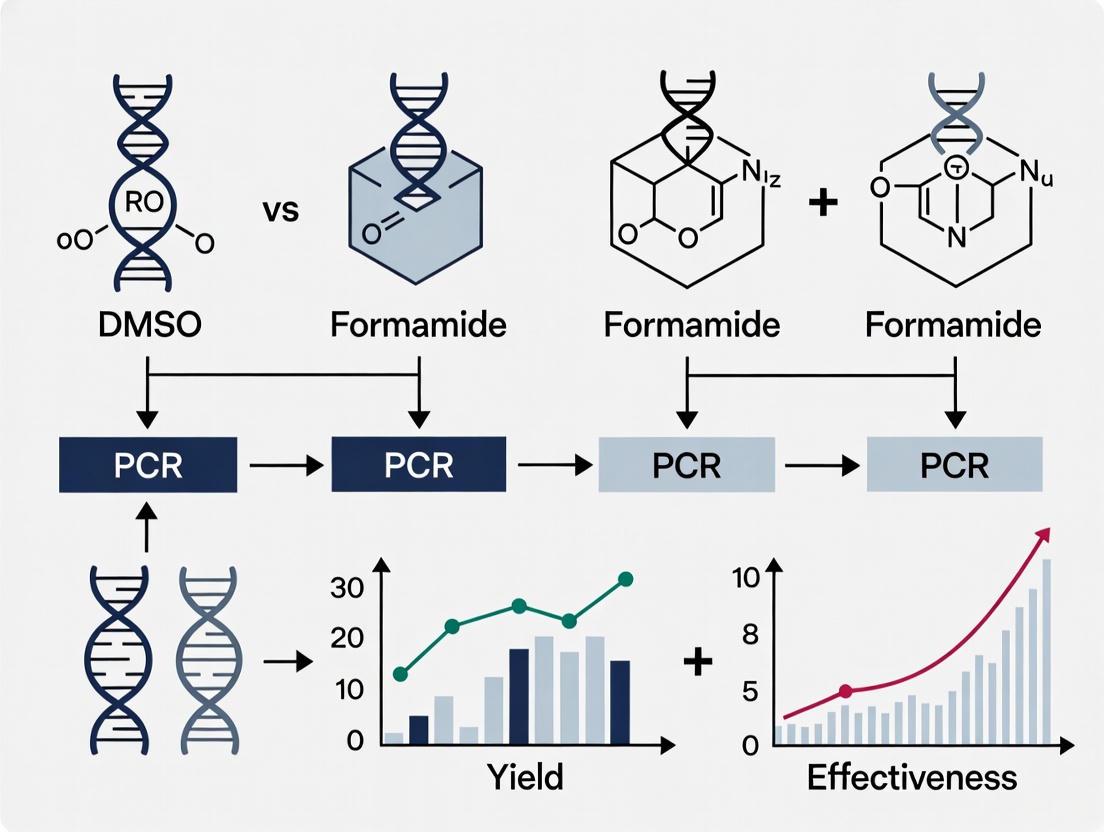
The following diagram illustrates how these enhancers interact with DNA during the PCR denaturation and annealing steps:
Comparative Performance Analysis
Direct comparative studies provide valuable insights into the relative effectiveness of DMSO and formamide across various PCR applications. The data reveal that while both enhancers can improve PCR outcomes, their performance profiles differ significantly.
Quantitative Comparison in Wastewater Samples
A 2024 study evaluating PCR-enhancing approaches for viral detection in wastewater provides direct comparative data on DMSO and formamide performance in challenging matrices [4]:
Table 1: Performance Comparison of DMSO vs. Formamide in Wastewater Sample PCR
| Enhancer | Concentration Tested | Effect on Cq Values | Inhibitor Resistance | Recommended Application |
|---|---|---|---|---|
| DMSO | 2%, 4%, 6% | No significant change | Moderate improvement | General purpose for GC-rich templates |
| Formamide | 1%, 2%, 3% | Increased Cq values (higher concentrations) | Minimal improvement | Specific applications requiring increased stringency |
The study demonstrated that DMSO provided more consistent results across the concentration range tested, while formamide at higher concentrations (2-3%) negatively impacted PCR efficiency as evidenced by increased Cq values [4]. This suggests that DMSO offers a broader effective concentration window, making it more robust for routine applications.
Effectiveness in GC-Rich Construct Amplification
Research on de novo synthesis of GC-rich constructs demonstrated that DMSO, particularly when combined with betaine, dramatically improved target product specificity and yield during PCR amplification [30]. The study successfully amplified two GC-rich gene fragments implicated in tumorigenesis (IGF2R and BRAF), noting that these additives were highly compatible with all reaction components without requiring additional protocol modifications.
Experimental Protocols and Methodologies
Standardized Protocol for GC-Rich PCR with DMSO
For amplification of GC-rich templates (≥60% GC content), the following protocol adapted from New England Biolabs recommendations and experimental studies provides a reliable starting point [29] [30]:
Reaction Setup:
- Use a polymerase specifically optimized for GC-rich templates (e.g., OneTaq Hot Start or Q5 High-Fidelity DNA Polymerase)
- Prepare master mix according to manufacturer's instructions
- Add DMSO to a final concentration of 3-5% (typically 1.5-2.5 μL in a 50 μL reaction)
- Include GC enhancer if supplied with polymerase
Thermal Cycling Parameters:
- Initial denaturation: 98°C for 30 seconds
- 35 cycles of:
- Denaturation: 98°C for 10 seconds
- Annealing: Temperature gradient based on primer Tm (5°C above calculated Tm)
- Extension: 68°C for 30-60 seconds per kb
- Final extension: 68°C for 5 minutes
Optimization Notes:
- For templates >80% GC content, increase DMSO to 5-10%
- Combine with betaine (1.0-1.7M final concentration) for extremely challenging templates
- Optimize Mg²⁺ concentration between 1.0-4.0 mM in 0.5 mM increments
Long Fragment Amplification Protocol
For amplification of long DNA fragments (>5 kb), the following methodology incorporating DMSO has demonstrated success [31]:
Reaction Composition:
- Use long-range DNA polymerase mix (e.g., AccuTaq LA DNA Polymerase)
- Add DMSO to final concentration of 1-4%
- Maintain Mg²⁺ concentration between 1-5 mM (optimize empirically)
- Use high-quality, minimally handled template DNA to prevent damage
Thermal Cycling Profile:
- Initial denaturation: 96°C for 30 seconds
- 30 cycles of:
- Denaturation: 96°C for 10-15 seconds
- Annealing/Extension: 68°C for 10-20 minutes (depending on product length)
- Apply auto-extension of 15 seconds per cycle for fragments >20 kb
Critical Considerations:
- Use primers with Tm between 60-72°C (within 3°C of each other)
- Design primers 21-34 bases in length with ~50% GC content
- Include a final CC, GG, CG, or GC on the 3' end to increase priming efficiency
- Maintain buffer pH >9.0 at 25°C to minimize depurination during cycling
Research Reagent Solutions
The following essential materials represent key solutions for implementing DMSO-enhanced PCR protocols:
Table 2: Essential Research Reagents for DMSO-Enhanced PCR
| Reagent | Function | Application Notes |
|---|---|---|
| DMSO (Molecular Biology Grade) | Reduces DNA secondary structures | Use at 2-10% final concentration; higher concentrations may inhibit polymerase |
| High-Fidelity DNA Polymerase | Catalyzes DNA synthesis with high accuracy | Essential for long fragment amplification; superior for GC-rich targets |
| Betaine (Monohydrate) | Reduces formation of secondary structures | Use at 1.0-1.7M final concentration; synergizes with DMSO |
| GC Enhancer Buffer | Optimized chemical environment | Often proprietary formulations included with specialized polymerases |
| dNTP Mix | Building blocks for DNA synthesis | Use balanced concentrations; quality affects long fragment amplification |
| MgCl₂ Solution | Cofactor for polymerase activity | Requires optimization (typically 1.5-4.0 mM); concentration gradients recommended |
Application Workflow and Decision Pathway
Selecting between DMSO and formamide requires careful consideration of template characteristics and experimental goals. The following decision pathway provides a systematic approach to enhancer selection:
The comparative analysis of DMSO and formamide reveals distinct advantages for each reagent in specific PCR applications. DMSO demonstrates superior performance for amplifying GC-rich templates and long DNA fragments, where its ability to reduce secondary structure formation directly addresses the primary amplification challenges. Experimental data consistently shows that DMSO improves target product specificity and yield without the negative impacts on Cq values observed with formamide at higher concentrations [4] [30].
Formamide offers specialized value in applications requiring increased stringency where non-specific amplification poses a greater problem than low yield. However, its narrower effective concentration range and potential for PCR inhibition at higher concentrations make it less suitable as a general-purpose enhancer.
For researchers and drug development professionals working with challenging templates, DMSO represents the more versatile and reliable option, particularly when combined with complementary additives like betaine and optimized polymerase systems. The experimental protocols provided offer robust starting points for implementation, with empirical optimization remaining essential for specific template-enhancer combinations.
Within molecular biology, the polymerase chain reaction (PCR) is a foundational technique, yet it often encounters challenges with specificity and efficiency, particularly with complex templates. To overcome these hurdles, various chemical enhancers are employed. This guide objectively compares the performance of formamide against dimethyl sulfoxide (DMSO) and other common alternatives, framing the analysis within the broader thesis of optimizing PCR protocols. Supported by recent experimental data, we demonstrate that formamide's primary strength lies in its exceptional ability to enhance amplification specificity and reduce non-specific binding, making it a powerful tool for researchers and drug development professionals tackling difficult PCR applications.
The amplification of difficult DNA targets—such as those with high GC-content, stable secondary structures, or complex repetitive sequences—often results in PCR failure or the generation of non-specific products [5]. These undesired amplicons compete for essential reaction components, reducing the yield of the target product and compromising the reliability of downstream analyses. The scientific community has addressed this challenge by adopting PCR enhancers, chemical additives that modify the physicochemical environment of the reaction to favor specific primer-template interactions. Among the most historically significant and widely debated of these additives are formamide and DMSO. While both are known to aid in the amplification of problematic templates, a direct comparison of their mechanisms, efficacies, and optimal applications is crucial for informed experimental design. This guide provides a comparative analysis of formamide and DMSO, with supporting data on emerging alternatives, to delineate their specific roles in modern PCR research.
Comparative Analysis of Formamide and DMSO
Formamide and DMSO are both polar, organic solvents that enhance PCR primarily by destabilizing the DNA double helix. They reduce the melting temperature (Tm) of DNA, thereby facilitating the denaturation of templates with strong secondary structures and promoting more stringent primer annealing [5] [32]. However, their specific effects on enzyme stability and overall PCR performance differ significantly, which dictates their suitability for various applications.
Table 1: Fundamental Properties and Mechanisms of Formamide and DMSO
| Property | Formamide | DMSO |
|---|---|---|
| Primary Mechanism | Lowers DNA Tm by disrupting hydrogen bonds and hydrophobic interactions between DNA strands [32]. | Lowers DNA Tm by interacting with water molecules, reducing their hydrogen bonding to DNA [32]. |
| Effect on DNA Polymerase | Thermal destabilization; can significantly inhibit PCR at high concentrations [5]. | Reduces Taq polymerase activity; inhibitory at high concentrations [5] [32]. |
| Key Strength | Dramatically improves the specificity of PCR, reducing non-specific amplification [33] [32]. | Effective at reducing DNA secondary structure, facilitating amplification of GC-rich targets [5]. |
| Reported Optimal Concentration | 1-5% (v/v) [32] | 2.5-5% (v/v) [5] |
Experimental Performance Data
A systematic 2024 study compared nine PCR enhancers across various conditions, providing quantitative data on their performance in amplifying DNA fragments with different GC contents [5] [16]. The following tables summarize the key findings for formamide and DMSO, measured by Cycle threshold (Ct) values in real-time PCR (lower Ct indicates higher efficiency).
Table 2: Amplification Efficiency (Ct values) at Various Concentrations [5]
| Enhancer | Concentration | 53.8% GC (Moderate) | 68.0% GC (High) | 78.4% GC (Super High) |
|---|---|---|---|---|
| Control (No enhancer) | - | 15.84 | 15.48 | 32.17 |
| Formamide | 2.5% | 16.28 | 15.11 | 15.91 |
| 5% | 18.08 | 15.44 | 16.32 | |
| 10% | ND* | ND* | ND* | |
| DMSO | 2.5% | 16.21 | 15.26 | 17.48 |
| 5% | 16.68 | 15.72 | 17.90 | |
| 10% | 18.78 | 17.15 | 21.15 |
*ND: Not Detected.
Table 3: Performance in Inhibitor-Rich Conditions (with Heparin) [5]
| Enhancer | Concentration | Moderate GC Target | High GC Target |
|---|---|---|---|
| Control (No enhancer) | - | Complete inhibition | Complete inhibition |
| Formamide | 2.5% | Partial relief | Partial relief |
| DMSO | 5% | Partial relief | Partial relief |
| Betaine | 1 M | Significant relief | Significant relief |
Analysis of Comparative Data
The data reveals distinct performance profiles:
- For Specificity and GC-Rich Targets: Formamide at 2.5% concentration dramatically improved the amplification of the super high GC-content fragment (78.4%), reducing the Ct value from 32.17 to 15.91 [5]. This represents a profound enhancement in efficiency, underscoring its utility for problematic, GC-rich templates. Its effect is highly concentration-dependent, with 5% formamide already showing strong inhibition on the moderate GC template and 10% leading to complete amplification failure [5].
- For Broad Applicability: DMSO also improved the amplification of GC-rich targets, though its effect on the 78.4% GC fragment was less pronounced than formamide's at lower concentrations [5]. DMSO exhibited a somewhat broader effective concentration range (2.5-5%) across different template types before severe inhibition occurred.
- For Inhibitor Tolerance: In the presence of the common PCR inhibitor heparin, both formamide and DMSO provided only partial relief from inhibition. The study identified betaine as a superior additive for this specific purpose [5], a finding supported by other research on complex samples like wastewater [4].
The Scientist's Toolkit: Key Research Reagent Solutions
The following table details essential reagents and their functions for experiments investigating PCR enhancers like formamide and DMSO.
Table 4: Essential Research Reagents for PCR Enhancement Studies
| Reagent | Function in PCR Enhancement Studies |
|---|---|
| Formamide | An organic solvent used to lower DNA melting temperature (Tm) and increase stringency, thereby dramatically improving amplification specificity [33] [32]. |
| Dimethyl Sulfoxide (DMSO) | An organic solvent that reduces DNA secondary structure stability by interacting with water molecules, facilitating the amplification of GC-rich sequences [5] [32]. |
| Betaine | An osmoprotectant that reduces the formation of DNA secondary structures and is particularly effective for amplifying GC-rich fragments and providing inhibitor tolerance [5] [32]. |
| Taq DNA Polymerase | A thermostable DNA polymerase that is the core enzyme in many PCR reactions; its activity and thermostability are directly affected by different enhancers [5] [34]. |
| dNTPs | Deoxynucleoside triphosphates (dATP, dCTP, dGTP, dTTP) are the building blocks for DNA synthesis; their incorporation can be influenced by the reaction environment modified by enhancers. |
| SYBR Green I Master Mix | A fluorescent dye used in real-time PCR to quantify amplification products; allows for the generation of Ct values for quantitative comparison of enhancer efficiency [35]. |
| Heparin | A glycosaminoglycan often used as a model PCR inhibitor in experimental settings to evaluate the inhibitor resistance conferred by various enhancers [5]. |
Experimental Protocols for Key Studies
Protocol: Evaluating Enhancers on GC-Rich Templates
This protocol is adapted from the 2024 systematic comparison study [5] [16].
1. Reaction Setup:
- Template: Plasmid containing a mouse Olig2 gene segment (0.1 ng/µL).
- Primers: Designed to amplify fragments with 68.0% (high) and 78.4% (super high) GC-content.
- Master Mix: Contains 0.2-0.4 µM of each primer, 0.2 mM dNTPs, and 1 unit of Taq DNA polymerase.
- Enhancers: Add formamide (2.5%, 5%) or DMSO (2.5%, 5%, 10%) from stock solutions. Include a no-enhancer control.
- Total Reaction Volume: 20 µL.
2. Real-Time PCR Cycling Conditions:
- Initial Denaturation: 95°C for 1-3 minutes.
- 35-40 Cycles of:
- Denaturation: 95°C for 15-30 seconds.
- Annealing: 60°C for 30 seconds (optimized for GC-rich primers).
- Extension: 72°C for 30 seconds.
- Perform a melting curve analysis from 72°C to 96°C.
3. Data Analysis:
- Compare Cycle threshold (Ct) values across different enhancer conditions. A lower Ct indicates higher amplification efficiency.
- Analyze melting curves to confirm the specificity of the amplification (a single, sharp peak indicates a specific product).
Protocol: Testing Enhancer Effects in Inhibitor-Rich Conditions
This protocol evaluates the ability of formamide and DMSO to mitigate PCR inhibition [5] [4].
1. Reaction Setup:
- The base reaction mixture is identical to section 4.1.
- Inhibitor: Add heparin to the reaction at concentrations of 0.0023 U/reaction and 0.0047 U/reaction.
- Enhancers: Test formamide (2.5%) and DMSO (5%) alongside other enhancers like betaine (1 M) for comparison.
2. PCR Cycling and Analysis:
- Use the same cycling conditions as in section 4.1.
- The primary outcome measure is the success or failure of amplification and the Ct value shift compared to a non-inhibited control.
Mechanisms and Workflows
The following diagram illustrates the comparative mechanism of action of formamide and DMSO in enhancing PCR specificity.
Diagram: Mechanisms of Formamide and DMSO in PCR.
The experimental evidence clearly positions formamide as a highly specialized reagent whose prime application is enhancing PCR specificity, particularly for GC-rich templates that resist standard amplification. Its ability to dramatically lower DNA melting temperature allows for more stringent annealing conditions, effectively reducing non-specific binding. However, its narrow window of effective concentration and tendency to inhibit DNA polymerase at higher levels demand careful optimization.
In contrast, DMSO serves as a more general-purpose enhancer, also effective against secondary structures but with a different mechanistic approach and a slightly more forgiving concentration profile. The broader thesis on the effectiveness of DMSO versus formamide concludes that there is no single superior reagent; rather, the choice is context-dependent. For pure specificity against extremely difficult, GC-rich targets, formamide at low concentrations (1.5%) may be optimal. For a broader range of applications or in combination with other enhancers like betaine [5], DMSO remains a versatile tool. Future research directions include the further exploration of synergistic effects of enhancer cocktails and the development of novel polymerase mutants resistant to inhibitors, reducing the reliance on additive optimization [36].
Protocol Adaptation for Multiplex PCR and Direct PCR from Complex Samples
In molecular diagnostics and research, the analysis of complex samples—ranging from wastewater and forensic swabs to whole blood and formalin-fixed tissues—presents significant challenges for polymerase chain reaction (PCR) amplification. The success of PCR, particularly in demanding applications like multiplex assays and direct amplification without DNA extraction, heavily depends on overcoming inhibition caused by sample-derived contaminants and inherent biochemical barriers. Among the various strategies employed, chemical enhancers play a pivotal role in facilitating robust amplification. This review objectively compares the effectiveness of two prominent PCR enhancers, Dimethyl Sulfoxide (DMSO) and formamide, within the broader context of protocol adaptations for challenging PCR applications.
The necessity for such enhancements is particularly acute in multiplex PCR, where multiple primer sets compete for reagents and amplify multiple targets simultaneously, and in direct PCR, where inhibitors co-purified with the template can dramatically reduce amplification efficiency. The choice between enhancers such as DMSO and formamide is not merely a procedural preference but a critical determinant of assay success, influencing specificity, sensitivity, and overall reliability. This article provides a comparative analysis based on recent experimental data, detailing protocols and performance metrics to guide researchers in selecting and optimizing the most appropriate enhancer for their specific sample type and application requirements.
Mechanism of Action: DMSO versus Formamide
Understanding the distinct biochemical mechanisms through which DMSO and formamide operate is fundamental to their appropriate application. While both aim to improve PCR outcomes, they target different aspects of the DNA amplification process.
Formamide functions primarily as a destabilizing agent that reduces the melting temperature (Tm) of DNA by disrupting hydrogen bonding between base pairs. By generating hydrogen bond coupling with DNA bases, formamide lowers the energy required for strand separation [37]. This property is particularly valuable for lowering denaturation temperatures in specialized applications like solid-phase PCR, where high temperatures can lead to significant loss of DNA immobilized on surfaces [37]. Furthermore, by increasing primer annealing stringency, formamide can dramatically improve the specificity of PCR, reducing non-specific amplification and primer-dimer formation [38].
In contrast, DMSO is primarily known as a secondary structure destabilizer. It alters the DNA structure by reducing the stability of GC-rich regions, which are prone to forming complex secondary structures such as hairpins that can stall polymerase progression [38]. This makes DMSO particularly effective for amplifying GC-rich templates. Additionally, DMSO is reported to counteract the inhibitory effects on Taq DNA polymerase, though its precise mechanism in this context is less clearly defined [4].
The conceptual workflow below illustrates how these enhancers integrate into a standard PCR protocol and the different points at which they exert their effects.
Comparative Performance Analysis
Direct comparative studies provide the most reliable evidence for selecting an appropriate PCR enhancer. A 2024 systematic evaluation of PCR-enhancing approaches in wastewater-based epidemiology offers a robust, head-to-head comparison of DMSO and formamide, among other additives [4]. Wastewater represents a highly complex and inhibitory matrix, making it a relevant model for challenging samples.
Quantitative Comparison in Wastewater Samples
The study assessed the effect of various enhancers on reverse transcription quantitative PCR (RT-qPCR) performance for detecting SARS-CoV-2 in raw wastewater. The results, summarized in the table below, were measured by the change in quantification cycle (Cq) value compared to a basic protocol without enhancers. A higher Cq value indicates poorer detection sensitivity [4].
Table 1: Performance of PCR Enhancers in Wastewater Viral Detection
| Enhancer | Concentration | Mean Cq Value | Performance vs. Basic Protocol | Key Finding |
|---|---|---|---|---|
| Basic Protocol | - | Undetected (Cq >40) | Baseline | Only 1 of 3 undiluted samples detected [4] |
| Formamide | 1% | 37.8 | Improved detection | Detected virus in otherwise undetected samples [4] |
| Formamide | 5% | 36.8 | Best performance | Lowest Cq value among chemical enhancers tested [4] |
| DMSO | 1% | 38.5 | Improved detection | Detected virus, but higher Cq than formamide [4] |
| DMSO | 5% | 39.1 | Inferior performance | Higher Cq than 1% DMSO and all formamide concentrations [4] |
| 10-Fold Dilution | - | Detected | Gold Standard | Common inhibition mitigation, but reduces sensitivity [4] |
The data demonstrates that 5% formamide yielded the lowest Cq value among the chemical enhancers tested, indicating it was the most effective at mitigating inhibition and enhancing sensitivity in this challenging matrix [4]. DMSO, while still improving detection over the basic protocol, was less effective, with its performance degrading at a higher concentration (5%) [4]. The study concluded that the optimized RT-qPCR protocol incorporating formamide was directly comparable to the more expensive and time-consuming digital PCR (ddPCR) for the final estimation of viral load, underscoring its practical utility [4].
Efficacy in Other Application Contexts
Beyond wastewater analysis, the effectiveness of these enhancers is context-dependent.
- Solid-Phase PCR: In solid-phase bridge amplification, a critical process for high-throughput sequencing, formamide denaturation enabled a higher cluster density (2.83 × 10^4 colonies/mm²) compared to high-temperature denaturation [37]. This was achieved with a lower template concentration and milder conditions. Furthermore, formamide denaturation resulted in a 15% higher DNA retention rate on the solid surface, demonstrating its ability to preserve the stability of immobilized DNA during thermal cycling [37].
- GC-Rich Amplification: For amplifying GC-rich templates, which are prone to forming stable secondary structures, DMSO is often recommended as a first-line additive. It works by reducing secondary structures that can inhibit the polymerase, thereby facilitating amplification of otherwise refractory targets [38]. In such cases, DMSO, along with other additives like glycerol and betaine, is specifically highlighted for its ability to improve yields [38].
Detailed Experimental Protocols
To facilitate the adoption and validation of these enhancers, detailed protocols from key studies are provided below.
Protocol 1: Evaluating Enhancers in Wastewater RT-qPCR
This protocol is adapted from the 2024 study that directly compared DMSO and formamide [4].
- Sample Material: 24-hour composite flow-proportional raw wastewater samples.
- Nucleic Acid Extraction: Use a commercial automated nucleic acid extraction system. Elute in a final volume of 60 µL.
- RT-qPCR Reaction Setup:
- Master Mix: Use a 1-step RT-qPCR master mix.
- Primers/Probes: Target-specific assays (e.g., for SARS-CoV-2).
- Reaction Volume: 20 µL total.
- Template: 5 µL of extracted nucleic acid.
- Enhancers: Add DMSO or formamide to the master mix from stock solutions to achieve the desired final concentration (e.g., 1% and 5% v/v).
- Thermocycling Conditions:
- Reverse Transcription: 50°C for 15 min.
- Initial Denaturation: 95°C for 2 min.
- Amplification (45 cycles): Denature at 95°C for 5 sec, anneal/extend at 60°C for 30 sec.
- Data Analysis: Compare Cq values and calculated viral loads between the basic protocol and enhancer-supplemented protocols.
Protocol 2: Direct Real-Time PCR from Whole Blood (GG-RT PCR)
This protocol enables real-time PCR from whole blood without DNA extraction, showcasing a direct PCR application [35].
- Sample Preparation (Blood Lysate):
- Mix 400 µL of EDTA-treated whole blood with distilled water to an 80% dilution.
- Incubate at 95°C for 20 minutes, vortexing 2-3 times during incubation.
- Centrifuge at 14,000 rpm for 5 minutes.
- Collect the supernatant (lysate) for use as a PCR template.
- Real-Time PCR Setup:
- Master Mix: SYBR Green I Master Mix.
- Primers: 5 pmol of each primer per reaction.
- Template: Use 2.5 µL of the blood lysate, diluted 1:5 or 1:10.
- Reaction Volume: 10 µL final.
- Thermocycling Conditions (LightCycler 480 II):
- Initial Denaturation: 95°C for 10 min.
- Amplification (40 cycles): 95°C for 15 sec, 60-61°C for 30 sec.
The workflow for this direct, inhibitor-resistant method is illustrated below.
Protocol 3: Multiplex PCR Capillary Electrophoresis for Respiratory Pathogens
This protocol describes a single-tube multiplex assay for 22 respiratory pathogens, combining PCR with capillary electrophoresis (CE) for detection [39].
- Primer Design:
- Retrieve target sequences from GenBank and align to identify conserved regions.
- Design primers with an annealing temperature near 60°C and GC content between 40-60%.
- Organize primers into three groups, labeling each group with a distinct fluorophore (FAM, VIC, NED).
- Ensure amplicon sizes within a group differ by at least 20 base pairs.
- Nucleic Acid Extraction: Extract from nasopharyngeal swabs or saliva using a magnetic bead-based kit.
- One-Tube Multiplex RT-PCR:
- Reverse Transcription: Convert RNA to cDNA using random primers and M-MLV Reverse Transcriptase.
- Multiplex PCR: Use 5 µL of cDNA in a 25 µL reaction containing:
- 1X Taq buffer
- 1.5 U Taq DNA polymerase
- 100 µM of each dNTP
- 6 pmol of each of the 22 specific primers
- 0.05 mM MgCl₂
- Thermocycling: Multiple temperature cycles ranging from 59°C to 94°C.
- Capillary Electrophoresis:
- Dilute PCR products 1:20 in water.
- Mix 1 µL diluted product with formamide and a size standard (e.g., GeneScan 600 LIZ).
- Denature at 94°C for 3 min, then immediately cool to 4°C.
- Perform CE on a genetic analyzer (e.g., Applied Biosystems 3500).
- Analyze data with fragment analysis software (e.g., GeneMapper).
The Scientist's Toolkit: Key Research Reagent Solutions
The following table compiles essential reagents and their functions for developing and optimizing multiplex and direct PCR protocols for complex samples.
Table 2: Essential Reagents for PCR Protocol Adaptation
| Reagent / Solution | Primary Function | Application Notes |
|---|---|---|
| Formamide | Lowers DNA melting temperature (Tm), increases primer annealing stringency [37] [38]. | Use at 1-5% in wastewater RT-qPCR [4]; enables low-temperature denaturation in solid-phase PCR [37]. |
| DMSO | Disrupts secondary structures in GC-rich DNA templates [38]. | Effective for GC-rich targets; performance can degrade at higher concentrations (5%) in inhibitory samples [4] [38]. |
| BSA (Bovine Serum Albumin) | Binds inhibitors (e.g., humic acids) in the reaction mix, relieving polymerase inhibition [4]. | A common additive for reducing inhibition in environmental and forensic samples. |
| Tween-20 | Non-ionic detergent that counteracts inhibitory effects on Taq DNA polymerase [4]. | Used for relief of inhibition in fecal and complex environmental samples. |
| Betaine | Destabilizes DNA secondary structures, acts as a crowding agent, and can reduce melting temperature [38]. | Particularly useful for amplifying GC-rich regions and in direct PCR protocols. |
| Hot-Start DNA Polymerase | Prevents non-specific amplification and primer-dimer formation by requiring heat activation [34]. | Critical for multiplex PCR to improve specificity and sensitivity. |
| Inhibitor-Tolerant Polymerase Blends | Engineered polymerases or blends with enhanced resistance to common PCR inhibitors [40]. | Enable direct PCR from crude samples like blood and soil. |
| MgCl₂ | Essential cofactor for DNA polymerase activity; concentration affects specificity and yield [38]. | Optimization (e.g., 1.0-4.0 mM) is crucial for challenging amplicons; excess causes non-specific binding. |
The comparative analysis of DMSO and formamide reveals that the choice of an optimal PCR enhancer is highly dependent on the specific application, sample type, and the primary challenge being addressed.
For highly inhibitory complex samples, such as wastewater, formamide at 5% concentration demonstrates superior performance in mitigating inhibition and enhancing detection sensitivity, outperforming DMSO and other enhancers like BSA and Tween-20 [4]. Its ability to lower denaturation temperatures also makes it invaluable for solid-phase amplification protocols where DNA stability on a surface is critical [37].
For applications focused on amplifying GC-rich templates where secondary structure formation is the main obstacle, DMSO remains a recommended and effective choice [38]. However, its concentration must be carefully optimized, as higher levels (e.g., 5%) can be detrimental in some contexts [4].
Protocol adaptations for complex samples increasingly favor integrated strategies. The success of direct PCR from blood via simple heat lysis and the robustness of large multiplex panels highlight that beyond chemical enhancers, a holistic approach—including inhibitor-resistant polymerases, optimized primer design, and specialized detection systems—is key to advancing molecular diagnostics and research.
Solving PCR Problems: An Optimizer's Guide to DMSO and Formamide
Polymerase chain reaction (PCR) is a cornerstone technique in molecular biology, yet it often fails to amplify difficult DNA targets efficiently. PCR enhancers are specialized reagents that can be added to reactions to overcome these challenges, with Dimethyl sulfoxide (DMSO) and formamide being two of the most prominent. Framed within a broader thesis on their effectiveness, this guide objectively compares the performance of DMSO and formamide against other common alternatives, providing the experimental data and protocols needed for informed reagent selection.
What Are PCR Enhancers and When Are They Needed?
PCR enhancers are chemical additives that modify the reaction environment to improve the amplification of specific targets. They are typically employed when standard PCR optimization fails, particularly with templates that have inherent challenges [5]. Your reaction is a candidate for an enhancer if you encounter the following scenarios:
- Amplification of GC-rich sequences: GC-rich DNA (≥60% GC content) has strong secondary structures and a high melting temperature, often leading to poor or failed amplification [41].
- Inhibitor tolerance: When the sample contains known PCR inhibitors (e.g., from blood, soil, or plant tissues), certain enhancers can help polymerase activity withstand their effects [5].
- Nonspecific amplification: The presence of multiple bands or smears on a gel indicates nonspecific primer binding, which some enhancers can suppress by increasing priming stringency [2].
The following diagram outlines a logical pathway for diagnosing PCR failure and deciding whether to use an enhancer.
Objective Comparison of Common PCR Enhancers
While DMSO and formamide are widely used, a systematic comparison reveals that other enhancers, such as betaine, can offer superior performance in specific contexts. The table below summarizes quantitative data on the effects of various enhancers on the amplification of DNA fragments with different GC contents, as measured by Cycle Threshold (Ct) values in real-time PCR [5].
Table 1: Effect of Enhancer Concentration on PCR Amplification Efficiency (Ct Values)
| Enhancer | Concentration | 53.8% GC (Moderate) Ct±SEM | 68.0% GC (High) Ct±SEM | 78.4% GC (Super High) Ct±SEM |
|---|---|---|---|---|
| Control | - | 15.84±0.05 | 15.48±0.22 | 32.17±0.25 |
| DMSO | 5% | 16.68±0.01 | 15.72±0.03 | 17.90±0.05 |
| 10% | 18.78±0.04 | 17.15±0.07 | 21.15±0.07 | |
| Formamide | 5% | 18.08±0.07 | 15.44±0.03 | 16.32±0.05 |
| Ethylene Glycol (EG) | 5% | 16.28±0.06 | 15.27±0.08 | 17.24±0.04 |
| Betaine | 0.5 M | 16.03±0.03 | 15.08±0.10 | 16.97±0.10 |
| Sucrose | 0.4 M | 16.39±0.09 | 15.03±0.04 | 16.67±0.08 |
| Trehalose | 0.4 M | 16.43±0.16 | 15.15±0.08 | 16.91±0.14 |
A lower Ct value indicates higher amplification efficiency. The control reaction shows clear failure (Ct=32.17) for the 78.4% GC template. Data adapted from [5].
Beyond amplification efficiency, enhancers have distinct effects on reaction specificity and polymerase stability. The following table provides a comparative overview based on experimental findings.
Table 2: Performance Profile of Common PCR Enhancers
| Enhancer | Primary Mechanism | Effect on Specificity | Effect on Taq Polymerase Activity | Best Use Case |
|---|---|---|---|---|
| DMSO | Reduces DNA Tm; disrupts secondary structures [2] [42] | Can reduce at high conc. [5] | Markedly reduces [5] [2] | GC-rich templates at low (2.5-5%) concentrations [5] |
| Formamide | Destabilizes DNA double helix; lowers Tm [2] [42] | Increases [6] [2] | Reduces [5] | Improving specificity; GC-rich templates [5] |
| Betaine | Reduces secondary structure; equalizes base-pair stability [5] [2] | Increases [5] | Thermally stabilizes [5] | Superior for GC-rich fragments; high inhibitor tolerance [5] |
| TMAC | Increases hybridization specificity [2] [42] | Markedly increases [6] | Mild effect at optimal conc. | Reactions with degenerate primers [2] |
| Glycerol | Reduces secondary structures [41] | Can reduce [2] | Thermally stabilizes [5] | General stabilizer; can be combined |
| Trehalose | Lowers DNA Tm [5] | Mildly increases [5] | Thermally stabilizes [5] | Inhibitor tolerance [5] |
Experimental Protocols for Evaluating Enhancer Efficacy
To generate reliable comparative data, consistent and well-controlled experimental methodologies are essential. The following protocols are based on cited research.
Protocol 1: Systematic Comparison of Enhancer Effects
This methodology was used to generate the quantitative data in Table 1 [5].
- 1. Reaction Setup: Prepare a master mix containing standard components: 1X PCR buffer, 0.2 mM dNTPs, 1.5 mM MgCl₂, 0.5 µM of each primer, 0.5–1 unit of Taq DNA polymerase, and a defined amount of template DNA (e.g., 10-100 ng genomic DNA) per reaction.
- 2. Additive Addition: Aliquot the master mix and add PCR enhancers to the final concentrations listed in Table 1. Include a no-additive control.
- 3. Thermal Cycling: Perform real-time PCR under the following conditions:
- Initial denaturation: 95°C for 5 min.
- 35–40 cycles of:
- Denaturation: 95°C for 15–30 sec.
- Annealing: Temperature optimized for the primer set (e.g., 55–60°C) for 30 sec.
- Extension: 72°C for 30–60 sec/kb.
- 4. Data Analysis: Compare the Cycle Threshold (Ct) values and amplicon melting temperatures (Tm) across different enhancer conditions. Also, analyze the final products via agarose gel electrophoresis to assess specificity and yield.
Protocol 2: Testing Enhancer Specificity with Difficult Primers
This protocol is adapted from research investigating the suppression of nonspecific amplification [6].
- 1. Reaction Setup: Use a complex template (e.g., 300 ng of rat or mouse genomic DNA) and primers that are known to produce nonspecific fragments under standard conditions.
- 2. Additive Titration: Test the enhancer (e.g., TMA oxalate, formamide) across a range of concentrations. For instance:
- TMA oxalate: 0.5 mM, 1 mM, 2 mM, 5 mM.
- Formamide: 0.5 M, 1 M, 1.5 M.
- 3. Thermal Cycling:
- Initial denaturation: 94°C for 1 min.
- 30 cycles of: 94°C for 15 sec, 49–58°C for 15 sec (depending on primer Tm), 72°C for 30 sec.
- Final extension: 72°C for 1 min.
- 4. Data Analysis: Resolve PCR products on an agarose gel. Use densitometric analysis to calculate:
- Specificity: Ratio of the intensity of the specific band to the intensity of all bands.
- Efficiency: Ratio of the specific band intensity with the additive to the specific band intensity without the additive.
The Scientist's Toolkit: Key Research Reagent Solutions
A successful PCR enhancement experiment requires more than just the additives themselves. The following table details essential materials and their functions.
Table 3: Essential Reagents for PCR Enhancement Experiments
| Reagent | Function | Specification & Notes |
|---|---|---|
| Thermostable DNA Polymerase | Enzymatically synthesizes new DNA strands. | Choice is critical. For GC-rich targets, use enzymes specifically optimized for this purpose (e.g., OneTaq or Q5 from NEB) [41]. |
| PCR Enhancers | Modify reaction environment to improve efficiency/specificity. | Use molecular biology grade. Prepare stock solutions (e.g., 5M betaine, 1M sucrose/trehalose, 100% DMSO/formamide) and store appropriately [5]. |
| dNTP Mix | Building blocks (dATP, dCTP, dGTP, dTTP) for DNA synthesis. | Use balanced, equimolar concentrations (e.g., 10mM each). Unbalanced dNTPs increase error rate [43] [26]. |
| Magnesium Salt (MgCl₂/MgSO₄) | Essential cofactor for DNA polymerase activity. | Concentration requires optimization (typically 1.0-4.0 mM). It interacts with dNTPs and affects enzyme fidelity [43] [41]. |
| Nuclease-free Water | Solvent for the reaction. | Must be pure and free of nucleases to prevent degradation of reaction components. |
| Template DNA & Primers | The target to be amplified and the sequences that define its boundaries. | Template integrity and purity are vital. Primers must be well-designed (Tm 55-70°C, 40-60% GC) and purified [7] [26]. |
The comparative data clearly show that while both DMSO and formamide can rescue failing PCRs, their effectiveness is highly context-dependent. DMSO is a good first choice for GC-rich templates at low concentrations, but its inhibitory effect on Taq polymerase is a significant drawback. Formamide excels at increasing specificity but can also be inhibitory. The experimental evidence positions betaine as a superior enhancer in many demanding scenarios, particularly for GC-rich fragments, due to its dual action of reducing secondary structures and thermally stabilizing the polymerase [5].
Future research will likely focus on developing proprietary, optimized blends of these enhancers. As shown in one study, a combination of 1 M betaine with 0.1-0.2 M sucrose effectively amplified GC-rich long DNA fragments while minimizing the negative impact on normal templates [5]. This trend towards sophisticated, multi-component enhancer systems, often supplied with high-performance polymerases, represents the evolving solution to PCR's most stubborn failures.
Polymersse chain reaction (PCR) is a foundational technique in molecular biology, yet the amplification of complex DNA templates often requires meticulous optimization. Guanine-cytosine (GC)-rich sequences, which constitute approximately 3% of the human genome and are frequently found in gene promoters, present particular challenges due to their tendency to form stable secondary structures that resist complete denaturation and cause polymerase stalling [44] [45]. To overcome these hurdles, scientists routinely employ PCR enhancers—organic molecules or ions that improve amplification efficiency, specificity, and yield [46].
Within this context, dimethyl sulfoxide (DMSO) and formamide have emerged as two of the most widely used organic solvent additives. Despite their common application, they function through distinct biochemical mechanisms and exhibit different effectiveness profiles across various experimental conditions. This guide provides an objective comparison of DMSO versus formamide, presenting structured experimental data and protocols to inform evidence-based selection and optimization for research and drug development applications.
Mechanistic Comparison of DMSO and Formamide
Understanding the distinct modes of action for DMSO and formamide is crucial for their rational application in PCR optimization.
Dimethyl Sulfoxide (DMSO) primarily functions by reducing the secondary structure stability of DNA. It achieves this by interacting with water molecules surrounding the DNA strand, thereby disrupting the hydrogen-bonding network and lowering the melting temperature (Tm) of the DNA. This effect facilitates strand separation and primer annealing at lower temperatures [47]. However, DMSO also reduces Taq polymerase activity, creating a balance that must be carefully optimized [47].
Formamide, also an organic solvent, acts by destabilizing the DNA double helix. It penetrates the major and minor grooves of DNA, disrupting hydrogen bonds and hydrophobic interactions between strands, which similarly results in a lowered melting temperature [47]. This action promotes more complete denaturation of GC-rich templates. Furthermore, formamide is noted for increasing primer annealing stringency, which enhances amplification specificity by reducing non-specific priming events [45].
The following diagram illustrates the differential mechanisms of these additives in the PCR process:
Quantitative Comparison of Additive Performance
Concentration Ranges and Effectiveness
The efficacy of both DMSO and formamide is highly concentration-dependent. The table below summarizes their optimal concentration ranges and effectiveness across various template types, synthesizing data from multiple experimental studies.
Table 1: Concentration ranges and performance characteristics of DMSO and formamide
| Parameter | DMSO | Formamide |
|---|---|---|
| Typical Effective Concentration | 2-10% [47] [44] | 1-5% [47] |
| Optimal Concentration | 5% (in combination with betaine) [44] | 1.5-2.5% (structure-activity study) [3] |
| Effect on DNA Melting Temperature | Lowers Tm [47] | Lowers Tm [47] |
| Effect on Polymerase Activity | Reduces activity [47] | Minimal negative effect at optimal concentrations [25] |
| Primary Application | GC-rich templates, long-range PCR [46] [44] | Improvement of specificity, GC-rich templates [47] |
| Reported Potency (Normalized to Formamide=1) | Not reported in studies | 1 (reference value) [3] |
| Best Specificity Obtained | ~81% (N-methylpyrrolidone derivative) [3] | ~86% (2-pyrrolidone) [3] |
Performance Across DNA Template Sizes
The effectiveness of PCR additives varies significantly with the size of the DNA template being amplified. The table below compares the performance of DMSO and formamide in amplifying GC-rich DNA fragments of different lengths, based on experimental data.
Table 2: Additive performance across different template sizes
| Template Size | DMSO Performance | Formamide Performance | Combination Approach |
|---|---|---|---|
| 0.4 kb | Effective with 2.5-5% DMSO [25] | Effective with 1.25-5% formamide [25] | BSA co-enhancement effective [25] |
| 1.6 kb | Good yield with 5% DMSO [25] | Reduced effectiveness [25] | BSA co-enhancement beneficial [25] |
| 2.6 kb | Good yield with 2.5-5% DMSO [25] | Limited effectiveness [25] | BSA co-enhancement extends effective range [25] |
| 7.1 kb | Moderate effectiveness [25] | Ineffective [25] | BSA co-enhancement improves yield [25] |
Experimental Protocols for Systematic Optimization
Concentration Gradient Testing Protocol
This protocol provides a systematic approach for empirically determining the optimal concentration of DMSO or formamide for a specific PCR application.
Materials & Reagents:
- Standard PCR components: DNA polymerase with buffer, dNTPs, MgCl₂, primers, template DNA
- Test additives: DMSO (molecular biology grade) and formamide (molecular biology grade)
- PCR tubes or plates
- Thermal cycler
Methodology:
- Prepare a master mix containing all standard PCR components except additives, calculated for the total number of reactions plus excess.
- Aliquot the master mix into individual PCR tubes.
- Add DMSO to final concentrations of: 0%, 2%, 4%, 6%, 8%, and 10% [47].
- Add formamide to final concentrations of: 0%, 1%, 2%, 3%, 4%, and 5% in parallel reactions [47].
- Include a negative control (no additive) in each experiment.
- Run PCR using optimized thermal cycling parameters with an annealing temperature gradient (if available).
- Analyze results by agarose gel electrophoresis for product yield and specificity.
- Use densitometric analysis for quantitative comparison of amplification products [3].
Combination Approach Testing Protocol
Based on research demonstrating enhanced performance with additive combinations, this protocol tests synergistic effects.
Materials & Reagents:
- All materials from Protocol 4.1
- Betaine (1-1.7 M final concentration) [44]
- Bovine Serum Albumin (BSA; 0.8-10 μg/μl) [25]
Methodology:
- Prepare a master mix as in Protocol 4.1.
- Test the combination of 1M betaine with 5% DMSO, which has shown optimal results for GC-rich FMR1 gene amplification [44].
- Evaluate BSA as a co-additive at 0.8 mg/ml with varying concentrations of DMSO (2.5-5%) or formamide (1.25-5%) [25] [47].
- For challenging templates >3kb, consider supplementing with fresh BSA after every 10 cycles to counteract thermal denaturation [25].
- Include controls with single additives for comparison.
- Analyze results as in Protocol 4.1.
The following workflow diagram summarizes the systematic optimization process:
Research Reagent Solutions
The following table catalogues essential reagents mentioned in the experimental protocols, providing researchers with a consolidated reference for experiment planning.
Table 3: Essential research reagents for PCR optimization with DMSO and formamide
| Reagent | Function | Example Application |
|---|---|---|
| DMSO (Molecular Biology Grade) | Reduces DNA secondary structure by disrupting hydrogen bonding; lowers Tm [47] | Amplification of GC-rich templates (e.g., FMR1 gene) [44] |
| Formamide (Molecular Biology Grade) | Destabilizes DNA double helix by binding grooves; increases primer stringency [47] | Improving specificity in amplification of complex templates [3] |
| Betaine | Osmoprotectant that reduces formation of DNA secondary structures; equalizes Tm of GC and AT pairs [47] [44] | Used in combination with DMSO for GC-rich templates [44] |
| Bovine Serum Albumin (BSA) | Binds inhibitors; stabilizes polymerase; enhances effects of organic solvents [25] [47] | Co-enhancer with DMSO or formamide for increased yield [25] |
| 7-deaza-dGTP | dGTP analog that reduces hydrogen bonding in GC-rich regions without disrupting Watson-Crick pairing [44] | Alternative approach for challenging GC-rich templates [44] |
| MgCl₂ | Essential cofactor for DNA polymerase activity; facilitates primer binding [47] [45] | Concentration optimization (1.0-4.0 mM) critical for GC-rich PCR [45] |
The empirical comparison of DMSO and formamide reveals a complex optimization landscape where template characteristics, particularly GC content and length, dictate the optimal choice. DMSO demonstrates broader effectiveness across various template sizes, while formamide offers advantages for specificity enhancement in smaller amplicons. Critically, combination approaches utilizing betaine or BSA with these solvents frequently outperform single-additive applications, providing researchers with powerful strategies for challenging amplification targets.
The experimental protocols presented enable systematic evaluation of these parameters, emphasizing the need for target-specific optimization rather than universal application. For drug development professionals and researchers working with genetically complex targets, this evidence-based approach to PCR enhancement ensures robust and reproducible results in diagnostic and research applications.
In polymerase chain reaction (PCR) research, scientists frequently encounter two significant challenges: enzyme inhibition that reduces amplification efficiency and signal reduction that compromises detection sensitivity. These drawbacks become particularly problematic when working with difficult DNA templates such as those with high GC-content or complex secondary structures. Among the various chemical additives available to counter these issues, dimethyl sulfoxide (DMSO) and formamide have emerged as prominent denaturants and cosolvents. This comparison guide objectively evaluates the effectiveness of DMSO versus formamide in PCR applications, examining their mechanisms, benefits, and limitations based on experimental data to inform researchers, scientists, and drug development professionals.
Mechanisms of Action: How DMSO and Formamide Work
Dimethyl sulfoxide (DMSO) functions primarily by reducing secondary DNA structures through destabilization of duplex DNA. It weakens hydrogen bonds between base pairs and opens up secondary structures, thereby facilitating denaturation of GC-rich templates that would otherwise remain double-stranded under standard PCR conditions [1] [2]. This property makes it particularly valuable for amplifying difficult DNA targets with complex secondary structures.
Formamide operates through a different mechanism, binding in the major and minor grooves of DNA to destabilize the template double-helix and lower melting temperature [2]. This denaturing effect promotes thorough separation of DNA strands during the denaturation step of PCR, improving accessibility for primers and DNA polymerase. Both additives serve as cosolvents that alter the physical properties of the PCR reaction mixture, though their specific interactions with DNA and enzymes differ significantly.
Direct Performance Comparison: Experimental Data
The effectiveness of DMSO and formamide has been systematically evaluated in controlled studies comparing their impact on PCR amplification efficiency. The table below summarizes key experimental findings from real-time PCR analyses measuring cycle threshold (Ct) values and melting temperatures (Tm) across DNA fragments with varying GC-content:
Table 1: Performance Comparison of DMSO and Formamide in PCR Amplification
| Enhancer | Concentration | 53.8% GC (Moderate) | 68.0% GC (High) | 78.4% GC (Super High) |
|---|---|---|---|---|
| Control | - | Ct: 15.84±0.05Tm: 85.6°C | Ct: 15.48±0.22Tm: 89.6°C | Ct: 32.17±0.25Tm: 94.6°C |
| DMSO | 2.5% | Ct: 16.21±0.04Tm: 83.7°C | Ct: 15.26±0.06Tm: 87.1°C | Ct: 17.48±0.11Tm: 92.7°C |
| DMSO | 5% | Ct: 16.68±0.01Tm: 82.0°C | Ct: 15.72±0.03Tm: 85.5°C | Ct: 17.90±0.05Tm: 91.3°C |
| DMSO | 10% | Ct: 18.78±0.04Tm: 78.8°C | Ct: 17.15±0.07Tm: 82.3°C | Ct: 21.15±0.07Tm: 88.0°C |
| Formamide | 2.5% | Ct: 16.28±0.06Tm: 83.9°C | Ct: 15.11±0.03Tm: 87.5°C | Ct: 15.91±0.05Tm: 92.8°C |
| Formamide | 5% | Ct: 18.08±0.07Tm: 82.1°C | Ct: 15.44±0.03Tm: 86.0°C | Ct: 16.32±0.05Tm: 91.5°C |
| Formamide | 10% | ND* | ND* | Tm: 88.2°C |
*ND: Not Detected [5]
The data reveal that both DMSO and formamide significantly improve amplification of GC-rich templates, with formamide showing particularly strong enhancement at lower concentrations for high GC-content targets. However, both additives exhibit concentration-dependent effects, with higher concentrations (10%) potentially inhibiting amplification of moderate GC-content fragments.
Specialized Applications: Mutation Scanning Sensitivity
Beyond standard PCR applications, DMSO has demonstrated particular value in enhancing mutation detection sensitivity. Research investigating high-resolution melting (HRM) analysis for mutation scanning has revealed that DMSO improves detection sensitivity by 2-5-fold, enabling identification of mutations with approximately 1% abundance compared to the 3-10% detection limit of conventional HRM [1].
Table 2: Mutation Detection Sensitivity with DMSO Enhancement
| Method | Typical Detection Limit | With DMSO Enhancement | Improvement Factor |
|---|---|---|---|
| Conventional PCR-HRM | 3-10% mutation abundance | ~1% mutation abundance | 2-5 fold |
| Full-COLD-PCR-HRM | Not applicable | 0.2-0.3% mutation abundance | ~20 fold overall with DMSO |
| Mutation Types | Varies by sequence context | Consistent improvement across types | Dependent on mutation type |
When combined with full-COLD-PCR (co-amplification at lower denaturation temperature-PCR), which enriches mutations during amplification, followed by HRM in the presence of DMSO, mutations with 0.2-0.3% abundance can be detected—representing an overall 20-fold improvement compared to conventional PCR-HRM without DMSO [1].
Enzyme Inhibition Profiles and Specificity Enhancement
A critical consideration when selecting PCR additives is their impact on DNA polymerase activity. Experimental evidence indicates significant differences between DMSO and formamide in this regard:
DMSO reduces Taq DNA polymerase activity, particularly at higher concentrations, creating a balance between template accessibility and enzyme functionality [2]. Research demonstrates that DMSO concentrations of 5-10% can improve specificity of amplification for GC-rich DNA when using genetically engineered DNA polymerases [48].
Formamide exhibits more pronounced enzyme inhibition, with complete amplification failure observed at 10% concentration for moderate and high GC-content targets [5]. This inhibition profile necessitates careful concentration optimization when implementing formamide in PCR protocols.
Both additives have demonstrated efficacy in improving amplification specificity for GC-rich DNA when using genetically engineered DNA polymerases, with studies showing production of single specific bands in cases where control reactions generated non-specific amplification [48].
Experimental Protocols and Methodologies
DMSO-Enhanced Mutation Scanning Protocol
The following protocol, adapted from published methodology [1], details the procedure for implementing DMSO in high-sensitivity mutation scanning:
PCR Reaction Setup:
- Prepare 25 μL PCR mix containing:
- 1X Phusion High-Fidelity Buffer
- 200 nM of each primer
- 200 μM of each dNTP
- 0.8X LCGreen Plus+ fluorescence dye
- 0.5 units of Phusion DNA polymerase
- 10 ng genomic DNA template
- Perform amplification using the following cycling parameters:
- Initial denaturation: 98°C for 2 minutes
- 45 cycles of:
- Denaturation: 98°C for 10 seconds
- Annealing: 58°C for 20 seconds
- Extension: 72°C for 10 seconds
- Prepare 25 μL PCR mix containing:
DMSO-Enhanced HRM Analysis:
- Transfer 10 μL of PCR product to a 96-well plate
- Add DMSO to each sample to achieve final concentration of 5-10%
- Mix thoroughly and overlay with 20 μL mineral oil
- Perform HRM analysis using a LightScanner system with the following parameters:
- Temperature range: 65°C to 95°C
- Increment: 0.2°C per step
- Hold time: 2-4 seconds before each acquisition
- Set software sensitivity to level 1.2 for computing DNA variant groups
Formamide-Enhanced Specificity Protocol
Based on historical and contemporary studies [5] [48], the following protocol optimizes formamide for specificity enhancement:
Reaction Setup:
- Prepare 50 μL PCR mix containing:
- 1X appropriate PCR buffer
- 1.5-2.5 mM MgCl₂ (optimize based on template)
- 200 μM of each dNTP
- 0.2-0.5 μM of each primer
- 0.5-1.0 units DNA polymerase
- 10-50 ng template DNA
- Formamide (1-5% final concentration)
- Prepare 50 μL PCR mix containing:
Thermal Cycling:
- Initial denaturation: 94°C for 2-3 minutes
- 30-35 cycles of:
- Denaturation: 94°C for 30 seconds
- Annealing: 50-60°C (template-dependent) for 30 seconds
- Extension: 72°C for 1 minute per kb
- Final extension: 72°C for 5-7 minutes
Research Reagent Solutions Toolkit
Table 3: Essential Reagents for PCR Enhancement Studies
| Reagent | Function | Application Notes |
|---|---|---|
| DMSO (Dimethyl sulfoxide) | Reduces DNA secondary structures, weakens hydrogen bonds between base pairs | Use at 2-10%; improves mutation detection sensitivity 2-5 fold [1] [2] |
| Formamide | Destabilizes DNA double helix, lowers melting temperature | Effective at 1-5%; complete inhibition may occur at 10% [5] [2] |
| Betaine | Reduces secondary structure formation, eliminates base pair composition dependence of DNA melting | Use at 0.5-1.0 M; outperforms other enhancers for GC-rich fragments [5] |
| Phusion High-Fidelity DNA Polymerase | High-fidelity amplification with enhanced performance on difficult templates | Preferred for mutation scanning studies [1] |
| LCGreen Plus+ | Saturation dye for high-resolution melting analysis | Use at 0.8X concentration for optimal results [1] |
| dNTP Mix | Building blocks for DNA synthesis | Use at 200 μM each; balance with Mg²⁺ concentration [7] |
| MgCl₂ Solution | Cofactor for DNA polymerase activity | Concentration must be optimized relative to dNTPs [7] |
Concentration Optimization Strategies
Both DMSO and formamide exhibit concentration-dependent effects that significantly impact their efficacy and potential inhibition. The following workflow illustrates the optimization process:
The comparative analysis of DMSO and formamide reveals distinct advantages and limitations for each additive in mitigating enzyme inhibition and signal reduction. DMSO demonstrates superior utility in mutation scanning applications, with proven ability to enhance detection sensitivity 2-5 fold and enable identification of low-abundance mutations down to 0.2-0.3% when combined with specialized amplification methods. Formamide shows particular strength in enhancing amplification specificity for GC-rich templates at lower concentrations but exhibits more pronounced inhibition at higher concentrations. The selection between these additives should be guided by specific application requirements, template characteristics, and necessary detection sensitivity, with empirical optimization remaining essential for optimal results in specialized research contexts.
In the realm of polymerase chain reaction (PCR) optimization, the debate often centers on the effectiveness of classic additives like dimethyl sulfoxide (DMSO) versus formamide. However, a more nuanced approach involves leveraging synergistic effects by combining multiple enhancers. This guide explores the strategic formulation of PCR cocktails, specifically examining the combined use of betaine, bovine serum albumin (BSA), and glycerol. We objectively compare the performance of these combinations against individual additives and standard alternatives, providing a detailed analysis of experimental data to inform researchers, scientists, and drug development professionals in their protocol optimization.
Mechanisms of Action: How PCR Additives Work
PCR additives enhance amplification efficiency and specificity through distinct biochemical mechanisms. Understanding these individual actions is prerequisite to appreciating their synergistic potential.
- Betaine (glycine betaine) acts as an osmoprotectant that improves the amplification of GC-rich templates, which are notoriously difficult due to their stable secondary structures. Betaine interacts with negatively charged groups on the DNA strand, reducing electrostatic repulsion and eliminating the base-pair composition dependence of DNA melting. This results in a more uniform melting temperature, facilitating primer binding and polymerase extension [49] [2].
- Glycerol is a polyol known to stabilize enzymes and reduce the melting temperature of DNA. In PCR, it helps to protect DNA polymerase from thermal denaturation during repeated heating and cooling cycles. Studies have shown that glycerol can yield desired PCR products across a wide concentration range, making it a versatile enhancer [50].
- Bovine Serum Albumin (BSA) functions primarily as a contaminant scavenger. It binds and neutralizes common PCR inhibitors such as phenolic compounds, humic acids, and other impurities that may be co-extracted with the template DNA. Additionally, BSA can prevent reaction components from adhering to the walls of the reaction tube, thereby increasing overall reaction efficiency [49] [2].
- DMSO, a common benchmark additive, destabilizes DNA secondary structures by interfering with hydrogen bonding, effectively lowering the melting temperature of the DNA. This is particularly useful for GC-rich regions. A significant drawback is its tendency to inhibit Taq polymerase activity at higher concentrations, requiring a careful balance in its application [50] [49] [2].
- Formamide, often compared to DMSO, also works by destabilizing the DNA double helix. It binds to the major and minor grooves of DNA, disrupting hydrogen bonds and hydrophobic interactions, which lowers the melting temperature and can reduce non-specific priming [2].
The following diagram illustrates the primary mechanisms through which these key additives enhance PCR amplification.
Diagram 1: Key mechanistic pathways of common PCR additives.
Comparative Performance Analysis of Additives
A direct comparison of experimental data reveals the relative strengths and optimal concentrations of individual additives and their combinations. The table below summarizes quantitative findings from key studies, focusing on specificity, efficiency, and practical application.
Table 1: Comparative performance data of PCR additives and combinations.
| Additive | Optimal Concentration | Key Effects on PCR | Reported Performance Metrics |
|---|---|---|---|
| DMSO | 2-10% [49] [2] | Reduces DNA secondary structure; can inhibit Taq polymerase. | At 7-10%, gave positive reaction for GC-rich EGFR promoter; lower concentrations (5-7%) resulted in unspecific yield [50]. |
| Glycerol | 5-25% [50] | Protects polymerase; enables amplification across wide concentration range. | Produced desired PCR products at all tested concentrations (5-25%); highest concentrations yielded lower product amounts [50]. |
| Betaine | 1-1.7 M [49] [2] | Reduces secondary structure formation; equalizes DNA melting temperature. | Showed positive effects at 1-3 M for GC-rich EGFR promoter; higher concentrations (4-5 M) inhibited the reaction [50]. |
| BSA | Up to 0.8 mg/ml [2] | Binds inhibitors; prevents adhesion to tube walls. | Commonly used to counteract contaminants in "dirty" samples like blood or plant extracts; no specific quantitative yield data in provided results [51] [49] [2]. |
| Betaine + Glycerol | e.g., 1 M Betaine + 5-10% Glycerol | Potential synergistic stabilization and denaturation enhancement. | In a study on EGFR, the combination yielded a "better result" than either additive used individually [50]. |
| TMA Oxalate | 2 mM [6] | Novel enhancer; increases specificity and yield. | Increased specificity to 1.0 (from 0.2 in control) and efficiency to 2.2 in rat Thy-1 gene amplification [6]. |
Experimental Protocols for Evaluating Additive Cocktails
To objectively assess the performance of betaine, BSA, and glycerol cocktails, researchers can adapt methodologies from published studies. The following workflow outlines a standardized experimental approach based on optimizing GC-rich EGFR gene promoter amplification [50].
Table 2: Key research reagents for PCR enhancement studies.
| Reagent | Function in Experiment | Typical Stock Concentration |
|---|---|---|
| KAPA Taq DNA Polymerase | Enzyme for PCR amplification | 5 U/μl |
| dNTP Mix | Building blocks for DNA synthesis | 10 mM each |
| Primers (Forward & Reverse) | Target sequence-specific amplification | 10 μM |
| MgCl₂ Solution | Essential co-factor for DNA polymerase | 25 mM |
| Betaine (monohydrate) | Additive for GC-rich target amplification | 5 M |
| Molecular Biology Grade Glycerol | Additive for enzyme stabilization and yield | 100% |
| Molecular Biology Grade BSA | Additive for inhibitor neutralization | 10 mg/ml |
| DMSO | Benchmark additive for comparison | 100% |
Sample Preparation and PCR Setup
- Template DNA: Use DNA extracted from formalin-fixed paraffin-embedded (FFPE) tissues or other challenging sources with known amplification difficulties, such as GC-rich regions [50]. In direct PCR protocols, a clear lysate prepared by heating whole blood samples mixed with distilled water at 95°C for 20 minutes, followed by centrifugation, can be used as a template [51].
- Reaction Composition: Set up a master mix containing 1x PCR buffer, 0.2 mM dNTPs, 1.5-2.5 mM MgCl₂, 0.4 μM of each primer, and 1 U of DNA polymerase per 25 μl reaction.
- Additive Formulations: Prepare separate reaction tubes with the following additive conditions:
- Control: No additives.
- Single Additives: e.g., 1 M Betaine; 5% Glycerol; 0.8 mg/ml BSA.
- Binary Cocktails: e.g., 1 M Betaine + 5% Glycerol; 1 M Betaine + 0.8 mg/ml BSA; 5% Glycerol + 0.8 mg/ml BSA.
- Ternary Cocktail: 1 M Betaine + 5% Glycerol + 0.8 mg/ml BSA.
- Benchmarks: e.g., 5% DMSO; 1.4 M Formamide.
- Thermal Cycling: Amplify using a standard thermocycler protocol: initial denaturation at 95°C for 5 min; 30-40 cycles of denaturation at 95°C for 15-30 s, annealing at a temperature optimized for the primer set for 15-30 s, and extension at 72°C for 30-60 s/kb; final extension at 72°C for 5 min [50] [6].
Analysis and Evaluation
- Gel Electrophoresis: Analyze PCR products by agarose gel electrophoresis. Evaluate the intensity of the specific band and the presence of non-specific products or primer-dimers.
- Densitometric Quantification: Use gel documentation software to perform densitometric analysis of the specific DNA band and any non-specific bands.
- Data Calculation:
- Efficiency: Calculate as the ratio of the specific band intensity with the additive to the specific band intensity of the control (no additive).
- Specificity: Calculate as the ratio of the specific band intensity to the sum of all band intensities (specific and non-specific) in the lane. A value of 1.0 indicates perfect specificity [6].
The experimental workflow for testing additive cocktails is summarized in the diagram below.
Diagram 2: Experimental workflow for testing PCR additive cocktails.
The empirical data indicates that while single additives like DMSO and formamide are powerful, their efficacy can be surpassed by strategic combinations. Research on amplifying the GC-rich EGFR promoter demonstrated that a cocktail of betaine and glycerol produced a superior outcome compared to either component alone [50]. This synergy likely arises from the complementary actions of betaine, which destabilizes secondary structures, and glycerol, which stabilizes the DNA polymerase.
Furthermore, the inclusion of BSA in a cocktail is particularly advantageous when working with complex or contaminated samples, as it protects the polymerase from inhibitors that betaine and glycerol cannot address [51] [2]. This makes ternary combinations highly robust for diagnostic and applied research settings where sample purity is variable.
While DMSO remains a potent tool, its tendency to inhibit Taq polymerase at concentrations required for maximal effect is a significant limitation [49] [2]. Betaine and glycerol combinations can achieve similar or better denaturation of complex templates without the same level of enzymatic inhibition. The novel enhancer TMA oxalate also presents a highly effective alternative, showing remarkable improvements in both specificity and efficiency in model systems [6].
In conclusion, the move towards optimized additive cocktails represents a sophisticated approach to PCR troubleshooting. The combination of betaine, glycerol, and BSA offers a versatile and synergistic solution for enhancing the amplification of difficult targets, surpassing the limitations of traditional single-additive protocols and providing researchers with a powerful toolkit for reliable genetic analysis.
Head-to-Head: Evidence-Based Comparison of DMSO and Formamide Performance
Within polymerase chain reaction (PCR) optimization, the strategic use of additives is paramount for successful gene amplification, particularly when dealing with challenging templates. Among the various enhancers available, Dimethyl Sulfoxide (DMSO) and formamide represent two of the most widely utilized reagents for improving amplification efficiency and specificity. This guide provides a direct, objective comparison of the performance of DMSO and formamide, framing the analysis within the broader thesis that while both reagents can enhance PCR outcomes, their mechanisms of action, optimal applications, and effects on critical parameters like Ct (Cycle Threshold) values and amplification efficiency are distinct. The data and protocols summarized herein are designed to aid researchers, scientists, and drug development professionals in making an informed, evidence-based choice between these two common additives.
Comparative Performance Data: DMSO vs. Formamide
The effectiveness of PCR additives is quantitatively assessed through parameters such as maximal amplification efficiency, maximal specificity, and the concentration at which they begin to inhibit the reaction. The following table consolidates key experimental findings from a direct comparative study.
Table 1: Quantitative Comparison of PCR Additive Performance [6]
| Additive | Concentration for Maximal Efficiency | Maximal Efficiency Value | Concentration for Maximal Specificity | Maximal Specificity Value | Concentration for 90% Inhibition |
|---|---|---|---|---|---|
| DMSO | 1.4 M | 1.0 | 1.4 M | 0.6 | 1.6 M |
| Formamide | 0.5 M | 1.4 | 1.0 M | 0.8 | 2.0 M |
| TMA Oxalate | 2 mM | 2.2 | 2 mM | 1.0 | 9 mM |
Key Findings Interpretation:
- Amplification Efficiency: Formamide demonstrated a superior ability to increase the yield of the specific target product, with a maximal efficiency value of 1.4 compared to 1.0 for DMSO. This indicates that under optimal conditions, formamide can produce a 40% higher yield of the specific amplicon than the control reaction, while DMSO, in this particular study, did not enhance yield [6].
- Reaction Specificity: Both additives improved specificity compared to the control (0.2). Formamide achieved higher specificity (0.8) than DMSO (0.6). Specificity is calculated as the ratio of the specific band intensity to the intensity of all amplified bands, with a value of 1.0 indicating no non-specific amplification [6].
- Inhibition Threshold: Formamide can be used at higher molar concentrations before inhibiting the PCR reaction by 90%, offering a wider optimization range compared to DMSO [6].
Mechanisms of Action and Experimental Applications
How DMSO and Formamide Work
DMSO and formamide employ distinct biochemical mechanisms to facilitate PCR amplification.
- DMSO is thought to function primarily by reducing the secondary structure stability of DNA. It interacts with water molecules around the DNA strand, disrupting hydrogen bonding and effectively lowering the melting temperature (Tm). This destabilization helps prevent the formation of complex secondary structures, such as those in GC-rich regions, making the template more accessible to primers and polymerase [2] [52]. A secondary, crucial mechanism for DMSO is the enhancement of mutation detection sensitivity in High-Resolution Melting (HRM) analysis. By differentially affecting the thermal stability of wild-type and mutant DNA, DMSO can enlarge the differences in their melting profiles, allowing for the detection of mutations with abundances as low as 1% in conventional PCR-HRM [1].
- Formamide is a denaturing agent that also lowers the melting temperature of DNA by binding to the major and minor grooves of the DNA double helix, disrupting hydrogen bonds and hydrophobic interactions. This action helps to destabilize the template and, importantly, is known to increase hybridization specificity, thereby reducing non-specific priming and primer-dimer formation [2] [52].
Detailed Experimental Protocols
The quantitative data presented in Table 1 were derived from the following methodology, which can serve as a template for internal validation experiments [6].
PCR Reaction Composition:
- Template: 300 ng of genomic DNA from rat or mouse tail.
- Primers: 0.5 µM of each sequence-specific primer.
- Master Mix: 0.2 mM dNTPs, 10 mM Tris-HCl (pH 8.8), 50 mM KCl, 0.1% Triton X-100, 1.5 mM MgCl₂, and 2.5 U Taq DNA polymerase.
- Additives: Tested over a range of concentrations (e.g., DMSO from 0.01 mM to 2 M).
- Final Volume: 50 µL.
Thermal Cycling Conditions:
- Initial Denaturation: 94°C for 1 minute.
- Amplification Cycles (30 cycles):
- Denaturation: 94°C for 15 seconds.
- Annealing: Temperature optimized for primer set (e.g., 49-58°C) for 15 seconds.
- Extension: 72°C for 30 seconds.
- Final Extension: 72°C for 1 minute.
Product Analysis:
- PCR products were separated via agarose gel electrophoresis.
- Gels were stained with ethidium bromide and visualized under UV light.
- The intensity of DNA bands was quantified using densitometric analysis.
- Efficiency was calculated as the ratio of the specific band intensity with additive to that without additive.
- Specificity was calculated as the ratio of the specific band intensity to the total intensity of all bands in the lane.
Impact on qPCR Metrics: Efficiency and Ct Values
In quantitative PCR (qPCR), the effects of additives translate directly into two critical metrics: amplification efficiency and the Ct value.
- Amplification Efficiency reflects how effectively the target sequence is doubled each cycle. Ideal efficiency is 100%. Inhibitors in the reaction can cause calculated efficiency to exceed 100%, as they flatten the standard curve slope; concentrated samples with inhibitors require more cycles, compressing the ΔCt between dilutions [53].
- Ct Value (Cycle Threshold) is the cycle number at which the fluorescence signal crosses a defined threshold. It is inversely correlated with the initial amount of target nucleic acid. A lower Ct indicates a higher starting copy number [54]. Factors that inhibit the polymerase or reduce priming efficiency will result in a higher Ct value than expected for a given template concentration [55].
Table 2: Interpreting Additive Effects on qPCR Parameters
| Parameter | Impact of DMSO | Impact of Formamide |
|---|---|---|
| Amplification Efficiency | Can be reduced at high concentrations due to inhibition of Taq polymerase [52]. | Can be enhanced (as in Table 1), but high concentrations are inhibitory [6]. |
| Ct Value | Can increase if the concentration is too high, as polymerase inhibition effectively reduces the apparent template concentration. | Similar to DMSO, excessive formamide will raise Ct values. Optimal concentrations can lower Ct by improving specificity and yield. |
| Optimal Use Case | GC-rich templates and HRM mutation scanning to improve sensitivity [1] [2]. | Improving specificity and reducing non-specific amplification, especially with complex templates [6] [2]. |
The Scientist's Toolkit: Essential Research Reagents
The following table details key reagents and their functions related to PCR optimization as discussed in this guide.
Table 3: Key Reagents for PCR Optimization
| Reagent | Function/Benefit | Key Considerations |
|---|---|---|
| DMSO | Reduces DNA secondary structure; enhances HRM sensitivity [1] [2]. | Titrate between 2-10%; can inhibit Taq polymerase at higher concentrations [2] [52]. |
| Formamide | Increases specificity by reducing non-specific priming; denatures DNA [2] [52]. | Typical working concentration 1-5% [2]. |
| Betaine | Reduces secondary structure, especially for GC-rich templates; eliminates base-pair composition dependence of DNA melting [2] [52]. | Use betaine monohydrate at 1.0-1.7 M, not betaine-HCl [2]. |
| Tetramethylammonium Chloride (TMAC) | Increases hybridization specificity, useful for degenerate primers [2]. | Typical concentration 15-100 mM [2]. |
| Bovine Serum Albumin (BSA) | Binds and neutralizes PCR inhibitors commonly found in biological samples [2] [52]. | Use up to 0.8 mg/ml [2]. |
| Magnesium Ions (Mg²⁺) | Essential cofactor for DNA polymerase activity [2] [52]. | Concentration requires optimization (e.g., 1.0-4.0 mM); excess can reduce fidelity [2]. |
Experimental Workflow for Additive Comparison
The following diagram illustrates the logical workflow for designing a direct comparative experiment to evaluate PCR additives, based on the methodologies cited.
PCR Additive Comparison Workflow
The direct comparative data clearly indicate that the choice between DMSO and formamide is application-dependent. Formamide shows a stronger profile for enhancing the efficiency and yield of specific amplification and achieves good specificity. In contrast, DMSO, while potentially less effective in the specific benchmark for yield, is a well-established tool for managing GC-rich templates and is particularly valuable for increasing the detection sensitivity of low-abundance mutations in HRM analysis. Therefore, the "effectiveness" of one over the other is not absolute but is defined by the specific challenges and goals of the PCR experiment. Researchers are advised to use the protocols and data provided as a starting point for empirical optimization in their own systems.
In the realm of polymerase chain reaction (PCR) optimization, the battle against non-specific amplification and primer-dimer formation represents a significant challenge for researchers, scientists, and drug development professionals. These undesirable artifacts can compromise assay sensitivity, specificity, and reliability, particularly in applications requiring precise quantification such as diagnostic test development and genetic analysis. Among the various strategies employed to enhance PCR specificity, additive incorporation has emerged as a powerful, cost-effective approach. This comprehensive comparison guide examines two prominent PCR additives—dimethyl sulfoxide (DMSO) and formamide—evaluating their respective capabilities to suppress non-specific amplification while framing this analysis within the broader thesis of PCR optimization methodology.
The fundamental mechanisms through which these additives operate differ substantially, dictating their suitability for specific experimental contexts. DMSO primarily functions by reducing the stability of DNA secondary structures, thereby facilitating amplification of challenging templates such as GC-rich regions [2]. Conversely, formamide operates as a duplex destabilizer, binding within DNA grooves to lower melting temperatures and increase stringency during primer annealing [2] [56]. This mechanistic distinction forms the critical basis for their differential performance across various PCR scenarios, which we will explore through experimental data and practical applications.
Mechanistic Insights and Comparative Action
Understanding the distinct molecular mechanisms through which DMSO and formamide operate provides critical insight for appropriate additive selection. The following diagram illustrates their primary modes of action in modulating PCR specificity:
DMSO: Secondary Structure Disruptor
DMSO improves PCR specificity primarily by reducing the formation of stable secondary structures in DNA templates, particularly in GC-rich regions that tend to form complex hairpins and stem-loop structures [2]. This additive interacts with water molecules surrounding the DNA strand, diminishing hydrogen bonding and effectively lowering the melting temperature (Tm) of the DNA [56]. This action facilitates primer binding to otherwise inaccessible template regions. However, this benefit comes with a significant consideration: DMSO concurrently reduces Taq polymerase activity, establishing a crucial balance between template accessibility and enzymatic function [2]. This trade-off necessitates careful concentration optimization to achieve optimal results.
Formamide: Hybridization Stringency Enhancer
Formamide operates through a distinct mechanism by binding within the major and minor grooves of DNA, thereby destabilizing the template double-helix and lowering overall melting temperature [2]. This action increases the stringency of primer-template interactions during the annealing phase of PCR, effectively discouraging non-specific binding while promoting correct primer annealing [56]. As an organic solvent, formamide reduces the stability of the DNA duplex, allowing for amplification at lower temperatures while maintaining specificity [56]. This property makes it particularly valuable in protocols where secondary structure formation poses significant challenges to amplification efficiency.
Experimental Data and Performance Comparison
Quantitative Comparison of PCR Additives
The following table summarizes key performance metrics for DMSO and formamide based on experimental data from published studies:
Table 1: Performance Comparison of DMSO and Formamide in PCR Applications
| Parameter | DMSO | Formamide |
|---|---|---|
| Optimal Concentration Range | 2-10% [2] | 1-5% [2] |
| Mechanism of Action | Reduces DNA secondary structure stability [2] | Binds DNA grooves, destabilizing double helix [2] |
| Effect on Melting Temperature (Tm) | Lowers Tm [56] | Lowers Tm [2] |
| Primary Application | GC-rich templates [2] | Reduction of non-specific priming [2] |
| Impact on Polymerase Activity | Reduces Taq polymerase activity [2] | Minimal direct inhibition reported |
| Reported Effect on Specificity | Improved specificity for difficult templates [57] | Enhanced specificity in complex samples [4] |
Experimental Evidence in Specific Applications
DMSO in Loop-Mediated Isothermal Amplification (LAMP)
In a 2015 study investigating LAMP for Listeria monocytogenes detection, researchers systematically optimized DMSO concentration to address non-specific amplification. The team tested varying DMSO concentrations (5%, 7.5%, and 10%) in LAMP reactions containing 1 pg of genomic DNA, with results highlighting the critical nature of concentration optimization [57].
At 5% DMSO, amplification occurred rapidly (detection time <25 minutes), but one of two negative controls showed non-specific amplification, indicating insufficient stringency. Conversely, 10% DMSO extended detection times, suggesting polymerase inhibition. The 7.5% concentration emerged as optimal, providing rapid detection (approximately 26 minutes) while eliminating false positives in negative controls [57]. This study demonstrates the precision required in DMSO concentration optimization, where even a 2.5% variation significantly impacts specificity.
Experimental Protocol: DMSO Optimization in LAMP
- LAMP reaction mixtures prepared with primers targeting the prfA gene sequence
- DMSO added at varying concentrations (5%, 7.5%, 10%)
- Reactions heated at 57°C for 60 minutes (30 seconds per cycle)
- Detection performed using real-time fluorescence monitoring
- Threshold time and melt curve analysis used for evaluation [57]
Formamide in Wastewater-Based Epidemiology
A 2024 investigation evaluating PCR-enhancing approaches for viral load measurements in wastewater samples provided insightful data regarding formamide's specificity-enhancing properties. Researchers compared multiple inhibition-reduction strategies, including formamide addition at varying concentrations, to develop optimized RT-qPCR protocols for challenging environmental samples [4].
The study revealed that formamide significantly improved amplification specificity in wastewater matrices containing complex inhibitors. When compared to other enhancers including DMSO, TWEEN-20, glycerol, BSA, and gp32, formamide demonstrated distinct advantages in reducing non-specific amplification while maintaining target sensitivity [4]. This application highlights formamide's utility in complex sample types where non-specific priming poses substantial challenges to assay accuracy.
Experimental Protocol: Formamide Evaluation in Wastewater Samples
- 24-hour composite flow proportional raw wastewater samples collected
- Nucleic acids extracted using standard concentration protocols
- Formamide added to RT-qPCR reactions at three different concentrations
- Cq values compared across different enhancers and dilutions
- Inhibition relief evaluated through recovery rates and final copy number estimation [4]
Research Reagent Solutions
The following table presents key reagents commonly employed in PCR specificity enhancement, providing researchers with a practical reference for experimental design:
Table 2: Essential Research Reagents for PCR Specificity Enhancement
| Reagent | Function | Application Notes |
|---|---|---|
| DMSO | Reduces secondary structures in GC-rich templates [2] | Test concentrations between 2-10%; balance with polymerase activity [2] |
| Formamide | Reduces non-specific priming by lowering DNA melting temperature [2] | Use at 1-5%; particularly effective with degenerate primers [2] |
| Betaine | Reduces formation of secondary structures; equalizes base pair stability [2] | Use betaine monohydrate (not HCl) at 1.0-1.7 M [2] |
| TMAC | Increases hybridization specificity; reduces mismatches [2] | Effective with degenerate primers at 15-100 mM [2] |
| BSA | Binds inhibitors; prevents component adhesion to tube walls [2] | Use up to 0.8 mg/ml to combat contaminants [2] |
| MgCl₂ | Essential cofactor for DNA polymerase activity [7] | Optimize between 1.0-4.0 mM; excess reduces fidelity [2] |
Alternative Specificity-Enhancing Strategies
Beyond chemical additives, several methodological approaches can significantly reduce non-specific amplification in PCR assays:
Touchdown PCR Implementation
The LAMP study investigating Listeria monocytogenes detection demonstrated that incorporating a touchdown approach substantially improved assay sensitivity. Researchers implemented a protocol where reactions were preheated at 95°C for 5 minutes, followed by sequential incubation at 63°C for 5 minutes, 61°C for 5 minutes, 59°C for 5 minutes, and finally 57°C for 60 minutes [57]. This methodology enhanced primer binding stringency during critical initial cycles, reducing non-specific amplification while maintaining target sensitivity. The touchdown LAMP method achieved a detection limit of 10 fg per reaction, representing a 100-fold improvement over conventional LAMP and previously reported assays [57].
Hot-Start PCR Techniques
Hot-start PCR represents another effective strategy for minimizing non-specific amplification by preventing polymerase activity during reaction setup. This approach maintains DNA polymerase in an inactive state until high temperatures are reached, thereby preventing primer dimer formation and mispriming at room temperature [34]. Implementation methods include antibody-mediated inhibition, aptamer binding, chemical modification, or physical separation of reaction components [34]. Hot-start activation occurs during the initial denaturation step (typically 94-98°C for 1-3 minutes), after which the polymerase becomes fully active and proceeds with amplification cycles [34].
The specificity showdown between DMSO and formamide reveals a nuanced landscape where each additive offers distinct advantages depending on experimental context. DMSO demonstrates superior performance for challenging templates with significant secondary structure, particularly GC-rich regions, though its polymerase inhibition necessitates careful concentration optimization. Formamide excels in applications requiring enhanced hybridization stringency, especially in complex sample matrices where non-specific priming poses substantial challenges.
The broader thesis on PCR optimization effectiveness acknowledges that successful amplification specificity often requires integrated approaches rather than reliance on a single solution. Researchers should consider template characteristics, sample purity, and primer design when selecting appropriate additives. Methodological enhancements such as touchdown protocols and hot-start activation provide complementary strategies that can be combined with chemical additives for maximal specificity. Through systematic optimization and mechanistic understanding of available tools, scientists can effectively navigate the specificity challenges inherent in molecular amplification, advancing research and diagnostic applications with improved reliability and precision.
The presence of polymerase chain reaction (PCR) inhibitors in complex biological samples presents a significant challenge in molecular diagnostics, forensic science, and biomedical research. These inhibitors, derived from samples such as blood, soil, or plant material, can disrupt the enzymatic activity of DNA polymerases, leading to reduced amplification efficiency or complete amplification failure. The strategic use of PCR enhancers has emerged as a critical approach to mitigate these inhibitory effects. This guide objectively compares the performance of dimethyl sulfoxide (DMSO) and formamide—two widely used PCR enhancers—in the context of inhibitor tolerance, providing experimental data to inform reagent selection for research and diagnostic applications.
PCR Inhibition: Mechanisms and Common Inhibitors
PCR inhibitors constitute a heterogeneous class of substances that originate from sample matrices or nucleic acid extraction procedures. Their mechanisms of action include:
- Binding to nucleic acids, preventing primer annealing or polymerase progression [36].
- Interacting with DNA polymerase to degrade, denature, or reduce enzymatic activity [58] [36].
- Depleting essential cofactors such as magnesium ions, which are crucial for polymerase function [36].
- Fluorescence quenching in real-time PCR and digital PCR applications, which interferes with detection [58].
Common inhibitors encountered across various sample types include hemoglobin from blood, humic acids from soil and plants, heparin from anticoagulants, collagen, hematin, and IgG immunoglobulins [5] [58] [36]. The inhibitory effect is often more pronounced for longer amplicons, with larger DNA fragments failing to amplify at inhibitor concentrations that still permit amplification of shorter targets [59].
Comparative Performance of DMSO and Formamide
Systematic Comparison of Enhancer Efficacy
A comprehensive 2024 study systematically evaluated nine PCR enhancers for their ability to improve amplification efficiency under challenging conditions, including the presence of inhibitors [5] [16]. The research assessed enhancer performance across multiple parameters: amplification of GC-rich targets, thermostabilization of Taq DNA polymerase, and resistance to heparin inhibition.
Table 1: Performance Comparison of DMSO and Formamide as PCR Enhancers
| Performance Parameter | DMSO | Formamide |
|---|---|---|
| Optimal Concentration Range | 2.5-5% (v/v) [5] [1] [13] | 2.5-5% (v/v) [5] |
| Amplification of GC-rich targets (68% GC) | Effective (Ct: 15.26 at 2.5%) [5] | Highly effective (Ct: 15.11 at 2.5%) [5] |
| Amplification of very GC-rich targets (78.4% GC) | Effective (Ct: 17.48 at 2.5%) [5] | Highly effective (Ct: 15.91 at 2.5%) [5] |
| Thermostabilization of Taq polymerase | Destabilizing at higher concentrations [5] | Destabilizing at higher concentrations [5] |
| Heparin inhibitor resistance | Moderate improvement [5] | Moderate improvement [5] |
| Negative impact on normal PCR | Moderate at 5% concentration [5] | Significant at 5% concentration [5] |
| Mutation detection sensitivity | Improves HRM sensitivity 2-5 fold [1] | Not specifically reported |
Table 2: Comparative Inhibitor Resistance in Presence of Enhancers
| Enhancer | Concentration | Heparin Resistance (Moderate GC Target) | Heparin Resistance (High GC Target) |
|---|---|---|---|
| Control (No enhancer) | - | Low | Low |
| DMSO | 5% | Moderate improvement [5] | Moderate improvement [5] |
| Formamide | 5% | Moderate improvement [5] | Moderate improvement [5] |
| Betaine | 0.5 M | Strong improvement [5] | Strong improvement [5] |
| Sucrose | 0.4 M | Moderate improvement [5] | Moderate improvement [5] |
Key Findings on Inhibitor Tolerance
The systematic comparison revealed that both DMSO and formamide provide moderate improvements in heparin resistance across targets with varying GC content [5]. However, their effectiveness is concentration-dependent, with higher concentrations (≥10%) potentially exerting negative effects on amplification efficiency, even in inhibitor-free reactions [5].
Notably, the study identified betaine as the top-performing enhancer for inhibitor tolerance, outperforming both DMSO and formamide in heparin resistance assays [5]. Sweet enhancers like sucrose and trehalose also demonstrated significant thermostabilization effects on Taq DNA polymerase and improved inhibitor tolerance while showing the mildest inhibitory effects on normal PCR amplification [5] [16].
For applications requiring maximum inhibitor resistance, the study recommended combining enhancers, specifically suggesting 0.5 M betaine with 0.2 M sucrose, or 1 M betaine with 0.1 M sucrose, to effectively promote amplification of challenging targets while minimizing negative effects on standard reactions [5].
Experimental Protocols for Assessing Inhibitor Tolerance
Standardized Protocol for Enhancer Evaluation
The methodology below outlines a standardized approach for evaluating PCR enhancer efficacy in the presence of inhibitors, adapted from the 2024 systematic comparison study [5]:
Reaction Setup:
- Template DNA: 1 μL of plasmid DNA (0.1 ng/μL) with varying GC content (53.8%, 68.0%, or 78.4% GC)
- Primers: 0.2-0.4 μM each, depending on target
- dNTPs: 0.2 mM each
- Taq DNA Polymerase: 1 unit
- PCR Buffer: 1× concentration
- Enhancers: DMSO (2.5-10%, v/v), formamide (2.5-5%, v/v), or other test compounds at various concentrations
- Inhibitor: Heparin (0.0023-0.0375 U/reaction, concentration varied based on target GC content)
- Total Reaction Volume: 20 μL
Thermal Cycling Conditions:
- Initial denaturation: 95°C for 3 minutes
- 35 cycles of:
- Denaturation: 95°C for 30 seconds
- Annealing: 55°C (moderate GC) or 60°C (GC-rich) for 15-30 seconds
- Extension: 72°C for 30 seconds
- Final extension: 72°C for 10 minutes
Assessment Methods:
- Real-time PCR: Monitoring of quantification cycle (Cq) values and melting temperature (Tm) shifts
- Gel electrophoresis: Analysis of amplification yield and specificity
- Inhibitor tolerance: Comparison of Cq values with and without heparin across enhancer conditions
Specialized Protocol for Mutation Scanning Sensitivity
For applications requiring detection of low-abundance mutations in the presence of inhibitors, the following protocol adapted from a 2015 study demonstrates DMSO-enhanced high-resolution melting (HRM) analysis [1]:
Reaction Components:
- Template DNA: 10 ng genomic DNA, including mixed wild-type and mutant samples
- Primers: 200 nM each for target region (e.g., TP53 exon 8)
- DNA Polymerase: Phusion High-Fidelity DNA Polymerase
- dNTPs: 200 μM each
- Fluorescent Dye: LCGreen Plus+ (0.8×)
- DMSO: 5-10% (v/v) final concentration
- Total Volume: 25 μL
Thermal Cycling and HRM:
- Initial denaturation: 98°C for 2 minutes
- 45 cycles of:
- Denaturation: 98°C for 10 seconds
- Annealing: 58°C for 20 seconds
- Extension: 72°C for 10 seconds
- HRM analysis: 65°C to 95°C with 0.2°C increments
This approach demonstrated a 2-5 fold improvement in mutation detection sensitivity when DMSO was included, enabling detection of mutations with abundances as low as 1% in conventional PCR-HRM and 0.2-0.3% when combined with full-COLD-PCR for mutation enrichment [1].
Mechanism of Action: How Enhancers Combat Inhibition
Both DMSO and formamide function primarily as DNA duplex destabilizers [1]. By lowering the melting temperature (Tm) of DNA, these enhancers promote more complete denaturation of template strands and prevent the formation of stable secondary structures, particularly in GC-rich regions that are prone to inhibition [5] [1]. This Tm-lowering effect is particularly beneficial when inhibitors are present, as it helps maintain DNA accessibility under suboptimal reaction conditions.
While both enhancers improve amplification of difficult targets, they exhibit differential effects on DNA polymerase stability. At higher concentrations (typically ≥10%), both DMSO and formamide can thermally destabilize DNA polymerases, thereby limiting their usable concentration range [5]. This destabilizing effect can be particularly problematic in inhibitor-laden reactions where polymerase stability is already compromised.
The Scientist's Toolkit: Essential Reagents for Inhibitor Resistance
Table 3: Research Reagent Solutions for PCR Inhibition Challenges
| Reagent Category | Specific Examples | Function in Inhibitor Resistance |
|---|---|---|
| PCR Enhancers | DMSO, Formamide, Betaine, Sucrose, Trehalose [5] | Destabilize DNA duplexes, stabilize polymerase, improve efficiency with inhibitors |
| Inhibitor-Tolerant Polymerases | Engineered Taq variants (e.g., C-66, H101) [60], Phusion Flash [58] | Intrinsic resistance to inhibitors in blood, soil, and plant extracts |
| Stabilizing Proteins | Bovine Serum Albumin (BSA), T4 gp32 protein [36] [59] | Bind inhibitory compounds, protect polymerase from degradation |
| Common PCR Inhibitors | Heparin, Humic Acid, Hematin, Blood components [5] [58] [59] | Quality control reagents for testing inhibitor tolerance |
| Direct PCR Systems | Prep-n-Go Buffer [13], Phusion Flash master mixes [58] | Enable amplification with minimal purification, handling inhibitor-rich samples |
DMSO and formamide both provide valuable enhancement of PCR amplification in the presence of common inhibitors, primarily through their DNA duplex destabilizing properties. While formamide may offer slightly superior performance for GC-rich targets, DMSO demonstrates advantages in mutation detection sensitivity and broader application flexibility. Both enhancers, however, are outperformed by betaine and sweet enhancers like sucrose for general inhibitor tolerance applications.
The selection of an appropriate enhancer should be guided by specific application requirements, inhibitor type, and target DNA characteristics. For the most challenging samples containing potent inhibitors, a combination of inhibitor-tolerant DNA polymerases and strategic enhancer use provides the most robust solution for reliable amplification.
The polymerase chain reaction (PCR) serves as a foundational technique in molecular biology, yet its success often hinges on the meticulous optimization of reaction components. Among the most influential factors are chemical additives that modify DNA melting behavior and polymerase enzyme activity. Dimethyl sulfoxide (DMSO) and formamide represent two of the most widely employed PCR enhancers, each with distinct chemical properties and mechanisms of action. While both additives can significantly improve amplification efficiency and specificity, their performance varies considerably across different application domains including genomics, diagnostics, and forensics. This comparative analysis examines the structural and functional characteristics of DMSO and formamide to establish data-driven selection criteria for research and clinical applications. Through systematic evaluation of experimental data across multiple studies, we provide evidence-based recommendations for deploying these critical reagents in specialized PCR contexts, with particular emphasis on their effects on amplification yield, specificity, and sensitivity under challenging conditions.
Comparative Mechanisms of Action
Structural Properties and Biochemical Interactions
DMSO and formamide employ distinct mechanisms to enhance PCR amplification, primarily through their differential effects on DNA duplex stability. DMSO, a polar aprotic solvent, functions by destabilizing secondary structures in GC-rich regions through disruption of hydrogen bonding networks and base stacking interactions. This destabilization lowers the melting temperature (Tm) of DNA templates, particularly in regions with high guanine-cytosine content where strong hydrogen bonding and secondary structure formation typically impede amplification [26]. Formamide, a water-miscible amide, operates through similar Tm-reducing mechanisms but with potentially greater effects on duplex stability due to its capacity to weaken hydrophobic interactions between nucleotide bases [3]. This fundamental difference in interaction with nucleic acids translates to varied effectiveness across different template types and amplification conditions.
At the molecular level, these additives influence polymerase activity through modulation of enzyme kinetics and fidelity. DMSO concentrations between 1-10% have been shown to enhance the amplification efficiency of thermostable polymerases on complex templates by reducing non-specific primer annealing and mitigating the formation of secondary structures that otherwise impede polymerase procession [26]. Formamide, particularly at concentrations ranging from 1.25-10%, demonstrates superior performance in applications requiring high specificity, as it more effectively weakens base pairing interactions and increases the stringency of primer annealing [3] [26]. The preferential use of one additive over another must therefore consider both template characteristics and the desired balance between yield and specificity for a given application.
Effects on DNA Polymerase Enzyme Activity
The influence of DMSO and formamide extends beyond template accessibility to direct effects on polymerase enzyme function. DMSO at optimal concentrations (typically 3-5%) generally maintains or slightly enhances Taq polymerase processivity while reducing error rates in amplification of repetitive or structured regions [1]. However, at elevated concentrations (>10%), DMSO begins to inhibit polymerase activity through partial enzyme denaturation and disruption of essential cofactor binding. Formamide exhibits a more pronounced inhibitory effect on DNA polymerases, with significant activity reduction observed at concentrations exceeding 5% in standard reaction buffers [3]. This narrower effective concentration window necessitates more precise titration when implementing formamide-based enhancement protocols.
Recent investigations into polymerase-additive interactions reveal that the proofreading activity of high-fidelity enzymes is differentially affected by these compounds. DMSO demonstrates better compatibility with proofreading polymerases such as Pfu and Q5, maintaining their 3'-5' exonuclease activity while still providing the benefits of template destabilization [26]. In contrast, formamide tends to more significantly impair proofreading function, potentially increasing error rates in applications requiring high-fidelity amplification such as cloning and sequencing. These distinctions highlight the importance of considering both template and enzyme selection when choosing between enhancement strategies.
Quantitative Comparison of Enhancement Efficacy
Amplification Yield and Specificity Metrics
Direct comparative studies provide quantitative insights into the performance differential between DMSO and formamide as PCR enhancers. A systematic investigation evaluating multiple amide-based additives reported that 2% formamide increased PCR efficiency by 40% compared to non-enhanced reactions, while DMSO at 5% concentration provided a 20% improvement in specific product yield [3]. In the same study, formamide demonstrated superior specificity enhancement, achieving a specificity value of 0.8 (where 1.0 represents perfect specificity) compared to 0.6 for DMSO at their respective optimal concentrations. These metrics establish formamide as the preferred choice for applications where amplification specificity outweighs raw yield considerations.
The concentration-dependent effects of these additives follow distinct patterns that inform their practical implementation. DMSO exhibits a broad optimal concentration range (1-10%), with peak performance typically observed between 3-5% for most applications [26]. Formamide operates effectively within a narrower window (1.25-5%), with rapid decline in beneficial effects outside this range. Notably, high concentrations of formamide (>10%) can completely inhibit amplification, while DMSO maintains partial activity even at concentrations up to 15%, albeit with reduced efficiency. This differential tolerance to excess additive makes DMSO the more forgiving option for initial optimization experiments where reagent concentrations may not be precisely calibrated.
Table 1: Quantitative Comparison of DMSO and Formamide as PCR Enhancers
| Performance Metric | DMSO | Formamide | Measurement Context |
|---|---|---|---|
| Optimal Concentration Range | 1-10% (3-5% typical) | 1.25-5% | Standard PCR buffer conditions [3] [26] |
| Specificity Enhancement | 0.6 (maximal) | 0.8 (maximal) | Specificity value where 1.0 = perfect specificity [3] |
| Efficiency Increase | 20% improvement | 40% improvement | Yield of specific product compared to non-enhanced reaction [3] |
| Tm Reduction Effect | Moderate | Strong | Relative ability to reduce DNA melting temperature [26] |
| Inhibition Threshold | >10% | >5% | Concentration at which significant amplification inhibition occurs [3] |
| GC-Rich Template Performance | Good | Excellent | Amplification efficiency on templates >60% GC content [23] [26] |
Application-Specific Performance Profiles
The relative performance of DMSO and formamide varies significantly across different PCR applications and template types. For GC-rich templates (>60% GC content), formamide consistently outperforms DMSO in both yield and specificity, with one study reporting successful amplification of a 65% GC target that failed completely without enhancement [23]. In mutation scanning applications, DMSO demonstrated a 2-5-fold improvement in detection sensitivity for low-abundance mutations (<3%) when added to high-resolution melting (HRM) analysis protocols [1]. This enhanced sensitivity stems from the differential effect of DMSO on wild-type and mutant DNA melting profiles, which enlarges their distinction during HRM analysis.
In diagnostic applications utilizing direct PCR from complex biological samples, DMSO shows particular utility for mitigating inhibition effects. When amplifying targets from whole blood lysates, the addition of 5% DMSO improved amplification efficiency by approximately 20% compared to non-enhanced reactions, while formamide provided minimal benefit under these conditions [35]. This advantage likely derives from DMSO's capacity to neutralize inhibitory compounds present in biological samples while simultaneously reducing secondary structure formation in the target template. For forensic applications where sample quantity is often limited, this inhibitor-neutralizing capacity makes DMSO the enhancer of choice for challenging sample types.
Table 2: Application-Specific Recommendations for DMSO and Formamide
| Application Domain | Preferred Additive | Optimal Concentration | Key Performance Advantage |
|---|---|---|---|
| GC-Rich Amplification | Formamide | 3-5% | Superior disruption of secondary structures in high-GC templates [3] [23] |
| Mutation Scanning/HRM | DMSO | 5% | 2-5-fold increase in detection sensitivity for low-abundance variants [1] |
| Direct PCR from Blood | DMSO | 5% | Mitigation of PCR inhibitors and improved amplification efficiency [35] |
| Long-Range PCR | DMSO | 3-5% | Enhanced polymerase processivity for longer amplicons [26] |
| High-Fidelity Applications | DMSO | 1-3% | Better compatibility with proofreading enzymes [26] |
| Multiplex PCR | Formamide | 2-4% | Improved primer specificity in complex primer mixtures [3] |
Experimental Protocols and Methodologies
Standardized Enhancement Evaluation Protocol
To systematically evaluate DMSO and formamide performance under controlled conditions, we recommend a standardized optimization protocol adapted from established methodologies [3] [26]. Begin with a master mix containing 1X PCR buffer, 1.5 mM MgCl₂, 200 µM each dNTP, 0.1-1 µM each primer, 0.5-1 U/µL DNA polymerase, and template DNA (10-100 ng genomic DNA or 0.1-1 ng plasmid DNA). Divide this master mix into aliquots and add DMSO at 1%, 3%, 5%, 7%, and 10% final concentration, or formamide at 1%, 2%, 3%, 4%, and 5% final concentration. Include a no-additive control for baseline comparison. Perform amplification using a touchdown protocol with initial denaturation at 95°C for 2 minutes, followed by 10 cycles of denaturation at 95°C for 30 seconds, annealing at 65-55°C (decreasing 1°C per cycle) for 30 seconds, and extension at 72°C for 1 minute per kb. Continue with 25 cycles of denaturation at 95°C for 30 seconds, annealing at 55°C for 30 seconds, and extension at 72°C for 1 minute per kb, with a final extension at 72°C for 5 minutes.
Analysis should include both qualitative and quantitative assessment methods. Separate PCR products by agarose gel electrophoresis (1.5-2%) and document band intensity following ethidium bromide staining. Perform densitometric analysis of specific versus non-specific products to calculate specificity values using established methods [3]. For quantitative comparison, use real-time PCR instrumentation to determine cycle threshold (Ct) values and amplification efficiency for each condition. The optimal additive concentration produces the lowest Ct value with minimal non-specific amplification and the highest specificity value. This protocol enables direct comparison of DMSO and formamide performance under identical template and amplification conditions, providing empirical data for evidence-based enhancer selection.
Specialized Protocol for GC-Rich Template Amplification
For challenging GC-rich templates (>70% GC content), a modified protocol with enhanced denaturation conditions is recommended [23]. Prepare reaction mixtures as described in section 4.1, incorporating either DMSO (5-7%) or formamide (3-5%) based on initial screening results. Implement a hot-start protocol with initial denaturation at 98°C for 30 seconds, followed by 35 cycles of denaturation at 98°C for 10 seconds, annealing at 68-72°C for 20 seconds (temperature optimized based on primer Tm), and extension at 72°C for 1 minute per kb. For exceptionally stubborn templates (>80% GC content), include a combined additive approach utilizing both DMSO (3%) and betaine (1-1.5 M), as this combination has demonstrated synergistic effects in amplifying highly structured regions [23]. When employing proofreading polymerases for GC-rich amplification, note that DMSO generally shows better compatibility than formamide, with less inhibition of 3'-5' exonuclease activity.
The success of GC-rich amplification should be evaluated not only by final product yield but also by reproducibility across multiple reactions and template preparations. Include control templates of known amplifiability to distinguish between template quality issues and amplification efficiency limitations. For quantitative applications, standard curve analysis using serial template dilutions provides precise measurement of enhancement effects on amplification efficiency. This specialized approach addresses the unique challenges posed by GC-rich templates while generating comparable data for informed enhancer selection.
Research Reagent Solutions Toolkit
Table 3: Essential Reagents for PCR Enhancement Studies
| Reagent/Chemical | Function/Purpose | Recommended Suppliers |
|---|---|---|
| Molecular Biology Grade DMSO | PCR enhancer for GC-rich templates and mutation scanning | Sigma-Aldrich, Thermo Fisher, Gibco BRL [3] [1] |
| Ultra-Pure Formamide | PCR enhancer for high-specificity applications | USB Corp., Gibco BRL [3] [6] |
| Betaine (Monohydrate) | Additive for GC-rich amplification, often used in combination | Sigma-Aldrich, Fluka Chemie AG [23] [6] |
| Hot-Start DNA Polymerases | High-specificity enzymes for complex templates | New England Biolabs, Thermo Fisher [1] [26] |
| dNTP Set (100mM) | Nucleotide substrates for PCR | Pharmacia LKB, Thermo Fisher [3] [7] |
| MgCl₂ Solution (25mM) | Cofactor for DNA polymerase activity | Included with most polymerase systems [7] [26] |
| SYBR Green I Master Mix | For real-time PCR monitoring and HRM analysis | Roche, BioFire Diagnostics [1] [35] |
| TMA Oxalate | Novel enhancer for challenging applications | Prepared by neutralizing TMA hydroxide with oxalic acid [6] |
The selection between DMSO and formamide as PCR enhancers should follow a systematic decision framework based on template characteristics, application requirements, and desired outcome metrics. For routine amplification of moderately complex templates (<60% GC content), DMSO at 3-5% concentration provides the most consistent improvement with minimal optimization requirements. When working with exceptionally GC-rich templates (>70%) or conducting multiplex PCR with multiple primer sets, formamide at 2-4% concentration typically yields superior specificity despite potentially requiring more extensive optimization. In diagnostic and forensic applications where sensitivity for variant detection is paramount, DMSO at 5% concentration enhances mutation scanning sensitivity when combined with HRM analysis.
Emerging enhancement strategies include novel additives such as tetramethylammonium (TMA) oxalate, which has demonstrated exceptional specificity enhancement in challenging amplification contexts [6]. Furthermore, combination approaches using suboptimal concentrations of multiple enhancers (e.g., DMSO with betaine) may provide synergistic benefits for the most recalcitrant templates. As PCR methodologies continue to evolve toward more specialized applications, the empirical optimization framework presented here will enable researchers to make evidence-based selections between enhancement options, ultimately improving reproducibility and success rates across genomics, diagnostics, and forensic applications.
Conclusion
DMSO and formamide are both powerful tools for overcoming PCR challenges, but their applications are distinct. DMSO excels in facilitating the amplification of GC-rich sequences and long DNA fragments by effectively reducing DNA secondary structures. Formamide, by contrast, is a superior choice for increasing reaction specificity and reducing non-specific priming, particularly in multiplex assays. The choice between them is not a matter of which is universally better, but which is more appropriate for a specific template, set of reagents, and experimental goal. Future directions in biomedical research will likely involve more sophisticated, application-specific enhancer cocktails and a deeper understanding of their interactions with next-generation polymerase enzymes. Ultimately, empirical optimization within a researcher's own system, guided by the evidence-based frameworks presented, remains the definitive step for achieving robust and reliable PCR amplification.
References
- 1. DMSO increases mutation-scanning detection sensitivity in ... [https://pmc.ncbi.nlm.nih.gov/articles/PMC5156568/]
- 2. Just What Do All These Additives Do? [https://bitesizebio.com/19420/just-what-do-all-these-additives-do/]
- 3. The enhancement of PCR amplification by low molecular ... [https://pmc.ncbi.nlm.nih.gov/articles/PMC55707/]
- 4. Evaluation of PCR-enhancing approaches to reduce ... [https://www.sciencedirect.com/science/article/abs/pii/S0048969724069250]
- 5. Sweet enhancers of polymerase chain reaction - PMC [https://pmc.ncbi.nlm.nih.gov/articles/PMC11521273/]
- 6. New specificity and yield enhancer of polymerase chain ... [https://pmc.ncbi.nlm.nih.gov/articles/PMC102717/]
- 7. PCR Setup—Six Critical Components to Consider [https://www.thermofisher.com/us/en/home/life-science/cloning/cloning-learning-center/invitrogen-school-of-molecular-biology/pcr-education/pcr-reagents-enzymes/pcr-component-considerations.html]
- 8. Polymerase chain reaction [https://en.wikipedia.org/wiki/Polymerase_chain_reaction]
- 9. Enhancement of DNA amplification efficiency by using ... [https://www.bocsci.com/blog/enhancement-of-dna-amplification-efficiency-by-using-pcr-additives/?srsltid=AfmBOopnsLP5CZTpkbZ4bUCUzCd0MnOZkeZhuOP7Nr3Ob5TJIjiLo_ws]
- 10. The Effect of Dimethyl Sulfoxide on Supercoiled DNA ... [https://pmc.ncbi.nlm.nih.gov/articles/PMC4670693/]
- 11. Thermodynamic effects of formamide on DNA stability - PMC [https://pmc.ncbi.nlm.nih.gov/articles/PMC145899/]
- 12. Single molecule study of the reaction between DNA and ... [https://www.sciencedirect.com/science/article/abs/pii/S0039914001004933]
- 13. DMSO Improves the Ski-Slope Effect in Direct PCR [https://www.mdpi.com/2076-3417/11/4/1943]
- 14. Low-Temperature and High-Efficiency Solid-Phase ... [https://www.mdpi.com/2072-666X/15/5/565]
- 15. How Double-Stranded DNA Is Denatured: The Role of ... [https://www.letstalkacademy.com/dna-denaturation-agents-formamide/]
- 16. Sweet enhancers of polymerase chain reaction | PLOS One [https://journals.plos.org/plosone/article?id=10.1371/journal.pone.0311939]
- 17. The incipient denaturation mechanism of DNA - PMC [https://pmc.ncbi.nlm.nih.gov/articles/PMC9383117/]
- 18. Hydrogen bonds in protein–DNA complexes [https://www.sciencedirect.com/science/article/abs/pii/S0300908407001964]
- 19. New insights into protein–DNA binding specificity from ... [https://pmc.ncbi.nlm.nih.gov/articles/PMC6868434/]
- 20. Alignment of major-groove hydrogen bond arrays uncovers ... [https://pmc.ncbi.nlm.nih.gov/articles/PMC9803871/]
- 21. Hydrogen bond geometry in DNA-minor groove binding ... [https://pmc.ncbi.nlm.nih.gov/articles/PMC146115/]
- 22. DMSO and betaine significantly enhance the PCR ... [https://pubmed.ncbi.nlm.nih.gov/32433893/]
- 23. Optimizing PCR amplification of GC-rich nicotinic ... [https://pmc.ncbi.nlm.nih.gov/articles/PMC12145843/]
- 24. PCR Problems? Try an Additive [https://bitesizebio.com/24/pcr-problems-try-an-additive/]
- 25. Bovine serum albumin further enhances the effects of ... [https://bmcresnotes.biomedcentral.com/articles/10.1186/1756-0500-5-257]
- 26. Optimizing PCR: Proven Tips and Troubleshooting Tricks [https://www.the-scientist.com/optimizing-pcr-proven-tips-and-troubleshooting-tricks-71660]
- 27. Enhancement of DNA amplification efficiency by using ... [https://www.bocsci.com/blog/enhancement-of-dna-amplification-efficiency-by-using-pcr-additives/?srsltid=AfmBOoo83K_esKVTyNXH2ajk_G3uhCfUgHNM9plxaiT98AM9sVCKZMEw]
- 28. Four tips for PCR amplification of GC-rich sequences [https://www.neb.com/en-us/nebinspired-blog/four-tips-for-pcr-amplification-of-gc-rich-sequences?srsltid=AfmBOopGx2VK9MoXn2xdUc5cLfL8f4FMH7DERy_2zbsH9lxFuX7bwzb3]
- 29. Four tips for PCR amplification of GC-rich sequences [https://www.neb.com/en-us/nebinspired-blog/four-tips-for-pcr-amplification-of-gc-rich-sequences?srsltid=AfmBOoonSIFT2ElbohlLz0MI2ufmCvV559hNJwAeu8JNlYhQukRN0toi]
- 30. DMSO and betaine greatly improve amplification of GC-rich ... [https://pubmed.ncbi.nlm.nih.gov/20552011/]
- 31. Long and Accurate PCR Amplification of DNA (D8045) [https://www.sigmaaldrich.com/US/en/technical-documents/protocol/genomics/pcr/long-and-accurate-pcr-amplification-of-dna?srsltid=AfmBOoqh8Mr1Z-2sXfY2hy4nj3gcw1MscBLzz4nEkVSX2NUF4MLM-eg6]
- 32. Enhancement of DNA amplification efficiency by using ... [https://www.bocsci.com/blog/enhancement-of-dna-amplification-efficiency-by-using-pcr-additives/?srsltid=AfmBOoo5sad9rIYse0fx6F6VEnDsd2SxUmaXqLrMl3o_gpB3OCGdp_Xh]
- 33. Formamide can dramatically improve the specificity of PCR [https://pmc.ncbi.nlm.nih.gov/articles/PMC332902/]
- 34. PCR Amplification | An Introduction to PCR Methods [https://www.promega.com/resources/guides/nucleic-acid-analysis/pcr-amplification/]
- 35. A simple and cost-effective real-time PCR method using ... [https://www.nature.com/articles/s41598-024-78802-8]
- 36. Inhibit inhibition: PCR inhibition and how to prevent it [https://www.bioecho.com/blog/inhibit-inhibition-pcr-inhibition-and-how-to-prevent-it]
- 37. Low-Temperature and High-Efficiency Solid-Phase ... [https://pmc.ncbi.nlm.nih.gov/articles/PMC11123353/]
- 38. Four tips for PCR amplification of GC-rich sequences [https://www.neb.com/en-us/nebinspired-blog/four-tips-for-pcr-amplification-of-gc-rich-sequences?srsltid=AfmBOorDE4RqQ-8PjNgqcVYDg_VUfkncYC3fBGLNV6Yk9bBka92bjuCy]
- 39. Detection of Microorganisms Causing Human Respiratory ... [https://pmc.ncbi.nlm.nih.gov/articles/PMC12385970/]
- 40. Engineering of novel DNA polymerase variants for single ... [https://www.nature.com/articles/s41598-025-10211-x]
- 41. Four tips for PCR amplification of GC-rich sequences [https://www.neb.com/en-us/nebinspired-blog/four-tips-for-pcr-amplification-of-gc-rich-sequences?srsltid=AfmBOoqMHBnEE6ZiZDa3PkP1DcO61DTRhLpkqf0jmJkWdZJn8arYN1ey]
- 42. Enhancement of DNA amplification efficiency by using ... [https://www.bocsci.com/blog/enhancement-of-dna-amplification-efficiency-by-using-pcr-additives/?srsltid=AfmBOoqzkh4-7mn1ch6BuOKVZMk3oi8Hzm70sB7RXX1SyUEfgpUrx7JY]
- 43. PCR Troubleshooting Guide | Thermo Fisher Scientific - US [https://www.thermofisher.com/us/en/home/life-science/cloning/cloning-learning-center/invitrogen-school-of-molecular-biology/pcr-education/pcr-reagents-enzymes/pcr-troubleshooting.html]
- 44. Polymerase chain reaction optimization for amplification of ... [https://pmc.ncbi.nlm.nih.gov/articles/PMC3722634/]
- 45. Four tips for PCR amplification of GC-rich sequences [https://www.neb.com/en-us/nebinspired-blog/four-tips-for-pcr-amplification-of-gc-rich-sequences?srsltid=AfmBOooHiz-3LYlWilhqJXXOcwCeK5WMGNM3Bo1Bw8KsgVntVNt2e6EY]
- 46. Types, mechanisms, and applications in long-range PCR [https://www.sciencedirect.com/science/article/abs/pii/S0300908422000499]
- 47. Enhancement of DNA amplification efficiency by using ... [https://www.bocsci.com/blog/enhancement-of-dna-amplification-efficiency-by-using-pcr-additives/?srsltid=AfmBOor0b8fzq2rCyaj_zCE9_Ecwy0pbrVeOQ45U2cD41A9ycwgioIX1]
- 48. Denaturants or cosolvents improve the specificity of PCR ... [https://pubmed.ncbi.nlm.nih.gov/8125324/]
- 49. Enhancement of DNA amplification efficiency by using ... [https://www.bocsci.com/blog/enhancement-of-dna-amplification-efficiency-by-using-pcr-additives/?srsltid=AfmBOorn3nC5fOV5fOaU3sex80LhEnmt6Wi7fMfxklI83JZIFxHPhAmd]
- 50. Effects of DMSO, glycerol, betaine and their combinations ... [https://www.sciencedirect.com/science/article/abs/pii/S0731708516302473]
- 51. A simple and cost-effective real-time PCR method using ... [https://pmc.ncbi.nlm.nih.gov/articles/PMC11549360/]
- 52. Enhancement of DNA amplification efficiency by using ... [https://www.bocsci.com/blog/enhancement-of-dna-amplification-efficiency-by-using-pcr-additives/?srsltid=AfmBOor-jU7ll34ZZ9S8zeU275rAJ904-x1tEH_24Tb2U_LcXFEhio4l]
- 53. Understanding qPCR Efficiency and Why It Exceeds 100% [https://biosistemika.com/blog/qpcr-efficiency-over-100/]
- 54. Understanding Ct Values in Real-Time PCR [https://www.thermofisher.com/blog/behindthebench/understanding-ct-values/]
- 55. The Reasonable Range of Ct Values in qPCR Experiments [https://www.yeasenbio.com/blogs/molecular-biology/the-reasonable-range-of-ct-values-in-qpcr-experiments]
- 56. Enhancement of DNA amplification efficiency by using ... [https://www.bocsci.com/blog/enhancement-of-dna-amplification-efficiency-by-using-pcr-additives/?srsltid=AfmBOoovyRfx4nqTaiunr7l0AWGgPxiyHxIsSQd8zQm1BR2LrH_18Ky1]
- 57. Two Methods for Increased Specificity and Sensitivity in ... [https://pmc.ncbi.nlm.nih.gov/articles/PMC6272222/]
- 58. PCR inhibition in qPCR, dPCR and MPS—mechanisms and ... [https://pmc.ncbi.nlm.nih.gov/articles/PMC7072044/]
- 59. The impact of common PCR inhibitors on forensic MPS ... [https://www.sciencedirect.com/science/article/pii/S1872497318306562]
- 60. Live culture-based qPCR screening of Taq DNA ... [https://www.frontiersin.org/journals/bioengineering-and-biotechnology/articles/10.3389/fbioe.2025.1624735/full]