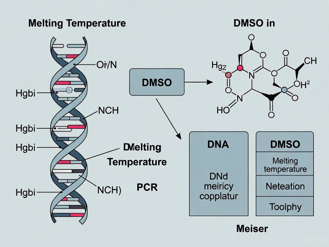
DMSO in PCR: Mechanisms and Optimization for DNA Melting Temperature Control
This article provides a comprehensive analysis of Dimethyl sulfoxide (DMSO) as a critical PCR enhancer, detailing its fundamental role in reducing DNA melting temperature (Tm) and destabilizing secondary structures, particularly...
DMSO in PCR: Mechanisms and Optimization for DNA Melting Temperature Control
Abstract
This article provides a comprehensive analysis of Dimethyl sulfoxide (DMSO) as a critical PCR enhancer, detailing its fundamental role in reducing DNA melting temperature (Tm) and destabilizing secondary structures, particularly in GC-rich templates. It offers methodological guidance for application and concentration optimization (typically 2-10%), alongside troubleshooting for common PCR challenges. The content validates DMSO's effects through single-molecule biophysical studies and compares its efficacy against other enhancers like formamide and betaine. Aimed at researchers and drug development professionals, this resource synthesizes current evidence to improve PCR sensitivity, specificity, and success rates in complex diagnostic and research applications.
The Biophysical Basis: How DMSO Modifies DNA Structure and Melting Dynamics
Dimethyl sulfoxide (DMSO) exerts significant effects on DNA structure and stability through complex mechanisms involving direct molecular interactions with both DNA and water molecules. This whitepaper synthesizes current research quantifying how DMSO promotes DNA denaturation by reducing persistence length, facilitating local denaturation bubbles, and lowering melting temperature—findings with critical implications for polymerase chain reaction (PCR) optimization. Single-molecule studies reveal that DMSO induces DNA flexibility even at low concentrations, while spectroscopic analyses elucidate the molecular aggregates DMSO forms with water that underpin its cryoprotective and denaturant properties. Understanding these fundamental interactions provides a rational basis for employing DMSO as a reagent in molecular biology applications, particularly in enhancing PCR specificity and efficiency.
DMSO is a polar aprotic solvent with widespread applications in biological research, including cryopreservation, drug delivery, and as a component in enzymatic reactions processing DNA. Within PCR research, DMSO is recognized for improving amplification efficiency, particularly for targets with high GC content or complex secondary structures. Its ability to lower DNA melting temperature (Tm) is well-documented empirically; however, the fundamental molecular mechanisms underlying this effect require elucidation. This technical guide integrates multi-faceted research approaches—from single-molecule biophysics to computational chemistry—to delineate the precise mechanisms through which DMSO interacts with water and DNA to influence DNA conformation and stability. Framing these findings within PCR research provides a mechanistic foundation for experimental optimization.
Molecular Interactions of DMSO with Water
The pronounced effects of DMSO on DNA begin with its fundamental interaction with the aqueous solvent environment. DMSO exhibits strongly non-ideal mixing behavior with water, manifested in various physicochemical properties including density, viscosity, and diffusion constants [1].
Hydrogen-Bonded Complex Formation
Research utilizing Fourier-transform microwave spectroscopy and molecular dynamics simulations has identified specific stoichiometric complexes that form between DMSO and water molecules:
- DMSO·2H2O Complex: Predominates in diluted aqueous solutions, where two water molecules form hydrogen bonds with the sulfoxide oxygen atom of a single DMSO molecule [2].
- 2DMSO·H2O Complex: Becomes predominant in DMSO-rich mixtures, where a single water molecule bridges two DMSO molecules through hydrogen bonding [2].
The structure of the 1:1 DMSO-water complex has been experimentally determined, revealing that water acts as a donor of a primary O-H···O=S hydrogen bond to the oxygen atom of DMSO while simultaneously accepting two weak C-H···Ow bonds from the methyl hydrogen atoms of DMSO [1]. This complex maintains overall Cs symmetry, with water residing in the symmetry plane of DMSO.
Impact on Water Structure and Dynamics
These specific molecular associations significantly alter the dynamics and hydrogen-bonding network of water:
- Librational Dynamics: The characteristic librational band of pure water around 700 cm-1 splits into two distinct bands in DMSO-rich mixtures, reflecting the restricted reorientational dynamics of water molecules bound within 2DMSO·H2O aggregates [2].
- Hydrogen-Bond Network Disruption: DMSO disrupts the natural donor/acceptor balance in water by accepting hydrogen bonds through its oxygen lone pairs while participating as a weak donor through its methyl C-H bonds [1].
Table 1: Physicochemical Properties of DMSO-Water Mixtures
| DMSO Concentration | Predominant Molecular Complex | Effect on Water Dynamics | Experimental Signature |
|---|---|---|---|
| Low (xD ≈ 0.30) | DMSO·2H2O | Modified H-bond network | Maximum deviation from ideal mixing |
| High (xD ≈ 0.80) | 2DMSO·H2O | Restricted librational motion | Split FIR absorption band |
Effects of DMSO on DNA Structure and Mechanics
DMSO directly influences DNA conformation and mechanical properties through mechanisms that operate below the bulk melting transition, as revealed by single-molecule techniques.
DNA Flexibility and Persistence Length
Atomic force microscopy (AFM) and magnetic tweezers studies quantitatively demonstrate that DMSO significantly increases DNA flexibility:
- Persistence Length Reduction: The bending persistence length of DNA decreases linearly with DMSO concentration up to 20% by approximately 0.43% per percent DMSO [3]. In 3% DMSO solution, the persistence length decreases dramatically to approximately 12 nm from about 50 nm without DMSO [4].
- Conformational Compaction: AFM imaging reveals a systematic decrease in the mean-squared end-to-end distance of DNA by 1.2% per percent DMSO, indicating moderate compaction of DNA conformations [3].
Local Denaturation and Structural Defects
Even low DMSO concentrations induce localized structural disruptions in double-stranded DNA:
- Local Denaturation Bubbles: AFM imaging directly visualizes local DNA denaturation, including kinks and bubbles, at DMSO concentrations as low as 0.1% [4]. These localized denatured regions represent flexible segments within the overall DNA chain.
- Pre-melting Transition: The initial mechanism of DNA denaturation involves increased flexibility due to partial hydrogen bond breaking before complete local separation of the complementary strands occurs [4].
Table 2: Quantitative Effects of DMSO on DNA Mechanical Properties
| DMSO Concentration (%) | Persistence Length (nm) | Reduction in Mean-Squared End-to-End Distance | Experimental Method |
|---|---|---|---|
| 0 | ~50 | Baseline | AFM [4] |
| 0.1 | - | - | Local denaturation observed via AFM [4] |
| 3 | ~12 | - | AFM [4] |
| 20 | - | 24% decrease | Magnetic tweezers/AFM [3] |
| Per % DMSO | 0.43% decrease | 1.2% decrease | Magnetic tweezers/AFM [3] |
Impact on DNA Melting Temperature in PCR
The molecular interactions between DMSO, water, and DNA collectively contribute to the observed depression of DNA melting temperature, a property exploited in PCR optimization.
Melting Temperature Depression
DMSO consistently lowers the melting temperature of DNA duplexes, with studies demonstrating:
- Concentration-Dependent Effect: The melting temperature decreases progressively with increasing DMSO concentration, with observable effects beginning at very low concentrations [4] [5].
- UV Absorbance Changes: Traditional ensemble studies indicate that at least 10% DMSO is required to observe significant hyperchromicity at 260 nm, indicating bulk DNA denaturation at room temperature [4].
Mechanism in PCR Applications
In PCR, DMSO serves multiple functions that enhance amplification efficiency:
- Reduction of Secondary Structures: By lowering the melting temperature, DMSO prevents the formation of stable secondary structures in DNA templates and primers, particularly in GC-rich regions [5].
- Inhibition of Reannealing: DMSO binds to DNA and inhibits reannealing of denatured DNA, providing primers greater access to their complementary binding sites [5].
- Enhanced Specificity and Yield: These combined effects result in improved primer annealing specificity and increased amplification yield [5].
Experimental Methodologies
Atomic Force Microscopy (AFM) for DNA Visualization
AFM provides direct visualization of DNA conformational changes induced by DMSO:
- Sample Preparation: DNA samples (plasmid or linear) are diluted to 1 ng/μL in solutions containing varying DMSO concentrations (0.1-10%) in 1 mM Tris-HCl buffer (pH 7.8) and incubated for 30 minutes at room temperature [4]. The solution is deposited onto APTES-treated mica substrate, incubated for 3 minutes, rinsed with deionized water, and dried with nitrogen gas [4].
- Imaging and Analysis: Samples are scanned using AC mode AFM with silicon probes. DNA contour length, denaturation regions, and end-to-end distances are quantified using image analysis software such as ImageJ [4]. Typically, 25 DNA molecules are analyzed per concentration group to ensure statistical significance.
Magnetic Tweezers for Mechanical Measurements
Magnetic tweezers provide quantitative measurements of DNA mechanical properties:
- Force-Extension Measurements: DNA molecules tethered between a magnetic bead and surface are subjected to controlled forces, allowing determination of persistence length from extension curves [3].
- Twist Measurements: Torque is applied to twisted DNA molecules to assess changes in twist modulus and melting torque under DMSO influence [3].
Spectroscopic Techniques
- UV Spectrophotometry: Traditional melting curves monitor absorbance at 260 nm to detect bulk DNA denaturation [4].
- Fourier-Transform Microwave Spectroscopy: Provides high-resolution structural information on DMSO-water complexes in the gas phase [1].
- Far-Infrared Spectroscopy: Detects changes in water librational dynamics (∼700 cm-1) due to DMSO-water aggregation [2].
Molecular Mechanism Workflow
The following diagram illustrates the sequential molecular events through which DMSO influences DNA structure and stability:
The Scientist's Toolkit: Research Reagent Solutions
Table 3: Essential Reagents for Investigating DMSO-DNA Interactions
| Reagent/Material | Specifications | Function/Application |
|---|---|---|
| DMSO | High purity (≥99.8%), molecular biology grade | Primary denaturant; ensure minimal impurities that could affect nucleic acids |
| DNA Substrates | Plasmid (e.g., pBR322) and linear DNA of defined length (e.g., 5000 bp) | Standardized substrates for reproducibility in structural studies |
| APTES-treated Mica | Freshly cleaved mica modified with 3-aminopropyltriethoxysilane (APTES) | AFM substrate for DNA immobilization with appropriate surface charge |
| Buffers | Tris-HCl (pH 7.8), HEPES (pH 7.5) with appropriate ionic strength | Maintain physiological pH and ionic conditions; HEPES preferred for liquid AFM |
| Salts | MgCl₂, KCl, NiCl₂ (ultra-pure) | Modulate DNA-surface interactions for AFM; Ni²⁺ enhances DNA-mica binding |
| Ultrapure Water | Milli-Q grade (18.2 MΩ·cm) | Minimize contaminants that could interfere with molecular interactions |
The fundamental mechanism of DMSO's interaction with DNA and water molecules involves a sophisticated hierarchy of molecular events. Initially, DMSO forms specific stoichiometric complexes with water molecules (DMSO·2H2O and 2DMSO·H2O) that alter the hydrogen-bonding network and dynamics of the aqueous environment. These modified solvent properties subsequently enhance DNA flexibility by reducing persistence length and inducing local denaturation bubbles through partial hydrogen bond breaking. The culmination of these effects is a systematic reduction in DNA melting temperature, which provides the mechanistic basis for DMSO's utility in PCR applications. By reducing secondary structure formation in GC-rich templates and improving primer accessibility, DMSO serves as a powerful adjunct in molecular biology applications. Future research directions include elucidating sequence-specific effects of DMSO and optimizing its concentration in conjunction with other PCR enhancers for challenging amplification targets.
Dimethyl sulfoxide (DMSO) is a polar aprotic solvent commonly used in polymerase chain reaction (PCR) protocols to facilitate the amplification of complex DNA templates, particularly those with high GC-content [6]. Its utility stems from its ability to lower the melting temperature (Tm) of double-stranded DNA, thereby promoting complete strand separation during the denaturation step and preventing the reformation of stable secondary structures [7] [8]. This whitepaper provides a detailed technical examination of the quantifiable effects of DMSO on DNA denaturation, situating this phenomenon within the broader context of its impact on DNA conformations and mechanics, and providing actionable experimental protocols for research and drug development applications.
Quantitative Effects of DMSO on DNA Mechanics and Conformation
Recent single-molecule studies have systematically quantified the impact of DMSO on DNA physical properties. The effects are concentration-dependent, with significant changes observed especially at higher concentrations.
Table 1: Quantitative Effects of DMSO on DNA Mechanical Properties
| Property | Effect of DMSO | Concentration Range Studied | Measurement Technique |
|---|---|---|---|
| Bending Persistence Length | Decreases linearly by (0.43 ± 0.02%) per %-DMSO [9] | 0–20% DMSO | Magnetic Tweezers Force-Extension |
| Helical Twist | Remains largely unchanged; slight unwinding at >20% DMSO [9] | 0–60% DMSO | Magnetic Tweezers Twist Measurements |
| Mean-Squared End-to-End Distance | Decreases by 1.2% per %-DMSO, indicating compaction [9] | 0–60% DMSO | AFM Imaging |
| Melting Torque | Reduction observed, indicating lowered stability against melting [9] | 0–60% DMSO | Magnetic Tweezers Twist Measurements |
These mechanical changes rationalize the utility of DMSO in PCR. The reduction in bending persistence length and overall compaction of DNA conformations suggest that DMSO introduces local flexibility, effectively destabilizing the duplex and lowering the energy required for denaturation [9] [3].
DMSO in PCR and Mutation Scanning: Protocols and Workflows
Enhancing PCR Amplification
DMSO is a critical additive for amplifying challenging templates. It is typically used at concentrations between 2.5% and 5% (v/v) in the PCR mixture [6].
Table 2: DMSO Application in Nucleic Acid Protocols
| Application | Recommended DMSO Concentration | Primary Function | Key Considerations |
|---|---|---|---|
| GC-Rich PCR | 2.5% to 5% [6] | Destabilizes DNA duplex, prevents secondary structure [7] | Optimize annealing temperature (may need lowering) [8] |
| High-Resolution Melting (HRM) | 5% to 10% [7] | Increases sensitivity by enlarging Tm difference between wild-type and mutant DNA [7] | Can detect mutations with ~1% abundance (vs. 3-10% without DMSO) [7] |
| Full-COLD-PCR-HRM | 5% to 10% [7] | Further enriches low-level mutations prior to HRM detection | Enables detection of mutations at 0.2–0.3% abundance [7] |
Experimental Workflow for HRM with DMSO
The following diagram illustrates a protocol for detecting low-abundance mutations using DMSO-enhanced HRM.
Figure 1: DMSO-enhanced HRM mutation scanning workflow. DMSO is added post-PCR to destabilize DNA, enhancing the detection of low-abundance mutations [7].
Detailed Methodology:
- PCR Amplification: Perform conventional PCR or full-COLD-PCR in a 25 µL reaction mixture containing template DNA (e.g., 10 ng genomic DNA), 1X PCR buffer, 200 nM of each primer, 200 µM dNTPs, 0.8X LCGreen Plus+ fluorescence dye, and 0.5 units of a high-fidelity DNA polymerase (e.g., Phusion) [7].
- DMSO Addition: Transfer 10 µL of the PCR product to a 96-well plate suitable for HRM analysis. Add DMSO to achieve a final concentration of 5%, 7%, or 10%. Mix thoroughly and overlay with 20 µL of mineral oil to prevent evaporation [7].
- High-Resolution Melting: Perform HRM on a dedicated system (e.g., LightScanner). Program a melting curve from 65°C to 95°C with small temperature increments (e.g., 0.2°C) and a hold before each fluorescence acquisition [7].
- Data Analysis: Use the instrument's software to analyze the melting profiles. Set the software sensitivity to a defined level (e.g., 1.2) to compute DNA variant groups and distinguish mutant samples from wild-type controls [7].
The Scientist's Toolkit: Essential Research Reagents
Table 3: Essential Reagents for DMSO-DNA Studies
| Reagent / Material | Function | Example Use Case |
|---|---|---|
| DMSO (Dimethyl Sulfoxide) | Polar aprotic solvent; destabilizes DNA duplex, lowers Tm, reduces secondary structure [7] [6]. | Added to PCR or HRM reactions to improve amplification or detection sensitivity [7] [6]. |
| LCGreen Plus+ | Saturating fluorescent DNA dye; used for High-Resolution Melting analysis [7]. | Detects subtle differences in DNA melting profiles between wild-type and mutant sequences [7]. |
| Phusion High-Fidelity DNA Polymerase | Thermostable enzyme with high fidelity; used for precise amplification prior to HRM [7]. | Amplifying target regions (e.g., TP53 exon 8) for subsequent mutation scanning [7]. |
| Full-COLD-PCR Protocol | A PCR method that enriches variant alleles from a mixture of wild-type and mutant DNA [7]. | Selectively amplifying low-abundance mutations (<1%) to improve their detection in downstream HRM [7]. |
Mechanism of Action: How DMSO Affects DNA Stability
The fundamental mechanism by which DMSO lowers DNA melting temperature is its ability to destabilize the DNA duplex. DMSO is a potent hydrogen-bond acceptor that competes with DNA base pairs for hydrogen-bonding interactions, thereby weakening the inter-strand bonds that stabilize the double helix [7]. Furthermore, its effects on DNA mechanics, such as reducing the bending persistence length, indicate the introduction of local flexibility, which can be modeled as flexible segments or local defects that facilitate strand separation [9]. This combination of hydrogen-bond disruption and structural destabilization lowers the thermal energy required to denature the DNA, which is directly exploited in PCR to melt GC-rich templates and in HRM to enhance the discrimination between heteroduplex and homoduplex DNA [7] [6].
While the capacity of dimethyl sulfoxide (DMSO) to lower the melting temperature ((T_m)) of DNA is well-documented in PCR research, its effects on fundamental biophysical DNA properties below the melting temperature are less explored. This whitepaper synthesizes recent single-molecule and biophysical studies demonstrating that DMSO induces significant changes to DNA persistence length (a measure of stiffness) and helical conformation, even at low concentrations prevalent in experimental assays. These alterations—increased flexibility, local denaturation, and transitions to non-canonical DNA forms—have profound implications for experimental outcomes and our understanding of DNA mechanics. A comprehensive understanding of these effects is crucial for researchers and drug development professionals utilizing DMSO as a solvent in genetic and molecular assays.
Dimethyl sulfoxide (DMSO) is a polar aprotic solvent ubiquitously employed in biological research. In the context of PCR and related molecular techniques, it is primarily valued for its ability to lower the melting temperature of DNA by destabilizing hydrogen bonds between base pairs, thereby facilitating the denaturation of complex secondary structures that can impede polymerase progression [3]. However, a growing body of evidence indicates that the influence of DMSO extends beyond the modulation of (T_m). This technical guide details the effects of DMSO on two critical parameters of DNA mechanics: its persistence length (defining its bending flexibility) and its helical conformation (defining its twist and base-pairing status). Understanding these subtler impacts is essential for the accurate interpretation of data from assays involving DNA mechanics, such as gel electrophoresis, DNA-protein interaction studies, and nanotechnology applications.
Quantitative Effects of DMSO on DNA Mechanics
The following sections synthesize quantitative findings on how DMSO alters the physical properties of DNA, with data summarized in Table 1.
Modulation of DNA Persistence Length
The persistence length of DNA is a key determinant of its stiffness and its packaging within the cell. Multiple studies using Atomic Force Microscopy (AFM) and magnetic tweezers have conclusively shown that DMSO causes a concentration-dependent decrease in DNA persistence length, indicating increased flexibility.
- Significant Reduction at Low Concentrations: A foundational study found that the persistence length of DNA decreased dramatically from approximately 50 nm in the absence of DMSO to about 12 nm in the presence of just 3% DMSO [10]. This finding was directly observed through AFM imaging, which revealed increased kinking and bending.
- Linear Decrease at Moderate Concentrations: Magnetic tweezers experiments have quantified a moderate and linear decrease in bending persistence length of (0.43 ± 0.02%) per percent DMSO, for concentrations up to 20% [3]. Concurrently, AFM imaging measured a compaction of DNA conformations, with the mean-squared end-to-end distance decreasing by 1.2% per percent DMSO [3].
- Proposed Mechanism: The observed flexibility is attributed to the induction of local denaturation or flexible defects. DMSO disrupts the hydrogen-bonding network of water and directly interacts with DNA bases, leading to partial breaking of hydrogen bonds within the double helix before full strand separation occurs [10]. This creates flexible "hinges" or "bubbles" that allow the DNA molecule to bend more easily.
Alterations to Helical Twist and Conformation
Beyond bending rigidity, DMSO affects the twist and canonical structure of the DNA helix.
- Helical Twist Stability: Up to 20% DMSO, the helical twist of DNA remains largely unchanged. However, at higher concentrations (≥20%), a slight unwinding of the helix has been reported [3]. Furthermore, magnetic tweezers twist measurements demonstrate a reduction in the torque required to melt DNA in the presence of DMSO, consistent with its role as a denaturant [3].
- Induction of Non-B-Form DNA: Fourier Transform Infrared (FT-IR) spectroscopy on cellular systems has revealed that even low concentrations of DMSO (0.1–1.5%) can induce the formation of Z-DNA, a left-handed helical conformation [11]. Molecular docking studies suggest that DMSO can directly stabilize this alternate DNA structure [11].
- Local Denaturation ("Bubbling"): AFM studies provide direct visual evidence of local denaturation, observed as kinks and bubbles on plasmid and linear DNA molecules, at DMSO concentrations as low as 0.1% [10]. This signifies that DMSO promotes localized separation of the two strands without causing global melting.
Table 1: Quantitative Effects of DMSO on DNA Physical Parameters
| DMSO Concentration (%) | Persistence Length | Helical Twist | Key Conformational Observations |
|---|---|---|---|
| 0.1% | --- | --- | Local denaturation (kinks/bubbles) observed via AFM [10] |
| 0.1% - 1.5% | --- | --- | Formation of Z-DNA in cellular systems [11] |
| 3% | Decreases to ~12 nm from ~50 nm [10] | --- | --- |
| Up to 20% | Linear decrease of (0.43 ± 0.02%) per %-DMSO [3] | Largely unchanged [3] | Mean-squared end-to-end distance decreases by 1.2% per %-DMSO [3] |
| >20% | --- | Slight unwinding [3] | --- |
Detailed Experimental Methodologies
To empower researchers in validating and building upon these findings, this section outlines the key experimental protocols used in the cited studies.
Single-Molecule Analysis via Magnetic Tweezers
Magnetic tweezers were used to probe the mechanical properties of single DNA molecules tethered between a glass surface and a magnetic bead [3].
- Sample Preparation: A DNA construct with known digoxigenin and biotin labels on opposite ends is bound to an anti-digoxigenin-coated glass surface and a streptavidin-coated magnetic bead.
- Buffer Conditions: Experiments are conducted in a buffer solution (e.g., PBS) with DMSO concentrations ranging from 0% to 60%.
- Force-Extension Measurements: A magnet controls the force applied to the bead, stretching the DNA molecule. The extension of the DNA is measured as a function of the applied force. This data is fitted to a worm-like chain model to extract the bending persistence length.
- Twist Measurements: The magnet is rotated to introduce supercoils into the DNA. The resulting change in extension and the torque at which DNA denatures (melting torque) are measured to assess the effects of DMSO on twist mechanics and stability.
Direct Visualization via Atomic Force Microscopy (AFM)
AFM allows for direct imaging of individual DNA molecules to assess conformation and flexibility [10].
- Substrate Preparation: A mica surface is functionalized with 3-aminopropyltriethoxysilane (APTES) to create a positively charged surface for firm adsorption of negatively charged DNA.
- DNA Deposition: Plasmid (e.g., pBR322) or linear DNA is diluted to a final concentration of ~1 ng/μL in a buffer containing a specific concentration of DMSO (e.g., 0.1% to 10%) and incubated for 30 minutes at room temperature.
- Sample Washing and Drying: A aliquot of the DNA-DMSO solution is deposited onto the APTES-mica, incubated briefly, rinsed with deionized water, and dried with a nitrogen gas stream.
- Imaging and Analysis: The sample is scanned in AC mode in air. Software like ImageJ is used to trace DNA molecules and measure contour lengths, end-to-end distances, long/short axes, and the number and size of kinks or denaturation bubbles. Persistence length can be calculated from the end-to-end distance and contour length of many molecules.
Spectroscopic and Computational Analysis of DNA Conformation
FT-IR spectroscopy and molecular modeling can detect gross molecular and conformational changes [11].
- Cell Culture and Treatment: Epithelial cells (e.g., HCT-116, SW-480) are treated with low concentrations of DMSO (0.1–1.5%) for 24 hours.
- FT-IR Spectroscopy: Treated and untreated cells are analyzed using Attenuated Total Reflectance (ATR) FT-IR spectroscopy. The spectrum, particularly regions corresponding to nucleic acids (e.g., 1250–1200 cm⁻¹ for phosphate backbone vibrations), is collected.
- Data Analysis: Pattern recognition algorithms like Principal Component Analysis (PCA) and Linear Discriminant Analysis (LDA) are applied to segregate treated from untreated samples based on spectral data. Specific band intensities (e.g., at ~915 cm⁻¹ for ribose rings) are analyzed to quantify nucleic acid content and conformational changes.
- Molecular Docking: Computational docking simulations are performed to model the interaction energy and binding stability between DMSO molecules and different DNA conformations (e.g., B-DNA vs. Z-DNA), providing a theoretical basis for stabilization of non-canonical forms.
The Scientist's Toolkit: Essential Research Reagents
Table 2: Key Reagents and Materials for Investigating DMSO-DNA Interactions
| Reagent/Material | Function in Experimental Context |
|---|---|
| DMSO (High Purity, ≥99.8%) | The chemical agent of interest; used to prepare concentration gradients in aqueous buffers for treatment [10]. |
| Plasmid DNA (e.g., pUC19, pBR322) | A well-defined, circular DNA model system for strand break assays, AFM imaging, and conformational studies [10] [12]. |
| APTES-Treated Mica | Provides a positively charged, atomically flat substrate essential for immobilizing DNA molecules for AFM imaging [10]. |
| Magnetic Beads (Streptavidin-Coated) | Coupled with biotin-labeled DNA for tethering and mechanical manipulation in magnetic tweezers experiments [3]. |
| Radiolabeled or Fluorescent Nucleotides | Enable sensitive detection of DNA strand breaks in gel electrophoresis assays or visualization under microscopy [12]. |
Conceptual and Experimental Workflow
The following diagrams illustrate the proposed mechanism of DMSO action and a generalized experimental workflow.
Conceptual Framework of DMSO Effects on DNA
Generalized Experimental Workflow for DNA Mechanics Analysis
Discussion and Implications for Research
The data unequivocally demonstrates that DMSO's influence on DNA is not limited to lowering the (T_m). The observed reduction in persistence length and induction of conformational changes like Z-DNA and local denaturation have several critical implications:
- Experimental Artifacts: In assays sensitive to DNA flexibility or topology (e.g., gel mobility, DNA looping, protein-binding assays), the presence of DMSO, even at low "carrier" concentrations (0.1-1.5%), may introduce unintended artifacts by altering the mechanical and conformational landscape of the DNA [11] [10].
- Biological Relevance: The DMSO-driven stabilization of Z-DNA is particularly significant, as this non-canonical structure is involved in gene regulation [11]. This suggests DMSO could indirectly influence cellular processes like gene expression and differentiation by modulating DNA topology.
- Radiobiological Research: DMSO's role as a radical scavenger can protect DNA from indirect radiation-induced strand breaks, a property useful for dissecting the mechanisms of radiation damage [12]. However, for high-LET emitters like α-particles, a component of direct, non-scavengeable damage remains [12].
- Guidelines for Use: Researchers must report and standardize DMSO concentrations across experiments. For highly sensitive mechanical studies, alternative solvents or minimal DMSO concentrations (≤5%) are recommended unless the flexibilizing effect is specifically desired.
The journey "Beyond (T_m)" reveals a complex interplay between DMSO and DNA mechanics. This whitepaper has detailed how DMSO systematically softens the DNA polymer, reduces the energy barrier for strand separation, and promotes alternative helical conformations. These effects, quantifiable at single-molecule and ensemble levels, underscore that DMSO is not a passive solvent but an active modulator of DNA structure. For the scientific community, a deeper appreciation of these phenomena is essential for refining experimental designs, accurately interpreting data in PCR research and beyond, and harnessing these properties for advanced applications in nanotechnology and drug development. Future work should focus on mapping the sequence dependence of these effects and their consequences in vivo.
Dimethyl sulfoxide (DMSO) serves as a pivotal solvent and additive within molecular biology, particularly influencing DNA thermodynamics across various experimental contexts. While its capacity to reduce DNA melting temperature (Tm) is empirically established, the underlying biophysical mechanisms connecting local structural alterations to global DNA properties demand systematic elucidation. This whitepaper delineates the concentration-dependent effects of DMSO on DNA, from the induction of localized flexible defects to consequent macroscale structural compaction and torsional stress reduction, with specific emphasis on implications for polymerase chain reaction (PCR) research and drug development. Recent single-molecule and biophysical studies provide a quantitative framework for understanding these effects, revealing moderate but systematic changes in DNA mechanics at concentrations commonly employed in biochemical assays (≤20%) [9] [3]. This analysis provides researchers with predictive models and practical guidelines for leveraging DMSO's properties while mitigating unintended consequences in experimental outcomes.
Quantitative Effects of DMSO on DNA Structure and Mechanics
The concentration-dependent relationship between DMSO and DNA structural parameters follows predictable, quantifiable trends. Single-molecule techniques, including magnetic tweezers and atomic force microscopy (AFM), have precisely characterized these effects across physiologically relevant DMSO concentrations (0–20%) [9].
Table 1: Quantitative Effects of DMSO on DNA Mechanical Properties
| DNA Property | Measurement Technique | Effect per %-DMSO | Proposed Mechanism |
|---|---|---|---|
| Bending Persistence Length | Magnetic Tweezers (Force-extension) | Decrease by ( 0.43 \pm 0.02\% ) [9] | Introduction of locally flexible regions or kinks [9] |
| Mean-Squared End-to-End Distance | AFM Imaging | Decrease by ( 1.2\% ) [9] | Global compaction due to increased flexibility [9] |
| Helical Twist | Magnetic Tweezers (Twist measurements) | Largely unchanged (up to 20%); slight unwinding at higher concentrations [9] | Alteration of base-stacking interactions [9] |
| Melting Torque | Magnetic Tweezers | Reduction [9] | Lowered energy barrier for strand separation [9] |
Beyond these mechanical parameters, DMSO significantly influences DNA thermal stability. In PCR applications, the addition of 10% DMSO can lower the annealing temperature required by 5.5–6.0°C [13]. This Tm reduction is attributed to DMSO's disruption of the hydrogen-bonding network of water and its direct interactions with DNA bases, thereby destabilizing the double-stranded state [9] [14]. The effect is sufficiently robust that DMSO concentrations in PCR are commonly optimized in 2% increments to balance enhanced specificity against potential polymerase inhibition [13] [15].
Underlying Mechanisms: From Local Defects to Global Changes
The concentration-dependent effects of DMSO on DNA originate from localized disruptions that propagate to alter global macromolecular structure and stability.
Induction of Local Structural Defects
At the molecular level, DMSO acts as a polar aprotic solvent that alters the solvation shell surrounding the DNA duplex. Evidence suggests that DMSO molecules preferentially interact with DNA bases, disrupting base-stacking interactions and hydrogen bonding within the double helix [9]. This interaction creates locally flexible regions or kinks, effectively acting as structural defects. Computational models, including coarse-grained Monte Carlo simulations, represent these as segments with increased flexibility interspersed within the semiflexible DNA polymer chain. The density of these flexible segments increases linearly with DMSO concentration, providing a mechanistic rationale for the observed linear decrease in persistence length [9].
Propagation to Global Structural Compaction
The local defects introduced by DMSO have direct consequences for the global architecture of the DNA molecule. The systematic reduction in the mean-squared end-to-end distance, as quantified by AFM imaging, signifies a moderate compaction of DNA conformations [9]. This compaction occurs because the introduction of flexible points allows the DNA to bend more easily, leading to a more compact equilibrium conformation. This effect is visually confirmed by AFM examinations, which show that DMSO can create "locally loose regions" in negatively supercoiled plasmids, increasing the availability of single-stranded DNA regions that are crucial for the activity of certain enzymes like type IA topoisomerases [16].
Figure 1: Mechanistic Pathway of DMSO Effects on DNA. The diagram illustrates the concentration-dependent pathway, from local defect formation at low DMSO concentrations to global structural and functional changes at higher concentrations.
Experimental Protocols for Characterizing DMSO Effects
Single-Molecule Analysis of DNA Mechanics
Protocol: Magnetic Tweezers for DNA Mechanics in DMSO [9]
- DNA Substrate: Lambda phage DNA or similar long DNA fragments (e.g., ~20 kbp) with biotin/digoxigenin labels for surface and bead attachment.
- Sample Chamber: Flow chamber constructed from glass coverslips functionalized with anti-digoxigenin.
- Buffer Conditions: Standard physiological buffer (e.g., 10-100 mM Tris or phosphate buffer, pH 7.0-7.5, with 50-150 mM NaCl).
- DMSO Titration: Prepare reaction buffers with DMSO concentrations ranging from 0% to 60% (v/v). For low-concentration studies (≤20%), use increments of 2-5%.
- Data Acquisition:
- Force-Extension Curves: For each DMSO condition, measure the DNA extension (z) as a function of applied force (F, typically 0.01-10 pN).
- Twist Measurements: For torsionally constrained DNA, measure the extension as a function of the number of turns applied to the magnetic bead.
- Data Analysis:
- Fit force-extension data to the Worm-Like Chain (WLC) model to extract the persistence length.
- From twist measurements, determine the twist persistence length and melting torque.
- Plot mechanical parameters (persistence length, twist) versus DMSO concentration to quantify linear coefficients.
AFM Imaging of DNA Conformational Changes
Protocol: AFM Imaging of DNA Conformations in DMSO [9] [16]
- Sample Preparation:
- DNA Sample: Use a suitable plasmid DNA (e.g., pBR322) or linear DNA at low concentration (0.1-0.01 μg/mL) in a buffer containing 20 mM Tris-HCl, pH 7.0.
- DMSO Incubation: Incubate DNA sample with target DMSO concentration for 5-15 minutes at room temperature.
- Substrate Preparation: Use APS-mica (aminopropyl silatrane-functionalized mica) as an adhesive surface. Place 5-10 μL of the DNA-DMSO solution onto freshly prepared APS-mica and incubate for 5 minutes.
- Imaging: Rinse surface gently with distilled water and air dry. Acquire images in Tapping Mode in air using an AFM with a silicon cantilever.
- Image Analysis: Trace individual DNA molecules to measure end-to-end distances and contour lengths. Calculate the mean-squared end-to-end distance (
) for a population of molecules (n > 100) at each DMSO concentration.
Implications for PCR Research and Assay Design
The effects of DMSO on DNA mechanics directly inform its utility as a PCR enhancer, particularly for challenging templates.
Table 2: DMSO in PCR: Applications and Optimization Guidelines
| Application | Recommended [DMSO] | Mechanistic Basis | Protocol Adjustment |
|---|---|---|---|
| Amplification of GC-Rich Templates [13] [15] | 3–10% [13] | Lowers Tm, disrupts stable secondary structures [9] | Lower annealing temperature by ~0.6°C per %-DMSO [13] |
| Reduction of Ski-Slope Effect in Direct PCR [17] | ~3.75% | Preferentially improves amplification efficiency of large amplicons [17] | Add DMSO directly to master mix with Prep-n-Go buffer [17] |
| General Specificity Enhancement | 2.5–5% [15] | Reduces non-specific primer binding by lowering Tm [9] | Optimize in 2% increments [13] |
The "ski-slope effect" – characterized by a systematic decrease in PCR product yield with increasing amplicon size – is markedly reduced by 3.75% DMSO. This occurs because DMSO preferentially enhances the amplification efficiency of larger DNA sequences, thereby improving intra-color peak balance in multiplex PCR fragment analysis [17]. Furthermore, DMSO's ability to promote single-stranded DNA regions in supercoiled plasmids explains its role in enhancing the activity of enzymes like E. coli topoisomerase I, which requires single-stranded regions for efficient relaxation of negative supercoils [16].
Figure 2: DMSO Integration Workflow for PCR Optimization. This workflow guides the decision-making process for incorporating DMSO into PCR assays based on template characteristics and desired outcomes.
The Scientist's Toolkit: Essential Reagents and Materials
Table 3: Research Reagent Solutions for Studying DMSO-DNA Interactions
| Reagent / Material | Function / Application | Example Use Case |
|---|---|---|
| Polar Aprotic Solvent (DMSO) | Disrupts DNA base-stacking and hydrogen bonding; reduces Tm. | Additive in PCR of GC-rich templates [13]; solvent for fluorescent dyes in enzymatic assays [9]. |
| Magnetic Tweezers Setup | Single-molecule manipulation and measurement of DNA mechanics. | Quantifying DMSO-induced changes in persistence length and melting torque [9]. |
| Atomic Force Microscope (AFM) | High-resolution imaging of DNA conformations on a surface. | Visualizing and quantifying DMSO-induced DNA compaction [9] [16]. |
| Type IA Topoisomerase (e.g., EcTopo I) | Enzyme that relaxes negatively supercoiled DNA, requiring single-stranded regions. | Probe for DMSO-induced single-stranded character in supercoiled plasmids [16]. |
| APS-Mica (Aminopropyl Silatrane-Mica) | Chemically modified substrate for stable DNA deposition for AFM. | Immobilizing DNA molecules from DMSO-containing solutions for AFM analysis [16]. |
| Saturating DNA Dyes (e.g., SYBR Green I) | Fluorescent dyes for monitoring DNA melting in real-time. | High-resolution melting analysis for Tm determination in various [DMSO] [18]. |
The effects of DMSO on DNA are fundamentally concentration-dependent, initiating with localized structural defects at the molecular level and culminating in global changes to DNA architecture and stability. Quantitative models now establish that each percent of DMSO linearly reduces the bending persistence length by approximately 0.43% and compacts the mean-squared end-to-end distance by 1.2% within the critical 0-20% concentration range [9]. These biophysical insights provide a mechanistic foundation for DMSO's established role in PCR optimization, particularly for mitigating amplification challenges associated with GC-rich secondary structures and long amplicons. For researchers in drug development and molecular biology, these findings enable the predictive tuning of DMSO concentrations to achieve desired DNA stability and structural outcomes while maintaining the integrity of enzymatic reactions. As such, a precise understanding of DMSO's concentration-dependent action transitions its use from an empirical additive to a rationally applied tool in the molecular sciences.
Practical Protocols: Implementing DMSO for Robust PCR Amplification
Optimal DMSO Concentration Ranges for Standard and Specialty PCR
Within the rigorous framework of polymerase chain reaction (PCR) research, the precise manipulation of DNA melting temperature is a fundamental thesis. Achieving optimal specificity and yield, particularly when dealing with challenging templates, often necessitates the use of chemical additives. Dimethyl sulfoxide (DMSO) is one of the most prevalent additives employed to modulate the PCR environment. This technical guide provides an in-depth analysis of optimal DMSO concentration ranges, establishing its role within the broader context of DNA denaturation dynamics. For researchers, scientists, and drug development professionals, a meticulous understanding of DMSO concentration is not merely beneficial but essential for the reproducibility and success of sophisticated molecular assays, from next-generation sequencing library preparation to cloning and mutation detection [19].
The Mechanistic Role of DMSO in Modifying DNA Thermodynamics
DMSO exerts its primary effect in PCR by altering the physical properties of DNA and the reaction environment. Its mechanism is multifaceted, directly impacting the stability of the DNA double helix and the activity of the polymerase enzyme.
- Reduction of DNA Melting Temperature (Tm): DMSO is a polar aprotic solvent that interacts with water molecules surrounding the DNA strand, reducing their hydrogen-bonding capacity. This interaction destabilizes the DNA double helix, leading to a direct lowering of its melting temperature [9] [20]. This effect is crucial for denaturing templates with strong secondary structures, such as those with high GC-content, allowing for strand separation at lower temperatures [19].
- Alteration of DNA Mechanics: Recent single-molecule studies confirm that DMSO induces moderate but systematic changes to DNA's mechanical properties. At concentrations up to 20%, DMSO linearly decreases the DNA bending persistence length and compacts its conformation, which may facilitate primer access and polymerase progression through structurally complex regions [9] [3].
- Impact on Enzyme Activity: A critical balance must be struck, as DMSO also reduces the activity of Taq DNA polymerase. Excessive DMSO can inhibit the PCR reaction, underscoring the necessity for empirical optimization to find a concentration that aids template denaturation without critically compromising enzymatic extension [20].
The following diagram illustrates the experimental workflow for determining the optimal DMSO concentration in a PCR assay, integrating the key steps from mechanistic understanding to validation.
Quantitative DMSO Concentration Guidelines for PCR Applications
Determining the correct amount of DMSO to add is critical, as its effects are concentration-dependent. The optimal range is influenced by the template characteristics and the specific PCR type.
Standard Concentration Ranges
For a typical PCR experiment, the recommended final concentration of DMSO is between 3% and 10% (v/v) [13]. A common starting point is 5%, which often provides a balance between the benefits of template denaturation and the risk of polymerase inhibition [21]. Fine-tuning within this range should be performed in increments of 2% to systematically assess the impact on amplification [13].
Template-Specific and Specialty PCR Adjustments
The nature of the DNA template is the primary driver for deviating from standard ranges. The table below summarizes quantitative DMSO recommendations for various PCR scenarios.
Table 1: Optimal DMSO Concentrations for Different PCR Applications
| PCR Application / Template Type | Recommended DMSO Concentration | Key Rationale |
|---|---|---|
| Standard PCR | 3% - 10% [13] | Balance between reducing secondary structures and maintaining polymerase activity [20]. |
| GC-Rich Templates (>65% GC) | 2.5% - 5% [21] | Aids in denaturing stable DNA secondary structures and hairpins by lowering Tm [19] [21]. |
| High-Fidelity PCR | Use with caution; optimize from 2% | Proofreading enzymes may be differentially affected; requires specific optimization. |
| Gradient PCR Optimization | Vary in 2% increments across a 0-10% range [13] | Empirical determination of the ideal concentration for a specific primer-template system. |
Interaction with Annealing Temperature
The addition of DMSO necessitates adjustments to thermal cycling parameters. Because DMSO lowers the melting temperature (Tm) of the DNA template and the primers, the annealing temperature (Ta) must often be reduced. Evidence indicates that 10% DMSO can lower the annealing temperature by 5.5–6.0°C [13]. Therefore, a concomitant optimization of both DMSO concentration and annealing temperature via gradient PCR is a highly effective strategy [19].
Experimental Protocol: Optimization of DMSO Concentration
This protocol provides a detailed methodology for empirically determining the optimal DMSO concentration for a specific PCR assay.
Research Reagent Solutions
Table 2: Essential Materials for DMSO Optimization Experiments
| Reagent / Material | Function / Description |
|---|---|
| Molecular Biology Grade DMSO | High-purity, sterile DMSO to avoid contaminants that inhibit PCR. |
| PCR Master Mix Components | Includes DNA polymerase with optimized buffer, dNTPs, and MgCl₂. |
| Primer Pair | Forward and reverse primers, designed per best practices (18-30 nt, Tm within 5°C) [22]. |
| Template DNA | The target DNA to be amplified, quantified and of high quality. |
| Thermal Cycler with Gradient Function | Instrument capable of generating a temperature gradient across a PCR block for parallel testing. |
Step-by-Step Procedure
- Prepare DMSO Stock Dilutions: Prepare working stocks of DMSO in sterile, nuclease-free water to facilitate accurate pipetting. For example, prepare a 20% (v/v) DMSO stock.
- Formulate Master Mix: Calculate the reagents for a single 50 µL reaction as detailed below. Scale the volumes according to the number of reactions (n), including excess for pipetting error.
- Template for a 50 µL reaction:
- Nuclease-free water: Q.S. to 50 µL
- 10X PCR Buffer: 5 µL
- dNTP Mix (10 mM): 1 µL
- Forward Primer (20 µM): 1 µL
- Reverse Primer (20 µM): 1 µL
- Template DNA: 0.5-100 ng (variable)
- DNA Polymerase: 0.5-2.5 units
- Template for a 50 µL reaction:
- Set Up DMSO Gradient: Aliquot the master mix into
nPCR tubes. Add the appropriate volume of DMSO stock to each tube to create a final concentration gradient (e.g., 0%, 2%, 4%, 6%, 8%, 10%). Adjust the volume of nuclease-free water added initially to compensate. - Execute Thermal Cycling: Place the tubes in a thermal cycler and use a gradient function for the annealing temperature. A sample cycling program is:
- Initial Denaturation: 94–98°C for 2–5 minutes.
- Amplification (30–35 cycles):
- Denaturation: 94–98°C for 20–30 seconds.
- Gradient Annealing: 45–65°C for 30 seconds.
- Extension: 68–72°C (1 minute per kb).
- Final Extension: 68–72°C for 5–10 minutes.
- Hold: 4°C.
- Analyze Results: Analyze the PCR products using agarose gel electrophoresis. The optimal condition is identified by the DMSO concentration and annealing temperature that produce a single, sharp band of the expected amplicon size with the highest yield and minimal non-specific products or primer-dimers [23].
Integrating DMSO into a PCR protocol is a powerful strategy for overcoming amplification challenges, firmly grounded in its ability to modulate DNA melting temperature. The consensus within the field dictates a standard operating range of 3-10%, with more specific applications like GC-rich amplification often favoring the 2.5-5% range. The single most critical practice is empirical, systematic optimization using a gradient of DMSO concentrations in tandem with annealing temperature. This approach ensures that the beneficial effects on DNA denaturation are not outweighed by the potential inhibition of the DNA polymerase, thereby guaranteeing high fidelity and yield in sensitive molecular applications.
Polymersse Chain Reaction (PCR) is a foundational technique in molecular biology, yet the amplification of GC-rich DNA sequences and long fragments presents significant technical challenges. Templates with high GC content (exceeding 60%) form stable secondary structures, such as hairpins, due to the triple hydrogen bonds between guanine and cytosine bases. This results in increased DNA melting temperatures (Tm) and incomplete denaturation, ultimately leading to PCR failure through reduced yield, non-specific amplification, or complete absence of product [24]. Similarly, the efficient amplification of long DNA fragments is often hindered by the cumulative effect of secondary structures and the increased likelihood of polymerase dissociation.
Within this context, the organic solvent Dimethyl Sulfoxide (DMSO) has emerged as a critical PCR additive. Its function is rooted in a fundamental effect on DNA biophysics: the reduction of DNA melting temperature. This guide provides an in-depth technical overview of how this property is harnessed to overcome the challenges of amplifying GC-rich and long templates, complete with quantitative data, optimized protocols, and practical tools for researchers and drug development professionals.
Mechanistic Basis: How DMSO Modifies DNA Properties
DMSO exerts its beneficial effects in PCR through two primary, interconnected mechanisms that alter the physical properties of DNA.
Reduction of DNA Melting Temperature and Destabilization of Secondary Structures
DMSO is a polar aprotic solvent that interferes with the hydrogen bonding network and base-stacking interactions within DNA. Single-molecule biophysical studies have confirmed that DMSO leads to a moderate compaction of DNA conformations and a linear decrease in the bending persistence length—a measure of DNA stiffness—by approximately 0.43% per percent DMSO (vol/vol) at concentrations up to 20% [9] [3]. This increase in flexibility facilitates the denaturation of DNA by lowering the energy required to separate the strands. Consequently, the effective melting temperature of the template is reduced, making it easier to denature GC-rich regions that would otherwise remain double-stranded at standard denaturation temperatures [24]. This action helps to eliminate persistent secondary structures that impede polymerase progression.
Prevention of Template Reannealing and Enhancement of Primer Specificity
By binding to single-stranded DNA, DMSO reduces the rate at which denatured template strands reanneal with each other. This provides a wider window of opportunity for the primers to access and bind to their complementary sequences. This process enhances the specificity of the reaction by favoring primer-template hybridization over non-specific template-template interactions. Furthermore, by lowering the Tm of the primer-template duplex, DMSO effectively increases the binding stringency at a given annealing temperature, which helps to prevent non-specific primer binding and reduces artifacts like primer-dimer formation [24] [25].
Table 1: Quantitative Effects of DMSO on DNA Mechanical Properties
| DNA Property | Effect of DMSO | Quantitative Change (per % DMSO) | Experimental Method | Citation |
|---|---|---|---|---|
| Bending Persistence Length | Decrease | (0.43 ± 0.02%) | Magnetic Tweezers | [9] |
| Mean-Squared End-to-End Distance | Decrease | 1.2% | Atomic Force Microscopy (AFM) | [9] |
| Helical Twist | Largely unchanged (up to 20% DMSO) | Slight unwinding at >20% | Magnetic Tweezers (Twist) | [9] |
| Melting Torque | Reduction | Not quantified | Magnetic Tweezers (Twist) | [9] |
Optimized Experimental Protocols
The following protocols detail the application of DMSO for specific challenging amplification scenarios.
Standard Protocol for GC-Rich PCR Amplification
This protocol is designed for robust amplification of templates with high GC content (>60%) using a standard thermal cycler.
Research Reagent Solutions
- DNA Polymerase: Use a high-fidelity polymerase such as Phusion Hot Start Flex (NEB).
- PCR Buffers: The supplied 5X HF or GC buffer.
- DMSO: Molecular biology grade, sterile-filtered.
- dNTPs: 10 mM stock solution.
- Primers: Highly purified, resuspended in nuclease-free water or TE buffer.
- Template DNA: 1-10 ng of genomic DNA or 0.1-1 ng of plasmid DNA.
Procedure
- Prepare Master Mix on ice in a sterile, nuclease-free microcentrifuge tube. A typical 25 µL reaction contains:
- 5.0 µL 5X HF/GC Buffer
- 0.5 µL 10 mM dNTPs
- 1.25 µL 10 µM Forward Primer
- 1.25 µL 10 µM Reverse Primer
- 1.25 µL DMSO (5% final concentration)
- 0.25 µL Phusion Hot Start Flex DNA Polymerase
- Variable µL Nuclease-Free Water
- 1.0 µL Template DNA
Thermal Cycling Conditions:
- Initial Denaturation: 98°C for 30 seconds.
- Amplification (35 cycles):
- Denaturation: 98°C for 5-10 seconds.
- Annealing: Optimized temperature (see Section 4.1) for 15-30 seconds.
- Extension: 72°C for 15-30 seconds per kb.
- Final Extension: 72°C for 5-10 minutes.
- Hold: 4°C.
Post-Amplification Analysis: Analyze 5 µL of the PCR product by agarose gel electrophoresis.
Diagram 1: Standard GC-rich PCR workflow.
Protocol for Mitigating Ski-Slope Effects in Long-Range and Multiplex PCR
The "ski-slope" effect—characterized by a progressive decrease in amplification efficiency with increasing amplicon size—is a common issue in multiplex PCR and long-range amplification. DMSO can preferentially enhance the yield of larger amplicons, thereby improving peak height balance [17].
Procedure
- Setup: Follow the master mix preparation from Section 3.1, but adjust the DMSO concentration to 3.75% (v/v). This concentration has been empirically demonstrated to optimally favor larger fragments without significantly suppressing smaller ones [17].
Thermal Cycling: Use the cycling conditions outlined in Section 3.1, with an extension time sufficient for the longest amplicon in the multiplex.
Validation: The success of the assay should be confirmed via capillary electrophoresis. The metric for improvement is a more balanced intra-color peak height ratio across all loci compared to a no-DMSO control.
Table 2: Optimized DMSO Concentrations for Various Applications
| Application | Recommended DMSO Concentration | Primary Purpose | Key Consideration |
|---|---|---|---|
| Standard GC-rich PCR | 3-5% | Reduce Tm, prevent secondary structures |
Optimize annealing temperature. |
| High GC-rich PCR (>70%) | 5.5-7% | Force open stable hairpins | Risk of non-specific amplification increases. |
| Multiplex PCR / Long Amplicons | 3.75% | Improve yield of large fragments, reduce ski-slope effect | Balance between aiding long fragments and not over-suppressing short ones [17]. |
| Mutation Scanning (HRM) | 5-10% | Enhance melting profile differences | Increases detection sensitivity to ~1% mutation abundance [7]. |
Practical Optimization and Troubleshooting
Determining Optimal Annealing Temperature and DMSO Concentration
The Tm-lowering effect of DMSO is approximately 0.5–0.6°C per 1% DMSO [25]. This must be accounted for when calculating the annealing temperature.
- Annealing Temperature Adjustment: If your calculated primer
Tmis 65°C and you are using 5% DMSO, the effectiveTmis reduced by 2.5–3.0°C. Therefore, set the annealing temperature to approximately 62°C. - Optimization Strategy: Always perform a gradient PCR, testing a range of annealing temperatures (e.g., ±5°C from the calculated effective
Tm) at your chosen DMSO concentration. Simultaneously, test a DMSO concentration gradient (e.g., 3%, 5%, 7%) at your best annealing temperature to find the optimal combination for specificity and yield.
Common Pitfalls and Solutions
Problem: Loss of PCR product or fainter bands with DMSO addition.
- Cause: The annealing temperature is too high for the new, lower effective
Tm. A reaction at 66°C with 5% DMSO behaves like a reaction at nearly 69°C without DMSO, which can prevent primer annealing entirely [25]. - Solution: Systematically lower the annealing temperature in 2°C increments.
- Cause: The annealing temperature is too high for the new, lower effective
Problem: Increased non-specific amplification or smearing.
- Cause: The DMSO concentration is too high, overly destabilizing the DNA and reducing the stringency of primer binding [24].
- Solution: Titrate the DMSO downward (e.g., from 5% to 3%) and/or increase the annealing temperature by 1-2°C.
Critical Consideration: DMSO can reduce the fidelity of some DNA polymerases, potentially introducing mutations [24]. Avoid using DMSO-generated amplicons for sequencing or cloning applications without subsequent verification (e.g., by sequencing multiple independent clones).
Advanced Research Applications
The principle of DMSO-induced DNA destabilization has been leveraged to develop sophisticated molecular assays.
Methylation-Sensitive DMSO-PCR (Ms-DMSO-PCR)
This technique exploits the differential sensitivity of methylated and unmethylated DNA to DMSO. Methylated DNA, being more stable, requires a higher concentration of DMSO for efficient amplification. The protocol involves running parallel PCR reactions with a gradient of DMSO (0-8%). A sample that amplifies only at higher DMSO concentrations is indicative of a hypermethylated template [26]. This provides a simple, one-step method to assess promoter methylation status without bisulfite conversion.
Enhancing High-Resolution Melting (HRM) Sensitivity
The addition of DMSO (5-10%) to HRM assays can improve mutation scanning sensitivity by 2 to 5-fold. DMSO appears to widen the melting profile differences between wild-type and heteroduplex DNA, allowing for the detection of mutations with abundances as low as 1% with conventional PCR, and 0.2-0.3% when combined with pre-amplification enrichment techniques like COLD-PCR [7].
Dimethyl sulfoxide (DMSO) is a polar aprotic solvent widely utilized in polymerase chain reaction (PCR) to enhance amplification efficiency, particularly for challenging templates. Its primary mechanism of action centers on its ability to significantly influence DNA melting temperature (Tm) and secondary structure stability. DMSO interacts with water molecules surrounding the DNA strand, reducing hydrogen bonding between water and DNA, thereby lowering the melting temperature required for DNA denaturation [27]. This effect allows DNA strands to separate at lower temperatures, facilitating primer binding to template DNA and subsequent polymerase elongation [5] [27].
The effect of DMSO on DNA is concentration-dependent, with research demonstrating it moderately and linearly decreases the bending persistence length of DNA by approximately (0.43 ± 0.02%) per percent DMSO at concentrations up to 20% [3]. This modification of DNA mechanical properties reduces the stability of secondary structures, which is particularly beneficial when amplifying GC-rich regions or templates with complex secondary structures that would otherwise impede efficient amplification [5] [27]. Understanding these biophysical principles provides the scientific foundation for strategically incorporating DMSO into PCR master mixes to overcome common amplification challenges.
Key Mechanisms of DMSO in PCR
DMSO exerts multiple beneficial effects in PCR amplification through distinct molecular mechanisms:
Reduction of DNA Melting Temperature: By disrupting the hydrogen-bonding network of the DNA solvation shell, DMSO effectively lowers the temperature required to separate double-stranded DNA into single strands [27]. This enables more complete denaturation of complex templates at standard denaturation temperatures (typically 94-95°C).
Prevention of Secondary Structure Formation: DMSO binds to single-stranded DNA, inhibiting reannealing of denatured DNA and preventing the formation of stable hairpin loops and other secondary structures [5]. This provides primers greater access to their complementary binding sites on the template DNA.
Enhancement of Amplification Specificity: By reducing non-specific primer annealing and preventing mispriming, DMSO increases the specificity of the PCR reaction, resulting in cleaner amplification products with reduced background [17].
It is crucial to recognize that DMSO also presents a significant limitation: it reduces Taq polymerase activity [27]. Therefore, optimization requires finding a balance between the benefits of improved template accessibility and the potential inhibition of polymerase function.
Research Reagent Solutions
The following table details essential reagents required for effectively incorporating DMSO into PCR experiments:
Table 1: Essential Research Reagents for DMSO-Enhanced PCR
| Reagent | Function | Specifications |
|---|---|---|
| DMSO (Dimethyl Sulfoxide) | Reduces DNA melting temperature (Tm) and prevents secondary structures [5] [27] | Molecular biology grade, ≥99% purity; store anhydrous |
| DNA Polymerase | Catalyzes DNA synthesis; activity can be reduced by DMSO [27] | Thermostable (e.g., Taq); consider enzyme sensitivity to DMSO |
| PCR Buffer | Provides optimal chemical environment for polymerase activity | Supplied with polymerase; may contain MgCl₂ |
| dNTPs | Building blocks (deoxynucleotides) for DNA synthesis [23] | Neutral pH; typically 200µM final concentration of each dNTP [23] |
| Primers | Sequence-specific initiation of DNA synthesis [23] | 20-50 pmol per reaction; designed to avoid secondary structures [23] |
| Template DNA | Target DNA to be amplified [23] | 1-1000 ng; purity affects efficiency but direct PCR is possible [23] [17] |
| Magnesium Ions (Mg²⁺) | Essential cofactor for DNA polymerase activity [27] | Typically 1.0-4.0 mM final concentration; required even with DMSO [27] |
Experimental Protocol: Incorporating DMSO
Reagent Preparation and Setup
Begin by assembling all necessary reagents on ice to maintain stability. Wear gloves throughout the procedure to prevent contamination and nuclease introduction. For multiple reactions, preparing a master mix ensures consistency and reduces pipetting error [23].
Table 2: Sample Master Mix Formulation for a 50µL Reaction
| Component | Final Concentration/Amount | Volume per 50µL Reaction |
|---|---|---|
| Sterile Water | Q.S. to 50µL | Variable (e.g., 29.6µL) |
| 10X PCR Buffer | 1X | 5.0µL |
| dNTP Mix | 200µM (each) | 1.0µL of 10mM total |
| MgCl₂ (if needed) | 1.5-4.0 mM | Variable (e.g., 0-8µL of 25mM) |
| Forward Primer | 20-50 pmol | 1.0µL of 20µM |
| Reverse Primer | 20-50 pmol | 1.0µL of 20µM |
| DMSO | 3-5% (v/v) | 1.5-2.5µL |
| DNA Polymerase | 0.5-2.5 Units | 0.5-1.0µL |
| Template DNA | 1-1000 ng | Variable (e.g., 0.5-5.0µL) |
Step-by-Step Procedure
- Label thin-walled PCR tubes with an ethanol-resistant marker [23].
- Add sterile water first to each reaction tube or master mix tube.
- Add remaining components in the following order: 10X PCR buffer, dNTPs, MgCl₂ (if not present in the buffer), primers, DMSO, and template DNA [23].
- Add DNA polymerase last, gently mixing the reaction by pipetting up and down at least 20 times to ensure homogeneity. Do not vortex, as this may denature the enzyme [23].
- Transfer appropriate volumes to individual PCR tubes if using a master mix.
- Include appropriate controls:
- Negative control: Contains all components except template DNA (replace with sterile water)
- Positive control: Template and primers known to amplify reliably under similar conditions
- Place tubes in thermal cycler and initiate the optimized cycling program.
Diagram 1: DMSO PCR Workflow
Optimization and Troubleshooting
DMSO Concentration Optimization
DMSO concentration must be carefully optimized as both insufficient and excessive amounts can compromise results. The optimal concentration range typically falls between 2-10%, with many protocols finding 3.75% particularly effective [27] [17].
Table 3: Effects of DMSO Concentration in PCR
| DMSO Concentration | Effect on PCR | Recommendation |
|---|---|---|
| < 2% | Minimal effect on Tm; may not resolve secondary structures | Increase concentration for GC-rich templates |
| 2-5% | Optimal range for most applications; balances Tm reduction with polymerase activity | Ideal for standard multiplex PCR [17] |
| 5-10% | Significant Tm reduction; may begin to inhibit polymerase | Use for extremely challenging templates with close monitoring |
| > 10% | Substantial polymerase inhibition; significantly altered DNA mechanics [3] | Generally not recommended |
Troubleshooting Common Issues
- Poor Amplification Yield: Consider increasing DMSO concentration in 0.5-1% increments, ensure DMSO hasn't inhibited polymerase activity, and verify magnesium concentration is optimal [27].
- Non-Specific Products: Consider decreasing DMSO concentration, increase annealing temperature slightly (1-2°C) to compensate for reduced Tm, or optimize primer design [23].
- Ski-Slope Effect (Size-Based Amplification Bias): Research demonstrates DMSO at 3.75% preferentially enhances amplification of larger fragments (>200 bp), potentially mitigating this common issue in direct PCR [17].
- Complete PCR Failure: Verify DMSO is molecular biology grade and not oxidized, ensure correct storage conditions, and test polymerase compatibility with DMSO.
Advanced Applications
The strategic use of DMSO extends beyond conventional PCR to several advanced applications:
- GC-Rich Template Amplification: DMSO is particularly valuable for amplifying GC-rich regions (>60% GC content) where strong secondary structures typically impede progression. The Tm-lowering effect facilitates denaturation of these stubborn regions [27].
- Direct PCR Applications: In direct PCR methods where extraction is omitted, DMSO at 3.75% has proven effective in reducing the "ski-slope effect" (decreasing amplification efficiency with increasing amplicon size), thereby improving profile quality from crude samples [17].
- Multiplex PCR Enhancement: In complex multiplex reactions with multiple primer pairs, DMSO improves specificity by reducing non-specific priming and primer-dimer formation, resulting in cleaner amplification of all targets [17].
- Long-Range PCR: For amplification of extended genomic regions, DMSO helps maintain DNA in a single-stranded state, facilitating polymerase processivity over long distances.
When incorporating DMSO into these specialized applications, more extensive optimization is typically required, with careful attention to the interplay between DMSO concentration, cycling parameters, and polymerase selection.
In polymerase chain reaction (PCR) optimization, the organic solvent dimethyl sulfoxide (DMSO) is recognized for its ability to facilitate the amplification of difficult templates, particularly those with high GC content. While its primary mechanism of reducing DNA melting temperature (Tm) is well-documented, its functional interactions with core PCR components define its practical utility. DMSO achieves Tm reduction by binding to DNA bases, altering local structure, and weakening hydrogen bonds in GC-rich regions [24]. This Tm reduction does not occur in isolation but significantly influences the behavior and requirements of polymerase enzymes, dNTPs, and Mg2+ ions. This guide examines these synergistic relationships, providing a technical framework for researchers to systematically optimize PCR performance, especially for challenging applications in genetic research and diagnostic assay development.
Mechanisms of DMSO in PCR
Molecular Interactions with DNA
DMSO exerts its effects through direct physicochemical interactions with the DNA molecule, which have been characterized through both biochemical and biophysical studies:
- Structural Destabilization: DMSO binds preferentially to cytosine bases, rendering them more heat-labile and consequently lowering the overall melting temperature required for DNA strand separation [24]. This binding induces conformational changes that make the DNA duplex more susceptible to denaturation at lower temperatures.
- Hydrogen Bond Modulation: The solvent reduces the strength of hydrogen bonding in both major and minor grooves of DNA [24]. This effect is particularly pronounced in GC-rich regions where triple hydrogen bonds between guanine and cytosine normally confer exceptional stability.
- Prevention of Secondary Structures: By binding to single-stranded DNA, DMSO impedes reannealing and prevents the formation of stable hairpin and other secondary structures that commonly plague GC-rich templates [24]. This action provides primers greater access to their complementary binding sites.
- Alteration of DNA Mechanics: Recent single-molecule studies demonstrate that DMSO moderately decreases the bending persistence length of DNA linearly with concentration (approximately 0.43% per %-DMSO up to 20%) and induces slight conformational compaction [3]. These mechanical changes contribute to easier strand separation during thermal cycling.
Effect on Melting Temperature
The effect of DMSO on melting temperature is both concentration-dependent and sequence-specific. At standard concentrations of 3-10%, DMSO typically reduces annealing temperature requirements by 2.5-5.5°C [24]. This reduction allows for more specific primer binding at lower temperatures, which is particularly beneficial for templates with secondary structures or exceptional stability. However, this benefit follows a therapeutic window, as excessive DMSO concentrations can over-destabilize DNA, compromising reaction specificity and polymerase fidelity [24].
Synergistic Interactions with Core PCR Components
DMSO and Polymerases
The interaction between DMSO and DNA polymerases represents a critical optimization parameter, as DMSO-induced DNA destabilization directly affects polymerase activity and fidelity.
Table 1: DMSO-Polymerase Interaction Profiles
| Polymerase Type | Interaction with DMSO | Optimal DMSO Range | Application Context |
|---|---|---|---|
| Taq Polymerase | Moderate tolerance; enhanced processivity on GC-rich templates with optimal concentrations | 3-5% | Standard PCR, routine amplification |
| Phusion & High-Fidelity Polymerases | Variable tolerance; some formulations may require buffer adjustment | 0-3% | Cloning, sequencing, applications requiring high fidelity |
| Proofreading Polymerases | Generally lower tolerance; requires careful titration | 0-5% (enzyme-specific) | High-accuracy applications, long amplicons |
DMSO influences polymerase function through multiple mechanisms. By reducing DNA secondary structures, DMSO decreases physical barriers to polymerase progression, thereby enhancing amplification efficiency for complex templates [28] [24]. However, this benefit is counterbalanced by potential fidelity concerns, as elevated DMSO concentrations (typically >10%) can reduce polymerase fidelity and promote misincorporation [24]. This effect is particularly problematic for applications requiring sequencing, as DMSO-induced mutations can generate false sequence signals [24].
Experimental optimization should include polymerase-specific DMSO titrations. A recommended protocol involves testing DMSO concentrations from 0-10% in 1% increments while maintaining constant polymerase concentration. Amplification success should be assessed through yield measurement (quantitative methods) and product specificity (gel electrophoresis) [28].
DMSO and Mg2+ Ions
The interaction between DMSO and Mg2+ represents one of the most critical synergies in PCR optimization, as both components influence reaction stringency and enzyme activity.
Table 2: DMSO and Mg2+ Coadjustment Guidelines
| DMSO Concentration | Recommended Mg2+ Adjustment | Effect on Reaction Stringency |
|---|---|---|
| 0-3% | Standard concentration (1.5-2.0 mM) | Minimal change from baseline |
| 3-7% | Consider 0.25-0.5 mM increase | Moderately reduced stringency |
| >7% | 0.5-1.0 mM increase with titration | Significantly reduced stringency |
Mg2+ serves as an essential cofactor for polymerase activity, and DMSO can alter its effective availability through several mechanisms. DMSO modifies DNA structure and charge distribution, potentially affecting how Mg2+ ions interact with the DNA template and polymerase [28]. Additionally, as DMSO reduces duplex stability, supplementary Mg2+ may be required to maintain optimal polymerase activity under these modified structural conditions [28].
A balanced optimization protocol should include coordinated titration of both components:
- Prepare a master PCR mix with fixed primer and template concentrations.
- Create a matrix with DMSO concentrations (0%, 3%, 5%, 7%, 10%) and MgCl2 concentrations (1.0, 1.5, 2.0, 2.5, 3.0 mM).
- Amplify using a gradient thermal cycler to simultaneously assess annealing temperature effects.
- Analyze products for yield, specificity, and fidelity [28].
Excessive Mg2+ concentrations can diminish Taq polymerase fidelity and promote nonspecific products, thereby counteracting the specificity benefits of optimal DMSO concentrations [28].
DMSO and dNTPs
The relationship between DMSO and dNTPs involves both direct and indirect effects on reaction efficiency. DMSO influences dNTP incorporation efficiency, particularly in balanced AT/GC amplification contexts. By reducing template stability, DMSO can enhance polymerase processivity and dNTP incorporation rates on otherwise challenging templates [24]. Standard dNTP concentrations (40-200 µM each dNTP) generally remain effective with DMSO, but GC-rich templates may benefit from the upper range to support efficient amplification [28].
For optimal results, researchers should consider the following protocol:
- Start with standard dNTP concentrations (200 µM each)
- If nonspecific products persist despite DMSO optimization, reduce dNTP concentrations incrementally (to as low as 40 µM) to increase specificity
- For difficult templates, combine DMSO (5%) with elevated dNTPs (200-250 µM) and appropriate Mg2+ adjustment [28]
Experimental Protocols and Workflows
Basic PCR Optimization with DMSO
This workflow provides a systematic approach to DMSO integration. Researchers should begin by characterizing their template, particularly noting GC content and potential secondary structures. For templates with GC content exceeding 60%, initial DMSO concentrations of 5-7% are recommended, while lower GC templates may require only 0-3% [28] [24]. A DMSO gradient test should be performed with coordinated Mg2+ adjustment as previously described. Product analysis should include not only yield assessment but also specificity evaluation through gel electrophoresis or melting curve analysis.
High-Sensitivity Mutation Detection Protocol
The combination of DMSO with specialized PCR methods enables exceptional detection sensitivity for low-abundance mutations, as demonstrated in cancer research applications:
Table 3: Enhanced Mutation Detection Protocol Components
| Component | Concentration/Range | Function in Sensitivity Enhancement |
|---|---|---|
| DMSO | 5-10% | Increases melting profile differences between wild-type and mutant DNA |
| Phusion High-Fidelity Polymerase | 0.5 units/25 µL reaction | Provides high processivity with DMSO compatibility |
| LCGreen Plus+ Dye | 0.8X concentration | Enables high-resolution melting analysis |
| Full-COLD-PCR Cycling | n (conventional) + m (COLD) = 45 cycles | Enriches low-abundance mutations during amplification |
| Critical Temperature (Tc) | Target-specific optimization | Determines mutant allele enrichment efficiency |
A proven experimental workflow for high-sensitivity mutation scanning includes:
- Template Preparation: Use 10 ng genomic DNA in a 25 µL reaction [7]
- Reaction Setup:
- 1X Phusion HF buffer
- 200 nM of each primer
- 200 µM of each dNTP
- 0.8X LCGreen Plus+
- 0.5 units Phusion polymerase
- 5-10% DMSO (optimized for target) [7]
- Thermal Cycling:
- Initial denaturation: 98°C for 2 minutes
- 45 cycles of: 98°C for 10 seconds, primer-specific annealing for 20 seconds, 72°C for 10 seconds
- Final extension: 72°C for 2 minutes [7]
- HRM Analysis:
- Add DMSO to PCR product if not already included in master mix (final concentration 5-10%)
- Perform high-resolution melting from 65°C to 95°C with 0.2°C increments [7]
This approach achieves a 2-5-fold improvement in detection sensitivity compared to conventional PCR-HRM, enabling reliable detection of mutations with abundances as low as 0.2-1.0% depending on mutation type and sequence context [7].
Research Reagent Solutions
Table 4: Essential Reagents for DMSO-Modified PCR
| Reagent/Category | Specific Examples | Function & Optimization Considerations |
|---|---|---|
| DNA Polymerases | Taq Polymerase, Phusion High-Fidelity DNA Polymerase | Catalyzes DNA synthesis; selection depends on fidelity requirements and DMSO tolerance [28] |
| PCR Additives | DMSO (Molecular Grade), Betaine, Ethylene Glycol | Reduces DNA melting temperature; prevents secondary structures; molecular grade recommended [28] [24] |
| Divalent Cations | MgCl₂ (25-50 mM stock) | Essential polymerase cofactor; concentration must be coordinated with DMSO [28] |
| dNTPs | dATP, dTTP, dCTP, dGTP (100 mM stocks) | DNA synthesis building blocks; typically used at 40-200 µM each; balance with DMSO effects [28] |
| Fluorescent Dyes | LCGreen Plus+, SYBR Green | For real-time monitoring or melting analysis; compatible with DMSO-containing reactions [7] |
| Template Types | Genomic DNA, Plasmid DNA | 100 ng genomic DNA or 1 ng plasmid DNA recommended; quality critical for DMSO-modified PCR [28] |
The strategic integration of DMSO with core PCR components enables researchers to overcome significant technical challenges in DNA amplification. The synergistic relationships between DMSO, polymerases, Mg2+, and dNTPs create an optimized biochemical environment for amplifying difficult templates, particularly those with high GC content or pronounced secondary structures. Through systematic coordination of these components—employing DMSO for DNA destabilization, adjusting Mg2+ to maintain polymerase activity, selecting appropriate polymerases for application-specific fidelity needs, and balancing dNTP concentrations—researchers can significantly enhance PCR specificity and yield. The experimental frameworks and reagent optimization strategies presented here provide a methodological foundation for advancing molecular research and diagnostic applications requiring robust and reliable DNA amplification.
Solving PCR Challenges: Strategic Use of DMSO for Enhanced Results
In polymerase chain reaction (PCR) optimization, researchers consistently encounter two pervasive artifacts that compromise data interpretation: the ski-slope effect and primer-dimer formation. The ski-slope effect, observed as a steady decrease in amplification efficiency from smaller to larger amplicons within a multiplex PCR, leads to pronounced peak height imbalance in capillary electrophoretograms, threatening genotyping accuracy and quantitative analysis [17]. Simultaneously, primer dimers—non-specific amplification products resulting from primer-to-primer interactions—consume precious reaction components and compete with target amplification, thereby reducing assay sensitivity and specificity. Within the broader thesis on the effect of dimethyl sulfoxide (DMSO) on DNA melting temperature in PCR research, this technical guide explores the mechanistic basis through which DMSO, a common PCR additive, mitigates these artifacts. DMSO's ability to lower DNA melting temperature (Tm) and modify DNA secondary structure provides a foundational chemical strategy for enhancing amplification fidelity [29] [30]. This guide provides drug development professionals and researchers with both the theoretical framework and practical protocols to systematically address these challenges, thereby improving the reliability of genetic analyses from forensic science to diagnostic assay development.
Mechanistic Insights: How DMSO Modifies DNA Physicochemical Properties
The efficacy of DMSO in reducing PCR artifacts stems from its direct interactions with DNA, which alter key physicochemical properties central to the amplification process.
Effects on DNA Melting Temperature and Structure
DMSO exerts a concentration-dependent effect on DNA duplex stability. Research has confirmed that DMSO systematically lowers the melting temperature (Tm) of DNA, which is particularly beneficial for denaturing GC-rich templates that form stable secondary structures [29]. Biophysical studies using magnetic tweezers have further quantified that DMSO concentrations up to 20% vol/vol moderately decrease the DNA bending persistence length by approximately 0.43% per percent DMSO, indicating increased DNA flexibility [9] [3]. This structural loosening is complemented by a compaction of DNA conformations, with atomic force microscopy (AFM) imaging revealing a systematic decrease of the mean-squared end-to-end distance by 1.2% per percent-DMSO [9]. These modifications collectively reduce the energy required for strand separation during the denaturation phase of PCR, thereby facilitating more complete template denaturation and primer access, especially for problematic GC-rich regions.
Consequences for PCR Specificity and Efficiency
The alteration of DNA physical parameters by DMSO translates directly to improved PCR performance. By lowering the Tm of both templates and primers, DMSO enables more stringent annealing conditions to be used effectively, thereby discouraging non-specific primer binding and primer-dimer formation [17] [29]. For GC-rich sequences, which are prevalent in regulatory genomic domains such as promoters and enhancers, DMSO improves amplification by disrupting stable secondary structures that would otherwise cause polymerase stalling and incomplete extension [29] [30]. This effect is particularly pronounced for longer amplicons, with studies demonstrating that DMSO (at 3.75% concentration) preferentially increases the amplification yield of large-sized DNA sequences (>200 bp) compared to smaller ones (<200 bp), directly countering the ski-slope effect by balancing amplification efficiency across different fragment sizes [17].
Table 1: Quantitative Effects of DMSO on DNA Mechanical Properties
| Property | Effect of DMSO | Experimental Method | Citation |
|---|---|---|---|
| Bending Persistence Length | Decreases linearly by (0.43 ± 0.02%) per %-DMSO (up to 20%) | Magnetic Tweezers | [9] [3] |
| Mean-Squared End-to-End Distance | Decreases by 1.2% per %-DMSO | AFM Imaging | [9] |
| Helical Twist | Largely unchanged up to 20% DMSO; higher concentrations cause slight unwinding | Magnetic Tweezers Twist Measurements | [9] [3] |
| Melting Torque | Reduction in melting torque | Magnetic Tweezers | [9] [3] |
Experimental Evidence: DMSO in Artifact Reduction
Mitigation of Ski-Slope Effect in Direct PCR
Forensic DNA research has provided compelling evidence for DMSO's utility in reducing the ski-slope effect in challenging direct PCR applications. A systematic investigation using the GlobalFiler PCR Amplification Kit with 50 Korean buccal samples demonstrated that incorporating 3.75% (v/v) DMSO significantly reduced the ski-slope effect observed in direct PCR using Prep-n-Go buffer [17]. The mechanism was identified as differential enhancement, where DMSO preferentially improved the amplification of larger-sized DNA sequences compared to smaller ones, thereby balancing the peak height distribution across the size range [17]. This size-dependent enhancement is critical in forensic applications and multiplex assays where amplicons span a wide size range, and the ski-slope effect can otherwise lead to allele dropout or misinterpretation of results.
Optimization for GC-Rich Amplification
The combination of DMSO with glycerol has shown synergistic effects in amplifying challenging GC-rich templates. Research on the human GNAS1 promoter region (∼84% GC content) and APOE gene (75.5% GC) established that a mixture of 3% DMSO and 5% glycerol provided optimal conditions for successful amplification when combined with bismuth-based materials [29] [30]. In these systems, DMSO primarily functions to destablize the secondary structures of GC-rich DNA, reducing its Tm and making it more accessible for primer binding and polymerase extension [29]. The glycerol component serves as a stabilizer for the DNA polymerase enzyme, maintaining activity throughout the thermal cycling process. This optimized solvent system was critical for dispersing insoluble bismuth subcarbonate and enhancing its PCR optimization effects, highlighting the utility of DMSO in multifaceted enhancement approaches [29].
Table 2: Documented Optimal DMSO Concentrations for PCR Enhancement
| Application Context | Optimal DMSO Concentration | Additional Components | Observed Effect | Citation |
|---|---|---|---|---|
| Reduction of Ski-Slope Effect in Direct PCR | 3.75% (v/v) | Prep-n-Go Buffer | Increased amplification yield of large-sized sequences (>200 bp) | [17] |
| GC-Rich PCR Amplification | 3% (v/v) | 5% Glycerol | Improved amplification of GNAS1 promoter (84% GC) | [29] [30] |
| DNA Biophysical Studies | ≤20% (v/v) | - | Moderate linear decrease in persistence length; minimal structural changes | [9] [3] |
Practical Implementation: Protocols and Methodologies
Standardized Protocol for Ski-Slope Effect Reduction
Based on established methodology from forensic DNA research [17], the following protocol can be implemented to reduce ski-slope effects in multiplex PCR systems:
Reaction Setup: Prepare a master mix containing 1X PCR buffer, 200 μM of each dNTP, 1.5-2.0 mM MgCl₂, 0.2-0.4 μM of each primer, and 0.5-1.0 U/μL of DNA polymerase.
DMSO Addition: Add DMSO to a final concentration of 3.75% (v/v) to the reaction mixture. For a 25 μL total reaction volume, this corresponds to approximately 0.9 μL of DMSO.
Template Addition: Include 2-10 ng of genomic DNA or direct sample equivalent (e.g., 1.2 mm punched buccal sample).
Thermal Cycling Conditions:
- Initial Denaturation: 95°C for 1 minute
- 29-35 Cycles of:
- Denaturation: 94°C for 10 seconds
- Annealing/Extension: 59°C for 90 seconds
- Final Extension: 60°C for 10 minutes
Analysis: Separate amplification products by capillary electrophoresis and analyze peak height balance across the size range. Compare with non-DMSO controls to quantify improvement in ski-slope effect.
This protocol has been validated using the GlobalFiler PCR Amplification Kit but can be adapted to other multiplex systems with optimization of DMSO concentration in the range of 2-5% [17].
Enhanced Protocol for GC-Rich Templates
For challenging GC-rich targets (>75% GC content), an optimized protocol incorporating DMSO and glycerol has been established [29] [30]:
Reaction Setup: Prepare master mix containing 1X PCR buffer, 200 μM of each dNTP, 1.5 mM MgCl₂, 0.2 μM of each primer, and 1.25 U of Ex Taq DNA polymerase per 25 μL reaction.
Additive Mixture: Add a solvent mixture containing 3% DMSO and 5% glycerol (v/v) to the reaction.
Optional Enhancement: For particularly challenging templates, include bismuth-based materials (e.g., 0.01-0.1 mg/mL ammonium bismuth citrate) dispersed in the DMSO/glycerol mixture.
Thermal Cycling with Stringent Conditions:
- Initial Denaturation: 95°C for 2 minutes
- 35-40 Cycles of:
- Denaturation: 95°C for 30 seconds
- Annealing: 60-65°C for 30 seconds (temperature determined by primer Tm)
- Extension: 72°C for 1 minute per kb
- Final Extension: 72°C for 7 minutes
Validation: Analyze amplification products by agarose gel electrophoresis and quantify yield compared to negative controls.
This protocol has successfully amplified the extremely GC-rich GNAS1 promoter region (84% GC) and can be adapted for other problematic templates [29].
The Scientist's Toolkit: Essential Research Reagent Solutions
Table 3: Key Reagents for DMSO-Enhanced PCR and Their Functions
| Reagent | Function | Application Notes |
|---|---|---|
| DMSO (Dimethyl Sulfoxide) | Lowers DNA melting temperature; reduces secondary structure; improves amplification efficiency of GC-rich templates and large amplicons | Use molecular biology grade; optimal concentration typically 2-5%; store anhydrous |
| Glycerol | Stabilizes DNA polymerase; cryoprotectant; enhances efficacy when combined with DMSO for GC-rich templates | Often used at 5-10% in combination with DMSO; contributes to enzyme stability during thermal cycling |
| Bismuth-Based Materials (Ammonium Bismuth Citrate, Bismuth Subcarbonate) | Enhances PCR specificity and yield for challenging templates; works synergistically with DMSO/glycerol mixtures | Requires dispersion in DMSO/glycerol for water-insoluble forms; optimal concentration range 0.01-0.1 mg/mL |
| Modified DNA Polymerases (Ex Taq, Hot Start) | Provides robust amplification under modified conditions with DMSO; reduces non-specific amplification | Select polymerases compatible with DMSO; hot-start versions minimize primer-dimer formation |
| Prep-n-Go Buffer | Direct PCR buffer enabling amplification without DNA extraction; compatible with DMSO enhancement | Used in forensic direct PCR applications; DMSO addition at 3.75% reduces ski-slope effect |
Mechanism Visualization: DMSO in PCR Enhancement
The following diagram illustrates the multifaceted mechanism through which DMSO reduces PCR artifacts, particularly the ski-slope effect and primer-dimer formation:
DMSO Mechanism in PCR Artifact Reduction
The strategic application of DMSO in PCR represents a powerful, evidence-based approach to addressing two persistent challenges in molecular biology: the ski-slope effect and primer-dimer formation. Through its fundamental action of lowering DNA melting temperature, modifying DNA mechanical properties, and disrupting secondary structures, DMSO enables more balanced amplification across fragment sizes and reduces non-specific amplification. The protocols and data presented herein provide researchers and drug development professionals with a validated framework for incorporating DMSO into their experimental workflows, with particular utility for demanding applications such as direct PCR in forensic science and amplification of GC-rich genomic targets. As research continues to elucidate the subtleties of solvent-DNA interactions, the targeted use of DMSO and complementary additives will remain an essential component of the molecular biologist's toolkit for achieving robust and reproducible amplification results.
The polymerase chain reaction (PCR) is a cornerstone of molecular biology, yet its efficiency is often compromised by inhibitory substances and challenging template sequences. Achieving robust amplification requires a delicate balance between mitigating polymerase inhibition and maintaining, or even enhancing, reaction specificity. This whitepaper explores the critical role of PCR enhancers, with a focused examination of dimethyl sulfoxide (DMSO), in navigating this balance. Framed within the context of its effect on DNA melting temperature (T~m~), we elucidate the mechanisms by which DMSO and other additives counteract inhibition and improve specificity. We provide a quantitative summary of enhancer performance, detailed experimental protocols for application, and visualizations of their modes of action, serving as a comprehensive technical guide for researchers and drug development professionals.
The presence of inhibitory substances in complex biological samples—such as humic acids, polyphenols, polysaccharides, and metal ions in wastewater or heme and immunoglobulin G in clinical specimens—poses a significant challenge to molecular diagnostics and research [31] [32]. These inhibitors interfere with PCR through various mechanisms, including degradation or sequestration of nucleic acids, chelation of essential metal ions like Mg^2+^, or direct inhibition of DNA polymerase activity [31] [33]. The consequence is often a reduction in amplification efficiency, leading to false-negative results or a substantial underestimation of target concentration, which is particularly critical in applications like wastewater-based epidemiology and pathogen detection [31].
A parallel challenge is the amplification of "difficult" DNA templates, such as those with high GC-content (>60%). These sequences form stable secondary structures and have a high melting temperature, which can prevent complete strand separation during the PCR denaturation step and promote non-specific primer binding [34] [35]. This results in poor yield, low specificity, or even complete amplification failure.
To overcome these hurdles, scientists routinely employ PCR enhancers—a diverse group of additives that improve amplification efficiency and specificity through defined biochemical mechanisms [35]. These enhancers can stabilize DNA polymerases, destabilize DNA secondary structures, or bind directly to inhibitory substances [31] [32]. Their use is a practical necessity for developing robust, inhibitor-tolerant assays capable of generating accurate and reproducible data from complex sample matrices [31]. Among these additives, DMSO stands out for its well-documented ability to lower the melting temperature of DNA, a property that is central to its function as a powerful PCR enhancer [9] [3].
The Core Mechanism: How DMSO Affects DNA Melting Temperature
The efficacy of DMSO as a PCR enhancer is fundamentally rooted in its ability to modulate the physical properties of DNA. It is well-established that DMSO lowers the melting temperature (T~m~) of double-stranded DNA [9] [3]. The melting temperature is the point at which half of the DNA duplexes dissociate into single strands. For GC-rich templates, the high thermodynamic stability makes complete denaturation difficult, allowing stable secondary structures to form and impede polymerase progression.
Recent single-molecule biophysical studies have provided deeper insights into how DMSO facilitates DNA denaturation. Research using magnetic tweezers and atomic force microscopy (AFM) has demonstrated that DMSO induces a moderate compaction of DNA conformations and a linear decrease in the DNA bending persistence length—by approximately 0.43% per percent DMSO concentration—up to 20% volume/volume [9] [3]. This indicates that DMSO makes the DNA helix more flexible and less stable. The primary mechanism is believed to be the disruption of the water shell around the DNA molecule and the promotion of base pair destacking, effectively weakening the hydrophobic and van der Waals interactions that stabilize the double helix [35]. This destabilization lowers the energy required for strand separation.
In the context of a PCR cycle, this translates to more thorough denaturation of the template at standard denaturation temperatures (e.g., 95°C), ensuring the DNA is accessible for primer annealing. Furthermore, by reducing the T~m~, DMSO helps to minimize the stability gap between the correct primer-template duplex and misprimed hybrids. This enhances the stringency of primer annealing, as the slightly mismatched primers are more readily dislodged at the annealing temperature, thereby reducing non-specific amplification and improving the yield of the desired product [17] [36].
Figure 1: Mechanism of DMSO in Enhancing PCR. DMSO interacts with GC-rich DNA to lower its melting temperature and reduce structural stability, leading to more efficient and specific amplification.
A Comparative Analysis of PCR Enhancers
While DMSO is a powerful tool, it is one of many available PCR enhancers. A systematic comparison of their performance is crucial for informed reagent selection. A 2024 study evaluated nine different enhancers for their ability to amplify DNA fragments with moderate (53.8%), high (68.0%), and very high (78.4%) GC-content [34]. The results, measured by quantification cycle (Cq) and melting temperature (T~m~), provide a clear quantitative basis for comparison.
Table 1: Performance of Common PCR Enhancers on Templates with Varying GC Content [34]
| Enhancer | Concentration | 53.8% GC (Cq) | 68.0% GC (Cq) | 78.4% GC (Cq) |
|---|---|---|---|---|
| Control | - | 15.84 | 15.48 | 32.17 |
| DMSO | 5% | 16.68 | 15.72 | 17.90 |
| Formamide | 5% | 18.08 | 15.44 | 16.32 |
| Betaine | 1 M | 16.30 | 14.80 | 16.20 |
| Glycerol | 5% | 16.13 | 15.16 | 16.89 |
| Ethylene Glycol (EG) | 5% | 16.28 | 15.27 | 17.24 |
| Trehalose | 0.4 M | 16.43 | 15.15 | 16.91 |
| Sucrose | 0.4 M | 16.39 | 15.03 | 16.67 |
The data reveals that for the super high GC-content template (78.4%), which failed to amplify efficiently in the control (Cq 32.17), all enhancers dramatically improved amplification, bringing Cq values down to the mid-teens. Betaine, glycerol, trehalose, and sucrose were among the most effective, while also showing the least inhibitory effect on the moderate GC-content fragment [34]. This highlights a key consideration: some enhancers can slightly inhibit the amplification of simple templates while being essential for difficult ones.
Beyond GC-richness, enhancers are critical for overcoming PCR inhibitors. A 2024 study on wastewater analysis, a matrix known for potent PCR inhibitors, evaluated eight different approaches to facilitate viral detection [31]. The most effective strategies were:
- The addition of T4 gene 32 protein (gp32) at 0.2 μg/μL, which binds to single-stranded DNA and prevents the action of inhibitors on the DNA polymerase.
- The addition of Bovine Serum Albumin (BSA), which can bind to inhibitory compounds.
- A 10-fold dilution of the sample.
- The use of a commercial inhibitor removal kit [31].
Table 2: Enhancers for Specific PCR Challenges
| Challenge | Recommended Enhancers | Proposed Mechanism | Key References |
|---|---|---|---|
| GC-Rich Templates | DMSO (5-10%), Betaine (0.5-2 M), Glycerol (5-10%) | Lowers DNA Tm, disrupts secondary structures | [34] [36] [35] |
| PCR Inhibitors | T4 gp32 (0.2 μg/μL), BSA (0.1-1 μg/μL), Betaine | Binds inhibitors, stabilizes polymerase/template | [31] [34] [32] |
| Long-Range PCR | Betaine, DMSO, Trehalose/Sucrose | Enhances polymerase processivity/thermostability | [34] [35] |
| Specificity (Ski-Slope) | DMSO (3.75-5%) | Improves intra-color peak balance in multiplex PCR | [17] |
These findings underscore that there is no universal "best" enhancer. The choice depends on the specific challenge—whether it's template GC-content, sample purity, or amplicon length—and often requires empirical optimization.
Detailed Experimental Protocols
This protocol is designed to optimize the detection of viral RNA (e.g., SARS-CoV-2) from inhibitor-rich wastewater samples using RT-qPCR.
Research Reagent Solutions & Materials:
- Sample: Concentrated nucleic acids extracted from 24-hour composite wastewater samples.
- Enhancers: T4 gene 32 protein (gp32), Bovine Serum Albumin (BSA), DMSO, formamide, TWEEN-20, glycerol, inhibitor removal kit.
- Enzymes & Kits: Reverse transcriptase, hot-start DNA polymerase, master mix, commercial inhibitor removal column.
- Equipment: Thermal cycler, real-time PCR detection system.
Methodology:
- Sample Preparation: Concentrate viruses from wastewater samples using polyethylene glycol (PEG) precipitation or similar methods. Extract nucleic acids using a commercial kit.
- Inhibitor Removal (Optional): Split the extracted nucleic acids. Treat one portion with a commercial inhibitor removal kit according to the manufacturer's instructions. Leave another portion untreated for comparison.
- Enhancer Setup: Prepare a series of RT-qPCR reactions containing:
- 1X master mix
- Primers and probes for the viral target
- Reverse transcriptase and hot-start DNA polymerase
- A fixed volume of the extracted nucleic acid template
- Individual enhancers at various concentrations:
- T4 gp32: 0.05, 0.1, 0.2 μg/μL
- BSA: 0.1, 0.5 μg/μL
- DMSO: 2.5%, 5%, 10% (v/v)
- Glycerol: 2.5%, 5%, 10% (v/v)
- Formamide: 2.5%, 5% (v/v)
- Include a control with a 10-fold diluted template and a no-enhancer control.
- Amplification: Run the RT-qPCR with the following cycling conditions:
- Reverse transcription: 50°C for 15-30 min
- Initial denaturation: 95°C for 2-5 min
- 40-45 cycles of: 95°C for 10-15 s (denaturation), 55-60°C for 30-60 s (annealing/extension)
- Data Analysis: Compare the Cq values and fluorescence amplification curves across the different reactions. The optimal enhancer is the one that yields the lowest Cq value and a standard sigmoidal curve, indicating successful relief of inhibition.
This protocol is optimized for the amplification of GC-rich regions, such as the EGFR promoter, for SNP detection from clinical samples like FFPE tissue.
Research Reagent Solutions & Materials:
- Sample: Genomic DNA from formalin-fixed paraffin-embedded (FFPE) tissue.
- Enhancers: DMSO, glycerol, betaine.
- Enzymes & Kits: Standard Taq DNA polymerase, dNTPs, agarose for gel electrophoresis.
- Equipment: Thermal cycler, gel electrophoresis system.
Methodology:
- Reaction Setup: Prepare a 25 μL PCR reaction containing:
- 1X standard PCR buffer
- 1.5-2.5 mM MgCl₂
- 0.2 mM of each dNTP
- 0.4 μM of each primer
- 1 U of Taq DNA polymerase
- 1 μL of genomic DNA template
- Test enhancers individually and in combination:
- DMSO: 7%, 10% (v/v)
- Glycerol: 10%, 15%, 20% (v/v)
- Betaine: 1 M, 1.5 M, 2 M
- Combination: 10% DMSO + 15% Glycerol
- Thermal Cycling: Use "Touchdown" PCR or a standard protocol with a higher denaturation temperature:
- Initial denaturation: 95°C for 5 min
- 35 cycles of: 95°C for 30 s, 65-68°C (or a calculated high T~a~) for 30 s, 72°C for 45 s
- Final extension: 72°C for 7 min
- Analysis: Analyze 5-10 μL of the PCR product by agarose gel electrophoresis (e.g., 3%) to check for a single band of the expected size. The success of the enhancer is determined by the presence of a strong, specific amplicon and the absence of smearing or non-specific bands.
Figure 2: PCR Enhancer Optimization Workflow. A systematic approach to selecting and optimizing PCR enhancers, from initial challenge identification to final protocol validation.
The Scientist's Toolkit: Research Reagent Solutions
The following table details key reagents essential for implementing the strategies discussed in this whitepaper.
Table 3: Essential Research Reagents for PCR Enhancement
| Reagent | Function/Mechanism | Example Applications |
|---|---|---|
| DMSO (Dimethyl Sulfoxide) | Lowers DNA Tm, reduces secondary structures, increases stringency. | Amplification of GC-rich templates; reducing ski-slope effect in multiplex PCR [17] [36]. |
| Betaine (Trimethylglycine) | Equalizes the thermodynamic stability of GC and AT base pairs, neutralizes osmolarity. | GC-rich and long-range PCR; enhances inhibitor tolerance [34] [35]. |
| T4 Gene 32 Protein (gp32) | Binds single-stranded DNA, protects nucleic acids and polymerase from inhibitors. | Detection of pathogens in inhibitor-rich samples (e.g., wastewater) [31]. |
| BSA (Bovine Serum Albumin) | Binds to and sequesters common PCR inhibitors (e.g., phenols, humic acids). | Amplification from complex matrices (soil, plant, clinical samples) [31]. |
| Hot-Start DNA Polymerase | Antibody or chemically modified enzyme that is inactive at room temperature. | Reduces non-specific amplification and primer-dimer formation; essential for high-throughput setups [32]. |
| Trehalose & Sucrose | Thermostabilizing agents that protect DNA polymerase from heat-induced denaturation. | Long-range PCR; improving enzyme longevity during cycling [34]. |
| Inhibitor Removal Kits | Column-based purification to remove humic acids, polyphenols, and other inhibitors. | Sample preparation for difficult matrices like wastewater and FFPE tissue [31]. |
Navigating the balance between mitigating polymerase inhibition and gaining reaction specificity is a fundamental aspect of robust assay development. As demonstrated, chemical enhancers like DMSO are indispensable tools in this endeavor. The core action of DMSO—destabilizing the DNA duplex and lowering its melting temperature—provides a mechanistic basis for its widespread success in facilitating the amplification of difficult templates and improving specificity. However, the optimal strategy is context-dependent. Researchers must consider the nature of the sample, the sequence of the target, and the presence of inhibitors. A systematic, empirical approach to testing enhancers, both singly and in combination, as outlined in this guide, is the most reliable path to achieving a highly sensitive, specific, and inhibitor-tolerant PCR protocol. This ensures the generation of accurate and reproducible data, which is the ultimate goal in both research and diagnostic applications.
Dimethyl sulfoxide (DMSO) serves as a pivotal additive in polymerase chain reaction (PCR) protocols, particularly for amplifying challenging DNA templates. Its primary mechanism of action involves reducing the secondary structural stability of DNA by interacting with water molecules on the DNA strand, thereby decreasing hydrogen bonding and effectively lowering the melting temperature (Tm) of DNA [37] [24]. This property makes DMSO particularly valuable for optimizing PCR reactions involving GC-rich templates, which typically form stable secondary structures that impede efficient amplification [24]. While DMSO enhances PCR specificity and yield under optimized conditions, improper concentration can inhibit polymerase activity and introduce non-specific amplification [37] [24]. This technical guide provides a systematic framework for fine-tuning DMSO concentration within the broader context of its effects on DNA melting temperature in PCR research, enabling researchers to develop robust, reproducible amplification protocols.
Theoretical Foundation: How DMSO Affects DNA Melting Temperature
Biophysical Mechanisms of DMSO-DNA Interaction
DMSO influences DNA conformation and mechanics through multiple biophysical mechanisms. As a polar aprotic solvent, DMSO moderately and linearly decreases the bending persistence length of DNA—by approximately (0.43 ± 0.02%) per percent DMSO—at concentrations up to 20% [9]. This modification of DNA flexibility facilitates strand separation during the denaturation step of PCR. Additionally, atomic force microscopy (AFM) imaging demonstrates a systematic decrease of the mean-squared end-to-end distance of DNA by 1.2% per percent DMSO, indicating compaction of DNA conformations [9]. These structural changes correlate with reduced DNA melting temperature, making DMSO particularly effective for denaturing GC-rich regions that contain three hydrogen bonds per base pair compared to two in AT-rich regions [24].
Impact on PCR Amplification Efficiency
The reduction of DNA melting temperature through DMSO addition addresses two primary challenges in PCR amplification of complex templates. First, DMSO binding to cytosine bases increases their heat-lability, effectively lowering the overall Tm required for primer annealing [24]. Second, DMSO prevents reannealing of denatured DNA strands by binding to DNA and reducing the strength of hydrogen bonding in major and minor grooves, thereby providing greater access for primers to their complementary target sequences [24]. This dual mechanism explains why DMSO significantly improves amplification specificity and yield for GC-rich templates (>60% GC content) that would otherwise form stable secondary structures such as hairpins [38] [24].
Diagram 1: DMSO Mechanism of Action Flowchart illustrating how DMSO modifies DNA properties to improve PCR outcomes.
Experimental Framework: Systematic DMSO Optimization
Establishing Baseline Parameters
Before initiating DMSO optimization, establish baseline PCR conditions without additives. Use standard reaction components: 1X PCR buffer, 200 μM dNTPs, 0.5-2.5 units DNA polymerase, 20-50 pmol of each primer, and 10-100 ng template DNA in a 50 μl reaction volume [23]. Ensure primer design follows best practices: length of 15-30 bases, 40-60% GC content, and melting temperatures between 52-58°C with less than 5°C difference between primers [23]. Document amplification efficiency and specificity of this negative control to establish baseline performance metrics for comparison during DMSO optimization.
Concentration Gradient Design
Empirical testing across a DMSO concentration gradient represents the most reliable optimization approach. Prepare a DMSO dilution series spanning 0-10% (v/v) in 1-2% increments, with particular attention to the 2-6% range where most PCR enhancements occur [37] [24]. For high GC-content templates (>65%), include additional intermediate points at 3%, 5%, and 7% to precisely identify optimal concentration [24]. Use molecular grade DMSO to prevent contaminants from interfering with reaction components. Include both positive controls (previously successful reactions) and negative controls (no template) in each run to monitor consistency and contamination.
Quantitative Assessment Metrics
Evaluate DMSO concentration effects using multiple quantitative metrics. Assess amplification yield through gel electrophoresis densitometry or fluorescence-based quantification methods. Determine specificity by calculating the ratio of target band intensity to non-specific amplification products [17]. For qPCR applications, analyze cycle threshold (Ct) values and amplification efficiency calculations. In multiplex PCR systems, evaluate the ski-slope effect by measuring the ratio of peak heights for large (>200 bp) versus small (<200 bp) amplicons, with optimal DMSO concentration improving amplification efficiency of larger fragments [17].
Table 1: DMSO Concentration Effects on PCR Performance
| DMSO Concentration | Effect on DNA Tm | Amplification Specificity | Recommended Applications | Potential Drawbacks |
|---|---|---|---|---|
| 0% (Control) | No change | Variable; poor for GC-rich templates | Standard templates with balanced GC content | May fail for complex templates |
| 1-2% | Slight decrease (~0.5-1°C) | Moderate improvement | Mildly GC-rich templates (50-55% GC) | May be insufficient for high GC content |
| 3-5% | Moderate decrease (~1.5-2.5°C) | Good specificity and yield | High GC content (>60%) | Minimal polymerase inhibition |
| 6-8% | Significant decrease (>3°C) | Possible non-specific amplification | Extremely GC-rich or structured templates | Reduced polymerase activity |
| >8% | Substantial decrease | High risk of non-specific products | Not generally recommended | Severe polymerase inhibition |
Research Reagent Solutions
Table 2: Essential Reagents for DMSO Optimization Experiments
| Reagent | Function | Optimization Considerations |
|---|---|---|
| Molecular Grade DMSO | PCR additive | Use high-purity grade; store in aliquots protected from light and moisture |
| PCR Buffer System | Reaction environment | Some commercial buffers may contain proprietary additives; adjust DMSO accordingly |
| Magnesium Chloride (MgCl₂) | Polymerase cofactor | Concentration typically 1.5-4.0 mM; interacts with DMSO effects [23] |
| Betaine (1-1.7 M) | Alternative additive | Can be used alone or combined with DMSO for particularly challenging templates [37] |
| Template DNA | Amplification target | Amount should be optimized (10 pg-500 ng depending on complexity) [38] |
| DNA Polymerase | Enzymatic amplification | Different polymerases have varying tolerance to DMSO; consult manufacturer specifications |
Advanced Applications and Synergistic Additives
Specialized PCR Applications
DMSO optimization provides particular benefits in several specialized PCR applications. For direct PCR protocols that bypass DNA extraction steps, DMSO at 3.75% significantly reduces the ski-slope effect by preferentially enhancing amplification of larger DNA fragments [17]. In long-range PCR amplification, DMSO helps maintain DNA integrity by reducing depurination at elevated temperatures through lowered melting requirements [38]. For mutagenesis studies, note that high DMSO concentrations (>10%) may reduce polymerase fidelity and increase mutation rates, necessitating careful concentration control [24].
Complementary Additive Combinations
For exceptionally challenging templates, combine DMSO with other PCR enhancers in systematic combinations. Betaine (1-1.7 M) can be used concurrently with DMSO (2.5-5%) to further destabilize DNA secondary structures through different mechanisms [37]. Formamide (1-5%) may enhance DMSO effects in reducing non-specific priming, particularly in multiplex PCR systems [23]. Bovine serum albumin (BSA) at 0.8 mg/ml can mitigate the effects of PCR inhibitors when using DMSO with complex sample types [37]. Always test combinatorial approaches systematically, varying one additive at a time while holding others constant to identify optimal synergistic concentrations.
Diagram 2: DMSO Optimization Workflow outlining systematic approach to concentration fine-tuning.
Troubleshooting and Quality Control
Common Optimization Challenges
Several common issues may arise during DMSO optimization. Excessive DMSO concentration (>8%) typically manifests as smeared bands on electrophoresis gels, indicating non-specific amplification, or complete reaction failure due to polymerase inhibition [24]. Insufficient DMSO concentration presents as reduced yield or complete amplification failure, particularly for GC-rich targets. If DMSO alone proves insufficient, consider complementary additives as described in section 5.2. Document all optimization attempts, including failed experiments, to establish a comprehensive database for future protocol development.
Quality Assurance Measures
Implement rigorous quality control throughout optimization. Include inter-experiment controls to account for day-to-day variability in reagent performance and instrument calibration. For clinical or diagnostic applications, conduct validation studies with at least three independent runs and multiple operators to establish reproducibility [39]. When storing pre-mixed reagents containing DMSO, note that DMSO concentrations may change due to evaporation, particularly in 96-well plates; use proper sealing methods and prepare fresh dilutions regularly [39]. For long-term storage, aliquot DMSO-containing solutions at -20°C in sealed containers to prevent oxidation and water absorption.
Systematic optimization of DMSO concentration represents a critical step in developing robust PCR protocols, particularly for challenging templates such as GC-rich sequences. The methodical approach outlined in this guide—establishing baselines, testing concentration gradients, and implementing appropriate quality controls—enables researchers to harness the DNA melting temperature modulation properties of DMSO effectively. As PCR applications continue to expand into more complex genomic targets and point-of-care diagnostics, precise optimization of enhancers like DMSO will remain essential for achieving reproducible, specific, and efficient DNA amplification. By integrating these DMSO optimization strategies within a broader understanding of DNA biophysics, researchers can significantly enhance their molecular biology toolkit for both basic research and applied diagnostic applications.
In molecular diagnostics and environmental monitoring, the accurate detection of pathogens via polymerase chain reaction (PCR) is paramount. However, complex sample matrices like wastewater present a significant challenge due to the presence of substances that inhibit enzymatic amplification. This technical guide explores the resolution of such inhibition, framed within a critical analytical variable: the effect of Dimethyl Sulfoxide (DMSO) on DNA melting temperature (Tm). Understanding this relationship is not merely an academic exercise; it is essential for developing robust, inhibitor-tolerant PCR assays essential for public health surveillance, as exemplified by wastewater-based epidemiology (WBE) for pathogens like SARS-CoV-2 [31].
The core challenge lies in the composition of wastewater, which contains complex polysaccharides, lipids, proteins, metal ions, and RNases. These compounds can interfere with PCR through various mechanisms, including inhibition of DNA polymerase activity, degradation of target nucleic acids, or chelation of essential metal ions [31]. Within this context, DMSO emerges as a powerful tool. It is well-established that DMSO lowers the melting temperature of DNA, thereby influencing the hybridization and denaturation steps that are fundamental to PCR [9] [4]. This guide provides an in-depth examination of the theory, experimental evidence, and practical protocols for leveraging this effect to overcome inhibition in complex samples.
Theoretical Foundation: DMSO's Mechanism of Action on DNA
DMSO is a polar aprotic solvent that exerts a profound influence on DNA structure and stability. Its effects are concentration-dependent and can be leveraged to optimize PCR conditions.
Impact on DNA Melting Temperature (Tm) and Flexibility
The primary effect of DMSO on DNA is the destabilization of the double helix. It disrupts the hydrogen bonding and base stacking interactions that hold the complementary strands together, leading to a lower melting temperature [4]. This means that a lower thermal energy is required to denature the DNA duplex in the presence of DMSO.
Recent single-molecule and biophysical studies have quantified this phenomenon. Atomic Force Microscopy (AFM) studies reveal that DMSO induces local denaturation, or "bubbles," even at very low concentrations (as low as 0.1%). This is accompanied by a significant increase in DNA flexibility. The bending persistence length of DNA, a measure of its stiffness, decreases markedly with increasing DMSO concentration [9] [4].
Table 1: Quantitative Effects of DMSO on DNA Mechanical Properties
| DMSO Concentration (%) | Effect on Persistence Length | Effect on Helical Twist | Observations from AFM Imaging |
|---|---|---|---|
| 0% | Baseline (≈50 nm) | Unchanged | Standard B-form DNA conformation |
| ≤ 20% | Linear decrease of (0.43 ± 0.02%) per %-DMSO | Largely unchanged | Systematic decrease of the mean-squared end-to-end distance by 1.2% per %-DMSO; moderate compaction of DNA conformations [9] |
| 3% | Decreases to ~12 nm [4] | - | Direct observation of local DNA denaturation (kinks and bubbles) [4] |
| > 20% | - | Slight unwinding of the helix [9] | - |
The Incipient Denaturation Mechanism
The process of DMSO-induced denaturation is not an all-or-nothing phenomenon. It begins with what is termed "incipient denaturation." Before the two DNA strands fully separate, DMSO molecules disrupt the hydrogen bonds within the minor groove, creating locally flexible regions or defects. This pre-melting state makes the DNA molecule more flexible and lowers the overall energy required for full denaturation during the PCR heating step [4]. This mechanism is crucial for PCR enhancement, as it facilitates the initial strand separation, especially for templates with high GC content that form stable secondary structures.
Diagram 1: DMSO's Molecular Mechanism in PCR. This diagram illustrates the cascade of effects, from molecular interaction to practical application, by which DMSO facilitates DNA strand separation.
Case Study: Overcoming PCR Inhibition in Wastewater for SARS-CoV-2 Detection
The Inhibition Problem in Wastewater-Based Epidemiology
Wastewater is a notoriously complex and inhibitory matrix. For SARS-CoV-2 detection, inhibitory substances often lead to false-negative results and underestimation of viral load, severely compromising the data's utility for public health decision-making [31]. A common, though suboptimal, solution is sample dilution, which reduces inhibitors but also diminishes sensitivity [31].
Systematic Evaluation of PCR Enhancers
A comprehensive study evaluated eight different strategies for mitigating PCR inhibition in wastewater samples [31]. The objective was to identify the most effective methods for developing an inhibitor-tolerant reverse-transcription quantitative PCR (RT-qPCR) assay.
Experimental Protocol: Evaluation of PCR Enhancers
- Sample Collection and Processing: 24-hour composite flow-proportional raw wastewater samples were collected from a treatment plant. Solids were removed via centrifugation, and viral RNA was concentrated from the supernatant using polyethylene glycol (PEG) precipitation [31] [40].
- RNA Extraction: Nucleic acids were extracted and purified from the concentrated samples using commercial kits to remove residual inhibitors [31] [40].
- Enhancer Testing: The following enhancers were added to the RT-qPCR reaction mixtures in various concentrations:
- DMSO and Formamide: To destabilize the DNA helix and lower Tm.
- TWEEN-20: A detergent to counteract Taq polymerase inhibition.
- Glycerol: To stabilize enzymes.
- Bovine Serum Albumin (BSA): To bind and sequester inhibitors like humic acids.
- T4 gene 32 protein (gp32): A single-stranded DNA binding protein that prevents renaturation and protects nucleic acids.
- Sample Dilution: A 10-fold dilution of the extracted RNA.
- Commercial Inhibitor Removal Kit: A column-based method for purifying nucleic acids [31].
- Analysis: Inhibition was assessed by comparing cycle threshold (Cq) values and the rate of false negatives between the basic protocol and the enhanced protocols [31].
Key Findings and Comparative Performance
The study provided a direct comparison of the efficacy of different enhancers.
Table 2: Comparison of PCR Inhibition Relief Strategies in Wastewater
| Strategy | Mechanism of Action | Key Finding in Wastewater | Relative Advantage/Limitation |
|---|---|---|---|
| DMSO | Lowers DNA Tm, destabilizes secondary structure [9] [4] | Eliminated false negatives; moderate enhancement [31] | Useful for difficult-to-amplify templates; effect is concentration-dependent [31]. |
| T4 gp32 Protein | Binds to ssDNA, prevents degradation & renaturation | Most significant effect; eliminated false negatives at 0.2 μg/μl [31] | Highly effective but adds cost to the reaction. |
| Bovine Serum Albumin (BSA) | Binds and neutralizes inhibitors (e.g., humic acids) | Eliminated false negatives [31] | Cost-effective and widely available. |
| 10-fold Dilution | Dilutes inhibitor concentration below effective threshold | Eliminated false negatives [31] | Simple but reduces target concentration, risking loss of sensitivity. |
| Inhibitor Removal Kit | Physically removes inhibitors via chromatography | Eliminated false negatives [31] | Effective but adds processing time and cost. |
| Formamide | Lowers DNA Tm, similar to DMSO | Did not eliminate false negatives in tested samples [31] | Less reliable for wastewater inhibition in this study. |
The optimized protocol, incorporating T4 gp32 protein, successfully detected SARS-CoV-2 RNA in all wastewater samples and showed a strong correlation with the more expensive RT-droplet digital PCR (RT-ddPCR) method [31]. This case demonstrates that while DMSO is a valuable tool, a strategic combination of understanding the sample matrix and testing multiple enhancers is often required for optimal results.
Practical Application and Workflow
Integrating the theoretical knowledge of DMSO's effects into a practical method development strategy is key. The following workflow and toolkit provide a roadmap for researchers.
The Scientist's Toolkit: Key Research Reagent Solutions
Table 3: Essential Reagents for Overcoming PCR Inhibition
| Reagent / Material | Function in Inhibition Relief | Application Notes |
|---|---|---|
| DMSO | Helix-destabilizer; reduces DNA melting temperature; increases DNA flexibility [9] [4]. | Use at low concentrations (e.g., 3-10%). Ideal for GC-rich templates and assays requiring precise Tm modulation. |
| T4 Gene 32 Protein (gp32) | Binds single-stranded DNA, preventing secondary structure formation and nuclease degradation [31]. | Highly effective in complex matrices like wastewater. Optimal concentration found at 0.2 μg/μl [31]. |
| Bovine Serum Albumin (BSA) | Nonspecific protein that binds to and neutralizes a wide range of inhibitory compounds [31]. | A cost-effective and general-purpose additive for reducing inhibition. |
| Inhibitor-Tolerant Polymerase | Engineered enzyme blends resistant to common inhibitors found in complex samples. | Often the first line of defense. Should be used in combination with optimized buffer systems. |
| Polyethylene Glycol (PEG) | Precipitates and concentrates viral particles and nucleic acids from large volume samples [40]. | Critical for the sample concentration step in wastewater analysis prior to nucleic acid extraction. |
| Peptide Nucleic Acid (PNA) / Locked Nucleic Acid (LNA) Probes | Synthetic probes with higher affinity and specificity for DNA/RNA targets, improving hybridization efficiency [41]. | Useful in hybridization-based detection methods like LC-MS/MS, especially for siRNA and double-stranded oligonucleotides. |
Method Development Workflow
A systematic approach is necessary to develop a robust, inhibitor-resistant PCR assay. The following workflow, which incorporates Tm-guided strategy, is adapted from successful case studies [31] [41].
Diagram 2: PCR Inhibition Resolution Workflow. This workflow outlines a step-by-step strategy for developing inhibitor-tolerant PCR methods, from initial assessment to final validation.
Detailed Experimental Protocol: Tm-Guided Optimization with DMSO
This protocol is ideal for fine-tuning assays for specific targets, such as siRNAs or GC-rich regions [41].
- Calculate Theoretical Tm: Determine the melting temperature of your target amplicon and any probes using appropriate software.
- Establish Baseline: Run the PCR assay without any enhancers to establish baseline performance (Cq, specificity, amplification efficiency).
- Titrate DMSO:
- Prepare a series of PCR master mixes containing DMSO at varying concentrations (e.g., 0%, 1%, 3%, 5%, 10%).
- It is critical to ensure that the DMSO concentration is consistent across all reaction components.
- Run Thermal Gradient: Perform amplification using a temperature gradient around the calculated annealing temperature.
- Analyze Results: Evaluate the results based on:
- Amplification Efficiency: Lower Cq values indicate improved efficiency.
- Specificity: Check for a single, sharp peak in the melt curve or a single band on a gel, indicating specific amplification.
- Signal Intensity: Higher fluorescence signals are generally desirable.
- Iterate: The optimal DMSO concentration will be the one that yields the lowest Cq with the highest specificity. This process may need to be repeated if other enhancers (e.g., BSA) are added concurrently.
Resolving PCR inhibition in complex samples like wastewater requires a mechanistic understanding of both the inhibitors and the chemical tools used to counteract them. The effect of DMSO on DNA melting temperature and conformation is a cornerstone of this understanding. As detailed in the case study, while DMSO is an effective PCR enhancer, a systematic evaluation of multiple strategies—including the highly effective T4 gp32 protein—is often the most reliable path to a robust assay.
The experimental protocols and data-driven strategies outlined in this guide provide a framework for researchers to develop highly sensitive and specific detection methods. By leveraging the DNA destabilizing properties of DMSO within a structured optimization workflow, scientists can overcome the significant analytical challenges posed by complex matrices, thereby generating the accurate data essential for public health surveillance and drug development.
Evidence and Alternatives: Validating DMSO's Efficacy Against Other Enhancers
Dimethyl sulfoxide (DMSO) is a widely used solvent in molecular biology, particularly in polymerase chain reaction (PCR) assays, where its ability to lower DNA melting temperature is empirically recognized. This whitepaper synthesizes recent single-molecule evidence elucidating the mechanistic basis of DMSO's effects on DNA biophysical properties. Through the complementary techniques of magnetic tweezers and atomic force microscopy (AFM), researchers have quantitatively characterized how DMSO modifies DNA conformation, flexibility, and stability. This technical guide presents experimental protocols and quantitative findings that validate DMSO's role in reducing DNA persistence length, compaction, and melting torque, providing a biophysical foundation for its application in PCR research and pharmaceutical development.
Dimethyl sulfoxide (DMSO) is a polar aprotic solvent routinely incorporated into PCR assays at concentrations typically ≤10% to enhance amplification efficiency, particularly for GC-rich templates [42]. Its established role in reducing DNA melting temperature (Tm) has been empirically leveraged for decades, though the underlying structural mechanisms remained partially characterized until recent single-molecule investigations. Beyond PCR, DMSO sees extensive application as a drug delivery vehicle and solvent for fluorescent dyes in enzymatic DNA processing assays [9] [3], making comprehensive understanding of its nucleic acid interactions biologically and pharmacologically relevant.
Traditional bulk biochemical approaches provide ensemble-averaged data but obscure heterogeneous molecular behaviors and transient intermediate states. Single-molecule techniques overcome these limitations by probing the real-time dynamics and mechanical properties of individual DNA molecules, enabling direct observation of DMSO-induced structural perturbations [43]. This whitepaper details how magnetic tweezers and AFM imaging provide complementary, quantitative evidence of DMSO's effects on DNA mechanics, offering researchers in PCR assay development and drug discovery a mechanistic framework for experimental optimization.
Experimental Methodologies for Single-Molecule DNA Analysis
Magnetic Tweezers for DNA Mechanics Characterization
Magnetic tweezers enable precise manipulation and measurement of DNA mechanical properties under physiological force regimes. The fundamental principle involves tethering superparamagnetic beads to single DNA molecules between a functionalized glass surface and applying controlled magnetic fields to exert piconewton-scale forces while monitoring bead displacement with nanometer precision [44].
DNA Tethering and Force Calibration: DNA molecules (typically λ-phage or similar long constructs) are functionalized at one end with digoxigenin for surface attachment to anti-digoxigenin-coated glass and at the opposite end with biotin for streptavidin-coated magnetic bead attachment [44]. The magnetic field gradient generated by permanent magnets or electromagnets applies tensile force, calibrated by analyzing Brownian motion fluctuations of the tethered bead or through reference to DNA's known force-extension relationship [44].
Force-Extension Measurements: As force is applied (typically 0.1-100 pN), DNA extension is tracked via video microscopy. The resulting force-extension curves are fitted to polymer elasticity models (e.g., Worm-Like Chain) to extract bending persistence length, representing DNA stiffness [9].
Twist Measurements: By rotating the magnets, controlled torque can be applied to torsionally-constrained DNA. Monitoring bead rotation and DNA extension during supercoiling allows determination of twist energy, melting torque, and helical parameters [9]. This reveals how DMSO affects DNA twist stability and propensity for melting transitions.
Table: Key Parameters Measurable with Magnetic Tweezers
| Parameter | Description | Biological Significance |
|---|---|---|
| Bending Persistence Length | Measure of DNA stiffness and resistance to bending | Affects DNA packaging, protein binding, and looping |
| Twist Persistence Length | Resistance to torsional deformation | Influences supercoiling and strand separation |
| Melting Torque | Torque required to initiate DNA strand separation | Relevant to transcription and replication initiation |
| Helical Pitch | Distance between consecutive base pairs | Affects protein-DNA recognition and binding |
Atomic Force Microscopy for DNA Conformational Visualization
AFM provides direct nanometer-resolution imaging of DNA conformation and topology in liquid or air environments. Recent methodological advances have significantly enhanced DNA visualization capabilities [45].
Sample Preparation for High-Resolution DNA Imaging: The critical step involves DNA adhesion to atomically flat mica surfaces. Traditional Ni²⁺-mediated adsorption facilitates electrostatic bridging but causes salt precipitation, increasing surface corrugation [45]. The recently optimized protocol utilizes Co²⁺ ions (1-10 mM CoCl₂ in deposition buffer) which effectively mediate DNA adsorption while minimizing salt crystallization, enabling more reproducible double-helix resolution imaging [45]. DNA sample (0.1-1 ng/μL) is incubated on Co²⁺-treated mica for 2-5 minutes, gently rinsed with deionized water, and imaged in tapping mode under appropriate fluid.
Imaging and Analysis: AFM scanning in liquid using sharp tips (spring constant ~0.1 N/m) produces height maps of surface-adsorbed DNA. End-to-end distances, contour lengths, and curvature distributions are extracted from traced DNA paths [9]. Mean-squared end-to-end distances provide quantitative measures of DNA compaction, with statistical analysis of hundreds of molecules yielding significant conformational metrics.
Figure 1: AFM Imaging Workflow for DNA Conformational Analysis
Quantitative Effects of DMSO on DNA Structure and Mechanics
DNA Flexibility and Persistence Length
Magnetic tweezers force-extension measurements quantitatively demonstrate that DMSO concentration-dependently increases DNA flexibility. The bending persistence length decreases linearly with DMSO concentrations up to 20%, with a reduction rate of (0.43 ± 0.02%) per %-DMSO [9] [3]. This represents a moderate but statistically significant enhancement of DNA flexibility, rationalized by DMSO-induced disruption of water structure and reduction of DNA duplex stability.
At higher DMSO concentrations (20-60%), this effect becomes more pronounced, though non-linearly, suggesting potential cooperative structural changes or localized melting phenomena. Coarse-grained Monte Carlo simulations corroborate these findings, modeling DNA as a semiflexible polymer with DMSO-induced flexible defects [9]. The simulations indicate that introducing locally flexible regions adequately explains the observed mechanical changes without invoking gross structural transitions at lower DMSO concentrations.
DNA Conformational Compaction
AFM imaging of surface-adsorbed DNA molecules reveals systematic compaction with increasing DMSO concentrations. Analysis of mean-squared end-to-end distances demonstrates a concentration-dependent decrease of 1.2% per %-DMSO [9] [3]. This compaction occurs despite the reduction in persistence length, which would theoretically promote expanded conformations in pure worm-like chain models.
This apparent paradox is resolved through computational modeling incorporating DMSO-induced localized flexibility, which demonstrates that introducing flexible segments within semiflexible polymers can generate more compact global conformations despite increased local flexibility [9]. The compaction observed via AFM has significant implications for DNA packaging and protein accessibility in cellular environments with varying solute composition.
Torsional Properties and Helical Stability
Magnetic tweezers twist measurements provide insights into DMSO effects on DNA torsional mechanics. DMSO concentrations up to 20% produce minimal changes to the natural helical twist of DNA, preserving approximately 10.5 base pairs per turn [9]. However, higher concentrations (20-60%) induce slight unwinding of the helix, reducing the number of base pairs per helical turn.
Crucially, twist measurements demonstrate a substantial reduction in melting torque—the torque required to initiate strand separation—in DMSO solutions [9]. This provides direct mechanical evidence for DMSO's destabilization of the DNA duplex, consistent with its established role in lowering melting temperature in PCR applications [42]. The reduced energetic barrier to strand separation facilitates DNA denaturation at lower temperatures, explaining DMSO's utility in amplifying GC-rich sequences with high inherent stability.
Table: Quantitative Effects of DMSO on DNA Mechanical Properties
| DMSO Concentration | Persistence Length Change | End-to-End Distance Change | Helical Twist Change | Melting Torque Change |
|---|---|---|---|---|
| 5% | -2.2% | -6.0% | No significant change | Moderate decrease |
| 10% | -4.3% | -12.0% | No significant change | Significant decrease |
| 20% | -8.6% | -24.0% | Slight unwinding | Substantial decrease |
| 40% | > -15% | > -40% | Moderate unwinding | Drastic decrease |
| 60% | > -20% | > -60% | Significant unwinding | Extreme decrease |
Mechanistic Interpretation and Implications for PCR
The single-molecule evidence supports a model where DMSO interacts with DNA through multiple complementary mechanisms. Primarily, DMSO disrupts water structure and hydration shells surrounding DNA, reducing the stability of the double helix through decreased base stacking and hydrogen bonding efficiency [42]. This manifests mechanically as reduced persistence length (increased flexibility) and lower melting torque (reduced strand separation energy).
These mechanical changes correlate directly with DMSO's practical application in PCR. The reduction in melting torque and increased flexibility lower the thermal energy required for denaturation, explaining the observed decrease in melting temperature [9] [42]. For GC-rich sequences with inherently high melting temperatures, this effect enables more complete denaturation at standard PCR cycling temperatures, improving amplification efficiency and yield.
Additionally, DMSO-induced DNA compaction may facilitate primer access to target sequences by reducing secondary structure formation in single-stranded templates, particularly in complex genomic regions [42]. However, at elevated concentrations (>10%), DMSO begins to inhibit Taq polymerase activity, creating an optimization balance between template accessibility and enzymatic efficiency [42].
Figure 2: Mechanism of DMSO Effects on DNA and PCR Implications
Research Reagent Solutions for DNA Mechanics Studies
Table: Essential Reagents for Single-Molecule DNA Mechanics Research
| Reagent/Material | Specification | Experimental Function |
|---|---|---|
| DMSO | Molecular biology grade, ≥99.9% purity | DNA destabilizing agent; reduces melting temperature and increases flexibility |
| DNA Constructs | λ-phage DNA or custom synthetic constructs with modified ends | Primary substrate for mechanical measurements |
| Functionalized Beads | Superparamagnetic beads (2.8-4.5 μm) with streptavidin coating | Magnetic manipulation and force transduction |
| Surface Chemistry | Glass coverslips with anti-digoxigenin functionalization | Immobilization platform for DNA tethers |
| Cation Solutions | CoCl₂, NiCl₂, or MgCl₂ (ACS grade) | Mediate DNA adsorption to mica for AFM imaging |
| Buffer Components | Tris-EDTA, PBS, or custom physiological buffers | Maintain pH and ionic strength during measurements |
The integration of magnetic tweezers and AFM imaging provides complementary, quantitative evidence of DMSO's effects on DNA structure and mechanics. Magnetic tweezers enable precise quantification of DNA flexibility and torsional mechanics under physiological force regimes, while AFM delivers direct visualization of conformational changes at nanometer resolution. Together, these techniques establish that DMSO concentration-dependently increases DNA flexibility, promotes compaction, reduces melting torque, and at higher concentrations, induces helical unwinding.
These findings provide a mechanistic biophysical foundation for DMSO's empirical use in PCR optimization, particularly for challenging templates such as GC-rich sequences. The single-molecule data inform rational DMSO concentration selection (typically 2-10%) [42], balancing enhanced amplification efficiency against polymerase inhibition. Furthermore, this mechanistic understanding extends to drug delivery applications where DMSO facilitates nucleic acid delivery, and to structural biology studies examining protein-DNA interactions in non-aqueous environments.
For researchers and drug development professionals, these insights enable evidence-based experimental design rather than empirical optimization. The quantitative relationships between DMSO concentration and DNA mechanical properties permit predictive modeling of DNA behavior in mixed solvents, advancing both fundamental molecular biology and applied biotechnology.
Within polymerase chain reaction (PCR) research, the amplification of difficult DNA templates, such as those with high GC content, remains a significant challenge. A key thesis in this field is that the organic solvent Dimethyl sulfoxide (DMSO) enhances PCR performance primarily by lowering the DNA melting temperature (T_m), thereby facilitating the denaturation of stable secondary structures. This technical guide provides a head-to-head comparison of DMSO against other common PCR enhancers—formamide, betaine, and bovine serum albumin (BSA)—framed within the context of this mechanistic understanding. We summarize quantitative data on their effects, detail experimental protocols for their application, and visualize their roles in the PCR process, providing researchers and drug development professionals with a definitive resource for optimizing their amplification strategies.
Comparative Mechanisms of PCR Enhancers
The primary challenge in amplifying GC-rich DNA sequences is the formation of stable, intramolecular secondary structures that prevent efficient primer annealing and polymerase extension. PCR additives mitigate this through distinct biochemical mechanisms.
DMSO: DNA Destabilizer andT_mReducer
DMSO is a polar aprotic solvent that significantly lowers the melting temperature of DNA [5]. Recent single-molecule biophysical studies demonstrate that DMSO moderately decreases the bending persistence length of DNA—by approximately 0.43% per percent-DMSO concentration—leading to a compaction of DNA conformations [9] [3]. This effect is attributed to the introduction of locally flexible regions or defects in the DNA helix, which destabilizes the double-stranded structure and reduces the energy required for denaturation. By lowering the T_m and preventing reannealing of denatured DNA, DMSO provides primers greater access to their complementary binding sites, thereby enhancing amplification yield and specificity [46] [5].
Formamide: A Complementary DNA Destabilizer
Like DMSO, formamide is thought to function by binding to the major and minor grooves of DNA, destabilizing the double helix and lowering its melting temperature [47] [46]. However, its effectiveness is often reported to be more concentration-sensitive and template size-dependent compared to DMSO, with optimal performance typically observed for DNA fragments up to approximately 2.5 kb [47].
Betaine: A Structure Homogenizer
Betaine (or betaine monohydrate) improves the amplification of GC-rich DNA by reducing the formation of secondary structures through a different mechanism. It acts as a chemical chaperone that equalizes the contribution of base pair composition to DNA stability. This eliminates the wide disparity between the melting temperatures of GC-rich and AT-rich regions within a template, promoting more uniform and efficient denaturation and annealing throughout the PCR cycle [46].
BSA: An Inhibitor Scavenger
BSA enhances PCR primarily through protein-based interactions rather than direct effects on DNA thermodynamics. It functions as a nonspecific binding agent that sequesters common PCR inhibitors—such as phenolic compounds, humic acids, tannins, and proteinases—that may be present in complex sample matrices or introduced during nucleic acid extraction [47] [31] [48]. By binding these inhibitors, BSA prevents them from interacting with and inhibiting the DNA polymerase, thereby restoring amplification efficiency [47] [48].
Figure 1: Mechanism of Action of PCR Additives. Additives overcome PCR challenges through distinct pathways: DMSO, formamide, and betaine target stable DNA secondary structures, while BSA neutralizes common polymerase inhibitors.
Quantitative Comparison of PCR Additives
The table below summarizes the typical working concentrations, primary mechanisms, key applications, and notable advantages or limitations for each additive, providing a quick reference for experimental design.
Table 1: Quantitative and Functional Profile of Common PCR Additives
| Additive | Typical Working Concentration | Primary Mechanism | Key Applications | Advantages & Limitations |
|---|---|---|---|---|
| DMSO | 2 - 10% [46] | Lowers DNA melting temperature; reduces DNA secondary structures by decreasing bending persistence length [9] [3] [5]. | GC-rich templates; long amplicons [47] [46]. | Advantage: Well-established, broad effectiveness.Limitation: Can inhibit Taq polymerase at higher concentrations [46]. |
| Formamide | 1 - 5% [46] | Binds DNA grooves, destabilizing double helix and lowering T_m [47] [46]. |
GC-rich templates (esp. up to ~2.5 kb) [47]. | Advantage: Increases specificity.Limitation: Narrow effective concentration range; effectiveness drops for larger fragments [47]. |
| Betaine | 1.0 - 1.7 M [46] | Reduces secondary structure formation; homogenizes base pair stability [46]. | GC-rich templates; problematic sequences with high secondary structure [46]. | Advantage: Can enhance specificity.Limitation: Must use betaine or betaine monohydrate, not Betaine HCl [46]. |
| BSA | 0.1 - 0.8 mg/mL (up to 10 µg/µL) [47] [46] [48] | Binds and neutralizes PCR inhibitors (e.g., phenolics, humic acids) [47] [31] [48]. | Inhibitor-laden samples (e.g., blood, soil, wastewater); can be co-added with solvents for GC-rich DNA [47] [31]. | Advantage: Cost-effective; combats diverse inhibitors.Limitation: Minimal effect in clean systems; may denature over many PCR cycles [47]. |
Synergistic Effects and Strategic Combinations
A critical finding in PCR research is that these additives can function synergistically, particularly BSA when combined with organic solvents. A systematic study demonstrated that while BSA alone had little effect on GC-rich DNA amplification in clean systems, its addition along with DMSO or formamide produced a significant co-enhancing effect, increasing PCR yields for fragments ranging from 0.4 kb to 7.1 kb [47]. This synergy allows for the reduction of organic solvent concentration, which is beneficial for sensitive downstream applications like cloning and sequencing [47]. The enhancing effect of BSA is most potent in the initial PCR cycles, suggesting it may become denatured over time [47].
Detailed Experimental Protocols
Protocol: Co-Enhancement with BSA and Organic Solvents for GC-Rich DNA
This protocol is adapted from a study that successfully amplified high GC-content (66-73%) DNA targets from Azospirillum brasilense [47].
5.1.1 Research Reagent Solutions Table 2: Essential Reagents for Co-Enhancement Protocol
| Item | Function in the Protocol |
|---|---|
| Thermostable DNA Polymerase | Catalyzes DNA synthesis. Standard Taq polymerase or high-fidelity blends can be used. |
| 10X Standard PCR Buffer | Provides optimal pH and salt conditions for polymerase activity. |
| dNTP Mix | Building blocks for new DNA strands. |
| Target-Specific Primers | Define the region of DNA to be amplified. |
| Template DNA | The GC-rich DNA to be amplified. |
| DMSO | Organic solvent to destabilize DNA secondary structures. |
| BSA Solution | Co-enhancer that binds potential inhibitors and synergizes with DMSO. |
| Nuclease-Free Water | Solvent to bring the reaction to its final volume. |
5.1.2 Procedure
- Master Mix Preparation: Assemble the PCR reaction on ice as outlined below. A negative control (no template) should always be included.
- Nuclease-Free Water: to final volume
- 10X PCR Buffer: 1X final concentration
- dNTP Mix: 200 µM each (typical)
- Forward Primer: concentration as determined
- Reverse Primer: concentration as determined
- DNA Polymerase: 1 - 2.5 units per reaction
- DMSO: 5% (v/v) final concentration
- BSA: 0.8 µg/µL (or 10 µg/µL for larger fragments) final concentration
- Template DNA: variable amount
- Thermal Cycling: Place the reactions in a thermal cycler and run the following program:
- Initial Denaturation: 94-95°C for 2-5 minutes.
- Amplification Cycles (30-35 cycles):
- Denature: 94-95°C for 30 seconds.
- Anneal: Temperature specific to primer
T_mfor 30 seconds. - Extend: 72°C (time based on amplicon size, e.g., 1 min/kb).
- Final Extension: 72°C for 5-10 minutes.
- Hold: 4-10°C.
- Analysis: Analyze the PCR products using agarose gel electrophoresis.
5.1.3 Notes on BSA Addition Strategy The study found that the enhancing effect of BSA is most pronounced in the first 10-15 cycles [47]. For exceptionally difficult amplifications, a supplemental strategy can be employed: pause the thermal cycler after the first 10 cycles, briefly open the tube to add a fresh aliquot of BSA, and then resume cycling. This can further boost the yield [47].
Figure 2: Experimental Workflow for GC-Rich PCR with DMSO and BSA Co-Enhancement. This protocol leverages the synergistic effect of combining a DNA-destabilizing solvent (DMSO) with an inhibitor-scavenging protein (BSA).
Protocol: Evaluation of Additives for Inhibitor-Rich Samples
This protocol is based on a study that evaluated various enhancers for detecting SARS-CoV-2 RNA in inhibitory wastewater samples [31].
5.2.1 Procedure
- Sample Preparation: Extract nucleic acids from the complex sample matrix (e.g., wastewater, blood, soil) using a standard protocol.
- Additive Testing: Prepare separate master mixes containing the candidate enhancer at its optimal concentration:
- BSA: 0.2 µg/µL final concentration.
- T4 gp32 Protein: 0.2 µg/µL final concentration.
- DMSO: 2-5% (v/v) final concentration.
- Formamide: 1-5% (v/v) final concentration.
- Control: No additive.
- PCR Amplification: Perform RT-qPCR or standard PCR using the established protocol for the target.
- Analysis: Compare the Cycle quantification (Cq) values and amplification yields between the different additive conditions and the control. The additive that produces the lowest Cq and highest yield is the most effective for that specific sample type.
5.2.2 Key Finding In the wastewater study, both BSA and T4 gene 32 protein (gp32) were highly effective at relieving inhibition, with gp32 at 0.2 µg/µL providing the most significant improvement [31].
The choice between DMSO, formamide, betaine, and BSA is not a matter of identifying a single "best" additive, but rather of selecting the right tool for the specific experimental challenge. The thesis that DMSO functions through direct modulation of DNA melting temperature and mechanics is well-supported by recent biophysical evidence [9] [3]. This mechanism makes it a powerful first-line choice for GC-rich templates. However, as the data and protocols herein illustrate, a strategic combination of additives—particularly leveraging the synergistic effect of BSA with organic solvents—often provides the most robust solution for challenging PCR applications, enabling successful amplification where single-additive approaches fail. Researchers are encouraged to use the quantitative tables and protocols in this guide as a starting point for systematic optimization.
The optimization of polymerase chain reaction (PCR) efficacy hinges on the precise evaluation of three fundamental performance metrics: amplification yield, specificity, and error rates. This whitepaper provides an in-depth technical guide for researchers and drug development professionals on the methodologies for quantifying these critical parameters, framed within the context of modulating DNA melting temperature using dimethyl sulfoxide (DMSO). We summarize quantitative findings on DMSO's effects, detail standardized experimental protocols for fidelity assessment and high-resolution melt analysis, and visualize the underlying molecular mechanisms. By integrating contemporary research on PCR additives with advanced analytical techniques, this review serves as a comprehensive resource for the rigorous optimization and validation of PCR-based assays.
Dimethyl sulfoxide (DMSO) is a polar aprotic solvent routinely employed in PCR to enhance the amplification of complex templates, particularly those with high GC-content. Its primary mechanism of action is the thermodynamic destabilization of the DNA double helix. Recent single-molecule studies confirm that DMSO systematically lowers the melting temperature (Tm) of DNA by reducing the strength of hydrogen bonding in the major and minor grooves [9] [3]. This deliberate reduction in Tm is a critical strategy for facilitating primer annealing and preventing the formation of stable secondary structures, but it simultaneously influences all key PCR performance metrics [5] [24].
The relationship between DMSO concentration and its effects on DNA mechanics is quantifiable. Magnetic tweezers and atomic force microscopy (AFM) data reveal that at concentrations up to 20%, DMSO induces a moderate, linear decrease in DNA's bending persistence length and a compaction of its conformation [9] [3]. While these changes are relatively minor at low concentrations (≤10%) typical of PCR, they underscore the profound influence DMSO has on the physical state of the template and the ensuing amplification process. Consequently, a meticulous analysis of how DMSO-driven Tm reduction impacts amplification yield, specificity, and polymerase fidelity is essential for robust assay design.
Quantitative Effects of DMSO on PCR Performance Metrics
The addition of DMSO to a PCR reaction directly influences its outcome. The following tables consolidate quantitative data on its effects across multiple performance metrics, providing a reference for evidence-based protocol optimization.
Table 1: Biomechanical Effects of DMSO on DNA Structure [9] [3]
| Parameter Measured | Effect of DMSO | Quantitative Change (per % DMSO) | Experimental Method |
|---|---|---|---|
| Bending Persistence Length | Decrease | -0.43 ± 0.02% | Magnetic Tweezers |
| Mean-Squared End-to-End Distance | Decrease (Compaction) | -1.2% | AFM Imaging |
| Helical Twist | Largely unchanged (up to 20%), slight unwinding at higher concentrations | Minimal change up to 20% | Magnetic Tweezers (Twist) |
| Melting Torque | Reduction | Not quantified | Magnetic Tweezers (Twist) |
Table 2: Performance Metrics of DNA Polymerases and the Impact of DMSO
| Polymerase | Published Error Rate (Errors/bp/duplication) | Fidelity Relative to Taq | Effect of DMSO |
|---|---|---|---|
| Taq | 1–20 × 10-5 [49] | 1x | Can enhance yield of specific product; potential reduction in activity at high concentrations [50]. |
| Pfu | 1–2 × 10⁻⁶ [49] | 6–10x better | Commonly used in high-fidelity mixes; DMSO can be added for GC-rich targets. |
| Phusion Hot Start | 4–9.5 × 10⁻⁷ [49] | >50x better | Often used with DMSO in HF buffer; improves amplification efficiency of complex templates. |
| KOD Hot Start | ~10-6 (comparable to Pfu) [49] | ~10x better | Benefits from DMSO as a standard additive for high GC content. |
The efficacy of DMSO is concentration-dependent. Optimal concentrations typically range from 3% to 10%, with 5% being a common starting point for optimization [24] [50]. Within this range, the benefits of reduced secondary structure and lower Tm are maximized. However, exceeding this range can be detrimental; high DMSO concentrations (e.g., >10%) can significantly inhibit Taq polymerase activity, promote non-specific amplification, and potentially compromise fidelity by inducing mutagenesis [24] [50]. Furthermore, DMSO can enhance the sensitivity of mutation scanning techniques like High-Resolution Melting (HRM) by 2 to 5-fold, allowing for the detection of mutations with an abundance as low as 1% [7].
Experimental Protocols for Performance Analysis
Measuring Polymerase Error Rates via Clone Sequencing
Direct sequencing of cloned PCR products remains a robust method for determining polymerase error rates and mutational spectra across a wide sequence space [49] [51].
Detailed Methodology:
- PCR Amplification: Amplify 94 or more unique target sequences (ranging from 360 bp to 3.1 kb) using the polymerase of interest. Use a high-fidelity polymerase if the goal is to measure baseline fidelity.
- Reaction Conditions: Utilize vendor-recommended buffers. Include DMSO at optimized concentrations (e.g., 0%, 5%, 10%) in separate reactions to test its effect on fidelity. Use a minimal amount of plasmid template (e.g., 25 pg per reaction) to maximize the number of doublings (typically 30 cycles).
- Cloning and Sequencing: Clone the purified PCR products into a sequencing vector. Sequence a sufficient number of clones (e.g., 37-75 per experiment) to achieve statistical significance.
- Data Analysis:
- Error Rate Calculation: Calculate the error rate using the formula: Error Rate = (Number of mutations observed) / (Total bp sequenced × Number of doublings). The number of doublings can be determined from the fold-amplification [49].
- Mutation Spectrum Analysis: Categorize all observed errors by type (e.g., transitions vs. transversions) to identify polymerase-specific biases [51].
Assessing Specificity and Yield via High-Resolution Melting (HRM)
HRM analysis is a powerful post-PCR method to assess amplicon specificity and homogeneity, and its sensitivity can be enhanced with DMSO [7].
Detailed Methodology:
- PCR Setup: Perform real-time PCR in the presence of a saturating DNA dye like LCGreen Plus+. The reaction mix should include 1X polymerase buffer, 200 nM of each primer, 200 μM dNTPs, 0.8X LCGreen, 0.5 units of a high-fidelity polymerase (e.g., Phusion), and 10 ng genomic DNA template in a 25 μL volume.
- DMSO Addition: Prepare identical reactions with and without DMSO (e.g., 5%, 7%, 10% final concentration).
- Amplification and Melting: Run the PCR for 45 cycles. After amplification, perform a high-resolution melt from 65°C to 95°C with small temperature increments (e.g., 0.2°C).
- Data Analysis: Normalize and difference the melting curves. A single, sharp peak typically indicates a single, specific product. Multiple peaks or broad peaks suggest non-specific amplification or multiple products. The presence of DMSO can help resolve heterozygotes or low-level mutants by increasing the difference between wild-type and variant melting profiles [7].
The Scientist's Toolkit: Essential Research Reagents
The following reagents are critical for conducting the experiments described in this guide.
Table 3: Essential Reagents for PCR Performance Analysis
| Reagent / Solution | Function / Rationale | Example Use Case |
|---|---|---|
| High-Fidelity DNA Polymerase | Engineered for low error rates (e.g., 10-6 to 10-7 errors/bp), essential for fidelity measurements and cloning. | Phusion, Pfu, and KOD polymerases for high-fidelity amplification [49]. |
| DMSO (Molecular Grade) | PCR additive that destabilizes DNA secondary structure, lowers Tm, and enhances specificity for GC-rich templates [5] [24]. | Added at 3-10% final concentration to improve yield and specificity in problematic amplifications. |
| Saturating DNA Dyes (e.g., LCGreen Plus+) | Fluorescent dyes that bind dsDNA non-specifically; essential for HRM analysis as they allow monitoring of DNA dissociation [7]. | Used in HRM protocols to distinguish homozygous and heterozygous sequences based on melt curve profiles. |
| Unique Molecular Identifiers (UMIs) | Random nucleotide tags used to label individual template molecules before amplification, enabling ultra-accurate error correction and quantification in NGS-based fidelity assays [51]. | Tagging templates in a linear pre-amplification step to trace and correct for errors introduced in subsequent PCR and sequencing. |
The strategic use of DMSO to modulate DNA melting temperature presents a powerful, yet double-edged, approach to PCR optimization. A comprehensive performance analysis that concurrently measures amplification yield, specificity, and error rates is non-negotiable for assays requiring high precision, such as those in clinical diagnostics and drug development. The experimental frameworks and quantitative data summarized in this whitepaper provide a foundational toolkit for researchers to systematically evaluate these metrics. By understanding the concentration-dependent effects of DMSO on both DNA biophysics and polymerase behavior, scientists can make informed decisions to strike an optimal balance, thereby ensuring the reliability and reproducibility of their PCR-based research.
Within the broader context of understanding the effect of dimethyl sulfoxide (DMSO) on DNA melting temperature in polymerase chain reaction (PCR) research, the selection of appropriate amplification enhancers becomes a critical determinant of experimental success. DMSO, a polar aprotic solvent utilized in a wide range of biological applications, is well-established for its ability to lower the melting temperature (Tm) of DNA by reducing secondary structure stability and weakening hydrogen bonds between base pairs [9] [52]. While this fundamental property provides significant advantages for amplifying challenging DNA templates, the optimal application of DMSO and alternative enhancers varies considerably across different experimental scenarios, including the amplification of GC-rich sequences, long templates, and the detection of low-abundance mutations.
This technical guide provides application-specific recommendations for selecting nucleic acid amplification enhancers, with a particular focus on DMSO's mechanisms and comparative performance against other common additives. We synthesize recent scientific evidence to establish a structured framework for enhancer selection, supported by quantitative data, detailed methodologies, and practical implementation workflows tailored to researchers, scientists, and drug development professionals engaged in molecular assay optimization.
Mechanistic Basis of PCR Enhancers
Primary Mechanisms of Action
PCR enhancers facilitate nucleic acid amplification through several distinct biochemical mechanisms that address specific barriers to efficient DNA synthesis. Understanding these core mechanisms provides a rational basis for selective enhancer application in challenging amplification scenarios.
Table 1: Fundamental Mechanisms of Common PCR Enhancers
| Enhancer | Primary Mechanism | Effect on DNA Melting Temperature (Tm) | Impact on Polymerase Activity |
|---|---|---|---|
| DMSO | Disrupts DNA secondary structure by interacting with water molecules, reducing hydrogen bonding [52] | Lowers Tm [9] [52] | Reduces enzyme activity at higher concentrations [52] |
| Betaine | Reduces electrostatic repulsion between DNA strands; eliminates base pair composition dependence during denaturation [52] | Reduces separation temperature for GC-rich regions [34] | Thermoprotective effect; stabilizes enzymes [34] |
| Formamide | Binds DNA grooves, disrupting hydrogen bonds and hydrophobic interactions [52] | Lowers Tm [34] | Can inhibit PCR at higher concentrations [34] |
| SSB | Binds single-stranded DNA, preventing secondary structure formation [35] | Minimal direct effect | No negative effect; may improve processivity |
| Trehalose | Forms stabilizing hydrogen bonds with enzymes under thermal stress [34] | Minimal direct effect | Significant thermostabilizing effect [34] |
The mechanistic diversity among common enhancers underscores the importance of matching enhancer properties to specific amplification challenges. DMSO exerts its effect primarily through direct interaction with the DNA template, while other additives like trehalose focus on enzyme stabilization, and proteins such as single-stranded binding proteins (SSB) prevent template reannealing.
DMSO's Effect on DNA Structure and Mechanics
Recent single-molecule studies have quantitatively characterized DMSO's effects on DNA conformational properties below the melting temperature, providing insights relevant to PCR applications. Magnetic tweezers force-extension measurements demonstrate that the bending persistence length of DNA decreases moderately and linearly with DMSO concentrations up to 20 vol%, by (0.43 ± 0.02%) per %-DMSO [9] [3]. This increased flexibility facilitates strand separation and primer access to template regions that would otherwise remain structurally constrained.
Atomic force microscopy (AFM) imaging further reveals a systematic decrease of the mean-squared end-to-end distance by 1.2% per %-DMSO, indicating moderate compaction of DNA conformations in DMSO-containing solutions [9]. This compaction effect contributes to reduced secondary structure formation in single-stranded regions, thereby improving amplification efficiency, particularly for structured templates. Importantly, the helical twist of DNA remains largely unchanged up to 20% DMSO, with only slight unwinding observed at even higher concentrations, preserving normal polymerase extension dynamics under standard reaction conditions [3].
Application-Specific Recommendations
Quantitative Comparison of Enhancer Performance
Systematic evaluation of PCR enhancers across different template types reveals significant performance variations that should guide selection strategies. The following table summarizes quantitative data from comparative studies assessing cycle threshold (Ct) values and amplification efficiency across different enhancer conditions.
Table 2: Performance Comparison of PCR Enhancers Across DNA Template Types
| Enhancer | Concentration | Moderate GC (53.8%) Ct±SEM | High GC (68.0%) Ct±SEM | Super High GC (78.4%) Ct±SEM |
|---|---|---|---|---|
| Control (No enhancer) | - | 15.84±0.05 | 15.48±0.22 | 32.17±0.25 |
| DMSO | 5% | 16.68±0.01 | 15.72±0.03 | 17.90±0.05 |
| Formamide | 5% | 18.08±0.07 | 15.44±0.03 | 16.32±0.05 |
| Ethylene Glycol | 5% | 16.28±0.06 | 15.27±0.08 | 17.24±0.04 |
| Glycerol | 5% | 16.13±0.01 | 15.16±0.04 | 16.89±0.12 |
| Betaine | 0.5 M | 16.03±0.03 | 15.08±0.10 | 16.97±0.07 |
| Sucrose | 0.4 M | 16.39±0.09 | 15.03±0.04 | 16.67±0.08 |
| Trehalose | 0.4 M | 16.43±0.16 | 15.15±0.08 | 16.91±0.14 |
Performance data clearly demonstrates that enhancers provide the most substantial benefits for templates with elevated GC content, where standard amplification often fails. While most enhancers moderately increase Ct values for moderate GC templates (indicating slight inhibition), they significantly improve amplification efficiency for high and super-high GC templates, with DMSO reducing the Ct value for super-high GC content from 32.17 to 17.90 [34]. This represents a dramatic improvement in amplification efficiency for challenging templates that would otherwise amplify poorly or not at all.
Scenario-Based Enhancer Selection
GC-Rich Templates
For GC-rich templates (>60% GC content), betaine consistently outperforms other enhancers when used at concentrations of 0.5-1 M [34] [53]. Betaine's mechanism of reducing the formation of DNA secondary structures and eliminating the dependence on base pair composition during denaturation makes it particularly effective for resolving stable secondary structures that impede polymerase progression [52].
For exceptionally challenging GC-rich regions, a combination approach using 1 M betaine with 0.1-0.2 M sucrose provides enhanced performance while minimizing the negative effects on normal amplification [34]. Alternatively, a cocktail of 5% DMSO with 1 M betaine has been successfully employed for amplifying GC-rich nicotinic acetylcholine receptor subunits, where standard protocols failed [53]. The DMSO component enhances the betaine effect by further reducing DNA melting temperature and secondary structure stability.
Long-Range PCR
Amplification of long DNA fragments (>5 kb) benefits from enhancer combinations that address multiple challenges simultaneously, including processivity limitations, template damage, and secondary structure formation. Enhanced enzyme stability becomes particularly critical for long extension times. Trehalose and betaine provide significant thermostabilization for DNA polymerases during prolonged incubation [34] [35].
Recommended enhancer cocktails for long-range PCR include:
- Non-ionic detergents (Tween 20, Triton X-100) at 0.1-1% to prevent polymerase aggregation and adhesion [52]
- BSA (0.1-0.8 mg/mL) to bind inhibitors and stabilize enzymes [35] [52]
- Betaine (0.5-1 M) or DMSO (2-5%) to reduce secondary structures [35]
- SSB proteins to prevent template reannealing and hairpin formation [35]
The combination of 1,2-propanediol with betaine has demonstrated particular efficacy for long-range PCR, addressing both enzyme stability and template structural challenges [35].
Low-Abundance Mutation Detection
Enhancing detection sensitivity for low-abundance mutations requires strategies that improve both amplification efficiency and discrimination capability. DMSO significantly improves the detection sensitivity of high-resolution melting (HRM) mutation scanning by 2-5 fold, typically enabling detection of mutation abundances as low as 1% compared to 3-10% without DMSO [7].
The following workflow diagram illustrates an optimized protocol for sensitive mutation detection incorporating DMSO enhancement:
Figure 1: Workflow for Enhanced Mutation Detection Sensitivity
For maximal sensitivity, combining full-COLD-PCR (co-amplification at lower denaturation temperature) with subsequent HRM in the presence of 5-10% DMSO enables detection of mutations at abundances as low as 0.2-0.3%, representing an approximately 20-fold improvement compared to conventional PCR-HRM without DMSO [7]. This combined approach first enriches mutant alleles during amplification, then enhances melting profile differences during detection.
Isothermal Amplification Methods
DMSO significantly enhances various isothermal amplification techniques, including helicase-dependent amplification (HDA). In HDA-based CRISPR/Cas12a biosensors, 5% DMSO improved sensitivity by four orders of magnitude for monkeypox virus detection, enabling detection limits as low as 9 aM for synthetic DNA targets [54]. The DMSO enhancement proved critical for clinical application, allowing detection of 0.4 copies/μL of MPXV pseudovirus.
For recombinase polymerase amplification (RPA), betaine-assisted systems have demonstrated enhanced specificity by reducing non-specific amplification, though DMSO remains effective for preventing primer dimer formation and structural obstacles [35]. The optimal concentration range for isothermal applications typically falls between 2-5% DMSO, balancing beneficial effects on DNA accessibility with minimal inhibition of enzyme components.
Experimental Protocols and Methodologies
Standardized Enhancer Screening Protocol
A systematic approach to enhancer optimization ensures identification of ideal conditions for specific applications. The following protocol adapts methodologies from multiple cited studies to create a comprehensive screening workflow:
Reaction Setup:
- Prepare a master mix containing 1X polymerase buffer, 200 μM dNTPs, 0.4 μM forward and reverse primers, 1-2 units DNA polymerase, and template DNA (10-100 ng).
- Aliquot equal volumes of master mix into individual reaction tubes.
- Add enhancers to target concentrations:
- DMSO: 2.5%, 5%, 10% (v/v)
- Betaine: 0.5 M, 1.0 M, 1.5 M
- Formamide: 2.5%, 5% (v/v)
- Sucrose: 0.1 M, 0.2 M, 0.4 M
- Combination: 0.5 M betaine + 5% DMSO
- Adjust total volume with nuclease-free water, ensuring consistent reaction volumes across conditions.
- Include enhancer-free control reactions.
Thermal Cycling Conditions:
- Initial denaturation: 98°C for 2 minutes
- 35-45 cycles of:
- Denaturation: 98°C for 10-30 seconds
- Annealing: Temperature gradient (50-65°C) for 20-30 seconds
- Extension: 72°C for 30-60 seconds/kb
- Final extension: 72°C for 5-10 minutes
Analysis:
- Evaluate amplification efficiency via gel electrophoresis or real-time PCR Ct values
- Assess specificity by band pattern or melting curve analysis
- Compare yield through fluorescence intensity or band brightness
This protocol enables direct comparison of enhancer efficacy under standardized conditions, facilitating data-driven selection of optimal additives and concentrations.
DMSO-Enhanced HDA-CRISPR/Cas12a Assay
For isothermal amplification applications, the following detailed protocol adapted from Yu et al. demonstrates DMSO-enhanced detection of specific targets:
Reaction Composition:
- 1X Annealing buffer II
- 5% DMSO (optimized concentration)
- 4 mM MgSO₄
- 40 mM NaCl
- 1 mM dNTP
- 4 mM ATP
- Target DNA
- 200 ng Tte UvrD Helicase
- 20 U Bst DNA polymerase
- 200 nM forward and reverse primers
- Nuclease-free water to 25 μL final volume
Reaction Assembly:
- Prepare HDA reaction mixture in tube bottom, excluding CRISPR/Cas12a components
- Pre-add CRISPR/Cas12a reagents (Cas12a enzyme, crRNA, reporter probe) to tube caps
- Incubate reactions at 59°C for 45 minutes in a heating block
- Centrifuge briefly to combine HDA products with CRISPR/Cas12a reagents
- Incubate additional 10-15 minutes for fluorescence development
Detection:
- Monitor real-time fluorescence or use lateral flow biosensors for endpoint detection
- Include appropriate positive and negative controls
This methodology successfully achieved detection of monkeypox virus B6R gene with a limit of 9 aM and dynamic range of 10 aM to 100 pM, demonstrating the substantial benefit of DMSO in complex diagnostic assays [54].
Research Reagent Solutions
Table 3: Essential Materials for PCR Enhancement Studies
| Reagent/Category | Specific Examples | Function/Application |
|---|---|---|
| Organic Solvent Enhancers | DMSO, formamide, ethylene glycol, 1,2-propanediol, glycerol | Reduce DNA melting temperature; disrupt secondary structures [34] [52] |
| Osmoprotectant Enhancers | Betaine, trehalose, sucrose | Stabilize enzyme activity; reduce DNA structure stability [34] |
| Polymerases | Phusion High-Fidelity, Platinum SuperFi, Bst DNA polymerase | DNA synthesis; varying processivity, fidelity, and thermal stability [53] |
| DNA Binding Proteins | Tte UvrD Helicase, SSB proteins | Unwind dsDNA (helicase) or prevent reannealing (SSB) [54] [35] |
| Detection Systems | LCGreen Plus+ dye, CRISPR/Cas12a, lateral flow biosensors | Product detection; fluorescence, enzymatic, or visual readouts [54] [7] |
| Buffer Components | MgSO₄, (NH₄)₂SO₄, KCl, ATP | Cofactors; enzyme activity, helicase function, reaction specificity [54] [35] |
The selection of appropriate research reagents should align with both the specific amplification challenge and the detection methodology. High-fidelity polymerases with proofreading capability (e.g., Phusion, Platinum SuperFi) often provide better performance for complex templates, while strand-displacing enzymes (e.g., Bst) are essential for isothermal applications [53]. Detection dye selection significantly impacts HRM sensitivity, with saturating dyes like LCGreen Plus+ preferred over SYBR Green for high-resolution applications [7].
The strategic selection of PCR enhancers, particularly DMSO with its well-characterized effects on DNA melting temperature and structure, represents a critical component in modern molecular assay development. Through its ability to reduce DNA secondary structure stability and lower melting temperature, DMSO addresses fundamental amplification barriers, especially for challenging templates characterized by high GC content, complex secondary structures, or low target abundance.
The experimental evidence and application guidelines presented in this technical guide provide a framework for rational enhancer selection across diverse research scenarios. Researchers should consider both the mechanistic properties of potential enhancers and the specific requirements of their amplification system, recognizing that combination approaches often yield superior results compared to single-additive strategies. As molecular diagnostics continue to advance toward more complex targets and point-of-care applications, the thoughtful implementation of enhancement strategies will remain essential for achieving robust, sensitive, and reliable nucleic acid detection.
Conclusion
DMSO serves as a versatile and powerful PCR enhancer primarily by lowering the DNA melting temperature and suppressing secondary structures, with effects on DNA mechanics that are relatively minor at recommended concentrations (≤10%). Its strategic application significantly improves the amplification of challenging templates, directly impacting the reliability of genetic analyses in drug development and clinical diagnostics. Future research should focus on quantifying DMSO's effects on high-fidelity polymerase error rates and developing standardized, application-specific formulations that combine DMSO with other enhancers for next-generation PCR applications in personalized medicine and complex sample analysis.
References
- 1. Characterizing the Interactions of Dimethyl Sulfoxide with ... [https://pmc.ncbi.nlm.nih.gov/articles/PMC9549460/]
- 2. Molecular association between water and dimethyl ... [https://www.sciencedirect.com/science/article/abs/pii/S0009261498010252]
- 3. The effects of DMSO on DNA conformations and mechanics [https://pubmed.ncbi.nlm.nih.gov/40605268/]
- 4. The incipient denaturation mechanism of DNA - PMC [https://pmc.ncbi.nlm.nih.gov/articles/PMC9383117/]
- 5. In PCR, what are the functions of DMSO? [https://www.aatbio.com/resources/faq-frequently-asked-questions/in-pcr-what-are-the-functions-of-dmso]
- 6. Optimizing your PCR [https://www.takarabio.com/learning-centers/pcr/faq/optimization?srsltid=AfmBOopRLF9KuNCOR2vw3k0_T2CO6K6pZnm2_7czZr4EkcQxmlcla7jf]
- 7. DMSO increases mutation-scanning detection sensitivity in ... [https://pmc.ncbi.nlm.nih.gov/articles/PMC5156568/]
- 8. PCR Cycling Parameters—Six Key Considerations for ... [https://www.thermofisher.com/us/en/home/life-science/cloning/cloning-learning-center/invitrogen-school-of-molecular-biology/pcr-education/pcr-reagents-enzymes/pcr-cycling-considerations.html]
- 9. The effects of DMSO on DNA conformations and mechanics [https://www.sciencedirect.com/science/article/pii/S0006349525004187]
- 10. The incipient denaturation mechanism of DNA - RSC Advances... [https://pubs.rsc.org/en/content/articlehtml/2022/ra/d2ra02480b]
- 11. Low dose dimethyl sulfoxide driven gross molecular changes have the... [https://www.nature.com/articles/s41598-018-33234-z?error=cookies_not_supported&code=073d724a-356c-4e69-9aaa-ea6c9bdc26fe]
- 12. The effect of dimethyl sulfoxide on the induction of DNA ... [https://pmc.ncbi.nlm.nih.gov/articles/PMC4893047/]
- 13. How much DMSO do I add to PCR? [https://www.aatbio.com/resources/faq-frequently-asked-questions/how-much-dmso-do-i-add-to-pcr]
- 14. Understanding DNA Melting Analysis Using UV-Visible ... [https://www.thermofisher.com/blog/behindthebench/understanding-dna-melting-analysis-using-uv-visible-spectroscopy/]
- 15. Optimizing your PCR [https://www.takarabio.com/learning-centers/pcr/faq/optimization?srsltid=AfmBOorUFjt6Qen48_w30BzmkDDDHRfKr-98odeFIRYjQcZ5y-aVApvI]
- 16. The Effect of Dimethyl Sulfoxide on Supercoiled DNA ... [https://pmc.ncbi.nlm.nih.gov/articles/PMC4670693/]
- 17. DMSO Improves the Ski-Slope Effect in Direct PCR [https://www.mdpi.com/2076-3417/11/4/1943]
- 18. DNA melting analysis [https://www.sciencedirect.com/science/article/abs/pii/S009829972400027X]
- 19. Optimizing PCR Reaction Conditions for High Fidelity and ... [https://www.labx.com/resources/optimizing-pcr-reaction-conditions-for-high-fidelity-and-yield/5579]
- 20. Enhancement of DNA amplification efficiency by using ... [https://www.bocsci.com/blog/enhancement-of-dna-amplification-efficiency-by-using-pcr-additives/?srsltid=AfmBOoqeeTCOo6ahATRnMd8vyvf5QcAkHx87bjttkbBEm0gDS945qPOl]
- 21. Optimizing your PCR [https://www.takarabio.com/learning-centers/pcr/faq/optimization?srsltid=AfmBOoq8W3CmfhBvcorBAcupdGK1tgxhFpDphckRXlY2nMNDDn-Ou9v3]
- 22. Four Tips for Optimizing Your PCR Amplification [https://www.genscript.com/synthetic-biology-news/four-tips-for-optimizing-your-pcr-amplification.html]
- 23. Polymerase Chain Reaction: Basic Protocol Plus ... [https://pmc.ncbi.nlm.nih.gov/articles/PMC4846334/]
- 24. Role of DMSO in PCR [https://geneticeducation.co.in/role-of-dmso-in-pcr/]
- 25. Loss of bands at higher Tm in presence of DMSO... - PCR ... [http://www.protocol-online.org/biology-forums-2/posts/18553.html]
- 26. A New Dimethyl Sulfoxide-Based Method for Gene ... [https://pmc.ncbi.nlm.nih.gov/articles/PMC2049055/]
- 27. Enhancement of DNA amplification efficiency by using ... [https://www.bocsci.com/blog/enhancement-of-dna-amplification-efficiency-by-using-pcr-additives/?srsltid=AfmBOorHCdribkhWUTKxw6aUgJA_qOEemXkkeZJNRfMWJ9Akoyt8YOFC]
- 28. Some Optimizing Strategies for PCR Amplification Success [https://www.creative-biogene.com/blog/some-optimizing-strategies-for-pcr-amplification-success]
- 29. Enhancement Effects and Mechanism Studies of Two ... [https://pmc.ncbi.nlm.nih.gov/articles/PMC10254864/]
- 30. Enhancement Effects and Mechanism Studies of Two ... [https://www.mdpi.com/1420-3049/28/11/4515]
- 31. Evaluation of PCR-enhancing approaches to reduce ... [https://www.sciencedirect.com/science/article/abs/pii/S0048969724069250]
- 32. DNA Polymerase—Four Key Characteristics for PCR [https://www.thermofisher.com/us/en/home/life-science/cloning/cloning-learning-center/invitrogen-school-of-molecular-biology/pcr-education/pcr-reagents-enzymes/dna-polymerase-characteristics.html]
- 33. Polymerase Chain Reaction (PCR) - StatPearls - NCBI - NIH [https://www.ncbi.nlm.nih.gov/books/NBK589663/]
- 34. Sweet enhancers of polymerase chain reaction - PMC [https://pmc.ncbi.nlm.nih.gov/articles/PMC11521273/]
- 35. Types, mechanisms, and applications in long-range PCR [https://www.sciencedirect.com/science/article/abs/pii/S0300908422000499]
- 36. Effects of DMSO, glycerol, betaine and their combinations ... [https://www.sciencedirect.com/science/article/abs/pii/S0731708516302473]
- 37. Enhancement of DNA amplification efficiency by using ... [https://www.bocsci.com/blog/enhancement-of-dna-amplification-efficiency-by-using-pcr-additives/?srsltid=AfmBOopaOZkxC5x-SDxWSLWkBNYRS08zsBf6YWKQefPdhWnyPBy4l0Ki]
- 38. Optimizing your PCR [https://www.takarabio.com/learning-centers/pcr/faq/optimization?srsltid=AfmBOoqiWTIPsD_aSi2V6pqnqaG743Ly6hL0cQneGlAnIGPKRQDIUVqK]
- 39. Optimization of cell viability assays to improve replicability and ... [https://pmc.ncbi.nlm.nih.gov/articles/PMC7118156/]
- 40. Analysis Methods [https://nywastewatcher.io/analysis-methods]
- 41. Analyte and probe melting temperature guided method ... [https://www.sciencedirect.com/science/article/pii/S0731708524005983]
- 42. Enhancement of DNA amplification efficiency by using ... [https://www.bocsci.com/blog/enhancement-of-dna-amplification-efficiency-by-using-pcr-additives/?srsltid=AfmBOorYv-M3_TIAdpndzYRcRwA6K9gk3CMb9oPp_F5aMusWeVStIMof]
- 43. Single-Molecule Methods for Characterizing Different DNA ... [https://www.sciencedirect.com/science/article/pii/S2095809922007676]
- 44. Single-molecule magnetic tweezers to probe the ... [https://pmc.ncbi.nlm.nih.gov/articles/PMC7616092/]
- 45. mediated adsorption facilitates atomic force microscopy ... [https://pubs.rsc.org/en/content/articlelanding/2025/nr/d5nr02314a]
- 46. Just What Do All These Additives Do? [https://bitesizebio.com/19420/just-what-do-all-these-additives-do/]
- 47. Bovine serum albumin further enhances the effects of organic ... [https://pmc.ncbi.nlm.nih.gov/articles/PMC3466135/]
- 48. Inhibit inhibition: PCR inhibition and how to prevent it [https://www.bioecho.com/blog/inhibit-inhibition-pcr-inhibition-and-how-to-prevent-it]
- 49. Error Rate Comparison during Polymerase Chain Reaction ... [https://pmc.ncbi.nlm.nih.gov/articles/PMC4150459/]
- 50. Enhancement of DNA amplification efficiency by using ... [https://www.bocsci.com/blog/enhancement-of-dna-amplification-efficiency-by-using-pcr-additives/?srsltid=AfmBOoqJPfXQWJjQVno2iBicq307vHffrF9qLqqFnc9FrOOhdDjMjEF5]
- 51. A high-throughput assay for quantitative measurement of ... [https://www.nature.com/articles/s41598-017-02727-8]
- 52. Enhancement of DNA amplification efficiency by using ... [https://www.bocsci.com/blog/enhancement-of-dna-amplification-efficiency-by-using-pcr-additives/?srsltid=AfmBOooTf-w-JTlpWB2mLxfQXloBfIaV1fXfq0tk1u8tnr0sNzPKCYwJ]
- 53. Optimizing PCR amplification of GC-rich nicotinic ... [https://pmc.ncbi.nlm.nih.gov/articles/PMC12145843/]
- 54. DMSO enhanced one-pot HDA-CRISPR/Cas12a biosensor ... [https://www.sciencedirect.com/science/article/abs/pii/S0039914025001468]