Conventional PCR vs. Real-Time PCR: A Comprehensive Guide for Life Science Researchers
This article provides a thorough comparative analysis for researchers and drug development professionals on the principles, applications, and technical considerations of conventional and real-time PCR.
Conventional PCR vs. Real-Time PCR: A Comprehensive Guide for Life Science Researchers
Abstract
This article provides a thorough comparative analysis for researchers and drug development professionals on the principles, applications, and technical considerations of conventional and real-time PCR. It explores the foundational mechanisms of both techniques, details their specific methodological workflows and applications in biomedical research, offers practical troubleshooting and optimization strategies, and delivers a critical validation of their performance characteristics. The content synthesizes current information to guide experimental design and technology selection, addressing key needs from basic research to clinical diagnostics and therapeutic development.
Core Principles and Historical Evolution of PCR Technologies
In the landscape of molecular biology, few techniques have had as profound an impact as the Polymerase Chain Reaction (PCR). Since its introduction in the 1980s by Kary Mullis, who was later awarded the Nobel Prize in Chemistry for this contribution, PCR has revolutionized genetic analysis, cloning, and cellular manipulation [1] [2]. This groundbreaking technology made complex molecular genetics accessible to everyday life science laboratories, enabling the rapid and efficient creation of constructs, cloning of genes, and diverse cellular manipulations [2]. Among the various iterations of this technology, conventional PCR, also known as endpoint PCR, stands as the foundational method upon which all other advanced forms are built. This foundational role is particularly evident when contrasted with its modern counterpart, real-time PCR, with each method serving distinct purposes in research and diagnostics. This technical guide delves into the core principles, methodologies, and applications of conventional PCR, providing researchers and drug development professionals with a comprehensive understanding of this essential laboratory workhorse.
# Core Principles and Mechanism of Conventional PCR
At its heart, conventional PCR is an elegant in vitro technique designed to amplify a specific DNA sequence, generating millions to billions of copies from a single or limited number of template molecules [2]. The process relies on the fundamental action of DNA polymerase, an enzyme that synthesizes new DNA strands complementary to a provided template strand. Since DNA polymerase can only extend from an existing 3' hydroxyl group, it requires short, synthetic DNA segments known as primers. These primers are meticulously designed to anneal (bind) to specific regions flanking the target DNA sequence, ensuring that the amplification process is highly targeted and cumulative [2].
The amplification magic of conventional PCR unfolds through a cyclical process known as thermocycling, typically performed in an instrument called a thermal cycler. Each cycle consists of three distinct temperature-dependent steps, which are repeated 25 to 40 times [1] [3]:
- Denaturation: The reaction mixture is heated to a high temperature (typically 94–98°C) to break the hydrogen bonds holding the double-stranded DNA template together. This separates the DNA into single-stranded components, making them accessible for primer binding [2] [4].
- Annealing: Following denaturation, the temperature is rapidly lowered to an optimal range (typically 50–65°C). This allows the primers to bind specifically to their complementary sequences on the single-stranded DNA templates. The annealing temperature is crucial for primer specificity and overall reaction efficiency [1] [2].
- Extension: The temperature is then raised to the optimal activity temperature for the DNA polymerase (typically 70–75°C). The polymerase extends the primers by adding complementary nucleotides to the 3' end, synthesizing new DNA strands [1] [2].
This cyclical process leads to the exponential amplification of the target DNA sequence. The amount of product theoretically doubles with each cycle, resulting in a billion-fold amplification after 30 cycles [5].
# Workflow of Conventional PCR
The following diagram illustrates the step-by-step workflow of a conventional PCR experiment, from sample preparation to final analysis:
A critical limitation of conventional PCR is that it measures the accumulated product only at the endpoint, or plateau phase, of the reaction [5]. In this phase, reaction components such as enzymes and nucleotides become depleted, and the amplification efficiency drops significantly and variably between samples. Consequently, the final yield of PCR product is not a reliable indicator of the initial starting quantity of the target DNA, making conventional PCR inherently qualitative or semi-quantitative at best [3] [5].
# Methodology and Experimental Protocol
Executing a successful conventional PCR experiment requires careful attention to each component and step in the protocol. The following section details the standard methodology.
# Key Research Reagent Solutions
The success of PCR is critically dependent on its reaction components, each playing a specific and vital role in amplification [4]. The table below outlines these essential reagents and their functions.
| Component | Function | Key Considerations & Typical Concentration |
|---|---|---|
| Template DNA | The DNA sample containing the target sequence to be amplified. | 0.1–1 ng of plasmid DNA; 5–50 ng of genomic DNA in a 50 µL reaction. Purity is key, as inhibitors can affect results [4]. |
| DNA Polymerase | Enzyme that synthesizes new DNA strands. | 1–2 units of Taq DNA polymerase per 50 µL reaction. Thermostable enzymes are essential for withstanding denaturation temperatures [4]. |
| Primers | Short, single-stranded DNA sequences that define the start and end of the target region. | 0.1–1 µM each. Sequences must be specific, with melting temperatures (Tm) of 55–70°C and within 5°C of each other [4]. |
| Deoxynucleoside Triphosphates (dNTPs) | The building blocks (dATP, dCTP, dGTP, dTTP) for new DNA strands. | 0.2 mM of each dNTP is standard. Higher concentrations can inhibit PCR [4]. |
| Buffer Solution | Provides optimal chemical environment for polymerase activity. | Contains Mg²⁺, a critical cofactor for the polymerase. Typical final concentration of MgCl₂ is 1.5–2.0 mM [4]. |
# Detailed Step-by-Step Protocol
Reaction Setup:
- Assemble the PCR master mix on ice to maintain stability of the components. A typical 50 µL reaction includes:
- 5 µL of 10X PCR Buffer (with MgCl₂)
- 1 µL of dNTP Mix (10 mM each)
- 2.5 µL of Forward Primer (10 µM)
- 2.5 µL of Reverse Primer (10 µM)
- 0.25 µL of Taq DNA Polymerase (5 U/µL)
- X µL of Template DNA (variable, see table above)
- Nuclease-free water to 50 µL [4].
- Mix the contents gently and centrifuge briefly to collect the reaction at the bottom of the tube.
- Assemble the PCR master mix on ice to maintain stability of the components. A typical 50 µL reaction includes:
Thermal Cycling:
- Program the thermal cycler with the following standard protocol [1] [2]:
- Initial Denaturation: 95°C for 2–5 minutes (one cycle).
- Amplification (25–40 cycles):
- Denaturation: 95°C for 15–30 seconds.
- Annealing: 55–65°C (primer-specific) for 15–30 seconds.
- Extension: 72°C for 1 minute per kilobase of target DNA.
- Final Extension: 72°C for 5–10 minutes (one cycle).
- Hold: 4–10°C indefinitely.
- Program the thermal cycler with the following standard protocol [1] [2]:
Post-Amplification Analysis via Agarose Gel Electrophoresis:
- Prepare a 1–2% agarose gel in 1X TAE or TBE buffer, containing a fluorescent DNA intercalating dye like ethidium bromide or a safer alternative [1] [3].
- Mix a portion of the PCR product (e.g., 5 µL) with a DNA loading dye and load into the gel wells. Include a DNA ladder with fragments of known sizes for molecular weight comparison.
- Run the gel at a constant voltage (e.g., 80–120 V) until the dye front has migrated sufficiently.
- Visualize the gel under ultraviolet (UV) light. The presence of a discrete band at the expected size confirms a successful amplification of the target DNA [1] [3].
# Applications and Distinctions from Real-Time PCR
Conventional PCR remains an indispensable tool in molecular biology, particularly in applications where its limitations are not a hindrance. Its primary strength lies in qualitative detection—answering the simple question of whether a specific DNA sequence is present or not [6] [3].
# Primary Applications of Conventional PCR
- Qualitative Pathogen Detection: Identifying the presence of bacterial, viral, fungal, and parasitic infections (e.g., tuberculosis, HIV, malaria) based on the presence or absence of a pathogen-specific DNA sequence [6].
- Genotyping and Mutation Detection: Amplifying specific genomic regions for subsequent analysis, such as restriction fragment length polymorphism (RFLP) or sequencing, to identify genetic mutations, hereditary diseases, or specific alleles [1] [3].
- DNA Cloning and Sequencing: Generating sufficient amounts of a specific DNA fragment for insertion into vectors, a crucial step in producing genetically modified organisms (GMOs) and in gene cloning for functional analysis [6] [2].
- Forensic Science and Paternity Testing: Amplifying DNA from minute biological samples like blood, hair, or skin cells for identification purposes [6] [1].
# Comparative Analysis: Conventional PCR vs. Real-Time PCR
The advent of real-time PCR (qPCR) addressed the key quantitative limitation of conventional PCR. The table below summarizes the critical differences between these two cornerstone techniques, illustrating their distinct roles in the laboratory.
| Feature | Conventional (Endpoint) PCR | Real-Time PCR (qPCR) |
|---|---|---|
| Core Principle | Amplifies target DNA for end-point analysis. | Amplifies and quantifies DNA in real-time. |
| Measurement Timing | Post-amplification (Plateau Phase). | During amplification (Exponential Phase). |
| Data Output | Qualitative (Presence/Absence). | Quantitative (Absolute or Relative). |
| Detection Method | Agarose Gel Electrophoresis and staining. | Fluorescent dyes (e.g., SYBR Green) or probes (e.g., TaqMan). |
| Throughput & Speed | Lower throughput due to post-processing; slower. | Higher throughput; results obtained in 1-2 hours [7] [3] [8]. |
| Sensitivity | High sensitivity, but can be affected by inhibitors. | Very high sensitivity; can detect rare targets more effectively [7]. |
| Quantification | No, only semi-quantitative at best. | Yes, precise quantification is possible. |
| Key Applications | Cloning, genotyping, qualitative pathogen screening. | Gene expression analysis, viral load quantification, SNP genotyping [6] [3]. |
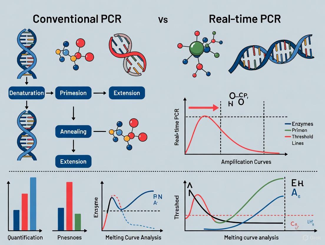
The most significant functional difference lies in the point of measurement. Conventional PCR detects the final yield after the reaction has stopped, where the relationship between the initial template amount and final product is lost. In contrast, real-time PCR monitors the reaction during the exponential phase, where the amount of product doubles each cycle. It uses the Cycle Threshold (Ct) value—the cycle number at which fluorescence crosses a defined threshold—to precisely determine the starting quantity of the target nucleic acid [3] [5].
Conventional PCR solidly remains the "original amplification workhorse" of molecular biology. Its simplicity, cost-effectiveness, and powerful ability to selectively amplify specific DNA sequences have cemented its role in foundational laboratory techniques such as cloning, genotyping, and qualitative diagnostics. While real-time PCR and digital PCR have expanded the horizons of nucleic acid analysis into the realm of precise quantification, absolute copy number determination, and rare allele detection [9] [7], they build upon the core principles established by conventional PCR. A thorough understanding of conventional PCR—its mechanisms, optimization requirements, and inherent limitations—is not merely a historical exercise. It is fundamental for researchers and drug development professionals to make informed decisions on selecting the most appropriate PCR technology for their specific experimental needs, ensuring both efficiency and scientific rigor.
This technical guide explores real-time quantitative PCR (qPCR), a transformative molecular biology technology that enables the precise quantification of nucleic acids. Framed within the broader thesis of PCR evolution, this document highlights how qPCR's real-time, quantitative capabilities represent a fundamental shift from the qualitative, end-point analysis of conventional PCR [10] [11]. The guide is structured for researchers, scientists, and drug development professionals, providing in-depth technical details, methodologies, and data analysis techniques.
The Polymerase Chain Reaction (PCR) revolutionized molecular biology by allowing for the exponential amplification of specific DNA sequences. Conventional PCR (also known as end-point PCR) provides a qualitative, yes/no answer regarding the presence of a target sequence, with results visualized via gel electrophoresis after the amplification is complete [6] [10]. In contrast, real-time PCR, or quantitative PCR (qPCR), monitors the amplification of DNA in real time as the reaction occurs, providing both detection and quantification of the initial amount of the target nucleic acid [6] [11]. This transition from qualitative to quantitative analysis has empowered researchers with unprecedented precision in applications ranging from gene expression analysis to pathogen quantification and cancer biomarker detection [6] [12].
The core principle of qPCR lies in the use of fluorescent reporters to track the accumulation of PCR product during every cycle of the amplification process. By measuring fluorescence, the technique allows for the determination of the starting quantity of the target DNA, combining amplification and detection into a single, closed-tube workflow that minimizes contamination risk and provides a wide dynamic range of detection [12] [10] [11].
Key Principles and Detection Chemistries
The qPCR Workflow and Data Interpretation
In qPCR, the fluorescence signal increases proportionally to the amount of amplified DNA. The instrument measures this fluorescence at each cycle, generating an amplification plot. The key quantitative metric is the Quantification Cycle (Cq), previously known as Ct or Cp, which is the PCR cycle number at which the sample's fluorescence crosses a defined threshold, set within the exponential phase of amplification [12] [13] [11]. A lower Cq value indicates a higher starting concentration of the target template.
Accurate quantification depends on proper baseline correction to account for background fluorescence and appropriate threshold setting within the exponential phase where amplification efficiency is optimal [13]. The exponential phase provides the most reliable data because reagents are fresh and the reaction efficiency is highest and most consistent [12].
Detection Chemistries
qPCR utilizes two primary types of fluorescent chemistries for detection, each with distinct advantages.
DNA-Binding Dyes (Non-Specific Detection): Dyes like SYBR Green bind to double-stranded DNA, emitting fluorescence upon binding [11]. The fluorescence increases as the PCR product accumulates.
- Advantages: Cost-effective, flexible as only sequence-specific primers are required, and suitable for multiple targets with different dyes [11].
- Disadvantages: Lack of specificity, as the dye will bind to any dsDNA, including non-specific products and primer-dimers, which can lead to overestimation of the target concentration [11].
Fluorescently Labeled Probes (Specific Detection): Methods such as TaqMan probes use oligonucleotide probes with a fluorescent reporter at one end and a quencher at the other [12] [11].
- Advantages: High specificity, as fluorescence is detected only after the probe hybridizes to its complementary sequence and is cleaved by the 5' nuclease activity of the DNA polymerase. This prevents interference from primer-dimers and allows for multiplexing (detecting multiple targets in the same tube) using probes with different colored dyes [12] [11].
- Disadvantages: Higher cost and more complex assay design are required [11].
The following diagram illustrates the mechanism of the TaqMan probe-based detection:
Figure 1: TaqMan Probe qPCR Detection Mechanism
Reverse Transcription qPCR (RT-qPCR) for Gene Expression Analysis
A pivotal application of qPCR is gene expression analysis via reverse transcription qPCR (RT-qPCR). This technique allows for the quantification of RNA transcripts by first converting RNA into complementary DNA (cDNA) using reverse transcriptase, followed by qPCR amplification [14] [12]. RT-qPCR is one of the most sensitive and widely used methods for analyzing changes in gene expression, verifying microarray results, and profiling gene patterns [12].
One-Step vs. Two-Step RT-qPCR
RT-qPCR can be performed using two primary approaches, each with distinct advantages and limitations [14].
One-Step RT-qPCR: Combines the reverse transcription and qPCR amplification in a single tube and buffer.
- Advantages: Faster, higher throughput, reduced pipetting steps which lowers the risk of contamination, and less experimental variation [14].
- Disadvantages: Less sensitive than two-step, impossible to optimize the reactions separately, and detection of fewer targets per sample [14].
- Best for: Target- or gene-specific studies when analyzing a single gene across many samples [12].
Two-Step RT-qPCR: Performs reverse transcription and qPCR in separate, optimized reactions.
- Advantages: A stable cDNA pool is generated that can be stored and used for multiple reactions (e.g., analyzing multiple transcripts from the same sample), flexible priming options, and the ability to use optimized conditions for each step [14] [12].
- Disadvantages: More time-consuming, requires more pipetting steps (increasing contamination risk), and needs more optimization [14].
- Best for: When detecting multiple targets from a single sample or when storing cDNA for later use [14] [12].
Critical Considerations for RT-qPCR
- Priming for Reverse Transcription: In two-step assays, cDNA synthesis can be primed using:
- Oligo(dT) primers: Anneal to the poly-A tail of mRNA, good for full-length cDNA synthesis, but biased towards the 3' end [14].
- Random primers: Anneal at multiple points along all RNA transcripts, useful for RNA with secondary structure or when little starting material is available [14].
- Gene-specific primers: Provide a specific cDNA pool and increased sensitivity for one gene of interest [14].
- RNA Template: Total RNA is often preferred over mRNA for relative quantification because it requires fewer purification steps and avoids skewed results from differential mRNA recovery [14].
- Controls: A minus reverse transcriptase control ("no RT" control) is essential to test for contaminating genomic DNA [14].
The workflow for RT-qPCR, from sample to result, is outlined below:
Figure 2: RT-qPCR Workflow: One-Step vs. Two-Step
Experimental Design and Protocols
Detailed Protocol: qPCR Gene Expression Analysis
The following methodology is compiled from established principles and a verified application in quality control [14] [12] [15].
- RNA Extraction and Quality Control: Extract high-quality total RNA from cells or tissue using a guanidinium thiocyanate-phenol-based method or a commercial kit (e.g., Qiagen PowerSoil Pro Kit) [15]. Assess RNA integrity and concentration using spectrophotometry (A260/A280 ratio) and/or capillary electrophoresis.
- Reverse Transcription to cDNA:
- For a 20 µL reaction, combine 1 µg of total RNA, reverse transcriptase (e.g., Moloney Murine Leukemia Virus Reverse Transcriptase, M-MLV RT), reaction buffer, dNTPs (e.g., 0.5 mM each), RNase inhibitor, and primers. Primers can be oligo(dT) (e.g., 2.5 µM), random hexamers (e.g., 50 ng/µL), or gene-specific primers [14].
- Incubate the reaction: 10 minutes at 25°C (annealing), 30–60 minutes at 42–50°C (extension), and 5 minutes at 85°C (enzyme inactivation). The generated cDNA can be stored at -20°C.
- qPCR Assay Preparation:
- Primer/Probe Design: Design primers (and probes if used) to span an exon-exon junction to avoid amplification of genomic DNA [14]. The recommended amplification efficiency is between 90–110% [12]. Use tools like NCBI BLAST or commercial assay design tools to ensure specificity.
- Reaction Setup: In a 96-well or 384-well plate, prepare a reaction mix for each cDNA sample. A typical 20 µL reaction contains: 1X qPCR master mix (containing DNA polymerase, dNTPs, and buffer), forward and reverse primers (e.g., 200–500 nM each), fluorescent reporter (e.g., 1X SYBR Green or 100–250 nM TaqMan probe), and cDNA template (e.g., 2–100 ng equivalent of input RNA) [12] [15]. Include controls: no-template control (NTC), no-reverse-transcription control (no-RT), and a positive control.
- qPCR Amplification and Data Acquisition: Run the plate in a real-time PCR instrument. A standard thermal cycling protocol is:
- Initial Denaturation: 95°C for 2–10 minutes.
- 40–50 Cycles of:
- Denaturation: 95°C for 15 seconds.
- Annealing/Extension: 60°C for 1 minute (acquire fluorescence at the end of this step).
- Data Analysis:
- Set the baseline and fluorescence threshold correctly to determine the Cq value for each reaction [13].
- Use a quantification method:
- Comparative Cq (ΔΔCq) method for relative quantification of gene expression relative to a reference sample and normalized to one or more endogenous control genes (e.g., GAPDH, β-actin) [12] [13].
- Standard curve method for absolute quantification, where the Cq values of unknown samples are interpolated from a standard curve of known concentrations [12] [13].
Case Study: Pathogen Detection in Cosmetics
A 2025 study demonstrated the superiority of qPCR for quality control, detecting pathogens (E. coli, S. aureus, P. aeruginosa, C. albicans) in cosmetic formulations [15].
- Method: Cosmetics were spiked with low levels (3–5 CFU) of pathogens and enriched for 20–24 hours. DNA was extracted automatically (QIAcube Connect). Pathogen detection used commercial qPCR kits (e.g., R-Biopharm SureFast PLUS) with duplicate reactions [15].
- Results: qPCR demonstrated 100% detection rates across all replicates, matching or surpassing traditional plate culture methods. qPCR was particularly effective in complex matrices and for detecting low inoculum levels, offering a rapid and reliable alternative for routine quality control [15].
Data Analysis and Quantification Methods
Ensuring Accurate Cq Values
The accuracy of qPCR quantification hinges on proper data acquisition. The baseline should be set in the early cycles where there is no significant increase in fluorescence, typically between cycles 5–15, to correct for background fluorescence [13]. The threshold must be set high enough to be above the background baseline but within the exponential phase of all amplification plots, where the curves are parallel, ensuring consistent ΔCq values between samples [13].
Quantification Strategies
qPCR data can be analyzed using absolute or relative quantification.
- Standard Curve Method (Absolute Quantification): A dilution series of a target with known concentration is run to create a standard curve by plotting Cq values against the logarithm of the concentration. The concentration of unknown samples is determined by interpolating their Cq values from this curve [13].
- Comparative Cq (ΔΔCq) Method (Relative Quantification): This method calculates the fold-change in gene expression between a test sample and a reference sample (e.g., untreated control). The Cq of the target gene is normalized to the Cq of one or more stable endogenous control genes (Ref) to account for variations in RNA input and quality. The formula used is: Fold Change = Efficiency^(ΔCqtarget - ΔCqRef), where Efficiency is the amplification efficiency of the assay [13]. This method does not require a standard curve and is commonly used for gene expression analysis.
Table 1: Key Quantitative Data Outputs in qPCR Analysis
| Parameter | Description | Interpretation |
|---|---|---|
| Cq (Quantification Cycle) | The cycle number at which the fluorescence crosses the threshold [12] [13]. | Inversely proportional to the log of the initial target concentration; a lower Cq means more starting template. |
| Standard Curve | A plot of Cq values vs. the log of known standard concentrations [13]. | Used for absolute quantification; slope and R² value indicate PCR efficiency and linearity. |
| Amplification Efficiency | The rate of product doubling per cycle, ideally 100% (corresponding to a slope of -3.32) [11]. | Efficiency between 90–110% is acceptable; lower efficiency reduces sensitivity and quantification accuracy [12]. |
| ΔCq | The difference in Cq between a target gene and a reference gene within the same sample [13]. | Used for normalization in relative quantification. |
| ΔΔCq | The difference in ΔCq between a test sample and a calibrator/reference sample [13]. | Used to calculate the fold-change in expression in the comparative Cq method. |
The qPCR Toolkit: Essential Reagents and Materials
Table 2: Research Reagent Solutions for qPCR
| Reagent/Material | Function | Key Considerations |
|---|---|---|
| Reverse Transcriptase | Enzyme that synthesizes cDNA from an RNA template [14]. | Choose a enzyme with high thermal stability for efficient transcription of RNA with secondary structures [14]. |
| Thermostable DNA Polymerase | Amplifies the cDNA or DNA template during PCR. | A key component of the qPCR master mix; often supplied as a hot-start enzyme to reduce non-specific amplification. |
| Fluorescent Reporters | Molecules that emit fluorescence to enable real-time detection of amplicons [11]. | SYBR Green (binds dsDNA) or TaqMan Probes (sequence-specific). For multiplexing, use dyes with non-overlapping spectra (e.g., FAM, HEX/VIC) [16] [11]. |
| Primers & Probes | Oligonucleotides that define the target sequence for amplification [14] [11]. | Must be specific and designed for high efficiency. For gene expression, design primers to span exon-exon junctions [14] [12]. |
| dNTPs | Nucleotides (dATP, dCTP, dGTP, dTTP) that are the building blocks for new DNA strands. | Added to the master mix for DNA polymerization. |
| qPCR Plates & Seals | Reaction vessels compatible with the real-time PCR instrument. | Optically clear plates and seals are essential for accurate fluorescence detection. |
| Commercial Kits & Assays | Pre-formulated reagents for specific targets or applications. | Include pre-designed primer/probe sets (e.g., TaqMan assays) and PCR arrays for pathway-focused analysis, saving optimization time [12]. |
Comparison with Conventional and Digital PCR
Table 3: PCR Technology Evolution: Conventional vs. qPCR vs. dPCR
| Feature | Conventional PCR | Real-Time PCR (qPCR) | Digital PCR (dPCR) |
|---|---|---|---|
| Quantification | Qualitative or semi-quantitative (end-point) [10]. | Quantitative (real-time) [6] [10]. | Absolute quantification (end-point) [17]. |
| Detection Method | Gel electrophoresis after amplification [10]. | Fluorescence during amplification [11]. | Fluorescence after amplification in partitioned samples [17]. |
| Sensitivity | Low | High (detection down to one copy) [12]. | Very high (can detect rare mutations) [17]. |
| Key Applications | Cloning, mutation detection, sequencing [6] [10]. | Gene expression, viral load, pathogen quantification [6] [12]. | Liquid biopsy, rare allele detection, copy number variation [17]. |
| Throughput | Low | High | Moderate to High [17]. |
| Standard Curve | Not applicable | Required for absolute quantification [13]. | Not required (absolute counting) [17]. |
| Key Differentiator | Answers "Is the target present?" | Answers "How much target is present?" | Answers "Exactly how many target molecules are present?" [17]. |
Real-time PCR has indisputably revolutionized molecular biology by providing a powerful quantitative framework for nucleic acid analysis. Its ability to precisely and sensitively measure DNA and RNA in real time, within a closed-tube system, has made it the gold standard for applications like gene expression analysis, pathogen detection, and biomarker validation [6] [12]. As the field advances, qPCR remains a foundational technology, while newer methods like digital PCR (dPCR) push the boundaries of sensitivity and absolute quantification even further, particularly for complex clinical applications such as liquid biopsies [17]. Understanding the principles, methodologies, and data analysis of qPCR is therefore essential for any researcher or drug development professional working in modern life sciences.
Within molecular biology and diagnostic research, the polymerase chain reaction (PCR) serves as a fundamental technique for nucleic acid amplification. This technical guide provides a comparative workflow analysis between conventional, or end-point, PCR and real-time PCR (also known as quantitative PCR, or qPCR). The core distinction lies in their names: end-point PCR analyzes the accumulated product after the amplification process is complete, while real-time PCR monitors product generation during each cycle of the amplification process [18] [10]. This difference in detection timing fundamentally influences their applications, data output, and capabilities in research and drug development. Real-time PCR has revolutionized quantitative gene expression analysis, pathogen detection, and viral load estimation by providing a method to precisely quantify the initial amount of a nucleic acid target [18] [1].
Core Principles and Workflows
The workflows for end-point and real-time PCR share initial steps but diverge significantly in their detection phases and data analysis methods. Understanding these distinct pathways is crucial for selecting the appropriate technique.
End-Point PCR Workflow
Conventional PCR is a qualitative technique that amplifies a specific DNA sequence through repeated thermal cycles, with detection occurring only after the final cycle is complete [10].
- Amplification Process: The process involves three core steps repeated for 25-40 cycles:
- Denaturation: The double-stranded DNA template is heated to ~95°C to separate it into single strands.
- Annealing: The temperature is lowered (typically 55-72°C) to allow short, synthetic primers to bind to their complementary sequences on either side of the target DNA.
- Extension: The temperature is raised to ~72°C, the optimal temperature for a thermostable DNA polymerase (e.g., Taq polymerase) to synthesize a new DNA strand by extending from the primers [1].
- Detection and Analysis: After the cycling is complete, the amplified products are separated by size using agarose gel electrophoresis. The gel is stained with a DNA-binding dye like ethidium bromide and visualized under UV light. The presence of a band of the expected size indicates a successful amplification [10] [1]. This method is termed "end-point" because measurement occurs during the plateau phase of the reaction, where reagents are depleted, and the amount of product is no longer doubling [18].
Real-Time PCR Workflow
Real-time PCR is a quantitative technique that builds upon conventional PCR by incorporating a fluorescent detection system that monitors the accumulation of PCR product in real-time, during the exponential phase of amplification [18] [1].
- Amplification with Fluorescent Detection: The thermal cycling process is identical to conventional PCR. However, each reaction contains a fluorescent reporter. Two common detection chemistries are:
- DNA-Binding Dyes (e.g., SYBR Green): These dyes fluoresce brightly when bound to double-stranded DNA. As the target amplicon accumulates, the fluorescence signal increases proportionally [19] [18].
- Sequence-Specific Probes (e.g., TaqMan Probes): These probes provide higher specificity through an oligonucleotide that binds to a sequence within the target amplicon. The probe incorporates a fluorophore and a quencher; during amplification, the fluorophore is cleaved, and its fluorescence is detected [18].
- Data Analysis: The fluorescence is measured at the end of each cycle. The instrument software generates an amplification plot. The key quantitative metric is the Cycle Threshold (Ct), which is the cycle number at which the fluorescence crosses a predetermined threshold, indicating a statistically significant increase in signal above the background. A sample with a higher starting concentration of the target will have a lower Ct value [18].
The following diagram illustrates the core logical relationship and workflow differences between these two techniques:
Critical Comparative Analysis
The fundamental difference in detection timing leads to a cascade of practical and technical distinctions. The following table summarizes the key parameters that differentiate these two techniques.
Table 1: Core Technical and Application Differences Between End-Point and Real-Time PCR
| Parameter | End-Point PCR | Real-Time PCR |
|---|---|---|
| Detection Principle | Measures accumulated product at the end of all cycles (plateau phase) [18]. | Monitors product formation during the exponential phase of every cycle [18]. |
| Quantification | Qualitative or semi-quantitative (e.g., band intensity on a gel) [18]. | Fully quantitative, determines the initial amount of the target template [18] [10]. |
| Dynamic Range | Short, typically less than 2 logs [18]. | Wide, often exceeding 7-8 logs of dynamic range [18]. |
| Sensitivity | Lower sensitivity; cannot detect small fold changes [18]. | High sensitivity; capable of detecting down to a two-fold change in concentration [18]. |
| Post-PCR Processing | Required (e.g., gel electrophoresis), which is time-consuming and increases contamination risk [10]. | Not required; the system is closed, minimizing contamination and hands-on time [18] [10]. |
| Throughput & Automation | Lower throughput due to manual post-processing. | Higher throughput and easier automation. |
| Primary Applications | Cloning, sequencing, mutation detection, simple presence/absence analysis [18] [10]. | Gene expression analysis (qPCR), viral load quantification, pathogen detection, SNP genotyping [18] [1]. |
Sensitivity and Specificity in Practice
The superior sensitivity of real-time PCR is clearly demonstrated in clinical studies. For instance, a 2021 study comparing PCR methods for detecting the SFTS virus found that a nested end-point PCR method (NPCR-M) demonstrated a higher detection rate (85%) in patient samples over 40 days compared to a single-round end-point PCR (44%) and a real-time PCR method (71%) targeting a different gene segment [20]. This highlights that while real-time PCR is generally more sensitive, assay design (e.g., target region, primer design) is also a critical factor. Furthermore, the use of target-specific probes (TaqMan) in real-time PCR provides an additional layer of specificity over dye-based methods (SYBR Green) or end-point analysis, as it requires not only primer binding but also probe hybridization for signal generation [18].
Experimental Protocols for Comparative Studies
To empirically compare the performance of end-point and real-time detection, the following protocol can be implemented.
Sample Preparation and Amplification
- Template Dilution Series: Prepare a serial dilution (e.g., 1:10 dilutions) of a known quantity of target DNA, spanning at least 5 orders of magnitude.
- Reaction Setup:
- For End-Point PCR: Set up reactions for each dilution using a standard master mix containing Taq DNA polymerase, dNTPs, primers, and buffer.
- For Real-Time PCR: Set up identical reactions in optical tubes or plates, using a master mix that also contains the fluorescent reporter (e.g., SYBR Green dye).
- Thermal Cycling:
- Run both sets of samples on compatible thermal cyclers. A typical protocol is:
- Initial Denaturation: 95°C for 2-5 minutes.
- 35-40 cycles of:
- Denaturation: 95°C for 15-30 seconds.
- Annealing: 55-65°C for 15-30 seconds.
- Extension: 72°C for 15-60 seconds (depending on amplicon size).
- The real-time cycler will collect fluorescence data during the annealing or extension step of each cycle.
- Run both sets of samples on compatible thermal cyclers. A typical protocol is:
Data Collection and Analysis
- End-Point Analysis:
- After cycling, run the end-point PCR products on an agarose gel.
- Visualize and capture an image under UV light.
- Estimate the quantity by comparing band intensity to a DNA ladder or between dilutions. Note the last dilution at which a visible band is detected.
- Real-Time Analysis:
- The instrument software will automatically generate an amplification plot for each dilution.
- Record the Ct value for each dilution.
- Generate a standard curve by plotting the Ct values against the logarithm of the known starting concentration. The slope of the curve can be used to calculate the amplification efficiency [18].
The relationship between template concentration and detection in each method is visualized below:
The Scientist's Toolkit: Essential Reagents and Materials
The successful execution of PCR experiments, whether end-point or real-time, relies on a core set of reagents and instruments.
Table 2: Key Research Reagent Solutions and Their Functions
| Reagent / Material | Function | Key Considerations |
|---|---|---|
| Thermostable DNA Polymerase (e.g., Taq) | Enzyme that synthesizes new DNA strands by adding nucleotides to the primer. | Thermostability is crucial for withstanding repeated denaturation temperatures. Fidelity (accuracy) can vary between enzymes [1]. |
| Primers | Short, single-stranded DNA sequences that are complementary to the ends of the target region. They define the region to be amplified. | Design is critical for specificity and efficiency. Parameters include length (18-24 bp), melting temperature (Tm), and avoidance of secondary structures [19]. |
| dNTPs (Deoxynucleotide Triphosphates) | The building blocks (A, T, C, G) used by the DNA polymerase to synthesize new DNA. | Quality and concentration are vital for efficient amplification and avoiding misincorporation. |
| Buffer Solution | Provides the optimal chemical environment (pH, ionic strength) for the polymerase to function. | Often contains MgCl₂, which is a co-factor for the polymerase and its concentration can significantly impact yield and specificity [1]. |
| Fluorescent Reporter (for Real-Time PCR) | Molecule that allows detection of amplified product during the reaction. | SYBR Green: Binds dsDNA; cost-effective but less specific. TaqMan Probes: Sequence-specific; higher specificity and multiplexing capability [18]. |
| Nucleic Acid Template | The sample DNA or RNA (via cDNA in RT-PCR) that contains the target sequence to be amplified. | Must be of sufficient quality and purity, free of inhibitors like phenol, heparin, or hemoglobin [1]. |
The choice between end-point and real-time PCR is fundamentally dictated by the research question. End-point PCR remains a robust, cost-effective tool for applications where simple detection or semi-quantitative analysis is sufficient, such as cloning, genotyping, or educational purposes. In contrast, real-time PCR is the unequivocal method for precise quantification, kinetic studies, and high-throughput applications where sensitivity, a broad dynamic range, and workflow efficiency are paramount. The integration of fluorescent detection in real-time PCR to monitor the exponential phase of amplification represents a significant technological advancement over the end-point analysis of the plateau phase, solidifying its role as the gold standard in quantitative molecular analysis for research and clinical diagnostics [18] [1].
The invention of the polymerase chain reaction (PCR) by Kary Mullis in 1983 represents a pivotal milestone that fundamentally transformed molecular biology, medical diagnostics, and pharmaceutical development. This technical guide examines the evolution of PCR technology from its conceptual origins through its contemporary applications, with particular emphasis on the critical technical distinctions between conventional PCR and real-time quantitative PCR (qPCR). Within the context of a broader thesis on molecular diagnostics, we provide a comprehensive analysis of methodologies, performance characteristics, and experimental considerations that distinguish these foundational techniques, supported by structured data comparisons and technical workflows relevant to research scientists and drug development professionals.
Historical Foundations: The Invention of PCR
Kary Mullis and the Conceptual Breakthrough
Kary Banks Mullis (1944-2019) invented the polymerase chain reaction technique in 1983 while working as a chemist at the Cetus Corporation in Emeryville, California [21]. The foundational insight occurred during a weekend drive to his countryside cabin in Mendocino County, when he conceptualized using a pair of primers to bracket a desired DNA sequence and copy it using DNA polymerase, enabling exponential amplification of specific DNA fragments [21]. This technique would ultimately divide biology into "the two epochs of before PCR and after PCR" according to The New York Times [21].
Mullis's innovation built upon earlier scientific discoveries, including the elucidation of DNA's structure by Watson and Crick in 1953 and the discovery of DNA polymerase by Arthur Kornberg in 1956 [22]. Preliminary concepts for in vitro DNA amplification involving oligonucleotide primers and DNA polymerase had been described by Kjell Kleppe in 1969 and published in 1971, but technical limitations prevented practical implementation at that time [22].
Technical Implementation and Development
The first practical demonstration of PCR occurred on December 16, 1983 [21]. Mullis faced initial skepticism from colleagues at Cetus, prompting his supervisor Thomas White to reassign him to focus exclusively on PCR development [21]. The technique was significantly improved when other Cetus scientists, including Randall Saiki and Henry Erlich, determined that PCR could amplify a specific human gene (β-globin) from genomic DNA [21]. Their work culminated in the landmark 1985 paper in Science titled "Enzymatic Amplification of β-globin Genomic Sequences and Restriction Site Analysis for Diagnosis of Sickle Cell Anemia" [21] [22].
A critical advancement came in 1986 when Saiki began using the thermostable Taq polymerase from Thermus aquaticus, which could withstand the high denaturation temperatures without requiring replenishment after each cycle, dramatically reducing costs and enabling automation [21]. For his invention, Mullis received the Nobel Prize in Chemistry in 1993, with the Nobel Committee recognizing "his invention of the polymerase chain reaction (PCR) method" [23].
Technical Evolution: From Conventional PCR to Real-Time PCR
Fundamental Principles of Conventional PCR
Conventional PCR, also known as standard or end-point PCR, amplifies specific DNA sequences through repeated thermal cycling involving three fundamental steps [1]:
- Denaturation: The double-stranded DNA template is heated to 90-95°C to separate the complementary strands by breaking hydrogen bonds between base pairs.
- Annealing: The temperature is lowered to 55-72°C to allow primers to bind to their complementary sequences on the single-stranded DNA templates.
- Extension: The temperature is raised to 75-80°C, optimal for DNA polymerase activity, enabling the enzyme to synthesize new DNA strands by adding nucleotides to the 3' end of the primers.
This process typically repeats for 25-40 cycles, theoretically generating an exponential increase in the target DNA sequence [1]. Conventional PCR relies on end-point detection, where the accumulated product is visualized after amplification completion using agarose gel electrophoresis with DNA-binding dyes such as ethidium bromide [18] [24].
The Advent of Real-Time Quantitative PCR
Real-time PCR (qPCR), introduced in 1992, represents a significant technological advancement over conventional PCR by enabling monitoring of DNA amplification as it occurs [18] [25]. This methodology employs fluorescent reporter molecules that increase in signal intensity proportionally to the amount of amplified DNA product. The key distinction lies in when measurements are taken: qPCR collects data during the exponential phase of amplification, while conventional PCR measures only the final accumulated product at the plateau phase [18].
The fundamental metric in qPCR is the Cycle Threshold (Ct), defined as the PCR cycle number at which the sample's fluorescence intensity crosses a predetermined threshold above background levels [18]. This value correlates inversely with the initial template concentration, enabling precise quantification.
Table 1: Core Technical Distinctions Between Conventional PCR and Real-Time PCR
| Parameter | Conventional PCR | Real-Time PCR |
|---|---|---|
| Detection Method | End-point gel electrophoresis | Fluorescent detection during amplification |
| Measurement Phase | Plateau phase | Exponential (log) phase |
| Quantification Capability | Semi-quantitative at best | Fully quantitative |
| Throughput | Lower | Higher with multi-well plates |
| Dynamic Range | < 2 logs | > 7-8 logs |
| Sensitivity | Lower | Higher |
| Post-PCR Processing | Required (gel electrophoresis) | Not required |
| Multiplexing Capability | Limited | Advanced with multiple fluorophores |
| Primary Output | Band intensity on gel | Cycle threshold (Ct) value |
| Contamination Risk | Higher (post-processing) | Lower (closed-tube) |
Detection Chemistry in Real-Time PCR
qPCR utilizes two principal detection chemistries [18]:
- DNA-binding dyes (e.g., SYBR Green): These dyes intercalate with double-stranded DNA and emit fluorescence upon binding. While cost-effective, they lack specificity as they bind to any double-stranded DNA, including non-specific products and primer-dimers.
- Sequence-specific probes (e.g., TaqMan probes): These oligonucleotide probes contain a fluorescent reporter dye and a quencher dye; when the probe is intact, the quencher suppresses fluorescence through fluorescence resonance energy transfer (FRET). During amplification, the 5'→3' exonuclease activity of Taq polymerase cleaves the probe, separating the reporter from the quencher and generating a fluorescent signal proportional to the amount of amplified product.
Methodological Comparisons and Experimental Protocols
Standard PCR Experimental Protocol
A basic conventional PCR protocol involves the following components and steps [24]:
Table 2: Standard PCR Reaction Components
| Component | Typical Concentration | Function |
|---|---|---|
| Template DNA | 1-100 ng | Target sequence to be amplified |
| Forward/Reverse Primers | 0.1-1 μM each | Sequence-specific amplification initiation |
| Taq DNA Polymerase | 0.5-2.5 units/reaction | Enzymatic DNA synthesis |
| dNTPs | 200 μM each | Nucleotide building blocks |
| Reaction Buffer | 1X | Optimal pH and salt conditions |
| Magnesium Chloride (MgCl₂) | 1.5-2.5 mM | Essential cofactor for polymerase activity |
Thermal Cycling Parameters for Conventional PCR [24]:
- Initial Denaturation: 94-95°C for 2-5 minutes
- Amplification Cycles (25-35 cycles):
- Denaturation: 94-95°C for 15-30 seconds
- Annealing: 55-65°C (primer-specific) for 15-60 seconds
- Extension: 72°C for 1 minute per kilobase of amplicon
- Final Extension: 72°C for 5-10 minutes
- Hold: 4°C indefinitely
Post-Amplification Analysis:
- Prepare 1-2% agarose gel in TAE or TBE buffer with ethidium bromide or alternative DNA stain
- Load PCR products with DNA size marker
- Electrophorese at 5-10 V/cm until adequate separation
- Visualize under UV transillumination
Advanced PCR Methodologies
Several specialized PCR methodologies have been developed to address specific experimental challenges:
Reverse Transcription PCR (RT-PCR): This technique combines reverse transcription of RNA into complementary DNA (cDNA) followed by PCR amplification, enabling gene expression analysis from RNA templates [1]. During the COVID-19 pandemic, RT-qPCR served as the primary diagnostic method for SARS-CoV-2 detection due to its high sensitivity, specificity, and rapid turnaround time [1].
Digital PCR (dPCR): The latest major evolution in PCR technology, dPCR works by partitioning a PCR sample into thousands of individual reactions, such that some contain the target molecule and others do not [18] [25]. Following amplification, the fraction of negative reactions is used in Poisson statistical analysis to determine the absolute copy number of the target sequence without requiring standard curves [18]. dPCR demonstrates superior accuracy, particularly for high viral loads, and shows greater consistency and precision than qPCR [9].
Hot-Start PCR: Employing enzyme modifiers (antibodies, aptamers, or chemical modifications) to inhibit DNA polymerase activity at room temperature, this technique prevents nonspecific amplification during reaction setup and increases specificity [26].
Multiplex PCR: This approach allows simultaneous amplification of multiple targets in a single reaction by using multiple primer pairs, conserving samples and reagents while enabling comparative analysis of different targets [26].
Comparative Performance Analysis
Analytical Performance Characteristics
Recent comparative studies between qPCR and dPCR reveal distinct performance advantages depending on application requirements. In respiratory virus detection during the 2023-2024 "tripledemic," dPCR demonstrated superior accuracy for high viral loads of influenza A, influenza B, and SARS-CoV-2, and for medium loads of RSV [9]. dPCR showed greater consistency and precision than Real-Time RT-PCR, particularly in quantifying intermediate viral levels, highlighting its potential to enhance respiratory virus diagnostics [9].
Table 3: Performance Comparison of PCR Technologies in Viral Detection
| Performance Metric | Conventional PCR | Real-Time PCR | Digital PCR |
|---|---|---|---|
| Quantification Type | Semi-quantitative | Relative quantitative | Absolute quantitative |
| Precision | Poor | Moderate | High |
| Sensitivity | Low | High (detects 2-fold changes) | Very High |
| Dynamic Range | < 2 logs | 7-8 logs | 5 logs but linear |
| Tolerance to Inhibitors | Low | Moderate | High |
| Dependence on Standards | No | Yes | No |
| Throughput | Low | High | Moderate |
| Cost per Reaction | Low | Moderate | High |
Application-Specific Considerations
The selection of appropriate PCR methodology depends fundamentally on the research or diagnostic objective:
Conventional PCR remains suitable for:
- Basic DNA amplification for cloning and sequencing
- Genotyping applications where presence/absence detection suffices
- Educational applications due to lower equipment costs
- Target detection when precise quantification is not required
Real-Time PCR is preferred for:
- Gene expression analysis through quantitative measurement
- Pathogen detection and viral load monitoring
- SNP genotyping and copy number variation analysis
- MicroRNA analysis and siRNA/RNAi experiments
- High-throughput applications requiring rapid results
Digital PCR offers advantages for:
- Absolute quantification without standard curves
- Detection of rare alleles and rare sequence variants
- Analysis of complex mixtures with abundant background
- Copy number variation analysis with precision
- Validation of qPCR assays and reference standards
The Researcher's Toolkit: Essential Reagents and Materials
Table 4: Essential Research Reagents for PCR Applications
| Reagent/Material | Function | Application Notes |
|---|---|---|
| Taq DNA Polymerase | Thermostable enzyme for DNA synthesis | Standard for conventional PCR; available with hot-start modifications |
| Reverse Transcriptase | RNA-to-cDNA conversion | Essential for RT-PCR and RT-qPCR applications |
| dNTP Mix | Nucleotide substrates for DNA synthesis | Quality critical for high-fidelity amplification |
| Primers | Sequence-specific amplification | Design critical for specificity; HPLC purification recommended |
| Fluorescent Probes/Dyes | Detection in real-time platforms | TaqMan probes offer superior specificity over intercalating dyes |
| PCR Buffer with MgCl₂ | Optimal reaction environment | Mg²⁺ concentration requires optimization for each assay |
| Nucleic Acid Extraction Kits | Sample preparation | Quality critical for sensitivity and inhibitor removal |
| Positive Control Templates | Assay validation | Essential for establishing assay performance characteristics |
Technical Workflows and System Relationships
PCR Method Selection and Analytical Workflow
PCR Methodologies and Their Fundamental Detection Principles
The evolution of PCR technology continues with emerging trends focusing on increased multiplexing capabilities, integration with advanced data analysis including artificial intelligence, and transition toward practical point-of-care applications [25]. Future developments will likely enhance the role of PCR in personalized medicine, environmental monitoring, and food safety testing.
The invention of PCR by Kary Mullis initiated a technological revolution that has progressively transformed through conventional PCR, real-time qPCR, and digital PCR. Each technological iteration has expanded application possibilities while maintaining the core principle of exponential nucleic acid amplification. The selection between these methodologies requires careful consideration of analytical requirements, with conventional PCR offering simplicity for qualitative applications, real-time PCR providing robust quantification for most research and diagnostic needs, and digital PCR delivering superior precision for absolute quantification challenges. Understanding these historical milestones and technical distinctions enables researchers and drug development professionals to strategically implement appropriate PCR methodologies to advance their scientific objectives.
Methodologies, Protocols, and Research Applications
The Polymerase Chain Reaction (PCR) is a foundational nucleic acid amplification technique that has revolutionized molecular biology since its introduction by Kary Mullis in 1985 [1]. This whitepaper focuses on conventional PCR (also termed end-point PCR), which represents the original form of the technology that amplifies specific DNA sequences through repeated thermal cycling [10]. Unlike real-time PCR, which monitors amplification progress as it occurs, conventional PCR provides a qualitative assessment of amplified DNA at the end of the process through gel electrophoresis [10]. The technique's profound impact stems from its ability to exponentially amplify target DNA sequences, enabling researchers to generate millions of copies from a minimal starting material—often as little as 1-100 ng of DNA [1].
Within the broader context of PCR technology evolution, conventional PCR establishes the fundamental principles upon which more advanced quantitative methods like real-time PCR were developed. While real-time PCR offers superior quantification capabilities through fluorescence monitoring during amplification [27], conventional PCR remains invaluable for applications where simple detection or visualization of amplified products suffices [10]. This protocol details the established methodology for conventional PCR amplification and subsequent gel analysis, providing researchers with a robust framework for fundamental molecular biology applications including cloning, sequencing, and mutation detection [10].
Theoretical Framework: Principles and Components
Core Principles of Conventional PCR
Conventional PCR operates through a cyclic three-step process that exploits the natural function of DNA polymerase. Each cycle theoretically doubles the amount of target DNA, resulting in exponential amplification [1]. The process relies on thermal cycling between distinct temperature phases that facilitate DNA denaturation, primer annealing, and enzymatic extension. This targeted amplification allows researchers to selectively enrich specific genomic regions from complex samples, making subsequent analysis through gel electrophoresis possible [10]. The fundamental limitation of conventional PCR lies in its qualitative nature—while it excels at confirming the presence or absence of specific sequences, it does not provide reliable quantitative data about the initial template concentration, which represents a key distinction from real-time PCR methodologies [10].
Essential Reaction Components
Successful PCR amplification requires precise formulation of reaction components, each serving a critical function in the enzymatic process [28] [29]. The table below summarizes these essential components and their functions:
Table 1: Essential Components of a Conventional PCR Reaction
| Component | Final Concentration/Amount | Function and Notes |
|---|---|---|
| Template DNA | 200 pg/µL [29] | The target DNA to be amplified; can be genomic DNA, plasmid DNA, or cDNA. |
| Forward & Reverse Primers | 0.1-0.5 µM each [29] | Short, single-stranded DNA sequences (20-25 nucleotides) that define the region to be amplified [1]. |
| Taq DNA Polymerase | 0.05 units/µL [29] | Thermostable enzyme from Thermus aquaticus that synthesizes new DNA strands; withstands repeated heating to 95°C [28] [1]. |
| dNTP Mix | 200 µM [29] | Deoxynucleotide triphosphates (dATP, dCTP, dGTP, dTTP); the building blocks for new DNA strands. |
| Reaction Buffer | 1X [29] | Provides optimal pH and salt conditions for Taq polymerase activity. |
| MgCl₂ | 0.1-0.5 mM [29] | Cofactor essential for Taq polymerase activity; concentration often requires optimization [28]. |
| Water | To final volume (typically 50 µL) [29] | Nuclease-free water to bring the reaction to its final volume. |
| Optional: DMSO | 1-10% w/v [29] | Can improve amplification of templates with high GC content or secondary structure. |
Methodology: PCR Amplification and Analysis
Reaction Setup and Thermal Cycling Protocol
Proper reagent preparation and assembly are critical for PCR success. Reagents should be thawed on ice, and the reaction mix assembled in thin-walled 0.2 mL PCR tubes in the following order: water, buffer, dNTPs, MgCl₂, template, primers, and finally Taq polymerase [29]. Gentle mixing by tapping the tube followed by brief centrifugation ensures settled contents [29]. Controls are essential: a negative control without template DNA detects contamination, while a positive control with a template of known size verifies reaction success [29].
The thermal cycling protocol consists of three core steps repeated for 30-35 cycles, preceded by an initial denaturation and followed by a final extension [28] [29]:
Table 2: Standard Three-Step PCR Thermal Cycling Parameters
| Step | Temperature | Time | Purpose |
|---|---|---|---|
| Initial Denaturation | 94°C | 5 minutes | Completely separate double-stranded DNA template. |
| Denaturation | 94°C | 30 seconds | Separate newly synthesized DNA strands before each cycle. |
| Annealing | Tm - 5°C | 45 seconds | Allow primers to bind to complementary sequences on template DNA. |
| Extension | 72°C | 1 minute per kb | Synthesize new DNA strands via Taq polymerase (extension rate ~1kb/min). |
| Final Extension | 72°C | 5-10 minutes | Ensure complete extension of all amplified fragments. |
The annealing temperature is critical and typically set 5°C below the calculated melting temperature (Tm) of the primers [29]. Optimal conditions for components like Taq polymerase concentration and MgCl₂ levels may require empirical determination for specific applications [28].
Diagram 1: Conventional PCR and Gel Analysis Workflow
Post-Amplification Gel Electrophoresis
Following amplification, DNA products are separated and visualized via agarose gel electrophoresis [28] [30]. This process involves:
Gel Preparation: A 1-2% agarose gel is prepared by dissolving agarose powder in buffer (commonly TBE or TAE) by heating, then adding a DNA-safe stain before pouring into a casting tray with a comb to create wells [30]. The percentage of agarose determines resolution—lower percentages better resolve larger DNA fragments.
Sample Preparation: PCR products are mixed with loading dye containing a dense compound (e.g., glycerol) to help samples sink into wells and tracking dyes to monitor migration [30].
Electrophoresis: Loaded gels are submerged in buffer and subjected to an electric field (typically 50-100V). DNA migrates toward the anode due to its negative charge, with smaller fragments moving faster [30].
Visualization: The gel is examined under UV light, where DNA bands fluoresce due to intercalating dyes. A DNA ladder containing fragments of known sizes is run alongside samples for size determination [30].
Comparative Analysis: Conventional PCR vs. Real-Time PCR
Understanding the distinction between conventional and real-time PCR is essential for selecting the appropriate methodology for specific research applications. The table below summarizes the key differences:
Table 3: Technical Comparison Between Conventional and Real-Time PCR
| Parameter | Conventional PCR | Real-Time PCR (qPCR) |
|---|---|---|
| Amplification Principle | Three-step thermal cycling (denaturation, annealing, extension) [1]. | Same three-step cycling with added fluorescence detection [27]. |
| Detection Method | End-point analysis via gel electrophoresis [10]. | Real-time fluorescence monitoring during amplification [27]. |
| Detection Technology | DNA-binding dyes (e.g., ethidium bromide) post-amplification [28] [30]. | Fluorescent dyes (SYBR Green) or target-specific probes (TaqMan, Molecular Beacons) [27]. |
| Quantification Capability | Qualitative or semi-quantitative [10]. | Fully quantitative (absolute or relative) [10] [27]. |
| Throughput | Lower (requires post-processing) [10]. | Higher (closed-tube system) [10]. |
| Sensitivity | High (can detect down to ~100 copies) [1]. | Very high (can detect single copies) [10]. |
| Key Output Data | Band presence/absence and size on gel [30]. | Threshold cycle (Ct), amplification curves, quantification [27]. |
| Primary Applications | Cloning, sequencing, mutation detection, presence/absence testing [10]. | Gene expression, pathogen load, SNP genotyping, viral quantification [31] [27]. |
| Contamination Risk | Higher (post-amplification handling required) [10]. | Lower (closed-tube system) [10]. |
Diagram 2: Application Selection Guide for PCR Methodologies
Research Reagent Solutions and Essential Materials
Successful implementation of conventional PCR protocols requires specific laboratory reagents and equipment. The following table details essential solutions and materials:
Table 4: Essential Research Reagent Solutions for Conventional PCR
| Reagent/Material | Function/Application | Technical Notes |
|---|---|---|
| Taq DNA Polymerase | Thermostable DNA polymerase for DNA strand elongation [28]. | Isolated from Thermus aquaticus; retains activity after repeated 95°C heating [28] [1]. |
| dNTP Mix | Nucleotide substrates for DNA synthesis [29]. | Typically used at 200 µM final concentration for each dNTP [29]. |
| PCR Buffer | Optimal reaction environment for Taq polymerase [28]. | Often supplied with enzyme; may contain Tris-HCl, KCl, (NH₄)₂SO₄ [28]. |
| MgCl₂ Solution | Essential cofactor for DNA polymerase activity [28]. | Concentration requires optimization (0.1-0.5 mM); significantly impacts specificity [28] [29]. |
| Oligonucleotide Primers | Sequence-specific amplification targeting [1]. | Typically 20-25 nucleotides; designed with similar Tm (55-72°C) [1]. |
| Agarose | Matrix for electrophoretic separation of DNA fragments [30]. | Standard gels: 1-2% in TBE/TAE buffer; percentage determines resolution [30]. |
| DNA Stain | Visualization of amplified DNA fragments [30]. | Ethidium bromide or safer alternatives (SYBR Safe, GelRed); intercalates with DNA [28] [30]. |
| Loading Dye | Densant for gel loading with migration tracking [30]. | Contains glycerol/ficoll and tracking dyes (bromophenol blue) [30]. |
| DNA Ladder | Molecular weight standard for size determination [30]. | Contains DNA fragments of known sizes; essential for product sizing [30]. |
Applications in Research and Drug Development
Conventional PCR serves as a fundamental tool across diverse research domains, particularly where qualitative detection rather than quantification is required. In basic research, it enables DNA cloning through amplification of target sequences with restriction sites incorporated into primers, facilitates DNA sequencing by generating template material, and detects genetic mutations via amplified product size variations on gels [1] [10].
In diagnostic and clinical applications, conventional PCR provides highly sensitive detection of bacterial and viral pathogens, including human papillomavirus, herpes simplex virus, and various hepatitis strains [1]. It serves as a gold standard for screening genetic disorders and enables prenatal genetic testing for disease-associated mutations [1]. While real-time PCR has gained prominence in clinical diagnostics due to its quantitative capabilities, conventional PCR remains widely used in research settings and for applications where cost-effectiveness is paramount [10].
For drug development, conventional PCR supports target identification and validation through gene expression analysis (when combined with reverse transcription), enables quality control in biopharmaceutical production by detecting microbial contaminants, and facilitates pharmacogenomic studies investigating genetic variants that influence drug response [31]. Despite the advancement toward real-time PCR in many quantitative applications, conventional PCR maintains its utility in foundational molecular biology workflows where its simplicity, reliability, and cost-effectiveness provide distinct advantages [10].
Troubleshooting and Quality Control
Several factors can compromise conventional PCR results. Contamination represents a significant challenge due to the technique's extreme sensitivity; even minimal foreign DNA can lead to false positives [1]. Prevention strategies include physical separation of pre- and post-amplification areas, use of dedicated equipment and reagents, and incorporation of appropriate negative controls [1] [29].
Reaction failures often manifest as absent or weak bands, non-specific amplification, or primer-dimer formation on gels. Common issues include suboptimal MgCl₂ concentrations, incorrect annealing temperatures, poor primer design, or enzyme inhibition [28] [1]. Troubleshooting involves systematic optimization of each component, particularly MgCl₂ concentration (0.1-0.5 mM range) and annealing temperature (typically 5°C below primer Tm) [28] [29].
Quality control measures should include both positive controls (template with known amplification characteristics) and negative controls (no template) in every run [29]. For clinical applications, proper laboratory practices are essential—personal protective equipment including face masks, gloves, and hair covers should be worn consistently, and PCR procedures should ideally be conducted within a laminar flow hood to minimize contamination risks [1].
The polymerase chain reaction (PCR) revolutionized molecular biology by allowing for the exponential amplification of specific DNA sequences. Conventional PCR is an end-point analysis, where the amplified product is detected after the reaction is completed, typically by gel electrophoresis. This provides qualitative or semi-quantitative data but cannot determine the initial amount of target DNA accurately. Real-time PCR, also known as quantitative PCR (qPCR), represents a significant advancement by enabling the monitoring of DNA amplification in real-time as the reaction occurs. This is achieved through the use of fluorescent reporters, allowing for precise quantification of the starting nucleic acid material [32] [33]. The cycle threshold (Ct), which is the number of PCR cycles required for the fluorescent signal to cross a certain threshold, is proportional to the initial quantity of the target, forming the basis for accurate quantification [33]. This guide focuses on the two most prevalent detection chemistries in real-time PCR: the DNA-binding dye SYBR Green and the sequence-specific TaqMan hydrolysis probes.
Core Detection Chemistries: Mechanisms and Workflows
SYBR Green Chemistry: DNA-Binding Dye
SYBR Green is an intercalating dye that fluoresces brightly when bound to the minor groove of double-stranded DNA (dsDNA). Its mechanism is straightforward:
- Binding: The dye is present in the reaction mix and binds indiscriminately to all dsDNA as it is synthesized [34] [35].
- Fluorescence: In its free state, SYBR Green exhibits minimal fluorescence. Upon binding to dsDNA, its fluorescence increases over 1,000-fold [32].
- Signal Accumulation: As the PCR progresses, the accumulation of amplicons leads to more dye binding, resulting in a fluorescent signal proportional to the total mass of dsDNA generated [34] [33]. A key consequence of this mechanism is that longer PCR products will bind more dye molecules and thus generate a stronger signal than shorter ones of the same molar concentration [34] [35].
TaqMan Probe Chemistry: Hydrolysis Probes
TaqMan probes rely on the 5'→3' nuclease activity of Taq DNA polymerase and provide a sequence-specific detection method. The process is more complex:
- Probe Design: An oligonucleotide probe is designed to anneal to a specific sequence between the forward and reverse primers. The probe is labeled with a fluorescent reporter dye at its 5' end and a quencher dye at its 3' end [34] [36].
- FRET Quenching: When the probe is intact, the proximity of the quencher to the reporter suppresses the reporter's fluorescence through Fluorescence Resonance Energy Transfer (FRET) [34] [35].
- Probe Cleavage: During the elongation phase of PCR, the Taq polymerase cleaves the probe that is annealed to the template. This cleavage separates the reporter dye from the quencher [34].
- Signal Liberation: The separation prevents FRET, allowing the reporter dye to fluoresce. The cumulative fluorescence increase with each cycle is proportional to the amount of specific amplicon produced [34] [36]. Unlike SYBR Green, this method releases one fluorophore per amplicon synthesized, independent of the product's length [34].
The following diagram illustrates the fundamental workflow and decision-making process in real-time PCR, from sample to data analysis, highlighting the key differences between the two chemistries.
Comparative Analysis: SYBR Green vs. TaqMan Probes
The choice between SYBR Green and TaqMan chemistries involves trade-offs between cost, specificity, and experimental requirements.
Table 1: Key Characteristics of SYBR Green and TaqMan Chemistries
| Feature | SYBR Green | TaqMan Probes |
|---|---|---|
| Detection Mechanism | Binds to any double-stranded DNA [34] [37] | Sequence-specific probe hydrolysis [34] [36] |
| Specificity | Lower*; requires melt curve analysis and careful primer design [34] [32] | Higher; inherent in probe hybridization [34] [38] |
| Sensitivity | Variable*; can detect down to 100 fg or 25 copies in optimized assays [38] [39] | High; consistently sensitive, can detect 1-10 copies or 10 fg [34] [38] |
| Reproducibility | Medium*; depends on reaction optimization [34] | High [34] |
| Multiplexing Capability | No [34] [36] | Yes; multiple probes with different dyes in one tube [34] [36] |
| Cost | Lower; no need for expensive probes [32] [37] | Higher; requires synthesis of fluorogenic probes [32] [37] |
| Experimental Setup | Requires design and optimization of primers [34] | Predesigned assays available; less optimization needed [34] |
| Primary Disadvantage | Prone to false positives from non-specific products/primer-dimers [34] [37] | A new probe must be synthesized for each target sequence [34] [35] |
| Best Suited For | Gene expression screening, melt curve analyses, applications with low specificity demands (e.g., mycoplasma testing) [34] | Gene expression analysis, SNP genotyping, pathogen detection, miRNA analysis, clinical research [34] |
Depends on template quality and primer/design optimization [34].
Experimental Protocols and Performance Validation
Protocol for SYBR Green-Based qPCR
A study on adenosine receptor gene expression provides a robust protocol for SYBR Green assays [32]:
- Primer Design: Primers are designed using specialized software (e.g., Beacon Designer, Primer Express) to span exon-exon junctions, preventing amplification of genomic DNA. The optimal amplicon length is 80-150 bp [32] [33].
- Reaction Setup: The 25 µL reaction mixture contains 2 µL of cDNA template, 1.5 µL each of forward and reverse primers, and a commercial SYBR Green master mix (e.g., Quantitect SYBR Green master mix) [32].
- Thermal Cycling: The protocol includes an initial denaturation at 95°C for 10 minutes, followed by 40 cycles of denaturation at 95°C for 10 seconds, and a combined annealing/extension at 60°C for 20 seconds [32].
- Post-Amplification Analysis: A melt curve analysis is performed immediately after amplification to verify the specificity of the PCR product by distinguishing it from primer-dimers or non-specific amplifications based on their melting temperatures (Tm) [32] [37].
Protocol for TaqMan Probe-Based qPCR
The same study outlines a corresponding TaqMan protocol [32]:
- Assay Preparation: Predesigned primer and probe mixes (e.g., Assays-on-Demand Gene Expression Products) are used, eliminating the need for in-house design and optimization [32].
- Reaction Setup: The 25 µL reaction mixture includes 2 µL of cDNA template, 1.5 µL of the primer-probe mix, and a TaqMan Universal PCR master mix [32].
- Thermal Cycling: Conditions consist of denaturation at 95°C for 10 minutes, followed by 40 cycles of 95°C for 10 seconds and 60°C for 20 seconds. Fluorescence is measured directly after the probe hydrolysis step in each cycle [32].
Performance Comparison in Research
Multiple studies have directly compared the performance of these two chemistries. Research on adenosine receptor gene expression in breast cancer tissues found that with high-performance primers and proper optimization, SYBR Green could produce data comparable to TaqMan, with amplification efficiencies above 97% for both methods and a significant positive correlation between the results [32]. A study quantifying residual host-cell DNA in biopharmaceuticals reported that while both methods were highly efficient (SYBR Green: 94.3%, TaqMan: 96.6%), the TaqMan assay demonstrated a lower limit of detection (LOD) of 10 fg compared to 100 fg for SYBR Green [38]. More recently, during the COVID-19 pandemic, SYBR Green assays developed for SARS-CoV-2 detection showed high concordance with the gold-standard TaqMan tests, with very similar Ct values in clinical samples, proving it to be a reliable and cost-effective alternative for large-scale screening [39] [40].
Table 2: Quantitative Performance Comparison from Peer-Reviewed Studies
| Study Context | SYBR Green Performance | TaqMan Performance | Conclusion |
|---|---|---|---|
| Gene Expression (Adenosine Receptors) [32] | Efficiency: >97% | Efficiency: >97% | High correlation; SYBR Green performance is comparable with good optimization. |
| Residual DNA Quantification [38] | Efficiency: 94.3%; LOD: 100 fg | Efficiency: 96.6%; LOD: 10 fg | TaqMan showed better sensitivity (10x lower LOD). |
| SARS-CoV-2 Detection [39] | LOD: ~25 copies/reaction; Ct difference: ~0.72 | Standard reference method | SYBR Green is a reliable and cost-effective alternative for diagnosis. |
The Scientist's Toolkit: Essential Reagents and Materials
Successful real-time PCR relies on a set of key reagents and instruments.
Table 3: Essential Reagents and Materials for Real-Time PCR
| Item | Function | Examples & Notes |
|---|---|---|
| SYBR Green Master Mix | Contains DNA polymerase, dNTPs, buffer, and the SYBR Green I dye for intercalating dye-based detection. | SensiFAST SYBR No-ROX One-Step Kit [40], Quantitect SYBR Green master mix [32]. |
| TaqMan Master Mix | Contains DNA polymerase, dNTPs, buffer, and is optimized for probe-based hydrolysis assays. | TaqMan Universal PCR Master Mix [32], Sansure 2019-nCoV Kit [40]. |
| qPCR Primers | Sequence-specific oligonucleotides that define the target region to be amplified. | Designed with software (e.g., PrimerExpress, Primer-BLAST); typically 18-22 bases long [32] [39]. |
| TaqMan Probes | Fluorogenic, dual-labeled oligonucleotides for sequence-specific detection. | Require a 5' reporter dye (e.g., FAM, VIC) and a 3' quencher (e.g., TAMRA, NFQ) [34] [36]. |
| Reverse Transcriptase | For RT-qPCR; synthesizes cDNA from an RNA template. | Included in one-step RT-qPCR kits or used separately in two-step protocols [39] [40]. |
| Nucleic Acid Template | The target DNA or RNA to be quantified. | Requires accurate quantification and purity assessment (A260/A280) [32] [38]. |
| Real-Time PCR Instrument | Thermocycler with optical system to excite fluorophores and detect fluorescence in real-time. | Instruments from manufacturers like Applied Biosystems, Bio-Rad, Roche [32]. |
SYBR Green and TaqMan probe chemistries are both powerful tools in the real-time PCR arsenal. The choice between them is not a matter of which is universally better, but which is more appropriate for the specific research context. SYBR Green offers flexibility and cost-efficiency, making it ideal for initial gene expression screening, assay development, and applications where target specificity can be assured through rigorous optimization and melt curve analysis. In contrast, TaqMan probes provide superior specificity, reliability, and the ability to multiplex, which is often essential for diagnostic applications, SNP genotyping, and experiments where absolute accuracy is paramount, despite the higher cost. As demonstrated by recent advancements during the COVID-19 pandemic, well-optimized SYBR Green methods can achieve performance comparable to TaqMan, offering a viable path for increasing testing capacity and affordability without sacrificing reliability [39] [40]. Understanding the fundamental principles, advantages, and limitations of each chemistry allows researchers, scientists, and drug development professionals to make an informed decision that best supports their experimental goals and resource constraints.
Reverse Transcription PCR (RT-PCR) for RNA Target Detection
Reverse Transcription PCR (RT-PCR) is a foundational molecular biology technique that enables the detection and amplification of RNA targets by first converting them into complementary DNA (cDNA). This method bridges the crucial gap between RNA expression analysis and the power of PCR amplification, which inherently requires a DNA template. [41] [42] Within the broader taxonomy of PCR technologies, RT-PCR occupies a distinct position relative to conventional PCR and real-time quantitative PCR (qPCR). Conventional PCR is used solely to amplify existing DNA sequences, providing qualitative, end-point detection typically analyzed by agarose gel electrophoresis. [41] [42] The advent of real-time PCR (qPCR) introduced fluorescence-based monitoring of amplification reactions, allowing for quantitative assessment of DNA targets as the reaction progresses. [41] [42] RT-PCR is often a preliminary step for qPCR when the starting material is RNA, in a combined approach known as RT-qPCR. This integrated method is particularly valuable for gene expression analysis, viral load detection, and other applications requiring quantification of RNA transcripts. [41] [43] [44] Understanding this methodological evolution contextualizes RT-PCR as an indispensable tool for researchers and drug development professionals working with RNA biomarkers, pathogens, or gene expression profiles.
Fundamental Principles of RT-PCR
The core mechanism of RT-PCR involves a two-part process that combines reverse transcription with the polymerase chain reaction. The first step utilizes the enzyme reverse transcriptase to synthesize a complementary DNA (cDNA) strand from an RNA template. [41] [42] This reaction is primed using either random hexamers, oligo(dT) primers, or gene-specific primers. The resulting cDNA/RNA hybrid is then converted into a stable double-stranded cDNA molecule. In the second step, this cDNA serves as the template for standard PCR amplification, where repeated cycles of denaturation, primer annealing, and DNA polymerase-driven extension exponentially amplify the target sequence for detection. [41] The efficiency of the initial reverse transcription reaction is critical to the success of the entire process, as incomplete cDNA synthesis can lead to inaccurate representation of the original RNA population. [41] The quality and purity of the starting RNA template are therefore paramount, as degradation or contamination can severely compromise results. [45]
One-Step vs. Two-Step RT-PCR Protocols
RT-PCR can be performed using either a one-step or two-step approach, each with distinct advantages for different experimental needs. [43]
Two-Step RT-PCR separates the reverse transcription and PCR amplification into discrete reactions. In the first tube, RNA is reverse transcribed into cDNA using reverse transcriptase. This cDNA can then be stored and used for multiple subsequent PCR reactions, either immediately or at a later time. [43] This approach is particularly advantageous when working with limited starting material, as the same cDNA synthesis reaction can be used to detect multiple targets. It also allows for independent optimization of both the reverse transcription and PCR steps, potentially improving overall efficiency and specificity. [43]
One-Step RT-PCR combines reverse transcription and PCR amplification in a single tube, with all necessary components included in the reaction mix. [43] This streamlined approach reduces hands-on time and minimizes the risk of contamination since tube transfers are eliminated. One-step methods are generally faster and more amenable to high-throughput applications. [43] Modern one-step systems have improved to allow detection of multiple targets from the same sample, narrowing the flexibility gap with two-step approaches. [43]
Table 1: Comparison of One-Step and Two-Step RT-PCR Approaches
| Parameter | One-Step RT-PCR | Two-Step RT-PCR |
|---|---|---|
| Workflow | Single-tube reaction | Two separate reactions |
| Hands-on Time | Shorter | Longer |
| Contamination Risk | Lower | Higher |
| cDNA Storage/Reuse | Not possible | Possible for multiple targets |
| Optimization Flexibility | Limited | High for each step |
| Ideal Use Case | High-throughput, single target | Limited RNA, multiple targets |
RT-PCR in the Broader PCR Technology Landscape
Understanding RT-PCR requires differentiation from similarly named but technically distinct PCR methods that form the broader technological landscape. The terminology "RT-PCR" is particularly prone to confusion, as it shares an abbreviation with "Real-Time PCR," though the latter is more accurately abbreviated as qPCR (quantitative PCR). [43] This potential for misunderstanding underscores the importance of precise terminology when describing molecular methods.
Conventional PCR (often simply called PCR) serves as the foundation of these technologies, enabling exponential amplification of DNA sequences through repeated temperature cycling (denaturation, annealing, and extension). [41] [42] It provides qualitative, end-point detection typically visualized through agarose gel electrophoresis, but lacks quantitative capability. [41] [42]
Quantitative PCR (qPCR/Real-Time PCR) builds upon this foundation by incorporating fluorescent detection systems that monitor amplification in real-time as the reaction progresses. [41] [42] This allows for quantification of the initial DNA template amount based on the cycle threshold (Ct) at which fluorescence crosses a detectable level. Detection chemistries include DNA-binding dyes (e.g., SYBR Green) or target-specific fluorescent probes (e.g., TaqMan probes). [41]
Reverse Transcription Quantitative PCR (RT-qPCR) combines the principles of both RT-PCR and qPCR, beginning with reverse transcription of RNA to cDNA followed by quantitative real-time PCR amplification. [41] [42] [44] This powerful combination allows for both detection and quantification of RNA targets, making it invaluable for gene expression analysis, viral load quantification, and other applications requiring RNA quantification. [42] [44]
Digital PCR (dPCR) represents a further evolution, partitioning the sample into thousands of individual reactions to provide absolute quantification of nucleic acid targets without requiring standard curves. [41] [42] [46] This method offers exceptional sensitivity for detecting rare targets and low-abundance transcripts, with emerging applications in detecting minimal residual disease and subtle gene expression changes. [42] [46]
Table 2: Comparative Analysis of PCR Methodologies
| Method | Starting Material | Quantification | Key Applications | Sensitivity |
|---|---|---|---|---|
| Conventional PCR | DNA | Qualitative (presence/absence) | Genotyping, cloning, mutation detection | Moderate |
| RT-PCR | RNA | Qualitative (presence/absence) | RNA virus detection, cDNA generation | Moderate |
| qPCR | DNA | Relative quantification | Gene dosage, pathogen quantification | High |
| RT-qPCR | RNA | Relative quantification | Gene expression, viral load monitoring | High |
| dPCR | DNA or RNA | Absolute quantification | Rare allele detection, low abundance targets | Very High |
Experimental Design and Protocol Implementation
RNA Extraction and Quality Control
Robust RNA extraction is a critical prerequisite for successful RT-PCR, particularly when working with complex biological samples. Different extraction methods yield varying quantities and qualities of RNA, significantly impacting downstream applications. A 2024 study comparing RNA extraction methods for stool samples evaluated three different approaches: the Stool total RNA purification kit (Norgen Biotech Corp.), the miRNeasy Mini kit (Qiagen), and the NucliSENS EasyMAG system (BioMérieux). [45] The Norgen kit, based on silica spin column technology with bead homogenization, provided high RNA purity and was identified as well-suited for large-scale studies. [45] Following extraction, comprehensive quality assessment is essential. Spectrophotometric methods (e.g., Nanodrop) can evaluate RNA purity through A260/A280 ratios, while fluorometric methods (e.g., Qubit RNA HS Assay Kit) provide more accurate concentration measurements. [45] To eliminate potential DNA contamination that could yield false-positive results, DNase treatment is recommended using commercial kits such as the Qiagen RNase-Free DNase Set or RQ1 RNase-Free DNase (Promega). [45]
Reverse Transcription and PCR Amplification
The reverse transcription reaction typically requires RNA template, reverse transcriptase enzyme, reaction buffer, dNTPs, RNase inhibitor, and primers. [41] Primer selection strategy depends on experimental goals: oligo(dT) primers target mRNA poly-A tails, random hexamers provide comprehensive coverage of all RNA species, and gene-specific primers offer heightened sensitivity for particular targets. [41] Following cDNA synthesis, PCR amplification utilizes specific primers flanking the target region, DNA polymerase (often Taq polymerase), dNTPs, and reaction buffer optimized with magnesium chloride. [41] [42] Reaction conditions must be carefully optimized for each target, with particular attention to annealing temperature and cycle number to ensure specific amplification while minimizing nonspecific products. For challenging templates such as GC-rich sequences, additives including DMSO, glycerol, or betaine can improve amplification efficiency by disrupting secondary structures. [41] To enhance specificity, Hot Start PCR methodologies can be employed, which keep DNA polymerase inactive until the initial denaturation step, preventing nonspecific amplification during reaction setup. [41] [42]
Detection and Analysis
In conventional RT-PCR, amplification products are typically analyzed by agarose gel electrophoresis, where DNA bands of expected sizes are visualized using intercalating dyes under UV light. [41] This endpoint analysis confirms target presence or absence but provides only qualitative data. For quantitative assessment, RT-PCR products can be further analyzed by qPCR or, for highest sensitivity and absolute quantification, by digital PCR methods. [41] [46] A 2023 study comparing RT-droplet digital PCR (RT-ddPCR) with RT-qPCR demonstrated that ddPCR showed higher sensitivity in the low-viral-load range, allowing accurate detection of positive results in exposed individuals where RT-qPCR produced negative results. [46] This enhanced sensitivity makes digital PCR particularly valuable for applications requiring detection of rare transcripts or low-abundance targets in complex sample backgrounds.
Table 3: Essential Research Reagent Solutions for RT-PCR
| Reagent Category | Specific Examples | Function | Technical Notes |
|---|---|---|---|
| Reverse Transcriptase | Superscript III (Invitrogen) | Synthesizes cDNA from RNA template | High temperature stability reduces secondary structure |
| DNA Polymerase | Taq polymerase, Pfu polymerase | Amplifies cDNA target | Hot Start versions improve specificity; proofreading enzymes (e.g., Pfu) enhance fidelity |
| Primers | Oligo(dT), random hexamers, sequence-specific | Targets specific RNA/cDNA sequences | Gene-specific primers increase sensitivity; random hexamers provide comprehensive coverage |
| dNTPs | dATP, dCTP, dGTP, dTTP | Building blocks for cDNA and DNA synthesis | Quality affects reaction efficiency; prevent freeze-thaw cycles |
| RNA Stabilization | RNAlater | Preserves RNA integrity in samples | Critical for field sampling or delayed processing |
| Nucleic Acid Purification Kits | Stool total RNA purification kit (Norgen), miRNeasy Mini kit (Qiagen) | Isolves high-quality RNA from complex samples | Method selection significantly impacts yield and purity |
Advanced Applications and Current Research
RT-PCR continues to evolve as a critical methodology across diverse research and diagnostic applications. In infectious disease diagnostics, RT-PCR forms the basis for detecting RNA viruses such as SARS-CoV-2, with recent advances focusing on ultra-sensitive detection for early infection identification. [44] [46] The COVID-19 pandemic accelerated development of novel RT-PCR applications, including the Xpert Xpress assay (FDA-approved) which provides rapid viral detection in less than 45 minutes. [47] In oncology, RT-PCR enables non-invasive detection of cancer biomarkers, as demonstrated by a 2024 study that successfully identified colorectal cancer-associated immune genes (IL1B, IL8, PTGS2) in stool samples using optimized RT-PCR protocols. [45] This application highlights the potential of RT-PCR in liquid biopsy approaches for early cancer detection and monitoring. Gene expression analysis remains a cornerstone application, where RT-PCR provides insights into transcriptional regulation in development, disease progression, and drug response. [41] [42] Emerging directions include point-of-care testing (POCT) applications using isothermal amplification methods (e.g., LAMP, RPA) that eliminate the need for thermal cyclers, making RNA detection feasible in resource-limited settings. [41] [47] Additionally, multiplexed RT-PCR approaches now enable simultaneous detection of multiple RNA targets, enhancing efficiency for comprehensive biomarker panels and pathogen identification. [47]
Reverse Transcription PCR remains an indispensable tool in molecular biology, providing the critical link between RNA expression analysis and DNA amplification technologies. Its evolution from a simple detection method to an integrated component of quantitative and digital PCR platforms demonstrates its ongoing relevance in both basic research and clinical applications. As PCR technologies continue to advance, with emerging methods offering enhanced sensitivity, absolute quantification, and point-of-care applicability, the fundamental principle of RT-PCR—converting RNA into amplifiable cDNA—remains central to RNA target detection. For researchers and drug development professionals, understanding the principles, applications, and methodological considerations of RT-PCR provides a foundation for selecting appropriate experimental approaches across diverse scenarios, from diagnostic test development to biomarker discovery and therapeutic monitoring.
The Polymerase Chain Reaction (PCR) has revolutionized molecular biology since its introduction by Kary Mullis in 1985, becoming an indispensable tool in research and clinical diagnostics [1]. This whitepaper examines the application spectrum of PCR technologies, framed within a comparative analysis of conventional and real-time PCR methodologies. While conventional PCR established the foundation for DNA amplification through end-point detection, the development of real-time PCR (also known as quantitative PCR or qPCR) enabled researchers to monitor amplification progress as it occurs, providing both qualitative and quantitative data [48] [10]. This evolution has significantly expanded PCR's utility across diverse fields, from basic gene cloning to advanced viral quantification and therapeutic monitoring. The core distinction lies in their measurement approaches: conventional PCR assesses accumulated product after amplification completion, while real-time PCR monitors product formation during the exponential phase of amplification, providing superior quantification capabilities [49]. Understanding these methodological differences is crucial for selecting the appropriate technique for specific research or diagnostic applications.
Technical Foundations: Conventional PCR vs. Real-Time PCR
Fundamental Principles and Workflows
Conventional PCR, the original form of PCR technology, amplifies specific DNA sequences through repeated thermal cycles of denaturation (separating DNA strands), annealing (primer binding), and extension (DNA synthesis) [1]. The process utilizes Taq polymerase, a thermostable DNA polymerase isolated from Thermus aquaticus, which remains functional after repeated exposure to high temperatures [1]. Following amplification, the products are typically analyzed using agarose gel electrophoresis with DNA-binding dyes like ethidium bromide, visualized under UV light to determine the presence or absence of the target DNA [10]. This end-point detection method provides qualitative or semi-quantitative data but has limitations for precise quantification due to reaction plateau effects where reagents become limiting [49].
Real-time PCR represents a significant methodological advancement by enabling continuous monitoring of DNA amplification throughout the reaction [48]. This is achieved through fluorescent reporting systems that measure DNA accumulation during each cycle. Two primary detection chemistries are employed: (1) double-stranded DNA intercalating dyes like SYBR Green I that fluoresce when bound to DNA, and (2) sequence-specific probes such as hydrolysis probes (TaqMan) that utilize fluorescence resonance energy transfer (FRET) [48] [49]. The quantitative capability stems from monitoring the reaction during the exponential phase, where a precise relationship exists between the starting DNA quantity and the amplification rate [48]. The critical parameter in real-time PCR is the threshold cycle (Ct), defined as the fractional PCR cycle number at which the reporter fluorescence exceeds a minimum detection level [48] [49]. Samples with higher initial target concentrations will display lower Ct values, enabling precise quantification through comparison with standard curves [48].
Comparative Technical Analysis
Table 1: Technical Comparison Between Conventional PCR and Real-Time PCR
| Parameter | Conventional PCR | Real-Time PCR |
|---|---|---|
| Quantification Capability | Semi-quantitative at best | Fully quantitative with high accuracy |
| Detection Method | End-point (gel electrophoresis) | Real-time (fluorescent monitoring) |
| Dynamic Range | Limited (<2 logs) | Wide (7-8 logarithmic decades) |
| Sensitivity | Lower | High technical sensitivity (<5 copies) |
| Throughput | Lower due to post-processing | Higher, closed-tube system |
| Post-PCR Processing | Required (gel electrophoresis) | Not required |
| Risk of Contamination | Higher from post-processing | Minimal, closed system |
| Multiplexing Capability | Limited | Possible with multiple probes/dyes |
| Cost | Lower equipment cost | Higher equipment cost |
The applications of conventional PCR primarily include DNA amplification for sequencing, molecular cloning, and basic detection where quantification is not essential [50] [10]. In contrast, real-time PCR excels in applications requiring precise quantification, such as gene expression analysis, pathogen load determination, viral titer measurement, and single nucleotide polymorphism (SNP) genotyping [48] [51]. The real-time format also eliminates post-amplification processing, reducing hands-on time and minimizing the risk of amplicon contamination [51]. Furthermore, real-time PCR demonstrates a wide dynamic range of 7-8 logarithmic decades and high precision with less than 2% standard deviation, making it invaluable for both research and diagnostic applications [51].
Application Spectrum: Methodologies and Protocols
Gene Cloning and Mutagenesis
Gene cloning represents a fundamental application of conventional PCR, enabling researchers to amplify and manipulate specific DNA fragments for further study. The PCR cloning methodology involves several key steps. First, the target DNA region is amplified from a source (genomic DNA, cDNA, or plasmid DNA) using sequence-specific primers [50]. These primers may be designed with additional nucleotides at their 5' end containing restriction enzyme sites for subsequent cloning steps. Following amplification, the PCR product is purified and inserted into a compatible vector system using techniques such as TA cloning, blunt-end cloning, or restriction enzyme-based cloning [50]. Critical to this process is the use of high-fidelity DNA polymerases to minimize unintended mutations during amplification. The success of cloning is typically verified by colony PCR, where primers designed to detect the insert's presence and orientation are used to screen bacterial colonies [50].
Site-directed mutagenesis utilizes PCR to introduce specific genetic alterations for functional studies. In this protocol, primers containing the desired mutation are designed to anneal to the target sequence. For point mutations, the mutated base is positioned near the middle of the primer, flanked by 15-20 complementary nucleotides on either side [50]. The PCR amplification incorporates these mutations into the DNA fragment, which is then ligated and transformed into competent cells. For more complex mutagenesis involving multiple sites, primers with overlapping homologous sequences can be employed, allowing amplicons to recombine directionally and generate plasmids with multiple desired mutations [50].
Gene Expression Analysis
Reverse transcription PCR (RT-PCR) combines conventional PCR with an initial reverse transcription step to convert RNA into complementary DNA (cDNA), enabling the analysis of gene expression patterns [1]. The process begins with RNA isolation from cells or tissues of interest, followed by quality assessment. The real-time RT-PCR protocol can be performed using either one-step or two-step approaches [48]. In the one-step method, reverse transcription and PCR amplification occur in a single reaction tube, reducing handling time and potential contamination. In the two-step approach, cDNA is first synthesized from RNA using reverse transcriptase with either random hexamers, oligo-dT primers, or gene-specific primers, followed by separate PCR amplification of the cDNA [48].
For quantitative analysis, real-time PCR monitors fluorescence accumulation at each cycle, with the Ct value providing quantification relative to standard curves [48]. Gene expression levels are typically normalized to reference/housekeeping genes (e.g., GAPDH, β-actin) using the ΔΔCt method for relative quantification or absolute standard curves for copy number determination [51]. This methodology offers high sensitivity, capable of detecting rare transcripts, and a wide dynamic range covering several orders of magnitude [51]. The selection of appropriate reference genes is critical, as their expression should remain constant across experimental conditions to ensure accurate normalization and reliable results [51].
Pathogen Detection and Viral Quantification
Real-time PCR has become the gold standard for pathogen detection and viral load monitoring due to its exceptional sensitivity and quantitative capabilities [51] [1]. A representative application is in tuberculosis diagnosis, where a 2025 study demonstrated that conventional DNA extraction followed by real-time PCR provided significantly higher sensitivity for detecting Mycobacterium tuberculosis complex (MTBC) in clinical specimens compared to standard methods [52]. The experimental protocol involved analyzing 402 clinical specimens using two commercial real-time PCR kits: the VIASURE MTBC + NTM Real-Time PCR Detection Kit (Certest, Spain) and the ANYPLEX MTB/NTM Real-Time Detection Kit (Seegene, South Korea) [52]. The results showed positivity values of 48.98-60.07% and 50.34-53.24% for the respective kits, substantially higher than reference standards including smear microscopy (14.75%), culture (32.65%), and GeneXpert assay (28.95%) [52].
During the COVID-19 pandemic, real-time RT-PCR served as the primary diagnostic method for SARS-CoV-2 detection [1]. The standard protocol begins with sample collection from nasopharyngeal swabs, followed by RNA extraction. The one-step RT-PCR approach is typically employed, where RNA reverse transcription and cDNA amplification occur sequentially in the same reaction tube [48]. SARS-CoV-2 specific primers and probes target conserved regions of the viral genome such as the N, E, or RdRp genes. The quantitative nature of real-time PCR enables estimation of viral load through Ct values, with lower values indicating higher viral concentrations [1]. This quantification has clinical significance, as higher viral loads may correlate with disease severity and transmission risk [1].
Genomic Variation Analysis and Variant Surveillance
PCR-based methods play a crucial role in detecting genetic variations and monitoring pathogen evolution. Several specialized approaches have been developed for this application:
Methylation-Specific PCR (MSP) enables locus-specific analysis of DNA methylation patterns [50]. The protocol begins with bisulfite treatment of DNA, which converts unmethylated cytosine residues to uracil while leaving methylated cytosine unchanged. Two sets of primers are then designed: one complementary to the methylated sequence (with guanine pairing with m5C) and another complementary to the unmethylated sequence (with adenine pairing with uracil) [50]. PCR amplification with these primer sets determines the methylation status based on which primer pair produces amplification. Real-time PCR can be combined with MSP for quantitative methylation analysis, while melting curve analysis of PCR amplicons provides an alternative detection method [50].
Variant Surveillance during the COVID-19 pandemic utilized real-time PCR for rapid screening of SARS-CoV-2 variants of concern (VOCs) [53]. A 2024 study from Bangladesh evaluated a multiplexed real-time RT-PCR approach targeting five spike protein mutations (N501Y, P681R, L452R, E484K, E484Q) in 600 confirmed SARS-CoV-2-positive cases [53]. The protocol demonstrated a 98.5% call rate, successfully distinguishing VOCs and variants of interest (VOIs) with strong agreement (92% congruence) with third-generation sequencing (TGS) [53]. This approach represented an ideal balance of cost, time, and accuracy for pandemic response, enabling rapid strain typing to guide treatment decisions and public health measures [53].
Table 2: Quantitative Performance of PCR Applications in Recent Studies
| Application | Target | Methodology | Performance Metrics | Reference |
|---|---|---|---|---|
| Tuberculosis Diagnosis | Mycobacterium tuberculosis | Real-time PCR after conventional DNA extraction | 48.98-60.07% positivity vs 14.75% with smear microscopy | [52] |
| SARS-CoV-2 Variant Surveillance | Spike protein mutations | Multiplex real-time RT-PCR | 98.5% call rate, 92% congruence with TGS | [53] |
| SARS-CoV-2 Whole Genome Sequencing | Complete viral genome | Target enrichment + NGS | >99% breadth of coverage at 10× for all methods | [54] |
Drug Response Monitoring
Real-time PCR facilitates therapeutic monitoring by quantifying biomarkers that indicate treatment efficacy or resistance development. In clinical oncology, PCR-based detection of minimal residual disease (MRD) can assess treatment response and predict relapse [51]. The protocol involves serial monitoring of cancer-specific genetic markers (e.g., chromosomal translocations, fusion genes) in patient samples during and after treatment. The exceptional sensitivity of real-time PCR enables detection of rare mutant molecules among excess wild-type sequences, with optimized assays capable of detecting <5 copies per reaction [51].
In infectious disease management, real-time PCR monitors viral load changes during antiviral therapy, providing early indicators of treatment response or emergence of resistance mutations [51]. For example, in HIV management, regular quantification of viral RNA levels guides therapeutic decisions. Similarly, during the COVID-19 pandemic, PCR-based variant screening informed monoclonal antibody treatment selections, as certain therapies showed reduced efficacy against specific variants carrying particular spike protein mutations [53]. The technical sensitivity of real-time PCR is sufficient for these applications; however, the maximal input DNA during sample preparation can be a limiting factor for absolute detection limits [51].
The Scientist's Toolkit: Essential Research Reagents
Table 3: Key Research Reagent Solutions for PCR Applications
| Reagent/Kit | Function | Application Examples |
|---|---|---|
| High-Fidelity DNA Polymerase | PCR amplification with minimal errors | Gene cloning, mutagenesis, sequencing template preparation |
| TaqMan Probes | Sequence-specific detection in real-time PCR | SNP genotyping, pathogen quantification, gene expression |
| SYBR Green I Dye | Intercalating dye for DNA detection | Melt curve analysis, gene expression, initial screening assays |
| Reverse Transcriptase | RNA-to-cDNA conversion | Gene expression analysis, viral RNA detection |
| Bisulfite Conversion Kit | Chemical modification of unmethylated cytosine | Methylation-specific PCR (MSP) |
| VIASURE MTBC + NTM Real-Time PCR Kit | Detection of Mycobacterium tuberculosis complex | Tuberculosis diagnosis with high sensitivity [52] |
| ANYPLEX MTB/NTM Real-Time Detection Kit | Simultaneous detection of MTBC and NTM | Differential diagnosis of tuberculosis [52] |
| Multiplex PCR Primers for Spike Mutations | Detection of specific viral mutations | SARS-CoV-2 variant screening [53] |
The application spectrum of PCR technologies spans from fundamental gene cloning to sophisticated viral quantification and therapeutic monitoring. While conventional PCR remains valuable for basic amplification needs, real-time PCR has dramatically expanded capabilities through its quantitative precision, wider dynamic range, and reduced contamination risk. The selection between these methodologies should be guided by specific application requirements: conventional PCR for straightforward detection and amplification tasks versus real-time PCR for quantitative analyses, rapid diagnostics, and dynamic monitoring. As molecular technologies continue evolving, PCR remains a cornerstone methodology in life sciences, with both conventional and real-time formats maintaining complementary roles in research and clinical practice. Future developments will likely enhance multiplexing capabilities, reduce costs, and further integrate PCR with downstream analytical platforms, continuing to expand its application spectrum across diverse scientific disciplines.
Optimization Strategies and Troubleshooting Common Pitfalls
The Polymerase Chain Reaction (PCR) is a foundational technique in molecular biology, enabling the targeted amplification of specific DNA sequences. Its utility spans basic research, clinical diagnostics, and drug development [1]. While the core principle of PCR remains consistent, the specific requirements for its key components—template DNA, primers, and DNA polymerase—vary significantly depending on the application, particularly when distinguishing between conventional and real-time quantitative PCR (qPCR) [18] [55]. This guide provides an in-depth technical examination of these critical components, framing their optimization within the context of selecting the appropriate PCR methodology. A thorough understanding of these elements is crucial for developing robust, reproducible, and accurate assays that meet the rigorous demands of modern scientific and clinical environments [4].
Template DNA
The template DNA is the target nucleic acid that contains the sequence to be amplified. Its quality, quantity, and type are primary determinants of PCR success.
Template Types and Optimal Input Amounts
The source of the DNA template dictates the optimal input amount for amplification, influenced by the template's complexity and the number of target sequence copies present [4]. The table below summarizes recommended starting amounts for common template types in a standard 50 µL reaction.
Table: Recommended Template DNA Input Amounts for a 50 µL PCR
| Template Type | Recommended Input Amount | Notes |
|---|---|---|
| Plasmid DNA | 0.1–1 ng | Low complexity; requires minimal input. |
| Genomic DNA (gDNA) | 5–50 ng | Higher complexity; requires more input. |
| Complementary DNA (cDNA) | 1–10 ng | Derived from mRNA; amount depends on gene expression level. |
| PCR Amplicons (re-amplification) | Dilution of 1:10 to 1:1000 | Purification is recommended to remove carryover reagents. |
Template Quality and Inhibitors
The presence of inhibitors in the DNA sample is a common cause of PCR failure. Substances such as proteinase K, phenol, EDTA, heparin, hemoglobin, and ionic detergents can interfere with DNA polymerase activity [1]. Template purification methods, including ethanol precipitation, chloroform extraction, dialysis, and chromatography, are often necessary to remove these contaminants and ensure efficient amplification [1]. For highly sensitive applications like qPCR, template purity is even more critical, as inhibitors can skew quantification cycle (Cq) values and lead to inaccurate quantification [56].
Template in Conventional vs. Real-Time PCR
While both conventional and real-time PCR require good-quality template, real-time PCR is more sensitive to the presence of inhibitors due to its quantitative nature. Furthermore, in reverse transcription PCR (RT-PCR)—often coupled with real-time detection—the initial template is RNA, requiring an additional reverse transcription step to generate cDNA before amplification can begin [57]. This adds another layer of complexity where the quality and quantity of the input RNA become critical parameters.
Primer Design
Primers are short, single-stranded DNA oligonucleotides that define the start and end points of the amplification product. Careful primer design is arguably the most critical factor for a specific and efficient PCR.
Core Principles and Design Parameters
Effective primers must bind uniquely and efficiently to the flanking regions of the target sequence. The following table outlines the key parameters for designing high-quality primers [4] [58] [59].
Table: Critical Parameters for PCR Primer Design
| Parameter | Recommended Range/Guideline | Rationale |
|---|---|---|
| Length | 17 - 30 nucleotides | Balances specificity and binding strength. |
| Melting Temperature (Tm) | 55 - 70°C; forward and reverse primers within 5°C | Ensures both primers anneal efficiently at the same temperature. |
| GC Content | 40 - 60% | Provides stable binding without promoting mispriming. |
| 3' End Sequence | Avoid >3 G or C bases; one C or G is beneficial | Prevents stable non-specific binding and promotes "anchoring." |
| Secondary Structures | Avoid hairpins and self-dimers | Prevents primers from binding to themselves instead of the template. |
| Specificity | Unique sequence with minimal homology to non-targets | Ensures amplification of the intended target only. |
Detailed Methodology for Primer Design and Validation
A robust primer design workflow ensures the creation of effective oligonucleotides for specific applications.
Step 1: Define the Target and Application The first step is to select the precise DNA sequence to be amplified. The application dictates the amplicon size: conventional PCR products typically range from 100–1000 bp and can vary between targets, while qPCR amplicons are typically shorter (e.g., 90–110 bp) to ensure high amplification efficiency and uniform fluorescence binding across multiple targets in a run [58].
Step 2: Sequence Retrieval and Input Retrieve the target DNA sequence from a trusted database (e.g., GenBank). For gene sequences, it is crucial to use the plus/coding/sense strand as the input for design software. This strand's sequence corresponds directly to the mRNA transcript (with T instead of U), ensuring correct primer orientation [58].
Step 3: Utilize Automated Design Tools Use specialized software (e.g., NCBI Primer-BLAST, Geneious Prime) to automate the design process based on the parameters in the table above. These tools help identify optimal binding sites while checking for specificity across genomes [59].
Step 4: Manual Verification and Optimization Automated designs must be manually verified. Use tools like OligoAnalyzer (IDT) to:
- Determine the actual Tm under specific reaction conditions (primer concentration, Mg²⁺ levels).
- Check for secondary structures like hairpins. Ensure the hairpin Tm is at least 10°C below the annealing temperature.
- Analyze self-dimerization and cross-dimerization potential to avoid primer-dimer artifacts [58].
Step 5: Experimental Validation Before full-scale use, primers must be validated experimentally. This involves running a PCR with a positive control template and analyzing the product. In conventional PCR, check for a single, sharp band of the expected size on an agarose gel. In qPCR, assess the amplification efficiency (ideally 90–110%), the presence of a single peak in the melt curve, and the absence of primer-dimer signals [56].
Primer Design for Conventional PCR vs. qPCR
The primary difference in primer design for these two methods lies in the application goals. Conventional PCR primers are often designed for cloning, sequencing, or general detection, allowing for more flexibility in amplicon size. In contrast, qPCR primers are designed for quantification, prioritizing short amplicons for high efficiency and using probe-based chemistries (like TaqMan) that require an additional level of sequence validation for the probe binding site [18] [55].
Diagram: Primer Design Workflow for PCR and qPCR
DNA Polymerase Selection
The DNA polymerase is the enzyme responsible for synthesizing new DNA strands by adding nucleotides to the 3' end of the primers. The choice of polymerase directly impacts amplification yield, fidelity, and the ability to handle complex templates.
Types of DNA Polymerases
Different polymerases have distinct properties suited for various applications. The table below compares key enzymes.
Table: Comparison of Common DNA Polymerases Used in PCR
| Polymerase | Source | Proofreading? | Typical Amplicon Length | Key Characteristics | Best Suited For |
|---|---|---|---|---|---|
| Taq | Thermus aquaticus | No | Up to 5 kb | Thermostable; standard for routine PCR. | Standard PCR, qPCR [1]. |
| Pfu | Pyrococcus furiosus | Yes | Up to 5 kb | Higher fidelity (lower error rate). | Cloning, mutagenesis, expression [57]. |
| Engineered Blends | N/A | Varies | >20 kb | High processivity, speed, and inhibitor tolerance. | Long-range, GC-rich, or fast PCR [4]. |
Polymerase Concentration and Reaction Setup
In a standard 50 µL reaction, 1–2 units of DNA polymerase are typically sufficient [4]. However, this amount may require optimization. Increasing the enzyme concentration can sometimes improve yields with difficult templates or samples containing inhibitors, but it can also promote the generation of nonspecific PCR products [4]. The polymerase must be matched to the specific dNTPs used; for example, proofreading enzymes like Pfu may not tolerate modified dNTPs such as dUTP, whereas Taq polymerase can incorporate them [4].
Role in Conventional vs. Real-Time PCR
The core function of the polymerase is identical in both methods. However, in real-time PCR, the polymerase must be compatible with fluorescent detection. For probe-based qPCR (e.g., TaqMan), the enzyme must possess 5'→3' exonuclease activity to cleave the probe and release the fluorescent reporter [18]. For dye-based qPCR (e.g., SYBR Green), the enzyme should produce minimal non-specific amplification to maintain low background fluorescence. The selection of a high-performance, validated polymerase is therefore more critical in qPCR to ensure accurate and reproducible quantification [55].
The Scientist's Toolkit: Research Reagent Solutions
A successful PCR setup relies on a suite of optimized reagents. The following table details essential components and their functions.
Table: Essential Reagents for PCR Setup and Optimization
| Reagent / Tool | Function / Description | Key Considerations |
|---|---|---|
| DNA Polymerase | Enzyme that synthesizes new DNA strands. | Choose based on fidelity, thermostability, and application (e.g., proofreading for cloning). |
| dNTP Mix | Building blocks (dATP, dCTP, dGTP, dTTP) for new DNA. | Use balanced, high-quality solutions; typical final concentration is 0.2 mM each. |
| MgCl₂ Solution | Cofactor for DNA polymerase; critical for enzyme activity and primer annealing. | Concentration requires optimization (e.g., 1.5-4 mM); binds dNTPs. |
| Primers | Oligonucleotides defining the start/end of the amplicon. | Must be well-designed, specific, and resuspended to a consistent concentration. |
| Buffer System | Provides optimal pH and salt conditions for polymerase activity. | Often supplied with the enzyme; may include MgCl₂ or require separate optimization. |
| Thermal Cycler | Instrument that automates precise temperature cycling. | Conventional PCR requires a basic block, while qPCR needs integrated optics. |
| Nucleic Acid Gel Electrophoresis System | For post-amplification analysis of conventional PCR products. | Used to visualize amplicon size and specificity via agarose gel. |
| qPCR Instrument | Real-time thermocycler with fluorescence detection capabilities. | Enables real-time monitoring and quantification of amplification. |
The core components of PCR—template, primers, and polymerase—form an interdependent system where each element must be carefully optimized. The choice between conventional and real-time PCR directly influences the optimization strategy for these components. Conventional PCR, being qualitative or semi-quantitative, is well-suited for applications like cloning and presence/absence detection, prioritizing cost-effectiveness and simplicity [55] [10]. In contrast, real-time PCR provides precise quantification for gene expression, viral load measurement, and advanced diagnostics, demanding rigorous optimization for sensitivity, specificity, and linear dynamic range [18] [31].
Understanding the distinct requirements of each method allows researchers to make informed decisions about component selection and experimental design. This ensures the generation of reliable, reproducible, and meaningful data, whether for basic research or critical drug development applications. As PCR technology continues to evolve, the fundamental principles of optimizing template quality, primer design, and polymerase selection remain the bedrock of successful assay development.
Optimizing Thermal Cycler Conditions and Reaction Additives
Within the broader context of conventional PCR versus real-time PCR research, optimization is not merely a procedural step but a fundamental requirement for assay reliability and reproducibility. While conventional (end-point) PCR provides qualitative analysis by detecting the presence or absence of an amplicon after amplification, real-time PCR (qPCR) enables quantitative monitoring of the amplification process as it occurs [18] [55] [10]. This fundamental difference dictates distinct optimization priorities: conventional PCR often prioritizes yield and specificity for visualization on gels, whereas real-time PCR requires precise efficiency for accurate quantification through cycle threshold (Ct) values [18] [1]. This guide provides a comprehensive framework for optimizing thermal cycler conditions and reaction additives to meet the rigorous demands of both methodologies.
Core Principles of PCR Optimization
Successful PCR optimization hinges on the systematic adjustment of key reaction components to achieve a balance between specificity, yield, and fidelity. The goal is to create conditions that favor exclusive amplification of the intended target sequence.
- Specificity vs. Yield: The annealing temperature is the primary determinant of specificity. Higher temperatures promote stringent primer binding to the exact target sequence, while lower temperatures can increase yield but at the risk of non-specific amplification and primer-dimer formation [60] [61]. In qPCR, non-specific products can lead to inaccurate fluorescence readings and compromised quantification [18].
- Fidelity Considerations: For applications like cloning or sequencing, the accuracy of the DNA polymerase is critical. Standard Taq polymerase lacks proofreading activity, while high-fidelity enzymes (e.g., Pfu) possess 3'→5' exonuclease activity to correct misincorporated nucleotides, resulting in significantly lower error rates [60] [62].
- The Magnesium Dependence: Magnesium ions (Mg²⁺) are an essential cofactor for DNA polymerase activity. The concentration directly influences enzyme processivity, primer-template stability, and reaction fidelity. Optimization of MgCl₂ concentration is therefore one of the most critical steps [60] [63] [64].
Optimizing Thermal Cycler Conditions
Thermal cycler protocols control the thermodynamics of nucleic acid denaturation, primer binding, and enzymatic synthesis. Precise calibration of these parameters is vital for efficient and specific amplification.
Denaturation
The denaturation step separates double-stranded DNA into single strands, making them accessible for primer annealing. Incomplete denaturation is a common cause of amplification failure.
- Temperature: Typically 94–98°C [62] [1].
- Duration: An initial prolonged denaturation of 2 minutes is often used to ensure complete separation of complex genomic DNA. Subsequent cycle denaturation steps are shorter, typically 15–30 seconds, to maintain polymerase activity over many cycles [64]. Overly long denaturation can unnecessarily degrade enzyme activity.
Annealing
The annealing step is the most variable parameter and the primary focus for increasing specificity. The primer sequence dictates the optimal temperature.
- Temperature Calculation: The annealing temperature (Ta) is typically set 3–5°C below the calculated melting temperature (Tm) of the primers [61] [64]. The Tm can be calculated using the formula:
Tm = 2°C × (A + T) + 4°C × (G + C)[61]. - Gradient PCR: Using a thermal cycler with a gradient function to test a range of annealing temperatures (e.g., 50–65°C) in a single run is the most efficient empirical method for determining the optimal Ta [60] [61].
- Touchdown PCR: This technique starts with an annealing temperature higher than the expected Tm and gradually decreases it in subsequent cycles. The early, high-stringency cycles favor specific amplification, which then outcompetes non-specific products in later cycles [61].
Extension
The extension temperature and time are determined by the DNA polymerase and the length of the amplicon.
- Temperature: Usually 68–72°C, which is the optimal activity range for many polymerases [62] [64].
- Duration: A common guideline is 1 minute per 1000 base pairs (kb) of product [61] [64]. For products less than 1 kb, 45–60 seconds is sufficient. Longer products or high-fidelity polymerases with slower extension rates may require longer times.
The following workflow provides a systematic approach to PCR optimization.
Quantitative Data for Thermal Cycler Parameters
Table 1: Summary of Thermal Cycler Parameters for Optimization
| Parameter | Typical Range | Optimization Guidelines | Impact of Deviation |
|---|---|---|---|
| Initial Denaturation | 94–98°C for 1–2 min [62] [64] | Increase for GC-rich templates. | Low temp/time: Incomplete denaturation. |
| Cycle Denaturation | 94–98°C for 15–30 sec [64] | Keep as short as possible to preserve enzyme. | High temp/time: Enzyme degradation. |
| Annealing Temperature | 50–65°C [62] [1] | Start 3–5°C below primer Tm; use gradient [61] [64]. | Too low: Non-specific binding. Too high: Reduced yield. |
| Extension Temperature | 68–72°C [62] [64] | Match polymerase's optimal activity. | Low temp: Slow/incomplete extension. |
| Extension Time | 1 min/kb [61] [64] | Increase for long amplicons (>3 kb) or slow enzymes. | Too short: Incomplete product. Too long: Increased errors. |
| Cycle Number | 25–40 cycles [62] [64] | Use minimum required for detection. | Too many: Plateau phase, artifacts. |
Optimizing Reaction Additives and Components
The chemical environment of the PCR mix profoundly affects performance. Key components and specialized additives can overcome common challenges like secondary structures and complex templates.
Magnesium Concentration
Mg²⁺ is an essential cofactor, and its optimal concentration is interdependent with dNTP, template DNA, and chelating agents present in the reaction.
- Optimal Range: A meta-analysis of 61 studies established an optimal MgCl₂ range of 1.5–3.0 mM for efficient PCR performance [63]. For Taq DNA Polymerase, 1.5–2.0 mM is often optimal [64].
- Thermodynamic Impact: Every 0.5 mM increase in MgCl₂ concentration raises the DNA melting temperature by approximately 1.2°C, directly affecting annealing and denaturation efficiency [63].
- Optimization Strategy: Titrate MgCl₂ in 0.5 mM increments from 1.0 mM to 4.0 mM. Low concentrations yield no product, while high concentrations promote non-specific amplification [60] [64].
PCR Additives
Table 2: Common PCR Additives and Their Functions
| Additive | Recommended Concentration | Primary Function | Application Context |
|---|---|---|---|
| DMSO | 1–10% [60] [62] | Disrupts secondary structures, lowers Tm. | GC-rich templates (>65%), complex templates [60] [62]. |
| Betaine | 1–2 M [60] | Homogenizes base stability, prevents secondary structures. | GC-rich templates, long-range PCR [60]. |
| BSA | ~400 ng/μL [62] | Binds inhibitors, stabilizes enzymes. | Presence of PCR inhibitors (e.g., from blood, plants) [62]. |
| Formamide | 1.25–10% [62] | Destabilizes DNA duplexes, increases stringency. | GC-rich templates, enhances specificity [62]. |
| Non-ionic Detergents | 0.1–1% [62] | Stabilizes polymerase, prevents secondary structures. | General use to improve reliability. |
Primer and dNTP Optimization
- Primers: The final concentration of each primer should typically be between 0.1–0.5 μM [64]. Higher concentrations (>1 μM) can promote non-specific priming and primer-dimer formation [61] [62]. Primers should be 18-30 nucleotides long with a GC content of 40-60% and matched Tm within 5°C [60] [64].
- dNTPs: A standard concentration is 200 μM of each dNTP [62] [64]. Lower concentrations (50-100 μM) can enhance fidelity but reduce yield, while higher concentrations may improve yield in long PCR but at the cost of fidelity [60] [64].
Experimental Protocols for Key Optimizations
Protocol: Magnesium Titration for a 50 μl Reaction
This protocol is essential for establishing the optimal Mg²⁺ concentration for any new primer-template system [63] [64].
- Prepare Master Mix: Create a master mix sufficient for 6 reactions containing:
- 1X PCR Buffer (without MgCl₂)
- 200 μM dNTPs
- 0.2 μM Forward Primer
- 0.2 μM Reverse Primer
- 1.25 U Taq DNA Polymerase
- Template DNA (e.g., 50 ng genomic DNA)
- Nuclease-free water to 45 μl.
- Aliquot and Supplement: Aliquot 45 μl of the master mix into each of 6 PCR tubes. Add MgCl₂ (25 mM stock) to achieve the following final concentrations:
- Tube 1: 1.0 mM (add 2 μl)
- Tube 2: 1.5 mM (add 3 μl)
- Tube 3: 2.0 mM (add 4 μl)
- Tube 4: 2.5 mM (add 5 μl)
- Tube 5: 3.0 mM (add 6 μl)
- Tube 6: 4.0 mM (add 8 μl)
- Run PCR: Use the cycling conditions determined in Section 3, with an annealing temperature 5°C below the calculated Tm.
- Analyze Results: Separate PCR products by agarose gel electrophoresis. The condition producing the brightest specific band with the least background is optimal.
Protocol: Annealing Temperature Gradient
This protocol utilizes a thermal cycler's gradient function to empirically determine the best annealing temperature [60] [61].
- Prepare Reactions: Prepare a single master mix with all components optimized, including MgCl₂, and aliquot it into the PCR tubes.
- Set Gradient: Program the thermal cycler with a gradient across the block during the annealing step, spanning a range (e.g., 50°C to 65°C).
- Run and Analyze: After amplification, analyze products by gel electrophoresis. The highest temperature that still produces a strong, specific band is the optimal Ta for high specificity.
The Scientist's Toolkit: Essential Research Reagent Solutions
Table 3: Key Reagents for PCR Optimization and Their Functions
| Reagent / Solution | Critical Function in Optimization |
|---|---|
| High-Fidelity DNA Polymerase | Provides 3'→5' proofreading activity for high accuracy in cloning and sequencing (e.g., Pfu, KOD) [60] [62]. |
| Hot-Start Taq Polymerase | Remains inactive until a high-temperature step, preventing non-specific amplification and primer-dimer formation during reaction setup [60] [62]. |
| MgCl₂ Solution | The essential cofactor for polymerase activity; requires precise concentration tuning for each assay [63] [64]. |
| PCR-Grade dNTPs | Building blocks for DNA synthesis; quality and concentration affect yield, specificity, and fidelity [62] [64]. |
| PCR Additives (DMSO, Betaine) | Modifies nucleic acid melting behavior to facilitate amplification of problematic templates like GC-rich regions [60] [62]. |
| Optimized Buffer Systems | Provides the optimal salt and pH environment for specific polymerase enzymes and reaction types. |
| Nuclease-Free Water | Ensures the reaction is not degraded by environmental nucleases, which is critical for reproducibility. |
Optimization in the Context of PCR Type Selection
The choice between conventional and real-time PCR directly influences optimization priorities. The decision pathway below guides researchers to the appropriate method and highlights key optimization targets for each.
Optimizing thermal cycler conditions and reaction additives is a deterministic process that bridges the methodological gap between conventional and real-time PCR. By systematically adjusting the annealing temperature, magnesium concentration, and specialized additives, researchers can develop robust, reproducible assays tailored to their specific application. The experimental protocols and quantitative data provided here offer a clear pathway for scientists and drug development professionals to enhance the precision and reliability of their PCR-based research, ensuring that data quality is never compromised by suboptimal reaction conditions.
This guide provides a systematic approach to resolving common challenges in conventional PCR, framed within a technical comparison with real-time PCR. For researchers in drug development, understanding these distinctions is crucial for selecting the appropriate method, from early gene target identification using conventional PCR to high-throughput, quantitative analysis in real-time PCR.
Core Principles: Conventional PCR versus Real-Time PCR
The fundamental differences between conventional and real-time PCR illuminate why specific troubleshooting paradigms are necessary for each.
Detection and Analysis Phases
- Conventional PCR: Utilizes end-point detection. Amplified products are visualized after the reaction is complete (at the plateau phase) using gel electrophoresis. This makes it susceptible to variability, as reaction kinetics can differ between samples, leading to different product yields despite identical starting material [18].
- Real-Time PCR: Employs real-time detection. Data is collected during the exponential phase of amplification, where the quantity of the PCR product is directly proportional to the initial amount of template. This provides more accurate and reproducible quantification [18] [48].
Quantitative and Qualitative Capabilities
- Conventional PCR: Considered qualitative or semi-quantitative. While the intensity of bands on a gel can be compared to standards, results are not precisely quantitative due to the variability of the plateau phase [18] [10].
- Real-Time PCR: Inherently quantitative. It allows for the precise measurement of initial template concentration, expressed as Cycle Threshold (Ct) values, enabling applications like gene expression analysis and viral load determination [48] [42].
Workflow and Contamination Risk
- Conventional PCR: Requires post-PCR processing (e.g., gel electrophoresis). This open-tube step increases the risk of amplicon contamination in the laboratory [18] [10].
- Real-Time PCR: A closed-tube system. Amplification and analysis occur in a sealed plate, dramatically reducing the risk of contamination and streamlining the workflow [65].
Table 1: Key Technical Distinctions Between Conventional and Real-Time PCR
| Feature | Conventional PCR | Real-Time PCR |
|---|---|---|
| Detection Method | End-point (Gel electrophoresis) | Real-time (Fluorescent detection) |
| Analysis Phase | Plateau Phase | Exponential (Log) Phase |
| Quantification | Semi-quantitative | Fully quantitative |
| Data Output | Band intensity on a gel | Cycle Threshold (Ct) value |
| Throughput | Lower | Higher |
| Contamination Risk | Higher (post-processing) | Lower (closed-tube) |
| Primary Application | Detection, cloning, sequencing | Quantification, gene expression, pathogen load |
Systematic Troubleshooting of Common PCR Failures
The following section addresses the three most common categories of PCR failure, with methodologies for diagnosis and resolution.
No Amplification or Low Yield
Problem Definition: A complete absence of the target band or a faint band upon gel visualization, indicating inefficient amplification [66].
Methodology for Diagnosis and Resolution:
Verify Template DNA Quality and Quantity:
- Quantification: Use spectrophotometry (NanoDrop) or fluorometry to confirm template concentration. The ideal input is typically 1-100 ng of genomic DNA for a 50 µL reaction [67] [66].
- Purity: Assess the A260/A280 ratio. A ratio of ~1.8 is ideal for pure DNA. Contaminants like phenol, EDTA, or salts can inhibit polymerase activity [68]. Re-purify the DNA using ethanol precipitation or commercial kits if contaminated [68].
- Integrity: Run the template on an agarose gel. Degraded DNA will appear as a smear. Use intact, high-molecular-weight DNA [68].
Optimize Reaction Components:
- Magnesium Ion (Mg²⁺) Concentration: Mg²⁺ is a critical cofactor for Taq polymerase. Test a range of concentrations (e.g., 1.5 to 4.0 mM) in 0.5 mM increments. If the buffer already contains Mg²⁺, this may need optimization [67] [68].
- Primer Concentration: Optimize primer concentrations, typically between 0.1–1 µM. High concentrations can promote primer-dimer formation; low concentrations yield insufficient product [68].
- DNA Polymerase Activity: Ensure the enzyme is not expired or inactivated. Increase the amount by 0.5-1.0 unit if necessary. For difficult templates (high GC%, secondary structures), use polymerases with high processivity or GC Enhancer additives [68].
Adjust Thermal Cycling Conditions:
- Annealing Temperature: If the temperature is too high, primers will not bind; if too low, non-specific binding occurs. Calculate the primer Tm and set the annealing temperature 3–5°C below the lowest Tm. Use a gradient thermal cycler to empirically determine the optimal temperature [67] [68].
- Extension Time: Ensure the extension time is sufficient for the polymerase to fully synthesize the amplicon. A general guideline is 1 minute per 1 kb of product [68].
- Number of Cycles: Increase the cycle number to 35-40 cycles for low-copy-number targets [68].
Non-Specific Amplification
Problem Definition: Multiple unwanted bands or a smear on the gel, caused by primers binding to non-target sequences [66].
Methodology for Diagnosis and Resolution:
Optimize Primer Design and Usage:
- Specificity: Use software (e.g., NCBI Primer-BLAST) to ensure primers are unique to the target sequence. Avoid primers with complementary 3' ends to prevent primer-dimer formation [67] [68].
- Concentration: As with low yield, high primer concentrations can cause non-specific binding. Titrate to the lowest effective concentration [68].
Increase Reaction Stringency:
- Hot-Start DNA Polymerase: Use a hot-start enzyme that is inactive until the initial high-temperature denaturation step. This prevents spurious priming and primer-dimer formation during reaction setup [68] [42].
- Annealing Temperature: This is the most critical parameter. Increase the annealing temperature in 1–2°C increments to enhance specificity [68] [66].
- Mg²⁺ Concentration: Reduce Mg²⁺ concentration, as excess Mg²⁺ can reduce fidelity and promote non-specific binding [68].
Utilize Additives for Complex Templates:
Primer-Dimer Formation
Problem Definition: A fast-migrating band (~50-100 bp) on the gel, resulting from primers annealing to each other and being extended, competing with the target amplification [66].
Methodology for Diagnosis and Resolution:
- Redesign Primers: This is the most definitive solution. Ensure primers have minimal self-complementarity or 3'-end complementarity to each other [67] [68].
- Optimize Reaction Conditions:
- Use Hot-Start PCR: As with non-specific amplification, hot-start polymerases are highly effective at suppressing primer-dimer formation during reaction setup [68] [42].
Table 2: Summary of Common PCR Problems and Key Solutions
| Problem | Possible Causes | Recommended Solutions |
|---|---|---|
| No Product / Low Yield | Degraded/poor quality template, insufficient Mg²⁺, low annealing temp, too few cycles. | Repurify DNA, optimize Mg²⁺, increase annealing temperature, increase cycles to 35-40 [68] [66]. |
| Non-Specific Bands / Smear | Low annealing temp, excess Mg²⁺, high primer concentration, non-specific primers. | Increase annealing temperature, reduce Mg²⁺ concentration, titrate primers, use hot-start polymerase [68] [66]. |
| Primer-Dimer | Primer 3'-end complementarity, excess primers, low annealing temp. | Redesign primers, lower primer concentration, increase annealing temperature, use hot-start PCR [67] [68]. |
Experimental Protocols for Optimization
Standard PCR Reaction Setup Protocol
This is a foundational protocol for a 50 µL reaction, which can be modified for optimization experiments [67].
Materials and Reagents:
- Sterile, nuclease-free water
- 10X PCR Buffer (often supplied with MgCl₂)
- 10 mM dNTP mix (2.5 mM each)
- 25 mM MgCl₂ (if not in buffer)
- 20 µM Forward Primer
- 20 µM Reverse Primer
- Template DNA (1-1000 ng)
- DNA Polymerase (e.g., Taq, 0.5-2.5 units)
Procedure:
- Prepare Master Mix: In a sterile 1.5 mL tube, combine the following components on ice to ensure homogeneity and prevent non-specific amplification [67] [68]. Scale volumes for the number of reactions +10% to account for pipetting error.
- Sterile Water: Q.S. to 50 µL
- 10X PCR Buffer: 5 µL
- 10 mM dNTP Mix: 1 µL
- 25 mM MgCl₂: Variable (e.g., 1.5 - 3.0 µL for 1.5-3.0 mM final)
- 20 µM Forward Primer: 1 µL
- 20 µM Reverse Primer: 1 µL
- Template DNA: Variable
- DNA Polymerase: 0.5 - 1.0 µL
- Aliquot and Add Template: Dispense the master mix into individual PCR tubes, then add the specific template DNA to each tube. Include a negative control (water instead of template).
- Thermal Cycling: Place tubes in a pre-heated thermal cycler and run a program such as:
- Initial Denaturation: 94-95°C for 2-5 minutes.
- Amplification (25-40 cycles): Denature: 94-95°C for 30 seconds. Anneal: Tm-dependent (55-72°C) for 30 seconds. Extend: 72°C for 1 minute/kb.
- Final Extension: 72°C for 5-10 minutes.
- Hold: 4-10°C.
- Analysis: Analyze 5-10 µL of the PCR product by agarose gel electrophoresis.
Protocol for Mg²⁺ Titration Experiment
Mg²⁺ concentration is one of the most critical variables to optimize.
Methodology:
- Set up a series of 6-8 identical 50 µL PCR reactions as described in Section 3.1.
- Vary only the volume of 25 mM MgCl₂ added to each tube to create a final concentration gradient (e.g., 0.5, 1.0, 1.5, 2.0, 2.5, 3.0, 4.0, 5.0 mM) [68].
- Run the PCR amplification and analyze the products on a gel.
- Identify the Mg²⁺ concentration that yields the brightest specific band with the least background or non-specific product.
The Scientist's Toolkit: Essential Reagents and Materials
Table 3: Key Research Reagent Solutions for PCR
| Reagent/Material | Function | Application Notes |
|---|---|---|
| Hot-Start DNA Polymerase | Enzyme inactive at room temp; activated at high temp. | Critical for enhancing specificity and reducing primer-dimers by preventing activity during setup [68] [42]. |
| dNTP Mix | Building blocks (A, T, C, G) for new DNA strands. | Use balanced, equimolar concentrations to prevent misincorporation and reduce error rate [68]. |
| MgCl₂ / MgSO₄ | Essential cofactor for DNA polymerase activity. | Requires optimization for nearly every new primer-template pair. Concentration directly affects specificity and yield [67] [68]. |
| PCR Additives (DMSO, Betaine) | Reduce secondary structure in template; lower melting temp. | Use for GC-rich templates or sequences with strong secondary structures [67] [68]. |
| Bovine Serum Albumin (BSA) | Binds to and neutralizes common PCR inhibitors. | Useful when amplifying from complex biological samples (e.g., blood, plant tissue) [68] [66]. |
| Gradient Thermal Cycler | Allows different annealing temps across a single block. | Indispensable tool for efficiently optimizing the annealing temperature for a new primer set [68]. |
Workflow and Decision Pathways
The following diagrams outline a logical troubleshooting workflow and the core technology differences.
PCR Troubleshooting Decision Pathway
PCR Troubleshooting Pathway
Core PCR Technology Comparison
PCR Technology Comparison
The evolution of Polymerase Chain Reaction (PCR) technology from a fundamental amplification tool to a sophisticated quantitative and analytical platform has been driven by significant advancements in enzyme engineering and detection methodologies. While conventional PCR allows for the exponential amplification of specific DNA sequences through repeated cycles of denaturation, annealing, and extension, its limitations in quantification and specificity have prompted the development of more advanced techniques [1]. The differences between conventional and real-time PCR provide important context for understanding these advancements; conventional PCR measures product accumulation only at the endpoint (plateau phase), where reaction kinetics are highly variable and not suitable for reliable quantification [18]. In contrast, real-time PCR monitors amplification during the exponential phase where product doubling occurs with each cycle, enabling precise quantification through fluorescence detection [18] [1].
This technical guide explores three critical advancements that have transformed molecular biology research: Hot-Start PCR for enhanced reaction specificity, high-fidelity enzymes for superior amplification accuracy, and digital PCR for absolute quantification. These technologies address fundamental limitations of early PCR methods and provide researchers with powerful tools for applications ranging from basic research to clinical diagnostics and drug development. We will examine the principles, methodologies, and practical implementations of each technology, supported by experimental data and protocol details to facilitate their effective application in scientific investigations.
Hot-Start PCR: Principles and Mechanisms
Hot-Start PCR represents a fundamental improvement in PCR specificity by preventing enzymatic activity during reaction setup until elevated temperatures are achieved. In conventional PCR, DNA polymerases exhibit residual activity at room temperature, which can lead to non-specific primer binding, primer-dimer formation, and amplification of non-target sequences [69] [70]. These artifacts become amplified throughout subsequent PCR cycles, reducing yield and compromising result reliability [69]. Hot-Start technology addresses this limitation by inhibiting polymerase activity until the initial denaturation step, typically at 95°C, ensuring that amplification initiates only under stringent conditions [69] [71].
The core principle of Hot-Start PCR involves reversible inhibition of the DNA polymerase during reaction setup at room temperature. This inhibition is maintained until a heat activation step disrupts the inhibitory mechanism, allowing polymerase activity to commence specifically at PCR temperatures [69]. This approach significantly reduces non-specific amplification and increases target yield by ensuring that the first amplification cycles—which are critical for establishing specific amplification—occur under optimal conditions [69] [70].
Comparison of Hot-Start Inhibition Methods
Three principal inhibition techniques have been established for Hot-Start PCR, each with distinct mechanisms, advantages, and limitations [69]:
1. Antibody-Mediated Inhibition In this approach, a monoclonal antibody binds to the active site of the DNA polymerase, blocking its activity at room temperature [69] [70]. During the initial denaturation step (typically 95°C for 1-3 minutes), the antibody denatures and dissociates from the polymerase, restoring full enzymatic activity [69]. The advantages of this method include rapid activation and complete restoration of polymerase function. However, potential disadvantages include the animal origin of antibodies and possible contamination issues, particularly when working with mammalian target DNA [69].
2. Chemical Modification Chemical inhibition involves covalent linkage of inhibitory chemical groups to the DNA polymerase, rendering it inactive at room temperature [69]. Activation requires extended heating (often 9-12 minutes at 94-95°C) to cleave the chemical inhibitors [69] [70]. Advantages include low contamination risk, high stability, and consistent performance. A unique benefit is the gradual activation of the enzyme throughout PCR cycles, potentially enhancing efficiency in later cycles. Disadvantages include longer activation times and potential DNA template damage during extended heating, making this method generally unsuitable for fragments longer than 3kb [69]. Additionally, complete detachment of chemical inhibitors is not always achieved, potentially limiting reaction efficiency [69].
3. Aptamer-Based Inhibition This method utilizes specific oligonucleotides (aptamers) that bind to and inhibit the DNA polymerase at room temperature [69]. Activation occurs rapidly (typically within 30 seconds) at high temperatures where the aptamers dissociate [69]. Advantages include very fast activation, no animal-derived components, and complete enzyme reactivation. The primary disadvantage is potentially less stringent binding compared to antibodies, which could lead to non-specific amplification in some cases [69].
Table 1: Comparison of Hot-Start PCR Inhibition Methods
| Method | Activation Time | Key Advantages | Key Limitations |
|---|---|---|---|
| Antibody-Mediated | 1-3 minutes [69] | Fast activation; complete enzyme reactivation [69] | Animal-derived antibodies; potential contamination [69] |
| Chemical Modification | 9-12 minutes [69] | Low contamination risk; high stability; gradual activation [69] | Longer activation; potential template damage; not for long fragments (>3kb) [69] |
| Aptamer-Based | ~30 seconds [69] | Very fast activation; no animal components [69] | Less stringent binding; potential for non-specific amplification [69] |
Experimental Implementation
Implementing Hot-Start PCR typically involves using commercially available polymerases pre-modified with one of the inhibition technologies. The following protocol outlines a standard approach using antibody-mediated Hot-Start polymerase:
Protocol: Standard Hot-Start PCR Setup
- Reaction Assembly: Prepare PCR mix on ice or at room temperature containing:
Initial Denaturation/Activation: 95°C for 2-5 minutes (for antibody-based methods) or 10-12 minutes (for chemically modified enzymes) [69]
Amplification Cycles (30-40 cycles):
- Denaturation: 95°C for 15-30 seconds
- Annealing: 55-72°C for 15-60 seconds (primer-specific)
- Extension: 72°C for 1 minute per kb [1]
Final Extension: 72°C for 5-10 minutes [1]
The benefits of incorporating Hot-Start technology include increased specificity, higher yields of the desired product, reduced primer-dimer formation, and greater tolerance to variations in laboratory setup conditions [69] [71]. These advantages make Hot-Start PCR particularly valuable for applications requiring high specificity, such as cloning, sequencing, and diagnostic applications.
High-Fidelity DNA Polymerases
High-fidelity DNA polymerases represent a critical advancement for applications requiring accurate DNA sequence replication. The term "fidelity" refers to the accuracy with which a DNA polymerase copies a template strand, minimizing errors during amplification [72]. These enzymes are particularly essential for applications where the correct DNA sequence is crucial, including cloning, sequencing, site-directed mutagenesis, and next-generation sequencing library preparation [73] [72].
Mechanisms of Fidelity
High-fidelity DNA polymerases achieve exceptional accuracy through two primary mechanisms:
1. Base Selection and Incorporation The polymerase active site has a strong binding preference for correct versus incorrect nucleoside triphosphates during polymerization [73] [72]. When an incorrect nucleotide binds in the active site, incorporation is slowed due to sub-optimal geometry, providing time for the incorrect nucleotide to dissociate before incorporation occurs [73]. This kinetic proofreading mechanism significantly enhances discrimination against mismatched nucleotides [72].
2. Proofreading (3'→5' Exonuclease) Activity Many high-fidelity polymerases possess an additional 3'→5' exonuclease domain that provides editing capability [73]. If an incorrect nucleotide is incorporated despite initial discrimination, the polymerase detects the distortion in the DNA helix and transfers the 3' end of the growing strand to the exonuclease domain [73]. Here, the misincorporated nucleotide is removed, after which the DNA is transferred back to the polymerase domain for continued synthesis with the correct nucleotide [73] [72]. This proofreading activity can reduce error rates by 10- to 100-fold compared to non-proofreading enzymes [72].
Engineering Enhanced Fidelity
Modern enzyme engineering has further improved polymerase fidelity through several approaches:
Fusion to DNA-Binding Domains Some high-fidelity polymerases are fused to DNA-binding proteins such as Sso7d, which binds double-stranded DNA in a sequence-independent manner [73]. This fusion enhances processivity—the number of nucleotides incorporated per binding event—by stabilizing the polymerase on the template DNA [73]. Increased processivity allows for longer amplicons, faster extension times, and improved amplification of difficult templates with high GC content or complex secondary structures [73].
Optimized Buffer Systems Advanced buffer formulations enable simplified PCR protocols, such as universal annealing temperatures. For example, Platinum SuperFi II DNA Polymerase features a buffer specially formulated for primer annealing at 60°C regardless of primer sequence, eliminating the need for precise Tm calculations and enabling co-cycling of different targets [74].
Table 2: Comparison of High-Fidelity DNA Polymerases
| Polymerase | Error Rate (errors/bp) | Fidelity Relative to Taq | Proofreading Activity | Key Features |
|---|---|---|---|---|
| Q5 High-Fidelity | 5.3×10⁻⁷ [72] | 280X [72] | Yes [73] | Sso7d fusion for high processivity [73] |
| Platinum SuperFi II | Not specified | >300X [74] | Yes (engineered) [74] | 60°C universal annealing; high inhibitor tolerance [74] |
| Phusion | 3.9×10⁻⁶ [72] | 39X [72] | Yes | Balanced performance for various applications |
| KAPA HiFi | 1.6×10⁻⁵ [72] | 9.4X [72] | Yes | Optimized for fast cycling protocols |
Experimental Considerations
Protocol: High-Fidelity PCR Amplification
- Reaction Setup:
- 1X High-Fidelity PCR Buffer
- 200µM dNTPs
- 0.3µM forward and reverse primers
- 1-2.5U High-Fidelity DNA Polymerase
- 10-100ng DNA template
- Nuclease-free water to final volume
- Thermal Cycling:
- Initial Denaturation: 98°C for 30 seconds
- 25-35 cycles of:
- Denaturation: 98°C for 5-10 seconds
- Annealing: 60-72°C for 10-30 seconds
- Extension: 72°C for 15-30 seconds per kb
- Final Extension: 72°C for 2-5 minutes [74]
For difficult templates (high GC content, complex secondary structures), addition of enhancers such as DMSO, betaine, or formamide may improve amplification efficiency [74]. The high processivity of enzymes like Q5 and Platinum SuperFi II also provides inherent tolerance to common PCR inhibitors, making them suitable for suboptimal samples [74].
Digital PCR: Principles and Applications
Digital PCR (dPCR) represents a fundamental shift in nucleic acid quantification, moving from relative quantification to absolute molecular counting. Unlike real-time PCR, which relies on standard curves and reference genes, dPCR provides absolute quantification without the need for external standards [18] [7]. This technology partitions a sample into thousands of individual reactions, each containing zero, one, or a few target molecules [18] [7]. Following endpoint PCR amplification, the fraction of negative reactions is used in Poisson statistical analysis to determine the absolute target concentration in the original sample [18].
Comparative Performance: dPCR vs Real-Time PCR
Recent studies have directly compared the performance of dPCR and real-time PCR across various applications. A 2025 study analyzing respiratory viruses (Influenza A, Influenza B, RSV, and SARS-CoV-2) during the 2023-2024 tripledemic found that dPCR demonstrated superior accuracy, particularly for high viral loads of influenza A, influenza B, and SARS-CoV-2, and for medium loads of RSV [9]. dPCR showed greater consistency and precision than real-time RT-PCR, especially in quantifying intermediate viral levels [9].
Table 3: Digital PCR vs. Real-Time PCR Performance Comparison
| Parameter | Digital PCR (dPCR) | Real-Time PCR (qPCR) |
|---|---|---|
| Quantification Method | Absolute counting (no standard curve) [18] [7] | Relative (requires standard curve) [18] [7] |
| Precision at Low Concentrations | Excellent (detects rare targets) [9] [7] | Limited (diminishes for low-abundance targets) [7] |
| Dynamic Range | Narrower [7] | Wide (6-7 orders of magnitude) [7] |
| Throughput | Lower [7] | High (96- or 384-well formats) [7] |
| Cost per Reaction | Higher ($5-10) [7] | Lower ($1-3) [7] |
| Tolerance to Inhibitors | High (partitioning reduces impact) [9] [7] | Sensitive to inhibitors [7] |
| Primary Applications | Rare mutation detection, liquid biopsy, viral load monitoring, copy number variation [7] | Gene expression, pathogen detection, SNP genotyping, routine quantification [18] [7] |
Experimental Implementation
Protocol: Droplet Digital PCR Workflow
- Sample Preparation:
- Extract nucleic acids using appropriate methods
- Prepare PCR mix containing:
- 1X ddPCR Supermix
- Target-specific primers and probes (FAM/HEX)
- 1-100ng cDNA/DNA or RNA
- Nuclease-free water
Droplet Generation:
- Use droplet generator to partition samples into 10,000-20,000 nanoliter-sized droplets
- Transfer droplets to 96-well PCR plate
PCR Amplification:
- Perform endpoint PCR with standard thermal cycling conditions:
- Initial Denaturation: 95°C for 10 minutes
- 40 cycles of:
- Denaturation: 95°C for 30 seconds
- Annealing/Extension: 55-60°C for 60 seconds
- Enzyme deactivation: 98°C for 10 minutes
- Perform endpoint PCR with standard thermal cycling conditions:
Droplet Reading and Analysis:
The partitioning process in dPCR significantly reduces the impact of PCR inhibitors, making it particularly suitable for challenging sample types such as forensic samples, environmental samples, and clinical specimens containing inhibitors [7]. This robustness, combined with absolute quantification capabilities, makes dPCR invaluable for applications requiring high precision, such as liquid biopsies for cancer monitoring, viral reservoir quantification, and validation of reference materials [9] [7].
Integrated Workflows and Research Applications
The combination of Hot-Start PCR, high-fidelity enzymes, and digital PCR technologies enables powerful integrated workflows for advanced research applications. These technologies address complementary challenges in molecular biology, working together to provide comprehensive solutions from amplification to quantification.
Research Reagent Solutions
Table 4: Essential Research Reagents for Advanced PCR Applications
| Reagent Category | Specific Examples | Function and Application |
|---|---|---|
| Hot-Start Polymerases | Antibody-modified (Platinum Taq) [74], Chemically modified, Aptamer-based [69] | Prevents non-specific amplification during reaction setup; improves specificity and yield [69] [71] |
| High-Fidelity Enzymes | Q5 High-Fidelity [73], Platinum SuperFi II [74], Phusion [72] | Accurate DNA replication with proofreading; essential for cloning, sequencing, NGS [73] [72] |
| dPCR Reagents | ddPCR Supermix, QIAcuity kits [9] | Enables absolute quantification without standard curves; ideal for rare variant detection [9] [7] |
| Specialized Buffers | Universal annealing buffers [74], Inhibitor-resistant formulations [74] | Simplifies protocol development; enhances performance with challenging samples |
| Sample Preservation | Stability TAG technology [71] | Maintains enzyme stability at room temperature; facilitates experimental workflow |
Application in Drug Development and Diagnostics
In pharmaceutical research and development, these advanced PCR technologies play critical roles in multiple stages:
Biomarker Discovery and Validation Digital PCR enables precise quantification of low-abundance transcripts and genetic variants, facilitating the identification and validation of disease biomarkers [7]. The absolute quantification capability of dPCR is particularly valuable for establishing reproducible biomarker thresholds across different laboratories and platforms [9] [7].
Therapeutic Monitoring The combination of high-fidelity PCR and dPCR allows for sensitive monitoring of minimal residual disease in oncology through detection of circulating tumor DNA [7]. Hot-Start technology ensures the specificity required to distinguish closely related sequences in liquid biopsy applications [69] [71].
Vaccine Development and Quality Control During the 2023-2024 "tripledemic" with influenza, RSV, and SARS-CoV-2, dPCR provided accurate quantification of viral loads, supporting vaccine efficacy studies and epidemiological monitoring [9]. The superior accuracy of dPCR for medium and high viral loads makes it particularly valuable for assessing vaccine impact on viral replication [9].
Quality Control in Biologics Production High-fidelity PCR ensures accurate sequence verification of plasmid DNA and other biological products, while dPCR provides absolute quantification of copy number and contaminant detection [73] [7]. These applications are essential for maintaining consistency and safety in biopharmaceutical manufacturing.
The advancements in PCR technology represented by Hot-Start methods, high-fidelity enzymes, and digital PCR have fundamentally expanded the capabilities of molecular biology research and diagnostic applications. Hot-Start PCR addresses the critical need for reaction specificity by controlling enzyme activity until optimal temperatures are reached [69] [70]. High-fidelity polymerases with proofreading capabilities ensure accurate DNA replication essential for downstream applications requiring sequence integrity [73] [72]. Digital PCR provides absolute quantification through molecular counting, overcoming limitations of relative quantification methods [18] [7].
The selection of appropriate PCR technologies depends on specific research goals and experimental requirements. For high-throughput routine quantification, real-time PCR remains the most practical and cost-effective approach [7]. However, for applications demanding absolute quantification, detection of rare variants, or analysis of challenging samples, digital PCR offers distinct advantages [9] [7]. High-fidelity enzymes are indispensable when sequence accuracy is paramount, while Hot-Start methods should be incorporated whenever reaction specificity is a concern [69] [73].
As PCR technologies continue to evolve, we can anticipate further improvements in speed, sensitivity, and integration. The convergence of these advanced techniques with emerging technologies such as single-cell analysis and portable sequencing will likely open new frontiers in basic research, clinical diagnostics, and therapeutic development. By understanding the principles, advantages, and limitations of each technology, researchers can make informed decisions to optimize their experimental designs and address increasingly complex biological questions.
Performance Validation and Comparative Analysis in Biomedical Research
In the realm of molecular diagnostics, the performance of detection assays is fundamentally characterized by their sensitivity and specificity, mathematically described parameters that define how well a test can identify true positives and true negatives, respectively [75]. Among the most critical performance parameters for any diagnostic procedure are those related to the minimum amount of target that can be reliably detected and quantified, defined as the Limit of Detection (LoD) and Limit of Quantification (LoQ) [76]. The Clinical Laboratory Standards Institute (CLSI) defines LoD as "the lowest amount of analyte in a sample that can be detected with stated probability, although perhaps not quantified as an exact value," while LoQ is "the lowest amount of measurand in a sample that can be quantitatively determined with stated acceptable precision and stated acceptable accuracy, under stated experimental conditions" [76]. Understanding these parameters is particularly crucial when evaluating different PCR technologies, as their methodological differences directly impact analytical sensitivity and specificity, influencing which platform is optimal for specific research or diagnostic applications.
The fundamental distinction between conventional PCR and real-time PCR lies in their approach to detection and quantification. Conventional PCR, also known as endpoint PCR, provides a qualitative assessment of amplification through gel electrophoresis after the reaction is complete, measuring accumulated product only in the plateau phase where quantification is unreliable [18]. In contrast, real-time PCR (qPCR) monitors amplification as it occurs during the exponential phase, where the quantity of PCR product is directly proportional to the initial amount of template, enabling precise quantification [18]. This technical difference profoundly affects their respective limits of detection and quantification, with real-time PCR generally offering superior sensitivity and quantitative capabilities essential for applications such as gene expression analysis, viral load quantification, and pathogen detection where precise measurement is critical [10].
Fundamental Concepts: Defining Sensitivity, Specificity, LoD, and LoQ
Sensitivity and Specificity in Diagnostic Testing
Sensitivity and specificity are foundational concepts in diagnostic test evaluation. Sensitivity, or the true positive rate, represents the probability of a positive test result when the individual truly has the condition [75]. Mathematically, sensitivity is defined as:
Sensitivity = Number of True Positives / (Number of True Positives + Number of False Negatives)
Specificity, or the true negative rate, represents the probability of a negative test result when the individual truly does not have the condition [75]. Specificity is defined as:
Specificity = Number of True Negatives / (Number of True Negatives + Number of False Positives)
A test with high sensitivity reliably detects the condition when present (few false negatives), making it particularly valuable when failing to identify a condition has serious consequences. Conversely, a test with high specificity correctly excludes individuals without the condition (few false positives), which is crucial when a positive result may lead to further invasive testing, expense, or anxiety [75]. In laboratory quality control, it's important to distinguish these clinical definitions from "analytical sensitivity" (equivalent to detection limit) and "analytical specificity" (an assay's ability to measure one particular substance rather than others) [75].
Limit of Detection (LoD) and Limit of Quantification (LoQ)
The Limit of Detection (LoD) represents the lowest concentration of an analyte that can be reliably distinguished from zero, typically with a stated probability (often 95% confidence) [76]. For techniques with a linear response, LoD is frequently estimated using the standard deviation of blank measurements (LoB = meanblank + 1.645 × σblank) and low concentration samples (LoD = LoB + 1.645 × σlowconcentration_sample) [76]. However, qPCR presents unique challenges as it generates a logarithmic response (Cq values proportional to log₂ concentration), complicating direct application of these standard statistical approaches [76].
The Limit of Quantification (LoQ) represents the lowest concentration at which the analyte can not only be detected but also quantified with acceptable precision and accuracy under stated experimental conditions [76]. While LoD answers "is it there?", LoQ answers "how much is there?" with defined reliability. Some approaches have proposed LoQ definitions based on clinical sensitivity and specificity for diagnostic purposes [76].
Table 1: Key Performance Parameter Definitions
| Parameter | Definition | Primary Question Answered | Typical Calculation Approach |
|---|---|---|---|
| Limit of Detection (LoD) | Lowest amount of analyte that can be detected with stated probability | "Is the target present?" | Based on probability of detection using logistic regression or statistical methods incorporating blank measurements |
| Limit of Quantification (LoQ) | Lowest amount that can be quantitatively determined with acceptable precision and accuracy | "How much target is present?" | Based on precision criteria (e.g., CV < 20-25%) at low concentrations |
| Sensitivity | Probability of a positive test given the condition is present | "How well does the test detect true positives?" | True Positives / (True Positives + False Negatives) |
| Specificity | Probability of a negative test given the condition is absent | "How well does the test exclude true negatives?" | True Negatives / (True Negatives + False Positives) |
Comparative Analysis: Conventional PCR vs. Real-Time PCR
Technical Principles and Methodological Differences
Conventional PCR, the original form of PCR technology, amplifies specific DNA sequences through repeated thermal cycling of denaturation, annealing, and extension [10]. The amplified product is then analyzed post-amplification using gel electrophoresis with DNA-binding dyes like ethidium bromide, visualized under UV light to determine presence or absence of the target [10]. This endpoint detection occurs during the plateau phase of amplification, where reagent depletion, product accumulation, and enzyme inactivation create variable reaction kinetics that limit reliable quantification [18]. The open-tube system for post-processing also introduces contamination risks that may compromise specificity [10].
Real-time PCR (qPCR) fundamentally differs by monitoring DNA amplification in real-time through fluorescent detection systems [1]. This approach employs either DNA-binding dyes (e.g., SYBR Green) or sequence-specific fluorescent probes (e.g., TaqMan) that generate signals proportional to accumulated PCR product during each amplification cycle [18]. Detection occurs during the exponential phase of amplification, where the relationship between product accumulation and initial template concentration remains predictable and quantifiable [18]. The closed-tube system minimizes contamination risks while enabling precise quantification through calculation of the Cycle Threshold (Ct)—the cycle number at which fluorescence exceeds background levels [18].
Performance Characteristics: Sensitivity, Specificity, LoD, and LoQ
The methodological differences between conventional and real-time PCR directly impact their performance characteristics. Real-time PCR typically demonstrates superior sensitivity, capable of detecting lower target concentrations than conventional PCR [10]. A comparative study of Mycoplasma gallisepticum detection in poultry found that real-time PCR identified significantly more positive samples (39.72%) than conventional PCR (32.87%), demonstrating enhanced detection capability [77]. Similarly, a study detecting Vibrio vulnificus reported 100% sensitivity for real-time PCR compared to 45% for conventional PCR and 86% for nested PCR when using blood samples [78].
Specificity also differs between platforms. Real-time PCR using hydrolysis probes (TaqMan) provides exceptional specificity through dual hybridization events (forward primer, reverse primer, and probe), while conventional PCR may show cross-reactivity visualized as non-specific bands on gels [78] [1]. The Vibrio vulnificus study confirmed 100% specificity for real-time PCR compared to 100% for conventional PCR and 73% for nested PCR [78]. However, real-time PCR with intercalating dyes like SYBR Green may have reduced specificity compared to probe-based methods due to binding to any double-stranded DNA, including primer-dimers [18].
Table 2: Performance Comparison of PCR Methodologies
| Parameter | Conventional PCR | Real-Time PCR | Digital PCR |
|---|---|---|---|
| Quantification Capability | Qualitative or semi-quantitative | Fully quantitative | Absolute quantification |
| Detection Phase | Plateau (end-point) | Exponential (log) phase | End-point of partitioned reactions |
| Typical Sensitivity | Lower | Higher | Highest |
| Specificity Control | Gel band size, sometimes with hybridization | Probe specificity or melt curve analysis | Probe specificity |
| LoD Determination | Based on dilution series with visual detection | Statistical approaches with logistic regression | Poisson statistics of positive partitions |
| Dynamic Range | Limited (<2 logs) [18] | Wide (up to 8-9 logs) | Limited but highly precise |
| Throughput | Lower due to post-processing | Higher, automated | Medium to high |
| Contamination Risk | Higher (open system) | Lower (closed system) | Lower (closed system) |
Experimental Protocols for Determining LoD and LoQ
Determining LoD in qPCR Using Logistic Regression
For qPCR with its logarithmic response characteristics, LoD determination requires specialized statistical approaches. A robust method involves analyzing dilution series with multiple replicates to model the probability of detection across concentrations [76]. The experimental protocol involves:
Sample Preparation: Prepare a dilution series of the target nucleic acid covering a concentration range where detection transitions from 100% to 0%. For the ValidPrime assay example, a 2-fold dilution series covering 1 to 2048 molecules per reaction was used [76].
Replicate Testing: Analyze each concentration with sufficient replicates (e.g., 64-128 replicates for lower concentrations) to establish reliable detection probabilities [76].
Data Collection and Preprocessing: Record Cq values for all replicates and identify/remove outliers using statistical methods like Grubb's test [76].
Logistic Regression Modeling: Use a binary indicator function where detected samples (Cq < cutoff) = 1 and undetected samples = 0. Fit a logistic regression model to the data using maximum likelihood estimation with the equation:
( fi = \frac{1}{1 + e^{-(\beta0 + \beta1 xi)}} )
where ( xi ) = log₂(concentration), ( \beta0 ) and ( \beta1 ) are parameters estimated from the data, and ( fi ) is the detection probability at concentration i [76].
LoD Determination: The LoD is typically defined as the concentration corresponding to a 95% detection probability, derived from the fitted logistic regression curve [76].
Determining LoQ Through Precision Profiling
LoQ determination focuses on establishing the lowest concentration where quantification meets defined precision criteria:
Dilution Series Preparation: Create a dilution series spanning the expected quantitative range, ideally using 5-10 concentrations in a logarithmic series [79].
Replicate Analysis: Run multiple technical replicates (minimum 3, preferably 5-10) at each concentration to assess precision [79].
Standard Curve Generation: Plot Cq values against log₁₀ concentration and determine PCR efficiency using the slope: Efficiency = (10^(-1/slope) - 1) × 100%. Acceptable efficiency typically ranges from 85-110% [79].
Precision Assessment: Calculate the coefficient of variation (CV) for measured concentrations at each dilution level. For qPCR data with lognormal distribution, use:
( CV = \sqrt{e^{SD_{ln(conc)}^2} - 1} ) [76]
where ( SD_{ln(conc)} ) is the standard deviation of the natural logarithm of the calculated concentrations.
LoQ Establishment: Identify the lowest concentration where CV meets acceptable criteria (typically <20-25% for biological assays) while maintaining stated accuracy requirements [76].
Figure 1: Experimental Workflow for LoD and LoQ Determination
Practical Applications and Case Studies
Clinical Diagnostic Applications
The differences in LoD and specificity between conventional and real-time PCR have significant implications for clinical diagnostics. In pertussis diagnosis, PCR provides timelier results with improved sensitivity compared to culture, but requires careful implementation to avoid false positives [80]. Optimal testing occurs during the first 3 weeks of cough when bacterial DNA is present in the nasopharynx, with sensitivity declining rapidly after antibiotic therapy [80]. Specimen collection methods significantly impact LoD, with nasopharyngeal aspirates or swabs with polyester, rayon, or nylon-flocked tips providing superior DNA recovery compared to cotton-tipped or calcium alginate swabs that may inhibit PCR [80].
For Vibrio vulnificus detection in patients with skin and soft tissue infections, real-time PCR demonstrated 100% sensitivity and specificity compared to microbiological culture when using a crossing-point cutoff of <38 cycles [78]. This performance surpassed conventional PCR (45% sensitivity, 100% specificity) and nested PCR (86% sensitivity, 73% specificity), highlighting the importance of selection of appropriate molecular diagnostic platforms for specific clinical scenarios [78].
Research Applications with Low Biomass Samples
In research settings involving low bacterial biomass samples, such as airway microbiome studies, modified PCR protocols can enhance sensitivity without altering community composition assessment [81]. A study comparing standard and modified 16S rRNA PCR protocols found that the modified approach allowed amplification from 24 additional samples that failed with the standard protocol, significantly improving detection capability for low bacterial load samples (3.1-6.2 log₁₀ 16S rRNA gene copies/reaction) [81]. The modified protocol incorporated a small percentage (10%) of primers without sequencing adapters during initial amplification cycles, increasing sensitivity while maintaining technical reproducibility (Morisita-Horn beta diversity index = 0.9997 between protocols) [81].
Table 3: Research Reagent Solutions for LoD/LoQ Studies
| Reagent/Equipment | Function in LoD/LoQ Studies | Technical Considerations |
|---|---|---|
| TaqMan Probe Master Mix | Enzyme, dNTPs, and optimized buffer for probe-based qPCR | Provides consistent performance for quantitative applications; preferred for clinical assays |
| SYBR Green Master Mix | Intercalating dye for DNA detection in qPCR | More economical but less specific than probe-based methods; requires melt curve analysis |
| Nucleic Acid Standards | Quantified reference material for standard curves | Essential for absolute quantification; should be traceable to recognized standards (e.g., NIST) |
| Inhibition Resistance Polymerases | Engineered enzymes tolerant to PCR inhibitors | Critical for clinical samples containing blood, mucus, or other inhibitory substances |
| Microtiter Plates and Seals | Reaction vessels for thermal cycling | Optical quality critical for fluorescence detection; must prevent evaporation and cross-contamination |
| Automated Nucleic Acid Extraction System | Standardized purification of nucleic acids from samples | Reduces variability in sample preparation; improves reproducibility of LoD/LoQ measurements |
| Digital PCR System | Absolute quantification without standard curves | Emerging technology for precise quantification at very low concentrations; useful for LoQ verification |
Factors Influencing Assay Performance and Optimization Strategies
Technical and Preanalytical Factors
Multiple technical factors impact the sensitivity, specificity, LoD, and LoQ of PCR assays. Sample collection methods significantly influence detection capability, as demonstrated in pertussis testing where nasopharyngeal aspirates provide superior sensitivity compared to throat swabs due to higher bacterial DNA recovery [80]. Sample transport and storage conditions must preserve nucleic acid integrity while preventing contamination, with semisolid or non-liquid transport media potentially reducing false positives from environmental DNA contamination [80].
Nucleic acid extraction efficiency directly affects LoD, with automated extraction systems typically providing more consistent recovery than manual methods [81]. Inhibitors co-purified during extraction (hemoglobin, ionic detergents, heparin, proteinase K) can profoundly impact sensitivity, requiring careful optimization of purification protocols or use of inhibition-resistant polymerase formulations [1]. For low biomass samples, the modified amplification protocol with limited cycle numbers and primer optimization can enhance sensitivity while minimizing background amplification [81].
Assay Design and Optimization
Primer and probe design critically impact both specificity and sensitivity. For real-time PCR assays, primers should be 20-25 nucleotides long with optimal annealing temperatures between 55-72°C [1]. Probe-based assays provide superior specificity through dual recognition events but require more extensive validation and optimization [18]. Multiplex assays targeting multiple genomic regions can enhance specificity, as demonstrated in pertussis testing where single-target IS481 assays are susceptible to false positives due to this sequence's presence in multiple copies in B. pertussis and related species [80].
PCR efficiency, calculated from standard curve slopes (Efficiency = [10^(-1/slope) - 1] × 100%), should ideally range between 90-110% for reliable quantification [79]. Efficiency deviations significantly impact quantification accuracy, particularly in relative quantification methods like the 2^(-ΔΔCt) approach [79]. Validation experiments should include efficiency measurements across different sample matrices to identify potential inhibition effects.
Figure 2: Factors Influencing PCR Sensitivity and Specificity
The determination of sensitivity, specificity, LoD, and LoQ represents a critical component of assay validation for both conventional and real-time PCR platforms. Real-time PCR generally offers superior performance for quantitative applications with lower limits of detection and precise quantification capabilities, while conventional PCR remains valuable for qualitative applications where cost-effectiveness is prioritized. The selection between these platforms should be guided by the specific research or diagnostic requirements, with real-time PCR preferred when precise quantification or maximum sensitivity is needed, and conventional PCR sufficient for basic detection applications. As molecular diagnostics continue to evolve, proper understanding and application of these performance parameters will remain essential for generating reliable, reproducible results across research and clinical settings.
This technical guide explores the quantitative foundations of real-time quantitative PCR (qPCR), a pivotal advancement over conventional PCR. Within the broader thesis that qPCR provides real-time, quantitative data where conventional PCR offers only endpoint, qualitative analysis, this paper details the core concepts of Cycle threshold (Ct) values and the methodologies of absolute and relative quantification. Aimed at researchers and drug development professionals, it provides detailed protocols, data analysis workflows, and essential reagent solutions to ensure robust and reproducible experimental outcomes.
The evolution from conventional Polymerase Chain Reaction (PCR) to real-time quantitative PCR (qPCR) represents a shift from qualitative to precise quantitative analysis. Conventional PCR amplifies a specific DNA region, with products visualized by gel electrophoresis at the end of the process, providing only semi-quantitative information based on band intensity [17]. In contrast, qPCR, also known as real-time PCR, monitors the amplification of DNA in real time as the reaction occurs, enabling true quantification [17]. This capability to measure the amount of target nucleic acid at each cycle is the cornerstone of its widespread application in gene expression analysis, infectious disease diagnostics, and drug development.
The Cycle Threshold (Ct): The Foundation of qPCR Quantification
Definition and Significance
The Cycle threshold (Ct) value, also known as quantification cycle (Cq), is a fundamental parameter in qPCR. It is defined as the PCR cycle number at which the fluorescence signal of a sample's amplification curve crosses a predetermined threshold line, indicating the detection of the target nucleic acid above background levels [82]. This value is inversely proportional to the starting quantity of the target; a lower Ct value indicates a higher initial amount of the target sequence, while a higher Ct value indicates a lower initial amount [82].
Factors Influencing Ct Values
Ct values are influenced by both biological and technical factors:
- Technical Factors: Fluorescence emission can be affected by pH and salt concentration in the master mix. The presence of passive reference dyes (e.g., ROX) and overall reaction efficiency also impact the measured fluorescence and calculated Ct [82].
- Reaction Efficiency: Ideal PCR efficiency (100%) means the target doubles every cycle, corresponding to a 3.3-cycle difference between 10-fold template dilutions. Efficiencies between 90-110% are generally acceptable [82] [83].
- Sample Quality: Common causes of unexpectedly late Ct values include too little template input, suboptimal nucleic acid isolation, degraded RNA or cDNA, or the presence of PCR inhibitors [82].
Diagram 1: Relationship between starting material and Ct value.
Absolute vs. Relative Quantification: Methodologies and Applications
qPCR quantification strategies are categorized into two main approaches: absolute and relative.
Absolute Quantification
Overview: Absolute quantification determines the exact input copy number of a target sequence in a sample, typically by relating the PCR signal to a standard curve constructed from samples of known concentration [84] [85].
Digital PCR (dPCR): An alternative method for absolute quantification is digital PCR. This technique works by partitioning a PCR mixture into thousands of individual reactions so that some contain the target molecule and others do not. Following amplification, the fraction of negative reactions is used with Poisson statistics to provide an absolute count of the target molecules without the need for a standard curve [84] [17]. Digital PCR is particularly valued for its high sensitivity, precision, and ability to detect rare genetic mutations [17].
Key Considerations:
- Standards of known concentration (e.g., plasmid DNA, in vitro transcribed RNA) are critical and must be accurately quantified and diluted [84].
- Accurate pipetting is essential due to the large dilution factors required [84].
- DNA standards are generally not suitable for absolute quantification of RNA due to uncontrolled reverse transcription efficiency [84].
Relative Quantification
Overview: Relative quantification analyzes changes in gene expression in a given sample relative to another reference sample, such as an untreated control (calibrator) [84]. It describes the fold-change in expression levels rather than an absolute copy number.
Common Calculation Methods:
- Standard Curve Method: The quantity of the target is determined for all samples and the calibrator from a standard curve. The target quantity of each sample is then divided by the target quantity of the calibrator to obtain a relative expression level [84].
- Comparative CT (ΔΔCT) Method: This method uses the formula 2-ΔΔCT to calculate relative changes, where ΔΔCT is the difference between the ΔCT (CT,target - CT,reference) of a test sample and the ΔCT of the calibrator sample [84] [85]. A key assumption is that the amplification efficiencies of the target and reference genes are approximately equal and close to 100% [84] [82].
Key Considerations:
- Requires stably expressed endogenous control genes (e.g., GAPDH, actin) for normalization [84] [82].
- The comparative CT method increases throughput by eliminating the need for standard curves [84].
Table 1: Comparison of Absolute and Relative Quantification Methods
| Feature | Absolute Quantification (Standard Curve) | Absolute Quantification (Digital PCR) | Relative Quantification |
|---|---|---|---|
| Overview | Quantitates unknowns based on a known standard curve [84] | Partitions sample and counts positive reactions using Poisson statistics [84] [17] | Analyzes gene expression changes relative to a reference sample [84] |
| Output | Absolute copy number [85] | Absolute copy number [84] | Fold-change (n-fold difference) [84] |
| Requires Standards | Yes, of known concentration [84] | No, calibration-free [84] | Yes, but only relative quantities are needed [84] |
| Key Applications | Viral load quantification, determining absolute copy number in a sample [84] | Rare allele detection, liquid biopsy, copy number variation [84] [17] | Gene expression studies (e.g., in response to drug treatment) [84] |
| Advantages | Provides concrete copy number data [84] | High precision, sensitivity, and no need for a standard curve [84] | Easier to perform, does not require pure standards of known concentration [84] |
Experimental Protocols
Protocol for Absolute Quantification Using a Standard Curve
- Standard Preparation: Serially dilute (e.g., 10-fold dilutions) a standard of known concentration (e.g., plasmid DNA) over several orders of magnitude to create a standard curve. Dilute unknowns to fall within this range [84].
- qPCR Setup: Run the standard dilutions and unknown samples in triplicate on the same qPCR plate. Include a no-template control (NTC).
- Data Analysis:
- The qPCR software plots the log of the starting quantity of each standard against its Ct value to generate a standard curve.
- The equation of the standard curve line (Y = mX + b, where Y = Ct, X = log(quantity)) is used to calculate the quantity of the unknown samples from their Ct values [84].
Protocol for Relative Quantification Using the Comparative CT Method
- Experimental Design: Include the calibrator sample (e.g., untreated control) and all test samples. Amplify the target gene and one or more validated reference genes (e.g., GAPDH) for all samples [82].
- qPCR Setup: Run all samples for both target and reference genes. Amplification can be performed in separate wells or, if validated, in multiplex reactions.
- Data Analysis using the 2-ΔΔCT Method [85] [82]:
- Calculate ΔCT for each sample: ΔCT = CT,target - CT,reference
- Calculate ΔΔCT for each test sample: ΔΔCT = ΔCT,test sample - ΔCT,calibrator sample
- Calculate the fold-change: Fold-change = 2-ΔΔCT
Diagram 2: Workflow for the 2^(-ΔΔCT) calculation method.
The Scientist's Toolkit: Essential Reagents and Materials
Successful qPCR experiments depend on high-quality, specific reagents. The following table details key components and their functions.
Table 2: Essential Research Reagent Solutions for qPCR
| Reagent/Material | Function | Key Design Considerations |
|---|---|---|
| Primers | Sequence-specific oligonucleotides that define the region to be amplified. | Length: 18-30 bases [86]. Tm: Optimal 60-64°C, with difference between primer pairs <2°C [86]. GC Content: 35-65%, ideal 50% [86]. Avoid runs of 4 or more Gs. |
| Hydrolysis Probes (e.g., TaqMan) | Sequence-specific probes that increase specificity by generating a fluorescent signal only upon cleavage during amplification. | Tm: Should be 5-10°C higher than primers [86]. Location: Should not overlap primer-binding site [86]. Double-quenched probes are recommended for lower background [86]. |
| DNA-Binding Dyes (e.g., SYBR Green) | Fluorescent dyes that intercalate into double-stranded DNA, allowing detection of any amplified product. | Less specific than probes; requires post-run melt curve analysis to verify amplicon specificity and absence of primer dimers. |
| Master Mix | A pre-mixed solution containing thermostable DNA polymerase, dNTPs, Mg2+, and reaction buffers. | Mg2+ Concentration: Affects Tm and reaction efficiency; use consistent conditions [86]. Passive Reference Dyes (e.g., ROX): Normalize for well-to-well variations. |
| Nuclease-Free Water | Solvent for dilutions and reactions. | Essential to prevent degradation of primers, probes, and templates by environmental nucleases. |
| Standard Curves (for Absolute Quantification) | Samples of known concentration used to interpolate unknown quantities. | Must be a single, pure species (e.g., plasmid DNA). Accurately quantify via A260 and perform serial dilutions [84]. |
Understanding Ct values and the distinction between absolute and relative quantification is paramount for designing and interpreting robust qPCR experiments. While absolute quantification provides concrete copy numbers, relative quantification is often sufficient for understanding biological changes like gene expression shifts. The choice between methods depends on the research question, available resources, and required output. Adherence to meticulous primer and probe design, proper normalization strategies, and awareness of factors influencing Ct values are critical for generating reliable, publication-quality data that advances research and drug development.
Comparative Analysis of Throughput, Cost, and Technical Requirements
The Polymerase Chain Reaction (PCR) has revolutionized molecular biology, providing an indispensable tool for amplifying specific DNA sequences. This technical guide presents a comparative analysis of Conventional PCR and Real-Time PCR (qPCR), framed within a broader thesis on their fundamental differences and optimal applications in research and drug development. The selection between these two methodologies is a critical strategic decision for scientists, as it directly impacts data quality, operational efficiency, and research outcomes [3]. Conventional PCR, as the original endpoint detection method, offers a straightforward, cost-effective approach for qualitative analysis [87] [55]. In contrast, Real-Time PCR provides quantitative data through continuous fluorescent monitoring during amplification, delivering greater precision and sensitivity for applications where accurate quantification is paramount [88] [10]. This analysis systematically examines the throughput, cost structures, and technical requirements of both platforms to inform evidence-based protocol selection for researchers and drug development professionals.
Fundamental Principles and Workflows
Core Technological Principles
The fundamental distinction between Conventional PCR and Real-Time PCR lies in the phase of the amplification process where data is collected, which fundamentally alters the nature of the resulting data—qualitative versus quantitative [3]. Both techniques share the same foundational amplification process involving repeated thermal cycling of denaturation (separating DNA strands at ~95°C), annealing (primer binding at 50-65°C), and extension (DNA synthesis at ~72°C) [89] [55]. However, their detection methodologies diverge significantly. Conventional PCR, also known as endpoint PCR, relies on post-amplification analysis after all cycles are complete, measuring product yield only after the reaction has reached the plateau phase where reagents become limiting [3]. Real-Time PCR, conversely, monitors amplification kinetics in real-time during the exponential phase of DNA synthesis, capturing quantitative data when the reaction efficiency is highest and most reproducible [3] [10].
Workflow Comparison and Visualization
The experimental workflows for Conventional PCR and Real-Time PCR differ substantially in their procedural requirements and hands-on time. The following diagram illustrates the key stages in each methodology, highlighting critical divergences in their operational pathways:
Diagram 1: Comparative Workflows of Conventional PCR vs. Real-Time PCR
The workflow divergence is most apparent in the post-amplification phase. Conventional PCR requires extensive manual processing after thermocycling, including gel casting, sample loading, electrophoresis, and UV visualization—processes that significantly increase hands-on time and contamination risk [3]. Real-Time PCR integrates detection within a closed-tube system during amplification, with automated software generating quantitative results without additional manual steps [10]. This fundamental difference in workflow architecture directly impacts throughput, contamination risk, and technical expertise requirements.
Quantitative Comparison: Performance and Economic Metrics
Throughput, Cost, and Technical Specifications
A comprehensive analysis of throughput, cost, and technical requirements reveals distinct operational profiles for each methodology, with significant implications for resource allocation and experimental design. The following table synthesizes comparative metrics from multiple studies and technical assessments:
Table 1: Comprehensive Comparison of Conventional PCR and Real-Time PCR Specifications
| Parameter | Conventional PCR | Real-Time PCR (qPCR) |
|---|---|---|
| Quantitative Capability | Qualitative/Semi-Quantitative | Fully Quantitative (Ct values, amplification curves) [87] |
| Detection Timing | After reaction (end-point only) [87] | During each cycle (real-time) [87] |
| Result Visualization | Gel electrophoresis (manual) [87] | On-screen, software-generated graphs [87] |
| Throughput (Samples/Run) | Medium to High (depending on gel capacity) | High (96 or 384-well plates) [7] |
| Hands-On Time | High (post-amplification processing required) [3] | Low (automated detection while running) [87] |
| Equipment Cost | $5,000 - $25,000 (standard thermal cycler) [87] | $15,000 - $100,000+ (varies by throughput) [7] |
| Cost Per Reaction | $0.50 - $2.00 (lower reagent costs) [87] | $1.00 - $5.00 (higher reagent costs) [7] |
| Detection Chemistry | Intercalating dyes (e.g., Ethidium Bromide) | Fluorescent dyes (SYBR Green) or sequence-specific probes (TaqMan) [3] |
| Sensitivity | Lower; limited detection of low-abundance targets [88] | High; can detect low amounts of target DNA [88] |
| Specificity | Moderate (confirmed by gel size) | High (melting curve analysis for specificity) [87] |
| Dynamic Range | Limited (plateau phase measurement) [3] | Wide (6-7 orders of magnitude) [7] |
| Contamination Risk | High (open-tube post-processing) [3] | Low (closed-tube system) [88] |
| Technical Expertise Required | Moderate (molecular biology skills essential) | High (experiment design, data interpretation) [88] |
Economic Analysis and Operational Considerations
The economic analysis reveals that while Conventional PCR equipment has a lower initial capital outlay ($5,000-$25,000 for standard thermal cyclers versus $15,000-$100,000+ for qPCR systems), the total cost of ownership must account for operational efficiencies [87] [7]. Conventional PCR incurs significant labor costs due to extensive post-amplification processing, including gel casting, electrophoresis, and visualization steps that require technical staff time and additional consumables [3]. Real-Time PCR systems offset their higher reagent costs ($1-$5 per reaction versus $0.50-$2 for conventional PCR) through substantially reduced hands-on time and integrated analysis workflows [7]. For high-throughput laboratories processing hundreds of samples daily, the automation capabilities of qPCR platforms can justify the initial investment through improved operational efficiency and reduced technical staff requirements.
Experimental Protocols and Methodologies
Conventional PCR Protocol for Qualitative Detection
The following protocol outlines a standardized methodology for Conventional PCR implementation, suitable for basic research applications requiring qualitative detection:
Table 2: Conventional PCR Experimental Protocol
| Step | Procedure | Technical Notes |
|---|---|---|
| 1. Sample Preparation | Extract DNA/RNA from samples using appropriate extraction kits. | For RNA targets, include reverse transcription step with reverse transcriptase enzyme. |
| 2. Reaction Setup | Prepare master mix containing: Taq DNA polymerase, dNTPs, primers, reaction buffer, MgCl₂. Aliquot into PCR tubes with template DNA. | Include negative controls (no template) and positive controls (known target sequence). |
| 3. Thermocycling | Program thermal cycler: Initial denaturation: 95°C for 2-5 min; 25-40 cycles of: Denaturation: 95°C for 15-30 sec, Annealing: 50-65°C for 15-30 sec, Extension: 72°C for 1 min/kb; Final extension: 72°C for 5-10 min. | Optimize annealing temperature based on primer Tm. Adjust cycle number based on target abundance. |
| 4. Post-Amplification Analysis | Prepare agarose gel (1.5-2%) with intercalating dye. Load amplified products with DNA ladder. Run electrophoresis at 80-120V for 30-60 min. | Gel concentration depends on amplicon size (1.5% for 500-1000bp). Include appropriate molecular weight markers. |
| 5. Visualization | Image gel under UV transilluminator. Document presence/absence of bands at expected molecular weights. | Band intensity provides semi-quantitative estimation only. Multiple bands may indicate non-specific amplification. |
This Conventional PCR protocol typically requires 3-4 hours for amplification plus an additional 1-2 hours for post-amplification analysis, with results limited to qualitative detection of target sequences [3] [55]. The method is susceptible to contamination during post-processing steps and provides limited information about reaction specificity beyond amplicon size confirmation.
Real-Time PCR Protocol for Quantitative Analysis
Real-Time PCR protocols incorporate fluorescent detection chemistries that enable real-time monitoring and quantitative analysis. The following methodology represents a standardized approach for quantitative applications:
Table 3: Real-Time PCR Experimental Protocol
| Step | Procedure | Technical Notes |
|---|---|---|
| 1. Assay Design | Design and validate sequence-specific primers and probes. | For SYBR Green, ensure primer specificity; for probe-based systems (TaqMan), design probe to target specific sequence. |
| 2. Reaction Setup | Prepare master mix containing: DNA polymerase, dNTPs, primers, fluorescent dyes/probes, reaction buffer. Aliquot into multi-well plates with template DNA. | Use optical plates/tubes compatible with detection system. Include standard curves for absolute quantification. |
| 3. Thermocycling with Detection | Program real-time thermocycler: Initial denaturation: 95°C for 2-10 min; 35-45 cycles of: Denaturation: 95°C for 15 sec, Annealing/Extension: 60°C for 1 min (with fluorescence acquisition). | Fluorescence data collection at each cycle during annealing/extension phase. Optimize acquisition temperature for specific probes. |
| 4. Data Analysis | Analyze amplification curves and determine Ct values. Generate standard curve for absolute quantification or use comparative Ct method for relative quantification. | Validate amplification efficiency (90-110%). Assess reaction specificity using melting curve analysis (SYBR Green). |
| 5. Interpretation | Calculate target concentration based on standard curve or relative to reference genes. Perform statistical analysis on replicate samples. | Normalize to endogenous controls for gene expression studies. Report results as copies/μL or fold-change. |
Real-Time PCR protocols typically complete in 1-2 hours with simultaneous detection and analysis, providing both quantitative data and quality control metrics including amplification efficiency and reaction specificity [3] [88]. The methodology supports both absolute quantification (using standard curves with known template concentrations) and relative quantification (comparative Ct method normalized to reference genes), making it suitable for diverse applications from viral load quantification to gene expression analysis [88] [10].
Detection Principles and Signal Visualization
The fundamental distinction in detection methodologies between Conventional PCR and Real-Time PCR can be visualized through their respective signal pathways and measurement approaches:
Diagram 2: Detection Principles of Conventional PCR vs. Real-Time PCR
Conventional PCR detection relies on endpoint measurement after the reaction has reached the plateau phase, where reagents become limiting and the relationship between initial template concentration and final product yield is no longer linear [3]. The signal pathway involves intercalating dyes that bind nonspecifically to double-stranded DNA after amplification, with visualization requiring gel separation and UV exposure. In contrast, Real-Time PCR detection occurs during the exponential phase of amplification, where the amount of product doubles with each cycle and maintains a precise logarithmic relationship with the initial template concentration [3] [10]. The fluorescent signal increases proportionally to accumulated product, with specialized optics detecting this signal in real-time and software calculating Ct values (cycle threshold) that enable precise quantification of starting material [3]. This fundamental difference in detection principles directly enables Real-Time PCR's quantitative capabilities while limiting Conventional PCR to primarily qualitative applications.
Essential Research Reagent Solutions
Successful implementation of either PCR methodology requires specific reagent systems optimized for each platform's technical requirements. The following table details essential research reagents and their functional applications:
Table 4: Essential Research Reagent Solutions for PCR Methodologies
| Reagent Category | Specific Examples | Function & Application |
|---|---|---|
| Polymerase Enzymes | Taq DNA Polymerase, Hot Start Taq | DNA strand elongation during extension phase; Hot Start variants reduce non-specific amplification [89]. |
| Detection Chemistries | SYBR Green, TaqMan Probes, Molecular Beacons | Signal generation: SYBR Green intercalates dsDNA; probe-based systems provide sequence-specific detection [3]. |
| Nucleotide Mixes | dNTPs (dATP, dCTP, dGTP, dTTP) | Building blocks for DNA synthesis; quality affects amplification efficiency and fidelity [89]. |
| Buffer Systems | MgCl₂-containing buffers, PCR reaction buffers | Optimal reaction environment for polymerase activity; Mg²⁺ concentration critically affects primer annealing and specificity [89]. |
| Reverse Transcriptase | M-MLV, AMV (for RNA templates) | Converts RNA to cDNA for RT-PCR applications including gene expression and RNA virus detection [88]. |
| Nucleic Acid Extraction Kits | Silica-membrane columns, magnetic beads | Isolate high-quality DNA/RNA from complex samples; purity critical for amplification efficiency [9]. |
| Quantification Standards | Synthetic oligonucleotides, gDNA standards | Calibrate standard curves for absolute quantification in Real-Time PCR [3]. |
Selection of appropriate reagent systems represents a critical methodological consideration. Conventional PCR typically utilizes simpler detection chemistries, primarily intercalating dyes for post-amplification visualization [55]. Real-Time PCR employs either inexpensive but less specific DNA-binding dyes (SYBR Green) or more expensive but highly specific fluorescent probe systems (TaqMan, Molecular Beacons) that enable multiplexing and enhance specificity through an additional hybridization step [3]. Recent innovations include inhibitor-resistant polymerase formulations that improve performance with complex sample matrices and integrated master mixes that reduce pipetting steps and improve reproducibility [7].
Application Scenarios and Strategic Implementation
Optimal Application Matching
The selection between Conventional PCR and Real-Time PCR should be guided by specific application requirements, with each technology demonstrating distinct advantages in different experimental contexts:
Conventional PCR excels in:
- Qualitative Detection Applications: Simple presence/absence determination for genotyping, pathogen screening, or transgene detection [3] [89].
- Educational Settings: Teaching fundamental molecular biology principles with lower equipment costs and visual results [87].
- Cloning and Sequencing Preparation: Generating sufficient DNA amplicons for downstream applications including TA cloning and Sanger sequencing [3].
- Resource-Limited Environments: Laboratories with budget constraints where quantitative data is not required [87] [10].
Real-Time PCR is indispensable for:
- Gene Expression Analysis: Precisely quantifying transcript levels using reverse transcription qPCR (RT-qPCR) with normalization to reference genes [3] [88].
- Viral Load Monitoring: Clinical diagnostics requiring absolute quantification of pathogen concentration, such as HIV, hepatitis, and SARS-CoV-2 viral load testing [9] [88].
- Copy Number Variation (CNV) Analysis: Detecting and quantifying gene amplifications or deletions in cancer research and genetic diagnostics [7].
- High-Throughput Screening: Applications requiring rapid, automated processing of large sample batches with minimal hands-on time [87] [10].
Emerging Methodologies and Future Directions
While this analysis has focused on Conventional PCR versus Real-Time PCR, technological evolution continues with emerging methodologies that address limitations of both platforms. Digital PCR (dPCR) represents the most significant recent advancement, providing absolute quantification without standard curves by partitioning samples into thousands of individual reactions [9] [7]. Though beyond this analysis' scope, dPCR offers superior precision for low-abundance targets and challenging sample matrices, positioning itself as a complementary technology rather than a direct replacement for either Conventional or Real-Time PCR [9] [90]. Current innovations focus on integrating PCR methodologies with downstream applications including next-generation sequencing, enhancing automation capabilities, and developing portable point-of-care systems that maintain laboratory-level accuracy in field settings [89].
This comparative analysis demonstrates that the selection between Conventional PCR and Real-Time PCR represents a strategic decision with significant implications for research capabilities, operational efficiency, and resource allocation. Conventional PCR maintains relevance for applications requiring simple qualitative detection, educational environments, and resource-constrained settings, offering lower capital investment and operational simplicity at the cost of quantitative capabilities and higher hands-on time [87] [10]. Real-Time PCR delivers advanced quantitative functionality, superior sensitivity, and streamlined workflows that justify its higher initial investment for applications requiring precise quantification, high-throughput processing, and clinical diagnostics [88] [7]. The optimal technological alignment depends on specific research questions, throughput requirements, and available resources, with many contemporary laboratories implementing both technologies to address different applications within a comprehensive molecular diagnostics strategy. As PCR technologies continue evolving, the fundamental distinction between endpoint and kinetic detection methodologies will continue to define their appropriate implementation in research and drug development contexts.
The evolution of Polymerase Chain Reaction (PCR) technologies has fundamentally transformed molecular diagnostics and biomedical research. Within the context of a broader thesis on the differences between conventional PCR and real-time PCR, this whitepaper examines how the progression from conventional to quantitative real-time PCR (qPCR) and digital PCR (dPCR) has enhanced our capabilities in pathogen detection, gene expression analysis, and biomarker validation. Conventional PCR, also known as end-point PCR, provides qualitative analysis by amplifying specific DNA sequences through repeated cycles of denaturation, annealing, and extension, with results visualized post-amplification via gel electrophoresis [10] [91]. In contrast, real-time PCR (qPCR) enables quantitative measurement by monitoring DNA amplification in real-time using fluorescent reporters, offering superior sensitivity, dynamic range, and quantification capabilities [18] [91]. This technical guide explores these critical distinctions through current clinical case studies, providing researchers and drug development professionals with experimental protocols, performance comparisons, and emerging applications of these foundational molecular techniques.
Technical Foundations: PCR Methodologies Compared
Core Principles and Mechanisms
The fundamental distinction between PCR methodologies lies in their detection approaches and quantification capabilities. Conventional PCR is a qualitative technique that amplifies target DNA sequences through thermal cycling, with detection and analysis performed after the amplification process is complete (end-point detection) [10] [1]. This method relies on gel electrophoresis and staining with DNA-binding dyes like ethidium bromide to visualize amplified products, providing simple presence/absence information but lacking reliable quantification [18] [91].
Real-time PCR (qPCR) builds upon this foundation by incorporating fluorescent detection systems that monitor amplicon accumulation during each amplification cycle [10] [1]. Two primary detection chemistries are employed: DNA-binding dyes like SYBR Green that fluoresce when bound to double-stranded DNA, and sequence-specific probes such as TaqMan probes that utilize fluorescence resonance energy transfer (FRET) [18] [91]. The critical measurement in qPCR is the Cycle Threshold (Ct), which represents the PCR cycle at which fluorescence intensity exceeds background levels and is proportional to the initial target concentration [18].
Digital PCR (dPCR) represents a further evolution, partitioning samples into thousands of individual reactions where PCR amplification occurs independently [18] [92]. Following amplification, positive and negative partitions are counted, and absolute quantification is determined using Poisson statistical analysis without requiring standard curves [18] [93].
Comparative Performance Characteristics
The analytical performance of each PCR methodology varies significantly based on the application requirements. The table below summarizes key performance characteristics:
Table 1: Performance Comparison of PCR Technologies
| Parameter | Conventional PCR | Real-Time PCR (qPCR) | Digital PCR (dPCR) |
|---|---|---|---|
| Quantification | Qualitative (presence/absence) | Quantitative (relative quantification) | Absolute quantification |
| Detection Method | End-point (gel electrophoresis) | Real-time fluorescence monitoring | End-point fluorescence in partitions |
| Sensitivity | Moderate | High (detection down to 2-fold changes) | Very high (single molecule detection) |
| Dynamic Range | Short (<2 logs) | Wide (up to 7-8 logs) | Wide (4-5 logs) |
| Precision | Poor precision | High precision | Very high precision |
| Throughput | Low to moderate | High | Moderate |
| Standard Curve | Not required | Required for quantification | Not required |
| Tolerance to Inhibitors | Low | Moderate | High [18] |
| Key Applications | Cloning, genotyping, mutation detection | Gene expression, viral load monitoring, pathogen detection | Rare allele detection, liquid biopsy, copy number variation [18] |
Workflow Visualization
The following diagram illustrates the core methodological differences between conventional PCR, real-time PCR, and digital PCR workflows:
Case Study 1: Pathogen Detection in Bloodstream Infections
Clinical Background and Methodology
Rapid and accurate identification of bloodstream pathogens is critical for patient survival, as mortality rates can reach up to 50% [93]. A recent 2025 retrospective study compared the performance of digital PCR against the gold standard of blood culture for detecting pathogens in 149 patients with suspected infections [93]. Blood samples were collected using standard aseptic procedures, with parallel testing by blood culture and dPCR.
Blood Culture Methodology: Two sets of blood culture samples were collected for anaerobic and aerobic culture with 10 mL venous blood per set. Samples were incubated at 37°C in the BacT/ALERT 3D system. Positive samples underwent gram staining and subculture on Columbia blood agar plates at 37°C with 5% CO₂ for 18-24 hours, followed by identification using the Vitek 2 Compact system [93].
Digital PCR Methodology: Plasma was separated from EDTA whole blood via centrifugation at 1,600 × g for 10 minutes. Plasma DNA was extracted using nucleic acid purification kits (Pilot Gene Technology) and the Auto-Pure10B Nucleic Acid Purification System. dPCR analysis was performed using a droplet digital PCR system (Pilot Gene) with 15 μL of extracted DNA added to reaction mixtures containing fluorescent probes and primers. Six fluorescence channels (FAM, VIC, ROX, CY5, CY5.5, A425) were detected to identify microorganisms, with data analyzed using Gene PMS software [93].
Comparative Results and Performance Metrics
The study demonstrated significant advantages of dPCR over traditional blood culture methods, with the quantitative results summarized below:
Table 2: Pathogen Detection Performance: dPCR vs. Blood Culture
| Parameter | Blood Culture | Digital PCR |
|---|---|---|
| Positive Samples | 6/149 (4.0%) | 42/149 (28.2%) |
| Total Pathogens Detected | 6 strains | 63 strains |
| Detection Time | 94.7 ± 23.5 hours | 4.8 ± 1.3 hours |
| Pathogen Diversity | 5 species | 13 species |
| Pathogen Concentration Range | N/A | 25.5 - 439,900 copies/mL |
| Polymicrobial Infections Detected | 0 | 14 cases (10 double, 2 triple, 1 quadruple, 1 quintuple) |
Notably, dPCR detected 13 species of pathogens across 8 bacteria, 2 fungi, and 3 viruses, with the most frequently detected bacteria being Acinetobacter baumannii (n=11) and Streptococcus spp. (n=10) [93]. The significantly higher detection rate and faster turnaround time position dPCR as a transformative technology for rapid sepsis diagnosis and antimicrobial stewardship.
Technical Workflow for Pathogen Detection
The comprehensive workflow for pathogen detection using advanced PCR technologies involves multiple critical steps from sample collection to data interpretation:
Case Study 2: Gene Expression Analysis with Low-Abundance Targets
Experimental Design and Technical Challenges
Gene expression analysis of low-abundance targets presents significant technical challenges, particularly when detecting small fold-changes (≤2-fold) that can be biologically meaningful but difficult to quantify reliably. A comprehensive study directly compared qPCR and droplet digital PCR (ddPCR) performance using identical reaction mixtures containing purified, synthetic DNA to eliminate biological variability and focus on methodological performance [94].
The experimental design maintained identical conditions for both platforms, with single reaction mixes split into 20 μL aliquots for parallel qPCR and ddPCR analysis. All reactions were pipetted into single 96-well plates for each technology to minimize inter-plate variability. The study specifically evaluated the impact of variable contamination levels, mimicking common laboratory conditions where reverse transcription (RT) reagents can inhibit Taq polymerase activity [94].
Performance Comparison with Inhibitors Present
The presence of contaminants had dramatically different effects on qPCR versus ddPCR performance:
Table 3: Impact of Sample Contamination on qPCR vs. ddPCR Performance
| Experimental Condition | qPCR Performance | ddPCR Performance |
|---|---|---|
| Clean samples (no contaminants) | 90-110% reaction efficiency; low variability between replicates (<15% CV) | Excellent separation between positive/negative droplets; precise quantification |
| Consistent contamination (4μL RT mix) | 89.6% reaction efficiency; moderate impact | Minimal effect on quantification; increased interface droplets |
| High contamination (5μL RT mix) | 67.1% reaction efficiency; significant 2 Cq shift; perceived 4-fold reduction in quantity | Minimal effect on absolute concentration measurements |
| Inconsistent contamination across samples | High variability; artifactual results; unreliable fold-change calculations | Maintained precision and reproducibility |
| Reference gene normalization with contaminants | 280% contamination effect on normalized data | <6% difference between contamination levels |
The fundamental difference emerges from their quantification approaches: qPCR measures amplification during the exponential phase (Cq values) which is highly sensitive to reaction efficiency, while ddPCR uses end-point detection and binary counting that is more tolerant to efficiency variations [94]. This makes ddPCR particularly advantageous for samples with low target abundance (Cq ≥ 29) where small variations have substantial impacts on quantitative accuracy.
Research Reagent Solutions for Gene Expression Analysis
Successful gene expression analysis requires careful selection of reagents and optimization based on experimental goals. The following table details essential research reagents and their applications:
Table 4: Research Reagent Solutions for Gene Expression Studies
| Reagent/Category | Function/Description | Application Notes |
|---|---|---|
| Nucleic Acid Extraction Kits | Isolate DNA/RNA from clinical samples; small RNA enrichment kits available for miRNA | Quality and purity critical for accuracy; enables exclusion of longer RNAs [95] |
| Reverse Transcriptase Enzymes | Convert RNA to cDNA for gene expression analysis | Source and quality affect efficiency; potential source of PCR inhibitors [94] |
| Hot-Start DNA Polymerases | Thermostable enzymes for PCR amplification; activated only at high temperatures | Reduce non-specific amplification; improve sensitivity and yield [1] |
| Fluorescent Detection Chemistries | SYBR Green (dsDNA binding), TaqMan probes (sequence-specific) | SYBR Green more economical; TaqMan offers superior specificity and multiplexing [18] [91] |
| Digital PCR Reagents | Specialized master mixes for partition-based PCR | Optimized for droplet or nanowell formation; maintain stability during partitioning [92] |
| Restriction Enzymes | Digest DNA to improve target accessibility | Enzyme selection impacts precision (e.g., HaeIII vs. EcoRI) [92] |
| Normalization Controls | Reference genes, synthetic oligonucleotides, DNA intercalating dyes | Essential for qPCR quantification; less critical for dPCR absolute quantification [94] |
Case Study 3: Biomarker Validation in Cancer Diagnostics
Circulating miRNA Biomarkers: Technical Challenges
Circulating microRNAs (miRNAs) have emerged as promising biomarkers for non-invasive cancer detection and monitoring, but their clinical translation has been limited by analytical challenges. These small non-coding RNAs (18-24 nucleotides) constitute only approximately 0.01% of total RNA mass in plasma and exhibit expression levels ranging from femtomolar to picomolar concentrations - a dynamic range spanning 4-5 orders of magnitude [95]. Their short length, absence of poly-A tails, and high sequence similarity among family members complicate specific detection and reliable quantification.
Traditional PCR-based methods, particularly reverse transcription quantitative PCR (RT-qPCR), face significant limitations for miRNA analysis. The requirement for reverse transcription introduces technical variability and bias, while the small size of miRNAs makes optimal primer and probe design challenging [95]. These limitations have prompted development of amplification-free technologies that enable direct miRNA interrogation without RNA extraction, reverse transcription, or amplification.
Digital PCR for Biomarker Validation
Digital PCR has demonstrated particular utility for biomarker validation studies due to its absolute quantification capabilities and superior precision for low-abundance targets. A 2025 study comparing QX200 droplet digital PCR (Bio-Rad) and QIAcuity One nanoplate digital PCR (QIAGEN) systems demonstrated both platforms achieved high precision across most analyses, with limits of detection below 0.5 copies/μL for synthetic oligonucleotides [92].
Key performance characteristics for miRNA biomarker analysis include:
- Precision and Accuracy: Both dPCR platforms showed high correlation between expected and measured gene copy numbers (R²adj = 0.98-0.99), though measured copies were consistently slightly lower than expected [92]
- Restriction Enzyme Optimization: Choice of restriction enzyme significantly impacted precision, with HaeIII demonstrating superior performance compared to EcoRI, particularly for the QX200 system [92]
- Linear Dynamic Range: Both platforms showed linear responses across increasing cell numbers, enabling reliable quantification for biomarker studies [92]
Emerging Amplification-Free Technologies
The field is rapidly evolving toward amplification-free "PCR-free" technologies that directly detect miRNAs in their native form without preprocessing steps. These innovative approaches include:
- Bead-based assays that enable multiplexed detection without nucleic acid extraction
- Sensor detection platforms that provide extraction-free analysis of clinical samples
- Direct quantification methods that eliminate reverse transcription and amplification requirements [95]
These technologies aim to achieve absolute quantification through robust, multiplexed assays comparable to protein immunoassays, potentially bridging the gap between miRNA research and routine clinical application for early cancer detection and personalized therapeutic monitoring.
Integrated Perspectives on PCR Technology Selection
The case studies presented demonstrate that PCR technology selection must be guided by specific application requirements rather than a one-size-fits-all approach. Conventional PCR remains adequate for basic amplification tasks where qualitative results suffice, while qPCR provides robust quantification for most routine applications including gene expression analysis and pathogen detection. Digital PCR offers distinct advantages for challenging scenarios requiring absolute quantification, exceptional precision for low-abundance targets, and analysis of complex samples with potential inhibitors [18] [94].
The emerging trend toward amplification-free technologies suggests a potential paradigm shift beyond PCR-based methodologies, particularly for challenging targets like miRNAs where sample processing introduces significant variability [95]. These developments align with the broader movement toward liquid biopsy approaches that enable non-invasive disease monitoring and personalized treatment strategies.
Future Trends in PCR Technologies
As we look toward 2025 and beyond, several key trends are shaping the evolution of PCR technologies and their applications in clinical research:
- Artificial Intelligence Integration: AI and machine learning are increasingly being applied to PCR data analysis, enabling more sophisticated predictive models for disease progression and treatment response based on biomarker profiles [96]
- Multi-Omics Approaches: Integration of PCR with genomic, proteomic, metabolomic, and transcriptomic data provides comprehensive biomarker signatures that better reflect disease complexity [96]
- Single-Cell Analysis: Advances in single-cell PCR technologies enable deeper insights into cellular heterogeneity and identification of rare cell populations driving disease progression [96]
- Liquid Biopsy Expansion: While initially focused on oncology, liquid biopsy applications are expanding into infectious diseases, autoimmune disorders, and other medical areas, enabled by PCR's sensitivity and quantification capabilities [96]
- Standardization and Regulatory Frameworks: Evolving regulatory standards emphasize real-world evidence and standardized protocols for biomarker validation, ensuring reliability across diverse populations [96]
In conclusion, the ongoing evolution from conventional to real-time to digital PCR technologies has progressively enhanced our ability to address increasingly complex clinical and research questions. While each methodology maintains distinct advantages and appropriate applications, the overarching trend toward more precise, reproducible, and clinically actionable data continues to drive innovation in this foundational field of molecular analysis.
Conclusion
The choice between conventional and real-time PCR is fundamentally dictated by experimental goals: conventional PCR remains ideal for qualitative applications like cloning and presence/absence detection, while real-time PCR is indispensable for quantitative analysis in gene expression, pathogen load measurement, and clinical diagnostics. The evolution of PCR technologies, including the emergence of digital PCR, continues to enhance sensitivity, precision, and accessibility. For researchers in drug development and biomedical science, a strategic combination of these techniques, guided by a clear understanding of their comparative advantages and limitations, will accelerate discovery, improve diagnostic accuracy, and personalize therapeutic interventions. Future directions will likely see increased automation, multiplexing capabilities, and point-of-care applications building upon these foundational molecular techniques.
References
- 1. Polymerase Chain Reaction (PCR) - StatPearls - NCBI - NIH [https://www.ncbi.nlm.nih.gov/books/NBK589663/]
- 2. Mastering PCR: A Comprehensive Guide to Polymerase ... [https://www.labx.com/resources/mastering-pcr-a-comprehensive-guide-to-polymerase-chain-reaction-techniques-optimizatio/89]
- 3. - Real . Endpoint Time : Choosing the Right Method for Your... vs PCR [https://www.labx.com/resources/real-time-vs-endpoint-pcr-choosing-the-right-method-for-your-experiment/5578]
- 4. PCR Setup—Six Critical Components to Consider [https://www.thermofisher.com/us/en/home/life-science/cloning/cloning-learning-center/invitrogen-school-of-molecular-biology/pcr-education/pcr-reagents-enzymes/pcr-component-considerations.html]
- 5. Real-Time vs Digital vs Traditional PCR [https://www.thermofisher.com/lc/en/home/life-science/pcr/real-time-pcr/qpcr-education/qpcr-vs-digital-pcr-vs-traditional-pcr.html]
- 6. Differences between Conventional and PCR Real Time PCR [https://www.linkedin.com/pulse/differences-between-conventional-pcr-real-time-h357c]
- 7. Digital PCR vs. Real-Time PCR: Understanding the Key ... [https://www.labmanager.com/digital-pcr-vs-real-time-pcr-understanding-the-key-differences-33665]
- 8. Difference Between PCR , RT- PCR and qPCR - Testbook.com [https://testbook.com/key-differences/difference-between-pcr-rt-pcr-and-qpcr]
- 9. Comparative Performance of Digital PCR and Real-Time RT ... [https://pmc.ncbi.nlm.nih.gov/articles/PMC12474457/]
- 10. What Is the Difference Between Conventional and PCR - Real ... Time [https://synapse.patsnap.com/article/what-is-the-difference-between-conventional-pcr-and-real-time-pcr]
- 11. Real-time polymerase chain reaction [https://en.wikipedia.org/wiki/Real-time_polymerase_chain_reaction]
- 12. Introduction to Quantitative PCR (qPCR) Gene Expression ... [https://www.thermofisher.com/us/en/home/life-science/pcr/real-time-pcr/real-time-pcr-learning-center/gene-expression-analysis-real-time-pcr-information/introduction-gene-expression.html]
- 13. PCR/qPCR Data Analysis [https://www.sigmaaldrich.com/US/en/technical-documents/technical-article/genomics/qpcr/data-analysis?srsltid=AfmBOopMfz8u-X_PiZmjydj8zDotRxXXmDiKgWdGuvgQ524NjM6dtNxX]
- 14. Basic Principles of RT-qPCR [https://www.thermofisher.com/us/en/home/brands/thermo-scientific/molecular-biology/molecular-biology-learning-center/molecular-biology-resource-library/spotlight-articles/basic-principles-rt-qpcr.html]
- 15. Development of real-time PCR methods for quality control ... [https://pmc.ncbi.nlm.nih.gov/articles/PMC12058689/]
- 16. HEX (Hexachlorofluorescein) in Fluorophores [https://www.lumiprobe.com/t/fluorophores/hex]
- 17. Digital PCR: from early developments to its future application ... [https://pubs.rsc.org/en/content/articlehtml/2025/lc/d5lc00055f]
- 18. Real-Time vs Digital vs Traditional PCR [https://www.thermofisher.com/us/en/home/life-science/pcr/real-time-pcr/real-time-pcr-learning-center/real-time-pcr-basics/real-time-vs-digital-vs-traditional-pcr.html]
- 19. Rapid and simple comparison of messenger RNA levels ... [https://pmc.ncbi.nlm.nih.gov/articles/PMC1352391/]
- 20. Comparison of RT-PCR, RT-nested PCRs, and real-time ... [https://www.nature.com/articles/s41598-021-96066-4]
- 21. Kary Mullis [https://en.wikipedia.org/wiki/Kary_Mullis]
- 22. The Discovery of PCR: ProCuRement of Divine Power - PMC [https://pmc.ncbi.nlm.nih.gov/articles/PMC4501591/]
- 23. Kary B. Mullis – Facts - NobelPrize.org [https://www.nobelprize.org/prizes/chemistry/1993/mullis/facts/]
- 24. Standard PCR Protocol [https://www.sigmaaldrich.com/US/en/technical-documents/protocol/genomics/pcr/standard-pcr?srsltid=AfmBOoq7Rj7ehZL2HvPVfpnRKOkh3hjBAQgMImvuqrFd7KkE1YZCnG7K]
- 25. Quantitative or digital PCR? A comparative analysis for ... [https://www.sciencedirect.com/science/article/abs/pii/S0165993624001584]
- 26. PCR Methods - Top Ten Strategies [https://www.thermofisher.com/us/en/home/life-science/cloning/cloning-learning-center/invitrogen-school-of-molecular-biology/pcr-education/pcr-reagents-enzymes/pcr-methods.html]
- 27. Insights into qPCR: Protocol, Detection Methods, and Analysis [https://www.the-scientist.com/insights-into-qpcr-protocol-detection-methods-and-analysis-71478]
- 28. Standard PCR Protocol [https://www.sigmaaldrich.com/US/en/technical-documents/protocol/genomics/pcr/standard-pcr?srsltid=AfmBOooZ4cmiqT2hKBIHU4Enu5BEtHxDdOgiyhH-bn1mYhZZRsPQgEvA]
- 29. PCR Protocol - PCR Steps - PCR Design Tool [https://www.genscript.com/pcr-protocol-pcr-steps.html]
- 30. Biotechnology 101 Protocol: Gel Electrophoresis [https://bento.bio/protocol/biotechnology-101/gel-electrophoresis/]
- 31. Quantitative PCR in the Real World: 5 Uses You'll Actually ... [https://www.linkedin.com/pulse/quantitative-pcr-real-world-5-uses-youll-actually-see-fotqe/]
- 32. Comparison of SYBR Green and TaqMan methods in ... [https://pmc.ncbi.nlm.nih.gov/articles/PMC3988599/]
- 33. Real-time PCR based on SYBR-Green I fluorescence [https://bmcbiotechnol.biomedcentral.com/articles/10.1186/1472-6750-3-18]
- 34. TaqMan vs. SYBR Chemistry for Real-Time PCR [https://www.thermofisher.com/us/en/home/life-science/pcr/real-time-pcr/real-time-pcr-learning-center/real-time-pcr-basics/taqman-vs-sybr-chemistry-real-time-pcr.html]
- 35. TaqMan® vs. SYBR® Green Chemistries [https://www.biosyn.com/tew/taqman-vs-sybr-green-chemistries.aspx]
- 36. SYBR ® Green dye and PrimeTime™ qPCR Probe Assays [https://www.idtdna.com/pages/technology/qpcr-and-pcr/sybr-green-dye-assay-or-primetime-probe-assays]
- 37. SYBR Green vs. TaqMan Probes in qPCR [https://synapse.patsnap.com/article/sybr-green-vs-taqman-probes-in-qpcr-whatE28099s-the-difference]
- 38. Comparison of SYBR Green and TaqMan real-time PCR ... [https://www.sciencedirect.com/science/article/abs/pii/S1045105614001171]
- 39. High Concordance Between SYBR Green and TaqMan ... [https://www.mdpi.com/1999-4915/17/8/1130]
- 40. Development and validation of cost-effective SYBR Green ... [https://www.nature.com/articles/s41598-024-52250-w]
- 41. What are the differences between PCR, RT-PCR, qPCR ... [https://www.enzo.com/note/what-are-the-differences-between-pcr-rt-pcr-qpcr-and-rt-qpcr/]
- 42. PCR vs qPCR vs RT-PCR vs RT-qPCR: What's the Difference [https://www.molsentech.com/post/pcr-vs-qpcr]
- 43. RT-PCR vs RT-PCR and how to not get confused [https://solisbiodyne.com/EN/rt-pcr-vs-rt-pcr-and-how-to-not-get-confused/]
- 44. Open-source real-time quantitative RT-PCR-based on a ... [https://pmc.ncbi.nlm.nih.gov/articles/PMC8803346/]
- 45. Selection of optimal extraction and RT-PCR protocols for ... [https://www.nature.com/articles/s41598-024-78680-0]
- 46. Comparison of Reverse Transcription (RT)-Quantitative PCR ... [https://pmc.ncbi.nlm.nih.gov/articles/PMC10100669/]
- 47. Cutting-edge RNA quantitative detection methods for rapid ... [https://www.sciencedirect.com/science/article/abs/pii/S0165993625001426]
- 48. Real-Time Polymerase Chain Reaction: Current Techniques ... [https://pmc.ncbi.nlm.nih.gov/articles/PMC9778061/]
- 49. 实时荧光定量PCR、数字PCR 与传统PCR 对比 - 赛默飞 [https://www.thermofisher.cn/cn/zh/home/life-science/pcr/real-time-pcr/real-time-pcr-learning-center/real-time-pcr-basics/real-time-vs-digital-vs-traditional-pcr.html]
- 50. PCR Applications—Top Seven Categories [https://www.thermofisher.com/us/en/home/life-science/cloning/cloning-learning-center/invitrogen-school-of-molecular-biology/pcr-education/pcr-reagents-enzymes/pcr-applications.html]
- 51. Quantification using real-time PCR technology [https://www.sciencedirect.com/science/article/pii/S1471491402023559]
- 52. Conventional DNA Extraction Followed by Real-Time PCR ... [https://pubmed.ncbi.nlm.nih.gov/40168976/]
- 53. SARS-CoV-2 variant survey: Comparison of RT-PCR ... [https://pubmed.ncbi.nlm.nih.gov/39418288/]
- 54. A comparison of high-throughput SARS-CoV-2 sequencing ... [https://www.nature.com/articles/s41598-022-16549-w]
- 55. Difference between Conventional PCR and Real-Time PCR [https://www.imlresearch.com/en/difference-between-conventional-pcr-and-real-time-pcr/]
- 56. Analyzing qPCR data: Better practices to facilitate rigor and ... [https://www.sciencedirect.com/science/article/pii/S2405580825004431]
- 57. Polymerase Chain Reaction (PCR) - NCBI - NIH [https://www.ncbi.nlm.nih.gov/probe/docs/techpcr/]
- 58. A Definitive, Practical Guide to Designing Primers for PCR ... [https://medium.com/@zweyehtut/a-definitive-practical-guide-to-designing-primers-for-pcr-qpcr-88d7bf6e2d93]
- 59. How to Design PCR Primers in Geneious Prime [https://www.geneious.com/tutorials/primer-design]
- 60. Optimizing PCR Reaction Conditions for High Fidelity and ... [https://www.labx.com/resources/optimizing-pcr-reaction-conditions-for-high-fidelity-and-yield/5579]
- 61. Faster PCR Optimization [https://bitesizebio.com/31937/optimizing-your-pcr-faster/]
- 62. Optimizing PCR: Proven Tips and Troubleshooting Tricks [https://www.the-scientist.com/optimizing-pcr-proven-tips-and-troubleshooting-tricks-71660]
- 63. Investigating concentration effects on reaction efficiency ... [https://www.sciencedirect.com/science/article/abs/pii/S0003269725001472]
- 64. Guidelines for PCR Optimization with Taq DNA Polymerase [https://www.neb.com/en-us/tools-and-resources/usage-guidelines/guidelines-for-pcr-optimization-with-taq-dna-polymerase?srsltid=AfmBOopiB4sHzrPhMEIDcQNLw14_o9w2DeAjj-LsI9GiKePQuMYofGKF]
- 65. Review Real-time PCR in the microbiology laboratory [https://www.sciencedirect.com/science/article/pii/S1198743X14631001]
- 66. PCR Troubleshooting: Common Problems and Solutions [https://www.mybiosource.com/learn/pcr-troubleshooting-common-problems-and-solutions/]
- 67. Polymerase Chain Reaction: Basic Protocol Plus ... [https://pmc.ncbi.nlm.nih.gov/articles/PMC4846334/]
- 68. PCR Troubleshooting Guide | Thermo Fisher Scientific - US [https://www.thermofisher.com/us/en/home/life-science/cloning/cloning-learning-center/invitrogen-school-of-molecular-biology/pcr-education/pcr-reagents-enzymes/pcr-troubleshooting.html]
- 69. Hot-Start PCR: Higher specificity, higher yield, ... [https://www.genaxxon.com/shop/en/blog/hot-start-pcr-higher-specificity-higher-yield-higher-sensitivity]
- 70. Hot Start PCR - Creative Biogene [https://www.pcrtechbio.com/resource/pcr-introduction/hot-start-pcr/]
- 71. Why hot start is so hot? [https://solisbiodyne.com/EN/why-hot-start-is-so-hot/]
- 72. What does DNA polymerase fidelity mean? [https://www.neb.com/en-us/tools-and-resources/feature-articles/polymerase-fidelity-what-is-it-and-what-does-it-mean-for-your-pcr?srsltid=AfmBOorlZAN_VFswtDXZAboC9s-jaX993OYEJ3EJSRaOciLZpvD7oK-Z]
- 73. What are the advantages of using Q5® High-Fidelity DNA ... [https://www.neb.com/en-us/faqs/2023/07/24/what-are-the-advantages-of-using-q5-high-fidelity-dna-polymerase?srsltid=AfmBOorRuF8k-kmazZ0e0ZJUrFoZRM8KqGo1XJ-qcv-VdUirSQiHtIDh]
- 74. Platinum SuperFi II DNA Polymerase-High Fidelity PCR ... [https://www.thermofisher.com/us/en/home/life-science/pcr/pcr-enzymes-master-mixes/platinum-high-fidelity-pcr-enzyme.html]
- 75. Sensitivity and specificity [https://en.wikipedia.org/wiki/Sensitivity_and_specificity]
- 76. Methods to determine limit of detection and ... [https://pmc.ncbi.nlm.nih.gov/articles/PMC5496743/]
- 77. Comparative study on conventional and real-time PCR ... [https://www.sciencedirect.com/science/article/pii/S2950194624000335]
- 78. Comparison of Conventional, Nested, and Real-Time PCR ... [https://pmc.ncbi.nlm.nih.gov/articles/PMC2546753/]
- 79. How To Interpret RT-qPCR Results [https://www.goldbio.com/blogs/articles/how-to-interpret-rt-qpcr-results?srsltid=AfmBOopELtHigB8cBCG_4rlgpF_Uz9hb-9jNKV-6g_9ur2zQUmuHtRzg]
- 80. Best Practices for Use of Polymerase Chain Reaction ... [https://www.cdc.gov/pertussis/php/pcr-bestpractices/index.html]
- 81. Modified PCR protocol to increase sensitivity for determination ... [https://microbiomejournal.biomedcentral.com/articles/10.1186/s40168-020-00958-y]
- 82. What is a Ct Value and does it differ from qPCR Cq Values? [https://bitesizebio.com/24581/what-is-a-ct-value/]
- 83. Dots in Boxes: Visualization of qPCR Data [https://www.neb.com/en-us/tools-and-resources/video-library/dots-in-boxes-visualization-of-qpcr-data?srsltid=AfmBOorcZuwy6lxt9CwXMXgK-_3DxsggzlgmyaMR5oz20Y-pCasIA_en]
- 84. Absolute vs. Relative Quantification for qPCR [https://www.thermofisher.com/us/en/home/life-science/pcr/real-time-pcr/real-time-pcr-learning-center/real-time-pcr-basics/absolute-vs-relative-quantification-real-time-pcr.html]
- 85. Analysis of relative gene expression data using real-time ... [https://pubmed.ncbi.nlm.nih.gov/11846609/]
- 86. Rules and Tips for PCR & qPCR Primer Design | IDT [https://www.idtdna.com/page/support-and-education/decoded-plus/how-to-design-primers-and-probes-for-pcr-and-qpcr/]
- 87. PCR and Real-Time PCR: Differences in Results, Cost ... [https://www.labconsulting.at/pcr-and-real-time-pcr-differences/]
- 88. Is qPCR More Accurate Than PCR? | PHCbi [https://www.phchd.com/eu/biomedical/knowledge-base/is-qpcr-more-accurate-than-pcr]
- 89. PCR and qPCR: Differences Explained [https://biopathogenix.com/what-is-the-difference-between-traditional-pcr-and-qpcr/]
- 90. Conventional vs. Real-Time vs. Digital PCR [https://synapse.patsnap.com/article/pcr-machines-explained-conventional-vs-real-time-vs-digital-pcr]
- 91. PCR vs qPCR: What's the Difference and Why Does It ... [https://blog.edvotek.com/2025/07/03/pcr-vs-qpcr-whats-the-difference-and-why-does-it-matter/]
- 92. Comparing the precision of two digital PCR applications for ... [https://www.nature.com/articles/s41598-025-13143-8]
- 93. Comparative analysis between digital PCR and blood ... [https://www.frontiersin.org/journals/medicine/articles/10.3389/fmed.2025.1615409/full]
- 94. Droplet Digital PCR versus qPCR for gene expression ... [https://www.nature.com/articles/s41598-017-02217-x]
- 95. Amplification-Free Testing of microRNA Biomarkers in ... [https://pmc.ncbi.nlm.nih.gov/articles/PMC12384345/]
- 96. Challenges and Future Trends in Biomarker Analysis for ... [https://blog.crownbio.com/challenges-and-future-trends-in-biomarker-analysis-for-clinical-research-whats-ahead-in-2025]