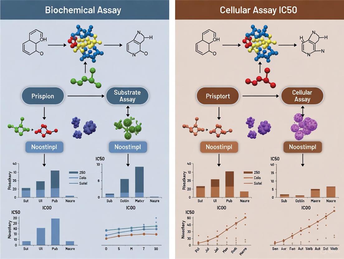
Bridging the Gap: A Comprehensive Guide to Biochemical vs. Cellular IC50 Values in Drug Discovery
This article provides a comprehensive analysis of the critical differences between IC50 values obtained from biochemical and cellular assays, a common challenge in drug discovery.
Bridging the Gap: A Comprehensive Guide to Biochemical vs. Cellular IC50 Values in Drug Discovery
Abstract
This article provides a comprehensive analysis of the critical differences between IC50 values obtained from biochemical and cellular assays, a common challenge in drug discovery. It explores the foundational principles of IC50 and Kd, details methodological best practices for both assay types, and investigates the root causes of frequent discrepancies. Drawing on recent research, the content offers troubleshooting strategies and optimization techniques to bridge the activity gap, ultimately guiding researchers in data validation and interpretation to build robust structure-activity relationships and accelerate lead optimization.
IC50 Decoded: Understanding Fundamental Concepts and Common Discrepancies
In drug discovery and development, quantifying the interaction between a molecule and its biological target is paramount. Researchers rely on specific, quantitative metrics to describe the potency and binding strength of potential therapeutics. Among these, the half-maximal inhibitory concentration (IC50), the dissociation constant (Kd), and the inhibition constant (Ki) are cornerstone parameters. Although sometimes used interchangeably, these terms represent distinct concepts. Understanding their definitions, the methodologies behind their determination, and their mathematical interrelationships is crucial for accurately interpreting experimental data and making informed decisions in the pipeline of drug development. This understanding is especially critical when reconciling data from different assay types, such as simplified biochemical assays and more complex cellular systems, where discrepancies in these values are frequently encountered [1] [2].
Defining the Metrics
Dissociation Constant (Kd)
The dissociation constant (Kd) is a thermodynamic parameter that describes the binding affinity between a ligand (L) and its target protein (P). It is defined as the concentration of free ligand at which half the binding sites on the protein are occupied at equilibrium [3]. The equilibrium for a simple binding reaction is represented as:
[ \text{P + L} \rightleftharpoons \text{PL} ]
The Kd is given by the formula:
[ K_d = \frac{[P][L]}{[PL]} ]
where [P] is the concentration of free protein, [L] is the concentration of free ligand, and [PL] is the concentration of the protein-ligand complex [3]. A lower Kd value indicates a higher affinity between the ligand and its target, meaning they bind more tightly. Kd is an intrinsic measure of affinity and, under ideal conditions, is independent of the concentrations of the interacting partners. It is typically determined using equilibrium binding assays such as Surface Plasmon Resonance (SPR) or Isothermal Titration Calorimetry (ITC) [4].
Inhibition Constant (Ki)
The inhibition constant (Ki) is a specific type of dissociation constant that describes the affinity of an inhibitor (I) for an enzyme. It is the equilibrium dissociation constant for the enzyme-inhibitor (EI) complex:
[ K_i = \frac{[E][I]}{[EI]} ]
Like Kd, Ki is an intrinsic property of the enzyme-inhibitor pair [5]. It quantitatively expresses the potency of an inhibitor, with a lower Ki value indicating a more potent inhibitor. While Ki can be determined directly in some cases, it is often derived from functional (IC50) experiments using the Cheng-Prusoff equation, which accounts for the assay conditions [6].
Half-Maximal Inhibitory Concentration (IC50)
The half-maximal inhibitory concentration (IC50) is a functional, operational parameter. It is defined as the total concentration of an inhibitor required to reduce a specific biological or biochemical activity by 50% under a given set of experimental conditions [6] [5]. Unlike Kd and Ki, IC50 is not an intrinsic constant. Its value is highly dependent on assay conditions, including the concentration of the enzyme or receptor, the concentration of the substrate or agonist, and the incubation time [6] [2] [4]. Consequently, IC50 is a measure of functional potency rather than binding affinity. It is commonly determined from dose-response curves generated in enzymatic or cellular assays.
Table 1: Core Definitions and Properties of Key Metrics
| Metric | Definition | What It Measures | Dependence on Assay Conditions |
|---|---|---|---|
| Kd | Concentration of free ligand at half-maximal target occupancy | Binding Affinity | Independent (intrinsic constant) |
| Ki | Dissociation constant for an enzyme-inhibitor complex | Inhibitor Affinity | Independent (intrinsic constant) |
| IC50 | Total inhibitor concentration for 50% activity reduction | Functional Potency | Highly dependent (enzyme, substrate, time) |
Mathematical and Conceptual Relationships
Relating IC50 to Ki with the Cheng-Prusoff Equation
The most critical relationship between these metrics is the conversion of the experimentally determined IC50 value to the more fundamental Ki constant. This conversion is formally achieved using the Cheng-Prusoff equation [6].
For a competitive inhibition assay, the relationship is:
[ Ki = \frac{IC{50}}{1 + \frac{[S]}{K_m}} ]
Where:
- ( K_i ) is the inhibition constant.
- ( IC_{50} ) is the half-maximal inhibitory concentration.
- ( [S] ) is the concentration of the substrate in the assay.
- ( K_m ) is the Michaelis constant for the substrate [6].
This equation demonstrates that the measured IC50 is always greater than the true Ki, and the difference is a function of the substrate concentration relative to its Km. Only when the substrate concentration [S] is negligible compared to Km does IC50 approximate Ki. Similar derivative equations exist for other modes of inhibition, such as non-competitive and uncompetitive [6] [7].
Distinguishing Between Affinity and Potency
A fundamental concept is the distinction between affinity and potency.
- Affinity (Kd/Ki): This is a pure measure of binding strength—how tightly a drug binds to its target. It is a molecular-level interaction [4].
- Potency (IC50): This is a functional measure of effectiveness—the concentration needed to achieve a defined biological effect (50% inhibition). While high affinity often leads to high potency, other factors like cell permeability, solubility, and metabolic stability also influence IC50, especially in cellular assays [1] [4].
This distinction is key to understanding why a compound with an excellent Kd from a biochemical assay might show a much less impressive (higher) IC50 in a cellular assay. The IC50 incorporates the compound's affinity in the context of the complex cellular environment [1].
Diagram 1: Relationship between IC50, Ki, and Kd. IC50 is influenced by experimental conditions and the cellular environment, and can be related to the intrinsic constant Ki via mathematical correction. Ki is itself a specific type of Kd for enzyme-inhibitor complexes.
Experimental Protocols for Determination
Determining IC50 via a Functional Enzymatic Assay
The following protocol outlines a general method for determining IC50 using the diphenolase activity of tyrosinase as a model system, as described in the literature [7].
1. Principle: The rate of enzyme-catalyzed conversion of a substrate (L-dopa) to its product (dopaquinone) is measured spectroscopically in the presence of varying concentrations of an inhibitor. The degree of inhibition at each inhibitor concentration is calculated, and a dose-response curve is fitted to determine the IC50.
2. Reagents and Solutions:
- Enzyme: Purified tyrosinase.
- Substrate: L-3,4-dihydroxyphenylalanine (L-dopa).
- Inhibitor: A solution of the test compound at a high stock concentration (e.g., in DMSO).
- Buffer: A suitable physiological buffer (e.g., phosphate buffer, pH 6.8).
3. Procedure:
- Prepare a series of reaction mixtures with a fixed, saturating concentration of tyrosinase and L-dopa substrate.
- Add increasing concentrations of the inhibitor to each tube. Include a control tube with no inhibitor.
- Initiate the enzymatic reaction and monitor the formation of the product (dopaquinone) in real-time by measuring the increase in absorbance at 475 nm using a spectrophotometer.
- Record the initial velocity (V₀) for each reaction.
4. Data Analysis:
- Calculate the percentage of enzyme activity for each inhibitor concentration relative to the uninhibited control.
- Plot the percentage activity (or the degree of inhibition, iD) against the logarithm of the inhibitor concentration ([I]₀).
- Fit the data points to a non-linear regression curve (e.g., log(inhibitor) vs. response -- variable slope model).
- The IC50 value is derived directly from the curve as the inhibitor concentration that corresponds to 50% remaining enzyme activity.
Determining Kd via a Saturation Binding Assay
1. Principle: A constant amount of the target protein is incubated with a range of concentrations of a labeled ligand (e.g., radioactively or fluorescently tagged). The amount of ligand bound to the protein is measured at equilibrium, allowing for the calculation of Kd.
2. Reagents and Solutions:
- Protein: Purified target protein.
- Ligand: Labeled ligand. A radioligand like [³H]-ligand is traditional, but fluorescent ligands are also common.
- Buffer: Appropriate binding buffer.
- Competitor: An unlabeled ligand at a high concentration (for defining non-specific binding).
3. Procedure:
- Set up a series of tubes with a fixed concentration of the protein.
- Add increasing concentrations of the labeled ligand to the tubes. To another set of tubes, add the same ligand concentrations plus a large excess of unlabeled competitor.
- Incubate the mixtures until equilibrium is reached.
- Separate the protein-bound ligand from the free ligand (e.g., by filtration, centrifugation, or chromatography).
- Quantify the amount of bound ligand in each sample.
4. Data Analysis:
- For each ligand concentration, subtract the non-specific binding (measured in the presence of competitor) from the total binding to obtain specific binding.
- Plot the specific bound ligand concentration ([Bound]) versus the free ligand concentration ([Free]).
- Fit the data to a one-site specific binding model: ( [Bound] = (B{max} * [Free]) / (Kd + [Free]) ).
- The Kd is the free ligand concentration at which half of the maximum binding (Bmax) is achieved.
Table 2: Comparison of Key Experimental Assays for Determining Kd/Ki and IC50
| Aspect | Equilibrium Binding Assay (for Kd) | Functional Inhibition Assay (for IC50) |
|---|---|---|
| What is Measured | Direct physical binding of a ligand | Functional consequence of inhibition (e.g., product formation) |
| Key Reagents | Purified protein, Labeled ligand | Enzyme/Receptor, Substrate/Agonist, Inhibitor |
| Primary Output | Saturation binding curve | Dose-response curve |
| Key Parameter | Kd (Affinity) | IC50 (Potency) |
| Data Conversion | - | IC50 converted to Ki using Cheng-Prusoff |
The Scientist's Toolkit: Essential Research Reagents
Successful execution of the experiments described above requires a suite of reliable reagents and instruments.
Table 3: Essential Research Reagents and Solutions
| Reagent/Solution | Function in Assays | Example Use Case |
|---|---|---|
| Purified Target Protein | The molecule of interest (enzyme, receptor) with which interactions are studied. | Essential for both biochemical binding (Kd) and functional (IC50) assays [1]. |
| Physiological Buffers | Maintain pH and osmotic pressure to mimic biological conditions. | PBS is common but mimics extracellular space; cytoplasm-mimicking buffers (high K+, crowding agents) may be better for intracellular targets [1]. |
| Labeled Ligand | A traceable molecule (radioactive, fluorescent) used to monitor binding events directly. | Critical for saturation binding assays to determine Kd [3]. |
| Substrate/Agonist | The natural molecule acted upon by the enzyme or that activates the receptor. | Required for functional assays (IC50) to measure enzymatic/biological activity [6] [7]. |
| Crowding Agents | Macromolecules (e.g., Ficoll, PEG) used to mimic the crowded intracellular environment. | Added to buffers to make biochemical assay conditions more physiologically relevant, which can significantly alter measured Kd/IC50 values [1]. |
Diagram 2: Workflow and discrepancy between biochemical and cellular assays. Simplified biochemical assays measure intrinsic affinity (Kd/Ki) and potency (IC50) under defined conditions. Cellular assays measure functional potency (IC50) in a complex environment, leading to frequent discrepancies with biochemical data.
Reconciling Biochemical and Cellular Assay Data
A significant challenge in drug discovery is the frequent discrepancy between IC50 values obtained from biochemical assays (using purified proteins) and those from cellular assays [1]. A compound may appear highly potent in a test tube but show reduced activity in a cellular context.
Several factors contribute to this divergence:
- Cellular Permeability: The compound must cross the cell membrane to reach an intracellular target, which is not a factor in a purified biochemical system [1].
- Intracellular Physicochemical Conditions: The cytoplasmic environment is vastly different from standard assay buffers like PBS. It features high macromolecular crowding (affecting viscosity and diffusion), different ionic concentrations (high K+, low Na+), and distinct redox potential [1]. These factors can significantly alter the apparent Kd of an interaction.
- Metabolic Stability: The compound may be metabolized or degraded within the cell, reducing its effective concentration.
- Off-Target Effects: Binding to other cellular components can sequester the compound away from its primary target.
Therefore, while a biochemical assay provides a clean measure of intrinsic affinity and potency, the cellular IC50 provides a more holistic, physiologically relevant measure of a compound's effectiveness, integrating both its binding affinity and its drug-like properties [1] [4]. Understanding the interrelationship of Kd, Ki, and IC50 allows scientists to deconvolute these factors and guide the optimization of truly effective therapeutics.
The half-maximal inhibitory concentration (IC50) is a crucial quantitative measure in pharmacological research, indicating the potency of a substance required to inhibit a specific biological or biochemical function by 50% [6]. This parameter is central to lead optimization, enabling researchers to compare compound potency, build chemogenomics models, and predict off-target activity [8]. However, IC50 values are highly dependent on experimental conditions, and significant discrepancies frequently arise between values obtained from biochemical assays (BcAs) using purified components and cell-based assays (CBAs) conducted in living systems [9] [10]. These inconsistencies can delay research progress and drug development, making it essential to understand the fundamental principles and environmental factors distinguishing these assay platforms [11] [10].
Core Principles and Fundamental Differences
Biochemical Assays are laboratory methods designed to measure the presence, concentration, or activity of a specific biomolecule (e.g., an enzyme, protein, or nucleotide) in a purified system [12]. They typically utilize purified protein targets in a simplified, controlled environment to measure direct molecular interactions, with results often expressed as binding affinity (Ka or Kd) or inhibitory potency (Ki or IC50) [13] [10].
Cellular Assays evaluate biological responses within the context of intact living cells. They measure a compound's effect on cellular phenotypes, such as viability, proliferation, or pathway activation, thereby accounting for cellular complexity, including membrane permeability, metabolic conversion, and off-target effects [14].
The table below summarizes the core characteristics of each assay type.
Table 1: Fundamental Characteristics of Biochemical vs. Cellular Assays
| Feature | Biochemical Assays (BcAs) | Cellular Assays (CBAs) |
|---|---|---|
| System Complexity | Reduced system; purified components | Complex system; intact living cells |
| Primary Measurement | Direct molecular interaction (e.g., binding, enzyme inhibition) | Cellular response (e.g., metabolic activity, reporter gene expression) |
| Key Readouts | Kd, Ki, IC50 for target binding | IC50, EC50 for functional response |
| Information Gained | Intrinsic binding affinity and mechanism | Functional potency in a cellular context |
| Throughput | Typically higher | Often lower due to cellular maintenance |
| Environmental Control | High; buffer conditions are defined and controllable | Lower; intracellular environment is complex and dynamic |
The Assay Environment: A Major Source of Discrepancy
A critical factor explaining IC50 differences between BcAs and CBAs is the profound divergence in their physicochemical (PCh) environments. Standard biochemical assays are often conducted in simplified buffer solutions like Phosphate-Buffered Saline (PBS), which mimics extracellular conditions but fails to replicate the intracellular milieu [10].
The intracellular environment is characterized by high macromolecular crowding, differential ionic concentrations (high K+, low Na+), specific viscosity, and unique lipophilicity [9] [10]. These conditions can significantly alter equilibrium binding constants; for instance, in-cell Kd values can differ by up to 20-fold or more from values measured in standard buffer [10]. Furthermore, enzyme kinetics can change dramatically (by as much as 2000%) under macromolecular crowding conditions [10].
Table 2: Key Physicochemical Differences Between Standard BcA Buffers and the Cytoplasm
| Parameter | Standard BcA Buffer (e.g., PBS) | Cytoplasmic Environment | Impact on Molecular Interactions |
|---|---|---|---|
| Cations | High Na+ (157 mM), Low K+ (4.5 mM) | High K+ (~140-150 mM), Low Na+ (~14 mM) [10] | Alters electrostatic interactions and protein stability. |
| Macromolecular Crowding | Negligible | High (≥ 80 mg/ml) [10] | Increases effective compound concentration, can enhance binding (depletion attraction). |
| Viscosity | Low, similar to water | Higher than water [10] | Slows diffusion, affects reaction kinetics and conformational dynamics. |
| Redox Potential | Oxidizing | Reducing (high glutathione) [10] | Can affect disulfide bond formation and stability of protein/compound. |
The following diagram illustrates the fundamental difference in what each assay type measures, which is a direct cause of IC50 discrepancies.
Experimental Protocols and Methodologies
A Standard Biochemical Assay Protocol: Enzyme Inhibition
This protocol outlines the key steps for determining an IC50 value using a purified enzyme system, such as a kinase, in a 96-well plate format [15] [12].
- Reagent Preparation: Prepare the assay buffer (e.g., PBS or HEPES-based), the purified enzyme, the substrate (often a peptide or small molecule), co-factors (e.g., ATP for kinases), and the detection reagent (e.g., a coupled enzyme system or fluorescent probe). Test reagent stability under storage and assay conditions [15].
- Compound Serial Dilution: Prepare a serial dilution of the inhibitor compound in DMSO, ensuring the final DMSO concentration in the assay is tolerated (typically <1% for enzymes) [15]. Include a vehicle control (DMSO only).
- Assay Plate Setup:
- Add buffer, inhibitor solution, enzyme, and substrate/co-factors to the wells in a defined order.
- Controls: Include "Max" signal wells (enzyme + substrate + vehicle), "Min" signal wells (no enzyme, or fully inhibited enzyme), and "Mid" signal wells (e.g., with a reference IC50 inhibitor) for quality control [15].
- Incubation and Reaction Initiation: Incubate the plate at the optimal temperature (e.g., 25°C or 37°C) for a predetermined time to allow the reaction to proceed linearly.
- Signal Detection: Measure the product formation using an appropriate method (e.g., spectrophotometry, fluorescence, or luminescence) with a microplate reader [12].
- Data Analysis: Calculate the percentage of inhibition relative to Max and Min controls for each compound concentration. Fit the dose-response data to a four-parameter logistic (sigmoidal) curve to determine the IC50 value [6].
A Standard Cellular Assay Protocol: WST-1 Cell Viability
The WST-1 assay is a common colorimetric method used to measure cell viability and proliferation, often applied in cytotoxicity and drug-sensitivity testing [14].
- Cell Seeding: Seed cells into the wells of a 96-well tissue culture plate at an optimized density. Incubate under standard culture conditions (37°C, 5% CO2) for 24-96 hours to allow adherence and recovery [14].
- Compound Treatment: Expose cells to a serial dilution of the test compound. Include appropriate controls: Blank (medium only, no cells), Untreated control (cells + vehicle), and Positive control (cells treated with a known cytotoxic agent) [14].
- Incubation: Incubate the plate for the desired treatment period (e.g., 48-72 hours).
- WST-1 Reagent Addition: Add WST-1 reagent directly to each well (typically 10 µL per 100 µL of culture medium). The WST-1 salt is cleaved by mitochondrial dehydrogenases in metabolically active cells to produce a water-soluble formazan dye [14].
- Formazan Development: Incubate the plate for 0.5-4 hours, monitoring color development. The amount of formazan dye produced is directly proportional to the number of viable cells.
- Signal Measurement: Measure the absorbance of the formazan dye at 440-450 nm using a microplate reader, with a reference wavelength above 600 nm for background correction [14].
- Data Analysis: Calculate the percentage of cell viability relative to the untreated control. Plot the dose-response curve and calculate the IC50 value, which represents the compound concentration that reduces cell viability by 50% [14].
The workflow below contrasts the key steps involved in these two major assay types.
Quantitative Data Comparison and Analysis
IC50 values for the same compound can vary significantly between biochemical and cellular assay formats. A statistical analysis of public IC50 data in the ChEMBL database found that the standard deviation of independently measured IC50 values for identical protein-ligand systems is approximately 25% larger than that of Ki data, reflecting the inherent noise and variability when combining data from different assay conditions [8].
The table below provides a theoretical comparison illustrating how different factors can influence the measured IC50.
Table 3: Factors Causing IC50 Discrepancies Between Biochemical and Cellular Assays
| Factor | Impact on Biochemical IC50 | Impact on Cellular IC50 | Example/Effect |
|---|---|---|---|
| Membrane Permeability | No impact (no membrane) | Major impact; poor permeability increases apparent IC50 [11] | Compound may be potent on purified target but inactive in cells. |
| Cellular Efflux Pumps | No impact | Major impact; efflux decreases intracellular concentration, increasing apparent IC50 [11] | Activity of transporters like P-glycoprotein. |
| Metabolic Conversion | No impact | Can activate (pro-drug) or inactivate a compound, altering apparent IC50. | Compound stability differs between buffer and cellular milieu. |
| Protein Binding | Minimal in purified systems | Significant; binding to serum or cellular proteins reduces free compound, increasing IC50. | Must account for free fraction in media with serum. |
| Target Engagement Specificity | Measures direct binding to purified target. | Measures net effect; inhibition may be indirect or via off-target effects. | SPR can resolve specific vs. functional IC50 [13]. |
| Physicochemical Conditions | Defined, simple buffer (e.g., PBS). | Complex, crowded cytoplasm with different ions, pH, and viscosity. | Kd can vary up to 20-fold between buffer and cells [10]. |
The Scientist's Toolkit: Essential Reagents and Materials
Table 4: Key Research Reagent Solutions for Biochemical and Cellular Assays
| Reagent / Material | Function | Typical Use Case |
|---|---|---|
| Purified Target Protein | The isolated molecule of interest (e.g., enzyme, receptor). | Biochemical assay to measure direct binding or inhibition. |
| Cell Lines | Genetically defined living cells. | Cellular assays to measure functional responses and compound efficacy in a biological system. |
| WST-1 Assay Reagent | A tetrazolium salt cleaved by mitochondrial dehydrogenases to a water-soluble formazan dye. | Colorimetric measurement of cell viability and proliferation in cellular assays [14]. |
| Surface Plasmon Resonance (SPR) Chip | A biosensor surface for immobilizing a target molecule to study binding interactions in real-time. | Label-free determination of binding kinetics (Ka, Kd) and IC50 for specific molecular interactions [13]. |
| 96-/384-Well Plates | Standard microplate formats for assay setup. | High-throughput screening in both biochemical and cellular formats [15]. |
| Microplate Reader | Instrument to detect optical signals (absorbance, fluorescence, luminescence). | Quantifying assay endpoints in both biochemical and cellular formats [14] [15]. |
| Cytoplasm-Mimicking Buffer | A buffer designed to replicate intracellular conditions (e.g., high K+, crowding agents). | Biochemical assays aiming to produce more physiologically relevant IC50 values [9] [10]. |
| DMSO (Dimethyl Sulfoxide) | A universal solvent for dissolving small molecule compounds for screening. | Stock solutions of test compounds; final concentration in assays must be optimized and controlled (<1%) [15]. |
The Prevalence and Impact of IC50 Discrepancies in Research and Development
In pharmacological research and drug discovery, the half maximal inhibitory concentration (IC50) is a fundamental metric used to quantify the potency of a substance. It represents the concentration of an inhibitor required to reduce a specific biological or biochemical process by half [6]. Despite its widespread use, a growing body of evidence indicates that IC50 values are not absolute and can vary significantly depending on experimental conditions, assay selection, and cellular context. These discrepancies pose substantial challenges for drug development, leading to irreproducible results and potentially misleading conclusions about compound efficacy [16] [17]. This guide objectively compares the performance of different assay methodologies used for IC50 determination, with a specific focus on the systematic variations observed between biochemical and cellular assay systems, and provides researchers with frameworks to enhance the reliability of their potency assessments.
Understanding IC50 and Its Critical Role in Drug Discovery
IC50 serves as a crucial parameter for evaluating antagonist drug potency in pharmacological research. It is a quantitative measure typically expressed as molar concentration, with lower values indicating greater compound potency [6]. In practice, IC50 values are determined by constructing dose-response curves that examine the effect of different antagonist concentrations on reversing agonist activity [6].
The pIC50 value, derived as -log10(IC50), is often used in high-throughput screening environments because higher pIC50 values correspond to exponentially more potent inhibitors, facilitating easier comparison of compound libraries [6]. It is critical to distinguish IC50 from related metrics such as EC50 (half maximal effective concentration), which measures the concentration of a substance that produces 50% of the maximal response in an excitatory context [18] [6].
A fundamental consideration in IC50 interpretation is that IC50 is not a direct measure of binding affinity. The Cheng-Prusoff equation provides a mathematical relationship to convert IC50 values to Ki (inhibition constant) values for competitive inhibitors, accounting for substrate concentration and enzyme affinity [6]. This relationship highlights the context-dependent nature of IC50 values, which can vary with experimental conditions, while Ki represents an absolute affinity value [6].
The Prevalence of IC50 Discrepancies: Evidence from Comparative Studies
Substantial evidence demonstrates that IC50 values for the same compound can vary significantly across different testing methodologies. A compelling 2019 study investigating human glioblastoma cell lines revealed striking variations in IC50 values when different cytotoxicity assays were applied to the same cell lines treated with identical chemical agents [16] [19].
Table 1: IC50 Value Variations Across Different Assay Methods in Glioblastoma Cell Lines
| Cell Line | Compound | MTT Assay IC50 | Alamar Blue IC50 | Acid Phosphatase IC50 | Trypan Blue IC50 |
|---|---|---|---|---|---|
| U87MG | Carboplatin | Variable | Variable | Variable | Variable |
| U87MG | Etoposide | Variable | Variable | Variable | Variable |
| U87MG | Paraquat | Variable | Variable | Variable | Variable |
| U373MG | Carboplatin | Variable | Variable | Variable | Variable |
| U373MG | Etoposide | Variable | Variable | Variable | Variable |
| U373MG | Paraquat | Variable | Variable | Variable | Variable |
The study concluded that "variations between IC50 values were seen in all experiments with differences observed between testing methods, cell lines and cytotoxic agents under investigation" [19]. This inconsistency persisted even when combining multiple endpoints including mitochondrial function, lysosomal activity, and membrane integrity, suggesting that no single assay provides a comprehensive toxicity profile [16].
The implications of these discrepancies are significant for drug discovery. Researchers noted that "the true IC50 value of valuable and beneficial compounds for glioblastoma may have been missed through over/underestimation," highlighting the critical impact of methodological selection on compound identification and development [19].
Biochemical vs. Cellular Assays: A Systematic Comparison
The distinction between biochemical and cell-based assays represents a fundamental division in IC50 determination methodologies, each with distinct advantages and limitations that systematically influence potency measurements.
Biochemical Assays
Biochemical assays typically employ purified enzymes or receptors in controlled environments to measure compound-target interactions directly. The FDA's Guidance for Industry acknowledges the distinction between these systems, providing "an algorithm for converting an IC50 value to a Ki value" that differs between cellular and purified enzyme systems [18]. A key advantage of biochemical assays is the reduced complexity that minimizes confounding variables, offering clearer structure-activity relationships for lead optimization [11].
Cellular Assays
Cellular assays measure compound effects within intact cells, preserving physiological context including cellular uptake, metabolism, and potential off-target effects. The in-cell Western assay, for instance, assesses protein expression and phosphorylation within intact cells, providing "a more accurate representation of drug effects in a cellular context" compared to traditional Western blotting that requires cell lysis [20]. This methodology maintains the physiological relevance of the cellular environment while enabling high-throughput screening [20].
Root Causes of Discrepancies
Several factors contribute to the systematic differences observed between biochemical and cellular IC50 values:
- Cellular permeability: Compounds may be unable to penetrate cell membranes or may be actively exported by cellular efflux pumps, leading to higher apparent IC50 values in cellular systems [11].
- Metabolic conversion: Prodrugs requiring metabolic activation or compounds degraded by cellular machinery will show different potency in cellular versus biochemical systems [11].
- Off-target effects: Compounds may engage unintended targets in cellular environments, altering apparent potency through parallel pathways [11] [20].
- Signal amplification: Cellular signaling pathways often incorporate amplification mechanisms that can magnify target engagement effects not captured in biochemical systems [20].
Table 2: Key Differences Between Biochemical and Cellular Assay Systems
| Parameter | Biochemical Assays | Cellular Assays |
|---|---|---|
| System Complexity | Purified components | Intact cellular environment |
| Physiological Relevance | Lower | Higher |
| Throughput Potential | Typically higher | Variable |
| Influence of Permeability | No | Yes |
| Metabolic Considerations | No | Yes |
| Signal Amplification | Controlled | Physiological |
| Cost and Technical Demand | Generally lower | Generally higher |
Methodological Artifacts and Technical Limitations
Beyond biological differences, specific technical artifacts associated with common assay methodologies contribute significantly to IC50 discrepancies. The MTT assay, one of the most widely used colorimetric techniques for IC50 determination, has been particularly scrutinized for its methodological limitations.
MTT Assay Limitations
A 2016 study examining cisplatin IC50 in ovarian cancer cells revealed that "IC50 errors caused by the technical deficiencies of the MTT assay are large and not adjustable (range: 300-11,000%)" [17]. The researchers identified several critical technical deficiencies:
- Density-dependent artifacts: MTT-measured IC50 values showed positive correlation with seeding densities across all five ovarian cancer cell lines tested [17].
- Enzyme activity confounding: The assay measures decreases in intracellular NAD(P)H-dependent oxidoreductase activity rather than direct cell killing, potentially misrepresenting viability under different metabolic conditions [17].
- Solubilization requirements: The insoluble nature of MTT formazan crystals requires additional solubilization steps that introduce variability [17].
The study noted that even within the same laboratory, "MTT and analogue assays produce variable IC50 values among different staff researchers and between different experimental repeats performed by the same researcher" [17], highlighting the profound impact of technical execution on results reproducibility.
Alternative Methodologies
To overcome these limitations, researchers have developed alternative approaches:
- Limiting dilution assay: This direct measurement method was developed to overcome MTT artifacts, leading to the discovery of "inherent density-dependent chemoresistance variation of cancer cells" [17].
- Growth rate-based analysis: A 2025 study proposed calculating effective growth rates for both control and drug-treated cells, deriving time-independent parameters (ICr0 and ICrmed) that avoid normalization artifacts associated with traditional IC50 determination [21].
- In-cell Western assays: These combine immunoassay specificity with cellular context preservation, allowing multiplex analysis of multiple targets within intact cells [20].
Assay Methods and Technical Limitations
Impact on Research Reproducibility and Drug Development
The cumulative effect of IC50 discrepancies extends beyond individual experiments to impact broader research validity and therapeutic development. A review of current literature revealed that MTT and analogous assays are extensively used, with 20.7% of studies in selected journals employing 96-well colorimetric techniques for IC50 determination [17]. Alarmingly, only 27.6% of these manuscripts reported per-well seeding numbers, despite the demonstrated impact of cell density on IC50 values [17].
This reporting deficiency compounds reproducibility challenges, as "the degree of chemoresistance identified through an MTT assay by one laboratory may not be reproducible and should not be used to depict the pharmacological and biological traits of the cancer cell line" [17]. The implications for drug development are particularly significant in light of the 3Rs ethic (Replace, Reduce, Refine), which encourages reduced reliance on animal models for therapeutic screening [16].
In clinical translation, precise IC50 determination enables more accurate prediction of patient chemoresistance. The development of an in situ immunohistochemical scoring system (IHCpAkt+p62) based on signaling pathways correlated with IC50 variations demonstrated superior diagnostic efficacy compared to MTT assays for predicting primary chemoresistance in ovarian cancer patients [17].
Best Practices for Robust IC50 Determination
Based on the comprehensive analysis of IC50 discrepancies, researchers can adopt several strategies to enhance the reliability of their potency assessments:
- Implement orthogonal validation: Employ multiple assay methodologies with different detection endpoints to cross-verify IC50 values and minimize technique-specific artifacts [16] [17].
- Standardize reporting: Document critical parameters including cell seeding densities, assay timepoints, and normalization methods to enable proper interpretation and replication [17].
- Contextualize results: Interpret IC50 values with consideration of the assay system (biochemical vs. cellular) and its relationship to the physiological context of interest [11] [6].
- Consider time-independent parameters: Explore emerging approaches like growth rate-based analysis (ICr0, ICrmed) that avoid time-dependent normalization artifacts [21].
- Account for density-dependent effects: Recognize that "density-related IC50 uncertainty is a natural property of cancer cells" and design experiments accordingly [17].
IC50 Determination Workflow
Essential Research Reagent Solutions
The following table details key reagents and methodologies used in IC50 determination, providing researchers with a reference framework for experimental design:
Table 3: Research Reagent Solutions for IC50 Determination
| Reagent/Method | Category | Primary Function | Key Considerations |
|---|---|---|---|
| MTT Assay | Viability Assay | Measures mitochondrial dehydrogenase activity | Subject to density artifacts; requires solubilization |
| MTS/CCK8 Assays | Viability Assay | Tetrazolium reduction with soluble formazan | "One-step" protocol; reduced technical variability |
| Alamar Blue | Viability Assay | Measures resazurin reduction | Fluorescent/colorimetric readout; less toxic to cells |
| Acid Phosphatase | Viability Assay | Measures lysosomal enzyme activity | Alternative metabolic endpoint |
| Trypan Blue | Cell Counting | Membrane integrity assessment | Gold standard but low-throughput; subjective |
| In-cell Western | Multiplex Assay | Protein expression in intact cells | Preserves cellular context; enables multiplexing |
| Limiting Dilution | Clonal Assay | Direct measurement of cell survival | Resource-intensive; avoids metabolic confounding |
IC50 discrepancies between biochemical and cellular assay systems represent a significant challenge in drug discovery and development. These variations arise from fundamental differences in assay design, physiological complexity, and methodological artifacts. The prevalence of such discrepancies underscores the importance of methodological transparency, orthogonal validation, and appropriate interpretation of IC50 values within their experimental context. By understanding the sources and implications of these variations, researchers can design more robust screening strategies, improve reproducibility, and enhance the predictive power of in vitro assays for clinical outcomes. As the field advances, the development of novel approaches that circumvent traditional limitations promises to deliver more reliable potency measurements, ultimately accelerating the identification and optimization of therapeutic compounds.
Distinguishing Functional Potency (IC50) from Binding Affinity (Kd)
In drug discovery, accurately interpreting data from biochemical and cellular assays is crucial. A common and critical challenge is the discrepancy between results obtained from these different experimental setups [11] [1]. Central to this challenge is understanding the distinct roles of IC50 (half-maximal inhibitory concentration) and Kd (dissociation constant). While sometimes used interchangeably, they represent fundamentally different concepts: IC50 measures functional potency in a specific assay, whereas Kd measures the intrinsic strength of the target-ligand interaction [22] [23] [2]. Confusing these parameters can lead to misinterpretation of a compound's potential, inefficient resource allocation, and costly delays in research and development [23].
Core Definitions and Conceptual Differences
The following table outlines the fundamental characteristics of Kd and IC50.
| Parameter | Kd (Dissociation Constant) | IC50 (Half-Maximal Inhibitory Concentration) |
|---|---|---|
| Definition | Concentration at which half the target binding sites are occupied by the ligand [22] [24]. | Concentration that inhibits a specific biological process or function by 50% [6] [23]. |
| What It Measures | Intrinsic binding affinity between a ligand and its target [24] [23]. | Functional potency of an inhibitor under specific experimental conditions [22] [23]. |
| Nature of Value | Thermodynamic constant; typically independent of assay conditions [22]. | Operational metric; highly dependent on assay conditions [6] [22]. |
| Key Influences | Determined by the chemical nature of the ligand and target [24]. | Influenced by ligand affinity, substrate concentration, enzyme concentration, and cellular environment [6] [1]. |
| Typical Use Cases | Characterizing the strength of a bimolecular interaction (e.g., drug-target binding) [22]. | Evaluating the effectiveness of an inhibitor in blocking a functional output (e.g., enzyme activity, cell growth) [6]. |
The Relationship Diagram
The diagram below illustrates the conceptual and experimental relationships between Kd, IC50, and the factors that cause their values to differ, particularly between biochemical and cellular assays.
Why IC50 and Kd Values Diverge: Biochemical vs. Cellular Assays
A central theme in pharmacology is the frequent observation that a compound's IC50 value determined in a purified biochemical assay differs, sometimes substantially, from its IC50 value measured in a cellular model [11] [1]. This discrepancy is a key reason why Kd and IC50 should not be conflated.
Key Factors Causing Discrepancies
- Cellular Permeability and Efflux: A compound may show high potency in a biochemical assay where it has direct access to the purified target. However, in a cellular assay, it must first cross the cell membrane. If the compound is unable to penetrate the membrane or is actively pumped out by efflux transporters, its effective intracellular concentration will be lower, leading to a higher (less potent) IC50 value [11].
- The Intracellular Physicochemical Environment: Biochemical assays are often conducted in simple buffered solutions like PBS, which mimics extracellular conditions. In contrast, the cytoplasm is a crowded, viscous environment with high concentrations of macromolecules, different salt compositions (high K+, low Na+), and distinct lipophilicity [1]. These factors can significantly alter the apparent binding affinity (Kd) of the interaction, changing the functional IC50 [1].
- Metabolic Conversion and Stability: A compound could be metabolically activated or deactivated within the cell, effects that are absent in a biochemical assay with purified components.
- Presence of Non-Specific Targets: In the complex cellular milieu, a compound might bind to other non-specific targets, effectively reducing its concentration available for the primary target of interest and altering the measured IC50 [11].
The Relationship Between IC50 and Kd
Although different, IC50 and Kd can be mathematically related under specific conditions. The most famous method is the Cheng-Prusoff equation [6] [22] [1]. For a competitive inhibition assay, the relationship is:
Ki = IC50 / (1 + [S]/Km)
Where:
- Ki is the inhibition constant (equivalent to Kd for competitive inhibitors).
- IC50 is the experimentally determined half-maximal inhibitory concentration.
- [S] is the concentration of the substrate in the assay.
- Km is the Michaelis constant for the substrate.
It is critical to remember that this conversion is only valid under a strict set of assumptions, including that the system is at equilibrium and follows the laws of mass action [6] [2]. Under many common experimental conditions, especially when receptor or tracer concentrations are high, the IC50 and Kd can be very different, and using this approximation can be misleading [2].
Experimental Protocols and Methodologies
Determining Kd: Measuring Intrinsic Affinity
Key Methods:
- Surface Plasmon Resonance (SPR): A label-free technique that measures binding in real-time, allowing for the determination of both affinity (Kd) and kinetics (association/dissociation rates) [22] [24].
- Isothermal Titration Calorimetry (ITC): Measures the heat released or absorbed during binding, providing Kd, as well as thermodynamic parameters (enthalpy, entropy) [22].
- Radioligand Binding Assays: A traditional method where a radioactively labeled ligand is used to compete with the unlabeled test compound for binding to the target. The IC50 from this assay can be converted to Ki (≈Kd) using the Cheng-Prusoff equation, provided its assumptions are met [6] [22] [2].
Determining IC50: Measuring Functional Potency
Key Methods:
- Dose-Response Curves: The standard approach for determining IC50 (for inhibitors) or EC50 (for agonists) [6] [23]. A range of compound concentrations is applied to the assay system, and the resulting level of inhibition or activation is measured. The data is then fitted to a logistic curve, with the IC50 being the concentration at the curve's inflection point (50% inhibition).
- Cellular Dielectric Spectroscopy (CDS): A "label-free" cellular assay technology that measures changes in impedance of a cell layer in response to receptor stimulation. This provides a functional readout that is independent of the specific signaling pathway used [25].
The Scientist's Toolkit: Key Research Reagent Solutions
The following table lists essential tools and reagents used in experiments to determine IC50 and Kd.
| Tool/Reagent | Function in Experimentation |
|---|---|
| Purified Target Protein | Essential for biochemical assays (BcAs) to study ligand binding and function without cellular complexity [1]. |
| Radioligands (e.g., [³H]rotigotine) | Radioactively labeled compounds used as tracers in competition binding assays to determine IC50 and estimate Kd [25] [24]. |
| Fluorescent Ligands/Probes | Used in probe-displacement assays (e.g., NanoBRET Target Engagement) to measure target binding in live cells, allowing estimation of apparent Kd [22]. |
| Cytoplasm-Mimicking Buffer | Assay buffers designed to replicate the intracellular environment (macromolecular crowding, viscosity, ion composition) to make biochemical assay data more predictive of cellular activity [1]. |
| Cell Lines with Recombinant Receptors | Genetically engineered cells (e.g., CHO cells) expressing the human target receptor, used for cellular functional assays and binding studies [25]. |
In summary, Kd and IC50 provide distinct yet complementary information in drug discovery. Kd is an absolute measure of binding affinity, while IC50 is a relative measure of functional potency that is inextricably linked to the specific experimental conditions [23] [2]. The recurring discrepancy between biochemical and cellular IC50 values underscores the importance of this distinction [11] [1]. Factors such as cellular permeability, the intracellular environment, and off-target binding all contribute to these differences. A robust drug discovery workflow therefore requires careful interpretation of both binding affinity (Kd) and functional potency (IC50) across multiple assay formats to accurately triage compounds and advance the most promising candidates.
From Bench to Data: Best Practices in Biochemical and Cellular IC50 Assay Execution
In the drug discovery pipeline, the accurate determination of a compound's half-maximal inhibitory concentration (IC50) is a critical step in evaluating its biological activity and therapeutic potential. For researchers investigating the disconnect between compound potency in simplified biochemical assays (BcAs) and more complex cellular environments (CBAs), the choice of biochemical technique is paramount [1]. This guide objectively compares three foundational technologies used for biochemical IC50 determination: Fluorescence Polarization (FP), Time-Resolved Fluorescence Resonance Energy Transfer (TR-FRET), and Surface Plasmon Resonance (SPR). We provide a detailed comparison of their working principles, performance characteristics, and experimental protocols, supported by quantitative data, to aid scientists in selecting the optimal platform for their specific research context.
Technology Comparison at a Glance
The following table summarizes the core characteristics, advantages, and limitations of FP, TR-FRET, and SPR assays.
Table 1: Core Characteristics of FP, TR-FRET, and SPR Assays
| Feature | Fluorescence Polarization (FP) | Time-Resolved FRET (TR-FRET) | Surface Plasmon Resonance (SPR) |
|---|---|---|---|
| Principle | Measures change in polarization of emitted light from a fluorescent tracer upon binding to a larger protein [26]. | Measures energy transfer from a donor to an acceptor fluorophore when in close proximity (5-10 nm) [27]. | Measures mass concentration changes on a sensor surface in real-time, without labels [26]. |
| Format | Homogeneous, solution-based | Homogeneous, solution-based | Surface-immobilized (one binding partner) |
| Throughput | High | Very High (suitable for HTS) [26] | Low (separate measurement per sample/concentration) [26] |
| Key Advantage | Simple protocol, low reagent consumption, kinetic capability. | High sensitivity, low background, ratiometric measurement, internal reference [26] [28]. | Label-free, provides direct binding affinity (KD) and kinetics (kon, koff). |
| Key Limitation | Susceptible to compound interference (auto-fluorescence). | Requires specific labeling or tagging of components. | Cannot differentiate between specific and non-specific binding to the surface. |
Detailed Principles and Workflows
Fluorescence Polarization (FP)
FP assays measure the change in the rotational speed of a small fluorescently labeled probe (tracer) when it binds to a larger protein. The bound complex rotates more slowly than the free tracer, resulting in a higher polarization value (measured in millipolarization units, mP) [29]. In a competitive IC50 assay, the test compound displaces the tracer from the protein, leading to a decrease in the polarization signal.
Diagram: FP Assay Principle for IC50 Determination
Experimental Workflow (e.g., for Prostaglandin Synthase) [29]:
- Assay Cocktail Preparation: Prepare a mixture containing the target protein (e.g., H-PGDS) and the fluorescein-conjugated tracer probe in the appropriate assay buffer.
- Compound Addition: Add serially diluted test compounds or controls (e.g., DMSO) to the assay cocktail in a multi-well plate.
- Incubation: Incubate the reaction mixture for a defined period (e.g., 90 minutes) at room temperature to reach equilibrium.
- Signal Detection: Read the plate using a fluorescence microplate reader equipped with polarizing filters. Typical settings for a fluorescein probe include an excitation filter of 482-16 nm and an emission filter of 530-40 nm with a dichroic cutoff at 504 nm.
- Data Analysis: Calculate the % inhibition and IC50 values by fitting the dose-response curve of polarization (mP) versus compound concentration.
Time-Resolved FRET (TR-FRET)
TR-FRET combines FRET with time-resolved detection. A donor fluorophore (e.g., a lanthanide like Tb or Eu cryptate) is excited by a light pulse. If an acceptor fluorophore is in close proximity (<10 nm), the donor transfers energy to it, which then emits light at its characteristic wavelength. The time delay between excitation and emission measurement allows short-lived background fluorescence to fade, resulting in a high signal-to-noise ratio [27]. In a competitive IC50 assay, the test compound disrupts the protein-protein interaction, preventing FRET.
Diagram: TR-FRET Competitive Binding Assay Principle
Experimental Workflow (e.g., for Keap1-Nrf2 PPI) [26]:
- Reagent Setup: The assay typically uses a tagged protein (e.g., His-Keap1 Kelch domain), a Tb-labeled anti-tag antibody as the donor, and a fluorescein-labeled peptide (e.g., FITC-Nrf2 peptide amide) as the acceptor.
- Optimization: System components are optimized for concentration (e.g., 0.5 nM Tb-antibody, 5 nM protein, 25 nM peptide) and buffer conditions (e.g., 10 mM HEPES, pH 7.4) to achieve a robust signal.
- Compound Addition & Incubation: Test compounds are added to a mixture of the donor-antibody/protein complex and the acceptor peptide. The plate is incubated to reach equilibrium.
- Signal Detection: The plate is read on a TR-FRET capable microplate reader. The emission ratio of the acceptor signal (e.g., 520 nm for FITC) to the donor signal (e.g., 495 nm for Tb) is calculated. This ratiometric measurement corrects for well-to-well variability and signal interference [28].
- Data Analysis: The emission ratio is plotted against the log of the compound concentration to generate an inhibition curve and determine the IC50.
Surface Plasmon Resonance (SPR)
SPR is a label-free technology that monitors biomolecular interactions in real-time. One interactant (the ligand) is immobilized on a sensor chip, while the other (the analyte) flows over the surface. Binding causes a change in the refractive index at the sensor surface, detected as a resonance angle shift (Response Units, RU). This provides a direct measurement of binding kinetics (association rate kon and dissociation rate koff) from which the equilibrium dissociation constant (KD) can be derived [26]. For inhibitors, the analysis can be adapted to determine IC50 values.
Experimental Workflow (e.g., for Keap1-Nrf2 interaction) [26]:
- Ligand Immobilization: One binding partner (e.g., a biotinylated Nrf2 peptide) is captured on a sensor chip (e.g., a streptavidin-coated chip).
- Baseline Establishment: Running buffer is flowed over the chip to establish a stable baseline.
- Analyte Injection: The second partner (e.g., the Keap1 Kelch domain protein) is injected over the chip surface at a constant flow rate, and the binding response (RU) is recorded in real-time.
- Dissociation Phase: Buffer flow is resumed, and the dissociation of the complex is monitored.
- Regeneration: The chip surface is regenerated by injecting a solution that breaks the interaction without damaging the immobilized ligand.
- Inhibition Assay (for IC50): To determine an inhibitor's IC50, a fixed concentration of the analyte (Keap1) is pre-mixed with varying concentrations of the inhibitor and then injected over the immobilized ligand. The reduction in binding response is used to calculate the IC50.
Performance and Experimental Data
Quantitative Performance Comparison
The following table synthesizes performance data from various assay development studies, highlighting the unique strengths of each platform.
Table 2: Experimental Performance Data from Case Studies
| Assay / Target | Key Performance Metrics | Dynamic Range & Sensitivity | Reference & Application |
|---|---|---|---|
| FP: Prostaglandin D Synthase (H-PGDS) [29] | Z'-factor: 0.84IC50 for TFC-007: 45 nM(Aligns with reported 83 nM) | High assay quality suitable for HTS. Robust detection with 10 flashes on PHERAstar FS (Z'=0.74). | Used for inhibitor screening. Validated with known inhibitors (HQL-79, TFC-007). |
| TR-FRET: Keap1-Nrf2 PPI [26] | Z'-factor: 0.82Sensitivity: Sub-nanomolar Ki | High dynamic range and stability (up to 5 hours). Capable of differentiating very potent inhibitors. | Ideal for HTS and lead optimization of protein-protein interaction inhibitors. |
| SPR: Keap1-Nrf2 Interaction [26] | Direct Kd measurement: ~21-24 nM for 9mer Nrf2 peptide. | Provides absolute affinity, not an indirect IC50. | Used for determining minimal binding sequences and direct binding affinities. Not suited for HTS. |
The IC50 Discrepancy: Biochemical vs. Cellular Assays
A significant challenge in drug discovery is the frequent discrepancy between IC50 values obtained from biochemical assays and those from cell-based assays (CBAs). These differences can be orders of magnitude apart [1]. While factors like membrane permeability and compound stability are often blamed, the physicochemical (PCh) conditions of the assay buffer itself play a crucial role [1].
- The Buffer Problem: Standard biochemical buffers like PBS (Phosphate-Buffered Saline) mimic extracellular, not intracellular, conditions. The cytoplasm has high levels of K+ (~150 mM) and macromolecular crowding agents, which can significantly alter protein-ligand binding equilibria (Kd) and enzyme kinetics [1].
- Implication for IC50: A Kd value measured under standard buffer conditions can differ from its value in a crowded cellular environment by up to 20-fold or more [1]. This directly impacts the measured biochemical IC50 and contributes to the disconnect with cellular activity.
- Technology Considerations: When selecting a biochemical IC50 method, researchers should consider whether the assay conditions can be adapted to better mimic the intracellular environment (e.g., by adding crowding agents like PEG or Ficoll and adjusting salt compositions) to generate more physiologically relevant data [1].
The Scientist's Toolkit: Key Reagents and Materials
Table 3: Essential Research Reagents for IC50 Assays
| Reagent / Material | Function | Example Products / Components |
|---|---|---|
| Fluorescent Tracers | Binds to the target; serves as the core probe for FP and TR-FRET. | FITC-labelled Keap1-Nrf2 peptide [30], JQ1-FITC (for BET bromodomains) [30], H-PGDS Fluorescent Probe - Green [29]. |
| TR-FRET Donors | Lanthanide-based donor fluorophores for time-resolved detection. | CoraFluor 1 (Tb-based, amine reactive) [30], LanthaScreen Tb-anti-His antibody [26]. |
| TR-FRET Acceptors | Acceptor fluorophores that receive energy from the donor. | FITC, BDY FL (BODIPY FL) labeled ligands [30]. |
| Assay Buffers | Provides the chemical environment for the reaction. Critical for maintaining pH and ionic strength. | HEPES buffer, pH 7.4 [26]. For cytoplasmic mimicry: buffers with adjusted K+/Na+ ratio and crowding agents [1]. |
| Tagged Proteins | Recombinant proteins with affinity tags for detection and immobilization. | His-tagged Keap1 Kelch domain [26], GST-BRD2(BD1) [31], Flag-His tagged full length Keap1 [26]. |
The choice between FP, TR-FRET, and SPR for biochemical IC50 determination is not a matter of identifying a single "best" technology, but rather of selecting the right tool for the specific research question.
- FP offers a simple, cost-effective, and robust solution for medium- to high-throughput screening where kinetic data is beneficial.
- TR-FRET is the superior choice for high-throughput screening campaigns requiring high sensitivity, low background, and the ability to detect weak or sub-nanomolar interactions, particularly in complex biological pathways like PPIs and ternary complex formation.
- SPR remains the gold standard for detailed biophysical characterization, providing direct, label-free measurement of binding affinity and kinetics, albeit at a lower throughput.
Ultimately, researchers must be cognizant that the biochemical IC50 value is not an immutable property of a compound, but is influenced by the assay technology and, critically, the buffer conditions used. Bridging the gap between biochemical and cellular IC50 data will require a concerted effort to develop biochemical assays that more accurately mimic the crowded, complex environment of the living cell.
The half-maximal inhibitory concentration (IC50) is a fundamental metric in pharmacological research, quantifying the potency of a substance required to inhibit a specific biological process by 50% [6]. In the context of drug discovery, accurately determining IC50 is essential for understanding the biological and pharmacological characteristics of chemotherapeutic agents and other therapeutic compounds [32] [21]. IC50 values are utilized to guide lead optimization, build chemogenomics analyses, and model off-target activity and toxicity [8]. However, a significant challenge persists in the common observed discrepancy between IC50 values obtained from biochemical assays (BcAs) using purified protein targets and those derived from cell-based assays (CBAs) [1]. These inconsistencies can delay research progress and complicate the establishment of robust structure-activity relationships (SAR) [1]. This guide objectively compares three critical cellular assay platforms—In-Cell Western, Viability (MTT), and other target engagement assays—evaluating their performance in generating reliable and reproducible IC50 data within the broader thesis of biochemical versus cellular assay IC50 values.
The following table summarizes the core principles, primary applications, and key differentiators of the three assay platforms.
Table 1: Core Characteristics of Cellular Assay Platforms
| Assay Platform | Primary Measurement | Typical IC50 Application | Key Distinguishing Feature |
|---|---|---|---|
| Viability (MTT) | Metabolic activity (NAD(P)H-dependent oxidoreductase enzymes) [32] | Measurement of anti-proliferative/cytotoxic activity of compounds [33] | Indirect proxy for cell viability; prone to artifacts from compound interference [32] [33] |
| Target Engagement | Binding affinity (Ki) at a specific molecular target (e.g., receptor, enzyme) [6] | Determining antagonist potency and inhibitor specificity [6] | Measures direct interaction with the target, often using competitive binding; can relate IC50 to Ki via Cheng-Prusoff equation [6] |
| In-Cell Western (ICW) | Protein expression or post-translational modification (e.g., phosphorylation) levels in fixed cells [34] [35] [36] | Screening inhibitors/stimulators to determine IC50/EC50 for signaling pathway modulation [35] [36] | In-situ quantification of target protein or signaling event within a relevant cellular context [34] [35] |
Quantitative Performance Comparison
When selecting an assay platform, understanding its quantitative performance and limitations is critical for data interpretation. The table below consolidates key performance data from the literature.
Table 2: Experimental Performance and Variability Metrics
| Performance Metric | Viability (MTT) | Target Engagement | In-Cell Western (ICW) |
|---|---|---|---|
| Typical IC50 Variability | High (300% to >11,000% error range reported); 2-fold higher IC50 for EGCG vs. ATP/DNA assays [32] [33] | Standard deviation of public IC50 data is ~25% larger than for Ki data [8] | High precision; very low coefficients of variation (CV); significantly smaller standard deviations vs. Western blot [35] [36] |
| Key Source of Error/Artifact | Cell seeding density; proliferation rate; compound interference with tetrazolium reduction [32] [33] | Assay conditions (e.g., substrate concentration for enzymes); differences between labs [8] [6] | Antibody specificity and optimization; fixation/permeabilization efficiency [34] |
| Correlation with Other Assays | Overestimates viable cell count vs. ATP/DNA assays for certain compounds (e.g., EGCG) [33] | IC50 from functional CBAs can be orders of magnitude higher than in BcAs [1] | Excellent correlation with Western blot profiles and published IC50 values from other assays (e.g., cAMP, radioligand binding) [36] |
| Z'-Factor (for HTS) | Not specifically reported | Not specifically reported | Excellent (Z' > 0.5) with optimized conditions, indicating robustness for screening [36] |
Detailed Methodologies and Experimental Protocols
Viability (MTT) Assay Protocol
The MTT assay is a colorimetric method for assessing cell metabolic activity [32]. The detailed protocol is as follows [33]:
- Cell Seeding: Seed cells into a 96-well plate at a density of ~1x10⁴ cells per well in 100 µL of culture medium. Include wells with medium only as negative controls.
- Compound Treatment: Incubate cells with the test compound across a range of concentrations for a designated time (e.g., 24-72 hours).
- MTT Incubation: Add 20 µL of MTT solution (5 mg/mL in PBS) to each well. Incubate the plate at 37°C for 3-4 hours to allow for the formation of formazan crystals.
- Solubilization: Carefully remove the medium and add 100 µL of dimethyl sulfoxide (DMSO) to each well to dissolve the insoluble formazan crystals.
- Absorbance Measurement: Measure the absorbance of each well at a wavelength of 570 nm using a microplate reader. The percentage of cell viability is typically calculated as: (Absorbance of treated sample / Absorbance of untreated control) x 100% [21].
In-Cell Western (ICW) Assay Protocol
The ICW assay is a quantitative immunofluorescence method performed in microplates [34] [35] [36]. The workflow is as follows:
Diagram 1: In-Cell Western assay workflow.
- Cell Seeding and Treatment: Seed cells into a 96-well or 384-well plate. After cells have adhered, treat them with various drugs or conditions [36].
- Fixation: Fix cells by adding chilled 100% methanol (or other fixatives like formaldehyde) and incubating for 20 minutes at 4°C. This preserves cellular architecture and post-translational modifications [34] [36].
- Permeabilization: Remove the fixative and add a permeabilization buffer (e.g., 0.2% Triton X-100 in PBS) for 30 minutes at room temperature with gentle shaking. This step allows antibodies to access intracellular targets [34].
- Blocking: Remove the permeabilization buffer and add a blocking buffer (e.g., LI-COR Odyssey Blocking Buffer) for 1.5 hours at room temperature with gentle shaking to prevent nonspecific antibody binding [34] [36].
- Primary Antibody Incubation: Prepare the primary antibody solution in the appropriate blocking buffer or diluent. Remove the blocking buffer, add the primary antibody solution (typically 50 µL/well), and incubate overnight at 4°C with gentle shaking [34].
- Washing: Carefully remove the primary antibody solution and wash the plate multiple times with phosphate-buffered saline containing Tween-20 (PBST) to remove unbound antibody [36].
- Secondary Antibody and Cell Stain Incubation: Prepare a solution containing the fluorescently-labeled secondary antibodies (e.g., IRDye 680RD or 800CW) and a cell normalization stain (e.g., CellTag 700). Incubate the plate with this solution for about 1 hour at room temperature, protected from light [36].
- Final Washing and Imaging: Perform a final series of washes with PBST to remove unbound reagents. Image the plate using a dedicated infrared fluorescence scanner (e.g., LI-COR Odyssey) [34] [36]. Protein expression levels are quantified based on the fluorescent signal intensity, which can be normalized to the cell stain signal to correct for well-to-well variation in cell number [36].
Key Research Reagent Solutions
Successful implementation of these assays relies on specific reagents. The following table outlines essential materials and their functions.
Table 3: Essential Research Reagents and Their Functions
| Assay Platform | Key Reagent | Function/Purpose |
|---|---|---|
| General Cell Culture | 96-/384-well tissue culture plates | Platform for cell growth and experimental treatment [34] |
| Fetal Bovine Serum (FBS) | Provides essential nutrients and growth factors for cell proliferation [21] | |
| Viability (MTT) | MTT Tetrazolium Salt | Yellow substrate reduced to purple formazan by metabolically active cells [33] |
| Dimethyl Sulfoxide (DMSO) | Solubilizes insoluble formazan crystals for colorimetric reading [33] | |
| In-Cell Western (ICW) | Methanol or Formaldehyde | Fixative that preserves cellular structure and protein epitopes [34] |
| Triton X-100 | Detergent that permeabilizes cell membranes for antibody access [34] | |
| Target-Specific Primary Antibodies | Bind specifically to the protein or phospho-protein of interest [34] [36] | |
| IRDye-conjugated Secondary Antibodies | Fluorescently-labeled antibodies for detection and signal amplification [34] [36] | |
| CellTag 700/520 Stain | Fluorescent cell stain for normalization to cell number [36] | |
| LI-COR Odyssey Imaging System | Scanner for quantifying near-infrared fluorescent signals [34] |
Causes of IC50 Discrepancies Between Assay Platforms
The observed differences in IC50 values between biochemical and cellular assays, and even among different cellular platforms, can be attributed to several factors.
Fundamental Assay Principle and Context
The core of the discrepancy lies in what each assay measures. Biochemical assays typically measure the direct binding to or inhibition of a purified target, providing a clean system to determine intrinsic affinity (Ki) [1] [6]. In contrast, cellular assays like MTT and ICW measure a functional outcome in a complex cellular environment. The MTT assay's readout is an indirect proxy for viability based on metabolic activity, which can be influenced by factors unrelated to the intended target, such as general cellular health or compound interference with mitochondrial enzymes [32] [33]. The ICW assay, while providing specific information about a target within cells, measures downstream signaling events or target expression levels, which are several steps removed from the initial drug-target binding event [35] [36].
Physicochemical and Biological Factors
The intracellular environment is a major contributor to the activity gap between biochemical and cellular assays.
- Cellular Permeability and Efflux: A compound may show high potency in a biochemical assay but be ineffective in a cellular assay because it cannot cross the cell membrane or is actively pumped out by efflux transporters [11] [1].
- Intracellular Physicochemical Conditions: The cytoplasmic environment is highly crowded, viscous, and has a distinct ionic composition (high K+, low Na+) compared to standard biochemical buffers like PBS [1]. These differences can significantly alter the dissociation constant (Kd) of protein-ligand interactions, sometimes by up to 20-fold or more, leading to major shifts in measured IC50 [1].
- Metabolic Instability and Off-Target Effects: Compounds may be metabolically degraded within cells or bind to non-specific targets, reducing their effective concentration at the target of interest and increasing the apparent IC50 [11] [1].
Technical and Methodological Artifacts
Technical aspects of the assays themselves introduce variability.
- MTT-Specific Artifacts: The MTT assay is notoriously susceptible to artifacts. The changing IC50 of compounds like cisplatin has been linked to variations in initial cell seeding density, which is often not reported [32]. Furthermore, certain compounds, like the green tea polyphenol EGCG, can directly interfere with the MTT reduction process, leading to an overestimation of cell viability and a consequently higher (less potent) IC50 value compared to more direct methods like ATP quantification [33].
- Assay Condition Specificity: For enzymatic targets, the IC50 value is highly dependent on assay conditions, particularly the substrate concentration for competitive inhibitors, as described by the Cheng-Prusoff equation [6]. Differences in these conditions between labs make it difficult to directly compare IC50 values from public databases [8].
The relationship between these factors and their impact on the measured IC50 is summarized below.
Diagram 2: Factors causing IC50 discrepancies between assays.
The choice of a cellular assay platform fundamentally shapes the interpretation of IC50 data and its alignment with biochemical data. The Viability (MTT) Assay, while cost-effective and simple, is a less reliable tool for precise IC50 determination when used in isolation, especially for novel compounds whose potential for interference is unknown. Its utility is greatest for initial, high-level cytotoxicity screening. The In-Cell Western Assay offers a powerful combination of specificity, cellular context, and superior precision, making it highly valuable for quantifying target modulation and signaling events within cells, and for screening where immunoblot quality data with high throughput is needed. Target Engagement assays, particularly competitive binding studies, provide the most direct link to biochemical affinity (Ki) but may not capture the functional consequences of target inhibition in a living system.
For a robust research program, the convergence of data from multiple platforms provides the most reliable path forward. A compound that demonstrates potency in a biochemical binding assay, shows effective cellular target engagement via ICW, and subsequently induces a functional response (like loss of viability in an MTT or, preferably, a more direct ATP assay) presents a compelling case for further development. Acknowledging and systematically investigating the inherent discrepancies between these assays, rather than ignoring them, is essential for building a solid foundation for drug discovery and basic research.
In pharmacological research and drug development, accurately quantifying a compound's inhibitory potency is fundamental. Two parameters stand as central pillars in this characterization: the half-maximal inhibitory concentration (IC50) and the inhibition constant (Ki). While sometimes used interchangeably by the uninitiated, these values represent fundamentally different concepts. The IC50 is an operational parameter observed under specific experimental conditions, whereas the Ki is an intrinsic thermodynamic property describing the binding affinity between an inhibitor and its enzyme target [5] [37].
This distinction carries profound implications for comparing compound potency, especially when researchers must integrate data from diverse assay formats—from purified biochemical systems to complex cellular environments. The Cheng-Prusoff equation, published in 1973, provides the seminal mathematical framework for bridging these concepts, allowing scientists to derive the intrinsic Ki from experimentally measured IC50 values [38] [6]. Understanding this relationship, its assumptions, and its limitations is crucial for researchers and drug development professionals seeking to make valid comparisons between compounds and prioritize lead optimization efforts.
Theoretical Foundations: Defining the Key Parameters
IC50 (Half-Maximal Inhibitory Concentration)
The IC50 represents the concentration of an inhibitor required to reduce a specific biological or biochemical activity by half [6]. It is a practical measure of potency determined empirically from dose-response curves. A critical limitation is that IC50 values are highly dependent on experimental conditions, particularly substrate concentration, enzyme concentration, and incubation time [38] [37]. Consequently, IC50 values obtained under different experimental setups cannot be directly compared without appropriate normalization.
Ki (Inhibition Constant)
The Ki is the dissociation constant for the enzyme-inhibitor complex, representing the concentration of inhibitor required to occupy 50% of the enzyme binding sites at equilibrium in the absence of substrate or competing ligands [39] [37]. Unlike IC50, Ki is an intrinsic thermodynamic parameter that characterizes the binding affinity between the inhibitor and enzyme independently of assay conditions (though it may depend on substrate concentration due to different mechanisms of inhibition) [5]. This makes Ki a superior parameter for comparing the potency of different inhibitors across laboratories and experimental platforms.
The Fundamental Relationship
The conceptual relationship between these parameters can be summarized as follows: IC50 represents the "total" concentration of inhibitor needed for 50% inhibition, while Ki represents the "free" concentration of inhibitor that results in 50% enzyme saturation at equilibrium [5]. In a simplified system at low enzyme concentrations, IC50 is always larger than Ki [5].
Table 1: Core Differences Between IC50 and Ki
| Parameter | IC50 | Ki |
|---|---|---|
| Definition | Concentration for 50% activity reduction | Dissociation constant of enzyme-inhibitor complex |
| Nature | Operational, condition-dependent | Intrinsic thermodynamic property |
| Dependence | Substrate concentration, enzyme concentration, assay time | Independent of enzyme concentration (varies with inhibition mechanism) |
| Comparability | Limited to identical conditions | Can be compared across different studies |
| Measurement | Directly from dose-response curves | Calculated from IC50 (e.g., via Cheng-Prusoff) or direct binding studies |
The Cheng-Prusoff Equation: Derivation and Application
The Original Formulation
The Cheng-Prusoff equation provides the mathematical relationship between the experimentally determined IC50 value and the thermodynamic Ki value for competitive inhibition. The derivation begins with the Michaelis-Menten equation under competitive inhibition conditions and solves for the inhibitor concentration where the reaction velocity is half of the uninhibited velocity [37].
For enzymatic reactions, the classic Cheng-Prusoff relationship is expressed as:
Ki = IC50 / (1 + [S]/Km) [38] [6]
Where:
- Ki = Inhibition constant
- IC50 = Half-maximal inhibitory concentration (determined experimentally)
- [S] = Concentration of substrate in the binding assay
- Km = Michaelis constant of the substrate (the concentration that yields half-maximal velocity) [38]
This equation demonstrates that the measured IC50 value equals the true Ki only when the substrate concentration [S] is negligible compared to Km ([S] << Km). As substrate concentration increases, the measured IC50 increasingly overestimates the true Ki for competitive inhibitors [37].
Application to Receptor Binding Studies
For receptor binding assays, a modified version of the Cheng-Prusoff equation is used:
Ki = IC50 / (1 + [A]/EC50) [6]
Where:
- [A] = Fixed concentration of agonist
- EC50 = Concentration of agonist that produces 50% of maximal response [6]
This adaptation allows researchers to determine the affinity constants for receptor antagonists based on functional inhibition assays.
Experimental Protocols: From Data Collection to Ki Calculation
Determining IC50 Experimentally
The accurate determination of Ki via the Cheng-Prusoff equation first requires precise measurement of IC50 through well-controlled experimental protocols.
Protocol 1: Functional Enzyme Inhibition Assay
- Prepare reaction mixtures containing a fixed, physiological concentration of enzyme and substrate.
- Add increasing concentrations of the inhibitory compound to create a dose-response series.
- Measure initial reaction rates for each inhibitor concentration using appropriate detection methods (e.g., spectrophotometric, fluorimetric).
- Plot % activity remaining versus inhibitor concentration on a logarithmic scale.
- Fit data to a sigmoidal dose-response curve using non-linear regression analysis.
- Determine IC50 as the point where the curve intersects the 50% activity level [37] [6].
Protocol 2: Competition Binding Assay
- Incubate a fixed concentration of radiolabeled ligand (at or below its Kd value) with the receptor/enzyme preparation.
- Add varying concentrations of unlabeled inhibitor compound to create competition conditions.
- Separate bound from free ligand using appropriate methods (e.g., filtration, centrifugation).
- Measure specific binding at each inhibitor concentration.
- Plot % specific binding versus inhibitor concentration.
- Determine IC50 as the concentration that displaces 50% of specific radioligand binding [6].
Calculating Ki from Experimental Data
Once IC50 is determined, Ki can be calculated using the following methodological workflow:
Calculation Workflow:
- Determine Km value for the substrate through separate Michaelis-Menten kinetics experiments.
- Record exact substrate concentration [S] used in the IC50 determination assay.
- Apply Cheng-Prusoff equation: Ki = IC50 / (1 + [S]/Km)
- Report Ki value with appropriate confidence intervals derived from curve-fitting uncertainties.
Experimental Workflow for Ki Determination
Critical Considerations and Limitations
Assumptions and Violations
The Cheng-Prusoff relationship rests on several critical assumptions that researchers must recognize:
- No cooperativity occurs in the receptor-ligand interaction [38]
- The system is at equilibrium, which may not hold true for pre-incubation designs
- The inhibitor is competitive, while the equation varies for other inhibition mechanisms
- The slope factor of the agonist concentration-response curve is unity [40]
When these assumptions are violated, the accuracy of Ki estimation diminishes substantially. For instance, when the slope function of agonist concentration-response curves deviates from unity, the Cheng-Prusoff equation can produce significant errors in Ki estimation [40].
Extensions and Modern Alternatives
To address limitations of the original Cheng-Prusoff equation, researchers have developed more sophisticated approaches:
The Power Equation: For cases where the slope function (K) of agonist concentration-response curves deviates from unity, a modified power equation has been proposed: KB = IC50 / [1 + (A/EC50)^K] [40]
Where K represents the slope function of the agonist concentration-response curve. This equation reduces to the classic Cheng-Prusoff equation when K=1 [40].
Direct Binding Methods: Techniques such as isothermal titration calorimetry (ITC), surface plasmon resonance (SPR), and fluorescence quenching allow direct measurement of binding constants without the assumptions required for IC50 conversion [37]. These methods provide more accurate Kd/Ki values but require specialized instrumentation.
Biochemical vs. Cellular Assay Discrepancies
A significant challenge in drug discovery emerges when IC50 values differ substantially between biochemical and cell-based assays. Several factors contribute to these discrepancies:
Table 2: Factors Affecting IC50 in Different Assay Formats
| Factor | Impact on Biochemical Assay IC50 | Impact on Cellular Assay IC50 |
|---|---|---|
| Membrane Permeability | Not applicable (direct exposure) | Major factor (compound may not reach target) |
| Efflux Transporters | No impact | Can significantly increase apparent IC50 |
| Cellular Metabolism | No impact | Can activate pro-drugs or degrade compound |
| Protein Binding | Minimal impact in purified systems | Significant impact in serum-containing media |
| Non-specific Targets | Minimal with purified target | Can significantly alter apparent potency |
| Signal Amplification | Direct measurement | Cellular pathways may amplify signal |
Key reasons for observed discrepancies include:
- Cellular permeability barriers: The compound may be unable to penetrate the cell membrane, resulting in higher apparent IC50 in cellular assays [11]
- Active efflux mechanisms: Cells may express transporters (e.g., P-glycoprotein) that pump the compound out, reducing intracellular concentration [11]
- Off-target interactions: Compounds may interact with non-specific targets in cellular environments that significantly affect IC50 values [11]
- Metabolic conversion: Cells may metabolize the compound to more or less active derivatives
These differences highlight why Ki values (when properly determined in biochemical assays) provide a more reliable measure of intrinsic target affinity, while cellular IC50 values reflect the combined effects of affinity, permeability, and other biological factors.
The Researcher's Toolkit: Essential Reagents and Materials
Successful determination of meaningful Ki values requires appropriate selection of research reagents and materials.
Table 3: Essential Research Reagent Solutions for Ki Determination
| Reagent/Material | Function/Purpose | Key Considerations |
|---|---|---|
| Purified Enzyme/Receptor | Primary target for inhibition studies | Purity, activity, conformational integrity |
| Natural or Synthetic Substrate | Enzyme activity measurement | Km determination, signal generation |
| Inhibitor Compounds | Test articles for potency assessment | Solubility, stability, purity |
| Detection Reagents | Quantifying reaction progress | Fluorogenic/chromogenic substrates, radioligands |
| Buffer Components | Maintaining optimal pH and ionic strength | Compatibility with enzyme, non-interference |
| Cofactors | Essential for enzymatic activity (if required) | Concentration optimization |
| Stop Solutions | Terminating reactions at precise times | Compatibility with detection method |
The relationship between IC50 and Ki, formalized by the Cheng-Prusoff equation, remains a cornerstone of quantitative pharmacology and drug discovery. While IC50 provides a practical measure of compound effectiveness under specific experimental conditions, Ki represents the intrinsic binding affinity that enables valid comparisons across different studies and laboratories.
For researchers and drug development professionals, strategic application of these principles involves:
- Consciously selecting the appropriate parameter (IC50 vs. Ki) based on the research question
- Rigorously controlling experimental conditions when IC50 determination is necessary
- Applying appropriate conversion equations while recognizing their assumptions and limitations
- Interpreting biochemical and cellular IC50 discrepancies through the lens of membrane permeability, efflux, and off-target effects
As drug discovery advances toward increasingly complex targets and therapeutic modalities, the fundamental distinction between operational potency and intrinsic affinity remains essential for prioritizing compound progression and understanding structure-activity relationships. The Cheng-Prusoff equation, despite its limitations, continues to provide this critical bridge between experimental observation and thermodynamic reality.
In pharmacological research and drug development, quantifying the potency of a substance—how much is required to inhibit a specific biological process by 50%—is fundamental. This measure, known as the half maximal inhibitory concentration (IC50), serves as a critical parameter for comparing compounds in both biochemical assays (which measure interactions with purified proteins or enzymes) and cellular assays (which analyze processes within live cells, such as apoptosis or proliferation) [6] [41]. IC50 values are typically expressed as molar concentration, and they provide a foundational metric for assessing the effectiveness of potential therapeutic agents [6].
A significant advancement in the handling of this data is the transition from using the raw IC50 value to its transformed version, the pIC50. The pIC50 is defined as the negative logarithm (base 10) of the IC50 value when expressed in molar units: pIC50 = -log10(IC50) [42] [6]. This simple mathematical transformation shifts the potency measurement from a linear scale to a logarithmic one. Consequently, higher pIC50 values indicate exponentially greater potency. For example, an IC50 of 1 µM (10-6 M) corresponds to a pIC50 of 6, while a more potent compound with an IC50 of 1 nM (10-9 M) has a pIC50 of 9 [42]. This logarithmic perspective is not merely a presentational preference; it fundamentally aligns with the nature of dose-dependent inhibition, which is an inherently logarithmic phenomenon [42]. This guide will objectively compare the use of IC50 and pIC50 within the context of biochemical and cellular research, highlighting the critical data handling advantages of the latter.
Quantitative Comparison: IC50 vs. pIC50 in Experimental Data
The following tables summarize the core differences between IC50 and pIC50 and their impact on experimental outcomes.
Table 1: Fundamental Characteristics of IC50 and pIC50
| Feature | IC50 | pIC50 |
|---|---|---|
| Definition | Half maximal inhibitory concentration | Negative log of IC50 in molar (pIC50 = -log10(IC50)) |
| Units | Molar (M, µM, nM, etc.), requiring context [6] | Unitless (log M), providing a universal scale [43] |
| Scale Nature | Linear (arithmetic) | Logarithmic |
| Interpretation | Lower values indicate higher potency | Higher values indicate higher potency |
| Data Distribution | Skewed (Log-logistic) [43] | Symmetrical (Logistic) [43] |
Table 2: Impact on Experimental Data Handling and Analysis
| Data Handling Aspect | IC50 | pIC50 |
|---|---|---|
| Averaging Replicates | Requires complex geometric mean [42] | Simple arithmetic mean is valid [42] [43] |
| Statistical Reliability | Standard error can be nonsensical (e.g., negative values) [42] | Confidence intervals are statistically sound [42] |
| Data Presentation | Varying units (mM, µM, nM) complicate comparison [42] | Two significant figures often suffice for all potencies [42] |
| Intuitive Understanding | Requires mental gymnastics to compare across orders of magnitude [42] | Enables immediate potency recognition (e.g., pIC50 > 6 = strong block) [43] |
Advantages of pIC50 in Data Analysis and Statistics
Simplified and Statistically Sound Averaging
A core challenge in research is dealing with experimental replicates. Consider an example where three replicates of the same compound yield IC50 values of 1 mM, 10 mM, and 5 mM. Calculating the arithmetic mean gives an average IC50 of 3.5 mM, which is incorrect because IC50 values are exponential in nature [42]. The correct method is to use the geometric mean, which in this case results in 3.7 mM. In contrast, if these values are converted to pIC50, the arithmetic mean of the pIC50 values can be taken directly because the data already resides in a logarithmic space. This approach is not only simpler but also statistically robust, avoiding the bias introduced by incorrectly averaging raw IC50 values [42] [43].
Superior Data Distribution for Analysis
The distribution of potencies from repeated experiments follows a log-logistic distribution for IC50 values. This distribution is skewed, meaning its mean, median, and mode are different values. When this data is transformed into pIC50, the resulting distribution is a symmetrical logistic distribution [43]. In a symmetrical distribution, the mean, median, and mode are identical, providing a single, robust central value that truly represents the "typical" potency of the drug. Using the mean of a small sample of pIC50 values provides an unbiased and consistent estimate of this center, whereas using the mean of a small sample of raw IC50 values can introduce a measurable bias in downstream calculations, such as overestimating the percentage of channel block in electrophysiology studies [43].
Practical Application: Experimental Design and Workflow
The advantages of logarithmic thinking with pIC50 extend to the planning stages of experiments, particularly in the design of dilution curves for dose-response assays.
Optimizing Dilution Series
A common pitfall in assay design is setting up half-decade dilution series (e.g., 1000, 500, 100, 50 nM) based on a linear intuition. When plotted on the logarithmic concentration scale used for curve fitting, these points become clumped and unevenly spaced, leading to a poorly defined inhibition curve [42]. Thinking in pIC50 encourages the recognition that the number halfway between 1 and 10 on a log scale is approximately 3. Therefore, an optimally spaced dilution series uses factors of 10 (e.g., 1000, 300, 100, 30, 10 nM). This ensures data points are evenly distributed across the logarithmic concentration axis, yielding a more reliable and accurate curve fit from the same amount of experimental work [42].
Computational Prediction of pIC50
Modern drug discovery increasingly leverages in silico methods to predict compound potency early in the development pipeline. Machine learning (ML) models are now routinely trained to predict pIC50 values directly from molecular structures. For instance, studies have used artificial neural networks (ANNs) and convolutional neural networks (CNNs) to predict pIC50 based on molecular properties and SMILES representations, achieving high coefficients of determination (R² ≈ 0.99) [44]. Other approaches, such as proteochemometric (PCM) modeling, map peptides and their target proteins to pIC50 values using features like z-scales descriptors [45]. Furthermore, Quantitative Structure-Activity Relationship (QSAR) models utilize molecular descriptors to build predictive regression models for pIC50 [46] [47]. These models benefit from working with pIC50s, as the search for a value within a reasonable range (e.g., 2 to 9) is more computationally efficient than searching across the many orders of magnitude spanned by IC50 values (e.g., 1 nM to 100 mM) [43].
Diagram 1: Experimental workflow highlighting the critical conversion step.
The Scientist's Toolkit: Essential Research Reagent Solutions
The following reagents and tools are fundamental for conducting IC50/pIC50 experiments in both biochemical and cellular contexts.
Table 3: Key Research Reagent Solutions for IC50/pIC50 Assays
| Reagent / Tool | Function | Assay Type |
|---|---|---|
| Enzyme & Protein Preparations | Purified biological targets for measuring direct binding and inhibition in a controlled system. | Biochemical |
| Cell Lines (Engineered or Native) | Models for studying compound effects in a physiologically relevant cellular environment. | Cellular |
| Cell Viability/Cytotoxicity Kits | Measure metabolic activity or membrane integrity to assess overall cell health and death. | Cellular |
| Apoptosis & Caspase Assay Kits | Detect and quantify programmed cell death, a key mechanism for many cancer therapeutics. | Cellular |
| Metabolic Assays (e.g., ATP assays) | Interrogate specific metabolic pathways and mitochondrial function. | Cellular |
| Radioligands | High-sensitivity tracers for use in competition binding assays to determine receptor affinity. | Biochemical |
| Curve-Fitting & Data Analysis Software | Essential for fitting dose-response data, calculating IC50, and converting to pIC50. | Both |
The transition from IC50 to pIC50 represents a critical evolution in data handling practices within biochemical and cellular research. While the IC50 value itself remains a fundamental experimental output, the pIC50 transformation provides overwhelming advantages for subsequent data analysis, interpretation, and communication. By embracing a logarithmic scale, researchers can simplify statistical calculations, avoid biased estimates, improve experimental design, and convey potency data in a more intuitive and universally comparable format. For the modern drug development professional, adopting pIC50 is not just a technical detail; it is a best practice that enhances the reliability, efficiency, and clarity of potency reporting across diverse assay platforms.
Diagram 2: Logical relationship showing how pIC50 transformation enables better science.
In drug discovery, the half maximal inhibitory concentration (IC50) is a crucial quantitative measure that indicates how much of a particular inhibitory substance is needed to inhibit a given biological process or component by 50% [6]. IC50 values are typically expressed as molar concentration and are used to characterize the potency of an antagonist in a biological system [48]. This workflow guide examines the parallel pathways of biochemical and cellular assay development, highlighting how these distinct approaches yield complementary data on compound activity, and provides a structured framework from initial design to final IC50 calculation.
The fundamental distinction lies in the testing environment: biochemical assays typically measure compound interactions with purified molecular targets (e.g., enzymes, receptors), while cell-based assays evaluate effects within the complex physiological context of living cells, providing critical information on cellular permeability and overall physiological response [49]. Understanding the relationship and differences between IC50 values derived from these two approaches is essential for accurate potency interpretation and effective lead compound optimization.
Assay Design Fundamentals
Core Design Considerations
Successful assay development requires careful consideration of several fundamental parameters that influence data reliability and reproducibility. These factors must be addressed during the initial design phase for both biochemical and cellular approaches.
- Specificity: The assay must reliably distinguish the target analyte from other molecules in the sample. Lack of specificity can lead to frequent false positives if other molecules interact with the assay in ways indistinguishable from the target molecule [50].
- Sensitivity: The assay must be sufficiently sensitive so that the molecule concentration falls within the dynamic range—the concentration range where the response is directly proportional to the analyte concentration, enabling accurate quantification [50].
- Reproducibility: Assays should be robust and reliable, producing consistent results across repeated tests despite variations in sample preparation, environmental conditions, or different personnel performing the procedure [50].
Key Differences in Assay Configuration
Table 1: Fundamental Differences Between Biochemical and Cellular Assays
| Design Parameter | Biochemical Assays | Cellular Assays |
|---|---|---|
| Biological Context | Purified proteins/enzymes in simplified systems [49] | Living cells with full physiological complexity [49] |
| Primary Measurement | Binding affinity or inhibitory activity on isolated targets [49] | Phenotypic response, viability, or pathway modulation [51] |
| Throughput Potential | Typically higher | Typically moderate |
| Information Gained | Target engagement, mechanism of action | Cellular permeability, toxicity, off-target effects [11] |
| Critical Factors | Temperature, pH, ion concentration, reagent stability [49] | Cell type, culture conditions, passage number, serum [49] |
Experimental Workflows
Biochemical Assay Workflow
Biochemical assays focus on measuring direct interactions between compounds and purified molecular targets, providing precise data on binding affinity and inhibitory mechanisms without the complexity of cellular environments.
Step-by-Step Protocol:
- Target Identification and Purification: Isolate and purify the target enzyme or receptor protein [49].
- Reaction Optimization: Establish optimal buffer conditions (pH, temperature, ion concentration) and determine the Michaelis constant (Km) for enzymatic substrates [49].
- Compound Dilution Series: Prepare serial dilutions of the test compound, typically using DMSO concentrations up to 10% [49].
- Inhibition Reaction: Incubate the compound with the target protein in the presence of substrate, ensuring proper controls are included.
- Signal Detection: Employ appropriate detection methods such as FRET, ELISA, or Surface Plasmon Resonance (SPR) to measure activity [49].
- Data Acquisition: Collect quantitative measurements of the remaining biological activity at each compound concentration.
- IC50 Calculation: Fit the dose-response data to an appropriate model to determine the half-maximal inhibitory concentration.
Cellular Assay Workflow
Cell-based assays evaluate compound effects in a more physiologically relevant context, capturing aspects of cellular permeability, metabolism, and potential toxicity that cannot be assessed in biochemical systems.
Step-by-Step Protocol:
- Cell Line Selection: Choose appropriate cell lines (primary cells, immortalized lines, or engineered reporter systems) relevant to the biological question [49].
- Culture Optimization: Maintain consistent culture conditions including media formulation, serum concentration, and passage number to ensure reproducibility [49].
- Compound Treatment: Apply serial dilutions of test compound to cells, keeping DMSO concentrations below 1% to maintain cell viability [49].
- Incubation: Allow 24-72 hours for compound exposure depending on the assay endpoint and mechanism of action.
- Response Measurement: Quantify cellular responses using methods such as MTT assays for viability, caspase activation for apoptosis, or reporter gene expression for pathway modulation [51].
- Data Acquisition: Collect quantitative measurements of the biological response at each compound concentration.
- IC50 Calculation: Fit the dose-response data to determine the half-maximal inhibitory concentration in the cellular context.
IC50 Calculation Methodology
Data Fitting and Model Selection
IC50 calculation requires fitting dose-response data to an appropriate model. The four-parameter logistic (4PL) regression is the most widely used model for this purpose, capable of generating the characteristic sigmoidal dose-response curve [48].
Four-Parameter Logistic Model: The 4PL model is described by the equation:
Where:
- Y = Response value
- Min = Minimum response (bottom plateau)
- Max = Maximum response (top plateau)
- X = Compound concentration
- IC50 = Half-maximal inhibitory concentration
- Hill coefficient = Steepness of the curve [48]
Calculation Approaches:
- Four-Parameter Logistic (4PL): The most comprehensive model, fitting both upper and lower plateaus along with the slope and IC50 [48].
- Three-Parameter Logistic (3PL): A simplified version that fixes the minimum response to zero, preventing the model from extending into negative response domains. The equation becomes:
Y = Max / (1 + (X/IC50)^Hill coefficient)[48]. - Linear Regression: Occasionally used with log-transformed concentration data for initial estimates, though it provides less accurate fitting for most biological responses [52].
Practical Calculation Tools
Several computational tools are available for IC50 determination:
- Specialized Software: Tools like ED50V10 Excel add-in can automatically calculate IC50 values from input data [52].
- Custom Scripting: R, Python, or other programming languages can implement nonlinear regression models for high-throughput analysis [8].
- Commercial Platforms: Automated workcell systems often include integrated software for data analysis and IC50 calculation [53] [54].
Comparative Analysis: Biochemical vs Cellular IC50
Quantitative Differences in IC50 Values
IC50 values generated from biochemical and cellular assays frequently differ, sometimes substantially, due to their distinct biological contexts and measurement principles.
Table 2: Comparative Analysis of Biochemical vs Cellular IC50 Values
| Parameter | Biochemical IC50 | Cellular IC50 |
|---|---|---|
| Typical Value Range | Often lower (direct target engagement) | Often higher (incorporates permeability) |
| Primary Influences | Substrate concentration, enzyme kinetics [6] | Membrane permeability, efflux transporters, metabolism [11] |
| Information Content | Target binding affinity, inhibitory mechanism | Cellular activity, phenotypic effect, toxicity |
| Variability Sources | Assay conditions (pH, temperature, ions) [49] | Cell passage number, culture conditions, density [49] |
| Data Interpretation | Direct measure of target potency | Functional potency in cellular context |
| Correlation with Ki | Can be converted using Cheng-Prusoff [6] | Indirect relationship, multiple influencing factors |
Causes of Discrepancy
Several biological factors contribute to the differences observed between biochemical and cellular IC50 values:
- Cellular Permeability: Compounds may be unable to penetrate the cell membrane, resulting in higher apparent IC50 in cellular assays despite high potency in biochemical systems [11].
- Efflux Transporters: Active transport systems (e.g., P-glycoprotein) may pump compounds out of cells, reducing intracellular concentration and increasing cellular IC50 [11].
- Cellular Metabolism: Compounds may be modified (activated or inactivated) by cellular enzymes, altering their effective concentration and activity [49].
- Non-Specific Binding: Interactions with cellular components like lipids, proteins, or nucleic acids can reduce free compound concentration [11].
- Off-Target Effects: Engagement of secondary targets may modulate the cellular response, complicating direct comparison with purified target assays [11].
Statistical analyses of public IC50 data reveal that the standard deviation of mixed IC50 data from different laboratories and assay conditions is approximately 25% larger than that of Ki data, indicating a moderate but significant amount of noise when combining data from different sources [8].
Essential Research Reagents and Tools
Table 3: Key Research Reagents and Solutions for IC50 Assays
| Reagent/Tool | Function | Example Applications |
|---|---|---|
| Purified Target Proteins | Direct binding and inhibition studies | Enzyme activity assays, receptor binding studies [49] |
| Cell Culture Systems | Physiological context for compound testing | ToxTracker, reporter gene, microarray assays [49] |
| Detection Reagents | Signal generation for activity measurement | FRET substrates, fluorescent dyes, luminescent probes [49] |
| Microplate Readers | Quantitative signal detection | Absorbance, fluorescence, luminescence measurement [54] |
| Automated Liquid Handlers | Precise reagent dispensing and serial dilutions | High-throughput screening, assay miniaturization [54] |
| IC50 Calculation Software | Data analysis and curve fitting | Four-parameter logistic regression, quality assessment [48] [52] |
The parallel workflows for biochemical and cellular assay development and IC50 calculation provide distinct yet complementary information crucial for comprehensive compound characterization. Biochemical assays yield precise data on direct target engagement and binding affinity, while cellular assays contextualize this information within physiological environments, capturing critical factors like permeability and metabolic processing. The observed differences between biochemical and cellular IC50 values are not merely experimental artifacts but reflect meaningful biological properties that should inform lead optimization strategies. Researchers should employ both approaches systematically, recognizing that their comparative analysis reveals structure-activity relationships beyond simple target binding, ultimately guiding the development of compounds with optimal efficacy in complex biological systems.
Solving the IC50 Puzzle: Identifying and Overcoming Sources of Variability
In drug discovery, the half-maximal inhibitory concentration (IC50) is a crucial measure of a compound's potency. However, researchers frequently encounter significant discrepancies between IC50 values obtained from biochemical assays and those from cell-based assays. Understanding the root causes—primarily centered on cellular permeability, solubility, and assay specificity—is essential for accurate data interpretation and decision-making in the drug development pipeline.
Core Biological Factors Behind IC50 Discrepancies
The divergence in IC50 values between assay formats arises from fundamental biological and experimental differences. The table below summarizes the key factors.
Table 1: Key Factors Causing Discrepancies in IC50 Values
| Factor | Impact on Biochemical Assay IC50 | Impact on Cell-Based Assay IC50 | Underlying Reason |
|---|---|---|---|
| Cellular Permeability | Not a factor; direct enzyme access. | Major factor; can significantly increase apparent IC50. | Compound may be unable to penetrate the cell membrane [11]. |
| Cellular Efflux Pumps | Not a factor. | Can increase apparent IC50; causes active compound removal. | Cells may actively pump the compound out (e.g., via P-glycoprotein) [11] [55]. |
| Intracellular ATP Concentration | Controlled, often set near the enzyme's Km. | High (mM levels), not controlled. | Impacts potency of ATP-competitive inhibitors; higher [ATP] requires higher [inhibitor] [56]. |
| Target Specificity | Isolated system; high specificity to single target. | Complex system; engagement of non-specific or off-target targets. | Compound may engage other cellular components, affecting potency values [11]. |
| Cellular Context & Pathway Crosstalk | Absent; target is studied in isolation. | Present; can modulate target activity and compound effect. | Intact signaling pathways and protein complexes influence the target's functional state [57]. |
The following diagram illustrates how these factors influence the measurement in a cell-based system, creating a discrepancy with the biochemical result.
Quantitative Data Showcasing IC50 Variability
Experimental data consistently demonstrates how these factors lead to measurable differences in inhibitory potency.
Table 2: Example of ATP-Dependent IC50 Shifts for a Competitive Inhibitor
This table demonstrates how intracellular ATP levels impact a hypothetical "Inhibitor Z" acting on two different kinases. The calculation is based on the Cheng-Prusoff equation (IC50 = Ki × (1 + [ATP]/Km)) [56].
| Kinase | ATP Km (μM) | Inhibitor Ki (μM) | IC50 at ATP = Km (μM) | IC50 at Cellular [ATP] = 1 mM (μM) | Fold-Change |
|---|---|---|---|---|---|
| Kinase A | 1 | 0.1 | 0.2 | 100.0 | 500x |
| Kinase B | 10 | 0.2 | 0.4 | 20.0 | 50x |
Table 3: Experimental IC50 Variability Due to Calculation Methods This table, adapted from a Caco-2 cell study on P-glycoprotein inhibition, shows how IC50 values for the same inhibitor can vary based on the calculation parameter and equation used [55].
| Inhibitor Compound | Parameter & Calculation Method | Resulting IC50 (μM) |
|---|---|---|
| Spironolactone | Efflux Ratio (% Inhibition) - Model A | 12.4 |
| Net Secretory Flux (% Control) - Model A | 27.8 | |
| Efflux Ratio (% Control) - Model B | 55.7 | |
| Itraconazole | Efflux Ratio (% Inhibition) - Model A | 0.4 |
| Net Secretory Flux (% Control) - Model A | 1.0 | |
| Efflux Ratio (% Control) - Model B | 2.3 |
Detailed Experimental Protocols for IC50 Determination
Biochemical Kinase Inhibition Assay (Example)
This protocol is typical for a cell-free, enzyme-based activity measurement [57] [56].
- Principle: A recombinant kinase is incubated with a substrate and ATP. Inhibitor potency is measured by its effect on the rate of substrate phosphorylation.
- Key Reagents:
- Recombinant kinase
- Specific peptide or protein substrate
- ATP (concentration set near the enzyme's Km value)
- Test inhibitor compounds (in DMSO)
- Detection reagents (e.g., anti-phosphoantibody for ELISA, or coupled enzyme system)
- Procedure:
- Prepare a dilution series of the test inhibitor in a buffer-compatible solvent like DMSO.
- In a 96-well plate, mix the kinase, substrate, and ATP in the appropriate reaction buffer.
- Add the inhibitor dilutions to the reaction wells. Include control wells with no inhibitor (100% activity) and no enzyme (background).
- Incubate the plate at a defined temperature (e.g., 30°C) for a fixed time period to allow the enzymatic reaction to proceed linearly.
- Stop the reaction and detect the amount of phosphorylated product using a suitable method (e.g., chemiluminescence, fluorescence, or colorimetry).
- Plot the signal (representing enzyme activity) against the log of the inhibitor concentration. Fit the data to a four-parameter logistic curve to determine the IC50.
Cell-Based Viability and Potency Assay (MTT/CCK-8 Example)
This protocol is widely used for determining compound cytotoxicity and potency in a cellular context [57] [32].
- Principle: Metabolically active cells reduce yellow tetrazolium salts (MTT, WST-8 in CCK-8) to purple formazan products. The amount of formazan produced is proportional to the number of viable cells and is used to calculate compound toxicity.
- Key Reagents:
- Cultured cell line (e.g., Caco-2, HeLa, SKOV-3)
- Cell culture medium and supplements
- Test inhibitor compounds
- MTT or CCK-8 reagent
- Solubilization solution (for MTT) [32]
- Procedure:
- Seed cells at a optimized, density in a 96-well plate and culture until they form a adherent monolayer or are in log-phase growth.
- Prepare a dilution series of the test compound in culture medium.
- Remove the old medium from the cells and add the compound-containing medium. Incubate for a defined period (e.g., 24-72 hours).
- Add MTT or CCK-8 reagent directly to the wells.
- Incubate for several hours to allow formazan crystal formation (MTT) or direct color development (CCK-8).
- For MTT, solubilize the formazan crystals with a solvent (e.g., DMSO or isopropanol). For CCK-8, measure absorbance directly.
- Measure the absorbance of the solubilized formazan product using a microplate reader.
- Calculate the percentage of cell viability relative to the untreated control wells. Plot viability against the log of the compound concentration and fit the data to determine the IC50.
The Scientist's Toolkit: Key Research Reagent Solutions
The choice of reagents and assays is critical for robust IC50 determination. The table below lists essential tools and their functions.
Table 4: Essential Reagents and Assays for IC50 Research
| Reagent / Assay Type | Primary Function | Key Considerations |
|---|---|---|
| FLUOR DE LYS HDAC Assay | Biochemical assay to screen modulators of HDAC and Sirtuin activity [57]. | Available in fluorescent, colorimetric, and chemiluminescent formats for flexibility. |
| CELLESTIAL Live Cell Assays | Fluorescence-based probes for imaging cell structure, viability, and signaling in live cells [57]. | Provides biologically relevant data in a cellular context. |
| NanoBRET Target Engagement | Cell-based assay to measure direct binding of a compound to its target kinase in live cells [56]. | Bypasses artifacts from downstream signaling; can measure binding residence time. |
| Lactate Dehydrogenase (LDH) Assay | Colorimetric assay to quantify cytotoxicity by measuring LDH enzyme released from damaged cells [57]. | Direct measure of membrane integrity; complements metabolic activity assays. |
| CELL COUNTING KIT-8 (CCK-8) | Colorimetric assay using WST-8 for determining cell viability in proliferation and cytotoxicity assays [57] [32]. | More stable and less cytotoxic than MTT; suitable for longer incubations. |
| Caco-2 Cell Line | In vitro model of the human intestinal barrier used to study permeability and efflux transport [55]. | Critical for predicting oral bioavailability and transporter-based drug-drug interactions. |
The discrepancy between biochemical and cell-based IC50 values is not merely an artifact but a reflection of a compound's behavior in a complex biological system. Key factors include cellular permeability, active efflux, high intracellular ATP levels, and off-target effects. Researchers must select the appropriate assay type—biochemical for understanding direct target engagement and cell-based for capturing physiological relevance—and strictly standardize protocols to ensure reproducible and meaningful IC50 data. A combination of both assay types, along with advanced tools like target engagement assays, provides the most comprehensive picture for effective drug discovery.
In the critical field of drug discovery, the half-maximal inhibitory concentration (IC50) serves as a fundamental metric for evaluating compound potency. However, a significant and often overlooked source of variability in these measurements stems from the choice of assay buffer. This guide objectively compares the performance of standard phosphate-buffered saline (PBS) with more physiologically relevant environments, demonstrating how its simplified composition can distort IC50 readings and compromise the translation of biochemical findings to cellular contexts. By examining experimental data and detailed protocols, we provide a framework for researchers to critically assess buffer selection, thereby enhancing the reliability and predictive power of their IC50 determinations.
The pursuit of new therapeutic agents relies heavily on accurate in vitro potency measurements, with the IC50 value being a cornerstone parameter. IC50 represents the half-maximal inhibitory concentration of a substance, indicating the potency required to inhibit a specific biological process by 50% [6]. Despite its widespread use, the reproducibility of IC50 data has been persistently problematic. Studies have documented alarming inconsistencies, with IC50 values for the same compound-cell line pair, such as cisplatin versus SKOV-3 cells, varying from 2 to 40 μM—a staggering 20-fold difference [32].
A fundamental contributor to this variability is the artificial environment in which many biochemical assays are conducted. Standard buffers, particularly PBS, are routinely employed for their simplicity and cost-effectiveness. However, PBS formulations lack the complex ionic composition and macromolecular crowding of the intracellular milieu [58] [59]. This oversimplification creates a significant disconnect, as the behavior of compounds in PBS-based biochemical assays frequently fails to predict their activity in living cellular systems [11] [32]. This article delineates the compositional and functional limitations of PBS, presents experimental evidence of its impact on IC50 determination, and provides guidance for selecting more physiologically relevant assay conditions.
Compositional Analysis: PBS vs. Physiological Intracellular Environment
A direct comparison of the chemical composition of standard PBS against the mammalian intracellular environment reveals profound differences that critically impact biological activity.
Table 1: Compositional Comparison of Standard PBS and Mammalian Intracellular Fluid
| Component | Standard PBS (1X) | Dulbecco's PBS (DPBS) | Mammalian Intracellular Fluid |
|---|---|---|---|
| Sodium (Na⁺) | 157 mM [59] | ~157 mM | 5-15 mM |
| Potassium (K⁺) | 4.45 mM [59] | ~4.45 mM | 140-150 mM |
| Chloride (Cl⁻) | 140-142 mM [59] | ~140-142 mM | 5-15 mM |
| Calcium (Ca²⁺) | None [58] [60] | 0.9 mM (in some formulations) [59] | ~0.1 μM (resting) |
| Magnesium (Mg²⁺) | None [58] [60] | 0.5 mM (in some formulations) [59] | 0.5-1.0 mM |
| Phosphate Buffer | 10-12 mM [59] | ~10 mM | Various organic phosphates |
| Osmolarity | 280-315 mOsm/kg [58] | ~280-315 mOsm/kg | ~290 mOsm/kg |
The most striking disparity lies in the potassium-to-sodium ratio. While the intracellular environment is potassium-rich and sodium-poor, PBS inverts this relationship, creating a sodium-rich, potassium-poor solution [59]. This is critically important because many cellular processes, including enzyme kinetics and receptor-ligand interactions, are highly sensitive to the ionic milieu. Furthermore, standard PBS lacks essential divalent cations like calcium (Ca²⁺) and magnesium (Mg²⁺), which are vital cofactors for numerous enzymatic reactions and stabilizing protein structures [58] [60]. While some DPBS formulations include these ions, they are still absent from the most commonly used PBS recipes, creating an non-physiological environment for biochemical assays.
Experimental Evidence: How PBS Skews Key Assay Results
Case Study 1: The MTT Assay Artifact and Density-Dependent Chemoresistance
The MTT assay and its analogues (MTS, CCK8) are extensively used for cell viability and IC50 measurements. A pivotal study on ovarian cancer cells treated with cisplatin revealed that technical deficiencies in these assays can lead to IC50 errors ranging from 300% to an astounding 11,000% [32]. A core issue is the assay's reliance on the optical density (OD) of control wells, which is used to normalize data and define 100% viability. This OD is not static but varies with initial cell seeding density and cellular proliferative potential.
When assays are conducted in simple buffers like PBS, which lack the complex growth factors and signaling molecules present in serum or physiological fluids, these density-dependent artifacts are exacerbated. The study found that the chemoresistance of cancer cells, as measured by IC50, is an inherent density-dependent property. The MTT assay, often performed in a PBS-based environment, fails to account for this, producing highly variable and unreliable IC50 values that are not reproducible even within the same laboratory [32]. This demonstrates that a buffer incapable of supporting or mimicking the complex cell-cell communication and signaling of a true cellular microenvironment can generate misleading pharmacological data.
Case Study 2: Discrepancies Between Biochemical and Cellular hERG Assays
The hERG potassium channel is a critical anti-target in drug safety assessment, as its inhibition can cause fatal cardiac arrhythmias. Its evaluation often involves both biochemical binding assays and functional cellular patch-clamp assays. Data from a fluorescence polarization biochemical assay using a proprietary buffer show a strong but not perfect correlation with functional patch-clamp data, which measures activity in the full cellular context [61].
Table 2: Comparison of hERG Inhibition IC50 Values (nM) in Different Assay Formats
| Compound | Biochemical FP Assay (IC50 nM) | Cellular Patch-Clamp (IC50 nM) |
|---|---|---|
| Astemizole | 2.7 | 1.2 |
| Pimozide | 7.2 | 18 |
| Dofetilide | 11 | 12 |
| Terfenadine | 33 | 16 |
| Haloperidol | 187 | 174 |
| Bepridil | 279 | 550 |
| Thioridazine | 655 | 1250 |
For some compounds like Astemizole and Terfenadine, the biochemical assay reports a potency within the same order of magnitude as the cellular assay. However, for others like Bepridil and Thioridazine, the biochemical IC50 is twofold lower than the cellular IC50, suggesting the compound appears more potent in the simplified biochemical system [61]. This discrepancy can be attributed to factors absent in the biochemical buffer, such as cellular metabolism, compound trafficking, and the complex electrophysiological environment of the living cell. A standard PBS-based biochemical assay would likely introduce even greater variance.
The Scientist's Toolkit: Key Research Reagents and Materials
Selecting the appropriate reagents is paramount for generating physiologically relevant and reproducible data.
Table 3: Essential Research Reagents for Biochemical and Cellular Assays
| Reagent / Material | Function in Assay | Key Considerations |
|---|---|---|
| PBS (without Ca²⁺/Mg²⁺) | Cell washing, reagent dilution, short-term transport [58]. | Lacks critical divalent cations; ideal for procedures where chelators are present (e.g., before cell dissociation) [58]. |
| DPBS (with Ca²⁺/Mg²⁺) | Maintaining cell integrity during procedures, supporting enzyme function [60] [59]. | Provides a more complete ionic environment; required for experiments where cell adhesion or specific metalloenzyme activity is crucial. |
| PBS with Azide | Preservation of biological samples (e.g., antibodies) [62]. | Sodium azide inhibits microbial growth but is toxic to live cells and can inhibit cytochrome oxidase, interfering with metabolic assays [62]. |
| Complete Cell Culture Medium | Supporting cell growth, viability, and signaling in cellular assays [63]. | Contains amino acids, vitamins, growth factors, and serum, providing the most physiologically relevant environment for IC50 determination. |
| MTT / MTS / CCK-8 Reagents | Colorimetric measurement of cell viability and metabolic activity [32]. | Subject to artifacts from cell density and metabolic perturbations; data should be interpreted with caution and supplemented with other methods [32]. |
Mechanisms and Pathways: Visualizing the Buffer's Impact
The following diagram illustrates the fundamental mechanistic disconnect that occurs when a drug's activity is assessed in a simplified buffer like PBS versus a complex cellular environment.
Diagram 1: Mechanistic Disconnect Between PBS and Cellular Assays. The simplified PBS environment (red pathway) measures direct target engagement but ignores critical cellular processes like membrane transport, metabolism, and off-target binding that collectively determine the true physiological IC50 (green pathway).
Experimental Protocols for Robust IC50 Determination
Protocol: Limiting Dilution Assay for Direct IC50 Measurement
To overcome the severe artifacts of MTT-type assays conducted in simplistic buffers, researchers have developed a direct cell counting method using limiting dilution.
- Step 1: Cell Seeding. Prepare a series of cell seeding densities (e.g., from 10 to 10,000 cells per well) in a 96-well plate using complete culture medium. Incubate for 24 hours to allow for cell attachment.
- Step 2: Drug Treatment. Expose the cells to a range of drug concentrations. Include controls with no drug and controls with the drug's vehicle (e.g., DMSO) to account for solvent toxicity.
- Step 3: Incubation and Observation. Incubate the plates for the desired duration (e.g., 72 hours). Monitor cell viability using a direct counting method like trypan blue exclusion under a microscope, or by allowing the cells to form colonies over 1-2 weeks.
- Step 4: Data Analysis. For each seeding density, determine the percentage of wells where no viable cells or colonies are present. The IC50 is the drug concentration that prevents cell proliferation in 50% of the wells at a given, low seeding density. This method eliminates the reliance on a colorimetric signal that is dependent on control well density and metabolic activity [32].
Protocol: Growth Rate-Based IC50 Analysis
This innovative method shifts the focus from a single-endpoint viability measurement to a time-independent analysis of cellular growth kinetics.
- Step 1: Determine Effective Growth Rate. For both control (untreated) cells and cells exposed to a range of drug doses, measure the cell population (e.g., via direct counting or absorbance) at multiple time points during the exponential growth phase.
- Step 2: Exponential Fitting. Fit the cell population data to the exponential growth equation,
N(t) = N₀ · e^(r·t), whereris the effective growth rate. The slope of a linear regression on a semi-logarithmic plot provides this growth rate. - Step 3: Relate Growth Rate to Concentration. Plot the effective growth rate (
r) against the drug concentration. Fit a curve to this relationship. - Step 4: Calculate New Indices. From this curve, derive two robust, time-independent parameters:
- ICr₀: The drug concentration at which the effective growth rate is zero (cytostatic effect).
- ICrₘₑ𝒹: The drug concentration that reduces the control population's growth rate by half [63].
The evidence is clear: the routine use of PBS in biochemical assays is a significant source of artifact and variability in IC50 determination. Its composition is fundamentally non-physiological, lacking the ionic complexity, divalent cations, and macromolecular crowding of the cellular interior. This leads to a systematic underestimation of the biological complexity that governs drug behavior in vivo, resulting in poorly predictive data and costly late-stage failures in drug development.
To enhance the reliability and translational value of your research, consider the following best practices:
- Audit Your Buffer Composition: Always match the assay buffer to the biological question. For initial biochemical screening, consider using DPBS with divalent cations or other specialized buffers that more closely mimic the target's native environment.
- Validate Biochemically in Cells: Never rely solely on a PBS-based biochemical IC50. Crucially, confirm the activity and mechanism in a cellular assay using complete culture medium.
- Embrace New Metrics: Move beyond traditional, single-time-point IC50 measurements. Explore time-independent parameters like ICr₀ and ICrₘₑ𝒹 based on growth rate analysis for a more robust evaluation of drug efficacy in cellular systems [63].
- Report in Detail: When publishing, provide comprehensive details of your assay conditions, including the exact buffer composition, to enable proper interpretation and replication of your results. By acknowledging and addressing the limitations of the assay buffer, researchers can generate more reliable, predictive, and impactful data in drug discovery.
In drug discovery and basic research, the biological activity of a compound is typically established by measuring its binding affinity (expressed as Kd or Ki) or functional strength (IC50/EC50) in a purified, biochemical assay (BcA). This initial data is further validated in more complex cell-based assays (CBAs) [9] [10]. However, a persistent and often puzzling inconsistency exists between the activity values obtained from these two types of assays [9] [11] [10]. This discrepancy can delay research progress and hinder drug development.
While factors such as a compound's membrane permeability, solubility, and metabolic stability are often blamed, the discrepancies frequently remain even when these parameters are well-characterized [10]. A critical, yet frequently overlooked, factor is that the simplified conditions of most in vitro biochemical assays do not reflect the complex intracellular environment where the drug target actually resides [9] [10]. The internal milieu of a cell is fundamentally different from common laboratory buffers like PBS. It is characterized by a high concentration of macromolecules (crowding), varied viscosity, distinct ionic composition, and specific lipophilic parameters [9] [10] [64]. These physicochemical conditions can significantly alter observed Kd and IC50 values [10].
This guide objectively compares standard buffer systems with emerging buffers designed for cytoplasmic mimicry, providing experimental data and protocols to help researchers make informed decisions. The core thesis is that replicating key cytoplasmic features in biochemical assays can yield activity data that more accurately predicts cellular activity, thereby bridging the gap between BcAs and CBAs.
The Cytoplasmic Environment vs. Standard Assay Buffers
Limitations of Common Buffers
Phosphate-Buffered Saline (PBS) is one of the most ubiquitous buffers in biological science, but it is modeled after extracellular fluid conditions [10]. Its composition, dominated by sodium ions (157 mM Na⁺) with low potassium (4.5 mM K⁺), is the inverse of the cytoplasmic environment. Furthermore, PBS completely lacks macromolecular crowders, does not account for cytoplasmic viscosity, and fails to replicate the lipophilicity of the cell interior [10]. Using PBS to study intracellular targets introduces a significant environmental disconnect.
Key Physicochemical Parameters of the Cytoplasm
To design effective mimicry buffers, one must consider the following key parameters of the cytoplasm [10]:
- Macromolecular Crowding: The concentration of diffusible macromolecules in the cytoplasm is estimated to be 130-170 mg/ml [64]. This creates a crowded volume that favors compact molecular states and can profoundly influence binding equilibria and reaction kinetics [10] [65].
- Ionic Composition: The cytoplasmic cation profile is dominated by potassium (K⁺ ~140-150 mM), with a much lower concentration of sodium (Na⁺ ~14 mM) compared to extracellular fluid [10].
- Viscosity: The presence of crowders and dissolved macromolecules increases the cytoplasmic viscosity above that of pure water, affecting molecular diffusion and conformational dynamics [10].
- Lipophilicity: The interior of the cell has distinct solvation properties that can be influenced by cosolvents and the presence of various biomolecules.
Table 1: Comparison of Standard and Cytoplasmic Buffer Compositions
| Parameter | Standard PBS | Cytoplasmic Environment | Impact on Assay Data |
|---|---|---|---|
| K⁺:Na⁺ Ratio | ~1:35 (Low K⁺) | ~10:1 (High K⁺) | Alters electrostatic interactions and protein stability [10]. |
| Macromolecular Crowding | None | 130-170 mg/ml [64] | Can shift Kd values by up to 20-fold or more; alters enzyme kinetics [10]. |
| Viscosity | Low (~1 cP) | Elevated | Reduces diffusion rates and influences conformational dynamics [10] [65]. |
| Redox Potential | Oxidizing | Reducing (High Glutathione) | Can affect disulfide bond formation and protein folding [10]. |
Comparing Crowding Agents and Buffer Formulations
Properties of Common Crowding Agents
Not all crowding agents are equivalent. Their molecular structure and physical properties determine their physiological relevance and their effect on biomolecules like DNA and proteins.
Table 2: Comparison of Common Macromolecular Crowding Agents
| Crowding Agent | Molecular Structure | Physiological Conformation | Observed Effect on DNA | Key Considerations |
|---|---|---|---|---|
| Ficoll 70 | Branched, cross-linked polymer of sucrose | Compact, spherical structure [65] | Compaction [65] | Inert, commonly used; good for mimicking excluded volume effects. |
| Dextran | Linear, flexible polymer of glucose | Flexible random coil, can be asymmetric [65] | Elongation [65] | May form entanglements; can induce nematic ordering at high concentrations. |
| Polyethylene Glycol (PEG) | Linear, flexible polymer | Self-associates at >7% w/v to form branched structures [65] | Compaction (ψ-compaction), highly salt-dependent [65] | Can promote aggregation/condensation; requires careful control of ionic strength. |
Experimental Data on Crowding Effects
Single-molecule studies provide clear evidence of how crowder structure influences biomolecular behavior. One study tracked the dynamics of 115 kbp DNA molecules in different crowding environments [65]. The key findings were:
- Branched, compact crowders like 420 kDa Ficoll and 10 kDa PEG drove DNA to compact.
- Linear, flexible crowders like 10 kDa and 500 kDa dextran caused DNA to elongate.
- The extent of reduced DNA mobility was largely insensitive to crowder structure, despite the highly different configurations DNA assumed.
Furthermore, the study revealed an emergent non-monotonic dependence of DNA diffusion and size on salt concentration in crowded environments, an effect not seen in the absence of crowders. This highlights the critical and complex interplay between ionic conditions and macromolecular crowding [65].
Experimental Protocols for Cytoplasmic Mimicry
Protocol 1: Isolating Cell Nuclei in a Crowded Environment
This protocol, adapted from Hancock & Hadj-Sahraoui, demonstrates how cytoplasmic crowding can be mimicked to maintain nuclear structure and function without relying on high cation concentrations [64].
Principle: Replace dispersed cytoplasmic macromolecules with inert polymers upon cell lysis to exert a crowding or osmotic effect that supports nuclear structure.
Key Reagents:
- 70 kDa Ficoll (50% w/v) or 70 kDa Dextran (35% w/v) in 100 µM K-Hepes buffer, pH 7.4.
- Digitonin solution.
Workflow:
- Pellet growing K562 or Raji cells.
- Resuspend the cell pellet directly in the polymer solution (Ficoll or Dextran) supplemented with digitonin.
- Permeabilize by vortexing at maximum speed to disperse cytoplasmic material, releasing intact nuclei.
- Collect nuclei by centrifugation through the polymer solution.
- Analyze nuclear volume, ultrastructure via electron microscopy, and compartmentalization via immunofluorescence.
Key Findings: Nuclei isolated in this manner conserved their in vivo volume, internal compartments (nucleoli, PML bodies), and transcriptional activity, despite the near-absence of ionic components other than 100 µM K-Hepes [64].
Protocol 2: Measuring Ligand Binding in Cytoplasm-Mimicking Buffers
This protocol outlines a general approach for comparing binding affinity (Kd) or inhibition (IC50) between standard and cytoplasmic-mimicry buffers.
Principle: Determine the equilibrium dissociation constant (Kd) or IC50 of a protein-ligand interaction under both simplified and complex, cytoplasm-like conditions.
Key Reagents:
- Test Buffer: Cytoplasm-mimicking buffer (e.g., 140 mM K⁺, 5-10 mM Na⁺, ~100-150 mg/ml Ficoll 70 or similar crowder, pH 7.2-7.4).
- Control Buffer: Standard buffer (e.g., PBS or Tris-HCl buffer).
- Purified protein target, ligand/inhibitor.
Workflow:
- Prepare the protein and ligand solutions in both the test and control buffers. Allow sufficient time for equilibration in the crowded environment.
- Perform the binding or enzymatic activity assay (e.g., SPR, ITC, or fluorescence-based activity assay) in parallel using both buffers.
- Measure the binding curves or dose-response curves (for IC50) under identical temperature and measurement conditions.
- Calculate the Kd or IC50 values from the data obtained in each buffer system.
- Compare the values. A shift in Kd/IC50 in the mimicry buffer towards values observed in cellular assays suggests improved physiological relevance.
Data Interpretation: It has been shown that in-cell Kd values can differ by up to 20-fold or more from values measured in standard biochemical assays [10]. Observing a significant change in the measured affinity/potency in the mimicry buffer is therefore an expected and informative outcome.
The Scientist's Toolkit: Key Research Reagent Solutions
Table 3: Essential Reagents for Cytoplasmic Mimicry Experiments
| Reagent / Solution | Function / Rationale | Example Usage |
|---|---|---|
| High K⁺ / Low Na⁺ Buffer | Replicates the cytoplasmic ionic milieu, correcting for cation-specific effects on protein function. | Base for any cytoplasm-mimicking buffer formulation [10]. |
| Ficoll 70 | Inert, branched crowder; mimics excluded volume effects without inducing DNA/protein elongation. | Studied at 50% w/v for nuclear isolation [64]; lower concentrations (e.g., 10-40% w/v) for binding/kinetic studies [65]. |
| Dextran | Inert, linear crowder; useful for studying the effects of polymer structure and for inducing elongation. | Used to study crowder shape-dependent effects on DNA conformation and dynamics [65]. |
| Polyethylene Glycol (PEG) | Linear polymer that can induce macromolecular compaction; effect is highly salt-dependent. | Used to study ψ-compaction of DNA and the effects of crowding on protein folding and aggregation [65]. |
Signaling and Logical Workflows
The following diagram illustrates the logical relationship between the choice of buffer system and its downstream consequences on experimental data and its interpretation, particularly in the context of drug discovery.
The evidence strongly supports the adoption of cytoplasmic mimicry in buffer formulation as a means to enhance the physiological relevance of in vitro biochemical assays. By moving beyond traditional buffers like PBS and incorporating critical elements such as a high K⁺:Na⁺ ratio, macromolecular crowding, and adjusted viscosity, researchers can generate data that more reliably bridges the gap to cellular observations. While no in vitro system can perfectly replicate the living cell, the strategic optimization of buffering conditions for crowding, viscosity, and ionic composition represents a significant and practical step toward reducing the costly and time-consuming discrepancies between biochemical and cellular assays in drug discovery and basic research.
Statistical Variability in Public IC50 Data and Strategies for Robust Comparison
The half-maximal inhibitory concentration (IC50) is a fundamental metric in pharmacological research and drug discovery, serving as a primary indicator of a substance's potency [6]. It represents the concentration of an inhibitor required to reduce a specific biological or biochemical activity by half in vitro [6]. In lead optimization, researchers increasingly rely on public bioactivity databases like ChEMBL, which contains roughly three times more IC50 values than Ki values, to build predictive models for off-target activity and toxicity [8] [66]. However, unlike the binding affinity constant (Ki), IC50 values are highly assay-specific [8] [66]. Their interpretation depends critically on specific experimental conditions, including substrate concentration, cell type, and detection method [6] [32]. This dependency creates significant challenges for comparing IC50 values across different studies and laboratories, particularly when merging public data where full assay details are often unavailable [8]. This guide objectively examines the statistical variability in public IC50 data, contrasts the properties of biochemical versus cellular assays, and provides evidence-based strategies for robust cross-study comparisons.
Quantitative Analysis of IC50 Variability in Public Databases
Statistical analysis of large public datasets quantifies the expected variability when combining IC50 data from diverse sources. A comprehensive study of ChEMBL database version 14 applied rigorous filtering to isolate independent IC50 measurements on identical protein-ligand systems, removing duplicates, unit-conversion errors, and data from overlapping author groups [8] [66]. After filtering 616,555 initial IC50 values down to 10,895 data points (a 93% reduction), researchers analyzed 20,356 pairs of independent measurements [8].
Table 1: Statistical Variability of Public IC50 Data
| Data Source | Standard Deviation (pIC50) | Comparison Benchmark | Key Finding |
|---|---|---|---|
| Public ChEMBL IC50 Data | 0.68 log units [67] | 25% larger than Ki data variability [8] [66] | Mixing IC50 data from different assays adds moderate noise |
| In-house Intra-laboratory IC50 Data | Lower than public data [8] [66] | Benchmark for best-case variability | Public data variability reflects both experimental error and assay differences |
| Ki vs IC50 Conversion | Factor of 2 (Ki ≈ IC50/2) [8] [66] | Enables data augmentation without quality deterioration | Mixed IC50 data can be expanded with Ki values using correction factor |
The standard deviation of public IC50 data was approximately 0.68 log units, only about 25% larger than the variability observed in Ki data [8] [66]. This suggests that while mixing IC50 data from different assays introduces additional noise, the increase is relatively moderate [8]. Furthermore, augmenting mixed public IC50 data with public Ki data does not significantly deteriorate data quality when a conversion factor of 2 is applied (Ki ≈ IC50/2) [8] [66].
Experimental Protocols for IC50 Determination
Biochemical versus Cellular Assay Workflows
The methodology for IC50 determination differs substantially between biochemical and cellular assays, contributing to observed value discrepancies. The following workflow diagrams illustrate these fundamental differences.
Critical Experimental Parameters Influencing IC50 Values
The following diagram outlines key parameters that systematically influence IC50 measurements in both assay formats, highlighting sources of variability.
Detailed Methodological Considerations
Biochemical Assay Protocol
For enzymatic assays, the standard approach involves preparing a purified target enzyme and setting substrate concentrations based on the known Michaelis constant (Km) [6]. The Cheng-Prusoff equation provides the theoretical relationship between IC50 and Ki for competitive inhibitors: Ki = IC50 / (1 + [S]/Km), where [S] is the substrate concentration [6]. This relationship means IC50 values are inherently dependent on substrate concentration [6]. Dose-response curves are generated by measuring residual enzyme activity at various inhibitor concentrations, typically using fluorescence, luminescence, or radioactive readouts [8].
Cellular Assay Protocol
Cellular assays begin with culturing appropriate cell lines and seeding them at optimal density [32]. The MTT assay and its analogues (MTS, CCK-8) measure cell viability based on intracellular NAD(P)H-dependent oxidoreductase activity [32]. These assays define the initial optical density (OD) of untreated control wells as 100% viability, with the IC50 representing the concentration reducing viability by 50% [32]. However, these assays contain significant artifacts; one study found IC50 errors ranging from 300% to 11,000% due to technical deficiencies in MTT-based methods [32]. To overcome these limitations, researchers have developed alternative methods like the limiting dilution assay, which directly measures cell viability without relying on metabolic enzymes [32].
Biochemical vs Cellular IC50 Values: A Systematic Comparison
Table 2: Key Differences Between Biochemical and Cellular IC50 Assays
| Parameter | Biochemical Assays | Cellular Assays |
|---|---|---|
| System Complexity | Purified target component (enzyme, receptor) | Whole living cells with intact membranes |
| Measured Endpoint | Direct target inhibition | Indirect cellular response (viability, function) |
| Key Influencing Factors | Substrate concentration, Km, reaction conditions | Cell membrane permeability, efflux pumps, non-specific targets |
| Cheng-Prusoff Applicability | Directly applicable for mechanism interpretation [6] | Not directly applicable due to cellular complexity |
| Primary Variability Sources | Enzyme batches, substrate concentrations | Cell passage number, seeding density, growth conditions |
| Advantages | Mechanism-specific, controlled environment | Physiological context, accounts for permeability |
| Disadvantages | Lacks cellular context | Multiple confounding variables, metabolic artifacts [32] |
Discrepancies between biochemical and cellular IC50 values often occur because compounds may be unable to penetrate cell membranes or may be actively pumped out by efflux transporters in cellular systems [11]. Additionally, compounds may target non-specific pathways in cellular environments, significantly altering apparent potency [11]. Cellular assays also exhibit density-dependent chemoresistance, where IC50 values vary with cell seeding density—an inherent property of cancer cells linked to pAkt and p62 signaling pathways [32].
Framework for Robust Cross-Study IC50 Comparison
Strategic Data Integration and Normalization
The following diagram illustrates a decision framework for robust IC50 data comparison and integration across studies.
Practical Comparison Strategies
Define Normalization Methods Explicitly: IC50 determination requires clear definition of 100% and 0% response levels [68]. The relative IC50 (concentration that reduces response halfway between top and bottom plateaus of the experimental curve) is preferred over absolute IC50 (concentration that reduces response halfway between blank and positive controls) for pharmacological characterization [68].
Apply Statistical Filters: When merging public IC50 data, employ filtering strategies to remove obvious errors: exclude qualified values ("<" or ">"), remove values with exact duplicates or unit-conversion errors (differences exactly 3, 6, or 9 log units), and prioritize data from different laboratories to ensure independence [8].
Account for Assay-Type Specific Variability: Cellular assays generally show greater variability than biochemical assays due to additional biological complexity [11] [32]. MTT and similar colorimetric assays exhibit particularly high technical variability, with errors potentially exceeding 300% [32].
Implement Appropriate Curve Fitting: Use four-parameter logistic regression for robust IC50 estimation: Y = Bottom + (Top - Bottom) / (1 + (X/IC50)^HillSlope) [48] [69]. Ensure data adequately define both upper and lower plateaus for reliable fitting [68] [70].
The Scientist's Toolkit: Essential Research Reagents and Solutions
Table 3: Key Research Reagents for IC50 Determination
| Reagent/Assay | Function | Applicable Context |
|---|---|---|
| MTT Assay | Measures cellular metabolic activity via NAD(P)H-dependent oxidoreductases | Cellular viability assessment [32] |
| MTS/CCK-8 Assays | Water-soluble tetrazolium dyes for one-step viability measurement | Cellular proliferation/viability with simplified protocol [32] |
| Crystal Violet Staining | Cell fixation and staining for direct cell mass quantification | Alternative to metabolic assays, minimizes enzymatic artifacts [69] |
| Limiting Dilution Assay | Direct measurement of cell viability without metabolic enzymes | Unbiased IC50 measurement, overcoming MTT artifacts [32] |
| Four-Parameter Logistic Model | Sigmoidal curve fitting for dose-response data | Standard IC50 calculation from continuous response data [48] [69] |
| pAkt & p62 IHC Scoring | Immunohistochemical assessment of signaling pathway activation | Predictive biomarker for density-dependent chemoresistance [32] |
Statistical analysis reveals that while public IC50 data contain substantial variability (σ ≈ 0.68 pIC50 units), strategically merging data from different sources adds only a moderate amount of noise compared to Ki data [8] [66]. The critical distinction between biochemical and cellular IC50 values necessitates careful interpretation, with cellular assays exhibiting additional complexity due to membrane permeability, efflux mechanisms, and density-dependent effects [11] [32]. For robust comparison, researchers should implement strict data filtering, explicit normalization procedures, appropriate curve-fitting techniques, and assay-specific variability expectations. By applying these evidence-based strategies, researchers can more effectively leverage public IC50 data while accounting for its inherent limitations, thereby enhancing the reliability of cross-study comparisons in drug discovery.
In high-throughput screening (HTS) and drug discovery, assessing assay quality is crucial before deploying large-scale experiments. A high-quality assay demonstrates a clear difference between positive and negative controls while minimizing variability [71]. Researchers employ various metrics to quantify this quality, with Z'-factor and signal-to-background ratio (S/B) representing two fundamental approaches with distinct advantages and limitations. These metrics become particularly important when comparing results between biochemical and cellular assays, where differences in membrane permeability, cellular export mechanisms, and non-specific targeting can lead to significant discrepancies in IC50 values [11]. Understanding these metrics ensures researchers can properly validate assays, interpret drug sensitivity data, and account for technical variability that may affect potency measurements like IC50.
Key Metrics for Assay Validation
Signal-to-Background Ratio (S/B)
Definition and Calculation: Signal-to-background ratio represents one of the simplest metrics for assessing assay dynamic range. It provides a straightforward comparison of the mean signal level to the mean background level without incorporating variability measures [71] [72]. The formula is expressed as:
S/B = μC+ / μC-
Where μC+ is the mean of the positive control and μC- is the mean of the negative control.
Advantages and Limitations: The primary advantage of S/B lies in its computational simplicity and intuitive interpretation. However, this metric fails to account for variation in both the signal and background measurements [71] [72]. This limitation becomes significant when comparing instruments or assays where one may have the same S/B but different variability profiles. As illustrated in Figures 1 and 2, two instruments can demonstrate identical S/B values while exhibiting dramatically different background variations, leading to potentially misleading conclusions about assay quality [72].
Z'-factor
Definition and Calculation: Z'-factor has emerged as the standard statistical parameter in the high-throughput screening community for measuring assay quality independent of test compounds [72]. This comprehensive metric incorporates all four critical parameters for assessing instrument performance: mean signal, signal variation, mean background, and background variation [71] [72]. The Z'-factor is calculated as:
Z' = 1 - [3(σC+ + σC-) / |μC+ - μC-|]
Where σC+ and σC- are the standard deviations of the positive and negative controls, respectively, and μC+ and μC- are their means [72] [73].
Interpretation Guidelines: The Z'-factor produces a dimensionless value typically interpreted according to the following scale [72] [73]:
| Z' Score | Interpretation |
|---|---|
| ~1 | Ideal assay (rarely achieved) |
| 0.5 - 1 | Excellent assay for biochemical applications; generally acceptable for cell-based assays when >0.4 |
| 0 - 0.5 | Marginal assay (requires optimization) |
| < 0 | Unacceptable assay (substantial overlap between controls) |
Table 1: Interpretation guidelines for Z'-factor scores
Comparative Analysis of Validation Metrics
Quantitative Comparison of Metrics
The following table summarizes the key characteristics, advantages, and limitations of major assay validation metrics:
| Metric | Formula | Key Components | Advantages | Limitations |
|---|---|---|---|---|
| Signal-to-Background (S/B) | μC+ / μC- | Mean positive control, Mean negative control | Simple calculation, Intuitive interpretation | No variability measures, Can be misleading |
| Signal-to-Noise (S/N) | (μC+ - μC-) / σC- | Mean difference, Background variability | Accounts for background noise | Ignores signal variation |
| Z'-factor | 1 - [3(σC+ + σC-) / |μC+ - μC-|] | Means and standard deviations of both controls | Comprehensive variability assessment, Standardized interpretation (-1 to 1 scale) | Assumes normal distribution, Sensitive to outliers |
| Strictly Standardized Mean Difference (SSMD) | (μC+ - μC-) / √(σ²C+ + σ²C-) | Mean difference, Combined variability | Robust with unusual distributions, Less sample size dependent | Less intuitive, Not widely adopted |
Table 2: Comprehensive comparison of major assay quality metrics
Practical Performance Comparison
The distinct characteristics of these metrics lead to different conclusions in practical applications. Figure 4 demonstrates a compelling case where Reader A shows S/B=5 and S/N=12, while Reader B demonstrates S/B=12 and S/N=32 [72]. Despite Reader B's superior S/B and S/N values, the Z'-factor tells a different story: Reader A scores 0.5 (excellent) while Reader B scores only 0.1 (marginal) [72]. This discrepancy occurs because Reader B exhibits much higher signal variation despite lower background variation, resulting in substantial overlap between positive and negative control populations that S/B and S/N fail to capture [72].
Experimental Protocols for Metric Implementation
Standardized Assay Validation Workflow
Implementing proper assay validation requires a systematic approach to ensure consistent and reliable results:
Control Selection: Establish appropriate positive and negative controls that represent the expected dynamic range of the assay. For drug sensitivity assays, this may include vehicle controls (negative) and known maximal inhibitors (positive) [74].
Plate Design: Incorporate controls in replicate across the plate (typically 3-12 replicates per control depending on plate format) to assess positional effects and variability [74] [75].
Data Collection: Perform multiple independent experiments (typically 3 or more) to assess inter-assay variability in addition to intra-assay precision [74].
Metric Calculation: Compute Z'-factor and S/B for each plate using the formulas in Section 2. Additionally, calculate coefficient of variation (CV) for controls to assess precision [74] [76].
Quality Threshold Application: Apply appropriate acceptance criteria based on the assay type. For biochemical assays, Z' > 0.5 is generally acceptable; for cell-based assays, Z' > 0.4 may be sufficient [73].
Visualization: Generate heat maps of plate data to identify spatial patterns such as edge effects that might not be captured by single-value metrics [75].
Troubleshooting and Optimization
When assays fail to meet quality thresholds, systematic investigation should target potential confounders:
Evaporation Effects: As demonstrated in drug sensitivity assays, evaporation during storage of diluted compounds can significantly impact apparent potency, reducing IC50 and AUC values [74]. Proper sealing of plates and minimizing storage time of prepared compounds is essential.
Solvent Toxicity: Vehicle controls must match the solvent concentration across all test conditions. Research has shown that even 1% DMSO can significantly impact cell viability in certain cell lines, necessitating matched vehicle controls for each concentration [74].
Edge Effects: Incubation can create temperature and evaporation gradients across plates, significantly affecting outer well measurements. Using specialized plates designed to minimize evaporation and including appropriate spatial controls is recommended [74].
Assay Incubation Parameters: Optimization of cell density, incubation time, and detection method (absorbance vs. fluorescence) can significantly improve Z'-factor values by reducing variability and increasing signal window [74].
Relationship Between Validation Metrics and IC50 Determination
The quality of validation metrics directly impacts the reliability of IC50 values derived from both biochemical and cellular assays. Discrepancies in IC50 values between these assay types can arise from biological factors including membrane permeability, cellular export mechanisms, and engagement of off-target pathways [11]. A robust assay with high Z'-factor values (>0.5) provides greater confidence that observed IC50 differences reflect true biological variation rather than technical artifacts.
Cell-based assays introduce additional complexity due to physiological processes that influence drug availability at the target site. The optimization of cell viability assays through careful attention to confounders such as cell culture conditions, drug storage, and assay parameters significantly improves replicability and reproducibility of cancer drug sensitivity screens [74]. Furthermore, the use of growth rate inhibition metrics (GR50, GRmax, GRAOC) rather than traditional IC50 values has been shown to produce more consistent interlaboratory results in cell-based assays due to better accounting for cellular division rate differences [74].
Diagram 1: Assay validation metrics relationship network. Z'-factor provides comprehensive assessment for HTS and IC50 reliability, while S/B offers simpler calculation for optimization.
The Scientist's Toolkit: Essential Research Reagents and Materials
Successful implementation of assay validation requires specific reagents and tools designed to ensure consistency and reliability:
| Tool/Reagent | Function in Validation | Implementation Notes |
|---|---|---|
| Positive Controls | Establish maximum signal response | Use well-characterized compounds with known mechanism; critical for Z'-factor calculation |
| Negative/Vehicle Controls | Establish baseline signal | Must match solvent concentration across all test conditions [74] |
| Reference Compounds | Assess assay performance consistency | Include in every assay run to monitor inter-assay variability [73] |
| Specialized Microplates | Minimize edge effects and evaporation | Use plates designed for HTS; monitor edge effects with heat maps [74] [75] |
| Automated Liquid Handlers | Ensure reagent addition precision | Critical for reducing variability in high-throughput applications |
| Plate Sealers | Prevent evaporation during incubation | Use sealers validated for specific incubation conditions and durations [74] |
| Cell Line Characterization | Ensure cellular assay consistency | Perform STR profiling and regular mycoplasma testing [74] |
Table 3: Essential research reagents and tools for robust assay validation
Diagram 2: Comprehensive assay validation workflow with optimization feedback loop. Failed quality assessment triggers investigation of common experimental factors.
Z'-factor and signal-to-background ratios provide complementary information for comprehensive assay validation. While S/B offers a simple assessment of dynamic range, Z'-factor delivers a more robust evaluation incorporating variability from both positive and negative controls. For IC50 comparisons between biochemical and cellular assays, ensuring high Z'-factor values (>0.5) provides greater confidence that observed potency differences reflect biology rather than technical artifacts. Implementation of systematic validation protocols using these metrics, along with appropriate troubleshooting of common confounders like evaporation and edge effects, significantly enhances the reliability and reproducibility of drug screening data across research laboratories.
Beyond the Number: Validating, Interpreting, and Applying IC50 Data
Inhibitory Concentration 50 (IC50) is a fundamental metric in pharmacology, representing the potency of a substance by quantifying how much of it is needed to inhibit a specific biological or biochemical function by half in vitro [6]. This quantitative measure is indispensable for guiding lead optimization and building structure-activity relationship (SAR) models, which explore the connection between a molecule's biological activity and its three-dimensional structure [77] [78]. In modern drug discovery, the biological activity of a compound is typically first established by measuring its binding affinity (e.g., IC50 or Ki) against a purified protein target in a biochemical assay (BcA). This initial validation is subsequently followed by confirmation of its biological activity in a cellular assay (CBA) [1]. However, a significant and frequent challenge faced by researchers is the inconsistency between the activity values obtained from these two types of assays [1]. This discrepancy can delay research progress and complicate the development of a coherent SAR. This guide provides an objective comparison of biochemical and cellular IC50 data, detailing the sources of variation and offering protocols for their systematic integration into a unified SAR narrative.
Comparative Analysis: Biochemical vs. Cellular IC50 Assays
The divergence between biochemical and cellular IC50 values is not merely experimental noise; it arises from fundamental differences in assay design and the biological complexity they represent. Understanding these differences is the first step toward meaningful data integration.
Key Reasons for IC50 Discrepancies:
- Cellular Permeability and Efflux: A compound may be unable to penetrate the cell membrane, or cellular efflux pumps may actively pump it out, reducing its effective intracellular concentration [11].
- Non-Specific Targeting: In the more complex cellular environment, a compound might interact with other, non-specific targets, which can significantly alter the observed IC50 value [11].
- Physicochemical (PCh) Conditions: The environment inside a cell is drastically different from standard biochemical assay buffers. Intracellular conditions are characterized by macromolecular crowding, different viscosity, and a distinct ionic balance (e.g., high K+/low Na+), all of which can influence the effective binding affinity (Kd) of an interaction [1]. Standard buffers like PBS mimic extracellular, not intracellular, conditions.
- Assay Configuration Dependence: In biochemical assays, particularly for competitive enzyme inhibition, the measured IC50 is not a direct measure of affinity but depends on assay conditions according to the Cheng-Prusoff equation (
Ki = IC50 / (1 + [S]/Km)), where[S]is the substrate concentration andKmis the Michaelis constant [6] [1]. Therefore, IC50 values are comparable only under identical assay conditions [8].
Table 1: Core Differences Between Biochemical and Cellular Assays
| Feature | Biochemical Assay (BcA) | Cellular Assay (CBA) |
|---|---|---|
| System Complexity | Reductionist; purified protein target in a buffer [1] | Holistic; target within the full cellular context [79] |
| Primary Readout | Direct target engagement (e.g., enzyme inhibition) [6] | Phenotypic response (e.g., cell death, viability) [80] |
| Influencing Factors | Substrate concentration [S], Km, buffer composition [6] [1] |
Cell membrane permeability, efflux, metabolism, non-specific targets [11] |
| Cellular Environment | Does not replicate intracellular PCh conditions (crowding, viscosity, ions) [1] | Native intracellular PCh environment [1] |
| Data Variability | Moderate; subject to inter-laboratory and inter-assay differences [8] | High; additional variability from cell density, proliferation rate, and health [32] |
Table 2: Advantages and Limitations for SAR Development
| Aspect | Biochemical Assay (BcA) | Cellular Assay (CBA) |
|---|---|---|
| SAR Guidance | Excellent for initial, direct SAR on target binding; guides chemical optimization based on purified target [77] | Provides a holistic SAR that includes cellular permeability and other physicochemical properties [78] |
| Throughput | Typically high-throughput, suitable for early-stage screening of large compound libraries | Often lower throughput due to more complex protocols and longer incubation times |
| Clinical Translation | May poorly predict cellular and in vivo efficacy due to oversimplified system [1] | Better models complex in vivo environment, but cell density and health can dramatically alter results [32] |
| Key Artifacts | IC50 is condition-dependent; requires Cheng-Prusoff correction to obtain absolute affinity Ki [6] [8] |
Susceptible to assay-specific artifacts; e.g., MTT assay results can be skewed by cell density and metabolic activity [32] |
Experimental Protocols for Robust IC50 Determination
Biochemical IC50 Assay Protocol
This protocol outlines a standard method for determining the IC50 of a small-molecule inhibitor against a purified enzyme, adapted from common practices in pharmacological research [6] [8].
1. Reagent Preparation:
- Enzyme Solution: Prepare a purified enzyme stock solution in an appropriate assay buffer. Keep on ice.
- Substrate Solution: Prepare the enzyme's specific substrate at a concentration that matches its predetermined Km value for the most sensitive results [6].
- Inhibitor (Compound) Dilutions: Prepare a serial dilution of the test compound (typically an 8-point or 10-point dilution series) in DMSO or buffer, ensuring the final DMSO concentration is consistent and non-inhibitory across all wells (e.g., ≤1%).
- Positive/Negative Controls: Include a control with enzyme and substrate but no inhibitor (100% activity) and a control with no enzyme (background signal).
2. Assay Execution:
- In a 96-well plate, add the specified volume of assay buffer.
- Add the compound solution or vehicle control to the respective wells.
- Add the enzyme solution to all wells except the background control. Pre-incubate for 15-30 minutes.
- Initiate the enzymatic reaction by adding the substrate solution to all wells.
- Incubate the plate under the optimal conditions for the enzyme (e.g., 37°C) for a predetermined time within the linear range of the reaction.
3. Data Collection and Analysis:
- Measure the reaction product using a plate reader (e.g., absorbance, fluorescence) according to the assay's detection method.
- Calculate the enzyme activity in each well by subtracting the background signal and normalizing to the 100% activity control (no inhibitor).
- Plot the normalized percent activity versus the logarithm of the inhibitor concentration.
- Fit the data to a four-parameter logistic (sigmoidal) curve and calculate the IC50 value, the concentration that gives 50% inhibition [6].
Cellular IC50 Assay Protocol (Direct Cell Counting)
To overcome the severe artifacts associated with metabolic assays like MTT, which can produce errors from 300% to 11,000% due to variations in initial cell seeding density and metabolic activity, a direct cell counting method is recommended [32].
1. Cell Seeding and Compound Treatment:
- Harvest exponentially growing cells and prepare a single-cell suspension.
- Seed cells at a defined density in a multi-well plate. Crucially, for a rigorous analysis, plate cells at multiple densities (e.g., low, medium, high) to account for inherent density-dependent chemoresistance [32].
- Allow cells to adhere overnight in a CO₂ incubator at 37°C.
- Prepare a serial dilution of the test compound in culture medium.
- Aspirate the old medium from the wells and add the medium containing the compound or vehicle control.
2. Incubation and Cell Harvesting:
- Incubate the plates for the desired treatment duration (e.g., 24, 48, or 72 hours).
- After incubation, carefully aspirate the compound-containing medium.
- Wash the cells with PBS and trypsinize to create a single-cell suspension.
3. Cell Counting and Viability Assessment:
- Mix the cell suspension with Trypan Blue solution (which stains dead cells) at a 1:1 ratio.
- Load the mixture onto a hemocytometer and count the number of viable (unstained) and dead (blue) cells.
- Alternatively, use an automated cell counter for higher throughput and consistency.
4. Data Analysis:
- For each compound concentration and cell density, calculate the percentage of viable cells relative to the vehicle-treated control (100% viability).
- Plot the normalized percent viability against the logarithm of the compound concentration for each cell density.
- Fit the data points to a sigmoidal dose-response curve and determine the IC50 value for each seeding density. This will reveal the spectrum of density-dependent chemoresistance [32].
The Scientist's Toolkit: Research Reagent Solutions
Table 3: Essential Reagents and Kits for IC50 and SAR Studies
| Item | Function in Research | Example Use Case |
|---|---|---|
| Tetrazolium Dyes (MTT, MTS, CCK-8) | Measure cell metabolic activity as a proxy for viability [32] | High-throughput screening in 96-well plates; requires caution due to density artifacts [32] |
| ATP-based Luminescence Assay | Quantifies intracellular ATP levels, a direct marker of metabolically active cells [80] | A more reliable alternative to MTT for viability assessment in 2D and 3D assays [80] |
| Cytoplasm-Mimicking Buffer | A buffer designed to replicate intracellular conditions (macromolecular crowding, ion concentration, viscosity) [1] | Improving biochemical assay predictability by making in vitro conditions more physiologically relevant [1] |
| SAR Modeling Platform (e.g., ChemSAR) | Online pipelining platforms that integrate tools for descriptor calculation, feature selection, and model building [81] | Generating predictive SAR classification models without requiring advanced programming skills [81] |
| Limiting Dilution Assay | A gold-standard method for quantifying the frequency of viable cells; used for direct, unbiased IC50 measurement [32] | Precisely measuring density-dependent chemoresistance, avoiding MTT artifacts [32] |
Integrated Data Analysis: Bridging the Gap for a Coherent SAR
Successfully integrating biochemical and cellular data requires more than just collecting numbers; it demands a strategic analytical approach.
1. Normalize and Contextualize the Data:
- Convert IC50 values from biochemical assays to inhibition constants (Ki) using the Cheng-Prusoff equation where appropriate. This provides an absolute measure of binding affinity that is less dependent on specific assay conditions, facilitating a more direct comparison with cellular activity [6] [8].
- Always report key experimental conditions alongside IC50 data, such as enzyme/substrate concentrations for BcAs and cell seeding density and passage number for CBAs [8] [32].
2. Identify and Interpret Trends, Not Just Absolute Values:
- The most valuable insight for SAR comes from trends across a compound series. A strong correlation between improving biochemical potency (lower Ki) and cellular potency (lower IC50) suggests that target engagement is the primary driver of cellular activity.
- A disconnect between these trends flags issues like poor cellular permeability, efflux, or off-target binding in the cellular milieu [11]. This "liability" becomes a new focus for chemical optimization.
3. Employ Computational Integration:
- Leverage QSAR (Quantitative Structure-Activity Relationship) models to quantitatively correlate structural features with both biochemical and cellular activity data [78] [81].
- Modern AI-driven drug discovery platforms can integrate multimodal data (chemical structures, omics, phenotypic images) to build holistic models that predict cellular activity directly from chemical structure and underlying biology, helping to explain discrepancies [79].
The following workflow diagram outlines the logical process for integrating biochemical and cellular data to build a robust SAR story.
Integrated Data Analysis Workflow for SAR
Navigating the differences between biochemical and cellular IC50 data is a central challenge in modern drug discovery. Rather than viewing these discrepancies as obstacles, researchers can leverage them to build a more profound and actionable SAR. By employing rigorous, artifact-minimizing experimental protocols, understanding the underlying reasons for data divergence, and systematically integrating both data types through trend analysis and computational modeling, scientists can construct a coherent narrative. This integrated story effectively guides the optimization of compounds toward becoming efficacious drugs with desirable cellular penetration and pharmacokinetic properties.
The transition from identifying hits in biochemical screens to confirming their activity in physiologically relevant cellular systems represents a critical juncture in early drug discovery. This guide objectively compares the application of orthogonal assays, focusing on the relationship between biochemical and cellular half-maximal inhibitory concentration (IC50) values, to validate target engagement and mitigate false positives. We present statistical analyses of public bioactivity data, detailed experimental protocols for cellular target engagement assays, and a direct comparison of successful case studies. The data underscore that while biochemical IC50 values provide an essential foundation, their integration with cellular target engagement assays creates a powerful, orthogonal framework for confirming the physiological relevance and mechanism of action of potential therapeutic compounds.
High-throughput screening (HTS) campaigns, whether biochemical or cell-based, typically generate hundreds to thousands of initial actives that must be rigorously validated before committing substantial resources to lead optimization. The core challenge lies in distinguishing true target-specific hits from false positives arising from assay interference or non-specific mechanisms. Orthogonal assays—those utilizing different detection technologies or biological systems—provide a strategic solution to this validation challenge by confirming activity through independent mechanisms [82] [83].
The validation cascade is particularly crucial for biochemical hits, which demonstrate activity against purified targets but may lack cellular permeability, susceptibility to physiological conditions, or may even act through artifact-based mechanisms. Incorporating cellular target engagement assays provides critical confirmation that a compound not only binds its purified target but also engages the intended target within the complex intracellular environment. This guide systematically compares the methodologies, data interpretation, and strategic integration of orthogonal cellular assays to validate biochemical screening hits, with particular emphasis on reconciling potency metrics across different assay formats.
Statistical Foundations: Comparing Biochemical and Cellular IC50 Data
The half-maximal inhibitory concentration (IC50) is the most commonly used metric for on-target activity in lead optimization, yet its interpretation varies significantly between biochemical and cellular contexts [6]. Understanding the statistical relationship between these measurements is fundamental to effective hit validation.
The Inherent Variability of Public IC50 Data
Large-scale analysis of public bioactivity data reveals important considerations for comparing IC50 values across different assay systems. A comprehensive statistical analysis of the ChEMBL database examined the variability of IC50 data independently measured in different laboratories under potentially different assay conditions [8] [66]. After rigorous filtering to remove erroneous entries, the study found that:
- The standard deviation of public IC50 data is approximately 25% larger than the standard deviation of Ki (binding affinity) data
- This suggests that mixing IC50 data from different assays, even without detailed knowledge of assay conditions, adds only a moderate amount of noise to overall data interpretation
- The conversion factor between Ki and IC50 was found to be approximately 2 for broad datasets, following the Cheng-Prusoff equation for competitive binding:
Ki = IC50 / (1 + [S]/Km)[6]
Table 1: Statistical Analysis of Public IC50 Data Variability
| Metric | IC50 Data | Ki Data | Comparison |
|---|---|---|---|
| Standard Deviation | 25% larger than Ki | Baseline | Moderate increase for mixed IC50 data |
| Data Availability | ~3x more available than Ki | Less prevalent | IC50 dominates public databases |
| Assay Dependence | High - assay specific | More direct measure of affinity | Ki is absolute, IC50 is conditional |
| Optimal Conversion Factor | - | - | Ki ≈ IC50 / 2 for broad datasets |
Key Differences Between Biochemical and Cellular IC50 Values
The interpretation of IC50 values differs fundamentally between biochemical and cellular assay systems, necessitating careful comparison:
- Biochemical IC50 values are measured against purified targets in optimized buffer systems and are highly dependent on specific assay conditions (e.g., substrate concentration, ATP levels for kinases) [6]
- Cellular IC50 values incorporate additional biological variables including cell permeability, compound metabolism, intracellular protein binding, and compensatory pathways
- The Cheng-Prusoff equation relates IC50 to Ki for competitive inhibitors in biochemical systems, but this relationship becomes more complex in cellular environments where multiple biological processes interact [6]
Orthogonal Assay Methodologies for Cellular Target Engagement
Multiple orthogonal technologies are available to confirm target engagement of biochemical hits in cellular systems. The most informative validation strategies employ multiple complementary methods to build confidence in hit authenticity.
Cellular Thermal Shift Assay (CETSA)
CETSA measures the thermal stabilization of a target protein upon ligand binding in a cellular context, providing direct evidence of intracellular target engagement [84].
Experimental Protocol:
- Cell Treatment: Treat intact cells with compound or vehicle control for appropriate time periods
- Heat Challenge: Aliquot cell suspensions and heat to different temperatures (e.g., 45-65°C) for 3-5 minutes
- Cell Lysis: Freeze-thaw cycles or detergent-based lysis to release soluble protein
- Protein Quantification: Centrifuge to remove aggregates and quantify soluble target protein in supernatants via Western blot or ELISA
- Data Analysis: Calculate melting temperature (Tm) shifts between compound-treated and vehicle control samples
Data Interpretation: A positive rightward shift in Tm (typically ≥2°C) indicates compound-mediated stabilization and direct target engagement. The technique was successfully used to confirm MCT4 engagement by lactic acid transport inhibitors, validating biochemical screening hits in a cellular environment [84].
Cellular Chemoproteomics with Photoaffinity Probes
Photoaffinity probes enable covalent capture of compound-target interactions directly in cells, allowing for subsequent target identification through proteomic approaches.
Experimental Protocol:
- Probe Design: Synthesize biologically active compounds containing photoactivatable groups (e.g., diazirines) and affinity tags (e.g., biotin, alkyne)
- Cellular Treatment: Incubate live cells with photoaffinity probes, often alongside excess parent compound for competition studies
- UV Cross-linking: Expose cells to UV light (~350-365 nm) to activate the photoaffinity group
- Cell Lysis and Enrichment: Lyse cells and enrich probe-bound proteins using streptavidin beads (biotin) or click chemistry conjugation to solid supports
- Target Identification: Identify captured proteins through liquid chromatography-mass spectrometry (LC-MS/MS)
- Validation: Confirm target identity through orthogonal methods like siRNA knockdown or CRISPR-Cas9 knockout
Data Interpretation: Specific targets are identified by comparing proteins enriched in probe-only samples versus those reduced in competition samples with excess parent compound. This approach was instrumental in confirming MCT4 as the cellular target of lactic acid transport inhibitors identified through biochemical screening [84].
Orthogonal Cellular Reporter Assays
Dual-reporter systems provide a gain-of-signal readout for target engagement in living cells, particularly useful for protease and transcription factor targets.
Experimental Protocol:
- Reporter Design: Construct dual-reporter system where target activity suppresses signal output, which is rescued by inhibitor compounds
- Cell Line Development: Stably transduce cells with reporter construct and validate responsiveness
- Compound Screening: Treat reporter cells with biochemical hits and measure signal restoration
- Counter-screening: Include cytotoxicity assays and orthogonal mechanism testing
- Data Analysis: Calculate fold-increase in signal relative to vehicle controls and determine cellular IC50 values
Data Interpretation: Compounds that genuinely inhibit the intended target will produce concentration-dependent signal increases. This approach was successfully employed for SARS-CoV-2 3CL protease inhibitor identification, circumventing false positives from nonspecific compounds and signal interference [85].
Table 2: Comparison of Cellular Target Engagement Assays
| Method | Key Readout | Throughput | Key Advantages | Limitations |
|---|---|---|---|---|
| CETSA | Thermal stability shift | Medium | Direct binding measurement in cells; No genetic modification required | Requires specific antibody or assay for target protein |
| Cellular Chemoproteomics | Direct protein capture and identification | Low | Unbiased target identification; Works in native cellular environment | Requires specialized probe synthesis; Complex data analysis |
| Orthogonal Reporter Assays | Functional rescue of signal output | High | Functional readout; Amenable to HTS formats | Requires genetic cell engineering; Can be pathway-dependent |
| Surface Plasmon Resonance (SPR) | Binding kinetics and affinity | Medium | Label-free; Provides kinetic parameters (kon, koff) | Typically uses purified protein; Limited cellular context |
Case Study: Integrated Target Engagement for MCT4 Inhibitors
A comprehensive example of orthogonal cellular validation comes from the development of monocarboxylate transporter 4 (MCT4) inhibitors, where biochemical hits required confirmation of cellular target engagement [84].
Initial Screening: Biochemical screening in a MCT4-dependent cell line identified compounds capable of inhibiting lactic acid efflux with IC50 values <10 nM in cellular assays.
Orthogonal Validation Cascade:
- Photoaffinity Labeling: A biologically active photoaffinity probe (IC50 < 10 nM) demonstrated selective engagement of MCT4 through confocal microscopy and in-cell chemoproteomics
- CETSA Confirmation: Cellular thermal shift assays independently confirmed direct binding to MCT4 in the cellular environment
- Selectivity Profiling: Comparisons of lactic acid efflux potencies in cells with differential expression of MCT family members confirmed specificity for MCT4 over related transporters
Key Finding: The combination of orthogonal chemical biology methods provided compelling evidence for cellular target engagement, particularly important for a membrane transport protein not readily amenable to traditional biophysical methods [84].
Research Reagent Solutions Toolkit
Implementing an orthogonal assay strategy requires specific reagents and technologies. The following table outlines essential research tools for establishing cellular target engagement workflows.
Table 3: Essential Research Reagents for Orthogonal Cellular Assays
| Reagent/Technology | Primary Function | Application Examples | Key Providers |
|---|---|---|---|
| Photoaffinity Probes | Covalent capture of protein targets in live cells | Target identification for MCT4 inhibitors [84] | Custom synthesis specialists |
| CETSA Kits | Measurement of thermal stability shifts in cellular systems | Confirmation of MCT4 engagement [84] | Commercial assay providers |
| Dual-Luciferase Reporter Systems | Orthogonal signal readouts in cellular pathway screens | SARS-CoV-2 3CL protease activity monitoring [85] | Promega, Thermo Fisher |
| Surface Plasmon Resonance (SPR) | Label-free binding kinetics and affinity measurements | KRAS-SOS1 interaction disruptors [86] | Cytiva, Bruker, Nicoya |
| Isothermal Titration Calorimetry (ITC) | Direct measurement of binding thermodynamics | Fragment validation for SOS1 binders [86] | Malvern Panalytical, TA Instruments |
| Cryo-EM Equipment | High-resolution structure determination of complexes | Challenging targets not amenable to crystallography | Thermo Fisher, JEOL |
Experimental Design and Workflow Visualization
Implementing a successful orthogonal assay strategy requires systematic planning and execution. The following workflow illustrates the key decision points in transitioning from biochemical hits to validated cellular leads.
Diagram 1: Workflow for Orthogonal Cellular Validation of Biochemical Hits. This diagram illustrates the key stages in transitioning from biochemical screening hits to validated cellular leads, highlighting critical comparison points between biochemical and cellular potency measurements.
Data Integration and Decision Framework
Successfully navigating the transition from biochemical to cellular assay data requires a systematic framework for data integration and decision-making. The relationship between biochemical and cellular IC50 values provides critical insights into compound behavior.
Diagram 2: Interpreting Biochemical vs. Cellular IC50 Relationship Patterns. This diagram illustrates how different patterns in the relationship between biochemical and cellular IC50 values inform decisions about compound progression and further optimization strategies.
Orthogonal cellular assays provide an essential framework for validating biochemical screening hits and building confidence in early-stage drug discovery programs. The strategic integration of cellular target engagement assays—including CETSA, cellular chemoproteomics, and orthogonal reporter systems—enables researchers to confirm that biochemical activity translates to relevant cellular contexts. Statistical analyses indicate that while IC50 values show some variability across assay systems and laboratories, this variability is manageable and doesn't preclude meaningful comparison between biochemical and cellular data. By implementing the systematic comparison approaches and experimental protocols outlined in this guide, research teams can make more informed decisions about compound progression, ultimately reducing attrition in later stages of drug development.
In biochemistry and pharmacology, the dissociation constant (Kd) is a fundamental thermodynamic parameter that quantifies the binding affinity between a ligand and its target, representing the concentration at which half of the binding sites are occupied at equilibrium [37] [22]. A lower Kd value indicates a tighter binding interaction. In drug discovery, accurately determining this parameter is crucial for assessing compound efficacy. However, researchers frequently observe significant discrepancies between Kd values obtained from purified biochemical assays and those derived from cellular environments (in-cell Kd) [9]. These discrepancies can profoundly impact the drug development process, leading to misinterpretation of compound potency and efficacy in physiological contexts.
The assessment of a ligand's activity is typically established by measuring its binding affinity in a biochemical assay, often expressed as Kd values. Further validation of its biological activity is achieved through cellular assays [9]. However, there is frequently an inconsistency between the activity values obtained from those assays, which can delay research progress and drug development. While factors such as permeability, solubility, specificity, and stability of active compounds are often implicated, these alone do not fully explain the observed inconsistencies [9]. This comprehensive guide examines specific case studies highlighting these divergences, analyzes their underlying causes, and provides methodological frameworks for researchers to contextualize affinity measurements across different experimental systems.
Fundamental Concepts: Kd, IC50, and Their Relationship
Defining Key Parameters
Dissociation Constant (Kd): Kd is a thermodynamic parameter providing a precise measurement of the binding affinity between a molecule and its target. Representing the concentration at which half of the available binding sites are occupied, Kd offers an intrinsic view of the interaction strength, independent of external variables [22]. Kd is directly measured using techniques such as surface plasmon resonance (SPR), isothermal titration calorimetry (ITC), and radioligand binding assays [87] [22].
Inhibitory Concentration (IC50): IC50 is an empirical metric used to measure the potency of a substance in inhibiting a specific biological function. It quantifies the concentration necessary to reduce a process, such as enzyme activity, by half its maximum value under specific experimental conditions [37] [22]. Unlike Kd, IC50 is an operational measure derived from fitting experimental data to logistic curves and is highly sensitive to experimental conditions.
Relationship Between IC50 and Kd: While both parameters inform about compound-target interactions, they are not directly comparable. IC50 reflects the composite effect of various interactions present in the assay system and is influenced by experimental conditions like target concentration and substrate levels [22]. The Cheng-Prusoff equation and similar mathematical approaches provide formulas for relating IC50 to Kd, but these require specific assumptions about the binding assay to hold true [22].
Why Kd and IC50 Values Frequently Diverge
Table 1: Key Factors Contributing to Discrepancies Between Biochemical and Cellular Assay Results
| Factor | Effect on Biochemical Kd | Effect on Cellular/Conditions Kd | Impact on Measurement |
|---|---|---|---|
| Molecular Crowding | Minimal effect in simplified buffer systems | Significant crowding from macromolecules at high concentrations | Altered binding kinetics and affinity due to excluded volume effects |
| Lipophilic Environment | Controlled aqueous environment | Heterogeneous cellular compartments with varying hydrophobicity | Differential compound partitioning and availability |
| Ionic Composition | Defined salt concentrations (e.g., PBS) | Complex intracellular ion composition | Altered electrostatic interactions in binding interfaces |
| pH Conditions | Fixed, optimal pH | Variable subcellular pH gradients | Ionizable group protonation states affect binding |
| Post-translational Modifications | Often absent in recombinant proteins | Native modifications in cellular systems | Can enhance or diminish binding affinity |
| Cellular Permeability | Not a factor | Physical barriers to intracellular access | Reduced effective intracellular concentration |
The intracellular physicochemical conditions are undoubtedly different from the simplified conditions used in most in vitro biochemical assays [9]. These differences can be minimized if biochemical measurements are performed under conditions that more accurately mimic the intracellular environment. Clarifying molecular crowding, salt composition, and lipophilic parameters inside the cell and their effect on molecular equilibrium is a crucial step toward replicating the intracellular environment [9].
Case Studies of Kd Divergence
RNA-Protein Interactions: The Puf4 Example
A detailed investigation of RNA-binding protein Puf4 revealed substantial discrepancies between biochemical and cellular binding affinities. In controlled biochemical assays using purified components, researchers measured equilibrium dissociation constants following rigorous methodology, including varying incubation times to ensure equilibration and controlling for titration effects [88]. However, these carefully determined values failed to predict cellular binding behavior accurately.
The divergence was particularly pronounced when experimental controls were overlooked. When researchers omitted essential controls for establishing appropriate incubation time and concentration regime, apparent Kd values were up to seven-fold higher than the actual Kd values [88]. In more extreme literature examples, discrepancies reached 1000-fold, significantly impacting biological interpretations [88]. This case highlights that even in reductionist biochemical systems, methodological rigor profoundly affects the accuracy of reported affinities and their predictive value for cellular behavior.
TNF Inhibition and Cytoplasmic Environment Effects
Research on TNF inhibitory effects demonstrated how buffer composition alone can significantly impact measured affinity values. A comparative analysis of biochemical and cellular assay conditions highlighted the need for buffers that mimic cytoplasmic environments [9]. The assessment of a ligand's activity is typically established by measuring its binding affinity in a biochemical assay, but there is frequently an inconsistency between these values and those obtained from cellular assays [9].
This discrepancy is not surprising since intracellular physicochemical conditions are undoubtedly different from the simplified conditions used in most in vitro biochemical assays [9]. The physicochemical parameters of the cellular environment, including crowding, viscosity, and ionic composition, can alter Kd values substantially. This case study emphasizes that the buffer environment itself—independent of cellular complexity—can drive divergence between biochemical and cellular affinity measurements.
Live-Cell Target Engagement Studies
Advanced techniques like NanoBRET Target Engagement assays have enabled direct measurement of target binding in live cells, providing critical insights into affinity discrepancies. These assays use probe-displacement to measure target-binding in live cells and can be performed to meet the assumptions required for the Cheng-Prusoff equation, allowing for the determination of apparent Kd (Kd-apparent) from IC50 measurements [22].
Theoretical models have also been established for relating experimental potency values to a compound's binding affinity in cellular thermal shift assays, an alternative method for assessing target-binding in live cells [22]. These approaches have consistently demonstrated that effective intracellular concentrations often differ dramatically from nominal dosing concentrations due to factors including cellular uptake, efflux, sequestration, and metabolism.
Methodological Approaches for Kd Determination
Biochemical Assay Techniques
Electrophoretic Mobility Shift Assay (EMSA): EMSA is one of the simplest, fastest, and cost-effective methods to measure Kd, making it valuable for screening protein-DNA interactions quickly [87]. It is based on the difference in mobility between DNA-protein complexes and free DNA in a gel. For Kd measurement, the concentration of DNA used is fixed at a level lower than the Kd value but sufficiently high to be detected, while the protein concentration is titrated and incubated with the DNA [87]. The DNA-protein complex is then resolved and visualized on a native polyacrylamide gel.
Surface Plasmon Resonance (SPR): SPR provides label-free determination of binding affinities and kinetics by measuring changes in refractive index at a sensor surface where one binding partner is immobilized. This technique allows simultaneous determination of both equilibrium (Kd) and kinetic (kon, koff) parameters [87].
Isothermal Titration Calorimetry (ITC): ITC measures the heat changes associated with binding interactions, providing direct measurement of Kd, stoichiometry (n), and thermodynamic parameters (ΔH, ΔS) without requiring labeling or immobilization [88].
Cellular and In-Cell Techniques
Single-Molecule FRET (smFRET): smFRET requires specialized equipment and fluorescence labeling but is highly sensitive, capable of detecting even tens of micromolar levels of affinity [87]. The smFRET-based method for Kd measurements has advantages over EMSA in that it requires only a single concentration measurement without substrate titration, and it can improve accuracy by measuring the Kd of only folded proteins when the sample contains a mixture between folded and denatured proteins [87].
Kinetic Exclusion Assays (KinExA): Kinetic exclusion assays measure binding interactions in solution by briefly exposing a solution containing receptor, ligand, and receptor-ligand complex to additional ligand immobilized on a solid phase [89]. During the assay, a fraction of the free receptor is captured by the solid phase ligand, and the short contact time does not allow significant dissociation of the pre-formed complexes in the solution [89]. This method has been used to measure Kd's in the nanomolar to femtomolar range and can be performed using unpurified molecules, in serum, and has measured binding to cell membrane proteins on intact whole cells [89].
Fluorescence Resonance Energy Transfer (FRET) Quenching: Quantitative FRET quenching represents a sophisticated approach for determining protein-protein affinity in environments approaching cellular conditions. This method measures the reduction in donor fluorescence due to acceptor quenching in FRET, providing a general application regardless of whether the acceptor is an excitable fluorophore or a quencher [90]. Recent developments have established mathematical algorithms and experimental procedures that yield Kd values consistent with those determined by other technologies, including SPR [90].
Diagram 1: Pathways Leading to Divergence Between Biochemical and Cellular Kd Values. This diagram illustrates the sequential biological factors that cause differences between purified system measurements and cellular binding affinities.
Experimental Protocols for Comparative Kd Determination
smFRET for In-Cell Kd Determination
Sample Preparation:
- Proteins are site-specifically labeled with fluorescence using methods such as cysteine labeling, sortase, or Sfp synthetase [87].
- Due to fluorescence background issues, proteins with low Kd values (less than 10 μM) are typically labeled with Cy3 as the donor, while proteins with high Kd values (greater than 10 μM) are labeled with Cy5 as the acceptor [87].
- DNA can be fluorescently labeled with a dye for FRET pairing through site-specific modification.
- DNA modified with biotin at the 3′ or 5′ end is immobilized on a biotinylated PEG-coated surface through biotin-neutravidin interaction.
Data Acquisition:
- Proteins labeled with the fluorescence dye are introduced to observe binding and dissociation kinetics.
- A representative FRET-time trajectory shows the real-time binding and dissociation events of the protein to DNA [87].
- The FRET signal appears when the protein binds to DNA and disappears upon dissociation.
- The criteria for defining "Bound" and "Unbound" times are based on the fluorescence intensities of the donor (Cy3) and acceptor (Cy5).
Data Analysis:
- The bound time (the time it takes for the protein to dissociate) and unbound time (the time from protein dissociation to the next protein binding) are measured by vbFRET software [87].
- The bound and unbound events are collected and binned to obtain the on and off values of tau (τ) through single-exponential decay fitting for the first-order reaction model.
- The dissociation rate constant (koff) is represented as the reciprocal of the bound time (τon), and the association rate constant (kon) is expressed as the reciprocal of the product of the unbound time (τoff) and the protein concentration (E).
- Kd is calculated as koff/kon [87].
EMSA for Biochemical Kd Determination
Sample Preparation:
- The concentration of DNA is fixed at a level lower than the Kd value but sufficiently high to be detected.
- The protein concentration is titrated and incubated with the DNA.
- The binding reaction is performed under conditions where the substrate is not altered by enzyme activity and the enzyme and substrate undergo a steady-state interaction.
Gel Electrophoresis and Visualization:
- The DNA-protein complex is resolved and visualized on a native PAGE (polyacrylamide gel electrophoresis) gel [87].
- Fluorescent labeling of DNA is commonly employed for visualization.
- For intensity analysis, software such as ImageJ or Image Lab is used to measure the band intensity of each well.
Kd Calculation:
- The fraction bound is defined as the ratio of the concentration of the enzyme-substrate (ES) complex to the total concentration of the substrate (DNA).
- Assuming a 1:1 binding interaction, the Kd is given by the equation: Kd = [E][S]/[ES], where [E] is the concentration of the free enzyme, [S] is the concentration of the free substrate, and [ES] is the concentration of the enzyme-substrate complex [87].
- For most EMSA experiments, [E] is typically much higher than [S], so it can be assumed that [E] ≈ [E]total.
- By plotting the fraction bound vs. the total enzyme concentration and fitting the data to the binding equation, the Kd value can be accurately determined [87].
Critical Controls for Reliable Kd Measurements
Equilibration Time Controls:
- The most basic test for whether a binding reaction has reached equilibrium is that the fraction of complex formed between two molecules does not change over time [88].
- Systematic time course experiments should be performed, particularly at the low end of the concentration range where equilibration is slowest [88].
- For practical purposes, reactions should be taken to five half-lives (96.6% completion) to ensure sufficient equilibration [88].
Titration Controls:
- Demonstrating that the Kd is not affected by titration is critical, as artifacts can arise when the concentration of the constant limiting component is too high relative to the dissociation constant [88].
- Systematically varying the concentration of the limiting component provides a definitive control for effects of titration.
- The concentration of the limiting component should be kept well below the Kd value to avoid titration artifacts.
Table 2: Comparison of Kd Determination Methods and Their Applications
| Method | Kd Range | Sample Requirements | Throughput | Key Advantages | Key Limitations |
|---|---|---|---|---|---|
| EMSA | nM-μM | Purified components | Medium | Simple, cost-effective, widely adopted | Not sensitive for very low Kd, may not reflect cellular conditions |
| ITC | nM-mM | High purity, relatively large quantities | Low | Label-free, provides full thermodynamics | Low throughput, high protein consumption |
| SPR | pM-mM | One immobilized partner | Medium-High | Provides kinetics and affinity, medium throughput | Immobilization may affect binding, surface effects |
| smFRET | μM-pM | Fluorescent labeling | Low | Single-molecule sensitivity, works in complex environments | Specialized equipment, labeling may affect function |
| KinExA | fM-nM | Solution binding, low volume | Medium | High sensitivity, works in serum and complex media | Specialized instrumentation, optimization intensive |
| Cellular BRET | nM-μM | Cell culture, transfection | High | Live-cell context, functional relevance | Throughput limited by cell culture, transfection efficiency |
The Scientist's Toolkit: Essential Research Reagents and Solutions
Table 3: Key Research Reagent Solutions for Kd Determination Studies
| Reagent/Category | Specific Examples | Function in Kd Studies | Considerations for Use |
|---|---|---|---|
| Buffers Mimicking Cytoplasmic Conditions | Molecular crowding agents (Ficoll, PEG), glutathione redox buffers, intracellular ion compositions | Replicate intracellular environment in biochemical assays | Viscosity may affect mixing; osmolarity must be controlled |
| Fluorescent Labels for FRET | Cy3/Cy5 pairs, CFP/YFP pairs, CyPet/YPet | Enable distance-dependent energy transfer measurements | Labeling efficiency and positioning critical; may alter protein behavior |
| Surface Immobilization Systems | Biotin-neutravidin, His-tag/NTA, amine coupling chemistry | Immobilize one binding partner for SPR, KinExA, smFRET | Immobilization may restrict conformational freedom or block binding sites |
| Live-Cell Reporter Systems | NanoBRET constructs, fluorescent protein tags, environmental sensitivity dyes | Monitor binding and engagement in cellular context | Overexpression artifacts; position effects on localization and function |
| Equilibration Assessment Tools | Time-course monitoring systems, rapid quenching methods | Establish sufficient incubation time for equilibrium measurements | Particularly critical for high-affinity interactions with slow off-rates |
| Membrane Permeabilization Agents | Digitonin, saponin, streptolysin O | Enable controlled access to intracellular targets without full cell disruption | Optimization required for each cell type; may release intracellular components |
The divergence between biochemical and cellular Kd values represents both a challenge and an opportunity in drug discovery. While biochemical Kd measurements provide crucial information about intrinsic binding affinity under controlled conditions, cellular environments introduce complexity that fundamentally alters binding behavior. The case studies and methodologies presented here demonstrate that these discrepancies arise from identifiable factors including cellular permeability, subcellular localization, molecular crowding, and compositional differences between simplified buffers and cytoplasmic environments.
Researchers should approach affinity determination with a strategic understanding of these limitations. Biochemical Kd values remain valuable for early-stage compound screening and optimization, but cellular Kd assessments provide essential context for predicting physiological efficacy. By employing orthogonal methods—combining biochemical techniques with cellular target engagement assays—researchers can develop a more comprehensive understanding of compound behavior across experimental systems.
The ongoing development of techniques that bridge this gap, including cytoplasmic-mimicking buffers and advanced cellular binding assays, continues to enhance our ability to predict how biochemical affinity translates to cellular efficacy. This progressive refinement of experimental approaches will ultimately improve the efficiency of drug discovery by providing more predictive affinity measurements throughout the development pipeline.
Diagram 2: Strategic Workflow for Addressing Kd Divergence in Drug Discovery. This diagram presents a systematic approach for researchers to identify, investigate, and leverage differences between biochemical and cellular binding measurements.
The half maximal inhibitory concentration (IC50) is a quantitative measure that indicates the concentration of a substance needed to inhibit a specific biological or biochemical process by 50% in vitro. It serves as a crucial metric for assessing the potency of drugs, typically expressed as molar concentration [6]. In pharmacological research, IC50 is the standard measure for antagonist drug potency, while EC50 (half maximal effective concentration) is used for excitatory drugs [6]. The negative logarithm of IC50, known as pIC50, provides a scale where higher values indicate exponentially more potent inhibitors, facilitating easier comparison of compound potency [6].
Understanding the distinction and relationship between biochemical and cellular IC50 values is fundamental to effective drug discovery. Biochemical assays typically measure compound activity against purified protein targets in a controlled environment, providing direct information on target binding. In contrast, cellular assays evaluate compound effects in the context of living cells, incorporating complex biological factors like cell membrane permeability, efflux transporters, and metabolic processes [11]. These fundamental differences mean that IC50 values generated from these distinct assay formats provide complementary information that, when analyzed together, offer a more comprehensive understanding of compound behavior and potential therapeutic utility.
Key Differences Between Biochemical and Cellular IC50 Assays
Fundamental Characteristics and Limitations
Table 1: Comparative Analysis of Biochemical vs. Cellular Assay Platforms
| Parameter | Biochemical Assays | Cellular Assays |
|---|---|---|
| System Complexity | Purified protein target | Living cellular system |
| Biological Context | Minimal, controlled environment | High, includes cellular processes |
| Membrane Permeability | Not a factor | Critical factor affecting activity |
| Cellular Efflux | Not applicable | May significantly impact results |
| Metabolic Conversion | Not applicable | Can activate or inactivate compounds |
| Throughput | Typically higher | Often lower due to complexity |
| Cost | Generally lower | Generally higher |
| Data Interpretation | Direct target engagement | Includes cellular permeability & off-target effects |
| Physiological Relevance | Lower | Higher |
Discrepancies between IC50 values obtained from biochemical versus cellular assays commonly occur due to several biological factors. A primary explanation is that compounds may be unable to penetrate the cell membrane or may be actively pumped out by cellular efflux mechanisms [11]. Additionally, compounds might target non-specific pathways or interact with other cellular components beyond the intended target, significantly affecting potency measurements [11]. These differences highlight why both assay types provide valuable yet distinct perspectives on compound activity.
The assay format significantly influences the resulting IC50 values. Biochemical assays typically employ purified enzyme or receptor systems with detection methods such as fluorescence, luminescence, or radiometric readouts. Cellular assays, conversely, utilize intact cell systems with endpoints measuring cell viability, reporter gene expression, or second messenger production [91]. These methodological differences contribute to variations in absolute IC50 values, necessitating careful interpretation when comparing data across platforms.
Experimental Protocols for IC50 Determination
Droplet-Based Microfluidic IC50 Profiling with 3D Cell Cultures
Advanced microfluidic technologies now enable high-resolution IC50 profiling that surpasses traditional well-plate systems. The pipe-based bioreactors (pbb) technology creates a continuous drug gradient, enabling the realization of 290 concentration levels within a single droplet sequence [92]. This represents a significant advancement over well-plate systems, which only permit discrete concentration testing.
The experimental workflow involves several key steps. First, 3D cell cultures are formed within a modular droplet-based microfluidic platform, better mimicking in vivo conditions through cell-cell and cell-matrix interactions [92]. For assessing cell viability in spheroids, a resazurin-based CellTiter-Blue assay is established on the droplet platform [92]. The high-throughput character of droplet-based microfluidics enables the generation of hundreds of droplets per minute with smaller volumes than well-plate systems, while the closed system minimizes evaporation issues, enhancing data reproducibility [92].
Real-Time Live/Dead Cell Staining Protocol for IC50 Determination
The IncuCyte Live and Dead Cell assay enables real-time, dynamic assessment of cell viability and cytotoxicity for IC50 determination [91]. This protocol provides significant advantages over endpoint assays by allowing continuous monitoring of compound effects.
For the pancreatic ductal adenocarcinoma cell line MIA PaCa-2, the procedure begins with cell culture in full DMEM media supplemented with 10% FBS, 1× non-essential amino acids, 10 mM HEPES, and 5 μg/mL insulin [91]. Cells are grown to 80-90% confluency in T75 flasks at 37°C with 5% CO₂. After trypsinization with 0.25% trypsin-EDTA, cells are counted and resuspended at 5 × 10⁴ cells/mL [91].
The staining and measurement protocol involves several critical steps. Cell suspension (100 μL) is added to each well of a 96-well plate, followed by treatment with compounds like chloroquine at various concentrations. The IncuCyte NucLight Rapid Red dye (membrane-permeable nuclear label for all cells) and Cytotox Green reagent (membrane-impermeable dead cell marker) are added simultaneously [91]. The plate is then placed in the IncuCyte S3 Live-Cell Analysis System residing in a tissue culture incubator, which captures images at fixed intervals. The IncuCyte software analyzes the images to quantify live versus dead cells over time, enabling calculation of IC50 values through concentration-response curves [91].
Data Management and Multi-Criteria Analysis Framework
Structured Data Management for Cross-Assay Comparison
Effective comparison of multi-assay data requires robust data management strategies to ensure data quality, accessibility, and appropriate interpretation. The FAIR principles (Findable, Accessible, Interoperable, Reusable) provide a framework for managing assay data, facilitated by annotating assays with universal terms from public ontologies like the BioAssay Ontology [93]. This standardized approach enables more reliable integration and comparison of data across different assay formats and experimental batches.
Assay Performance Monitoring (APM) is critical for maintaining data quality across multiple experiments conducted over time. Integrated dashboards that display historical session, plate, and sample metrics in trend charts and summary statistics allow scientists to identify data quality issues in running assays and improve overall result quality over time [93]. This longitudinal quality control is particularly important when comparing biochemical and cellular IC50 data generated across different experimental sessions.
For hit profiling, decisions rarely rely on single experiments. Researchers must integrate results from different assays, which can be facilitated by specialized software platforms that enable compilation and comparison of cross-assay results [93]. These systems incorporate built-in filters and APIs that allow implementation of custom selection algorithms, significantly accelerating the process of identifying and prioritizing compound hits based on multi-assay data.
Multi-Criteria Decision Analysis for Compound Selection
Multi-criteria analysis (MCA) provides a structured framework for evaluating compounds across multiple, often competing, objectives and decision criteria [94]. Unlike mono-criterion methods that assess compounds against a single objective, MCA explicitly incorporates various dimensions of interest and the interplay between multiple objectives, making it particularly suitable for integrating diverse data types from biochemical and cellular assays [94].
The MCA process typically involves several key elements: establishing a clear set of objectives (e.g., potency, selectivity, solubility), defining appropriate evaluation criteria for each objective, assigning weights to reflect relative importance of criteria, scoring compound performance across all criteria, and finally aggregating scores to support decision-making [94]. This structured approach helps research teams make more transparent and defensible decisions when prioritizing compounds for further development.
Table 2: Multi-Criteria Analysis Framework for Compound Prioritization
| Decision Criterion | Data Source | Weighting Factor | Threshold Value |
|---|---|---|---|
| Target Potency | Biochemical IC50 | High | < 100 nM |
| Cellular Activity | Cellular IC50 | High | < 1 μM |
| Selectivity | Counter-screen IC50 | Medium | > 10-fold |
| Cytotoxicity | Cell Viability IC50 | Medium | > 10 μM |
| Solubility | Kinetic solubility | Low | > 100 μM |
| Metabolic Stability | Microsomal half-life | Medium | > 30 minutes |
Statistical Considerations for Cross-Assay Data Integration
Comparability of Mixed IC50 Data
The use of public IC50 data presents significant challenges due to assay-specific variability. Statistical analysis of ChEMBL IC50 data reveals that the standard deviation of public IC50 measurements is approximately 25% larger than that of Ki data, suggesting that mixing IC50 data from different assays adds only a moderate amount of noise to overall data analysis [8]. However, this requires careful data curation to remove erroneous entries, including unit-conversion errors, exact duplicates, and values from non-original sources [8].
For competitive inhibition assays, the Cheng-Prusoff equation provides a mathematical relationship to convert between IC50 and Ki values:
Where [S] is the substrate concentration and Km is the Michaelis-Menten constant [6]. This relationship highlights the dependency of IC50 values on experimental conditions, particularly substrate concentration. Similarly, for receptor binding assays, the equation becomes:
Where [A] is the agonist concentration and EC50 is the half-maximal effective concentration [6]. These relationships underscore why absolute IC50 values cannot be directly compared across different experimental conditions without appropriate normalization.
Data Visualization Best Practices for Multi-Assay Comparison
Effective data visualization is crucial for interpreting complex multi-assay datasets. Adherence to established data visualization guidelines ensures clear communication of comparative results [95] [96]. Key principles include maximizing the data-ink ratio by removing non-essential elements, using color strategically to encode information while maintaining accessibility, and providing clear context through comprehensive labels and annotations [95].
For IC50 data comparison, bar charts effectively display discrete comparisons of IC50 values across different assay types or conditions [96]. Line charts optimally illustrate dose-response curves and trends over time [96]. All visualizations should include descriptive titles, axis labels with units, source data references, and alternative text for accessibility [96]. These practices ensure that visual representations of multi-assay IC50 data accurately and clearly communicate key findings to diverse audiences.
The Scientist's Toolkit: Essential Research Reagents and Solutions
Table 3: Key Research Reagent Solutions for IC50 Assays
| Reagent/Technology | Application | Function | Example Use Case |
|---|---|---|---|
| IncuCyte Cytotox Green | Cellular Viability | Membrane-impermeable dead cell marker | Real-time cytotoxicity in live cells [91] |
| IncuCyte NucLight Rapid Red | Cellular Nuclear Labeling | Membrane-permeable nuclear dye | All cell enumeration in viability assays [91] |
| CellTiter-Blue | Cell Viability | Resazurin-based metabolic activity assay | 3D spheroid viability in microfluidics [92] |
| Chloroquine | Autophagy Inhibition | Late-stage autophagy inhibitor | Positive control for cell death assays [91] |
| DMSO | Compound Solubilization | Universal solvent for water-insoluble compounds | Drug vehicle in screening assays [92] |
| Pipe-based Bioreactors (pbb) | Concentration Gradient Generation | Creates continuous drug gradient | High-resolution IC50 profiling [92] |
The selection of appropriate research reagents significantly impacts the quality and interpretability of IC50 data. For cellular assays, the IncuCyte Live and Dead Cell assay combines cell-permeable and cell-impermeable dyes to simultaneously label all cells and dead cells, enabling real-time quantification of cell viability without requiring endpoint measurements [91]. This approach provides dynamic information about compound effects over time, offering advantages over single time point measurements.
In advanced screening platforms, droplet-based microfluidics enables high-throughput generation of 3D cell cultures and continuous concentration gradients, permitting high-resolution IC50 determination with 290 concentration levels in a single experiment [92]. This technology surpasses the limitations of traditional well-plate systems where only discrete concentrations can be tested, providing more precise potency measurements while using smaller reagent volumes.
The comparative analysis of biochemical and cellular IC50 values provides complementary insights that are essential for informed decision-making in drug discovery. Biochemical assays deliver direct information on target engagement in purified systems, while cellular assays incorporate the complex biological context of membrane permeability, efflux mechanisms, and metabolic processes. Advanced technologies such as droplet-based microfluidics and real-time live-cell analysis systems now enable more precise and physiologically relevant IC50 determination through continuous concentration gradients and dynamic viability monitoring.
Structuring multi-assay data for effective decision-making requires robust data management practices adhering to FAIR principles, coupled with multi-criteria analysis frameworks that explicitly incorporate diverse data types and their relative importance. Statistical understanding of variability in IC50 measurements and appropriate application of conversion equations like Cheng-Prusoff further enhance cross-assay data integration. By implementing these structured approaches to multi-assay data comparison, researchers can make more informed decisions in compound prioritization, ultimately accelerating the drug discovery process.
In the realm of drug discovery and development, the half-maximal inhibitory concentration (IC50) serves as a fundamental measure of compound potency. This parameter quantifies the concentration of a drug required to inhibit a specific biological process by half in vitro. However, the translation of in vitro IC50 data to predict in vivo efficacy presents significant challenges, necessitating careful experimental design and data interpretation. This guide objectively examines the critical factors influencing IC50 determination and its predictive value, comparing biochemical versus cellular assay approaches with supporting experimental data to inform research and development strategies.
Experimental Calculation Methods
Variability in IC50 values can stem from differences in calculation methods alone. Research has demonstrated that IC50 values varied substantially depending on the parameter evaluated, whether percent inhibition or percent control was applied, and the computational IC50 equation employed [55]. This variability can lead to different conclusions regarding in vivo interaction predictions and highlights the necessity for standardized protocols within laboratories [55].
Table 1: Impact of Calculation Methods on IC50 Variability
| Inhibitor Compound | Parameter Evaluated | Calculation Method | Resulting IC50 Variation |
|---|---|---|---|
| Spironolactone | Efflux ratio | Percent inhibition | Significant differences observed |
| Itraconazole | Net secretory flux | Percent control | Across multiple calculations |
| Vardenafil | Multiple parameters | Various equations | According to method used |
Biochemical vs. Cellular Assay Discrepancies
Fundamental differences between biochemical and cell-based assays contribute significantly to divergent IC50 values:
- Cell Membrane Permeability: Compounds may be unable to penetrate cell membranes in cellular assays, while facing no such barrier in biochemical systems [11].
- Active Transport Mechanisms: Cells may actively pump compounds out via efflux transporters, reducing intracellular concentrations and increasing apparent IC50 values [11].
- Cellular Metabolism: Compounds may undergo metabolic transformation in cellular environments, altering their effective concentration and activity [11].
- Non-Specific Targets: Cellular environments contain multiple potential off-target interactions that can influence potency measurements [11].
Experimental Protocols for Robust IC50 Determination
Bidirectional Caco-2 Cell Transport Assay
This well-established method determines P-glycoprotein (P-gp) inhibition and mimics intestinal interactions [55]:
Materials and Reagents:
- Caco-2 cells (passage 61-66)
- Dulbecco's Modified Eagle's Medium (DMEM) with 4.5 g/L glucose
- Fetal bovine serum (FBS), nonessential amino acids, sodium pyruvate
- Hank's balanced salt solution (HBSS) with HEPES (pH 7.4) or MES (pH 6.8)
- [3H]-digoxin (40 Ci/mmol) as probe substrate
- Inhibitor compounds (spironolactone, itraconazole, vardenafil)
- Transwell plates (1.13 cm² area, 0.4 μm pore size)
Methodology:
- Cell Culture: Seed Caco-2 cells at 60,000 cells/cm² onto collagen-coated polycarbonate membranes and culture for 21-24 days to form confluent monolayers [55].
- TEER Measurement: Confirm monolayer integrity using an epithelial voltohmmeter, accepting values ≥250 Ω × cm² after subtracting blank resistance [55].
- Pre-incubation: Expose monolayers to inhibitor solutions for 30 minutes at 37°C with 5% CO₂ [55].
- Transport Assay: Replace donor chamber solution with digoxin (5 μM) containing inhibitor. Sample from both apical and basolateral chambers at designated time points [55].
- Data Analysis: Calculate apparent permeability, efflux ratio, and net secretory flux. Determine IC50 values using appropriate regression models [55].
IC50 Calculation Methods
The four-parameter logistic regression model provides a standard approach for IC50 calculation [48]:
Where Y is the response, X is the compound concentration, Min and Max define the lower and upper asymptotes, and the Hill coefficient describes curve steepness. For biological inhibition, the Hill coefficient is positive, producing a falling curve [48].
Quantitative Comparison of IC50 Data
Table 2: Experimental IC50 Values for P-gp Inhibitors in Caco-2 Assays
| Inhibitor | Reported IC50 Range | Assay Type | Clinical Relevance |
|---|---|---|---|
| Spironolactone | Variable with method | Bidirectional Caco-2 | Reduces digoxin renal and nonrenal clearances [55] |
| Itraconazole | Variable with method | Bidirectional Caco-2 | Increases digoxin plasma AUC; decreases renal clearance [55] |
| Vardenafil | Variable with method | Bidirectional Caco-2 | No significant alteration of digoxin AUC in vivo [55] |
Table 3: In Vitro to In Vivo Correlation Factors for Small Molecule Kinase Inhibitors
| Parameter | Impact on IVIVC | Experimental Evidence |
|---|---|---|
| Xenograft growth rate (g) | Major determinant of tumor stasis [97] | Semi-mechanistic modeling reveals greater significance than PTR |
| Xenograft decay rate (d) | Major determinant of tumor stasis [97] | Combined with g, often more decisive than compound-specific parameters |
| Peak-Trough Ratio (PTR) | Dependency increases with Hill coefficient [97] | Higher Hill coefficients shift importance to maximum/trough values |
| Hill coefficient | Higher values increase PTR dependency [97] | Indicates shift from average exposure-driven to Cmax/Ctrough-driven realm |
| IC50 coverage | 76% of compounds show free plasma concentration/IC50 ratio of 0.4-4 [97] | Analysis of 21 receptor tyrosine kinase inhibitors and 4 PARP inhibitors |
The Scientist's Toolkit: Essential Research Reagent Solutions
Table 4: Key Research Reagents for IC50 Determination
| Reagent/Assay System | Function in IC50 Determination |
|---|---|
| Caco-2 Cell Line | Model intestinal permeability and P-gp mediated efflux; predict absorption and drug-drug interactions [55] |
| Digoxin | Probe P-gp substrate for in vitro and in vivo assays; recommended due to known clinical interactions [55] |
| Bidirectional Transport Assay | Directly evaluates potential of new drugs as substrates or inhibitors of efflux transporters [55] |
| Four-Parameter Logistic Model | Calculates IC50 values from dose-response data; accommodates asymmetric curves with Hill coefficient [48] |
| CRISPR/Cas9 Screening | Identifies genes associated with drug resistance; validates targets through functional genomics [98] |
Mechanistic Workflow for IC50 Validation
The following diagram illustrates the strategic experimental approach for validating IC50 data and establishing in vitro to in vivo correlations:
Advanced Validation Strategies
Three-Stage Experimental Validation
A systematic approach to validate interplate IC50 formats ensures data quality equivalent to historical formats [99]:
- Stage 1 - Feasibility Assessment: Compare interplate versus intraplate format performance using control compounds.
- Stage 2 - Method Optimization: Refine experimental parameters to maximize data quality and reproducibility.
- Stage 3 - Comprehensive Validation: Test a diverse set of compounds to establish assay robustness and predictability.
Integrated Data Analysis Approaches
Contemporary research integrates multiple data sources for enhanced validation:
- CRISPR Screening Data: Identify genes associated with drug resistance through functional genomics [98].
- Cell Line IC50 Databases: Leverage resources like CTRP and GDSC for correlation analysis across diverse cellular contexts [98].
- Transcriptomic Correlation: Analyze relationships between gene expression and predicted IC50 values across cancer types [98].
Well-validated IC50 data provides invaluable insights for drug development, yet its predictive power for in vivo efficacy depends critically on rigorous experimental design, standardized calculation methods, and recognition of inherent limitations between assay systems. Biochemical assays offer simplified systems for direct target engagement assessment, while cellular assays incorporate physiological complexities like membrane permeability and transport mechanisms. The integration of orthogonal approaches—including standardized assay protocols, computational modeling, and functional genomics—strengthens the translation from in vitro potency to in vivo efficacy. By implementing the systematic validation strategies and comparative approaches outlined in this guide, researchers can enhance the reliability of IC50 data for informed decision-making throughout the drug development pipeline.
Conclusion
The discrepancy between biochemical and cellular IC50 values is not merely a technical artifact but a central challenge in modern drug discovery. Successfully navigating this gap requires a multifaceted approach: a solid grasp of fundamental concepts, meticulous assay execution, proactive troubleshooting of physicochemical factors, and rigorous cross-validation of data. The future of accurate potency assessment lies in the adoption of cytoplasm-mimicking assay conditions that better reflect the intracellular environment, the strategic use of cellular target engagement methods to confirm mechanism of action, and the intelligent application of pIC50 for robust data analysis. By synthesizing insights from both biochemical and cellular worlds, researchers can build more predictive models, optimize compounds with greater confidence, and ultimately improve the translation of early-stage hits into effective clinical candidates.
References
- 1. Comparative Analysis of Biochemical and Cellular Assay Conditions and the Need for a Buffer That Mimics Cytoplasmic Environments - PMC [https://pmc.ncbi.nlm.nih.gov/articles/PMC12429910/]
- 2. Equilibrium, affinity, dissociation constants, IC5O: Facts and fantasies - PubMed [https://pubmed.ncbi.nlm.nih.gov/30977282/]
- 3. Dissociation constant - Wikipedia [https://en.wikipedia.org/wiki/Dissociation_constant]
- 4. Drug discovery, a biophysics perspective - IC and 50 (26.06.2024) Kd [https://www.linkedin.com/pulse/drug-discovery-biophysics-perspective-ic50-kd-f%C3%A9lix-torres-hubiche-wgxre]
- 5. What is the difference between Ki and IC in enzyme... | AAT Bioquest 50 [https://www.aatbio.com/resources/faq-frequently-asked-questions/What-is-the-difference-between-Ki-and-IC50-in-enzyme-inhibition]
- 6. IC50 - Wikipedia [https://en.wikipedia.org/wiki/IC50]
- 7. The Relationship between the IC Values and the Apparent Inhibition... 50 [https://pmc.ncbi.nlm.nih.gov/articles/PMC9142954/]
- 8. Comparability of Mixed IC Data – A Statistical Analysis - PMC 50 [https://pmc.ncbi.nlm.nih.gov/articles/PMC3628986/]
- 9. Comparative Analysis of Biochemical and Cellular Conditions... Assay [https://pubmed.ncbi.nlm.nih.gov/40942155/]
- 10. Comparative Analysis of Biochemical and Cellular Conditions... Assay [https://www.mdpi.com/1420-3049/30/17/3630]
- 11. What could cause IC or EC50 values to differ between 50 ... biochemical [https://www.aatbio.com/resources/faq-frequently-asked-questions/What-could-cause-IC50-or-EC50-values-to-differ-between-biochemical-and-cell-based-assays]
- 12. : Medicine & Biochemical | StudySmarter Assays Principles [https://www.studysmarter.co.uk/explanations/medicine/biomedicine/biochemical-assays/]
- 13. Determination of half-maximal inhibitory concentration using biosensor-based protein interaction analysis - PMC [https://pmc.ncbi.nlm.nih.gov/articles/PMC4955526/]
- 14. WST-1 Assay : principles , protocol & best practices for cell viability [https://www.abcam.com/en-us/knowledge-center/cell-biology/wst-1-assay-principles-protocols-and-best-practices-for-cell-viability]
- 15. HTS Assay Validation - Assay Guidance Manual - NCBI Bookshelf [https://www.ncbi.nlm.nih.gov/books/NBK83783/]
- 16. How reliable are in vitro IC50 values? Values vary with cytotoxicity assays in human glioblastoma cells - ScienceDirect [https://www.sciencedirect.com/science/article/abs/pii/S0378427418319192]
- 17. The changing 50% inhibitory concentration (IC50) of cisplatin: a pilot study on the artifacts of the MTT assay and the precise measurement of density-dependent chemoresistance in ovarian cancer - PMC [https://www.ncbi.nlm.nih.gov/pmc/articles/PMC5342590/]
- 18. sciencedirect.com/topics/immunology-and-microbiology/ ic 50 [https://www.sciencedirect.com/topics/immunology-and-microbiology/ic50]
- 19. How reliable are in vitro IC values? Values vary with cytotoxicity... 50 [https://pubmed.ncbi.nlm.nih.gov/30562549/]
- 20. Using In-cell Western Assays to Determine IC50 [https://azurebiosystems.com/blog/in-cell-western-assays-for-ic50-determination/]
- 21. Assessment of Cell Viability in Drug Therapy: IC50 and Other New Time-Independent Indices for Evaluating Chemotherapy Efficacy - PMC [https://pmc.ncbi.nlm.nih.gov/articles/PMC11859577/]
- 22. Exploring the Relationship Between IC and 50 in Pharmacology Kd [https://www.promegaconnections.com/exploring-the-relationship-between-ic50-and-kd-in-pharmacology/]
- 23. IC50, EC50 and Kd: What is the Difference and Why Do They matter? - Promega Connections [https://www.promegaconnections.com/ic50-ec50-and-kd-what-is-the-difference-and-why-do-they-matter/]
- 24. Ligand (biochemistry) - Wikipedia [https://en.wikipedia.org/wiki/Ligand_(biochemistry)]
- 25. Rotigotine is a potent agonist at dopamine D1 receptors as well as at... [https://pmc.ncbi.nlm.nih.gov/articles/PMC4314200/]
- 26. Development of a homogeneous time-resolved fluorescence energy... [https://pmc.ncbi.nlm.nih.gov/articles/PMC10506337/]
- 27. Time-Resolved Fluorescence TRF / TR - FRET (HTRF) [https://www.moleculardevices.com/technology/time-resolved-fluorescence-trf-tr-fret-htrf]
- 28. Drug Discovery Assays ... | Thermo Fisher Scientific - US [https://www.thermofisher.com/us/en/home/technical-resources/technical-reference-library/drug-discovery-development-support-center/drug-discovery-assays-support/drug-discovery-assays-support-troubleshooting.html]
- 29. Prostaglandin synthase inhibitor screening | BMG LABTECH [https://www.bmglabtech.com/en/application-notes/fluorescence-polarization-based-assay-suitable-for-screening-h-prostaglandin-d-synthase-inhibitors/]
- 30. TR-FRET and FP Assay Reagents | Bio-Techne [https://www.bio-techne.com/reagents/tr-fret-and-fp-assay-reagents]
- 31. General Stepwise Approach to Optimize a TR - FRET for... Assay [https://pmc.ncbi.nlm.nih.gov/articles/PMC8033776/]
- 32. The changing 50% inhibitory concentration ( IC ) of... | Oncotarget 50 [https://www.oncotarget.com/article/12223/text/]
- 33. Limitations of MTT and MTS-Based Assays for Measurement of Antiproliferative Activity of Green Tea Polyphenols | PLOS One [https://journals.plos.org/plosone/article?id=10.1371/journal.pone.0010202]
- 34. In-Cell Western Protocol for Semi-High-Throughput Screening of Single Clones - PMC [https://pmc.ncbi.nlm.nih.gov/articles/PMC9486692/]
- 35. Quantitative Analysis of Signal Transduction with In-Cell Western Immunofluorescence Assays - PubMed [https://pubmed.ncbi.nlm.nih.gov/26139260/]
- 36. In-Cell Western™ Assay [https://www.licorbio.com/applications/in-cell-western-assay]
- 37. The difference between Ki, Kd, IC50, and EC50 values - The Science Snail [https://www.sciencesnail.com/science/the-difference-between-ki-kd-ic50-and-ec50-values]
- 38. Canadian Society of Pharmacology and Therapeutics... [https://pharmacologycanada.org/Cheng-Prusoff-equation]
- 39. What is the difference between Ki and Km in enzyme inhibition? | AAT Bioquest [https://www.aatbio.com/resources/faq-frequently-asked-questions/What-is-the-difference-between-Ki-and-Km-in-enzyme-inhibition]
- 40. The power issue: determination of KB or Ki from IC . A closer look at... 50 [https://pubmed.ncbi.nlm.nih.gov/12481843/]
- 41. and Biochemical | abcam cellular assays [https://www.abcam.com/en-mq/products/assay-kits?page=50]
- 42. Why Changing from IC to 50 Will Change Your Life pIC 50 [https://www.collaborativedrug.com/cdd-blog/why-changing-from-ic50-to-pic50-will-change-your-life]
- 43. Should I work with IC s or 50 s? | Mathematical Matters of the Heart pIC 50 [https://mirams.wordpress.com/2016/06/13/should-i-work-with-ic50s-or-pic50s/]
- 44. On Approximating the pIC50 Value of COVID-19 Medicines In Silico with Artificial Neural Networks - PubMed [https://pubmed.ncbi.nlm.nih.gov/36830823/]
- 45. Proteochemometric Method for pIC50 Prediction of Flaviviridae - PMC [https://pmc.ncbi.nlm.nih.gov/articles/PMC9499780/]
- 46. Explainable AI-driven prediction of APE1 inhibitors: enhancing cancer therapy with machine learning models and feature importance analysis | Molecular Diversity [https://link.springer.com/article/10.1007/s11030-025-11133-6]
- 47. Predicting the DPP-IV Inhibitory Activity pIC50 Based on Their Physicochemical Properties - PMC [https://www.ncbi.nlm.nih.gov/pmc/articles/PMC3705804/]
- 48. IC50 Calculator | AAT Bioquest [https://www.aatbio.com/tools/ic50-calculator]
- 49. Assay Development in Drug Discovery | Danaher Life Sciences [https://lifesciences.danaher.com/us/en/library/assay-development.html]
- 50. Fundamentals of Assay Design and Development [https://www.news-medical.net/life-sciences/Fundamentals-of-Assay-Design-and-Development.aspx]
- 51. Resources for cellular assays [https://www.abcam.com/en-us/technical-resources/applications/cellular-assay]
- 52. How to calculate IC50 [https://www.sciencegateway.org/protocols/cellbio/drug/hcic50.htm]
- 53. Cell-based assay workcell - Automata [https://automata.tech/assays-and-screening/cell-based-assays/]
- 54. Lab Automation for High-Throughput Plate-Based Assays [https://www.moleculardevices.com/service-support/lab-automation-solutions/lab-automation-for-high-throughput-plate-based-assays]
- 55. Use of Different Parameters and Equations for Calculation of IC ... 50 [https://pmc.ncbi.nlm.nih.gov/articles/PMC3889528/]
- 56. The significance of ATP concentration in cell -free and cell -based assays [https://shop.carnabio.com/blogs/news/the-significance-of-atp-concentration-in-cell-free-and-cell-based-assays]
- 57. Cell-Based Assay or Biochemical Assay? Now That’s the Question… - Enzo [https://www.enzo.com/note/cell-based-assay-or-biochemical-assaynow-thats-the-question/]
- 58. , pH 7.4 500 mL | Contact Us | Gibco™ | thermofisher.com PBS [https://www.thermofisher.com/order/catalog/product/10010023]
- 59. - Phosphate - Wikipedia buffered saline [https://en.wikipedia.org/wiki/Phosphate-buffered_saline]
- 60. What is the difference between PBS and dPBS? | AAT Bioquest [https://www.aatbio.com/resources/faq-frequently-asked-questions/What-is-the-difference-between-PBS-and-dPBS]
- 61. hERG Biochemical | Thermo Fisher Scientific - LT Assays [https://www.thermofisher.com/lt/en/home/industrial/pharma-biopharma/drug-discovery-development/target-based-adme-tox-assays/herg-assays.html]
- 62. with Azide | SCBT - Santa Cruz Biotechnology PBS [https://www.scbt.com/p/pbs-with-azide]
- 63. Assessment of Cell Viability in Drug Therapy: IC50 and Other New Time-Independent Indices for Evaluating Chemotherapy Efficacy [https://www.mdpi.com/1999-4923/17/2/247]
- 64. Isolation of Cell Nuclei Using Inert Macromolecules to Mimic the... [https://journals.plos.org/plosone/article?id=10.1371/journal.pone.0007560]
- 65. Frontiers | Crowding Induces Entropically-Driven Changes to DNA... [https://www.frontiersin.org/journals/physics/articles/10.3389/fphy.2018.00053/full]
- 66. Comparability of Mixed IC50 Data – A Statistical Analysis | PLOS One [https://journals.plos.org/plosone/article?id=10.1371/journal.pone.0061007]
- 67. Comparability of mixed IC – A 50 - OAK... data Statistical Analysis [https://oak.novartis.com/9427/]
- 68. 50% of what? How exactly are IC50 and EC50 defined? - FAQ 1356 - GraphPad [https://www.graphpad.com/support/faq/50-of-what-how-exactly-are-ic50-and-ec50-defined/]
- 69. NONLINEAR MODEL-BASED ESTIMATES OF IC50 FOR STUDIES INVOLVING CONTINUOUS THERAPEUTIC DOSE-RESPONSE DATA - PMC [https://pmc.ncbi.nlm.nih.gov/articles/PMC2586183/]
- 70. How Do I Estimate the IC50 and EC50? - FAQ 2187 - GraphPad [https://www.graphpad.com/support/faq/how-do-i-estimate-the-ic50-and-ec50/]
- 71. What Metrics Are Used to Assess Assay Quality? – BIT 479/579... [https://htds.wordpress.ncsu.edu/topics/what-metrics-are-used-to-assess-assay-quality/]
- 72. Metrics for Comparing Instruments and Assays [https://www.moleculardevices.com/en/assets/app-note/br/better-metrics-for-comparing-instruments-and-assays]
- 73. The Challenges In Small Molecule Drug... - Tempo Bioscience [https://www.tempobioscience.com/the-challenges-in-small-molecule-drug-development-part-3-of-our-drug-discoveries-series-to-zs-and-z/]
- 74. Optimization of cell viability assays to improve replicability and reproducibility of cancer drug sensitivity screens | Scientific Reports [https://www.nature.com/articles/s41598-020-62848-5]
- 75. Activity and Curve Validation – CDD Support [https://support.collaborativedrug.com/hc/en-us/articles/214359343-Activity-and-Curve-Validation]
- 76. Measuring Precision in Bioassays: Rethinking Assay Validation - PMC [https://pmc.ncbi.nlm.nih.gov/articles/PMC5771887/]
- 77. SAR | Structure Activity Relationships [https://www.collaborativedrug.com/cdd-blog/what-is-sar]
- 78. SAR & QSAR Model - Creative Biolabs [https://www.creative-biolabs.com/drug-discovery/therapeutics/sar-and-qsar-models.htm]
- 79. Beyond Legacy Tools: Defining Modern AI Drug Discovery for 2025 and Beyond [https://www.biopharmatrend.com/ai-drug-discovery-pipeline-2024/]
- 80. The Importance of IC50 Determination | Visikol [https://visikol.com/blog/2022/06/07/the-importance-of-ic50-determination/]
- 81. ChemSAR: an online pipelining platform for molecular SAR modeling | Journal of Cheminformatics | Full Text [https://jcheminf.biomedcentral.com/articles/10.1186/s13321-017-0215-1]
- 82. Orthogonal Assay Service - Creative Biolabs [https://dataverify.creative-biolabs.com/orthogonal-assays.htm]
- 83. A pragmatic approach to hit validation following biochemical ... [https://www.drugtargetreview.com/article/27913/hit-validation-high-throughput-screening/]
- 84. Chemical Biology Approaches Confirm MCT4 as the Therapeutic... [https://pubmed.ncbi.nlm.nih.gov/36602435/]
- 85. Orthogonal dual reporter-based gain-of-signal assay for probing SARS-CoV-2 3CL protease activity in living cells: inhibitor identification and mutation investigation - PubMed [https://pubmed.ncbi.nlm.nih.gov/37144395/]
- 86. Finding success in biochemical screening [https://www.news-medical.net/health/Finding-success-in-biochemical-screening-and-cell-based-pathway-screening.aspx]
- 87. Simple methods to determine the dissociation constant, Kd - PMC [https://pmc.ncbi.nlm.nih.gov/articles/PMC11471161/]
- 88. How to measure and evaluate binding affinities - PMC [https://pmc.ncbi.nlm.nih.gov/articles/PMC7452723/]
- 89. Kinetic exclusion assay - Wikipedia [https://en.wikipedia.org/wiki/Kinetic_exclusion_assay]
- 90. Protein–Protein Affinity Determination by Quantitative FRET Quenching [https://www.nature.com/articles/s41598-018-35535-9?error=cookies_not_supported]
- 91. Real-time IncuCyte® Assay for the Dynamic Assessment of Live and... [https://pmc.ncbi.nlm.nih.gov/articles/PMC11825301/]
- 92. Droplet-based cell viability assay for analysis of spheroid formation... [https://pubs.rsc.org/en/content/articlelanding/2025/lc/d5lc00495k]
- 93. Assay Data Management & Decision-Making | Genedata Screener [https://www.genedata.com/screener/assay-data-management]
- 94. Multi-criteria analysis - ScienceDirect [https://sciencedirect.com/science/article/abs/pii/S2543000920300147]
- 95. 10 Essential Data Visualization Best Practices for 2025 [https://www.timetackle.com/data-visualization-best-practices/]
- 96. Data visualization guidelines - CFPB Design System [https://cfpb.github.io/design-system/guidelines/data-visualization-guidelines]
- 97. Explaining in - vitro - to efficacy correlations in oncology... in vivo [https://link.springer.com/article/10.1007/s10928-023-09891-7]
- 98. CRISPR screening and cell line IC reveal novel key genes for... 50 data [https://link.springer.com/article/10.1007/s10238-024-01538-2]
- 99. A three-stage experimental strategy to evaluate and validate an... [https://pubmed.ncbi.nlm.nih.gov/18052848/]