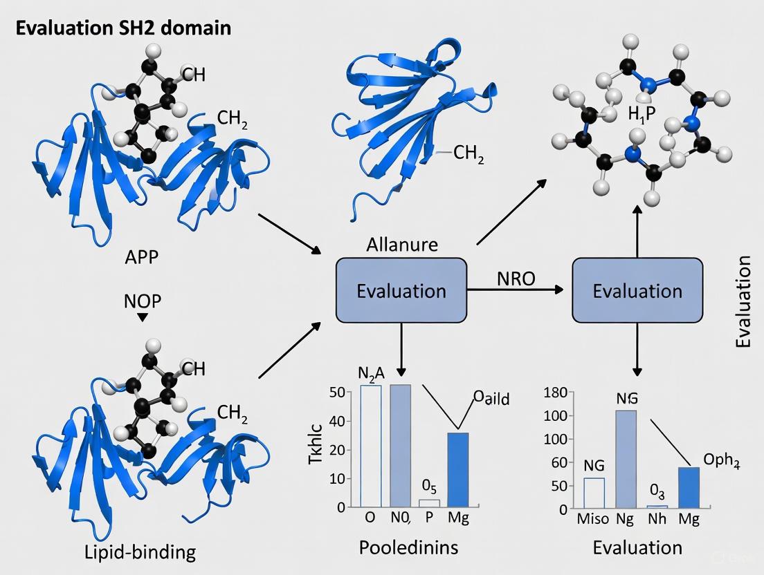
Beyond Phosphotyrosine: Evaluating the Lipid-Binding Properties of SH2 Domains in Signal Transduction and Drug Discovery
This article provides a comprehensive evaluation of the lipid-binding properties of different SH2 domain types, a paradigm-shifting function beyond their canonical role as phosphotyrosine readers.
Beyond Phosphotyrosine: Evaluating the Lipid-Binding Properties of SH2 Domains in Signal Transduction and Drug Discovery
Abstract
This article provides a comprehensive evaluation of the lipid-binding properties of different SH2 domain types, a paradigm-shifting function beyond their canonical role as phosphotyrosine readers. We synthesize foundational knowledge on SH2 domain structure with recent genome-wide studies revealing that ~90% of human SH2 domains bind plasma membrane lipids, often with high phosphoinositide specificity. The content explores advanced methodologies for characterizing these interactions, addresses historical controversies through troubleshooting insights, and validates findings through comparative analysis of diverse SH2 domains. For researchers and drug development professionals, this resource establishes lipid binding as a crucial regulatory mechanism controlling SH2 domain localization, function, and spatiotemporal signaling dynamics in health and disease.
The Dual-Function SH2 Domain: From Phosphotyrosine Reader to Lipid-Binding Module
Src Homology 2 (SH2) domains have long been recognized as essential modular domains that direct phosphotyrosine (pY)-mediated signaling pathways in eukaryotic cells. Traditionally, these approximately 100-amino acid domains have been characterized as protein-interaction modules that specifically recognize and bind to phosphorylated tyrosine residues on partner proteins, thereby inducing the assembly of multiprotein signaling complexes downstream of protein tyrosine kinases [1] [2]. This canonical function has established SH2 domains as crucial components in signal transduction mechanisms that govern cellular processes ranging from development and differentiation to immune responses and homeostasis.
Recent research has dramatically expanded our understanding of SH2 domain capabilities, revealing non-canonical functions that extend beyond phosphotyrosine recognition. Lipid-binding properties have emerged as a significant non-canonical function, with studies demonstrating that a remarkable 74-90% of human SH2 domains can bind plasma membrane lipids with high affinity and specificity [3] [4]. Additionally, unexpected structural arrangements such as tandem SH2 domains in Spt6 transcription factors, where a canonical SH2 domain pairs with a highly non-canonical SH2 domain, further challenge the traditional paradigm [5]. This evolution in understanding redefines SH2 domains as multifunctional modules capable of integrating both protein and lipid signals to achieve exquisite spatiotemporal control over cellular signaling networks.
Canonical SH2 Domain Functions: Phosphotyrosine Recognition
Structural Basis of pY Recognition
The canonical function of SH2 domains centers on their ability to specifically recognize and bind phosphorylated tyrosine residues within specific peptide contexts. Structurally, SH2 domains maintain a conserved fold characterized by a central three-stranded antiparallel β-sheet flanked by two α-helices, forming a compact structure that provides both stability and specificity [6]. The phosphotyrosine-binding pocket is highly conserved across most SH2 domains and features an invariant arginine residue (at position βB5) within the FLVR motif that forms a critical salt bridge with the phosphate moiety of the phosphotyrosine [6]. This interaction provides the fundamental energy for phosphopeptide binding.
Beyond this primary phosphate contact, specificity is achieved through interactions with residues C-terminal to the phosphotyrosine, typically at the +1 to +5 positions. These residues contact variable regions of the SH2 domain, particularly the BC-loop, DE-loop, and EF-loop, which differ across SH2 domains and confer sequence specificity [7] [8]. This dual recognition mechanism—conserved phosphate binding coupled with variable sequence specificity—allows SH2 domains to achieve both high affinity (Kd values typically ranging from 0.1-10 μM) and remarkable selectivity for their cognate ligands [6] [7].
Biological Roles of Canonical pY Binding
The canonical protein-protein interaction function of SH2 domains serves as the primary mechanism for immediate downstream signaling from activated tyrosine kinase receptors. By recruiting specific effector proteins to phosphorylated receptor complexes, SH2 domains facilitate the assembly of multimolecular signaling complexes that dictate cellular responses to extracellular stimuli [1] [2]. This recruitment function is exemplified in numerous signaling pathways:
- Growth factor signaling: SH2 domains in adaptor proteins like Grb2 link activated growth factor receptors to the Ras-MAPK pathway
- Immune receptor signaling: SH2 domains in ZAP70, SYK, and other signaling proteins mediate T-cell and B-cell receptor signaling
- Cytokine signaling: STAT proteins utilize SH2 domains for receptor recruitment and subsequent dimerization following JAK-mediated phosphorylation [9]
This canonical signaling paradigm enables rapid, precise cellular responses to changing environmental conditions and represents the foundational understanding of SH2 domain function that has guided research for decades.
Table 1: Key Characteristics of Canonical SH2 Domain Functions
| Feature | Description | Biological Significance |
|---|---|---|
| Primary Ligand | Phosphotyrosine-containing peptides | Recruits effectors to activated receptors |
| Conserved Binding Motif | FLVR motif with invariant arginine | Essential for phosphate recognition |
| Specificity Determinants | Residues C-terminal to pY (+1 to +5) | Discriminates between different pY sites |
| Binding Affinity | 0.1-10 μM range | Allows reversible, regulated interactions |
| Structural Fold | α-β sandwich with central β-sheet | Provides conserved binding platform |
Non-Canonical SH2 Domain Functions: Beyond Phosphotyrosine Recognition
Lipid Binding Capabilities
A paradigm-shifting discovery in SH2 domain biology has been the identification of high-affinity lipid binding as a prevalent non-canonical function. Systematic screening of human SH2 domains revealed that approximately 90% can bind plasma membrane lipids, with many displaying remarkable specificity for particular phosphoinositides such as phosphatidylinositol-4,5-bisphosphate (PIP₂) and phosphatidylinositol-3,4,5-trisphosphate (PIP₃) [3] [1]. This lipid-binding capability represents a fundamental expansion of SH2 domain function beyond traditional protein-protein interactions.
The mechanism of lipid recognition differs significantly from phosphotyrosine binding. SH2 domains typically bind lipids through surface cationic patches distinct from the pY-binding pocket, allowing for independent yet potentially coordinated binding to both phosphotyrosine motifs and membrane lipids [3]. These lipid-binding sites generally take one of two forms: (1) grooves that accommodate specific lipid headgroups through precise molecular complementarity, or (2) flat surfaces that mediate non-specific membrane association through electrostatic interactions [3]. The functional consequences of this dual-specificity are profound, as it enables SH2 domain-containing proteins to integrate signals from both protein phosphorylation and membrane lipid composition.
Contextual Peptide Recognition
Another non-canonical aspect of SH2 domain function involves the complexity of peptide recognition, which extends beyond simple position-specific binding motifs. Research has revealed that SH2 domains employ contextual sequence information that integrates both permissive residues (that enhance binding) and non-permissive residues (that oppose binding) in the vicinity of the essential phosphotyrosine [7] [8]. This contextual recognition allows SH2 domains to distinguish subtle differences in peptide ligands that would be indistinguishable based on traditional binding motifs alone.
The structural basis for contextual recognition involves interactions between peptide residues and regions of the SH2 domain beyond the primary specificity pockets. Neighboring positions in the peptide ligand can affect one another, meaning that local sequence context significantly influences binding affinity and specificity [7]. This sophisticated recognition mechanism substantially increases the information content that SH2 domains can extract from their peptide ligands, enabling a higher degree of signaling specificity than previously appreciated.
Non-Canonical Structural Arrangements
The discovery of unconventional structural arrangements further expands the functional repertoire of SH2 domains. In the transcription factor Spt6, the C-terminal region contains not one but two SH2 domains arranged in tandem [5]. Surprisingly, while the first SH2 domain has a canonical organization, the second SH2 domain is highly non-canonical and appears to be unique in the SH2 family. Both domains possess phosphate-binding determinants, and the complete tandem—but not individual SH2 domains—is necessary and sufficient for interaction with RNA polymerase II and important for Spt6 function in vivo [5].
This tandem arrangement represents a significant departure from the typical single-SH2 domain architecture and demonstrates how structural innovations can create new functional capabilities. The requirement for both domains in RNAPII binding suggests a more extensive interaction interface than simple recognition of a doubly phosphorylated peptide, indicating that non-canonical SH2 domains can participate in complex macromolecular assemblies that extend beyond traditional phosphotyrosine signaling [5].
Table 2: Non-Canonical SH2 Domain Functions and Representative Examples
| Non-Canonical Function | Mechanism | Representative Examples |
|---|---|---|
| Lipid Binding | Cationic surface patches distinct from pY pocket | ZAP70, LCK, ABL, VAV2, C1-Ten/Tensin2 [3] [6] |
| Contextual Peptide Recognition | Integration of permissive and non-permissive residues | Various SH2 domains recognizing physiological ligands [7] |
| Non-Canonical Structural Arrangements | Tandem SH2 domains with unique architecture | Spt6 transcription factor [5] |
| Phase Separation | Multivalent interactions driving condensate formation | GRB2, Gads, LAT in T-cell signaling [6] |
Comparative Analysis: Canonical vs. Non-Canonical Properties
Functional and Mechanistic Comparisons
The canonical and non-canonical functions of SH2 domains represent complementary rather than mutually exclusive capabilities. The traditional pY-recognition function operates through a well-defined binding pocket with conserved features, while non-canonical lipid binding utilizes distinct molecular surfaces that vary considerably between different SH2 domains. This mechanistic separation enables SH2 domains to perform dual roles in cellular signaling, simultaneously sensing both protein phosphorylation status and membrane localization signals.
From a biological perspective, canonical pY-binding primarily mediates specific protein-protein interactions that propagate signals through defined pathways, while lipid binding contributes to spatial organization and compartmentalization of signaling events. The emerging understanding of contextual peptide recognition further enhances the information-processing capacity of SH2 domains, allowing them to integrate multiple inputs to determine binding outcomes. These capabilities collectively transform SH2 domains from simple recruitment modules into sophisticated signal integration hubs.
Quantitative Comparison of Binding Properties
Table 3: Quantitative Comparison of Canonical and Non-Canonical SH2 Domain Interactions
| Parameter | Canonical pY-Peptide Binding | Non-Canonical Lipid Binding |
|---|---|---|
| Prevalence | Universal SH2 domain function | ~90% of human SH2 domains [3] |
| Binding Affinity | 0.1-10 μM (Kd) [6] | High affinity for specific lipids [3] |
| Specificity Determinants | Residues at +1 to +5 positions C-terminal to pY | Lipid headgroup structure and membrane composition |
| Structural Features | Conserved pY pocket with invariant arginine | Surface cationic patches distinct from pY pocket [3] |
| Functional Impact | Pathway-specific signaling complex assembly | Spatiotemporal control of protein localization and activity [3] |
Experimental Approaches for Characterizing SH2 Domain Functions
Methodologies for Studying Lipid Binding Properties
Investigating the lipid-binding capabilities of SH2 domains requires specialized methodologies that differ from traditional approaches for studying protein-protein interactions. Lipid binding assays utilizing plasma membrane-mimetic vesicles have been instrumental in demonstrating that approximately 74% of SH2 domains have high affinity for such structures [4]. These assays typically involve testing binding to vesicles containing specific lipid compositions, including defined phosphoinositides, to determine both binding affinity and specificity.
For precise quantification of lipid interactions, surface plasmon resonance (SPR) and related biophysical techniques have been employed to measure binding constants between purified SH2 domains and lipid surfaces [3] [1]. These approaches have revealed that many SH2 domains show preference for specific phosphoinositides, such as PIP₂ or PIP₃, rather than engaging in non-specific electrostatic binding [3]. Complementary cellular studies using fluorescence microscopy to monitor localization of SH2 domain constructs in response to lipid modifications have validated the physiological relevance of these interactions.
Advanced Techniques for Profiling Peptide Binding Specificity
Characterizing the contextual peptide recognition properties of SH2 domains has been revolutionized by high-throughput approaches. Bacterial peptide display combined with next-generation sequencing (NGS) enables comprehensive profiling of SH2 domain binding across extremely diverse peptide libraries [10]. This approach involves multi-round affinity selection on random phosphopeptide libraries, followed by NGS analysis of selected peptides.
The experimental workflow for these studies typically includes:
- Library construction: Generating highly diverse random peptide libraries (10⁶-10⁷ sequences) displayed on bacterial surfaces
- Affinity selection: Incubating SH2 domains with the peptide library and performing multiple rounds of selection to enrich high-affinity binders
- Sequencing and analysis: Using NGS to sequence selected peptides and computational tools like ProBound to build quantitative sequence-to-affinity models [10]
These data allow researchers to move beyond simple classification of binders versus non-binders to quantitative prediction of binding free energies across the full theoretical ligand sequence space [10]. This represents a significant advancement over earlier methods such as SPOT synthesis and positional scanning peptide libraries.
Diagram 1: Experimental workflow for quantitative SH2 domain specificity profiling
Structural Biology Approaches
Understanding the molecular basis of both canonical and non-canonical SH2 domain functions heavily relies on structural biology techniques. X-ray crystallography has been fundamental in revealing the conserved fold of SH2 domains and the atomic details of phosphopeptide recognition [5] [6]. For example, the structure of the Spt6 C-terminal region revealing the unexpected tandem SH2 arrangement was solved using X-ray crystallography at 2.2 Å resolution [5].
Nuclear magnetic resonance (NMR) spectroscopy provides complementary information about dynamics and has been particularly valuable for studying weak interactions and conformational changes associated with lipid binding. These structural approaches, combined with mutational analyses and biochemical assays, have been instrumental in identifying the distinct binding surfaces for phosphotyrosine peptides and lipids on SH2 domains.
Signaling Integration: How SH2 Domains Combine Canonical and Non-Canonical Functions
Spatiotemporal Control of Signaling
The combination of canonical and non-canonical functions enables SH2 domains to exert exquisite spatiotemporal control over signaling processes. The lipid-binding capability localizes SH2-containing proteins to specific membrane compartments, while simultaneous pY-binding allows recruitment to activated receptor complexes. This dual targeting mechanism is exemplified by ZAP70 in T-cell signaling, where lipids facilitate and sustain ZAP70 interactions with TCR-ζ chain in a spatiotemporally specific manner [3] [6].
The integration of these functions creates a sophisticated control system where signaling output depends on both membrane localization (governed by lipid binding) and pathway activation (governed by pY recognition). This dual-input system enhances signaling specificity and prevents inappropriate activation of pathways, representing an important regulatory mechanism in complex signaling networks.
Role in Phase Separation and Condensate Formation
Recent research has revealed that SH2 domain-containing proteins contribute to the formation of intracellular condensates through protein phase separation (PPS) [6]. Multivalent interactions mediated by SH2 domains, often in combination with other interaction domains like SH3 domains, drive the assembly of these membrane-less organelles that enhance signaling specificity and efficiency.
In T-cell receptor signaling, interactions among GRB2, Gads, and the LAT receptor contribute to liquid-liquid phase separation (LLPS), creating concentrated hubs that enhance signaling efficiency [6]. Similarly, in kidney podocyte cells, LLPS increases the ability of adapter NCK to promote N-WASP–Arp2/3-mediated actin polymerization by increasing the membrane dwell time of key complexes [6]. These findings position SH2 domains as important players in the emerging paradigm of phase separation in cellular organization and signaling regulation.
Diagram 2: Integration of canonical and non-canonical functions in biomolecular condensate formation
The Scientist's Toolkit: Essential Research Reagents and Methodologies
Table 4: Essential Research Reagents and Methods for SH2 Domain Studies
| Reagent/Method | Function/Application | Key Features |
|---|---|---|
| Random Peptide Libraries | Profiling SH2 domain specificity | High diversity (10⁶-10⁷ sequences); includes pY residues [10] |
| Bacterial Display Systems | High-throughput affinity selection | Links genotype to phenotype; compatible with NGS [10] |
| Next-Generation Sequencing | Quantitative analysis of selection output | Deep sequencing of selected peptides; enables quantitative modeling [10] |
| ProBound Computational Tool | Building sequence-to-affinity models | Free energy regression; predicts binding across sequence space [10] |
| Plasma Membrane-Mimetic Vesicles | Lipid binding assays | Defined lipid composition; includes phosphoinositides [3] |
| SPOT Synthesis Arrays | Semiquantitative interaction screening | Addressable peptide arrays; rapid specificity profiling [7] |
Implications for Therapeutic Development and Disease
The expanding understanding of SH2 domain functions, particularly their lipid-binding capabilities, opens new avenues for therapeutic intervention. Traditional approaches to targeting SH2 domains have focused on developing inhibitors that block the pY-binding pocket, but the discovery of lipid-binding sites provides alternative targeting strategies [3] [6]. These alternative sites may offer improved specificity and reduced off-target effects compared to targeting the highly conserved pY-binding pocket.
Disease-associated mutations in SH2 domains frequently cluster within functional sites, including both pY-binding and lipid-binding regions [6]. This observation underscores the physiological importance of both canonical and non-canonical functions and suggests that disrupting either function can have pathological consequences. For example, mutations affecting the lipid-binding capability of the TNS2 SH2 domain impair regulation of insulin receptor substrate-1 phosphorylation, potentially contributing to insulin signaling dysfunction [6].
The emerging role of SH2 domains in phase separation further suggests novel therapeutic strategies aimed at modulating condensate formation rather than simply inhibiting binary interactions. Such approaches could be particularly valuable in diseases characterized by aberrant signaling complex formation, including cancer and autoimmune disorders, where retuning rather than completely inhibiting signaling pathways may provide therapeutic benefits with reduced toxicity.
The traditional view of SH2 domains as specialized phosphotyrosine-binding modules has been fundamentally transformed by research revealing their diverse non-canonical functions. Lipid-binding capabilities, contextual peptide recognition, participation in phase separation, and unconventional structural arrangements collectively redefine SH2 domains as sophisticated signal integration hubs rather than simple recruitment devices. This expanded functional repertoire enables SH2 domains to contribute to exquisite spatiotemporal control of cellular signaling by simultaneously sensing multiple inputs, including protein phosphorylation status, membrane localization signals, and local concentration gradients.
These advances have profound implications for both basic research and therapeutic development. Methodologies for studying SH2 domain functions have evolved from simple binding assays to sophisticated integrated approaches combining high-throughput experimental techniques with computational modeling. Therapeutically, the discovery of non-canonical functions, particularly lipid binding, reveals new targeting opportunities that may enable more specific modulation of pathogenic signaling pathways. As research continues to unravel the complexities of SH2 domain biology, our understanding of these multifunctional modules will undoubtedly continue to evolve, potentially revealing additional unexpected capabilities that further expand their known functional repertoire.
Src Homology 2 (SH2) domains are modular protein interaction domains that serve as fundamental components of eukaryotic signaling networks. These approximately 100-amino-acid domains were initially characterized for their ability to recognize and bind phosphorylated tyrosine (pTyr) residues in specific peptide contexts, thereby facilitating the assembly of signaling complexes in response to tyrosine kinase activation [1] [2]. For decades, the central paradigm of SH2 domain function centered on this phosphotyrosine-dependent protein-protein interaction. However, emerging research has revealed a remarkable functional expansion: many SH2 domains have evolved to interact with membrane lipids, particularly phosphoinositides, while maintaining their highly conserved structural fold [1] [6]. This dual-binding capability allows SH2 domains to integrate both protein-based and lipid-based signaling information, enabling more sophisticated regulation of cellular processes. This guide provides a comparative analysis of the structural architecture of different SH2 domain types, focusing on how their conserved folds have been adapted for lipid-binding functionality, with supporting experimental data and methodological insights for researchers in signaling and drug development.
Canonical SH2 Domain Structure and Variations
The SH2 domain fold exhibits remarkable conservation across diverse proteins, maintaining a core structural framework while permitting variations that enable functional diversity.
Conserved Structural Core
All SH2 domains share a characteristic βαββββαβ secondary structure arrangement, forming a compact structure where a central antiparallel β-sheet is flanked by two α-helices [6] [11]. This conserved architecture creates two critical binding surfaces: a deep basic pocket that binds the phosphorylated tyrosine residue, and adjacent specificity pockets that recognize residues C-terminal to the pTyr, typically at the +3 position [12] [13]. The pTyr-binding pocket contains several highly conserved features, most notably an invariant arginine residue at position βB5 that forms part of the "FLVR" motif and provides critical electrostatic interactions with the phosphate moiety [13]. This arginine contributes approximately half of the binding free energy and enables discrimination between pTyr and phosphoserine/phosphothreonine [13].
Structural Classification and Variations
Despite their conserved core, SH2 domains can be divided into distinct structural and functional classes:
- Src-type SH2 domains: Characterized by a basic residue at position αA2 that assists in pTyr coordination [13]. This group includes domains from Src family kinases and many adaptor proteins.
- SAP-type SH2 domains: Utilize a basic residue at position βD6 for pTyr binding instead of αA2 [13]. The SAP SH2 domain also exhibits the unusual capability of binding SH3 domains via a surface distal to its pTyr binding site [13].
- STAT-type SH2 domains: Lack the βE and βF strands and feature a split αB helix, adaptations that facilitate SH2 domain-mediated dimerization required for transcriptional regulation [6].
- Ancestral SH2 domains: The SPT6 protein contains the most evolutionarily ancient SH2 domains, which recognize phosphoserine and phosphothreonine in RNA polymerase II rather than pTyr, representing an evolutionary transition to tyrosine-specific binding [13].
Table 1: Key Structural Features of Major SH2 Domain Classes
| SH2 Class | Distinguishing Structural Features | pTyr Coordination | Representative Members |
|---|---|---|---|
| Src-type | Basic residue at αA2 | αA2 and βB5 (FLVR) | Src, Fyn, Lck, Abl |
| SAP-type | Basic residue at βD6 | βD6 and βB5 (FLVR) | SAP, EAT-2, SHIP |
| STAT-type | Lacks βE and βF strands; split αB helix | βB5 (FLVR) | STAT1, STAT3, STAT5 |
| Ancestral | Variant pTyr pocket | Adapted for pSer/pThr | SPT6 N-SH2 and C-SH2 |
Lipid-Binding Adaptations in SH2 Domains
Recent research has fundamentally expanded our understanding of SH2 domain function by revealing that many can interact with membrane lipids in addition to phosphotyrosine motifs. This lipid-binding capability provides a mechanism for membrane recruitment and regulation that complements traditional protein-protein interactions.
Prevalence and Lipid Specificity
Comprehensive studies indicate that approximately 75% of SH2 domains interact with membrane lipids, with particular preference for phosphatidylinositol-4,5-bisphosphate (PIP₂) and phosphatidylinositol-3,4,5-trisphosphate (PIP₃) [6]. These interactions are not merely incidental; they play specific regulatory roles in the function of SH2-containing proteins. Lipid binding often occurs through cationic regions near the pTyr-binding pocket that are flanked by aromatic or hydrophobic side chains, creating specialized membrane interaction surfaces [6].
Molecular Mechanisms of Lipid Recognition
SH2 domains have evolved distinct structural adaptations for lipid binding:
- Overlapping binding sites: Some SH2 domains, like that of Abl tyrosine kinase, feature overlapping binding sites for phosphotyrosine and phosphoinositides. PIP₂ interacts with the Abl SH2 domain via an electrostatic mechanism that competes with phosphotyrosine binding, particularly at residues R152 (within the FLVRES motif) and R175 [1]. This creates a mutually exclusive binding switch that may regulate Abl localization and activity.
- Distinct lipid-binding pockets: Other SH2 domains possess dedicated lipid-binding surfaces separate from the pTyr pocket. The SH2 domain of ZAP70, a critical kinase in T-cell receptor signaling, binds PIP₃ through specific cationic patches, facilitating sustained activation at the membrane [1] [6].
- Membrane proximity mechanisms: In lymphocyte-specific kinase (Lck), the SH2 domain interacts with PIP₂ and PIP₃ through a cationic patch that shows differential engagement in open versus closed conformations, suggesting that lipid binding contributes to conformational regulation [14]. Molecular dynamics simulations reveal that these interactions help position Lck optimally for T-cell receptor signaling [14].
Table 2: Experimentally Characterized Lipid-Binding SH2 Domains
| Protein | SH2 Domain | Lipid Specificity | Functional Role of Lipid Binding |
|---|---|---|---|
| Abl | Single SH2 | PIP₂ | Membrane recruitment, activity modulation, mutually exclusive with pTyr binding [1] |
| Lck | Single SH2 | PIP₂, PIP₃ | Modulates interaction with partners in TCR signaling; sustains activation [1] [6] |
| ZAP70 | C-terminal SH2 | PIP₃ | Facilitates and sustains interactions with TCR-ζ chain; membrane localization [6] |
| C1-Ten/Tensin2 | C-terminal SH2 | PIP₃ | Activation and specific targeting on IRS-1 in insulin signaling [1] [6] |
| Vav2 | Single SH2 | PIP₂, PIP₃ (weak) | Targeting to membrane subdomains; interaction with EphA2 receptor [6] |
| SYK | Tandem SH2 | PIP₃ | PIP₃-dependent membrane binding required for non-catalytic activation of STAT3/5 [6] |
Experimental Approaches for Studying SH2-Lipid Interactions
The investigation of SH2 domain lipid-binding properties employs multidisciplinary approaches that provide complementary information about affinity, specificity, and structural determinants.
Methodological Framework
Several well-established experimental protocols have been adapted to characterize SH2-lipid interactions:
Lipid Binding Assays: In vitro lipid binding experiments typically involve incubating purified SH2 domains with lipid vesicles or filters containing spotted phospholipids. For example, research on the Abl SH2 domain demonstrated direct PIP₂ binding using protein-lipid overlay assays and liposome binding studies [1]. These approaches determine lipid specificity and relative binding affinities.
Molecular Dynamics (MD) Simulations: MD simulations have proven invaluable for studying SH2 domain membrane interactions at atomic resolution. Recent simulations of full-length Lck in complex with realistic membrane bilayers revealed that the SH2 domain interacts differently with lipids in open versus closed conformations, suggesting a role for lipid binding in conformational regulation [14]. These simulations typically involve modeling the protein in membranes containing specific phosphoinositides and analyzing interaction patterns over microsecond timescales.
Phylogenetic Profiling: Bioinformatics approaches like the Gestalt Domain Detection Algorithm (GDDA-BLAST) can predict lipid-binding potential by analyzing sequence conservation patterns. Application of this method to SH2 domains revealed that lipid-binding capacity is widespread and identified key residues involved in membrane interactions [15]. This method successfully predicted the lipid-binding capability of Tec family kinase SH2 domains, which was subsequently validated experimentally.
Structural Biology Approaches: X-ray crystallography and NMR spectroscopy have provided direct structural information about SH2-lipid interactions. The structure of the Abl SH2 domain revealed the molecular details of its interaction with PIP₂, showing how specific basic residues mediate membrane contact [1]. NMR studies have been particularly useful for characterizing weak and transient protein-lipid interactions.
Experimental Workflow Diagram
The following diagram illustrates a typical integrated workflow for characterizing SH2 domain lipid-binding properties:
The Scientist's Toolkit: Essential Research Reagents and Methods
This section details key experimental resources and approaches for investigating SH2 domain lipid-binding properties, drawing from methodologies represented in the cited literature.
Table 3: Essential Research Tools for SH2-Lipid Interaction Studies
| Reagent/Method | Function/Application | Key Features and Considerations |
|---|---|---|
| PIP Strips / Lipid Arrays | High-throughput lipid specificity screening | Membrane-based spotted arrays of different lipids; enable rapid assessment of binding specificity [15] |
| Liposome Binding Assays | Quantitative lipid binding measurements | Synthetic liposomes with defined lipid composition; can incorporate specific phosphoinositides at physiological concentrations [1] |
| Molecular Dynamics Simulations | Atomic-level analysis of membrane interactions | Reveals dynamic interaction patterns and conformational dependencies; requires high-performance computing resources [14] |
| Surface Plasmon Resonance (SPR) | Kinetic analysis of lipid interactions | Provides quantitative data on binding affinity and kinetics; requires specialized instrumentation [6] |
| NMR Spectroscopy | Structural analysis of lipid interactions | Can characterize weak/transient interactions; provides residue-specific information [1] |
| Phylogenetic Profiling Algorithms | Bioinformatics prediction of lipid binding | GDDA-BLAST and related tools can identify potential lipid-binding domains from sequence information [15] |
SH2 domains represent a remarkable example of evolutionary adaptation within a conserved structural framework. While maintaining their characteristic α-helical/β-sheet fold and core phosphotyrosine-binding function, many SH2 domains have acquired the ability to interact specifically with membrane lipids, particularly phosphoinositides. This dual-binding capability significantly expands their functional repertoire, enabling sophisticated regulation of protein localization, activity, and signaling dynamics. The structural adaptations for lipid binding are diverse, ranging from overlapping phosphotyrosine/lipid binding sites to distinct membrane interaction surfaces. Experimental approaches combining biochemical assays, structural biology, computational modeling, and phylogenetic analysis have revealed that lipid binding is not an exceptional property of a few SH2 domains, but rather a widespread phenomenon affecting approximately three-quarters of all human SH2 domains. Understanding these structural adaptations and their functional consequences provides valuable insights for drug discovery efforts targeting SH2 domain-mediated interactions in cancer and other diseases.
The Src homology 2 (SH2) domain has long been established as a critical modular protein interaction domain that specifically recognizes phosphotyrosine (pY) motifs, thereby directing myriad cellular signaling pathways [16] [17]. For decades, the prevailing paradigm defined SH2 domains primarily as readers of protein phosphorylation states. However, recent genomic-scale studies have fundamentally expanded this understanding by revealing that a substantial majority of human SH2 domains also function as specific lipid-binding modules [17] [18]. This dual-ligand capacity enables SH2 domain-containing proteins to integrate phosphorylation signals with lipid-mediated spatial cues, providing an additional layer of regulation in cellular signaling networks. This guide systematically compares the lipid-binding properties across diverse SH2 domain types, providing objective experimental data and methodologies relevant for researchers investigating signaling mechanisms and developing therapeutic strategies.
Genomic Prevalence of SH2-Lipid Interactions
Comprehensive genomic screening of human SH2 domains has quantitatively demonstrated that lipid binding is not an exceptional property of a few SH2 domains, but rather a widespread characteristic across this protein family. Systematic analysis of 121 human SH2 domains revealed striking findings about their membrane interaction capabilities [17].
Table 1: Genomic Prevalence of SH2 Domain Lipid Binding
| Affinity Category | Number of SH2 Domains | Percentage of Total | Representative Examples |
|---|---|---|---|
| High-affinity (Sub-μM Kd) | 56 | 74% | STAT6, GRB7, FRK, BLNK, APS |
| Moderate-affinity (1-5 μM Kd) | 13 | 17% | Not specified in sources |
| No detectable binding | 8 | 10% | Not specified in sources |
This quantitative assessment indicates that approximately 90% of human SH2 domains tested demonstrated measurable lipid binding, with the vast majority (74%) exhibiting high-affinity interactions comparable to established lipid-binding domains [17]. This prevalence suggests that lipid binding represents a fundamental, evolutionarily conserved function within the SH2 domain family, with potentially broad implications for cellular signaling mechanisms.
Comparative Analysis of Lipid-Binding SH2 Domains
Different SH2 domains exhibit distinct lipid-binding affinities and specificities, which correspond to their specific cellular functions. The following table synthesizes experimental data from multiple studies to provide a comparative view of representative SH2 domains and their lipid interaction properties.
Table 2: Lipid-Binding Properties of Representative SH2 Domains
| Protein Name | SH2 Domain | Lipid Affinity (Kd) | Lipid Specificity | Biological Role of Lipid Interaction |
|---|---|---|---|---|
| STAT6 | C-terminal | 20 ± 10 nM | Not specified | Transcriptional regulation [17] |
| GRB7 | Single | 70 ± 12 nM | Low selectivity | Adapter function in signaling [17] |
| ZAP70 | C-terminal | 340 ± 35 nM | PIP3 > PI(4,5)P2 > others | Sustained T-cell activation [17] [1] |
| Lck | Single | 220 ± 20 nM | Low specificity (PI(4,5)P2, PIP3) | TCR signaling complex formation [17] [19] |
| SHIP1 | N-terminal | 190 ± 30 nM | PIP3 ≈ PI(4,5)P2 ≫ others | Regulation of autoinhibition [17] [20] |
| Tensin2 | C-terminal | 200 ± 67 nM | PIP3 | Insulin signaling regulation [16] [17] |
| Abl | Single | Not specified | PI(4,5)P2 | Membrane recruitment [18] [1] |
| VAV2 | Single | Not specified | Weak PI(4,5)P2/PIP3 | Targeting to membrane subdomains [16] [1] |
The data reveal several important patterns. First, lipid-binding affinity varies considerably across different SH2 domains, with dissociation constants ranging from nanomolar to micromolar. Second, many SH2 domains show distinct preferences for specific phosphoinositides, particularly phosphatidylinositol-4,5-bisphosphate (PI(4,5)P2) and phosphatidylinositol-3,4,5-trisphosphate (PIP3), which are key signaling lipids in the plasma membrane [16] [17]. Third, the biological consequences of lipid binding differ significantly depending on the cellular context and the specific protein harboring the SH2 domain.
Structural Mechanisms of SH2-Lipid Interactions
Understanding the structural basis of SH2-lipid interactions is crucial for interpreting their functional consequences. Structural studies have revealed that SH2 domains employ topologically distinct binding sites for lipids and phosphotyrosine motifs, enabling coincident binding of both ligands [17] [19].
The canonical SH2 domain fold consists of a three-stranded antiparallel beta-sheet flanked by two alpha helices (αA-βB-βC-βD-αB), forming a compact structure [16]. The pY-binding pocket is located within the βB strand and contains a highly conserved arginine residue (at position βB5) that forms a salt bridge with the phosphate moiety of phosphotyrosine [16]. In contrast, lipid-binding sites typically consist of cationic surface patches composed of basic, aromatic, and hydrophobic residues that are spatially distinct from the pY-binding pocket [17] [19].
Two primary types of lipid-binding interfaces have been identified:
- Groove-type interfaces that accommodate specific lipid headgroups with high specificity
- Flat cationic surfaces that mediate non-specific membrane interactions through electrostatic forces [17]
This structural arrangement allows for complex regulatory mechanisms. For instance, in the Lck SH2 domain, lipid binding involves surface-exposed basic, aromatic, and hydrophobic residues that do not participate in phosphotyrosine recognition [19]. Similarly, molecular dynamics simulations of full-length Lck have revealed that its SH2 domain interacts differently with lipids in open versus closed conformations, suggesting that lipid interactions can allosterically regulate protein conformation and function [14].
Figure 1: SH2 Domain Dual-Ligand Binding Mechanism. SH2 domains can simultaneously bind phosphotyrosine motifs and membrane lipids through topologically distinct binding sites, enabling integrated signaling responses.
Methodologies for Studying SH2-Lipid Interactions
Experimental Approaches for Lipid Binding Analysis
Rigorous biochemical and biophysical methods have been essential for characterizing SH2-lipid interactions. The following experimental approaches represent the current gold standards in the field:
Surface Plasmon Resonance (SPR) SPR provides quantitative measurements of SH2-lipid binding affinity and kinetics. In this approach, SH2 domains (often as EGFP-fusion proteins to enhance expression yield) are flowed over sensor chips containing immobilized lipid vesicles with defined composition [17] [21]. The lipid vesicles typically mimic the cytofacial leaflet of the plasma membrane, containing phosphoinositides such as PI(4,5)P2 or PIP3 in a background of phosphatidylcholine, phosphatidylethanolamine, and phosphatidylserine [17]. This method allows determination of dissociation constants (Kd) and analysis of lipid specificity through competition experiments.
Fluorescence Quenching Analysis This technique monitors changes in fluorescence intensity when SH2 domains interact with lipid vesicles containing quenching groups [21]. It enables high-throughput screening for inhibitors of SH2 domain-lipid binding and can be performed in multi-well plate formats, facilitating rapid characterization of multiple SH2 domains or mutant variants.
Hydrogen-Deuterium Exchange Mass Spectrometry (HDX-MS) HDX-MS identifies membrane interaction surfaces by measuring the protection of amide protons from exchange when SH2 domains bind to lipids [20]. This approach has been particularly valuable for identifying intramolecular contacts, such as those between the SH2 and C2 domains in SHIP1 that regulate autoinhibition [20].
Molecular Dynamics (MD) Simulations MD simulations provide atomic-level insights into SH2-lipid interactions over time. Recent simulations of full-length Lck in complex lipid bilayers have revealed preferential interactions with PIP lipids and conformational-dependent lipid binding modes [14]. These computational approaches complement experimental methods by offering dynamic information not accessible through static structural biology.
Figure 2: Experimental Workflow for SH2-Lipid Binding Analysis. Standardized methodology for quantifying SH2 domain interactions with membrane lipids.
The Scientist's Toolkit: Essential Research Reagents
Table 3: Key Research Reagents for SH2-Lipid Interaction Studies
| Reagent / Method | Function / Application | Key Features |
|---|---|---|
| EGFP-Fusion SH2 Domains | Enhances protein expression yield and stability for biochemical studies | Improved solubility for 76 human SH2 domains [17] |
| PM-mimetic Lipid Vesicles | Recapitulate cytosolic leaflet of plasma membrane for in vitro binding assays | Contains PI(4,5)P2, PIP3, phosphatidylserine, phosphatidylcholine, phosphatidylethanolamine [17] |
| Surface Plasmon Resonance (SPR) | Quantitative measurement of binding affinity and kinetics | Determines Kd values; measures lipid specificity [17] [21] |
| Fluorescence Quenching Assays | High-throughput screening of lipid-binding inhibitors | Compatible with multi-well plate formats [21] |
| Hydrogen-Deuterium Exchange MS | Identification of membrane interaction surfaces and conformational changes | Maps protein-lipid interfaces; detects allosteric effects [20] |
| Molecular Dynamics Simulations | Atomic-level analysis of dynamic lipid interactions | Reveals conformational-dependent binding; models full-length proteins [14] |
Functional Consequences and Therapeutic Implications
The functional significance of SH2-lipid interactions extends across multiple cellular processes and disease contexts. These interactions typically exert spatiotemporal control over signaling activities by regulating membrane localization, interaction with binding partners, and catalytic activity.
In T-cell receptor signaling, the Lck SH2 domain binds anionic plasma membrane lipids, which modulates its interaction with partners in the TCR signaling complex [19]. Mutation of lipid-binding residues markedly reduces Lck's interaction with the ζ chain and overall TCR signaling capacity [19]. Similarly, the ZAP70 C-terminal SH2 domain binds PIP3 with high specificity, which is essential for sustaining its activation in T cells [17].
In insulin signaling, the Tensin2 SH2 domain preferentially binds PIP3, regulating phosphorylation of insulin receptor substrate-1 (IRS-1) [16]. Disruption of this interaction impairs proper insulin signal transduction.
SH2-lipid interactions also contribute to the formation of biomolecular condensates through liquid-liquid phase separation. Multivalent interactions involving SH2 domains drive the assembly of signaling hubs such as the LAT-GRB2-SOS1 complex in T-cell activation [16]. These condensates enhance signaling efficiency by concentrating components and increasing membrane dwell time.
Therapeutic targeting of SH2-lipid interactions represents a promising avenue for drug development. For example, nonlipidic small molecules have been developed that specifically inhibit Syk kinase by disrupting its lipid binding [16]. This approach may yield potent, selective inhibitors for various SH2 domain-containing kinases with potential applications in cancer, autoimmune disorders, and inflammatory diseases.
Genomic-scale studies have fundamentally transformed our understanding of SH2 domains from specialized phosphotyrosine readers to dual-specificity modules that integrate protein phosphorylation and lipid signaling. The prevalence of lipid binding across diverse SH2 domain families suggests this is an evolutionarily conserved feature that enhances the specificity and spatiotemporal control of cellular signaling networks. The experimental frameworks and comparative data presented in this guide provide researchers with essential resources for investigating these interactions in specific biological contexts and developing therapeutic strategies that target this newly recognized functional dimension of SH2 domains.
Src-homology 2 (SH2) domains have long been recognized as prototypical protein interaction modules that direct cellular signaling networks by binding to phosphorylated tyrosine (pY) motifs [17] [1]. However, emerging research has fundamentally expanded this paradigm, revealing that SH2 domains also function as crucial lipid-binding modules. A comprehensive genomic-scale study demonstrated that approximately 90% of human SH2 domains can bind plasma membrane lipids, with many exhibiting remarkable specificity for particular phosphoinositides [17] [4]. These findings establish that SH2 domains serve dual roles in cellular signaling, engaging both protein partners through their canonical pY-binding pockets and lipid partners through separate cationic surface patches. This dual-specificity mechanism provides an additional layer of spatiotemporal control over signaling processes, localizing SH2-containing proteins to specific membrane microdomains and modulating their interactions within phosphotyrosine signaling networks [6] [1]. This comparison guide systematically evaluates the phosphoinositide specificity and membrane interaction mechanisms across different SH2 domain types, providing researchers with objective experimental data and methodologies for investigating these crucial lipid-protein relationships.
Lipid Binding Landscape of SH2 Domains
Prevalence and Affinity of SH2-Lipid Interactions
The lipid-binding capabilities of SH2 domains are not merely incidental properties of a few unusual domains but represent a widespread characteristic across this protein family. Systematic analysis using surface plasmon resonance (SPR) to quantitatively measure binding affinities revealed that 74% of the 76 human SH2 domains tested exhibited submicromolar affinity (Kd < 1 μM) for plasma membrane-mimetic vesicles, with an additional 13% showing moderate affinity (Kd = 1-5 μM) [17]. Only approximately 10% of SH2 domains showed no detectable lipid binding under experimental conditions. This broad binding profile establishes membrane lipid interaction as a fundamental property of most SH2 domains, comparable to other dedicated lipid-binding modules [17].
Table 1: Lipid Binding Affinities of Representative SH2 Domains
| SH2 Domain | Kd for PM-mimetic Vesicles | Phosphoinositide Specificity | Lipid Binding Residues |
|---|---|---|---|
| STAT6-SH2 | 20 ± 10 nM | Not specified | Not specified |
| GRB7-SH2 | 70 ± 12 nM | Low selectivity | Not specified |
| FRK(PTK5)-SH2 | 80 ± 12 nM | Not specified | Not specified |
| YES1-SH2 | 110 ± 12 nM | PI45P2 > PIP3 > others | R215, K216 |
| BLNK-SH2 | 120 ± 19 nM | PIP3 > PI45P2 ≫ others | Not specified |
| ZAP70-cSH2 | 340 ± 35 nM | PIP3 > PI45P2 > others | K176, K186, K206, K251 |
| ABL-SH2 | Not specified | PIP2 interaction | R152, R175 |
| LCK-SH2 | Not specified | PIP2, PIP3 | Surface-exposed basic, aromatic, and hydrophobic residues |
Structural Mechanisms of Lipid Recognition
SH2 domains employ distinct structural mechanisms for lipid binding that are separate from their canonical pY-recognition functions. The lipid-binding sites typically comprise surface-exposed cationic patches formed by basic residues, often flanked by aromatic or hydrophobic side chains [6] [1]. These structural arrangements create either grooves for specific phosphoinositide headgroup recognition or flat surfaces for non-specific membrane association [17].
For example, the Lck SH2 domain utilizes surface-exposed basic, aromatic, and hydrophobic residues—distinct from its phospho-Tyr binding pocket—to interact with anionic lipids [19]. Similarly, the Abl SH2 domain employs an electrostatic mechanism where R152 in the FLVRES motif (critical for phosphotyrosine recognition) and R175 both contribute to phosphatidylinositol-4,5-bisphosphate binding [1]. This structural separation enables SH2 domains to simultaneously or alternatively engage protein and lipid partners, significantly expanding their regulatory potential in signaling processes.
Experimental Approaches for Characterizing SH2-Lipid Interactions
Surface Plasmon Resonance (SPR) Methodology
Protocol Summary: SPR has emerged as the primary technique for quantitatively evaluating SH2 domain lipid-binding affinity and specificity [17]. This methodology involves immobilizing liposomes with controlled lipid composition on sensor chips and measuring binding kinetics as SH2 domains flow across the surface.
Key Technical Details:
- Membrane Composition: PM-mimetic vesicles that recapitulate the cytofacial leaflet of the plasma membrane are standard [17]. These typically include phosphoinositides (PIP2, PIP3) at physiological concentrations within a background of phosphatidylcholine, phosphatidylserine, and cholesterol.
- Protein Preparation: SH2 domains are often expressed as EGFP-fusion proteins to improve expression yield and stability without affecting membrane binding properties [17].
- Data Analysis: Equilibrium dissociation constants (Kd) are calculated from binding curves generated across a range of protein concentrations. Specificity is determined by comparing binding to vesicles containing different phosphoinositides.
Applications: This approach successfully characterized 76 human SH2 domains, revealing their diverse lipid-binding affinities and specificities [17]. For example, it identified that the YES1-SH2 domain preferentially binds PI(4,5)P2 over PI(3,4,5)P3, while BLNK-SH2 shows the opposite preference [17].
Nuclear Magnetic Resonance (NMR) Spectroscopy
Protocol Summary: NMR provides atomic-resolution insights into SH2-lipid interactions by identifying specific residues involved in membrane binding and characterizing potential conformational changes [22] [19].
Key Technical Details:
- Sample Preparation: Uniformly 15N-labeled SH2 domains are incubated with liposomes or water-soluble phosphoinositide analogs. Chemical shift perturbations are monitored in 2D 1H-15N HSQC spectra.
- Binding Site Mapping: Residues exhibiting significant chemical shift changes or line broadening upon lipid addition are identified as participation in membrane interaction.
- Mutational Validation: Proposed lipid-binding residues are mutated to assess their functional contribution.
Applications: NMR studies of the Lck SH2 domain identified a lipid-binding site comprising surface-exposed basic, aromatic, and hydrophobic residues distinct from the pY-binding pocket [19]. Similarly, NMR analysis of the p85α SH2 domains revealed their ability to accommodate phosphoinositides and inositol polyphosphates within their Tyr(P) binding pockets, though with lower specificity than for pY peptides [22].
Peptide Array and Display Technologies
Protocol Summary: High-density peptide chip technology enables profiling of SH2 domain binding specificity across large peptide libraries [23]. More recently, bacterial display of genetically-encoded peptide libraries combined with next-generation sequencing has advanced quantitative modeling of SH2 binding specificity [10].
Key Technical Details:
- Library Design: SPOT synthesis creates arrays of thousands of oligopeptides on cellulose membranes [23]. Alternatively, fully random peptide libraries (106-107 sequences) are displayed on bacterial surfaces [10].
- Binding Assays: Fluorescently tagged SH2 domains are incubated with peptide arrays, or affinity-based selection is performed on displayed libraries.
- Data Analysis: Sequence logos are generated from binding data, and artificial neural network predictors (NetSH2) are trained to predict binding preferences [23]. ProBound software enables free-energy regression to build quantitative sequence-to-affinity models [10].
Applications: This approach has profiled the recognition specificity of 70 SH2 domains, identifying 15 with previously uncharacterized binding preferences [23]. Recent advances now enable accurate prediction of binding free energy across the full theoretical ligand sequence space [10].
Phosphoinositide Specificity Across SH2 Domain Types
Specificity Patterns and Functional Implications
Different SH2 domains exhibit distinct phosphoinositide binding preferences that correlate with their cellular functions. While some domains show high specificity for particular phosphoinositides, others display more promiscuous lipid binding behavior [17].
Table 2: Functional Consequences of SH2 Domain Lipid Interactions
| SH2 Domain-Containing Protein | Phosphoinositide Specificity | Biological Function of Lipid Interaction | Cellular Signaling Pathway |
|---|---|---|---|
| ZAP70 | PIP3 > PI45P2 > others | Facilitates and sustains interactions with TCR-ζ chain; sustained activation | T-cell receptor signaling |
| LCK | PIP2, PIP3 | Modulates interaction with binding partners in TCR signaling complex | T-cell receptor signaling |
| ABL | PIP2 interaction | Membrane recruitment and modulation of Abl activity | Cell transformation and leukemia |
| VAV2 | Weak PIP2 and PIP3 interaction | Targeting to membrane subdomains; interaction with EphA2 receptor | Rho GTPase signaling |
| C1-Ten/Tensin2 | Preferential PIP3 binding | Regulation of Abl activity and IRS-1 phosphorylation | Insulin signaling |
| SYK | PIP3 | Required for scaffolding function and noncatalytic STAT3/5 activation | Immune receptor signaling |
The C-terminal SH2 domain of ZAP70 exemplifies highly regulated lipid interaction, with multiple lipids binding in a spatiotemporally specific manner to exert exquisite control over its protein binding and signaling activities in T cells [17]. Similarly, the Lck SH2 domain binds anionic PM lipids with high affinity but low specificity, enabling PM lipids to modulate Lck's interaction with partners in the TCR signaling complex [19]. In contrast, the p85α subunit of PI3K exhibits more complex behavior, with its SH2 domains showing minimal specificity for particular phosphoinositides despite engaging them through their Tyr(P) binding pockets [22].
Molecular Determinants of Specificity
Structural analyses reveal that phosphoinositide specificity stems from complementary interactions between lipid headgroups and distinctive features of SH2 domain lipid-binding sites. The YES1-SH2 domain, which prefers PI(4,5)P2 over PI(3,4,5)P3, utilizes residues R215 and K216 to form a binding groove that sterically and electrostatically accommodates the simpler PI(4,5)P2 headgroup more favorably [17]. Conversely, BLNK-SH2, which shows preference for PI(3,4,5)P3, likely possesses a more expansive binding pocket that better accommodates the larger, more highly phosphorylated headgroup.
Interestingly, disease-causing mutations frequently localize within lipid-binding pockets of SH2 domains [6], underscoring the functional importance of these interactions. For instance, mutations affecting the N-SH2 domain of SHP2 enhance its affinity for pY-ligands and are implicated in Noonan and LEOPARD syndromes as well as various malignancies [24].
Signaling Pathways Regulated by SH2-Lipid Interactions
T-Cell Receptor Signaling
SH2-lipid interactions play particularly crucial roles in immune cell signaling, as exemplified by ZAP70 and Lck function in T-cell receptor activation. The coordinated lipid binding of these SH2 domains helps localize and regulate their activities at the immune synapse.
As illustrated above, TCR engagement triggers phosphoinositide production at the plasma membrane, particularly PIP3. The SH2 domains of Lck and ZAP70 then bind these lipids, recruiting and activating these kinases at the appropriate membrane locations [17] [19]. Mutation of lipid-binding residues in Lck SH2 substantially reduces its interaction with the ζ chain in the activated TCR signaling complex and impairs overall TCR signaling [19], demonstrating the functional significance of these interactions.
Insulin Signaling Pathway
The SH2 domain of C1-Ten/Tensin2 (TNS2) provides another compelling example of functionally significant lipid interaction. This protein tyrosine phosphatase negatively regulates Akt/PKB signaling through its preferential binding to PIP3 via its C-terminal SH2 domain [6] [1]. This lipid interaction is essential for regulating phosphorylation of insulin receptor substrate-1 (IRS-1) in the insulin signaling pathway [6], directly linking SH2 domain lipid binding to metabolic regulation.
The Scientist's Toolkit: Essential Research Reagents
Table 3: Key Research Reagents for Studying SH2-Lipid Interactions
| Reagent/Category | Specific Examples | Function/Application | Experimental Context |
|---|---|---|---|
| Lipid Vesicles | PM-mimetic vesicles, PIP2/PIP3-containing liposomes | Mimic native membrane environments for binding studies | SPR, NMR, cellular assays |
| Expression Systems | EGFP-SH2 fusion constructs, GST-SH2 domains | Improve protein solubility and yield; enable detection | Protein production, pull-down assays |
| Peptide Libraries | pTyr-chip arrays, bacterial display libraries | High-throughput specificity profiling | Binding specificity mapping |
| Analytical Instruments | Surface Plasmon Resonance, NMR Spectrometer | Quantify binding affinity and kinetics; structural analysis | Biophysical characterization |
| Cell Culture Models | T-cell lines, insulin-responsive cell lines | Study physiological signaling contexts | Functional validation |
This toolkit enables researchers to comprehensively characterize SH2-lipid interactions from initial in vitro binding studies through functional validation in cellular systems. The combination of quantitative biophysical techniques like SPR with high-throughput specificity profiling using peptide arrays provides complementary data on both affinity and specificity [17] [23]. Meanwhile, engineered SH2 domains with mutated lipid-binding residues serve as crucial controls for establishing functional significance in cellular contexts [19].
The comprehensive evaluation of SH2 domain lipid-binding properties reveals that phosphoinositide specificity and membrane interactions represent fundamental mechanisms regulating cellular signaling. Rather than functioning exclusively as pY-binding modules, most SH2 domains employ structurally distinct cationic patches to engage membrane lipids, enabling spatiotemporal control over their signaling activities. The experimental approaches summarized here—particularly SPR with PM-mimetic vesicles, NMR for structural analysis, and advanced peptide display technologies—provide robust methodologies for characterizing these interactions. As research in this field advances, understanding the precise lipid specificity of different SH2 domains and their functional consequences in specific signaling pathways will continue to illuminate complex cellular regulation mechanisms and potentially reveal new therapeutic opportunities for modulating phosphotyrosine signaling networks.
Src homology 2 (SH2) domains are protein interaction modules approximately 100 amino acids in length that specialize in recognizing phosphorylated tyrosine (pTyr) motifs, thereby orchestrating phosphotyrosine-dependent signaling networks [16] [25]. For decades, the paradigm held that SH2 domains function exclusively through protein-protein interactions. However, emerging research has fundamentally expanded this view, revealing that SH2 domains also serve as lipid-binding modules for phosphotyrosine signaling proteins [3] [18]. Genome-wide screening demonstrates that approximately 75-90% of human SH2 domains bind plasma membrane lipids with high affinity and specificity [16] [3]. These interactions are mediated through surface cationic patches distinct from the pTyr-binding pocket, enabling SH2 domains to independently engage lipids and pTyr motifs [3]. This dual-binding capability provides a sophisticated mechanism for the spatiotemporal control of cellular signaling, regulating protein localization, complex assembly, and activation dynamics in pathways critical for immune response, growth, and metabolism [16] [14] [19]. This guide compares the lipid-binding properties of different SH2 domain types, detailing the experimental data and methodologies used to decipher their roles in cellular localization and signaling.
Lipid-Binding Properties of Representative SH2 Domains
The lipid-binding function is not uniform across the SH2 domain family. Different SH2 domains exhibit distinct lipid preferences and binding affinities, which dictate their specific roles in cellular signaling. The table below provides a comparative summary of key SH2 domains with experimentally validated lipid-binding properties and functions.
Table 1: Comparative Lipid-Binding Properties of Selected SH2 Domains
| Protein Name | Lipid Specificity | Affinity (Kd) | Function of Lipid Association | Cellular Signaling Pathway |
|---|---|---|---|---|
| ZAP70-SH2 | PIP₃ | High [3] | Facilitates/sustains interaction with TCR-ζ; spatiotemporal control of signaling [16] [3] | T-Cell Receptor (TCR) Signaling |
| Lck-SH2 | PIP₂, PIP₃ | High [19] | Modulates interaction with binding partners in TCR complex; regulates kinase conformation [16] [14] [19] | T-Cell Receptor (TCR) Signaling |
| SYK-SH2 | PIP₃ | High [16] | Required for PIP₃-dependent membrane binding and non-catalytic activation of STAT3/5 [16] | B-Cell Receptor / Fc Receptor Signaling |
| ABL-SH2 | PIP₂ | High [16] | Membrane recruitment and allosteric modulation of Abl kinase activity [16] [18] | Growth Factor / Cytoskeletal Signaling |
| VAV2-SH2 | PIP₂, PIP₃ | High [16] | Modulates interaction with membrane receptors (e.g., EphA2) [16] | Actin Remodeling / Cell Motility |
| TENSIN2-SH2 | PIP₃ | High [16] | Regulates Abl activity & IRS-1 phosphorylation in insulin signaling [16] | Insulin Signaling / Metabolic Regulation |
Molecular Mechanisms of Lipid Interaction
SH2 domains bind membranes through electstatic interactions between a positively charged (cationic) surface patch on the domain and the negatively charged headgroups of anionic lipids like phosphoinositides [3] [19]. This lipid-binding site is structurally separate from the deep pocket that binds the phosphotyrosine residue [3]. Two primary modes of interaction have been observed:
- Groove-binding: Some SH2 domains possess a defined groove that specifically recognizes the headgroup of particular phosphoinositides, such as phosphatidylinositol-4,5-bisphosphate (PIP₂) or phosphatidylinositol-3,4,5-trisphosphate (PIP₃) [3].
- Flat surface-binding: Other SH2 domains use a flatter cationic surface for non-specific electrostatic interaction with the membrane, which can be strengthened by the insertion of hydrophobic residues [3].
dot code 1: SH2 Domain Dual-Binding Mechanism
Diagram 1: The dual-binding mechanism of SH2 domains. The cationic patch binds membrane lipids, while the distinct pTyr-pocket engages phosphorylated proteins, enabling integrated signal processing.
Experimental Approaches for Studying SH2-Lipid Interactions
A combination of biophysical, computational, and cell biological methods is essential to quantitatively profile SH2 domain lipid-binding specificity and affinity.
Key Methodologies and Workflows
The following diagram outlines a typical integrated workflow for profiling SH2 domain lipid interactions, from in vitro binding assays to functional validation in cells.
dot code 2: Experimental Workflow for Profiling SH2-Lipid Interactions
Diagram 2: A concerted experimental workflow for profiling SH2-lipid interactions.
Detailed Experimental Protocols
Surface Plasmon Resonance (SPR) for Lipid Affinity Measurement
SPR is a primary technique for quantifying the affinity of SH2 domains for lipid membranes. The protocol involves:
- Liposome Preparation: Creating small unilamellar vesicles (SUVs) with a lipid composition mimicking the inner leaflet of the plasma membrane (e.g., containing PC, PE, PS, and a variable percentage of PIP₂ or PIP₃) [3] [19].
- Sensor Chip Immobilization: The lipid vesicles are captured on a dedicated liposome sensor chip (e.g., L1 chip) [3].
- Binding Measurement: Purified recombinant SH2 domains are flowed over the chip surface at a range of concentrations. The interaction is measured in real-time as a change in the refractive index (Response Units, RU) [3] [19].
- Data Analysis: Sensoryrams are processed and fitted to a binding model to calculate the equilibrium dissociation constant (Kd). Studies using this method have revealed Kd values for many SH2 domains in the micromolar range, comparable to canonical lipid-binding domains [3] [19].
Molecular Dynamics (MD) Simulations for Atomistic Insight
MD simulations provide atomic-level detail on how SH2 domains interact with membranes, complementing experimental data [14].
- System Setup: A model of the full-length protein (e.g., Lck) is constructed and embedded in a complex symmetric or asymmetric lipid bilayer representing the native membrane environment [14].
- Simulation Run: Coarse-grained (CGMD) or all-atom molecular dynamics (ATMD) simulations are performed over microsecond timescales to observe spontaneous binding events and stable conformations. For example, simulations of Lck revealed that its SH2 domain interacts with PIP lipids differently in the protein's open and closed conformations [14].
- Analysis: Trajectories are analyzed to identify key lipid-binding residues, interaction lifetimes, and the impact of lipid binding on protein conformation. This approach can pinpoint specific residues that form a "cationic patch" for lipid interaction, which can be validated experimentally [14] [19].
Cellular Imaging and Mutagenesis for Functional Validation
The physiological relevance of lipid binding is tested in cells.
- Mutagenesis: Key lipid-binding residues (e.g., basic, aromatic, or hydrophobic residues in the cationic patch) are mutated to alanine to create lipid-binding-deficient mutants [3] [19].
- Localization Studies: Wild-type and mutant SH2 domains (often as fluorescent protein fusions) are expressed in cells (e.g., T-cells). Their plasma membrane localization is monitored by live-cell microscopy before and after depletion of specific phosphoinositides (e.g., via ionomycin treatment or pharmacological inhibition of PI3K) [3].
- Signaling Assays: The functional consequence of disrupted lipid binding is assessed by measuring downstream signaling events. For instance, Lck SH2 mutants with impaired lipid binding show reduced interaction with the T-cell receptor ζ-chain and diminished T-cell receptor signaling activity [19].
The Scientist's Toolkit: Essential Research Reagents
The following table catalogs key reagents and tools used in the featured experiments for studying SH2-lipid interactions.
Table 2: Essential Research Reagents for SH2-Lipid Interaction Studies
| Reagent / Tool | Function / Utility | Example Application |
|---|---|---|
| Defined Lipid Vesicles (Liposomes) | In vitro reconstitution of membrane environments with controlled lipid composition. | SPR binding assays; liposome pulldown experiments [3] [19]. |
| SPR with L1 Chip | Label-free, real-time quantification of biomolecular interactions with immobilized liposomes. | Determining binding affinity (Kd) and kinetics of SH2 domains for specific lipid membranes [3]. |
| MD Simulation Software (e.g., GROMACS) | Computational modeling of protein-lipid interactions at atomic resolution over time. | Predicting lipid-binding sites and understanding conformational regulation (e.g., Lck open/closed states) [14]. |
| Lipid-Binding Deficient Mutants | Genetically engineered SH2 domains with point mutations in the cationic patch. | Establishing the causal link between lipid binding and cellular function in localization and signaling assays [3] [19]. |
| Phosphoinositide-Depleting Agents (e.g., Ionomycin) | Chemicals that trigger rapid depletion of PIP₂ and/or PIP₃ from the plasma membrane. | Testing the dependence of SH2 domain membrane localization on specific phosphoinositides in live cells [3]. |
Biological Consequences: Integration into Cellular Signaling
The binding of SH2 domains to lipids is not an isolated event but is critically integrated into larger signaling mechanisms, including phase-separated condensates and conformational switches.
Regulation of T-Cell Receptor Signaling by Lck and ZAP70
In T-cells, the SH2 domains of Lck and ZAP70 are pivotal for initiating activation. Their lipid-binding properties ensure these kinases are recruited to and maintained at the membrane, facilitating rapid phosphorylation of immune receptor tyrosine-based activation motifs (ITAMs) on the TCR complex [14] [3] [19]. Molecular dynamics simulations suggest that the Lck-SH2 domain interacts with PIP lipids differently in the protein's open (active) and closed (inactive) conformations, indicating that lipids can allosterically regulate kinase activity [14].
Role in Biomolecular Condensate Formation
SH2 domain-containing proteins are increasingly linked to forming intracellular condensates via liquid-liquid phase separation (LLPS) [16] [6]. Multivalent interactions—simultaneous engagement of multiple pTyr motifs and membrane lipids—drive the assembly of these membrane-associated condensates, which enhance signaling output by concentrating components. For example, interactions between GRB2 (SH2 domain), Gads, and the LAT receptor contribute to LLPS formation, which amplifies T-cell receptor signaling [16] [6].
Advanced Techniques for Profiling SH2-Lipid Interactions: From Bench to Therapeutic Applications
Surface Plasmon Resonance (SPR) for Quantitative Lipid Affinity Measurements
Surface Plasmon Resonance (SPR) is a powerful, label-free biophysical technique that enables real-time, quantitative analysis of molecular interactions. The technology functions by immobilizing a ligand on a sensor chip and flowing an analyte in solution over the surface, with binding events detected as changes in the resonance angle of reflected light. This change is directly proportional to the mass concentration of analyte bound to the ligand, expressed in resonance units (RU), allowing detection of picomolar quantities of material [26]. For lipid-protein interaction studies, SPR has emerged as a particularly valuable method for determining the affinity, specificity, and kinetics of proteins binding to lipid membranes, making it indispensable for characterizing interactions involving SH2 domains and other lipid-binding modules [26] [27].
The application of SPR to lipid-binding studies provides significant advantages over traditional biochemical methods. Interactions can be monitored in real-time without requiring fluorescent or radioactive labeling of components, instruments offer high sensitivity, and the platform supports medium-throughput screening of multiple samples [26] [28]. These capabilities are especially relevant for studying SH2 domains, which recent genomic studies have revealed possess unexpected lipid-binding properties, with approximately 90% of human SH2 domains binding plasma membrane lipids, many with high phosphoinositide specificity [17] [6].
Experimental Principles of SPR Technology
The fundamental principle of SPR relies on the phenomenon of total internal reflection and surface plasmon resonance. When light travels through an optically dense medium (such as glass) and reaches an interface with a less dense medium (such as a buffer solution), total internal reflection occurs under specific conditions. A component of the incident light, known as the evanescent wave, can couple with free oscillating electrons (plasmons) in a thin gold film at the interface [26]. At a specific angle of incidence (the resonance angle), this energy transfer produces a measurable SPR signal that is exquisitely sensitive to changes in mass concentration on the gold surface [26].
In practical terms, SPR instruments based on the attenuated total reflectance configuration measure binding events by detecting changes in the refractive index at the sensor surface. As molecules from the flowing solution bind to the immobilized ligand, the accumulated mass alters the local refractive index, producing a corresponding change in the resonance angle that is monitored in real-time [26] [28]. This enables researchers to obtain quantitative data on binding specificity, affinity (Kd), and kinetics (association and dissociation rates) in a single experiment without requiring labels that might structurally compromise or functionally interfere with the molecules under investigation [26].
SH2 Domains as Lipid-Binding Modules
Canonical and Non-Canonical SH2 Domain Functions
SH2 (Src Homology 2) domains are approximately 100-amino acid protein modules that were originally characterized as specific readers of phosphotyrosine (pY) motifs in signaling proteins [6] [1]. The human genome encodes 121 SH2 domains distributed across 111 proteins, including kinases, adaptors, phosphatases, and other signaling molecules [17] [1]. These domains share a conserved structural architecture consisting of a three-stranded antiparallel beta-sheet flanked by two alpha helices, forming a binding pocket that recognizes phosphorylated tyrosine residues and a few adjacent C-terminal amino acids [6].
Recent research has revealed that SH2 domains possess a previously unrecognized capability: binding to membrane lipids. Genomic-scale screening of human SH2 domains demonstrated that approximately 90% interact with plasma membrane lipids, with many showing specificity for particular phosphoinositides such as phosphatidylinositol-4,5-bisphosphate (PIP2) and phosphatidylinositol-3,4,5-trisphosphate (PIP3) [17] [6]. These lipid interactions occur through cationic surface patches distinct from the pY-binding pocket, enabling SH2 domains to simultaneously engage both phosphorylated proteins and membrane lipids [17]. This dual-binding capacity significantly expands our understanding of SH2 domain functionality in cellular signaling.
Structural Basis for Lipid Recognition by SH2 Domains
The lipid-binding properties of SH2 domains are mediated through specialized structural features separate from their phosphotyrosine-recognition sites. Two primary types of lipid interaction have been identified: (1) specific headgroup recognition where cationic patches form grooves that accommodate particular phosphoinositide headgroups, and (2) non-specific membrane binding utilizing flat cationic surfaces that interact electrostatically with anionic membrane surfaces [17].
These lipid-binding interfaces are typically located near the pY-binding pocket and are often flanked by aromatic or hydrophobic amino acid side chains that may facilitate membrane insertion [6]. The structural organization allows SH2 domains to browse membrane lipids while simultaneously searching for tyrosine-phosphorylated protein partners, significantly enhancing the efficiency and specificity of signaling complex assembly at membrane interfaces [17] [1].
Quantitative Lipid Binding Affinities of SH2 Domains
SPR-based screening has provided comprehensive quantitative data on the membrane binding affinities of numerous SH2 domains. The following table summarizes representative Kd values for SH2 domains binding to plasma membrane-mimetic vesicles:
Table 1: Lipid Binding Affinities of Selected SH2 Domains to PM-Mimetic Vesicles [17]
| SH2 Domain | Kd (nM) for PM-Mimetic Vesicles | Phosphoinositide Selectivity |
|---|---|---|
| STAT6-SH2 | 20 ± 10 | Not specified |
| GRB7-SH2 | 70 ± 12 | Low selectivity |
| FRK(PTK5)-SH2 | 80 ± 12 | Not specified |
| YES1-SH2 | 110 ± 12 | PI45P2 > PIP3 > others |
| BLNK-SH2 | 120 ± 19 | PIP3 > PI45P2 >> others |
| PLCγ2-cSH2 | 150 ± 13 | PIP3 > PI45P2 >> others |
| ZAP70-cSH2 | 340 ± 35 | PIP3 > PI45P2 > others |
| P85α-cSH2 | 220 ± 20 | Not specified |
| SRC-SH2 | 450 ± 60 | Not specified |
| GRB2-SH2 | 520 ± 15 | Not specified |
| BTK-SH2 | 640 ± 55 | Low selectivity |
The data reveal that approximately 74% of characterized SH2 domains exhibit submicromolar affinity for plasma membrane-mimetic vesicles, with binding affinities comparable to dedicated lipid-binding modules [17]. Only about 10% of SH2 domains show no detectable lipid binding, indicating that membrane interaction is a widespread property among SH2 domains rather than a rare exception [17].
SPR Experimental Design for Lipid-SH2 Domain Studies
Instrument Preparation and Lipid Immobilization
Successful SPR analysis of lipid-SH2 domain interactions requires careful experimental design beginning with proper instrument preparation. The following workflow outlines the key steps in establishing a robust SPR assay for lipid-protein interactions:
For instrument preparation, routine maintenance should be performed before experiments, especially if the SPR instrument has been idle. This typically involves running a desorb procedure using BIAdesorb solutions (0.5% w/v SDS and 50 mM glycine-NaOH pH 9.5) followed by a sanitize procedure with 10% bleach solution [26]. These steps should be performed with a blank maintenance chip to avoid damaging expensive sensor chips. Following cleaning, the system should remain in continuous flow mode with running buffer until the experiment begins. For optimal performance, a dedicated L1 Sensor Chip should be docked at least 12 hours before experimentation to ensure proper equilibration with the running buffer [26].
Liposome Preparation and Characterization
Lipid vesicle preparation is a critical step in generating biologically relevant data. Small unilamellar vesicles (SUVs) are typically prepared using the following protocol [26] [29]:
Lipid Mixture Preparation: Combine appropriate lipid species in organic solvent using gas-tight Hamilton syringes for precision. Control vesicles often consist of 100 mol% POPC (1-palmitoyl-2-oleoyl-sn-glycero-3-phosphocholine) or an 80:20 mole percent mixture of POPC:POPE (1-palmitoyl-2-oleoyl-sn-glycero-3-phosphoethanolamine). For testing specificity, variable vesicles can be created by "spiking in" specific anionic lipids such as phosphoinositides into the control composition [26].
Solvent Evaporation: Dry lipid mixtures under a stream of nitrogen gas or using a rotary evaporator to form a thin lipid film [26].
Hydration and Extrusion: Rehydrate the lipid film in SPR running buffer (e.g., 10 mM HEPES, 150 mM KCl, pH 7.4) and subject to multiple freeze-thaw cycles. Extrude the lipid mixture through a polycarbonate membrane with defined pore size (typically 50-100 nm) using an extrusion apparatus. It is recommended to extrude lipids 41 times (an odd number ensures the mixture passes through the membrane an additional time) to achieve uniform small unilamellar vesicles [26] [29].
Vesicle size and polydispersity should be characterized by dynamic light scattering (DLS) to ensure consistent preparation [29]. For SPR experiments, vesicles are immobilized on specialized sensor chips (L1 chips) through injection of 1 mM lipid SUV samples over the sensor surface for 2400 seconds at a slow flow rate (2 μL/min), typically achieving response values of 8000-10,500 RU depending on lipid composition [29].
SH2 Domain Protein Analysis
For SH2 domain analysis, proteins should be prepared following established purification protocols, with consideration that large affinity tags may potentially interfere with lipid binding [26]. If proteins are stored in glycerol for stability, the running buffer should contain 5% glycerol to minimize refractive index differences that could create buffer mismatch artifacts [26] [29]. Proteins should be kept on ice until immediately before SPR analysis to maintain stability.
During SPR experiments, SH2 domain proteins at various concentrations (typically spanning a 100-1000 fold range around the expected Kd) are injected over the lipid-coated sensor surface at controlled flow rates (typically 5-30 μL/min) while monitoring binding in real-time [26] [29]. The association phase typically lasts 200-300 seconds, followed by a dissociation phase of 600-800 seconds where running buffer flows over the surface to monitor complex disintegration [26] [29].
Data Analysis and Interpretation
SPR data analysis for lipid-protein interactions requires specialized approaches beyond standard 1:1 binding models. For quantitative analysis of membrane partition, researchers have developed mathematical models that treat lipid bilayers as a bulk phase rather than discrete binding sites [29]. These include:
- Steady-State Model: Determines partition constants (Kp) from association phase response data when maximum steady-state response is achieved.
- Dissociation Model: Provides dissociation rate constants (koff) from kinetic analysis of dissociation data.
These complementary approaches allow determination of both kinetic and equilibrium partition constants, extending SPR application beyond simple 1:1 stoichiometric binding to the more complex realm of solute-membrane interactions [29].
Technical Considerations and Optimization Strategies
Buffer Composition and Surface Regeneration
Proper buffer selection is essential for obtaining reliable SPR data. The running buffer should ideally match the analyte storage buffer to minimize refractive index changes caused by buffer mismatches [26]. A common SPR running buffer is HEPES-KCl (10 mM HEPES, 150 mM KCl, pH 7.4), which provides physiological ionic strength and pH [26]. All buffers should be prepared with ultrapure water (18 MΩ resistivity at 25°C), thoroughly degassed to prevent bubble formation in the microfluidics, and sterile-filtered through 0.2 μm membranes [26].
Surface regeneration is crucial for reusing sensor chips and collecting multiple data sets consistently. An effective regeneration protocol for L1 chips involves sequential injections of [29]:
- 20 mM CHAPS (5 μL/min for 60 seconds)
- 0.5% (w/v) SDS (5 μL/min for 60 seconds)
- 10 mM NaOH containing 20% (v/v) methanol (50 μL/min for 36 seconds)
- 10 mM NaOH (50 μL/min for 36 seconds)
Baseline response values should be compared before and after regeneration to verify surface integrity and effectiveness of the cleaning procedure [29].
Sensor Chip Selection
Choosing the appropriate sensor chip is critical for experimental success. The following table compares sensor chips commonly used in lipid-protein interaction studies:
Table 2: SPR Sensor Chips for Lipid-Protein Interaction Studies [26] [30]
| Sensor Chip Type | Immobilization Mechanism | Applications | Advantages |
|---|---|---|---|
| L1 Chip | Captures intact liposomes via hydrophobic interactions | Liposome-protein interactions; Membrane protein studies | Preserves lipid bilayer integrity; Mimics natural membrane environment |
| HPA Chip | Forms supported lipid bilayers | Lipid bilayer-protein interactions; Studies requiring planar bilayers | Creates uniform planar surface for certain applications |
| Hydrophobic Chip | Immobilizes individual lipid molecules in monolayer | Lipid-protein interactions; Lipid-lipid interactions | Precise control over lipid composition; No vesicle preparation needed |
The L1 sensor chip is most commonly used for lipid-protein interaction studies as it maintains liposomes in their native vesicular structure, best mimicking biological membranes [26] [30].
Essential Research Reagents and Materials
Table 3: Essential Research Reagents for SPR Lipid Binding Studies [26] [29]
| Reagent/Material | Specifications | Function in Experiment |
|---|---|---|
| Lipids | High-purity (e.g., Avanti Polar Lipids); POPC, POPE, POPS, phosphoinositides | Membrane composition; Testing binding specificity |
| Sensor Chips | L1 Sensor Chip (GE Healthcare) or equivalent | Platform for lipid immobilization |
| Running Buffer | 10 mM HEPES, 150 mM KCl, pH 7.4; Detergent-free | Experimental buffer; Must match analyte storage buffer |
| Regeneration Solutions | 20 mM CHAPS; 0.5% SDS; 10 mM NaOH with/without methanol | Cleaning sensor surface between experiments |
| Extrusion Membranes | Polycarbonate, 50-100 nm pore size (Whatman) | Preparing uniform small unilamellar vesicles |
| Detergents | CHAPS; Octyl-β-D-Glucopyranoside | Solubilizing lipids; Surface regeneration |
Comparison of SPR with Alternative Methodologies
While SPR provides exceptional sensitivity and real-time binding data, several complementary techniques offer additional insights into lipid-protein interactions:
SPR Advantages:
- Label-free detection maintains protein and lipid integrity
- Real-time monitoring provides both kinetic and equilibrium binding parameters
- High sensitivity (detects picomolar concentrations)
- Medium-throughput capability for screening multiple interactions
- Requires relatively small quantities of materials [26] [29] [28]
Limitations and Complementary Approaches:
- Does not provide structural information (complement with X-ray crystallography or NMR)
- Membrane system is simplified compared to cellular membranes (complement with cellular assays)
- Potential for nonspecific binding to sensor surface (requires careful controls)
Other biophysical techniques such as fluorescence spectroscopy, isothermal titration calorimetry (ITC), and analytical ultracentrifugation can provide complementary data to validate SPR findings and address specific questions about binding stoichiometry, thermodynamics, and structural changes [29].
Application to SH2 Domain Signaling Mechanisms
The integration of SPR-derived lipid binding data with cellular studies has revealed how lipid interactions control SH2 domain function in space and time. For example, research on ZAP70, a critical tyrosine kinase in T-cell signaling, demonstrated that multiple lipids bind its C-terminal SH2 domain in a spatiotemporally specific manner, exerting exquisite control over its protein binding and signaling activities in activated T cells [17] [6].
Similarly, studies of the TNS2 (Tensin2) SH2 domain revealed that its PIP3 binding activity regulates phosphorylation of insulin receptor substrate-1 (IRS-1) in insulin signaling pathways [6] [1]. These examples illustrate how SPR-derived quantitative lipid binding data provide mechanistic insights into the regulation of cellular signaling networks, suggesting new strategies for therapeutic intervention in diseases characterized by signaling pathway dysregulation.
The emerging understanding of SH2 domains as dual-specificity modules that engage both phosphotyrosine motifs and membrane lipids has fundamentally expanded our understanding of tyrosine kinase signaling mechanisms and opened new avenues for modulating these pathways therapeutically.
Lipid vesicle assays have emerged as indispensable tools for deciphering the complex interactions between proteins and membrane lipids in controlled laboratory settings. These biomimetic systems provide a simplified yet physiologically relevant platform for investigating how signaling proteins perceive and respond to lipid composition changes in cellular membranes. Within the context of SH2 domain research, lipid vesicle assays have fundamentally challenged the traditional paradigm that viewed these domains exclusively as phosphoryrosine-binding modules. Through systematic investigation, researchers have uncovered that approximately 90% of human SH2 domains can bind plasma membrane lipids, with many exhibiting remarkable phosphoinositide specificity [17] [18]. This discovery has revealed an additional layer of regulation in tyrosine kinase signaling networks and opened new avenues for therapeutic intervention.
Comparative Analysis of SH2 Domain Lipid-Binding Properties
Membrane Affinity Profiles of Selected SH2 Domains
Table 1: Lipid binding affinities of SH2 domains for plasma membrane-mimetic vesicles
| SH2 Domain | Protein Name | Membrane Binding Kd (nM) | Phosphoinositide Selectivity |
|---|---|---|---|
| STAT6-SH2 | Signal Transducer and Activator of Transcription 6 | 20 ± 10 | Not specified |
| GRB7-SH2 | Growth Factor Receptor Bound protein 7 | 70 ± 12 | Low selectivity |
| FRK-SH2 | Fyn-Related Kinase | 80 ± 12 | Not specified |
| YES1-SH2 | Proto-oncogene Tyrosine-protein Kinase Yes | 110 ± 12 | PI(4,5)P₂ > PIP₃ > others |
| BLNK-SH2 | B-cell Linker protein | 120 ± 19 | PIP₃ > PI(4,5)P₂ ≫ others |
| ZAP70-cSH2 | Zeta-chain-associated protein kinase 70 | 340 ± 35 | PIP₃ > PI(4,5)P₂ > others |
| Lck-SH2 | Lymphocyte-specific protein tyrosine kinase | 320 ± 56 | Preferential PIP₂/PIP₃ binding |
| Abl-SH2 | Abelson tyrosine-protein kinase | Not specified | Preferential PI(4,5)P₂ binding |
Table 2: Lipid binding properties of additional signaling domains
| Domain | Protein | Lipid Specificity | Biological Function |
|---|---|---|---|
| Unique domain | c-Src | PA, CL, PS, PtdIns(4)P, PtdIns(3,4,5)P₃ | Membrane anchoring beyond SH4 domain |
| SH3 domain | c-Src | Acidic phospholipids | Additional membrane contact point |
| C1 domain | PKC isoforms | Diacylglycerol | Membrane recruitment |
| PH domain | Pleckstrin/Akt | Phosphoinositides | Membrane targeting |
The quantitative data reveal striking diversity in both membrane affinity and lipid specificity among SH2 domains. While some domains like STAT6-SH2 exhibit exceptionally high affinity (Kd = 20 nM), others show more moderate binding, and approximately 10% of human SH2 domains demonstrate no detectable lipid binding [17] [18]. The phosphoinositide selectivity profiles are particularly varied, with some domains preferring PIP₃ (phosphatidylinositol-3,4,5-trisphosphate), while others show greater affinity for PI(4,5)P₂ (phosphatidylinositol-4,5-bisphosphate) [17].
Structural Basis for Lipid Recognition
SH2 domains employ distinct structural mechanisms for lipid binding that differ from their phosphotyrosine-recognition capabilities. They typically utilize surface cationic patches separate from pY-binding pockets, enabling simultaneous or alternative binding to lipids and pY motifs [17]. Two primary interaction modes have been observed:
- Groove-type binding: Forms specific lipid headgroup recognition sites
- Flat surface binding: Enables non-specific membrane association
For example, the Lck-SH2 domain interacts with PIP lipids via a cationic patch, and this interaction differs between open and closed conformations, suggesting a regulatory mechanism for T cell signaling [14]. Similarly, the Abl-SH2 domain binds PI(4,5)P₂ through an electrostatic mechanism involving residues R152 and R175, with the binding site overlapping with the phosphotyrosine-binding pocket, suggesting potential competition between lipid and protein partners [1] [18].
Experimental Protocols for Lipid Vesicle Assays
Vesicle Preparation and Characterization
Lipid vesicle assays begin with the careful preparation of biomimetic membranes that recapitulate key aspects of the physiological membrane environment:
1. Lipid Film Formation and Hydration
- Dissolve lipid mixtures in organic solvents (typically chloroform or chloroform/methanol/water mixtures)
- Form thin lipid films through solvent evaporation using rotary evaporation
- Hydrate with aqueous buffer solution under agitation
- Optional: Perform freeze-thaw cycles to improve lipid mixing [31]
2. Vesicle Size Control
- Sonication: Apply ultrasonic energy to form small unilamellar vesicles (SUVs, <100 nm radius)
- Extrusion: Pass multilamellar vesicle suspensions through polycarbonate membranes with defined pore sizes to create homogeneous unilamellar vesicles [31]
- Size categories: Small unilamellar vesicles (SUVs, <100 nm), large unilamellar vesicles (LUVs, >100 nm), giant unilamellar vesicles (GUVs, ≥1000 nm) [31]
3. Plasma Membrane Mimetic Composition The cytofacial leaflet of mammalian plasma membranes typically contains:
- 40-50 mol% cholesterol
- ~30 mol% phosphatidylcholine (PC)
- ~26 mol% sphingomyelin (SM)
- ~27 mol% phosphatidylethanolamine (PE)
- ~17 mol% anionic phospholipids (PS, PI, PA) [31]
The cytosolic leaflet is enriched in anionic phospholipids such as phosphatidylserine (PS) and phosphoinositides (PI), which are crucial for SH2 domain interactions [31].
Surface Plasmon Resonance (SPR) Binding Measurements
SPR has been the cornerstone methodology for quantitatively assessing SH2 domain lipid interactions:
Experimental Workflow for SH2 Domain Lipid Binding Analysis
Step-by-Step Protocol:
- Sensor chip preparation: Immobilize plasma membrane-mimetic vesicles on SPR sensor chips
- SH2 domain preparation: Express SH2 domains as EGFP-fusion proteins to improve solubility and yield while maintaining native binding properties [17]
- Binding measurements: Inject purified SH2 domains at varying concentrations over lipid surfaces
- Real-time monitoring: Measure resonance unit changes reflecting binding events
- Data analysis: Determine equilibrium dissociation constants (Kd) by fitting binding curves
- Specificity assessment: Compare binding to vesicles with different lipid compositions, particularly varying phosphoinositide content [17]
This approach enabled researchers to characterize 76 human SH2 domains, finding that 74% have submicromolar affinity for plasma membrane-mimetic vesicles, comparable to established lipid-binding proteins [17].
Supporting Methodologies
Nuclear Magnetic Resonance (NMR) Spectroscopy NMR provides residue-specific information on lipid interaction sites:
- Use (^{15})N-labeled SH2 domains
- Monitor chemical shift perturbations in (^{1})H-(^{15})N HSQC spectra upon lipid addition
- Employ bicelle model membranes (lipid bilayers stabilized by detergents)
- Identify specific residues involved in lipid recognition [32]
For c-Src Unique and SH3 domains, this approach revealed two lipid-binding regions: the N-terminal SH4 domain and a Unique Lipid Binding Region (ULBR) at residues 60EPKLFGGF67, with the latter showing significant perturbation even with neutral lipids [32].
Molecular Dynamics Simulations Computational approaches complement experimental data:
- Model full-length proteins in membrane environments
- Simulate open and closed conformations
- Identify specific lipid contact residues
- Analyze binding orientation and membrane penetration [14]
MD simulations of Lck revealed that its SH2 domain interacts differently with lipids in open versus closed conformations, suggesting a mechanism for lipid-mediated regulation of kinase activity [14].
The Scientist's Toolkit: Essential Research Reagents
Table 3: Key research reagents for lipid vesicle assays
| Reagent Category | Specific Examples | Research Application |
|---|---|---|
| Lipid Components | DMPC, DMPG, DOPS, cholesterol, PI(4,5)P₂, PIP₃ | Vesicle composition tailoring |
| Expression Systems | E. coli SH2 domain expression, EGFP-fusion constructs | Protein production and purification |
| Detection Tools | Surface plasmon resonance, NMR spectroscopy, fluorescence microscopy | Binding quantification and visualization |
| Model Membranes | SUVs, LUVs, GUVs, bicelles, nanodiscs | Specific experimental applications |
| Cellular Assays | Xenopus oocyte model, T cell signaling models | In vivo functional validation |
Biological Significance and Regulatory Mechanisms
Spatiotemporal Control of Signaling
Lipid binding provides SH2 domain-containing proteins with sophisticated regulatory mechanisms beyond simple membrane recruitment:
1. Allosteric Regulation Lipid binding can induce conformational changes that modulate protein function. For ZAP70, multiple lipids bind its C-terminal SH2 domain in a spatiotemporally specific manner, exerting exquisite control over protein interactions and signaling activities in T cells [17].
2. Binding Site Competition Some SH2 domains feature overlapping binding sites for phosphotyrosine motifs and lipids. The Abl-SH2 domain binds PI(4,5)P₂ primarily in the absence of tyrosine-phosphorylated protein ligands, suggesting competitive regulation [1] [18].
3. Multidomain Cooperation Full-length proteins often employ multiple domains for membrane interaction. C-Src utilizes not only its SH4 domain but also the Unique domain and SH3 domain for comprehensive membrane engagement, with each domain contributing different lipid binding specificities [32].
4. Phosphorylation Modulation Post-translational modifications can regulate lipid binding affinity. Phosphorylation of specific residues in the c-Src Unique domain (S17, T37, S75) significantly reduces lipid binding capacity, providing a mechanism for signal integration [32].
Lipid-Mediated Regulation of SH2 Domain Function
Lipid vesicle assays have fundamentally transformed our understanding of SH2 domain biology, revealing these domains as dual-specificity modules that integrate phosphotyrosine and lipid signals. The experimental approaches outlined here provide researchers with robust methodologies for quantifying these interactions and assessing their biological significance. As the field advances, promising directions include developing more natural lipid mixtures, incorporating membrane proteins into biomimetic systems, and leveraging these insights for therapeutic discovery. The recognition that numerous SH2 domains possess specific lipid-binding capabilities suggests new strategies for modulating phosphotyrosine signaling pathways in disease contexts, particularly in cancer and immune disorders where tyrosine kinase signaling is frequently disrupted.
The Src Homology 2 (SH2) domain is a classic phosphotyrosine-binding module that has long been defined by its role in mediating specific protein-protein interactions. Recent research has fundamentally expanded this paradigm, revealing that many SH2 domains also bind membrane lipids with high affinity and specificity. This dual-binding capability allows SH2 domains to integrate phosphotyrosine signaling with spatial membrane targeting, providing an essential mechanism for the precise spatiotemporal control of cellular signaling. This guide compares the lipid-binding properties of different SH2 domain types and provides validated experimental approaches for imaging their membrane localization in living cells, offering researchers a framework for investigating this crucial aspect of signal transduction.
SH2 domains, approximately 100 amino acids in length, have been extensively characterized as phosphotyrosine (pY) recognition modules that direct the assembly of signaling complexes downstream of tyrosine kinases [6]. The human genome encodes approximately 110 SH2 domain-containing proteins, including kinases, phosphatases, adaptor proteins, and regulatory molecules that orchestrate diverse cellular processes from development to immune function [6] [23].
Groundbreaking research has revealed that approximately 75-90% of SH2 domains interact with membrane lipids, particularly phosphoinositides such as phosphatidylinositol-4,5-bisphosphate (PIP2) and phosphatidylinositol-3,4,5-trisphosphate (PIP3) [6] [3]. These findings demonstrate that SH2 domains function as dual-specificity interaction modules that integrate pY signaling with membrane localization, challenging the traditional view of their exclusive role in protein-protein interactions.
This guide provides a comprehensive comparison of SH2 domain membrane localization properties and detailed experimental protocols for their cellular validation, offering researchers essential tools for investigating this newly appreciated aspect of SH2 domain function.
Lipid-Binding Properties of Representative SH2 Domains
Table 1: Comparison of Lipid-Binding Properties Across Different SH2 Domains
| Protein Name | SH2 Domain Type | Lipid Specificity | Biological Function of Lipid Binding | Cellular Process Regulated |
|---|---|---|---|---|
| ZAP70 | C-terminal SH2 | PIP3 | Sustained activation and membrane recruitment | T-cell receptor signaling [6] [3] |
| LCK | Src-type | PIP2, PIP3 | Modulates interaction with TCR signaling partners | T-cell activation [6] |
| ABL | Src-type | PIP2 | Membrane recruitment and activity modulation | Cell transformation, leukemia [1] [6] |
| VAV2 | Src-type | PIP2, PIP3 | Targeting to membrane subdomains | Rho GTPase signaling, Ephrin receptor signaling [6] |
| C1-Ten/Tensin2 | C-terminal SH2 | PIP3 | Activation and specific targeting on IRS-1 | Insulin signaling pathway [1] [6] |
| SYK | N-terminal SH2 | PIP3 | Non-catalytic activation of STAT3/5 | Immune cell signaling [6] |
| SHIP1 | N-terminal SH2 | Phosphoinositides | Regulates autoinhibition and membrane localization | PI3K signaling, membrane oscillations [20] |
The lipid-binding properties of SH2 domains are mediated through cationic surface patches distinct from the pY-binding pocket, enabling simultaneous or competitive binding to both lipids and phosphorylated proteins [3]. These interactions occur through two primary mechanisms: (1) specific headgroup recognition via grooves that accommodate particular lipid moieties, and (2) non-specific membrane binding through flat cationic surfaces that interact with negatively charged membrane phospholipids [3].
Table 2: SH2 Domain Lipid-Binding Mechanisms and Structural Features
| Binding Mechanism | Structural Basis | Representative SH2 Domains | Affinity Range | Functional Consequence |
|---|---|---|---|---|
| Specific headgroup recognition | Grooves for lipid headgroup accommodation | ZAP70, C1-Ten/Tensin2 | High (nM-μM) | Precise membrane subdomain targeting |
| Non-specific membrane association | Flat cationic surfaces | ABL, VAV2 | Moderate (μM) | General membrane proximity |
| Competitive pY/lipid binding | Overlapping binding sites | ABL | Variable | Mutual exclusivity in signaling |
| Non-competitive pY/lipid binding | Distinct binding sites | ZAP70 | Independent | Cooperative signaling enhancement |
Experimental Validation: Imaging SH2 Domain Membrane Localization
Genetic Manipulation Strategies for SH2 Domain Studies
The critical role of SH2 domains in membrane localization and signaling has been demonstrated through targeted genetic approaches. Research on the Shep1 SH2 domain utilized Cre-mediated excision of the exon encoding the SH2 domain in mice, which resulted in decreased Cas phosphorylation and impaired downstream signaling in brain tissues [33]. This approach revealed that the SH2 domain is essential for proper Src family kinase activation and Crk adaptor protein recruitment, with most homozygous knockout mice dying soon after birth, underscoring the functional importance of this domain [33].
For cellular imaging studies, the following genetic manipulation strategies are recommended:
- SH2 domain deletion constructs to assess membrane localization requirements
- Point mutations in cationic lipid-binding patches to disrupt lipid interactions while preserving pY-binding capability
- Fluorescent protein fusions (e.g., GFP, mNeonGreen) for live-cell imaging
- Conditional knockout/knockdown systems to assess acute loss-of-function effects
Live-Cell Imaging and Membrane Binding Assays
Advanced imaging techniques have been developed to visualize SH2 domain membrane dynamics in real time. Studies of SHIP1 autoinhibition utilized total internal reflection fluorescence microscopy (TIRF-M) on supported lipid bilayers to demonstrate that the SH2 domain regulates membrane binding and phosphatase activity [20]. Single-molecule measurements in neutrophil-like cells confirmed that SH2 domain autoinhibition limits membrane localization frequency, with mutations disrupting this autoinhibition enhancing both membrane binding and catalytic efficiency [20].
A representative experimental workflow for imaging SH2 domain membrane localization includes:
- Construct Design: Fuse SH2 domain of interest to fluorescent reporter (e.g., mNeonGreen)
- Cell Transfection: Express construct in appropriate cell model (e.g., HEK293, immune cells)
- Membrane Preparation: Utilize supported lipid bilayers with specific phosphoinositide composition
- Image Acquisition: Employ TIRF microscopy for high-resolution membrane imaging
- Quantitative Analysis: Measure binding frequency, dwell time, and diffusion coefficients
Experimental workflow for imaging SH2 domain membrane localization.
Quantitative Membrane Binding Measurements
The lipid-binding specificity of SH2 domains can be quantified using several complementary approaches:
- Surface plasmon resonance (SPR) with lipid vesicles of defined composition
- Protein-lipid overlay assays using nitrocellulose membranes spotted with different lipids
- Fluorescence correlation spectroscopy (FCS) to measure membrane binding affinities
- Hydrogen-deuterium exchange mass spectrometry (HDX-MS) to identify membrane interaction interfaces, as successfully employed for SHIP1 [20]
For ZAP70, studies have shown that multiple lipids bind its C-terminal SH2 domain in a spatiotemporally specific manner, exerting exquisite control over protein interactions and signaling activities in T cells [3]. Similar approaches can be adapted for other SH2 domain-containing proteins to characterize their membrane binding properties.
The Scientist's Toolkit: Essential Research Reagents
Table 3: Essential Research Reagents for SH2 Domain Membrane Localization Studies
| Reagent Category | Specific Examples | Experimental Function | Key Applications |
|---|---|---|---|
| Expression Vectors | pGEX-2TK (GST fusions), pcDNA3, fluorescent protein vectors | SH2 domain expression and purification | Protein-lipid binding assays, cellular imaging [33] [7] |
| Lipid Components | PIP2, PIP3, phosphoserine, cholesterol | Membrane composition control | Supported lipid bilayer formation, liposome preparation [20] |
| Cell Lines | HEK293, Syk-negative mast cells, neutrophil-like cells | Cellular expression systems | Functional validation, signaling studies [33] [34] [20] |
| Imaging Systems | TIRF microscopy, confocal microscopy, SPR instruments | Visualization and quantification | Live-cell imaging, binding affinity measurements [20] |
| Antibodies | Anti-phosphotyrosine (4G10), anti-Src pY418, domain-specific antibodies | Detection and quantification | Immunoblotting, immunoprecipitation, cellular staining [33] |
Integrated Signaling Mechanisms: Connecting Membrane Localization to Cellular Responses
SH2 domain-mediated membrane localization integrates with phosphotyrosine binding to regulate diverse signaling pathways. In the Shep1/p130Cas signaling axis, the SH2 domain of Shep1 is required for proper Src family kinase activation and subsequent phosphorylation of Cas on tyrosine residues in its substrate domain [33]. This phosphorylation creates docking sites for the Crk adaptor protein, linking Cas to downstream Rac1 and Rap1 GTPase pathways that regulate cell adhesion and actin cytoskeleton organization [33].
SH2 domains integrate phosphotyrosine and lipid signals to regulate cellular responses.
The dual-binding capability of SH2 domains—recognizing both phosphorylated proteins and membrane lipids—enables precise spatiotemporal control of signaling complexes. For ZAP70 in T cells, lipid binding by the SH2 domain provides exquisite spatiotemporal control over protein interactions and signaling activities [3]. Similarly, the SH2 domain of SHIP1 maintains autoinhibition by sterically occluding the C2 domain, limiting membrane localization and phosphatase activity until receptor activation provides phosphotyrosine ligands that relieve this inhibition [20].
The emerging role of SH2 domains as lipid-binding modules represents a fundamental expansion of their canonical function in phosphotyrosine recognition. The experimental approaches outlined in this guide provide researchers with robust methods for validating SH2 domain membrane localization and its functional consequences in living cells.
Understanding the dual-specificity of SH2 domains opens new avenues for therapeutic intervention in cancer, immunodeficiencies, and other diseases driven by aberrant tyrosine kinase signaling. The lipid-binding sites of SH2 domains represent particularly attractive pharmacological targets, as demonstrated by the development of non-lipidic inhibitors for Syk kinase that target its lipid-protein interactions [6]. As research in this field advances, the integrated analysis of both protein and lipid interactions will be essential for fully understanding SH2 domain function in health and disease.
High-Throughput Screening Platforms for Comprehensive SH2 Domain Profiling
Src-homology 2 (SH2) domains are modular protein interaction domains that have long served as prototypical readers of phosphotyrosine (pTyr) signaling information [17] [1]. The human genome encodes 121 SH2 domains distributed across 111 different proteins, including kinases, adaptors, phosphatases, and other signaling molecules [17] [1]. Traditional understanding posits that SH2 domains specifically recognize phosphorylated tyrosine residues and a few immediately C-terminal residues using a conserved binding pocket [17]. However, emerging research has fundamentally expanded this paradigm, revealing that approximately 90% of human SH2 domains also function as lipid binding modules that interact with plasma membrane lipids [17]. These dual-function domains bind lipids through surface cationic patches separate from their pTyr-binding pockets, enabling independent yet coordinated recognition of both protein and lipid ligands [17] [1]. This lipid-binding capability exerts exquisite spatiotemporal control over SH2 domain-mediated protein interactions and signaling activities [17] [19]. The evaluation of SH2 domains now requires sophisticated high-throughput screening platforms that can simultaneously assess sequence-specific phosphopeptide recognition and lipid-binding properties—capabilities essential for deciphering phosphotyrosine-dependent signaling networks and developing novel therapeutic strategies.
Comparative Analysis of SH2 Domain Screening Platforms
Key Technology Platforms and Performance Metrics
Table 1: Comparison of High-Throughput SH2 Domain Screening Platforms
| Screening Platform | Throughput Capacity | Primary Application | Quantitative Output | Lipid Binding Assessment | Key Advantages |
|---|---|---|---|---|---|
| Bacterial Peptide Display with Deep Sequencing [35] [36] [37] | 10⁶-10⁷ peptides/screen | Sequence specificity profiling | Binding free energy parameters (ΔΔG/RT) | Limited | Covers full theoretical ligand sequence space; models sequence-to-affinity relationships |
| Surface Plasmon Resonance (SPR) Lipid Binding [17] | 76-121 SH2 domains/screen | Lipid affinity and specificity | Equilibrium dissociation constants (Kd) | Comprehensive | Direct quantitative measurement of lipid binding affinity and specificity |
| SH2 Profiling via Far-Western Blotting [38] | Battery of SH2 domains/probe | Global tyrosine phosphorylation state | Semi-quantitative binding intensity | No | Functional profiling of tyrosine phosphoproteome; molecular diagnostic potential |
| Peptide Array/Microarray [37] | 10³-10⁴ discrete sequences | Sequence preference mapping | Relative binding enrichment | Limited | Pre-defined sequence analysis; cost-effective for focused libraries |
Quantitative Performance Data Across Platforms
Table 2: Experimental Performance Metrics for SH2 Domain Screening Platforms
| Platform | Signal Dynamic Range | Reproducibility (Z'-factor) | Sensitivity | Key Experimental Findings |
|---|---|---|---|---|
| Bacterial Peptide Display [35] | High (multi-round affinity selection) | Not explicitly reported | Model consistency: r²=0.81 between libraries | Robust ΔΔG/RT parameters superior to log-enrichment analysis (r²=0.56) |
| SPR Lipid Binding [17] | Broad (Kd range: 20nM->5μM) | High (precise Kd determination) | 74% of SH2 domains with submicromolar lipid affinity | 8 SH2 domains (≈10%) showed no detectable PM-mimetic vesicle binding |
| Quantitative HTS (General) [39] | Dependent on assay optimization | Z'=0.5-1.0 (good to excellent) | Variable based on concentration range | Parameter estimation reliability depends strongly on asymptote definition in dose-response |
Experimental Methodologies for Comprehensive SH2 Domain Profiling
Bacterial Peptide Display with Deep Sequencing
The bacterial peptide display platform combines genetically-encoded peptide libraries displayed on the surface of E. coli cells with deep sequencing to quantitatively profile SH2 domain specificity [36] [37]. Peptides are displayed as fusions to an engineered bacterial surface-display protein (eCPX), enabling exposure to purified SH2 domains in solution [37]. The experimental workflow encompasses:
Library Design: Two primary library formats are employed: (1) The X5YX5 library containing random 11-residue sequences with a central tyrosine and theoretical diversity of ~10¹³ (actual diversity ~10⁶), and (2) The pTyrVar library encompassing thousands of human proteome-derived peptides and natural variants [35] [37].
Affinity Selection: Biotinylated SH2 domains are used as bait with avidin-functionalized magnetic beads to isolate binding peptides, enabling benchtop processing of multiple samples simultaneously [37].
Data Analysis with ProBound: The ProBound computational method employs maximum likelihood estimation over input and binding-selected libraries to learn a free-energy matrix that encodes how SH2 domains interact with peptide sequences [35]. This approach models total sequence-specific binding affinity while controlling for non-specific binding effects, yielding intrinsic binding free energy differences (ΔΔG/RT) that are consistent across different library designs [35].
Diagram 1: Bacterial peptide display workflow for SH2 domain specificity profiling.
Surface Plasmon Resonance for Lipid Binding Analysis
Surface plasmon resonance provides direct quantitative measurement of SH2 domain-lipid interactions using purified SH2 domains and lipid vesicles that recapitulate the cytofacial leaflet of the plasma membrane [17]. The methodology includes:
Membrane Mimetic Preparation: PM-mimetic vesicles with physiological lipid composition are prepared and immobilized on sensor chips [17].
SH2 Domain Expression: SH2 domains are expressed as enhanced green fluorescence protein (EGFP) fusions to improve protein expression yield and stability while maintaining native binding properties [17].
Quantitative Binding Measurements: Equilibrium dissociation constants (Kd) are determined for each SH2 domain, enabling classification based on lipid binding affinity and specificity [17]. This approach identified that 74% of human SH2 domains have submicromolar affinity for PM-mimetic vesicles, while only approximately 10% show no detectable binding [17].
Diagram 2: SH2 domain lipid binding mechanism and SPR detection.
Research Reagent Solutions for SH2 Domain Screening
Table 3: Essential Research Reagents for SH2 Domain Profiling Experiments
| Reagent Category | Specific Examples | Function in Assay | Key Characteristics |
|---|---|---|---|
| Peptide Display Libraries [35] [37] | X5YX5 (fully random), pTyrVar (proteome-derived) | Provides diverse ligand repertoire for specificity profiling | X5YX5: Theoretical diversity ~10¹³; pTyrVar: 3000 human phosphosites + 5000 variants |
| SH2 Domain Constructs [17] | EGFP-fusion SH2 domains, Biotinylated SH2 domains | Binding reagents for selection and detection | EGFP fusion improves expression yield; biotinylation enables bead-based selection |
| Lipid Vesicles [17] | PM-mimetic vesicles, Phosphoinositide-containing vesicles | Membrane models for lipid binding studies | Recapitulate cytofacial leaflet composition; enable specificity profiling |
| Computational Tools [35] | ProBound software | Data analysis and model building | Infers binding free energy parameters; accounts for multiple binding offsets |
| Detection Systems [17] [37] | SPR instrumentation, Deep sequencers, Avidin-functionalized beads | Binding event detection and quantification | Enables quantitative measurement of affinity constants and sequence enrichment |
Discussion: Integrated Platforms for Comprehensive SH2 Domain Characterization
The evolving understanding of SH2 domains as dual-specificity modules recognizing both phosphopeptides and membrane lipids necessitates screening platforms that simultaneously address both capabilities. Bacterial peptide display with deep sequencing excels at comprehensive sequence specificity profiling, generating quantitative models that accurately predict binding affinity across theoretical ligand sequence space [35] [36]. Meanwhile, SPR-based lipid binding analysis provides direct measurement of membrane interactions, revealing that most SH2 domains possess significant lipid affinity mediated through surface cationic patches distinct from their pTyr-binding pockets [17] [19].
The integration of these complementary approaches enables researchers to build complete functional profiles of SH2 domains, accounting for both sequence-dependent phosphopeptide recognition and membrane localization mechanisms. For instance, studies of the Lck SH2 domain in T-cell receptor signaling demonstrate how lipid interactions modulate association with signaling partners in a spatiotemporally specific manner [19]. Similarly, the ZAP70 C-terminal SH2 domain binds specifically to phosphoinositides, particularly PIP3, which controls its protein binding and signaling activities in T cells [17] [1].
These advanced screening platforms provide the foundation for deciphering complex SH2 domain-mediated signaling networks, elucidating the impact of disease-associated mutations, and developing novel therapeutic strategies for modulating phosphotyrosine signaling pathways. The continuing refinement of high-throughput approaches, particularly through the integration of structural data and machine learning methodologies, promises to further enhance our understanding of how SH2 domains achieve specificity in coordinating cellular signaling responses.
Src Homology 2 (SH2) domains are protein interaction modules, approximately 100 amino acids in length, that specifically recognize phosphorylated tyrosine (pY) motifs and are crucial for signal transduction in eukaryotic cells [16] [2]. The human genome encodes 121 SH2 domains within 111 different proteins, including kinases, phosphatases, adaptors, and other signaling molecules [17] [1] [18]. Traditionally characterized as phosphotyrosine readers, recent research has unveiled that a substantial majority of SH2 domains also function as specific lipid-binding modules [17] [4]. This dual-binding capability enables SH2-containing proteins to integrate phosphotyrosine signaling with lipid-mediated membrane localization, creating a sophisticated spatiotemporal control mechanism for cellular signaling complexes [1] [19].
The discovery that SH2 domains bind membrane lipids independently of their pY-binding function has opened new avenues for therapeutic intervention [16] [6]. Targeting the lipid-binding sites offers a strategic approach to modulate dysregulated signaling pathways in human diseases, including cancer, developmental disorders, and immune diseases [40]. This guide systematically compares the lipid-binding properties across different SH2 domain types, providing experimental data and methodologies essential for researchers developing inhibitors against these critical domains.
Lipid-Binding Properties of SH2 Domains: A Quantitative Comparison
Genomic Scale Analysis of SH2-Lipid Interactions
Comprehensive genomic screening using surface plasmon resonance (SPR) has quantitatively measured the lipid-binding affinity and specificity of human SH2 domains [17]. This systematic analysis revealed that approximately 74% of characterized SH2 domains exhibit high affinity (submicromolar Kd) for plasma membrane-mimetic vesicles, demonstrating that lipid binding is a widespread property rather than a rare exception [17]. The table below summarizes the binding affinities of a representative selection of SH2 domains:
Table 1: Membrane Binding Affinities of Selected SH2 Domains
| SH2 Domain | Kd (nM) for PM-mimetic Vesicles | Phosphoinositide Selectivity | Key Lipid-Binding Residues |
|---|---|---|---|
| STAT6-SH2 | 20 ± 10 | Not Specified | Not Specified |
| GRB7-SH2 | 70 ± 12 | Low selectivity | Not Specified |
| FRK(PTK5)-SH2 | 80 ± 12 | Not Specified | Not Specified |
| YES1-SH2 | 110 ± 12 | PI45P2 > PIP3 > others | R215, K216 |
| BLNK-SH2 | 120 ± 19 | PIP3 > PI45P2 ≫ others | Not Specified |
| ZAP70-cSH2 | 340 ± 35 | PIP3 > PI45P2 > others | K176, K186, K206, K251 |
| LCK-SH2 | 220 ± 20* | PIP2, PIP3 | Surface-exposed basic, aromatic, and hydrophobic residues [19] |
| ABL-SH2 | Not Quantified | PIP2 | R152, R175 [1] |
| TENSIN2-SH2 | 200 ± 67 | PIP3 | Not Specified |
*Value for LCK-SH2 is from webpage 1, Table 2; other values from webpage 2, Table 1.
Structural Basis of Lipid Recognition
SH2 domains recognize lipids through cationic surface patches distinct from their phosphotyrosine-binding pockets [16] [6]. These lipid-binding sites typically comprise clusters of basic residues flanked by aromatic or hydrophobic side chains that form grooves for specific lipid headgroup recognition or flat surfaces for non-specific membrane binding [16] [17]. For instance:
- The ABL SH2 domain binds phosphatidylinositol-4,5-bisphosphate (PIP2) through electrostatic interactions involving R152 in the FLVRES motif (which also participates in phosphotyrosine recognition) and R175 (specifically required for phosphoinositide binding) [1].
- The LCK SH2 domain utilizes surface-exposed basic, aromatic, and hydrophobic residues that are separate from its phosphotyrosine-binding pocket [19].
- Multiple SH2 domains, including those of ZAP70, VAV2, and TENSIN2, show specific preferences for phosphoinositides such as PIP2 and phosphatidylinositol-3,4,5-trisphosphate (PIP3) [16] [6].
This structural arrangement allows concurrent or competitive binding of phosphotyrosine motifs and lipids, providing a regulatory mechanism for membrane recruitment and activation of SH2-containing proteins [1] [19].
Diagram: SH2 Domain Structure and Binding Sites
Figure 1: Schematic representation of SH2 domain structural elements and binding sites. SH2 domains feature a conserved fold with distinct binding sites for phosphorylated tyrosine peptides and membrane lipids.
Experimental Protocols for Investigating SH2-Lipid Interactions
Surface Plasmon Resonance (SPR) for Lipid Binding Analysis
Protocol Overview: SPR represents the gold standard for quantitatively measuring interactions between SH2 domains and membrane lipids [17].
Detailed Methodology:
- Vesicle Preparation: Create liposomes with lipid composition mimicking the cytofacial leaflet of the plasma membrane (PM-mimetic vesicles). These typically include phosphoinositides (PIP2, PIP3) at physiological concentrations within a background of phosphatidylcholine, phosphatidylethanolamine, and other membrane lipids [17].
- Immobilization: Anchor lipid vesicles or planar bilayers to the SPR sensor chip surface. The L1 sensor chip is ideal for capturing intact liposomes.
- Protein Injection: Introduce purified SH2 domains (often as EGFP-fusion proteins to enhance expression yield and stability) over the lipid surface at varying concentrations [17].
- Binding Measurement: Monitor association and dissociation phases in real-time to determine kinetic parameters (ka, kd) and calculate equilibrium dissociation constants (Kd).
- Specificity Assessment: Repeat measurements with vesicles containing different phosphoinositide compositions to determine lipid binding specificity.
Key Considerations:
- Include control experiments to verify that fusion tags do not influence membrane binding properties [17].
- Perform replicates with different protein preparations to ensure reproducibility.
- Use reference flow cells for background subtraction to improve data accuracy.
NMR Spectroscopy for Binding Site Mapping
Protocol Overview: Nuclear Magnetic Resonance (NMR) spectroscopy provides atomic-resolution insights into lipid-binding sites on SH2 domains [19].
Detailed Methodology:
- Isotope Labeling: Express SH2 domains in bacterial systems using 15N-labeled ammonium chloride and/or 13C-labeled glucose to generate isotopically labeled protein for NMR studies.
- Sample Preparation: Prepare protein samples in appropriate NMR buffers, often with the addition of lipid micelles or nanodiscs to mimic membrane environments.
- Spectra Collection: Acquire 2D 1H-15N HSQC spectra of the SH2 domain in the absence and presence of lipid ligands or lipid-mimicking compounds.
- Chemical Shift Analysis: Monitor chemical shift perturbations (CSPs) in backbone amide resonances upon lipid binding to identify interacting residues.
- Structural Mapping: Project CSPs onto the three-dimensional structure of the SH2 domain to visualize the lipid-binding surface.
Application Example: NMR analysis of the LCK SH2 domain identified a lipid-binding site comprising surface-exposed basic, aromatic, and hydrophobic residues distinct from the phosphotyrosine-binding pocket [19].
Cellular Imaging and Mutational Validation
Protocol Overview: Fluorescence microscopy in live cells validates the physiological relevance of SH2 domain lipid binding [17] [18].
Detailed Methodology:
- Construct Design: Create fluorescent protein fusions (e.g., mCherry-SH2) for expression in mammalian cells.
- Transfection and Expression: Introduce constructs into appropriate cell lines (e.g., T-cells for immune-related SH2 domains) under controlled expression conditions.
- Membrane Localization Imaging: Capture high-resolution confocal microscopy images to document subcellular localization, particularly plasma membrane association.
- Perturbation Experiments: Deplete specific phosphoinositides (e.g., PIP2) through chemical or enzymatic manipulation or express lipid phosphatases to assess dependence on specific lipids.
- Mutational Analysis: Introduce point mutations in identified lipid-binding residues and quantify changes in membrane localization and signaling function.
Comparative Analysis of SH2 Domain Lipid-Binding Properties
The lipid-binding capabilities of SH2 domains vary significantly across different protein families, reflecting their diverse cellular functions and regulatory mechanisms. The table below provides a comparative analysis of representative SH2 domains:
Table 2: Functional Comparison of SH2 Domain Lipid Binding
| SH2 Domain | Lipid Specificity | Cellular Function of Lipid Binding | Therapeutic Implications |
|---|---|---|---|
| ZAP70-cSH2 | PIP3 > PIP2 > others | Facilitates/sustains interactions with TCR-ζ; essential for T-cell activation [16] [6] | Potential target for immunomodulation; inhibition could suppress aberrant T-cell responses |
| LCK-SH2 | PIP2, PIP3 | Modulates interaction with binding partners in TCR signaling complex; sustains T-cell receptor signaling [16] [19] | Target for autoimmune diseases and T-cell leukemias; lipid-binding inhibitors may modulate LCK activity |
| ABL-SH2 | PIP2 | Membrane recruitment and modulation of Abl activity; may interact preferentially in absence of tyrosine phosphorylation [1] [6] | Combination therapy with kinase inhibitors; potential approach to combat resistance in CML |
| TENSIN2-SH2 | PIP3 | Regulation of Abl activity and phosphorylation of IRS-1 in insulin signaling pathways [16] [6] | Potential target for metabolic disorders and diabetes-related complications |
| SYK-SH2 | PIP3 | Required for activation of SYK scaffolding function, leading to noncatalytic activation of STAT3/5 [16] [6] | Target for B-cell malignancies and allergic disorders; nonlipidic inhibitors developed [16] |
| VAV2-SH2 | PIP2, PIP3 | Modulates interaction with membrane receptors (e.g., EphA2); targeting to membrane subdomains [16] [1] | Potential approach for targeting Rho GTPase signaling in cancer |
Key Functional Patterns
Analysis of the comparative data reveals several important patterns:
Immune Signaling SH2 Domains (ZAP70, LCK, SYK) predominantly recognize PIP3, which is enriched at the immune synapse during T-cell and B-cell activation, providing spatiotemporal control of immune signaling [16] [17] [19].
Oncogenic SH2 Domains (ABL, SRC family) frequently show PIP2 preference, potentially linking them to more general membrane recruitment mechanisms [1] [6].
Regulatory SH2 Domains (TENSIN2, SHIP) often exhibit dual-specificity (PIP2/PIP3), suggesting roles in integrating multiple signaling inputs [16] [17].
Diagram: SH2 Domain-Mediated Signaling Pathway
Figure 2: SH2 domain-mediated signaling pathway. SH2 domains integrate phosphorylated receptor recognition with membrane lipid binding to facilitate the formation of signaling hubs that drive cellular responses.
Therapeutic Targeting Strategies
Allosteric Inhibition of Lipid-Binding Sites
Targeting lipid-binding sites offers a novel allosteric approach to modulate SH2 domain function without directly competing with high-affinity phosphotyrosine interactions [16] [6]. This strategy presents several advantages:
- Greater Specificity: Lipid-binding sites are more diverse in sequence and structure compared to the conserved pY-binding pockets, enabling development of highly selective inhibitors [16].
- Overcoming Resistance: Allosteric inhibitors may overcome resistance mutations that develop in response to catalytic site inhibitors [16] [40].
- Fine-Tuned Modulation: Rather than complete inhibition, lipid-binding inhibitors can subtly modulate signaling output by affecting membrane localization and duration of signaling complex formation [17] [19].
Case Study: SHP2 Targeting
SHP2 (encoded by PTPN11) contains two SH2 domains and is implicated in Noonan syndrome, leukemias, and solid tumors [40]. Both catalytic pocket inhibitors and allosteric inhibitors have been developed:
- Catalytic Inhibitors: Target the active site but face challenges with selectivity across protein tyrosine phosphatases [40].
- Allosteric Inhibitors: Exploit the autoinhibitory mechanism of SHP2 where the N-SH2 domain folds into the catalytic cleft; stabilization of this closed conformation represents a promising approach with potentially better specificity [40].
Recent structural studies have resolved cocrystal complexes of SHP2 with allosteric inhibitors, providing insights for rational drug design [40].
Emerging Clinical Approaches
Several therapeutic strategies targeting SH2 lipid-binding interfaces are in development:
- Nonlipidic Small Molecules: Cologna and colleagues developed nonlipidic inhibitors of Syk kinase that specifically target its lipid-protein interactions, demonstrating potent and selective inhibition [16].
- Mimetic Peptides: Short peptides designed to disrupt SH2 domain membrane localization without affecting phosphotyrosine binding.
- Bifunctional Inhibitors: Compounds that simultaneously engage both the lipid-binding site and adjacent structural features for enhanced specificity.
The Scientist's Toolkit: Essential Research Reagents
Table 3: Key Reagents for Studying SH2-Lipid Interactions
| Reagent/Category | Specific Examples | Function/Application |
|---|---|---|
| Lipid Vesicles | PM-mimetic vesicles; PIP2/PIP3-containing liposomes | In vitro binding studies; mimic native membrane environments for biophysical assays |
| SPR Consumables | L1 sensor chips; lipid capture kits | Immobilization of lipid membranes for quantitative binding affinity measurements |
| NMR Reagents | 15N-labeled ammonium chloride; 13C-labeled glucose | Isotope labeling for structural studies and binding site mapping |
| Fluorescent Tags | EGFP, mCherry fusion constructs | Protein labeling for cellular localization studies and fluorescence-based assays |
| Cell Lines | T-cell lines (e.g., Jurkat); transfected HEK293 | Cellular validation of membrane localization and signaling function |
| Phosphoinositide Manipulators | PI3K inhibitors; PIP2-depleting agents | Perturbation of specific lipid pools to assess binding dependencies in cellular contexts |
| Mutagenesis Kits | Site-directed mutagenesis systems | Generation of lipid-binding deficient mutants for functional validation |
The systematic comparison of SH2 domain lipid-binding properties reveals a remarkable diversity in affinity, specificity, and functional outcomes across different protein families. The dual-specificity of SH2 domains for both phosphotyrosine motifs and membrane lipids provides an elegant mechanism for spatiotemporal control of signaling complex assembly [17] [4]. From a therapeutic perspective, targeting lipid-binding sites offers distinct advantages over traditional phosphotyrosine-competitive approaches, including potentially greater specificity and the ability to fine-tune rather than completely abolish signaling output [16] [6].
The experimental methodologies outlined here—particularly SPR for quantitative binding analysis, NMR for structural mapping, and cellular imaging for functional validation—provide robust frameworks for characterizing SH2-lipid interactions and developing targeted inhibitors [17] [19]. As structural insights into SH2 domain-lipid complexes continue to accumulate, the rational design of therapeutics exploiting these interfaces represents a promising frontier for treating cancer, immune disorders, and metabolic diseases driven by dysregulated tyrosine kinase signaling.
Resolving Controversies and Optimizing SH2-Lipid Interaction Studies
For decades, the potential lipid-binding capacity of SH2 domains remained a subject of scientific debate, with isolated reports often viewed as anecdotal curiosities rather than fundamental biological mechanisms. This controversy persisted due to limited systematic evidence and the predominant focus on phosphotyrosine recognition as these domains' primary function. However, advanced genomic-scale screening approaches have now provided conclusive validation, demonstrating that approximately 90% of human SH2 domains specifically bind plasma membrane lipids through structurally distinct sites. This paradigm shift, confirming SH2 domains as dual-specificity interaction modules, has profound implications for understanding phosphotyrosine signaling fidelity and developing targeted therapeutic interventions.
Src homology 2 (SH2) domains have long been recognized as essential readers in phosphotyrosine signaling, with their canonical function involving specific recognition of phosphorylated tyrosine motifs in cellular proteins. The human genome encodes 121 SH2 domains across 111 proteins, including kinases, adaptors, phosphatases, and other signaling molecules [17] [2]. Early isolated studies suggested some SH2 domains could interact with lipids, with reports indicating these interactions either inhibited or promoted host protein activity [17]. However, these findings remained controversial for years, with uncertain mechanisms, disputed physiological significance, and questionable universality across the SH2 domain family [17].
The controversy stemmed from several fundamental questions: Were lipid interactions artifacts of experimental conditions or genuine biological phenomena? Did they represent a rare specialization of few SH2 domains or a widespread capability? How could membrane lipids influence SH2 domain function without disrupting phosphotyrosine binding? This guide systematically evaluates the experimental journey from anecdotal reports to genomic-scale validation of SH2 domain lipid-binding properties, providing researchers with comparative data and methodological frameworks for continued investigation.
Systematic Validation: From Controversy to Consensus
Genomic-Scale Lipid Binding Analysis
The turning point in resolving the lipid-binding controversy came through comprehensive genomic-scale screening of human SH2 domains using surface plasmon resonance (SPR) technology. By systematically characterizing 76 human SH2 domains with PM-mimetic lipid vesicles, researchers demonstrated conclusively that approximately 74% exhibited submicromolar affinity for plasma membrane lipids, with an additional 13% showing measurable binding in the 1-5 μM range [17]. This represented a fundamental shift from considering lipid binding as anomalous to recognizing it as a widespread property of SH2 domains.
Table 1: Lipid Binding Affinities of Representative SH2 Domains
| SH2 Domain | Kd (nM) for PM-mimetic Vesicles | Phosphoinositide Selectivity | Key Lipid-Binding Residues |
|---|---|---|---|
| STAT6-SH2 | 20 ± 10 | Not specified | Not specified |
| GRB7-SH2 | 70 ± 12 | Low selectivity | Not specified |
| FRK(PTK5)-SH2 | 80 ± 12 | Not specified | Not specified |
| YES1-SH2 | 110 ± 12 | PI45P2 > PIP3 > others | R215, K216 |
| BLNK-SH2 | 120 ± 19 | PIP3 > PI45P2 ≫ others | Not specified |
| ZAP70-cSH2 | 340 ± 35 | PIP3 > PI45P2 > others | K176, K186, K206, K251 |
| SRC-SH2 | 450 ± 60 | Not specified | Not specified |
| GRB2-SH2 | 520 ± 15 | Not specified | Not specified |
| BTK-SH2 | 640 ± 55 | Low selectivity | K311, K314 |
The data revealed remarkable diversity in lipid recognition specificity, with many SH2 domains showing preference for specific phosphoinositides including phosphatidylinositol-4,5-bisphosphate (PIP2) and phosphatidylinositol-3,4,5-trisphosphate (PIP3) [6] [17]. For example, the SH2 domains of BLNK and ZAP70 showed strong preference for PIP3, while YES1-SH2 preferentially bound PIP2 [17]. This systematic analysis transformed our understanding from considering lipid binding as rare exceptions to recognizing it as a prevalent feature across most SH2 domains.
Structural Mechanisms of Dual-Specificity Recognition
A critical question was how SH2 domains could simultaneously engage both phosphotyrosine motifs and membrane lipids without structural conflict. Structural analyses revealed that SH2 domains employ spatially distinct binding interfaces—the canonical phosphotyrosine-binding pocket remains dedicated to peptide recognition, while separate cationic surface patches mediate membrane lipid interactions [6] [17].
These lipid-binding sites typically form grooves for specific lipid headgroup recognition or flat surfaces for non-specific membrane binding, utilizing surface-exposed basic, aromatic, and hydrophobic residues that are distinct from the phosphotyrosine-binding pocket [6] [19]. For instance, the Lck SH2 domain employs a lipid-binding site containing surface-exposed basic, aromatic, and hydrophobic residues that does not overlap with its phosphotyrosine recognition interface [19]. This structural segregation enables simultaneous binding to both membrane lipids and phosphorylated signaling proteins, facilitating the assembly of complex signaling networks at membrane surfaces.
Figure 1: Dual-Specificity Recognition Mechanism of SH2 Domains. SH2 domains utilize spatially distinct binding interfaces for phosphotyrosine motifs and membrane lipids, enabling formation of membrane-proximal signaling complexes.
Experimental Approaches: Methodological Comparison
Key Experimental Protocols for Lipid Binding Analysis
Surface Plasmon Resonance (SPR) for Lipid Binding
Protocol Summary: SPR has emerged as the gold standard for quantitatively characterizing SH2 domain lipid interactions, allowing precise determination of binding affinity and specificity [17].
Detailed Methodology:
- Lipid Vesicle Preparation: Create PM-mimetic vesicles with lipid composition recapitulating the cytofacial leaflet of the plasma membrane. Typical compositions include phosphatidylcholine, phosphatidylethanolamine, phosphatidylserine, sphingomyelin, and phosphoinositides (PIP2/PIP3) in approximate physiological ratios [17].
- SH2 Domain Preparation: Express SH2 domains as EGFP-fusion proteins to improve expression yield and stability while verifying the tag does not affect membrane binding properties through control experiments [17].
- Binding Measurements: Immobilize lipid vesicles on sensor chips and measure SH2 domain binding responses across a concentration series. Analyze sensorgrams to determine kinetic parameters and equilibrium dissociation constants (Kd) [17].
- Specificity Profiling: Repeat measurements with vesicles containing different phosphoinositides to determine lipid binding specificity preferences [17].
Data Interpretation: The methodology enables classification of SH2 domains based on binding affinity (submicromolar indicating strong binding) and specificity profiles (preference for PIP2, PIP3, or low selectivity) [17].
Hydrogen-Deuterium Exchange Mass Spectrometry (HDX-MS)
Protocol Summary: HDX-MS provides complementary structural insights by identifying protein regions involved in lipid interactions through monitoring solvent accessibility changes [20].
Detailed Methodology:
- Deuterium Labeling: Incubate SH2 domains with or without lipid vesicles in deuterated buffer for varying timepoints.
- Quenching and Digestion: Rapidly decrease pH and temperature to quash exchange, then digest proteins with pepsin.
- Mass Analysis: Use liquid chromatography-mass spectrometry to measure deuterium incorporation differences between lipid-bound and unbound states.
- Mapping: Identify regions with reduced deuterium incorporation in lipid-bound states, indicating protection from solvent exchange due to lipid interactions [20].
This approach successfully identified intramolecular contacts between the N-terminal SH2 domain and C2 domain in SHIP1 that regulate autoinhibition and membrane binding [20].
Comparative Analysis of Methodological Approaches
Table 2: Comparison of Experimental Methods for SH2 Domain Lipid-Binding Analysis
| Method | Key Applications | Throughput | Information Gained | Limitations |
|---|---|---|---|---|
| Surface Plasmon Resonance | Quantitative affinity measurements, lipid specificity profiling | Medium | Binding constants (Kd), kinetics, specificity | Requires purified proteins, cannot identify exact binding residues |
| HDX-MS | Mapping interaction interfaces, conformational changes | Low | Structural regions involved in binding, allosteric effects | Technically challenging, indirect binding information |
| NMR Spectroscopy | Residue-specific binding information, structural dynamics | Low | Atomic-resolution binding details, residue-specific perturbations | Limited to smaller proteins, low throughput |
| Mutational Analysis | Validating binding mechanisms, functional studies | Medium | Functional significance of specific residues | Can cause structural perturbations beyond binding |
| Single-Molecule Imaging | Cellular dynamics, membrane binding frequency | Low | Real-time binding events in near-native environments | Technically demanding, low throughput |
Functional Consequences of Lipid Binding
Spatiotemporal Regulation of Signaling
Lipid binding exerts exquisite spatiotemporal control over SH2 domain function in cellular contexts. For ZAP70 in T-cell signaling, multiple lipids bind its C-terminal SH2 domain in a spatiotemporally specific manner, controlling protein interactions and signaling activities [17]. Similarly, lipid interactions modulate Lck association with binding partners in the TCR signaling complex, directly influencing T-cell receptor signaling potency [19].
The PIP3 binding activity of the TNS2 SH2 domain plays a crucial role in regulating insulin receptor substrate-1 (IRS-1) phosphorylation, directly linking lipid interactions to metabolic signaling pathway regulation [6]. These findings collectively demonstrate how lipid binding provides a secondary layer of specificity beyond phosphotyrosine recognition, enabling precise contextual regulation of SH2 domain function.
Autoregulatory Mechanisms
Lipid binding can directly regulate SH2 domain-containing proteins through autoinhibitory mechanisms. In SHIP1, the N-terminal SH2 domain suppresses lipid phosphatase activity through intramolecular contacts with the CBL1 motif of the C2 domain, limiting membrane localization and activity [20]. This autoinhibition can be relieved through interactions with receptor-derived phosphotyrosine peptides presented on membranes or in solution, demonstrating sophisticated contextual regulation [20].
Single-molecule measurements of purified SHIP1 on supported lipid bilayers support a model where the SH2 domain sterically blocks membrane binding of the central catalytic module, with mutations disrupting autoinhibition enhancing membrane binding frequency and catalytic efficiency [20].
Phase Separation and Signalosome Assembly
Emerging evidence indicates SH2 domain lipid interactions contribute to higher-order signaling complex assembly through liquid-liquid phase separation (LLPS). Multivalent interactions involving SH2 domains drive condensate formation, with phosphorylation modulating assembly and disassembly dynamics [6].
In T-cells, interactions among GRB2, Gads, and the LAT receptor contribute to LLPS formation, enhancing T-cell receptor signaling potency and specificity [6]. In kidney podocyte cells, phase separation increases adapter NCK capacity to promote N-WASP–Arp2/3–mediated actin polymerization by increasing membrane dwell time of complexes [6]. These findings reveal how lipid interactions cooperate with other binding activities to enable sophisticated spatial organization of signaling networks.
Figure 2: Functional Consequences of SH2 Domain Lipid Binding. Membrane recruitment, autoregulation, and phase separation represent key mechanisms through which lipid binding controls signaling output.
The Scientist's Toolkit: Essential Research Reagents
Table 3: Essential Research Reagents for SH2 Domain Lipid-Binding Studies
| Reagent/Category | Specific Examples | Function/Application | Key Features |
|---|---|---|---|
| Expression Systems | EGFP-fusion constructs, GST-tagged domains | Improved protein yield and stability | Enhanced solubility and expression for problematic SH2 domains [17] |
| Lipid Vesicles | PM-mimetic vesicles, PIP2/PIP3-containing vesicles | Binding affinity and specificity measurements | Recapitulate physiological membrane composition [17] |
| Biosensors | SPR chips, fluorescent lipid analogs | Quantitative binding measurements | Real-time binding kinetics and affinity determination [17] |
| Structural Tools | Deuterated buffers, crystallization screens | Structural mapping of binding interfaces | Identifies interaction regions and conformational changes [20] |
| Cellular Imaging | Supported lipid bilayers, TIRF microscopy | Single-molecule binding dynamics in near-native environments | Measures binding frequency and dwell times in cellular contexts [20] |
| Computational Tools | CoDIAC package, ProBound | Comprehensive domain interface analysis | Predicts binding interfaces and effects of mutations [41] |
Therapeutic Targeting and Future Directions
The systematic validation of SH2 domain lipid binding has opened new avenues for therapeutic intervention in cancer, immun disorders, and metabolic diseases. Targeting lipid binding interfaces represents a promising strategy for developing selective inhibitors that modulate pathological signaling without disrupting global phosphotyrosine networks.
Emerging approaches include developing nonlipidic small molecules that specifically inhibit lipid-protein interactions, as demonstrated with Syk kinase, where this strategy produced potent, selective inhibitors potentially resistant to developing resistance [6]. The comprehensive mapping of SH2 domain interfaces through tools like CoDIAC enables identification of clinically relevant mutations affecting lipid binding and prediction of functional consequences [41].
Future research directions include developing more sophisticated multi-domain targeting approaches, understanding how post-translational modifications regulate lipid binding, and creating context-specific modulators that exploit the newly recognized dual-specificity nature of SH2 domains in therapeutic development.
The journey from anecdotal reports to systematic validation of SH2 domain lipid-binding properties represents a compelling case study in scientific paradigm shifts. What began as isolated observations has transformed into a fundamental understanding of SH2 domains as dual-specificity interaction modules that integrate phosphotyrosine and lipid signals to achieve exquisite signaling specificity. The methodological advances and comparative data presented in this guide provide researchers with the tools to continue exploring this expanding field, with promising implications for understanding cellular signaling networks and developing targeted therapeutic interventions.
Distinguishing Specific vs. Non-Specific Membrane Binding Events
The Src homology 2 (SH2) domain has long been recognized as a critical reader in cellular signaling, specifically binding to phosphorylated tyrosine (pY) motifs to facilitate protein-protein interactions [18]. However, emerging research has fundamentally expanded this paradigm, revealing that membrane lipid binding constitutes a fundamental and widespread property of SH2 domains [17] [1]. This discovery necessitates a framework for distinguishing between specific and non-specific membrane binding events, as this distinction has profound implications for understanding the spatiotemporal control of signaling pathways and developing targeted therapeutic interventions.
Historically, the SH2 domain was viewed primarily through the lens of its phosphotyrosine recognition capability, with its conserved structure comprising a three-stranded antiparallel beta-sheet flanked by two alpha helices forming a specialized pY-binding pocket [16] [18]. Recent genome-wide studies have dramatically shifted this perspective, demonstrating that approximately 74-90% of human SH2 domains bind plasma membrane lipids with affinities comparable to established lipid-binding proteins [17] [18]. This lipid-binding capability operates through mechanisms distinct from phosphotyrosine recognition, enabling SH2 domains to integrate multiple signals for precise cellular localization and function.
Table 1: Classification of SH2 Domain Membrane Binding Properties
| Binding Type | Molecular Mechanism | Structural Features | Functional Consequences | Representative Examples |
|---|---|---|---|---|
| Specific Lipid Recognition | High-affinity, selective binding to specific phosphoinositide headgroups | Cationic grooves for headgroup recognition; often shows phosphoinositide preference | Spatiotemporal targeting to specific membrane microdomains; allosteric regulation | ZAP70-cSH2 (PIP3), Tensin2-SH2 (PIP3), BLNK-SH2 (PIP3 > PI45P2) |
| Non-Specific Membrane Association | Electrostatic interactions with anionic membrane surfaces | Flat cationic surfaces with exposed basic, aromatic, and hydrophobic residues | Membrane proximity enhancing encounter rates with pY partners; avidity effects | Lck-SH2 (low specificity), FYN-SH2 (low specificity), GRB2-SH2 |
| Competitive Binding | Mutually exclusive binding to lipids vs. pY motifs | Overlapping binding sites; shared basic residues | Switching between membrane-associated and signaling states | Abl-SH2 (PIP2 binding overlaps pY pocket) |
Molecular Mechanisms of SH2-Lipid Interactions
Structural Determinants of Lipid Binding
The structural basis for SH2 domain lipid binding involves surface-exposed cationic patches distinct from the canonical pY-binding pocket [16] [17]. These lipid-binding sites typically comprise clusters of basic residues (lysine and arginine) often flanked by aromatic or hydrophobic amino acid side chains [16]. The specific architecture of these cationic regions determines the nature of the membrane interaction, with two primary structural patterns emerging:
- Groove-type binding sites form specialized pockets that accommodate specific phosphoinositide headgroups through precise electrostatic and hydrogen-bonding interactions. These sites demonstrate high selectivity for particular phosphoinositides such as phosphatidylinositol-4,5-bisphosphate (PIP2) or phosphatidylinositol-3,4,5-trisphosphate (PIP3) [17].
- Flat cationic surfaces provide non-specific membrane anchoring through broad electrostatic interactions with the anionic phospholipid membrane surface. These sites exhibit lower phosphoinositide specificity but effectively recruit SH2 domain-containing proteins to the plasma membrane [19].
Notably, the lipid-binding and pY-recognition functions of SH2 domains can operate independently, allowing simultaneous or competitive binding depending on the structural arrangement [17]. For instance, the Abl SH2 domain exhibits competitive binding where PIP2 interaction occurs through a site overlapping with the pY-binding pocket, creating a molecular switch between membrane association and signaling complex formation [18] [1].
Functional Consequences of Lipid Binding
The biological implications of SH2 domain lipid binding extend far beyond simple membrane recruitment, encompassing sophisticated regulatory mechanisms:
- Spatiotemporal Control of Signaling: Lipid binding enables precise subcellular localization of SH2 domain-containing proteins, constraining their interactions to specific membrane microdomains and timeframes. For ZAP70 in T-cell signaling, PIP3 binding to its C-terminal SH2 domain controls protein interactions and signaling activities in a spatiotemporally specific manner [17].
- Allosteric Regulation: Membrane binding can induce conformational changes that modulate protein function. In the case of SYK kinase, PIP3-dependent membrane binding is required for activation of its scaffolding function, leading to noncatalytic activation of STAT3/5 signaling pathways [16].
- Avidity Enhancement: The dual binding capability of SH2 domains for both pY motifs and membrane lipids creates multivalent interactions that significantly increase binding avidity and specificity. This mechanism is particularly important in the formation of signaling condensates through liquid-liquid phase separation, as observed in LAT-GRB2-SOS1 complexes in T-cell activation [16].
Table 2: Quantitative Lipid Binding Affinities of Selected SH2 Domains
| SH2 Domain | Kd for PM-mimetic Vesicles (nM) | Phosphoinositide Selectivity | Key Lipid-Binding Residues | Cellular Function |
|---|---|---|---|---|
| STAT6-SH2 | 20 ± 10 | Not specified | Not specified | Transcription factor signaling |
| GRB7-SH2 | 70 ± 12 | Low selectivity | Not specified | Adaptor in receptor signaling |
| FRK(PTK5)-SH2 | 80 ± 12 | Not specified | Not specified | Tumor suppressor kinase |
| YES1-SH2 | 110 ± 12 | PI45P2 > PIP3 > others | R215, K216 | Src-family kinase signaling |
| BLNK-SH2 | 120 ± 19 | PIP3 > PI45P2 ≫ others | Not specified | B-cell adaptor protein |
| ZAP70-cSH2 | 340 ± 35 | PIP3 > PI45P2 > others | K176, K186, K206, K251 | T-cell receptor signaling |
| Lck-SH2 | 220 ± 20 | Low specificity | K182, R206, K207 | Initial T-cell activation |
| GRB2-SH2 | 520 ± 15 | Not specified | Not specified | Adaptor in growth factor signaling |
Experimental Approaches for Distinguishing Binding Specificity
Biophysical Methods for Lipid Binding Analysis
Surface plasmon resonance (SPR) has emerged as the cornerstone technique for quantitatively evaluating SH2 domain lipid binding properties [17]. The standard methodology involves:
- Membrane Mimetic Systems: Using lipid vesicles that recapitulate the composition of the cytofacial leaflet of the plasma membrane (PM-mimetic vesicles), containing physiologically relevant proportions of anionic lipids, including phosphoinositides [17].
- Comprehensive Screening: Systematic analysis of multiple SH2 domains under identical conditions enables direct comparison of binding affinities and specificities. The large-scale study by Park et al. characterized 76 human SH2 domains, providing a quantitative framework for classifying binding events [17].
- Specificity Profiling: Testing binding against vesicles with different phosphoinositide compositions identifies domains with selective lipid recognition versus non-specific membrane association [17] [18].
The resulting equilibrium dissociation constants (Kd) provide the primary metric for classifying binding events, with values below 1 μM indicating significant membrane affinity, while values above 5 μM suggest weak or non-specific interactions [17].
Figure 1: Experimental workflow for distinguishing specific versus non-specific SH2 domain membrane binding events
Mutational Analysis and Structural Mapping
Definitive classification of specific versus non-specific binding requires integration of biophysical data with structural analysis:
- Cationic Patch Identification: Computational analysis of surface electrostatic potential identifies basic residue clusters potentially involved in membrane interactions [19]. These predictions are validated through systematic alanine scanning mutagenesis of candidate residues.
- Binding Site Characterization: Specific lipid recognition is confirmed when mutations in the cationic patch abolish binding to specific phosphoinositides while preserving the structural integrity of the SH2 domain [17] [19].
- Functional Segregation: For SH2 domains with distinct lipid and pY binding sites, mutations can selectively disrupt one function without affecting the other, enabling precise dissection of their individual contributions to signaling [17].
The power of this integrated approach is exemplified by studies of the Lck SH2 domain, where mutational analysis identified a lipid-binding site comprising surface-exposed basic, aromatic, and hydrophobic residues distinct from the pY-binding pocket [19]. Mutation of these lipid-binding residues significantly impaired Lck interaction with the ζ chain in the activated TCR signaling complex, demonstrating the functional significance of membrane association [19].
Cellular Validation and Physiological Relevance
Ultimately, in vitro binding data must be correlated with cellular function through several key approaches:
- Subcellular Localization: Fluorescently tagged SH2 domains are expressed in cells to visualize membrane localization [18]. Specific lipid binding is indicated by redistribution following acute depletion of specific phosphoinositides, while non-specific binding persists despite phosphoinositide manipulation.
- Functional Assays: The physiological significance of lipid binding is tested by expressing lipid-binding-deficient mutants in appropriate cellular models and assessing signaling capacity [19]. For example, Lck mutants defective in lipid binding show impaired TCR signaling despite intact pY recognition capability [19].
- Phase Separation Considerations: The role of lipid binding in liquid-liquid phase separation provides an additional functional dimension, with multivalent interactions driving condensate formation in systems like LAT-GRB2-SOS1 complexes [16].
Research Toolkit: Essential Reagents and Methodologies
Table 3: Essential Research Reagents and Methodologies for SH2-Lipid Binding Studies
| Reagent/Methodology | Specifications | Experimental Function | Key Considerations |
|---|---|---|---|
| PM-mimetic Vesicles | 15-30% anionic lipids; including PIP2/PIP3 at physiological levels | SPR binding studies; in vitro reconstitution | Maintain physiological lipid ratios; include control vesicles |
| EGFP/mCherry-SH2 Fusions | N- or C-terminal fluorescent tags | Cellular localization; FACS analysis; live imaging | Verify tag does not affect domain structure/function |
| Lipid-Binding Mutants | Alanine substitutions in cationic patches | Functional dissection of lipid vs pY binding | Preserve structural integrity; confirm proper folding |
| SPR Lipid Capture System | L1 chip for vesicle capture; quantitative Kd determination | High-quality kinetic and affinity measurements | Include reference flow cells; control for nonspecific binding |
| Amber Codon Suppression System | Incorporation of non-canonical amino acids | Probe effects of PTMs on lipid binding | Assess incorporation efficiency; maintain protein stability |
| Bacterial Peptide Display | X5-Y-X5 or pTyr-Var libraries | Specificity profiling; natural variant analysis | Library diversity assessment; deep sequencing readout |
The distinction between specific and non-specific membrane binding events of SH2 domains represents more than an academic classification—it provides critical insights into the fundamental organization of cellular signaling networks. Specific lipid recognition enables precise subcellular targeting and allosteric regulation, while non-specific membrane association enhances encounter rates and contributes to avidity effects. Both mechanisms work in concert to achieve the exquisite spatiotemporal control required for fidelity in phosphotyrosine signaling.
The emerging understanding of SH2 domain lipid binding also opens new therapeutic avenues. The identification of specific lipid-binding sites provides novel targets for pharmacological intervention, particularly for signaling proteins resistant to conventional catalytic site inhibitors [16] [16]. The successful development of nonlipidic inhibitors targeting the lipid-protein interaction interface of Syk kinase demonstrates the feasibility of this approach [16]. As our structural understanding of these interactions matures, targeted modulation of SH2 domain lipid binding may offer new strategies for manipulating pathological signaling pathways in cancer, immune disorders, and metabolic diseases.
Optimizing Lipid Compositions for Physiologically Relevant Assays
The Src homology 2 (SH2) domain is a protein interaction module of approximately 100 amino acids that specifically recognizes phosphorylated tyrosine (pY) motifs, facilitating the assembly of signaling complexes in response to tyrosine kinase activation [6]. While this phosphotyrosine-binding function has long been characterized, emerging research reveals that many SH2 domains also function as lipid-binding modules that interact with plasma membrane phosphoinositides with high affinity and specificity [3] [18]. This dual ligand capacity enables SH2 domains to integrate protein phosphorylation signals with lipid-mediated spatial cues, precisely positioning signaling complexes within specific membrane microenvironments [6] [3].
Understanding these lipid interactions is crucial for developing physiologically relevant assays that accurately recapitulate cellular signaling contexts. This guide compares experimental approaches for investigating SH2 domain lipid-binding properties, providing researchers with methodologies to optimize lipid compositions for studying SH2 domain function in health and disease.
Lipid-Binding Properties of SH2 Domains
Prevalence and Specificity of SH2-Lipid Interactions
Systematic analysis of human SH2 domains reveals that approximately 75-90% bind plasma membrane lipids, with many exhibiting specific phosphoinositide preferences [6] [3]. These interactions occur through surface cationic patches distinct from pY-binding pockets, enabling simultaneous or competitive binding to both lipids and pY-containing proteins [3].
The table below summarizes the lipid-binding properties and specificities of representative SH2 domain-containing proteins:
Table 1: Lipid-Binding Properties of Selected SH2 Domain-Containing Proteins
| Protein Name | Preferred Lipid | Functional Role of Lipid Interaction | Affinity Range |
|---|---|---|---|
| SYK | PIP₃ | PIP₃-dependent membrane binding required for noncatalytic activation of STAT3/5 [6] | High affinity |
| ZAP70 | PIP₃ | Facilitates and sustains ZAP70 interactions with TCR-ζ chain in T cell signaling [6] [3] | High affinity |
| LCK | PIP₂, PIP₃ | Modulates interaction with binding partners in TCR signaling complex [6] [14] | High affinity |
| ABL | PIP₂ | Membrane recruitment and modulation of Abl activity [6] [18] | Micromolar Kd |
| VAV2 | PIP₂, PIP₃ | Modulates interaction with membrane receptors (e.g., EphA2) [6] | High affinity |
| C1-Ten/Tensin2 | PIP₃ | Regulates Abl activity and IRS-1 phosphorylation in insulin signaling [6] | High affinity |
Structural Basis of Lipid Recognition
SH2 domains utilize different structural motifs for lipid binding. Some form specific grooves for lipid headgroup recognition, while others present flat cationic surfaces for non-specific membrane association [3]. These lipid-binding sites typically comprise cationic residues flanked by aromatic or hydrophobic side chains, often located near the pY-binding pocket but functionally distinct [6].
Molecular dynamics simulations of full-length LCK revealed that its SH2 domain interacts differently with lipids in open versus closed conformations, suggesting that lipid interactions can allosterically regulate kinase activity [14]. Similarly, studies of SHIP1 demonstrated that its SH2 domain mediates autoinhibition by sterically occluding the membrane-binding surface of the C2 domain, with this inhibition being relieved by pY peptide binding [42].
Experimental Approaches for Investigating SH2-Lipid Interactions
Surface Plasmon Resonance (SPR) with Lipid Vesicles
Protocol Summary:
- Vesicle Preparation: Create lipid vesicles with compositions mimicking the cytosolic plasma membrane leaflet (e.g., PC:PE:PS:PI:PIP2 at 5:3:1:1:0.1 molar ratio) [18].
- Sensor Chip Coating: Immobilize lipid vesicles on L1 biosensor chips.
- Binding Measurements: Inject purified SH2 domains at varying concentrations (nM-μM range).
- Data Analysis: Determine affinity constants (Kd) from binding kinetics.
Key Applications:
- Quantitative assessment of lipid-binding affinities
- Specificity profiling across different phosphoinositides
- Competition studies with pY peptides
This approach revealed that 75% of 75 tested SH2 domains bound plasma membrane-mimetic vesicles, with Kd values ranging from micromolar to nanomolar [18].
Hydrogen-Deuterium Exchange Mass Spectrometry (HDX-MS)
Protocol Summary:
- Sample Preparation: Incubate SH2 domain or full-length protein in deuterated buffer.
- Reaction Quenching: Lower pH and temperature at defined timepoints.
- Proteolytic Digestion: Use pepsin to generate peptide fragments.
- Mass Analysis: Measure deuterium incorporation by mass spectrometry.
- Data Interpretation: Identify regions with altered solvent accessibility upon lipid binding.
Key Applications:
- Mapping lipid-interaction surfaces
- Detecting conformational changes
- Identifying autoinhibitory interfaces
HDX-MS analysis of SHIP1 revealed intramolecular contacts between the N-terminal SH2 domain and C2 domain that mediate autoinhibition, with pY peptide binding relieving this inhibition through conformational changes [42].
Single-Molecule Total Internal Reflection Fluorescence (smTIRF) Microscopy
Protocol Summary:
- Membrane Preparation: Create supported lipid bilayers (SLBs) with defined composition.
- Protein Labeling: Tag SH2 domains or full-length proteins with fluorescent markers.
- Imaging: Visualize single protein molecules interacting with SLBs using TIRF microscopy.
- Kinetic Analysis: Quantify binding frequency, dwell times, and diffusion characteristics.
Key Applications:
- Real-time observation of membrane binding events
- Measurement of binding kinetics and diffusion
- Correlation of membrane binding with enzymatic activity
smTIRF studies of SHIP1 on PIP₃-containing membranes demonstrated that autoinhibition reduces membrane binding frequency, while activating mutations or pY peptides enhance both membrane localization and phosphatase activity [42].
Molecular Dynamics (MD) Simulations
Protocol Summary:
- System Construction: Model full-length protein structure and embed in complex lipid bilayer.
- Parameterization: Apply appropriate force fields for proteins and lipids.
- Simulation Run: Perform microsecond-scale simulations of protein-lipid interactions.
- Trajectory Analysis: Identify preferential lipid contacts and binding motifs.
Key Applications:
- Atomic-level resolution of lipid-protein interactions
- Prediction of lipid-binding residues
- Analysis of conformational dynamics in membrane contexts
MD simulations of full-length LCK in complex lipid bilayers revealed that its SH2 domain interacts preferentially with PIP lipids differently in open versus closed conformations, suggesting lipid-mediated regulation of kinase activity [14].
Comparison of Experimental Platforms
Table 2: Comparison of Experimental Methods for Studying SH2-Lipid Interactions
| Method | Resolution | Throughput | Key Information | Limitations |
|---|---|---|---|---|
| Surface Plasmon Resonance | Medium | Medium | Binding affinities (Kd), kinetics, specificity | Requires purified protein, artificial membrane systems |
| HDX-MS | Amino acid level | Low | Binding interfaces, conformational changes | Technical complexity, limited membrane incorporation |
| smTIRF Microscopy | Single molecule | Low | Real-time binding events, diffusion parameters | Specialized instrumentation, labeling may affect function |
| MD Simulations | Atomic | Low (computationally intensive) | Atomic interactions, dynamics, mechanisms | Computational cost, force field limitations |
| Lipid-Binding Assays | Low | High | Lipid specificity, screening capabilities | Limited mechanistic information |
Signaling Pathways Regulated by SH2-Lipid Interactions
Figure 1: SH2 domains integrate phosphoinositide and protein phosphorylation signals. Membrane recruitment via lipid binding facilitates protein-protein interactions while simultaneously positioning enzymes like SHIP1 near their substrates.
Research Reagent Solutions
Table 3: Essential Research Reagents for SH2-Lipid Interaction Studies
| Reagent Category | Specific Examples | Research Applications |
|---|---|---|
| SH2 Domain Binders | Affimer reagents (e.g., Grb2 binders) [43], Monobodies | Domain-specific inhibition, intracellular targeting |
| Lipid Probes | PIP₂/PIP₃ sensors (e.g., Btk-PH, Grp1) [42] | Lipid localization and quantification in assays |
| Expression Systems | Bacterial SH2 domain expression [43] | High-throughput production of purified domains |
| Membrane Models | Supported lipid bilayers (SLBs) [42] | Controlled lipid composition for binding studies |
| Detection Tools | Anti-HA antibodies [43], Fluorescent tags | Detection and visualization in binding assays |
The optimization of lipid compositions for physiologically relevant assays requires careful consideration of SH2 domain specificity, membrane contextual factors, and experimental objectives. The methodologies presented here enable researchers to quantitatively characterize these interactions and develop assays that more accurately recapitulate cellular signaling environments. As research in this field advances, continued refinement of these approaches will further enhance our understanding of how SH2 domains integrate phosphotyrosine and lipid signals to regulate cellular physiology, potentially revealing new therapeutic opportunities for targeting dysregulated signaling in disease.
Challenges in Expressing and Purifying SH2 Domains for Biophysical Studies
Src Homology 2 (SH2) domains are modular protein interaction domains of approximately 100 amino acids that specifically recognize phosphorylated tyrosine (pY) motifs in target proteins [6]. Since their discovery in the v-fps/fes oncogene, SH2 domains have been identified in diverse cellular proteins including kinases, adaptors, phosphatases, and other signaling molecules [17]. The human genome encodes 121 SH2 domains in 111 different proteins, establishing them as crucial readers in phosphotyrosine signaling networks that control cellular processes such as development, homeostasis, immune responses, and cytoskeletal rearrangement [17] [6]. While traditionally studied for their protein-protein interaction capabilities, emerging research has revealed that approximately 70-90% of human SH2 domains can bind membrane lipids, with many showing high specificity for phosphoinositides such as PIP2 and PIP3 [17] [1] [6]. This dual-binding capacity significantly expands the functional complexity of SH2 domains but simultaneously introduces substantial challenges in producing high-quality, biophysically competent protein for structural and functional studies.
Key Challenges in SH2 Domain Production
Expression and Solubility Issues
Obtaining sufficient yields of soluble, stable SH2 domains represents a primary bottleneck in biophysical characterization. Many SH2 domains express poorly in standard bacterial expression systems such as Escherichia coli due to inherent structural instability or improper folding in prokaryotic hosts [44]. The problem is particularly pronounced for SH2 domains that require eukaryotic post-translational modifications or those with hydrophobic surfaces that promote aggregation. Researchers have addressed these challenges by employing fusion protein strategies, with enhanced Green Fluorescence Protein (EGFP) tags significantly improving expression yields for many problematic SH2 domains without affecting their membrane-binding properties [17]. Similarly, both GST and poly-His tags have been successfully implemented to enhance solubility and provide purification handles [44].
Structural Stability and Proteolytic Degradation
The conserved SH2 domain fold consists of a three-stranded antiparallel beta-sheet flanked by two alpha helices—a structure that can be susceptible to proteolytic degradation during expression and purification [6]. Some SH2 domains, particularly those from non-enzymatic proteins like STAT transcription factors, lack additional beta strands (βE and βF) and feature split alpha helices, creating unique stability challenges [6]. The length and composition of intervening loops, especially the CD-loop which varies considerably between SH2 domain families, further influence stability, with longer loops typically found in enzymatic proteins presenting additional proteolytic targets [6]. Empirical optimization of expression conditions, including temperature reduction, co-expression with molecular chaperones, and protease inhibitor cocktails, has proven necessary to obtain intact domains [44].
Preservation of Lipid-Binding Capabilities
A significant recent advancement in SH2 domain biology is the recognition that most SH2 domains bind lipids through surface cationic patches distinct from pY-binding pockets, enabling concurrent binding to both membranes and pY-motifs [17] [45]. However, maintaining these structurally delicate lipid-interaction surfaces during purification requires careful consideration of buffer composition and purification strategies. Detergents or lipids used to stabilize these domains can interfere with downstream biophysical assays, creating a technical balancing act. Quantitative studies have confirmed that these lipid-binding sites form either grooves for specific lipid headgroup recognition or flat surfaces for non-specific membrane binding, both of which are functionally important for spatiotemporal control of signaling [17].
Comparative Analysis of SH2 Domain Lipid-Binding Properties
Table 1: Lipid-Binding Affinities and Specificities of Selected SH2 Domains
| SH2 Domain | Kd for PM-mimetic Vesicles (nM) | Lipid Specificity | Key Lipid-Binding Residues |
|---|---|---|---|
| STAT6-SH2 | 20 ± 10 | Not specified | Not specified |
| GRB7-SH2 | 70 ± 12 | Low selectivity | Not specified |
| FRK(PTK5)-SH2 | 80 ± 12 | Not specified | Not specified |
| YES1-SH2 | 110 ± 12 | PI45P2 > PIP3 > others | R215, K216 |
| BLNK-SH2 | 120 ± 19 | PIP3 > PI45P2 ≫ others | Not specified |
| ZAP70-cSH2 | 340 ± 35 | PIP3 > PI45P2 > others | K176, K186, K206, K251 |
| BTK-SH2 | 640 ± 55 | Low selectivity | K311, K314 |
Table 2: Functional Consequences of SH2 Domain-Lipid Interactions
| Protein Name | Lipid Specificity | Biological Function of Lipid Interaction |
|---|---|---|
| SYK | PIP3 | Required for scaffolding function and noncatalytic activation of STAT3/5 |
| ZAP70 | PIP3 | Facilitates and sustains interactions with TCR-ζ in T cell signaling |
| LCK | PIP2, PIP3 | Modulates interaction with binding partners in TCR signaling complex |
| ABL | PIP2 | Membrane recruitment and modulation of Abl activity |
| VAV2 | PIP2, PIP3 | Modulates interaction with membrane receptors (e.g., EphA2) |
| C1-Ten/Tensin2 | PIP3 | Regulates Akt/PKB signaling through IRS-1 phosphorylation |
Experimental Protocols for SH2 Domain Studies
Expression and Purification Methodologies
Successful production of recombinant SH2 domains typically begins with bacterial expression systems, though some challenging domains require eukaryotic expression platforms. A standardized protocol involves:
Vector Construction: Clone SH2 domain sequences into expression vectors containing N-terminal GST or poly-His tags, which enhance solubility and facilitate purification [44]. For problematic domains, consider EGFP fusion tags, which have been shown to improve expression yields without affecting biophysical properties [17].
Expression Optimization: Express SH2 domains in E. coli strains such as BL21(DE3) or Rosetta strains for rare codons. Test various induction conditions including temperature (16-30°C), IPTG concentration (0.1-1.0 mM), and induction duration (4-16 hours) to optimize soluble expression [44].
Purification: Employ affinity chromatography appropriate to the tag (glutathione resin for GST, Ni-NTA for His-tags). For biophysical studies, include a tag cleavage step followed by secondary purification (size exclusion or ion exchange chromatography) to obtain tag-free protein [44]. Throughout purification, maintain physiological pH and salt conditions to preserve lipid-binding capabilities.
Lipid-Binding Assays
Quantitative assessment of SH2 domain-lipid interactions requires specialized biophysical approaches:
Surface Plasmon Resonance (SPR): Immobilize liposomes with controlled lipid composition on L1 sensor chips. Use PM-mimetic vesicles that recapitulate the cytofacial leaflet of the plasma membrane [17] [45]. Inject purified SH2 domains at varying concentrations and measure binding kinetics. This method provides quantitative Kd values and specificity information, as demonstrated in genomic-scale SH2 domain lipid-binding studies [17].
Fluorescence Quenching Assays: Utilize environment-sensitive fluorescent probes to monitor membrane binding. The fluorescence of tryptophan residues or extrinsic fluorophores changes upon membrane interaction, allowing determination of binding affinity and specificity [45]. This high-throughput approach enables screening of multiple SH2 domains or conditions in parallel.
Protein-Lipid Overlay Assays: Spot various lipids on nitrocellulose membranes and probe with purified SH2 domains. Detect binding using tagged proteins or specific antibodies [45]. While less quantitative, this method provides rapid specificity screening for large numbers of lipid species.
Experimental Workflow for SH2 Domain Characterization
Research Reagent Solutions for SH2 Domain Studies
Table 3: Essential Research Reagents for SH2 Domain Expression and Analysis
| Reagent Category | Specific Examples | Function and Application |
|---|---|---|
| Expression Vectors | pGEX (GST-tag), pET (His-tag), EGFP fusion vectors | Enhanced solubility and purification handles for challenging SH2 domains |
| Expression Hosts | BL21(DE3), Rosetta, eukaryotic cell lines | Production of soluble, properly folded SH2 domains |
| Purification Resins | Glutathione Sepharose, Ni-NTA, anti-GFP resin | Affinity purification of tagged SH2 domains |
| Lipid Components | PIP2, PIP3, PC, PS, cholesterol | Formation of PM-mimetic vesicles for binding assays |
| Biophysical Tools | SPR chips (L1), fluorescent dyes, calorimeters | Quantitative measurement of lipid-binding affinity and specificity |
| Stabilization Additives | Glycerol, ligands, protease inhibitor cocktails | Enhanced stability during purification and storage |
Structural and Functional Implications
The structural basis for dual recognition of pY-motifs and lipids lies in topologically distinct binding sites. The canonical pY-binding pocket contains a conserved arginine residue (βB5) within the FLVR motif that forms a salt bridge with the phosphate group of phosphotyrosine [6]. In contrast, lipid-binding sites comprise surface cationic patches often flanked by aromatic or hydrophobic residues that can interact with membrane surfaces [17] [6]. These two binding activities can function independently or cooperatively to regulate subcellular localization and signaling output.
Recent research has identified disease-causing mutations within lipid-binding pockets of SH2 domains, highlighting the functional importance of these interactions [6]. For example, the PIP3-binding activity of the TNS2 SH2 domain regulates insulin receptor substrate-1 (IRS-1) phosphorylation in insulin signaling [1] [6]. Similarly, lipid binding by ZAP70, LCK, and VAV2 SH2 domains is essential for their functions in immune cell signaling [6]. These findings underscore the necessity of preserving both pY and lipid-binding capabilities during SH2 domain production for biologically relevant studies.
SH2 Domain Structure-Function Relationship
The expression and purification of SH2 domains for biophysical studies presents unique challenges stemming from solubility issues, structural instability, and the need to preserve dual pY and lipid-binding capabilities. Successful strategies employ fusion tags, optimized expression conditions, and appropriate purification schemes to yield functional protein. The growing recognition that most SH2 domains interact with membrane lipids has significant implications for understanding their cellular functions and regulatory mechanisms. As research continues to elucidate the structural basis and functional consequences of SH2 domain-lipid interactions, the development of robust protocols for producing biophysically competent protein remains essential. These advances will facilitate the exploration of SH2 domains as therapeutic targets, particularly through modulation of their lipid-binding activities in disease contexts.
The Src Homology 2 (SH2) domain is a protein interaction module long recognized for its canonical role in binding phosphorylated tyrosine (pTyr) motifs to propagate cellular signals. Recent genomic and biophysical studies have fundamentally expanded this understanding, revealing that approximately 90% of human SH2 domains also bind plasma membrane lipids with high affinity and specificity [17] [3]. This dual-binding capability introduces substantial complexity when interpreting lipid-binding data, as observed effects may stem from direct lipid interactions or indirect allosteric mechanisms. Approximately 74% of human SH2 domains exhibit submicromolar affinity for plasma membrane-mimetic lipids, with many showing marked specificity for phosphoinositides such as phosphatidylinositol-4,5-bisphosphate (PIP₂) and phosphatidylinositol-3,4,5-trisphosphate (PIP₃) [17] [6]. This guide systematically compares experimental approaches for distinguishing direct membrane recruitment from lipid-induced conformational changes across major SH2 domain types, providing researchers with frameworks for accurate data interpretation in signaling studies and drug discovery programs.
Quantitative Lipid Binding Profiles of SH2 Domains
Comparative Affinity and Specificity Measurements
Table 1: Lipid Binding Affinities of Representative SH2 Domains
| SH2 Domain | Kd for PM Lipids (nM) | Primary Lipid Specificity | Lipid Binding Residues |
|---|---|---|---|
| STAT6-SH2 | 20 ± 10 | Not specified | Not specified |
| GRB7-SH2 | 70 ± 12 | Low selectivity | Not specified |
| FRK(PTK5)-SH2 | 80 ± 12 | Not specified | Not specified |
| YES1-SH2 | 110 ± 12 | PI45P2 > PIP3 > others | R215, K216 |
| BLNK-SH2 | 120 ± 19 | PIP3 > PI45P2 ≫ others | Not specified |
| ZAP70-cSH2 | 340 ± 35 | PIP3 > PI45P2 > others | K176, K186, K206, K251 |
| SHIP1-SH2 | 190 ± 30 | PIP3 ≈ PI45P2 ≫ others | Not specified |
| Tensin1-SH2 | 300 ± 30 | PIP3 ≫ others | Not specified |
| BTK-SH2 | 640 ± 55 | Low selectivity | K311, K314 |
| SRC-SH2 | 450 ± 60 | Not specified | Not specified |
Note: PM = plasma membrane mimetic vesicles; Data obtained from surface plasmon resonance (SPR) measurements [17]
The quantitative binding data reveal remarkable diversity in both affinity and specificity across the SH2 domain family. While some domains like YES1-SH2 and BLNK-SH2 show clear phosphoinositide preferences, others like GRB7-SH2 and BTK-SH2 display relatively non-selective membrane interactions [17]. This variability suggests distinct biological roles: high-specificity domains may respond to precise lipid second messengers, while promiscuous binders could provide general membrane anchoring. The ZAP70 C-terminal SH2 domain exemplifies functional complexity, employing multiple basic residues to engage PIP₃ and PIP₂ lipids, which enables spatiotemporal control of its signaling activities in T cells [17] [3].
Structural Localization of Lipid Binding Sites
Table 2: Lipid vs. pTyr Binding Site Characteristics
| Feature | Lipid Binding Site | pTyr Binding Site |
|---|---|---|
| Location | Surface cationic patches | Deep pocket between α-helices |
| Key Motifs | Basic residue clusters | FLVR motif (Arg βB5) |
| Conservation | Variable across family | Highly conserved (Arg βB5 in 118/121 domains) |
| Specificity Determinants | Grooves for headgroup recognition or flat surfaces for membrane interaction | Residues C-terminal to pTyr (+1 to +5 positions) |
| Functional Relationship | Operates independently of pTyr binding | Can be modulated by membrane proximity |
Structural analyses indicate that lipid-binding sites utilize surface cationic patches distinct from the pTyr-binding pocket, enabling independent but potentially cooperative binding interactions [17] [3]. These patches form either grooves for specific lipid headgroup recognition or flat surfaces for non-specific membrane binding [17]. The highly conserved FLVR motif (particularly arginine at position βB5) remains primarily dedicated to pTyr coordination, with mutation of this residue typically causing 1000-fold reductions in pTyr binding affinity [13]. This structural segregation allows membrane lipids to spatially localize SH2 domains while preserving their capacity to engage pTyr ligands upon recruitment.
Experimental Approaches for Mechanism Determination
Methodologies for Direct Binding Verification
Surface Plasmon Resonance (SPR) has been instrumental in quantifying SH2-lipid interactions. The standard protocol involves immobilizing lipid vesicles mimicking the inner plasma membrane leaflet (containing PIP₂, PIP₃, phosphatidylserine, and phosphatidylcholine) on L1 sensor chips, followed by injection of purified SH2 domains at varying concentrations [17]. This approach directly measures binding affinity and kinetics without cellular confounding factors. For example, SPR measurements revealed that 74% of tested SH2 domains bound PM-mimetic vesicles with submicromolar affinity [17].
Hydrogen-Deuterium Exchange Mass Spectrometry (HDX-MS) provides complementary information by detecting conformational changes upon lipid engagement. In SHIP1 studies, HDX-MS identified specific interactions between the N-terminal SH2 domain and the C2 domain that mediate autoinhibition [42] [20]. The methodology involves diluting protein into deuterated buffer, incubating for varying times (seconds to hours), then quenching and analyzing by mass spectrometry. Increased deuterium incorporation indicates enhanced flexibility or solvent exposure, while decreased exchange suggests stabilization or occlusion [42].
Single-Molecule Total Internal Reflection Fluorescence Microscopy (smTIRF-M) visualizes membrane interactions in real time. For SHIP1, this technique demonstrated that autoinhibition reduces membrane binding frequency, which is relieved by phosphotyrosine peptide engagement [42] [20]. The experimental setup incorporates supported lipid bilayers with defined composition and fluorescently tagged proteins observed at low concentrations to track individual binding events [42].
Molecular Dynamics Simulations
Molecular dynamics (MD) simulations offer atomic-resolution insights into membrane interaction mechanisms. Recent simulations of full-length Lck in complex lipid bilayers revealed that the SH2 domain interacts differently with PIP lipids in open versus closed conformations [14]. These computational approaches model protein flexibility and lipid dynamics over microsecond timescales, identifying specific residue-lipid contacts that would be challenging to detect experimentally. For Lck, simulations predicted conserved basic residues in the SH2 domain that preferentially contact PIP₂ and PIP₃ headgroups, suggesting a family-wide lipid recognition mechanism among Src kinases [14].
Figure 1: Experimental workflow for distinguishing direct lipid binding from indirect effects in SH2 domain studies
Case Studies in Functional Lipid Interactions
ZAP70: Spatiotemporal Control Through Lipid Binding
The C-terminal SH2 domain of ZAP70 exemplifies how direct lipid binding regulates signaling specificity. This domain employs multiple basic residues (K176, K186, K206, K251) to engage PIP₃ and PIP₂ with nanomolar affinity [17]. In T cell activation, this lipid interaction provides spatiotemporal control by localizing ZAP70 to specific membrane microdomains before immune receptor engagement. Mutational studies confirm that disrupting these lipid-binding residues impairs ZAP70 signaling without affecting its capacity to bind phosphorylated ITAM motifs, demonstrating functional independence of lipid and pTyr recognition [17] [3].
SHIP1: Autoinhibition Through Domain Occlusion
SHIP1 illustrates how lipid binding can be allosterically regulated through intramolecular interactions. The N-terminal SH2 domain of SHIP1 sterically occludes the C2 domain's membrane-binding surface, maintaining the phosphatase in an autoinhibited state [42] [20]. HDX-MS studies identified specific contacts between the SH2 domain and the CBL1 motif of the C2 domain that limit membrane association [20]. This autoinhibition is relieved when the SH2 domain engages phosphorylated ITIM motifs, exposing the C2 domain's lipid-binding site and increasing phosphatase activity approximately 5-fold [42].
Lck: Conformational Regulation by Membranes
Molecular dynamics simulations of full-length Lck reveal that the SH2 domain interacts differently with PIP lipids in open versus closed conformations [14]. In the closed state, the SH2 domain engages the kinase domain, limiting its accessibility to membrane components. Transition to the open state exposes basic residues that preferentially contact PIP₂ and PIP₃, suggesting a model where lipid composition can influence Lck conformation and activity [14]. This represents an indirect mechanism where membrane lipids don't necessarily bind the SH2 domain directly but create an environment that favors specific conformational states.
The Scientist's Toolkit: Essential Research Reagents
Table 3: Key Reagents for SH2-Lipid Binding Studies
| Reagent/Category | Specific Examples | Primary Function |
|---|---|---|
| Lipid Vesicles | PM-mimetic vesicles (PIP₂, PIP₃, PS, PC) | Direct binding measurements in SPR and fluorescence assays |
| Protein Constructs | EGFP-fused SH2 domains, isolated domains vs. multi-domain proteins | Expression and purification for in vitro studies |
| Supported Bilayers | DOPC-based membranes with defined PIP lipids | smTIRF-M experiments to visualize membrane interactions |
| Binding Biosensors | Btk-derived PIP₃ sensor, Grp1-derived lipid probes | Detect lipid presentation and turnover in cellular assays |
| Mutational Reagents | Cationic patch mutants, FLVR motif mutants | Distinguish lipid-binding from pTyr-binding functionality |
| Structural Tools | HDX-MS protocols, molecular dynamics simulation setups | Conformational analysis and residue-specific interaction mapping |
Note: PM = plasma membrane; DOPC = 1,2-dioleoyl-sn-glycero-3-phosphocholine [17] [42] [14]
Critical reagents for distinguishing direct versus indirect lipid effects include well-defined lipid vesicle preparations that mimic native membrane composition, particularly containing PIP₂ and PIP₃ for studying phosphoinositide-specific SH2 domains [17]. EGFP-fused SH2 domains have proven valuable for improving expression yields while maintaining native binding properties [17]. For cellular studies, Btk-derived lipid biosensors provide specific detection of PIP₃ dynamics with rapid equilibration kinetics superior to earlier Grp1-based probes [42]. Finally, comprehensive mutational sets targeting both cationic lipid-binding patches and the canonical pTyr-binding pocket are essential for mechanistic dissection.
Interpretation Framework and Emerging Implications
The experimental data collectively support a model where SH2 domains function as integrated signal processors that combine membrane lipid inputs with pTyr signaling cues. Direct lipid binding localizes SH2-containing proteins to specific membrane compartments and can allosterically modulate their affinity for pTyr ligands [17] [3]. Indirect effects manifest through lipid-dependent conformational changes or through competition between membrane binding and autoinhibitory intramolecular interactions [42] [20] [14].
These mechanistic insights have significant implications for therapeutic development. The lipid-binding sites of SH2 domains represent novel pharmacological targets distinct from the conserved pTyr-binding pocket [17] [6]. For example, nonlipidic small molecules that target the Syk kinase SH2 domain have shown specific inhibition of lipid-protein interactions, suggesting a viable strategy for developing selective inhibitors against other SH2 domain-containing kinases [6]. Understanding whether lipid interactions are direct or indirect informs drug discovery approaches, with direct binders being more amenable to competitive inhibition and allosteric regulators requiring alternative modulation strategies.
Figure 2: Direct versus indirect mechanisms of lipid regulation of SH2 domain function
Comparative Analysis of SH2 Domain Lipid-Binding Properties: Specificity and Functional Consequences
The Src-homology 2 (SH2) domain, comprising approximately 100 amino acids, has been historically defined as a protein interaction module that specifically recognizes and binds to phosphotyrosine (pY) motifs, thereby directing myriad cellular signaling pathways [18] [11]. Found in over 110 human proteins, including kinases, phosphatases, and adaptors, SH2 domains play indispensable roles in tyrosine kinase signaling by facilitating the induced assembly of signaling complexes [18] [6]. Traditionally, the paradigm held that SH2 domains functioned primarily through a two-pronged plug-and-socket mechanism, engaging both the phosphorylated tyrosine and residues immediately C-terminal to it [11].
However, a paradigm-shifting discovery has substantially expanded the functional understanding of these domains. Genome-wide screening revealed that approximately 90% of human SH2 domains bind plasma membrane lipids with high affinity and specificity, a property independent of their phosphotyrosine-binding capability [46] [3]. These domains utilize surface cationic patches, distinct from the canonical pY-binding pocket, to interact with membrane lipids [46] [3]. These patches can form grooves for specific phosphoinositide headgroup recognition or flat surfaces for non-specific membrane association [46]. This lipid-binding capability introduces a new layer of spatiotemporal control over signaling proteins, positioning SH2 domains as dual-specificity modules that integrate both protein and lipid signals [18] [6]. This case study focuses on the ZAP70 kinase, a critical component in T-cell activation, to dissect the mechanisms and functional consequences of SH2 domain lipid binding.
Lipid-Binding Properties of Different SH2 Domain Types: A Comparative Analysis
The lipid-binding capability is not uniform across all SH2 domains; they exhibit varying degrees of affinity and specificity for different lipid species. The following table summarizes the lipid-binding properties of several key SH2 domain-containing proteins, illustrating the diversity of these interactions.
Table 1: Comparative Lipid-Binding Properties of Select SH2 Domain-Containing Proteins
| Protein Name | Lipid Specificity | Functional Role of Lipid Association | Key References |
|---|---|---|---|
| ZAP70 | Prefers phosphatidylinositol-3,4,5-trisphosphate (PIP₃) | Essential for facilitating and sustaining ZAP70 interactions with the T-cell receptor ζ-chain; provides spatiotemporal control of signaling. | [46] [6] |
| Lck | Prefers PIP₂ and PIP₃ with low specificity | Modulates Lck's interaction with binding partners in the TCR complex; regulates its conformation and signaling activity. | [19] [14] [6] |
| Syk | Prefers PIP₃ | Required for membrane binding and non-catalytic activation of STAT3/5 scaffolding function. | [6] |
| Abl | Prefers phosphatidylinositol-4,5-bisphosphate (PIP₂) | Recruits Abl to the membrane and modulates its activity; can be mutually exclusive with phosphotyrosine binding. | [18] [6] |
| Vav2 | Weakly binds PIP₂ and PIP₃ | Targets Vav2 to membrane subdomains and modulates interaction with receptors like EphA2. | [18] [6] |
| C1-Ten/Tensin2 | Preferentially binds PIP₃ | Regulates activation and specific targeting on insulin receptor substrate-1 (IRS-1). | [18] [6] |
Quantitative biophysical studies, such as surface plasmon resonance (SPR), have been instrumental in establishing these interactions. For instance, systematic testing of 75 human SH2 domains showed that three-quarters bound to plasma membrane-mimetic vesicles with affinities in the low micromolar range, comparable to established lipid-binding domains [18]. Among 18 SH2 domains studied for specificity, 12 showed a clear bias for specific phosphoinositides, with a majority preferring PIP₂ or PIP₃ [18].
Experimental Methodologies for Studying SH2-Lipid Interactions
The discovery and validation of SH2-lipid interactions have relied on a suite of sophisticated biochemical, biophysical, and computational techniques.
Key Experimental Protocols
1. Genome-Wide Lipid-Binding Screening using Surface Plasmon Resonance (SPR)
- Objective: To systematically profile the lipid-binding affinity and specificity of a comprehensive library of human SH2 domains.
- Protocol: Purified SH2 domains are flowed over sensor chips coated with lipid vesicles of defined composition. Vesicles often mimic the cytosolic leaflet of the plasma membrane or are enriched with specific phosphoinositides like PIP₂ or PIP₃ [46] [18].
- Measurements: The response units from SPR are used to calculate affinity constants (Kd). This allows for the quantitative ranking of SH2 domains based on their membrane binding strength and the identification of phosphoinositide preferences [18].
2. Cellular Validation via Fluorescent Microscopy and Lipid Depletion
- Objective: To confirm the physiological relevance of in vitro lipid-binding data within a cellular context.
- Protocol: Fluorescently tagged SH2 domains (e.g., mCherry-tagged) are expressed in cells. To test specificity, enzymes or drugs that deplete specific phosphoinositides (e.g., PIP₂) from the plasma membrane are applied [18].
- Measurements: Displacement of the SH2 domain from the plasma membrane upon specific lipid depletion provides direct evidence for the importance of that lipid in cellular targeting [18].
3. Structural Analysis via NMR and Mutagenesis
- Objective: To pinpoint the exact molecular surface on the SH2 domain responsible for lipid binding.
- Protocol: Nuclear Magnetic Resonance (NMR) chemical shift perturbations are monitored when SH2 domains are exposed to lipid vesicles or soluble lipid headgroups. Residues affected identify the lipid-binding site [19].
- Validation: Key basic, aromatic, and hydrophobic residues identified are mutated (e.g., arginine to alanine). The mutant domains are then tested in lipid-binding assays (SPR) and functional cellular assays to confirm the loss of interaction and signaling function [19].
4. Molecular Dynamics (MD) Simulations
- Objective: To model the interaction of full-length proteins, including their SH2 domains, with a complex lipid bilayer at an atomic level.
- Protocol: Full-length protein models are built and embedded in a simulated membrane whose lipid composition mimics the inner leaflet of the T cell plasma membrane. Coarse-grained simulations are run for extended times (e.g., 100 microseconds) [14].
- Analysis: Simulations reveal preferential lipid contacts, the orientation of protein domains relative to the membrane, and conformational changes induced by lipid binding (e.g., differences between open and closed states of Lck) [14].
The Scientist's Toolkit: Essential Research Reagents
Table 2: Key Reagents for Investigating SH2-Lipid Interactions
| Research Reagent / Tool | Function in Experimental Workflow |
|---|---|
| Recombinant SH2 Domains | Purified, isolated SH2 domains used in in vitro assays (SPR, NMR) to study lipid binding without interference from other protein regions. |
| Defined Lipid Vesicles (Liposomes) | Synthetic membranes of controlled lipid composition used to measure binding affinity and specificity in SPR and other in vitro assays. |
| Fluorescent Protein Tags (e.g., mCherry, GFP) | Fused to SH2 domains or full-length proteins to visualize subcellular localization and membrane trafficking in live cells via microscopy. |
| PI-phosphatases / Kinase Inhibitors | Chemical or enzymatic tools to acutely deplete specific phosphoinositides (e.g., PIP₂) in cells, allowing researchers to test the functional consequence of lipid loss. |
| Mutant SH2 Domain Constructs | SH2 domains with point mutations in the cationic lipid-binding patch, used as critical controls to demonstrate the specificity of the lipid interaction. |
| Complex Lipid Bilayers for MD | Computationally modeled membranes with a physiologically relevant mix of lipids, enabling realistic simulation of protein-membrane interactions. |
Detailed Analysis: Spatiotemporal Regulation of ZAP70 by Lipids
ZAP70 is a spleen tyrosine kinase (Syk) family member that is absolutely critical for T-cell receptor (TCR) signaling. It contains two SH2 domains (N-terminal and C-terminal) connected by a interdomain linker, which together bind to doubly phosphorylated ITAM motifs (Immunoreceptor Tyrosine-based Activation Motifs) on the activated TCR [47].
Mechanism of Lipid-Mediated Regulation
Research has demonstrated that the C-terminal SH2 (C-SH2) domain of ZAP70 specifically binds PIP₃ and other anionic lipids [46] [6]. This interaction is mediated by a cationic surface patch that is physically distinct from its phosphotyrosine-binding pocket [46]. This structural arrangement allows ZAP70 to bind lipids and the phosphorylated TCR ζ-chain independently and simultaneously.
The functional role of this lipid binding is spatiotemporal control. The lipid interaction serves to pre-concentrate ZAP70 at the plasma membrane, specifically in microdomains enriched with PIP₃, even before full TCR engagement. This drastically increases the local concentration of ZAP70, facilitating a more rapid and efficient scanning for its cognate phosphorylated ITAMs [46]. Upon TCR stimulation, the coordinated binding of both SH2 domains to the doubly phosphorylated ITAM and the C-SH2 domain to membrane lipids creates a stable, high-avidity interaction. This sustained membrane attachment is crucial for the prolonged kinase activity of ZAP70 required for full T-cell activation [46] [6]. Disruption of the lipid-binding site, while leaving the pY-binding pocket intact, severely impairs ZAP70's signaling capacity, highlighting the non-redundant role of lipid binding [46].
Diagram Title: ZAP70 Spatiotemporal Activation by Lipids
Implications for Drug Development and Therapeutic Targeting
The discovery that SH2 domains are prevalent lipid-binding modules opens a new frontier for pharmaceutical intervention. Targeting the lipid-binding site offers a potential strategy to modulate pathological tyrosine kinase signaling with high specificity.
Traditional drug discovery has focused on inhibiting the kinase domain or the pY-binding pocket of SH2 domains. The latter has been challenging due to the charged nature of phosphopeptide mimetics, which often result in poor pharmacological properties [11]. In contrast, the lipid-binding sites, which are often shallow cationic surfaces, may be more amenable to targeting with small, non-lipidic molecules that have better drug-like properties [6]. This approach could inhibit the membrane recruitment and activation of specific signaling proteins without directly competing with ATP or endogenous pY ligands.
Proof-of-concept for this strategy is emerging. For instance, non-lipidic small molecules have been developed that specifically inhibit the lipid-protein interaction of Syk kinase, another SH2-containing kinase involved in immune signaling [6]. These inhibitors are potent, selective, and may circumvent resistance mechanisms that plague ATP-competitive kinase inhibitors. Given the conservation of lipid-binding patches among related kinases like ZAP70 and Lck, this approach represents a promising and novel therapeutic avenue for modulating immune responses and treating related diseases.
The case of ZAP70 provides a compelling illustration of how lipid interactions with the SH2 domain confer exquisite spatiotemporal control over a critical signaling protein. This mechanism, which involves pre-concentration at the membrane via phosphoinositide binding followed by stable engagement with the activated receptor, ensures both the speed and fidelity of T-cell activation. The broader finding that lipid binding is a general property of most SH2 domains fundamentally rewrites our understanding of tyrosine kinase signaling networks. It reveals that the plasma membrane is not merely a passive platform but an active regulator that provides combinatorial input through its lipid composition. As experimental techniques, from single-cell imaging to molecular dynamics simulations, continue to advance, our understanding of these dual-specificity modules will deepen. Furthermore, the strategic targeting of SH2-lipid interfaces holds significant promise for the development of a new class of therapeutic agents aimed at disrupting pathogenic signaling in cancer, autoimmune disorders, and beyond.
Src homology 2 (SH2) domains are protein interaction modules approximately 100 amino acids long that have long been recognized for their crucial role in phosphotyrosine (pY) signaling networks [6]. These domains are found in roughly 111 proteins in the human proteome, including kinases, phosphatases, adaptor proteins, and transcription factors, where they facilitate the assembly of multiprotein complexes in response to tyrosine phosphorylation events [6] [17]. For decades, the prevailing paradigm held that SH2 domains function exclusively as phosphoryrosine-binding modules, but groundbreaking research has revealed that approximately 90% of SH2 domains also serve as lipid-binding modules [17] [4]. This dual specificity significantly expands our understanding of how SH2 domain-containing proteins achieve precise spatiotemporal regulation within cellular signaling pathways.
The discovery that SH2 domains can recognize both phosphorylated tyrosine motifs and membrane lipids represents a fundamental shift in our understanding of their biological function. Genome-wide screening of human SH2 domains demonstrates that the majority can bind plasma membrane lipids with high affinity and often with remarkable specificity for particular phosphoinositides [17] [4]. These lipid-binding interactions occur through surface cationic patches distinct from the pY-binding pockets, enabling SH2 domains to engage both types of ligands independently [17]. This capacity for dual ligand recognition provides an additional layer of regulation for SH2 domain-containing proteins, allowing for exquisite control over their signaling activities in response to both protein phosphorylation and lipid second messenger production.
Lipid Binding Affinity Spectrum of SH2 Domains
Quantitative Analysis of SH2 Domain-Lipid Interactions
Systematic analysis of SH2 domain-lipid interactions has revealed a remarkable spectrum of binding affinities and specificities. Through surface plasmon resonance (SPR) studies using plasma membrane-mimetic vesicles, researchers have quantified the lipid-binding properties of 76 human SH2 domains, finding that 74% exhibit submicromolar affinity for these membranes [17]. This level of membrane affinity is comparable to that of dedicated lipid-binding domains and suggests significant biological relevance. The remaining SH2 domains show a range of affinities, with approximately 10% demonstrating no detectable lipid binding [17].
Table 1: Lipid Binding Affinities of Selected SH2 Domains
| SH2 Domain | Kd (nM) for PM-mimetic Vesicles | Phosphoinositide Selectivity | Cellular Function |
|---|---|---|---|
| STAT6-SH2 | 20 ± 10 | Not specified | Transcriptional regulation |
| GRB7-SH2 | 70 ± 12 | Low selectivity | Adaptor in signaling |
| FRK(PTK5)-SH2 | 80 ± 12 | Not specified | Tyrosine kinase |
| YES1-SH2 | 110 ± 12 | PI(4,5)P₂ > PIP₃ > others | Src family kinase |
| BLNK-SH2 | 120 ± 19 | PIP₃ > PI(4,5)P₂ ≫ others | Adaptor in B-cell signaling |
| APS(SH2B2)-SH2 | 140 ± 11 | Low selectivity | Adaptor in signaling |
| NPLCγ2-cSH2 | 150 ± 13 | PIP₃ > PI(4,5)P₂ ≫ others | Phospholipase activity |
| BRK(PTK6)-SH2 | 150 ± 50 | Low selectivity | Tyrosine kinase |
| Tensin3-SH2 | 180 ± 23 | Not specified | Cytoskeletal organization |
| SHIP1-SH2 | 190 ± 30 | PIP₃ ≈ PI(4,5)P₂ ≫ others | Phosphatase regulation |
| ZAP70-cSH2 | 340 ± 35 | PIP₃ > PI(4,5)P₂ > others | T-cell receptor signaling |
| SRC-SH2 | 450 ± 60 | Not specified | Proto-oncogenic tyrosine kinase |
| GRB2-SH2 | 520 ± 15 | Not specified | Adaptor in Ras signaling |
| BTK-SH2 | 640 ± 55 | Low selectivity | B-cell signaling |
Molecular Determinants of High vs. Low Affinity Binding
The structural basis for lipid binding varies considerably between high-affinity and low-affinity SH2 domains, with significant implications for their cellular functions. High-affinity lipid-binding SH2 domains typically feature surface-exposed cationic patches composed of basic, aromatic, and hydrophobic residues that facilitate electrostatic interactions with anionic membrane lipids [17] [19]. For instance, the YES1-SH2 domain utilizes residues R215 and K216 for lipid binding, while the ZAP70 C-terminal SH2 domain employs multiple basic residues (K176, K186, K206, K251) to achieve its membrane affinity [17]. These lipid-binding sites are structurally distinct from the phosphotyrosine-binding pockets, allowing for simultaneous or competitive binding to both types of ligands depending on cellular context.
The functional significance of these interactions is profound. For ZAP70, a key regulator of T-cell receptor signaling, multiple lipids bind its C-terminal SH2 domain in a spatiotemporally specific manner to exert exquisite control over its protein binding and signaling activities [17]. Similarly, the Lck SH2 domain binds anionic plasma membrane lipids with high affinity but low specificity, and mutation of lipid-binding residues greatly reduces its interaction with the ζ chain in the activated TCR signaling complex [19]. These findings suggest that plasma membrane lipids, including phosphatidylinositol 4,5-bisphosphate (PI(4,5)P₂) and phosphatidylinositol 3,4,5-trisphosphate (PIP₃), modulate interactions of SH2 domains with their binding partners in signaling complexes in a spatiotemporally specific manner [19].
Experimental Approaches for Profiling SH2-Lipid Interactions
Genomic-Scale Surface Plasmon Resonance Analysis
The comprehensive profiling of SH2 domain lipid-binding specificities has been enabled by systematic surface plasmon resonance (SPR) analysis conducted at a genomic scale. This experimental approach involves expressing SH2 domains as enhanced green fluorescence protein (EGFP)-fusion proteins to improve stability and expression yields, followed by quantitative measurement of their binding to lipid vesicles that recapitulate the composition of the cytofacial leaflet of the plasma membrane [17]. The use of PM-mimetic vesicles containing physiologically relevant concentrations of anionic lipids, particularly phosphoinositides, allows researchers to determine dissociation constants (Kd) under conditions that closely resemble the native membrane environment.
This methodology revealed that different SH2 domains exhibit distinct phosphoinositide specificities. For example, while some domains like YES1-SH2 show preference for PI(4,5)P₂ over PIP₃, others such as BLNK-SH2 and the C-terminal SH2 domain of ZAP70 display higher affinity for PIP₃ [17]. These specificities are determined by the structural features of the lipid-binding sites, which can form either grooves for specific lipid headgroup recognition or flat surfaces for non-specific membrane binding [17]. The SPR-based approach provides quantitative data essential for classifying SH2 domains into high, medium, and low-affinity binders, forming the foundation for understanding their diverse cellular functions.
Structural and Biophysical Mapping of Lipid-Binding Sites
Complementary to the SPR-based affinity measurements, researchers have employed a multifaceted approach to precisely map the lipid-binding sites on SH2 domains. This methodology combines electrostatic potential calculations, NMR analysis, and mutational studies to identify specific residues critical for membrane interactions [19]. For example, studies on the Lck SH2 domain revealed that its lipid-binding site comprises surface-exposed basic, aromatic, and hydrophobic residues that are distinct from its phosphotyrosine-binding pocket [19]. This separation of binding sites enables simultaneous or competitive interactions with both phosphorylated proteins and membrane lipids, significantly expanding the regulatory potential of SH2 domains.
The functional validation of these lipid-binding interactions is typically performed through cellular studies examining how mutations in lipid-binding residues affect signaling outcomes. In the case of Lck, mutation of lipid-binding residues substantially reduced its interaction with the ζ chain in the activated TCR signaling complex and impaired overall TCR signaling activities [19]. Similarly, studies on ZAP70 demonstrated that multiple lipids bind its C-terminal SH2 domain in a spatiotemporally specific manner to control its protein binding and signaling functions in T cells [17]. These findings collectively establish how lipids control SH2 domain-mediated cellular protein-protein interaction networks and suggest novel strategies for therapeutic modulation of pY signaling pathways.
Functional Implications of Lipid Binding Diversity
Mechanisms of Membrane Recruitment and Signaling Regulation
The lipid-binding properties of SH2 domains significantly influence their cellular functions through multiple mechanisms, with membrane recruitment being one of the most prominent. High-affinity lipid-binding SH2 domains, such as those found in ZAP70 and Lck, facilitate the targeted localization of their host proteins to specific membrane microdomains enriched in particular phosphoinositides [17] [19]. This spatial regulation ensures that signaling complexes assemble at the appropriate subcellular locations and with the correct timing relative to extracellular stimuli. For instance, the interaction between the ZAP70 C-terminal SH2 domain and PIP₃ is essential for sustained T-cell receptor signaling and immune activation [17].
Beyond simple membrane recruitment, lipid binding can directly modulate the conformation and activity of SH2 domain-containing proteins. In the case of SHIP1, an immune cell-specific inositol polyphosphate 5-phosphatase, the N-terminal SH2 domain suppresses lipid phosphatase activity through autoinhibition [20]. Hydrogen-deuterium exchange mass spectrometry (HDX-MS) studies have identified intramolecular contacts between the N-terminal SH2 domain and the CBL1 motif of the C2 domain that limit SHIP1 membrane localization and activity [20]. This autoinhibition can be relieved through interactions with receptor-derived phosphotyrosine peptides presented on membranes or in solution, demonstrating how lipid and protein binding activities can be integrated to regulate signaling output.
Role in Phase Separation and Higher-Order Complex Assembly
Emerging evidence indicates that SH2 domains contribute to the formation of intracellular condensates through protein phase separation, a process driven by multivalent interactions [6]. The dual specificity of SH2 domains for both phosphoproteins and membrane lipids positions them as ideal mediators of phase separation events at membrane surfaces. For example, interactions among GRB2, Gads, and the LAT receptor contribute to liquid-liquid phase separation (LLPS) formation, enhancing T-cell receptor signaling [6]. Similarly, in podocyte kidney cells, LLPS is thought to increase the ability of adapter NCK to promote N-WASP–Arp2/3–mediated actin polymerization by increasing the membrane dwell time of N-WASP and Arp2/3 complexes [6].
The lipid-binding affinity of SH2 domains directly influences their propensity to participate in these higher-order assemblies. High-affinity lipid-binding SH2 domains can nucleate phase separation at membrane surfaces by simultaneously engaging specific phosphoinositides and phosphorylated signaling proteins. This capacity for multivalent interactions allows SH2 domain-containing proteins to integrate multiple signaling inputs and translate them into coordinated cellular responses through the spatial organization of signaling complexes.
The Scientist's Toolkit: Essential Research Reagents
Table 2: Key Research Reagents for SH2-Lipid Interaction Studies
| Reagent / Method | Function / Application | Key Features |
|---|---|---|
| PM-mimetic lipid vesicles | Recapitulate cytofacial leaflet of plasma membrane for in vitro binding studies | Contains physiological ratios of phosphoinositides, phosphatidylserine, and other lipids |
| Surface Plasmon Resonance (SPR) | Quantitative measurement of binding affinity and kinetics | Enables real-time monitoring of protein-lipid interactions; determines Kd values |
| Enhanced GFP (EGFP) fusion proteins | Improve expression yield and stability of SH2 domains for biochemical studies | Fluorescent tag facilitates purification and visualization |
| Hydrogen-Deuterium Exchange Mass Spectrometry (HDX-MS) | Maps protein conformational changes and interaction surfaces | Identifies intramolecular contacts and autoinhibitory mechanisms |
| Supported Lipid Bilayers (SLBs) | Model membrane system for single-molecule studies | Enables visualization of protein-membrane interactions in reconstituted systems |
| Phosphoinositide-specific probes | Detect specific lipid species in cellular contexts | Antibodies or labeled lipid-binding domains for imaging PIP₂, PIP₃, etc. |
The comprehensive affinity profiling of SH2 domains has revealed a remarkable diversity in lipid-binding capabilities, with significant implications for both basic signaling biology and therapeutic development. The discovery that approximately 90% of SH2 domains can engage membrane lipids, with affinities spanning several orders of magnitude, fundamentally expands our understanding of how these domains contribute to the spatiotemporal regulation of cellular signaling [17] [4]. The structural separation of lipid-binding and phosphotyrosine-binding sites enables SH2 domains to integrate multiple inputs, allowing for sophisticated control over signaling complex assembly and disassembly in response to both protein phosphorylation and lipid second messenger production.
From a therapeutic perspective, the lipid-binding properties of SH2 domains represent promising targets for modulating pathological signaling pathways. The emerging understanding that many disease-causing mutations in SH2 domains are localized within lipid-binding pockets further highlights the functional importance of these interactions [6]. Novel targeting strategies that exploit the lipid-binding capabilities of SH2 domains, such as the development of nonlipidic inhibitors that target lipid-protein interactions, may offer new avenues for therapeutic intervention with improved specificity and reduced off-target effects [6] [4]. As our understanding of SH2 domain lipid-binding diversity continues to evolve, it will undoubtedly yield new insights into both normal cellular regulation and disease mechanisms, potentially leading to innovative therapeutic strategies for cancer, immune disorders, and other conditions driven by aberrant tyrosine kinase signaling.
Src Homology 2 (SH2) domains represent a fundamental class of protein interaction modules that specifically recognize phosphorylated tyrosine (pTyr) motifs, enabling the assembly of precise signaling complexes in tyrosine kinase pathways [16] [48]. For decades, the canonical understanding of SH2 domains centered exclusively on this phosphotyrosine-directed protein-protein interaction. However, groundbreaking research has revealed that a majority of SH2 domains also function as lipid-binding modules, a discovery that fundamentally expands their role in cellular signaling [17] [1]. This dual-binding capability allows SH2 domain-containing proteins to integrate both protein phosphorylation and lipid second messenger signals, providing an additional layer of spatiotemporal control over signaling fidelity.
The structural mechanisms enabling lipid recognition depend critically on specific molecular features on the SH2 domain surface. Through the formation of cationic patches, specialized grooves, and flat binding surfaces, SH2 domains achieve remarkable diversity in their membrane interactions [17]. These features facilitate recognition of specific phosphoinositides such as phosphatidylinositol-4,5-bisphosphate (PIP2) and phosphatidylinositol-3,4,5-trisphosphate (PIP3), which are crucial for membrane recruitment and activation of signaling complexes [16] [1]. This review systematically compares the lipid-binding properties across different SH2 domain types, examining the structural determinants that govern membrane interactions and their functional consequences in cellular signaling and disease.
Structural Biology of SH2 Lipid-Binding Interfaces
Canonical Architecture and Conservation
The SH2 domain adopts a conserved fold consisting of a central antiparallel β-sheet flanked by two α-helices, creating a compact structure approximately 100 amino acids in length [16] [13]. The canonical pTyr-binding pocket is located on one face of the domain and features a highly conserved arginine residue at position βB5 within the "FLVR" motif, which forms critical hydrogen bonds with the phosphate group of phosphotyrosine [13] [48]. This primary binding site is complemented by a secondary specificity pocket that recognizes amino acids at the +3 position C-terminal to the pTyr, typically accommodating hydrophobic residues to enhance binding specificity [13] [48].
The lipid-binding capabilities of SH2 domains arise from distinct structural elements separate from the pTyr recognition site. Two primary types of lipid interaction interfaces have been identified: (1) basic grooves that provide specific recognition for phosphoinositide headgroups, and (2) flat cationic surfaces that mediate non-specific electrostatic interactions with acidic membrane phospholipids [17]. These interfaces typically consist of clusters of positively charged residues (lysine and arginine) that may be supplemented with hydrophobic or aromatic residues to facilitate membrane penetration and stabilization.
Cationic Patches and Their Composition
Cationic patches represent the fundamental structural feature enabling SH2 domains to interact with anionic membrane lipids. These patches are formed by clusters of basic amino acids (lysine and arginine) distributed across the protein surface, creating localized regions of positive electrostatic potential [17] [1]. The spatial arrangement and chemical properties of these residues determine the specificity and affinity for different lipid species.
In groove-type binding interfaces, the cationic residues form a concave surface that sterically and electrostatically complements the headgroup of specific phosphoinositides. For example, the SH2 domains of YES1 and BMX contain precisely positioned arginine and lysine residues that create grooves with high specificity for PIP2 and PIP3 [17]. These specialized grooves enable selective recognition of specific phosphoinositide isoforms, allowing the SH2 domain to function as a discriminating lipid sensor within cellular membranes.
In contrast, flat binding surfaces feature a more dispersed arrangement of basic residues that create extended regions of positive charge. These surfaces mediate non-specific membrane binding through electrostatic interactions with acidic phospholipids, serving primarily to enhance membrane proximity rather than specific lipid recognition [17]. This mode of interaction is particularly common among SH2 domains with broad lipid binding specificity, such as those found in FYN and ITK [17].
Comparative Analysis of SH2 Domain Lipid-Binding Properties
Quantitative Lipid Binding Affinities
Systematic genome-wide screening of human SH2 domains has revealed remarkable diversity in lipid-binding capabilities. Surface plasmon resonance (SPR) studies measuring affinity for plasma membrane-mimetic vesicles demonstrate that approximately 90% of SH2 domains exhibit significant lipid binding, with affinities ranging from nanomolar to micromolar dissociation constants [17].
Table 1: Lipid Binding Affinities and Specificities of Selected SH2 Domains
| SH2 Domain | Kd for PM (nM) | Lipid Binding Residues | Phosphoinositide Selectivity |
|---|---|---|---|
| STAT6-SH2 | 20 ± 10 | Not specified | Not specified |
| GRB7-SH2 | 70 ± 12 | Not specified | Low selectivity |
| FRK(PTK5)-SH2 | 80 ± 12 | Not specified | Not specified |
| YES1-SH2 | 110 ± 12 | R215, K216 | PI45P2 > PIP3 > others |
| BLNK-SH2 | 120 ± 19 | Not specified | PIP3 > PI45P2 ≫ others |
| ZAP70-cSH2 | 340 ± 35 | K176, K186, K206, K251 | PIP3 > PI45P2 > others |
| SRC-SH2 | 450 ± 60 | Not specified | Not specified |
| BTK-SH2 | 640 ± 55 | K311, K314 | Low selectivity |
The data reveal that 74% of human SH2 domains bind plasma membrane-mimetic vesicles with submicromolar affinity, comparable to dedicated lipid-binding domains [17]. Only approximately 10% of SH2 domains show no detectable membrane binding, indicating that lipid interaction is a widespread property across the SH2 domain family [17].
Structural Determinants of Lipid Binding Specificity
The molecular basis for lipid recognition varies considerably among SH2 domains, with specific structural features dictating phosphoinositide selectivity and binding mode.
Table 2: Lipid-Binding Mechanisms of Characterized SH2 Domains
| SH2 Domain | Binding Mechanism | Key Structural Features | Biological Function |
|---|---|---|---|
| Abl-SH2 | Overlapping pTyr/lipid site | R152 (FLVRES), R175 | Mutually exclusive lipid or pTyr binding |
| ZAP70-cSH2 | Basic groove for PIP3 | K176, K186, K206, K251 | Sustained T-cell activation |
| LCK-SH2 | Flat cationic surface | Not specified | Modulation of TCR complex interactions |
| VAV2-SH2 | Weak interaction interface | Not specified | Targeting to membrane subdomains |
| C1-Ten/Tensin2 | Specific PIP3 recognition | Not specified | Regulation of IRS-1 phosphorylation |
The Abl tyrosine kinase SH2 domain exemplifies a distinctive mechanism where lipid and pTyr binding are mutually exclusive. Phosphatidylinositol-4,5-bisphosphate interacts with the Abl SH2 domain via an electrostatic mechanism at a site that overlaps with the phosphotyrosine-binding pocket, with R152 in the FLVRES motif required for both pTyr recognition and PIP2 binding [1]. This creates a competitive binding scenario where membrane localization and protein-protein interactions are reciprocally regulated.
In contrast, the C-terminal SH2 domain of ZAP70 employs a different strategy, using multiple basic residues (K176, K186, K206, K251) to form a specialized groove with high specificity for PIP3 [17] [1]. This arrangement allows simultaneous binding to both membrane lipids and phosphorylated ITAM motifs on the T-cell receptor, enabling sustained ZAP70 activation during T-cell signaling [17].
Experimental Approaches for Characterizing SH2-Lipid Interactions
Methodologies and Protocols
The investigation of SH2 domain lipid binding employs sophisticated biophysical and computational approaches that provide complementary information about affinity, specificity, and structural determinants.
Surface Plasmon Resonance (SPR) has emerged as a powerful technique for quantitative analysis of SH2-lipid interactions. The standard protocol involves:
- Vesicle Preparation: Creating liposomes with controlled lipid composition that mimic the cytofacial leaflet of the plasma membrane (typically containing PC, PE, PS, PI, and phosphoinositides) [17].
- Sensor Chip Functionalization: Immobilizing lipid vesicles on L1 sensor chips that capture intact liposomes through hydrophobic interactions.
- Binding Measurements: Flowing purified SH2 domains (often as EGFP-fusion proteins to enhance expression yield) over the lipid surface while monitoring binding response in real-time.
- Data Analysis: Determining affinity constants (Kd) by fitting binding curves to appropriate interaction models [17].
Structural Analysis and Computational Modeling provide atomic-level insights into lipid recognition mechanisms:
- Structure Determination: Solving high-resolution structures of SH2 domains alone or in complex with lipid headgroups using X-ray crystallography or NMR spectroscopy.
- Electrostatic Potential Mapping: Calculating surface electrostatic properties to identify cationic patches and predict membrane interaction interfaces.
- Energy Calculations: Using empirical force fields like FoldX to predict binding energetics and specificity by modeling interactions between SH2 domains and phosphoinositides [49].
- Molecular Dynamics Simulations: Investigating membrane association processes and the stability of SH2 domain-membrane complexes in near-physiological conditions.
Experimental Workflow
The following diagram illustrates a generalized experimental pipeline for characterizing SH2 domain lipid-binding properties:
Functional Consequences in Cellular Signaling and Disease
Spatiotemporal Control of Signaling
The lipid-binding capacity of SH2 domains provides a critical mechanism for precise spatiotemporal control of signaling activities. In T-cells, the ZAP70 tyrosine kinase exemplifies this principle, where multiple lipids bind its C-terminal SH2 domain in a spatiotemporally specific manner to exert exquisite control over protein interactions and signaling activities [17]. This lipid-mediated regulation ensures that ZAP70 activation occurs specifically at the immune synapse where its phosphoinositide ligands are enriched, preventing aberrant signaling in other cellular compartments.
Similarly, the LCK SH2 domain's interaction with PIP2 and PIP3 modulates its association with binding partners in the T-cell receptor signaling complex, demonstrating how lipid binding can fine-tune protein interaction networks without completely abrogating pTyr recognition [16]. This dual recognition capability allows SH2 domains to integrate multiple input signals, serving as coincidence detectors that require both membrane localization and specific phosphorylation events for full activation.
Disease Implications and Therapeutic Targeting
Dysregulation of SH2 domain lipid binding contributes to various pathological conditions. Studies have identified that many disease-causing mutations in SH2 domains are localized within lipid-binding pockets, disrupting normal membrane association and signaling regulation [16]. For example, mutations affecting the ABL1 SH2 domain can alter its competitive binding between phosphoinositides and pTyr-containing proteins, potentially contributing to transformation and leukemia [1].
The emerging understanding of SH2 domain lipid binding has opened new avenues for therapeutic intervention. Targeting lipid-binding interfaces offers a potential strategy for modulating pTyr signaling pathways with greater specificity than conventional ATP-competitive kinase inhibitors [17] [16]. Notably, nonlipidic small molecules have been developed that specifically inhibit lipid-protein interactions of Syk kinase, demonstrating the feasibility of this approach for developing selective inhibitors resistant to common resistance mechanisms [16].
The Scientist's Toolkit: Essential Research Reagents
Table 3: Key Research Reagents for Investigating SH2-Lipid Interactions
| Reagent/Method | Function | Application Examples |
|---|---|---|
| PM-mimetic vesicles | Mimic inner leaflet of plasma membrane | SPR binding assays [17] |
| EGFP-fusion SH2 domains | Enhance protein expression and stability | Genome-wide SPR screening [17] |
| Phosphoinositide arrays | Profile lipid binding specificity | High-throughput specificity screening |
| FoldX algorithm | Predict binding energetics from structure | Specificity prediction for SH2 domains [49] |
| SH2 domain mutants | Identify critical binding residues | Functional validation of lipid-binding sites [17] |
The discovery that SH2 domains function as lipid-binding modules represents a paradigm shift in our understanding of phosphotyrosine signaling. The structural determinants of these interactions—cationic patches, specialized grooves, and flat binding surfaces—provide a sophisticated structural vocabulary for membrane recognition that complements canonical pTyr binding. The quantitative data and experimental approaches summarized in this review provide researchers with essential tools for investigating these dual-specificity domains across different biological contexts. As our structural understanding progresses, targeting SH2 domain lipid interfaces offers promising opportunities for developing novel therapeutic agents with enhanced specificity for pathological signaling pathways.
The Src homology 2 (SH2) domain has long been recognized as a crucial module for mediating protein-protein interactions in tyrosine kinase signaling pathways by specifically binding phosphorylated tyrosine (pY) motifs [11]. However, emerging research has fundamentally expanded this paradigm, revealing that approximately 75% of SH2 domains also serve as lipid-binding modules that interact with plasma membrane phosphoinositides [6] [17]. This dual-specificity capability allows SH2 domains to integrate phosphotyrosine signaling with lipid-mediated localization and regulation, providing an additional layer of spatiotemporal control over cellular signaling events.
The traditional view of SH2 domains as purely phosphotyrosine-directed interaction modules has been transformed by systematic genomic studies demonstrating that the majority of human SH2 domains possess membrane lipid-binding capabilities independent of their pY-binding function [17] [18]. These findings have significant implications for understanding the precision of signal transduction and open new avenues for therapeutic intervention in diseases driven by aberrant tyrosine kinase signaling. This review comprehensively compares the lipid-binding properties across different SH2 domain types, examines how lipid interactions modulate protein-protein interactions, and details the experimental approaches used to investigate these phenomena.
Comparative Lipid Binding Profiles of SH2 Domain-Containing Proteins
Quantitative Lipid Binding Affinities
Systematic studies using surface plasmon resonance (SPR) have quantified the membrane binding affinities of SH2 domains, revealing significant diversity in their interaction strengths with plasma membrane-mimetic vesicles [17].
Table 1: Membrane Binding Affinities of Selected SH2 Domains
| Protein Name | SH2 Domain | Kd for PM-mimetic Vesicles (nM) | Phosphoinositide Specificity |
|---|---|---|---|
| STAT6 | Single | 20 ± 10 | Not specified |
| GRB7 | Single | 70 ± 12 | Low selectivity |
| FRK(PTK5) | Single | 80 ± 12 | Not specified |
| YES1 | Single | 110 ± 12 | PI45P2 > PIP3 > others |
| BLNK | Single | 120 ± 19 | PIP3 > PI45P2 ≫ others |
| ZAP70 | C-terminal | 340 ± 35 | PIP3 > PI45P2 > others |
| LCK | Single | 320 ± 56 | PIP2, PIP3 |
| ABL | Single | Not specified | PIP2 interaction |
| VAV2 | Single | Not specified | PIP2, PIP3 |
| C1-Ten/Tensin2 | Single | 200 ± 67 | PIP3 |
Functional Classification of Lipid-Binding SH2 Domains
The biological outcomes of SH2 domain lipid binding vary significantly depending on the protein context and specific lipid interactions involved.
Table 2: Functional Consequences of SH2 Domain Lipid Binding
| Protein Name | Protein Function | Lipid Specificity | Biological Outcome of Lipid Binding |
|---|---|---|---|
| ZAP70 | Tyrosine kinase | PIP3 | Sustained activation; facilitates interaction with TCR-ζ |
| LCK | Src family kinase | PIP2, PIP3 | Modulates interaction with TCR signaling complex partners |
| ABL | Non-receptor tyrosine kinase | PIP2 | Membrane recruitment and modulation of Abl activity |
| VAV2 | GEF for Rho GTPases | PIP2, PIP3 | Targets protein to membrane subdomains; modulates interaction with EphA2 |
| C1-Ten/Tensin2 | Protein tyrosine phosphatase | PIP3 | Regulation of Abl activity and IRS-1 phosphorylation in insulin signaling |
| SYK | Tyrosine kinase | PIP3 | PIP3-dependent membrane binding required for noncatalytic activation of STAT3/5 |
| SHIP1 | Inositol phosphatase | PIP3 | Regulates autoinhibition and membrane localization |
Molecular Mechanisms of Lipid-Mediated Modulation
Structural Basis of Dual Specificity
SH2 domains achieve dual specificity through distinct structural features that enable independent binding to phosphotyrosine motifs and membrane lipids:
- Cationic patches for lipid binding: Surface cationic regions separate from the pY-binding pocket facilitate membrane association [17]. These patches form either grooves for specific lipid headgroup recognition or flat surfaces for non-specific membrane binding [17].
- Conserved pY-binding pocket: The deep pocket within the βB strand contains an invariable arginine residue (βB5) that directly binds the phosphate moiety of phosphotyrosine through a salt bridge [6] [16].
- Independent binding sites: Lipid and pY binding can occur simultaneously as they utilize distinct interaction surfaces [17], though some SH2 domains like Abl exhibit overlapping binding sites that may enable mutually exclusive binding [1].
The following diagram illustrates how these structural features enable an SH2 domain to participate in both protein-protein and protein-lipid interactions:
Spatiotemporal Control of Signaling Complexes
Lipid binding provides SH2 domain-containing proteins with precise spatiotemporal control over their signaling activities through several mechanisms:
- Membrane recruitment: Lipid interactions localize SH2 proteins to specific membrane microdomains enriched in particular phosphoinositides, increasing local concentration for encounters with cognate pY partners [17] [18].
- Allosteric regulation: Membrane binding can induce conformational changes that modulate SH2 domain affinity for pY ligands, as demonstrated in Lck where the SH2 domain interacts differently with lipids in open versus closed conformations [14].
- Autoinhibition relief: In proteins like SHIP1, the SH2 domain maintains autoinhibition by sterically blocking membrane binding of the catalytic domain, which can be relieved through interactions with receptor-derived phosphotyrosine peptides on membranes [20].
Experimental Methodologies for Investigating SH2-Lipid Interactions
Key Technical Approaches
Research into SH2 domain lipid binding employs sophisticated biophysical and computational techniques to quantify and visualize these interactions:
The Scientist's Toolkit: Essential Research Reagents
Table 3: Key Research Reagents for Studying SH2-Lipid Interactions
| Reagent / Method | Specific Application | Function in Research |
|---|---|---|
| PM-mimetic lipid vesicles | SPR binding assays | Recapitulate cytosolic leaflet of plasma membrane for quantitative binding studies |
| Phosphoinositide-containing liposomes | In vitro reconstitution | Test specificity for PIP2, PIP3, and other phosphoinositides |
| EGFP/mCherry-SH2 fusion constructs | Cellular localization | Visualize membrane targeting and dynamics in live cells |
| Supported lipid bilayers (SLBs) | Single-molecule imaging | Study binding frequency and dwell times at membrane interface |
| Hydrogen-deuterium exchange mass spectrometry (HDX-MS) | Structural analysis | Identify intramolecular contacts and conformational changes |
| Coarse-grained molecular dynamics (CGMD) | Computational modeling | Simulate protein-lipid interactions at microsecond timescales |
The dual specificity of SH2 domains for both phosphotyrosine motifs and membrane lipids represents a sophisticated mechanism for ensuring fidelity and precision in cellular signaling. By integrating localization cues from membrane lipids with specific recognition of phosphorylated signaling partners, SH2 domain-containing proteins achieve exquisite spatiotemporal control over their functions. This expanded understanding of SH2 domain biology reveals new therapeutic opportunities, suggesting that targeting the lipid-binding interfaces of SH2 domains may provide an alternative strategy for modulating pathological signaling in cancer, immune disorders, and metabolic diseases. The development of nonlipidic inhibitors that target lipid-protein interactions, as demonstrated for Syk kinase [6], represents a promising approach for future therapeutic development that may yield more selective and resistance-resistant inhibitors compared to traditional kinase-targeted drugs.
The Src Homology 2 (SH2) domain, a modular protein interaction domain approximately 100 amino acids in length, has long been recognized as a crucial reader of phosphotyrosine (pTyr) signaling, directing the formation of complex cellular signaling networks [6] [18]. Found in diverse signaling proteins including kinases, adaptors, and phosphatases, the canonical function of SH2 domains involves binding to specific amino acid sequences containing phosphorylated tyrosine residues, thereby facilitating protein-protein interactions and ensuring signaling specificity [6] [17]. The human genome encodes 121 SH2 domains across 111 different proteins, highlighting their fundamental importance in cellular communication [17] [18].
Recent research has revealed a more complex picture of SH2 domain functionality. Beyond phosphotyrosine recognition, a significant majority of SH2 domains (approximately 74-90%) demonstrate specific lipid-binding capabilities, particularly toward phosphoinositides such as phosphatidylinositol-4,5-bisphosphate (PI(4,5)P₂) and phosphatidylinositol-3,4,5-trisphosphate (PI(3,4,5)P₃) [6] [17] [4]. These lipid interactions occur through surface cationic patches distinct from the phosphotyrosine-binding pocket, enabling SH2 domains to engage in dual-specificity binding events that provide an additional layer of regulation [17] [3]. This review comprehensively compares the structural features, lipid-binding properties, and functional implications of SH2 domains across three major protein categories—kinases, adaptors, and phosphatases—providing researchers with experimental data and methodological guidance for investigating these critical signaling modules.
Structural Fundamentals of SH2 Domains
Conserved Architecture with Functional Variations
All SH2 domains share a conserved structural fold consisting of a central three-stranded antiparallel beta-sheet flanked by two alpha helices, forming a characteristic "sandwich" structure (αA-βB-βC-βD-αB) [6] [17]. The N-terminal region contains a deep pocket within the βB strand that binds the phosphate moiety of phosphotyrosine, featuring an invariant arginine residue (at position βB5) that forms part of the FLVR motif present in nearly all SH2 domains [6]. This arginine directly engages the phosphotyrosine residue in peptide ligands through a salt bridge, constituting the primary phosphotyrosine recognition mechanism [6].
Despite this conserved core architecture, significant structural variations exist between SH2 domains from different protein families. The most notable distinction separates STAT-type SH2 domains from SRC-type domains. STAT-type SH2 domains lack the βE and βF strands and the C-terminal adjoining loop present in SRC-type domains, with their αB helix split into two separate helices [6]. This structural difference likely represents an adaptation for dimerization, a critical step in STAT-mediated transcriptional regulation [6]. Additionally, SH2 domains of enzymatic proteins (kinases, phosphatases) tend to feature longer connecting loops compared to non-enzymatic proteins such as adaptors, with the EF and BG loops playing particularly important roles in determining binding selectivity by controlling access to ligand specificity pockets [6].
Table 1: Key Structural Features of SH2 Domain Types
| Structural Element | Kinase SH2 Domains | Adaptor SH2 Domains | Phosphatase SH2 Domains |
|---|---|---|---|
| Overall Fold | Conserved αA-βB-βC-βD-αB sandwich | Conserved αA-βB-βC-βD-αB sandwich | Conserved αA-βB-βC-βD-αB sandwich |
| STAT vs SRC Type | Primarily SRC-type | Primarily SRC-type | Both types (STAT in some phosphatases) |
| Loop Length | Longer connecting loops | Variable loop lengths | Longer connecting loops |
| Special Features | Often regulated by adjacent SH3 domains | May function in multi-domain complexes | Frequently involved in autoinhibition |
Lipid-Binding Structural Determinants
The lipid-binding capability of SH2 domains is mediated through surface cationic patches that are spatially distinct from the phosphotyrosine-binding pocket [17] [3]. These lipid-binding sites typically consist of basic, aromatic, and hydrophobic amino acid side chains that form either grooves for specific lipid headgroup recognition or flat surfaces for non-specific membrane binding [6] [17]. Structural analyses have identified two main types of lipid-binding interfaces on SH2 domains: (1) specialized grooves that recognize specific phosphoinositide headgroups with high specificity, and (2) flat cationic surfaces that mediate non-specific electrostatic interactions with anionic membrane lipids [17] [3].
The following diagram illustrates the conserved structural architecture of SH2 domains and their dual binding capabilities for both phosphopeptides and membrane lipids:
Figure 1: SH2 Domain Structure and Dual Binding Capabilities. SH2 domains feature a conserved core structure with variable loops that confer binding specificity. They possess two distinct binding interfaces: a phosphotyrosine (pTyr) pocket that recognizes specific peptide sequences, and a cationic lipid-binding patch that interacts with membrane phosphoinositides.
Lipid-Binding Properties Across SH2 Domain Types
Kinase SH2 Domains
Kinase SH2 domains demonstrate diverse but specific lipid-binding profiles that contribute to their regulation and membrane localization. Systematic screening of human SH2 domains revealed that kinase SH2 domains generally exhibit high affinity for plasma membrane-mimetic vesicles, with dissociation constants (Kd) typically in the nanomolar to low micromolar range [17].
The Lck SH2 domain (a Src family kinase) binds anionic plasma membrane lipids with high affinity (Kd = 160 ± 15 nM) but relatively low phosphoinositide specificity, modestly preferring PIP₃ over other phosphoinositides [19] [50]. This interaction is mediated through a surface-exposed lipid-binding site containing basic, aromatic, and hydrophobic residues that is distinct from the phosphotyrosine-binding pocket [19] [50]. Functional studies demonstrate that mutation of lipid-binding residues (K182A/R184A) significantly reduces Lck interaction with the ζ chain in activated TCR signaling complexes and impairs overall TCR signaling activities, highlighting the physiological importance of lipid binding for Lck function [19].
Similarly, the ZAP-70 C-terminal SH2 domain binds lipids with high affinity (Kd = 340 ± 35 nM) and shows preference for PIP₃ over other phosphoinositides [17] [3]. This domain employs multiple lipid-binding sites that operate in a spatiotemporally specific manner to exert exquisite control over its protein binding and signaling activities in T cells [3]. Other kinase SH2 domains with demonstrated lipid-binding capabilities include ABL, which interacts with PIP₂ at a site that may overlap with the phosphotyrosine-binding pocket, and SYK, whose PIP₃-dependent membrane binding is required for non-catalytic activation of STAT3/5 [6] [18].
Adaptor SH2 Domains
Adaptor SH2 domains serve as critical connectors in signaling networks, and their lipid-binding properties significantly influence their function in coordinating multi-protein complexes. The GRB2 SH2 domain demonstrates moderate affinity for plasma membrane-mimetic vesicles (Kd = 520 ± 15 nM) [17]. While comprehensive phosphoinositide specificity data for GRB2 is limited, studies indicate that adaptor SH2 domains generally show varying degrees of lipid specificity.
Adaptor proteins like GRB2, Gads, and the LAT receptor contribute to liquid-liquid phase separation (LLPS) through multivalent interactions involving both SH2 and SH3 domains [6]. In T-cell receptor signaling, these phase-separated condensates enhance signaling efficiency by concentrating components and increasing local reaction rates [6]. The lipid-binding capabilities of adaptor SH2 domains likely contribute to this process by promoting membrane association and influencing the spatial organization of signaling complexes.
In kidney podocyte cells, the adapter protein NCK undergoes LLPS that increases the membrane dwell time of N-WASP and Arp2/3 complexes, promoting actin polymerization [6]. This process demonstrates how the combined protein and lipid binding activities of adaptor domains can directly influence cytoskeletal dynamics and cell morphology.
Phosphatase SH2 Domains
Phosphatase SH2 domains often play dual roles in both enzymatic regulation and substrate targeting, with lipid binding contributing to both functions. The SH2 domains of SHP2 (also known as PTPN11) are particularly well-characterized due to their role in autoinhibition and disease pathogenesis [51].
SHP2 contains two SH2 domains (N-SH2 and C-SH2) that maintain the phosphatase in a closed, autoinhibited state through interactions with the catalytic PTP domain [51]. Canonical activation occurs when these SH2 domains bind to phosphotyrosine-bearing sequences on receptors and scaffold proteins, destabilizing the autoinhibited state and allowing access to substrates [51]. Deep mutational scanning of full-length SHP2 has revealed that disease-associated mutations can dysregulate this equilibrium through diverse mechanisms, with many pathogenic mutations showing gain-of-function characteristics that promote aberrant signaling in cancers and developmental disorders [51].
The SHIP1 SH2 domain demonstrates high affinity for plasma membrane-mimetic vesicles (Kd = 190 ± 30 nM) and binds both PIP₃ and PI(4,5)P₂ with similar affinity [17]. This lipid binding likely contributes to the regulation and membrane localization of this important lipid phosphatase, which degrades PIP₃ to PI(3,4)P₂ and thereby modulates PI3K signaling.
Table 2: Quantitative Lipid-Binding Properties of Representative SH2 Domains
| SH2 Domain | Protein Type | Kd for PM-mimetic Vesicles | Phosphoinositide Specificity | Functional Role of Lipid Binding |
|---|---|---|---|---|
| Lck-SH2 | Kinase | 160 ± 15 nM [50] | PIP₃ > other PtdInsPs [50] | Modulates interaction with TCR signaling complex [19] |
| ZAP70-cSH2 | Kinase | 340 ± 35 nM [17] | PIP₃ > PI(4,5)P₂ ≫ others [17] | Spatiotemporal control of signaling activities [3] |
| GRB2-SH2 | Adaptor | 520 ± 15 nM [17] | Not fully characterized | Contributes to phase separation in signaling [6] |
| SHIP1-SH2 | Phosphatase | 190 ± 30 nM [17] | PIP₃ ≈ PI(4,5)P₂ ≫ others [17] | Membrane recruitment and regulation |
| PI3K p85α-nSH2 | Adaptor | 440 ± 80 nM [17] | Not fully characterized | Regulatory subunit recruitment |
| Tensin2-SH2 | Phosphatase | 200 ± 67 nM [17] | Preferential PIP₃ binding [6] | Regulation of IRS-1 phosphorylation [6] |
Experimental Approaches for Studying SH2-Lipid Interactions
Methodological Framework
Investigating SH2 domain-lipid interactions requires a multidisciplinary approach combining biophysical, biochemical, and cell biological techniques. Surface plasmon resonance (SPR) has emerged as a particularly powerful method for quantitatively assessing the affinity and specificity of SH2 domain-membrane interactions [17] [50]. In typical SPR experiments, SH2 domains (often expressed as EGFP fusion proteins to improve stability and expression yield) are flowed over sensor chips containing immobilized lipid vesicles of defined composition [17] [50]. This approach allows precise determination of dissociation constants (Kd) and quantitative comparison of different SH2 domains under consistent experimental conditions.
Nuclear magnetic resonance (NMR) spectroscopy provides atomic-level resolution of SH2 domain structures and dynamics, including identification of lipid-binding sites and characterization of conformational changes upon lipid binding [50] [52]. Chemical shift perturbation mapping can reveal residues involved in lipid interactions, while dynamics measurements can probe allosteric networks that influence binding [52].
Cellular validation typically employs fluorescence microscopy to monitor subcellular localization of fluorescently tagged SH2 domains in response to perturbations of membrane lipid composition [50]. Acute depletion of specific phosphoinositides using chemically induced dimerization systems or pharmacological inhibitors allows researchers to establish causal relationships between lipid binding and cellular localization [50].
The following diagram outlines a comprehensive experimental workflow for characterizing SH2 domain-lipid interactions:
Figure 2: Experimental Workflow for SH2-Lipid Interaction Studies. A comprehensive approach combining in vitro biophysical methods with cellular validation techniques provides robust characterization of SH2 domain-lipid interactions. This integrated workflow enables researchers to quantitatively measure binding affinity, identify interaction sites, and determine functional consequences.
The Scientist's Toolkit: Essential Research Reagents
Table 3: Key Research Reagents for SH2-Lipid Interaction Studies
| Reagent/Category | Specific Examples | Function/Application | Experimental Notes |
|---|---|---|---|
| Expression Systems | EGFP-fusion constructs [17] [50] | Improve protein stability and yield | C-terminal tags generally don't interfere with membrane binding |
| Lipid Vesicles | PM-mimetic vesicles (POPC/POPE/POPS/cholesterol/PI/PI(4,5)P₂) [17] [50] | Quantitative binding measurements | Recapitulate cytofacial leaflet composition |
| SPR Platforms | Biacore systems [17] | Measure binding affinity and kinetics | Determine Kd values from equilibrium measurements |
| NMR Equipment | High-field spectrometers (600 MHz+) [52] | Structural and dynamics characterization | Chemical shift perturbation identifies binding sites |
| Lipid Depletion Tools | Rapamycin-induced dimerization [50], PI3K inhibitors (LY294002) [50] | Cellular validation of lipid interactions | Acute manipulation of membrane composition |
| Mutagenesis Kits | Site-directed mutagenesis systems [52] | Functional dissection of binding sites | Alanine scanning of cationic patches |
Functional Implications and Therapeutic Targeting
The lipid-binding capabilities of SH2 domains have profound implications for cellular signaling dynamics and offer potential avenues for therapeutic intervention. By combining protein and lipid recognition, SH2 domains can integrate multiple signaling inputs and respond to changes in both protein phosphorylation status and membrane lipid composition [6] [17]. This dual-specificity binding enables more sophisticated regulatory mechanisms than phosphotyrosine recognition alone.
In T-cell receptor signaling, the lipid-binding activities of Lck and ZAP-70 SH2 domains contribute to the spatiotemporal control of signal initiation and propagation [19] [3]. Mutation of lipid-binding residues in the Lck SH2 domain significantly impairs TCR signaling, demonstrating the physiological relevance of these interactions [19]. Similarly, in the insulin signaling pathway, the PIP₃ binding activity of the TNS2 SH2 domain regulates phosphorylation of insulin receptor substrate-1 (IRS-1) [6] [18].
The emerging role of SH2 domains in liquid-liquid phase separation represents another functionally important consequence of their multivalent binding capabilities [6]. Interactions between GRB2, Gads, and the LAT receptor drive phase separation that enhances T-cell receptor signaling efficiency [6]. In kidney podocytes, phase separation of adapter NCK increases membrane dwell time of N-WASP and Arp2/3 complexes, promoting actin polymerization [6]. In both cases, lipid binding likely contributes to the membrane association and spatial organization of these condensates.
From a therapeutic perspective, the lipid-binding sites of SH2 domains represent promising targets for pharmacological intervention [6] [3]. Successful development of non-lipidic inhibitors of Syk kinase demonstrates the feasibility of targeting lipid-protein interactions [6]. These non-lipidic small molecules achieve specific and potent inhibition of lipid-protein interactions, suggesting that similar approaches could yield selective inhibitors for other SH2 domain-containing kinases [6]. Targeting lipid-binding sites offers potential advantages over traditional active-site inhibitors, including reduced off-target effects and novel mechanisms of action that may overcome resistance mutations.
SH2 domains exhibit remarkable functional versatility, serving as dual-specificity modules that recognize both phosphotyrosine-containing protein motifs and membrane lipids. While all SH2 domains share a conserved structural core, significant variations in lipid-binding affinity, specificity, and functional consequences exist between kinase, adaptor, and phosphatase SH2 domains. Kinase SH2 domains often employ lipid binding for membrane recruitment and regulation of catalytic activity; adaptor SH2 domains utilize lipid interactions to facilitate the assembly of multi-protein signaling complexes; and phosphatase SH2 domains frequently integrate lipid binding with autoinhibitory mechanisms.
The comprehensive characterization of SH2 domain-lipid interactions requires integrated experimental approaches combining quantitative biophysical methods with cellular validation. The continuing development of novel therapeutic strategies targeting these interactions holds promise for treating diseases characterized by aberrant signaling, particularly in cancer and immune disorders. As research in this field advances, our understanding of how different SH2 domain types integrate multiple signaling inputs through their dual-specificity binding capabilities will continue to grow, potentially revealing new principles of cellular communication and opportunities for therapeutic intervention.
Conclusion
The evaluation of SH2 domain lipid-binding properties reveals a sophisticated dual-recognition system where these domains integrate phosphotyrosine and lipid signals for exquisite spatiotemporal control of cellular signaling. The discovery that approximately 90% of human SH2 domains bind lipids represents a fundamental expansion of their functional repertoire beyond traditional phosphotyrosine reading. These findings resolve long-standing controversies while opening new avenues for therapeutic intervention. By targeting the specific lipid-binding sites on SH2 domains, distinct from their phosphotyrosine pockets, researchers can develop more precise modulators of tyrosine kinase signaling pathways with potential applications in cancer, immunology, and metabolic disorders. Future research should focus on mapping the complete lipid-binding landscape of all SH2 domains, understanding how lipid interactions are dysregulated in disease, and translating these insights into novel therapeutic strategies that exploit this newly recognized layer of signal transduction control.
References
- 1. Novel Roles of SH2 and SH3 Domains in Lipid Binding - PMC [https://pmc.ncbi.nlm.nih.gov/articles/PMC8152464/]
- 2. SH2 Domains: Folding, Binding and Therapeutical Approaches [https://pmc.ncbi.nlm.nih.gov/articles/PMC9783222/]
- 3. SH2 Domains Serve as Lipid-Binding Modules for pTyr- ... [https://www.sciencedirect.com/science/article/pii/S109727651600054X]
- 4. Dual specificity of SH2 domains [https://www.nature.com/articles/nrm.2016.49]
- 5. Noncanonical Tandem SH2 Enables Interaction of Elongation ... [https://pmc.ncbi.nlm.nih.gov/articles/PMC2992272/]
- 6. Update on Structure and Function of SH2 Domains [https://www.mdpi.com/1422-0067/26/18/9060]
- 7. SH2 Domains Recognize Contextual Peptide Sequence ... [https://pmc.ncbi.nlm.nih.gov/articles/PMC2984226/]
- 8. The language of SH2 domain interactions defines ... [https://www.sciencedirect.com/science/article/pii/S0014579312003559]
- 9. STAT proteins: a kaleidoscope of canonical and non ... [https://jhoonline.biomedcentral.com/articles/10.1186/s13045-021-01214-y]
- 10. Accurate affinity models for SH2 domains from peptide binding ... [https://pmc.ncbi.nlm.nih.gov/articles/PMC12521615/]
- 11. Review The SH2 domain: versatile signaling module and ... [https://www.sciencedirect.com/science/article/abs/pii/S1570963904002882]
- 12. Loops Govern SH2 Domain Specificity by Controlling Access ... [https://pmc.ncbi.nlm.nih.gov/articles/PMC6590088/]
- 13. SH2 Domain Binding: Diverse FLVRs of Partnership [https://www.frontiersin.org/journals/endocrinology/articles/10.3389/fendo.2020.575220/full]
- 14. Molecular dynamics simulations reveal membrane lipid ... [https://www.nature.com/articles/s41598-022-25603-6]
- 15. Phylogenetic Profiles Reveal Structural and Functional ... [https://pmc.ncbi.nlm.nih.gov/articles/PMC2782862/]
- 16. Update on Structure and Function of SH2 Domains [https://pmc.ncbi.nlm.nih.gov/articles/PMC12470777/]
- 17. SH2 domains serve as lipid binding modules for pTyr ... [https://pmc.ncbi.nlm.nih.gov/articles/PMC4826312/]
- 18. Novel Roles of SH2 and SH3 Domains in Lipid Binding [https://www.mdpi.com/2073-4409/10/5/1191]
- 19. Lipids Regulate Lck Protein Activity through Their ... [https://www.sciencedirect.com/science/article/pii/S0021925820334050]
- 20. SH2-mediated steric occlusion of the C2 domain regulates ... [https://www.sciencedirect.com/science/article/pii/S0021925825026407]
- 21. Lipid Binding of SH2 Domains - PubMed - NIH [https://pubmed.ncbi.nlm.nih.gov/37668978/]
- 22. Structural and Biochemical Evaluation of the Interaction ... [https://www.sciencedirect.com/science/article/pii/S0021925819730749]
- 23. The SH2 domain interaction landscape - PMC - PubMed Central [https://pmc.ncbi.nlm.nih.gov/articles/PMC4110347/]
- 24. Specificity and regulation of phosphotyrosine signaling ... [https://pmc.ncbi.nlm.nih.gov/articles/PMC7337045/]
- 25. Phosphotyrosine recognition domains: the typical, the atypical ... [https://biosignaling.biomedcentral.com/articles/10.1186/1478-811X-10-32]
- 26. Using Surface Plasmon Resonance to Quantitatively ... [https://pmc.ncbi.nlm.nih.gov/articles/PMC5964981/]
- 27. Using Surface Plasmon Resonance to Quantitatively Assess ... [https://pubmed.ncbi.nlm.nih.gov/26552681/]
- 28. Surface plasmon resonance spectroscopy for ... [https://www.sciencedirect.com/science/article/pii/S0005273613001466]
- 29. Quantitative analysis of molecular partition towards lipid ... [https://www.nature.com/articles/srep45647]
- 30. Binding Kinetics of Protein-Lipid Interactions Using Surface ... [https://nicoyalife.com/blog/lipid-binding-kinetics-surface-plasmon-resonance/]
- 31. An Overview of Lipid Membrane Models for Biophysical Studies [https://pmc.ncbi.nlm.nih.gov/articles/PMC7838988/]
- 32. Lipid binding by the Unique and SH3 domains of c-Src ... [https://www.nature.com/articles/srep01295]
- 33. The SH2 domain protein Shep1 regulates the in vivo signaling ... [https://pmc.ncbi.nlm.nih.gov/articles/PMC2948029/]
- 34. SH2 domain–mediated targeting, but not localization, of ... [https://www.sciencedirect.com/science/article/pii/S0006497120562563]
- 35. Accurate sequence-to-affinity models for SH2 domains from ... [https://pmc.ncbi.nlm.nih.gov/articles/PMC11703206/]
- 36. High-throughput profiling of sequence recognition by ... [https://elifesciences.org/articles/82345v1]
- 37. High-throughput profiling of sequence recognition by ... [https://elifesciences.org/articles/82345]
- 38. Profiling the global tyrosine phosphorylation state - PubMed [https://pubmed.ncbi.nlm.nih.gov/12754303/]
- 39. Quantitative high-throughput screening data analysis [https://pmc.ncbi.nlm.nih.gov/articles/PMC4375054/]
- 40. Therapeutic potential of targeting SHP2 in human ... [https://www.sciencedirect.com/science/article/abs/pii/S0223523420300842]
- 41. CoDIAC: A comprehensive approach for interaction analysis ... [https://pmc.ncbi.nlm.nih.gov/articles/PMC11291013/]
- 42. SH2-mediated steric occlusion of the C2 domain regulates ... [https://pmc.ncbi.nlm.nih.gov/articles/PMC12424678/]
- 43. Generating and validating renewable affimer protein ... [https://www.nature.com/articles/s41598-024-79357-4]
- 44. Expression and Production of SH2 Domain Proteins - PubMed [https://pubmed.ncbi.nlm.nih.gov/28092031/]
- 45. of Lipid | SpringerLink Binding SH 2 Domains [https://link.springer.com/protocol/10.1007/978-1-0716-3393-9_13]
- 46. SH2 Domains Serve as Lipid-Binding Modules for pTyr ... [https://pubmed.ncbi.nlm.nih.gov/27052731/]
- 47. Molecular basis for interaction of the protein tyrosine ... [https://www.nature.com/articles/377032a0]
- 48. Molecular Mechanisms of SH2- and PTB-Domain-Containing ... [https://pmc.ncbi.nlm.nih.gov/articles/PMC3839611/]
- 49. Genome-Wide Prediction of SH2 Domain Targets Using ... [https://journals.plos.org/ploscompbiol/article?id=10.1371/journal.pcbi.1000052]
- 50. Lipids Regulate Lck Protein Activity through Their ... [https://pmc.ncbi.nlm.nih.gov/articles/PMC5016160/]
- 51. Deep mutational scanning of the multi-domain ... [https://www.nature.com/articles/s41467-025-60641-4]
- 52. Demonstration of Binding Induced Structural Plasticity in a ... [https://www.frontiersin.org/journals/molecular-biosciences/articles/10.3389/fmolb.2020.00089/full]