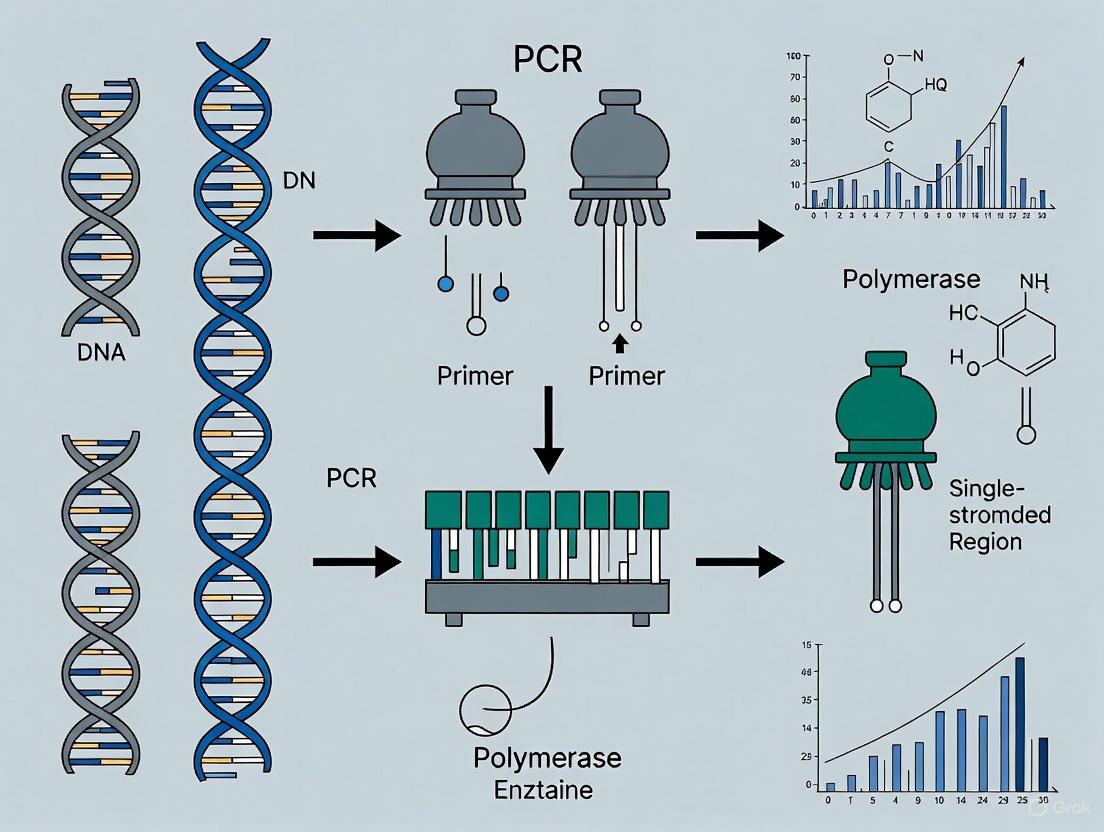
Amplifying GC-Rich Promoter Regions: A Comprehensive Guide to Betaine Optimization for Biomedical Research
This article provides a complete guide for researchers and drug development professionals struggling to amplify GC-rich promoter regions, a common challenge in gene regulation studies.
Amplifying GC-Rich Promoter Regions: A Comprehensive Guide to Betaine Optimization for Biomedical Research
Abstract
This article provides a complete guide for researchers and drug development professionals struggling to amplify GC-rich promoter regions, a common challenge in gene regulation studies. It explores the fundamental science behind PCR obstacles, delivers step-by-step methodological protocols for using betaine, offers advanced troubleshooting strategies, and presents validation data from successful applications. By synthesizing current research and practical optimization techniques, this resource enables reliable amplification of critical genomic regions including tumor suppressor and housekeeping gene promoters, facilitating advancements in molecular diagnostics and therapeutic development.
Understanding the GC-Rich Challenge: Why Promoter Regions Resist Amplification
The Biological Significance of GC-Rich Promoters in Gene Regulation
Gene promoters are critical regulatory DNA sequences that control the initiation of transcription. A defining feature of many promoters, particularly those of highly expressed and housekeeping genes, is their high guanine-cytosine (GC) content. These GC-rich promoters are not merely random sequence occurrences but are fundamental genomic elements that influence DNA structure, nucleosome positioning, epigenetic modifications, and interactions with the transcriptional machinery. This application note explores the multifaceted biological significance of GC-rich promoters, examining their roles in transcriptional regulation, their distinctive structural properties, and the associated experimental challenges. The content is framed within the context of research involving the amplification of GC-rich promoter regions, for which additives like betaine are often essential for successful experimental outcomes.
Structural and Functional Roles of GC-Rich Promoters
GC-rich promoter sequences directly influence the physical properties of DNA and chromatin, which in turn govern their regulatory capacity.
Nucleosome Positioning and Biophysical Properties
The intrinsic biophysical properties encoded in GC-rich sequences directly influence nucleosome behavior and higher-order chromatin organization. Research using the "condense-seq" assay, which measures the intrinsic condensability of native mononucleosomes, has revealed that promoter regions, which are frequently GC-rich, display characteristically low condensability. This property is crucial for maintaining an open chromatin state that is accessible to the transcription machinery [1]. The condensability of a nucleosome is an emergent property that is strongly anticorrelated with gene expression levels; the most highly expressed genes exhibit the lowest condensability precisely around their transcription start sites (TSS) [1]. This relationship is cell type-specific, indicating that condensability is a functional property reflective of cellular state rather than a fixed sequence artifact [1].
Non-Canonical DNA Structures
Beyond standard B-DNA, GC-rich sequences have a high propensity to form stable non-canonical secondary structures, such as G-quadruplexes (G4) and i-motifs. These four-stranded structures act as conformational switches that can dynamically regulate genomic events [2]. In the human genome, sequences with the potential to form these structures are overrepresented in key regulatory domains, including the promoters of oncogenes like c-MYC, hTERT, and KRAS [2]. A proposed hypothesis suggests that gene expression can be regulated thermodynamically through a fine-tuned equilibrium between duplex DNA and G-quadruplex conformations, with the G-quadruplex modulating RNA polymerase activity. Deviations from this tuned equilibrium, caused by mutations or changes in cellular conditions, can lead to pathological gene expression [2].
Histone Variant Incorporation
The DNA sequence at promoters directly influences chromatin composition by guiding the activity of ATP-dependent chromatin remodelers. The SWR complex, for example, incorporates the histone variant H2A.Z into nucleosomes, a process stimulated by poly(dA:dT) tracts often found near promoters. The incorporation of H2A.Z leads to increased DNA unwrapping from the histone octamer and a more open nucleosome conformation, thereby facilitating transcription initiation [3]. This demonstrates a direct mechanism by which specific DNA sequences, common in promoter regions, can instigate an epigenetic environment favorable to transcription.
Table 1: Key Structural Features and Functional Impacts of GC-Rich Promoters
| Feature | Description | Biological Impact |
|---|---|---|
| Low Nucleosome Condensability | Biophysical property of low propensity to form condensed chromatin | Maintains open, accessible chromatin architecture; facilitates transcription factor binding [1] |
| G-Quadruplex/i-Motif Formation | Stable non-canonical four-stranded DNA structures | Acts as a conformational switch to modulate RNA polymerase activity and transcription levels [2] |
| H2A.Z Histone Incorporation | Preferential incorporation of histone variant H2A.Z by SWR remodeler | Promotes DNA unwrapping and an open nucleosome conformation, stimulating transcription initiation [3] |
| CpG Islands | Clusters of CpG dinucleotides frequently encompassing promoters | Associated with robust, high-level gene expression; hypomethylated state protects from mutational decay [4] |
Gene Regulation and Evolutionary Dynamics
GC-rich promoters are central to sophisticated regulatory mechanisms and are subject to distinct evolutionary pressures.
Transcriptional Regulation and Convergent Transcription
Promoters do not function in isolation. A recent discovery shows that convergent promoters, a constellation where two juxtaposed promoters drive convergent transcription, represent a widespread co-regulated promoter class. Surprisingly, the convergent transcription originating from these promoters displays a significant positive correlation, challenging the long-held model that convergent transcription primarily leads to interference and diminished expression [5]. These convergent promoters initiate a variety of RNAs, including upstream antisense RNAs (uaRNAs) and downstream antisense RNAs (daRNAs), substantially expanding the cis-regulatory repertoire of the genome [5].
Evolutionary Dynamics of GC-Content
The characteristic peak of GC-content at the 5' end of genes is an ancient feature, likely present in the last common ancestor of vertebrates. This GC-peak promotes efficient nuclear export and translation of mRNAs [4]. However, its evolutionary maintenance is dynamic and shaped by non-adaptive forces. The key driver is GC-biased gene conversion (gBGC), a process linked to recombination that favors the transmission of G and C alleles over A and T [4]. In species with the PRDM9 gene (e.g., apes and rodents), recombination is directed away from promoters, leading to a ongoing mutational decay of the GC-peak. Conversely, in species lacking PRDM9 (e.g., canids), recombination occurs at promoters, resulting in a current increase in GC-content. The observed decay in apes and rodents suggests that the GC-peak is not actively maintained by selection on most genes in these lineages [4].
Table 2: Evolutionary Dynamics of GC-Content at Promoters Across Species
| Species Group | PRDM9 Status | Recombination Site | Impact on GC-Peak | Interpretation |
|---|---|---|---|---|
| Apes & Rodents | Present | Away from TSSs | Mutational decay | GC-peak not maintained by selection in most genes [4] |
| Canids | Absent | At TSSs | Current increase | Ongoing gBGC drives increase in GC-content at promoters [4] |
Experimental Challenges and Protocols
The very properties that make GC-rich promoters biologically significant also present substantial technical challenges for molecular biology techniques like PCR.
Challenges in Amplifying GC-Rich Promoters
Amplifying GC-rich DNA sequences (>60% GC) is notoriously difficult due to the formation of stable secondary structures and strong hydrogen bonding, which hinder DNA polymerase progression and primer annealing [6]. These challenges can lead to PCR failure, low yield, or non-specific amplification, complicating the study of promoter regions in genes such as those for nicotinic acetylcholine receptors (nAChRs) [6].
Optimized PCR Protocol for GC-Rich Sequences
A multipronged approach is required to successfully amplify GC-rich promoter regions. The following optimized protocol is adapted from research on amplifying nAChR subunits [6].
Application Note: Protocol for PCR Amplification of GC-Rich Promoter Regions
Objective: To amplify GC-rich DNA templates for downstream analysis (e.g., cloning, sequencing).
Key Reagent Solutions:
- Betaine (5M): Acts as a destabilizing agent, disrupting secondary structures and equalizing the melting temperatures of GC- and AT-rich regions.
- DMSO (100%): Serves as a cosolvent to reduce DNA secondary structure stability and improve polymerase processivity.
- High-Fidelity DNA Polymerase: An enzyme blend specifically formulated for amplifying complex templates, often with enhanced processivity and proofreading activity.
Methodology:
- Reaction Setup: Prepare a 50 µL PCR reaction mixture containing:
- 1x High-Fidelity PCR Buffer
- 200 µM of each dNTP
- 0.5 µM of each forward and reverse primer
- 1 M Betaine (final concentration)
- 5% DMSO (final concentration)
- ~50 ng of genomic DNA template
- ~2 U of high-fidelity DNA polymerase (e.g., Q5, KAPA HiFi)
- Thermal Cycling: Perform PCR using the following cycling conditions, optimized in a thermocycler:
- Initial Denaturation: 98°C for 2 minutes
- Amplification (35 cycles):
- Denaturation: 98°C for 20 seconds
- Annealing: Use a temperature gradient (e.g., 60–72°C) to determine the optimal temperature for your specific primer-template set. A higher annealing temperature may improve specificity.
- Extension: 72°C for 1 minute per kb of amplicon length
- Final Extension: 72°C for 5 minutes
- Hold: 4°C indefinitely
- Analysis: Analyze the PCR product by agarose gel electrophoresis to verify amplicon size and specificity.
Troubleshooting: If amplification remains inefficient, consider further optimizing primer design to target regions with slightly lower GC-content, increasing the concentration of DNA polymerase, or testing other specialized polymerases or enhancer combinations [6].
The Scientist's Toolkit: Research Reagent Solutions
Successfully studying GC-rich promoters requires a suite of specialized reagents and tools to overcome technical hurdles and gain accurate biological insights.
Table 3: Essential Research Reagents for Studying GC-Rich Promoters
| Research Reagent | Function/Application | Example Use Case |
|---|---|---|
| Betaine | PCR enhancer that disrupts secondary structures, homogenizes melting temperatures | Amplification of high-GC-content promoter sequences for cloning [6] |
| DMSO | Cosolvent that reduces DNA secondary structure stability | Improving yield and specificity in PCR of GC-rich templates [6] |
| High-Fidelity DNA Polymerase | Specialized enzyme blends for amplifying complex templates | Robust amplification of long or structured GC-rich promoter regions [6] |
| TET2/APOBEC Enzymes (for EM-seq) | Enzymatic conversion for methylation detection, preserves DNA integrity | Assessing methylation status in GC-rich CpG islands without the DNA fragmentation of bisulfite treatment [7] |
| PacBio HiFi/ONT Sequencing | Long-read sequencing technologies for direct methylation detection | Detecting methylation in repetitive and GC-rich regions that are challenging for short-read BS-seq [7] [8] |
| SWR Complex | Chromatin remodeler for H2A.Z histone variant exchange | In vitro studies of H2A.Z incorporation kinetics at poly(dA:dT)-containing promoter sequences [3] |
GC-rich promoters are genomic elements of profound biological significance, whose functions are encoded in their DNA sequence. Their low condensability dictates an open chromatin state, their capacity to form non-canonical structures like G-quadruplexes provides a mechanism for thermodynamic regulation, and their sequence composition guides the incorporation of activating histone variants. Furthermore, their organization into complex units like convergent promoters reveals an additional layer of transcriptional coordination. From a practical standpoint, researching these regions demands specific strategies, such as the use of betaine and specialized polymerases for PCR, and the selection of appropriate modern sequencing technologies for epigenetic profiling. A comprehensive understanding of GC-rich promoters is therefore indispensable for advancing both basic molecular biology and applied drug development research.
The polymerase chain reaction (PCR) serves as a fundamental technique in molecular biology, yet the amplification of genomic regions with high guanine-cytosine (GC) content (>60%) presents substantial technical challenges. These difficulties primarily stem from the molecular obstacles of robust hydrogen bonding and stable secondary structure formation within GC-rich DNA sequences. The triple hydrogen bonding between G and C bases, compared to the double hydrogen bonding of A and T pairs, significantly increases the thermodynamic stability of the DNA duplex. This enhanced stability raises the melting temperature (Tm) of the DNA, often preventing complete denaturation during standard PCR cycling conditions and consequently leading to amplification failure or the production of non-specific products [6] [9].
The formation of intramolecular secondary structures—such as hairpins, stem-loops, and G-quadruplexes—poses another significant barrier. These structures arise when single-stranded DNA templates fold back upon themselves, creating stable configurations that block polymerase progression and primer annealing. The issue is particularly pronounced in promoter regions of many eukaryotic genes, which frequently exhibit GC-rich characteristics, including CpG islands. For researchers investigating gene regulation or developing therapeutic interventions, overcoming these amplification hurdles is essential for cloning, sequencing, and functional analysis of these genetically significant regions [9] [10]. Within the broader context of betaine research, understanding these molecular obstacles provides the foundation for developing effective countermeasures that facilitate successful amplification of refractory sequences.
Molecular Mechanisms of Amplification Failure
The Hydrogen Bonding Dilemma
The fundamental challenge in amplifying GC-rich DNA lies in the inherent molecular stability conferred by GC base pairs. Each GC pair forms three hydrogen bonds, creating a significantly more stable duplex than AT pairs, which form only two. This differential bonding strength directly translates to a higher melting temperature requirement for DNA denaturation. During PCR, if the denaturation step fails to completely separate DNA strands, polymerase access to the template is impeded, resulting in inefficient amplification or complete reaction failure. The problem is exacerbated in sequences where GC content exceeds 70%, as the cumulative effect of numerous triple hydrogen bonds creates an exceptionally stable duplex that may resist complete denaturation even at standard PCR denaturation temperatures (94-95°C) [6] [9].
Secondary Structure Formation
Beyond the simple duplex stability, GC-rich sequences have a strong propensity to form complex secondary structures that present physical barriers to amplification. These structures include:
- Hairpin Loops: Caused by inverted repeats within the sequence, leading to self-complementarity and intra-strand folding.
- Stem-Loops: More extensive structures with double-stranded stems and single-stranded loops.
- G-Quadruplexes: Higher-order structures formed in guanine-rich stretches where four guanine bases associate via Hoogsteen hydrogen bonding.
These secondary structures are particularly problematic during the primer annealing and extension phases of PCR. When DNA templates form stable intramolecular structures, primers cannot access their complementary binding sites. Furthermore, DNA polymerases frequently stall or dissociate when encountering these physical barriers, leading to truncated amplification products. Research on the putative mouse PeP promoter (71.01% GC content) revealed nine independent secondary structures with high internal energies ranging from -199.73 to -209.77 kcal/mol, illustrating the significant thermodynamic stability these structures can achieve [10].
Table 1: Common Secondary Structures in GC-Rich DNA and Their Impact on PCR
| Structure Type | Molecular Characteristics | Consequence for PCR |
|---|---|---|
| Hairpin Loops | Short inverted repeats creating small stem-loop structures | Blocks primer binding sites; causes polymerase pausing |
| Stem-Loops | Extensive intra-strand pairing with longer duplex regions | Prevents complete primer annealing; generates truncated products |
| G-Quadruplexes | Four guanine tracts forming planar arrays via Hoogsteen bonding | Creates severe polymerase blocking structures; causes complete amplification failure |
Template Length Considerations
The challenges of GC-rich amplification compound with increasing template length. While shorter GC-rich fragments (<1000 bp) might be amplified with moderate optimization, longer targets frequently require extensive protocol modifications. This length-dependent difficulty stems from the increased probability of secondary structure formation and the cumulative effect of hydrogen bonding across extended regions. Studies on Mycobacterium bovis genes demonstrated that a large gene of 1794 bp with 77.5% GC content (Mb0129) presented significantly more amplification challenges than a smaller gene of 663 bp with 63% GC content (mpb83), despite both originating from the same high-GC genome [9].
Chemical Additives and Their Mechanisms of Action
Betaine: The GC Equalizer
Betaine (N,N,N-trimethylglycine) stands as one of the most effective and widely adopted additives for ameliorating GC-rich amplification challenges. Its primary mechanism involves acting as an isostabilizing agent that equalizes the differential contribution of AT and GC base pairs to DNA duplex stability. Betaine achieves this effect by preferentially excluding itself from the hydration sphere of GC base pairs, thereby reducing the energy required for strand separation. This action effectively lowers the melting temperature of GC-rich DNA without significantly affecting AT-rich regions, creating a more uniform melting profile across the template [11] [12].
At the molecular level, betaine functions as a protein stabilizer and osmoprotectant that helps maintain polymerase activity in suboptimal reaction conditions. The compound's zwitterionic structure, with both positive and negative charges near neutral pH, enables interactions with DNA that disrupt the strong hydrogen bonding network of GC pairs. Research has demonstrated that betaine concentrations typically ranging from 0.5 M to 1.3 M can dramatically improve amplification efficiency of GC-rich templates, including the prostate-specific membrane antigen mRNA and the c-jun coding cDNA region [11] [13].
Dimethyl Sulfoxide (DMSO): Hydrogen Bond Disruptor
DMSO functions as a polar aprotic solvent that interferes with hydrogen bond formation between DNA strands. By disrupting the water structure around DNA molecules and competing for hydrogen bonding sites, DMSO effectively reduces the thermodynamic stability of GC-rich duplexes. This action facilitates strand separation during the denaturation step and helps prevent reannealing of complementary sequences during lower temperature phases of the PCR cycle. Additionally, DMSO appears to enhance PCR specificity by reducing non-specific primer binding, particularly in templates with complex secondary structures [12] [14].
Standard protocols typically employ DMSO at concentrations between 5-10% (v/v), with higher concentrations potentially inhibiting polymerase activity. Studies on de novo synthesis of GC-rich constructs demonstrated that DMSO significantly improved target product specificity and yield during PCR amplification when used in combination with betaine [12]. The dual approach of using both additives capitalizes on their complementary mechanisms—betaine equalizes base pair stability while DMSO directly disrupts hydrogen bonding networks.
Specialized Additive Combinations
For particularly challenging templates, researchers have developed powerful combinatorial approaches that integrate multiple additives with distinct mechanisms of action:
Betaine-DMSO-7-deaza-dGTP Trio: This combination has proven essential for amplifying extremely GC-rich sequences (67-79% GC), including disease-related genes such as RET promoter region (79% GC), LMX1B (67.8% GC), and PHOX2B (72.7% GC). The 7-deaza-dGTP nucleotide analog incorporates into nascent DNA strands in place of dGTP, preventing the formation of stable secondary structures by disrupting the Hoogsteen bonding necessary for G-quadruplex formation [14].
Organic Solvent Alternatives: Recent investigations have identified ethylene glycol and 1,2-propanediol as potentially superior alternatives to betaine for some applications. In a comprehensive study evaluating 104 GC-rich human genomic amplicons (60-80% GC content), 1,2-propanediol successfully amplified 90% of targets compared to 72% with betaine alone, while ethylene glycol achieved 87% success. These additives appear to function through a different mechanism than betaine, possibly through differential affinities to single-stranded versus double-stranded DNA [15].
Table 2: PCR Additives for GC-Rich Amplification and Their Applications
| Additive | Common Concentrations | Mechanism of Action | Applicable Scenarios |
|---|---|---|---|
| Betaine | 0.5-1.3 M | Equalizes DNA melting temperatures; reduces secondary structure formation | General GC-rich templates; often used as first-line approach |
| DMSO | 5-10% (v/v) | Disrupts hydrogen bonding; prevents inter- and intrastrand reannealing | Templates with strong secondary structures; often combined with betaine |
| 7-deaza-dGTP | 50 μM (partial replacement of dGTP) | Prevents Hoogsteen bonding and G-quadruplex formation | Extremely GC-rich targets (>75%); sequences with G-repeats |
| Ethylene Glycol | 1.075 M | Reduces DNA melting temperature; mechanism distinct from betaine | Betaine-resistant templates; high-throughput applications |
| Formamide | 1-5% (v/v) | Increases stringency; denatures secondary structures | Improves specificity in complex templates |
Optimized Experimental Protocols
Standardized PCR Protocol for GC-Rich Templates
Based on the collective research findings, the following protocol provides a robust starting point for amplifying GC-rich promoter regions and other challenging templates:
Reaction Composition:
- DNA Polymerase: PrimeSTAR GXL polymerase or other high-fidelity polymerases with strong processivity [9]
- Buffer System: Ammonium sulfate-based buffer (e.g., 10× PCR buffer AMS: 750 mM Tris-HCl [pH 8.8], 200 mM (NH₄)₂SO₄, 0.1% Tween 20) [10]
- Magnesium Concentration: 3-4 mM MgCl₂ (optimized empirically) [10]
- Additives: 1 M betaine + 5-10% DMSO (v/v) [12] [10]
- Nucleotide Analogs: For extremely refractory templates, replace 25-50% of dGTP with 7-deaza-dGTP [14]
- Template DNA: 100-200 ng genomic DNA or 10-50 ng plasmid DNA
- Primers: 10-20 pmol each, with Tm between 65-72°C
Thermal Cycling Conditions:
- Initial Denaturation: 95°C for 5 minutes
- Amplification Cycles (30-35 cycles):
- Denaturation: 98°C for 10 seconds
- Annealing/Extension: 68-72°C for 1 minute per kb (2-step PCR)
- Final Extension: 72°C for 10 minutes
Critical Protocol Notes:
- Employ a slow ramp rate (0.5-1°C/second) between annealing and denaturation steps to facilitate proper primer binding and polymerase initiation [9]
- For templates >1 kb, extend the elongation time to 2-4 minutes per kb
- Implement touchdown PCR for templates with complex secondary structures: start 5-10°C above calculated Tm and decrease by 0.5-1°C per cycle for the first 10-15 cycles [10]
Specialized Protocol for Extremely GC-Rich Targets (>75% GC)
For the most challenging templates, such as the RET promoter region (79% GC), a modified approach is necessary:
Reaction Composition:
- All components from the standard protocol, plus:
- Enhanced Additive Cocktail: 1.3 M betaine + 5% DMSO + 50 μM 7-deaza-dGTP (replacing 50% of standard dGTP) [14]
- Polymerase Selection: Taq polymerase or specialized GC-rich polymerases
- Magnesium Optimization: 2.5 mM MgCl₂ (empirically determined for each template)
Thermal Cycling Conditions:
- Initial Denaturation: 95°C for 5 minutes
- Amplification Cycles (35-40 cycles):
- Denaturation: 94°C for 30 seconds
- Annealing: 60°C for 30 seconds
- Extension: 72°C for 45 seconds to 1 minute
- Final Extension: 72°C for 5 minutes
This specialized protocol successfully amplified the 392-bp RET promoter region where standard methods and individual additives failed, producing a unique specific PCR product as confirmed by DNA sequencing [14].
Research Reagent Solutions
Table 3: Essential Reagents for GC-Rich DNA Amplification
| Reagent Category | Specific Examples | Function & Application Notes |
|---|---|---|
| DNA Polymerases | PrimeSTAR GXL, Platinum Pfx, KOD FX, GC-Rich Enhancer System | High processivity and strand displacement activity; some specialized for GC-rich templates |
| PCR Additives | Betaine (1 M), DMSO (5-10%), 7-deaza-dGTP (50 μM), Ethylene Glycol (1.075 M) | Reduce secondary structure; equalize melting temperatures; enhance specificity |
| Buffer Systems | Ammonium sulfate-based buffers, Potassium glutamate-enhanced buffers | Stabilize polymerase activity; improve DNA denaturation efficiency |
| Nucleotide Mixes | dNTPs with 7-deaza-dGTP substitution, Balanced dNTP concentrations | Prevent polymerization pauses; maintain replication fidelity |
| Enhancer Solutions | Commercial GC-rich enhancers, Q-Solution, Perfect Amp GC Enhancer | Proprietary formulations to overcome amplification barriers |
Workflow Visualization
The following diagram illustrates the strategic approach to overcoming molecular obstacles in GC-rich amplification, integrating both diagnostic steps and interventional strategies:
The amplification of GC-rich DNA sequences, particularly promoter regions with importance in gene regulation and drug development, requires strategic approaches to overcome the inherent molecular obstacles of hydrogen bonding and secondary structure formation. Through the application of chemical additives like betaine, DMSO, and 7-deaza-dGTP—combined with optimized PCR protocols featuring modified thermal cycling parameters and specialized polymerase systems—researchers can successfully navigate these challenges. The protocols and mechanistic insights presented herein provide a foundation for investigating previously refractory genomic targets, advancing both basic research and therapeutic development in molecular biology and genetics.
The polymerase chain reaction (PCR) is a foundational technology in molecular biology, yet the amplification of GC-rich DNA sequences presents particular challenges that can compromise experimental results. GC-rich templates, typically defined as sequences where 60% or more of the bases are guanine or cytosine, constitute only about 3% of the human genome but are disproportionately found in critical regulatory regions such as gene promoters, enhancers, and CpG islands [16]. The amplification of these regions is frequently hampered by the formation of specific artifacts: hairpins, smears, and primer dimers. These artifacts arise from the intrinsic biophysical properties of GC-rich DNA, where the three hydrogen bonds in G-C base pairs confer greater thermodynamic stability compared to the two bonds in A-T pairs [17] [16]. This increased stability leads to incomplete denaturation, facilitating the formation of stable secondary structures and mispriming events. This application note, framed within broader research on optimizing GC-rich promoter amplification using betaine, details the origins of these common artifacts and provides validated protocols for their mitigation, enabling more reliable analysis of these biologically significant genomic regions.
Understanding the Artifacts and Their Origins
Hairpins and Secondary Structures
Hairpins are intramolecular secondary structures that form when single-stranded DNA folds back on itself to create stable, double-stranded regions. In GC-rich sequences, the strong triple hydrogen bonds of G-C base pairs make these structures particularly stable and problematic. When a DNA polymerase encounters these hairpins during the extension phase of PCR, its progression can stall, leading to premature termination of synthesis [12] [16]. This results in the accumulation of truncated, incomplete DNA fragments. The detection of these fragments typically reveals multiple shorter bands or a smear upon gel electrophoresis, rather than a single, clear band of the expected amplicon size. The formation of secondary structures is not merely an inconvenience; it directly competes with primer annealing and polymerase processivity, dramatically reducing the yield of the desired full-length product [17].
Smears on Agarose Gels
A smear appearing on an agarose gel, visualized as a broad, diffuse band spanning a range of molecular weights, is a common indicator of nonspecific amplification and heterogeneous products. In the context of GC-rich PCR, smears primarily result from two interconnected phenomena. First, the polymerase stalling at stable secondary structures generates a population of DNA fragments of varying lengths. Second, the incomplete separation of DNA strands during the denaturation step, caused by the high thermal stability of GC-rich duplexes, provides a suboptimal template for the subsequent annealing and extension steps [16]. This combination can lead to a cascade of non-specific priming events and the synthesis of a heterogeneous mixture of products, which manifests as a smear. This artifact is a clear sign that the reaction conditions are not sufficiently stringent or supportive to overcome the template's challenging biophysics.
Primer Dimers
Primer dimers are short, duplex artifacts formed by the hybridization and extension of two primers onto each other, rather than onto the intended template. They typically appear on an agarose gel as a low molecular weight band, often around 50-100 bp. GC-rich templates exacerbate primer dimer formation in several ways. The templates themselves can be difficult for primers to access due to secondary structures, increasing the likelihood that free primers will interact with each other. Furthermore, primers designed for GC-rich targets often themselves have high GC content and high melting temperatures (Tm), which can promote mispriming and increase the chance of complementary regions between primers interacting [16]. The formation of primer dimers not only consumes reagents that would otherwise be used for target amplification but can also outcompete the desired reaction, leading to PCR failure.
The Role of Betaine in Ameliorating Artifacts
Betaine (N,N,N-trimethylglycine) is a kosmotropic molecule that functions as a powerful PCR enhancer for GC-rich templates. Its primary mechanism of action is the equalization of the thermal stability difference between G-C and A-T base pairs. Betaine interacts directly with DNA, disrupting the base-stacking interactions and reducing the energy required to denature GC-rich duplexes [18]. By doing so, it promotes more complete strand separation during the denaturation step, which in turn minimizes the reformation of template secondary structures like hairpins and allows for more efficient primer annealing [18] [10].
The foundational study by Henke et al. demonstrated that betaine enabled the co-amplification of alternatively spliced variants of the prostate-specific membrane antigen mRNA and the coding cDNA region of c-jun, both of which are GC-rich [18]. The researchers concluded that betaine acts by "reducing the formation of secondary structure caused by GC-rich regions," making it a generally applicable solution for such challenging amplifications [18] [19]. Subsequent research has confirmed that betaine's efficacy is often enhanced when used in combination with other additives, such as dimethyl sulfoxide (DMSO), forming a synergistic mixture that more effectively destabilizes secondary structures and improves polymerase processivity [20] [12] [10].
Recommended Protocols and Experimental Workflows
Standardized Protocol for GC-Rich Amplification with Betaine
The following protocol is optimized for the amplification of GC-rich promoter regions and other challenging templates, incorporating betaine to suppress common artifacts.
Reagent Setup (50 μL Reaction):
- Template DNA: 10-100 ng
- Forward & Reverse Primers (10 μM each): 2.5 μL each
- dNTP Mix (10 mM each): 1 μL
- 10x PCR Buffer (with MgCl₂): 5 μL
- Betaine (5M stock): 10 μL (Final concentration: 1.0-1.5 M)
- DMSO: 2.5-5 μL (Final concentration: 5-10%)
- MgCl₂ (25 mM stock): 0-4 μL (Final concentration: 1.5-4.0 mM; optimize)
- High-Fidelity DNA Polymerase: 0.5-1 U
- Nuclease-free water to 50 μL
Thermocycling Conditions:
- Initial Denaturation: 95-98°C for 2-5 minutes
- Amplification (30-35 cycles):
- Denaturation: 95-98°C for 20-30 seconds (longer than standard)
- Annealing: Temperature gradient recommended, typically 5°C above primer Tm
- Extension: 72°C (time according to amplicon length, 1 kb/min)
- Final Extension: 72°C for 5-10 minutes
- Hold: 4°C
Advanced Multi-Additive Protocol
For exceptionally recalcitrant sequences with GC content exceeding 75%, a powerful combination of additives is recommended, as demonstrated by Musso et al. [20]. This protocol was essential for amplifying disease genes with GC content ranging from 67% to 79%.
Reagent Setup (50 μL Reaction):
- Standard PCR components (as above)
- Betaine: 10 μL (Final concentration: 1.0 M)
- DMSO: 5 μL (Final concentration: 10%)
- 7-deaza-2'-deoxyguanosine (7-deaza-dGTP): Substitute for 50-100% of the dGTP in the dNTP mix. Note: 7-deaza-dGTP reduces ethidium bromide staining, requiring alternative nucleic acid stains. [20] [16]
Critical Notes: The combination of these three additives works synergistically. Betaine and DMSO destabilize secondary structures, while 7-deaza-dGTP, a dGTP analog, is incorporated into the nascent DNA and prevents the formation of stable secondary structures by disrupting Hoogsteen base-pairing, thereby allowing the polymerase to read through previously impassable GC-rich regions [20].
Workflow for Troubleshooting Amplification Artifacts
The following diagram illustrates a systematic approach to diagnosing and resolving the common artifacts discussed in this note, emphasizing the strategic use of betaine.
Diagram: Troubleshooting Workflow for PCR Artifacts. This chart outlines a systematic diagnostic and remedial approach for common amplification artifacts, linking each artifact to its root cause and proposing targeted solutions.
Research Reagent Solutions
The following table details key reagents and their specific functions in mitigating artifacts during the amplification of GC-rich sequences.
| Reagent | Recommended Concentration | Primary Function in GC-Rich PCR |
|---|---|---|
| Betaine | 1.0 - 1.5 M | Equalizes Tm of GC and AT base pairs; reduces secondary structure formation by promoting DNA denaturation [18] [10]. |
| DMSO | 5 - 10% (v/v) | Disrupts secondary structures by interfering with hydrogen bonding; improves strand separation and polymerase processivity [20] [16]. |
| 7-deaza-dGTP | 50-100% dGTP substitution | dGTP analog that incorporates into DNA and prevents Hoogsteen base-pairing, thereby stabilizing the DNA duplex and reducing polymerase pausing [20] [16]. |
| High-Fidelity Polymerase | As per manufacturer | Engineered polymerases with enhanced processivity are less prone to stalling at complex secondary structures [16]. |
| MgCl₂ | 1.5 - 4.0 mM (optimize) | Critical cofactor for polymerase activity; increased concentrations can help stabilize DNA duplexes but require optimization to avoid non-specific priming [16] [10]. |
| GC Enhancer | As per manufacturer | Proprietary blends (e.g., from NEB) often contain a optimized mix of agents like betaine and DMSO to facilitate amplification of difficult templates [16]. |
The efficacy of betaine and related additives is well-documented across multiple studies. The table below summarizes key experimental outcomes from the literature.
| Study (Year) | GC-Rich Target | GC Content | Additives Used | Key Experimental Outcome |
|---|---|---|---|---|
| Henke et al. (1997) [18] | Prostate-specific membrane antigen mRNA; c-jun cDNA | High | Betaine | Enabled co-amplification of spliced variants and c-jun region where standard PCR failed. |
| Musso et al. (2006) [20] | Three disease genes | 67% - 79% | Betaine, DMSO, 7-deaza-dGTP | The triple-additive mixture was essential to achieve successful amplification. |
| Seifi et al. (2012) [10] | Putative mouse PeP promoter | 71.01% | Betaine (1 M), DMSO (10%), 4 mM MgCl₂ in AMS buffer | Significantly improved amplification yield; method verified on other human GC-rich loci. |
| Jensen et al. (2010) [12] | IGF2R and BRAF gene fragments | High | DMSO and Betaine | Additives greatly improved target product specificity and yield during PCR amplification in de novo synthesis. |
| Adey et al. (2011) [21] | Illumina sequencing libraries | 6% - 90% GC | Betaine (2 M), extended denaturation | Shifted amplification plateau to cover 23% to 90% GC, rescuing extreme GC-rich fragments. |
The reliable amplification of GC-rich promoter regions is critical for advancing research in gene regulation and drug development. The artifacts of hairpins, smears, and primer dimers are direct consequences of the robust biophysical properties of GC-rich DNA, but they are not insurmountable. As detailed in these application notes, the strategic incorporation of betaine, often in combination with DMSO and other enhancers like 7-deaza-dGTP, provides a robust chemical solution to destabilize secondary structures and promote uniform amplification. The protocols and data summarized herein offer researchers a validated toolkit for troubleshooting and optimizing their PCR assays, ensuring accurate representation and analysis of these therapeutically relevant genomic targets.
Within molecular biology, the amplification of GC-rich DNA sequences presents a significant challenge, particularly as these regions are frequently found in the promoters of housekeeping and tumor suppressor genes [22]. The core of the issue lies in the inherent stability of GC-rich DNA; guanine-cytosine (G-C) base pairs form three hydrogen bonds, compared to the two bonds in adenine-thymine (A-T) pairs. This results in DNA regions with high GC content requiring more energy to denature, exhibiting heightened thermostability, and readily forming stable secondary structures like hairpins that can stall polymerases [22]. Overcoming these challenges is crucial for accurate genetic analysis, and the zwitterionic osmolyte betaine has emerged as a key solution. This Application Note delineates the mechanisms by which betaine destabilizes GC-rich DNA, summarizes quantitative data on its efficacy, and provides detailed protocols for its application in PCR to support research and drug development.
Mechanisms of Betaine Action
Betaine (N,N,N-trimethylglycine) enhances the amplification of GC-rich templates through two primary, interconnected mechanisms that lower the thermal stability of DNA duplexes.
Preferential Destabilization of GC-Rich DNA
Betaine's destabilizing effect is not uniform across all DNA sequences; it exhibits a preferential effect on GC-rich duplexes. Research on RNA duplexes has demonstrated that the accumulation of betaine at the nucleic acid surface exposed during denaturation is temperature-dependent and greater for high-GC content sequences. Consequently, betaine destabilizes higher GC content RNA duplexes to a greater extent than low GC content duplexes at their respective melting temperatures [23]. This preferential interaction eliminates the base pair composition dependence of DNA melting, effectively creating a more uniform melting temperature across different sequences [23] [24]. The interaction is strongly temperature-sensitive and characterized by entropy-enthalpy compensation, with the entropic contribution becoming more significant at higher temperatures [23].
Reduction of DNA Melting Temperature
The primary mechanism of action involves betaine's interaction with the DNA helix. Betaine is excluded from the hydration layer of anionic phosphate groups in the DNA backbone. During denaturation, the exposure of aromatic and amine surface areas creates sites for thermodynamically favorable interactions with betaine. This shifts the equilibrium towards the single-stranded state by decreasing the melting temperature (Tm) of the DNA, particularly for GC-rich regions [23] [25]. By lowering the energy required to separate DNA strands, betaine facilitates the denaturation of complex secondary structures and promotes primer annealing, thereby enabling polymerases to synthesize through previously impassable regions [22] [26].
Table 1: Impact of Betaine and Analogs on DNA Melting Temperature (Tm)
| Compound Class | Example | Effect on Tm | Influence on Base Pair Dependence |
|---|---|---|---|
| Carboxylate Betaine | Glycine Betaine | Decreases Tm | Decreases dependence on GC content [24] |
| Sulfonate Betaine Analogs | Synthetic sulfonates | Up to 2x greater Tm decrease than betaine [24] | Up to 2x more effective at decreasing dependence [24] |
| Hydroxyl-Substituted Carboxylates | Homologs with OH groups | Can increase Tm (especially in low GC DNA) [24] | Varies |
| Hydroxyl-Substituted Sulfonates | Sulfonates with OH groups | Usually decrease Tm [24] | Varies |
Quantitative Analysis of Betaine Interactions
The thermodynamic interaction between betaine and nucleic acids can be quantified using the m-value, which represents the change in the observed free energy of unfolding per unit change in betaine molality. Negative m-values indicate favorable interactions between the cosolute (betaine) and the surface area exposed during denaturation, leading to destabilization of the folded structure [23].
Experimental data on RNA dodecamer duplexes shows that the m-value becomes more negative with increasing GC content, demonstrating that betaine has a stronger destabilizing effect on GC-rich sequences. For instance, a duplex with 17% GC had an m-value of -0.188 kcal mol⁻¹ m⁻¹, while a 100% GC duplex had a value of -1.010 kcal mol⁻¹ m⁻¹ [23]. Furthermore, these m-values are highly temperature-dependent, a relationship driven almost exclusively by a large, favorable entropy change [23].
Table 2: Experimentally Determined m-values and Interaction Potentials for RNA Dodecamers in Glycine Betaine
| GC Content (%) | Reference Temp. (°C) | m-value (kcal mol⁻¹ m⁻¹) | Interaction Potential (Δμ23,4/RT m⁻¹) |
|---|---|---|---|
| 17 | 27.3 | -0.188 ± 0.017 | -0.315 ± 0.029 |
| 25 | 34.8 | -0.244 ± 0.027 | -0.398 ± 0.044 |
| 33 | 45.5 | -0.378 ± 0.017 | -0.598 ± 0.027 |
| 42 | 44.6 | -0.423 ± 0.044 | -0.670 ± 0.069 |
| 50 | 49.0 | -0.655 ± 0.045 | -1.02 ± 0.07 |
| 67 | 59.6 | -0.627 ± 0.024 | -0.948 ± 0.037 |
| 100 | 80.9 | -1.010 ± 0.023 | -1.44 ± 0.03 |
Data adapted from [23]. m-values and interaction potentials are negative, indicating betaine preferentially interacts with the unfolded state, destabilizing the duplex. This effect is more pronounced at higher GC content and temperature.
Application Notes & Protocols
Standard PCR Protocol for GC-Rich Templates
The following protocol is optimized for the amplification of challenging GC-rich targets (e.g., >60% GC content) using a standard thermocycler and standalone polymerase.
Materials & Reagents
- DNA Template: 1-20 ng of genomic DNA or equivalent.
- Primers: Forward and reverse, resuspended to a working concentration.
- Betaine: 5M stock solution (Sigma, B0300-1VL).
- Polymerase: Use a polymerase known for robust performance on difficult templates (e.g., Q5 High-Fidelity DNA Polymerase, NEB #M0491).
- Corresponding Polymerase Buffer (5X).
- MgCl₂: 50 mM stock solution (if required separately).
- dNTP Mix: 10 mM each.
- Nuclease-free Water.
Procedure
- Prepare Reaction Master Mix on ice. For a single 50 µL reaction:
- Nuclease-free Water: to 50 µL final volume
- 5X Polymerase Buffer: 10 µL
- 10 mM dNTPs: 1 µL
- 50 mM MgCl₂: 0-2 µL (Adjust based on optimization; start with vendor's recommendation)
- 5M Betaine: 10 µL (Final concentration of 1.0 M)
- Forward Primer (10 µM): 2.5 µL
- Reverse Primer (10 µM): 2.5 µL
- DNA Template: X µL
- DNA Polymerase: 0.5-1.0 µL (per manufacturer's instructions)
- Thermal Cycling Conditions:
- Initial Denaturation: 98°C for 30-60 seconds.
- Amplification (30-35 cycles):
- Denaturation: 98°C for 10-15 seconds.
- Annealing: Use a temperature gradient starting 5°C below the calculated Tm of the primers. For GC-rich targets, a higher annealing temperature (e.g., 65-72°C) may improve specificity [22].
- Extension: 72°C (time based on polymerase speed and amplicon length).
- Final Extension: 72°C for 2-5 minutes.
- Hold: 4°C.
Analysis
- Analyze 5-10 µL of the PCR product by agarose gel electrophoresis.
- Expect a single, sharp band of the expected size. Smearing or multiple bands indicate a need for further optimization.
Optimization Strategies for Intractable Targets
Should the standard protocol fail, consider these systematic optimization steps:
- Betaine Concentration: Titrate betaine from 0.5 M to 1.5 M. The standard 1.0 M is effective, but some targets may require a different concentration [26].
- Magnesium Concentration: Test Mg²⁺ concentrations in 0.5 mM increments from 1.0 mM to 4.0 mM. Mg²⁺ is a critical cofactor, and its optimal concentration can vary with betaine present [22].
- Combination with DMSO: For extremely resistant templates, a combination of 1.0 M betaine and 1-5% DMSO can be highly effective. DMSO also aids in disrupting secondary structures [22] [26].
- Polymerase Choice: Utilize polymerases supplied with proprietary GC enhancers, which are often optimized cocktails containing betaine and other additives (e.g., OneTaq GC Enhancer or Q5 GC Enhancer) [22].
- Touchdown PCR: Implement a touchdown protocol, starting with an annealing temperature 10°C above the calculated Tm and decreasing by 1°C every cycle for the first 10 cycles, then continuing at the lower temperature. This increases stringency and specificity in early cycles.
The Scientist's Toolkit: Essential Research Reagents
Table 3: Key Reagents for GC-Rich DNA Amplification with Betaine
| Reagent / Solution | Function / Mechanism | Example Products |
|---|---|---|
| Glycine Betaine (5M Stock) | Reduces DNA melting temperature; preferentially destabilizes GC-rich sequences; minimizes base composition dependence [24] [26]. | Sigma-Aldrich B0300 |
| High-Fidelity DNA Polymerase | Engineered for processivity on complex templates; often supplied with optimized buffers for difficult amplicons. | Q5 High-Fidelity (NEB #M0491), OneTaq DNA Polymerase (NEB #M0480) [22] |
| GC Enhancer | Proprietary additive cocktails that typically include betaine, DMSO, or other compounds to inhibit secondary structure formation. | OneTaq GC Enhancer, Q5 High GC Enhancer [22] |
| Dimethyl Sulfoxide (DMSO) | Serves as a duplex-destabilizing agent; often used in conjunction with betaine for synergistic effects on GC-rich templates [22] [26]. | Sigma-Aldrich D8418 |
| 7-deaza-2'-deoxyguanosine | dGTP analog that incorporates into DNA and reduces the stability of GC base pairs, improving polymerase progression [22]. | Roche Diagnostic 988 662 |
Workflow and Mechanism Visualization
The following diagram illustrates the experimental workflow for optimizing GC-rich PCR with betaine and the conceptual mechanism of its action.
Betaine serves as a powerful tool for destabilizing GC-rich DNA, thereby enabling the reliable amplification of previously intractable promoter regions and other high-GC sequences. Its mechanism, rooted in preferential interaction with the surface area exposed during DNA denaturation and the consequent reduction in melting temperature, is both well-defined and effective. The protocols and optimization strategies detailed herein provide researchers and drug development professionals with a robust framework for incorporating betaine into their molecular biology workflows. As the study of complex genomes advances, the strategic use of betaine and related additives will remain integral to successful PCR-based analyses of GC-rich genetic elements.
Proven Protocols: Implementing Betaine for Successful GC-Rich Promoter Amplification
Optimal Betaine Concentrations and Preparation Guidelines
The amplification of GC-rich DNA sequences, such as those frequently found in gene promoter regions, presents a significant challenge in molecular biology. These regions often form stable secondary structures and exhibit high melting temperatures (Tm), which can cause polymerase enzymes to stall during amplification, resulting in poor yield or complete amplification failure [27] [28]. Betaine (N,N,N-trimethylglycine) has emerged as a powerful chemical additive that can dramatically improve the amplification of these difficult templates.
Betaine functions as an isostabilizing agent that reduces the formation of secondary structures by altering the melting characteristics of DNA. It equilibrates the differential Tm between AT and GC base pairings, thereby facilitating strand separation and preventing the formation of hairpins and other complex structures that impede polymerase progression [27] [11]. This application note provides detailed protocols and concentration guidelines for effectively utilizing betaine in PCR amplification of GC-rich targets, with particular emphasis on promoter region analysis relevant to drug development research.
Optimal Betaine Concentration Ranges
Based on experimental data from multiple studies, the effective concentration range for betaine in PCR applications is well-established. The table below summarizes the optimal concentrations for various application contexts.
Table 1: Recommended Betaine Concentrations for GC-Rich PCR
| Application Context | Final Concentration | Key Experimental Findings | Citation |
|---|---|---|---|
| Standard GC-rich PCR | 1.0 - 1.6 M | Significantly improved amplification of GC-rich templates (IGF2R, BRAF, c-jun, PSMA) by reducing secondary structure formation. | [27] [11] |
| Illumina Library Amplification | 2.0 M | Rescued amplification of extremely high-GC loci (up to 90% GC) when combined with extended denaturation times. | [21] |
| Multiplex & Long Amplicon PCR | 0.8 - 1.6 M | Enhanced specificity and yield in complex amplification reactions; optimal concentration should be determined for specific targets. | [29] [28] |
For most standard applications, a final concentration of 1.0 M to 1.6 M betaine is recommended as a starting point [29] [28]. However, specific challenging templates may require optimization within this range. When working with extremely GC-rich targets (exceeding 80% GC content), increasing the concentration to 2.0 M betaine may be necessary, particularly when combined with protocol modifications such as extended denaturation times [21].
Betaine Preparation and Storage Guidelines
Reagent Preparation
Proper preparation of betaine solutions is critical for experimental consistency and reproducibility.
Stock Solution Concentration: Prepare a 5 M stock solution of molecular biology-grade betaine in nuclease-free water [11]. This concentrated stock allows for convenient addition to PCR reactions without significantly altering final reaction volume or component concentrations.
Sterile Filtration: Filter the solution through a 0.22 μm membrane to ensure sterility and remove any particulate matter that might interfere with PCR.
Aliquoting and Storage: Dispense the stock solution into small, single-use aliquots to minimize repeated freeze-thaw cycles and prevent contamination. Store at -20°C for long-term stability.
Quality Control Considerations
Purity Specification: Use betaine certified for molecular biology applications with ≥99% purity to avoid potential PCR inhibitors.
pH Verification: Check that the prepared solution has a neutral pH (approximately 7.0) to maintain optimal buffer conditions in PCR reactions.
Conformation Validation: Confirm the identity of betaine by its characteristic zwitterionic structure with both positive and negative charges close to neutral pH, which is essential for its isostabilizing function [27].
Experimental Protocol for GC-Rich Amplification
PCR Reaction Setup with Betaine
The following protocol is adapted from established methods for de novo synthesis of GC-rich constructs and amplification of challenging templates [27] [21].
Table 2: PCR Master Mix Formulation with Betaine
| Component | Final Concentration | Volume for 50 μL Reaction | Notes |
|---|---|---|---|
| 10X PCR Buffer | 1X | 5 μL | Use buffer supplied with polymerase |
| dNTP Mix | 200 μM each | 1 μL (10 mM stock) | Higher concentrations may improve yield |
| Forward Primer | 0.5 μM | 2.5 μL (10 μM stock) | Design with Tm calculation adjustment |
| Reverse Primer | 0.5 μM | 2.5 μL (10 μM stock) | Design with Tm calculation adjustment |
| DNA Template | Varies | 1-5 μL | 10-100 ng genomic DNA or equivalent |
| Betaine (5M stock) | 1.0-1.6 M | 10-16 μL | Optimize for specific template |
| DNA Polymerase | As recommended | 0.5-1 μL | Use GC-rich optimized enzymes |
| Nuclease-free Water | To volume | To 50 μL |
Diagram 1: PCR workflow with betaine enhancement
Thermal Cycling Parameters
The following thermal cycling conditions have been specifically optimized for betaine-enhanced amplification of GC-rich regions:
Initial Denaturation:
- 94°C for 3-5 minutes
- Note: Longer denaturation may be beneficial for extremely GC-rich templates [21]
Amplification Cycles (25-35 cycles):
- Denaturation: 94°C for 30-60 seconds (extended time improves GC-rich denaturation)
- Annealing: Primer-specific temperature (typically 55-65°C) for 30 seconds
- Extension: 68-72°C for 1 minute per kilobase of amplicon length
Final Extension:
- 68-72°C for 5-10 minutes
- Ensures complete extension of all amplified products
Hold:
- 4°C indefinitely
Critical Protocol Modifications for GC-Rich Templates
When amplifying particularly challenging GC-rich promoter regions, the following modifications to the standard protocol are recommended:
Extended Denaturation Times: Increase denaturation steps during cycling to 60-80 seconds at 94°C to ensure complete separation of DNA strands [21].
Polymerase Selection: Use polymerases specifically optimized for GC-rich amplification, such as Q5 High-Fidelity DNA Polymerase or OneTaq DNA Polymerase with their respective GC enhancers [28].
Combined Additive Approaches: For extremely challenging templates (≥80% GC), consider combining betaine with DMSO (1-5%) or using specialized GC enhancer formulations that contain multiple additives [27] [28].
Temperature Gradient Optimization: Initially perform annealing temperature gradients to identify optimal priming conditions for each specific GC-rich target.
The Scientist's Toolkit: Essential Reagents
Table 3: Research Reagent Solutions for Betaine-Enhanced PCR
| Reagent/Material | Specification | Function in GC-Rich PCR | Example Products |
|---|---|---|---|
| Betaine | Molecular biology grade, ≥99% purity | Reduces secondary structure formation, equilibrates Tm of AT and GC base pairs | Sigma-Aldrich B2629, Thermo Fisher B0300 |
| High-Fidelity DNA Polymerase | Engineered for GC-rich templates | Processive enzymes resistant to stalling at secondary structures | NEB Q5, NEB OneTaq, Clontech HF Advantage |
| GC Enhancer | Proprietary formulations | Contains multiple additives to improve GC-rich amplification | OneTaq GC Enhancer, Q5 GC Enhancer |
| dNTPs | Molecular biology grade, neutral pH | Balanced nucleotide concentrations for accurate incorporation | Thermo Fisher R0181, NEB N0447 |
| MgCl2 | PCR grade, optimized concentration | Essential cofactor for polymerase activity; may require adjustment | Supplied with polymerase buffers |
Mechanism of Action: How Betaine Improves GC-Rich Amplification
Betaine functions through multiple mechanisms to enhance amplification of GC-rich sequences:
Diagram 2: Betaine mechanism in GC-rich PCR
The primary mechanism involves betaine's action as a isostabilizing agent that reduces the base composition dependence of DNA melting. Betaine contains both positive and negative charges close to neutral pH, which allows it to interact with DNA bases and weaken the extra stability of GC base pairs relative to AT base pairs [27] [11]. This equalization of melting temperatures across sequences with varying GC content enables more uniform amplification of mixed templates and prevents polymerase stalling at regions of extreme GC content.
Additionally, betaine disrupts the formation of secondary structures such as hairpins and G-quadruplexes that are prevalent in GC-rich sequences, particularly in gene promoter regions. By preventing these structures from forming, betaine ensures uninterrupted polymerase progression and significantly improves amplification efficiency and specificity [27] [28]. This property is especially valuable when amplifying promoter regions where secondary structure elements may have functional significance that must be preserved in the amplified product.
Troubleshooting and Optimization Guidelines
Despite the effectiveness of betaine, some GC-rich templates may require additional optimization. The following troubleshooting guide addresses common challenges:
Poor Amplification Yield:
- Increase betaine concentration to 1.6-2.0 M
- Extend denaturation time to 60-80 seconds per cycle
- Increase magnesium concentration in 0.5 mM increments (up to 4 mM)
Non-Specific Amplification:
- Increase annealing temperature by 2-5°C
- Reduce betaine concentration to 1.0 M
- Use hot-start polymerase formulation
- Add formamide (1-3%) to increase primer stringency [28]
Incomplete Products:
- Combine betaine with DMSO (1-3%)
- Use polymerase blends with enhanced processivity
- Increase extension time to 2 minutes per kilobase
For particularly challenging templates such as promoter regions with extreme GC content (>80%), a systematic approach combining betaine with specialized polymerase formulations and optimized thermal profiles typically yields successful amplification where standard protocols fail [27] [21].
The amplification of GC-rich DNA sequences presents a significant challenge in molecular biology, particularly in the context of promoter region analysis which is crucial for understanding gene regulation in fields like cancer research and drug development [30] [31]. Sequences with a GC content exceeding 60% resist standard amplification due to their propensity to form stable secondary structures and exhibit higher thermostability from G-C base pairs possessing three hydrogen bonds compared to the two in A-T pairs [31]. This technical hurdle is especially relevant when studying promoter regions of housekeeping and tumor suppressor genes, which are often exceptionally GC-rich [31]. Within this research context, betaine has established itself as a fundamental additive for facilitating GC-rich amplification [32]. This application note explores how betaine's efficacy can be significantly enhanced through strategic combination with dimethyl sulfoxide (DMSO) and 7-deaza-dGTP within optimized buffer systems, providing researchers with a powerful synergistic approach to overcome these persistent amplification barriers.
The Challenge of GC-Rich Amplification
GC-rich DNA sequences (≥60% GC content) pose multiple technical challenges that can lead to PCR failure, including the formation of stable secondary structures such as hairpins and cruciforms, which physically block polymerase progression [14] [31]. These structures cause polymerase stalling, resulting in truncated amplification products or complete reaction failure [12]. Additionally, the increased thermostability of GC-rich templates requires higher denaturation temperatures, which can compromise polymerase activity over multiple cycles [31]. The issues extend to primer annealing, where inaccurate temperature calculations lead to non-specific binding or failure to anneal, generating multiple non-specific bands or no product [31]. Furthermore, GC-rich regions are prone to "pausing" sites where polymerases frequently disassociate from the template, creating incomplete products that appear as smears on electrophoretic gels [32].
Additive Mechanisms and Synergistic Action
Individual Additive Mechanisms
Each additive in the synergistic mixture addresses specific aspects of the GC-rich amplification challenge through distinct biochemical mechanisms:
Betaine (N,N,N-trimethylglycine): This zwitterionic amino acid analog acts as a chemical chaperone that equalizes the thermodynamic stability of AT and GC base pairs [32]. It preferentially binds to AT-rich sequences in the major groove, thereby stabilizing these typically less stable regions, while simultaneously having a sequence-independent destabilizing effect on all DNA [32]. The net effect is a reduction in the overall melting temperature (Tm) and narrowed Tm difference between different sequence regions, facilitating more uniform amplification [32]. Betaine also improves polymerase processivity, reducing pauses at secondary structures that can cause enzyme disassociation [32].
Dimethyl Sulfoxide (DMSO): This polar organic solvent disrupts hydrogen bonding networks and reduces DNA melting temperatures by interfering with base stacking interactions [31]. DMSO effectively destabilizes secondary structures by preventing reannealing of GC-rich segments between annealing and extension steps, thereby maintaining templates in a single-stranded state accessible to primers and polymerase [12]. Its action is particularly beneficial for preventing mispriming and enhancing the specificity of primer annealing [31].
7-deaza-2'-deoxyguanosine-5'-triphosphate (7-deaza-dGTP): This dGTP analog incorporates into nascent DNA strands in place of dGTP but lacks the N-7 nitrogen position that is critical for Hoogsteen bonding in GC base pairs [14]. By reducing the hydrogen bonding capacity without significantly altering base pairing properties, 7-deaza-dGTP minimizes the formation of stable secondary structures while maintaining accurate base pairing with cytosine [14]. This modification allows polymerases to traverse through regions that would normally form impassable secondary structures.
Synergistic Enhancement
The power of this approach lies in the complementary mechanisms through which these additives address the multifaceted challenges of GC-rich amplification. Research demonstrates that while individual additives provide limited improvement, their combination creates a synergistic effect that enables successful amplification of even the most challenging templates [14] [33].
Betaine and DMSO work cooperatively to destabilize secondary structures through different but complementary pathways—betaine through thermodynamic equalization and DMSO through direct hydrogen bond disruption [12]. Meanwhile, 7-deaza-dGTP provides a structural solution by creating DNA polymers that are inherently less prone to secondary structure formation [14]. The combination proved essential for achieving specific amplification of disease genes with GC content ranging from 67% to 79%, where individual additives or pairwise combinations failed to produce satisfactory results [14] [33].
Quantitative Optimization Data
Additive Concentration Ranges
Extensive experimental optimization has established effective concentration ranges for each additive, both individually and in combination. The table below summarizes the optimal concentrations for successful amplification of GC-rich sequences.
Table 1: Optimal Concentration Ranges for PCR Additives in GC-Rich Amplification
| Additive | Working Concentration | Optimal Concentration in Combination | Notes |
|---|---|---|---|
| Betaine | 1.0 M - 2.0 M [34] | 1.3 M [14] | Higher concentrations may inhibit reaction; requires Tm adjustment |
| DMSO | 5% - 10% [34] | 5% [14] | >10% can inhibit Taq polymerase activity |
| 7-deaza-dGTP | 50 μM - 150 μM | 50 μM [14] | Partial substitution for dGTP (typically 3:1 ratio dGTP:7-deaza-dGTP) |
| MgCl₂ | 1.5 mM - 4.0 mM [31] | 2.5 mM [30] | Must be optimized for each template |
Comparative Efficacy of Additive Combinations
The synergistic effect of the three-additive combination was systematically demonstrated through amplification of three disease genes with varying GC content. The table below illustrates the performance of different additive combinations on these challenging templates.
Table 2: Efficacy of Additive Combinations on GC-Rich Templates [14] [33]
| Template | GC Content | Amplicon Size | No Additives | Single Additives | Pairwise Combinations | Three Additives |
|---|---|---|---|---|---|---|
| RET Promoter | 79% (peaks to 90%) | 392 bp | Multiple non-specific products [14] | Betaine: reduced background but incorrect product [14] | Betaine + DMSO: insufficient [14] | Specific amplification [14] |
| LMX1B Region | 67.8% (peaks to 75.6%) | ~500 bp | Multiple non-specific products [14] | All single additives: only non-specific products [14] | Betaine + 7-deaza-dGTP: specific band with non-specific background [14] | Clean specific product [14] |
| PHOX2B Exon 3 | 72.7% | Variable (triplet expansion) | Allele amplification bias [14] | 7-deaza-dGTP: preferential short allele amplification [14] | Improved but inconsistent allele representation [14] | Balanced allele amplification [14] |
Comprehensive Experimental Protocol
Reagent Preparation and Stock Solutions
Table 3: Research Reagent Solutions for GC-Rich PCR
| Reagent | Function | Stock Concentration | Storage Conditions |
|---|---|---|---|
| Betaine Solution | Equalizes DNA template stability, reduces secondary structure [32] | 5 M aqueous solution | -20°C |
| Molecular Biology Grade DMSO | Disrupts hydrogen bonding, prevents secondary structure formation [31] | 100% | Room temperature, desiccated |
| 7-deaza-dGTP Solution | dGTP analog that reduces secondary structure formation [14] | 10 mM in TE buffer | -20°C, protected from light |
| Ammonium Sulfate PCR Buffer | Provides optimal ionic environment with betaine compatibility [30] | 10X concentration (160 mM (NH₄)₂SO₄) | -20°C |
| Enhanced MgCl₂ Solution | Cofactor for DNA polymerase; increased concentrations often needed [31] | 25 mM | -20°C |
Standardized PCR Protocol for GC-Rich Sequences
Reaction Setup:
- Prepare master mix on ice with the following components in a 25 μL total reaction volume:
- 1X Ammonium Sulfate PCR Buffer [30]
- 2.5 mM MgCl₂ (optimize between 1.5-4.0 mM) [30] [31]
- 200 μM each dATP, dCTP, dTTP
- 150 μM dGTP + 50 μM 7-deaza-dGTP (3:1 ratio) [14]
- 1.3 M Betaine (from 5M stock) [14]
- 5% DMSO [14]
- 0.4 μM each forward and reverse primer
- 1.25 units DNA polymerase (e.g., Taq, OneTaq, or Q5 polymerase) [14]
- 100 ng genomic DNA or equivalent template
Thermal Cycling Conditions:
- Initial Denaturation: 94°C for 3-5 minutes
- 35-40 Cycles of:
- Denaturation: 94°C for 30-45 seconds
- Annealing: 60-68°C for 30-60 seconds (optimize based on primer Tm) [14]
- Extension: 68-72°C for 1 minute per kb
- Final Extension: 68-72°C for 5-10 minutes
- Hold: 4°C indefinitely
Critical Optimization Notes:
- Annealing temperature should be optimized using a gradient, typically 3-5°C below calculated Tm due to Tm-lowering effects of additives [31] [32]
- For extremely challenging templates (>80% GC), consider "slowdown PCR" with gradual annealing temperature decrease [35]
- Polymerase selection significantly impacts success; specialized polymerases like Q5 or OneTaq often outperform standard Taq [31]
Mechanism and Workflow Visualization
Applications and Case Studies
The synergistic additive combination has proven particularly valuable in several research contexts. In promoter studies, researchers successfully amplified a 71.01% GC-rich putative mouse peroxisomal protein (PeP) promoter using a cocktail of ammonium sulfate buffer supplemented with betaine, DMSO, and elevated MgCl₂ concentrations [30]. This enabled subsequent characterization of retinoic acid-induced transcriptional regulation during neurogenesis. For disease gene analysis, the mixture was essential for amplifying promoter regions of the RET proto-oncogene (79% GC), LMX1B gene (67.8% GC), and PHOX2B exon 3 (72.7% GC), all relevant to human genetic disorders [14] [33]. In de novo gene synthesis, DMSO and betaine significantly improved amplification of GC-rich constructs for IGF2R and BRAF genes implicated in tumorigenesis, with ligase chain reaction (LCR) assembly proving superior to polymerase chain assembly (PCA) for generating stable templates [12].
Troubleshooting and Technical Notes
Common issues encountered during GC-rich amplification and their solutions include:
- No Amplification Product: Increase betaine concentration to 1.5-2.0 M; extend initial denaturation time; optimize MgCl₂ concentration in 0.5 mM increments; reduce annealing temperature by 2-5°C [34] [31].
- Non-specific Bands: Increase annealing temperature; reduce DMSO concentration to 3-5%; utilize touchdown PCR protocols; optimize primer design to avoid secondary structures [35] [31].
- Smearing or Multiple Bands: Reduce template amount; decrease cycle number; increase extension temperature; include a hot-start polymerase; combine betaine with 7-deaza-dGTP without DMSO [14].
- Preferential Allele Amplification: Employ the full three-additive mixture to balance amplification of alleles with different GC content or length polymorphisms, as demonstrated with PHOX2B triplet expansion variants [14].
For optimal results, always include appropriate controls: a previously successfully amplified GC-rich template as a positive control, a no-template control to detect contamination, and a no-additive control to verify enhancement efficacy.
The strategic combination of betaine, DMSO, and 7-deaza-dGTP within enhanced buffer systems provides a powerful solution for amplifying refractory GC-rich DNA sequences. This synergistic approach addresses the multifaceted challenges of secondary structure formation, high thermostability, and polymerase stalling through complementary biochemical mechanisms. The optimized protocols presented enable reliable amplification of templates with GC content exceeding 70%, facilitating research on promoter regions, disease genes, and synthetic biology constructs that were previously intractable. As research continues to focus on GC-rich genomic regions, particularly in gene regulation and disease mechanisms, this robust methodology provides an essential tool for advancing molecular biology and drug development research.
Step-by-Step PCR Protocol Modifications for Promoter Regions
Amplifying GC-rich promoter regions presents significant challenges due to the formation of stable secondary structures and increased thermostability of G-C base pairs, which can lead to polymerase stalling, non-specific amplification, and complete PCR failure. This application note provides a detailed, optimized protocol incorporating betaine as a key additive to overcome these obstacles. Within the broader context of GC-rich promoter research, we present a systematic approach to reagent selection, buffer modification, and cycling condition adjustment to ensure successful and reliable amplification of these difficult targets for research and drug development applications.
GC-rich DNA sequences, defined as regions where 60% or more of the bases are guanine or cytosine, pose substantial challenges for PCR amplification due to their unique biochemical properties [36]. The presence of three hydrogen bonds in G-C base pairs compared to two in A-T pairs creates regions of exceptional thermostability that resist complete denaturation during standard PCR cycling conditions. Furthermore, these sequences readily form complex secondary structures such as hairpins and stem-loops that physically block polymerase progression [36]. Promoter regions of genes, particularly housekeeping and tumor suppressor genes, are frequently enriched in GC content, making their amplification essential yet problematic for transcriptional regulation studies and therapeutic development pipelines. Betaine (N,N,N-trimethylglycine) serves as a chemical chaperone that disrupts these secondary structures and equalizes the melting temperatures of DNA, thereby significantly improving amplification efficiency and specificity [29].
Systematic Troubleshooting Framework for GC-Rich PCR
When standard PCR protocols fail with GC-rich promoter regions, systematic optimization of multiple parameters is essential. The following table provides a comprehensive troubleshooting guide with specific recommendations for each critical parameter.
Table 1: Comprehensive Troubleshooting Guide for GC-Rich Promoter Amplification
| Parameter | Common Issue | Optimal Setting for GC-Rich Targets | Mechanism of Action |
|---|---|---|---|
| Polymerase Selection | Polymerase stalling at secondary structures | Use GC-optimized polymerases (OneTaq Hot Start, Q5 High-Fidelity) | Specialized enzymes with enhanced strand displacement activity [36] |
| Betaine Concentration | Incomplete denaturation of secondary structures | 0.8 - 1.6 M final concentration | Disrupts hydrogen bonding, homogenizes DNA melting temperatures [29] |
| Mg²⁺ Concentration | Non-specific binding or reduced yield | Gradient optimization: 1.0 - 4.0 mM (try 0.5 mM increments) | Critical cofactor affecting primer binding and polymerase activity [36] |
| Annealing Temperature | Non-specific products or no amplification | Temperature gradient starting 5°C above calculated Tm | Balances specificity and efficiency; higher temperatures increase stringency [36] |
| Additional Additives | Persistent secondary structures | DMSO (1-10%), GC Enhancer solutions | Complementary to betaine; further reduce DNA structure stability [36] |
| Cycling Conditions | Incomplete denaturation of template | Extended denaturation time (up to 60 seconds) | Ensures complete separation of DNA strands before elongation |
| Initial Denaturation | Partial amplification | Higher temperature (98°C) for 2-3 minutes | Guarantees complete template denaturation at reaction start |
Optimized Step-by-Step Protocol with Betaine
Reagent Preparation and PCR Setup
The following protocol has been specifically optimized for amplification of GC-rich promoter regions up to 80% GC content. The procedure is designed for a standard 50μL reaction volume.
Table 2: Optimized Reaction Setup for GC-Rich Promoter Amplification
| Component | Volume | Final Concentration | Notes |
|---|---|---|---|
| Template DNA | 1-100 ng | Variable | High purity; avoid contaminants |
| 10X PCR Buffer | 5 μL | 1X | Use manufacturer's recommended buffer |
| dNTP Mix (10mM each) | 1 μL | 200 μM each | Quality ensures high processivity |
| Forward Primer (10μM) | 2.5 μL | 0.5 μM | Optimized using design guidelines in Table 3 |
| Reverse Primer (10μM) | 2.5 μL | 0.5 μM | Optimized using design guidelines in Table 3 |
| Betaine (5M stock) | 8-16 μL | 0.8-1.6 M | Prepare fresh or aliquot from frozen stock [29] |
| MgCl₂ (25mM) | 3-6 μL | 1.5-3.0 mM | Optimize using gradient for specific target |
| GC Enhancer | 5-10 μL | 10-20% | If using compatible polymerase systems [36] |
| DNA Polymerase | 0.5-1 μL | 1.25-2.5 U | Use high-fidelity, GC-optimized enzyme |
| Nuclease-free Water | To 50 μL | - | Ensure purity and sterility |
Thermal Cycling Conditions
The following cycling parameters have been specifically optimized for betaine-enhanced amplification of GC-rich promoter regions:
- Initial Denaturation: 98°C for 2-3 minutes
- Amplification Cycles (35-40 cycles):
- Denaturation: 98°C for 20-30 seconds
- Annealing: Temperature gradient starting at 5°C above calculated Tm for 30 seconds
- Extension: 72°C for 30-60 seconds per kb
- Final Extension: 72°C for 5-10 minutes
- Hold: 4°C indefinitely
For exceptionally challenging templates (>75% GC content), implement a "touchdown" approach where the annealing temperature is decreased by 0.5°C per cycle for the first 10-12 cycles, then maintained at the lower temperature for the remaining cycles.
The Scientist's Toolkit: Essential Reagents and Materials
Successful amplification of GC-rich promoter regions requires careful selection of specialized reagents and materials. The following table outlines the essential components of the optimized protocol.
Table 3: Research Reagent Solutions for GC-Rich Promoter Amplification
| Reagent/Material | Function | Specific Recommendations |
|---|---|---|
| Betaine (Molecular Grade) | Chemical chaperone that disrupts secondary structures, equalizes base-pair stability | Final concentration 0.8-1.6 M; prepare as 5M stock solution in nuclease-free water [29] |
| High-Fidelity DNA Polymerase | Enzyme with proofreading activity and enhanced processivity through difficult templates | Q5 High-Fidelity (NEB #M0491) or OneTaq DNA Polymerase (NEB #M0480) [36] |
| GC Enhancer Solution | Proprietary additive mixtures that complement betaine action | Use manufacturer-supplied enhancers with compatible polymerase systems [36] |
| MgCl₂ Solution | Essential cofactor for polymerase activity and primer binding | Optimize concentration from 1.0-4.0 mM in 0.5 mM increments [36] |
| Primer Design Software | Computational tools for designing optimal primers against GC-rich templates | Geneious Prime, NEB Tm Calculator; follow parameters in Table 4 [37] |
| Thermal Cycler with Gradient | Instrument allowing simultaneous testing of multiple annealing temperatures | Essential for empirical optimization of annealing conditions |
Primer Design Considerations for GC-Rich Targets
Effective primer design is particularly critical for successful amplification of GC-rich promoter regions. The following specifications should be strictly followed to ensure specific binding and efficient amplification.
Table 4: Primer Design Guidelines for GC-Rich Promoter Regions
| Parameter | Recommended Range | Rationale |
|---|---|---|
| Primer Length | 17-27 nucleotides | Balances specificity and binding energy [37] |
| Melting Temperature (Tm) | 50-65°C | Compatible with standard cycling conditions [37] |
| GC Content | 40-60% | Maintains stable binding without excessive secondary structure [37] |
| Tm Difference (Forward vs. Reverse) | ≤4°C maximum | Ensures both primers work efficiently at the same annealing temperature [37] |
| 3'-End Sequence | Avoid GC-rich stretches (>3 G/C in last 5 bases) | Prevents mispriming and enhances specificity |
| Secondary Structures | Minimize hairpins and self-dimers | Reduces competition for template binding |
| Degeneracy | Avoid when possible; keep <100 if essential | Maintains effective primer concentration [37] |
Experimental Workflow for Protocol Optimization
The following diagram illustrates the systematic workflow for optimizing PCR amplification of GC-rich promoter regions, from initial primer design through final amplification.
Mechanism of Betaine Action in GC-Rich PCR
Understanding the biochemical basis of betaine's efficacy provides valuable insights for troubleshooting challenging amplification. The following diagram illustrates the multifaceted mechanism by which betaine facilitates amplification of GC-rich promoter regions.
The strategic incorporation of betaine at concentrations between 0.8-1.6 M, combined with careful optimization of polymerase selection, Mg²⁺ concentration, and cycling parameters, provides a robust solution for amplifying challenging GC-rich promoter regions. Implementation of this optimized protocol enables researchers to reliably access these critical regulatory sequences for functional studies, biomarker development, and therapeutic target validation. For persistent amplification challenges, we recommend sequential optimization of each parameter using gradient PCR and systematic documentation of all modifications to establish laboratory-specific standard operating procedures for GC-rich template amplification.
The amplification of GC-rich DNA sequences presents a significant challenge in molecular biology due to the formation of stable secondary structures and complex templates that hinder polymerase progression. This case study details a successful strategy for amplifying the 71.01% GC-rich putative promoter region of the mouse peroxisomal protein (PeP) gene, a critical step for subsequent transcriptional regulation studies [30] [38]. The protocol outlined herein, which employs a specialized buffer system and PCR enhancers, provides a reliable method for researchers working with similarly recalcitrant genomic targets. This work is situated within a broader research thesis on optimizing amplification strategies for GC-rich promoter regions, with a particular focus on the efficacy of chemical additives like betaine.
Background
The Mouse PeP Gene and Its GC-Rich Promoter
The mouse peroxisomal protein (PeP) gene, also identified as a fibronectin type III domain-containing protein (FNDC5), has been implicated in important biological processes, including neurogenesis and energy metabolism. Characterization of its promoter is essential for understanding the molecular mechanisms governing its expression, which is induced by retinoic acid during neurogenesis [38].
Computational analysis predicted a putative promoter region spanning 561 base pairs upstream of the transcription start site. A significant challenge emerged: this region has a GC content of 71.01%, classifying it as extremely GC-rich [30] [38]. In silico secondary structure prediction revealed this sequence could form up to nine stable structures with high internal energy (ranging from -199.73 to -209.77 kcal/mol), explaining why conventional PCR methods consistently failed to yield the desired 875-bp amplicon [38].
The Problem of GC-Rich Amplification
Amplifying GC-rich DNA templates is notoriously difficult for several reasons:
- Formation of Stable Secondary Structures: Intramolecular base pairing creates complex hairpins and loops that block DNA polymerase.
- High Melting Temperatures: The three hydrogen bonds in G:C base pairs increase the energy required to separate DNA strands.
- Premature Primer Annealing: Secondary structures can cause primers to bind non-specifically at lower temperatures, leading to truncated or non-specific products [30] [6].
These factors collectively result in PCR failure, characterized by low yield, multiple non-specific bands, or a complete absence of the target product [30].
Materials and Methods
Reagent Solutions and Equipment
Table 1: Key Research Reagents and Materials
| Item | Function/Role | Specific Example/Details |
|---|---|---|
| DNA Template | Source of target sequence | Mouse heart tissue genomic DNA [38] |
| PCR Buffer | Reaction environment | 10x PCR Buffer AMS [(NH₄)₂SO₄-based] or Pfu Buffer [38] |
| Betaine | PCR enhancer | Destabilizes secondary structures; final conc. 0.5-1 M [30] [38] |
| DMSO | PCR enhancer | Reduces DNA melting temperature; final conc. 5-10% (v/v) [30] [38] |
| MgCl₂ | Cofactor | Essential for polymerase activity; final conc. 4 mM [38] |
| Primers | Target specificity | Forward and reverse primers for 875 bp PeP promoter region [38] |
| DNA Polymerase | Amplification enzyme | Used with specialized buffers [38] |
| Thermal Cycler | Reaction platform | Eppendorf thermocycler [38] |
Optimized PCR Protocol
A combination strategy was developed to overcome the amplification challenges. The core of the method involves a customized PCR cocktail and a tailored thermal cycling program.
PCR Reaction Setup
The optimized reaction mixture included the following components:
- 1X 10x PCR Buffer AMS (composed of 750 mM Tris-HCl pH 8.8, 200 mM (NH₄)₂SO₄, 0.1% Tween 20) or Pfu Buffer (200 mM Tris-HCl pH 8.8, 100 mM KCl, 100 mM (NH₄)₂SO₄, 1% Triton X-100, 1 mg/mL BSA) [38]
- 4 mM MgCl₂ [38]
- 1 M Betaine [38]
- 10% (v/v) Dimethyl Sulfoxide (DMSO) [38]
- 200 µM of each dNTP
- 0.2-0.5 µM of each forward and reverse primer
- ~50-100 ng Genomic DNA template
- 1-2 U DNA Polymerase
- Nuclease-free water to a final volume of 25-50 µL
Table 2: Optimization of Additive Cocktail for 875 bp PeP Promoter Amplification
| Buffer System | [Betaine] | [DMSO] | Resulting Amplification |
|---|---|---|---|
| Standard PCR Buffer | 0 M | 0% | No product |
| PCR Buffer AMS | 0.5 M | 5% | Weak product band |
| PCR Buffer AMS | 0.75 M | 7.5% | Moderate product band |
| PCR Buffer AMS | 1 M | 10% | Strong, specific band |
| Pfu Buffer | 0.5 M | 5% | Specific product band |
Thermal Cycling Conditions
The following touch-down PCR protocol was successfully employed:
- Initial Denaturation: 95°C for 5 minutes
- 20 Touch-down Cycles:
- Denaturation: 94°C for 10 seconds
- Annealing: Start at 66°C for 30 seconds, decreasing by 0.5°C per cycle
- Extension: 72°C for 4 minutes
- 20 Standard Cycles:
- Denaturation: 94°C for 10 seconds
- Annealing: 56°C for 30 seconds
- Extension: 72°C for 4 minutes
- Final Extension: 72°C for 10 minutes
- Hold: 4°C indefinitely
It was noted that while the touch-down protocol was used, the additive cocktail was the most critical factor for success, as conventional PCR with the optimized mixture also yielded good results [38].
Results and Discussion
Efficacy of the Optimized Protocol
The implementation of the optimized PCR conditions resulted in the successful and specific amplification of the 875-bp fragment corresponding to the PeP promoter region, which had previously been unattainable using standard PCR methods [38]. The combination of ammonium sulfate-based buffer, high MgCl₂ concentration, and the betaine-DMSO cocktail was critical. Substitution of the PCR buffer AMS with Pfu buffer in the presence of 5% DMSO also produced the desired fragment, though the highest yield was achieved with the former system [38].
Mechanism of Action
The success of this protocol is attributed to the synergistic effects of its components:
- Betaine: Functions as a chemical chaperone that reduces the formation of secondary DNA structures by equalizing the contribution of GC and AT base pairs to DNA stability. This prevents polymerase pausing and strand misalignment [30] [38].
- DMSO: Interferes with DNA hydrogen bonding, thereby lowering the melting and annealing temperatures of the GC-rich template. This facilitates strand separation and primer annealing without compromising specificity, especially when used in a touch-down protocol [30] [38].
- Ammonium Sulfate Buffer: Provides a stabilizing ionic environment for the polymerase enzyme, enhancing its processivity on difficult templates [30].
- Elevated MgCl₂: Serves as an essential cofactor for DNA polymerase. The increased concentration (4 mM) helps to counteract the chelating effect of betaine and ensures optimal enzyme activity [38].
Protocol Validation and Generality
To verify the general applicability of this method, the optimized conditions were tested on other human genomic loci with high GC content:
- Androgen Receptor (Exon 1): 71.8% GC content - Successful amplification required 5-10% DMSO and 0.5-0.75 M betaine [38].
- Eukaryotic Releasing Factor 3a (Exon 1): 75.94% GC content - Amplified successfully with 5-7.5% DMSO and 0.5-0.75 M betaine in PCR buffer AMS [38].
- Elongation Factor 1a Promoter: 60.09% GC content - Required 4 mM MgCl₂ in Pfu buffer for amplification [38].
These results confirm that this optimized PCR composition is a versatile tool that can be adapted to facilitate the amplification of various GC-rich DNA sequences beyond the initial target.
Table 3: Application of Optimized Conditions to Other GC-Rich Targets
| Target Gene (Species) | GC Content | Product Size | Key Additives for Success |
|---|---|---|---|
| PeP Promoter (Mouse) | 71.01% | 875 bp | 1M Betaine + 10% DMSO in AMS Buffer |
| Androgen Receptor, Exon 1 (Human) | 71.8% | 270 bp | 0.5-0.75M Betaine + 5-10% DMSO |
| Eukaryotic Releasing Factor 3a, Exon 1 (Human) | 75.94% | 142 bp | 0.5-0.75M Betaine + 5-7.5% DMSO |
| Elongation Factor 1a Promoter (Human) | 60.09% | 1186 bp | 4 mM MgCl₂ in Pfu Buffer |
This case study demonstrates a robust and reproducible method for amplifying a highly GC-rich (71%) putative promoter region of the mouse PeP gene. The core of the strategy is a combination of an ammonium sulfate-based PCR buffer, the synergistic additive cocktail of betaine and DMSO, and an elevated MgCl₂ concentration. This protocol effectively mitigates the challenges posed by secondary structure formation and high melting temperatures, which are typical of GC-rich templates.
The successful validation of this method on several other GC-rich human targets confirms its general utility in molecular biology and genomics research. This approach provides a valuable tool for researchers characterizing promoter regions, studying gene regulation, or working with any DNA sequences that have proven refractory to standard amplification techniques. The principles outlined here contribute significantly to the broader thesis on optimizing amplification strategies for GC-rich regions, underscoring the enduring importance of betaine and related additives in modern PCR practice.
Advanced Optimization: Fine-Tuning Your Betaine-Enhanced PCR Reactions
The amplification of guanine-cytosine (GC)-rich DNA templates represents a significant technical challenge in molecular biology, particularly when working with eukaryotic promoter regions that are frequently enriched in GC content. These regions, defined as having ≥60% GC composition, pose substantial obstacles to conventional polymerase chain reaction (PCR) methods due to their propensity to form stable secondary structures through strong hydrogen bonding between G and C bases [39] [40]. Specifically, GC base pairs form three hydrogen bonds compared to only two in AT base pairs, creating regions with unusually high thermostability that resist standard denaturation temperatures and promote the formation of hairpins and other complex structures that hinder polymerase progression [40]. These technical challenges are particularly relevant in pharmacological research, where understanding the regulation of genes—including tumor suppressor genes and drug targets like nicotinic acetylcholine receptor subunits—often requires reliable amplification of their GC-rich promoter regions [39] [40].
Within this context, betaine (N,N,N-trimethylglycine) has emerged as a particularly valuable chemical additive for facilitating the amplification of recalcitrant GC-rich templates. As a zwitterionic molecule, betaine functions as an isostabilizing agent that equalizes the melting temperature between AT and GC base pairs, thereby reducing the formation of secondary structures and enabling more efficient polymerase extension through previously problematic regions [11]. This application note provides detailed protocols and data-driven recommendations for selecting and utilizing specialized polymerase systems, with particular emphasis on optimizing betaine-based amplification strategies for demanding GC-rich templates encountered in drug development research.
Polymerase Systems for GC-Rich Templates
The selection of an appropriate DNA polymerase is paramount for successful amplification of GC-rich sequences. Standard Taq polymerase often stalls at the complex secondary structures that form in GC-rich regions, leading to truncated amplification products and poor yields [40]. Specialized polymerase systems have been developed to address these challenges through enhanced processivity and compatibility with GC-rich enhancing buffers.
Table 1: Comparison of Polymerase Systems for GC-Rich Amplification
| Polymerase | Fidelity Relative to Taq | Recommended GC Content | Special Features | Supplier |
|---|---|---|---|---|
| OneTaq DNA Polymerase | 2X higher | Up to 80% with GC Enhancer | Blend of Taq and Deep Vent; supplied with GC Buffer and High GC Enhancer | New England Biolabs [41] |
| Q5 High-Fidelity DNA Polymerase | >280X higher | Up to 80% with GC Enhancer | High fidelity; ideal for long or difficult amplicons; available with GC Enhancer | New England Biolabs [40] |
| PCRBIO Ultra Polymerase | Not specified | Up to 80% | Designed for GC-rich templates, low abundance targets, and inhibitor-containing samples | PCR Biosystems [42] |
| VeriFi Hot Start Polymerase | High (proofreading) | High GC content | Proofreading enzyme with hot start technology; ideal for multiplex reactions | PCR Biosystems [42] |
The mechanism by which these specialized polymerases overcome GC-rich challenges involves both enzymatic properties and compatible buffer systems. For instance, OneTaq DNA Polymerase represents an optimized blend of Taq and Deep Vent DNA polymerases, where the 3'→5' exonuclease activity of Deep Vent increases fidelity while maintaining robust amplification performance across a wide range of template complexities [41]. This system is supplied with both Standard and GC Reaction Buffers, plus a High GC Enhancer that can be added at 10-20% final concentration for particularly challenging amplicons [41]. Similarly, Q5 High-Fidelity DNA Polymerase provides exceptional accuracy for applications requiring high-fidelity amplification, such as cloning and sequencing of regulatory regions, with specialized formulations that maintain performance even with blood-derived samples containing PCR inhibitors [40].
Experimental Protocols for GC-Rich Amplification
Standardized Protocol for Betaine-Enhanced PCR
The following protocol has been optimized specifically for the amplification of GC-rich promoter regions and incorporates betaine as a primary enhancing agent, based on methodologies successfully applied to mouse PeP promoter amplification (71.01% GC content) and human EGFR promoter regions (up to 88% GC content) [10] [43].
Reagent Composition:
- DNA Template: 2-20 ng/μL (higher concentrations recommended for suboptimal samples) [43]
- Primer Forward/Reverse: 0.2-0.5 μM each
- dNTP Mix: 0.25 mM each
- PCR Buffer: 1X (AMS buffer recommended: 750 mM Tris-HCl [pH 8.8], 200 mM (NH₄)₂SO₄, 0.1% Tween 20) [10]
- MgCl₂: 3-4 mM (optimized range for GC-rich templates) [10]
- Betaine: 0.5-1 M final concentration [10] [12]
- DMSO: 5-10% (v/v) [10] [43]
- DNA Polymerase: 0.5-1 U/μL (OneTaq or Q5 recommended)
- Nuclease-free water to final volume
Thermal Cycler Parameters:
- Initial Denaturation: 95°C for 3-5 minutes
- Amplification Cycles (35-45 cycles):
- Denaturation: 94°C for 10-30 seconds
- Annealing: Temperature gradient recommended (start 7°C above calculated Tm) [43]
- Extension: 72°C for 30-60 seconds/kb
- Final Extension: 72°C for 5-10 minutes
- Hold: 4°C indefinitely
Table 2: Troubleshooting Guide for GC-Rich PCR
| Problem | Potential Cause | Solution |
|---|---|---|
| No amplification | Excessive secondary structure | Increase betaine to 1 M; Add 10% DMSO; Use touchdown PCR |
| Multiple bands | Non-specific priming | Increase annealing temperature; Optimize MgCl₂ concentration (1.5-2.0 mM) [43] |
| Smear formation | Primer dimer formation | Increase primer specificity; Use hot-start polymerase; Optimize template concentration |
| Weak yield | Polymerase stalling | Use polymerase blends; Add GC enhancer; Increase extension time |
Specialized Workflow for Complex Promoter Regions
For exceptionally challenging templates exceeding 75% GC content, such as the EGFR promoter (88% GC), a modified approach with additional optimization steps is recommended [43]:
Preamplification Optimization:
- Template Quality Assessment: Verify DNA integrity and concentration using fluorometric methods (Qubit recommended over spectrophotometry).
- Primer Design Considerations: Design primers with balanced GC content (40-60%) and avoid long poly-G/C stretches at 3' ends.
- Additive Titration: Test betaine concentrations from 0.5-1.5 M in 0.25 M increments combined with DMSO at 5%, 7.5%, and 10%.
- MgCl₂ Optimization: Perform MgCl₂ titration from 1.0-4.0 mM in 0.5 mM increments.
- Thermal Profile Adjustment: Employ a touchdown approach with annealing temperature starting 7-10°C above calculated Tm, decreasing 0.5°C per cycle for 10-15 cycles, followed by 25-30 cycles at the final annealing temperature.
This comprehensive approach has demonstrated success in amplifying the putative mouse PeP promoter region (71.01% GC), where the combination of ammonium sulfate-based PCR buffer supplemented with 1 M betaine, 10% DMSO, and elevated MgCl₂ (4 mM) significantly improved amplification efficiency by reducing secondary structure formation [10].
Diagram 1: Experimental workflow for optimizing GC-rich PCR amplification
Research Reagent Solutions
Successful amplification of GC-rich templates requires careful selection of specialized reagents formulated to address the unique challenges posed by high GC content. The following table details essential solutions that have been experimentally validated for GC-rich applications.
Table 3: Essential Research Reagents for GC-Rich Amplification
| Reagent | Function | Optimization Range | Mechanism of Action |
|---|---|---|---|
| Betaine | Primary enhancer | 0.5-1.5 M | Equalizes Tm difference between AT and GC base pairs; reduces secondary structure formation [11] [10] |
| DMSO | Secondary structure destabilizer | 5-10% (v/v) | Disrupts hydrogen bonding and base stacking; lowers DNA melting temperature [43] [12] |
| MgCl₂ | Cofactor optimization | 1.5-4.0 mM | Essential for polymerase activity and primer binding; elevated concentrations often needed for GC-rich templates [10] |
| 7-deaza-dGTP | Nucleotide analog | Partial substitution for dGTP | Reduces hydrogen bonding capacity; improves polymerase processivity through GC-rich stalls [40] |
| GC Enhancer | Commercial supplement | 10-20% of reaction | Proprietary formulations containing multiple additives to improve yield and specificity [41] |
| High GC Buffer | Specialized reaction buffer | 1X concentration | Optimized salt compositions (often ammonium sulfate-based) for destabilizing secondary structures [41] [10] |
The synergistic effect of combining betaine with DMSO has been particularly well-documented in de novo synthesis of GC-rich constructs, where these additives greatly improved target product specificity and yield during PCR amplification without requiring additional protocol modifications [12]. This combination has proven effective for a range of challenging applications, including the amplification of nicotinic acetylcholine receptor subunits from invertebrates and the prostate-specific membrane antigen mRNA variants, demonstrating broad applicability across diverse research contexts [11] [39].
The strategic selection of specialized polymerase systems, combined with optimized concentrations of betaine and complementary additives, provides a robust methodological foundation for amplifying demanding GC-rich templates. The protocols and reagent formulations detailed in this application note have been experimentally validated across multiple research contexts, from pharmacogenetic biomarker development to functional characterization of promoter regions. By implementing these standardized approaches, researchers can significantly improve amplification efficiency and reliability for even the most challenging GC-rich targets, thereby advancing drug development programs that depend on accurate genetic analysis. The continued refinement of betaine-based amplification strategies promises to further enhance our capability to interrogate complex genomic regions of therapeutic relevance.
Mg2+ Concentration Titration for GC-Rich Targets
The amplification of GC-rich DNA sequences, particularly those found in promoter regions of genes, presents a significant challenge in molecular biology due to the formation of stable secondary structures that impede polymerase progression [43] [44]. These challenges are frequently encountered in pharmacogenetic research, such as studies focusing on the epidermal growth factor receptor (EGFR) promoter, which features GC content exceeding 75% [43]. Successfully amplifying these regions is crucial for understanding genetic variations that may influence drug responses in cancer treatment.
Within this context, magnesium ion (Mg2+) concentration emerges as a critical parameter requiring precise optimization. Magnesium serves as an essential cofactor for DNA polymerase activity, influencing both enzyme fidelity and reaction specificity [45] [46]. This application note provides a detailed framework for titrating Mg2+ concentrations specifically for GC-rich targets, integrating this optimization within a broader strategy that includes complementary additives like betaine to facilitate the amplification of refractory promoter sequences.
The Critical Role of Magnesium in PCR
Magnesium (Mg2+) is an indispensable component of any PCR reaction, functioning primarily as a cofactor for thermostable DNA polymerases [45]. The ion catalyzes the phosphodiester bond formation between incoming dNTPs and the 3' hydroxyl group of the growing DNA strand by binding to the α-phosphate group of dNTPs [44]. Furthermore, Mg2+ facilitates primer binding by neutralizing the negative charge on the DNA backbone, thereby reducing electrostatic repulsion between the primer and template strands [44].
For GC-rich templates, the role of Mg2+ becomes particularly crucial. The strong hydrogen bonding in GC-rich regions (three bonds per GC base pair versus two for AT pairs) results in higher melting temperatures and promotes the formation of stable secondary structures, such as hairpins and G-quadruplexes [43] [44]. These structures can cause polymerase stalling, leading to incomplete or non-specific amplification products. While elevated Mg2+ concentrations can enhance polymerase processivity through these difficult regions, excessive amounts reduce enzyme fidelity and promote non-specific primer binding, resulting in spurious amplification [45] [47]. This delicate balance necessitates empirical optimization of Mg2+ concentration for each specific GC-rich target.
Integrated Experimental Strategy
Research Reagent Solutions
The following table catalogs essential reagents for optimizing GC-rich amplifications, with particular emphasis on magnesium and additive components:
Table 1: Key Research Reagents for GC-Rich PCR Amplification
| Reagent | Function/Application | Optimization Notes |
|---|---|---|
| MgCl₂ Solution | Essential polymerase cofactor [45] | Titrate between 1.0-4.0 mM in 0.5 mM steps; optimal range typically 1.5-2.5 mM for GC-rich targets [43] [44]. |
| Betaine | Reduces secondary structure formation; equalizes Tm [38] [12] | Use final concentration of 0.5-1.7 M; betaine monohydrate recommended over HCl form [38] [46]. |
| DMSO | Disrupts secondary DNA structures [43] [47] | Test concentrations from 2-10%; note that >5% can significantly reduce Taq polymerase activity [43] [46]. |
| High-Fidelity DNA Polymerase | Amplification of difficult templates with high fidelity | Polymerases specifically optimized for GC-rich targets (e.g., Q5, OneTaq) are preferable [44]. |
| Ammonium Sulfate-based Buffer | Provides ionic environment for enhanced specificity | Can maintain primer annealing specificity across a broader temperature range [38] [47]. |
| dNTPs | Building blocks for DNA synthesis | Consistent 200 μM concentration; Mg2+ concentration must exceed total dNTP concentration [48] [45]. |
The following table synthesizes Mg2+ optimization data and complementary conditions from published studies successfully amplifying GC-rich targets:
Table 2: Experimental Mg2+ Concentrations and Associated Conditions for GC-Rich Targets
| Target Sequence (GC %) | Optimal [Mg2+] | Key Additives | Effective Annealing Temperature | Source |
|---|---|---|---|---|
| EGFR Promoter (>75%) [43] | 1.5 - 2.0 mM | 5% DMSO | 63°C (7°C > calculated Ta) | [43] |
| Mouse PeP Promoter (71%) [38] | 4.0 mM | 1 M Betaine, 10% DMSO | Touch-down from 66°C to 56°C | [38] |
| nAChR Subunits (58-65%) [39] | Optimized empirically | DMSO, Betaine | Optimized empirically | [39] |
| General GC-rich Targets [44] | 1.0 - 4.0 mM (gradient recommended) | Commercial GC Enhancers | Gradient recommended | [44] |
Detailed Mg2+ Titration Protocol
Materials:
- Template DNA (GC-rich target, e.g., promoter region)
- Forward and reverse primers (designed according to standard guidelines) [48]
- PCR reagents: DNA polymerase, corresponding buffer (without Mg2+), dNTP mix, sterile water
- 25 mM MgCl2 stock solution
- PCR additives: Betaine (5M stock), DMSO
- Thermal cycler, PCR tubes, and standard electrophoresis equipment
Method:
- Prepare Master Mix: Create a master mix for n+1 reactions (accounting for pipetting error) containing the following components per reaction:
- 5.0 μL of 10X PCR Buffer (Mg-free)
- 1.0 μL of 10 mM dNTP Mix (final 200 μM)
- 1.0 μL of Forward Primer (20 μM)
- 1.0 μL of Reverse Primer (20 μM)
- 1.0 μL of DNA Template (10-100 ng)
- 0.5-1.0 M Betaine (final concentration, e.g., 10 μL of 5M stock for 1M final) [38]
- 2.5-5.0% DMSO (final concentration, e.g., 1.25 μL for 5% final) [43]
- 0.5-2.5 U DNA Polymerase
- Sterile water to a final volume of 49 μL (before adding Mg2+)
Set Up Mg2+ Titration: Aliquot 49 μL of the master mix into each of eight PCR tubes. Add MgCl2 stock solution to achieve the following final concentrations in a 50 μL reaction:
- Tube 1: 1.0 mM Mg2+ (add 2.0 μL of 25 mM stock)
- Tube 2: 1.5 mM Mg2+ (add 3.0 μL of 25 mM stock)
- Tube 3: 2.0 mM Mg2+ (add 4.0 μL of 25 mM stock)
- Tube 4: 2.5 mM Mg2+ (add 5.0 μL of 25 mM stock)
- Tube 5: 3.0 mM Mg2+ (add 6.0 μL of 25 mM stock)
- Tube 6: 3.5 mM Mg2+ (add 7.0 μL of 25 mM stock)
- Tube 7: 4.0 mM Mg2+ (add 8.0 μL of 25 mM stock)
- Tube 8: Control (no Mg2+ added)
Thermal Cycling: Place tubes in a thermal cycler and run the following program:
Analysis: Analyze 5-10 μL of each PCR product by agarose gel electrophoresis. Identify the Mg2+ concentration that yields the strongest, most specific amplicon with minimal background or non-specific products.
Workflow for Systematic Optimization
The following diagram illustrates the decision-making pathway for optimizing Mg2+ concentration and related parameters in the amplification of GC-rich targets:
Discussion
The data presented in this application note underscores that no single Mg2+ concentration universally applies to all GC-rich targets. The observed variability in optimal Mg2+ levels—ranging from 1.5 mM for the EGFR promoter to 4.0 mM for the mouse PeP promoter—highlights the necessity of empirical titration [43] [38]. This variability likely stems from differences in the degree of GC richness, the length of the amplicon, and the specific secondary structures formed by each unique sequence.
Successful amplification of GC-rich promoter regions hinges on an integrated approach where Mg2+ optimization works synergistically with other reaction components. The combination of betaine and DMSO appears particularly effective, as betaine acts as a universal base-pair partner that equalizes the stability of AT and GC pairs, while DMSO directly interferes with hydrogen bonding, destabilizing secondary structures [38] [12] [46]. Furthermore, selecting a polymerase engineered for high processivity through difficult templates can significantly improve outcomes [44]. When these elements are combined with a systematically titrated Mg2+ concentration that provides optimal polymerase activity without compromising specificity, researchers can reliably overcome the historical challenges associated with GC-rich promoter amplification, thereby advancing pharmacogenetic and drug development research.
Within the context of amplifying GC-rich promoter regions for pharmacological research, the annealing step in polymerase chain reaction (PCR) is a critical determinant of success. GC-rich DNA sequences, defined as having a guanine-cytosine content greater than 60%, are prevalent in the promoter regions of many genes, including housekeeping and tumor suppressor genes, and are frequent targets in drug development research [49] [43]. These regions pose a significant technical challenge due to their high melting temperatures and stable secondary structures, which can hinder primer annealing and polymerase progression [50] [9]. This application note details optimized protocols for annealing time and temperature, with a specific focus on incorporating betaine, to reliably amplify these difficult yet therapeutically relevant targets.
Technical Challenges and Optimization Rationale
The primary challenge in amplifying GC-rich promoter regions stems from the robust hydrogen bonding between guanine and cytosine bases, which confers high thermostability and promotes the formation of secondary structures such as hairpins and loops [50] [49]. These structures can block DNA polymerase activity and prevent primers from accessing their complementary template sequences, leading to PCR failure, low yield, or non-specific amplification [9].
Optimizing the annealing conditions is therefore essential. The goal is to find a temperature that is high enough to promote specific primer-template binding, thereby minimizing off-target amplification, yet low enough to allow for sufficient primer binding to drive the reaction efficiently [51]. Furthermore, the use of additives like betaine can homogenize the DNA melting process, making this optimization process more tractable for GC-rich sequences [52].
The following tables consolidate key quantitative data from published optimization studies on GC-rich targets, providing a reference for initial experiment setup.
Table 1: Optimized Annealing Temperature Ranges for GC-Rich Targets
| Target Gene / Region | GC Content (%) | Calculated Tm/Initial Ta (°C) | Optimized Ta (°C) | Temperature Shift | Citation |
|---|---|---|---|---|---|
| EGFR Promoter | 75.5% | 56 | 63 | +7°C | [43] |
| Ir-nAChRb1 & Ame-nAChRa1 | 65% & 58% | Not Specified | Adjusted upwards | Tailored protocol | [50] |
| General GC-rich targets | >60% | Varies | Gradient recommended | Varies | [49] |
Table 2: Optimized Reagent Concentrations for GC-Rich PCR
| Reagent / Additive | Standard Concentration | Optimized Concentration for GC-Rich DNA | Citation |
|---|---|---|---|
| Betaine | Not added | 1 M - 2 M | [49] [51] |
| DMSO | Not added | 2% - 10% (5% common) | [43] [51] |
| MgCl₂ | 1.5 - 2.0 mM | Titration from 1.0 mM to 4.0 mM recommended | [49] [43] |
Detailed Experimental Protocols
Core Protocol: Annealing Temperature Optimization with Betaine
This protocol is adapted from studies on nicotinic acetylcholine receptor subunits and the EGFR promoter, which successfully amplified GC-rich sequences using a combination of temperature optimization and betaine [50] [43].
Materials:
- Template: GC-rich genomic DNA (e.g., from FFPE tissue or cell lines).
- Primers: Designed for the GC-rich promoter target.
- PCR Master Mix: Including a high-fidelity DNA polymerase (e.g., Q5 or PrimeSTAR GXL).
- Additives: 5M Betaine stock, DMSO.
- Equipment: Thermal cycler with gradient functionality.
Method:
- Reaction Setup: Prepare a master mix on ice. A 25 µL reaction is typical.
- Template DNA: 2 µg/mL (minimum concentration) [43].
- Forward/Reverse Primers: 0.2 µM each.
- dNTPs: 0.25 mM each.
- High-Fidelity DNA Polymerase: 0.625 U (or as per manufacturer's instructions).
- PCR Buffer: 1X (supplied with polymerase).
- MgCl₂: 1.5 mM (start; titrate if needed) [43].
- Additives: Include 1 M betaine and 5% DMSO [50] [43].
Thermal Cycling:
- Initial Denaturation: 94°C for 3 minutes.
- Amplification (45 cycles):
- Denaturation: 94°C for 30 seconds.
- Annealing: Perform gradient PCR across a temperature range (e.g., 60°C to 72°C) for 20-30 seconds.
- Extension: 72°C for 60 seconds (adjust based on amplicon length and polymerase).
- Final Extension: 72°C for 7 minutes.
Analysis: Analyze PCR products using agarose gel electrophoresis. The optimal annealing temperature is the highest temperature that yields a strong, specific band.
Advanced Protocol: Two-Step PCR for Challenging Targets
For extremely long or GC-rich targets (>1 kb, >70% GC), a two-step protocol that combines annealing and extension can be more effective, as demonstrated for Mycobacterium bovis genes [9].
Materials: As per Core Protocol 4.1.
Method:
- Reaction Setup: Identical to Core Protocol 4.1.
- Thermal Cycling:
- Initial Denaturation: 98°C for 2 minutes.
- Amplification (35 cycles):
- Denaturation: 98°C for 10 seconds.
- Combined Annealing/Extension: 68°C for 1-5 minutes (depending on product length). Using a slow ramp speed (e.g., 1-2°C/second) between these steps can improve results [9].
- Final Extension: 68°C for 5-10 minutes.
The Scientist's Toolkit: Research Reagent Solutions
Table 3: Essential Reagents for Amplifying GC-Rich Promoter Regions
| Reagent / Material | Function / Rationale | Example Products / Formulations |
|---|---|---|
| High-Fidelity DNA Polymerase | Possesses proofreading activity for accurate amplification of difficult templates; often supplied with specialized buffers. | Q5 High-Fidelity DNA Polymerase (NEB), PrimeSTAR GXL (Takara) [49] [9] |
| Betaine | Homogenizes the melting temperature of DNA by destabilizing GC-rich bonds and preventing secondary structure formation. | Molecular biology grade 5M stock solution [50] [52] |
| DMSO | Disrupts hydrogen bonding, lowers DNA melting temperature, and aids in denaturing stable secondary structures. | Molecular biology grade, PCR Reagent [50] [43] |
| GC Enhancer | Proprietary buffer additives that combine the effects of multiple enhancers to inhibit secondary structure formation. | OneTaq High GC Enhancer (NEB), Q5 High GC Enhancer (NEB) [49] |
| 7-deaza-dGTP | A dGTP analog that incorporates into DNA and reduces secondary structure stability by weakening base pairing. | Used in combination with betaine and DMSO for extremely GC-rich targets [52] |
Workflow and Decision Pathway
The following diagram illustrates the logical workflow for optimizing annealing conditions for a GC-rich promoter target, integrating the protocols and reagents described above.
Additive Cocktail Formulations for Maximum Efficacy
The amplification of GC-rich DNA sequences presents a significant challenge in molecular biology, particularly in the context of promoter regions which are often implicated in gene regulation and disease mechanisms. These regions, defined as having a guanine-cytosine content of 60% or greater, form stable secondary structures that impede polymerase progression and primer annealing, resulting in poor amplification efficiency and specificity [53]. Within this challenging framework, betaine (N,N,N-trimethylglycine) has emerged as a crucial reagent, with research demonstrating its capacity to equilibrate the melting temperature between AT and GC base pairs, thereby facilitating the amplification of previously intractable templates [12]. This application note details optimized additive cocktail formulations and corresponding protocols to maximize efficacy in amplifying GC-rich promoter regions, providing researchers with practical methodologies to overcome these persistent experimental hurdles.
The Challenge of GC-Rich Amplification
GC-rich DNA sequences, particularly those found in promoter regions of housekeeping and tumor suppressor genes, pose three primary challenges for amplification: (1) the formation of stable secondary structures such as hairpins due to strong triple hydrogen bonding between G-C base pairs, (2) increased melting temperatures (Tm) that resist standard denaturation conditions, and (3) polymerase stalling at these structures leading to truncated amplification products [53] [38]. These challenges manifest experimentally as complete amplification failure, smeared bands on gels, or the production of non-specific artifacts. The promoter region of the mouse peroxisomal protein (PeP), with a GC content of 71.01%, exemplifies this problem, forming multiple independent secondary structures with internal energies ranging from -199.73 to -209.77 kcal/mol that initially resisted conventional amplification attempts [38].
Additive Cocktail Formulations
Standard Betaine-Based Cocktail for PCR
The foundational cocktail for GC-rich PCR amplification combines betaine with dimethyl sulfoxide (DMSO), which operate through complementary mechanisms to enhance amplification. Betaine functions as an isostabilizing agent that reduces the differential Tm between AT and GC base pairs, while DMSO disrupts hydrogen bonding and secondary structure formation [12] [38]. This combination has proven effective across multiple GC-rich targets, including promoter regions and exon 1 sequences.
Table 1: Standard Betaine-DMSO Cocktail Composition
| Component | Final Concentration | Function | Considerations |
|---|---|---|---|
| Betaine | 0.5 - 1.2 M | Reduces secondary structure formation, equilibrates Tm | Use betaine or betaine monohydrate; hydrochloride salts affect pH [54] |
| DMSO | 5 - 10% (v/v) | Disrupts hydrogen bonding, lowers DNA Tm | Higher concentrations may inhibit polymerase activity [54] |
| MgCl₂ | 3 - 4 mM | Essential polymerase cofactor | Concentration must be optimized; too little reduces activity, too much increases non-specific binding [38] [53] |
| PCR Buffer | Ammonium sulfate-based | Provides optimal ionic environment | Superior to standard KCl-based buffers for GC-rich targets [38] |
Enhanced Cocktail for Extreme GC Content
For templates exceeding 75% GC content or exhibiting particularly recalcitrant secondary structures, an enhanced formulation incorporating additional additives is recommended. This cocktail builds upon the standard formulation with components that further increase specificity and efficiency.
Table 2: Enhanced Cocktail for Extreme GC-Rich Targets (>75% GC)
| Component | Final Concentration | Function | Evidence |
|---|---|---|---|
| Betaine | 1 - 1.7 M | Primary secondary structure reduction | Enabled amplification of 75.94% GC region in Eukaryotic Releasing Factor 3a [38] |
| DMSO | 7.5 - 10% (v/v) | Secondary structure disruption | Critical for de novo synthesis of GC-rich constructs [12] |
| Formamide | 1 - 5% (v/v) | Increases primer stringency | Improves specificity in complex reactions [54] |
| MgCl₂ | 3.5 - 4.5 mM | Polymerase cofactor | Elevated concentrations improve yield in GC-rich templates [38] |
| dNTPs | Standard concentration | Balanced nucleotide availability | Ensure equal molarity of all four dNTPs |
Betaine-Assisted Recombinase Polymerase Amplification (B-RPA)
For isothermal amplification applications, betaine significantly enhances the specificity of Recombinase Polymerase Amplification (RPA), particularly in samples with substantial background DNA. The addition of 0.8 M betaine to RPA reactions effectively eliminates non-specific amplification while maintaining target sensitivity, as demonstrated in the detection of Hepatitis B virus DNA in clinical plasma samples [55]. This formulation offers a powerful alternative for point-of-care diagnostics and field applications where thermal cycling is impractical.
Experimental Protocols
Protocol 1: Standard PCR Amplification of GC-Rich Promoter Regions
This protocol is adapted from successful amplification of the mouse PeP promoter (71.01% GC content) and has been validated for multiple GC-rich targets [38].
Reagents and Equipment:
- High-fidelity DNA polymerase (e.g., OneTaq or Q5 series)
- 10× PCR buffer AMS [750 mM tris-HCl (pH=8.8), 200 mM (NH₄)₂SO₄, 0.1% Tween 20]
- Betaine (molecular biology grade)
- Molecular biology grade DMSO
- MgCl₂ (25-50 mM stock solution)
- dNTP mix (10 mM each)
- Template DNA (10-100 ng genomic DNA or equivalent)
- Target-specific primers (10 μM each)
- Thermal cycler with gradient capability
Procedure:
- Prepare master mix on ice with the following composition in a total volume of 25 μL:
- 2.5 μL 10× PCR buffer AMS
- 1.5-2.0 μL MgCl₂ (25 mM stock to achieve 3-4 mM final)
- 0.5 μL dNTP mix (10 mM each)
- 0.5 μL each forward and reverse primer (10 μM)
- 2.5 μL DMSO (10% final concentration)
- 1.5-3.0 μL betaine (5 M stock to achieve 0.5-1.2 M final)
- 0.2-0.5 μL high-fidelity DNA polymerase
- Template DNA (variable volume)
- Nuclease-free water to 25 μL total volume
Mix gently by pipetting and centrifuge briefly.
Perform amplification using a touch-down PCR program:
- Initial denaturation: 95°C for 5 minutes
- 20 cycles of:
- Denaturation: 94°C for 10 seconds
- Annealing: Start at 66°C, decreasing by 0.5°C per cycle to 56°C for 30 seconds
- Extension: 72°C for 4 minutes
- 20 additional cycles with constant annealing at 56°C for 30 seconds
- Final extension: 72°C for 10 minutes
- Hold at 4°C
Analyze 5 μL of PCR product by agarose gel electrophoresis.
Expected Results: A single, clear band of expected size should be visible. If non-specific amplification occurs, increase annealing temperature in 2°C increments or titrate MgCl₂ concentration downward.
Protocol 2: De Novo Synthesis of GC-Rich Constructs
This protocol, adapted from Jensen et al. (2010), is specifically designed for synthesizing GC-rich gene fragments where nucleotide conservation is essential, such as in promoter regions with regulatory significance [12].
Reagents and Equipment:
- Overlapping oligonucleotides (40mers with 20 bp overlaps, 100 μM each)
- T4 DNA ligase buffer with ATP
- T4 Polynucleotide Kinase
- High Fidelity Advantage polymerase mix
- Micro Bio-Spin 6 Chromatography columns
- Betaine and DMSO as previously described
Procedure:
- Oligonucleotide Phosphorylation:
- Pool oligonucleotides separately into plus and minus strands (100 μM each).
- For phosphorylation: combine 3 μL DNA, 41 μL water, 5 μL 10× T4 DNA ligase buffer with ATP, and 10 U T4 Polynucleotide Kinase.
- Incubate at 37°C for 30 minutes, then heat-inactivate at 60°C for 20 minutes.
Desalting and Assembly:
- Desalt 25 μL each of phosphorylated plus and minus strands using Micro Bio-Spin 6 columns.
- Pool desalted plus and minus strands together.
Ligase Chain Reaction (LCR) Assembly:
- Combine 2 μL pooled oligonucleotides with LCR reagents.
- Perform assembly without betaine or DMSO: 95°C for 5 minutes, then 30 cycles of 95°C for 30 seconds and 60°C for 4 minutes.
PCR Amplification with Additives:
- Use 1 μL of LCR product as template for PCR with standard betaine-based cocktail.
- Amplify using the touch-down program described in Protocol 1.
Expected Results: Successful assembly should yield a single predominant product of expected size, with significant improvement in yield and specificity compared to assemblies without additives.
Mechanism of Action and Workflow
The mechanistic pathway illustrates how betaine, DMSO, and elevated Mg²⁺ concentrations function synergistically to overcome the challenges of GC-rich amplification. Betaine acts as a isostabilizing agent that equalizes the melting temperature difference between AT and GC base pairs, effectively reducing the formation of stable secondary structures [12]. DMSO complements this action by disrupting hydrogen bonding between DNA strands, further lowering the melting temperature and preventing reassociation of denatured strands [54]. Elevated Mg²⁺ concentrations enhance polymerase processivity through improved cofactor binding, enabling the enzyme to traverse through regions that would normally cause stalling [38] [53]. This multi-pronged approach ensures successful amplification of even the most challenging GC-rich templates.
The Scientist's Toolkit: Essential Research Reagents
Table 3: Essential Research Reagents for GC-Rich Amplification
| Reagent/Category | Specific Examples | Function & Application Notes |
|---|---|---|
| Polymerases | OneTaq DNA Polymerase with GC Buffer, Q5 High-Fidelity DNA Polymerase with GC Enhancer | Optimized for GC-rich templates; proprietary enhancers reduce secondary structures [53] |
| Chemical Additives | Betaine (molecular biology grade), DMSO (molecular biology grade), Formamide | Betaine: use 0.5-1.2 M final concentration; DMSO: 5-10% v/v; Formamide: 1-5% for increased stringency [38] [54] |
| Buffer Systems | Ammonium sulfate-based buffers (e.g., PCR buffer AMS), Pfu buffer with Triton X-100 | Provide optimal ionic environment; AMS buffer composition: 750 mM tris-HCl (pH=8.8), 200 mM (NH₄)₂SO₄, 0.1% Tween 20 [38] |
| Magnesium Salts | MgCl₂ (25-50 mM stock solutions) | Essential cofactor; requires optimization between 1.0-4.0 mM with 0.5 mM increments; GC-rich templates typically require 3-4 mM [38] [53] |
| Specialized Kits | DNA TOP-PCR Kit for pre-amplification, TwistAmp Basic RPA Kit for isothermal applications | TOP-PCR enables whole genome pre-amplification; RPA with betaine enhances specificity in isothermal applications [56] [55] |
Troubleshooting and Optimization Guidelines
Despite optimized formulations, certain templates may require additional optimization. The following troubleshooting guide addresses common challenges:
No Amplification Product:
- Verify betaine concentration (increase to 1.2 M)
- Increase MgCl₂ concentration to 4 mM
- Reduce annealing temperature by 2-4°C for initial cycles
- Increase template amount or perform pre-amplification with TOP-PCR [56]
Non-specific Amplification/Multiple Bands:
- Increase annealing temperature using gradient PCR
- Reduce MgCl₂ concentration in 0.5 mM increments
- Add formamide (1-3%) to increase primer stringency [54]
- Implement strict hot-start conditions
Smearing or High Molecular Weight Artifacts:
- Reduce PCR cycle number
- Decrease extension time
- Add BSA (0.8 mg/mL) to absorb contaminants [54]
- Ensure complete denaturation at 95°C
For particularly challenging templates, consider employing a "slow-down" PCR approach with extended annealing and extension times, or incorporating nucleotide analogs such as 7-deaza-2'-deoxyguanosine for particularly stable secondary structures [53].
The strategic formulation of additive cocktails represents a powerful approach to overcoming the persistent challenge of GC-rich DNA amplification. The combination of betaine at 0.5-1.2 M with DMSO at 5-10% v/v in an ammonium sulfate-based buffer system with elevated Mg²⁺ concentrations (3-4 mM) provides a robust foundation for successful amplification of even the most recalcitrant templates. When implemented according to the detailed protocols provided, these formulations enable researchers to consistently obtain high-specificity, high-yield amplification of GC-rich promoter regions essential for advancing research in gene regulation, diagnostic development, and therapeutic discovery.
Evidence and Applications: Validating Betaine Efficacy in Biomedical Research
Amplifying GC-rich promoter regions is a common yet significant challenge in molecular biology, often leading to inefficient or failed Polymerase Chain Reaction (PCR) due to the formation of stable secondary structures and strong hydrogen bonding. This application note provides a comparative analysis of the chemical additive betaine against alternative solutions for optimizing these difficult PCRs. Framed within a broader thesis on amplifying GC-rich targets, this document provides detailed protocols and data-driven recommendations for researchers, scientists, and drug development professionals working with complex genetic targets like promoter regions.
Quantitative Comparison of PCR Additives
The effectiveness of a PCR additive is determined by its ability to improve amplification yield and specificity while reducing non-specific products. The following table summarizes the key characteristics and performance metrics of betaine and common alternative additives, based on a standardized model of amplifying GC-rich nicotinic acetylcholine receptor subunits [39].
Table 1: Performance Comparison of PCR Additives for GC-Rich Amplification
| Additive | Optimal Concentration | Mechanism of Action | Reported Effect on Amplification Yield | Key Advantages | Noted Limitations |
|---|---|---|---|---|---|
| Betaine | 1.0 - 1.5 M | Equalizes DNA strand stability by neutralizing base composition bias [39]. | Substantial Increase | Non-toxic; can be combined with DMSO; effective for very high GC content (>65%) [39]. | May require optimization of annealing temperature [39]. |
| DMSO | 5 - 10% (v/v) | Disrupts secondary structure by interfering with hydrogen bonding [39]. | Moderate to Substantial Increase | Widely available and commonly used [39]. | Can inhibit Taq polymerase at concentrations >10% [39]. |
| Formamide | 1 - 5% (v/v) | Lowers DNA melting temperature (Tm) by denaturing DNA. | Moderate Increase | Powerful denaturant. | Can be toxic and may inhibit polymerase if concentration is too high. |
| GC-Rich Enhancers (Commercial) | As per manufacturer | Proprietary blends often containing a combination of agents. | Varies by product | Optimized and tested for performance; easy to use. | Cost can be high; exact composition is often undisclosed. |
Detailed Experimental Protocol for GC-Rich PCR Amplification
This protocol is adapted from optimized methods for amplifying two challenging nicotinic acetylcholine receptor subunits: Ir-nAChRb1 (GC content 65%) and Ame-nAChRa1 (GC content 58%) [39]. The workflow involves a multi-factorial optimization approach.
Workflow for PCR Additive Optimization
The following diagram outlines the systematic workflow for testing and selecting the optimal PCR additive condition.
Materials and Reagents
Table 2: The Scientist's Toolkit: Essential Reagents for PCR Optimization
| Item | Function/Description | Example/Catalog Note |
|---|---|---|
| High-Fidelity DNA Polymerase | Enzyme with high processivity and proofreading for accurate amplification of long, complex targets. | Often a blend like Q5 or Phusion. |
| Betaine (5M Stock) | Primary additive to homogenize DNA melting temperatures. Prepare as a 5M aqueous stock solution, filter sterilized [39]. | Sigma-Aldrich, B0300. |
| DMSO (Molecular Biology Grade) | Secondary additive to disrupt DNA secondary structures. | Thermo Fisher, BP231. |
| dNTP Mix | Nucleotide building blocks for DNA synthesis. | 10mM of each dNTP. |
| Template DNA | High-quality, intact genomic DNA or plasmid. | Quantify via spectrophotometry. |
| Primers (GC-Rich) | Target-specific forward and reverse primers. | Resuspend in TE buffer; design with higher Tm. |
| PCR Buffer (10X) | Provides optimal ionic and pH conditions for the polymerase. | Use the buffer supplied with the enzyme. |
| Agarose Gel Electrophoresis System | For visualizing and quantifying PCR products. | 1% gel for products >1kb. |
Step-by-Step Procedure
Baseline Reaction Setup: First, perform a standard 25 µL PCR reaction without any additives using the following components. This serves as a negative control to confirm the need for optimization.
- Nuclease-free H₂O: to 25 µL
- 10X PCR Buffer: 2.5 µL
- dNTP Mix (10mM each): 0.5 µL
- Forward Primer (10 µM): 1.0 µL
- Reverse Primer (10 µM): 1.0 µL
- Template DNA (50-100 ng): 1.0 µL
- DNA Polymerase: 0.5 µL (e.g., 1 unit)
Additive Testing Matrix: Prepare a set of reactions with single additives and combinations based on the data in Table 1.
- Tube A (Betaine): Use 1.0 M final concentration (e.g., 5 µL of 5M stock in a 25 µL reaction).
- Tube B (DMSO): Use 5% final concentration (v/v).
- Tube C (Combination): Use both 1.0 M Betaine and 5% DMSO.
- Note: When adding these, reduce the volume of nuclease-free water accordingly to maintain a total volume of 25 µL.
Thermal Cycling Conditions: Use a "touchdown" or gradient PCR approach to empirically determine the optimal annealing temperature (Tm). The following program is a starting point [39]:
- Initial Denaturation: 98°C for 2 minutes.
- Amplification (35 cycles):
- Denaturation: 98°C for 20 seconds.
- Annealing: Test a gradient from 65°C to 72°C for 30 seconds.
- Extension: 72°C for 1 minute per kb of product.
- Final Extension: 72°C for 5 minutes.
- Hold: 4°C.
Analysis and Iteration:
- Analyze 5 µL of each PCR product on an agarose gel.
- Identify the condition (additive and annealing temperature) that produces the strongest, most specific band.
- If the combination of betaine and DMSO is successful but non-specific bands persist, consider titrating the betaine concentration between 0.5 M and 1.5 M or slightly increasing the annealing temperature in 0.5°C increments.
Decision Pathway for Additive Selection
The following decision diagram synthesizes the experimental data to guide researchers in selecting the most appropriate additive strategy based on their specific PCR challenge.
The systematic comparison demonstrates that betaine is a highly effective first-line additive for mitigating the challenges of GC-rich PCR amplification, primarily by acting as a universal base-pair stabilizer [39]. Its primary advantage lies in its ability to be safely and effectively combined with secondary additives like DMSO, creating a synergistic effect that addresses both DNA stability and secondary structure formation. For researchers working with notoriously difficult templates, such as GC-rich promoter regions, a protocol beginning with a combination of betaine and DMSO, coupled with rigorous annealing temperature optimization, provides the highest probability of success.
Within the broader scope of thesis research on amplifying GC-rich promoter regions using betaine, the validation of sequencing fidelity and amplification specificity is paramount. GC-rich DNA sequences, typically defined as those with a guanine-cytosine content exceeding 60%, present significant challenges for polymerase chain reaction (PCR) amplification due to their propensity to form stable secondary structures and their high melting temperatures [50] [57]. These technical hurdles can compromise both the accuracy of DNA replication and the specificity of the amplification process, potentially leading to erroneous results in downstream applications such as gene expression analysis, cloning, and functional studies of promoter regions [9]. This application note provides detailed protocols and methodologies for researchers, scientists, and drug development professionals to rigorously validate the success of their GC-rich amplifications, with particular emphasis on approaches optimized for betaine-enhanced PCR.
Results and Data Presentation
Quantitative Assessment of PCR Additives for GC-Rich Amplification
The effectiveness of PCR amplification for GC-rich templates is significantly enhanced by incorporating specialized additives. The data below compare the performance of various reagents.
Table 1: Performance Comparison of PCR Additives on GC-Rich Templates
| Additive | Final Concentration | Reported Efficacy on GC-Rich Templates | Primary Proposed Mechanism of Action |
|---|---|---|---|
| Betaine | 1.0 - 2.2 M | 72% of 104 tested GC-rich amplicons were rescued [15]. | Reduces secondary structure formation by decreasing the energy required for DNA denaturation [9] [11]. |
| Ethylene Glycol | 1.075 M | 87% of 104 tested GC-rich amplicons were rescued [15]. | Decreases DNA melting temperature; mechanism distinct from betaine [15]. |
| 1,2-Propanediol | 0.816 M | 90% of 104 tested GC-rich amplicons were rescued [15]. | Decreases DNA melting temperature; mechanism distinct from betaine [15]. |
| DMSO | 5-10% (v/v) | Often used in combination with betaine for synergistic effects [50]. | Interferes with hydrogen bond formation, preventing inter- and intrastrand reannealing [9]. |
| Formamide | 0.5-5% (v/v) | Increases specificity for GC-rich targets [9]. | Increases primer annealing stringency [57]. |
Polymerase and Cycling Condition Optimization
The choice of DNA polymerase and thermocycling parameters is equally critical for successful amplification. The following table summarizes key optimization strategies.
Table 2: Polymerase and Cycling Condition Optimization for GC-Rich DNA
| Parameter | Optimization Strategy | Impact on Fidelity and Specificity |
|---|---|---|
| DNA Polymerase | Use of high-fidelity, GC-enhanced polymerases (e.g., Q5 High-Fidelity, OneTaq GC-rich kits) [57]. | Proofreading activity increases sequencing fidelity; specialized buffers improve yield and specificity for difficult amplicons [50] [57]. |
| Mg²⁺ Concentration | Gradient testing in 0.5 mM increments between 1.0 and 4.0 mM [57]. | Optimizes polymerase activity and primer binding; too little reduces yield, too much promotes non-specific amplification [57]. |
| Annealing Temperature | Use of a temperature gradient, touchdown PCR, or higher initial annealing temperature [57] [35]. | Higher temperatures increase primer stringency, reducing off-target binding and improving amplicon specificity [57]. |
| Denaturation Time/Temp | Extended initial denaturation (up to 3 min) and cycle denaturation (up to 80 s) [21]. | Ensures complete separation of GC-rich duplexes, which resist denaturation, thus improving amplification efficiency [21]. |
| Ramp Speed | Slower temperature ramp rates (e.g., 2.2°C/s) [21]. | Allows sufficient time for complex secondary structures to denature at critical temperatures [21]. |
Experimental Protocols
Optimized Two-Step PCR Protocol for Lengthy GC-Rich Targets
This protocol is designed for amplifying long GC-rich targets (>1 kb), such as promoter regions, and has been successfully applied to amplify 51 GC-rich targets from Mycobacterium bovis without individual optimization [9].
Reaction Setup:
- Template DNA: 100 ng genomic DNA or equivalent.
- Primers: 0.4 µM each, designed with a Tm of ~65-68°C for two-step protocol.
- PCR Mix:
- Positive Control: A known, easier-to-amplify template.
- Negative Control: No template DNA.
Thermocycling Conditions (Using a slow ramp speed):
- Initial Denaturation: 98°C for 2 minutes.
- 35 Cycles of:
- Denaturation: 98°C for 10-30 seconds.
- Annealing/Extension: 68°C for 1 minute per kb of amplicon length.
- Final Extension: 68°C for 5-10 minutes.
- Hold: 4°C.
Betaine-Modified Touchdown PCR for High Specificity
This protocol is highly effective for targets with extreme GC content (>70%) and is designed to maximize specificity by progressively lowering the annealing temperature [35].
Reaction Setup:
- Template DNA: 100 ng of template DNA.
- Primers: 0.4 µM each.
- PCR Mix:
Thermocycling Conditions:
- Initial Denaturation: 95°C for 5 minutes.
- 20 Cycles of Touchdown:
- Denaturation: 95°C for 30 seconds.
- Annealing: Start at 1.5°C below the primer Tm, then decrease by 0.2°C per cycle. (Extension can be included in this step if using a polymerase with activity at the annealing temperature, or a separate step can be added).
- 15 Cycles of Standard Amplification:
- Denaturation: 95°C for 30 seconds.
- Annealing: Use the final touchdown temperature for the remaining 15 cycles.
- Extension: 72°C for 1 minute per kb.
- Final Extension: 72°C for 7 minutes.
- Hold: 4°C.
Validation Workflow for Sequencing Fidelity and Specificity
Following amplification, the products must be rigorously validated before sequencing.
1. Primary Analysis by Agarose Gel Electrophoresis:
- Analyze 5 µL of the PCR product on a high-percentage (e.g., 2%) agarose gel.
- Validation Criterion: A single, sharp band of the expected size indicates high amplification specificity. A smear or multiple bands indicate non-specific amplification or primer dimers, requiring further optimization.
2. Purification of PCR Product:
- Use a PCR purification kit or gel extraction kit to isolate the specific amplicon from residual primers, enzymes, and non-specific products.
3. Quantification for Sequencing:
- Quantify the purified DNA using a fluorometric method (e.g., Qubit) for accuracy.
4. Sanger Sequencing and Analysis:
- Submit the purified product for Sanger sequencing using the PCR primers as sequencing primers.
- Analyze the chromatogram: A clean, single-phase sequence with no overlapping peaks after the primer binding site confirms the specificity of the amplification and the absence of significant polymerase errors or heterogeneous products.
- Sequence Alignment: Align the obtained sequence to the reference sequence using software like BLAST or Geneious. A perfect match with no indels or substitutions confirms high sequencing fidelity, which is particularly crucial for promoter region analysis where single nucleotide changes can alter function.
Validation Workflow for PCR Amplicons
The Scientist's Toolkit: Research Reagent Solutions
Table 3: Essential Reagents for GC-Rich PCR Amplification
| Reagent / Solution | Function / Rationale | Example Products |
|---|---|---|
| Betaine (Molecular Grade) | Equalizes the melting temperature of GC-rich and AT-rich domains, reducing secondary structure formation and stabilizing polymerase [35] [11]. | Sigma-Aldriftch Betaine, Thermo Scientific Betaine |
| GC-Enhanced DNA Polymerase | Specialized enzyme blends with proofreading activity and buffer systems formulated to overcome PCR inhibition from stable secondary structures [57] [9]. | NEB Q5 High-Fidelity, NEB OneTaq GC Rich, Invitron Platinum SuperFi, PrimeSTAR GXL |
| Co-Solvent Additives | Used alone or with betaine to further lower DNA melting temperature and disrupt secondary structures via distinct mechanisms [15]. | DMSO, Ethylene Glycol, 1,2-Propanediol |
| High-Fidelity dNTPs | Ensure high-quality nucleotide incorporation, minimizing polymerase errors and thus safeguarding sequencing fidelity [50]. | Various molecular biology suppliers |
| dGTP Analog | Can be substituted for dGTP to reduce hydrogen bonding and improve amplification yield of extremely GC-rich targets [57]. | 7-deaza-2'-deoxyguanosine 5'-triphosphate |
GC-Rich PCR Optimization Strategy
The analysis of gene promoters is fundamental to understanding the molecular basis of disease. However, a significant technical challenge impedes this research: a substantial proportion of mammalian gene promoters, including those of crucial housekeeping and tumor suppressor genes, reside in GC-rich genomic regions (GC content >60%) [58]. These sequences resist standard polymerase chain reaction (PCR) amplification due to strong hydrogen bonding and a high propensity to form stable secondary structures, such as hairpins and tetraplexes, which hinder DNA polymerase progression and primer annealing [50].
This application note details how the use of betaine and other PCR enhancers has overcome this barrier, enabling the successful amplification and analysis of disease-associated GC-rich promoters. We present specific success stories, structured quantitative data, and detailed protocols to provide a reliable framework for researchers in genetic disease analysis and drug development.
Success Stories in Disease Research
The strategic application of optimized PCR protocols has directly contributed to advances in several areas of disease genetics. The following table summarizes key successful applications involving GC-rich promoter amplification.
Table 1: Success Stories of GC-Rich Promoter Analysis in Disease Research
| Disease/Gene Context | Gene/Region Amplified | GC Content & Challenge | Optimization Strategy & Key Reagents | Research Outcome |
|---|---|---|---|---|
| Rare Genetic Diagnoses | Promoters of 1,536 known dominant disease genes [59] | High GC content; variants largely excluded from clinical testing due to amplification/interpretation difficulty. | Development of a specific framework for annotating promoter and UTR variants. | Identification of likely diagnostic variants in undiagnosed individuals with rare diseases, enabling new diagnoses [59]. |
| Neurogenesis & Metabolic Disease | Putative promoter of mouse Peroxisomal Protein (PeP/FNDC5) [38] | 71.01% GC content; formation of nine stable secondary structures. | Ammonium sulfate buffer, 1M betaine, 10% DMSO, and 4 mM MgCl₂ [38]. | Enabled cloning of the promoter, facilitating study of its role in neurogenesis and as the cleaved myokine "irisin" in glucose homeostasis [38]. |
| Tumorigenesis | IGF2R and BRAF gene fragments [27] | GC-rich constructs prone to secondary structure formation and mispriming in de novo synthesis. | LCR assembly followed by PCR amplification with betaine and DMSO. | Greatly improved target product specificity and yield, allowing for the production of GC-rich constructs without codon optimization, preserving phenotypically important sequences [27]. |
| Type 2 Diabetes | Insulin Receptor Substrate 2 (IRS2) gene segment [35] | 74.5% GC content; failed standard and gradient PCR. | Modified Touchdown PCR with 1.5M betaine. Initial annealing 1.5°C below Tm, decreasing 0.2°C/cycle for 20 cycles [35]. | Successful and specific amplification, enabling genetic association studies for type 2 diabetes susceptibility [35]. |
Quantitative Data on PCR Enhancement
Optimizing GC-rich PCR requires a multi-pronged approach. The efficacy of various additives and conditions, as demonstrated in the literature, is summarized below.
Table 2: Summary of Effective Reagents and Conditions for GC-Rich PCR
| Optimization Factor | Recommended Range / Type | Mechanism of Action | Key Evidence from Literature |
|---|---|---|---|
| Betaine | 0.5 M - 1.5 M | Equilibrates Tm between AT and GC base pairs, disrupting secondary structures and preventing polymerase stalling [27] [35]. | Essential for amplifying IRS2 (74.5% GC) and PeP promoter; most effective co-solvent in modified touchdown PCR [38] [35]. |
| DMSO | 5% - 10% (v/v) | Disrupts inter- and intrastrand re-annealing of DNA, reducing secondary structure formation [27]. | Used in combination with betaine for PeP promoter and IGF2R/BRAF synthesis, significantly improving yield and specificity [38] [27]. |
| Polymerase Choice | Polymerases optimized for GC-rich templates (e.g., Q5, OneTaq) | Specialized enzymes are less prone to stalling at complex secondary structures. Often supplied with proprietary GC Enhancer buffers [58]. | Q5 High-Fidelity DNA Polymerase with GC Enhancer can robustly amplify sequences with up to 80% GC content [58]. |
| MgCl₂ Concentration | 2 mM - 4 mM | Serves as a critical cofactor for polymerase activity. Higher concentrations can be required for efficient amplification of GC-rich templates [38] [58]. | Increasing MgCl₂ to 4 mM was critical for successful amplification of the mouse PeP promoter [38]. |
| Buffer System | Ammonium sulfate-based buffers | Provides a more robust ionic environment for denaturing stable GC-rich templates compared to standard KCl-based buffers [38]. | Use of 10x PCR buffer AMS (with ammonium sulfate) was a key factor in amplifying the 71% GC-rich PeP promoter [38]. |
Detailed Experimental Protocols
Protocol 1: Standardized Two-Additive PCR for GC-Rich Promoters
This protocol, adapted from studies on the PeP promoter and IGF2R/BRAF gene fragments, provides a robust starting point for amplifying GC-rich targets (60-75% GC) [38] [27].
Research Reagent Solutions:
- Polymerase: OneTaq or Q5 High-Fidelity DNA Polymerase.
- Buffer: Ammonium sulfate-based buffer (e.g., OneTaq GC Buffer, PCR buffer AMS).
- Additives: Betaine (5M stock solution) and DMSO.
- MgCl₂: 50 mM stock solution.
Methodology:
- Reaction Setup: Assemble a 50 µL reaction mixture on ice.
- 1X PCR buffer (ammonium sulfate-based)
- 1M Betaine (e.g., 10 µL from 5M stock)
- 5-10% DMSO (e.g., 2.5 - 5 µL)
- 3-4 mM MgCl₂ (e.g., 3-4 µL from 50 mM stock)
- 200 µM of each dNTP
- 0.4 µM of each forward and reverse primer
- 10 - 100 ng of genomic DNA template
- 1 - 1.25 U of DNA polymerase
- Nuclease-free water to 50 µL
- Thermal Cycling:
- Initial Denaturation: 95°C for 5 minutes.
- Amplification (35 cycles):
- Denature: 94°C for 30 seconds.
- Anneal: Temperature gradient of 60°C to 72°C is recommended for the first run to determine optimal Ta.
- Extend: 72°C for 1 minute per kb.
- Final Extension: 72°C for 10 minutes.
Protocol 2: Modified Betaine-Touchdown PCR
This protocol, developed for extremely GC-rich targets like the 74.5% GC IRS2 segment, uses a precise touchdown approach to maximize specificity and yield [35].
Methodology:
- Reaction Setup: Prepare a 25 µL reaction mixture.
- 1X standard Tris buffer
- 1.5M Betaine
- 150 µM of each dNTP
- 0.4 µM of each primer
- 100 ng genomic DNA
- 2.5 U of DNA polymerase
- Thermal Cycling:
- Initial Denaturation: 95°C for 5 minutes.
- Touchdown Phase (20 cycles):
- Denature: 94°C for 30 seconds.
- Anneal: Start at 1.5°C below the calculated primer Tm. Decrease by 0.2°C per cycle.
- Extend: 72°C for 45 seconds.
- Standard Phase (15 cycles):
- Denature: 94°C for 30 seconds.
- Anneal: Use the final touchdown temperature (e.g., Tm - 5.5°C).
- Extend: 72°C for 45 seconds.
- Final Extension: 72°C for 7 minutes.
The Scientist's Toolkit
Table 3: Essential Reagents for GC-Rich Promoter Amplification
| Reagent / Solution | Function / Rationale | Example Products / Comments |
|---|---|---|
| Betaine (N,N,N-trimethylglycine) | Isostabilizing agent; reduces secondary structure formation by equilibrating the melting temperature of GC and AT base pairs [50] [35]. | Sigma-Aldrich B0300; typically used at 0.5-1.5 M final concentration. |
| Dimethyl Sulfoxide (DMSO) | Polar solvent; disrupts hydrogen bonding and DNA secondary structures, facilitating strand separation [38] [27]. | Molecular biology grade; typically used at 5-10% (v/v). |
| High-Fidelity DNA Polymerase | Engineered enzymes with reduced stalling at complex structures; often include proprietary enhancers. | Q5 High-Fidelity (NEB #M0491) or OneTaq DNA Polymerase (NEB #M0480) with GC Buffer [58]. |
| Ammonium Sulfate-based Buffer | Provides a superior ionic environment for denaturing stable GC-rich DNA compared to standard KCl-based buffers [38]. | Supplied with specific polymerases or available separately (e.g., PCR buffer AMS). |
| 7-deaza-dGTP | dGTP analog that incorporates into DNA and reduces hydrogen bonding, thereby lowering the Tm and preventing secondary structure formation [50] [58]. | Can be used to partially replace dGTP in reactions; note it stains poorly with ethidium bromide. |
Mechanism of Betaine Action in PCR
Betaine's efficacy stems from its direct action on the physical properties of DNA, which is crucial for overcoming the challenges of GC-rich templates.
Limitations and Considerations for Clinical Diagnostic Applications
The amplification of GC-rich promoter regions is a critical yet challenging step in modern clinical diagnostics, particularly in oncology for detecting mutations in genes that regulate cell growth and survival [2]. These regions are notorious for forming stable secondary structures, such as hairpins and G-quadruplexes, which can hinder polymerase progression during amplification and lead to assay failure [2] [60]. While additives like betaine are widely used to overcome these challenges, their application in a clinical setting demands careful consideration of parameters that can influence diagnostic accuracy and reliability. This application note details the specific limitations and optimized protocols for using betaine in the clinical amplification of GC-rich promoter targets.
Technical Challenges in GC-Rich Amplification
The core challenges in amplifying GC-rich promoter regions stem from their fundamental biophysical properties, which can compromise assay performance in clinical diagnostics.
Thermal and Structural Stability
GC-rich DNA sequences, typically defined as having a guanine-cytosine content exceeding 60%, are inherently more stable than AT-rich regions [60]. This stability is primarily due to base stacking interactions, which result in a higher melting temperature (Tm) [60]. In a diagnostic PCR, this can lead to incomplete denaturation at standard temperatures (e.g., 94-95°C), reducing product yield.
Formation of Secondary Structures
Perhaps the most significant challenge is the propensity of GC-rich sequences to form stable secondary structures. These include hairpin loops and, as highlighted in recent research, G-quadruplexes [2] [60]. In promoter regions of oncogenes, these non-canonical structures act as conformational switches that can modulate transcription but also present a formidable barrier to efficient DNA polymerase extension during PCR [2]. These structures do not melt efficiently at standard denaturation temperatures and can cause the polymerase to stall, resulting in truncated amplification products or complete failure [60].
Impact on Diagnostic Accuracy
In a clinical context, these amplification challenges directly threaten the validity of a test. Incomplete or inefficient amplification can lead to false-negative results, especially when detecting low-frequency mutations in liquid biopsies. Furthermore, secondary structures can promote primer mis-annealing and non-specific amplification, potentially causing false positives [60]. The presence of betaine can mitigate these issues but introduces its own set of variables that require stringent control.
Optimized Protocol for Betaine-Enhanced Amplification
This protocol is optimized for the amplification of GC-rich promoter targets, such as the TERT promoter, for downstream mutation detection in clinical samples like plasma-derived cell-free DNA.
Research Reagent Solutions
| Reagent | Function in GC-Rich Amplification |
|---|---|
| Betaine | Equalizes the thermodynamic stability of GC and AT base pairing, promoting proper denaturation and primer annealing [39]. |
| DMSO | Disrupts secondary structures (e.g., hairpins) by reducing DNA melting temperature [39]. |
| GC-Rich DNA Polymerase | Specialized enzymes with high processivity (e.g., from Pyrolobus fumarius) remain stable at high temperatures needed to denature GC-rich structures [60]. |
| 7-deaza-dGTP | dGTP analog incorporated into DNA to disrupt G-quadruplex formation and reduce secondary structure stability [61]. |
Step-by-Step Procedure
Reaction Setup
- Prepare a master mix on ice with the following components for a 25 µL reaction:
- 1X PCR Buffer (provided with the polymerase)
- 1.5 M Betaine [39]
- 3% DMSO [39]
- 2.5 mM MgCl₂ (optimize concentration between 1.5-3.5 mM) [60]
- 0.2 mM of each dNTP (or a dNTP mix where dGTP is partially replaced with 7-deaza-dGTP for challenging templates) [61]
- 0.5 µM each of forward and reverse primer
- 1.0 unit of GC-Rich DNA Polymerase
- 10-20 ng of template cfDNA
- Prepare a master mix on ice with the following components for a 25 µL reaction:
Thermal Cycling
- Use the following cycling conditions in a thermocycler:
- Initial Denaturation: 95°C for 3 minutes.
- 35-40 Cycles of:
- Denaturation: 98°C for 20 seconds. (A higher denaturation temperature may be necessary for extreme GC-content).
- Annealing: Use a short annealing time of 3-6 seconds [61]. The temperature should be optimized via gradient PCR, typically 3-5°C above the calculated Tm of the primers.
- Extension: 72°C for 30-60 seconds per kb.
- Final Extension: 72°C for 5 minutes.
Critical Control Steps for Clinical Validity
- Negative Controls: Include a no-template control (NTC) and a wild-type cfDNA control to monitor for contamination and artefactual amplification.
- Positive Control: A synthetic control or previously characterized patient sample with the target mutation should be included in every run.
- Post-Amplification Analysis: Verify amplification success and specificity using gel electrophoresis or capillary-based systems (e.g., Agilent TapeStation). For quantitative assays like ddPCR, establish a clear positivity threshold above the background signal observed in negatives [56].
Performance Data and Limitations
The use of betaine and optimized protocols must be balanced against inherent limitations, particularly when aiming for the high sensitivity required in clinical diagnostics.
Quantitative Performance of Optimized Amplification
| Parameter | Performance with Optimization | Key Limitation |
|---|---|---|
| PCR Efficiency | Can reach 90-116% per cycle with optimized input and cycle number [56]. | Efficiency can be lower for specific GC-rich targets (e.g., TERT promoter) compared to others (e.g., BRAF, TP53) [56]. |
| Input DNA | Optimal input of 20 ng cfDNA enables a theoretical limit of detection of 0.02% [56]. | Yield can be inversely correlated with input when cycle number is too high, due to reaction component saturation [56]. |
| Specificity | Additives like betaine and DMSO improve specificity by reducing secondary structures [39]. | PCR errors can emerge in pre-amplified cfDNA, necessitating stringent mutation calling thresholds [56]. |
| Amplicon Size | Pre-amplification preserves overall cfDNA size profile but adds ~22 bp from adapter ligation [56]. | Accentuates di-nucleosomal DNA peak, which may affect downstream analysis if not accounted for [56]. |
Key Limitations in the Diagnostic Workflow
- Introduction of Amplification Errors: As noted in studies using pre-amplification techniques like TOP-PCR, the process itself can introduce errors that complicate variant calling. This underscores the necessity for negative controls and the establishment of stringent mutation positivity thresholds to maintain specificity [56].
- Variable Efficiency: The amplification efficiency is not uniform across all GC-rich targets. For example, the GC-rich TERT promoter amplicon consistently shows lower amplification efficiency compared to other targets, even in optimized systems [56]. This necessitates target-specific validation.
- Template Quality and Integrity: The success of amplification is highly dependent on the quality of the input cfDNA. Fragmentation patterns and the presence of PCR inhibitors in patient samples can significantly impact assay performance.
Successfully integrating the amplification of GC-rich promoter regions into clinical diagnostics requires a meticulous, multi-faceted approach. The use of betaine, often in combination with DMSO, is a powerful strategy to neutralize the thermodynamic barriers posed by these sequences. However, this must be coupled with optimized thermal cycling parameters—notably shorter annealing times—and the use of robust, processive DNA polymerases.
The primary limitations of variable target efficiency and the risk of PCR-derived errors must be actively managed through rigorous validation, the implementation of sensitive and specific detection technologies like ddPCR, and the strict application of controls and thresholds. By adhering to these detailed protocols and considerations, researchers and clinicians can reliably leverage the diagnostic power locked within GC-rich genomic regions.
Workflow Diagrams
Conclusion
The strategic application of betaine, particularly in combination with complementary additives like DMSO, provides a powerful solution for amplifying refractory GC-rich promoter regions essential for gene regulation studies. This synthesis of foundational principles, optimized methodologies, and validation data demonstrates that betaine-enhanced PCR enables reliable access to critical genomic targets, including promoters for housekeeping genes, tumor suppressors, and other therapeutic targets. Future directions should focus on standardizing protocols for clinical diagnostics, expanding applications in ctDNA analysis, and developing next-generation additives that further improve amplification fidelity. As personalized medicine advances, mastering these techniques becomes increasingly vital for unlocking the regulatory secrets embedded in the most challenging portions of the genome.
References
- 1. Native nucleosomes intrinsically encode genome ... [https://www.nature.com/articles/s41586-025-08971-7]
- 2. Thermodynamic control of gene regulation - PMC [https://pmc.ncbi.nlm.nih.gov/articles/PMC12554805/]
- 3. Gene promoters are genomically encoded to facilitate histone ... [https://journals.plos.org/plosbiology/article?id=10.1371/journal.pbio.3003160]
- 4. The GC-content at the 5′ ends of human protein-coding ... [https://genomebiology.biomedcentral.com/articles/10.1186/s13059-024-03364-x]
- 5. Gene regulation by convergent promoters | Nature Genetics [https://www.nature.com/articles/s41588-024-02025-w]
- 6. Optimizing PCR amplification of GC-rich nicotinic ... [https://pubmed.ncbi.nlm.nih.gov/40486497/]
- 7. Comparison of current methods for genome-wide DNA ... [https://pmc.ncbi.nlm.nih.gov/articles/PMC12376410/]
- 8. A comparison of DNA methylation detection between HiFi ... [https://journals.plos.org/plosone/article?id=10.1371/journal.pone.0329593]
- 9. PCR procedures to amplify GC-rich DNA sequences of ... [https://www.sciencedirect.com/science/article/abs/pii/S016770122030837X]
- 10. Amplification of GC-rich Putative Mouse PeP Promoter ... [https://www.ajmb.org/Article?id=101]
- 11. Betaine improves the PCR amplification of GC-rich DNA ... [https://pmc.ncbi.nlm.nih.gov/articles/PMC146979/]
- 12. DMSO and Betaine Greatly Improve Amplification of GC-Rich ... [https://journals.plos.org/plosone/article?id=10.1371/journal.pone.0011024]
- 13. Betaine Improves the PCR Amplification of GC-Rich DNA ... [https://scite.ai/reports/betaine-improves-the-pcr-amplification-3baQGJ]
- 14. Betaine, Dimethyl Sulfoxide, and 7-Deaza-dGTP, a Powerful ... [https://pmc.ncbi.nlm.nih.gov/articles/PMC1876170/]
- 15. Better Than Betaine: PCR Additives That Actually Work [https://bitesizebio.com/2592/better-than-betaine-pcr-additives-that-actually-work/]
- 16. Four tips for PCR amplification of GC-rich sequences [https://www.neb.com/en-us/nebinspired-blog/four-tips-for-pcr-amplification-of-gc-rich-sequences?srsltid=AfmBOorqVV5hyQ86wRzPJK48ZC0J1NODTLUR5-KOjAjTyP5FHLhqGw3n]
- 17. Breaking the Bias: Amplifying GC-Rich Sequences with ... [https://www.linkedin.com/pulse/breaking-bias-amplifying-gc-rich-sequences-precision-d-harrington--bspzc]
- 18. improves the PCR Betaine of amplification - GC DNA sequences rich [https://pubmed.ncbi.nlm.nih.gov/9380524/]
- 19. [PDF] Betaine improves the PCR amplification of GC - rich DNA... [https://www.semanticscholar.org/paper/Betaine-improves-the-PCR-amplification-of-GC-rich-Henke-Herdel/dd769510dfd10b5d184eb4f6383bb87f743a3b8d]
- 20. (PDF) Betaine , Dimethyl Sulfoxide, and 7-Deaza-dGTP, a Powerful... [https://www.academia.edu/102379673/Betaine_Dimethyl_Sulfoxide_and_7_Deaza_dGTP_a_Powerful_Mixture_for_Amplification_of_GC_Rich_DNA_Sequences]
- 21. Analyzing and minimizing PCR amplification bias in Illumina ... [https://genomebiology.biomedcentral.com/articles/10.1186/gb-2011-12-2-r18]
- 22. Four tips for PCR amplification of GC-rich sequences [https://www.neb.com/en-us/nebinspired-blog/four-tips-for-pcr-amplification-of-gc-rich-sequences?srsltid=AfmBOoqDjHjrRB-KqlJs6vyCguoBuiQI02OOORzOOQNl6TnlBPNOcuji]
- 23. Quantifying the Temperature Dependence of Glycine ... [https://pmc.ncbi.nlm.nih.gov/articles/PMC3886373/]
- 24. Betaine structure and the presence of hydroxyl groups ... [https://pubmed.ncbi.nlm.nih.gov/18781629/]
- 25. A Highly Efficient Method for Long-Chain cDNA Synthesis ... [https://www.sciencedirect.com/science/article/pii/S0003269701954740]
- 26. Types, mechanisms, and applications in long-range PCR [https://www.sciencedirect.com/science/article/abs/pii/S0300908422000499]
- 27. DMSO and Betaine Greatly Improve Amplification of GC- ... [https://pmc.ncbi.nlm.nih.gov/articles/PMC2883997/]
- 28. Four tips for PCR amplification of GC-rich sequences [https://www.neb.com/en-us/nebinspired-blog/four-tips-for-pcr-amplification-of-gc-rich-sequences?srsltid=AfmBOooRYHFAzpVyJLI2jOFPrsfVOQPSzXOvs32q4l3q_rLki91-2Zi0]
- 29. Optimizing multiplex and LA-PCR with betaine [https://www.sciencedirect.com/science/article/abs/pii/S0968000497010694]
- 30. Amplification of GC-rich Putative Mouse PeP Promoter ... [https://pubmed.ncbi.nlm.nih.gov/23408119/]
- 31. Four tips for PCR amplification of GC-rich sequences [https://www.neb.com/en-us/nebinspired-blog/four-tips-for-pcr-amplification-of-gc-rich-sequences?srsltid=AfmBOopOHO2a2aseDxNLxcZUsYJxBaGqSuo8UvAeCWLZDj7qhDD4WdYb]
- 32. Betaine Enhancer Solution 5 M [https://www.amerigoscientific.com/betaine-enhancer-solution-5-m-item-136266.html?srsltid=AfmBOoqCoo9s6rNx7Cdz59UKVk2Io4gyenmcM6pwNtBwFH8P8I_3Rwif]
- 33. Betaine, dimethyl sulfoxide, and 7-deaza-dGTP, a powerful ... [https://pubmed.ncbi.nlm.nih.gov/17065422/]
- 34. Effects of DMSO, glycerol, betaine and their combinations ... [https://www.sciencedirect.com/science/article/abs/pii/S0731708516302473]
- 35. A new approach to touch down method using betaine as co ... [https://www.sciencedirect.com/science/article/abs/pii/S0378111912000741]
- 36. Four tips for PCR amplification of GC-rich sequences [https://www.neb.com/en-us/nebinspired-blog/four-tips-for-pcr-amplification-of-gc-rich-sequences?srsltid=AfmBOoqVPwPhxm6wIeX3yak7MQNprBb54M6975EqwKUl9gT8KTDlShj5]
- 37. How to Design PCR Primers in Geneious Prime [https://www.geneious.com/tutorials/primer-design]
- 38. Amplification of GC-rich Putative Mouse PeP Promoter ... [https://pmc.ncbi.nlm.nih.gov/articles/PMC3558226/]
- 39. Optimizing PCR amplification of GC-rich nicotinic ... [https://www.sciencedirect.com/science/article/pii/S2405580825001396]
- 40. Four tips for PCR amplification of GC-rich sequences [https://www.neb.com/en-us/nebinspired-blog/four-tips-for-pcr-amplification-of-gc-rich-sequences?srsltid=AfmBOopM2DL_lZIYiVl4w9onhRdrYUAL8UB3-3ty7F5vbeOpN5UwiH8B]
- 41. OneTaq® DNA Polymerase [https://www.neb.com/en-us/products/m0480-onetaq-dna-polymerase?srsltid=AfmBOopNwVOPxReT86Bx1snWCh0Fxc0meeoz2lFwVrfPiJsq9rq7oVZd]
- 42. Polymerases for GC-Rich PCR [https://pcrbio.com/applications/pcr/gc-rich-pcr/]
- 43. Optimization of PCR Conditions for Amplification of GC‐ ... [https://pmc.ncbi.nlm.nih.gov/articles/PMC6807403/]
- 44. Four tips for PCR amplification of GC-rich sequences [https://www.neb.com/en-us/nebinspired-blog/four-tips-for-pcr-amplification-of-gc-rich-sequences?srsltid=AfmBOoqzt5qNUuzAhkL7VwpeukbqkzUOPWkyFHSbA_kRWNH0_goIHbdB]
- 45. What is the role of magnesium in PCR, and what ... [https://help.takarabio.com/36168/kb/article/110034/what-is-the-role-of-magnesium-in-pcr-and-what-is-the-optimal-concentration]
- 46. Just What Do All These Additives Do? [https://bitesizebio.com/19420/just-what-do-all-these-additives-do/]
- 47. PCR conditions | Primer annealing specificity | PCR buffers [https://www.qiagen.com/us/knowledge-and-support/knowledge-hub/bench-guide/pcr/introduction/pcr-conditions]
- 48. Polymerase Chain Reaction: Basic Protocol Plus ... [https://pmc.ncbi.nlm.nih.gov/articles/PMC4846334/]
- 49. Four tips for PCR amplification of GC-rich sequences [https://www.neb.com/en-us/nebinspired-blog/four-tips-for-pcr-amplification-of-gc-rich-sequences?srsltid=AfmBOoo4t8U3BYx6mwH_0--7yoZjNzbc9Rg-aBbsdowOTOXlTFS1SU-e]
- 50. Optimizing PCR amplification of GC-rich nicotinic ... [https://pmc.ncbi.nlm.nih.gov/articles/PMC12145843/]
- 51. Optimizing PCR Reaction Conditions for High Fidelity and ... [https://www.labx.com/resources/optimizing-pcr-reaction-conditions-for-high-fidelity-and-yield/5579]
- 52. Amplification of GC-rich Putative Mouse PeP Promoter ... [https://www.semanticscholar.org/paper/Amplification-of-GC-rich-Putative-Mouse-PeP-using-Seifi-Ghaedi/d5de8adddb814196f172698b1f550889032ab13e]
- 53. Four tips for PCR amplification of GC-rich sequences [https://www.neb.com/en-us/nebinspired-blog/four-tips-for-pcr-amplification-of-gc-rich-sequences?srsltid=AfmBOoq30sk5k9hWonP4s-UJGueAAFb_JMaOZLSmWL6mcuS2MblMnQcc]
- 54. Enhancement of DNA amplification efficiency by using ... [https://www.bocsci.com/blog/enhancement-of-dna-amplification-efficiency-by-using-pcr-additives/?srsltid=AfmBOoqr9qrUl7rsD6wWTaiE-Pq7EX8_VujtHYdojZDGwazbadRTWNGP]
- 55. Betaine-assisted recombinase polymerase assay with ... [https://www.sciencedirect.com/science/article/abs/pii/S0003269719301812]
- 56. Balancing Amplification Errors with Enhanced Sensitivity - PMC [https://pmc.ncbi.nlm.nih.gov/articles/PMC12191314/]
- 57. Four tips for PCR amplification of GC-rich sequences [https://www.neb.com/en-us/nebinspired-blog/four-tips-for-pcr-amplification-of-gc-rich-sequences?srsltid=AfmBOorxc-0YPa6Wf-OF4Az1JsqBYVV2yyvKQ0Xm8r8ZZxyleUFFQvxk]
- 58. Four tips for PCR amplification of GC-rich sequences [https://www.neb.com/en-us/nebinspired-blog/four-tips-for-pcr-amplification-of-gc-rich-sequences?srsltid=AfmBOorewIFiP-3yDIFYUe1HO9nr322cXeNL_uNKyM5NXR6mdMZzPgBo]
- 59. Systematic identification of disease-causing promoter and ... [https://genomemedicine.biomedcentral.com/articles/10.1186/s13073-025-01464-2]
- 60. 5 easy tips to address problems amplifying GC-rich regions [https://bitesizebio.com/24002/problems-amplifying-gc-rich-regions-problem-solved/]
- 61. A fundamental study of the PCR amplification of GC-rich ... [https://www.sciencedirect.com/science/article/abs/pii/S1476927108000881]