Activation and Analysis of Human NDR1/2 Kinases: A Comprehensive Guide to MOB1-Dependent Activity Assays
This article provides a comprehensive resource for researchers and drug development professionals on conducting and interpreting NDR1/2 kinase activity assays with the essential co-activator MOB1.
Activation and Analysis of Human NDR1/2 Kinases: A Comprehensive Guide to MOB1-Dependent Activity Assays
Abstract
This article provides a comprehensive resource for researchers and drug development professionals on conducting and interpreting NDR1/2 kinase activity assays with the essential co-activator MOB1. We cover the foundational biology of the NDR-MOB1 interaction, detailing its critical role in Hippo signaling, neuronal development, and cell cycle regulation. The guide explores established and emerging methodological approaches for in vitro and cellular kinase assays, including troubleshooting common optimization challenges. Furthermore, we present validation strategies and comparative analyses of NDR1/2 activity across biological contexts, offering a complete framework for basic research and therapeutic discovery targeting this pivotal kinase pathway.
The NDR-MOB1 Axis: Unraveling the Core Mechanism of Kinase Activation
Nuclear Dbf2-related (NDR) kinases are a subgroup of evolutionarily conserved AGC family serine-threonine kinases that function as critical regulators of cell growth, morphogenesis, and neuronal development [1]. In mammals, the NDR kinase family comprises four members: LATS1, LATS2, STTK8/NDR1, and STK38L/NDR2 [1]. These kinases represent a conserved subclass of the AGC family of protein kinases, sharing partial relatedness with protein kinase B (PKB) and protein kinase C (PKC) [2]. The fundamental importance of NDR kinases spans from yeast to humans, with roles in cell division, cell morphology, and polarity establishment [2] [3].
A key regulatory mechanism of NDR kinases involves their interaction with MOB (Mps one binder) proteins [2] [4]. Human MOB proteins (hMOBs), particularly hMOB1A and hMOB1B, serve as essential coactivators that bind to the N-terminal domains of NDR kinases, leading to significant kinase activation [2] [4]. Structural studies reveal that MOB1 exists in an autoinhibited form where its N-terminal extension, containing a Switch α-helix, blocks the LATS1/NDR-binding surface [5]. Phosphorylation of MOB1 at Thr12 and Thr35 residues by upstream kinases like MST1/2 relieves this autoinhibition, enabling high-affinity binding to and activation of NDR kinases [5].
Table 1: Core Components of the NDR Kinase Signaling Module
| Component | Type | Function | Key Features |
|---|---|---|---|
| NDR1/NDR2 | Ser/Thr Kinase | Cell growth, morphogenesis, neuronal development | Require phosphorylation at two sites for full activity; regulated by MOB binding [2] |
| MOB1A/MOB1B | Co-activator | Binds and activates NDR kinases | Exists in autoinhibited state; phosphorylation at Thr12/Thr35 relieves inhibition [5] |
| Upstream Kinases | HM Kinases | Phosphorylate NDR at hydrophobic motif | Includes members like MST1/2; identity in humans not fully resolved [2] |
Key Quantitative Parameters of NDR Kinase Regulation
The activation of NDR kinases requires dual phosphorylation at specific conserved residues. For NDR1, phosphorylation at Ser281 (autophosphorylation site) and Thr444 (hydrophobic motif phosphorylation site) is essential for full kinase activity both in vitro and in vivo [2]. Similarly, NDR2 requires phosphorylation at equivalent positions (Ser282 and Thr442) for complete activation [2]. Membrane targeting of NDR kinases results in constitutive phosphorylation and activation at these sites, which can be further enhanced by co-expression of hMOBs [2].
Research demonstrates that the activation of human NDR by membrane-bound hMOBs occurs rapidly—within minutes after hMOB association with membranous structures—and depends critically on their physical interaction [2]. This rapid activation mechanism highlights the dynamic nature of NDR kinase regulation and its responsiveness to cellular localization cues.
Table 2: Key Activation Parameters for Human NDR Kinases
| Parameter | NDR1 | NDR2 | Functional Significance |
|---|---|---|---|
| Activation Phosphorylation Sites | Ser281, Thr444 | Ser282, Thr442 | Both sites essential for full kinase activity [2] |
| Major Activator | hMOB1A, hMOB1B | hMOB1A, hMOB1B | MOB binding stimulates kinase activity [2] [4] |
| Cellular Localization | Predominantly cytoplasmic | Predominantly cytoplasmic | Both active and inactive forms mainly cytoplasmic [2] |
| Activation Kinetics | Minutes after membrane recruitment | Minutes after membrane recruitment | Rapid activation upon MOB-membrane association [2] |
Experimental Protocols for NDR Kinase Research
Protocol: Analyzing NDR Kinase Activation by MOB1
Principle: This protocol details a methodology for assessing NDR kinase activation through its interaction with MOB1, based on co-immunoprecipitation and kinase activity assays [2] [4].
Reagents and Solutions:
- Cell lines (COS-7, HEK 293, HeLa, or U2-OS)
- Expression plasmids for NDR1/NDR2 and hMOB1 variants
- Fugene 6 or Lipofectamine 2000 transfection reagents
- Anti-HA (12CA5, Y-11, 3F10) and anti-myc (9E10) antibodies
- Protein A/G agarose beads
- Kinase reaction buffer: 20 mM HEPES (pH 7.4), 10 mM MgCl₂, 1 mM DTT
- [γ-³²P]ATP or ATP with detectable substrates
- SDS-PAGE and immunoblotting equipment
Procedure:
- Cell Culture and Transfection: Plate cells at consistent confluence (e.g., 3 × 10⁵ cells/6-cm dish) and transfect the next day with NDR and MOB1 constructs using appropriate transfection reagents [2].
- Protein Extraction and Co-immunoprecipitation:
- Harvest cells 24-48 hours post-transfection using mild lysis buffer (e.g., 1% NP-40, 20 mM Tris-HCl pH 7.5, 150 mM NaCl, plus protease and phosphatase inhibitors)
- Incubate cleared lysates with anti-HA or anti-myc antibodies for 2 hours at 4°C
- Add Protein A/G agarose beads and incubate for additional 1-2 hours
- Wash beads 3-4 times with lysis buffer [2]
- Kinase Activity Assay:
- Resuspend immunoprecipitates in kinase reaction buffer
- Add appropriate substrate (e.g., myelin basic protein) and 100 μM ATP containing 5 μCi [γ-³²P]ATP
- Incubate at 30°C for 20-30 minutes
- Stop reaction with SDS sample buffer and separate proteins by SDS-PAGE
- Detect phosphorylated substrates by autoradiography or phosphorimaging [4]
- Activation State Analysis:
- Parallel samples should be immunoblotted with phospho-specific antibodies against NDR phosphorylation sites (Ser281/Thr444 for NDR1) to confirm activation status [2]
Protocol: Cellular Localization Studies of NDR and MOB
Principle: This protocol examines the subcellular localization of NDR kinases and their coactivators MOB, which provides critical insights into their regulation [2].
Reagents and Solutions:
- Plasmid constructs with targeting motifs: mp-HA/mp-myc (membrane-targeted), NLS-HA/NLS-myc (nuclear-targeted)
- Fluorescent protein tags (GFP, RFP)
- Cell culture reagents and chambered coverslips
- Fixation and permeabilization solutions
- Primary antibodies: anti-NDR CT, anti-NDR NT, anti-phospho-NDR (Ser281, Thr444)
- Fluorescently-labeled secondary antibodies
- Confocal microscopy equipment
Procedure:
- Construct Design: Generate targeted versions of NDR and MOB using specific targeting sequences:
- Membrane targeting: N-terminal myristoylation/palmitylation motif of Lck tyrosine kinase (MGCVCSSN)
- Nuclear targeting: SV40 NLS (MLYPKKKRKGVEDQYK) [2]
- Cell Transfection and Treatment:
- Plate cells on chambered coverslips and transfect with targeted constructs
- For activation studies, treat cells with 1 μM okadaic acid (OA) for 60 minutes to inhibit PP2A and enhance NDR phosphorylation [2]
- Immunofluorescence and Imaging:
- Fix cells with 4% paraformaldehyde for 15 minutes
- Permeabilize with 0.1% Triton X-100 for 5 minutes
- Block with 5% BSA for 1 hour
- Incubate with primary antibodies (1:100-1:500) overnight at 4°C
- Incubate with fluorescent secondary antibodies (1:1000) for 1 hour at room temperature
- Mount and image using confocal microscopy [2]
- Colocalization Analysis:
- Quantify colocalization of NDR and MOB signals at specific subcellular compartments
- Assess correlation between membrane localization and phosphorylation status using phospho-specific antibodies
Signaling Pathways and Molecular Interactions
The activation mechanism of NDR kinases involves a coordinated series of molecular events that integrate upstream signals with subcellular localization cues. The pathway below illustrates the core regulatory circuit governing NDR kinase activation:
Pathway Description: The regulatory circuit begins with upstream kinases (such as MST1/2) phosphorylating MOB1 at Thr12 and Thr35, relieving its autoinhibition [5]. Activated MOB1 then binds to the N-terminal regulatory domain of NDR kinases, facilitating their phosphorylation at two critical sites (Ser281 and Thr444 in NDR1) [2] [4]. This dual phosphorylation enables full kinase activity, allowing NDR to phosphorylate downstream substrates involved in various cellular processes. Membrane recruitment of both MOB1 and NDR accelerates this activation process, occurring within minutes of their association with membranous structures [2].
The Scientist's Toolkit: Essential Research Reagents
Table 3: Essential Research Reagents for NDR Kinase Studies
| Reagent Category | Specific Examples | Application Notes | Key References |
|---|---|---|---|
| Cell Lines | COS-7, HEK 293, HeLa, U2-OS | Suitable for transfection and localization studies; maintain in DMEM + 10% FCS [2] | [2] |
| Expression Vectors | pcDNA3 derivatives with HA/myc tags; pEGFP-C1 for fusion proteins | Include membrane-targeted (mp-HA/mp-myc) and nuclear-targeted (NLS-HA/NLS-myc) variants [2] | [2] |
| Antibodies for Detection | Anti-NDR CT, Anti-NDR NT, Anti-phospho-Ser281, Anti-phospho-Thr444 | Phospho-specific antibodies require validation with phospho/dephospho peptides [2] | [2] |
| Kinase Modulators | Okadaic acid (1 μM), 12-O-tetradecanoylphorbol 13-acetate (TPA) | OA inhibits PP2A to enhance NDR phosphorylation; TPA as stimulus [2] | [2] |
| MOB Constructs | hMOB1A, hMOB1B, membrane-targeted variants | Wild-type and phosphomimetic mutants (T12D/T35D) for activation studies [2] [5] | [2] [5] |
Functional Roles in Physiological and Pathological Contexts
NDR kinases play diverse roles in cellular homeostasis and disease processes. In the nervous system, NDR kinases are crucial for proper neuronal development and function. Research in C. elegans has demonstrated that the NDR kinase homolog SAX-1, together with its conserved interactors SAX-2/Furry and MOB-2, promotes branch-specific elimination of dendrites during neuronal remodeling [3]. This function extends to regulating membrane dynamics through interactions with the guanine-nucleotide exchange factor RABI-1/Rabin8 and the small GTPase RAB-11.2, highlighting the conserved role of NDR kinases in controlling neuronal morphology [3].
In cancer biology, NDR2 has emerged as a significant player, particularly in lung cancer progression. Despite high sequence similarity to NDR1, NDR2 exhibits distinct functions and interacts with specific partners in tumor contexts [6]. NDR2 controls critical processes including vesicle trafficking, autophagy, and cell proliferation, behaving as an oncogene in most cancers [6]. The NDR2 interactome reveals networks that support lung cancer progression, suggesting that NDR2 and its specific interaction partners represent potential therapeutic targets for metastatic cancer [6].
The role of NDR kinases in retinal development and homeostasis further underscores their importance in neuronal tissues. In the ocular system, NDR kinases regulate cell proliferation, differentiation, and migration, with Ndr deletion leading to concurrent apoptosis and proliferation of retinal neurons [1]. These kinases also contribute to the regulation of vesicle trafficking and polarity establishment in neuronal tissues, positioning them as key players in neurodevelopmental processes and potential therapeutic targets for neuronal diseases [1].
The NDR (Nuclear Dbf2-related) kinase family, comprising NDR1 and NDR2 in humans, represents a crucial subgroup of AGC protein kinases that function as key effectors in the Hippo tumor suppressor pathway, governing fundamental processes including cell proliferation, morphogenesis, and apoptosis [7] [8]. The activity of these kinases is stringently controlled through phosphorylation and by association with regulatory scaffolds, most notably the MOB (Mps one binder) family proteins [2] [9]. Among these, MOB1 emerges as a master signaling adaptor and co-activator, directly interacting with and potentiating the kinase activity of NDR1/2 [2] [10]. This application note delineates the structural mechanisms underpinning the MOB1-NDR kinase interaction, provides detailed protocols for investigating this complex, and contextualizes these findings within the framework of NDR1/2 kinase activity assays, offering researchers a comprehensive toolkit for probing this critical regulatory axis in health and disease.
Structural Mechanisms of MOB1-Mediated NDR Kinase Activation
Atomic Architecture of the MOB1/NDR Complex
The foundational insight into MOB1-dependent activation of NDR kinases was revealed through the crystal structure of human MOB1 bound to the N-terminal regulatory domain (NTR) of NDR2, resolved at 2.1 Å resolution [10]. This structure demonstrates that MOB1 adopts a globular fold consisting of nine α-helices, while the NDR2-NTR engages MOB1 via a V-shaped structure formed by two antiparallel α-helices [10]. The complex is stabilized primarily by complementary electrostatic interactions, where a positively charged surface on NDR2 docks into a negatively charged groove on MOB1 [10]. Key intermolecular contacts involve hydrogen bonds and van der Waals interactions at two primary interfaces: the α1 helix of NDR2 (Lys25, Leu28, Tyr32) interacting with MOB1 residues (Leu36, Gln67, Leu173), and the α2 helix of NDR2 (Arg42, Arg79, Arg82) bonding with MOB1 (Glu51, Trp56, Phe132) [10].
Table 1: Key Intermolecular Interactions in the MOB1/NDR2 Complex
| NDR2 Residue | MOB1 Residue | Interaction Type | Functional Significance |
|---|---|---|---|
| Lys25 | Leu36, Gly39 | Van der Waals | Stabilizes N-terminal helix engagement |
| Tyr32 | Gln67, His185 | Hydrogen Bonding | Positions NTR for optimal binding |
| Arg42 | Glu51, Glu55 | Electrostatic | Critical for binding specificity and affinity |
| Arg79 | Trp56, Phe132 | Hydrogen Bonding | Stabilizes C-terminal helix interaction |
| Arg82 | Pro133, Lys135 | Electrostatic | Contributes to binding energy |
Distinct Binding Specificity for NDR versus LATS Kinases
A pivotal discovery from structural comparisons is that MOB1 differentiates between its kinase binding partners through specific molecular determinants. While the overall architecture of MOB1/NDR2 resembles that of MOB1/LATS1 complexes, a critical distinction occurs at MOB1 residue Asp63 [10]. In LATS kinases, this residue bonds with His646 of LATS1, supported by a cluster of surrounding residues [10]. In contrast, NDR2 (Phe31) does not interact with MOB1 Asp63, revealing this residue as a key specificity determinant that preferentially mediates MOB1 binding to LATS kinases [10]. This finding provides a molecular basis for the formation of distinct MOB1-kinase complexes with potentially non-redundant cellular functions.
Diagram Title: MOB1 Binding Specificity for NDR vs. LATS Kinases
Allosteric Release of Auto-inhibition
NDR1 kinase domain possesses an atypically long activation segment that functions as an auto-inhibitory module in the non-phosphorylated state [9]. This segment blocks substrate binding and stabilizes the kinase in a catalytically inactive conformation by positioning the αC helix suboptimally [9]. MOB1 binding to the N-terminal regulatory domain of NDR1/2 induces conformational changes that allosterically release this auto-inhibition, facilitating phosphorylation by upstream kinases and enhancing catalytic activity through a mechanism distinct from direct activation segment phosphorylation [9].
Functional Consequences of MOB1-NDR Interaction
Cellular Localization and Activation Dynamics
The MOB1-NDR interaction directly governs the subcellular localization and activation dynamics of NDR kinases. Inactive NDR kinases are predominantly cytoplasmic, but membrane targeting of either NDR or MOB1 results in rapid kinase activation at the plasma membrane [2]. Inducible membrane translocation experiments demonstrate that NDR phosphorylation and activation occur within minutes after MOB1 associates with membranous structures, highlighting the dynamic nature of this regulatory mechanism [2]. This membrane recruitment is dependent on direct MOB1-NDR interaction and is essential for full kinase activation [2].
Table 2: Functional Consequences of MOB1-NDR Kinase Interaction
| Cellular Process | Effect of MOB1-NDR Interaction | Biological Outcome |
|---|---|---|
| Kinase Activation | 10- to 20-fold increase in NDR activity [2] | Enhanced substrate phosphorylation |
| Subcellular Localization | Recruitment to plasma membrane [2] | Spatial regulation of signaling |
| Cell Polarity | Phosphorylation of Pard3 at Ser144 [11] | Directed cell migration and wound healing |
| Endomembrane Trafficking | Regulation of Raph1/Lpd1 phosphorylation [8] | Control of endocytosis and autophagy |
| Tissue Homeostasis | Formation of specific functional modules [12] | Proper morphogenesis and growth control |
Biological Significance in Development and Disease
Genetic studies across model organisms reveal that the MOB1-NDR interaction is indispensable for normal development and tissue homeostasis. In Drosophila, the MOB1/Warts (LATS) interaction is essential for development and tissue growth control, while stable MOB1/Hippo (MST) binding is dispensable [10]. In mammalian neurons, dual knockout of NDR1/2 causes neurodegeneration associated with impaired endocytosis and autophagy, establishing a critical role in maintaining neuronal health [8]. Additionally, NDR kinases regulate cell polarization and motility during wound healing by controlling Cdc42 GTPase dynamics and phosphorylating the polarity protein Pard3 [11].
Experimental Protocols and Methodologies
Protocol 1: Co-immunoprecipitation of Endogenous MOB1-NDR Complexes
This protocol enables the detection and analysis of native MOB1-NDR complexes from mammalian cell lysates, useful for assessing interaction dynamics under different physiological conditions.
Reagents and Materials:
- Lysis Buffer: 50 mM Tris-HCl (pH 7.5), 150 mM NaCl, 1% NP-40, 1 mM EDTA, supplemented with protease and phosphatase inhibitors
- Protein A/G Agarose Beads
- Anti-NDR1 antibody (or control IgG)
- Wash Buffer: 50 mM Tris-HCl (pH 7.5), 150 mM NaCl, 0.1% NP-40
- Elution Buffer: 0.2 M Glycine (pH 2.5) or 2X SDS-PAGE Sample Buffer
Procedure:
- Culture HEK 293 or COS-7 cells to 80-90% confluence in 10-cm dishes.
- Lyse cells in 1 mL ice-cold Lysis Buffer for 30 minutes with gentle rotation at 4°C.
- Clarify lysates by centrifugation at 16,000 × g for 15 minutes at 4°C.
- Incubate 1 mg of lysate with 2 μg of anti-NDR1 antibody or control IgG overnight at 4°C with rotation.
- Add 50 μL of Protein A/G Agarose Beads and incubate for 2-4 hours at 4°C.
- Pellet beads and wash 4 times with 1 mL Wash Buffer.
- Elute bound proteins with 40 μL Elution Buffer or directly with 2X SDS-PAGE Sample Buffer by boiling for 5 minutes.
- Analyze by immunoblotting using anti-MOB1 and anti-NDR1 antibodies.
Technical Notes:
- For phosphorylation studies, include phosphatase inhibitors in all buffers.
- To test activation-dependent interactions, treat cells with 1 μM okadaic acid for 60 minutes prior to lysis to inhibit PP2A and enhance NDR phosphorylation [2].
Protocol 2: In Vitro Kinase Assay with Purified NDR and MOB1
This protocol measures the direct effect of MOB1 on NDR kinase activity toward specific substrates, providing quantitative assessment of the co-activator function.
Reagents and Materials:
- Purified recombinant human NDR1 or NDR2 kinase domain
- Purified recombinant human MOB1 protein
- Kinase Reaction Buffer: 25 mM Tris-HCl (pH 7.5), 10 mM MgCl₂, 1 mM DTT
- ATP Mixture: 100 μM ATP with 5 μCi [γ-³²P]-ATP per reaction
- Substrate: Myelin Basic Protein (MBP) or specific peptide substrates
- 2X SDS-PAGE Sample Buffer
Procedure:
- Set up 25 μL reactions containing 100 ng NDR kinase, 0-500 ng MOB1, and 5 μg MBP in Kinase Reaction Buffer.
- Initiate reactions by adding ATP Mixture.
- Incubate at 30°C for 30 minutes.
- Terminate reactions by adding 25 μL 2X SDS-PAGE Sample Buffer and boiling for 5 minutes.
- Separate proteins by SDS-PAGE, transfer to PVDF membrane, and visualize phosphorylation by autoradiography.
- Quantify incorporation using phosphorimaging or by cutting and scintillation counting of substrate bands.
Technical Notes:
- Include controls without kinase and without substrate to assess background.
- To examine phosphorylation-specific mobility shifts, use Phos-tag SDS-PAGE followed by immunoblotting with anti-NDR antibodies [9].
Diagram Title: Co-IP Experimental Workflow
The Scientist's Toolkit: Essential Research Reagents
Table 3: Key Research Reagent Solutions for MOB1-NDR Kinase Studies
| Reagent / Material | Function / Application | Example / Source |
|---|---|---|
| MOB1 Binding-Deficient Mutants | Determine specific functional interactions; e.g., MOB1-D63A loses preferential LATS binding [10] | Site-directed mutagenesis of pcDNA3-MOB1 |
| Phospho-Specific Antibodies | Detect activation-specific phosphorylation; e.g., anti-NDR1 pThr444 [2] | Custom antibodies against phospho-epitopes |
| Membrane-Targeting Constructs | Study localization-dependent activation; e.g., mp-HA-NDR1 with Lck motif [2] | mp-HA or mp-myc tagged vectors |
| Inducible Translocation System | Analyze kinetics of activation; e.g., chemically induced membrane recruitment [2] | hMOB1 fused to inducible dimerization domain |
| NDR1/2 Knockout Models | Investigate physiological functions; dual knockout required for viability [8] | Ndr1 KO and Ndr2-floxed mice with Cre drivers |
The structural and functional insights into MOB1-NDR kinase interactions provide a sophisticated framework for understanding the regulation of this critical signaling axis. The atomic-resolution view of the complex reveals the molecular determinants of binding specificity and allosteric activation, while functional studies demonstrate the profound biological consequences of this interaction in processes ranging from cell polarization to neuronal health. The experimental protocols and research tools detailed herein empower researchers to dissect the mechanisms of MOB1-mediated NDR kinase activation in specific biological contexts, facilitating the exploration of this pathway in both fundamental biology and therapeutic development.
NDR1 and NDR2 (NDR1/2) kinases are essential serine-threonine kinases belonging to the AGC family, playing critical roles in fundamental cellular processes including cell cycle progression, apoptosis, and tissue growth control [2] [13]. Understanding the precise activation mechanism of these kinases is paramount for research and drug development targeting the Hippo signaling pathway and its implications in cancer and other diseases. This Application Note delineates the established two-step activation mechanism involving phosphorylation at critical residues and relief of autoinhibition through MOB1 binding, providing researchers with detailed methodologies for studying NDR kinase activity.
Molecular Mechanism of NDR Kinase Activation
The activation of human NDR kinases is a multi-step process requiring two crucial phosphorylation events and protein-protein interactions that relieve intrinsic autoinhibition.
Essential Phosphorylation Events
Full activation of NDR1/2 kinases necessitates phosphorylation at two conserved residues: a serine residue in the activation segment (Ser281 in NDR1; Ser282 in NDR2) and a threonine residue within the C-terminal hydrophobic motif (Thr444 in NDR1; Thr442 in NDR2) [2] [14]. Phosphorylation at these sites synergistically enhances kinase activity, with Thr444/442 phosphorylation being particularly critical for achieving maximal activation.
Table 1: Key Phosphorylation Sites in Human NDR Kinases
| Kinase | Activation Segment Site | Hydrophobic Motif Site | Upstream Kinase | Activation Mechanism |
|---|---|---|---|---|
| NDR1 | Ser281 | Thr444 | MST1/2, MST3 | Autophosphorylation (Ser281) and trans-phosphorylation (Thr444) |
| NDR2 | Ser282 | Thr442 | MST1/2, MST3 | Autophosphorylation (Ser282) and trans-phosphorylation (Thr442) |
Structural Basis of Autoinhibition and MOB1-Mediated Activation
The kinase domain of NDR1 features an atypically long activation segment that functions as an autoinhibitory element. In the non-phosphorylated state, this segment adopts a conformation that blocks substrate binding and stabilizes the kinase in an inactive state [15]. Structural analyses reveal that this autoinhibitory segment obstructs substrate-binding surfaces near the kinase active site and positions helix αC in a non-productive conformation.
MOB1 binding to the N-terminal regulatory domain (MBD) of NDR1 induces conformational changes that partially relieve this autoinhibition. Strikingly, MOB1-mediated activation and autoinhibitory segment regulation represent distinct mechanistic pathways that cooperatively enhance NDR1 catalytic function [15]. This dual regulatory mechanism ensures tight control over NDR kinase activity in cellular contexts.
Quantitative Data on NDR Kinase Activation
Experimental data from reconstituted kinase systems provide quantitative insights into the contribution of each activation component.
Table 2: Quantitative Activation of NDR2 Kinase by Phosphorylation and MOB1
| Activation Condition | Relative Kinase Activity | Phosphorylation Status | Key Interactors |
|---|---|---|---|
| Unphosphorylated NDR2 | Baseline | Unphosphorylated at Ser282/Thr442 | None |
| NDR2 + MST3 (Thr442 phosphorylation) | ~10-fold increase | Phosphorylated at Thr442 | MST3 |
| NDR2 + MOB1A | Moderate increase | Unphosphorylated at Ser282/Thr442 | MOB1A |
| NDR2 + MST3 + MOB1A | Fully active kinase | Fully phosphorylated | MST3, MOB1A |
Membrane targeting of NDR kinases produces a constitutively active kinase phosphorylated at both Ser281 and Thr444, demonstrating the importance of subcellular localization in regulation [2]. This membrane-targeted NDR can be further activated by MOB1 coexpression, highlighting the multi-level control of NDR kinase activity.
Experimental Protocols
Protocol 1: In Vitro NDR Kinase Activation Assay
This protocol describes the reconstitution of NDR kinase activation using recombinant components, adapted from established methodologies [14].
Materials:
- Purified recombinant NDR2 kinase (wild-type and phosphorylation-deficient mutants)
- Active MST3 kinase (commercial or purified)
- Recombinant MOB1A protein
- Kinase reaction buffer (50 mM HEPES pH 7.5, 10 mM MgCl₂, 1 mM DTT, 100 μM ATP)
- ATP containing [γ-³²P]ATP for radioactive assays or ATP for phospho-specific antibody detection
Procedure:
- Reaction Setup: In a 50 μL reaction volume, combine 100 ng NDR2, 50 ng MST3, and 200 ng MOB1A in kinase reaction buffer.
- Kinase Reaction: Initiate the reaction by adding ATP to a final concentration of 100 μM. Include control reactions missing individual components.
- Incubation: Incubate reactions at 30°C for 30 minutes.
- Termination: Stop reactions by adding SDS-PAGE loading buffer and heating at 95°C for 5 minutes.
- Analysis: Resolve proteins by SDS-PAGE and perform:
- Western blotting with phospho-specific antibodies against pSer282-NDR2 and pThr442-NDR2
- Autoradiography for radioactive incorporation
- Kinase activity assays using appropriate substrates
Protocol 2: Cellular Activation and Membrane Translocation Assay
This protocol monitors NDR activation in response to physiological stimuli and membrane recruitment, based on published research [2] [16].
Materials:
- COS-7, HEK293, or HeLa cell lines
- Expression plasmids for HA- or myc-tagged NDR1, MOB1, and membrane-targeted constructs
- Fas ligand (FasL) or TNF-α for apoptotic stimulation
- Okadaic acid (OA, 1 μM) for phosphatase inhibition
- Immunofluorescence reagents: anti-HA/anti-myc antibodies, phospho-specific NDR antibodies
- Cell lysis and immunoprecipitation buffers
Procedure:
- Cell Transfection: Transfect cells with NDR and MOB1 constructs using Fugene 6 or Lipofectamine 2000 according to manufacturer protocols.
- Stimulation: 24-48 hours post-transfection, treat cells with:
- FasL (100 ng/mL) or TNF-α for 2-4 hours to induce apoptosis
- Okadaic acid (1 μM) for 60 minutes to inhibit PP2A
- Membrane Translocation Assay: For inducible membrane recruitment, transfer cells expressing membrane-targeted MOB1 constructs to fresh media and monitor NDR translocation over time (5-60 minutes).
- Sample Collection: Lyse cells in IP buffer (20 mM Tris-HCl pH 8.0, 150 mM NaCl, 1% NP-40, protease and phosphatase inhibitors).
- Analysis:
- Perform immunoprecipitation with anti-HA/anti-myc antibodies
- Analyze phosphorylation by Western blotting with pSer281 and pThr444 antibodies
- Assess subcellular localization by immunofluorescence microscopy
Signaling Pathway Visualization
Diagram 1: NDR kinase activation pathway. External stimuli like Fas receptor activation initiate signaling through RASSF1A and MST1/2 kinases, which phosphorylate NDR at Thr444/442. Concurrent MOB1 binding and NDR autophosphorylation at Ser281/282 yield the fully active kinase that promotes apoptosis [14] [16].
Research Reagent Solutions
Essential reagents for investigating NDR kinase activation mechanisms.
Table 3: Essential Research Reagents for NDR Kinase Studies
| Reagent Category | Specific Examples | Research Application | Key Features |
|---|---|---|---|
| Expression Plasmids | pcDNA3-HA-NDR1, pcDNA3-myc-MOB1A, membrane-targeted constructs (mp-HA-NDR1) | Cellular localization and activation studies | Epitope-tagged for detection, mammalian expression vectors |
| Phospho-Specific Antibodies | Anti-pSer281-NDR1, Anti-pThr444-NDR1, Anti-pSer282-NDR2, Anti-pThr442-NDR2 | Monitoring activation status | Specific for phosphorylated forms, validated in immunoblotting |
| Activating Reagents | Okadaic acid (1 μM), Fas Ligand (100 ng/mL), 12-O-tetradecanoylphorbol 13-acetate (TPA) | Inducing cellular NDR activation | PP2A inhibition, death receptor activation |
| Kinase Components | Recombinant MST3 kinase, Purified MOB1A protein | In vitro reconstitution assays | Active upstream kinase, essential coactivator |
| Cell Lines | COS-7, HEK293, HeLa, U2-OS | Cellular and biochemical studies | High transfection efficiency, appropriate for pathway analysis |
Technical Considerations
When investigating NDR kinase activation, researchers should consider several technical aspects. The use of phosphorylation-deficient mutants (S281A/S282A and T444A/T442A) provides essential negative controls for activation studies [2] [14]. Membrane-targeted constructs of either NDR or MOB1 can yield constitutively active kinases that bypass normal regulatory mechanisms [2]. Furthermore, the competitive interaction between MOB1 and MOB2 with NDR kinases creates an additional regulatory layer that modulates NDR activity, as MOB2 binding inhibits rather than activates NDR kinases [17].
The interconnection between the Hippo pathway and NDR kinase regulation offers important experimental opportunities. While stable MOB1 interaction with MST1/2 (Hippo) appears dispensable for development and tissue growth control, MOB1 binding to LATS1/2 (Warts) is essential for tumor suppression, highlighting the complexity of these regulatory networks [10]. Researchers should therefore consider both canonical and non-canonical Hippo pathway interactions when designing experiments and interpreting results related to NDR kinase function in physiological and pathological contexts.
The NDR (Nuclear Dbf2-related) kinase family, particularly NDR1 and NDR2 (NDR1/2), in complex with their essential co-activator MOB1, constitutes a crucial signaling node coordinating diverse cellular processes. This application note delineates the core signaling mechanisms, biological functions, and experimental methodologies for studying the NDR-MOB1 complex. We provide a detailed framework for researchers investigating how this complex regulates critical functions from cell cycle progression at the G1/S transition to morphological processes such as dendritic arborization, while also highlighting its implications in disease contexts including cancer and neurodegeneration. The protocols and data summaries herein are designed to facilitate the study of NDR-MOB1 within broader kinase activity assay research.
The NDR kinase family, comprising NDR1/2 and LATS1/2 in mammals, represents a subgroup of AGC serine/threonine kinases that function as essential regulators of tissue homeostasis, cell division, and cell polarity [18] [19]. These kinases require binding to MOB (Mps One Binder) co-activator proteins for full activation, with MOB1A/B serving as the primary regulators of NDR1/2 kinase activity [5] [19]. The NDR-MOB1 complex has emerged as a versatile signaling hub that integrates signals from multiple upstream pathways, including the Hippo tumor suppressor pathway, to control fundamental cellular processes whose dysregulation contributes to cancer, neurodevelopmental disorders, and neurodegenerative diseases [18] [13] [8].
This application note provides a comprehensive experimental framework for investigating NDR-MOB1 complex formation, signaling outputs, and biological functions. We present standardized protocols for key assays, quantitative data summaries, and visualization tools to support research into this crucial regulatory complex.
Core Signaling Mechanisms and Molecular Regulation
Activation Mechanism of the NDR-MOB1 Complex
The NDR-MOB1 complex undergoes a multi-step activation process requiring phosphorylation and conformational changes. Structural studies reveal that MOB1 exists in an autoinhibited state where its N-terminal extension, containing a β-strand (SN strand) and Switch α-helix, blocks the LATS1/NDR1-binding surface [5]. Phosphorylation of MOB1 at Thr12 and Thr35 by upstream kinases (primarily MST1/2) induces a conformational change that relieves this autoinhibition through a "pull-the-string" mechanism, enabling MOB1 binding to the N-terminal regulatory (NTR) domain of NDR1/2 [5].
Concurrently, NDR1/2 kinases themselves are regulated by phosphorylation at two critical sites: the activation segment (Ser281/282) and the hydrophobic motif (Thr444/442) [19]. MOB1 binding to the NTR domain promotes NDR1/2 autophosphorylation of the activation segment, while hydrophobic motif phosphorylation is primarily mediated by upstream MST kinases (MST1, MST2, or MST3) [20] [19]. This dual phosphorylation mechanism ensures precise spatiotemporal control of NDR kinase activity.
Diagram Title: NDR-MOB1 Activation and Downstream Signaling
Structural Basis of NDR Auto-inhibition and Activation
Recent structural insights have revealed the molecular mechanism of NDR kinase regulation. The crystal structure of the human NDR1 kinase domain in its non-phosphorylated state shows an atypically long activation segment that blocks substrate binding and stabilizes an inactive conformation by positioning helix αC in a non-productive orientation [9]. This autoinhibitory mechanism is distinct from MOB1-mediated regulation, as mutations within the activation segment dramatically enhance in vitro kinase activity independently of MOB1 binding [9]. The structural data provide a foundation for understanding how phosphorylation and co-activator binding synergistically activate NDR kinases for substrate recognition and phosphorylation.
Biological Functions and Substrate Specificity
NDR-MOB1 in Cell Cycle Regulation
The NDR-MOB1 complex plays a critical role in regulating G1/S cell cycle progression through multiple mechanisms. During G1 phase, NDR kinases are activated by MST3 (rather than MST1/2) and control the G1/S transition by directly regulating the stability of the cyclin-dependent kinase inhibitor p21 [20]. NDR1/2 phosphorylate p21 at Ser146, which modulates p21 protein stability and thereby influences cyclin-CDK activity necessary for S-phase entry [20]. Interference with NDR and MST3 kinase expression results in G1 arrest and subsequent proliferation defects, establishing an MST3-NDR-p21 axis as an important regulator of G1/S progression in mammalian cells [20].
Table 1: Key NDR1/2 Kinase Substrates and Functional Consequences
| Substrate | Phosphorylation Site | Functional Consequence | Biological Process | Reference |
|---|---|---|---|---|
| p21/CIP1 | Ser146 | Regulates protein stability | G1/S cell cycle progression | [20] |
| YAP | Ser61, Ser109, Ser127, Ser164 | Cytoplasmic retention and degradation | Transcriptional regulation, Hippo signaling | [18] |
| Pard3 | Ser144 | Regulates subcellular localization | Cell polarization and motility | [21] [11] |
| HP1α | Ser95 | Modifies heterochromatin binding | Mitotic progression | [18] |
| Rabin8 | Ser272/240 | Promotes primary cilia formation | Ciliogenesis | [18] |
Roles in Cell Polarization, Motility, and Neuronal Function
Beyond cell cycle regulation, the NDR-MOB1 complex governs essential processes in cell morphology and migration. NDR1/2 kinases regulate cell polarization and directional motility during wound healing by controlling the spatial and temporal dynamics of Cdc42 GTPase and phosphorylating Pard3 at Serine144 [21] [11]. This phosphorylation controls Pard3 subcellular localization and is essential for proper cell polarization, as overexpression of wild-type Pard3 but not a S144A mutant can partially restore wound healing in NDR-depleted cells [21].
In neuronal contexts, NDR1/2 kinases are critical for maintaining neuronal health through regulation of endomembrane trafficking and autophagy [8]. Dual knockout of NDR1/2 in neurons causes neurodegeneration associated with impaired endocytosis, defective ATG9A trafficking, and reduced autophagosome formation, leading to accumulation of p62 and ubiquitinated proteins [8]. These findings establish NDR kinases as essential regulators of protein homeostasis in post-mitotic neurons.
Experimental Protocols and Methodologies
Protocol 1: Assessing NDR-MOB1 Complex Formation and Activation
Purpose: To evaluate NDR-MOB1 complex formation and kinase activation in response to upstream signals.
Reagents and Solutions:
- Lysis Buffer: 50 mM Tris-HCl (pH 7.5), 150 mM NaCl, 1 mM EDTA, 1% Triton X-100, supplemented with protease and phosphatase inhibitors
- Wash Buffer: 50 mM Tris-HCl (pH 7.5), 150 mM NaCl, 0.1% Triton X-100
- Kinase Reaction Buffer: 25 mM Tris-HCl (pH 7.5), 5 mM β-glycerophosphate, 2 mM DTT, 0.1 mM Na3VO4, 10 mM MgCl2
- Antibodies: Anti-NDR1/2, anti-MOB1, anti-phospho-NDR1/2 (T444/442), anti-phospho-MOB1 (T12/T35)
Procedure:
- Cell Lysis and Protein Extraction:
- Culture cells in appropriate medium until 70-80% confluency
- Lyse cells in ice-cold lysis buffer (500 μL per 10-cm dish) for 20 minutes with gentle agitation
- Clarify lysates by centrifugation at 16,000 × g for 15 minutes at 4°C
Co-Immunoprecipitation:
- Incubate 500 μg of protein lysate with 2 μg of anti-NDR1/2 antibody for 2 hours at 4°C with rotation
- Add 20 μL of Protein A/G agarose beads and incubate for an additional 1 hour
- Pellet beads by centrifugation at 2,500 × g for 5 minutes and wash three times with wash buffer
- Elute bound proteins with 2× Laemmli buffer at 95°C for 5 minutes
Western Blot Analysis:
- Separate proteins by SDS-PAGE (8-12% gradient gels)
- Transfer to PVDF membranes and block with 5% BSA in TBST
- Probe with primary antibodies (1:1,000 dilution) overnight at 4°C
- Incubate with HRP-conjugated secondary antibodies (1:5,000) for 1 hour at room temperature
- Develop using enhanced chemiluminescence substrate
In Vitro Kinase Assay:
- Immunoprecipitate NDR1/2 as described above
- Wash beads twice with kinase reaction buffer
- Resuspend in 30 μL kinase reaction buffer containing 200 μM ATP and 2 μg of recombinant substrate (e.g., p21 or YAP)
- Incubate at 30°C for 30 minutes
- Terminate reaction by adding Laemmli buffer and analyze by Western blotting with phospho-specific antibodies
Technical Notes:
- For activation studies, treat cells with 100 nM okadaic acid for 1 hour to inhibit PP2A phosphatase activity, which enhances NDR phosphorylation [19]
- Include kinase-dead NDR (K118R) as negative control
- Use phospho-specific antibodies to monitor activation loop and hydrophobic motif phosphorylation
Protocol 2: Functional Assessment of NDR-MOB1 in G1/S Transition
Purpose: To evaluate the role of NDR-MOB1 in regulating G1/S cell cycle progression through p21 phosphorylation.
Reagents and Solutions:
- Synchronization Medium: DMEM containing 2 mM thymidine
- BrdU Labeling Solution: 10 μM BrdU in culture medium
- Fixation Buffer: 70% ethanol in PBS
- Denaturation Buffer: 2M HCl containing 0.5% Triton X-100
- Neutralization Buffer: 0.1M Na2B4O7 (pH 8.5)
- Antibodies: Anti-BrdU, anti-p21, anti-phospho-p21 (S146), anti-cyclin A, anti-cyclin E
Procedure:
- Cell Cycle Synchronization:
- Culture HeLa or U2OS cells to 30% confluency
- Add thymidine to 2 mM final concentration and incubate for 18 hours
- Wash twice with PBS and release into fresh medium for 9 hours
- Add second thymidine block for 17 hours
- Release into fresh medium and collect samples at 2-hour intervals
siRNA-Mediated Knockdown:
- Design siRNA targeting NDR1/2 and MST3 using validated sequences
- Transfect cells with 50 nM siRNA using Lipofectamine 2000 according to manufacturer's protocol
- Perform second transfection at 24-hour intervals for enhanced knockdown efficiency
- Assay cells 72 hours post-transfection
BrdU Incorporation Assay:
- Incubate cells with BrdU labeling solution for 30 minutes at 37°C
- Harvest cells by trypsinization and fix in 70% ethanol at -20°C for 2 hours
- Denature DNA with denaturation buffer for 30 minutes at room temperature
- Neutralize with borate buffer for 5 minutes
- Incubate with anti-BrdU antibody (1:200) for 1 hour at room temperature
- Analyze by flow cytometry or immunofluorescence
p21 Stability Assay:
- Treat cells with 50 μg/mL cycloheximide to inhibit protein synthesis
- Harvest cells at 0, 30, 60, 120, and 240 minutes post-treatment
- Analyze p21 protein levels by Western blotting with quantitative densitometry
- Compare p21 half-life between control and NDR1/2-deficient cells
Technical Notes:
- Include non-targeting siRNA as negative control
- Validate knockdown efficiency by Western blotting
- For p21 phosphorylation studies, use Phos-tag SDS-PAGE to improve separation of phospho-isoforms
- Confirm cell cycle position by co-staining with propidium iodide and analyzing DNA content
Table 2: Quantitative Effects of NDR1/2 Perturbation on Cell Cycle Parameters
| Experimental Condition | G1 Population (%) | S Population (%) | p21 Protein Level | BrdU Incorporation | Reference |
|---|---|---|---|---|---|
| Control siRNA | 45.2 ± 3.1 | 32.5 ± 2.8 | 1.0 ± 0.1 | 100% | [20] |
| NDR1/2 siRNA | 68.7 ± 4.2* | 15.3 ± 2.1* | 2.8 ± 0.3* | 42 ± 5%* | [20] |
| MST3 siRNA | 62.4 ± 3.8* | 18.6 ± 2.4* | 2.3 ± 0.2* | 51 ± 6%* | [20] |
| NDR1/2 + p21 siRNA | 49.5 ± 3.5 | 29.8 ± 2.6 | N/A | 88 ± 7% | [20] |
*Statistically significant difference (p < 0.05) compared to control
The Scientist's Toolkit: Essential Research Reagents
Table 3: Key Research Reagent Solutions for NDR-MOB1 Studies
| Reagent Category | Specific Examples | Function/Application | Technical Notes |
|---|---|---|---|
| Cell Lines | HeLa, U2OS, HEK293 | Model systems for NDR-MOB1 signaling | Use tetracycline-inducible shRNA systems for conditional knockdown [20] |
| Expression Plasmids | NDR1/2 (wild-type and kinase-dead), MOB1A/B, MST1/2/3 | Overexpression and rescue experiments | Kinase-dead NDR1 (K118R) serves as critical negative control [20] |
| siRNA/shRNA | Predesigned siRNA targeting NDR1/2, MST3, p21 | Loss-of-function studies | Perform two transfections at 24-hour intervals for enhanced efficacy [20] |
| Antibodies | Anti-NDR1/2, anti-phospho-NDR1/2 (T444/442), anti-MOB1, anti-phospho-MOB1 (T12/T35) | Detection and quantification | Phospho-specific antibodies require special buffer conditions [20] [5] |
| Chemical Inhibitors/Activators | Okadaic acid (PP2A inhibitor), cycloheximide (protein synthesis inhibitor) | Pathway modulation | 100 nM okadaic acid treatment for 1 hour enhances NDR phosphorylation [19] |
| Kinase Assay Components | Recombinant p21, YAP, Pard3 proteins, ATP, MgCl2 | In vitro kinase assays | Use kinase reaction buffer with β-glycerophosphate and Na3VO4 to preserve phosphorylation [20] |
Advanced Research Applications
Proteomic Approaches for Mapping NDR-MOB1 Interactions
Recent advances in proximity-dependent biotin identification (BioID) have enabled comprehensive mapping of MOB protein interactions, revealing novel components of NDR-MOB1 signaling networks. This approach has identified over 200 interactions for MOB proteins, with at least 70% representing previously unreported associations [22]. Notably, BioID screens have uncovered unexpected connections, such as the specific association between MOB3C and multiple protein subunits of the RNase P complex, suggesting novel roles for MOB proteins beyond kinase regulation [22].
Protocol Overview for BioID:
- Generate tetracycline-inducible HEK293 or HeLa cells expressing BirA*-FLAG-MOB fusion proteins
- Culture cells in medium containing 50 μM biotin for 24 hours to enable proximity-dependent biotinylation
- Harvest cells and solubilize in RIPA buffer containing 0.1% SDS
- Capture biotinylated proteins using streptavidin-coated beads
- Analyze captured proteins by mass spectrometry and bioinformatic analysis
Analyzing NDR-MOB1 in Neuronal Health and Disease
The development of conditional knockout mouse models for NDR1/2 has enabled detailed investigation of their roles in neuronal development and maintenance. Dual deletion of NDR1/2 in neurons causes progressive neurodegeneration associated with impaired endocytosis, defective ATG9A trafficking, and reduced autophagosome formation [8]. These models provide critical tools for understanding how NDR-MOB1 signaling contributes to protein homeostasis and neuronal survival.
Key Phenotypic Assessments:
- Immunofluorescence analysis of p62 and ubiquitinated protein accumulation
- Electron microscopy evaluation of autophagosome formation and morphology
- Live imaging of ATG9A trafficking in primary neurons
- Western blot analysis of LC3-I to LC3-II conversion
- Endocytosis assays using transferrin uptake and membrane dye recycling
Diagram Title: NDR Kinase Loss and Neurodegeneration Pathway
The NDR-MOB1 complex represents a central signaling node coordinating diverse cellular processes from cell cycle progression to cell polarization and neuronal homeostasis. The experimental frameworks and methodologies outlined in this application note provide researchers with standardized approaches for investigating this crucial regulatory complex. As research advances, important future directions include elucidating the context-specific regulation of NDR-MOB1 by different upstream activators (MST1/2 vs. MST3), understanding how substrate specificity is achieved in different cellular compartments, and developing targeted therapeutic strategies that modulate NDR-MOB1 signaling for cancer and neurodegenerative disorders. The integrated protocols, reagent resources, and visualization tools presented herein will facilitate these investigations and promote standardized methodologies across the research community.
The Nuclear Dbf2-related (NDR) kinases, NDR1 and NDR2 (also known as STK38 and STK38L), are serine/threonine AGC kinases that function as integral components of the evolutionarily conserved Hippo tumor suppressor pathway [23] [13]. This pathway is a critical regulator of tissue growth, organ size, and cellular homeostasis, with dysregulation linked to cancer and other diseases [24] [23]. The canonical Hippo core cassette comprises the MST1/2 kinases, the LATS1/2 kinases, and the MOB1 scaffold protein, which together inhibit the transcriptional co-activators YAP and TAZ [23] [13]. Within this network, NDR1/2 kinases represent a parallel branch to LATS1/2, sharing upstream regulators and downstream effectors, thereby contributing to the complexity and robustness of Hippo signaling output [23]. The MOB1 adaptor protein serves as a central molecular hub, physically linking and coordinating the activities of these core kinase components [10] [25]. This application note details the molecular integration of NDR kinases with the Hippo pathway and provides established protocols for studying their regulatory interactions.
Molecular Mechanisms of the NDR-MOB1-Hippo Axis
The Core Signaling Cascade
The activation of the Hippo core cascade involves a series of phosphorylation-dependent binding events. The upstream kinase MST1/2 (Hippo) phosphorylates and activates both the LATS1/2 (Warts) kinase and the MOB1 adaptor protein [26] [10]. Central to this process is the MOB1-dependent activation of the core Mst-Lats kinase cascade [26]. Phosphorylated MOB1 undergoes a conformational change, enhancing its binding to and promoting the activation of LATS1/2 [26]. Activated LATS1/2, in turn, phosphorylate the transcriptional co-activators YAP/TAZ, leading to their cytoplasmic retention and degradation [24] [23].
NDR1/2 as Hippo Effector Kinases
The NDR1/2 kinases are regulated in a manner highly analogous to LATS1/2. Their activation is dependent on binding to MOB1 and phosphorylation by MST1/2 on a critical threonine residue (Thr444 in NDR1, Thr442 in NDR2) within their hydrophobic motif [23]. This phosphorylation event, supported by MOB1 binding, facilitates the auto-phosphorylation of NDR1/2 on their T-loop (Ser281/Ser282), resulting in full kinase activation [23]. Like LATS1/2, activated NDR1/2 can phosphorylate YAP, contributing to its inhibition and thus functioning as bona fide YAP kinases within the Hippo pathway [23].
MOB1 as the Central Adaptor
MOB1 functions as a phosphorylation-regulated coupler and allosteric activator for NDR1/2 and LATS1/2 kinases [26] [10]. Structural studies have revealed that MOB1 binds to the N-terminal regulatory domains (NTR) of both NDR2 and LATS1 [10]. While the core interactions are conserved, key specificity determinants exist. For instance, MOB1 Asp63 forms a specific bond with LATS1 His646, an interaction that is absent in the MOB1/NDR2 complex, explaining differential binding affinities and functional priorities [10]. Genetic studies in Drosophila and human cells have demonstrated that the stable interaction between MOB1 and LATS1/2 (Warts) is essential for tumor suppression, development, and tissue growth control, whereas stable MOB1 binding to MST1/2 (Hippo) is dispensable, and MOB1 binding to NDR1/2 (Tricornered) alone is insufficient for these functions [10].
The diagram below illustrates the core interactions and phosphorylation events within this network.
Quantitative Analysis of Key Interactions
Table 1: Key Molecular Interactions in the NDR-MOB1-Hippo Axis
| Interacting Proteins | Structural Basis / Binding Domain | Functional Consequence | Regulatory Phosphorylation |
|---|---|---|---|
| MOB1 / NDR2 | MOB1 binds NDR2 N-terminal domain (NTR); electrostatic interactions [10] | MOB1 acts as allosteric activator; required for NDR2 full activation [10] [23] | MST1/2 phosphorylates NDR2 on Thr442; promotes auto-phosphorylation on Ser282 [23] |
| MOB1 / LATS1 | MOB1 binds LATS1 NTR; specific interaction via MOB1 Asp63-LATS1 His646 [10] | MOB1-dependent LATS1 activation; essential for growth control & tumor suppression [10] | MST1/2 phosphorylates MOB1; enhances LATS1 binding and activation [26] |
| MOB1 / MST2 | MOB1 binds phosphorylated docking motifs in active MST2 [26] | Enables MOB1 phosphorylation by MST2; initiates kinase cascade [26] | MST2 autophosphorylation creates docking site for MOB1 [26] |
| NDR2 / YAP | Kinase-Substrate interaction | NDR2 phosphorylates YAP; promotes cytoplasmic retention & degradation [23] | Direct phosphorylation of YAP by activated NDR2 [23] |
Application Notes: Experimental Protocols for Investigating NDR-MOB1-Hippo Signaling
Protocol 1: Co-Immunoprecipitation (Co-IP) to Probe MOB1-Kinase Interactions
Purpose: To detect and validate physical interactions between MOB1 and its kinase partners (NDR1/2, LATS1/2, MST1/2) in mammalian cells.
Reagents & Cells:
- HEK293T or HeLa cells (readily transferable)
- Expression plasmids: FLAG- or HA-tagged MOB1, NDR2, LATS1, MST2
- Lysis Buffer: 50 mM Tris-HCl (pH 7.5), 150 mM NaCl, 1 mM EDTA, 1% Triton X-100, supplemented with protease and phosphatase inhibitors
- Anti-FLAG M2 Affinity Gel or Anti-HA Agarose
- Wash Buffer: Lysis buffer with 0.1% Triton X-100
- Elution Buffer: 3x FLAG peptide or HA peptide in wash buffer
Procedure:
- Transfection: Co-transfect HEK293T cells with expression plasmids for tagged MOB1 and its kinase partner (e.g., FLAG-MOB1 and HA-NDR2). Include controls (e.g., MOB1 with empty vector).
- Lysis: 48 hours post-transfection, lyse cells in 500 µL ice-cold lysis buffer for 30 minutes with gentle rotation. Clarify lysates by centrifugation at 15,000 x g for 15 minutes at 4°C.
- Immunoprecipitation: Incubate 1 mg of clarified lysate with 20 µL of pre-equilibrated Anti-FLAG M2 Affinity Gel for 4 hours at 4°C.
- Washing: Pellet beads and wash 4 times with 500 µL of wash buffer.
- Elution: Elute bound proteins by incubating beads with 50 µL of elution buffer for 30 minutes at 4°C.
- Analysis: Analyze eluates and input lysates by SDS-PAGE and western blotting using anti-FLAG and anti-HA antibodies to detect interaction.
Protocol 2: In Vitro Kinase Assay for NDR2 Activity
Purpose: To measure the kinase activity of NDR2 immunopurified from mammalian cells, using a generic substrate.
Reagents:
- Lysis Buffer (as in Protocol 1)
- Kinase Buffer: 25 mM HEPES (pH 7.4), 50 mM KCl, 5 mM MgCl₂, 1 mM DTT
- ATP: 100 µM ATP in kinase buffer
- Substrate: 2 µg/sample of recombinant myelin basic protein (MBP) or a specific peptide substrate
- [γ-³²P]-ATP (for radioactive assay) or ADP-Glo Kinase Assay kit (for non-radioactive)
Procedure:
- Immunoprecipitation: Express FLAG-NDR2 in HEK293T cells. Immunoprecipitate FLAG-NDR2 as described in Protocol 1, steps 2-4. Include a kinase-dead NDR2 (K118A) as a negative control.
- Kinase Reaction: Resuspend the washed beads in 30 µL of Kinase Buffer. Add ATP and the substrate (MBP). For radioactive detection, include 0.5 µCi/µL [γ-³²P]-ATP.
- Incubation: Incubate the reaction at 30°C for 30 minutes with gentle shaking.
- Termination & Detection:
- Radioactive: Spot reaction mixture onto P81 phosphocellulose paper, wash extensively in 1% phosphoric acid, and measure incorporated radioactivity by scintillation counting.
- Non-Radioactive: Transfer supernatant to a new tube and use the ADP-Glo Kit according to the manufacturer's instructions to quantify ADP generation, which correlates with kinase activity.
- Normalization: Normalize kinase activity to the amount of immunoprecipitated NDR2 determined by western blot.
Protocol 3: Proximity-Dependent Biotin Identification (BioID) for Mapping MOB Interactomes
Purpose: To identify novel, proximal, and transient protein interactions for all MOB family members in a native cellular context [22].
Reagents & Cells:
- HEK293 or HeLa Flp-In T-REx cell lines expressing tetracycline-inducible BirA*-FLAG-MOB fusions (for all seven human MOBs) [22]
- Control: BirA-FLAG or BirA-FLAG-EGFP
- Biotin
- Lysis Buffer: 50 mM Tris-HCl (pH 7.5), 150 mM NaCl, 1% SDS, 1 mM EDTA, supplemented with protease inhibitors
- Streptavidin-coated beads
Procedure:
- Induction & Biotinylation: Induce BirA*-FLAG-MOB expression with tetracycline (e.g., 1 µg/mL) for 24 hours. Add 50 µM biotin to the culture medium for the final 18-24 hours.
- Lysis: Lyse cells in SDS-containing lysis buffer. Sonicate lysates to shear DNA and reduce viscosity.
- Streptavidin Pulldown: Dilute lysates to 0.1% SDS. Incubate with Streptavidin-coated beads overnight at 4°C.
- Washing: Wash beads stringently: twice with lysis buffer, twice with 1M KCl, twice with 0.1M Na₂CO₃, and twice with 2M urea in 10 mM Tris-HCl (pH 8.0). Perform a final wash with lysis buffer.
- On-bead Digestion & MS Analysis: Perform on-bead tryptic digestion. Analyze the resulting peptides by liquid chromatography-tandem mass spectrometry (LC-MS/MS) to identify biotinylated proteins.
- Bioinformatics: Process MS data to generate a list of high-confidence proximal interactors for each MOB protein, comparing against controls [22].
The workflow for this proteomic screening is outlined below.
The Scientist's Toolkit: Essential Research Reagents
Table 2: Key Reagents for NDR-MOB1-Hippo Pathway Research
| Reagent / Tool | Function / Application | Key Characteristics / Example Use |
|---|---|---|
| Tetracycline-Inducible BirA*-FLAG-MOB Cell Lines | Proximity-dependent interactome mapping (e.g., BioID) [22] | Enables identification of transient/weak interactors; available for all 7 human MOB proteins in HEK293/HeLa. |
| MOB1 Variants (Point Mutants) | Functional dissection of specific protein interactions [10] | e.g., MOB1-D63A: selectively impaired in LATS1/2 binding, but not NDR1/2 binding. |
| Anti-Phospho-NDR1/2 (Thr444/442) Antibody | Detection of activated NDR1/2 kinases | Readout for MST1/2 kinase activity towards NDR1/2; essential for kinase assays. |
| Recombinant NDR2 & MOB1 Proteins | Structural studies & in vitro kinase/ binding assays | Used for crystallography (e.g., MOB1/NDR2 complex structure [10]) and biochemical characterization. |
| Kinase Assay Kits (e.g., ADP-Glo) | Non-radioactive measurement of kinase activity | Quantifies NDR1/2 activity in vitro using substrates like MBP or specific peptides. |
Concluding Remarks
The NDR-MOB1 link is a fundamental aspect of Hippo pathway architecture, providing a parallel signaling branch that enhances the network's robustness and functional diversity. The protocols and tools outlined here provide a foundation for researchers to dissect the complex biochemical relationships and functional outputs of this critical tumor suppressor axis. Future research, leveraging these methodologies, will continue to elucidate the specific contexts under which the NDR branch versus the LATS branch dictates cellular outcomes, with significant implications for understanding development, homeostasis, and disease.
Practical Guide: Setting Up Robust NDR1/2 Kinase Activity Assays with MOB1
The NDR (Nuclear Dbf2-related) kinase family, comprising NDR1 and NDR2, and their regulatory binding partners, the MOB (Mps one binder) proteins, are central components of the evolutionarily conserved Hippo signaling pathway [6] [27]. These kinases regulate critical cellular processes, including centrosome duplication, cell division, apoptosis, and cell polarity [27]. A thorough investigation of their biochemical functions necessitates the availability of highly purified and active proteins. This application note provides detailed methodologies for the purification of active NDR1/2 and MOB1 proteins, framed within the context of establishing robust kinase activity assays. The protocols outlined herein are designed to yield reagents suitable for in vitro kinetic studies, structural biology, and high-throughput drug screening.
The Scientist's Toolkit: Research Reagent Solutions
The following table catalogues the essential materials and reagents required for the successful purification and activation of NDR and MOB proteins.
Table 1: Key Research Reagents and Their Functions
| Reagent/Solution | Function/Explanation |
|---|---|
| MOB1A/B Proteins | Core regulatory subunits; bind to the N-terminal regulatory domain of NDR1/2 kinases, dramatically stimulating their catalytic activity [28] [29]. |
| NDR1/2 Kinase Domains | Serine/Threonine kinases belonging to the AGC family; primary enzymatic components for phosphorylation assays [27] [15]. |
| MST1/2 Kinases | Upstream kinases in the Hippo pathway; phosphorylate NDR1/2 on a C-terminal hydrophobic motif (e.g., Thr444 in NDR1) and MOB1 on Thr12 and Thr35, which is essential for full pathway activation [5] [29]. |
| Okadaic Acid (OA) | A potent cell-permeable inhibitor of protein phosphatase 2A (PP2A); used in cellular assays to demonstrate that NDR kinases require phosphorylation for activation [2]. |
| S100B Calcium-Binding Protein | Binds to the N-terminal regulatory domain of NDR1/2; implicated in the calcium-dependent regulation of NDR kinase autophosphorylation [27]. |
| Fugene 6 & Lipofectamine 2000 | Transfection reagents used for the delivery of plasmid DNA encoding NDR and MOB proteins into mammalian cell lines (e.g., COS-7, HEK 293, HeLa) for protein expression [2]. |
| Glutathione Sepharose Resin | Affinity chromatography medium for purifying recombinant NDR and MOB proteins expressed as N-terminal glutathione S-transferase (GST) fusion proteins [29]. |
| Tobacco Etch Virus (TEV) Protease | Highly specific protease used to cleave and remove the affinity tag (e.g., GST, His) from the purified protein of interest, yielding a tag-free native sequence [29]. |
| Superdex 75 Size-Exclusion Column | Used for final polishing step via size-exclusion chromatography (SEC) to separate the purified, tag-cleaved protein into its monomeric form and remove any aggregates or contaminants [29]. |
Experimental Protocols
Expression and Purification of Recombinant MOB1 fromE. coli
This protocol is adapted from procedures used for biochemical and structural studies of MOB1 [29].
Procedure:
- Vector Construction: Subclone the cDNA for human MOB1A (residues 2-216) into a modified pETM-30 vector or similar, which provides an N-terminal dual 6xHistidine (His) and glutathione S-transferase (GST) tag, followed by a Tobacco Etch Virus (TEV) protease recognition site.
- Protein Expression: Transform the plasmid into E. coli BL21 (DE3) CodonPlus RIL cells. Grow the culture in LB medium at 37°C until the OD600 reaches ~0.6-0.8. Induce protein expression with 0.1-0.5 mM Isopropyl β-d-1-thiogalactopyranoside (IPTG) and incubate overnight at a reduced temperature (e.g., 18-20°C) to enhance soluble protein yield.
- Cell Lysis and Clarification: Harvest cells by centrifugation and resuspend the pellet in a suitable lysis buffer (e.g., 50 mM Tris-HCl pH 8.0, 150 mM NaCl, 1 mM DTT). Lyse the cells by sonication or high-pressure homogenization. Clarify the lysate by centrifugation at high speed (e.g., 20,000 x g for 30 minutes).
- Affinity Purification: Incubate the clarified lysate with Glutathione Sepharose resin. Wash the resin extensively with lysis buffer to remove non-specifically bound proteins.
- Tag Cleavage: While the protein is still bound to the resin, add His-tagged TEV protease to cleave off the GST and His tags. Incubate overnight at 4°C. This will release the untagged MOB1 into the supernatant.
- Tag Removal: Remove the TEV protease and any uncleaved protein by passing the eluate over a nickel-affinity column, which will bind the His-tagged TEV and any residual His-tagged protein. The flow-through will contain the purified, untagged MOB1.
- Polishing: Concentrate the MOB1 protein and inject it onto a Superdex 75 size-exclusion column pre-equilibrated with a storage or assay buffer (e.g., 20 mM HEPES pH 7.5, 150 mM NaCl, 1 mM DTT). Collect the peak corresponding to monomeric MOB1. Analyze the purity by SDS-PAGE and confirm the identity by mass spectrometry.
Expression and Purification of the NDR1 Kinase Domain fromE. coli
This protocol describes the purification of the human NDR1 kinase domain for structural and biochemical studies, based on the work of Xiong et al. [15].
Procedure:
- Construct Design: Express a fragment of human NDR1 (residues 82-418), which constitutes the isolated kinase domain (NDR1_KD_), from a suitable prokaryotic expression vector (e.g., pET series).
- Expression and Lysis: Follow steps 2 and 3 from the MOB1 protocol for protein expression in E. coli and cell lysis.
- Immobilized Metal Affinity Chromatography (IMAC): If an N-terminal His-tag is present, purify the protein from the clarified lysate using a Ni-NTA affinity column. Wash with a buffer containing 20-50 mM imidazole and elute with a buffer containing 250-300 mM imidazole.
- Tag Cleavage (if applicable): If a cleavable tag was used, dialyze the eluted protein to remove imidazole and incubate with TEV protease.
- Ion-Exchange Chromatography: Load the tag-cleaved protein onto an anion-exchange column (e.g., Mono Q or Resource Q). Elute with a linear gradient of increasing salt concentration (e.g., 0 to 1 M NaCl). NDR1_KD_ should elute at a specific salt concentration.
- Size-Exclusion Chromatography: As a final polishing step, concentrate the pooled fractions and run them on a Superdex 200 or similar size-exclusion column equilibrated with a crystallization or assay buffer. The purified NDR1_KD_ is stable and can be concentrated for downstream applications.
Activation of NDR Kinases in Mammalian Cell Systems
For functional studies requiring post-translational modifications, purification from mammalian cells is essential [2].
Procedure:
- Plasmid Construction: Subclone cDNAs for full-length NDR1, NDR2, or MOB1 into mammalian expression vectors (e.g., pcDNA3) containing an N-terminal epitope tag (e.g., HA or myc).
- Cell Culture and Transfection: Culture mammalian cells such as COS-7, HEK 293, or HeLa in Dulbecco's Modified Eagle Medium (DMEM) supplemented with 10% Fetal Calf Serum (FCS). Transfect the cells at 50-70% confluence using a transfection reagent such as Fugene 6 or Lipofectamine 2000, according to the manufacturer's instructions.
- Kinase Activation (Optional): To activate the NDR pathway, treat the cells 24-48 hours post-transfection with 1 μM Okadaic Acid (OA) for 60 minutes. OA inhibits PP2A, leading to increased phosphorylation and activation of NDR kinases [2].
- Cell Lysis and Immunoprecipitation: Harvest the cells and lyse them in a non-denaturing lysis buffer (e.g., containing 1% Triton X-100, protease inhibitors, and phosphatase inhibitors). Clarify the lysate by centrifugation.
- Purification: Incubate the clarified lysate with an antibody specific to the epitope tag, followed by precipitation with Protein A/G beads. Alternatively, use tag-specific affinity resins. Wash the beads extensively with lysis buffer to remove non-specifically bound proteins.
- Elution and Analysis: Elute the bound proteins using a competitive peptide (e.g., HA peptide) or low-pH buffer. Analyze the eluates by SDS-PAGE and immunoblotting using phospho-specific antibodies (e.g., anti-T444-P for NDR1) to confirm activation status [2].
Data Presentation and Analysis
Table 2: Key Quantitative Parameters for NDR1/2 and MOB1 Activation
| Parameter | NDR1 | NDR2 | MOB1 |
|---|---|---|---|
| Critical Phospho-Sites | Ser281, Thr444 [2] [15] | Ser282, Thr442 [2] | Thr12, Thr35 [5] [29] |
| Activating Upstream Kinase | MST1/2, MST3 [15] | MST1/2 [6] | MST1/2 [5] [29] |
| Required Co-activator | MOB1 [28] [10] | MOB1 [28] [10] | --- |
| Subcellular Localization | Predominantly nuclear [28] | Cytoplasmic, punctate distribution [28] | Cytoplasmic, colocalizes with NDR at plasma membrane [2] |
| Effect of MOB1 Binding | Dramatic stimulation of catalytic activity [28] [15] | Dramatic stimulation of catalytic activity [28] | Relieves autoinhibition, exposes LATS/NDR binding surface [5] |
Pathway and Workflow Visualizations
NDR Kinase Activation Pathway
The following diagram illustrates the core signaling pathway leading to NDR kinase activation, integrating key regulatory steps and reagents.
Protein Purification and Assay Workflow
This flowchart outlines the integrated experimental workflow for purifying NDR/MOB proteins and conducting a kinase activity assay.
The Nuclear Dbf2-related (NDR) kinases, NDR1 and NDR2, are serine-threonine kinases belonging to the AGC family and play crucial roles in fundamental cellular processes, including the regulation of the cell cycle, transcription, and apoptosis [2] [13]. Their activity is tightly regulated by phosphorylation and through interaction with co-activators. A key breakthrough in understanding their regulation was the discovery that human MOB (hMOB) proteins function as essential coactivators, dramatically stimulating NDR kinase activity [2] [28]. This application note provides detailed methodologies for establishing robust in vitro kinase assays to study NDR1/2 kinase activation by MOB1, forming a critical foundation for ongoing research and drug discovery efforts targeting this regulatory pathway.
The NDR Kinase Signaling Pathway
NDR kinases function as core components of the conserved Hippo signaling pathway, which regulates organ size and cell proliferation [13]. A critical regulatory step is their association with MOB proteins at the plasma membrane, which leads to rapid kinase activation [2]. The following diagram illustrates the key relationships and regulatory mechanisms within this pathway.
Diagram Title: NDR Kinase Regulatory Pathway
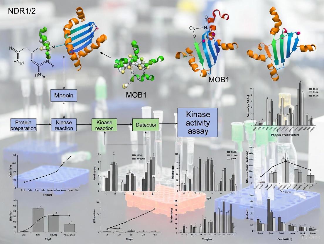
Experimental Workflow for In Vitro Kinase Assay
A standardized protocol is essential for generating reproducible and reliable data on NDR kinase activity. The workflow below outlines the key steps from sample preparation to data analysis, ensuring consistent evaluation of kinase function and MOB1-mediated activation.
Diagram Title: In Vitro Kinase Assay Workflow
Research Reagent Solutions
The following table catalogues essential reagents and materials required for conducting in vitro kinase assays to study NDR1/2 activation, based on established protocols [2] [30].
| Reagent/Material | Function/Description | Example/Catalog |
|---|---|---|
| Kinase Buffer (10X) | Provides optimal pH and ionic conditions for kinase activity [30]. | 500 mM Tris-HCl (pH 7.4), 10 mM DTT, 250 mM β-Glycerophosphate, 50 mM MgCl₂ [30]. |
| ATP Mix | Phosphoryl group donor for the kinase reaction; radiolabeled ATP allows reaction detection. | 100 μM ATP + 50 μCi [γ-³²P]ATP or [γ-³³P]ATP [30]. |
| MOB1 Protein | Co-activator that binds NDR1/2, dramatically stimulating kinase activity [2] [28]. | Recombinant human MOB1A or MOB1B. |
| Substrate | Molecule phosphorylated by NDR1/2; can be a generic substrate (e.g., myelin basic protein) or a specific physiological target. | To be determined by researcher. |
| LDS Sample Buffer | Terminates the kinase reaction and denatures proteins for SDS-PAGE analysis [30]. | Commercially available (e.g., Thermo Fisher). |
| Antibodies | For immunoprecipitation of epitope-tagged kinases and detection of proteins. | Anti-FLAG M2 [2], Anti-HA (12CA5, Y-11, 3F10) [2]. |
Detailed In Vitro Kinase Assay Protocol
Sample Preparation (Immunoprecipitation)
- Cell Culture and Transfection: Culture appropriate cell lines (e.g., COS-7, HEK 293, HeLa) and transfect with plasmids encoding epitope-tagged NDR1/2 (e.g., HA-NDR1, myc-NDR2) and MOB1 using a suitable transfection reagent (e.g., Fugene 6, Lipofectamine 2000) [2].
- Cell Lysis: Harvest cells and lyse using a non-denaturing lysis buffer (e.g., RIPA buffer) supplemented with protease and phosphatase inhibitors.
- Immunoprecipitation: Incubate the clarified cell lysate with an antibody specific to the epitope tag (e.g., anti-HA) for several hours at 4°C. Subsequently, add Protein A/G beads and incubate with gentle agitation to capture the immune complexes [2].
- Washing: Pellet the beads and wash them thoroughly 3-4 times with lysis buffer, followed by a final wash with 1X kinase assay buffer to remove detergents and prepare the sample for the kinase reaction.
Kinase Reaction Assembly
The following table provides a detailed setup for the kinase reaction, ensuring consistent and quantitative results. The final reaction volume is 25 µL [30].
| Component | Volume | Final Concentration/Amount |
|---|---|---|
| 10X Kinase Buffer | 2.5 µL | 1X |
| ATP Mix (100 µM ATP + [γ-³²P]ATP) | 2.5 µL | 10 µM ATP + ~5 µCi |
| Immunoprecipitated Kinase (on beads) | 10 µL | - |
| MOB1 Protein (optional, for activation) | Variable | Researcher determined |
| Substrate | Variable | Researcher determined |
| Nuclease-free Water | To 25 µL | - |
Reaction Incubation and Termination
- Incubation: Gently mix the reaction components and incubate the tube at 30°C for 30 minutes in a thermomixer or water bath [30].
- Termination: Stop the reaction by adding an appropriate volume of LDS Sample Buffer [30]. Heat the samples at 70-95°C for 5-10 minutes to fully denature the proteins.
Analysis by SDS-PAGE and Autoradiography
- Gel Electrophoresis: Load the denatured samples onto an SDS-polyacrylamide gel (e.g., 8% or 12%) and run to separate the proteins based on molecular weight [2] [30].
- Coomassie Staining: Stain the gel with Coomassie Brilliant Blue R-250 to visualize total protein, confirming equal loading and successful immunoprecipitation [30].
- Drying and Autoradiography: Dry the gel and expose it to a phosphorimager screen. Detect the incorporated radioactive signal using a scanner (e.g., FLA 7000 scanner) to identify phosphorylated proteins [30].
Critical Buffer Compositions and Experimental Parameters
Optimal buffer conditions are paramount for maintaining kinase stability and activity. The tables below summarize the key quantitative parameters for the assay.
Table 1: 10X Kinase Buffer Composition
| Component | Final Concentration (in 1X) | Function |
|---|---|---|
| Tris-HCl (pH 7.4) | 50 mM | Maintains physiological pH. |
| DTT | 1 mM | Reducing agent, maintains kinase cysteine residues. |
| β-Glycerophosphate | 25 mM | Phosphatase inhibitor. |
| MgCl₂ | 5 mM | Essential divalent cation for kinase activity. |
Table 2: Key Experimental Parameters
| Parameter | Optimal Condition | Rationale |
|---|---|---|
| Reaction Temperature | 30°C | Standard for enzymatic activity, prevents denaturation [30]. |
| Reaction Time | 30 minutes | Ensures reaction is within linear range [30]. |
| ATP Concentration | 10 µM (with tracer) | Near physiological levels, sufficient for phosphorylation. |
| Mg²⁺ Concentration | 5 mM | Required co-factor for phosphotransfer. |
Within the framework of investigations into NDR1/2 kinase activity and its regulation by MOB proteins, the selection of an appropriate detection method is paramount for generating reliable and quantitative data. The NDR kinase subfamily, comprising serine-threonine AGC kinases, are core components of the Hippo signaling pathway and require phosphorylation for full activation, often facilitated by their co-activators, the MOB proteins [2] [13]. This application note provides a detailed comparison of contemporary, non-radioactive methods—specifically FRET and HTRF—alongside traditional radioactive assays, for measuring kinase activity through phosphorylation and ATP depletion. It includes standardized protocols designed for research focused on the NDR-MOB signaling axis, crucial for processes like cell polarity, motility, and carcinogenesis [21] [6].
Technology Comparison and Selection
Selecting the optimal assay depends on the research question, required sensitivity, throughput, and available instrumentation. The table below summarizes the core characteristics of each major technology.
Table 1: Comparison of Key Kinase Activity Detection Methods
| Method | Detection Principle | Throughput | Sensitivity | Key Advantages | Key Limitations |
|---|---|---|---|---|---|
| HTRF | Time-Resolved FRET using lanthanide cryptates [31] | High | High (e.g., 2,000 cells for STAT5 detection) [32] | Homogeneous, no-wash format; high signal-to-noise; minimized short-lived background fluorescence [32] [31] | Requires specific antibody pairs; initial reagent cost can be high |
| FRET-Based Biosensors | Conformational change in a single polypeptide upon phosphorylation alters FRET efficiency [33] | Medium | High (real-time monitoring in live cells) | Enables real-time, spatiotemporal monitoring in live cells; high specificity for target kinases (e.g., EGFR) [33] | Requires genetic engineering; probe diffusion can cause mislocalization of signal [33] |
| ATP Depletion/ADP Accumulation | Enzyme-coupled detection of ADP generation [34] | High | Moderate (Dynamic range: 1.5-120 μM ADP) [34] | Universal assay for any kinase; flexible substrate choice (peptides or proteins); gain-of-signal format [34] | Coupled enzyme system can be susceptible to interference |
For research specifically on NDR1/2 and MOB1, where understanding spatiotemporal activation dynamics is key, FRET-based biosensors offer unique insights. Furthermore, the development of a high-throughput TR-FRET assay for the FAK-paxillin interaction demonstrates the applicability of this technology for studying challenging protein-protein interactions, similar to those in the NDR-MOB complex [35].
Application in NDR1/2 and MOB1 Research
Biological Context and Significance
The NDR1/2 kinases are activated through phosphorylation on critical serine and threonine residues (Ser281/282 and Thr444/442 in humans) and through interaction with MOB proteins [2]. Activated NDR kinases regulate essential cellular processes, including cell cycle progression, migration, and transcription [13]. Disruption of NDR kinase function, such as knockdown of NDR1/2, significantly impairs cell polarization and motility in wound healing assays by dysregulating Cdc42 GTPase and Pard3 signaling [21]. The interaction between MOB1 and NDR/LATS kinases is a critical regulatory node. Structural studies reveal that MOB1 exists in an autoinhibited state, where its N-terminal Switch helix blocks the LATS1/NDR-binding surface. Phosphorylation of MOB1 on Thr12 and Thr35 by upstream kinases like MST1/2 relieves this autoinhibition, enabling high-affinity binding to and full activation of NDR kinases [5]. Measuring this phosphorylation event is therefore crucial for assessing pathway activity.
Recommended Experimental Workflow
The following diagram illustrates a logical workflow for selecting and applying these detection methods in an NDR/MOB research context.
Detailed Experimental Protocols
Protocol 1: HTRF-Based Phospho-NDR/MOB Detection
This protocol is designed for the quantitative, cell-based detection of phosphorylation events within the NDR-MOB signaling axis, such as the phosphorylation of MOB1, using an optimized mix-and-read, no-wash HTRF format [32].
Table 2: Key Reagents for HTRF Assay
| Reagent | Function | Example/Note |
|---|---|---|
| HTRF Phospho-Specific Antibody Pair | Donor (Europium Cryptate) and Acceptor (d2) labeled antibodies for FRET detection. | One antibody binds phosphorylated motif; the other binds protein regardless of phosphorylation state [32]. |
| Cell Lysis Buffer | Lyse cells to release and solubilize target proteins. | Must be compatible with HTRF; often a 4X supplemented lysis buffer is used [32]. |
| White, Low-Volume Microplates | Plate format for optimal signal detection in HTRF readers. | 384-well low volume plates are standard for assay miniaturization [32]. |
| Recombinant MOB1 / NDR Protein | For constructing a standard curve or positive control. | Phosphomimetic (e.g., T12D/T35D) MOB1 can serve as a positive control [5]. |
Procedure:
- Cell Culture and Stimulation: Plate cells (e.g., HEK293) in a 96-well or 384-well culture plate. After appropriate treatments (e.g., okadaic acid to inhibit PP2A and activate NDR [2]), stimulate cells as required.
- Cell Lysis: Remove cell culture medium. Add a pre-optimized volume of 4X supplemented lysis buffer (e.g., 10 µL) to cells and incubate for 30 minutes at room temperature to ensure complete lysis [32].
- Lysate Transfer: For a two-plate protocol, transfer 16 µL of cell lysate to a white, low-volume 384-well detection plate [32].
- HTRF Detection: Add HTRF detection reagents containing the anti-phospho-protein antibody pair directly to the lysates. Incubate the plate for 1-4 hours at room temperature, protected from light.
- Signal Measurement: Read the plate on an HTRF-compatible microplate reader. Calculate the HTRF ratio (Acceptor emission at 665 nm / Donor emission at 620 nm) multiplied by 10,000 to obtain the final result [31].
Protocol 2: FRET-Based Biosensor for Live-Cell Kinase Activity
This protocol utilizes the Picchu biosensor to monitor kinase activity in real-time within live cells, which can be adapted for studying NDR kinase dynamics [33].
Procedure:
- Biosensor Expression: Transfect cells with a plasmid encoding the FRET biosensor (e.g., pPBbsr2-TRE-PicchuEV-X for membrane-targeted expression). A Tet-off inducible system can be used to control expression levels [33].
- Live-Cell Imaging: Seed transfected cells onto glass-bottom imaging dishes. Allow cells to adhere and express the biosensor for the required time (e.g., 24-48 hours).
- Stimulate and Image: Place the dish on a confocal or epifluorescence microscope with environmental control (37°C, 5% CO₂). Acquire baseline FRET images, then stimulate cells (e.g., with serum or a specific ligand) while continuing time-lapse imaging.
- FRET Signal Calculation: The FRET ratio is calculated as the emission intensity of the acceptor (e.g., mCherry) divided by the emission intensity of the donor (e.g., ECFP) upon donor excitation. A decrease in the FRET ratio typically indicates kinase activation and biosensor phosphorylation [33].
Protocol 3: ADP Hunter Assay for Biochemical NDR Kinase Activity
This protocol is a generic biochemical assay for measuring the ADP produced by NDR kinase activity, ideal for inhibitor screening [34].
Procedure:
- Kinase Reaction Setup: In a non-binding polypropylene 384-well plate, set up a kinase reaction mixture containing:
- NDR kinase (recombinant, immunoprecipitated)
- MOB1 co-activator protein
- ATP (at the Km concentration for the kinase)
- Suitable substrate (e.g., a peptide derived from Pard3 [21])
- Assay buffer Incubate to allow the kinase reaction to proceed.
- ADP Detection: Add ADP Hunter Reagent A and Reagent B to the kinase reaction. Reagent B contains enzymes that couple ADP production to the generation of a fluorescent resorufin product.
- Incubation: Incubate the plate for 60 minutes at room temperature.
- Signal Stabilization: Add the provided Stop Solution to stabilize the fluorescence signal.
- Fluorescence Measurement: Read the plate using a fluorescence microplate reader (Ex: 530 nm / Em: 590 nm). The signal is directly proportional to the amount of ADP generated and thus to the kinase activity [34].
The Scientist's Toolkit
Table 3: Essential Research Reagent Solutions for NDR/MOB Kinase Research
| Item | Function / Application | Specific Example / Note |
|---|---|---|
| Phospho-Specific Antibodies | Detect activation-specific phosphorylation of NDR/MOB. | Antibodies against NDR1 pThr444 / pSer281 [2] or MOB1 pThr12/pThr35. |
| Recombinant NDR & MOB Proteins | For in vitro kinase assays, interaction studies, and standard curves. | N-terminal truncated MOB1 is constitutively active and readily binds NDR kinases [5]. |
| HTRF Kinase Kits | Ready-to-use kits for quantifying phosphorylation. | HTRF Phospho-STAT5 (Tyr694) Detection Kit as a model for phospho-tyrosine detection [32]. |
| FRET Biosensor Plasmids | Enable real-time visualization of kinase activity in live cells. | Picchu-X plasmid for EGFR; design required for NDR1/2 specificity [33]. |
| ADP Detection Kits | Universal, biochemical kinase activity and inhibitor screening. | ADP Hunter Kit from DiscoverX; applicable to any NDR substrate [34]. |
| Membrane-Targeting Constructs | Study the role of subcellular localization in kinase activation. | Lck myristoylation/palmitylation motif (mp) targets proteins to the plasma membrane [2]. |
The strategic application of FRET, HTRF, and ATP-depletion assays provides a powerful, multi-faceted toolkit for dissecting the complex regulation of NDR1/2 kinases by MOB1. While HTRF offers robust, high-throughput quantification of specific phosphorylation events, FRET-based biosensors unlock the dynamic, spatiotemporal analysis of kinase activity in living cells. The universal ADP detection method serves as an excellent platform for initial drug discovery screens. Integrating data from these complementary techniques will significantly advance our understanding of the NDR-MOB signaling pathway in both physiological and pathological contexts.
The Nuclear Dbf2-related kinases 1 and 2 (NDR1/2) are serine/threonine kinases belonging to the AGC family of protein kinases and are core components of the Hippo signaling pathway [27]. These evolutionarily conserved kinases play fundamental roles in controlling cell proliferation, apoptosis, cell polarity, and morphogenesis [27] [21]. The regulatory significance of NDR1/2 extends to pathological contexts, particularly in cancer, where their expression is frequently dysregulated [27] [6]. NDR1 functions as a tumor suppressor in several cancers, including prostate cancer, where it inhibits metastasis by suppressing epithelial-mesenchymal transition (EMT) [36]. Consequently, the pharmacological activation of NDR1 is being investigated as a potential therapeutic strategy [36]. Monitoring the activation status of NDR1/2 in live cells is therefore crucial for both basic research into cell biology and applied drug discovery efforts.
The activity of NDR1/2 kinases is tightly regulated through phosphorylation and protein-protein interactions. A critical upstream regulator is the adaptor protein MOB1, which directly binds to and activates NDR1/2 [5]. MOB1 itself is activated by phosphorylation at Thr12 and Thr35 by MST1/2 kinases, enabling its interaction with the N-terminal regulatory (NTR) domain of NDR1/2 [5]. This interaction is a pivotal point in the Hippo pathway and serves as a key indicator of NDR1/2 activation status in live cells.
Key Principles of NDR1/2 Activation
The NDR1/2-MOB1 Signaling Axis
The activation mechanism of NDR1/2 kinases centers on their interaction with MOB1. Structural studies reveal that MOB1 exists in an autoinhibited state where its N-terminal Switch helix blocks the LATS1/NDR1-binding surface [5]. Phosphorylation of MOB1 at Thr12 and Thr35 by upstream MST1/2 kinases structurally relieves this autoinhibition through a "pull-the-string" mechanism, enabling high-affinity binding to the NTR domain of NDR1/2 [5]. This binding event triggers a conformational change in NDR1/2, facilitating their phosphorylation and full activation [5]. The activated NDR1/2 kinases then phosphorylate downstream substrates such as Pard3 at Serine144, which regulates cell polarization and directional motility [21]. This entire cascade can be monitored in live cells using the techniques described in this application note.
Visualization of the NDR1/2 Activation Pathway
The following diagram illustrates the core signaling pathway leading to NDR1/2 kinase activation, highlighting the critical role of MOB1.
Quantitative Techniques for Monitoring NDR1/2 Activation
Comparison of Key Methodologies
The following table summarizes the primary quantitative approaches for assessing NDR1/2 activation status in cellular contexts, each offering distinct advantages and limitations.
Table 1: Quantitative Techniques for Monitoring NDR1/2 Activation
| Technique | Key Target | Readout | Throughput | Key Advantage |
|---|---|---|---|---|
| Kinase Activity Assay | NDR1 enzymatic activity | Luminescence (ATP consumption) | Medium | Direct measurement of kinase function [36] |
| Phospho-Specific Flow Cytometry | Phospho-NDR1/2 (Thr444/Thr442) | Fluorescence intensity per cell | High | Single-cell resolution in complex populations [37] |
| FRET/BRET Biosensors | NDR1/2 conformation or interaction with MOB1 | Fluorescence/Bioluminescence Ratio | Medium-High | Real-time kinetics in live cells |
| Immunofluorescence Microscopy | Cellular localization & phosphorylation | Spatial fluorescence pattern | Low | Subcellular spatial information [21] |
| Co-Immunoprecipitation with MOB1 | NDR1/2-MOB1 protein complex | Protein band intensity (Western) | Low | Confirms functional protein-protein interaction [5] |
Key Experimental Parameters for NDR1/2 Kinase Activity
For biochemical kinase assays, specific reaction conditions must be optimized to accurately quantify NDR1/2 activity. The following table outlines established parameters from recent studies.
Table 2: Key Experimental Parameters for NDR1 Kinase Activity Assays
| Parameter | Condition/Value | Experimental Notes |
|---|---|---|
| Recombinant Protein | GST-fused NDR1 purified from E. coli BL21 | Use pGEX-GST-NDR1 plasmid; induce with 1mM IPTG at 16°C overnight [36] |
| Reaction Buffer | Kinase assay buffer with ATP | Commercial Kinase-Lumi luminescent kit or equivalent [36] |
| Substrate Peptide | KKRNRRLSVA | Optimal for NDR1 phosphorylation [36] |
| Agonist Example | aNDR1 (small molecule) | Promotes NDR1 expression, enzymatic activity, and phosphorylation [36] |
| Cellular Assay Duration | 24-hour treatment | Effective duration for observing NDR1-mediated effects on cell proliferation and apoptosis [36] |
| Key Functional Readout | Pard3 phosphorylation at Serine144 | Critical downstream event indicating functional NDR1/2 activity [21] |
Detailed Experimental Protocols
Protocol 1: Luminescent Kinase Activity Assay for Recombinant NDR1
This protocol describes a quantitative method for measuring the enzymatic activity of purified NDR1 kinase in response to potential activators, such as the recently identified small-molecule agonist aNDR1 [36].
Workflow for NDR1 Kinase Activity Assay
Step-by-Step Procedure:
Protein Expression and Purification:
- Transform E. coli BL21 with the pGEX-GST-NDR1 plasmid [36].
- Induce mid-logarithmic phase cultures with 1 mM isopropyl-β-D-thiogalactopyranoside (IPTG) and incubate overnight at 16°C [36].
- Harvest bacteria by centrifugation and lyse using an ultrasonic cell crusher in lysis buffer (20 mM Tris-Cl, pH 8.0, 100 mM NaCl, 0.2 mM EDTA, 0.5% NP-40) supplemented with lysozyme and phenylmethylsulfonyl fluoride (PMSF) [36].
- Clarify the lysate by centrifugation and purify the GST-fused NDR1 proteins from the supernatant using glutathione-agarose resin [36].
- Verify purity and concentration via SDS-PAGE with Coomassie blue staining.
Kinase Reaction Setup:
- Prepare reaction mixtures containing purified NDR1 proteins, specific substrate peptide (KKRNRRLSVA), ATP, and reaction buffer according to the manufacturer's protocol for the Kinase-Lumi luminescent kinase assay kit [36].
- Include experimental groups with different concentrations of the compound being tested (e.g., aNDR1) and appropriate controls (no enzyme, no substrate, vehicle control).
Reaction Incubation and Measurement:
- Incubate reactions under optimized conditions (typically 30°C for 30-60 minutes).
- Measure luminescence using a plate-reading luminometer to quantify ATP consumption, which directly correlates with kinase activity.
Data Analysis:
- Calculate kinase activity relative to control groups.
- Express results as fold-change in activity compared to vehicle-treated samples.
- Determine EC₅₀ values for agonist compounds through dose-response curves.
Protocol 2: Monitoring Endogenous NDR1/2 Activation in Live Cells Using Phospho-Flow Cytometry
This protocol enables the quantification of NDR1/2 phosphorylation status in individual cells within a heterogeneous population, providing both quantitative and population distribution data.
Workflow for Phospho-Flow Cytometry of NDR1/2
Step-by-Step Procedure:
Cell Preparation and Stimulation:
- Culture and treat cells according to experimental design (e.g., with NDR1 agonists or pathway modulators).
- Harvest both adherent and suspension cells, using 0.5 mM EDTA for adherent cells to facilitate removal without trypsin, which can damage cell surface epitopes [37].
- Wash cells three times in isotonic PBS buffer supplemented with 0.5% BSA by centrifugation at 350-500 × g for 5 minutes to remove residual culture components [37].
- Aliquot up to 1 × 10⁶ cells per 100 μL into FACS tubes [37].
Cell Staining:
- Block Fc receptors with blocking IgG (1 μg IgG/10⁶ cells) for 15 minutes at room temperature to prevent non-specific antibody binding [37].
- Without washing, add fluorochrome-conjugated phospho-specific primary antibodies against NDR1/2 (e.g., targeting phospho-Thr444 for NDR1 or phospho-Thr442 for NDR2) at a previously titrated concentration (typically 5-10 μL/10⁶ cells) [37].
- Vortex gently and incubate cells for 30 minutes at room temperature in the dark to protect fluorochromes from light degradation [37].
Washing and Data Acquisition:
- Remove unbound antibody by washing cells in 2 mL Flow Cytometry Staining Buffer, centrifuging at 350-500 × g for 5 minutes, and decanting the supernatant [37].
- Repeat this wash step two times to ensure complete removal of unbound antibodies [37].
- Resuspend the final cell pellet in 200-400 μL of Flow Cytometry Staining Buffer for analysis [37].
- Acquire data using a flow cytometer, collecting sufficient events for statistical analysis (typically 10,000-50,000 events per sample).
Controls and Data Analysis:
- Include appropriate controls: unstained cells, isotype control antibodies, and cells with known phosphorylation status for instrument calibration and gating [37].
- Analyze data using flow cytometry software, gating on live, single cells and measuring median fluorescence intensity (MFI) of the phospho-specific channel.
- Compare MFI values between experimental conditions to assess changes in NDR1/2 phosphorylation status.
The Scientist's Toolkit: Essential Research Reagents
Table 3: Key Research Reagent Solutions for NDR1/2 Kinase Studies
| Reagent/Category | Specific Examples | Function & Application |
|---|---|---|
| Recombinant Proteins | GST-fused NDR1 purified from E. coli [36] | Substrate for in vitro kinase assays; structural studies |
| Cell Lines | DU145, PC3 (prostate cancer) [36] | Models for studying NDR1 function in cancer contexts |
| Chemical Agonists | aNDR1 (small molecule) [36] | Pharmacological activation of NDR1 for functional studies |
| Antibodies | Phospho-specific NDR1/2 (Thr444/Thr442) | Detection of activated NDR1/2 in immunoassays |
| Kinase Assay Kits | Kinase-Lumi luminescent kinase assay kit [36] | Quantitative measurement of NDR1 enzymatic activity |
| Flow Cytometry Buffers | Flow Cytometry Staining Buffer [37] | Maintain cell viability and reduce non-specific binding during staining |
| Key Substrates | Pard3 peptide, KKRNRRLSVA peptide [21] [36] | Direct phosphorylation targets for measuring NDR1/2 activity |
Troubleshooting and Technical Considerations
When implementing these protocols, several technical considerations are essential for success. For kinase activity assays, maintain strict temperature control during protein purification and activity measurements, as NDR1 stability can be compromised at higher temperatures [36]. For cellular assays, note that NDR1 knockdown significantly alters cell size, shape, and actin cytoskeleton organization, which may indirectly affect experimental outcomes [21]. When studying the NDR1/2-MOB1 interaction, remember that MOB1 requires phosphorylation at Thr12 and Thr35 by MST1/2 kinases to achieve high-affinity binding to NDR1/2 [5]. Always include appropriate controls for phosphorylation-dependent interactions. For flow cytometry applications requiring tissue-derived cells, follow established protocols for generating single-cell suspensions that preserve cell viability and surface epitopes, using collagenase IV and DNase I digestion followed by density gradient centrifugation [38].
The precise identification of direct kinase substrates is a fundamental challenge in molecular cell biology. Conventional methods often struggle with specificity and the transient nature of kinase-substrate interactions. Within the context of NDR1/2 kinase research, the chemical genetics approach has proven to be a powerful technique for uncovering novel physiological substrates, such as AAK1 and Rabin8, which are instrumental in neuronal development [39] [40].
This application note details the protocol and strategic implementation of the chemical genetics method, often termed the "bump-and-hole" or "analog-sensitive" strategy, as applied to the identification of NDR1/2 kinase substrates. The methodology hinges on the generation of a unique kinase-engineered ATP-binding pocket that can utilize bulky ATP analogs not recognized by other wild-type cellular kinases [40]. When framed within a broader thesis on NDR1/2 kinase activity, this technique provides an unambiguous method to connect kinase activity to specific downstream phosphorylation events and complex phenotypic outcomes, such as dendrite arborization and spine development [39] [40].
Key Principles and Workflow
The core principle of the chemical genetics strategy involves two complementary components: an engineered kinase and a modified ATP analog. A key mutation in the kinase's ATP-binding pocket (the "bump") creates a space that can accommodate ATP analogs with a bulky substituent (the "hole"). This orthogonal pair enables the specific labeling and covalent capture of direct kinase substrates [40] [41].
The experimental workflow can be broken down into several key stages, as illustrated below.
Experimental Workflow Diagram
Detailed Experimental Protocol
Generation of the Analog-Sensitive (AS) NDR1 Kinase
The first critical step is engineering the ATP-binding pocket of NDR1 to create a unique "hole" without compromising its intrinsic kinase activity and substrate specificity.
- Mutagenesis Target: A conserved catalytic lysine residue (e.g., K118 in NDR1) is mutated to alanine or glycine (K118A) to create the analog-sensitive (AS) kinase [40]. This mutation is located in the ATP-binding pocket and is critical for the kinase's ability to utilize the bulky ATP analogs.
- Molecular Biology: Site-directed mutagenesis is performed on a human NDR1 cDNA clone. The mutant kinase is typically cloned into an appropriate mammalian expression vector (e.g., pCMV) with an N-terminal tag (e.g., FLAG or HA) for subsequent immunopurification.
- Activity Validation: The mutant kinase must be rigorously tested to ensure the mutation does not drastically alter its function. This involves:
- Transient Transfection: into mammalian cell lines (e.g., HEK293T).
- Immunopurification: of the tagged kinase.
- In Vitro Kinase Assay: using a known substrate peptide and [γ-32P]ATP or a luminescence-based system to confirm retained catalytic activity relative to wild-type NDR1 [40].
Substrate Identification and Validation
This section outlines the core procedure for labeling, capturing, and identifying direct substrates from a complex biological mixture, such as brain lysate.
Kinase Reaction Setup:
- Prepare reaction buffer (e.g., 25 mM HEPES pH 7.4, 10 mM MgCl2, 1 mM DTT).
- Combine immunopurified AS-NDR1 kinase with mouse brain lysate as a source of potential substrates.
- Initiate the reaction by adding the ATP analog N6-benzyl-ATPγS (final concentration 50-100 μM).
- Incubate at 30°C for 30-60 minutes [40] [42].
Covalent Capture of Thiophosphorylated Substrates:
- Terminate the kinase reaction.
- Add the alkylating agent p-nitrobenzyl mesylate (PNBM) (final concentration 2.5 mM) to the mixture.
- Incubate for 2 hours at room temperature. PNBM specifically substitutes the thiophosphate group with a p-nitrobenzyl group, creating a stable thioether bond and a unique epitope [42].
Purification and Identification:
- Immunoprecipitate the tagged AS-NDR1 kinase and its associated proteins using an anti-FLAG (or equivalent) affinity gel.
- Wash beads stringently to remove non-specifically bound proteins.
- Elute bound proteins and resolve them by SDS-PAGE.
- Analyze the gel by liquid chromatography-tandem mass spectrometry (LC-MS/MS) to identify co-purified proteins. Proteins consistently and specifically identified in the AS-kinase samples, but not in controls, are considered high-confidence candidate substrates [40] [42].
Functional Validation of Identified Substrates
- In Vitro Kinase Assay: Confirm direct phosphorylation by incubating purified, recombinant candidate substrates (e.g., AAK1, Rabin8) with wild-type NDR1/2 kinase in the presence of [γ-32P]ATP. Phosphorylation is detected by autoradiography after SDS-PAGE [39] [43].
- Cell-Based Assays: To establish physiological relevance, perturb the candidate substrate (via siRNA knockdown or overexpression of a phospho-deficient mutant) in an appropriate cellular model, such as cultured rat hippocampal neurons. Analyze the resulting phenotype (e.g., changes in dendrite arborization or spine morphology) and compare it to the phenotype observed upon perturbation of NDR1/2 itself [39] [40].
Key Findings and Data Analysis
Application of this chemical genetics strategy to NDR1/2 kinase research in the mouse brain successfully identified several direct substrates, with two—AAK1 and Rabin8—being functionally characterized.
Identified NDR1/2 Substrates and Their Functions
| Substrate | Full Name & Function | Phenotypic Role of Phosphorylation | Validation Method |
|---|---|---|---|
| AAK1 | AP-2 Associated Kinase 1: Regulates clathrin-mediated endocytosis [39] [40]. | Contributes to the regulation of dendrite arborization and proximal branching [39] [40]. | siRNA knockdown in neurons [39]. |
| Rabin8 | Rab8 Guanine Nucleotide Exchange Factor (GEF): Activates Rab8 GTPase for vesicle trafficking [39] [40]. | Regulates dendritic spine development and maturation of excitatory synapses [39] [40]. | siRNA knockdown and spine analysis [39]. |
NDR1/2 Kinase Activity and Phenotypic Consequences
Manipulation of NDR1/2 kinase activity in mammalian pyramidal neurons produces distinct and opposing effects on neuronal morphology, as summarized below.
| NDR1/2 Construct Expressed | Effect on Kinase Activity | Dendrite Length & Branching | Dendritic Spine Development |
|---|---|---|---|
| Kinase-Dead (K118A) or siRNA | Loss of Function | Increase [39] [40] | More immature spines; reduced mEPSC frequency [39] [40] |
| Constitutively Active | Gain of Function | Decrease [39] [40] | Promotes mushroom spine formation [39] [40] |
The Scientist's Toolkit: Research Reagent Solutions
| Essential Material | Function in the Protocol | Specific Example / Catalog Number Considerations |
|---|---|---|
| Analog-Sensitive NDR1 Kinase | Engineered kinase for orthogonal substrate labeling. | Plasmid: NDR1 (K118A) mutant [40]. |
| N6-benzyl-ATPγS | ATP analog donor for thiophosphorylation by AS-kinase. | Key reagent for covalent capture; available from specialty chemical suppliers [42]. |
| p-Nitrobenzyl Mesylate (PNBM) | Alkylating agent for covalent capture of thiophosphorylated substrates. | Converts thiophosphate to a purification tag [42]. |
| Anti-FLAG M2 Affinity Gel | For immunopurification of the kinase-substrate complex. | Sigma-Aldrich A2220 or equivalent. |
| Protein A/G Magnetic Beads | Alternative for immunoprecipitation with other tags. | Thermo Fisher Scientific 10002D / 10004D. |
Integrated Signaling Pathway
The discovery of AAK1 and Rabin8 as NDR1/2 substrates reveals how a single kinase orchestrates distinct cellular processes through multiple downstream effectors. The following diagram integrates these findings into a coherent signaling pathway.
Solving Common Problems: Optimizing Your NDR1/2-MOB1 Assay for Reproducibility and Sensitivity
The activation of the NDR1/2 kinases is a critical regulatory node in the Hippo signaling pathway, governing essential cellular processes such as cell proliferation, apoptosis, and mitochondrial health [13] [44]. The Mps One Binder (MOB1) proteins function as essential co-activators for these kinases, dramatically enhancing their catalytic activity [2] [15]. However, researchers frequently encounter challenges with low signal intensity and high background noise in NDR kinase activity assays, often stemming from suboptimal MOB1 preparation and utilization. This application note provides a detailed methodological framework to overcome these challenges by optimizing MOB1 concentration and phosphorylation status, thereby ensuring robust and reproducible measurement of NDR1/2 kinase activity.
Scientific Background and Rationale
The MOB1-NDR Signaling Axis
NDR kinases (NDR1 and NDR2) are serine/threonine kinases belonging to the AGC family. Their full activation is a multi-step process that requires phosphorylation at two key sites: a serine residue in the activation segment (Ser281 in NDR1, Ser282 in NDR2) and a threonine residue in the hydrophobic motif (Thr444 in NDR1, Thr442 in NDR2) [2] [27]. While auto-phosphorylation can occur at the activation segment, phosphorylation of the hydrophobic motif is often mediated by upstream kinases like MST1/2 [15].
The binding of MOB1 to the N-terminal regulatory domain of NDR (MBD) is a critical event that induces conformational changes, potentiating kinase activity. Structural studies reveal that MOB1 binding and the auto-inhibitory function of NDR's atypically long activation segment are mechanistically distinct, yet both are essential for proper regulation [15] [9]. The formation of the NDR-MOB1 complex at the plasma membrane leads to rapid and robust kinase activation, highlighting the spatial dimension of this regulation [2].
Signaling Pathway Logic
The following diagram illustrates the core regulatory steps and protein interactions leading to NDR kinase activation, emphasizing the critical role of MOB1.
Diagram 1: NDR Kinase Activation Pathway. This diagram outlines the key steps in NDR1/2 activation. Upstream kinases phosphorylate the NDR Hydrophobic Motif (HM), leading to a partially active state. Concurrently, MOB1 is activated via phosphorylation. The binding of active MOB1 to the N-terminal MBD domain of NDR, coupled with phosphorylation within its activation segment, leads to full kinase activation and the formation of a productive complex capable of phosphorylating downstream substrates.
Experimental Protocols
Protocol 1: Production of Active MOB1 Protein
Objective: To express and purify recombinant human MOB1 protein, ensuring it is in a phosphorylated state competent for activating NDR kinases.
Materials:
- Expression Vector: pcDNA3.1 containing cDNA for human MOB1A or MOB1B [2].
- Cell Line: HEK 293T cells (or similar, e.g., COS-7, HeLa) [2].
- Transfection Reagent: Lipofectamine 2000 or Fugene 6 [2].
- Lysis Buffer: 50 mM Tris-HCl (pH 7.5), 150 mM NaCl, 1% Triton X-100, supplemented with protease and phosphatase inhibitors.
- Purification: Anti-HA or anti-myc affinity resin, depending on the epitope tag used [2].
Method:
- Cell Culture and Transfection: Culture HEK 293T cells in Dulbecco's Modified Eagle Medium (DMEM) supplemented with 10% fetal calf serum. At 60-70% confluence, transfert cells with the MOB1 expression plasmid using a suitable transfection reagent.
- Stimulation for Phosphorylation: 24-48 hours post-transfection, treat cells with 1 μM Okadaic Acid (OA) for 60 minutes. OA is a PP2A phosphatase inhibitor that helps maintain and enhance the phosphorylation status of MOB1 and other pathway components [2].
- Cell Lysis and Harvesting: Wash cells with ice-cold PBS and lyse using the lysis buffer. Clarify the lysate by centrifugation at 16,000 × g for 15 minutes at 4°C.
- Affinity Purification: Incubate the clarified lysate with the appropriate affinity resin for 2-4 hours at 4°C with gentle agitation.
- Washing and Elution: Wash the resin extensively with lysis buffer containing 500 mM NaCl to remove non-specifically bound proteins. Elute the purified, phosphorylated MOB1 protein using a competitive peptide (e.g., HA peptide) or low-pH elution buffer.
- Quality Control: Analyze the eluate by SDS-PAGE and immunoblotting. Confirm phosphorylation using phospho-specific antibodies if available. Quantify protein concentration and aliquot for storage at -80°C.
Protocol 2: In Vitro NDR Kinase Activation Assay
Objective: To measure NDR1/2 kinase activity in vitro using a quantitative phospho-transfer assay, and to determine the optimal concentration of MOB1 required for maximal activation.
Materials:
- NDR Kinase: Recombinant, inactive NDR1 or NDR2 (can be purified from bacteria to ensure a non-phosphorylated baseline) [15].
- Active MOB1: Purified as described in Protocol 1.
- Upstream Kinase: Recombinant, active MST1/2 kinase for HM phosphorylation [15].
- Reaction Buffer: 25 mM HEPES (pH 7.4), 10 mM MgCl₂, 1 mM DTT.
- ATP Solution: 100 μM ATP, including [γ-³²P]-ATP for radiometric detection or cold ATP for other detection methods.
- Kinase-Specific Substrate: A known peptide substrate (e.g., a fragment of YAP) or a generic substrate like myelin basic protein (MBP).
Method:
- Priming of NDR Kinase: In a 1.5 mL microcentrifuge tube, incubate 100 nM NDR kinase with 50 nM active MST1/2 kinase in reaction buffer containing 100 μM ATP for 30 minutes at 30°C. This step ensures phosphorylation of the hydrophobic motif (Thr444/Thr442) [15].
- MOB1 Titration: Set up a series of reactions with the primed NDR kinase and titrate MOB1 protein across a concentration range (e.g., 0 nM, 25 nM, 50 nM, 100 nM, 200 nM, 500 nM). Keep the final reaction volume consistent.
- Kinase Reaction: Initiate the kinase reaction by adding the ATP solution and the chosen substrate. Allow the reaction to proceed for 30 minutes at 30°C.
- Reaction Termination: Stop the reaction by adding SDS-PAGE loading buffer and heating at 95°C for 5 minutes.
- Detection and Analysis: Resolve proteins by SDS-PAGE. Transfer to a membrane for autoradiography (if using radioactive ATP) or immunoblot with phospho-specific antibodies against the substrate or the NDR activation segment (e.g., anti-Ser281-P for NDR1) [2]. Quantify the band intensity to determine kinase activity.
Optimization Data and Guidelines
Quantitative Optimization of MOB1
The following table summarizes key quantitative data and recommendations for optimizing MOB1 in NDR kinase assays.
Table 1: MOB1 Optimization Parameters for NDR Kinase Assays
| Parameter | Optimal Range / Condition | Experimental Impact / Rationale | Supporting Reference |
|---|---|---|---|
| MOB1:NDR Molar Ratio | 1:1 to 5:1 (Start with 2:1) | Maximizes kinase activation without significant off-target effects or aggregation. Supra-optimal ratios may not yield further benefit. | [2] [15] |
| Phosphorylation Status | Phosphorylated MOB1 | Active, phosphorylated MOB1 is critical for robust NDR activation. Use Okadaic Acid (1 μM) treatment during production. | [2] |
| Cellular Localization | Membrane-Targeted | Membrane targeting of MOB1 results in constitutively active NDR, enhancing assay sensitivity. Consider using myristoylation motifs (e.g., mp-HA). | [2] |
| Activation Timeline | Rapid (within minutes) | Phosphorylation and activation of NDR at the membrane occur within a few minutes of MOB1 association. Design time-course experiments accordingly. | [2] |
| Key Phosphorylation Sites | NDR1: p-Ser281, p-Thr444NDR2: p-Ser282, p-Thr442 | Use phospho-specific antibodies against these sites to quantitatively monitor NDR activation status in your assays. | [2] [15] |
The Scientist's Toolkit: Essential Reagents
This table lists critical reagents and their functions for successfully conducting MOB1-NDR kinase research.
Table 2: Research Reagent Solutions for MOB1-NDR Kinase Studies
| Reagent / Tool | Function / Specificity | Example & Catalog # (if provided) |
|---|---|---|
| Phospho-Specific Antibodies | Detect active, phosphorylated NDR1/2. Critical for readout. | Anti-NDR1 p-Thr444, Anti-NDR1 p-Ser281 [2] |
| MOB1 Expression Plasmids | Source of recombinant MOB1. Epitope-tagged for purification. | pcDNA3-hMOB1A (HA- or myc-tagged) [2] |
| Kinase Inhibitors/Activators | Manipulate pathway activity. Okadaic acid blocks phosphatases, enhancing phosphorylation. | Okadaic Acid (OA) [2] |
| Membrane-Targeting Constructs | Forces localization to sites of activation, boosting signal. | Lck myristoylation/palmitylation motif (mp-HA/mp-myc) [2] |
| Upstream Kinase | Essential for priming NDR kinases via HM phosphorylation. | Recombinant active MST1/2 kinase [15] |
Optimizing the concentration and phosphorylation status of MOB1 is not merely a technical step but a fundamental requirement for obtaining high-fidelity data on NDR1/2 kinase activity. By adhering to the protocols and guidelines outlined here—specifically, employing a 1:1 to 5:1 molar ratio of phosphorylated, membrane-localized MOB1 to NDR kinase—researchers can effectively overcome the common challenge of low signal. This approach ensures the reliable activation of NDR kinases, facilitating more accurate investigations into the Hippo pathway and its profound implications in cell biology, cancer research, and therapeutic development.
The Nuclear Dbf2-related kinases, NDR1 and NDR2, represent crucial signaling components within the conserved Hippo pathway, regulating essential cellular processes including cell cycle progression, apoptosis, and inflammatory responses [13] [6]. Their activation is tightly controlled through phosphorylation and interaction with MOB proteins, which dramatically stimulate NDR catalytic activity [2] [28]. When developing kinase activity assays for NDR1/2 with MOB1, ensuring methodological specificity is paramount to generate reliable, reproducible data free from confounding off-target kinase activities. This application note provides detailed strategies and protocols to minimize background and off-target effects in NDR kinase research, framed within the context of a broader thesis on NDR1/2 kinase activity regulation.
The high sequence similarity among the approximately 500 human kinases presents a significant challenge for achieving assay specificity [45]. Furthermore, NDR1 and NDR2 themselves share approximately 87% sequence identity yet exhibit distinct subcellular localizations—with NDR1 found predominantly in the nucleus while NDR2 displays a punctate cytoplasmic distribution [28]. These differences suggest non-redundant functions and highlight the necessity for isoform-specific investigation techniques.
Molecular Basis of NDR-MOB Interaction and Activation
Structural Mechanisms of Activation
The activation mechanism of NDR kinases involves a sophisticated multi-step process requiring phosphorylation and MOB protein interaction:
Phosphorylation Requirements: Full activation of NDR1 requires phosphorylation at two critical residues: Ser281 (autophosphorylation site) and Thr444 (phosphorylated by an upstream kinase) [2]. Similarly, NDR2 requires phosphorylation at Ser282 and Thr442 for complete activation [2].
MOB Binding and Activation: Human MOB proteins (hMOB1A, hMOB1B, hMOB2) bind to the N-terminal regulatory region of NDR kinases, dramatically stimulating their catalytic activity [2] [28]. This interaction mimics the functional relationship between cyclins and cyclin-dependent kinases.
Subcellular Localization: The NDR-MOB interaction occurs primarily at the plasma membrane, where membrane-targeted MOB1 robustly promotes NDR activation [2]. Strikingly, induced membrane translocation of MOB1 leads to NDR phosphorylation and activation within minutes of association with membranous structures [2].
Structural Insights from Related Pathways
Research on the closely related Hippo kinase cascade reveals that Mob1 acts as a phosphorylation-regulated coupler of kinase activation through dynamic scaffolding and allosteric mechanisms [26]. Structural analyses demonstrate that phosphorylated Mob1 undergoes conformational activation and serves as a phosphorylation-regulated coupler between Mst and Lats kinases [26]. While this specific structural data comes from the Mst-Lats interaction, the conservation within the Hippo pathway suggests similar mechanisms may apply to NDR-MOB interactions.
Quantitative Parameters for NDR Kinase Specificity
Table 1: Key Phosphorylation Sites and Regulatory Elements in NDR Kinases
| Kinase | Activation Phosphorylation Sites | MOB Binding Region | Subcellular Localization | Key Regulatory Features |
|---|---|---|---|---|
| NDR1 | Ser281, Thr444 | N-terminus | Predominantly nuclear [28] | Activated by membrane recruitment [2] |
| NDR2 | Ser282, Thr442 | N-terminus | Punctate cytoplasmic [28] | Excluded from nucleus [28] |
| MOB1 | N/A | N-terminal region of NDR | Plasma membrane, cytoplasm [2] | Promotes NDR membrane translocation [2] |
Table 2: Experimental Conditions for Optimal NDR-MOB Specificity
| Parameter | Recommended Conditions | Rationale | References |
|---|---|---|---|
| Activation Method | Co-expression with MOB1 + okadaic acid (1μM, 60min) | Maximizes phosphorylation while inhibiting dephosphorylation | [2] |
| Cellular System | COS-7, HEK 293, HeLa cells | Validated model systems for NDR-MOB interaction studies | [2] |
| Critical Controls | Kinase-dead NDR mutants, MOB-binding deficient mutants | Confirms specificity of observed effects | [2] [28] |
| Specificity Validation | Phospho-specific antibodies (Ser281/282, Thr444/442) | Direct monitoring of activation-specific phosphorylation | [2] |
Experimental Protocols for Specific NDR Activity Assessment
Protocol: MOB-Dependent NDR Kinase Activation Assay
Purpose: To measure NDR1/2 kinase activity specifically enhanced by MOB1 interaction while minimizing background and off-target kinase contributions.
Reagents and Materials:
- Purified NDR1 or NDR2 kinase (active form)
- Purified MOB1 protein (hMOB1A or hMOB1B)
- Kinase reaction buffer (50 mM Tris-HCl pH 7.5, 10 mM MgCl₂, 1 mM DTT)
- ATP solution (100 μM, containing [γ-³²P]ATP for radioactive detection)
- Appropriate peptide substrate (e.g., NDR-specific target sequence)
- Okadaic acid (1 μM final concentration) [2]
- Phospho-specific antibodies for NDR (anti-Ser281-P, anti-Thr444-P) [2]
- Stop solution (5% trichloroacetic acid or SDS-PAGE loading buffer)
Procedure:
- Pre-activation Step: Incubate NDR kinase (0.1-0.5 μg) with MOB1 protein (molar ratio 1:2-1:5) in reaction buffer containing 1 μM okadaic acid for 30 minutes at 30°C [2].
- Kinase Reaction: Initiate phosphorylation by adding ATP mixture and appropriate substrate. For quantitative analysis, include [γ-³²P]ATP for radiometric detection.
- Incubation: Continue reaction for 15-30 minutes at 30°C with gentle agitation.
- Termination: Stop reaction by adding SDS-PAGE loading buffer for immunoblot analysis or trichloroacetic acid for radiometric filtration assays.
- Detection:
- For immunoblotting: Resolve proteins by SDS-PAGE, transfer to PVDF membrane, and probe with phospho-specific NDR antibodies [2].
- For radiometric assays: Spot reaction mixture on phosphocellulose paper, wash extensively with 0.5% phosphoric acid, and measure incorporated radioactivity by scintillation counting.
- Validation: Confirm specificity using kinase-dead NDR mutants and MOB-binding deficient mutants as negative controls.
Protocol: Cellular NDR Activation with Inducible MOB Translocation
Purpose: To activate endogenous NDR kinases in live cells through induced MOB1 membrane recruitment while monitoring specificity of downstream effects.
Reagents and Materials:
- Inducible membrane-targeted hMOB1A construct [2]
- Phospho-specific antibodies for NDR (Ser281-P, Thr444-P) [2]
- Cell culture reagents for COS-7, U2-OS, HEK 293, or HeLa cells [2]
- Transfection reagent (Fugene 6 or Lipofectamine 2000) [2]
- Immunofluorescence reagents (fixative, permeabilization buffer, blocking solution)
- Okadaic acid (1 μM) for positive control [2]
Procedure:
- Cell Culture and Transfection: Plate appropriate cells (COS-7, HEK 293) at consistent confluence (3 × 10⁵ cells/6-cm dish) and transfect with inducible membrane-targeted MOB1 construct using Fugene 6 according to manufacturer's instructions [2].
- Induction of Membrane Translocation: Activate the membrane targeting system according to specific inducible construct specifications.
- Time-Course Analysis: Fix cells at multiple time points (0, 5, 15, 30, 60 minutes) post-induction to capture rapid activation kinetics [2].
- Immunofluorescence and Imaging:
- Fix cells with 4% paraformaldehyde for 15 minutes
- Permeabilize with 0.1% Triton X-100 for 5 minutes
- Block with 5% BSA in PBS for 1 hour
- Incubate with phospho-specific NDR antibodies (1:1000 dilution) overnight at 4°C
- Incubate with fluorescent secondary antibodies (1:2000) for 1 hour at room temperature
- Image using confocal microscopy with appropriate filters
- Biochemical Validation: Parallel samples should be processed for immunoblotting with phospho-NDR antibodies to confirm activation.
Visualization of NDR-MOB Signaling and Experimental Workflow
Diagram 1: MOB1-Mediated NDR Activation Pathway. This diagram illustrates the sequential process from upstream signals to cellular responses, highlighting key activation steps including phosphorylation events and subcellular translocation.
Diagram 2: NDR Kinase Specificity Assay Workflow. This experimental flowchart outlines the parallel processing approach for validating NDR activation specificity, incorporating critical control points at multiple stages.
The Scientist's Toolkit: Essential Research Reagents
Table 3: Key Research Reagent Solutions for NDR-MOB Specificity Studies
| Reagent Category | Specific Examples | Function in Specificity Control | Validation Approach |
|---|---|---|---|
| Phospho-Specific Antibodies | Anti-NDR1 pSer281, Anti-NDR1 pThr444 [2] | Direct detection of activation-specific phosphorylation | Peptide competition assays [2] |
| MOB Expression Constructs | Membrane-targeted hMOB1A, Inducible membrane-hMOB1A [2] | Controlled spatial and temporal activation of NDR | Colocalization studies with NDR [2] |
| Chemical Inhibitors/Activators | Okadaic acid (1μM) [2] | PP2A inhibition to enhance NDR phosphorylation | Dose-response comparison with MOB activation |
| NDR Mutants | Kinase-dead NDR, MOB-binding deficient mutants [2] | Specificity controls for kinase activity and protein interaction | Comparative activity assays [2] [28] |
| FRET-Based Biosensors | Picchu-B derivative for NDR activity [46] | Real-time monitoring of kinase activity in live cells | Selectivity testing against kinase panels [46] |
Advanced Strategies for Enhanced Specificity
Computational Selectivity Prediction
Recent advances in computational methods offer promising approaches for enhancing kinase specificity. Researchers at Schrödinger have developed a technique that simulates single gatekeeper residue mutations in target kinases to predict compound selectivity [45]. Instead of modeling full proteins—a computationally expensive process—this method mutates critical binding residues in the target kinase (e.g., NDR1/2) to mimic corresponding regions in off-target kinases [45]. Compounds that maintain strong binding to the native kinase but show collapsed binding after the virtual swap are predicted to have high selectivity. This approach can be applied during inhibitor design phases to minimize off-target effects in NDR pharmacological studies.
FRET-Based Kinase Activity Monitoring
The development of FRET-based biosensors like Picchu-B (originally designed for EGFR kinase) provides a template for potential NDR-specific biosensor development [46]. These recombinant biosensors enable real-time monitoring of kinase activity with high specificity, demonstrating minimal cross-reactivity with unrelated kinases such as JAK-2 [46]. Adapting this technology for NDR kinases would allow continuous kinetic analysis of MOB-dependent activation in live cells, providing superior temporal resolution compared to endpoint assays like immunoblotting.
Context-Specific Considerations
The functional diversity of NDR kinases across tissues and pathological states necessitates context-specific validation. For example, NDR2 protein expression increases significantly in microglial cells under high-glucose conditions without equivalent changes in mRNA levels [47]. This post-transcriptional regulation highlights the importance of directly measuring protein expression and phosphorylation status rather than relying solely on transcriptional readouts. Furthermore, NDR2 downregulation impairs mitochondrial respiration and reduces metabolic flexibility in microglial cells [47], indicating that assay conditions should carefully control metabolic parameters to prevent confounding effects.
Achieving specificity in NDR1/2 kinase activity assays with MOB1 requires a multi-faceted approach combining structural insights, careful experimental design, and appropriate controls. The strategies outlined in this application note—including the use of phospho-specific antibodies, inducible recruitment systems, computational selectivity prediction, and advanced biosensors—provide a comprehensive framework for minimizing background and off-target activities. As research continues to elucidate the distinct functions of NDR1 and NDR2 despite their high sequence similarity [6], these specificity-focused methodologies will become increasingly crucial for understanding their unique roles in both physiological and pathological contexts. The integration of these approaches will accelerate the development of targeted therapeutic strategies modulating NDR kinase activity in disease states such as cancer, diabetic retinopathy, and age-related disorders [47] [13] [6].
The NDR1/2 (Nuclear Dbf2-related) kinases are central components of the Hippo tumor suppressor pathway, playing critical roles in processes such as cell proliferation, apoptosis, and morphogenesis [18]. Research into their activation mechanism has revealed a conserved regulatory model wherein MST1/2 kinases phosphorylate NDR1/2 on a hydrophobic motif site (Thr444 in NDR1; Thr442 in NDR2), while binding to the MOB1 adaptor protein promotes autophosphorylation of a critical serine residue in the activation loop (Ser281 in NDR1; Ser282 in NDR2) [18] [10]. Validating the specificity of NDR1/2 kinase activity assays requires well-characterized loss-of-function mutants. This application note details the use of kinase-dead (K118A) and phospho-deficient (S281A/T444A) mutants as essential negative controls, providing detailed protocols for their application in assays measuring NDR1/2 kinase function within the context of MOB1-dependent regulation.
Background: NDR1/2 Kinase Regulation and Key Functional Sites
The NDR kinase family is highly conserved from yeast to humans, with mammalian NDR1/2 functioning downstream of MST and MOB1 in the Hippo signaling network [18] [10]. The regulatory mechanism involves a series of ordered phosphorylation and protein-protein interaction events. The table below summarizes the core regulatory sites targeted by the critical control mutants discussed in this protocol.
Table 1: Key Functional Sites in NDR1/2 Kinases and Their Mutants
| Site Name | Amino Acid Position (NDR1/NDR2) | Functional Significance | Control Mutant | Expected Phenotype |
|---|---|---|---|---|
| Kinase Active Site | Lys118 (both) | Catalytic residue essential for ATP binding and phosphotransfer | K118A (Kinase-Dead) | Abolishes all catalytic activity |
| T-loop Site | Ser281/Ser282 | Activation loop site; autophosphorylation enhanced by MOB1 binding | S281A (Phospho-deficient) | Disrupts kinase activation; acts as dominant-negative [48] |
| Hydrophobic Motif | Thr444/Thr442 | Phosphorylated by upstream MST1/2 kinases | T444A (Phospho-deficient) | Prevents activation by upstream kinases |
The functional importance of these sites is underscored by in vivo evidence from Drosophila, where mutations in the phosphorylation sites corresponding to Ser281 and Thr444 in NDR1 result in dominant-negative proteins that disrupt normal epidermal morphogenesis [48]. The K118A kinase-dead mutant serves as a fundamental control for assessing background signal in kinase assays, while the S281A/T444A double mutant is critical for studying the step-wise activation process and validating the specificity of observed phosphosignals.
Experimental Protocols
Protocol 1: Recombinant Protein Expression and Purification
This protocol describes the generation of the control mutant proteins for in vitro assays.
- Plasmid Construction: Subclone the cDNA for human NDR1 or NDR2 into a mammalian expression vector (e.g., pcDNA3.1 with an N-terminal FLAG tag) or a baculovirus transfer vector (e.g., pFastBac with a GST tag) for insect cell expression.
- Site-Directed Mutagenesis: Generate the K118A, S281A, and T444A mutants using a commercial mutagenesis kit. For the phospho-deficient double mutant, create the S281A and T444A mutations sequentially.
- Primer Design Example (NDR1 K118A):
- Forward: 5'-GAC ATC AAG GCC TTC TGG GAC TAC-3'
- Reverse: 5'-GTA GTC CCA GAA GGC CTT GAT GTC-3'
- Verify all constructs by Sanger sequencing of the entire coding region.
- Primer Design Example (NDR1 K118A):
- Protein Expression:
- Mammalian System: Transfect HEK293T cells using a standard calcium phosphate or PEI method. Harvest cells 48 hours post-transfection.
- Baculovirus System: Generate recombinant baculovirus and infect Sf9 insect cells. Harvest cells 72 hours post-infection.
- Protein Purification:
- Lyse cells in a suitable lysis buffer (e.g., 50 mM Tris-HCl pH 7.5, 150 mM NaCl, 1% Triton X-100, 1 mM DTT, protease/phosphatase inhibitors).
- Clarify the lysate by centrifugation at 16,000 × g for 15 minutes.
- Incubate the supernatant with anti-FLAG M2 affinity gel (for FLAG-tagged protein) or Glutathione Sepharose (for GST-tagged protein) for 2-4 hours at 4°C.
- Wash the resin extensively with wash buffer (lysis buffer with 500 mM NaCl, then with no detergent).
- Elute the protein with FLAG peptide (for FLAG-tagged) or reduced glutathione (for GST-tagged).
- Determine protein concentration, aliquot, and store at -80°C.
Protocol 2: In Vitro Kinase Assay with MOB1 Co-factor
This protocol measures the direct kinase activity of wild-type and mutant NDR1/2, typically using a generic substrate like myelin basic protein (MBP).
- Reaction Setup:
- Prepare a 2X Kinase Assay Buffer (40 mM HEPES pH 7.4, 20 mM MgCl₂, 2 mM DTT, 0.2 mg/mL BSA).
- In a low-protein-binding microcentrifuge tube, mix the following on ice:
- 15 μL of 2X Kinase Assay Buffer
- 50-100 ng of purified wild-type or mutant NDR1/2 kinase
- 200 ng of purified, active MST2 kinase (to phosphorylate the hydrophobic motif)
- 200 ng of purified MOB1 protein
- 2 μg of MBP substrate
- 100 μM ATP (containing 1-2 μCi of [γ-³²P]-ATP for radiometric detection)
- Adjust the final volume to 30 μL with nuclease-free water.
- Include a "no enzyme" control and a "no substrate" control to account for background and autophosphorylation.
- Reaction Incubation:
- Incubate the reaction mixture at 30°C for 30 minutes.
- Reaction Termination and Detection:
- Radiometric Detection: Spot the reaction mixture onto P81 phosphocellulose squares. Wash the squares extensively in 1% phosphoric acid, then once in acetone. Air dry and quantify radioactivity by scintillation counting.
- Alternative Luminescent Detection: Use an ADP-Glo Kinase Assay kit per the manufacturer's instructions to measure ADP generation.
- Data Analysis: Subtract the background signal from the "no enzyme" control. Normalize the kinase activity of mutants to the wild-type NDR1/2 activity, which is set to 100%.
Protocol 3: Cell-Based Validation in a Wound Healing Assay
Given the role of NDR1/2 in cell polarization and motility [21] [11], this protocol uses a phenotypic wound healing assay to validate the functional impact of the mutants.
- Cell Transfection and Plating:
- Seed human fibroblasts (e.g., NIH-3T3) in a 12-well plate and grow to 90-95% confluence.
- Transfect cells with plasmids expressing wild-type, K118A, or S281A/T444A NDR1/2 using a lipofection reagent.
- Include a control transfected with an empty vector.
- 24 hours post-transfection, create a linear "wound" in the cell monolayer using a sterile 200 μL pipette tip.
- Image Acquisition and Analysis:
- Rinse the well with PBS to remove detached cells and add fresh serum-free medium.
- Place the plate on a motorized stage of an automated microscope (e.g., IN Cell Analyzer 2000 or equivalent) equipped with an environmental chamber (37°C, 5% CO₂).
- Acquire images at the wound edge at 10x magnification every 30 minutes for 24 hours. Acquire images in multiple channels if using fluorescently tagged proteins.
- Image Analysis:
- Use HCS image analysis software (e.g., Developer Toolbox 1.7, CellProfiler) to quantify wound closure.
- Segment the wound area in each image and plot the relative wound density over time.
- Quantify cell polarization by calculating the eccentricity of cells at the leading edge.
Expected Results and Data Interpretation
When the protocols are executed correctly, the control mutants should produce consistent and quantitatively distinct results from the wild-type kinase, as summarized below.
Table 2: Expected Quantitative Outcomes for NDR1/2 Mutants in Key Assays
| NDR Construct | In Vitro Kinase Activity (% of WT) | YAP/TAZ Phosphorylation (Cell-Based) | Wound Closure Rate (% of WT) | Cell Polarization Defect |
|---|---|---|---|---|
| Wild-Type | 100% | ++++ | 100% | None |
| K118A (Kinase-Dead) | <5% | + | ~30-40% | Severe |
| S281A/T444A (Phospho-Deficient) | 5-15% | + | ~40-50% | Severe |
| Empty Vector | N/A | + | ~50-60% | Moderate |
The K118A mutant should exhibit a near-total loss of catalytic function across all assays. The S281A/T444A double mutant is expected to show a severe but not necessarily complete loss of activity, as it specifically disrupts the regulated activation mechanism. In cell-based assays like wound healing, both mutants should significantly impair the ability of cells to polarize and close the wound, phenocopying the effects of NDR1/2 knockdown [21] [11]. The following diagram illustrates how these mutants integrate into the broader NDR signaling pathway and the expected experimental outcomes.
The Scientist's Toolkit: Research Reagent Solutions
Table 3: Essential Reagents for NDR1/2 Kinase Assay Development and Validation
| Reagent / Material | Function / Application | Example / Notes |
|---|---|---|
| Kinase-Deady Mutant (K118A) | Negative control for all kinase activity assays; confirms assay specificity and identifies non-specific signals. | Can be used in both in vitro and cell-based assays. |
| Phospho-Deficient Mutant (S281A/T444A) | Control for studying activation mechanism; validates phospho-specific antibodies and acts as a dominant-negative in cells. | Crucial for dissecting MST1/2 vs. MOB1 contributions. |
| Recombinant MOB1 Protein | Essential co-factor for full NDR1/2 kinase activation in in vitro assays. | Required to recapitulate physiological activation. |
| Anti-NDR1/2 Phospho-Ser281/282 Antibody | Validates activation loop phosphorylation in cell lysates; readout for pathway activity. | Specificity should be confirmed using the S281A mutant. |
| High-Content Imaging System | Automated quantification of phenotypic outcomes like wound healing and cell morphology. | IN Cell Analyzer 2000 enabled whole-well imaging for robust cluster quantification [49]. |
| HCS Image Analysis Software | Extracts quantitative data from microscopic images (e.g., wound area, cell shape). | GE Healthcare Developer Toolbox, CellProfiler, or other third-party software [50] [51]. |
The study of NDR1/2 kinase activity and its regulation by the MOB1 protein is a fundamental aspect of research into the Hippo signaling pathway, which controls crucial cellular processes including organ size, cell proliferation, and apoptosis [13]. However, researchers frequently encounter substantial experimental hurdles due to the inherent protein instability of these components, particularly MOB1. This application note addresses the primary degradation issues encountered in NDR1/2-MOB1 functional studies and provides validated, detailed protocols to overcome these challenges, ensuring reliable and reproducible results for the scientific and drug development communities.
The core of the problem lies in the structural vulnerability of MOB1. Its N-terminal extension, which contains critical regulatory phosphorylation sites (Thr12 and Thr35), is highly susceptible to proteolytic cleavage [5]. This degradation not only compromises protein yield during purification but, more critically, impairs the formation of functional complexes with NDR1/2 kinases, leading to inconsistent kinase activity data. Furthermore, the activation of NDR kinases themselves is dependent on specific phosphorylation events (Ser281/282 and Thr444/442 in NDR1/NDR2, respectively) and interaction with MOB proteins, making the stability of this protein complex paramount for accurate assay results [2] [4].
Understanding the Degradation Mechanisms
Structural Vulnerabilities of MOB1
The MOB1 protein exists in an autoinhibited state in its full-length form. The crystal structure of full-length MOB1B reveals that its N-terminal extension forms a short β-strand (the SN strand), followed by a conformationally flexible, positively-charged linker and a Switch α-helix. This Switch helix physically blocks the LATS1/NDR kinase-binding surface of MOB1 [5]. The activation of MOB1 requires MST1/2-mediated phosphorylation at Thr12 and Thr35, which triggers a conformational change that relieves this autoinhibition and allows binding to NDR kinases [5]. The very region that undergoes this critical regulatory transformation—the N-terminal extension—is also the most vulnerable to proteolytic degradation. This cleavage severs the regulatory domain, producing a truncated protein that cannot be properly activated, thus disrupting the downstream signaling cascade.
Consequences on NDR1/2 Kinase Activation
The functional impact of MOB1 instability is direct and severe. The binding of activated MOB1 to the N-terminal regulatory domain of NDR1/2 is essential for relieving NDR's autoinhibition and achieving full kinase activity [4]. Research demonstrates that membrane-targeted MOB1 robustly promotes NDR activation, and this activation is dependent on their interaction [2]. Therefore, degraded MOB1 fails to activate NDR1/2 effectively, leading to:
- Inconsistent kinase activity measurements in vitro.
- Misinterpretation of inhibitor or activator screening results in drug discovery.
- Failure to phosphorylate key downstream substrates like p21 [20] and others involved in processes such as dendrite morphogenesis [40].
The table below summarizes the key vulnerable domains and the consequences of their degradation.
Table 1: Key Protein Vulnerabilities and Functional Impacts
| Protein | Vulnerable Domain | Critical Function | Impact of Degradation |
|---|---|---|---|
| MOB1 | N-terminal extension (residues ~1-40) | MST1/2 phosphorylation site; relieves autoinhibition [5] | Loss of NDR1/2 binding and kinase activation capability |
| NDR1/2 | N-terminal Regulatory Domain | MOB1 binding site; relief of autoinhibition [4] | Disrupted complex formation with MOB1/2 |
| NDR1/2 | Hydrophobic Motif (Thr444/442) | Phosphorylation site for activation by MST kinases [2] [20] | Reduced kinase activity; misregulation of signaling |
Stabilization Strategies and Optimized Protocols
Protein Expression and Purification of MOB1
The standard protocol for recombinant protein purification often results in truncated MOB1. The following optimized methodology, adapted from structural studies, successfully preserves the full-length, functional protein [5].
Detailed Protocol: Purification of Full-Length MOB1
Low-Temperature Expression:
- Transform the MOB1 plasmid into an appropriate E. coli strain (e.g., BL21(DE3)).
- Induce protein expression with a low concentration of IPTG (e.g., 0.1-0.5 mM) when the culture reaches an OD600 of ~0.6.
- Incubate the culture with shaking at 18°C for 16-20 hours. This slow, low-temperature expression is critical for promoting correct folding and reducing protease activity.
Rapid, Cold Lysis and Purification:
- Harvest cells by centrifugation and resuspend in chilled Lysis Buffer (e.g., 50 mM Tris-HCl pH 8.0, 300 mM NaCl, 5% glycerol, 1 mM DTT) supplemented with a comprehensive protease inhibitor cocktail (without EDTA to preserve zinc binding).
- Lyse cells using a high-pressure homogenizer or sonication on ice or in a cold room.
- Clarify the lysate by centrifugation at high speed (>15,000 x g for 30 min at 4°C).
- Immediately load the supernatant onto an affinity chromatography column (e.g., Ni-NTA for His-tagged MOB1).
- Perform all wash and elution steps quickly with pre-chilled buffers.
Quality Control:
- Analyze the purified protein via SDS-PAGE and Coomassie staining to confirm the presence of a single band at the expected molecular weight for full-length MOB1.
- Use western blotting with an antibody against the N-terminus of MOB1 to confirm the integrity of the vulnerable region.
Stabilizing the NDR1/2-MOB1 Complex for Activity Assays
For reliable kinase activity assays, maintaining the stability of the NDR1/2-MOB1 complex is essential. The following workflow and table of reagents outline the key steps and critical additives.
Workflow for Kinase Assay Preparation
Activate NDR1/2:
Pre-form the NDR1/2-MOB1 Complex:
- Incubate activated NDR1/2 with a 2-3 molar excess of purified, full-length MOB1 in assay buffer for 30 minutes on ice. This allows the complex to stabilize before the kinase reaction is initiated.
Configure the Kinase Reaction:
- Use the pre-formed complex in standard kinase reactions, including a negative control with a kinase-dead NDR mutant (e.g., K118A) [40].
Table 2: Research Reagent Solutions for Complex Stabilization
| Reagent / Method | Function in Stabilization | Application Note |
|---|---|---|
| Okadaic Acid (OA) | Inhibits PP2A phosphatase, protecting NDR's activating phosphorylations (pSer281, pThr444) [2] [40] | Use at 1 μM in cell-based assays; include in lysis buffers for immunoprecipitation. |
| Phosphatase Inhibitor Cocktails | Broad-spectrum inhibition of serine/threonine phosphatases. | Essential component of all lysis and purification buffers. |
| Protease Inhibitor Cocktails (EDTA-free) | Inhibits metalloproteases and other proteases while preserving zinc ion in MOB1's core domain [5]. | Use throughout protein purification and cell lysis. |
| Low-Temperature Purification | Preserves the labile N-terminal extension of full-length MOB1 [5]. | Critical for obtaining functional, non-degraded MOB1. |
| Glycerol (5-10%) | Acts as a chemical chaperone, stabilizing protein conformation. | Add to storage and assay buffers to enhance protein stability. |
Figure 1: A diagram comparing the consequences of MOB1 degradation and the pathway to successful complex stabilization.
Concluding Remarks
The challenges of protein degradation in the NDR1/2-MOB1 system are significant but surmountable. By understanding the structural basis of MOB1's instability and implementing rigorous, cold-handling techniques supplemented with appropriate chemical inhibitors, researchers can reliably produce functional protein complexes. The protocols detailed herein for purifying full-length MOB1 and stabilizing its active complex with NDR1/2 are foundational for generating kinetically robust and reproducible data, thereby accelerating research and drug discovery efforts targeting the Hippo pathway.
The Nuclear Dbf2-related (NDR) kinases, NDR1 (STK38) and NDR2 (STK38L), are serine-threonine kinases belonging to the AGC kinase family and are core components of the Hippo signaling pathway [18]. They share approximately 87% sequence identity, raising significant questions about functional redundancy versus unique biological roles in both basic research and drug discovery [52] [53]. A critical step in their activation involves binding to MOB proteins, which dramatically stimulate their catalytic activity [52] [53]. This application note provides a structured framework for distinguishing between NDR1 and NDR2 activities in experimental settings and offers protocols to address their potential functional redundancy, with a specific focus on MOB1-dependent activation assays.
Key Distinguishing Features of NDR1 and NDR2
Despite their high degree of similarity, NDR1 and NDR2 exhibit distinct characteristics, primarily in their subcellular localization and expression patterns. The table below summarizes the key differences that researchers must consider when interpreting experimental data.
Table 1: Key Characteristics of NDR1 and NDR2
| Feature | NDR1 (STK38) | NDR2 (STK38L) |
|---|---|---|
| Subcellular Localization | Predominantly nuclear [52] | Excluded from the nucleus; punctate cytoplasmic distribution [52] |
| Tissue Expression | Widely expressed [53] | Highest expression in the thymus; expressed in most tissues [52] [53] |
| Expression in Brain | Present throughout development (e.g., P5-P20 in mice) [40] | Present throughout development (e.g., P5-P20 in mice) [40] |
| Response to MOB1 | Activated by MOB1 binding [2] [52] | Activated by MOB1 binding [2] [52] |
Experimental Protocols for NDR Kinase Activity Assays
Protocol: Investigating Subcellular Localization
This protocol is essential for establishing the baseline cellular distribution of NDR1 and NDR2, which hints at their non-overlapping functions.
- Cell Culture and Transfection: Plate COS-7, HEK 293, or HeLa cells in Dulbecco's Modified Eagle's Medium (DMEM) supplemented with 10% fetal calf serum. Transfect cells at a consistent confluence (e.g., 3 × 10^5 cells/6-cm dish) using a transfection reagent like Fugene 6 (Roche) or Lipofectamine 2000 (Invitrogen), according to the manufacturer's instructions [2].
- Plasmid Constructs: Express epitope-tagged versions of NDR1 and NDR2 (e.g., HA-NDR1, myc-NDR2) using mammalian expression vectors such as pcDNA3. To probe localization mechanisms, target NDR to specific compartments using fused motifs: the myristoylation/palmitylation motif of Lck tyrosine kinase for membrane targeting or the NLS of simian virus 40 (SV40) for nuclear targeting [2].
- Immunofluorescence and Imaging: Fix transfected cells, permeabilize, and stain with primary antibodies (e.g., monoclonal anti-NDR1, specific anti-NDR2 polyclonal antibody) followed by fluorescent dye-conjugated secondary antibodies [40]. Analyze localization using confocal microscopy. Co-staining with markers for the nucleus (DAPI), plasma membrane, or cytoplasm can provide essential spatial context.
Protocol: MOB1-Dependent Kinase Activation Assay
This protocol measures the direct enzymatic activity of NDR kinases and their response to the critical co-activator MOB1.
- Kinase Expression and Immunoprecipitation: Co-transfect cells with constructs for NDR1/2 (wild-type or mutants) and MOB1 (e.g., myc-MOB1A). As a critical control, include kinase-dead mutants of NDR1/2 (e.g., K118A for NDR1) [40]. After 24-48 hours, lyse cells in IP buffer (20 mM Tris-HCl pH 8.0, 150 mM NaCl, 1% Nonidet P-40, 10% glycerol, supplemented with protease and phosphatase inhibitors) [14]. Immunoprecipitate the kinase complexes using an antibody against the tag (e.g., anti-HA 12CA5) bound to protein A/G beads.
- In Vitro Kinase Reaction: Wash the immunoprecipitated complexes and resuspend them in kinase reaction buffer. Use an NDR1/2 substrate peptide (sequence available in [40]) as the phosphorylation target. Initiate the reaction by adding ATP (including [γ-³²P]ATP for radiometric assays or cold ATP for phospho-specific antibody detection) and incubate at 30°C for 30 minutes [40] [14].
- Activity Measurement and Analysis:
- Radiometric: Stop the reaction and spot the supernatant on phosphocellulose paper. Measure incorporated radioactivity by scintillation counting [40].
- Phospho-Specific Immunoblotting: Resolve the proteins by SDS-PAGE and transfer to a membrane. Probe with phospho-specific antibodies that recognize the active, phosphorylated forms of NDR1/2 (e.g., anti-phospho-Ser281 for NDR1 and anti-phospho-Thr444 for NDR1) [2] [14]. This allows for direct visualization of kinase activation.
The following diagram illustrates the core regulatory pathway and key experimental steps for assessing NDR kinase activity:
Diagram 1: NDR kinase activation pathway and assay principle.
Addressing Functional Redundancy with Knockout Models
Genetic knockout models are the definitive method for probing functional redundancy.
- Model Generation: Use single (Ndr1 ⁻/⁻ or Ndr2 ⁻/⁻) and double knockout (Ndr1/2 ⁻/⁻) mouse models. For conditional deletion in specific tissues or in adulthood, cross Ndr2-floxed mice with appropriate Cre-driver lines (e.g., NEX-Cre for excitatory neurons) [8].
- Phenotypic Analysis: Compare phenotypes between single and double knockouts.
- Viability: Ndr1/2 double knockout embryos display defective somitogenesis and cardiac looping, leading to letharity around E10, whereas single knockouts may be viable [18].
- Neuronal Health: Dual deletion of Ndr1/2 in neurons, but not single knockout, causes prominent neurodegeneration, accumulation of autophagy markers (p62, ubiquitinated proteins), and impaired endomembrane trafficking [8].
- Biochemical Validation: Perform proteomic and phosphoproteomic analyses on tissues from knockout models (e.g., hippocampal lysates) to identify pathway-specific changes and novel substrates that are dysregulated only in the double knockout, indicating redundant functions [8].
The Scientist's Toolkit: Essential Research Reagents
Table 2: Key Reagents for NDR1/2 Research
| Reagent / Assay | Function / Utility in NDR Research | Key Examples / Notes |
|---|---|---|
| Activation State Antibodies | Detect active, phosphorylated NDR1/2; essential for activity assays. | Anti-phospho-Ser281 (NDR1) & Anti-phospho-Thr444 (NDR1) [2] [14]. |
| MOB1 Expression Constructs | Critical co-activator for stimulating NDR1/2 kinase activity in vitro and in vivo. | myc-MOB1A, myc-MOB1B [2] [52]. |
| NDR1/2 Mutants | Tools to dissect activation mechanics and define specific functions. | Kinase-dead (K118A), Phospho-site mutants (S281A, T444A), Constitutively active (CA) [40]. |
| Chemical Inhibitors/Activators | Probe upstream regulation and pathway connectivity. | Okadaic acid (OA) inhibits PP2A, leading to NDR1/2 activation [2] [40]. |
| Validated Substrates | Direct readout of NDR1/2 kinase activity in specific pathways. | AAK1 (dendrite growth), Rabin8 (spine development), YAP (transcription) [40] [18]. |
Data Interpretation Guide
Interpreting data from NDR kinase experiments requires careful consideration of their shared and unique properties. The flowchart below outlines a logical decision process for data interpretation.
Diagram 2: Data interpretation logic for assessing redundancy.
Furthermore, when kinase activity assays with MOB1 are performed, the quantitative data should be interpreted with the following critical parameters in mind:
Table 3: Quantitative Parameters in NDR1/2 Kinase Activation
| Parameter | Experimental Question | Data Interpretation Guide |
|---|---|---|
| Level of Activation by MOB1 | Does MOB1 activate NDR1 and NDR2 to a similar extent? | Comparable fold-activation suggests redundant regulatory mechanisms. Significant differences hint at unique roles. |
| Phosphorylation Kinetics | Are the rates of Ser281/282 autophosphorylation and Thr444/442 trans-phosphorylation similar? | Differences in kinetics can indicate distinct activation thresholds or preferences for upstream kinases. |
| Substrate Phosphorylation Profile | Do NDR1 and NDR2 phosphorylate the same substrates with equal efficiency? | Use chemical genetics [40] to identify unique versus shared substrates (e.g., AAK1, Rabin8 are shared). |
| Phenotypic Severity | Does loss of both kinases produce a more severe phenotype than loss of either alone? | A synergistic effect in double knockouts is the hallmark of genetic redundancy, as seen in neuronal health [8]. |
Distinguishing between NDR1 and NDR2 activities and definitively addressing their functional redundancy requires a multi-faceted approach. Key differentiators include their distinct subcellular localizations, while their shared activation by MOB1 and upstream kinases points to overlapping functions. The most compelling evidence for redundancy comes from genetic models, where the deletion of both Ndr1 and Ndr2 is required to reveal severe phenotypes, such as impaired autophagy and neurodegeneration, which are absent in single knockouts [8]. The experimental protocols and data interpretation framework provided here will enable researchers to design rigorous studies, accurately attribute specific functions to each kinase, and develop targeted therapeutic strategies that either selectively inhibit one kinase or broadly target the redundant functions of the NDR family.
Beyond the Assay: Validating NDR1/2 Activity and Function in Physiological Contexts
The Nuclear Dbf2-related kinases NDR1 and NDR2 are serine/threonine kinases belonging to the AGC kinase family, serving as crucial regulators of cellular morphogenesis from yeast to mammals [40] [54]. While their biochemical activation mechanisms through MOB proteins and phosphorylation have been characterized in vitro [2] [55], translating these findings to biologically relevant contexts requires robust functional validation in cellular systems. This application note details integrated methodologies for correlating in vitro NDR1/2 kinase data with critical neuronal phenotypes—dendrite arborization and spine development—providing a framework for researchers investigating NDR kinase signaling in neuronal development and disease. Evidence indicates that the evolutionarily conserved NDR1/2 kinase pathway controls polarized growth and cellular morphology, with particular importance in neuronal circuitry formation [40] [39].
Experimental Protocols
In Vitro NDR1/2 Kinase Activation Assay
Purpose: To quantify NDR1/2 kinase activity in response to MOB1 co-activation and establishing phosphorylation dependencies for cellular studies.
Procedure:
- Protein Purification: Express and purify recombinant human NDR1/2 (wild-type and mutant forms) and hMOB1A/hMOB1B from mammalian expression systems (e.g., HEK293 cells) using C-terminal tandem affinity tags to preserve native folding and interactions [2] [55].
- Kinase Reaction:
- Assemble 50 μL reactions containing:
- 20 mM HEPES buffer (pH 7.4)
- 10 mM MgCl₂
- 1 mM DTT
- 100 μM ATP (including [γ-³²P]ATP for radiometric detection)
- 0.2-1.0 μg purified NDR1/2 kinase
- 0.5-2.0 μg hMOB1A/hMOB1B (optimal ratio ~1:2 kinase:MOB)
- 2-5 μg substrate peptide (e.g., NDRtide or recombinant protein substrates like GST-AAK1)
- Include controls with kinase-dead NDR1 (K118A) and phosphorylation-deficient NDR1 (S281A/T444A) to establish baseline [40].
- Assemble 50 μL reactions containing:
- Incubation and Detection:
- Incubate reactions at 30°C for 30 minutes.
- Terminate by adding EDTA to 10 mM final concentration or Laemmli buffer for gel analysis.
- Quantify phosphorylation using radiometric filters, phospho-specific antibodies (anti-pS281/pT444 for NDR1; anti-pS282/pT442 for NDR2), or Western blotting with phospho-substrate antibodies [2] [40].
- Data Analysis: Calculate kinase activity as pmol phosphate incorporated/min/mg enzyme using standard curves. Plot activation fold-change relative to NDR alone with MOB1 co-activation.
Cellular Phenotyping of Dendrite Arborization
Purpose: To quantitatively assess how modulating NDR1/2 kinase activity affects dendrite morphology in mammalian neurons.
Procedure:
- Primary Neuron Culture and Transfection:
- Prepare dissociated hippocampal neurons from E18-E19 rat embryos or P0-P1 mouse pups.
- Plate neurons on poly-D-lysine/laminin-coated coverslips in neurobasal medium with B27 supplement and GlutaMAX.
- At days in vitro (DIV) 6-8, transfect with 1-2 μg plasmid DNA using Lipofectamine 2000 or calcium phosphate, maintaining low transfection efficiency (<5%) to enable single-cell analysis [40].
- Expression Constructs:
- Kinase-dead NDR1/2: NDR1-K118A or NDR1-S281A/T444A (NDR1-AA)
- Constitutively active NDR1/2: NDR1-CA (C-terminal hydrophobic motif replaced with PRK2 PIFtide sequence)
- Endogenous Knockdown: NDR1/2-specific siRNA pools or scrambled control siRNA
- Co-transfection Marker: pEGFP (0.2-0.5 μg) for visualization
- Fixation and Staining:
- At DIV14-16, fix neurons with 4% paraformaldehyde/4% sucrose in PBS for 15 min.
- Permeabilize with 0.2% Triton X-100, block with 10% normal goat serum.
- Immunostain for MAP2 (dendrites) and cotransfected markers (HA-tag for NDR constructs).
- Imaging and Analysis:
- Acquire z-stack images (0.5 μm steps) of GFP-positive neurons using 20× or 40× objectives on a confocal microscope.
- Trace dendrites and quantify using Semi-Automated Neuronal Morphology Analysis (e.g., Neurolucida, ImageJ Simple Neurite Tracer):
- Total dendrite length
- Number of primary dendrites emanating from soma
- Number of branch points
- Sholl analysis at 20 μm intervals from soma [40]
Functional Analysis of Dendritic Spine Development
Purpose: To evaluate how NDR1/2 kinase activity regulates spine morphogenesis and synaptic function.
Procedure:
- Neuron Culture and Transfection:
- Transfert hippocampal neurons (DIV 10-12) with NDR1/2 mutants and GFP as above to target spine development period.
- Spine Imaging and Classification:
- At DIV21-28, fix neurons and immunostain for pre- (synapsin) and post-synaptic (PSD-95) markers.
- Image secondary/tertiary dendrites using high-resolution confocal microscopy (63× oil immersion, 2-4× digital zoom).
- Classify spines based on morphology:
- Mushroom: Head diameter > 0.6 μm, distinct head/neck
- Stubby: Length ≈ width, no discernible neck
- Thin: Long, slender morphology, head diameter < 0.6 μm
- Filopodia: > 2 μm length, no enlarged head [40]
- Electrophysiological Analysis:
- Record miniature excitatory postsynaptic currents (mEPSCs) at DIV21-28.
- Maintain neurons in artificial cerebrospinal fluid at 32°C.
- Patch clamp in whole-cell configuration with cesium-based internal solution.
- Record continuously for 5-10 min in the presence of 1 μM tetrodotoxin (TTX) and 10 μM bicuculline.
- Analyze frequency, amplitude, and kinetics of mEPSCs using MiniAnalysis or similar software [40].
Data Integration and Analysis
Quantitative Correlation of Kinase Activity and Phenotypic Outcomes
Table 1: Summary of NDR1/2 Kinase Activity and Corresponding Neuronal Phenotypes
| Kinase Construct | In Vitro Kinase Activity (% of WT) | Relative Dendrite Length | Proximal Branch Points | Mushroom Spines (%) | mEPSC Frequency |
|---|---|---|---|---|---|
| NDR1-WT | 100% | 1.00 | 1.00 | 42.5 ± 2.1 | 1.00 |
| NDR1-KD (K118A) | <5% | 1.38 ± 0.09* | 1.52 ± 0.11* | 28.3 ± 1.7* | 0.72 ± 0.08* |
| NDR1-AA (S281A/T444A) | <5% | 1.41 ± 0.07* | 1.49 ± 0.10* | 26.9 ± 2.3* | 0.69 ± 0.06* |
| NDR1-CA | 280% | 0.72 ± 0.05* | 0.65 ± 0.06* | 55.8 ± 3.2* | 1.31 ± 0.11* |
| NDR2 siRNA | N/A | 1.35 ± 0.08* | 1.48 ± 0.09* | 30.1 ± 2.5* | 0.75 ± 0.07* |
| Control siRNA | N/A | 1.02 ± 0.06 | 0.99 ± 0.05 | 41.8 ± 2.9 | 0.98 ± 0.05 |
Data presented as mean ± SEM; *p < 0.05 compared to WT/control (adapted from Ultanir et al. [40] [39])
Table 2: Key NDR1/2 Substrates Identified via Chemical Genetics and Their Neuronal Functions
| Substrate | Protein Function | Phosphorylation Site | Neuronal Phenotype When Phosphorylated |
|---|---|---|---|
| AAK1 | AP-2 associated kinase; regulates clathrin-mediated endocytosis | Serine 635 (S635) | Limits dendrite branching and total dendrite length; modulates endocytic trafficking |
| Rabin8 | Rab8 GEF; regulates vesicular trafficking to spines | Serine 272 (S272) | Promotes mature mushroom spine formation; enhances excitatory synaptic function |
Signaling Pathway Schematic
Diagram 1: NDR1/2 Signaling Pathway in Neuronal Development. The schematic illustrates the core signaling cascade whereby upstream kinases (MST) phosphorylate and activate MOB1, which in turn binds and activates NDR1/2 kinases. Activated NDR1/2 phosphorylates downstream substrates AAK1 and Rabin8, ultimately regulating dendrite growth and spine development, respectively [40] [55] [39].
Experimental Workflow for Functional Validation
Diagram 2: Integrated Workflow for Functional Validation. The workflow outlines a sequential approach from in vitro kinase characterization through cellular phenotyping to substrate identification and final functional validation, enabling comprehensive correlation of NDR1/2 kinase activity with neuronal morphological outcomes [40] [39].
The Scientist's Toolkit: Key Research Reagents
Table 3: Essential Reagents for NDR1/2 Functional Studies
| Reagent/Category | Specific Example | Function/Application | Key Features/Considerations |
|---|---|---|---|
| Kinase Constructs | NDR1-WT, NDR2-WT (human) | Baseline activity measurement; positive control | Full-length with native tags; verify cellular localization [2] |
| NDR1-K118A (kinase-dead) | Negative control for kinase-dependent functions | Catalytic activity abolished; maintains binding capability [40] | |
| NDR1-S281A/T444A (phospho-deficient) | Tests phosphorylation requirement | Eliminates autophosphorylation (S281) and HM phosphorylation (T444) sites [40] | |
| NDR1-CA (constitutively active) | Tests effects of maximal activation | C-terminal PRK2 PIFtide replacement; ~3× WT activity [40] | |
| Activation Components | hMOB1A, hMOB1B | Kinase co-activation in vitro and in vivo | Co-expression enhances membrane translocation and activation [2] [5] |
| Okadaic acid (OA) | PP2A inhibition to enhance phosphorylation | 1 μM, 60 min treatment; increases T444 phosphorylation [2] [40] | |
| Detection Reagents | Anti-NDR1 pT444 | Active kinase detection in cells and tissues | Specific for hydrophobic motif phosphorylation [2] |
| Anti-NDR1 pS281 | Autophosphorylation status monitoring | Correlates with kinase activation state [2] [40] | |
| Anti-AAK1 pS635 | Substrate phosphorylation validation | Validates NDR1/2 signaling to dendrite regulatory pathway [40] [39] | |
| Specialized Tools | NDR1/2 siRNA pools | Endogenous protein knockdown | Use validated sequences; control for off-target effects [40] |
| Analog-sensitive NDR1 (AS-NDR1) | Chemical genetics substrate identification | Allows specific labeling with bulky ATP analogs [40] [39] |
This integrated approach for correlating in vitro NDR1/2 kinase data with neuronal phenotypes provides a robust framework for validating kinase function in biologically relevant contexts. The protocols outlined enable researchers to bridge molecular biochemistry with cellular neurobiology, offering comprehensive insights into how NDR kinase activation by MOB1 proteins translates to specific morphological outcomes in neurons. The consistent observation that kinase-dead NDR1/2 increases dendrite branching while constitutively active NDR1/2 restricts it [40] underscores the importance of precise kinase activity regulation in neuronal development. These methodologies support ongoing investigations into NDR kinase roles in neurodevelopmental disorders and potential therapeutic applications targeting this pathway.
The Hippo tumor suppressor pathway is a highly conserved signaling cascade that plays a critical role in controlling organ size, tissue homeostasis, and cell proliferation [56] [18]. At the core of this pathway operates a kinase cascade wherein mammalian Ste20-like kinases 1 and 2 (MST1/2) phosphorylate and activate two closely related AGC kinase subfamilies: the Nuclear Dbf2-related kinases (NDR1/2, also known as STK38/STK38L) and the Large Tumor Suppressor kinases (LATS1/2) [56] [18]. While both NDR1/2 and LATS1/2 function as downstream effectors of MST1/2 and are regulated by MOB1 adaptor proteins, they exhibit distinct regulatory mechanisms, substrate preferences, and cellular functions despite their structural similarities [56] [6].
The NDR kinase family, comprising NDR1 and NDR2, is evolutionarily conserved from yeast to humans and characterized by two unique structural features: a conserved N-terminal regulatory domain (NTR) and an insert between subdomains VII and VIII of the catalytic kinase domain [56]. Similarly, LATS1/2 kinases share these structural characteristics and together with NDR1/2 form a distinct subgroup of AGC kinases [56]. Understanding the comparative regulation and activity profiles of these kinase families is essential for deciphering their specific roles in Hippo signaling and their implications in cancer and other diseases [18] [6].
Regulatory Mechanisms and Activation
Comparative Activation Mechanisms
Both NDR1/2 and LATS1/2 kinases require phosphorylation on two conserved residues for full activation: a threonine residue in the activation segment (T-loop) and a threonine residue in the hydrophobic motif (HM) [56]. However, their regulatory mechanisms display both similarities and critical differences, as detailed in the table below.
Table 1: Key Regulatory Phosphorylation Sites of NDR and LATS Kinases
| Kinase | T-loop Site | T-loop Kinase | Hydrophobic Motif Site | HM Kinase | Key Regulator |
|---|---|---|---|---|---|
| NDR1 | Ser281 | Autophosphorylation [2] [56] | Thr444 | MST1/2, MST3 [2] [56] [18] | MOB1 [2] [4] |
| NDR2 | Ser282 | Autophosphorylation [2] [56] | Thr442 | MST1/2, MST3 [2] [56] [18] | MOB1 [2] [4] |
| LATS1/2 | - | - | - | - | MOB1 [10] |
For NDR kinases, activation involves a coordinated mechanism where MST1/2 or MST3 phosphorylate the hydrophobic motif (Thr444 in NDR1, Thr442 in NDR2), while MOB1 binding to the N-terminal regulatory domain promotes autophosphorylation of the T-loop (Ser281 in NDR1, Ser282 in NDR2) [2] [56]. The phosphorylation of both sites is essential for full kinase activity [2]. Membrane targeting of NDR kinases results in constitutive activation that can be further enhanced by co-expression of MOB proteins [2]. Protein phosphatase 2A (PP2A) counteracts NDR activation, as demonstrated by increased NDR phosphorylation upon treatment with the PP2A inhibitor okadaic acid [2] [56].
While the complete regulatory mechanism of LATS1/2 is less defined, current evidence suggests that their regulation shares similarities with the NDR paradigm [56]. Like NDR kinases, LATS1/2 are regulated by MST1/2 and MOB1, with MOB1 binding being essential for LATS1/2 activation and function in tumor suppression [10]. Structural studies have revealed that while the overall complex formation between MOB1 and both kinase families is similar, key differences exist in specific interaction residues, such as Asp63 of MOB1 which specifically bonds with LATS kinases through His646 [10].
Regulatory Pathways Diagram
The following diagram illustrates the core regulatory mechanisms and functional relationships between NDR1/2 and LATS1/2 kinases within the Hippo signaling pathway:
Experimental Profiling and Assay Methodologies
Kinase Activity Assay Protocols
NDR Kinase Activation and Translocation Assay
This protocol describes a method to assess NDR kinase activation through inducible membrane translocation, as demonstrated by Bichsel et al. [2].
Materials:
- COS-7, U2-OS, HEK293, or HeLa cell lines
- Expression plasmids for NDR, hMOB1A, and membrane-targeted constructs
- Fugene 6 (Roche) or Lipofectamine 2000 (Invitrogen) transfection reagents
- Okadaic acid (1 μM, Alexis Corp.)
- 12-O-tetradecanoylphorbol 13-acetate (TPA, 100 ng/mL)
- Phospho-specific antibodies for Ser281 and Thr444 of NDR1
Procedure:
- Plate cells at consistent confluence (3 × 10⁵ cells/6-cm dish) and transfect the next day using Fugene 6 or Lipofectamine 2000 according to manufacturer's instructions.
- For inducible activation studies, serum-starve cells for 2 hours prior to transfection.
- Remove transfection mixture after 4 hours and serum-starve cells overnight before stimulation with 100 ng/mL TPA.
- To assess PP2A involvement, treat cells with 1 μM okadaic acid for 60 minutes.
- For membrane translocation studies, use chimeric hMOB1 constructs that allow inducible membrane translocation.
- Monitor NDR phosphorylation and activation at the membrane at various time points after hMOB1 membrane association using phospho-specific antibodies.
- Analyze samples by 8% or 12% SDS-PAGE followed by transfer to PVDF membranes and immunoblotting with appropriate antibodies.
Key Observations:
- Membrane targeting of NDR results in constitutive activation through phosphorylation on Ser281 and Thr444.
- Membrane-targeted hMOBs robustly promote NDR activation.
- NDR activation at the membrane occurs within minutes after hMOB1 association with membranous structures.
Comparative Kinase Inhibition Profiling
This protocol outlines a comprehensive approach for biochemical kinase profiling to compare inhibitor specificity across kinase families, adapted from methodologies described by Kooijman et al. [57].
Materials:
- Panel of 255 wild-type kinases
- Kinase inhibitors of interest (e.g., AT-7867 derivatives for LATS inhibition)
- Mobility shift assays (MSA) or immobilized metal ion affinity particle (IMAP) assays
- ATP at concentrations within 2-fold of KM,ATP for each kinase
- NanoBRET assay system for cellular target engagement
- Cancer cell line panels (134 cell lines recommended)
Procedure:
- Biochemical Profiling:
- Test compounds at 1 μmol/L concentration across 255-kinase panel.
- Perform duplicate 10-point dilution series for primary and secondary kinase targets.
- Use MSA or IMAP assay formats with ATP at optimized concentrations.
- Categorize inhibition values into four groups: >95%, ≥90-95%, ≥50-90%, and ≤50% inhibition.
Cellular Target Engagement:
- Utilize NanoBRET cellular assay to measure displacement of cellular probes from target enzymes.
- Determine IC₅₀ values for LATS1 and related kinases in cellular environment.
Functional Output Assessment:
- Measure mRNA expression of downstream genes (AMOTL2, CTGF, CYR61) via qPCR.
- Treat HepG2 cells with 5 μM compound for 4 hours and assess gene expression relative to DMSO control.
Cell Viability Assays:
- Profile compounds across 134 cancer cell lines.
- Seed cells in 384-well plates at optimized density.
- After 24 hours, add compound dilution series in duplicate.
- Incubate for 72 hours and measure intracellular ATP content as viability readout.
- Calculate IC₅₀ values by fitting 4-parameter logistic curve.
Quantitative Profiling Data
Table 2: Comparative Biochemical and Cellular Profiling of NDR and LATS Kinases
| Parameter | NDR1/2 | LATS1/2 | Experimental Context |
|---|---|---|---|
| MOB1 Binding | Kd not quantified [4] | Kd not quantified [10] | Co-immunoprecipitation studies |
| Activation Time | Minutes after membrane translocation [2] | Not specified | Inducible translocation assays |
| PP2A Sensitivity | Yes (okadaic acid sensitive) [2] [56] | Not fully characterized | Okadaic acid treatment |
| Structural Specificity | MOB1 binds via basic residues [10] | MOB1 binds via Asp63-His646 interaction [10] | Crystal structure analysis |
| Cancer Association | Upregulated in certain cancers [2] | Tumor suppressor function [56] | Cancer tissue analysis |
Research Reagent Solutions
Table 3: Essential Research Reagents for NDR/LATS Kinase Studies
| Reagent Category | Specific Examples | Function/Application | Key Features |
|---|---|---|---|
| Cell Lines | COS-7, U2-OS, HEK293, HeLa [2] | Kinase localization and activation studies | Well-characterized, transfertable |
| Activation Reagents | Okadaic acid (1 μM) [2], TPA (100 ng/mL) [2] | PP2A inhibition, kinase stimulation | Experimental control of activity |
| Expression Constructs | Membrane-targeted NDR/MOB1 [2], MOB1 selective interaction mutants [10] | Subcellular targeting, interaction studies | Constitutive or inducible activation |
| Detection Antibodies | Anti-NDR CT [2], Anti-NDR NT peptide antibody [2], Phospho-specific Ser281/Thr444 [2] | Immunodetection, activity monitoring | Phosphorylation status assessment |
| Kinase Profiling Systems | 255-kinase panel [57], NanoBRET assay [58] | Selectivity screening, cellular target engagement | Comprehensive selectivity profiling |
| Inhibitor Compounds | AT-7867 derivatives [58], TRULI, TDI-011536 [58] | Functional studies, therapeutic exploration | Tool compounds for pathway modulation |
Functional Consequences and Substrate Specificity
Differential Substrate Recognition
While both NDR1/2 and LATS1/2 kinases function within the Hippo pathway and can phosphorylate the transcriptional co-activators YAP/TAZ, they display distinct substrate preferences and phosphorylation patterns [18]. NDR1/2 kinases recognize a consensus motif characterized by basic (positively charged) residues, typically following the pattern HXRXXpS/T [18]. This motif is evident in known NDR1/2 substrates including YAP1 (phosphorylated on Ser61, Ser109, Ser127, and Ser164), p21/CIP1 (phosphorylated on Ser146), and HP1α (phosphorylated on Ser95) [18].
In contrast, LATS1/2 kinases preferentially phosphorylate YAP on different residues, most notably Ser127 and Ser397, which promote 14-3-3 binding and cytoplasmic retention or proteasomal degradation, respectively [56] [58]. The differential phosphorylation of YAP by these kinase families suggests complementary rather than redundant functions in Hippo pathway regulation.
Experimental Workflow for Comparative Kinase Profiling
The following diagram outlines a comprehensive experimental approach for comparative analysis of NDR and LATS kinase activities, incorporating biochemical, cellular, and functional assessments:
Comparative profiling of NDR1/2 and LATS1/2 kinase activities reveals a complex regulatory network within the Hippo pathway characterized by both overlapping and distinct functions. While both kinase families are regulated by MST1/2 and MOB1, they differ in their activation kinetics, subcellular localization, substrate preferences, and functional outputs. The experimental protocols outlined in this application note provide researchers with robust methodologies for investigating the specific contributions of each kinase family to Hippo signaling and their potential as therapeutic targets in cancer and other diseases.
The development of selective inhibitors for LATS1/2, as exemplified by the scaffold-hopping approach from AKT inhibitor AT-7867 [58], highlights the potential for targeted therapeutic intervention in the Hippo pathway. Similarly, the identification of specific interaction interfaces between MOB1 and different Hippo core kinases [10] opens new avenues for the development of protein-protein interaction inhibitors that could selectively modulate specific branches of Hippo signaling. Continued comparative profiling of these essential kinase families will undoubtedly yield further insights into their physiological functions and therapeutic potential.
The monopolar spindle-one-binder (MOB) family of proteins represents crucial signal transducers that play pivotal regulatory roles through their interactions with serine/threonine kinases of the Nuclear Dbf2-related (NDR/LATS) family. These evolutionarily conserved proteins are found throughout eukaryotes, with mammalian genomes encoding at least six different MOB genes (MOB1A, MOB1B, MOB2, MOB3A, MOB3B, and MOB3C) [59]. This application note focuses specifically on the contrasting functions of MOB1 and MOB2 in regulating NDR1/2 kinase activity, providing researchers with detailed methodologies and analytical frameworks for investigating this critical signaling axis.
NDR1 (STK38) and NDR2 (STK38L) constitute a subfamily of AGC kinases that function as essential regulators of diverse cellular processes including cell cycle progression, DNA damage response, apoptosis, autophagy, and neuronal development [40] [13]. The activation state of these kinases is precisely controlled by their interaction with MOB co-factors, creating a regulatory node with significant implications for both basic biology and therapeutic development. Understanding the molecular mechanism underlying MOB isoform specificity is therefore paramount for researchers investigating Hippo signaling and related pathways.
Biochemical Mechanisms of MOB-NDR Interactions
Structural Basis of MOB1-Mediated NDR Activation
MOB1 functions as a direct activator of NDR1/2 kinases through binding to the N-terminal regulatory domain (NTR) of these kinases. Structural analyses reveal that MOB1 adopts a globular shape consisting of nine α-helices and two β-strands, interacting with the V-shaped bihelical NTR of NDR kinases through complementary electrostatic surfaces [10]. The central intermolecular interactions involve hydrogen bonds and van der Waals forces, with key residues in the α1 helix (Lys25, Leu28, Tyr32, Leu35, and Ile36) and α2 helix (Arg42, Leu78, Arg79, and Arg82) of NDR2 engaging conserved pockets on the MOB1 surface [10].
This binding event induces a conformational change that releases an autoinhibitory sequence located within the catalytic domain insert between subdomains VII and VIII of NDR kinases [4]. The relief of this autoinhibition, coupled with phosphorylation at critical residues (Ser281/282 and Thr444/442 in NDR1/2 respectively), results in full kinase activation [2]. The structural basis of this activation mechanism provides researchers with specific targets for experimental manipulation and drug discovery efforts.
Molecular Mechanism of MOB2-Mediated Inhibition
In contrast to MOB1, MOB2 exerts an inhibitory effect on NDR1/2 kinase activity through a competitive binding mechanism. Biochemical evidence demonstrates that MOB2 competes with MOB1 for interaction with the same N-terminal regulatory domain on NDR1/2 [59] [17]. However, the MOB2/NDR complex fails to induce the conformational changes necessary for kinase activation, instead forming a complex associated with diminished NDR activity [59].
The functional outcome of this competitive inhibition is a dampening of NDR1/2 signaling output. While MOB1 binding correlates with increased NDR kinase activity, MOB2 binding effectively blocks activation [59] [17]. This regulatory mechanism allows cells to fine-tune NDR signaling through the relative expression levels and activation states of MOB1 and MOB2, creating a balanced regulatory system with implications for cell cycle control, DNA damage response, and cellular morphogenesis.
Experimental Protocols for Investigating MOB-NDR Interactions
Protocol 1: Co-Immunoprecipitation for MOB-NDR Complex Analysis
Purpose: To detect and quantify protein-protein interactions between MOB isoforms and NDR1/2 kinases in mammalian cells.
Reagents and Solutions:
- Mammalian expression vectors encoding tagged MOB1, MOB2, and NDR1/2
- HEK293T or COS-7 cell lines
- Transfection reagent (e.g., Lipofectamine 2000 or FuGENE 6)
- Lysis Buffer: 50 mM Tris-HCl (pH 7.5), 150 mM NaCl, 1% NP-40, 1 mM EDTA, supplemented with protease and phosphatase inhibitors
- Antibodies: Anti-FLAG M2, Anti-HA (12CA5 or Y-11), Anti-MOB2, Anti-NDR1/2
- Protein A/G Agarose beads
- Wash Buffer: 50 mM Tris-HCl (pH 7.5), 150 mM NaCl, 0.1% NP-40
- Elution Buffer: 2X SDS-PAGE sample buffer
Procedure:
- Plate HEK293T cells at 70-80% confluence in 6-cm dishes and transfect with appropriate plasmid combinations (e.g., FLAG-NDR1 with MOB1-HA or MOB2-HA) using transfection reagent according to manufacturer's protocol.
- 24-48 hours post-transfection, wash cells with ice-cold PBS and lyse in 500 μL Lysis Buffer for 30 minutes at 4°C with gentle agitation.
- Clarify lysates by centrifugation at 16,000 × g for 15 minutes at 4°C.
- Incubate supernatant with 20 μL anti-FLAG M2 agarose beads for 2-4 hours at 4°C with end-over-end mixing.
- Pellet beads by brief centrifugation (2,500 × g for 2 minutes) and wash three times with 500 μL Wash Buffer.
- Elute bound proteins by boiling in 40 μL 2X SDS-PAGE sample buffer for 5 minutes.
- Analyze eluates by SDS-PAGE and immunoblotting using appropriate antibodies to detect co-precipitated proteins.
Technical Notes: Include appropriate controls such as empty vector transfections and individual protein expressions. For quantitative comparisons, ensure transfection efficiencies are comparable across conditions by normalizing to constitutive markers.
Protocol 2: In Vitro Kinase Activity Assay
Purpose: To directly measure the effect of MOB1 versus MOB2 on NDR1/2 kinase activity.
Reagents and Solutions:
- Purified recombinant proteins: NDR1/2, MOB1, MOB2
- Kinase Reaction Buffer: 50 mM HEPES (pH 7.4), 10 mM MgCl₂, 1 mM DTT, 100 μM ATP
- [γ-³²P]ATP or ATP analog for chemiluminescent detection
- NDR substrate peptide (e.g., derived from established substrates like Raph1)
- Stop Solution: 5 M Guanidine HCl
- Phosphocellulose P81 paper
Procedure:
- Pre-incubate 100 ng purified NDR1 or NDR2 with 200 ng MOB1 or MOB2 in Kinase Reaction Buffer (without ATP) for 15 minutes at 30°C.
- Initiate kinase reaction by adding ATP (final concentration 100 μM) containing 1 μCi [γ-³²P]ATP and 200 μM substrate peptide.
- Incubate reactions for 30 minutes at 30°C with gentle shaking.
- Terminate reactions by spotting 25 μL onto P81 phosphocellulose squares.
- Wash squares extensively with 1% phosphoric acid (3 × 10 minutes) to remove unincorporated [γ-³²P]ATP.
- Quantify incorporated radioactivity by scintillation counting or use alternative detection methods for non-radioactive assays.
- Normalize kinase activity to NDR-only controls and calculate fold activation or inhibition.
Technical Notes: Kinase dead NDR (K118A) should be included as negative control. Reactions should be performed in triplicate with appropriate buffer-only blanks. For phospho-specific analysis, substitute peptide with full-length protein substrates and analyze by immunoblotting with phospho-specific antibodies.
Protocol 3: Cellular Functional Assays for MOB-NDR Signaling
Purpose: To assess functional consequences of MOB-NDR interactions in cellular contexts.
Reagents and Solutions:
- Lentiviral vectors for MOB1/MOB2 overexpression or CRISPR/Cas9 knockout
- Cell lines relevant to research context (e.g., SMMC-7721 for motility assays)
- Transwell chambers (8.0 μm pore size)
- Crystal violet staining solution
- Wound healing assay tools
- Antibodies for downstream signaling analysis (phospho-YAP, total YAP, LATS1)
Procedure for Migration/Invasion Assays:
- Establish stable cell lines overexpressing MOB1, MOB2, or with MOB2 knockout using lentiviral transduction and antibiotic selection.
- For Transwell migration assays, seed 5.0 × 10⁴ cells in serum-free medium into the upper chamber of Transwell inserts.
- Place complete growth medium in lower chamber as chemoattractant.
- Incubate for 24-48 hours at 37°C to allow migration through pores.
- Remove non-migrated cells from upper surface with cotton swab.
- Fix migrated cells on lower surface with methanol and stain with 0.1% crystal violet.
- Count cells from six random fields per insert under phase-contrast microscopy.
- For invasion assays, pre-coat Transwell inserts with Matrigel before seeding cells.
Technical Notes: Include pharmacological inhibitors of NDR kinases to validate specificity of observed effects. Analyze parallel samples for NDR phosphorylation status and downstream signaling components to correlate functional readouts with pathway activity.
Quantitative Comparison of MOB1 vs. MOB2 Functional Effects
Table 1: Comparative Effects of MOB1 and MOB2 on NDR Kinase Activity and Cellular Functions
| Parameter | MOB1 | MOB2 | Experimental Context | Reference |
|---|---|---|---|---|
| NDR1/2 Binding Affinity | High (Kd ~nM range) | High (Kd ~nM range) | Yeast two-hybrid, co-IP | [59] [17] |
| Effect on NDR Kinase Activity | Activation (2-5 fold) | Inhibition (50-70% reduction) | In vitro kinase assays | [59] [2] |
| Competition with MOB1 | N/A | Competes for NDR binding | Competitive binding assays | [59] [17] |
| Cell Migration | Context-dependent | Inhibits migration | HCC cell lines (SMMC-7721) | [17] |
| YAP Phosphorylation | Increases | Decreases | Immunoblotting | [17] |
| Cell Cycle Progression | Promotes G1/S transition | Causes G1/S arrest (knockdown) | Flow cytometry | [59] |
| DNA Damage Response | Not reported | Required for DDR signaling | γH2AX foci, ATM activation | [59] |
Table 2: Key Research Reagents for Investigating MOB-NDR Interactions
| Reagent Type | Specific Example | Function/Application | Source/Reference |
|---|---|---|---|
| Expression Plasmids | pcDNA3-NDR1/2, hMOB1A, hMOB2 | Mammalian expression with HA/FLAG tags | [2] |
| Antibodies | Anti-NDR1 (Transduction Labs), Anti-T444-P | Detection of total and phosphorylated NDR | [2] [40] |
| Cell Lines | SMMC-7721 (HCC) | Migration/invasion assays | [17] |
| Kinase Mutants | NDR1-AA (S281A/T444A), NDR1-KD (K118A) | Kinase-dead negative controls | [40] |
| MOB2 Knockout Tools | lentiCRISPRv2-sgMOB2 | CRISPR/Cas9-mediated knockout | [17] |
| Activity Reporters | YAP phosphorylation (Ser127) | Downstream pathway activity readout | [17] |
Signaling Pathway Visualization
Figure 1: MOB-NDR Signaling Network. This diagram illustrates the contrasting regulatory roles of MOB1 (activator, green) and MOB2 (inhibitor, red) on NDR1/2 kinase activity, along with key upstream regulators and downstream functional consequences.
Research Applications and Therapeutic Implications
The MOB1/2-NDR1/2 signaling axis presents numerous research applications and potential therapeutic implications. From a basic research perspective, this system offers an excellent model for studying competitive protein interactions and kinase regulation mechanisms. The quantitative data and protocols provided herein enable researchers to systematically investigate how competing protein interactions fine-tune kinase activity and downstream signaling outcomes.
In drug discovery, the MOB-NDR interface represents a potential target for modulating Hippo pathway activity. Small molecules that mimic MOB1's activating function or disrupt MOB2's inhibitory interaction could provide novel approaches to targeting cancers with dysregulated Hippo signaling. Additionally, the role of MOB2 in DNA damage response suggests potential applications in sensitizing cancer cells to genotoxic therapies.
For neuroscientists, the established roles of NDR1/2 in dendrite arborization, spine development, and autophagy highlight the importance of proper MOB-NDR regulation in neuronal health and disease [40] [8]. Researchers studying neurodegeneration may find particular value in investigating how imbalances in MOB isoform expression contribute to protein homeostasis defects observed in various neurological disorders.
The experimental frameworks provided in this application note offer standardized methodologies for cross-study comparisons and facilitate the translation of basic findings into potential therapeutic strategies. As research in this field advances, these protocols and analytical approaches will support continued investigation into the complex regulatory relationships between MOB isoforms and their kinase partners.
Nuclear Dbf2-related kinases 1 and 2 (NDR1/2) are serine/threonine kinases belonging to the AGC family of protein kinases, which are highly conserved from yeast to humans [40] [60]. These kinases function as critical regulators of diverse cellular processes including cell polarization, morphogenesis, dendrite development, and membrane trafficking [21] [40] [8]. The activation of NDR1/2 kinases requires phosphorylation at two key sites: a hydrophobic motif in the C-terminal region (Thr444 in NDR1, Thr442 in NDR2) and a site in the activation loop (Ser281 in NDR1, Ser282 in NDR2) [2]. This phosphorylation is facilitated by upstream kinases from the MST family and the essential coactivator MOB1, which binds to the N-terminal regulatory domain of NDR kinases [29] [5] [2].
Identifying direct phosphorylation targets of NDR1/2 has proven challenging yet essential for understanding their diverse cellular functions. Early research identified p21 as one of the first NDR1/2 substrates [40], but the full repertoire of physiological substrates remains incomplete. This application note details established and emerging methodologies for identifying and validating NDR1/2 kinase substrates, providing researchers with practical protocols for comprehensive substrate screening.
Established NDR1/2 Substrates and Their Functions
Known Physiological Substrates
Table 1: Confirmed Physiological Substrates of NDR1/2 Kinases
| Substrate | Phosphorylation Site | Biological Function | Validation Methods |
|---|---|---|---|
| p21 | Not specified | Cell cycle regulation | Biochemical assays [40] |
| AAK1 | Not specified | Dendrite growth regulation | Chemical genetics, functional assays [40] |
| Rabin8 | Not specified | Spine synapse development | Chemical genetics, functional assays [40] |
| Pard3 | Serine144 | Cell polarization and motility | Phosphomimetic mutants, rescue experiments [21] |
| Raph1/Lpd1 | HXRXXS motif | Endocytosis and membrane recycling | Phosphoproteomics, validation assays [8] |
Functional Significance of Key Substrates
The identified substrates reveal the diverse functional roles of NDR1/2 kinases in cellular regulation. Phosphorylation of Pard3 at Serine144 represents a key mechanism by which NDR kinases regulate cell polarity and directional migration during wound healing [21]. This phosphorylation event is functionally significant, as overexpression of wild-type Pard3 can partially restore wound healing in NDR-depleted cells, while a S144A mutant fails to do so [21].
In neuronal development, NDR1/2 kinases phosphorylate AAK1 (AP-2 associated kinase) to regulate dendritic branching and Rabin8, a GDP/GTP exchange factor for Rab8 GTPase, to control spine synapse formation [40]. These findings established a molecular link between NDR kinases and neuronal morphogenesis, providing insight into their roles in brain development and function.
More recent research has identified Raph1/Lpd1 (lamellipodin) as a novel NDR1/2 substrate involved in endocytosis and membrane trafficking [8]. Phosphoproteomic analyses revealed that Raph1 contains the characteristic HXRXXS motif, and its phosphorylation by NDR1/2 is critical for proper endocytic function, with implications for neuronal protein homeostasis and autophagy.
Experimental Approaches for Substrate Identification
Chemical Genetics and Analog-Sensitive Kinase Alleles
The chemical genetics approach enables specific labeling and identification of direct kinase substrates through engineering of an analog-sensitive kinase allele:
Protocol: Chemical Genetic Substrate Identification
- Generation of analog-sensitive NDR1/2 mutants: Create kinase mutants with a enlarged ATP-binding pocket (gatekeeper mutation) using site-directed mutagenesis [40].
- Kinase reaction with bulky ATP analogs: Incubate mutant kinases with N⁶-benzyl-ATPγS or similar analogs in cell lysates or with purified candidate proteins.
- Thiophosphorylation labeling: Incorporate thiophosphate onto substrate proteins, creating a selective tag for purification.
- Substrate capture and identification: Use anti-thiophosphate esters antibodies or covalent capture to isolate thiophosphorylated proteins. Identify captured proteins through mass spectrometry analysis [40].
This approach was successfully used to identify AAK1 and Rabin8 as direct NDR1/2 substrates in brain lysates, revealing the role of these kinases in neuronal development [40].
Phosphoproteomic Screening Strategies
Protocol: Phosphoproteomic Analysis of NDR1/2-Dependent Phosphorylation
- Condition manipulation: Compare phosphoproteomes of control vs. NDR1/2-deficient cells or tissues. Use siRNA knockdown or CRISPR-Cas9 knockout in relevant cell models (e.g., hippocampal neurons) [8].
- Sample preparation and phosphopeptide enrichment: Lyse cells, digest proteins with trypsin, and enrich phosphopeptides using TiO₂ chromatography or IMAC.
- LC-MS/MS analysis: Analyze phosphopeptides by liquid chromatography coupled to tandem mass spectrometry.
- Data analysis: Identify phosphopeptides with significantly altered abundance in NDR1/2-deficient samples. Focus on peptides containing the consensus motif HXRXXS/T [8].
This approach identified Raph1 as a novel NDR1/2 substrate and revealed widespread alterations in endocytic pathways upon NDR1/2 deletion [8].
Biochemical Validation of Candidate Substrates
Protocol: In Vitro Kinase Assay for Substrate Validation
- Protein purification: Express and purify recombinant candidate substrates as GST-fusion proteins from E. coli.
- Kinase preparation: Purify active NDR1/2 kinases from mammalian cells or use immunoprecipitated kinases from transfected cells.
- Kinase reaction: Incubate 0.1-1 μg substrate with 10-50 ng active NDR1/2 kinase in kinase buffer (25 mM Tris-HCl pH 7.5, 5 mM β-glycerophosphate, 2 mM DTT, 0.1 mM Na₃VO₄, 10 mM MgCl₂) with 100 μM ATP for 30 minutes at 30°C.
- Detection: Resolve proteins by SDS-PAGE and transfer to PVDF membrane. Detect phosphorylation by autoradiography with [γ-³²P]ATP or phospho-specific antibodies.
Protocol: Cell-Based Validation with Phosphomimetic Mutants
- Site-directed mutagenesis: Create phosphodeficient (Ser/Ala) and phosphomimetic (Ser/Asp) mutants of candidate phosphorylation sites.
- Cell transfection: Express wild-type and mutant substrates in relevant cell lines (e.g., HEK293, HeLa, or primary neurons).
- Functional assays: Assess functional consequences through migration assays, dendrite morphology analysis, or synaptic function tests based on the substrate's proposed role [21] [40].
Research Reagent Solutions
Table 2: Essential Research Reagents for NDR1/2 Substrate Identification
| Reagent Category | Specific Examples | Application Notes |
|---|---|---|
| Kinase Expression Constructs | Wild-type NDR1/2, Kinase-dead (K118A), Constitutively active (T444E) | Use for gain/loss-of-function studies; CA mutants mimic phosphorylation [40] |
| Chemical Genetics Tools | Analog-sensitive NDR alleles, N⁶-benzyl-ATPγS | Enable specific substrate labeling and capture [40] |
| Phospho-Specific Antibodies | Anti-NDR1 pT444, Anti-NDR1 pS281, Anti-MOB1 pT12/pT35 | Monitor kinase activation status [2] |
| Cell Line Models | COS-7, HEK293, HeLa, hippocampal neurons | Choose based on biological context; neurons for synaptic substrates [40] [2] |
| Proteomics Materials | TiO₂ phosphopeptide enrichment columns, LC-MS/MS systems | For comprehensive phosphoproteome analysis [8] |
NDR1/2-MOB1 Signaling and Substrate Phosphorylation
The following diagram illustrates the complete NDR1/2 activation pathway and its relationship to substrate phosphorylation:
Experimental Workflow for Comprehensive Substrate Screening
The following diagram outlines an integrated workflow for identifying and validating novel NDR1/2 substrates:
Technical Considerations and Troubleshooting
Optimization of Kinase Activation
Proper NDR1/2 activation is crucial for meaningful substrate identification. Researchers should:
- Co-express MOB1 with NDR1/2 to ensure full kinase activation [29] [2]
- Monitor activation status using phospho-specific antibodies against T444/S281 (NDR1) or T442/S282 (NDR2) [2]
- Consider cellular context as NDR1/2 function may vary between cell types and physiological conditions
Consensus Motif Considerations
While many NDR1/2 substrates contain variations of the HXRXXS/T motif [8], researchers should remain open to alternative sequence contexts, as comprehensive motif analysis for these kinases remains incomplete.
Experimental Validation
Robust substrate validation requires multiple complementary approaches:
- Dose-dependent phosphorylation in in vitro kinase assays
- Phosphosite mapping through mutagenesis of candidate serine/threonine residues
- Functional rescue experiments in NDR1/2-deficient cells using wild-type vs. phosphomutant substrates [21]
The identification of physiological NDR1/2 substrates has progressed significantly from the initial discovery of p21 to the current recognition of multiple substrates regulating diverse cellular processes. The integrated experimental approaches outlined here—combining chemical genetics, phosphoproteomics, and rigorous validation—provide researchers with a comprehensive toolkit for expanding our understanding of NDR1/2 signaling networks. As these methodologies continue to evolve, they will undoubtedly reveal additional substrates and provide deeper insights into the physiological and pathophysiological roles of these important kinases, potentially opening new avenues for therapeutic intervention in cancer, neurological disorders, and other conditions linked to NDR1/2 dysfunction.
The NDR1/2 kinases (Nuclear Dbf2-related 1 and 2), members of the NDR/LATS subfamily of AGC kinases, have emerged as crucial regulators of cellular homeostasis with significant implications in human disease [27] [13]. These evolutionarily conserved serine/threonine kinases, also known as STK38 and STK38L, function as critical nodes in signaling networks that control cell proliferation, morphogenesis, and apoptosis [40] [20]. Originally identified as components of the Hippo tumor suppressor pathway, NDR1/2 kinases integrate signals from multiple upstream regulators to coordinate diverse biological processes, with their dysregulation increasingly linked to oncogenesis and neurodevelopmental pathologies [7] [13]. This application note delineates the pathophysiological mechanisms of NDR1/2 kinases in cancer and neurodevelopmental contexts, providing structured experimental data, detailed methodologies, and visualization tools to support research and therapeutic development.
Pathophysiological Mechanisms of NDR1/2 Dysregulation
NDR1/2 in Cancer Biology
NDR1/2 kinases exhibit context-dependent roles in oncogenesis, functioning as both tumor suppressors and context-specific promoters of cancer cell survival [20] [27]. Their position within the Hippo signaling cascade places them at the center of proliferative control mechanisms frequently dysregulated in human cancers.
Table 1: NDR1/2 Dysregulation in Cancer Pathophysiology
| Cancer Type | NDR1/2 Alteration | Functional Consequence | Molecular Mechanism |
|---|---|---|---|
| Prostate Cancer | NDR1 downregulation | Enhanced metastasis | Activation of epithelial-mesenchymal transition (EMT) [27] |
| Breast Cancer | NDR1 overexpression | Enhanced breast cancer stem cell properties | NDR1 impairs Fbw7-mediated NICD degradation, increasing NOTCH1 signaling [27] |
| Hepatocellular Carcinoma | MOB2 knockout (affects NDR regulation) | Promoted migration and invasion | Increased phosphorylation of NDR1/2, decreased YAP phosphorylation [17] |
| B-cell Lymphoma | NDR1 dependency | Tumor growth promotion | Regulation of MYC expression; NDR1 knockdown promotes apoptosis [27] |
| Ras-transformed cells | NDR1 activity | Potential therapeutic target | Novel strategy for targeting Ras-transformed cells [27] |
The molecular basis for NDR1/2 regulation involves a sophisticated autoinhibitory mechanism. Structural analyses reveal that human NDR1 kinase domain contains an atypically long activation segment that blocks substrate binding and stabilizes a non-productive position of helix αC in its non-phosphorylated state [9]. Mutations within this activation segment dramatically enhance kinase activity, confirming its autoinhibitory function. This autoinhibition is relieved through phosphorylation and MOB1 binding, which operate through distinct mechanistic pathways to activate NDR kinases [9] [5].
The regulation of NDR1/2 involves complex interactions with MOB proteins. While MOB1 activates NDR1/2 by binding to their N-terminal regulatory domain, MOB2 competes with MOB1 for the same binding site, thereby inhibiting NDR1/2 activity [17]. This balance between MOB1 and MOB2 binding provides a regulatory switch for NDR1/2 function in cellular processes including cell cycle progression and cell motility [17].
NDR1/2 in Neurodevelopmental Disorders
In neuronal development, NDR1/2 kinases orchestrate crucial aspects of dendrite arborization and synapse formation, with dysregulation leading to profound morphological and functional deficits.
Table 2: NDR1/2 Roles in Neuronal Development and Dysfunction
| Neuronal Process | NDR1/2 Function | Consequence of Dysregulation | Experimental Evidence |
|---|---|---|---|
| Dendrite growth | Limit dendrite length and proximal branching | Increased dendrite length and branching with kinase-dead NDR1/2 [40] | Cultured hippocampal neurons and in vivo mouse cortical neurons |
| Dendritic spine development | Required for mushroom spine synapse formation | More immature spines with loss of function [40] | Analysis of spine morphology in cultures and in vivo |
| Synaptic function | Regulation of excitatory synaptic transmission | Reduced mEPSC frequency with loss of function [40] | Electrophysiological recordings in neuronal cultures |
| Neural circuitry | Control dendrite arborization and synapse formation | Defects in neural network formation [40] | Morphological and functional analyses |
Research demonstrates that expression of dominant negative (kinase dead) NDR1/2 mutants or siRNA-mediated knockdown increases dendrite length and proximal branching in mammalian pyramidal neurons both in cultures and in vivo, whereas constitutively active NDR1/2 produces the opposite effect [40]. These morphological changes correlate with functional impairments, as NDR1/2 deficiency leads to reduced frequency of miniature excitatory postsynaptic currents (mEPSCs), indicating compromised synaptic transmission [40].
Experimental Data and Quantitative Analysis
Key Quantitative Findings on NDR1/2 in Disease
Table 3: Quantitative Experimental Data on NDR1/2 Pathophysiology
| Experimental Context | Parameter Measured | Key Finding | Biological Impact |
|---|---|---|---|
| Dendrite morphogenesis (kinase-dead NDR1/2) | Proximal dendrite branching | Significant increase | Disrupted neuronal connectivity [40] |
| Dendrite morphogenesis (constitutively active NDR1/2) | Proximal dendrite branching | Major reduction | Impaired neuronal arborization [40] |
| Cell cycle regulation (NDR1/2 knockdown) | G1/S transition | G1 arrest | Proliferation defects [20] |
| Cancer cell motility (MOB2 knockout) | Migration and invasion | Promoted migration | Enhanced metastatic potential [17] |
| p21 stability regulation | p21 phosphorylation at Ser146 | Direct phosphorylation by NDR1/2 | Control of G1/S progression [20] |
Chemical genetic approaches have identified several NDR1/2 substrates that mediate their pathophysiological effects, including AAK1 (AP-2 associated kinase), which regulates dendritic branching, and Rabin8, a GDP/GTP exchange factor for Rab8 GTPase that regulates spine development [40]. These substrates connect NDR1/2 activity to fundamental cellular processes underlying both cancer and neurodevelopmental disorders.
Experimental Protocols
Protocol 1: Assessing NDR1/2 Kinase Activity in Neuronal Morphogenesis
Purpose: To evaluate how NDR1/2 kinase activity regulates dendrite arborization and spine development in mammalian neurons.
Materials:
- Primary hippocampal neurons from E18 rat embryos
- Plasmids: GFP, dominant negative NDR1 (K118A or S281A/T444A), constitutively active NDR1
- Lipofectamine 2000 transfection reagent
- Neurobasal medium with B27 supplement
- Immunocytochemistry reagents: anti-MAP2 antibody, anti-NDR1/2 antibodies, fluorescent secondary antibodies
- Confocal microscopy equipment
- Image analysis software (e.g., ImageJ with NeuronJ plugin)
Procedure:
- Culture hippocampal neurons in neurobasal medium supplemented with B27 on poly-L-lysine coated coverslips at 37°C with 5% CO₂.
- At DIV6-8, transfect neurons with experimental plasmids using Lipofectamine 2000 according to manufacturer's protocol.
- At DIV16, fix cells with 4% paraformaldehyde for 15 minutes at room temperature.
- Perform immunostaining using anti-MAP2 antibody (neuronal dendrites) and anti-NDR1/2 antibodies.
- Image neurons using confocal microscopy with consistent settings across experimental groups.
- Quantify total dendrite length, branch points, and spine morphology using image analysis software.
- Analyze data with appropriate statistical tests (e.g., ANOVA with post-hoc testing).
Technical Notes: Maintain low transfection efficiency to assess cell-autonomous effects. Include kinase-dead and constitutively active NDR1/2 as critical controls. For spine analysis, classify spines into morphological categories (mushroom, thin, stubby) [40].
Protocol 2: Evaluating NDR1/2 in Cancer Cell Proliferation and Migration
Purpose: To investigate NDR1/2 function in cancer cell cycle progression and motility.
Materials:
- Cancer cell lines (e.g., SMMC-7721 hepatocellular carcinoma cells, PC3 prostate cancer cells)
- siRNA targeting NDR1/2 and non-targeting control
- Lentiviral vectors for MOB2 expression and CRISPR/Cas9-mediated MOB2 knockout
- Boyden chambers for Transwell migration and invasion assays
- Cell cycle analysis reagents: propidium iodide staining solution
- Western blot reagents: antibodies for NDR1/2, p-NDR1/2 (T444), YAP, p-YAP, cyclin D1, p21
- BrdU incorporation assay kit
Procedure:
- Culture cancer cells in appropriate medium (DMEM with 10% FBS for SMMC-7721 cells).
- Knock down NDR1/2 using siRNA or overexpress/knockout MOB2 using lentiviral transduction.
- For cell cycle analysis: Harvest cells, fix in 70% ethanol, stain with propidium iodide, and analyze DNA content by flow cytometry.
- For proliferation assays: Perform BrdU incorporation according to manufacturer's protocol.
- For migration assays: Seed serum-starved cells in upper chamber of Transwell inserts; place complete medium in lower chamber. After 24-48 hours, fix, stain with crystal violet, and count migrated cells.
- For invasion assays: Coat Transwell inserts with Matrigel before seeding cells.
- Analyze NDR1/2 signaling pathway components by Western blotting.
Technical Notes: Confirm knockdown/overexpression efficiency by Western blot. For migration/invasion assays, use consistent cell numbers and incubation times across replicates. Include positive and negative controls for pathway activation [17] [20] [27].
Signaling Pathway Visualization
Research Reagent Solutions
Table 4: Essential Research Reagents for NDR1/2 Investigations
| Reagent Category | Specific Examples | Research Application | Key Considerations |
|---|---|---|---|
| NDR1/2 Expression Constructs | Dominant negative (K118A, S281A/T444A), Constitutively active (PIFtide) | Functional studies of kinase activity | Verify specificity using in vitro kinase assays [40] |
| siRNA/shRNA | Predesigned siRNA against NDR1/2 | Knockdown studies | Confirm knockdown efficiency by Western blot [20] |
| Antibodies for Detection | Anti-NDR1/2, anti-phospho-NDR1/2 (T444) | Expression and activation analysis | Validate specificity using knockout/knockdown controls [40] [20] |
| Chemical Genetic Tools | Analog-sensitive NDR mutants | Substrate identification | Requires specific ATP analogs for activity [40] |
| Cell Lines | SMMC-7721 (HCC), HeLa, U2OS, Primary neurons | Disease modeling | Consider cell type-specific NDR1/2 functions [40] [17] [20] |
| MOB Protein Constructs | MOB1, MOB2 expression vectors | Regulation studies | MOB2 competes with MOB1 for NDR1/2 binding [17] |
NDR1/2 kinases represent crucial regulatory nodes whose dysregulation contributes significantly to cancer and neurodevelopmental disorders. Their context-dependent functions—as tumor suppressors in some contexts and promoters of neuronal growth in others—highlight the sophisticated regulation of these kinases and the importance of understanding their precise mechanisms in specific pathophysiological settings. The experimental frameworks and reagents detailed in this application note provide robust methodologies for advancing both basic research and therapeutic development targeting NDR1/2 signaling networks. As research progresses, the development of context-specific modulators of NDR1/2 activity holds promise for innovative therapeutic strategies in oncology and neurodevelopmental medicine.
Conclusion
The NDR1/2-MOB1 kinase complex represents a central signaling node governing critical processes from cell cycle progression to neuronal connectivity. Mastering its activity assay is not merely a technical exercise but a gateway to understanding fundamental biology and developing novel therapeutics. The key takeaways are the absolute dependence of NDR1/2 on MOB1 for full activation, the critical need to monitor specific phosphorylation events for accurate activity measurement, and the importance of contextual validation in relevant cellular systems. Future research should focus on exploiting high-resolution structural data for rational drug design, developing isoform-specific inhibitors or activators, and translating the understanding of NDR1/2 signaling into targeted therapies for cancer and neurological disorders. The methodologies and validations outlined here provide a solid foundation for these next-generation discoveries.
References
- 1. The NDR/LATS protein kinases in neurobiology [https://www.sciencedirect.com/science/article/pii/S0171933523000481]
- 2. Human NDR Kinases Are Rapidly Activated by MOB Proteins ... [https://pmc.ncbi.nlm.nih.gov/articles/PMC1234321/]
- 3. NDR kinase SAX-1 controls dendrite branch-specific ... [https://pmc.ncbi.nlm.nih.gov/articles/PMC12259037/]
- 4. Mechanism of Activation of NDR (Nuclear Dbf2-related) ... [https://www.sciencedirect.com/science/article/pii/S0021925820730991]
- 5. Structural basis for autoinhibition and its relief of MOB1 in ... [https://www.nature.com/articles/srep28488]
- 6. NDR2 kinase: A review of its physiological role and ... [https://www.sciencedirect.com/science/article/pii/S0141813025042084]
- 7. Atypical Hippo signaling network: uncovering novel insights ... [https://pmc.ncbi.nlm.nih.gov/articles/PMC12141295/]
- 8. Loss of NDR1/2 kinases impairs endomembrane trafficking ... [https://www.life-science-alliance.org/content/6/2/e202201712]
- 9. Structural Basis for Auto-Inhibition of the NDR1 Kinase ... [https://www.sciencedirect.com/science/article/pii/S0969212618301783]
- 10. Stable MOB1 interaction with Hippo/MST is not essential ... [https://www.nature.com/articles/s41467-017-00795-y]
- 11. NDR1/2 kinases regulate cell polarization and cell motility ... [https://pmc.ncbi.nlm.nih.gov/articles/PMC12636402/]
- 12. Two NDR kinase–MOB complexes function as distinct ... [https://pmc.ncbi.nlm.nih.gov/articles/PMC4617822/]
- 13. The NDR family of kinases: essential regulators of aging [https://www.frontiersin.org/journals/molecular-neuroscience/articles/10.3389/fnmol.2024.1371086/full]
- 14. Regulation of NDR Protein Kinase by Hydrophobic Motif ... [https://pmc.ncbi.nlm.nih.gov/articles/PMC1316964/]
- 15. Structural Basis for Auto-Inhibition of the NDR1 Kinase ... [https://pmc.ncbi.nlm.nih.gov/articles/PMC6087429/]
- 16. NDR Kinase Is Activated by RASSF1A/MST1 in Response ... [https://www.sciencedirect.com/science/article/pii/S0960982208014309]
- 17. Monopolar spindle-one-binder protein 2 regulates ... [https://www.spandidos-publications.com/10.3892/ol.2018.7952]
- 18. The Roles of NDR Protein Kinases in Hippo Signalling - PMC [https://pmc.ncbi.nlm.nih.gov/articles/PMC4880841/]
- 19. Regulation and functions of mammalian LATS/NDR kinases [https://cellandbioscience.biomedcentral.com/articles/10.1186/2045-3701-3-32]
- 20. Human NDR Kinases Control G1/S Cell Cycle Transition ... [https://pmc.ncbi.nlm.nih.gov/articles/PMC3135299/]
- 21. NDR1/2 kinases regulate cell polarization and cell motility ... [https://pubmed.ncbi.nlm.nih.gov/41279181/]
- 22. Mapping the MOB proteins' proximity network reveals a ... [https://pmc.ncbi.nlm.nih.gov/articles/PMC10480535/]
- 23. The Roles of NDR Protein Kinases in Hippo Signalling [https://www.mdpi.com/2073-4425/7/5/21]
- 24. Targeting the Hippo/YAP Pathway: A Promising Approach for ... [https://pmc.ncbi.nlm.nih.gov/articles/PMC12394893/]
- 25. Mob Family Proteins: Regulatory Partners in Hippo and ... [https://www.frontiersin.org/journals/cell-and-developmental-biology/articles/10.3389/fcell.2020.00161/full]
- 26. Structural basis for Mob1-dependent activation of the core Mst ... [https://genesdev.cshlp.org/content/29/13/1416?ijkey=4707520927ac0929108565a2f35d6f812691aef9&keytype2=tf_ipsecsha]
- 27. A review of nuclear Dbf2-related kinase 1 (NDR1) protein ... [https://www.sciencedirect.com/science/article/abs/pii/S0141813023060877]
- 28. Human Mob Proteins Regulate the NDR1 and NDR2 ... [https://www.sciencedirect.com/science/article/pii/S0021925820665796]
- 29. Regulation of Protein Interactions by Mps One Binder (MOB1 ... [https://pmc.ncbi.nlm.nih.gov/articles/PMC5461541/]
- 30. 4.3. In vitro kinase assay [https://bio-protocol.org/exchange/minidetail?id=18646119&type=30]
- 31. Time-Resolved Fluorescence TRF / TR-FRET (HTRF) [https://www.moleculardevices.com/technology/time-resolved-fluorescence-trf-tr-fret-htrf]
- 32. HTRF Human Phospho-STAT5 (Tyr694) Detection Kit, 500 ... [https://www.revvity.com/product/htrf-stat5-p-y694-kit-500-pts-64at5peg]
- 33. Rapid kinase activity detection and kinetic analysis using a ... [https://www.sciencedirect.com/science/article/abs/pii/S0304416525000996]
- 34. ADP Hunter kinase inhibitor screen [https://www.bmglabtech.com/en/application-notes/adp-hunter-assay-for-hts-of-kinase-inhibitors-using-the-pherastar/]
- 35. Development of a high-throughput TR-FRET assay to ... [https://www.sciencedirect.com/science/article/pii/S2472555225000309]
- 36. Discovery of a small-molecule NDR1 agonist for prostate ... [https://pmc.ncbi.nlm.nih.gov/articles/PMC10896269/]
- 37. Flow Cytometry Protocol for Cell Surface Markers [https://www.rndsystems.com/resources/protocols/flow-cytometry-protocol-staining-membrane-associated-proteins-suspended-cells]
- 38. Guidelines for preparation and flow cytometry analysis of ... [https://pmc.ncbi.nlm.nih.gov/articles/PMC11739683/]
- 39. Chemical Genetic Identification of NDR1/2 Kinase Substrates ... [https://pubmed.ncbi.nlm.nih.gov/22445341/]
- 40. Chemical genetic identification of NDR1/2 kinase substrates ... [https://pmc.ncbi.nlm.nih.gov/articles/PMC3333840/]
- 41. Chemical genetics strategy to profile kinase target ... [https://www.nature.com/articles/s41467-020-17027-5]
- 42. Chemical Genetic Identification of PKC Epsilon Substrates ... [https://www.sciencedirect.com/science/article/pii/S1535947623000324]
- 43. Current technologies to identify protein kinase substrates in ... [https://pmc.ncbi.nlm.nih.gov/articles/PMC4126243/]
- 44. Loss of NDR1/2 kinases impairs endomembrane trafficking ... [https://pmc.ncbi.nlm.nih.gov/articles/PMC9711861/]
- 45. A computational shortcut to selectivity creates precise kinase ... [https://cen.acs.org/physical-chemistry/computational-chemistry/computational-shortcut-selectivity-creates-precise/103/web/2025/09]
- 46. Rapid kinase activity detection and kinetic analysis using a ... [https://pubmed.ncbi.nlm.nih.gov/40930331/]
- 47. NDR2 Kinase Regulates Microglial Metabolic Adaptation and ... [https://pmc.ncbi.nlm.nih.gov/articles/PMC12609617/]
- 48. The Tricornered Ser/Thr Protein Kinase Is Regulated by ... [https://pmc.ncbi.nlm.nih.gov/articles/PMC545904/]
- 49. Validation of a High-Content Screening Assay Using Whole ... [https://pmc.ncbi.nlm.nih.gov/articles/PMC3123874/]
- 50. Assay Development Guidelines for Image-Based High ... - NCBI [https://www.ncbi.nlm.nih.gov/books/NBK100913/]
- 51. Assay development for image-based high-content screening [https://www.drugtargetreview.com/article/25549/assay-image-based-high-content-screening/]
- 52. Human Mob proteins regulate the NDR1 and NDR2 serine- ... [https://pubmed.ncbi.nlm.nih.gov/15067004/]
- 53. Human Mob proteins regulate the NDR1 and NDR2 ... - Synapse [https://synapse.mskcc.org/synapse/works/53118]
- 54. The Emerging Roles of NDR1/2 in Infection and Inflammation [https://pmc.ncbi.nlm.nih.gov/articles/PMC7105721/]
- 55. MOB1 Mediated Phospho-recognition in the Core ... [https://pmc.ncbi.nlm.nih.gov/articles/PMC5461540/]
- 56. Regulation and functions of mammalian LATS/NDR kinases [https://pmc.ncbi.nlm.nih.gov/articles/PMC3849777/]
- 57. Comparative kinase and cancer cell panel profiling of ... [https://www.frontiersin.org/journals/oncology/articles/10.3389/fonc.2022.953013/full]
- 58. Discovery and Optimization of LATS1 and LATS2 Kinase ... [https://pmc.ncbi.nlm.nih.gov/articles/PMC12406206/]
- 59. The Possible Crosstalk of MOB2 With NDR1/2 Kinases in Cell ... [https://pmc.ncbi.nlm.nih.gov/articles/PMC5321467/]
- 60. The Emerging Roles of NDR1/2 in Infection and Inflammation [https://www.frontiersin.org/journals/immunology/articles/10.3389/fimmu.2020.00534/full]