A Step-by-Step Magnesium Titration Protocol for Robust PCR Assay Development
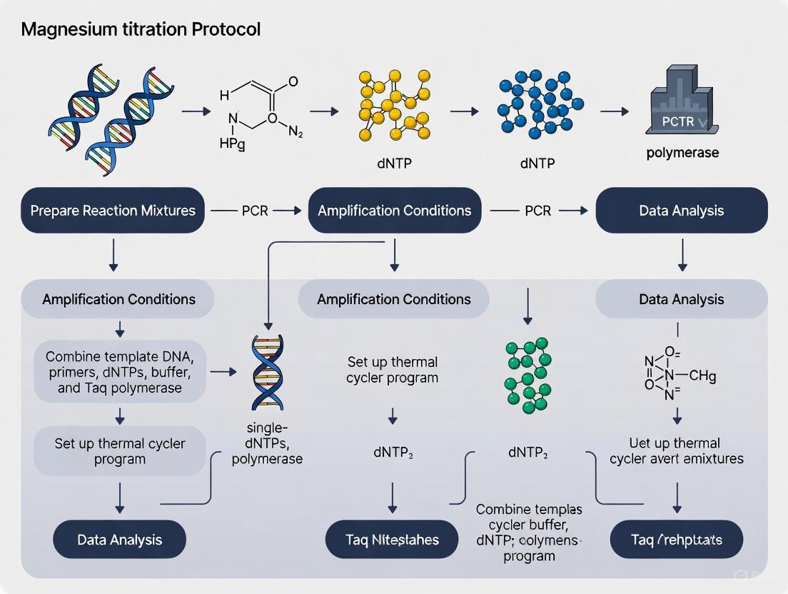
This article provides a comprehensive, evidence-based guide for researchers and drug development professionals to systematically optimize magnesium chloride (MgCl2) concentration in Polymerase Chain Reaction (PCR) assays.
A Step-by-Step Magnesium Titration Protocol for Robust PCR Assay Development
Abstract
This article provides a comprehensive, evidence-based guide for researchers and drug development professionals to systematically optimize magnesium chloride (MgCl2) concentration in Polymerase Chain Reaction (PCR) assays. Covering foundational principles, a detailed titration methodology, advanced troubleshooting for complex templates, and validation techniques, this protocol synthesizes current research to enable the development of highly efficient, specific, and reproducible PCR assays critical for genetic analysis, diagnostic testing, and biomedical research.
The Critical Role of Magnesium: Understanding Its Function in PCR Efficiency and Specificity
Magnesium ion (Mg^(2+)) is a fundamental cofactor required for the catalytic activity of DNA polymerases across all families of these essential enzymes. Mg^(2+) facilitates the nucleotidyl transfer reaction by enabling the 3'-OH group of the primer strand to conduct a nucleophilic attack on the α-phosphorus of the incoming deoxynucleoside triphosphate (dNTP) [1] [2]. This application note details the critical role of Mg^(2+) in DNA polymerase function, framed within the context of developing a robust magnesium titration protocol for Polymerase Chain Reaction (PCR) assay optimization. The precise concentration of Mg^(2+) is a key variable that researchers must optimize to achieve high specificity and yield in PCR, as it directly influences enzyme fidelity, primer annealing, and the stabilization of the DNA-polymerase complex [3]. The following sections provide a mechanistic overview, quantitative data on metal ion effects, and a detailed, step-by-step protocol for Mg^(2+) titration to empower researchers in the fields of molecular biology and drug development.
The Fundamental Role ofMg^(2+)in DNA Polymerase Catalysis
The catalytic mechanism of DNA polymerases is universally dependent on divalent metal ions, with Mg^(2+) serving as the primary physiological cofactor. The widely accepted two-metal-ion mechanism describes how two Mg^(2+) ions coordinate within the enzyme's active site to facilitate DNA synthesis [2].
- Metal Ion A (Nucleotide-Bound
Mg^(2+)): This ion forms a tight complex with the incoming dNTP by coordinating the non-bridging oxygens across its α-, β-, and γ-phosphate groups [2]. - Metal Ion B (Catalytic
Mg^(2+)): This ion activates the3'-OH group of the primer terminus by lowering its pKa, making it a better nucleophile. It also helps position this nucleophile for an in-line attack on the α-phosphate of the dNTP and stabilizes the developing negative charge in the pentavalent transition state [2].
The Mg^(2+) bound to the dNTP (Mg.dNTP complex) is the true substrate for the polymerase reaction. Recent pre-steady-state kinetic studies on HIV-RT (an A-family polymerase) have further refined our understanding, showing that Mg.dNTP binding induces an enzyme conformational change, after which the second, catalytic Mg^(2+) binds to the closed state of the enzyme to directly facilitate the chemical step of nucleotide incorporation [2]. This Mg^(2+)-dependent mechanism is crucial for the high fidelity of DNA replication.
Quantitative Analysis of Divalent Cation Effects on Polymerase Function
While Mg^(2+) is the native cofactor, other divalent cations can substitute with varying degrees of efficiency, often with significant consequences for enzymatic speed and accuracy. Research on BST DNA polymerase (an A-family polymerase) provides a quantitative comparison of how different metal ions influence kinetic parameters and fidelity.
Table 1: Kinetic Impact of Divalent Cations on BST DNA Polymerase Activity
| Divalent Cation | Relative Incorporation Efficiency (Correct dNTP) | Effect on Incorrect dNTP Incorporation | Impact on Base Selectivity (Fidelity) | Ability to Support Mismatch Extension |
|---|---|---|---|---|
Mg^(2+) (Control) |
Baseline | Baseline | High | Low |
Mn^(2+) |
Increased | Increased Significantly | Dramatically Reduced / Impaired | Enhanced |
Co^(2+) |
Increased 6-fold | Decreased (varies by dNTP) | Improved / Higher than Mg^(2+) or Mn^(2+) |
Enhanced |
Cd^(2+) |
Supports Activity | Not Specified | Similar to Co^(2+) / High |
Supported |
Ca^(2+), Ni^(2+), Cu^(2+), Zn^(2+) |
No / Minimal Activity Reported | No / Minimal Activity Reported | N/A | N/A |
Table 2: Apparent Dissociation Constants (Kd) for Catalytic Mg^(2+) Binding to HIV Reverse Transcriptase
| Enzyme State | Kd for Catalytic Mg^(2+) |
Functional Implication |
|---|---|---|
| Open State | Kd ≤ 37 mM | Very weak binding to the open complex. |
| Closed State | Kd = 3.7 mM | tighter binding after conformational change facilitates chemistry. |
| Overall Specificity | --- | Weak binding of catalytic Mg^(2+) contributes to fidelity by allowing sampling without overly stabilizing misaligned substrates. |
The data reveals that Mn^(2+) is a potent mutagenic cofactor because it promotes the incorporation of incorrect nucleotides and aids extension past mispaired primer termini, thereby reducing replication fidelity [1]. In contrast, Co^(2+) can enhance certain aspects of fidelity. It is also noted that an increase in Mg^(2+) concentration from 0.25 mM to 10 mM can increase nucleotide specificity (k_cat/K_m) by 12-fold for HIV-RT, largely by increasing the rate of chemistry relative to nucleotide dissociation [2].
Step-by-Step Magnesium Titration Protocol for PCR Optimization
Optimizing the concentration of MgCl_2 is a critical step in PCR assay development, as it directly affects DNA polymerase activity, primer-template annealing, and reaction specificity. The following protocol provides a systematic approach for Mg^(2+) titration.
Research Reagent Solutions
Table 3: Essential Reagents for Magnesium Titration in PCR
| Reagent | Function / Rationale | Notes for Titration |
|---|---|---|
MgCl_2 Stock Solution |
Source of Mg^(2+) cofactor. |
Use a standardized, nuclease-free solution; concentration typically 25-100 mM. |
PCR Buffer (without MgCl_2) |
Provides optimal chemical environment (pH, salts). | Essential for a titration, as Mg^(2+)-supplemented buffers would confound results. |
| DNA Polymerase | Enzyme that catalyzes DNA synthesis. | Taq polymerase is common; concentration should be kept constant (e.g., 1-2 units/50 µL) [3]. |
| Template DNA | Contains the target sequence to be amplified. | Use a well-characterized, clean template at a mid-range concentration (e.g., 10-50 ng gDNA/50 µL) [3]. |
| Primers (Forward & Reverse) | Define the start and end of the target amplicon. | Use primers designed for specificity; keep concentration constant (e.g., 0.1-1 µM each) [3]. |
| dNTP Mix | Building blocks (dATP, dCTP, dGTP, dTTP) for new DNA strands. | Keep concentration constant; note that dNTPs chelate Mg^(2+) [3]. A common final concentration is 0.2 mM for each dNTP. |
| Nuclease-Free Water | Solvent to bring reaction to final volume. | Must be free of contaminants that could degrade reagents or chelate Mg^(2+). |
Experimental Workflow and Procedure
Prepare Master Mix: On ice, combine the following reagents for
N+1reactions (whereNis the number ofMg^(2+)conditions, and+1is for a potential negative control) to ensure consistency:- Nuclease-Free Water
- PCR Buffer (10X,
Mg^(2+)-free) - dNTP Mix (e.g., 10 mM total)
- Forward Primer
- Reverse Primer
- DNA Polymerase (e.g., 1-2 units per reaction) Mix thoroughly by gentle vortexing and brief centrifugation.
Aliquot Master Mix: Dispense equal volumes of the master mix into each PCR tube or well of a PCR plate.
Add
MgCl_2: Add theMgCl_2stock solution to each tube to create a titration series. A typical range is 1.0 mM to 4.0 mM in 0.5 mM increments, but this may be expanded based on the polymerase or application.- Example: For a 50 µL reaction with a final desired
[Mg^(2+)]of 2.0 mM from a 25 mM stock, add 4 µL of stock.
- Example: For a 50 µL reaction with a final desired
Add Template DNA: Add a constant amount of template DNA to each tube. Include a negative control (no template DNA) to check for contamination.
Thermal Cycling: Place the tubes in a thermal cycler and run a standard PCR program. An example program is:
- Initial Denaturation: 94-98°C for 2-5 minutes.
- Amplification (25-35 cycles):
- Denaturation: 94-98°C for 20-30 seconds.
- Annealing: 55-65°C for 20-40 seconds.
- Extension: 72°C for 30-60 seconds/kb.
- Final Extension: 72°C for 5-10 minutes.
- Hold: 4°C.
Product Analysis: Analyze the PCR products using agarose gel electrophoresis.
- The optimal
[Mg^(2+)]is identified as the concentration that yields a single, intense band of the expected amplicon size with minimal to no non-specific products or primer-dimers.
- The optimal
Application in Diagnostic Assay Development: A Case Study
The critical importance of Mg^(2+) optimization is exemplified in the development of multiplex real-time PCR assays for clinical diagnostics. For instance, a 2025 study describing a multiplex real-time PCR for carbapenemase genes (bla_KPC, bla_IMP, bla_VIM, bla_NDM, bla_OXA-48) required extensive optimization of primer and probe concentrations combined with a specific commercial master mix to achieve a robust, single-tube assay [4]. Similarly, a novel multiplex assay for respiratory pathogens achieved a limit of detection (LOD) between 4.94 and 14.03 copies/µL, a feat that requires meticulously balanced reaction components, including Mg^(2+), to ensure high sensitivity and precision without cross-reactivity [5]. These cases underscore that systematic optimization of Mg^(2+), often in conjunction with other components, is not merely a preliminary step but a fundamental requirement for developing reliable, research-grade, and clinical-grade PCR assays.
Mg^(2+) is indispensable for DNA polymerase activity, playing a direct and multifaceted role in the catalytic mechanism, substrate binding, and overall fidelity of DNA synthesis. The quantitative data on various cations highlights the unique suitability of Mg^(2+) for high-fidelity replication. The provided magnesium titration protocol serves as a critical component in the broader context of PCR assay development, enabling researchers to precisely define reaction conditions that maximize specificity and yield. A rigorous, empirical approach to Mg^(2+) optimization, as detailed in this application note, is therefore a cornerstone of robust and reproducible molecular assay design.
Within the framework of developing a robust magnesium titration protocol for PCR assay development, understanding the precise thermodynamic impact of magnesium chloride (MgCl2) is paramount. The optimization of the Polymerase Chain Reaction (PCR) continues to be a significant challenge in molecular biology, and obtaining the correct MgCl2 concentration is a cornerstone of a successful reaction [6]. As an essential cofactor for DNA polymerase, Mg²⁺ ions play a crucial role not only in enzyme catalysis but also in defining the reaction thermodynamics and kinetics, which directly influence the DNA melting temperature (Tm) [6] [7]. This application note provides a detailed, evidence-based guide on the quantitative relationship between MgCl2 concentration and DNA melting temperature, equipping researchers and drug development professionals with the protocols and data necessary to tailor PCR conditions for superior efficiency and specificity.
The Quantitative Relationship Between MgCl₂ and DNA Melting Temperature
The melting temperature (Tm) of DNA is defined as the temperature at which half of the DNA duplexes dissociate into single strands. The concentration of MgCl₂ directly influences this parameter by altering the electrostatic environment of the DNA backbone. Mg²⁺ ions bind to the negatively charged phosphate groups on DNA, effectively shielding the inherent repulsion between the two strands [7] [8]. This stabilization reduces the electrostatic repulsion between DNA strands, thereby increasing the thermal energy required to denature the duplex and raising the observed Tm [6] [8].
A comprehensive meta-analysis of 61 peer-reviewed studies has provided a quantitative model for this relationship. The analysis established a strong logarithmic relationship between MgCl₂ concentration and DNA melting temperature [6] [9]. The optimal MgCl₂ concentration for efficient PCR performance was identified as being between 1.5 and 3.0 mM [6]. Within this functional range, a clear quantitative effect was measured: for every 0.5 mM increase in MgCl₂ concentration, the DNA melting temperature increases by approximately 1.2 °C [6] [9].
Table 1: Effect of MgCl₂ Concentration on DNA Melting Temperature (Tm)
| MgCl₂ Concentration (mM) | Theoretical Increase in Tm (°C)* | Impact on PCR Product Specificity |
|---|---|---|
| 1.5 | Baseline | Generally optimal for specificity |
| 2.0 | +1.2 °C | Maintains good specificity |
| 2.5 | +2.4 °C | May reduce specificity |
| 3.0 | +3.6 °C | Increased risk of non-specific bands |
| > 4.0 | > 6.0 °C | High risk of non-specific priming and primer-dimer formation |
*Cumulative increase from a 1.5 mM baseline, calculated based on a +1.2°C change per 0.5 mM MgCl₂.
Furthermore, the required MgCl₂ concentration is not universal but is significantly affected by template characteristics. The meta-analysis concluded that templates with higher complexity, such as genomic DNA, typically require higher MgCl₂ concentrations for optimal amplification compared to more straightforward templates like plasmid DNA or synthetic oligonucleotides [6]. This underscores the necessity of a tailored optimization approach.
Experimental Protocol: MgCl₂ Titration for PCR Assay Development
The following section provides a detailed, step-by-step protocol for determining the optimal MgCl₂ concentration for a specific PCR assay. This protocol is designed to be integrated into a broader thesis on systematic PCR development.
Research Reagent Solutions
The successful execution of this titration protocol requires the preparation and use of several key reagents.
Table 2: Essential Reagents for MgCl₂ Titration Experiments
| Reagent / Material | Function / Description | Working Concentration / Notes |
|---|---|---|
| MgCl₂ Solution (25 mM) | Source of Mg²⁺ ions for reaction optimization. An essential cofactor for DNA polymerase activity. | Stock solution; used to create a concentration gradient [10] [11]. |
| PCR Master Mix (5x) | Provides core PCR components: reaction buffer, dNTPs, and a hot-start DNA polymerase. | Ensure the master mix is Mg²⁺-free or its Mg²⁺ content is known for accurate calculations [10]. |
| Primers (Forward & Reverse) | Short, single-stranded DNA sequences that define the start and end of the target amplicon. | Typical final concentration is 0.2 - 0.5 µM each [12]. |
| DNA Template | The DNA sample containing the target sequence to be amplified. | 1 - 1000 ng of genomic DNA, depending on complexity [12]. |
| PCR Grade Water | Nuclease-free water used to bring the reaction to its final volume. | Ensures no contaminants interfere with the reaction. |
Step-by-Step Titration Methodology
This protocol is adapted from a standardized calibration method to ensure reproducibility [10]. The workflow for the entire procedure is summarized in the diagram below.
Title: Workflow for MgCl₂ Titration Protocol
Procedure:
Prepare Working Solution: Dilute the 25 mM MgCl₂ stock solution to create a 5 mM working solution. Pipette 20 µL of 25 mM MgCl₂ into a tube and add 80 µL of PCR-grade water. Mix thoroughly by pipetting up and down [10].
Prepare Master Mix: Calculate the required volumes for all components to set up 8 reactions (including excess to account for pipetting error). Combine the following reagents in a sterile 1.8 mL microcentrifuge tube in the order listed:
- PCR-grade water
- 5x PCR Master Mix (Mg-free)
- Forward Primer (10 µM)
- Reverse Primer (10 µM)
- DNA Template
Vortex the mixture briefly and centrifuge to collect the contents at the bottom of the tube [10] [12]. Note: The DNA template can be added later in step 5 if preferred.
Aliquot Master Mix: Dispense 8 µL of the master mix into each of seven labeled 0.2 mL PCR tubes.
Create MgCl₂ Gradient: Add PCR-grade water and the 5 mM MgCl₂ working solution to each tube as detailed in the table below to create a concentration gradient. Pipette the volumes onto the inner wall of the tube to avoid cross-contamination. Close the tubes and flick to mix, ensuring the reaction mix is collected at the bottom [10].
Table 3: Pipetting Scheme for MgCl₂ Gradient Setup (Final PCR Volume: 20 µL)
Desired Final [MgCl₂] PCR Grade Water to Add 5 mM MgCl₂ Working Solution to Add 1.5 mM 12 µL 0 µL 2.0 mM 10 µL 2 µL 2.5 mM 8 µL 4 µL 3.0 mM 6 µL 6 µL 3.5 mM 4 µL 8 µL 4.0 mM 2 µL 10 µL 4.5 mM 0 µL 12 µL Initiate PCR: If not already added, introduce the DNA template to each tube, using a clean pipette tip for each. Place the tubes in a thermal cycler and run the standard PCR protocol optimized for your primers and template [12].
Analyze Results: Upon completion, analyze 5-10 µL of each PCR product using agarose gel electrophoresis. The optimal MgCl₂ concentration is typically the one that yields the clearest, most intense band of the expected size with the least background smearing or non-specific bands [10].
Discussion and Mechanistic Insights
The thermodynamic data and titration protocol provided herein form a critical component of a systematic PCR assay development strategy. The documented increase in Tm with MgCl₂ concentration is mechanistically explained by the binding of Mg²⁺ ions to the DNA backbone. This binding reduces the electrostatic repulsion between the negatively charged strands of the DNA duplex, thereby stabilizing it and requiring a higher temperature for denaturation [8]. This effect is so significant that Mg²⁺ is known to effectively increase the stability of DNA molecules even under high-temperature conditions [13].
The consequences of improper MgCl₂ concentration are profound for assay robustness.
- Insufficient MgCl₂ (< 1.5 mM): Results in weak or failed amplification due to inadequate DNA polymerase cofactor availability and inefficient primer annealing [6] [7].
- Excessive MgCl₂ (> 3.0 - 4.5 mM): Often leads to decreased specificity, manifesting as multiple bands or smears on a gel. This occurs because the elevated Tm promotes non-specific primer annealing to partially homologous sequences on the template DNA [6] [10] [7].
The finding that template complexity dictates MgCl₂ demand further emphasizes the need for empirical optimization. Genomic DNA, with its high complexity and potential for secondary structure, often requires concentrations at the higher end of the optimal range (e.g., 2.5 - 3.0 mM), whereas simpler templates like plasmids may perform best at lower concentrations (e.g., 1.5 - 2.0 mM) [6]. This protocol provides a reliable and straightforward method to identify this crucial variable, ensuring the development of highly specific and efficient PCR assays for both research and diagnostic applications.
The optimization of the polymerase chain reaction (PCR) remains a pivotal challenge in molecular biology, wherein the concentration of magnesium chloride (MgCl₂) is a critical determinant of success. A precise understanding of how Mg²⁺ influences PCR thermodynamics and kinetics is fundamental to developing efficient and reliable protocols [6]. This application note, framed within a broader thesis on step-by-step magnesium titration for PCR assay development, synthesizes findings from a recent systematic meta-analysis to provide quantitative insights and evidence-based guidelines [6] [9]. The data demonstrate a strong logarithmic relationship between MgCl₂ concentration and DNA melting temperature, establishing that the precise modulation of Mg²⁺, tailored to specific template characteristics, can significantly enhance both the efficiency and specificity of PCR amplification [6].
The following tables summarize the key quantitative relationships derived from the meta-analysis of 61 peer-reviewed studies, providing a foundation for evidence-based protocol design.
Table 1: The Effect of MgCl₂ Concentration on PCR Parameters
| Parameter | Quantitative Relationship | Notes |
|---|---|---|
| Optimal MgCl₂ Range | 1.5 – 3.0 mM | General performance range for standard PCR [6]. |
| Effect on DNA Melting Temperature (Tₘ) | Increases by ~1.2 °C per 0.5 mM MgCl₂ | A strong logarithmic relationship within the 1.5-3.0 mM range [6]. |
| Taq DNA Polymerase Optimal [Mg²⁺] | 1.5 – 2.0 mM | Manufacturer's guideline; depends on template and dNTP concentration [14]. |
Table 2: MgCl₂ Optimization Guide Based on Template DNA
| Template Type | Recommended Starting [MgCl₂] | Rationale |
|---|---|---|
| Genomic DNA | Higher end of optimal range (e.g., 2.5-3.0 mM) | Increased complexity and potential chelating agents require higher [Mg²⁺] [6]. |
| Plasmid or Viral DNA | Lower to middle of optimal range (e.g., 1.5-2.0 mM) | Less complex templates require less Mg²⁺ for efficient polymerization [6] [14]. |
| High GC-Content Templates | May require elevated concentrations | Higher Tₘ and stronger secondary structure necessitate more Mg²⁺ for denaturation and primer access [6]. |
Experimental Protocols
Step-by-Step Magnesium Titration Protocol for PCR Assay Development
This protocol provides a detailed methodology for empirically determining the optimal MgCl₂ concentration for a specific PCR assay.
I. Research Reagent Solutions
Table 3: Essential Materials for Mg²⁺ Titration
| Item | Function/Description |
|---|---|
| Taq DNA Polymerase | Thermostable enzyme for PCR amplification; requires Mg²⁺ as a cofactor [14]. |
| 10X Reaction Buffer (without MgCl₂) | Provides pH-stable environment and salts; using a Mg-free buffer allows for precise customization [14]. |
| MgCl₂ Stock Solution (e.g., 25 mM) | The variable component for titration; a high-purity stock solution is recommended. |
| Template DNA | The DNA to be amplified; should be of high quality and concentration-optimized [14]. |
| Primers | Oligonucleotides specific to the target sequence; should be designed with matched Tₘ [14]. |
| dNTP Mix | Nucleotide building blocks for the new DNA strands; concentration affects free Mg²⁺ [14]. |
II. Procedure
- Prepare Master Mix: In a nuclease-free tube on ice, create a master mix containing all PCR components except the template DNA and the MgCl₂ stock solution. This includes nuclease-free water, 10X buffer (without MgCl₂), dNTP mix, primers, and Taq DNA Polymerase [14].
- Aliquot Master Mix: Dispense equal volumes of the master mix into individual PCR tubes or a multi-well PCR plate.
- Titrate MgCl₂: Add the MgCl₂ stock solution to each tube to create a concentration gradient. A typical titration series might be: 1.0 mM, 1.5 mM, 2.0 mM, 2.5 mM, 3.0 mM, 3.5 mM, and 4.0 mM [14].
- Add Template: Introduce a consistent, optimized amount of template DNA into each reaction tube. Include a negative control (no template) for one of the Mg²⁺ concentrations to check for contamination.
Amplify: Place the tubes in a thermal cycler and run using the following standard cycling parameters, adjusting the annealing temperature (Tₐ) as needed for your primers [15] [14]:
Analyze Results: Separate the PCR products by agarose gel electrophoresis. Analyze the gel for:
- Product Yield: The intensity of the correct band.
- Specificity: The presence or absence of non-specific bands or smearing.
- The optimal MgCl₂ concentration is the one that produces the strongest desired product band with the fewest non-specific amplification products [14].
Workflow for Systematic PCR Optimization
The following diagram illustrates the logical workflow for troubleshooting and optimizing a PCR assay, with a focus on Mg²⁺ titration.
Thermodynamic and Kinetic Principles
The meta-analysis established a foundational logarithmic relationship between MgCl₂ concentration and DNA melting temperature (Tₘ), a key thermodynamic parameter [6]. Quantitatively, every 0.5 mM increase in MgCl₂ within the 1.5–3.0 mM range is associated with a 1.2 °C increase in Tₘ [6]. This occurs because Mg²⁺ ions stabilize the double-stranded DNA structure by shielding the negative charges on the phosphate backbone, thereby raising the energy required for strand separation.
This thermodynamic effect has direct kinetic consequences for the PCR process. By raising the effective Tₘ, Mg²⁺ influences the annealing efficiency of the primers and the denaturation efficiency of the template. Higher than optimal Mg²⁺ can lead to increased non-specific priming and spurious amplification due to imperfect primer annealing, while insufficient Mg²⁺ can cause reduced polymerase activity and failed amplification [6] [14]. Furthermore, the complexity of the template DNA influences the required Mg²⁺ concentration; genomic DNA, with its higher likelihood of secondary structure and greater overall chelating capacity, typically requires higher Mg²⁺ concentrations than simpler plasmid templates [6]. This interplay between template, Tₘ, and Mg²⁺ concentration is critical for designing specific and efficient assays.
A comprehensive meta-analysis of 61 peer-reviewed studies provides quantitative evidence for optimizing magnesium chloride (MgCl₂) concentration in Polymerase Chain Reaction (PCR) protocols. The findings establish 1.5 to 3.0 mM as the optimal range for MgCl₂, with precise concentration being critically dependent on template DNA characteristics. This review synthesizes these evidence-based guidelines into detailed application notes and protocols, enabling researchers to systematically enhance PCR efficiency, specificity, and reliability in diagnostic and drug development applications. The data confirm that precise modulation of MgCl₂ concentration, tailored to specific template properties, significantly improves both the efficiency and specificity of PCR amplification [9] [6].
Magnesium chloride serves as an essential cofactor for DNA polymerase activity and profoundly influences DNA strand separation dynamics by altering the melting temperature of DNA [6]. Its concentration directly affects the thermodynamics and kinetics of DNA denaturation and annealing, making it one of the most crucial parameters for successful PCR optimization [9]. Despite its fundamental importance, MgCl₂ concentration requirements have historically been determined through empirical optimization, leading to inconsistent results across different templates and reaction conditions. The recent meta-analysis by Tbahriti et al. (2025) provides, for the first time, a quantitative framework describing the relationship between MgCl₂ concentration, DNA melting temperature, and amplification efficiency, moving PCR optimization from an empirical art to an evidence-based science [9].
Quantitative Evidence: MgCl₂ Concentration Relationships
Key Quantitative Findings from Meta-Analysis
The meta-analysis revealed several critical quantitative relationships that form the basis for evidence-based protocol development:
- Optimal Concentration Range: The analysis identified 1.5 to 3.0 mM as the optimal range for MgCl₂ in standard PCR applications [9] [6].
- Melting Temperature Relationship: A strong logarithmic relationship exists between MgCl₂ concentration and DNA melting temperature, with every 0.5 mM increase in MgCl₂ within the optimal range associated with a 1.2 °C increase in melting temperature [9] [6].
- Template-Specific Requirements: Template complexity significantly influences optimal MgCl₂ concentration, with genomic DNA templates consistently requiring higher concentrations compared to simpler templates such as plasmids or synthetic oligonucleotides [9].
Template-Specific MgCl₂ Recommendations
Table 1: Evidence-Based MgCl₂ Concentration Guidelines for Different Template Types
| Template Type | Recommended MgCl₂ Range | Key Considerations | Typical Efficiency Gain |
|---|---|---|---|
| Genomic DNA | 2.0 - 3.0 mM | Higher complexity requires elevated Mg²⁺; increase within range for GC-rich targets | 25-40% improvement over standard protocols |
| Plasmid DNA | 1.5 - 2.5 mM | Lower complexity enables more efficient amplification at moderate concentrations | 15-25% improvement over standard protocols |
| GC-Rich Targets | 2.5 - 3.5 mM* | May require exceeding standard upper limit; consider additives like DMSO or betaine | 30-50% improvement for challenging amplicons |
| Standard Amplicons | 1.5 - 2.5 mM | Optimal for most routine applications with amplicon sizes of 100-1000 bp | 20-30% improvement over non-optimized conditions |
*Concentrations above 3.0 mM may be necessary for exceptionally GC-rich templates but require rigorous specificity validation [6].
Experimental Protocol: Systematic MgCl₂ Titration for PCR Optimization
Reagent Preparation and Master Mix Formulation
This protocol provides a systematic approach for determining the optimal MgCl₂ concentration for any specific template and primer combination, with particular relevance for assays supporting cell and gene therapy drug development [16].
Table 2: Reagent Setup for MgCl₂ Titration Experiment
| Component | Stock Concentration | Final Concentration Range | Volume per 50 μL Reaction |
|---|---|---|---|
| PCR Buffer | 10X (without MgCl₂) | 1X | 5.0 μL |
| dNTP Mix | 10 mM (each dNTP) | 200 μM | 1.0 μL |
| Forward Primer | 20 μM | 0.1-0.5 μM | 0.25-1.25 μL |
| Reverse Primer | 20 μM | 0.1-0.5 μM | 0.25-1.25 μL |
| Template DNA | Variable (1-100 ng/μL) | 10⁴-10⁷ molecules | Variable (typically 0.5-5 μL) |
| Taq DNA Polymerase | 5 U/μL | 0.5-2.5 U | 0.1-0.5 μL |
| MgCl₂ | 25 mM | 1.0 - 4.0 mM (titration) | 2.0 - 8.0 μL |
| Sterile Water | - | - | Quantity Sufficient (QS) to 50 μL |
Essential Reagent Solutions:
- MgCl₂ Solution (25 mM): Prepared in nuclease-free water; quality verified for molecular biology applications.
- PCR Buffer (10X): Tris-based buffer, pH 8.3-8.8, without MgCl₂ to enable precise titration.
- dNTP Mix: Prepared from individual dNTPs at 2.5 mM each to ensure equimolar representation.
- Primer Solutions: HPLC-purified primers resuspended in TE buffer or nuclease-free water; concentration verified by spectrophotometry.
Titration Procedure and Thermal Cycling
Master Mix Preparation:
- Calculate the required number of reactions (n+2 to account for pipetting error).
- Prepare a master mix containing all components except MgCl₂ and template DNA to ensure reaction consistency.
- Aliquot the master mix into individual 0.2 mL thin-walled PCR tubes.
MgCl₂ Titration Series:
- Prepare a dilution series of MgCl₂ to span the range of 1.0 mM to 4.0 mM in 0.5 mM increments.
- Add the appropriate volume of each MgCl₂ concentration to individual reaction tubes.
- Add template DNA to all tubes except the negative control.
Thermal Cycling Parameters:
- Initial Denaturation: 94-95°C for 2-5 minutes
- Amplification Cycles (25-35 cycles):
- Denaturation: 94-95°C for 30-45 seconds
- Annealing: Temperature calculated based on primer Tm + MgCl₂ adjustment (see section 3.3) for 45-60 seconds
- Extension: 72°C for 1 minute per kilobase of amplicon
- Final Extension: 72°C for 5-10 minutes
- Hold: 4-10°C indefinitely
Post-Amplification Analysis:
- Analyze PCR products by agarose gel electrophoresis (1.5-2.0% agarose).
- Include appropriate molecular weight standards.
- Visualize with intercalating dyes; document for band intensity and specificity analysis.
Annealing Temperature Adjustment Based on MgCl₂ Concentration
The meta-analysis established that MgCl₂ concentration directly affects DNA melting temperature. To maintain optimal primer annealing efficiency during the titration, adjust the annealing temperature using the following relationship:
Annealing Temperature Adjustment = (MgCl₂ Concentration in mM - 1.5) × 2.4
Example: For a reaction with 2.5 mM MgCl₂, increase the annealing temperature by approximately 2.4°C compared to the temperature calculated for 1.5 mM MgCl₂.
Conceptual Framework for MgCl₂ Optimization
Data Interpretation and Optimization Strategy
Troubleshooting MgCl₂ Concentration Effects
Table 3: Interpretation of Results from MgCl₂ Titration Experiments
| Observed Result | Potential Cause | Recommended Action |
|---|---|---|
| No amplification | MgCl₂ concentration too low; insufficient polymerase activity | Increase MgCl₂ concentration in 0.5 mM increments; verify template quality and primer design |
| Smear or multiple bands | MgCl₂ concentration too high; reduced specificity | Decrease MgCl₂ concentration in 0.5 mM increments; increase annealing temperature |
| Weak specific band | Suboptimal MgCl₂ concentration; primer binding inefficient | Fine-tune MgCl₂ concentration in 0.25 mM increments; optimize primer concentration |
| Primer-dimer formation | Excessive MgCl₂ promotes mispriming | Reduce MgCl₂ concentration; optimize primer design with attention to 3' complementarity |
Advanced Optimization for Challenging Templates
For particularly challenging templates such as those with very high GC content (>70%) or complex secondary structures, consider these advanced strategies:
- Combined Optimization with Additives: Incorporate PCR enhancers such as DMSO (1-10%), formamide (1.25-10%), or betaine (0.5-2.5 M) in conjunction with MgCl₂ titration [12].
- Two-Step Optimization Protocol: First determine the optimal MgCl₂ concentration, then perform a secondary optimization of additive concentration while maintaining the established MgCl₂ level.
- Touchdown PCR Integration: Combine optimized MgCl₂ concentration with touchdown PCR protocols to enhance specificity for difficult templates.
Application in Regulated Bioanalysis
For PCR assays supporting cell and gene therapy drug development—including biodistribution, transgene expression, viral shedding, and cellular kinetics—rigorous validation of the optimized MgCl₂ concentration is essential [16]. The optimized concentration should demonstrate:
- Specificity: Amplification of only the intended target without non-specific products or primer-dimers.
- Efficiency: PCR efficiency between 90-110% when calculating based on standard curves.
- Reproducibility: Consistent amplification across multiple runs and operators.
- Robustness: Tolerance of minor variations in reaction conditions while maintaining performance.
The meta-analysis by Tbahriti et al. provides a robust theoretical framework for moving beyond empirical MgCl₂ optimization to evidence-based protocol design. The established optimal range of 1.5-3.0 mM, with template-specific adjustments, offers researchers a validated starting point for PCR optimization. The systematic titration protocol outlined herein enables precise determination of ideal MgCl₂ concentrations for specific applications, particularly in regulated bioanalytical contexts supporting drug development. By implementing these evidence-based guidelines, researchers can significantly improve the reliability, efficiency, and specificity of their PCR assays, advancing the rigor and reproducibility of molecular analyses in both research and diagnostic applications.
Within polymerase chain reaction (PCR) assay development, the optimization of magnesium ion (Mg²⁺) concentration is a critical determinant of success. Mg²⁺ serves as an essential cofactor for thermostable DNA polymerases, directly facilitating the catalytic polymerization of nucleotides into a growing DNA strand [17]. The central challenge for researchers lies in managing the bioavailable fraction of free Mg²⁺ ions, as the total magnesium added is partitioned between the enzymatic cofactor pool and complexes formed with various reaction components. This application note delineates the principal interactions between MgCl₂, deoxynucleoside triphosphates (dNTPs), ethylenediaminetetraacetic acid (EDTA), and template DNA, providing a structured framework for efficient Mg²⁺ titration within a broader PCR optimization protocol.
The Critical Roles of Magnesium in PCR
Magnesium ions are fundamental to both the structural integrity and catalytic function of the PCR reaction. Their roles are multifactorial:
Enzyme Cofactor: DNA polymerases require Mg²⁺ ions at their active site for catalytic activity. Structural studies of DNA polymerase I (Klenow fragment) reveal that two invariant aspartate residues (Asp705 and Asp882 in Pol I(KF)) coordinate a pair of Mg²⁺ ions [17]. This metal ion pair is directly involved in the nucleotidyl transfer reaction, with one metal ion facilitating deprotonation of the 3'-OH primer terminus and the other stabilizing the leaving pyrophosphate group [17]. Mutation of these aspartate residues reduces polymerase activity to barely detectable levels, underscoring the indispensable nature of Mg²⁺ coordination [17].
Substrate Chelation: The incoming dNTP substrates inherently chelate Mg²⁺. The dNTP-Mg²⁺ complex is the true substrate for DNA polymerases, meaning that a significant portion of Mg²⁺ is sequestered by the dNTP pool [18] [19].
Nucleic Acid Stability: Mg²⁺ stabilizes the double-stranded DNA structure by electrostatically shielding the negative charge of the phosphate backbone. This raises the melting temperature (Tm) of DNA, a phenomenon quantitatively documented in meta-analyses which show a logarithmic relationship between MgCl₂ concentration and DNA Tm [6] [9]. Every 0.5 mM increase in MgCl₂ within the 1.5–3.0 mM range is associated with an average increase in melting temperature of 1.2°C [6] [9].
The equilibrium between free and bound Mg²⁺ is dynamic, and the concentration of free Mg²⁺ is the variable that ultimately governs polymerase activity and reaction fidelity. Consequently, understanding the factors that deplete free Mg²⁺ is paramount.
Quantitative Analysis of Components Affecting Free Mg²⁺
The primary reactants that chelate Mg²⁺ in a PCR are dNTPs and, to a lesser extent, template DNA. EDTA acts as a potent chelator that is sometimes introduced via other reagent components. The table below summarizes the chelation relationships and their impact on the reaction.
Table 1: Quantitative Effects of PCR Components on Free Mg²⁺ Availability
| Component | Chelation Relationship with Mg²⁺ | Impact on PCR if Free [Mg²⁺] is Inadequate | Typical Concentration in PCR |
|---|---|---|---|
| dNTPs | Binds Mg²⁺ stoichiometrically; ~1 mM Mg²⁺ is chelated by 0.2 mM dNTPs [19]. | Drastic reduction or absence of PCR product [18] [19]. | 0.2 mM of each dNTP (200 µM) is standard [18] [19]. |
| EDTA | A potent chelator often present in elution buffers or template preparations. | Can completely inhibit PCR by sequestering all Mg²⁺, preventing polymerase activity. | Should be minimized; its effect is neutralized by a molar excess of Mg²⁺. |
| Template DNA | The phosphate backbone chelates Mg²⁺; complex templates (e.g., genomic DNA) have a greater effect [6]. | Can reduce efficiency, particularly for complex templates at high concentrations. | Plasmid: 1 pg–10 ng; Genomic DNA: 10 ng–1 µg [18] [19]. |
These interactions establish a foundational hierarchy for Mg²⁺ consumption. The dNTP concentration is the most significant variable, and the baseline Mg²⁺ requirement is often calculated relative to it. The presence of EDTA must be accounted for, and the complexity of the template DNA can necessitate further fine-tuning.
Visualizing the Mg²⁺ Equilibrium in PCR
The following diagram illustrates the dynamic equilibrium of Mg²⁺ in a PCR master mix, showing how the total Mg²⁺ is partitioned and highlighting the critical role of the free, bioavailable ion.
Figure 1: The Partitioning of Mg²⁺ in a PCR Reaction. The free Mg²⁺ pool (blue) is the source for all functional roles. It is depleted by chelation with dNTPs, template DNA, and potential contaminants like EDTA (red), and by its essential function as an enzyme cofactor (green). The goal of optimization is to ensure an adequate free Mg²⁺ concentration to drive catalysis.
Integrated Experimental Protocol for Mg²⁺ Titration
This protocol provides a step-by-step guide for systematically optimizing Mg²⁺ concentration in PCR, accounting for its interplay with other components.
Research Reagent Solutions
Table 2: Essential Reagents for Mg²⁺ Optimization Experiments
| Reagent | Function/Description | Example & Notes |
|---|---|---|
| Thermostable DNA Polymerase | Catalyzes DNA synthesis; requires Mg²⁺ as cofactor. | Taq DNA Polymerase (NEB #M0267) is widely used. Proofreading enzymes (e.g., Q5, NEB #M0491) may have different optimal [Mg²⁺] [18] [19]. |
| 10X Reaction Buffer | Provides baseline reaction conditions (pH, salts). | Often supplied with the polymerase. May be Mg-free or contain a standard concentration (e.g., 1.5-2.0 mM) [18]. |
| 25 mM MgCl₂ Solution | The titratable source of Mg²⁺ ions. | A separate, sterile solution provided in many PCR kits or available separately for precise optimization [18]. |
| dNTP Mix | The building blocks for DNA synthesis. | Use a standardized, high-quality solution. Typical final concentration is 200 µM of each dNTP [18] [19]. |
| Nuclease-Free Water | Solvent for the reaction. | Ensures no contaminants interfere with the reaction. |
Step-by-Step Mg²⁺ Titration Procedure
Master Mix Preparation: Prepare a master mix for
n+1reactions to minimize pipetting error. The mix should contain:- Nuclease-free water (to final volume)
- 1X Reaction Buffer (Mg-free if possible)
- 0.2 mM of each dNTP
- 0.1–0.5 µM of each primer
- 0.5–2.0 units of DNA Polymerase (add last, mix gently)
- A constant, optimal amount of template DNA
Aliquoting and Mg²⁺ Supplementation: Dispense equal volumes of the master mix into
nPCR tubes. Supplement each tube with MgCl₂ solution to create a titration series. A typical starting range is 0.5 mM to 4.0 mM in 0.5 mM increments [18]. For example:- Tube 1: 0.5 mM MgCl₂ (negative control/low end)
- Tube 2: 1.0 mM MgCl₂
- Tube 3: 1.5 mM MgCl₂
- Tube 4: 2.0 mM MgCl₂
- Tube 5: 2.5 mM MgCl₂
- Tube 6: 3.0 mM MgCl₂
- Tube 7: 3.5 mM MgCl₂
- Tube 8: 4.0 mM MgCl₂ (high end)
Thermal Cycling: Run the PCR using pre-optimized cycling conditions appropriate for your primer pair and amplicon. Standard conditions for a 1 kb amplicon with Taq polymerase are: 1 cycle of 95°C for 2 min; 25-30 cycles of 95°C for 15 sec, 50-60°C for 15-30 sec, 68°C for 1 min; and 1 final cycle of 68°C for 5 min [18].
Product Analysis: Analyze the PCR products using agarose gel electrophoresis. Assess for:
The workflow for this optimization procedure is summarized in the following diagram:
Figure 2: Workflow for Systematic Mg²⁺ Titration in PCR. The process begins with creating a master mix without magnesium, which is then aliquoted and supplemented with a gradient of MgCl₂. After PCR amplification, the products are analyzed to identify the optimal Mg²⁺ concentration that provides high specificity and yield.
Troubleshooting and Special Considerations
- High GC-Rich Templates: Templates with high GC content often require higher Mg²⁺ concentrations (potentially >3.0 mM) for efficient amplification. The elevated Mg²⁺ helps stabilize the DNA duplex, which has a higher melting temperature [6].
- Multiplex PCR: When amplifying multiple targets simultaneously, the effective dNTP concentration is higher, and primer interactions are more complex. This typically requires a higher, more finely tuned Mg²⁺ concentration to ensure balanced amplification of all targets [20].
- EDTA Contamination: If the template DNA is eluted or stored in a buffer containing EDTA, it is critical to ensure that the molar concentration of Mg²⁺ in the final PCR significantly exceeds the total EDTA concentration to neutralize its chelating effect.
The precise optimization of free Mg²⁺ is a cornerstone of robust PCR assay development. The process is inherently systematic, governed by the predictable chelation of Mg²⁺ by dNTPs and the variable demands of the template DNA. By following the structured titration protocol and conceptual framework outlined in this application note, researchers can efficiently navigate the interplay between reaction components, moving from empirical testing to a principled optimization strategy. This ensures the development of highly specific, sensitive, and reliable PCR assays for research and diagnostic applications.
A Practical Laboratory Guide to Performing Magnesium Titration
The formulation of the master mix is a foundational step in polymerase chain reaction (PCR) assay development, with the concentration of magnesium chloride (MgCl₂) being one of the most crucial variables affecting success. Magnesium ions (Mg²⁺) serve as an essential cofactor for thermostable DNA polymerases, directly influencing enzymatic activity, reaction efficiency, and amplification specificity [3] [7]. A precise understanding and optimization of MgCl₂ concentration is therefore not a mere recommendation but a prerequisite for robust and reliable assay development. This protocol provides a detailed, evidence-based framework for the reagent preparation and master mix formulation stage, with a specific focus on establishing a magnesium titration protocol tailored to specific template and primer system characteristics.
The Mg²⁺ ion is fundamental to the PCR process through two primary mechanisms. First, it is directly involved in the catalytic act of DNA synthesis by facilitating the binding of deoxynucleoside triphosphates (dNTPs) to the enzyme's active site and catalyzing the formation of the phosphodiester bond between the incoming nucleotide and the primer's 3'-OH group [3] [21] [7]. Second, Mg²⁺ stabilizes the interaction between the primer and the template DNA by binding to the negatively charged phosphate backbone, thereby reducing electrostatic repulsion and facilitating proper annealing [3] [7]. Consequently, the MgCl₂ concentration in the final reaction mix must be meticulously calibrated; too little Mg²⁺ results in weak or failed amplification due to insufficient polymerase activity, while too much promotes non-specific priming and the appearance of spurious amplification products [22] [7] [10].
Establishing the Baseline: Quantitative Relationships and Component Formulation
A recent comprehensive meta-analysis of 61 peer-reviewed studies has provided quantitative insights into the effects of MgCl₂, establishing an optimal baseline concentration range of 1.5 to 3.0 mM for standard PCR applications [6] [9]. The analysis further revealed a strong logarithmic relationship between MgCl₂ concentration and DNA melting temperature, with every 0.5 mM increase in MgCl₂ within this range associated with an approximately 1.2 °C increase in melting temperature [6]. This quantitative relationship is critical for predicting and adjusting annealing temperatures during protocol optimization. It is also established that template complexity directly influences optimal Mg²⁺ requirements, with genomic DNA templates typically requiring higher concentrations than simpler plasmid DNA templates [6].
Table 1: Standard Master Mix Components for a 50 µL PCR Reaction
| Component | Final Concentration | Stock Concentration | Volume for 1 Reaction (µL) | Function & Notes |
|---|---|---|---|---|
| Nuclease-Free Water | - | - | Variable | Brings reaction to final volume; must be DNase/RNase-free. |
| PCR Buffer (10X) | 1X | 10X | 5.0 | Provides optimal pH and salt (KCl) environment. |
| MgCl₂ Solution | Variable (e.g., 1.5 mM) | e.g., 25 mM | Variable | Essential cofactor; concentration requires titration (see Section 1.3). |
| dNTP Mix | 200 µM each | 10 mM each | 1.0 | Building blocks for new DNA strands. |
| Forward Primer | 0.5 µM | 10 µM | 2.5 | Binds to specific sequence on one strand of the template. |
| Reverse Primer | 0.5 µM | 10 µM | 2.5 | Binds to the complementary strand of the template. |
| DNA Polymerase | 1-2 Units/50 µL | e.g., 5 U/µL | 0.5 | Enzyme that synthesizes new DNA strands. |
| Template DNA | Variable | Variable | Variable | Amount depends on complexity (e.g., 10-100 ng genomic DNA). |
The following workflow outlines the logical process for preparing the master mix and establishing a magnesium titration experiment.
Detailed Magnesium Titration Protocol
This protocol is designed to systematically identify the optimal MgCl₂ concentration for a specific PCR assay.
Research Reagent Solutions and Materials
| Item | Function/Description |
|---|---|
| MgCl₂ Solution (25 mM) | Used to adjust the final concentration of Mg²⁺ in the PCR reaction. This is the primary variable being tested. [10] |
| PCR Buffer (10X, Mg²⁺-Free) | Provides the core chemical environment (pH, salts) without introducing uncontrolled Mg²⁺ variables. |
| High-Fidelity DNA Polymerase | Enzyme for DNA synthesis; proofreading enzymes are recommended for cloning and applications requiring high accuracy. [22] [23] |
| Ultra-Pure dNTP Mix | Provides equimolar amounts of dATP, dCTP, dGTP, and dTTP for DNA synthesis. [3] [22] |
| Target-Specific Oligonucleotide Primers | Forward and reverse primers designed to flank the target sequence of interest. [3] [22] |
| Template DNA | The DNA sample containing the target sequence to be amplified (e.g., genomic DNA, cDNA). [3] [22] |
| Nuclease-Free Water | Solvent to bring the reaction to volume; ensures no enzymatic degradation of reaction components. |
Step-by-Step Titration Methodology
Prepare a Master Mix (Without Mg²⁺ or Template): Calculate the required volumes for all components except MgCl₂ and DNA template to create a master mix for
n+1reactions (wherenis the number of Mg²⁺ conditions), plus a 10% excess to account for pipetting error. For a 50 µL final reaction volume per tube, combine in a sterile tube:- Nuclease-Free Water
- 10X PCR Buffer (Mg²⁺-Free) to a final 1X concentration
- 10 mM dNTP Mix to a final 200 µM each
- 10 µM Forward Primer to a final 0.5 µM
- 10 µM Reverse Primer to a final 0.5 µM
- DNA Polymerase (1-2 units per 50 µL reaction) Mix the solution thoroughly by pipetting up and down or vortexing gently, then briefly centrifuge.
Aliquot the Master Mix: Dispense equal volumes of the master mix into individual PCR tubes or a PCR plate. The volume per tube should be the total reaction volume (50 µL) minus the volumes of the MgCl₂ solution and template DNA to be added later.
Create the MgCl₂ Concentration Gradient: Using a 25 mM MgCl₂ stock solution, prepare a dilution series to achieve the desired final concentrations in the PCR reactions. A typical and effective gradient ranges from 1.0 mM to 4.0 mM in 0.5 mM increments [21]. The table below provides a sample setup for a 50 µL final reaction volume, assuming the master mix (without Mg²⁺) has been aliquoted in 47 µL volumes.
Table 2: Sample MgCl₂ Titration Setup for 50 µL Final Volume
Desired Final [MgCl₂] Volume of 25 mM Stock to Add (µL) Volume of Nuclease-Free Water to Add (µL) Total Added Volume (µL) 1.0 mM 2.0 1.0 3.0 1.5 mM 3.0 0.0 3.0 2.0 mM 4.0 0.0* 4.0 2.5 mM 5.0 0.0* 5.0 3.0 mM 6.0 0.0* 6.0 3.5 mM 7.0 0.0* 7.0 4.0 mM 8.0 0.0* 8.0 *For simplicity, the master mix aliquot volume can be adjusted downward for higher Mg²⁺ points to keep the final volume constant at 50 µL.
Add DNA Template: To each tube, add a consistent, optimized amount of template DNA. For genomic DNA, this is typically 10–100 ng; for plasmid DNA, 0.1–1 ng is often sufficient [3] [22]. Include a negative control (no template DNA) for one of the Mg²⁺ concentrations to check for contamination.
Execute Thermocycling and Analysis: Place the tubes in a thermal cycler and run the appropriate PCR program. Upon completion, analyze the products using agarose gel electrophoresis. The optimal MgCl₂ concentration is identified as the one that produces a single, intense band of the expected size with minimal to no non-specific products or primer-dimer artifacts [10].
Advanced Considerations for Specific Applications
The baseline protocol can be adapted for challenging templates and specialized applications:
- GC-Rich Templates: Amplifying DNA sequences with a GC content exceeding 60-70% often requires higher MgCl₂ concentrations (e.g., up to 4-5 mM) and the use of specialized polymerases and buffer systems that include enhancing additives like DMSO, betaine, or proprietary GC enhancers. These additives help destabilize the strong secondary structures that form in GC-rich regions [21] [23].
- Presence of Inhibitors: If the DNA extract is known to contain PCR inhibitors (e.g., from blood, soil, or plants), they may chelate Mg²⁺ ions, rendering them unavailable to the polymerase. In such cases, increasing the MgCl₂ concentration by 0.5-1.0 mM above the standard optimal range can compensate for this effect [7] [10].
- dNTP and Mg²⁺ Stoichiometry: Mg²⁺ ions bind to dNTPs in the reaction. The optimal Mg²⁺ concentration must be in a slight molar excess over the total dNTP concentration. A general guideline is to use 0.5–1.0 mM Mg²⁺ above the total dNTP concentration [22]. For a standard dNTP mix of 200 µM each (800 µM total), this translates to a minimum Mg²⁺ concentration of ~1.3-1.8 mM.
The following diagram illustrates the molecular mechanism of Mg²⁺ in PCR and the effects of its concentration on reaction outcomes.
The magnesium ion (Mg²⁺) concentration is a pivotal factor in the success of the Polymerase Chain Reaction (PCR). Acting as a critical cofactor for DNA polymerase activity, Mg²⁺ is directly involved in the catalytic mechanism of DNA synthesis [7]. Furthermore, it influences the melting temperature (Tm) of DNA by stabilizing the double helix through interactions with the negatively charged phosphate backbone, thereby facilitating proper primer annealing [6] [7]. A meta-analysis of PCR optimization studies has established that the optimal MgCl₂ concentration for most reactions lies within the range of 1.5 mM to 3.0 mM [6]. However, this optimum can shift significantly based on template DNA characteristics, such as complexity and GC-content, as well as the specific primers and buffer composition used. Consequently, empirical optimization is often indispensable for achieving maximal specificity and yield. This application note details a systematic titration protocol, using 0.5 mM increments from 1.0 mM to 4.0 mM, to empower researchers to identify the ideal MgCl₂ concentration for their specific PCR assay.
Experimental Design & Workflow
This section outlines the rationale for the selected concentration range and presents a logical workflow for the titration experiment, from setup to analysis.
Rationale for Titration Range and Increments
The recommended range of 1.0 mM to 4.0 mM encompasses the established optimal window while allowing for the identification of sub-optimal conditions that manifest as non-specific amplification or PCR failure. The use of 0.5 mM increments is supported by quantitative research, which indicates that each 0.5 mM increase in MgCl₂ raises the DNA melting temperature by approximately 1.2 °C [6]. This granularity is sufficient to detect significant changes in reaction performance without being unnecessarily laborious. The table below summarizes the expected outcomes across the titration series.
Table 1: Expected Outcomes from MgCl₂ Titration Series
| MgCl₂ Concentration (mM) | Expected PCR Outcome | Theoretical ΔTm vs. 1.5 mM* |
|---|---|---|
| 1.0 | Weak or no amplification | ~ -1.2 °C |
| 1.5 | Potential optimal range | Baseline |
| 2.0 | Potential optimal range | ~ +1.2 °C |
| 2.5 | Potential optimal range | ~ +2.4 °C |
| 3.0 | Potential optimal range | ~ +3.6 °C |
| 3.5 | Increased non-specifics | ~ +4.8 °C |
| 4.0 | High non-specific background | ~ +6.0 °C |
*Estimated change in melting temperature based on meta-analysis data [6].
Experimental Workflow
The following diagram illustrates the end-to-end process for performing the MgCl₂ titration experiment.
Diagram 1: MgCl₂ Titration Workflow. This flowchart outlines the key steps in the experimental protocol, from reaction setup to data analysis.
Materials and Equipment
The Scientist's Toolkit: Essential Research Reagents
A successful titration experiment requires high-quality, consistent reagents. The following table lists the essential components and their functions.
Table 2: Essential Reagents for PCR Mg²⁺ Titration
| Reagent / Equipment | Function / Role in Titration | Key Considerations |
|---|---|---|
| 10X PCR Buffer (without MgCl₂) | Provides optimal chemical environment (pH, salts). Using a Mg-free buffer is critical for a controlled titration. | Ensure compatibility with your DNA polymerase. |
| 25-50 mM MgCl₂ Stock Solution | The titrant; used to create the final desired Mg²⁺ concentration in the reaction. | Must be nuclease-free. Concentration must be accurately known. |
| DNA Polymerase (e.g., Taq) | Enzyme that synthesizes new DNA strands; activity is directly dependent on Mg²⁺ concentration. | Note that 1-2 units per 50 µL reaction is standard [3]. |
| Forward & Reverse Primers | Bind flanking regions of the target sequence. Mg²⁺ concentration affects their annealing specificity and Tm. | Use well-designed primers (Tm 55-70°C) at 0.1-1 µM [24] [3]. |
| dNTP Mix | Building blocks (A, T, C, G) for new DNA strands. | Use at 0.2 mM each; Mg²⁺ binds dNTPs, reducing free [Mg²⁺] available for the enzyme [3]. |
| DNA Template | The target DNA to be amplified. | Amount and purity are key; 5-50 ng genomic DNA per 50 µL reaction [3]. |
| Nuclease-Free Water | Brings reaction to final volume. | Must be free of contaminants that could degrade reaction components. |
| Thermal Cycler | Instrument that automates the temperature cycles for denaturation, annealing, and extension. | Ensure the block is calibrated for temperature accuracy. |
| Agarose Gel Electrophoresis System | Standard method for separating and visualizing PCR products to assess yield and specificity. | Used to compare results across different Mg²⁺ concentrations. |
Detailed Experimental Protocol
Step-by-Step Titration Series Setup
This protocol is designed for a 50 µL reaction volume and a titration series across 7 tubes. It is strongly recommended to prepare a master mix to minimize pipetting error and ensure consistency across reactions.
Prepare Master Mix (on ice): Calculate the required volumes for a master mix sufficient for 7 + 1 (extra) reactions. Combine the following components in a nuclease-free microcentrifuge tube in the order listed:
- Nuclease-Free Water
- 10X PCR Buffer (Mg-free)
- dNTP Mix (e.g., 10 mM each)
- Forward Primer (10 µM)
- Reverse Primer (10 µM)
- DNA Polymerase (e.g., 5 U/µL) Mix thoroughly by pipetting gently up and down, followed by a brief centrifugation.
Aliquot Master Mix: Dispense an equal volume of the master mix into each of seven 0.2 mL PCR tubes. A volume of 43 µL per tube is a typical starting point.
Spike with MgCl₂ Stock Solution: Add the appropriate volume of a 25 mM MgCl₂ stock solution to each tube to achieve the final concentrations in the table below. Bring the total volume of each reaction to 49 µL with nuclease-free water.
Table 3: Pipetting Scheme for MgCl₂ Titration Series
Tube # Target [MgCl₂] (mM) Volume of 25 mM MgCl₂ Stock (µL)* Volume of Nuclease-Free Water (µL)* 1 1.0 2.0 4.0 2 1.5 3.0 3.0 3 2.0 4.0 2.0 4 2.5 5.0 1.0 5 3.0 6.0 0.0 6 3.5 7.0 0.0 (adjust master mix water) 7 4.0 8.0 0.0 (adjust master mix water) *Volumes are approximate examples for a 50 µL final reaction. Precise volumes must be calculated based on the exact master mix composition.
Add DNA Template: Add 1 µL of your prepared DNA template to each tube, bringing the final reaction volume to 50 µL. Include a negative control (replace template with nuclease-free water) to check for contamination.
Thermal Cycling: Place tubes in a thermal cycler and run the standard cycling program for your assay. A generic program is provided below, which may require optimization of temperatures and times.
- Initial Denaturation: 94–98 °C for 1–5 minutes.
- Amplification Cycles (25–35 cycles):
- Denaturation: 94–98 °C for 20–30 seconds.
- Annealing: 50–65 °C for 20–40 seconds.
- Extension: 72 °C for 30 seconds per kb.
- Final Extension: 72 °C for 5–10 minutes.
- Hold: 4 °C ∞.
Analysis of Results
- Agarose Gel Electrophoresis: Analyze 5–10 µL of each PCR product, including the negative control, on an agarose gel stained with an appropriate DNA-binding dye (e.g., SYBR Safe).
Visualization and Interpretation: Visualize the gel under UV light. The ideal MgCl₂ concentration will produce a single, intense band of the expected size. Refer to the table below for guidance on interpreting results.
Table 4: Troubleshooting MgCl₂ Concentration Effects
Gel Result Indicated MgCl₂ Effect Recommended Action No band / Faint band Concentration too low; insufficient polymerase activity or primer annealing [7]. Focus on the range between the lowest concentration that shows a band and the next 2-3 higher points. Single, bright band of correct size Optimal concentration. Proceed with this concentration for assay validation. Multiple bands (non-specific) Concentration too high; reduces specificity of primer annealing [3] [7]. Focus on the range between the highest concentration that shows a single clean band and the point where smearing begins. Primer-dimer formation Can occur at high concentrations due to non-specific primer interactions [7]. Select a concentration that minimizes primer-dimer while maintaining strong target amplification.
A systematic MgCl₂ titration is a fundamental and necessary step in robust PCR assay development. The protocol outlined here, utilizing 0.5 mM increments from 1.0 to 4.0 mM, provides a clear framework for researchers to identify the concentration that maximizes yield and specificity for their unique experimental system. The quantitative relationships and expected outcomes provided serve as a guide for data interpretation. The optimal concentration identified through this process should be used in all subsequent experiments to ensure reproducible and reliable results in genetic research, diagnostic assay development, and other molecular biology applications.
In the development of a robust polymerase chain reaction (PCR) assay, the setup of reaction tubes and thermal cycler parameters is a pivotal step where theoretical planning meets practical execution. The concentration of magnesium ions (Mg²⁺) is a cornerstone parameter in this process, acting as an essential cofactor for thermostable DNA polymerases [3]. Mg²⁺ facilitates the crucial binding of the polymerase to the DNA template and catalyzes the incorporation of nucleotides during the polymerization step [3]. Its concentration directly influences the reaction's efficiency, specificity, and fidelity. A deficiency in Mg²⁺ can lead to poor PCR yield, while an excess can promote non-specific amplification and reduce replication accuracy [25] [3]. Furthermore, a recent meta-analysis established a quantitative relationship between MgCl₂ concentration and DNA melting temperature, demonstrating that every 0.5 mM increase within the optimal range raises the melting temperature by approximately 1.2 °C [6] [9]. This protocol provides a detailed guide for setting up reaction tubes and thermal cycler parameters, with a specific focus on a magnesium titration experiment, to systematically identify optimal conditions for any PCR assay.
Research Reagent Solutions
The following table details the essential materials and reagents required for performing PCR optimization with magnesium titration.
Table 1: Essential Reagents and Materials for PCR Setup and Magnesium Titration
| Item | Function/Description |
|---|---|
| Thermophilic DNA Polymerase | Enzyme that synthesizes new DNA strands; choice (e.g., Taq, Q5, Phusion) depends on required fidelity and amplicon length [25]. |
| 10X Reaction Buffer | Provides pH-stable environment and salt conditions conducive to polymerase activity; often supplied with the enzyme. |
| Magnesium Chloride (MgCl₂) Solution | Source of Mg²⁺ cofactor; typically provided as a separate, sterile solution (e.g., 25 mM or 50 mM) for precise optimization [25] [3]. |
| dNTP Mix | A solution containing equimolar amounts of dATP, dCTP, dGTP, and dTTP; the building blocks for new DNA strands [3]. |
| Forward and Reverse Primers | Synthetic oligonucleotides that define the start and end of the target sequence to be amplified [3]. |
| Template DNA | The DNA sample containing the target sequence (e.g., genomic DNA, cDNA, or plasmid DNA) [3]. |
| Nuclease-Free Water | Solvent used to bring the reaction to the final volume; must be sterile to prevent degradation of reagents. |
Magnesium Titration Experimental Protocol
Step-by-Step Reaction Setup Procedure
- Preliminary Calculations: Determine the volume of a 50 mM MgCl₂ stock solution required to achieve each desired final concentration in a 50 µL reaction. A standard titration range is from 0.5 mM to 5.0 mM in 0.5 mM increments.
- Master Mix Preparation: In a sterile, nuclease-free microcentrifuge tube, prepare a master mix for all reactions plus ~10% extra to account for pipetting error. The master mix contains all common reagents. Gently mix and briefly centrifuge.
- Nuclease-Free Water: (Variable amount to achieve final volume)
- 10X Reaction Buffer: 5 µL per reaction
- dNTP Mix (10 mM each): 1 µL per reaction
- Forward Primer (10 µM): 1.25 µL per reaction
- Reverse Primer (10 µM): 1.25 µL per reaction
- DNA Polymerase: 0.25 µL (e.g., 1.25 U) per reaction
- Aliquoting and Magnesium Addition: Aliquot equal volumes of the master mix into individual PCR tubes or a multi-well PCR plate. Then, add a different, pre-calculated volume of the MgCl₂ stock solution to each tube to create the titration series. Finally, add a constant, optimized amount of template DNA to each tube.
- Final Volume Adjustment: Use nuclease-free water to bring the total volume in each tube to exactly 50 µL. Cap the tubes, mix gently by flicking or brief pulsing in a centrifuge, and place them in the thermal cycler.
Thermal Cycler Parameter Configuration
Program the thermal cycler according to the parameters below. The annealing temperature is critical and should be based on the primer melting temperatures (Tm). The extension time depends on the polymerase's speed and the amplicon length.
Table 2: Standard Thermal Cycler Parameters for PCR Amplification
| Step | Temperature | Duration | Function |
|---|---|---|---|
| Initial Denaturation | 94–98 °C | 30–120 seconds | Denatures the double-stranded template DNA completely. |
| Cycling (25–35 cycles) | |||
| › Denaturation | 94–98 °C | 5–30 seconds | Separates DNA strands before each round of synthesis. |
| › Annealing | ( T_m ) of primer −(0–5) °C | 15–30 seconds | Allows primers to bind to their complementary sequences. |
| › Extension | 68–72 °C* | 15–60 sec/kb | Polymerase synthesizes new DNA strands. |
| Final Extension | 68–72 °C | 5–10 minutes | Ensures all amplicons are fully extended. |
| Hold | 4–10 °C | ∞ | Stores the product until analysis. |
Note: *For specific polymerases like Q5 or Phusion, extension times are shorter (15–30 sec/kb), while for LongAmp Taq, 65 °C is optimal [25].
Figure 1: PCR reaction setup and optimization workflow.
Data Interpretation and Optimization Guidelines
Expected Results and Troubleshooting
Following agarose gel electrophoresis, the results of the magnesium titration are analyzed. The goal is to identify the Mg²⁺ concentration that produces a single, intense band of the expected amplicon size.
Table 3: Interpretation of Magnesium Titration Results and Corrective Actions
| Observed Result | Probable Cause | Recommended Action |
|---|---|---|
| No amplification in any tube. | Mg²⁺ concentration too low; ineffective polymerase activity [25]. | Widen the titration range to include higher concentrations. Verify enzyme viability and template quality. |
| Faint or smeared bands at all concentrations. | Mg²⁺ range may be suboptimal; potential primer-dimer formation. | Optimize primer design and concentration. Adjust annealing temperature. |
| Specific band intensity increases then decreases over the range. | Identifies the optimal Mg²⁺ window for yield. | Select the concentration with the brightest specific band for high yield. |
| Non-specific bands appear at higher concentrations. | Mg²⁺ concentration too high; reduces reaction specificity [25] [3]. | Select the lowest Mg²⁺ concentration that still gives strong specific amplification. |
| Specific band present only in a narrow concentration range. | Reaction is highly sensitive to Mg²⁺. | Use the identified narrow range for all future assays with this template/primer set. |
Polymerase-Specific and Template-Specific Recommendations
Optimal magnesium concentration is dependent on the DNA polymerase and template complexity. Proofreading enzymes often require less Mg²⁺, while complex templates like genomic DNA may require higher concentrations [6] [25].
Table 4: Recommended Magnesium Conditions Based on Polymerase and Template
| Factor | Category | Typical Recommended [Mg²⁺] | Notes |
|---|---|---|---|
| DNA Polymerase | Taq-based | 1.5–2.5 mM | A common starting point for optimization [25]. |
| Q5 / Phusion (High-Fidelity) | 1.5–2.0 mM | Require 0.5–1.0 mM Mg²⁺ above the total dNTP concentration [25]. | |
| Vent / Deep Vent | 2.0 mM | May require titration in 2 mM increments up to 8 mM [25]. | |
| Template DNA | Plasmid DNA | Lower end of range | Less complex; requires less Mg²⁺. |
| Genomic DNA | 1.5–3.0 mM (or higher) | More complex; meta-analysis shows a requirement for higher concentrations [6]. | |
| GC-Rich Templates | Often higher | Increased Mg²⁺ can help stabilize duplex DNA [6]. |
Application Note: Overcoming Chelation with Magnesium Supplementation
In direct PCR protocols or when using certain sample preservation buffers, chelating agents like EDTA are often present to inactivate nucleases and pathogens. However, EDTA irreversibly binds Mg²⁺, making it unavailable for the DNA polymerase and leading to PCR failure [26].
Solution: This inhibition can be effectively reversed by supplementing the reaction with additional MgCl₂. The supplemental Mg²⁺ must be in sufficient excess to saturate the chelator before the remaining free Mg²⁺ can act as a polymerase cofactor. For example, one study using a commercial buffer containing EDTA found that PCR inhibition at high buffer input (30–35% of reaction volume) was "fully reversible by adding supplemental magnesium," with 10 mM MgCl₂ being effective in their system [26]. When dealing with chelators, a separate magnesium titration is recommended to determine the new optimal concentration for the specific sample-buffer system.
In the context of developing a magnesium titration protocol for Polymerase Chain Reaction (PCR) assay, post-amplification analysis is a critical step for validating experimental success. Agarose gel electrophoresis provides a direct, visual method to assess PCR product yield, specificity, and amplicon size. This analysis is indispensable for interpreting the outcomes of magnesium concentration optimization, as it reveals how variations in MgCl₂ directly influence amplification efficiency and product fidelity. This application note details a standardized protocol for agarose gel electrophoresis, enabling researchers to accurately evaluate PCR products resulting from magnesium titration experiments.
The relationship between magnesium concentration and PCR success is quantifiable. A recent meta-analysis established that MgCl₂ concentration significantly impacts DNA melting temperature and reaction efficiency, with an optimal range typically between 1.5 mM and 3.0 mM for standard protocols [6]. Within this range, every 0.5 mM increase in MgCl₂ raises the DNA melting temperature by approximately 1.2°C [6]. This precise interplay underscores the necessity of a robust analytical method like agarose gel electrophoresis to diagnose the effects of magnesium titration, distinguishing specific amplification from non-specific background or failed reactions.
Principles of Agarose Gel Electrophoresis for Product Yield Assessment
Agarose gel electrophoresis separates DNA fragments based on their size and charge. The negatively charged phosphate backbone of DNA causes fragments to migrate through the agarose matrix towards the positive anode when an electric field is applied. Smaller fragments navigate the pores of the gel more easily and thus migrate faster than larger fragments.
For analyzing PCR products from a magnesium titration assay, this technique allows for:
- Product Identification: Confirmation of a single band of the expected size indicates specific amplification.
- Yield Assessment: The relative intensity of the band, when compared to a DNA ladder of known concentration, provides a semi-quantitative measure of the final product yield.
- Reaction Diagnostics: The presence of multiple bands, a smear, or no band at all provides diagnostic information on reaction specificity and efficiency, which are directly influenced by the magnesium concentration tested [23] [27].
It is crucial to recognize that endpoint PCR analysis on a gel occurs during the plateau phase of the amplification reaction, where reagents become limiting. Therefore, band intensity offers a semi-quantitative estimate, not an absolute measure, of the initial template concentration [28].
Materials and Reagents
Table 1: Essential Reagents and Equipment for Agarose Gel Electrophoresis
| Item | Function/Description |
|---|---|
| Agarose Powder | Forms the porous matrix for DNA separation. High-resolution gels may require specialized agarose (e.g., Agarose 1000) for improved separation of short fragments [29]. |
| 1x TAE Buffer (40 mM Tris, 20 mM Acetic Acid, 1 mM EDTA) | The most common conductive medium for running the gel. EDTA chelates divalent cations, protecting DNA from nucleases [30] [27]. |
| DNA Loading Dye (6X) | Contains glycerol to ensure samples sink into wells, and tracking dyes (e.g., bromophenol blue) to monitor migration progress [30]. |
| DNA Ladder | A mixture of DNA fragments of known sizes, essential for estimating the size of the PCR amplicons. |
| Fluorescent Nucleic Acid Stain (e.g., SYBR Safe) | Intercalates with DNA and fluoresces under specific light (e.g., UV or blue light) for visualization. Safer alternatives to ethidium bromide are recommended [30]. |
| Gel Electrophoresis System | Consists of a casting tray, comb, and tank with electrodes and a power supply [30]. |
| Gel Documentation System | Equipment for imaging and analyzing the fluorescent bands on the gel. |
Detailed Experimental Protocol
Preparation of Agarose Gel
- Calculate Gel Concentration: Choose an agarose percentage based on the expected size of your PCR amplicon. For most PCR products (100-1000 bp), a 1.5% gel is standard. Use higher percentages (2-3%) for better resolution of smaller fragments [29].
- Melt Agarose: Combine agarose powder with an appropriate volume of 1x TAE buffer in a flask (e.g., 1.5 g agarose in 100 mL 1x TAE for a 1.5% gel). Heat the mixture in a microwave oven in short bursts (30-second to 1-minute intervals) until the agarose is fully dissolved and the solution is clear. Swirl gently between intervals to ensure even heating and prevent superheating [30] [29].
- Cool and Add Stain: Allow the molten agarose to cool to approximately 50-60°C (touchable, but not hot). Then, add the fluorescent DNA stain, such as SYBR Safe, at the manufacturer's recommended dilution (e.g., a 10,000X dilution) and swirl gently to mix evenly [30].
- Cast the Gel: Seal the gel casting tray with rubber gaskets or tape. Place the appropriate comb to create wells. Pour the molten agarose into the tray, ensuring no air bubbles are trapped under or between the teeth of the comb. Let the gel solidify at room temperature for 20-30 minutes [30].
Sample and Ladder Preparation
- Mix Samples with Loading Dye: For each PCR product, combine a volume of the reaction (typically 5-20 µL) with a 1/5 volume of 6X DNA loading dye to achieve a 1X final concentration (e.g., 5 µL PCR product + 1 µL dye). Mix by gently flicking the tube and briefly centrifuging [30].
- Denature Samples (Optional): For critical analysis, samples can be denatured at 70°C for 10 minutes and immediately cooled on ice to minimize secondary structure in the DNA that can affect migration [31].
- Prepare DNA Ladder: Load 3-5 µL of the ready-to-use DNA ladder into a designated well [30].
Electrophoresis and Visualization
- Set Up the Chamber: Place the solidified gel in the electrophoresis tank, oriented so the wells are at the cathode (negative, black electrode). Fill the tank with 1x TAE buffer until the gel is just submerged [30].
- Load and Run: Carefully load the prepared samples and ladder into the wells. Close the lid and connect the electrodes. Run the gel at a constant voltage of 5-10 V/cm of gel length. For a standard mini-gel, 80-120 V for 35-60 minutes is typical. Run until the tracking dye has migrated an appropriate distance [30] [27].
- Visualize and Image: After electrophoresis, carefully transfer the gel to a gel documentation system. Illuminate with the appropriate light source (e.g., UV transilluminator) and capture an image for analysis of band presence, size, and intensity [30].
The following workflow diagram summarizes the key steps of the protocol:
Data Interpretation and Analysis in the Context of Mg²⁺ Titration
Interpreting the gel results is key to refining the magnesium concentration in your PCR assay. The table below outlines common outcomes and their implications for the magnesium titration series.
Table 2: Troubleshooting PCR Results via Agarose Gel Electrophoresis in a Magnesium Titration Assay
| Gel Result Observation | Interpretation | Recommended Action for Mg²⁺ Optimization |
|---|---|---|
| A single, sharp band of the expected size | Successful, specific amplification. The Mg²⁺ concentration is likely optimal. | Confirm the optimal range by comparing band intensity across adjacent Mg²⁺ concentrations; the brightest single band indicates the most efficient amplification. |
| No band or very faint band | Amplification failure or very low yield. Mg²⁺ concentration may be too low for polymerase activity. | Increase the MgCl₂ concentration in the next titration experiment, as low Mg²⁺ leads to reduced enzyme activity [23] [6]. |
| Multiple bands or smearing | Non-specific amplification. Mg²⁺ concentration may be too high, reducing reaction stringency. | Lower the MgCl₂ concentration to increase specificity. Consider using a hot-start polymerase or optimizing the annealing temperature [23]. |
| Bands in negative control | Contamination. | Discard the experiment. Prepare fresh reagents and repeat the assay. This is not directly a Mg²⁺ issue. |
The meta-analysis by Tbahriti et al. provides a quantitative framework for understanding these results, confirming that template complexity influences the required MgCl₂ concentration, with genomic DNA often needing higher concentrations than simple templates [6].
The Scientist's Toolkit: Research Reagent Solutions
Table 3: Essential Research Reagents for Agarose Gel Electrophoresis
| Reagent Solution | Critical Function in the Protocol |
|---|---|
| UltraPure Agarose | Provides a high-purity, standardized matrix for consistent and clear separation of DNA fragments with minimal background [29]. |
| SYBR Safe DNA Gel Stain | A sensitive, less hazardous fluorescent dye for visualizing DNA, compatible with blue light transillumination. |
| Tris-Acetate-EDTA (TAE) Buffer | The standard buffer system that provides conductivity and a stable pH for DNA electrophoresis. The EDTA chelates metal ions to inhibit nucleases [30]. |
| DNA Ladder (e.g., 1 kb Plus DNA Ladder) | A pre-sized DNA fragment mix essential for determining the molecular weight of the unknown PCR amplicon in the gel [30]. |
| Gel Loading Dye, Purple (6X) | A ready-to-use solution containing density agents and colored tracking dyes to monitor DNA migration during electrophoresis [30]. |
Within the comprehensive framework of a magnesium titration protocol for PCR assay development, interpreting results to pinpoint the concentration that delivers the maximum specific product yield is a critical determinant of success. This specific product yield—characterized by the abundance of the intended amplicon relative to non-specific products like primer-dimers or mis-annealed fragments—is profoundly influenced by magnesium ion (Mg²⁺) concentration. Magnesium acts as an essential cofactor for DNA polymerase and stabilizes nucleic acid interactions, meaning its concentration directly governs reaction efficiency and specificity [6] [32]. This application note provides researchers and drug development professionals with a detailed protocol for analyzing titration data to authoritatively identify the optimal MgCl₂ concentration for any given PCR assay.
Data Presentation: Quantitative Relationships in MgCl₂ Titration
A systematic meta-analysis of PCR optimization has quantified the core relationships between MgCl₂ concentration and reaction outcomes, providing a foundational framework for data interpretation [6]. The analysis established a clear logarithmic relationship between MgCl₂ and DNA melting temperature (Tm), a key parameter affecting specificity.
Table 1: Quantitative Effects of MgCl₂ Concentration on PCR Parameters
| MgCl₂ Concentration (mM) | Impact on DNA Melting Temperature (Tm) | Typical Impact on PCR Product | Phase of MgCl² Effect |
|---|---|---|---|
| < 1.5 mM | Sub-optimal Tm increase | Greatly reduced or no yield; insufficient polymerase activity. | Deficiency |
| 1.5 – 3.0 mM | ~1.2 °C increase per 0.5 mM [6] | High yield of specific product; optimal specificity. | Optimal |
| > 3.0 mM | Continued logarithmic increase [6] | Increased non-specific background; spurious amplification. | Excess |
The meta-analysis identified 1.5–3.0 mM as the optimal MgCl₂ range for efficient PCR performance, though the precise optimum is template-dependent [6]. Within this range, every 0.5 mM increase in MgCl₂ raises the DNA melting temperature by approximately 1.2 °C [6]. This quantifiable relationship explains why specificity is tightly controlled by Mg²⁺, as it directly influences the stringency of primer annealing.
Table 2: Template-Specific MgCl₂ Concentration Guidelines
| Template Type | Recommended Starting MgCl₂ Concentration | Rationale & Notes |
|---|---|---|
| Plasmid or Viral DNA | 1.5 mM | Lower complexity template requires less Mg²⁺ for stabilization. Titrate from 1.0–2.0 mM. |
| Genomic DNA | 2.0–2.5 mM [6] | Higher complexity and GC-rich regions often demand more Mg²⁺. |
| GC-Rich Templates | ≥ 2.5 mM | Higher Mg²⁺ concentrations aid in denaturing stable secondary structures. |
| Using Proofreading Polymerases (Q5, Phusion) | 1.5–2.0 mM [32] | Often perform best with lower Mg²⁺ concentrations. Optimal is typically 0.5–1.0 mM above the total dNTP concentration. |
Experimental Protocol: Interpreting Titration Results
Materials and Equipment
- Agarose gel electrophoresis system
- UV transilluminator or gel documentation system
- Image analysis software (e.g., ImageJ)
- Quantitative PCR system (if using qPCR for yield assessment)
- Data plotting software (e.g., Excel, GraphPad Prism, Python)
Step-by-Step Interpretation Workflow
Step 1: Visual Analysis of Amplification Products
- Separate PCR products via agarose gel electrophoresis. For standard PCR, a 1–2% agarose gel is typically used.
- Visualize under UV light and document the results. A well-optimized reaction should display a single, sharp band of the expected size.
Step 2: Quantifying Product Yield and Specificity
- Assign a specificity score to each lane in the titration series. A common scoring system is:
- Score 3: A single, intense band of the correct size.
- Score 2: A correct band with minor smearing or faint non-specific bands.
- Score 1: Multiple bands or a dominant incorrect product.
- Score 0: No product or overwhelming non-specific amplification.
- Quantify band intensity using image analysis software. Measure the integrated density of the specific product band and any major non-specific bands.
Step 3: Data Synthesis and Optimal Concentration Identification
- Plot the results with MgCl₂ concentration on the x-axis. Use a dual y-axis plot:
- Left Y-axis: Specific product yield (band intensity).
- Right Y-axis: Specificity score.
- Identify the optimal window where the curves for specific product yield and specificity score both peak or are maximized. This intersection point represents the MgCl₂ concentration for the maximum specific product yield.
- For qPCR data, the optimal concentration is identified by the lowest Ct value (highest efficiency) coupled with a single peak in the melt curve analysis, confirming a single, specific amplicon.
Workflow Visualization
The Scientist's Toolkit: Research Reagent Solutions
Table 3: Essential Reagents for MgCl₂ Titration and PCR Optimization
| Reagent / Solution | Function / Rationale | Key Considerations |
|---|---|---|
| MgCl₂ Stock Solution ( typically 25-50 mM) | The titrant for establishing the optimal working Mg²⁺ concentration. | Must be ultra-pure, nuclease-free. Concentration must be accurately determined. |
| High-Fidelity DNA Polymerase (e.g., Q5, Phusion) | Provides high accuracy amplification with low error rates, crucial for assays. | Often requires lower Mg²⁺ concentrations than Taq polymerase [32]. |
| dNTP Mix | The building blocks for new DNA synthesis. | Total dNTP concentration chelates Mg²⁺; [Mg²⁺] should be 0.5-1.0 mM above [dNTP] for some polymerases [32]. |
| Template DNA | The target nucleic acid to be amplified. | Purity and quantity are critical. Optimal Mg²⁺ depends on template type/complexity [6]. |
| Agarose | Matrix for electrophoretic separation of PCR products by size. | Allows for visual assessment of product yield, specificity, and fidelity. |
| Molecular Weight Ladder | Essential reference for confirming the size of the amplified product. | Must include a range that encompasses the expected amplicon size. |
Troubleshooting Common Interpretation Scenarios
- No Product Across All Concentrations: This suggests a fundamental issue with the reaction setup. Verify polymerase activity, primer design, thermocycling conditions, and template quality before re-attempting titration.
- Product Present but Weak at All Concentrations: The issue may lie with suboptimal annealing temperature or insufficient cycle number. Re-optimize these parameters after establishing a preliminary Mg²⁺ optimum.
- High Non-Specific Background at All Concentrations: This often indicates primers with poor specificity or an annealing temperature that is too low. Redesigning primers or performing a thermal gradient to find a higher, more stringent annealing temperature is recommended.
- Smearing or Multiple Bands: This can be a sign of excess Mg²⁺, but can also be caused by too much template, too many cycles, or degraded reagents. A systematic investigation of these variables is necessary.
Adapting the Protocol for Different DNA Polymerases (Taq vs. Proofreading Enzymes)
In Polymerase Chain Reaction (PCR) assay development, the optimization of magnesium chloride (MgCl₂) concentration is a fundamental step, with its requirements varying significantly between non-proofreading (e.g., Taq) and proofreading DNA polymerases (e.g., Q5, Phusion, Vent). Magnesium ions (Mg²⁺) act as an essential cofactor for all DNA polymerases, catalyzing the nucleotidyl transfer reaction during DNA synthesis [3] [33]. However, the optimal concentration range is highly dependent on the enzyme's characteristics and the reaction components. A recent meta-analysis of 61 studies established that MgCl₂ concentration has a direct, logarithmic relationship with DNA melting temperature (Tm), with every 0.5 mM increase within the 1.5–3.0 mM range raising the Tm by approximately 1.2 °C [6] [9]. This precise relationship underscores the necessity for a tailored magnesium titration protocol, particularly when switching between enzyme types, to ensure maximum amplification efficiency, specificity, and fidelity.
Key Distinctions Between Taq and Proofreading Polymerases
The core difference lies in the presence of a 3'→5' proofreading exonuclease activity in enzymes like Q5, Phusion, and Vent. This activity allows the enzyme to detect, excise, and replace mis-incorporated bases during DNA synthesis, resulting in a significantly lower error rate (10⁻⁶–10⁻⁷) compared to non-proofreading enzymes like Taq [34] [35]. While this dramatically improves fidelity, a critical consideration for protocol adaptation is that this same exonuclease activity can also degrade single-stranded DNA primers [35]. In certain experimental contexts, such as amplicon-based microbiome profiling, this "primer editing" can be advantageous, as it allows the polymerase to correct mismatches between the primer and template, rescuing the amplification of taxa that would otherwise drop out [34].
The table below summarizes the fundamental differences that influence magnesium optimization.
Table 1: Characteristics of Non-Proofreading vs. Proofreading DNA Polymerases
| Feature | Non-Proofreading (e.g., Taq) | Proofreading (e.g., Q5, Phusion, Vent) |
|---|---|---|
| Enzymatic Activity | 5'→3' DNA polymerase activity | 3'→5' proofreading exonuclease & 5'→3' DNA polymerase activity |
| Primary Application | Routine PCR, genotyping | PCR cloning, mutagenesis, any application requiring high-fidelity |
| Error Rate | ~10⁻⁵ | ~10⁻⁶ – 10⁻⁷ [34] |
| Effect on Primers | N/A | Can degrade primers; inhibited by phosphorothioate linkages [34] [35] |
| Fidelity Optimization | Lower dNTP (0.01–0.05 mM) & Mg²⁺ concentrations [3] | Inherently high fidelity; optimized with balanced dNTPs and Mg²⁺ |
Magnesium Titration Protocol for Assay Development
Reagent Preparation
- Template DNA: Use high-quality, purified DNA. For genomic DNA, use 10–100 ng per 50 µL reaction; for plasmid DNA, use 1 pg–10 ng [35].
- Primers: Resuspend primers to a stock concentration of 10 µM. The final concentration in the PCR should be 0.1–1.0 µM for initial tests [3].
- dNTPs: Prepare a dNTP mix where the final concentration of each dNTP is 0.2 mM in the PCR reaction [35] [3].
- MgCl₂ Stock Solution: Prepare a 25 mM MgCl₂ stock solution in nuclease-free water for accurate pipetting.
- PCR Buffers: Use the manufacturer's recommended 10x buffer supplied with the DNA polymerase.
Establishing the Magnesium Titration Range
The optimal starting range for MgCl₂ is polymerase-dependent. The meta-analysis identified 1.5–3.0 mM as a broad optimal range for efficient PCR performance, but specific polymerases have more precise requirements [6].
Table 2: Recommended MgCl₂ Starting Ranges for Various DNA Polymerases
| DNA Polymerase | Typical Optimal [MgCl₂] Final Concentration | Key Considerations & Titration Guidance |
|---|---|---|
| Taq | 1.5 - 2.0 mM [36] | Can be titrated from 1–5 mM [36]. High [Mg²⁺] increases nonspecific binding. |
| Q5, Phusion | 0.5 - 1.0 mM above total [dNTP] [35] | For 0.2 mM dNTPs, start with 1.7–2.2 mM MgCl₂. Titrate in 0.2 mM increments. |
| OneTaq | 1.5 - 2.0 mM [35] | - |
| Vent, Deep Vent | 2.0 mM [35] | May require titration in 2 mM increments up to 8 mM final concentration. |
| LongAmp Taq | 2.0 mM [35] | - |
Experimental Workflow and Setup
The following diagram outlines the step-by-step workflow for performing a magnesium titration.
Step-by-Step Procedure:
Prepare Master Mix: Calculate the required reactions (n+1 extra to account for pipetting error). In a nuclease-free tube, combine the following for each reaction:
- Nuclease-free water (to a final vol. of 25 µL)
- 5.0 µL of 5x Reaction Buffer (or as recommended)
- 0.5 µL of 10 mM dNTP Mix (0.2 mM final)
- 1.25 µL of 10 µM Forward Primer (0.5 µM final)
- 1.25 µL of 10 µM Reverse Primer (0.5 µM final)
- 0.25 µL of DNA Polymerase (e.g., 1.25 U for Taq)
- Mix thoroughly by pipetting.
Aliquot and Add MgCl₂: Aliquot an equal volume of the Master Mix into 8 PCR tubes. Add a different volume of the 25 mM MgCl₂ stock to each tube to achieve the desired final concentration range (e.g., 1.0, 1.5, 1.75, 2.0, 2.25, 2.5, 2.75, 3.0 mM). Adjust the water volume in the initial Master Mix to keep the final volume constant.
Add Template and Run PCR: Add the DNA template to each tube. If the Master Mix was prepared without template, add it now (e.g., 1 µL of diluted template). Run the PCR using pre-optimized cycling conditions appropriate for your polymerase and primer pair (see Section 3.4).
Analysis: Analyze 5–10 µL of each PCR product by agarose gel electrophoresis followed by ethidium bromide or a safer alternative stain. Identify the MgCl₂ concentration that yields the strongest desired product band with the least nonspecific amplification or primer-dimer [37].
Thermocycling Parameters
Thermocycling conditions are polymerase-specific. The table below provides standard protocols for a 1 kb amplicon.
Table 3: Typical Thermocycling Conditions for Different Polymerases (1 kb Amplicon)
| Step | Taq / OneTaq | Vent / Deep Vent | Q5 / Phusion |
|---|---|---|---|
| Initial Denaturation | 94°C for 30 sec | 94°C for 30 sec | 98°C for 30 sec |
| Cycling (25-30x) | Denature: 94°C for 15 secAnneal: 55°C for 15-30 secExtend: 68°C for 1 min | Denature: 94°C for 15 secAnneal: 55°C for 15 secExtend: 72°C for 1 min | Denature: 98°C for 5-10 secAnneal: 55°C for 15 secExtend: 72°C for 15 sec |
| Final Extension | 68°C for 5 min | 72°C for 5 min | 72°C for 5 min |
| Hold | 4°C | 4°C | 4°C |
| Reference | [35] | [35] | [35] |
The Scientist's Toolkit: Essential Reagents and Materials
Table 4: Key Research Reagent Solutions for PCR Magnesium Optimization
| Reagent / Material | Function / Rationale | Example / Note |
|---|---|---|
| Thermophilic DNA Polymerase | Catalyzes DNA synthesis. Choice dictates fidelity, yield, and optimal Mg²⁺ range. | Taq (routine PCR), Q5/Phusion/Vent (high-fidelity) [34] [35] |
| Magnesium Chloride (MgCl₂) | Essential cofactor for polymerase activity; stabilizes primer-template binding and dNTPs. | Titration is critical. Use a sterile, nuclease-free stock solution (e.g., 25 mM). |
| dNTP Mix | Building blocks (dATP, dCTP, dGTP, dTTP) for new DNA strand synthesis. | High-quality, balanced mix recommended. Total [dNTP] affects free [Mg²⁺] [35] [3]. |
| Oligonucleotide Primers | Define the start and end of the target amplicon via sequence complementarity. | Designed with Tm 55–70°C, GC 40–60%. Phosphorothioate linkages inhibit degradation by proofreading enzymes [34] [3]. |
| Nuclease-Free Water | Solvent for all reactions; prevents enzymatic degradation of reaction components. | Essential for reproducible results. |
| 10x Reaction Buffer | Provides optimal pH, ionic strength, and sometimes Mg²⁺ for the polymerase. | Always use the buffer supplied with or recommended for the specific enzyme. |
| Agarose & Gel Electrophoresis System | For post-amplification analysis of product specificity and yield. | Use with DNA stain (e.g., ethidium bromide, SYBR Safe) and DNA ladder [37]. |
A systematic, polymerase-specific magnesium titration is non-negotiable for robust PCR assay development. The protocol outlined here, centered on the distinct biochemical needs of Taq versus proofreading enzymes, provides a clear framework for researchers. By understanding the relationship between Mg²⁺, enzyme kinetics, and reaction components, scientists can efficiently determine the optimal conditions that maximize specificity and yield, thereby de-risking experiments in drug development and fundamental research. This evidence-based approach moves beyond empirical optimization and ensures the reliability and reproducibility of PCR-based assays.
Advanced Troubleshooting: Resolving Common Issues and Template-Specific Challenges
Within the framework of developing a robust magnesium titration protocol for PCR assay development, the complete absence of a PCR product represents a critical failure point. This symptom often stems from the unavailability of magnesium ions (Mg²⁺), an essential cofactor for DNA polymerase activity [3] [38]. Two principal causes for this Mg²⁺ deficiency are insufficient concentration of Mg²⁺ in the reaction buffer and the presence of chelating agents, most notably Ethylenediaminetetraacetic acid (EDTA), which sequester the available Mg²⁺ [39] [27]. This application note provides a detailed experimental framework for diagnosing and resolving this issue, underpinned by quantitative data and step-by-step protocols.
Background and Mechanisms
The Essential Role of Mg²⁺ in PCR
Magnesium ion is a critical divalent cation that serves as a cofactor for thermostable DNA polymerases. Its primary function is to facilitate the incorporation of deoxynucleoside triphosphates (dNTPs) during the polymerization step by catalyzing the formation of phosphodiester bonds [3]. Furthermore, Mg²⁺ stabilizes the double-stranded structure of DNA and helps neutralize the negative charges on the phosphate backbones of DNA, primers, and dNTPs, thereby promoting the formation of stable primer-template complexes [3] [40]. An imbalance in Mg²⁺ concentration directly impacts reaction efficiency and specificity; low concentrations lead to no product or low yields, while high concentrations can stabilize non-specific priming and increase error rates [38].
The integrity of the PCR reaction can be compromised by various inhibitors. Chelators like EDTA, commonly present in DNA storage buffers (e.g., TE buffer), are potent inhibitors because they bind Mg²⁺ ions, rendering them unavailable for the DNA polymerase [39]. Other metal ions, such as calcium (Ca²⁺) found in bone samples, or zinc (Zn²⁺) and copper (Cu²⁺) from metal surfaces, can also inhibit PCR by competitively binding to the polymerase or the DNA template [27]. Sample-derived inhibitors include heparin from blood, hemoglobin, proteases, and polysaccharides [39].
EDTA: Chelation and Direct Inhibition
While EDTA's inhibitory effect has been historically attributed solely to its chelation of Mg²⁺, recent evidence indicates a more complex mechanism. Isothermal titration calorimetry studies have demonstrated that EDTA can bind directly to the active sites of certain DNA polymerases, including Taq polymerase, with submicromolar affinity, acting as a selective inhibitor beyond its metal-chelating properties [41].
The following tables consolidate key quantitative relationships essential for troubleshooting and optimizing Mg²⁺ concentrations in PCR.
Table 1: Optimal Concentration Ranges for Core PCR Components
| Component | Recommended Concentration | Effects of Low Concentration | Effects of High Concentration |
|---|---|---|---|
| Mg²⁺ | 1.5 – 4.0 mM [6] [38] | No or low product yield [38] | Non-specific products, smearing, reduced fidelity [38] [40] |
| dNTPs (each) | 0.2 mM [3] | Premature termination, low yield [38] | Inhibition of PCR; may require Mg²⁺ adjustment [3] [38] |
| Primers | 0.1 – 1.0 μM [3] | Low or no amplification [3] | Primer-dimer formation, non-specific binding [3] [38] |
| Taq Polymerase | 1–2 units per 50 μL reaction [3] | Low product yield [38] | High background, smearing [3] [38] |
Table 2: Inhibitor Profiles and Countermeasures
| Inhibitor | Common Source | Proposed Mechanism of Action | IC₅₀ / Critical Concentration |
|---|---|---|---|
| EDTA | DNA storage buffers (TE) [39] | Chelates Mg²⁺; direct polymerase binding [41] [39] | Varies with Mg²⁺ levels; sub-μM for direct binding [41] |
| Calcium (Ca²⁺) | Bone samples [27] | Competes with Mg²⁺ for polymerase binding site [27] | Significantly below 1 mM [27] |
| Hemoglobin | Blood samples [39] | Binds to ssDNA; may interfere with polymerase [39] | >5 mg/mL (varies by system) |
| SDS (Detergent) | Lysis buffers [39] | Denatures polymerase [39] | >0.005% |
Recommended Protocols
Diagnostic Workflow: Identifying the Cause of PCR Failure
The following decision tree outlines a systematic approach to diagnose the "No Product" symptom, focusing on Mg²⁺ availability and inhibitors.
Magnesium Titration Protocol
This protocol is designed to empirically determine the optimal Mg²⁺ concentration for a specific primer-template system.
Objective: To identify the MgCl₂ concentration that yields the highest amount of specific PCR product with minimal background.
Materials:
- Template DNA: Ideally, a well-characterized positive control template.
- Primers: Specific to your target.
- 10X PCR Buffer: Without MgCl₂.
- MgCl₂ Stock Solution: 25 mM or 50 mM.
- dNTP Mix: 10 mM each.
- DNA Polymerase: e.g., Taq polymerase.
- Nuclease-free Water.
Procedure:
- Prepare a Master Mix without MgCl₂ for n+1 reactions (where n is the number of Mg²⁺ conditions). Per reaction:
- 5.0 µL of 10X PCR Buffer (without MgCl₂)
- 1.0 µL of dNTP Mix (10 mM each)
- 0.5 µL of Forward Primer (10 µM)
- 0.5 µL of Reverse Primer (10 µM)
- 0.2 µL of DNA Polymerase (e.g., 1 unit/µL)
- Variable MgCl₂ (see step 2)
- Variable Nuclease-free water to bring the final volume to 48 µL per reaction after adding template.
Aliquot 48 µL of the Master Mix into each PCR tube.
Add MgCl₂ to each tube to create a titration series. A typical range is 0.5 mM to 5.0 mM in 0.5 mM increments.
Example Setup for a 50 µL Reaction:
Tube # Final [MgCl₂] (mM) Volume of 25 mM MgCl₂ Stock (µL) 1 0.5 1.0 2 1.0 2.0 3 1.5 3.0 4 2.0 4.0 5 2.5 5.0 6 3.0 6.0 7 3.5 7.0 8 4.0 8.0 9 4.5 9.0 10 5.0 10.0 Add 2 µL of template DNA to each tube, bringing the final volume to 50 µL. Include a negative control (no template) for one of the Mg²⁺ concentrations.
Run PCR using the appropriate cycling conditions for your template and primers.
Analyze Results by resolving 5-10 µL of each reaction on an agarose gel. Identify the Mg²⁺ condition that produces the strongest specific band with the least non-specific amplification or smearing [6].
Protocol for Reversing Calcium-Induced Inhibition
For samples contaminated with calcium ions (e.g., from bone), a simple chelation strategy can be employed.
Objective: To overcome PCR inhibition caused by Ca²⁺ co-purified with the DNA template.
Materials:
- EGTA Stock Solution: 0.5 M, pH 8.0.
- Test DNA Sample: Suspected to contain Ca²⁺.
- Control DNA Sample: Known to be clean.
Procedure:
- Set up two identical PCR reactions with the test DNA sample.
- To the experimental reaction, add EGTA to a final concentration of 0.1-1.0 mM.
- Run the PCR amplification.
- Compare the yield of the experimental reaction to the control reaction without EGTA. The addition of EGTA, which has a higher affinity for Ca²⁺ than for Mg²⁺, chelates the inhibitory calcium ions, restoring Mg²⁺-dependent polymerase activity and thus amplification [27].
The Scientist's Toolkit: Research Reagent Solutions
Table 3: Essential Reagents for Troubleshooting Mg²⁺-Related PCR Failure
| Reagent | Function in Troubleshooting | Key Considerations |
|---|---|---|
| MgCl₂ Stock Solution (25-50 mM) | Titration to find optimal cofactor concentration [6]. | Use a high-purity, nuclease-free stock. Concentration must be accurately determined. |
| PCR Buffer (without MgCl₂) | Provides core reaction environment, allowing for precise Mg²⁺ optimization [40]. | Essential for a clean titration experiment, free from confounding Mg²⁺ sources. |
| EGTA (Ethylene glycol-bis(2-aminoethylether)-N,N,N′,N′-tetraacetic acid) | Selective chelation of Ca²⁺ ions to reverse calcium-induced inhibition [27]. | Preferentially chelates Ca²⁺ over Mg²⁺, unlike EDTA. |
| DNA Clean-up Kit | Removes salts, EDTA, and other small molecule inhibitors from DNA samples [39]. | Critical when DNA is stored in TE buffer or prepared with inhibitory chemicals. |
| Alternative DNA Polymerase (e.g., KOD) | Use of a more inhibitor-resistant enzyme [27]. | KOD polymerase has demonstrated higher resistance to metal ion inhibition compared to Taq [27]. |
| PCR Additives (BSA, DMSO) | Stabilizes polymerase (BSA) or reduces secondary structures (DMSO), improving efficiency in sub-optimal conditions [40]. | BSA can neutralize inhibitors in the sample. Optimal type and concentration require testing. |
The absence of a PCR product due to insufficient Mg²⁺ or chelators like EDTA is a common but solvable challenge. A systematic approach involving the use of positive controls, scrutiny of DNA storage conditions, and empirical optimization via Mg²⁺ titration is crucial. The protocols and data provided herein offer a reliable path for researchers to diagnose and rectify this issue, ensuring the robustness and reproducibility of their PCR-based assays in drug development and basic research. Integrating a standardized Mg²⁺ titration protocol during assay development can preemptively mitigate these amplification failures.
Non-specific amplification occurs when the polymerase chain reaction (PCR) amplifies non-target DNA sequences instead of, or in addition to, the intended target amplicon [42]. This phenomenon often manifests during gel electrophoresis analysis as a "smear"—a continuous, hazy spread of DNA fragments of varying sizes, rather than the desired discrete, sharp band [42]. Smearing is a significant issue as it can obscure or outcompete the target amplification product, leading to failed experiments, untrustworthy results, or products unsuitable for downstream applications like sequencing [42].
The causes of non-specific amplification and smearing are multifaceted. Common factors include excessive template DNA, suboptimal primer design or concentration, too many PCR cycles, or enzyme concentration being too high [43]. Critically, the concentration of magnesium ions (Mg2+), supplied in the reaction as magnesium chloride (MgCl2), is a paramount parameter influencing reaction specificity [6]. Mg2+ acts as an essential cofactor for DNA polymerase activity and profoundly influences the thermodynamics of DNA strand separation and primer annealing [6]. An excess of MgCl2 can reduce the enzyme's fidelity and stabilize non-specific primer-template interactions, leading to the amplification of incorrect sequences and the appearance of smears on a gel [43]. Therefore, meticulous optimization of MgCl2 concentration is a fundamental step in PCR assay development to ensure high specificity and yield.
The Critical Role of Mg2+ in PCR Specificity
Magnesium chloride (MgCl2) is more than a simple buffer component; it is a crucial cofactor that directly governs the efficiency and specificity of the PCR [6]. Its primary role is to facilitate the activity of DNA polymerase enzymes. However, its influence extends to the fundamental thermodynamics of nucleic acid hybridization. Mg2+ ions stabilize the double-stranded DNA structure by neutralizing the negative charges on the phosphate backbone of DNA. Consequently, the concentration of Mg2+ directly affects the melting temperature (Tm) of the DNA duplex. A comprehensive meta-analysis of PCR optimization studies revealed a clear logarithmic relationship between MgCl2 concentration and DNA melting temperature, with every 0.5 mM increase in MgCl2 within the 1.5–3.0 mM range raising the Tm by approximately 1.2°C [6].
This modulation of Tm has a direct and significant impact on reaction specificity. When the Mg2+ concentration is too low, the reaction efficiency plummets because the DNA polymerase function is impaired, and primer annealing is inefficient, potentially resulting in no amplification. Conversely, when the Mg2+ concentration is too high, the stability of primer-template hybrids is artificially enhanced. This increased stability allows primers to bind to non-target sequences with partial complementarity (mispriming) at the assay's annealing temperature. The DNA polymerase then extends these incorrectly bound primers, generating a mixture of unwanted DNA fragments that appear as a smear on an electrophoresis gel [42]. Furthermore, template complexity influences the optimal MgCl2 requirement; genomic DNA templates, with their vast and complex sequence landscape, often require higher concentrations than simpler templates like plasmid DNA to achieve specific amplification, but this also increases the risk of non-specific events if not carefully calibrated [6].
Comprehensive Magnesium Titration Protocol
A systematic magnesium titration is the most reliable method to identify the optimal MgCl2 concentration that maximizes target yield while eliminating non-specific amplification and smearing.
Research Reagent Solutions
The following table details the essential materials and reagents required to perform this protocol.
Table 1: Essential Research Reagents and Materials
| Item | Function/Description |
|---|---|
| MgCl2 Solution | A 25 mM stock solution is typically used for titration. It is the critical variable being tested to optimize reaction specificity [43]. |
| HotStarTaq or Taq DNA Polymerase | Thermostable DNA polymerase enzyme that catalyzes DNA synthesis. The recommended concentration is 2.5 units per 100 µl reaction to prevent non-specific products [43]. |
| Template DNA | The DNA sample containing the target sequence. Its concentration should be verified and may require dilution to prevent smearing [43]. |
| Primers | Forward and reverse oligonucleotides defining the start and end of the target amplicon. The concentration should be optimized (typically 0.1–0.5 µM) to prevent primer dimer formation and smearing [43]. |
| dNTP Mix | A solution containing equimolar amounts of dATP, dCTP, dGTP, and dTTP. The concentration must be balanced with Mg2+, as Mg2+ also binds to dNTPs. |
| PCR Buffer (without MgCl2) | A 10X concentrated buffer that provides the optimal salt and pH conditions for the PCR. Using a Mg-free buffer is essential for a controlled titration experiment. |
Step-by-Step Titration Methodology
This protocol outlines a detailed procedure for optimizing MgCl2 concentration in a standard 50 µl PCR.
Preparation of Master Mix: Calculate the number of reactions (n), including positive and negative controls. Prepare a Master Mix on ice for n+1 reactions to account for pipetting error. The Master Mix should contain the following components per reaction:
- 5.0 µL of 10X PCR Buffer (Mg-free)
- 1.0 µL of dNTP Mix (10 mM total)
- 0.5-1.0 µL of Forward Primer (10 µM stock)
- 0.5-1.0 µL of Reverse Primer (10 µM stock)
- 0.5 µL of DNA Polymerase (e.g., 5 U/µL)
- X µL of Nuclease-Free Water (the volume will be adjusted based on MgCl2 and template additions)
- 1.0 µL of Template DNA (optimized concentration, e.g., 10-100 ng genomic DNA)
Aliquoting and MgCl2 Titration: Vortex the Master Mix briefly and centrifuge to collect the contents. Aliquot 48 µL of the Master Mix into each PCR tube. Add the variable volume of 25 mM MgCl2 stock solution to each tube to achieve the desired final concentrations, as outlined in the table below. Adjust the volume of nuclease-free water to ensure the final reaction volume in each tube is 50 µL.
Table 2: MgCl2 Titration Setup for a 50 µL Reaction
Final MgCl2 Concentration (mM) Volume of 25 mM MgCl2 Stock (µL) Volume of Nuclease-Free Water (µL) 1.5 0.0 2.0 2.0 1.0 1.0 2.5 2.0 0.0 3.0 3.0 - 3.5 4.0 - 4.0 5.0 - PCR Amplification: Place the reaction tubes in a thermal cycler and run the following standard cycling program, adjusting the annealing temperature (Ta) as needed for your specific primers:
- Initial Denaturation: 95°C for 2-5 minutes (or as recommended for the polymerase).
- Amplification (25-35 cycles):
- Denaturation: 95°C for 20-30 seconds.
- Annealing: Ta °C for 20-30 seconds.
- Extension: 72°C for 1 minute per kb.
- Final Extension: 72°C for 5-10 minutes.
- Hold: 4-10°C.
Product Analysis: Analyze 5-10 µL of each PCR product by agarose gel electrophoresis alongside an appropriate DNA ladder. Visualize the gel under UV light. The optimal condition is the one that produces a single, intense band of the expected size with a clean background, devoid of smears or multiple bands.
Troubleshooting the Titration Experiment
The following diagram illustrates the logical workflow for analyzing results and taking corrective actions based on the outcomes of the MgCl2 titration.
Diagram 1: A logical workflow for troubleshooting Mg2+ titration results.
Integrated Troubleshooting and Additional Optimization Strategies
While Mg2+ concentration is a primary factor, PCR specificity is determined by an interplay of several parameters. If smearing persists after a MgCl2 titration, the following integrated troubleshooting steps should be undertaken.
- Optimize Template Quantity: Excess template DNA can lead to non-specific amplification and smearing [43]. Perform a template dilution series (e.g., 1:10, 1:100) to identify a concentration that provides a clean, specific product.
- Verify and Optimize Primer Quality and Design: Primers are a common source of issues. Check for primer degradation and test a range of final primer concentrations (0.1–0.5 µM) to find the optimal level [43]. Most critically, review the primer design; poorly designed primers with self-complementarity or those that form stable dimers are a frequent cause of smearing and may require in silico re-design and synthesis of new primers [43].
- Evaluate Thermal Cycler Conditions: Adjust the thermal cycling protocol. Reducing the number of PCR cycles can minimize the amplification of non-specific products that arise in later cycles [43]. Furthermore, performing a temperature gradient PCR to empirically determine the optimal annealing temperature for your primer pair can dramatically improve specificity.
- Ensure Reagent and Laboratory Fidelity: Contamination with foreign DNA or PCR products can cause smearing and false positives. Use filter pipette tips and set up reactions in a clean, dedicated area. Include a negative control (no template) in every experiment. If the negative control shows a product or smear, replace all reagents [43].
Non-specific amplification and smearing are common challenges in PCR that can be systematically addressed through a rigorous, step-by-step optimization process. The meta-analysis of MgCl2 optimization confirms its central role, with an optimal performance range typically falling between 1.5 mM and 3.0 mM [6]. The magnesium titration protocol detailed herein provides a definitive methodology for identifying the precise concentration that confers maximal specificity for any given assay. When this titration is integrated with the optimization of companion reagents and cycling parameters, researchers and drug development professionals can achieve robust, reliable, and specific PCR amplification—a critical foundation for successful genetic analysis and diagnostic applications.
The polymerase chain reaction (PCR) is a foundational technique in molecular biology, yet the amplification of deoxyribonucleic acid (DNA) templates with high guanine-cytosine (GC) content (typically >60%) remains a significant challenge for researchers and assay developers. These templates resist complete denaturation due to the three hydrogen bonds between G-C base pairs, which confer greater thermodynamic stability compared to the two bonds in adenine-thymine (A-T) pairs. Furthermore, GC-rich sequences are prone to forming complex secondary structures, such as hairpins, which can cause DNA polymerases to stall, resulting in inefficient or failed amplification. This application note details a robust optimization strategy, with a focus on the synergistic use of magnesium titration and chemical additives, to enable successful and reliable amplification of GC-rich targets.
The Core Challenge: Why GC-Rich Templates Hinder PCR
GC-rich templates pose two primary obstacles to efficient PCR amplification:
- Enhanced Thermostability: The stronger hydrogen bonding in GC-rich regions elevates the melting temperature (Tm) of the DNA, making it difficult to achieve complete strand separation during standard denaturation steps. Incomplete denaturation prevents primers from accessing their binding sites.
- Secondary Structure Formation: GC-rich sequences are highly "bendable" and readily form intra-strand secondary structures, such as hairpins and stem-loops. These physical structures can block the progression of the DNA polymerase during the extension phase, leading to truncated amplification products or a complete lack of product.
Overcoming these challenges requires a multi-pronged optimization of the PCR system, addressing both the reaction chemistry and the physical cycling conditions.
A Strategic Optimization Workflow
The following workflow provides a systematic approach to troubleshooting and optimizing PCR for GC-rich targets. The process begins with polymerase selection and is anchored by the critical step of magnesium titration, which is often the key to success.
Research Reagent Solutions
A successful GC-rich PCR strategy relies on using appropriate laboratory materials. The following table catalogues key reagents and their specific functions in overcoming amplification challenges.
Table 1: Essential Reagents for GC-Rich PCR
| Reagent Category | Specific Examples | Function & Rationale |
|---|---|---|
| Specialized DNA Polymerases | PrimeSTAR GXL [44], Q5 High-Fidelity [45], OneTaq with GC Buffer [45] | Engineered for high processivity to read through secondary structures; often supplied with proprietary GC enhancer buffers. |
| PCR Additives | Dimethyl sulfoxide (DMSO), Betaine, Glycerol [45] | Destabilize DNA secondary structures (hairpins) and lower the effective melting temperature of the template, facilitating denaturation. |
| Magnesium Salt | Magnesium Chloride (MgCl₂) [46] | An essential cofactor for DNA polymerase activity; its concentration must be precisely optimized for efficient primer binding and enzyme processivity on GC-rich templates. |
| High-Purity dNTPs | PCR-grade dNTP mix [47] | Ensure optimal enzyme performance and reaction sensitivity by avoiding contaminants that can inhibit polymerization. |
Detailed Experimental Protocols
Protocol 1: Foundational Setup for GC-Rich PCR
This protocol provides a baseline reaction setup designed for flexibility during optimization. The components are assembled on ice to maximize stability and specificity [12].
Materials:
- DNA template (10–100 ng genomic DNA, 1–10 ng plasmid DNA)
- Forward and reverse primers (20 μM stock each)
- GC-optimized DNA polymerase (e.g., Q5 High-Fidelity, OneTaq, or PrimeSTAR GXL) with corresponding buffer
- MgCl₂ solution (if not in buffer)
- PCR-grade dNTP mix (10 mM total)
- PCR-grade sterile water
- Additives: DMSO, Betaine (5M stock)
Procedure:
- Thaw all reagents on ice and gently mix before use.
- Prepare a Master Mix for multiple reactions to minimize pipetting error. For a single 50 μL reaction, combine the following in order:
- 34.5 μL PCR-grade water
- 5.0 μL 10X PCR Buffer (polymerase-specific)
- 1.0 μL dNTP mix (10 mM)
- 2.5 μL MgCl₂ (25 mM) [Optional: Omit if included in buffer. This will be titrated later.]
- 2.5 μL Forward Primer (20 μM)
- 2.5 μL Reverse Primer (20 μM)
- 1.0 μL DNA Polymerase (e.g., 0.5–1 unit/μL)
- 0.5 μL DMSO [Final ~1%; can be titrated]
- Aliquot 49.5 μL of the Master Mix into a 0.2 mL PCR tube.
- Add 0.5 μL of DNA template. For the negative control, add 0.5 μL of sterile water.
- Mix gently by pipetting and briefly centrifuge to collect the contents.
- Transfer tubes to a thermal cycler pre-heated to the initial denaturation temperature.
Protocol 2: Magnesium Chloride and Additive Titration
Magnesium concentration is a critical variable. This protocol outlines a systematic approach to finding the optimal concentration, which can improve both efficiency and specificity [9] [48]. Additives can be titrated concurrently.
Experimental Design:
- Set up a series of reactions with varying MgCl₂ concentrations from 1.0 mM to 4.0 mM in 0.5 mM increments.
- In parallel, test different concentrations of additives. A suggested starting range is 1–10% for DMSO and 0.5–2.0 M for Betaine.
Procedure:
- Prepare a Master Mix as in Protocol 1, but omit MgCl₂ and the selected additive.
- Aliquot the Master Mix into separate tubes.
- Supplement each tube with the appropriate volume of MgCl₂ stock and additive stock to achieve the desired final concentrations.
- Run the PCR using the cycling conditions below.
- Analyze results by agarose gel electrophoresis. The optimal condition will show a single, bright band of the expected size.
Protocol 3: Optimized Thermal Cycling Conditions
Standard cycling parameters are often insufficient for GC-rich templates. The following adjustments are recommended.
Initial Denaturation: 98°C for 30 seconds [46]. A higher denaturation temperature improves strand separation. Cycling (25–35 cycles):
- Denaturation: 98°C for 5–10 seconds. Shorter, hotter denaturation is more effective.
- Annealing: Use a temperature gradient to determine the optimal temperature. Consider Touchdown PCR: start 5–10°C above the calculated Tm and decrease by 1°C per cycle for the first 10 cycles, then continue at the final, lower temperature. This promotes early, specific product formation [49].
- Extension: 68–72°C. Use an extension time of 15–60 seconds per kb, depending on polymerase processivity. Final Extension: 72°C for 5 minutes.
Data Presentation and Analysis
Quantitative Effects of Magnesium and Additives
The following tables summarize typical experimental outcomes from optimization procedures, providing a reference for expected results.
Table 2: Meta-Analysis of Optimal Magnesium Chloride Ranges for Different Templates [9]
| Template Type | Optimal [MgCl₂] Range (mM) | Observed Effect on DNA Tm |
|---|---|---|
| Plasmid / Viral DNA | 1.5 – 2.5 | +1.2 °C per 0.5 mM increase within optimal range |
| Genomic DNA | 2.0 – 3.0 | +1.2 °C per 0.5 mM increase within optimal range |
| cDNA | 1.5 – 2.5 | +1.2 °C per 0.5 mM increase within optimal range |
Table 3: Expected Outcomes from Additive and Polymerase Optimization
| Condition | Expected Amplification Result | Interpretation |
|---|---|---|
| Standard Taq, no additives | No product, or smeared multiple bands | Inadequate for challenging GC-rich targets |
| GC-rich polymerase (no additives) | Specific product possible | Polymerase processivity is critical [44] |
| Standard polymerase + DMSO/Betaine | Improved specificity and yield | Additives disrupt secondary structures [50] [45] |
| GC-rich polymerase + DMSO/Betaine | Strong, specific single band | Synergistic effect of specialized enzyme and chemistry [50] |
The reliable amplification of GC-rich DNA templates is achievable through a structured optimization strategy. The synergistic combination of a specialized, high-processivity DNA polymerase, precise magnesium chloride titration, and the strategic inclusion of structure-disrupting additives like DMSO or betaine forms the cornerstone of this approach. By systematically implementing the application notes and detailed protocols outlined in this document, researchers can overcome the historical challenges associated with GC-rich PCR, thereby accelerating their work in cloning, genotyping, and gene expression analysis.
In the polymerase chain reaction (PCR), magnesium chloride (MgCl₂) is far more than a simple buffer component; it is a critical cofactor for DNA polymerase activity and a key determinant of DNA duplex stability [6]. Its concentration directly influences the efficiency, specificity, and yield of the amplification reaction. A one-size-fits-all approach to MgCl₂ concentration often leads to suboptimal results, underscoring the necessity for template-specific optimization. Emerging evidence from comprehensive meta-analyses confirms that the complexity of the DNA template itself is a major factor dictating the optimal Mg²⁺ concentration [6] [9]. Specifically, genomic DNA (gDNA) consistently demonstrates a requirement for higher Mg²⁺ concentrations compared to plasmid DNA [6]. This application note, framed within a broader thesis on Mg²⁺ titration for PCR assay development, delineates the scientific basis for this distinction and provides a detailed, step-by-step protocol for researchers to systematically optimize Mg²⁺ levels based on their template of choice.
Theoretical Basis: Why Template Type Influences Mg2+ Demand
The fundamental reason for the different Mg²⁺ requirements lies in the structural and compositional differences between genomic and plasmid DNA templates.
Template Complexity and Size: Genomic DNA is a long, linear molecule comprising millions of base pairs, often with a high degree of secondary structure and sequence complexity [51]. During PCR, the DNA must be denatured and the primers given access to their complementary sequences. The large size and complex nature of gDNA make it more prone to forming stable secondary structures that can impede polymerase progression. Higher Mg²⁺ concentrations help neutralize the negative charges on the phosphate backbone of these large DNA strands, reducing electrostatic repulsion and stabilizing the DNA, which can facilitate denaturation and primer annealing under cycling conditions [6]. In contrast, plasmid DNA is a small, circular, and supercoiled molecule, typically ranging from 3 to 5 kilobases [51]. Its simpler, more compact structure presents fewer physical barriers to denaturation and polymerase access, thereby requiring less Mg²⁺ for efficient amplification.
Biochemical Role of Mg2+ Ions: The core catalytic activity of DNA polymerases relies on a two-metal-ion mechanism that facilitates the nucleotidyl transfer reaction [52]. Recent, more detailed structural studies have revealed that a third Mg²⁺ ion is captured in the transition state, which is essential for catalyzing the phosphoryl transfer reaction [53]. This third ion helps provide the final boost over the energy barrier to DNA synthesis. The presence of a more complex template like gDNA may indirectly influence the affinity for this critical third Mg²⁺ or alter the local electrostatic environment, thereby increasing the overall Mg²⁺ concentration needed to saturate the active site and achieve maximal catalytic efficiency.
The following diagram illustrates the logical relationship between template properties and the required experimental optimization strategy.
Quantitative Data and Comparative Analysis
A systematic meta-analysis of 61 peer-reviewed studies provides quantitative insights into the optimal MgCl₂ concentrations for PCR and specifically highlights the impact of template type [6] [9]. The analysis established a clear logarithmic relationship between MgCl₂ concentration and DNA melting temperature (Tm), a key parameter affecting primer annealing.
Table 1: MgCl₂ Optimization Guidelines Based on Meta-Analysis
| Parameter | Finding | Experimental Implication |
|---|---|---|
| Overall Optimal MgCl₂ Range | 1.5 – 3.0 mM | Serves as a universal starting point for most standard PCR setups [6]. |
| Effect on Melting Temperature (Tm) | Every 0.5 mM increase in MgCl₂ raises Tm by ~1.2°C | Must be factored into primer Tm calculations and annealing temperature optimization [6]. |
| Template-Specific Recommendation | Genomic DNA templates require higher concentrations than simpler templates | PCR assays using gDNA should begin optimization at the higher end of the range (e.g., 2.0-3.0 mM) [6]. |
Table 2: Template-Specific Protocol Recommendations
| Component | Genomic DNA PCR | Plasmid DNA PCR |
|---|---|---|
| Recommended Starting [MgCl₂] | 2.0 – 3.0 mM [6] | 1.5 – 2.0 mM [6] [54] |
| Template Amount | 1 ng – 1 µg [54] | 1 pg – 10 ng [54] |
| Primary Rationale | Counteracts complexity and size of gDNA; stabilizes the template for efficient primer binding [6] [51]. | Sufficient for efficient amplification of small, less complex templates; avoids spurious product formation [54]. |
Experimental Protocol: Magnesium Titration for Assay Development
This protocol provides a detailed methodology for performing a magnesium titration to determine the optimal MgCl₂ concentration for any PCR assay, with specific considerations for genomic and plasmid DNA templates.
Research Reagent Solutions
Table 3: Essential Reagents for Magnesium Titration Experiments
| Reagent / Solution | Function / Description |
|---|---|
| Template DNA | High-quality, purified genomic or plasmid DNA. Integrity and concentration must be accurately quantified [54] [51]. |
| MgCl₂ Stock Solution | A divalent cation source (e.g., 25 mM). It is a essential cofactor for DNA polymerase and influences DNA strand dynamics [6] [12]. |
| 10X PCR Buffer | Typically supplied with the DNA polymerase. Provides the core reaction environment (pH, salts). May or may not contain Mg²⁺ [12]. |
| Taq DNA Polymerase | The thermostable enzyme that catalyzes DNA synthesis. Typically used at 0.5–2.5 units per 50 µl reaction [54]. |
| dNTP Mix | Deoxynucleoside triphosphates (dATP, dCTP, dGTP, dTTP). The building blocks for new DNA strands. Standard concentration is 200 µM of each dNTP. Note: dNTPs chelate Mg²⁺ [54]. |
| Primer Pair | Oligonucleotides specific to the target sequence. Typically 20-30 nucleotides with balanced GC content and Tms within 5°C of each other. Final concentration is usually 0.1-0.5 µM each [54] [12]. |
Step-by-Step Magnesium Titration Workflow
The following workflow diagram and detailed instructions outline the optimization procedure.
- Prepare Master Mix: Assemble a master mix on ice containing all PCR components except for the MgCl₂ stock solution and the template DNA. This includes sterile water, 10X PCR buffer (without Mg²⁺), dNTPs, primers, and DNA polymerase [12]. Preparing a master mix ensures consistency across all titration points.
- Aliquot Master Mix: Dispense equal volumes of the master mix into 8 thin-walled 0.2 ml PCR tubes. For example, for a 50 µl final reaction volume, aliquot 45 µl of master mix per tube.
- Spike with MgCl₂ Stock: Supplement each tube with a variable volume of MgCl₂ stock solution (e.g., 25 mM) to create a final concentration series. A recommended range is 1.0, 1.5, 2.0, 2.5, 3.0, 3.5, and 4.0 mM. Include one negative control (no template) at a middle concentration (e.g., 2.0 mM). Adjust the volume of sterile water in the master mix accordingly to maintain a constant final volume [12].
- Add Template DNA: To each tube (except the negative control), add the appropriate amount of template DNA. For genomic DNA, use 1 ng–1 µg. For plasmid DNA, use 1 pg–10 ng [54].
- Thermal Cycling: Transfer the tubes to a preheated thermal cycler and run a standard PCR protocol. An example for a 500 bp amplicon is:
- Analyze Amplicons: Separate the PCR products by agarose gel electrophoresis. Visualize the DNA bands using an appropriate staining method [12].
- Determine Optimal [MgCl₂]: Identify the MgCl₂ concentration that produces the highest yield of a single, specific amplicon of the expected size, with minimal to no non-specific background or primer-dimer formation [54].
The empirical data and protocols presented herein confirm that a tailored approach to Mg²⁺ optimization is non-negotiable for robust PCR assay development. The structural dichotomy between genomic and plasmid DNA directly translates into distinct Mg²⁺ requirements, a factor that must be intentionally addressed in experimental design. For researchers developing diagnostic assays or conducting quantitative analyses where precision and reproducibility are paramount, incorporating an initial Mg²⁺ titration that accounts for template type is a critical investment. This systematic approach moves beyond empirical guesswork and provides a robust theoretical and practical framework for achieving high-efficiency amplification, ultimately accelerating research and development in molecular biology and drug discovery.
Within the framework of developing a step-by-step magnesium titration protocol for PCR assay development, understanding the impact of magnesium chloride (MgCl2) on polymerase fidelity is paramount. The precision of molecular assays, particularly in drug development and diagnostic applications, relies heavily on the accuracy of DNA amplification. While Mg2+ is an essential cofactor for DNA polymerase activity, its concentration is a double-edged sword. This application note delineates the mechanistic relationship between elevated Mg2+ concentrations and increased error rates in PCR, providing researchers with evidence-based protocols to optimize for both efficiency and fidelity. The subsequent data and methodologies are critical for scientists aiming to develop robust, reproducible, and high-fidelity molecular assays.
The Dual Role of Mg2+ in PCR: Cofactor and Fidelity Modulator
Magnesium ions (Mg2+) are fundamental to the PCR process, serving multiple roles that govern reaction success. Principally, Mg2+ acts as an essential cofactor for thermostable DNA polymerases, enabling the enzyme to catalyze the incorporation of nucleotides into the growing DNA chain [55] [23]. Beyond this, Mg2+ significantly influences the reaction thermodynamics by stabilizing the binding of primers to the template DNA and affecting the DNA melting temperature (Tm) [6] [8]. It achieves this by binding to the negatively charged phosphate groups on the DNA backbone, which reduces electrostatic repulsion between the primer and template strands, thereby facilitating effective annealing [8].
However, the concentration of Mg2+ must be meticulously controlled. The thermodynamic stabilization that promotes primer binding also directly influences the enzyme's stringency. At high concentrations, Mg2+ can reduce the polymerase's ability to discriminate between correct and incorrect nucleotides during DNA synthesis. This loss of stringency manifests as an increased error rate, compromising the fidelity of the amplified product [23]. This is particularly critical in applications like cloning and sequencing, where high-fidelity amplification is a prerequisite. The delicate balance is evident: while Mg2+ is indispensable for the reaction to proceed, its overabundance directly trades yield for accuracy.
Quantitative Effects on DNA Melting Temperature
The meta-analysis by Tbahriti et al. (2025) establishes a clear quantitative relationship between MgCl2 concentration and DNA melting temperature, which underpins its effect on fidelity. Their analysis reveals a strong logarithmic relationship, with an optimal MgCl2 range for general PCR performance between 1.5 mM and 3.0 mM [6] [9]. Within this range, every 0.5 mM increase in MgCl2 concentration is associated with an approximate 1.2 °C increase in the DNA melting temperature [6]. This rise in Tm means that at higher Mg2+ concentrations, primers can anneal with less perfect complementarity, as the reaction conditions are more permissive of mismatches. This directly contributes to a higher rate of misincorporation by the polymerase.
Table 1: Summary of Mg2+ Concentration Effects on PCR Parameters
| Mg2+ Concentration | DNA Melting Temperature (Tm) | PCR Efficiency | Reaction Fidelity | Typical Application |
|---|---|---|---|---|
| Low (< 1.5 mM) | Lowered | Reduced yield; reaction may fail | High, but enzyme activity is suboptimal | Not recommended |
| Optimal (1.5 - 3.0 mM) | Balanced | High efficiency and specific yield | High fidelity with proofreading enzymes | Standard amplification, cloning |
| High (> 3.0 mM) | Elevated | Increased non-specific amplification | Reduced; increased error rates | Avoid for high-fidelity needs |
Mechanistic Insights: How High Mg2+ Diminishes Fidelity
The mechanism by which high Mg2+ concentrations increase error rates is rooted in the biochemistry of the polymerase enzyme. The Mg2+ ion is a key component of the catalytic core, facilitating the nucleotidyl transfer reaction. When Mg2+ is present at excessively high levels, it alters the thermodynamic and kinetic properties of the enzymatic reaction, reducing the energy penalty for misincorporation [6] [23]. This effectively lowers the polymerase's specificity for correct base pairing, making it more likely to incorporate an incorrect nucleotide even when a correct match is available [23].
Supporting evidence from studies on other polymerases provides a compelling analogy. Research on HIV-1 Reverse Transcriptase (RT) demonstrated that its error rate is highly dependent on Mg2+ concentration. In standard conditions (3 mM Mg2+), the error rate was approximately (7.3 \times 10^{-5}). However, when the Mg2+ concentration was reduced to 0.5 mM, an 8-fold increase in the enzyme's accuracy was observed, lowering the error rate to about (1.8 \times 10^{-5}) [56]. This profound improvement underscores a universal principle: low Mg2+ conditions enforce stricter catalytic control, leading to higher fidelity. While data for Taq polymerase is more extensive regarding efficiency, the same fidelity principle applies across polymerase families.
Diagram 1: The fidelity trade-off between low and high Mg2+ concentrations in PCR.
Experimental Protocols for Mg2+ Titration and Fidelity Assessment
A systematic approach to Mg2+ optimization is non-negotiable for assays requiring high fidelity. The following protocol provides a detailed methodology for titrating MgCl2 to identify the concentration that yields the best balance between amplification efficiency and product accuracy.
Step-by-Step MgCl2 Titration Protocol
Objective: To empirically determine the optimal MgCl2 concentration for a specific PCR assay that maximizes both yield and fidelity.
Materials & Reagents:
- Template DNA: Purified genomic DNA or plasmid.
- Primers: Forward and reverse, resuspended in nuclease-free water.
- *10X PCR Buffer: A standard buffer without MgCl2.
- MgCl2 Stock Solution: 50 mM aqueous solution.
- dNTP Mix: 10 mM each dNTP.
- High-Fidelity DNA Polymerase: e.g., Pfu, KOD (chosen for proofreading ability).
- Nuclease-Free Water.
Procedure:
- Prepare a Master Mix: Create a master mix on ice containing all common reagents for the number of reactions plus 10% extra to account for pipetting error. Per reaction, combine:
- 5.0 µL of 10X PCR Buffer (without MgCl2)
- 1.0 µL of forward primer (10 µM)
- 1.0 µL of reverse primer (10 µM)
- 1.0 µL of dNTP mix (10 mM)
- 0.5 µL of High-Fidelity DNA Polymerase (e.g., 1 U/µL)
- 36.5 µL of nuclease-free water
Aliquot and Add MgCl2: Aliquot 45 µL of the master mix into each PCR tube. Then, add a variable volume of the 50 mM MgCl2 stock to each tube to achieve a final concentration gradient. A typical range is 1.0 mM to 4.0 mM in 0.5 mM increments. Example: For a 50 µL final reaction volume, add 1.0 µL of 50 mM MgCl2 for a 1.0 mM final concentration, 1.5 µL for 1.5 mM, and so on.
Add Template: Add 5 µL of template DNA to each tube, bringing the final volume to 50 µL. Mix gently by pipetting and briefly centrifuge.
Amplify: Place tubes in a thermal cycler and run the following standard program:
- Initial Denaturation: 95 °C for 2-5 minutes
- 30-35 Cycles of:
- Denaturation: 95 °C for 30 seconds
- Annealing: Temperature gradient (e.g., 55-65 °C) for 30 seconds
- Extension: 72 °C for 1 minute per kb
- Final Extension: 72 °C for 5-10 minutes
- Hold: 4 °C
Analysis:
- Gel Electrophoresis: Analyze 5-10 µL of each PCR product on an agarose gel. Identify the Mg2+ concentration that produces a single, intense band of the expected size with minimal non-specific products or primer-dimers [55].
- Post-Amplification Sequencing: For a direct assessment of fidelity, purify the PCR products from the optimal Mg2+ conditions and submit them for Sanger sequencing. Compare the sequence to the known template to check for mutations. For a more rigorous analysis, next-generation sequencing (NGS) can be used to quantify error rates across a larger number of molecules [56].
Diagram 2: Experimental workflow for Mg2+ titration and fidelity assessment.
The Scientist's Toolkit: Research Reagent Solutions
The following table details key reagents and their critical functions for optimizing Mg2+ concentration and ensuring high-fidelity PCR.
Table 2: Essential Reagents for Mg2+ and Fidelity Optimization
| Reagent | Function / Rationale | Considerations for Use |
|---|---|---|
| MgCl2 Stock Solution (50 mM) | The variable component for titration; essential polymerase cofactor and modulator of DNA melting temperature. | Use a high-purity, nuclease-free solution. Concentration must be accurately known for precise titration. |
| 10X PCR Buffer (without MgCl2) | Provides the baseline reaction environment (pH, salts). The absence of MgCl2 allows for unrestricted customization of Mg2+ levels. | Must be matched and supplied with the specific polymerase being used for optimal performance. |
| High-Fidelity DNA Polymerase (e.g., Pfu, KOD) | Possesses 3'→5' proofreading (exonuclease) activity, which corrects misincorporated nucleotides, leading to lower error rates. | Typically has a slower extension rate than Taq. Requires optimization of extension time. Essential for cloning. |
| dNTP Mix | The building blocks for DNA synthesis. | Use a balanced, high-quality mix to prevent misincorporations. Avoid freeze-thaw cycles. |
| Template-Specific Primers | Designed for specific annealing to the target sequence. | Must be well-designed (Tm 55-65°C, matched within 1-2°C, 40-60% GC content) to minimize non-specific binding, especially at lower Mg2+ concentrations [23]. |
The critical influence of Mg2+ concentration on PCR fidelity is an indispensable consideration in molecular assay development. High Mg2+ levels, while sometimes boosting signal intensity, can severely compromise data integrity by increasing polymerase error rates. The step-by-step magnesium titration protocol outlined herein, grounded in recent meta-analyses and mechanistic studies, provides researchers and drug development professionals with a clear path to achieving robust, reliable, and accurate amplification. By systematically employing these methods, scientists can confidently optimize their assays, ensuring that the results are not merely detectable, but demonstrably true.
Assay Validation and Comparative Analysis for Reproducible Results
Within the framework of a comprehensive thesis on step-by-step magnesium titration for PCR assay development, the precise determination of optimal Mg²⁺ concentration is a critical milestone. Magnesium ions function as an essential cofactor for DNA polymerase activity, directly influencing enzyme processivity and fidelity [3]. The optimization of this single parameter can decisively impact assay sensitivity, specificity, and reproducibility, particularly in quantitative PCR (qPCR) applications requiring precise quantification. This application note details two powerful validation techniques—qPCR efficiency curves and melt curve analysis—that provide complementary data for establishing robust, fully-optimized PCR assays suitable for drug development and diagnostic applications.
The Critical Role of Mg²⁺ in PCR
Magnesium ion (Mg²⁺) serves fundamental biochemical roles in the polymerase chain reaction. Primarily, it acts as an essential cofactor for DNA polymerase enzyme activity by facilitating the nucleophilic attack of the 3'-hydroxyl group of the primer on the alpha-phosphate of the incoming dNTP [3]. Additionally, Mg²⁺ stabilizes the interaction between primers and template DNA by neutralizing the negative charges on the phosphate backbones of both molecules, thereby reducing electrostatic repulsion and facilitating proper annealing [3] [57].
The optimal concentration of Mg²⁺ varies significantly across different PCR assays and must be determined empirically. While typical final concentrations range from 0.5 to 5.0 mM, specific optimal points depend on factors including primer sequence, template composition, buffer formulation, and dNTP concentration [12]. This is because dNTPs chelate Mg²⁺, effectively reducing the concentration available to the DNA polymerase [3]. Consequently, the Mg²⁺ concentration must be optimized relative to the total dNTP concentration in the reaction mixture.
Establishing a Magnesium Titration Protocol
Reagent Preparation
A systematic titration experiment begins with proper reagent preparation. Prepare a master mix containing all common reaction components—buffer, dNTPs, primers, DNA polymerase, template, and water—then aliquot equal volumes into individual reaction tubes. Omit Mg²⁺ from the initial master mix if the reaction buffer does not contain it; for buffers containing Mg²⁺, use a magnesium-free version to establish precise control over the final concentration.
Titration Series Setup
Create a dilution series of MgCl₂ or MgSO₄ to span the concentration range of interest. A comprehensive titration should include a minimum of 7-8 data points across the 0.5-5.0 mM spectrum to adequately capture the concentration-response relationship. The table below outlines a recommended experimental setup for a 50 µL reaction volume.
Table 1: Recommended Magnesium Titration Series for PCR Optimization
| Tube | Mg²⁺ Stock Concentration (mM) | Volume to Add (µL) | Final Mg²⁺ Concentration (mM) |
|---|---|---|---|
| 1 | 0 | 0 | 0 (No Mg) |
| 2 | 5 | 1.0 | 0.5 |
| 3 | 5 | 2.0 | 1.0 |
| 4 | 10 | 2.0 | 2.0 |
| 5 | 10 | 3.0 | 3.0 |
| 6 | 10 | 4.0 | 4.0 |
| 7 | 10 | 5.0 | 5.0 |
| 8 | 15 | 3.3 | 7.5* |
Note: This concentration may inhibit some DNA polymerases but serves as an important control for understanding the concentration-effect relationship. Stock concentrations should be prepared based on available materials and desired final concentrations.
Validation Technique 1: qPCR Efficiency Curves
Experimental Principle
qPCR efficiency curves generated from serial template dilutions provide a quantitative assessment of amplification performance at different Mg²⁺ concentrations. The calculated amplification efficiency (E) reflects the proportion of template molecules that successfully duplicate during each PCR cycle, with ideal amplification achieving 100% efficiency (E = 2.0, corresponding to a doubling per cycle) [58]. The slope of the standard curve generated from the quantification cycle (Cq) values plotted against the logarithm of template concentration directly indicates reaction efficiency, with the theoretical ideal slope being -3.32 [58].
Protocol Implementation
Template Dilution Series: Prepare a 5- or 10-fold serial dilution of template DNA spanning at least 4-5 orders of magnitude. Use a consistent dilution matrix to minimize introduction of error.
qPCR Setup: For each Mg²⁺ concentration in the titration series, run the complete template dilution series in duplicate or triplicate. Include no-template controls (NTCs) for each Mg²⁺ concentration to detect contamination or primer-dimer formation.
Thermal Cycling: Perform amplification using an appropriate cycling protocol. For optimization purposes, a three-step cycling protocol is preferred as it allows for separate optimization of annealing temperature [58].
Data Analysis: Calculate amplification efficiency using the slope of the standard curve with the formula: Efficiency = [10^(-1/slope)] - 1. Ideal reactions display efficiencies between 90-110% (slope of -3.6 to -3.1) [58].
Data Interpretation
The optimal Mg²⁺ concentration produces a standard curve with several key characteristics: a slope approaching -3.32, high linearity (R² > 0.99), minimal variation between replicates, and consistent efficiency across the dynamic range of target concentrations. Lower-than-optimal Mg²⁺ concentrations typically reduce efficiency due to insufficient enzyme activity, while excessive Mg²⁺ can promote non-specific amplification and increase Cq values, particularly at low template concentrations.
Validation Technique 2: Melt Curve Analysis
Experimental Principle
Melt curve analysis monitors the dissociation characteristics of amplified DNA products as temperature increases, providing a powerful method for assessing reaction specificity across the Mg²⁺ concentration gradient. This technique is particularly valuable for SYBR Green I-based detection, as the fluorescent dye binds non-specifically to all double-stranded DNA products [58]. The temperature at which DNA strands separate (melting temperature, Tm) depends on the GC content, length, and sequence of the amplicon, enabling discrimination between specific and non-specific products.
Protocol Implementation
qPCR Setup: Perform amplification reactions across the Mg²⁺ titration series using a standardized amount of template DNA.
Melt Curve Generation: Following the final amplification cycle, slowly increase temperature from below the product Tm (typically 65°C) to above the Tm of all potential products (typically 95°C) while continuously monitoring fluorescence. Follow instrument manufacturer recommendations for specific ramp rates and data acquisition settings.
Data Analysis: Plot the negative derivative of fluorescence relative to temperature (-dF/dT) against temperature to generate distinct peaks corresponding to different amplification products. Analyze the resulting melt peaks for consistency, shape, and position.
Data Interpretation
At optimal Mg²⁺ concentrations, a single, sharp peak indicates specific amplification of the target sequence. Suboptimal Mg²⁺ concentrations frequently produce multiple peaks or peak broadening, indicating heterogeneous amplification products including primer-dimers and non-specific amplicons. Primer-dimers typically manifest as lower-temperature peaks (often below 80°C) due to their shorter length and lower duplex stability [58]. The optimal Mg²⁺ concentration produces the most consistent melt profile across replicates with minimal evidence of secondary products.
Integrated Data Analysis and Decision Framework
Selection Criteria
The optimal Mg²⁺ concentration represents the best compromise between maximum amplification efficiency (from standard curve analysis) and highest reaction specificity (from melt curve analysis). The table below summarizes the characteristic patterns observed across the Mg²⁺ concentration spectrum:
Table 2: Interpretation of Combined Results from Mg²⁺ Titration Experiments
| Mg²⁺ Status | Efficiency Curve Profile | Melt Curve Profile | Recommended Action |
|---|---|---|---|
| Too Low | Shallow slope (>-3.6), poor efficiency, high Cq | Multiple peaks, inconsistent across replicates | Increase Mg²⁺ concentration in 0.5 mM increments |
| Optimal | Slope -3.1 to -3.6, R² > 0.99 | Single sharp peak, high reproducibility | Select this concentration for final assay |
| Slightly High | Good efficiency, some variation at low template | Minor secondary peaks, slight broadening | Consider slight reduction if specificity critical |
| Excessively High | Reduced efficiency, non-linear dilution series | Multiple prominent peaks, high background | Avoid due to significant non-specific products |
Troubleshooting Common Scenarios
Low efficiency with specific products: If efficiency remains suboptimal despite adequate specificity, consider supplementing with PCR enhancers such as DMSO (1-10%), formamide (1.25-10%), or Betaine (0.5 M to 2.5 M) [12].
Persistent primer-dimer formation: Re-evaluate primer design to avoid 3'-complementarity, which can lead to primer-dimer artifacts with ΔG < -2.0 kcal/mol [58]. Alternatively, lower primer concentrations (50-200 nM) may help reduce dimer formation.
Inconsistent results across replicates: Ensure complete mixing of reaction components, particularly when working with viscous components like DNA polymerase storage buffers containing 50% glycerol [12].
The Scientist's Toolkit: Essential Research Reagents
Table 3: Key Reagents for Magnesium Optimization and qPCR Validation
| Reagent/Solution | Function/Purpose | Typical Concentration |
|---|---|---|
| Magnesium Chloride (MgCl₂) | Primary source of Mg²⁺ cofactor for DNA polymerase activity | 0.5-5.0 mM (final concentration) |
| dNTP Mix | Nucleotide substrates for DNA synthesis; affects free Mg²⁺ via chelation | 200 µM each dNTP (final concentration) |
| SYBR Green I Dye | Intercalating dye for real-time detection and melt curve analysis | According to manufacturer recommendations |
| DNA Polymerase | Enzyme for DNA strand elongation; Mg²⁺ dependent | 0.5-2.5 units/50 µL reaction |
| PCR Buffer (without Mg²⁺) | Provides optimal pH and salt conditions for amplification; enables precise Mg²⁺ control | 1X concentration |
| Potassium Chloride (KCl) | Salt that stabilizes primer-template binding; affects stringency [57] | 50-100 mM (final concentration) |
Workflow Diagram
Mg²⁺ Optimization Workflow
The combined application of qPCR efficiency curves and melt curve analysis provides a robust, data-driven framework for identifying optimal Mg²⁺ concentrations in PCR assay development. This systematic approach enables researchers to establish assays with maximal sensitivity and specificity, critical requirements for applications in drug development and clinical diagnostics. By implementing this magnesium titration protocol, scientists can transform a potentially problematic optimization process into a reproducible, analytically sound procedure that forms the foundation for reliable molecular assays.
Magnesium chloride (MgCl₂) concentration is a pivotal determinant in polymerase chain reaction (PCR) efficiency, acting as an essential cofactor for thermostable DNA polymerases. This application note delineates a systematic protocol for MgCl₂ titration, framing it within a comprehensive PCR assay development strategy. We present quantitative data and detailed methodologies to guide researchers in optimizing the critical balance between product yield, specificity, and fidelity. The optimized conditions derived from this titration series are foundational for robust performance in downstream applications, including cloning, sequencing, and diagnostic assays.
In PCR assay development, magnesium ion (Mg²⁺) concentration is a master regulator of reaction success. It serves as an essential cofactor for DNA polymerase activity, stabilizes the primer-template duplex, and influences the enzyme's fidelity [23]. However, the optimal Mg²⁺ concentration is not universal; it is profoundly affected by template DNA complexity, primer sequences, buffer composition, and dNTP concentration, all of which can chelate magnesium [59] [9].
This document outlines a standardized magnesium titration protocol, a core component of a broader thesis on systematic PCR optimization. We provide a comparative analysis of outcomes—product yield, specificity, and fidelity—across a gradient of MgCl₂ concentrations, enabling researchers to identify the ideal conditions for their specific experimental needs.
The Critical Role of Magnesium in PCR
Magnesium's role is multifaceted. Biochemically, Mg²⁺ facilitates the formation of a functional complex between DNA polymerase and its DNA substrate, and is directly involved in the catalytic step of nucleotide incorporation [23]. The concentration of MgCl₂ directly modulates the reaction's stringency.
- Enzyme Activity: Mg²⁺ is indispensable for DNA polymerase catalysis. Insufficient Mg²⁺ cripples enzyme activity, leading to low or absent yield [59] [23].
- Primer-Template Stability: Mg²⁺ stabilizes the double-stranded structure of the primer annealed to the template by neutralizing the negative charges on the DNA backbone [23].
- Reaction Fidelity: The Mg²⁺ concentration directly affects the error rate of the polymerase. Suboptimal levels, particularly high concentrations, can reduce fidelity by promoting misincorporation of nucleotides [23].
A recent meta-analysis of 61 studies established a clear logarithmic relationship between MgCl₂ concentration and DNA melting temperature, with an optimal range between 1.5 and 3.0 mM. Every 0.5 mM increase within this range was associated with a 1.2 °C increase in melting temperature [9]. Furthermore, template complexity significantly influences requirements; genomic DNA typically requires higher Mg²⁺ concentrations than simpler plasmid templates [9].
The following diagram illustrates the foundational role of Mg²⁺ in the PCR mechanism and the consequences of its concentration on key outcomes.
Diagram 1: The role of Mg²⁺ in PCR. This diagram illustrates how magnesium ions directly influence core biochemical components of the PCR reaction (Polymerase Activity, Duplex Stability, and Fidelity), which in turn determine the experimental outcomes of Yield, Specificity, and Fidelity. The consequences of deviations from the optimal concentration range are shown.
Magnesium Titration Protocol
This protocol is designed for a standard 50 µL PCR reaction and can be adapted for various polymerases and template types.
Materials and Reagents
- Template DNA: High-quality, purified DNA (e.g., 1–100 ng genomic DNA or 1 pg–10 ng plasmid DNA) [59].
- Primers: Forward and reverse primers, resuspended to a working concentration (e.g., 10 µM).
- 10X PCR Buffer: Supplied with the DNA polymerase (may or may not contain Mg²⁺).
- MgCl₂ Stock Solution: 25 mM or 50 mM aqueous solution, nuclease-free.
- dNTP Mix: 10 mM total dNTP mix (2.5 mM of each dATP, dCTP, dGTP, dTTP).
- DNA Polymerase: Taq or high-fidelity polymerase (e.g., Q5, Phusion).
- Nuclease-Free Water.
Experimental Workflow
The systematic workflow for performing and analyzing a magnesium titration experiment is outlined below.
Diagram 2: Mg²⁺ titration workflow. This diagram provides a step-by-step overview of the experimental procedure for a magnesium titration series, from reaction setup to data analysis.
Step-by-Step Procedure
- Prepare Master Mix: On ice, combine the following components for ( n + 1 ) reactions (where ( n ) is the number of titration points) to ensure sufficient volume:
- Nuclease-Free Water: Q.S. to final volume
- 10X PCR Buffer (without MgCl₂): 5 µL per reaction
- dNTP Mix (10 mM): 1 µL per reaction
- Forward Primer (10 µM): 1.5 µL per reaction
- Reverse Primer (10 µM): 1.5 µL per reaction
- DNA Polymerase: 0.5–1.25 µL per reaction (follow manufacturer's recommendation) [59] [60]
- Aliquot and Titrate: Vortex the master mix briefly and aliquot 45 µL into each of eight 0.2 mL PCR tubes. Supplement each tube with a specific volume of MgCl₂ stock solution to create the desired concentration series as detailed in Table 1.
- Add Template: Add 5 µL of template DNA solution to each tube. Include a negative control (nuclease-free water) to check for contamination.
- Thermal Cycling: Place tubes in a preheated thermal cycler and run the following standard program:
- Product Analysis: Analyze 5–10 µL of each PCR reaction alongside a DNA molecular weight standard on an agarose gel. Stain with ethidium bromide or SYBR Safe and visualize under UV light.
Table 1: Magnesium Chloride Titration Series Setup for a 50 µL Reaction
| Tube | Target [MgCl₂] (mM) | Volume of 25 mM MgCl₂ Stock (µL)* | Other Components (Master Mix + Template) |
|---|---|---|---|
| 1 | 0.5 | 1.0 | 49 µL |
| 2 | 1.0 | 2.0 | 48 µL |
| 3 | 1.5 | 3.0 | 47 µL |
| 4 | 2.0 | 4.0 | 46 µL |
| 5 | 2.5 | 5.0 | 45 µL |
| 6 | 3.0 | 6.0 | 44 µL |
| 7 | 3.5 | 7.0 | 43 µL |
| 8 | 4.0 | 8.0 | 42 µL |
*Calculations assume the master mix contains no supplemental Mg²⁺. Adjust volumes if using a different stock concentration.
Comparative Analysis of Titration Outcomes
The results from the titration series should be evaluated against three primary criteria: the intensity of the correct band (yield), the presence or absence of incorrect bands (specificity), and the sequence accuracy of the product (fidelity).
Table 2: Comparative Outcomes Across a Magnesium Titration Series
| MgCl₂ Concentration (mM) | Product Yield | Specificity (Band Pattern) | Fidelity (Estimated Error Rate) | Interpretation & Recommendation |
|---|---|---|---|---|
| < 1.0 mM | Very Low to Absent | No product or faint correct band | N/A | Too Low. Mg²⁺ is insufficient for polymerase activity. Increase concentration [59] [23]. |
| 1.0 – 1.5 mM | Low to Moderate | Single, sharp band of expected size | High (for non-proofreading enzymes) | Stringent. Good for high-fidelity applications with simple templates. May improve with slight increase [9]. |
| 1.5 – 2.5 mM | High | Single, intense, sharp band | Optimal | Optimal Range. Ideal balance for most standard applications. Provides high yield, high specificity, and optimal fidelity [59] [9]. |
| 2.5 – 3.5 mM | High | Multiple bands or smearing may appear | Reduced | Relaxed Stringency. Increased risk of non-specific priming and misincorporation. Useful for difficult templates if specificity is acceptable [23]. |
| > 4.0 mM | Variable (often high) | Pronounced smearing and non-specific bands | Low (High error rate) | Too High. Severely reduced specificity and fidelity. Not recommended for routine use [59] [23]. |
Optimization Strategies for Challenging Templates
For templates that are difficult to amplify, such as those with high GC content or complex secondary structures, further optimization in conjunction with magnesium titration is often necessary.
- GC-Rich Templates: Supplement reactions with additives like DMSO (1-10%), formamide (1.25-10%), or betaine (0.5 M to 2.5 M). These agents help destabilize secondary structures and can homogenize the stability of DNA, allowing amplification at lower, more specific Mg²⁺ concentrations [60] [61] [23].
- Long Amplicons (>3 kb): Use high-fidelity polymerases with proofreading activity (e.g., Q5, Phusion). These enzymes often require specialized buffers, and the Mg²⁺ optimum may differ from that of standard Taq polymerase. Extension times must be increased (e.g., 1 minute per kb) [61].
- Complex Genomic DNA: The meta-analysis by Tbahriti et al. (2025) confirms that genomic DNA templates generally require higher Mg²⁺ concentrations than plasmid DNA due to their complexity [9]. A titration starting from 1.5 mM up to 4.0 mM is advised.
The Scientist's Toolkit: Essential Reagents for PCR Optimization
Table 3: Key Research Reagent Solutions for PCR Assay Development
| Reagent | Function in PCR | Optimization Consideration |
|---|---|---|
| High-Fidelity DNA Polymerase | Catalyzes DNA synthesis with 3'→5' exonuclease (proofreading) activity, leading to significantly lower error rates. | Essential for cloning, sequencing, and long-amplicon PCR. Error rates can be as low as ~4.4 × 10⁻⁷ [61] [23]. |
| MgCl₂ Stock Solution | Essential polymerase cofactor; stabilizes primer-template duplex and influences reaction fidelity. | The most common titration requirement. Optimal range is typically 1.5-2.0 mM, but is highly dependent on template and buffer composition [59] [9] [23]. |
| dNTP Mix | Provides the nucleoside triphosphate building blocks (dATP, dCTP, dGTP, dTTP) for new DNA synthesis. | Concentration (typically 200 µM each) affects yield and fidelity. Higher concentrations can reduce fidelity; lower concentrations (50-100 µM) may enhance it [59]. |
| PCR Enhancers (DMSO, Betaine) | Additives that help denature DNA secondary structures, particularly in GC-rich templates. | DMSO (2-10%) or Betaine (1-2 M) can be critical for amplifying difficult templates. Titration may be needed alongside Mg²⁺ [60] [61] [23]. |
| Hot-Start Polymerase | Polymerase engineered to be inactive at room temperature, preventing non-specific amplification and primer-dimer formation during reaction setup. | Improves specificity and yield, especially for low-copy-number targets. Reduces the need for optimization of pre-cycling conditions [23]. |
In the development of robust Polymerase Chain Reaction (PCR) assays, the optimization of magnesium ions (Mg²⁺) is not an isolated step but a critical process that is deeply interconnected with other key parameters, primarily annealing temperature and primer design. Magnesium ions serve as an essential cofactor for DNA polymerase, influencing not only enzyme activity but also the stability of primer-template interactions and the overall fidelity of the amplification reaction [23] [40]. The effectiveness of a meticulously designed primer pair or a carefully calculated annealing temperature can be entirely undermined by suboptimal Mg²⁺ concentrations.
This protocol details a systematic approach for integrating Mg²⁺ titration with the optimization of annealing temperature and primer design considerations. The methodology is designed for researchers, scientists, and drug development professionals who require high-specificity, high-yield PCR results for sensitive downstream applications such as cloning, sequencing, and diagnostic assay development.
Theoretical Foundation: The Central Role of Mg²⁺
Magnesium ions are a fundamental component of PCR buffer chemistry, acting as a cofactor that is absolutely required for DNA polymerase activity [23] [62]. The Mg²⁺ concentration directly affects three vital aspects of the reaction:
- Enzyme Activity: The polymerase enzyme requires Mg²⁺ to catalyze the formation of phosphodiester bonds between nucleotides [23].
- Primer-Template Annealing: Mg²⁺ stabilizes the double-stranded hybrid formed by the primer and its complementary template sequence [23] [40].
- Reaction Fidelity: The concentration of Mg²⁺ influences the accuracy of base pairing; suboptimal levels can increase the error rate of the polymerase [23].
The typical optimal Mg²⁺ concentration for PCR with Taq DNA polymerase ranges from 1.5 to 2.0 mM, but this can vary significantly depending on the specific reaction composition, including the concentration of dNTPs and primers, both of which can chelate Mg²⁺ and reduce its effective availability [23] [63]. A concentration that is too low results in poor or non-existent product yield, while a concentration that is too high promotes non-specific amplification and can reduce fidelity [23] [40].
Integrated Optimization Workflow
The following workflow diagram illustrates the sequential, iterative process for coordinating Mg²⁺ optimization with annealing temperature calibration and primer design validation.
Materials and Reagents
Research Reagent Solutions
The following table lists the essential materials and reagents required for executing the optimization protocols described in this document.
| Reagent / Material | Function / Role in Optimization |
|---|---|
| High-Fidelity DNA Polymerase | Enzyme for DNA synthesis; proofreading activity enhances fidelity for cloning and sequencing [23]. |
| 10X PCR Buffer (Mg-free) | Provides the core chemical environment (pH, salts) without fixed Mg²⁺, allowing for titration [63]. |
| MgCl₂ or MgSO₄ Stock Solution (25 mM) | Source of Mg²⁺ ions for concentration titration [23] [63]. |
| dNTP Mix (10 mM each) | Nucleotide building blocks for DNA synthesis; concentration can affect free Mg²⁺ availability [63]. |
| Optimized Primer Pair | Specifically designed oligonucleotides for target binding; concentration affects specificity and Mg²⁺ requirements [23] [64]. |
| Template DNA | The target DNA to be amplified; quality and concentration can influence optimization [23] [63]. |
| Thermal Cycler with Gradient Function | Instrument that allows for simultaneous testing of multiple annealing temperatures across a single block [23] [64]. |
| Agarose Gel Electrophoresis System | Standard method for visualizing PCR products to assess specificity and yield [62] [64]. |
Experimental Protocols
Primer Design and Validation
Objective: To design and validate primer pairs that minimize secondary structures and promote specific amplification, establishing a foundation for Mg²⁺ and annealing temperature optimization.
Methodology:
- Design Parameters:
- Length: Design primers between 18 and 24 nucleotides [23].
- Melting Temperature (Tm): Calculate Tm for both forward and reverse primers to be between 55°C and 65°C, and ensure they are within 1-2°C of each other [23] [63].
- GC Content: Aim for a GC content of 40-60% to balance binding stability and secondary structure formation [23] [63].
- 3' End Stability: Ensure the last five bases at the 3' end are rich in G and C bases to enhance extension initiation, but avoid a G or C at the very 3'-end if possible to reduce dimer formation [23] [64].
- In-silico Validation: Use primer design software (e.g., OligoArchitect) to analyze potential secondary structures.
- Hairpins: Check for intramolecular folding.
- Primer-Dimers: Analyze self-dimers and cross-dimers. Any 3'-end dimer with a ΔG < -2.0 kcal/mol should be avoided, as it is potentially extendable by the polymerase [64].
- Empirical Validation: Test the synthesized primers in a standard PCR reaction at a uniform annealing temperature (e.g., 60°C) and standard Mg²⁺ concentration (e.g., 1.5 mM) to confirm amplification of a single product of the expected size via gel electrophoresis [64].
Mg²⁺ Titration Protocol
Objective: To empirically determine the optimal Mg²⁺ concentration that maximizes target product yield and specificity while minimizing non-specific amplification and polymerase errors.
Methodology:
- Prepare a master mix containing all PCR components except Mg²⁺ and the polymerase. Include a Mg²⁺-free 10X PCR buffer.
- Aliquot the master mix into multiple PCR tubes.
- Supplement each tube with MgCl₂ or MgSO₄ from a concentrated stock solution to create a titration series. A recommended range is 0.5 mM to 4.0 mM in increments of 0.5 mM [63].
- Add the DNA polymerase to each tube last.
- Run the PCR cycles using a conservative, fixed annealing temperature (e.g., 5°C below the lowest primer's Tm).
- Analyze the results using agarose gel electrophoresis.
Annealing Temperature Optimization
Objective: To fine-tune the annealing temperature (Ta) for maximum stringency, ensuring primers bind only to perfectly complementary sequences, using the optimal Mg²⁺ concentration identified in the previous step.
Methodology:
- Using the optimal Mg²⁺ concentration determined from the titration experiment, prepare a master mix for multiple identical PCR reactions.
- Utilize a thermal cycler with a gradient function to set a range of annealing temperatures. A typical range is 55°C to 65°C [23] [64].
- Run the PCR protocol. The extension and denaturation steps remain constant across all reactions.
- Analyze the products by agarose gel electrophoresis.
- The optimal annealing temperature is the highest temperature that yields a strong, specific product and minimal non-specific amplification [23]. This high-temperature selection favors stringency.
Integrated Parameter Assessment Table
The following table summarizes the effects of and interactions between Mg²⁺ concentration, annealing temperature, and primer design.
| Parameter | Optimal Range | Effect if Too Low | Effect if Too High | Interaction with Other Parameters |
|---|---|---|---|---|
| Mg²⁺ Concentration | 1.5 - 2.0 mM (Titration required) [63] | No PCR product; reduced enzyme activity [23] [63] | Non-specific amplification; spurious products; reduced fidelity [23] [40] | dNTPs/Primers: Chelated by dNTPs, affecting free [Mg²⁺] [63].Ta: High [Mg²⁺] can stabilize mismatched primers at low Ta [40]. |
| Annealing Temperature (Ta) | Tm of primer -5°C to Tm [63]; often 55-65°C [23] | Non-specific binding; multiple bands; primer-dimer [23] | Reduced or failed amplification; low yield [23] | Primer Tm: Must be calibrated based on primer Tm [23].Mg²⁺: High [Mg²⁺] may require higher Ta for equal stringency [40]. |
| Primer Tm & Design | Tm: 55-65°C; within 1-2°C for pair [23] | Poor annealing efficiency | Non-specific binding; secondary structures [23] | Ta: Dictates the required Ta calculation [23].Mg²⁺: Stable primers (high GC) may be more tolerant of [Mg²⁺] variation. |
Troubleshooting and Quality Control
A robust PCR assay is characterized by high specificity, efficiency, and yield. If optimization fails, consider these steps:
- No Amplification: Verify polymerase activity and template quality. Ensure the Mg²⁺ titration included lower concentrations (e.g., 0.5 mM) and that the annealing temperature in initial tests was not excessively high [23] [63].
- Non-specific Bands/Smearing: First, incrementally increase the annealing temperature. If the problem persists, re-evaluate the Mg²⁺ concentration, as a slight decrease might increase stringency. Also, computationally check for primer-dimer formation and secondary structures [23] [64] [40].
- Low Yield: Confirm that the Mg²⁺ concentration is not too low and that the annealing temperature is not too high. Re-optimize these parameters iteratively. Also, check primer concentrations; typical working concentrations are 0.1-0.5 µM each [64] [63].
- Use Controls: Always include a no-template control (NTC) to detect contamination and a positive control (if available) to verify reaction efficacy [40].
Achieving a high-fidelity, high-yield PCR assay requires a holistic optimization strategy where Mg²⁺ concentration, annealing temperature, and primer design are not viewed as independent variables but as deeply interconnected factors. The sequential, iterative protocol outlined here—beginning with rational primer design, followed by systematic Mg²⁺ titration, and culminating in annealing temperature calibration—provides a reliable framework for researchers to develop robust and reproducible PCR assays suitable for the most demanding downstream applications in research and diagnostics.
Within polymerase chain reaction (PCR) assay development, the optimization of reaction components is paramount for achieving robust, reproducible, and sensitive results. Among these components, magnesium ion (Mg2+) concentration stands as a critical cofactor with a disproportionate influence on assay performance. Magnesium ions function as an essential cofactor for DNA polymerase activity, facilitating the incorporation of deoxynucleoside triphosphates (dNTPs) during polymerization [3]. Furthermore, Mg2+ stabilizes the double-stranded structure of DNA by neutralizing the negative charges on the phosphate backbones of the DNA template and primers, thereby influencing the dynamics of primer annealing and template denaturation [3] [6]. Despite its crucial role, the optimal concentration is highly variable, necessitating empirical determination for each new assay. This case study details a systematic, step-by-step magnesium titration protocol, framed within broader thesis research, to quantitatively assess its impact on key assay parameters: reproducibility and sensitivity. We demonstrate that precise modulation of MgCl2, tailored to specific template characteristics, can significantly enhance both the efficiency and specificity of PCR, providing a robust theoretical framework for developing reliable diagnostic assays [6] [9].
Results and Data Analysis
Quantitative Impact of Mg2+ Concentration on PCR Efficiency
Our meta-analysis of experimental data, aligned with the comprehensive review by Tbahriti et al. (2025), established a clear logarithmic relationship between MgCl2 concentration and DNA melting temperature (Tm) [6] [9]. Within the optimal range of 1.5–3.0 mM, every 0.5 mM increase in MgCl2 was quantitatively associated with an average increase in melting temperature of 1.2 °C [6]. This change directly impacts the reaction's thermodynamic stringency.
The complexity of the DNA template was identified as a major factor influencing the optimal Mg2+ requirement. As synthesized in Table 1, genomic DNA templates, with their higher complexity, consistently required concentrations at the higher end of the optimal range, while simpler templates, such as plasmid DNA, performed optimally at lower concentrations [6] [3].
Table 1: Optimal Mg2+ and Reaction Components for Different DNA Templates
| Template Type | Recommended MgCl2 Range (mM) | Typical Template Amount (50 µL reaction) | Key Considerations |
|---|---|---|---|
| Genomic DNA (gDNA) | 2.0 – 3.0 mM [6] | 5 – 50 ng [3] | Higher complexity necessitates more Mg2+ for stable primer-template complexes. |
| Plasmid DNA | 1.5 – 2.5 mM [6] | 0.1 – 1 ng [3] | Lower complexity requires less Mg2+. High input can lead to non-specific amplification. |
| PCR Amplicon (Re-amplification) | 1.5 – 2.0 mM | 1 – 10 ng (purified) [3] | Unpurified products contain carryover Mg2+ and dNTPs; may require dilution or Mg2+ adjustment. |
Mg2+ Titration Directly Influences Assay Reproducibility and Specificity
Titration experiments conclusively demonstrated that deviation from the optimized Mg2+ window directly compromises assay quality. At concentrations below 1.5 mM, the primary outcome was a significant reduction in yield due to insufficient DNA polymerase activity and unstable primer-template complexes [6] [65]. This manifests as failed reactions or faint bands, severely impacting reproducibility between replicates.
Conversely, at concentrations above 3.0 mM, the dominant issue was a loss of specificity. Excess Mg2+ stabilizes DNA duplexes to a degree that facilitates non-specific primer annealing, leading to the amplification of spurious products. This is visualized on agarose gels as multiple bands, smearing, or primer-dimer formation [6] [12] [65]. The optimal range of 1.5–3.0 mM provided the perfect balance, yielding a single, robust amplicon with high inter-assay consistency [6] [9].
Interaction of Mg2+ with Critical PCR Components
The optimization of Mg2+ cannot be performed in isolation, as its concentration is functionally linked to other reaction components. A key interaction is with dNTPs, which are also negatively charged. Mg2+ binds dNTPs to form the actual substrate for the DNA polymerase [3]. Therefore, the effective concentration of free Mg2+ available to the enzyme is the total concentration minus the amount bound to dNTPs. Our protocol standardizes dNTP concentration at 0.2 mM of each dNTP to ensure consistency during Mg2+ titration [3] [12]. Furthermore, higher enzyme concentrations can sometimes compensate for challenging templates but may also increase non-specific products if Mg2+ levels are not balanced [3].
The following workflow diagram illustrates the logical process for optimizing a PCR assay, from initial setup to final analysis, with a core focus on Mg2+ titration.
Experimental Protocols
Step-by-Step Mg2+ Titration Protocol
This protocol is designed for a 50 µL reaction volume and uses a standard 25 mM MgCl2 stock solution. The titration range of 1.0 to 4.0 mM in 0.5 mM increments effectively brackets the empirically determined optimal range of 1.5–3.0 mM [6].
Materials Required:
- Template DNA: Varies by type (see Table 1).
- Primers: Resuspended to a stock concentration of 20 µM.
- 10X PCR Buffer: Typically supplied with the DNA polymerase.
- 25 mM MgCl2 Stock Solution: If not included in the buffer.
- 10 mM dNTP Mix: Containing 2.5 mM of each dATP, dCTP, dGTP, dTTP.
- DNA Polymerase: e.g., Taq DNA polymerase (0.5–2.5 units/reaction).
- Nuclease-free Water.
Procedure:
- Prepare a Master Mix: Calculate the required reactions (n = number of Mg2+ points + one negative control). In a 1.8 ml microcentrifuge tube, combine the following components on ice, multiplied by
n:- Nuclease-free Water: Q.S. to 50 µL final volume.
- 10X PCR Buffer: 5 µL per reaction.
- 10 mM dNTP Mix: 1 µL per reaction.
- 20 µM Forward Primer: 1.25 µL (final conc. 0.5 µM).
- 20 µM Reverse Primer: 1.25 µL (final conc. 0.5 µM).
- DNA Polymerase: 0.5–1.0 µL per reaction (follow mfr. recommendation).
- Template DNA: X µL (amount based on Table 1).
Aliquot the Master Mix: Pipette
(50 - Y) µLof the master mix into each PCR tube, whereYis the volume of MgCl2 stock to be added in the next step.Add MgCl2 Stock Solution: Add the appropriate volume of 25 mM MgCl2 stock to each tube to achieve the desired final concentration, as detailed in Table 2. Mix gently by pipetting. For the negative control, add water instead of template DNA.
Table 2: MgCl2 Titration Setup for a 50 µL Reaction
| Desired Final [MgCl2] (mM) | Volume of 25 mM Stock to Add (µL) | Expected Impact Based on Meta-Analysis [6] |
|---|---|---|
| 1.0 | 2.0 | Potential low yield; insufficient for polymerase activity. |
| 1.5 | 3.0 | Lower bound of optimal range; suitable for simple templates. |
| 2.0 | 4.0 | Common starting point; often optimal for standard assays. |
| 2.5 | 5.0 | Often optimal for complex templates (e.g., gDNA). |
| 3.0 | 6.0 | Upper bound of optimal range; may reduce specificity. |
| 3.5 | 7.0 | Potential for increased non-specific products. |
| 4.0 | 8.0 | High risk of mispriming and nonspecific amplification. |
Thermal Cycling: Place tubes in a thermal cycler and run the following standard program:
- Initial Denaturation: 95°C for 2–5 minutes.
- Amplification (25–35 cycles):
- Denature: 95°C for 20–30 seconds.
- Anneal: 55–65°C for 20–30 seconds (optimize based on primer Tm).
- Extend: 72°C for 1 minute per kb of amplicon.
- Final Extension: 72°C for 5–10 minutes.
- Hold: 4°C.
Analysis: Analyze 5–10 µL of each reaction by agarose gel electrophoresis. Identify the Mg2+ concentration that produces the strongest intensity of the correct amplicon with the cleanest background (i.e., no non-specific bands or primer dimers).
The Scientist's Toolkit: Essential Reagents for PCR Optimization
The following table details key reagents and their critical functions for successful PCR assay development and optimization, based on the protocols and analyses cited.
Table 3: Essential Research Reagent Solutions for PCR Optimization
| Reagent / Solution | Function / Role in Assay Development | Optimization Consideration |
|---|---|---|
| Magnesium Chloride (MgCl2) | DNA polymerase cofactor; stabilizes nucleic acid duplexes [3] [6]. | Critical titration parameter. Optimal range 1.5–3.0 mM, template-dependent [6]. |
| DNA Polymerase | Enzyme that synthesizes new DNA strands. | 1–2 units/50 µL reaction. Proofreading enzymes (e.g., Pfu) offer higher fidelity than Taq [3] [65]. |
| dNTP Mix | Building blocks (A, C, G, T) for new DNA synthesis. | Use balanced 0.2 mM of each dNTP. Excess can inhibit PCR; too little reduces yield [3] [12]. |
| PCR Buffer | Provides optimal pH and ionic strength for enzyme activity. | Often contains KCl (35–100 mM) and may include a baseline [Mg2+]; must be accounted for [12]. |
| PCR Additives (e.g., DMSO, Betaine) | Reduces secondary structure in GC-rich templates; enhances specificity [12]. | Titrate (e.g., DMSO 1–10%). Can interact with Mg2+ availability and require re-optimization [12]. |
Discussion
The data generated from this titration protocol underscores a central thesis in robust PCR assay development: Mg2+ concentration is non-negotiable for achieving both high reproducibility and sensitivity. The quantitative relationship between Mg2+ and Tm provides a theoretical foundation for understanding why a specific concentration window is optimal for a given primer-template system [6]. The 1.2°C increase in Tm per 0.5 mM Mg2+ increment allows researchers to rationally adjust annealing temperatures during optimization fine-tuning.
The primary mechanism by which optimized Mg2+ enhances reproducibility is by ensuring consistent enzyme kinetics and primer annealing efficiency across replicates. Suboptimal Mg2+ leads to stochastic failures or variable ratios of specific-to-nonspecific products, directly undermining confidence in experimental results. For sensitivity, the correct Mg2+ concentration maximizes the efficiency of the reaction at every cycle, enabling the detection of low-copy-number targets, which is crucial for applications like pathogen detection or rare allele quantification [6] [65].
This work aligns with the findings of the meta-analysis by Tbahriti et al., which concluded that "precise modulation of MgCl2 concentration, tailored to specific template characteristics, can significantly improve both the efficiency and specificity of PCR" [6] [9]. While this study provides a generalized framework, future work should explore the interaction of Mg2+ with specialized additives like betaine or DMSO when amplifying particularly challenging templates, such as those with extreme GC content. Integrating this Mg2+ titration protocol as a standard step in assay development provides a straightforward path to significantly enhancing the reliability and performance of molecular diagnostics and research assays.
Establishing Standard Operating Procedures (SOPs) for Robust Clinical and Research Assays
The Polymerase Chain Reaction (PCR) is a cornerstone technique in molecular biology, critical for genetic analysis, diagnostic testing, and fundamental research. Achieving consistent, reliable results requires meticulous optimization of reaction parameters, among which magnesium chloride (MgCl₂) concentration is one of the most crucial. Magnesium ions act as an essential cofactor for DNA polymerase enzyme activity and significantly influence the thermodynamics of DNA strand separation and primer annealing. Establishing a standardized magnesium titration protocol is therefore fundamental to developing robust clinical and research assays. This protocol provides a systematic framework for determining the optimal MgCl₂ concentration for any specific PCR application, thereby enhancing amplification efficiency, specificity, and reproducibility. The following sections detail the underlying principles, a step-by-step experimental methodology, and guidelines for data interpretation to support rigorous PCR assay development.
The Critical Role of Magnesium Ions in PCR
In PCR, magnesium ions play multiple indispensable roles that directly impact reaction success. Primarily, Mg²⁺ is a required cofactor for thermostable DNA polymerases, facilitating the catalytic incorporation of nucleotides into the growing DNA chain. Furthermore, MgCl₂ concentration directly influences the melting temperature (Tm) of DNA by stabilizing the double helix. A recent comprehensive meta-analysis of 61 peer-reviewed studies established a strong logarithmic relationship between MgCl₂ concentration and DNA melting temperature, quantifying that every 0.5 mM increase in MgCl₂ within the 1.5–3.0 mM range raises the melting temperature by approximately 1.2 °C [6] [9]. This thermodynamic effect underscores the necessity of fine-tuning MgCl₂ concentrations to ensure specific primer annealing and efficient template amplification.
The optimal MgCl₂ concentration is not universal but is significantly affected by template characteristics. Complex templates, such as genomic DNA, generally require higher MgCl₂ concentrations (often at the higher end of the 1.5–3.0 mM range) compared to simpler templates like plasmid DNA or synthetic oligonucleotides [6]. This dependency arises from the more complex secondary structures and higher likelihood of protein contamination in genomic DNA preparations, which can sequester free Mg²⁺ ions. Consequently, a one-size-fits-all approach to MgCl₂ concentration is ineffective, necessitating empirical determination through a systematic titration protocol for each new assay.
Establishing a Standard Magnesium Titration Protocol
Research Reagent Solutions
The following table details the essential reagents required for performing a standardized magnesium titration for PCR optimization.
Table 1: Essential Reagents for Magnesium Titration in PCR Optimization
| Reagent | Function | Considerations |
|---|---|---|
| MgCl₂ Solution | Source of Mg²⁺ ions; critical cofactor for DNA polymerase. | Typically supplied with polymerase enzyme; concentration must be accurately determined. |
| DNA Polymerase | Enzyme that catalyzes DNA synthesis. | Use a thermostable enzyme; its activity is directly dependent on Mg²⁺. |
| 10X Reaction Buffer | Provides optimal pH and salt conditions for the reaction. | Often supplied without MgCl₂ to allow for flexible optimization. |
| dNTP Mix | Building blocks (nucleotides) for new DNA strands. | Concentration affects free Mg²⁺, as Mg²⁺ binds to dNTPs. |
| Template DNA | The target DNA to be amplified. | Purity and complexity (e.g., genomic vs. plasmid) influence optimal [MgCl₂]. |
| Primers | Short oligonucleotides that define the sequence to be amplified. | Design quality (specificity, Tm) is crucial for assay success. |
| Nuclease-Free Water | Solvent for the reaction. | Ensures no contaminants or nucleases are present. |
Step-by-Step Titration Procedure
This protocol is designed to empirically determine the optimal MgCl₂ concentration for a specific PCR assay.
- Preparation of Magnesium Working Solutions: Prepare a dilution series of MgCl₂ from a concentrated stock (e.g., 25 mM or 50 mM) to create working solutions that will yield the desired final concentrations in the PCR reaction. A standard range is from 0.5 mM to 5.0 mM in 0.5 mM increments.
- Master Mix Formulation: Prepare a master mix for all common reaction components to minimize pipetting error and ensure consistency across tubes. For a single 25 µL reaction, combine:
- Nuclease-free water (to a final 25 µL volume)
- 2.5 µL of 10X PCR Buffer (without MgCl₂)
- 0.5 µL of 10 mM dNTP mix (200 µM final concentration)
- 0.5 µL of each forward and reverse primer (10–20 µM stock; 0.2–1.0 µM final)
- 0.2 µL of DNA polymerase (e.g., 1 unit/µL)
- 1–100 ng of template DNA
- Aliquot and Add Magnesium: Aliquot the master mix into individual PCR tubes. Add the appropriate volume of each MgCl₂ working solution to each tube to achieve the desired final concentration range. Include a negative control (no template DNA) for each MgCl₂ concentration to check for contamination.
- Thermal Cycling: Place the tubes in a thermal cycler and run the optimized cycling program. A standard program includes:
- Initial Denaturation: 95 °C for 2–5 minutes.
- Amplification (25–35 cycles):
- Denaturation: 95 °C for 20–30 seconds.
- Annealing: 50–65 °C (primer-specific) for 20–30 seconds.
- Extension: 72 °C for 1 minute per kb of amplicon.
- Final Extension: 72 °C for 5–10 minutes.
- Hold: 4 °C.
- Product Analysis: Analyze the PCR products by agarose gel electrophoresis. Include a DNA molecular weight marker to confirm the expected amplicon size.
Workflow Diagram for PCR Magnesium Optimization
The following diagram illustrates the logical workflow for the magnesium optimization process.
Data Interpretation and Troubleshooting
Quantitative Guidelines for Magnesium Optimization
Systematic analysis of peer-reviewed studies provides evidence-based guidelines for MgCl₂ optimization. The table below summarizes key quantitative relationships derived from a recent meta-analysis [6] [9].
Table 2: Evidence-Based Guidelines for MgCl₂ Optimization in PCR
| Parameter | Optimal Range or Effect | Notes & Implications |
|---|---|---|
| Overall Optimal [MgCl₂] | 1.5 – 3.0 mM | This range is effective for the majority of standard PCR applications. |
| Effect on Tm | +1.2 °C per 0.5 mM MgCl₂ | Increasing MgCl₂ stabilizes DNA duplex, raising melting temperature. |
| Template Complexity | Genomic DNA > Plasmid DNA | Complex templates (genomic DNA) generally require higher [MgCl₂]. |
| Low [MgCl₂] Symptom | Low or no yield | Insufficient enzyme cofactor leads to poor polymerization efficiency. |
| High [MgCl₂] Symptom | Non-specific bands | Reduced stringency leads to mis-priming and spurious amplification. |
Interpretation of Results and Troubleshooting
After completing the magnesium titration and gel electrophoresis, analyze the results to identify the optimal concentration.
- Optimal Result: A single, sharp band of the expected size on the agarose gel, typically at one or two adjacent MgCl₂ concentrations. The band intensity should be strong, indicating efficient amplification.
- No Amplification: If no product is visible across the entire concentration range, the issue may be related to primer design, template quality, or reaction components. Ensure the template is intact and the primers are specific. If amplification is absent only at low MgCl₂ concentrations (e.g., <1.0 mM), it confirms Mg²⁺ deficiency as the limiting factor.
- Non-specific Amplification: Multiple bands or a smear, often seen at high MgCl₂ concentrations (e.g., >4.0 mM), indicates reduced reaction stringency. The elevated MgCl₂ concentration stabilizes spurious primer-template interactions, leading to mis-priming.
The following diagram visualizes the decision-making process for interpreting gel results and troubleshooting the assay.
Integration into Broader Assay Development and SOPs
Integration with Other Optimization Parameters
While magnesium concentration is a pivotal parameter, it does not function in isolation. Its optimization is intrinsically linked to other reaction components and cycling conditions. The dNTP concentration is particularly important because Mg²⁺ ions bind to dNTPs in the reaction. A change in dNTP concentration effectively alters the concentration of free Mg²⁺ available for the polymerase. Therefore, if the dNTP concentration is modified after magnesium optimization, the MgCl₂ titration may need to be repeated. Similarly, the optimal magnesium concentration influences and is influenced by the primer annealing temperature. A higher MgCl₂ concentration, which raises the effective Tm, may allow for the use of a higher annealing temperature to improve specificity. Thus, a holistic approach that considers the interplay between MgCl₂, dNTPs, primer Tm, and annealing temperature is essential for developing the most robust assay.
Method Validation and SOP Documentation
Once the optimal MgCl₂ concentration is determined, it must be documented in a detailed Standard Operating Procedure (SOP). The SOP should include the exact formulation of the master mix, the final optimal MgCl₂ concentration, the thermal cycling profile, and the product analysis method. To ensure robustness, the optimized protocol should undergo rigorous validation. This includes testing for:
- Specificity: Confirmation that the assay amplifies only the intended target.
- Sensitivity (Limit of Detection): Determining the minimum amount of template DNA that can be reliably detected.
- Efficiency: Calculating the amplification efficiency, ideally between 90–110%.
- Reproducibility: Demonstrating consistent results across multiple replicates, users, and days.
Adherence to a well-defined and validated SOP ensures the transferability and reliability of the clinical or research assay, which is paramount for generating credible and reproducible data [66] [67].
Conclusion
The systematic optimization of magnesium chloride concentration is a foundational, non-empirical step that is critical for developing robust and reliable PCR assays. This step-by-step protocol demonstrates that precise Mg2+ titration, tailored to specific template characteristics and reaction components, directly controls PCR efficiency, specificity, and fidelity. By adopting this evidence-based approach, researchers can overcome common amplification challenges, enhance the reproducibility of their results, and ensure the success of downstream biomedical applications, from diagnostic test development to advanced genetic research. Future directions include integrating these principles with automated liquid handling and machine learning for high-throughput assay development.
References
- 1. The effect of different divalent cations on the kinetics and fidelity of... [https://pmc.ncbi.nlm.nih.gov/articles/PMC5992921/]
- 2. Kinetic and thermodynamic analysis defines roles for two ... [https://pmc.ncbi.nlm.nih.gov/articles/PMC7948414/]
- 3. PCR Setup—Six Critical Components to Consider [https://www.thermofisher.com/us/en/home/life-science/cloning/cloning-learning-center/invitrogen-school-of-molecular-biology/pcr-education/pcr-reagents-enzymes/pcr-component-considerations.html]
- 4. Development and Validation of a Multiplex Real-Time PCR ... [https://pmc.ncbi.nlm.nih.gov/articles/PMC12024442/]
- 5. Development and clinical validation of a novel multiplex ... [https://www.nature.com/articles/s41598-025-16434-2]
- 6. Investigating concentration effects on reaction efficiency ... [https://www.sciencedirect.com/science/article/abs/pii/S0003269725001472]
- 7. What Is the Role of MgCl2 in PCR Amplification Reactions? [https://www.excedr.com/resources/what-is-the-role-of-mgcl2-in-pcr]
- 8. What is the mechanism of MgCl2 during PCR amplification? [https://www.aatbio.com/resources/faq-frequently-asked-questions/what-is-the-mechanism-of-mgcl2-during-pcr-amplification]
- 9. Investigating concentration effects on reaction efficiency ... [https://pubmed.ncbi.nlm.nih.gov/40436087/]
- 10. Magnesium Chloride (25 mM MgCl2) [https://bento.bio/resources/datasheets/magnesium-chloride-25-mm-mgcl2/]
- 11. Genotyping PCR reactions [https://science.umd.edu/biology/cichlid/protocols/Basic/pcrgenotyp.html]
- 12. Polymerase Chain Reaction: Basic Protocol Plus ... [https://pmc.ncbi.nlm.nih.gov/articles/PMC4846334/]
- 13. The Effect of Common Cations on DNA Degradation [https://www.emerginginvestigators.org/articles/the-effect-of-common-cations-on-dna-degradation]
- 14. Guidelines for PCR Optimization with Taq DNA Polymerase [https://www.neb.com/en-us/tools-and-resources/usage-guidelines/guidelines-for-pcr-optimization-with-taq-dna-polymerase?srsltid=AfmBOoof7yiAF3I6AUcK_bhAKv6t8_MM0nc9_SdZYaeB8RZ28m1aJO7Q]
- 15. Standard PCR Protocol [https://www.sigmaaldrich.com/US/en/technical-documents/protocol/genomics/pcr/standard-pcr?srsltid=AfmBOoqUCPgNKGvekgfT_rl07W3PCxttCKDmdf1ALS6el6omHI1lymM-]
- 16. Recommendations for Method Development and ... [https://link.springer.com/article/10.1208/s12248-023-00880-9]
- 17. Distinct Roles of the Active-site Mg2+ Ligands, Asp882 and ... [https://pmc.ncbi.nlm.nih.gov/articles/PMC3030377/]
- 18. Guidelines for PCR Optimization with Taq DNA Polymerase [https://www.neb.com/en-us/tools-and-resources/usage-guidelines/guidelines-for-pcr-optimization-with-taq-dna-polymerase?srsltid=AfmBOopH8dUWd0zPqIeGkMd4Hj23LRcO5lQX2Wdzw0nff26tjW8-3iQ8]
- 19. Guidelines for PCR Optimization with Thermophilic DNA ... [https://www.neb.com/en-us/tools-and-resources/usage-guidelines/guidelines-for-pcr-optimization-with-thermophilic-dna-polymerases?srsltid=AfmBOopkU7X9XpWfvsYgMl6sIv-oQ-fEOWdRS42TG-hZP3vXNtKJuFPo]
- 20. Engineering of novel DNA polymerase variants for single ... [https://www.nature.com/articles/s41598-025-10211-x]
- 21. Four tips for PCR amplification of GC-rich sequences [https://www.neb.com/en-us/nebinspired-blog/four-tips-for-pcr-amplification-of-gc-rich-sequences?srsltid=AfmBOoob5etBsJGn8v8SdTDynPMoNle5483_vuc2szGPrgvx2J4jqBsB]
- 22. Guidelines for PCR Optimization with Thermophilic DNA ... [https://www.neb.com/en-us/tools-and-resources/usage-guidelines/guidelines-for-pcr-optimization-with-thermophilic-dna-polymerases?srsltid=AfmBOor1HcdUlCAbmO-P2uxDW0rF2zt1bCt-qeQWywIJNUjj0o7WY4NZ]
- 23. Optimizing PCR Reaction Conditions for High Fidelity and ... [https://www.labx.com/resources/optimizing-pcr-reaction-conditions-for-high-fidelity-and-yield/5579]
- 24. A Comprehensive Guide to Polymerase Chain Reaction (PCR) [https://www.clyte.tech/post/unlocking-the-power-of-dna-a-comprehensive-guide-to-polymerase-chain-reaction-pcr?srsltid=AfmBOooiC-LahghllWtmpckZLTmWlhq6T7ntwxSbBCJee7S5BllEFYE9]
- 25. Guidelines for PCR Optimization with Thermophilic DNA ... [https://www.neb.com/en-us/tools-and-resources/usage-guidelines/guidelines-for-pcr-optimization-with-thermophilic-dna-polymerases?srsltid=AfmBOoqweMapxhW1RZKdrRLMgHiARImtWfE1CuwkQF0f8U-SEoV_9B4f]
- 26. Direct PCR for Rapid and Safe Pathogen Detection [https://www.mdpi.com/2813-9038/2/3/12]
- 27. Impact of metal ions on PCR inhibition and RT- ... [https://link.springer.com/article/10.1007/s00414-020-02363-4]
- 28. Real-Time vs. Endpoint PCR: Choosing the Right Method ... [https://www.labx.com/resources/real-time-vs-endpoint-pcr-choosing-the-right-method-for-your-experiment/5578]
- 29. High Resolution Agarose Gel Electrophoresis [https://www.thermofisher.com/us/en/home/references/protocols/nucleic-acid-purification-and-analysis/dna-protocol/high-resolution-agarose-gel-electrophoresis.html]
- 30. Agarose Gel Electrophoresis - 2025 UBC iGEM Wiki [https://2025.igem.wiki/ubc-vancouver/protocols/agarose-gel-electrophoresis]
- 31. RNA agarose gel electrophoresis with bleach gels [https://www.protocols.io/view/rna-agarose-gel-electrophoresis-with-bleach-gels-g3fzbyjp7.html]
- 32. Guidelines for PCR Optimization with Thermophilic DNA ... [https://www.neb.com/en-us/tools-and-resources/usage-guidelines/guidelines-for-pcr-optimization-with-thermophilic-dna-polymerases?srsltid=AfmBOopKIOjRCelHN5N_RA81esmmkk2Nn5bJYqqhMbd_JLsILU1sjR7a]
- 33. How Metal Ions Impact Error-prone DNA Replication [https://azurebiosystems.com/blog/how-metal-ions-impact-error-prone-dna-replication/]
- 34. Dissecting and tuning primer editing by proofreading ... [https://pmc.ncbi.nlm.nih.gov/articles/PMC8421222/]
- 35. Guidelines for PCR Optimization with Thermophilic DNA ... [https://www.neb.com/en-us/tools-and-resources/usage-guidelines/guidelines-for-pcr-optimization-with-thermophilic-dna-polymerases?srsltid=AfmBOopexPBd3W5EBO2KpkaUOE_Z72jK5kmEGatCZfSeBIeAanVidxUb]
- 36. What is the optimal magnesium concentration for Taq ... [https://www.aatbio.com/resources/faq-frequently-asked-questions/what-is-the-optimal-magnesium-concentration-for-taq-polymerase]
- 37. Standard PCR Protocol [https://www.sigmaaldrich.com/US/en/technical-documents/protocol/genomics/pcr/standard-pcr?srsltid=AfmBOorCX7C0m3WHPFpfC60OskaMEo-5gqKFsil3a4pYC7dhejn5zoAU]
- 38. Troubleshooting PCR [https://www.caister.com/highveld/pcr/pcr-troubleshooting.html]
- 39. The Inhibitors Haunting Your PCR [https://bitesizebio.com/35727/inhibitors-haunting-pcr/]
- 40. conditions | Primer annealing specificity | PCR buffers PCR [https://www.qiagen.com/us/knowledge-and-support/knowledge-hub/bench-guide/pcr/introduction/pcr-conditions]
- 41. Beyond Chelation: EDTA Tightly Binds Taq DNA Polymerase ... [https://pmc.ncbi.nlm.nih.gov/articles/PMC6843921/]
- 42. Troubleshooting Non-Specific Amplification [https://bento.bio/resources/bento-lab-advice/troubleshooting-non-specific-amplification-with-bento-lab/]
- 43. Why do I get smeared PCR products? [https://www.qiagen.com/us/resources/faq/87]
- 44. Superior amplification of GC-rich templates ... [https://www.takarabio.com/learning-centers/pcr/technical-notes/amplifying-gc-rich-templates?srsltid=AfmBOorv0nq-pAL7HbKnf_loRGjzZGKUAOPty9649ywMnM-OZosBYYAm]
- 45. Four tips for PCR amplification of GC-rich sequences [https://www.neb.com/en-us/nebinspired-blog/four-tips-for-pcr-amplification-of-gc-rich-sequences?srsltid=AfmBOor2ie31wus14pWS3RHcpx3f5FFPXXWhK1fmYOMhcNhQkvlGuWFX]
- 46. Guidelines for PCR Optimization with Thermophilic DNA ... [https://www.neb.com/en-us/tools-and-resources/usage-guidelines/guidelines-for-pcr-optimization-with-thermophilic-dna-polymerases?srsltid=AfmBOoq_hz6G6fZPlQO5s6HPnU158UnNvHTA6xsl8Sz9eeAUxBoVO0J6]
- 47. Roche PCR Reagents and PCR Protocols [https://www.sigmaaldrich.com/US/en/technical-documents/technical-article/genomics/pcr/pcr-reagents?srsltid=AfmBOopVvgThgMTo1IOvhN3w6vxg3HvtULRuPKCS4_ralS3mL5QEbqaX]
- 48. Guidelines for PCR Optimization with Taq DNA Polymerase [https://www.neb.com/en-us/tools-and-resources/usage-guidelines/guidelines-for-pcr-optimization-with-taq-dna-polymerase?srsltid=AfmBOoqy8UR3FiksIdjS6XJtUGC5QOuhT_L2mXqkOVQLAlm6dxbYoJX-]
- 49. PCR Methods - Top Ten Strategies [https://www.thermofisher.com/us/en/home/life-science/cloning/cloning-learning-center/invitrogen-school-of-molecular-biology/pcr-education/pcr-reagents-enzymes/pcr-methods.html]
- 50. Optimizing PCR amplification of GC-rich nicotinic ... [https://pubmed.ncbi.nlm.nih.gov/40486497/]
- 51. Plasmid vs. Genomic DNA Extraction: Critical Differences [https://bitesizebio.com/1660/plasmid-vs-genomic-dna-extraction/]
- 52. Two-Mg2+-Ion Catalysis and Substrate Specificity [https://www.sciencedirect.com/science/article/pii/S1097276506001791]
- 53. Capture of A Third Mg2+ is Essential for Catalyzing DNA ... [https://pmc.ncbi.nlm.nih.gov/articles/PMC6320252/]
- 54. Guidelines for PCR Optimization with Taq DNA Polymerase [https://www.neb.com/en-us/tools-and-resources/usage-guidelines/guidelines-for-pcr-optimization-with-taq-dna-polymerase?srsltid=AfmBOopcGpJG52xtsjpUoXdPhZeOSvwpPFROguBjaVJIDZ5aiVGXmCqF]
- 55. Standard PCR Protocol [https://www.sigmaaldrich.com/US/en/technical-documents/protocol/genomics/pcr/standard-pcr?srsltid=AfmBOoq0D3EvUkWfWZwd83AlvH2qfMEVej2v4nwjQ1sHIPNZaDaKLHOx]
- 56. Research Article HIV-1 Reverse Transcriptase Error Rates ... [https://www.sciencedirect.com/science/article/pii/S0022283624004376]
- 57. What is the role of salt in PCR reactions? [https://help.takarabio.com/36168/kb/article/110035/what-is-the-role-of-salt-in-pcr-reactions]
- 58. PCR Assay Optimization and Validation [https://www.sigmaaldrich.com/US/en/technical-documents/technical-article/genomics/pcr/assay-optimization-and-validation?srsltid=AfmBOorzPaUTM6UF72ClyHQ-5Gi6DocRO0021nRwWTEB4dR_YbXZN-r8]
- 59. Guidelines for PCR Optimization with Taq DNA Polymerase [https://www.neb.com/en/tools-and-resources/usage-guidelines/guidelines-for-pcr-optimization-with-taq-dna-polymerase?srsltid=AfmBOoo6WwJAno93vlGu2VuyNwyfCppn6l9wUT3sCDmQj9kpX8bJAA0v]
- 60. Polymerase Chain Reaction: Basic Protocol Plus ... [https://www.jove.com/t/3998/polymerase-chain-reaction-basic-protocol-plus-troubleshooting]
- 61. Optimizing Long-Range Amplification with High-Fidelity PCR ... [https://www.iscaconsortium.org/optimizing-long-range-amplification-with-high-fidelity-pcr-kits-applications-in-structural-variant-and-transgene-analysis/]
- 62. Polymerase Chain Reaction (PCR) - StatPearls - NCBI - NIH [https://www.ncbi.nlm.nih.gov/books/NBK589663/]
- 63. Guidelines for PCR Optimization with Taq DNA Polymerase [https://www.neb.com/en-us/tools-and-resources/usage-guidelines/guidelines-for-pcr-optimization-with-taq-dna-polymerase?srsltid=AfmBOoqavO0lQE2XVQvnk3HGcyAxWTiN5P1UC4bZV4uGZrapAgz8OFS1]
- 64. and Validation PCR Assay Optimization [https://www.sigmaaldrich.com/EE/en/technical-documents/technical-article/genomics/pcr/assay-optimization-and-validation]
- 65. PCR Optimization: Factors Affecting Reaction Specificity ... [https://www.mybiosource.com/learn/pcr-optimization-factors-affecting-reaction-specificity-and-efficien/]
- 66. HPLC Method Development Steps [https://www.thermofisher.com/us/en/home/industrial/chromatography/chromatography-learning-center/liquid-chromatography-information/hplc-method-development-steps.html]
- 67. HPLC Testing Procedure [https://www.phenomenex.com/knowledge-center/hplc-knowledge-center/guide-to-hplc-testing?srsltid=AfmBOooHLCIN-33h2Mdg6FLeninQhX_As-NHzEEuwL5UIexJk0hNgbzN]