A Comprehensive Guide to Long-Range PCR: Protocols, Optimization, and Applications in Biomedical Research
This article provides a complete guide to long-range PCR, a powerful technique for amplifying large DNA fragments (5 kb to over 30 kb) critical for advanced genomic applications.
A Comprehensive Guide to Long-Range PCR: Protocols, Optimization, and Applications in Biomedical Research
Abstract
This article provides a complete guide to long-range PCR, a powerful technique for amplifying large DNA fragments (5 kb to over 30 kb) critical for advanced genomic applications. Tailored for researchers, scientists, and drug development professionals, it covers foundational principles, detailed methodological protocols for techniques like tiling PCR for HIV-1 sequencing and Nanopore library preparation, systematic troubleshooting, and rigorous validation frameworks. By synthesizing recent comparative enzyme studies and optimization strategies, this guide serves as an essential resource for implementing robust, high-fidelity long-range PCR in next-generation sequencing and diagnostic assay development.
Understanding Long-Range PCR: Principles, Enzymes, and Core Applications
Long-range polymerase chain reaction (LR-PCR) is an advanced molecular technique optimized for the amplification of substantially larger DNA fragments than what is achievable with conventional PCR methods. While standard PCR typically amplifies targets up to 3-5 kilobases (kb), long-range PCR enables reliable amplification of fragments ranging from 5 kb to over 30 kb from genomic DNA [1] [2]. This capability was pioneered in the 1990s through modifications to polymerase enzyme systems and reaction conditions, allowing researchers to apply the speed and simplicity of PCR to larger genomic regions for mapping, sequencing, and genetic analysis [2].
The fundamental advancement enabling long-range PCR lies in the use of specialized DNA polymerase mixtures. These typically combine a standard DNA polymerase (such as Taq) with a proofreading enzyme possessing 3'→5' exonuclease activity [3]. This combination increases both processivity (the ability to amplify long continuous fragments) and fidelity, as the proofreading function corrects misincorporated nucleotides during amplification, preventing premature termination that would otherwise limit product size [2] [3].
Capabilities and Size Range of Long-Range PCR
Established Size Range and Performance
Long-range PCR has demonstrated consistent amplification across a broad spectrum of fragment sizes, with performance dependent on enzyme selection, template quality, and reaction optimization. Under standard laboratory conditions, long-range PCR routinely achieves amplification of fragments between 5 kb and 30 kb, with exceptional enzymes and optimized protocols extending this range beyond 50 kb [4].
Recent studies have validated these capabilities across multiple applications. Using optimized long-range PCR protocols, researchers have successfully generated PCR products of 6.6, 7.2, 13, and 20 kb from human genomic DNA samples [1]. In some cases, successful amplification of the larger fragments required the use of PCR enhancers to overcome technical challenges associated with complex templates [1].
The upper limits of long-range PCR continue to expand with enzyme improvements. PrimeSTAR LongSeq DNA Polymerase has demonstrated amplification of human genomic DNA targets up to 53 kb, significantly pushing the boundaries of what is achievable with PCR-based methods [4]. This ultra-long-range capability opens new possibilities for genomic analysis without requiring more complex cloning strategies.
Comparison of Commercial Long-Range PCR Enzymes
Multiple commercial enzymes are available for long-range PCR, each with different performance characteristics. A comparative study of six long-range DNA polymerases evaluated their ability to amplify three amplicons of different sizes (12.9 kb, 9.7 kb, and 5.8 kb) with varying Tm values under identical PCR conditions [2].
Table 1: Performance Comparison of Six Long-Range PCR Enzymes
| Enzyme | 12.9 kb Target | 9.7 kb Target | 5.8 kb Target | Performance Notes |
|---|---|---|---|---|
| PrimeSTAR GXL | Success | Success | Success | Amplified almost all amplicons under identical conditions [2] |
| SequalPrep | Success | Success | Success | Consistent performance across all targets [2] |
| AccuPrime | Success | Failure | Success | Required altered PCR conditions for optimal performance [2] |
| LA Taq Hot Start | Success | Failure | Success | Required condition optimization [2] |
| KAPA Long Range | Failure | Failure | Success | Limited to smaller amplicons under tested conditions [2] |
| QIAGEN LongRange | Failure | Failure | Success | Limited to smaller amplicons under tested conditions [2] |
This systematic comparison revealed that TaKaRa PrimeSTAR GXL DNA polymerase exhibited the most robust performance, amplifying almost all amplicons with different sizes and Tm values under identical PCR conditions, while other enzymes required alteration of PCR conditions to obtain optimal performance [2].
A more recent evaluation of four PCR kits for long-range amplification of targets between 1-22 kb further informed enzyme selection. The UltraRun LongRange PCR Kit demonstrated a 90% success rate for DNA amplification up to 22 kb, showing particular utility for applications requiring high consistency across multiple fragment sizes [5].
Specialized Applications and Recent Advances
Long-range PCR has found particular utility in next-generation sequencing applications, where it provides a flexible, efficient, and cost-effective method for targeting specific genomic regions in a small number of samples [2]. When combined with sequencing platforms, long-range PCR achieves higher sensitivity and provides a faster approach for detecting genetic variations compared to traditional methods [2].
The integration of long-range PCR with third-generation sequencing technologies represents a particularly significant advance. Oxford Nanopore Technologies sequencing, for example, benefits substantially from longer amplicons, which enable phasing of distantly separated variants and analysis of genomic regions with high homology [5]. This capability is critical for determining whether genetic variants reside on the same chromosomal copy (in cis) or different copies (in trans), information essential for accurate identification of compound heterozygosity in recessive disorders [5].
Recent optimizations have also addressed the challenge of amplifying complex genomic regions. PrimeSTAR LongSeq DNA Polymerase has demonstrated successful amplification of GC-rich targets (65-66% GC content spanning 17-20 kb) and AT-rich targets (65-66% AT content spanning 16-21 kb) without special buffers or reaction conditions [4]. Furthermore, this enzyme maintained performance in multiplex PCR scenarios simultaneously targeting both GC-rich and AT-rich regions, significantly expanding the application range for complex genomic studies [4].
Experimental Protocols and Methodologies
Standard Long-Range PCR Protocol
The following protocol adapts methodologies from recent publications and established commercial systems for reliable amplification of fragments in the 5-20 kb range [1] [3].
Table 2: Reaction Components for Standard Long-Range PCR
| Component | Final Concentration/Amount | Function |
|---|---|---|
| Long-range PCR buffer | 1X | Optimized salt and pH conditions |
| MgCl₂ | 1.5-2.5 mM (if not in buffer) | Cofactor for polymerase activity |
| dNTP mix | 200-250 µM each | DNA synthesis building blocks |
| Forward primer | 0.2-0.5 µM | Target sequence specificity |
| Reverse primer | 0.2-0.5 µM | Target sequence specificity |
| Template DNA | 100-500 ng genomic DNA | Amplification template |
| Long-range polymerase | 0.5-2.5 units | DNA synthesis |
| PCR enhancers | Variable (e.g., DMSO, betaine) | Improve efficiency for difficult templates |
| Nuclease-free water | To volume | Reaction consistency |
Thermal Cycling Conditions: The thermal cycling protocol typically follows a two-step approach after initial denaturation:
- Initial denaturation: 94°C for 1-2 minutes
- Cycling (30-35 cycles):
- Denaturation: 98°C for 10-20 seconds
- Annealing/Extension: 68°C for 1 minute per kb (adjust based on enzyme)
- Final extension: 68°C for 5-10 minutes
- Hold: 4°C indefinitely
For difficult templates or suboptimal results, a three-step protocol with separate annealing and extension steps can be employed, with annealing temperatures optimized based on primer Tm [1].
Protocol for Nanopore Sequencing Applications
This specialized protocol optimizes long-range PCR for subsequent Nanopore long-read sequencing, focusing on maintaining amplicon integrity and minimizing artifacts [5] [6].
Primer Design Considerations:
- Design primers in 5' and 3' UTR regions close to start and stop codons
- Primer size: 18-27 bases with optimal length of 20 bases
- Tm: 57-63°C with maximum 2°C difference between forward and reverse primers
- Avoid designing primers across exon-exon boundaries to prevent amplification bias
- Check specificity using in silico tools like UCSC BLAT and Primer-BLAST [6]
PCR Setup and Cycling:
- Use barcoded primers with Oxford Nanopore universal primer sequences
- Keep cycles to minimum (typically 26-30) to reduce PCR bias and artifacts
- Use high-fidelity polymerases such as LongAmp Taq or PrimeSTAR GXL
- Include DMSO (1-3%) for templates with secondary structures
- For 20µL reactions: use 150 ng DNA template and 0.5 µM each primer [5]
Product Analysis and Cleanup:
- Verify amplification by agarose gel electrophoresis (0.8-1.0% gel)
- Purify amplicons using AMPure XP bead-based purification
- Quantify using fluorescence-based methods (Qubit)
- Pool equimolar amounts of amplicons for library preparation [5]
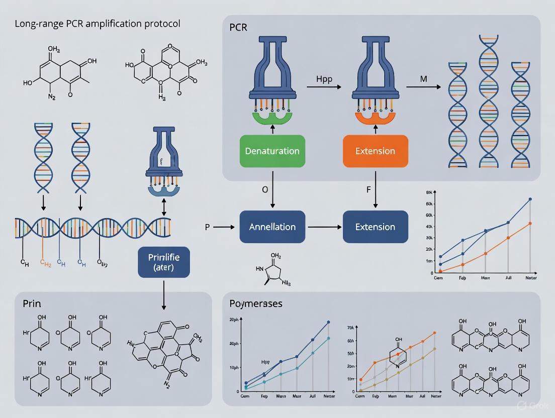
Diagram 1: LR-PCR Workflow. The diagram outlines the key steps in long-range PCR, highlighting critical parameters that require optimization for successful amplification of large fragments.
Essential Reagents and Research Solutions
Successful long-range PCR requires careful selection of reagents and specialized kits optimized for amplifying large fragments. The following table details key research reagent solutions used in established protocols.
Table 3: Essential Research Reagents for Long-Range PCR
| Reagent/Kits | Manufacturer | Key Features | Applications |
|---|---|---|---|
| PrimeSTAR GXL | TaKaRa | Polymerase blend, high processivity | General long-range PCR (up to 30 kb) [2] |
| LA Taq | TaKaRa | Proofreading activity, GC buffer option | Routine extensions up to 20 kb [3] |
| UltraRun LongRange | QIAGEN | High success rate for targets up to 22 kb | Clinical applications requiring consistency [5] |
| LongAmp Taq | New England Biolabs | Robust amplification, master mix format | High-throughput applications [6] |
| Platinum SuperFi II | Invitrogen | High fidelity, room temperature stability | Complex templates [5] |
| AMPure XP Beads | Beckman Coulter | Size-selective purification | Pre-sequencing clean-up [5] [6] |
| Nextera XT | Illumina | Tagmentation-based library prep | NGS library preparation [2] |
| Native Barcoding Kit | Oxford Nanopore | Barcoding for multiplexing | Long-read sequencing [5] |
Troubleshooting and Optimization Strategies
Common Challenges and Solutions
Despite standardized protocols, long-range PCR can present several technical challenges that require specific optimization strategies.
Template Quality and Integrity: The integrity of template DNA is paramount for successful long-range PCR. Template degradation significantly reduces amplification efficiency, particularly for larger fragments. High-quality genomic DNA with a DNA Integrity Number (DIN) greater than 7 is recommended for optimal results [1]. Storage conditions also affect performance; long-term storage at -30°C or lower helps maintain DNA integrity for long-range applications [1].
PCR Enhancers: For challenging templates, PCR enhancers can dramatically improve results. Studies have demonstrated that successful amplification of some long fragments was not possible without the use of specific enhancers [1]. Common additives include:
- DMSO (1-3%): Reduces secondary structure in GC-rich templates
- Betaine (0.5-1.5 M): Equalizes Tm differences in heterogeneous templates
- Formamide (1-5%): Destabilizes DNA secondary structures
- BSA (0.1-0.5 μg/μL): Counteracts inhibitors in crude preparations
Minimizing Artifacts: Long-range PCR is particularly susceptible to chimeric reads—artifacts formed when incomplete amplicons act as megaprimers in subsequent cycles. Optimized conditions can maintain the median proportion of chimeric reads at 2.80% (range 1.79-16.12%) [5]. Strategies to minimize chimeras include:
- Reducing cycle number (often 26-30 cycles)
- Limiting extension times to discourage incomplete synthesis
- Using polymerases with high processivity
- Implementing touchdown cycling protocols
Diagram 2: LR-PCR Troubleshooting. The diagram outlines common problems encountered in long-range PCR experiments and their corresponding evidence-based solutions for effective optimization.
Special Considerations for Sequencing Applications
When long-range PCR is used as a precursor to sequencing, additional considerations apply to ensure high-quality results. For Nanopore sequencing, researchers must balance sufficient product yield with the need to minimize PCR artifacts. Typically, only 1 ng of PCR product is required for reliable barcoding, enabling lower cycle numbers that reduce amplification bias [6].
Temperature stability becomes crucial when processing multiple samples, particularly in automated workflows. PrimeSTAR LongSeq DNA Polymerase maintains high specificity after prepared PCR reactions are stored at 4°C for 17 hours or at room temperature for 1 hour, providing flexibility for high-throughput applications [4].
For multiplexed long-range PCR targeting multiple genomic regions simultaneously, reaction conditions require additional optimization. Uniform amplification across targets with different characteristics (e.g., varying GC content) can be achieved with advanced polymerase systems that demonstrate consistent performance across 20-plex reactions of repetitive DNA sequences [4].
Long-range PCR represents a powerful methodology that has substantially expanded the capabilities of PCR-based genomic analysis. The technique now reliably supports amplification of fragments from 5 kb to over 30 kb, with advanced systems pushing these limits beyond 50 kb. Successful implementation requires careful attention to enzyme selection, template quality, and reaction optimization, particularly when integrating with downstream applications like next-generation sequencing. As polymerase formulations continue to improve and sequencing technologies advance, long-range PCR remains an essential tool for modern genomic research, enabling efficient analysis of large genomic regions, complex structural variations, and challenging sequences that were previously inaccessible to PCR-based approaches.
The Polymerase Chain Reaction (PCR) has been an indispensable tool in molecular biology since its inception, but its application was traditionally limited to the amplification of small DNA fragments. The advent of Long-Range PCR has fundamentally expanded these possibilities, enabling reliable amplification of DNA targets exceeding 5 kilobases (kb) and extending up to 20 kb or more [7]. This transition from standard to long-amplicon amplification was not a trivial increment but required key technological advancements, primarily in DNA polymerase engineering and buffer chemistry. These innovations have, in turn, empowered major scientific applications, particularly in next-generation sequencing (NGS), where long-range PCR provides a flexible and cost-effective method for targeting large genomic regions for detailed analysis [2]. This application note details the critical technical advancements underpinning long-range PCR and provides a standardized protocol for its implementation in research and development.
The Fundamental Technical Advancements
The primary limitation of standard PCR with a single Taq polymerase was its inability to efficiently amplify long products. This was largely due to the accumulation of misincorporated nucleotides, which would stall the polymerase. The key breakthrough came from rethinking the enzyme system itself.
Advancement 1: Blended Polymerase Systems The most significant technical advancement was the development of optimized polymerase blends. These blends typically combine a major proportion of a non-proofreading polymerase (like Taq) for high processivity and fast elongation with a minor proportion of a proofreading polymerase (like Pfu or Pwo) that possesses 3'→5' exonuclease activity [7]. The proofreading enzyme repairs mismatches incorporated by the main polymerase, effectively clearing the path for continued DNA synthesis and allowing for the successful amplification of much longer fragments.
Advancement 2: Enhanced Buffer Formulations Concurrently, specialized buffer systems were developed to support these complex reactions. These buffers often include optimized salt concentrations, pH stabilizers, and critical additives such as betaine or DMSO. These compounds help to disrupt secondary structures in the DNA template (e.g., GC-rich regions or hairpins) that would otherwise impede the polymerase's progress during elongation, thereby increasing the yield and specificity of long amplicons [2].
The following workflow diagram illustrates the conceptual shift from standard PCR to the advanced long-range PCR process.
Comparative Performance of Long-Range PCR Enzymes
Multiple commercial long-range DNA polymerases are available, each with varying performance characteristics. A systematic evaluation of six different enzymes was conducted to amplify three challenging amplicons (12.9 kb, 9.7 kb, and 5.8 kb) under both manufacturer-recommended and optimized conditions [2]. The results, summarized in the table below, provide critical empirical data for enzyme selection.
Table 1: Performance Comparison of Six Long-Range PCR Enzymes [2]
| Enzyme | Manufacturer | Advertised Max Size | 12.9 kb Amplicon | 9.7 kb Amplicon | 5.8 kb Amplicon | Key Characteristics |
|---|---|---|---|---|---|---|
| PrimeSTAR GXL | TaKaRa | > 30 kb | Success | Success | Success | Robust performance under universal conditions; high fidelity. |
| SequalPrep | Invitrogen | > 20 kb | Success | Success | Success | Reliable performance across multiple fragment sizes. |
| AccuPrime | Invitrogen | > 10 kb | Success | Failure | Success | Requires specific conditions for optimal performance. |
| LA Taq Hot Start | TaKaRa | > 40 kb | Success | Failure | Success | High processivity but may require condition optimization. |
| KAPA LongRange | KAPA Biosystems | > 15 kb | Failure | Failure | Success | Effective for shorter long-range targets. |
| QIAGEN LongRange | QIAGEN | > 10 kb | Failure | Failure | Success | Effective for shorter long-range targets. |
The study concluded that PrimeSTAR GXL DNA polymerase demonstrated superior performance, successfully amplifying almost all amplicons of different sizes and Tm values under a single, unified PCR condition [2]. This makes it a particularly versatile choice for applications requiring the simultaneous amplification of multiple large genomic regions, such as in the sequencing of entire genes like BRCA1 and BRCA2.
Application Notes: Long-Range PCR for Nanopore Sequencing
Long-range PCR is exceptionally well-suited for preparing templates for long-read sequencing platforms, such as Oxford Nanopore Technologies (ONT) [6]. The following section outlines a optimized protocol for generating long amplicons specifically for Nanopore sequencing.
Research Reagent Solutions
Table 2: Essential Reagents for Long-Range PCR and Library Preparation [6]
| Item | Function / Rationale | Example Product |
|---|---|---|
| High-Fidelity LR Polymerase | Provides high processivity and accuracy for amplifying long DNA fragments. | PrimeSTAR GXL, LongAmp Taq, Platinum SuperFi II |
| dNTPs | Building blocks for DNA synthesis. | Standard dNTP mix |
| Primers with ONT Overhangs | Gene-specific primers with universal primer sequences for subsequent library prep. | Custom synthesized primers |
| High-Quality DNA Template | Intact, pure genomic DNA is critical for long amplicon yield. | Phenol-chloroform extracted DNA |
| AMPure XP Beads | For post-PCR purification to remove primers, enzymes, and salts. | Agencourt AMPure XP |
| Library Prep Kit | Reagents for attaching sequencing adapters to amplicons. | ONT Ligation Sequencing Kit (e.g., LSK114) |
Detailed Protocol: Long-Range PCR for Nanopore Sequencing
- Target Selection: Retrieve the genomic sequence of your target gene, including 5' and 3' UTRs, using a browser like UCSC Genome Browser.
- Primer Design:
- Use Primer3Plus software. Mark the coding region to focus primer placement.
- Placement: Design primers within the 5' and 3' UTRs, close to the start and stop codons, to capture all isoforms.
- Critical: Avoid designing primers that span exon-exon boundaries, as this will miss isoforms that skip one of the exons.
- Parameters: Set product size to 1000-15000 bp. Primer Tm: Min 57°C, Opt 60°C, Max 63°C. GC clamp: 1-2.
- Specificity Check:
- Validate primer uniqueness using UCSC BLAT to check for off-target binding.
- Perform in-silico PCR (UCSC tool) to confirm the amplification of the intended single product.
II. PCR Amplification and Purification
- Reaction Setup: Assemble the following components on ice:
- 25 µL: 2X Long-Range PCR Master Mix (e.g., PrimeSTAR GXL or LongAmp Taq)
- 10 µL: 10 µM forward primer (with ONT universal primer overhang)
- 10 µL: 10 µM reverse primer (with ONT universal primer overhang)
- 100 ng: High-quality genomic DNA or cDNA
- Nuclease-free water to 50 µL
- Optional: Add DMSO to a final concentration of 2-5% for difficult templates [2].
- Thermal Cycling: Use the following 2-step protocol for PrimeSTAR GXL:
- Initial Denaturation: 98°C for 2 minutes
- 35 Cycles:
- Denaturation: 98°C for 10 seconds
- Annealing/Extension: 68°C for 1 minute per kb
- Final Extension: 68°C for 5 minutes
- Post-Amplification Analysis and Clean-up:
- Verify 1-5 µL of the PCR product on a 0.8% agarose gel.
- Purify the remaining PCR product using AMPure XP beads at a 1:1 bead-to-sample ratio to remove primers and dNTPs. Elute in nuclease-free water.
- Quantify the purified DNA using a fluorescence-based assay (e.g., Qubit).
The complete experimental workflow, from primer design to sequencing-ready library, is depicted below.
The journey from standard PCR to robust long amplicon amplification has been driven by key technical innovations in enzyme biochemistry, primarily the development of specialized polymerase blends and enhanced buffer systems. As demonstrated, enzymes like PrimeSTAR GXL offer researchers the capability to reliably amplify large genomic regions up to 20 kb or more. This capability, when integrated with modern long-read sequencing platforms like Nanopore, provides a powerful and streamlined workflow for targeted sequencing of complex genes, facilitating advanced research in genomics, diagnostics, and drug development. The protocols and data presented herein offer a reliable foundation for implementing this technology in a scientific setting.
The selection of an appropriate DNA polymerase is a critical step in experimental design, fundamentally influencing the success, accuracy, and reproducibility of polymerase chain reaction (PCR) outcomes. This decision is particularly crucial within the context of long-range PCR amplification research, where amplifying longer DNA fragments increases the probability of enzyme-introduced errors. The core distinction often lies in choosing between standard and high-fidelity DNA polymerases, each possessing unique biochemical properties tailored for specific applications [8]. Standard polymerases, such as Taq DNA polymerase, are renowned for their robustness and speed, making them ideal for routine applications like genotyping or qualitative PCR. In contrast, high-fidelity polymerases incorporate a proofreading mechanism, resulting in significantly higher replication accuracy, which is indispensable for downstream applications such as cloning, next-generation sequencing (NGS), and functional protein expression [9] [10]. This application note provides a detailed, data-driven comparison to guide researchers and drug development professionals in selecting the optimal polymerase for their long-range PCR protocols.
Polymerase Mechanisms and Fidelity
The fidelity of a DNA polymerase refers to its accuracy in incorporating nucleotides during DNA replication, ensuring the newly synthesized strand is a perfect copy of the template [9]. This accuracy is paramount for experiments where the correct DNA sequence is essential.
Mechanisms of Fidelity
DNA polymerases maintain high fidelity through a two-tiered system:
- Nucleotide Selection: The geometry of the polymerase active site preferentially selects and correctly aligns the complementary nucleoside triphosphate with the template base, ensuring efficient incorporation. An incorrect nucleotide creates a suboptimal architecture, slowing incorporation and increasing the chance it will dissociate before the polymerase proceeds [9].
- Proofreading (3´→5´ Exonuclease Activity): Some polymerases possess a separate 3´→5´ exonuclease domain. When an incorrect nucleotide is incorporated, it causes a perturbation that the polymerase detects. The growing DNA chain is then moved into this proofreading domain, where the mispaired nucleotide is excised. The chain is subsequently returned to the polymerase active site for the addition of the correct nucleotide [9] [8]. This proofreading activity is the defining feature of high-fidelity DNA polymerases.
Quantitative Fidelity Comparisons
Fidelity is quantitatively expressed as an error rate, typically in errors per base pair per duplication event. As shown in Table 1, error rates vary significantly between enzyme classes.
Table 1: DNA Polymerase Fidelity and Error Rates
| Polymerase Class | Example Enzymes | 3´→5´ Exo (Proofreading) | Error Rate (per bp/doubling) | Accuracy (1 error per X bases) | Fidelity Relative to Taq |
|---|---|---|---|---|---|
| Standard | Taq | No | ~1.5 x 10⁻⁴ [9] | ~6,500 bases [9] | 1x [11] |
| High-Fidelity | Q5, Phusion, Pfu | Yes | ~5.3 x 10⁻⁷ to ~5.1 x 10⁻⁶ [9] | ~1.87 million to ~195,000 bases [9] | 30x to 280x [11] [9] |
The data in Table 1, derived from advanced sequencing methods like PacBio SMRT sequencing, demonstrates that high-fidelity enzymes like Q5 can reduce error rates by up to 280-fold compared to Taq polymerase [9] [12]. This translates to a dramatically lower probability of introducing mutations during amplification, which is especially critical when amplifying long targets.
Comparative Analysis of DNA Polymerases
Beyond fidelity, several other enzymatic properties are critical for selecting a polymerase, particularly for long-range PCR. These properties determine how the enzyme interacts with the template and primers, and the characteristics of the final product.
Table 2: Key Properties of Standard and High-Fidelity DNA Polymerases
| Property | Standard Polymerase (e.g., Taq) | High-Fidelity Polymerase (e.g., Q5, Phusion) |
|---|---|---|
| DNA Polymerase Family | Family A [8] | Family B [8] |
| 5'→3' Exonuclease Activity | Yes [11] [8] | No [11] [8] |
| 3'→5' Exonuclease Activity (Proofreading) | No [11] [8] | Yes [11] [8] |
| Extension Speed | High (~150 nucleotides/second) [8] | Slower (~25 nucleotides/second) [8] |
| Resulting PCR Product Ends | 3´ 'A-overhangs' [11] | Blunt ends [11] |
| Primary Applications | Routine PCR, genotyping, colony PCR [11] | Cloning, sequencing, site-directed mutagenesis, NGS [11] [8] |
The presence of 3´→5´ proofreading activity in high-fidelity polymerases is the key factor behind their superior accuracy [8]. However, this activity can sometimes lead to the degradation of primers if the enzyme is not used in a hot-start format. Furthermore, the blunt-ended PCR products generated by most high-fidelity polymerases require different cloning strategies compared to the 'A-tailed' products from Taq polymerase [11] [8].
Workflow for Polymerase Selection
The following diagram outlines a systematic decision process for selecting between standard and high-fidelity DNA polymerases based on experimental goals.
Application Notes for Long-Range PCR
Long-range PCR presents unique challenges, including the need to amplify across complex or high-GC regions and the increased risk of introducing errors over longer sequences. Optimized protocols are essential for success.
Protocol: Long-Range PCR for Nanopore Sequencing
The following validated protocol is adapted from a study on phasing distantly separated variants, which utilized long-range PCR followed by Nanopore sequencing [5].
1. Reagent Setup:
- Template DNA: 150 ng of high-quality genomic DNA.
- Polymerase: UltraRun LongRange PCR Kit (Qiagen), LongAmp Taq 2X Master Mix (NEB), or similar long-range capable enzyme [5].
- Primers: 0.5 µM each of forward and reverse primer.
- PCR Mix: Prepare a 20 µl reaction volume with the manufacturer's recommended buffer.
2. Thermal Cycling Conditions:
- Initial Denaturation: 98°C for 30 seconds.
- Amplification (25-30 cycles):
- Denaturation: 98°C for 10 seconds.
- Annealing: Use a single temperature (e.g., 62-65°C) for 15-30 seconds. A universal annealing temperature protocol can be used with some polymerases like Q5 [12].
- Extension: 68°C for 1-2 minutes per kb. Note: The extension time is critical and must be optimized for the polymerase and amplicon length.
- Final Extension: 68°C for 5-10 minutes.
- Hold: 4°C.
3. Post-PCR Analysis:
- Verify amplification success and specificity by analyzing 5 µl of the PCR product using the Agilent 4200 TapeStation System or agarose gel electrophoresis. A clear, single band at the expected size indicates a successful amplification [5].
4. Critical Step - Minimizing Chimeras:
- Long-range PCR is susceptible to chimeric reads, a PCR artefact where two different biological sequences are joined. To minimize this:
- Limit Cycle Number: Using 25-30 cycles, instead of a higher number, significantly reduces chimeric formation. One study maintained a median chimera rate of 2.8% using 26 cycles [5].
- Optimize Template Quality: Use intact, high-quality DNA.
Workflow for Long-Range PCR and Sequencing
This workflow illustrates the end-to-end process, from sample preparation to data analysis, for a long-range PCR project aimed at sequencing.
The Scientist's Toolkit: Essential Reagents for Long-Range PCR
Successful long-range PCR requires a set of critical reagents, each serving a specific function to ensure high yield and accuracy.
Table 3: Research Reagent Solutions for Long-Range PCR
| Reagent | Function | Example Products & Notes |
|---|---|---|
| High-Fidelity/LR Polymerase | Catalyzes DNA synthesis with high accuracy over long distances. | Q5 Hot Start (NEB), LongAmp Taq (NEB), UltraRun LongRange (Qiagen) [11] [5]. Fused to processivity-enhancing Sso7d in Q5 [12]. |
| Optimized Reaction Buffers | Provides optimal pH, ionic strength, and co-factors (Mg²⁺) for polymerase activity. | Often supplied with polymerase. Specialized buffers (e.g., GC Enhancer) available for difficult templates [12]. |
| dNTP Mix | The building blocks (dATP, dCTP, dGTP, dTTP) for DNA synthesis. | Use high-quality, neutral-pH dNTPs to prevent degradation and ensure efficient incorporation. |
| Target-Specific Primers | Short DNA sequences that define the start and end points of amplification. | Designed with tools like NCBI Primer BLAST. High purity (HPLC purified) is recommended for long-range PCR [5]. |
| Template DNA | The source DNA containing the target sequence to be amplified. | High-quality, intact genomic DNA is crucial. Assess quality via spectrophotometry and gel electrophoresis. |
| Library Prep Kit | For preparing PCR amplicons for sequencing (e.g., end-repair, barcoding). | Ligation Sequencing Kit & Native Barcoding Kit (Oxford Nanopore) [5]. |
| Nucleic Acid Stain | For visualizing amplified DNA fragments post-PCR. | SYBR Green, Ethidium Bromide, or GelRed for agarose gel analysis [13]. |
The selection between a high-fidelity and a standard DNA polymerase is a strategic decision that directly impacts the integrity of experimental data. For long-range PCR amplification research, where error accumulation and amplification efficiency are major concerns, the use of a proofreading, high-fidelity polymerase is strongly recommended. Enzymes such as Q5 and Phusion provide the ultra-low error rates necessary for cloning, sequencing, and other applications demanding sequence perfection [9] [12]. Standard polymerases like Taq remain excellent tools for rapid, qualitative analyses where ultimate sequence accuracy is less critical. By adhering to the detailed protocols and selection guidelines outlined in this document, researchers can robustly implement long-range PCR strategies, thereby enhancing the reliability and success of their genetic analyses and drug development workflows.
Long-range polymerase chain reaction (LR-PCR) enables the amplification of DNA fragments spanning several kilobases, a capability that has become indispensable in modern genomics. [7] By leveraging specialized enzyme blends that combine high processivity with proofreading activity, researchers can reliably generate amplicons from 5 kb to over 20 kb. [7] [5] This technical advancement provides the foundation for key applications across next-generation sequencing (NGS) library preparation, comprehensive viral genome sequencing, and complex cloning workflows. The protocol detailed herein establishes a standardized framework for implementing LR-PCR within research and diagnostic settings, with particular emphasis on its utility in targeted enrichment for NGS and genomic surveillance of viral pathogens.
Application Note: Key Genomic Applications of LR-PCR
Long-range PCR serves as a critical enabling technology across multiple genomics domains, each with distinct requirements and experimental considerations.
Table 1: Core Applications of Long-Range PCR in Modern Genomics
| Application Domain | Primary Utility | Typical Amplicon Size | Key Benefit |
|---|---|---|---|
| NGS Library Preparation | Targeted enrichment for sequencing [14] | 5-10 kb [14] | Cost-effective, customizable alternative to commercial capture kits [14] |
| Viral Genome Sequencing | Whole-genome sequencing of pathogens like SARS-CoV-2 and HIV-1 [15] [16] | 1-4.5 kb [15] [16] | Reduces amplicon dropout; enables sequencing from low-input samples [15] |
| Genetic Disorder Diagnosis | Multi-gene panel testing for conditions like Lysosomal Storage Disorders (LSDs) [14] | 5-10 kb [14] | Identifies variants across heterogeneous genetic conditions [14] |
| Variant Phasing | Determining cis/trans relationships of distantly spaced variants [5] | 1-20+ kb [5] | Resolves compound heterozygosity; critical for autosomal recessive conditions [5] |
NGS Library Preparation and Targeted Enrichment
LR-PCR provides a highly customizable and cost-effective method for targeted capture and enrichment of genomic regions of interest prior to NGS. This approach is particularly valuable for multigene panel testing, as demonstrated in the molecular diagnosis of lysosomal storage disorders (LSDs), where researchers successfully designed LR-PCR primers for 22 genes, creating fragments of 5-10 kb that covered entire exonic regions with flanking sequences. [14] The method proved reliable for detecting both homozygous and heterozygous variants, offering a financially viable strategy for resource-poor settings compared to commercial target enrichment kits. [14]
Viral Genome Sequencing
In viral genomics, LR-PCR and tiling PCR approaches have revolutionized surveillance efforts for pathogens such as SARS-CoV-2 and HIV-1. For SARS-CoV-2, a primer set generating seven ~4.5 kb amplicons tiled across the viral genome demonstrated a significant advantage: by positioning primer binding sites in highly conserved regions flanking the mutation-prone spike (S) gene, this method minimized amplicon dropouts that plagued earlier multiplex PCR schemes. [15] Similarly, for HIV-1, a novel tiling PCR protocol amplifying the 5' half of the genome in six overlapping 1 kb segments enabled more comprehensive drug resistance profiling and identification of additional resistance mutations missed by conventional Sanger sequencing. [16]
Clinical Variant Phasing and Localization
Long-range PCR coupled with long-read sequencing (e.g., Oxford Nanopore Technologies) enables precise phasing of distantly separated variants, a critical capability for diagnosing autosomal recessive disorders. [5] One optimized workflow achieved 100% concordance in phasing heterozygous single nucleotide variants and small indels separated by 5.8 to 21.4 kb. [5] This approach is also invaluable for analyzing genomic regions with high homology or low mappability, where short-read NGS often fails, by allowing primers to be placed in unique sequences far from the variant of interest. [5]
Quantitative Performance Data
The performance of LR-PCR across different experimental conditions and kit systems has been systematically evaluated in recent studies.
Table 2: Performance Metrics of Long-Range PCR Across Applications
| Experimental Context | Performance Metric | Result | Reference |
|---|---|---|---|
| LSD Genetic Testing | Success rate for variant detection | Reliable detection of homozygous/heterozygous variants [14] | [14] |
| SARS-CoV-2 Sequencing | Amplicon size vs. primer count | 7 amplicons of ~4.5 kb vs. 98 (ARTIC) or 29 (Midnight) [15] | [15] |
| Variant Phasing | Phasing accuracy over long distances | 100% concordance for variants 5.8-21.4 kb apart [5] | [5] |
| HIV-1 Tiling PCR | Amplification success rate | 100% (90/90 samples) [16] | [16] |
| PCR Kit Comparison | Optimal kit success rate | 90% for amplification up to 22 kb [5] | [5] |
Experimental Protocol: Long-Range PCR for Genomic Applications
Primer Design and In Silico Validation
Effective LR-PCR begins with meticulous primer design. Follow these steps for optimal results:
- Design Parameters: Use Primer3Plus with the following settings: product size ranges 1000-15000 bp; primer size min 18, opt 20, max 27; Tm min 57°C, opt 60°C, max 63°C; GC clamp 1-2; max poly-X: 3-4. [6]
- Binding Site Selection: For RNA isoform profiling, place primers in 5' and 3' UTRs close to start and stop codons. Avoid designing primers across exon-exon boundaries to prevent amplification failure of specific isoforms. [6]
- Specificity Checking: Validate primer uniqueness using UCSC BLAT tool to find sequences with ≥95% similarity of length 25 bases or more. Perform in-silico PCR via UCSC tools to check for off-target amplification. [6]
RNA Extraction and Reverse Transcription
For viral sequencing applications, proper nucleic acid handling is critical:
- RNA Extraction: Use high-throughput systems like Roche MagNA Pure 96 Instrument with DNA and Viral NA small volume kit. Input 200 µL plasma, output 50 µL RNA. [16]
- RNA Quality: For RNA isoform profiling, RNA Integrity Number (RIN) should be >7, though RIN >6 works for many genes. Higher quality becomes more critical with longer amplification lengths. [6]
- Reverse Transcription: Use SuperScript VILO IV mastermix: add 4 µL VILO enzyme to 8 µL RNA sample in 20 µL final volume. Incubate 10 min at 25°C, 20 min at 50°C, 5 min at 85°C, then hold at 4°C. [16]
Long-Range PCR Amplification
The core amplification protocol varies by application:
- Standard LR-PCR: Use 150 ng genomic DNA, 0.5 µM each primer, and selected PCR mastermix in 20 µL reaction. Run for 26 cycles to minimize chimeric reads. [5]
- Tiling PCR for HIV-1: Create two primer pools (A and B) with primers combined equivalently. Mastermix contains 5 µL cDNA, 4 µL primer pool (1× A, 1× B), and 1X SuperFi II Green mastermix. [16]
- Thermocycler Conditions: Configure according to manufacturer recommendations. A single PCR program using a single annealing temperature and extension time can be established for each kit to enable running samples simultaneously. [5]
Library Preparation and Sequencing
For Nanopore sequencing applications:
- Library Preparation: Use Ligation Sequencing Kit V14 with Native Barcoding Kit. Repair amplicons with Ultra II End-prep Enzyme Mix, ligate barcodes with Blunt/TA Ligase Master Mix, and pool samples. [5]
- Sequencing: Ligate sequencing adapters using NEBNext Quick Ligation Reaction Buffer and Quick T4 DNA Ligase. Sequence 10 femtomoles of library on Flongle flow cells (R10.4.1) on GridION device. [5]
- Basecalling: Perform super accuracy basecalling (SUP) during sequencing using MinKNOW software with minimum Phred score of 10. [5]
Workflow Visualization
Figure 1: Comprehensive workflow for long-range PCR applications in genomics, spanning from sample preparation to bioinformatic analysis.
Research Reagent Solutions
Successful implementation of LR-PCR requires carefully selected reagents and kits optimized for long-fragment amplification.
Table 3: Essential Reagents for Long-Range PCR Applications
| Reagent Category | Specific Product Examples | Key Features & Applications |
|---|---|---|
| LR-PCR Polymerases | PrimeSTAR GXL DNA Polymerase [14] [6], Platinum SuperFi II PCR Master Mix [5] [6], LongAmp Taq 2X Master Mix [5] [6], UltraRun LongRange PCR Kit [5] | High-fidelity amplification; blend of Taq and proofreading polymerase; capable of amplifying targets >20 kb [7] [5] |
| Nucleic Acid Extraction | QIAamp DNA Blood Mini Kit [14], Roche MagNA Pure 96 with DNA and Viral NA kit [16] | High-quality DNA/RNA extraction crucial for long amplicon generation [14] [16] |
| Reverse Transcription | SuperScript VILO IV [16], Maxima H Minus Reverse Transcriptase [6] | Efficient cDNA synthesis for viral RNA sequencing and RNA isoform analysis [16] [6] |
| Library Preparation | Ligation Sequencing Kit V14 (SQK-LSK114) [5], Native Barcoding Kit 24 V14 (SQK-NBD114.24) [5] | Barcoding and adapter ligation for multiplexed Nanopore sequencing [5] |
| Purification & QC | Agencourt AMPure XP beads [6], Agilent Tape Station System [5] | Size selection and quality control of long amplicons prior to sequencing [5] [6] |
Long-range PCR has evolved into a fundamental tool enabling advances across multiple genomics domains. Its applications in NGS library preparation, viral sequencing, and clinical diagnostics demonstrate how this technology addresses specific challenges such as cost-effective targeted enrichment, comprehensive variant detection, and resolution of complex genomic rearrangements. The protocols and reagents detailed herein provide researchers with a robust framework for implementing these methods across diverse experimental contexts. As sequencing technologies continue to advance toward longer read lengths, LR-PCR remains an essential component of the genomic toolkit, bridging the gap between PCR-based amplification and the information-rich data generated by modern sequencing platforms.
Step-by-Step Protocols for Robust Long-Range PCR and NGS Integration
Within the framework of long-range polymerase chain reaction (LR-PCR) research, successful amplification of DNA fragments exceeding several kilobases hinges on meticulous primer design. Unlike standard PCR, LR-PCR places greater demands on primer specificity and thermodynamic stability to ensure efficient and accurate amplification over longer distances [17] [18]. This application note details a refined protocol for designing primers that optimize melting temperature (Tm), GC content, and minimize secondary structures, thereby enhancing the reliability of long-range amplification for downstream applications such as sequencing and functional genomic analysis.
Core Principles of Primer Design
The design of primers for LR-PCR is governed by several interdependent physicochemical principles. Adherence to these guidelines mitigates common pitfalls like nonspecific amplification, primer-dimer formation, and inefficient extension, which are more detrimental when targeting long amplicons [19] [20].
Primer Length and Specificity: Primers should be 18–30 nucleotides in length [19] [21] [22]. This range provides a balance between specificity and binding efficiency; longer primers within this range are often beneficial for complex templates like genomic DNA [19].
Melting Temperature (Tm): The Tm is the temperature at which 50% of the primer-DNA duplex dissociates. For a robust PCR, both the forward and reverse primers should have Tm values within 2–5°C of each other [22] [23]. The ideal Tm generally falls between 60–75°C [21] [22]. The annealing temperature (Ta) is typically set 2–5°C below the lowest Tm of the primer pair [22] [23].
GC Content and Stability: The optimal GC content for a primer is between 40–60% [19] [21] [24]. This ensures sufficient duplex stability without promoting nonspecific binding. A GC clamp—the presence of one or two G or C bases at the 3' end—strengthens terminal binding [21] [24]. However, runs of three or more consecutive G or C bases should be avoided [21] [22].
Avoiding Secondary Structures: Primers must be screened for self-complementarity to prevent the formation of hairpins (intramolecular folding) and primers-dimers (intermolecular annealing between primers) [19] [23]. These structures consume primers and can lead to spurious amplification products. Thermodynamic analysis tools can predict these interactions; structures with a free energy (ΔG) more negative than -9.0 kcal/mol should be avoided [22].
Table 1: Optimal Quantitative Parameters for PCR Primer Design
| Parameter | Optimal Range | Rationale | Special Consideration for Long-Range PCR |
|---|---|---|---|
| Primer Length | 18–30 nucleotides [19] [22] | Balances specificity and binding efficiency. | Longer primers (e.g., 24–30 nt) can enhance specificity for complex genomic templates [19]. |
Tm (Melting Temperature) |
60–75°C [21] [22] | Ensures stable hybridization under reaction conditions. | Primer pairs must be within 2°C of each other for synchronized binding [22]. |
Ta (Annealing Temperature) |
Tm - (2–5°C) [22] [23] |
Optimizes specific primer binding while reducing off-target annealing. | May require empirical optimization via gradient PCR [23]. |
| GC Content | 40–60% [19] [21] | Provides thermodynamic stability without increasing mispriming risk. | Avoid stretches of >3 G/Cs at the 3' end to prevent nonspecific initiation [21] [24]. |
| GC Clamp | 1–2 G/C bases at 3' end [21] [24] | Stabilizes the primer-template complex at the point of extension. | Critical for efficient initiation of polymerization in long fragments. |
Experimental Protocol for Primer Design and Validation
The following step-by-step protocol ensures systematic design and validation of primers suitable for long-range PCR.
In Silico Primer Design Workflow
- Define Target Sequence: Obtain the reference sequence (e.g., from NCBI in FASTA format). For LR-PCR, ensure the flanking regions are unique and suitable for primer binding [25].
- Utilize Design Tools: Use specialized software (e.g., NCBI Primer-BLAST, Primer3) with parameters set to the values in Table 1. Input the target sequence and constraints for product size (e.g., 3–20 kb for LR-PCR),
Tm, and GC content [25] [26]. - Screen for Specificity: Select candidate primer pairs and perform an in silico specificity check using BLAST against the relevant genome database to minimize off-target binding [22] [25].
- Analyze Secondary Structures: Use tools like OligoAnalyzer to check for hairpins and self-dimers. Discard primers with stable secondary structures (ΔG < -9.0 kcal/mol) [22].
Empirical Validation and Optimization
- Gradient PCR: Perform a thermal cycling reaction using a temperature gradient spanning the calculated
Ta (e.g., ±5°C). Use a high-fidelity DNA polymerase mix formulated for long-range amplification [17] [20]. - Analyze Products: Separate the PCR products by gel electrophoresis. The optimal
Ta yields a single, clear band of the expected size with minimal nonspecific products [23]. - Cycling Conditions for LR-PCR: Use short denaturation times (e.g., 10 seconds at 94°C) to reduce template depurination, which disproportionately affects long DNA fragments. A lower extension temperature of 68°C (vs. 72°C) can dramatically improve the yield of longer products [17].
- Sequencing Verification: For critical applications, Sanger or long-read sequencing (e.g., Nanopore) of the amplicon should be performed to confirm sequence fidelity and the absence of spurious mutations introduced during amplification [26].
The following workflow diagram summarizes the key steps from design to validation.
The Scientist's Toolkit: Research Reagent Solutions
Successful long-range PCR relies on a combination of optimized primers and specialized enzymatic and chemical reagents.
Table 2: Essential Reagents for Long-Range PCR
| Reagent / Material | Function / Rationale | Example Specifications |
|---|---|---|
| High-Fidelity DNA Polymerase Mix | A blend of a processive polymerase (e.g., Taq) and a proofreading polymerase (e.g., from archaebacteria). The proofreading activity (3'→5' exonuclease) corrects misincorporated nucleotides, which is critical for accurately replicating long templates [17] [18] [20]. | Kits such as UltraRun LongRange PCR Kit [26] or mixes containing proofreading enzymes [20]. |
| Template DNA of High Integrity | The starting DNA must be high molecular-weight and undegraded. Fragmented or depurinated template will prevent full-length amplification [20]. | High-quality genomic DNA with A260/A280 ratio of ~1.8, assessed by agarose gel for intact high molecular weight. |
| Betaine | An additive that destabilizes GC-rich secondary structures in the template DNA, which can halt polymerase progression. It significantly aids in the amplification of long and/or GC-rich targets [20]. | Typically used at a concentration of 1–1.5 M in the final reaction [20]. |
| dNTP Mix | The building blocks for DNA synthesis. A balanced, high-quality mixture is essential to prevent misincorporation that can lead to chain termination [17]. | Neutralized pH, PCR-grade, used at 200-400 µM each. |
| HPLC-Purified Primers | Purification by HPLC or cartridge methods removes truncated oligonucleotides and synthesis byproducts, ensuring a high concentration of full-length primer for efficient and specific initiation [19] [21]. | >80% full-length sequence, resuspended in TE buffer or nuclease-free water. |
Mastering the intricacies of primer design for Tm, GC content, and secondary structure avoidance is a foundational skill for successful long-range PCR. By adhering to the quantitative guidelines, following the systematic experimental protocol, and utilizing the appropriate reagent toolkit outlined in this document, researchers can significantly improve the yield and fidelity of long amplicons. This, in turn, enhances the reliability of downstream analyses in advanced genetic research and diagnostic assay development.
Within the broader research on long-range PCR amplification protocols, the standardization of reaction setup is a critical determinant of success. Amplifying DNA fragments longer than 3–4 kb presents unique challenges, including nonspecific primer annealing, the formation of stable secondary structures, and enzyme-associated errors, which are less prevalent in standard PCR [17]. This application note provides a detailed, actionable framework for researchers and drug development professionals to achieve robust, reproducible amplification of long DNA targets. By meticulously optimizing buffer compositions and thermal cycling parameters, it is possible to overcome these hurdles, ensuring high fidelity and yield for sensitive downstream applications such as cloning and next-generation sequencing.
Optimized Buffer Compositions
The chemical environment of the PCR reaction is fundamental to its success, especially for long and complex templates. An optimized buffer goes beyond providing a simple salt solution; it stabilizes the DNA polymerase, facilitates specific primer-template binding, and denatures stubborn secondary structures.
Core Buffer Components
Table 1: Essential Components of a Long-Range PCR Buffer
| Component | Typical Concentration | Function | Optimization Consideration |
|---|---|---|---|
| Mg2+ | 1.5–2.5 mM [27] | Essential cofactor for DNA polymerase activity; stabilizes primer-template duplex [27]. | Concentration is critical; too low causes no yield, too high reduces fidelity and promotes nonspecific binding [27]. |
| Proofreading Polymerase | Enzyme-specific | Provides 3'→5' exonuclease activity to remove misincorporated bases, drastically reducing error rates [17] [27]. | A blend of Taq and a proofreading enzyme (e.g., Pfu) often yields optimal results. |
| dNTPs | 200–250 µM each | Building blocks for DNA synthesis. | Balance with Mg2+ concentration, as Mg2+ chelates dNTPs [27]. |
Specialized Additives
For challenging templates, such as those with high GC content (>65%), the inclusion of specific additives can dramatically improve results [27].
- DMSO (Dimethyl Sulfoxide): Used at 2–10%, DMSO interferes with the formation of hydrogen bonds in DNA, helping to resolve secondary structures like hairpin loops by lowering the template's melting temperature (Tm) [27].
- Betaine: At a concentration of 1–2 M, betaine homogenizes the thermal stability of DNA duplexes. It equalizes the contribution of GC-rich and AT-rich regions, preventing the "breathing" of AT-rich areas and facilitating the denaturation of GC-rich stretches, thereby improving the yield and specificity of long-range PCR [27].
Experimental Protocol for Long-Range PCR
Reagent Setup and Workflow
The following workflow outlines the standardized procedure for setting up a long-range PCR reaction.
Procedure:
- Master Mix Preparation: Assemble all reaction components on ice in a sterile, nuclease-free microcentrifuge tube. A typical 50 µL reaction is outlined below. Always include a negative control (no template DNA) to check for contamination.
- Template DNA: 10–100 ng genomic DNA or 1–10 ng plasmid DNA.
- 10x Reaction Buffer: 5 µL (supplied with polymerase).
- MgCl2 (25 mM): 3–5 µL (adjust to final optimal concentration).
- dNTP Mix (10 mM each): 1 µL.
- Forward Primer (10 µM): 1.25 µL.
- Reverse Primer (10 µM): 1.25 µL.
- High-Fidelity DNA Polymerase Blend: 0.5–1 µL (or as per manufacturer's instructions).
- Additive (e.g., DMSO or Betaine): 0–5 µL (e.g., 2.5 µL of 100% DMSO for a 5% final concentration).
- Nuclease-Free Water: to 50 µL.
- Mixing and Loading: Gently mix the reaction by pipetting and briefly centrifuge to collect the contents at the bottom of the tube.
- Thermal Cycling: Transfer the tubes to a pre-heated thermal cycler and initiate the program detailed in Section 3.2.
Optimized Cycling Conditions
Thermal cycling parameters for long-range PCR require careful adjustment to minimize DNA damage and ensure complete extension. The relationship between these steps is critical.
Table 2: Standardized Long-Range PCR Cycling Protocol [17]
| Step | Temperature | Time | Cycles | Rationale |
|---|---|---|---|---|
| Initial Denaturation | 95°C | 2–5 min | 1 | Ensures complete separation of double-stranded template and may activate hot-start enzymes [28]. |
| Denaturation | 94°C | 10–30 seconds | 25–40 | Very short denaturation minimizes depurination of the long DNA template, which is a major cause of amplification failure [17]. |
| Annealing | 50–68°C | 0.5–2 min | 25–40 | Temperature is critical and must be optimized (see 3.3). Time is typically sufficient for primer binding [28]. |
| Extension | 68°C | 1 min/kb | 25–40 | A slightly lower temperature (vs. 72°C) improves the yield of longer products. Time is based on polymerase speed and product length [17] [28]. |
| Final Extension | 68°C | 5–15 min | 1 | Ensures all nascent strands are fully extended, improving the proportion of full-length product [28]. |
| Hold | 4°C | ∞ | – | Short-term storage of the reaction. |
Annealing Temperature Optimization
The annealing temperature (Ta) is the most critical variable for specificity. A starting Ta can be calculated from the primer melting temperature (Tm) using the formula: Tm = 4(G + C) + 2(A + T) [28]. The most efficient method for determination is gradient PCR, where a range of annealing temperatures (e.g., 50–68°C) is tested across different wells of the same thermal cycler run [27]. The optimal Ta is the highest temperature that produces a strong, specific target band. If nonspecific products are observed, increase the Ta in 2°C increments; if yield is low, decrease it [28].
The Scientist's Toolkit: Research Reagent Solutions
Table 3: Essential Materials for Long-Range PCR
| Reagent / Solution | Function | Key Considerations |
|---|---|---|
| High-Fidelity DNA Polymerase Blend | Catalyzes DNA synthesis with 3'→5' proofreading exonuclease activity for high-fidelity amplification of long fragments [17] [27]. | Select enzymes derived from hyperthermophilic archaea for superior thermostability and low error rates. |
| MgCl2 Solution | Provides Mg2+ ions, an essential cofactor for polymerase activity and primer-template duplex stability [27]. | Requires precise titration (1.5-2.5 mM) for each new primer/template set. Sold separately from the buffer for optimization. |
| GC-Rich Enhancer / Additives | A proprietary solution or additives like DMSO and betaine that modify DNA melting behavior to resolve secondary structures in GC-rich templates [17] [27]. | Critical for amplifying difficult templates. The composition may be optimized for the specific polymerase system. |
| Nuclease-Free Water | The solvent for the reaction, free of nucleases that would degrade primers and template. | Essential for reaction consistency and preventing false negatives. |
| dNTP Mix | The equimolar solution of deoxynucleoside triphosphates (dATP, dCTP, dGTP, dTTP) that serve as the building blocks for DNA synthesis [27]. | Quality is paramount; impurities can inhibit the polymerase and reduce yield. |
The characterization of the human immunodeficiency virus type 1 (HIV-1) genome is a critical component of clinical management, enabling the detection of drug resistance mutations (DRMs) that can compromise antiretroviral therapy efficacy. Historically, standard of care genotyping has focused primarily on sequencing the pol region of the HIV-1 genome using Sanger sequencing methods, providing limited coverage of key drug targets including protease (PR), reverse transcriptase (RT), and integrase (IN) [16] [29]. This targeted approach, while clinically useful, fails to capture the full genetic complexity of HIV-1 and misses potential resistance mutations outside the pol region.
The widespread adoption of next-generation sequencing (NGS) during the SARS-CoV-2 pandemic has transformed viral diagnostic sequencing, creating new opportunities for more comprehensive HIV-1 genomic analysis [16]. The tiling PCR approach, successfully implemented for SARS-CoV-2 whole genome sequencing, offers a promising methodology for HIV-1 that leverages the strengths of NGS while overcoming limitations of traditional methods [16]. This application note details the development and verification of a novel tiling PCR method for long-range HIV-1 sequencing in a diagnostic setting, providing researchers with a framework for implementing this advanced approach to achieve more comprehensive HIV-1 genomic characterization.
Tiling PCR Methodology: Design Principles and Workflow
Primer Design Strategy for Pan-Subtype Coverage
The fundamental challenge in HIV-1 primer design stems from the virus's exceptional genetic diversity. To address this, the tiling PCR assay was specifically designed to target the most common HIV-1 subtypes, with primers optimized for subtypes B, C, and CRF01_AE [16]. The design process incorporated the following key steps:
- Reference Sequence Curation: All full-length (>8500 bp) subtype B (n=6757), C (n=2588), and CRF01_AE (n=1505) HIV-1 genome sequences were downloaded from GenBank to create comprehensive reference alignments [16].
- Iterative Primer Optimization: PrimalScheme analysis was performed with iterative redesign based on multiple parameters, including <3 mismatches to representative alignments, amplicon length of 0.6–1.5 kb, Tm between 55°C and 60°C, presence of GC clamp, and absence of self-dimer/hairpin formation [16].
- Segmental Amplification Approach: The final design consists of six overlapping segments of approximately 1,000 bp each, covering the 5' half of the HIV-1 genome (gag-vpu) to ensure coverage of clinically important regions [16].
Table 1: Tiling PCR Primer Design Specifications
| Design Parameter | Specification | Rationale |
|---|---|---|
| Amplicon Length | 0.6-1.5 kb | Balance between amplification efficiency and coverage |
| Segment Overlap | >100 bp | Ensure complete genome coverage and assembly |
| Annealing Temperature | 55-60°C | Standardize thermal cycling conditions |
| Primer Binding Sites | Conserved across subtypes B, C, CRF01_AE | Ensure broad subtype coverage |
Experimental Workflow: From Sample to Sequencer
The tiling PCR method features a streamlined workflow that can move from sample to sequencer in under one day [16], representing a significant improvement over traditional nested PCR approaches that typically require approximately 20 hours for the total workflow [29]. The complete experimental procedure consists of the following key stages:
- RNA Extraction: High-throughput extraction of RNA from plasma samples using the Roche MagNA Pure 96 Instrument with the DNA and Viral NA small volume kit, processing 200 µl input volume with 50 µl output volume [16].
- Reverse Transcription: Conversion of extracted RNA to cDNA using SuperScript VILO IV mastermix, with incubation for 10 min at 25°C, 20 min at 50°C, and 5 min at 85°C [16].
- Tiling PCR Amplification: Two parallel PCR reactions are performed using primer pools A and B, with each pool containing primers for non-overlapping segments of the genome. The reaction uses SuperFi II Green mastermix with 5 µl of cDNA template and 4 µl of primer pool per reaction [16].
- Library Preparation and Sequencing: Standard NGS library preparation followed by sequencing on appropriate platforms.
The following workflow diagram illustrates the complete experimental procedure from sample collection through sequencing:
Key Research Reagent Solutions
Successful implementation of the tiling PCR method requires specific reagent systems optimized for long-range amplification and HIV-1 sequencing. The following table details essential research reagents and their functions within the protocol:
Table 2: Essential Research Reagents for HIV-1 Tiling PCR
| Reagent/System | Function | Specification/Notes |
|---|---|---|
| Roche MagNA Pure 96 | Automated nucleic acid extraction | DNA and Viral NA small volume kit; 200 µl input/50 µl output [16] |
| SuperScript VILO IV | Reverse transcription | Generates cDNA from viral RNA templates [16] |
| SuperFi II Green Mastermix | Tiling PCR amplification | High-fidelity polymerase for long-range amplification [16] |
| Custom Primer Pools | Genome-specific amplification | Pool A and B for non-overlapping segments [16] |
| PrimeSTAR LongSeq DNA Polymerase | Alternative for challenging templates | Amplifies targets up to 53 kb; effective for GC/AT-rich regions [4] |
Performance Verification and Comparative Analysis
Analytical Performance Across Diverse Samples
The tiling PCR method was rigorously verified following procedures from the WHO HIVResNet HIV Drug Resistance Laboratory Operational Framework [16]. Performance assessment utilized a panel of 90 HIV-infected samples from 54 individuals with viral loads ranging from 1,295 to 1,301,198 copies/mL, encompassing both common (CRF01AE, B, C) and rare (CRF02AG, D, F, G) subtypes in the Australian epidemic [16]. The verification results demonstrated:
- Universal Amplification Success: The tiling PCR generated HIV-1 sequences from all 90 (100%) samples in the comparison panel [16].
- Viral Load Dependency: Complete protease-reverse transcriptase and integrase regions were amplified in >90% of samples with a viral load >5000 copies/mL [16].
- Enhanced Mutation Detection: Seven additional drug resistance mutations were identified using the novel tiling PCR method compared to conventional Sanger sequencing approaches [16].
Table 3: Performance Metrics of HIV-1 Tiling PCR Across Viral Load Ranges
| Viral Load (copies/mL) | Sample Success Rate | PR-RT Amplification | IN Amplification |
|---|---|---|---|
| <5,000 | 100% | >90% | >90% |
| 5,000-50,000 | 100% | >90% | >90% |
| >50,000 | 100% | >90% | >90% |
Advantages Over Conventional Sequencing Methods
The tiling PCR method for HIV-1 sequencing offers several significant advantages compared to traditional Sanger sequencing and targeted NGS approaches:
- Expanded Genomic Coverage: By sequencing the 5' half of the HIV-1 genome (gag-vpu) rather than just the pol region, the assay captures potential DRMs outside conventional target regions and provides more comprehensive data for molecular epidemiology studies [16].
- Efficiency for NGS: The multiplexed amplification strategy requires only two PCR reactions for long-range sequencing, significantly reducing hands-on time and reagent costs compared to multiple individual amplifications [16].
- Robustness: The overlapping segment design makes the method less susceptible to the impacts of sample degradation than full-genome PCR methods and more effective for amplifying low viral load samples [16].
- Future-Proofing: As curative, therapeutic, and vaccine strategies targeting regions outside pol advance, this comprehensive sequencing approach ensures the assay will not require modification to capture additional relevant mutations [16].
Technical Considerations and Implementation Guidelines
Addressing PCR Challenges for Complex Templates
Amplification of HIV-1 genomic material presents unique technical challenges due to the virus's genetic diversity, secondary structures, and the potential for PCR inhibitors in clinical samples. Several strategies can enhance PCR performance for these complex templates:
- PCR Enhancers: Chemical additives such as betaine, dimethyl sulfoxide (DMSO), formamide, and dithiothreitol (DTT) can improve yields of difficult PCRs by various mechanisms, including stabilizing DNA polymerases, lowering strand separation temperatures, and promoting DNA denaturation [30].
- Specialized Polymerase Systems: Enzymes such as PrimeSTAR LongSeq DNA Polymerase are specifically optimized for amplifying long, GC-rich, or repetitive templates and can efficiently generate amplicons up to 53 kb, even with challenging sequences [4].
- Enhanced Specificity Designs: The tiling approach with carefully designed overlapping segments minimizes amplification gaps and ensures complete coverage even with primer-binding site variations [16].
Quality Control and Bioinformatics Considerations
Successful implementation of the tiling PCR method requires robust quality control measures and appropriate bioinformatic analysis:
- Batch Processing: Samples should be randomly shuffled and split into multiple batches to ensure laboratory operators remain blinded to input characteristics and to assess assay reliability across separate runs [16].
- Negative Controls: Uninfected plasma samples should be included in each run to monitor for potential contamination [16].
- Bioinformatic Processing: Specialized pipelines are required for analyzing tiling PCR data, including reference-based assembly, variant calling, and DRM identification. Platforms such as Exatype provide specialized analysis for HIV-1 sequencing data [29].
- Validation Framework: Verification should follow established guidelines such as the WHO HIVResNet HIV Drug Resistance Laboratory Operational Framework to ensure clinical reliability [16].
The development of this novel tiling PCR method represents a significant advancement in HIV-1 genomic sequencing, bridging the gap between traditional targeted approaches and the potential of NGS technologies. By providing comprehensive coverage of the 5' HIV-1 genome in a efficient, cost-effective workflow suitable for diagnostic settings, this methodology enables more complete characterization of drug resistance mutations and enhances molecular epidemiology capabilities.
As HIV-1 cure research advances, with strategies targeting diverse regions of the viral genome moving toward clinical application, comprehensive sequencing approaches like tiling PCR will become increasingly essential for both clinical management and research applications. The method's adaptability to different sequencing platforms and efficiency in resource utilization make it particularly valuable for implementation in both high-resource and limited-resource settings, potentially expanding access to advanced HIV-1 genotyping globally.
Future development directions include extending coverage to the 3' half of the genome, particularly env, adapting the method for proviral DNA sequencing in reservoir studies, and further optimizing primer designs to encompass additional HIV-1 subtypes and recombinant forms. As NGS technologies continue to evolve, tiling PCR methodologies provide a flexible framework that can leverage these advances to further enhance HIV-1 characterization and clinical management.
Within the framework of a comprehensive thesis on long-range PCR amplification, the integration of amplification products with next-generation sequencing platforms is a critical step. This protocol details the precise methodologies for preparing long-range PCR amplicons for sequencing on both Oxford Nanopore Technologies (ONT) and Illumina platforms. Long-range PCR enables the amplification of genomic targets from 1 kb up to 20 kb or more, facilitating the analysis of large genes, haplotype phasing, and sequencing through complex regions [5]. The selection between ONT for long-read, real-time sequencing and Illumina for high-accuracy short-read sequencing is dictated by the specific research objectives, such as the need for variant phasing or ultra-deep coverage of targeted regions [5] [31] [32]. This application note provides a standardized, end-to-end workflow to ensure the generation of high-quality sequencing data from long-range PCR products.
Platform Comparison and Selection
The choice between sequencing platforms dictates the experimental design, library preparation protocol, and the type of biological information that can be recovered. The table below summarizes the core characteristics of each platform in the context of long-range amplicon sequencing.
Table 1: Key sequencing platform characteristics for long-range amplicon analysis
| Feature | Oxford Nanopore Technologies (ONT) | Illumina |
|---|---|---|
| Read Length | Long-reads (full-length amplicons up to 20+ kb) [5] | Short-reads (e.g., 2x300 bp) [33] |
| Primary Application | Phasing distantly separated variants, resolving regions of high homology, structural variant analysis [5] [34] | Ultra-deep targeted sequencing, somatic variant discovery, 16S rRNA profiling [33] [31] |
| Typical Workflow Speed | Rapid, real-time data availability; library prep in hours [32] | Library prep in 5-7.5 hours; sequencing in 17-32 hours [35] |
| Key Strength | Determines haplotype and phase of variants up to ~20 kb apart [5] | High per-base accuracy, excellent for single nucleotide variant detection [31] [32] |
| Example Data Outcome | Phasing of compound heterozygous variants [5] | High-confidence variant calls for mutation screening [35] |
The decision pathway for selecting the appropriate sequencing platform for a given research goal can be visualized in the following workflow.
The Scientist's Toolkit: Research Reagent Solutions
Successful workflow integration depends on the selection of appropriate reagents and kits. The following table catalogues essential materials and their functions for long-range PCR and subsequent sequencing library preparation.
Table 2: Essential research reagents and kits for long-range PCR and sequencing
| Reagent / Kit | Function / Application | Specific Example Kits (Vendor) |
|---|---|---|
| High-Fidelity Long-Range PCR Master Mix | Amplifies long DNA targets (1–22 kb) with high accuracy; minimizes misincorporation errors. | UltraRun LongRange PCR Kit (Qiagen), Platinum SuperFi II (Invitrogen), LongAmp Taq 2X Master Mix (NEB) [5] [6] |
| Library Prep Kit (ONT) | Prepares amplicon libraries for Nanopore sequencing via barcoding and adapter ligation. | Ligation Sequencing Kit (SQK-LSK114) with Native Barcoding Kit (SQK-NBD114.24) [5] [6] |
| Library Prep Kit (Illumina) | Prepares amplicon libraries for Illumina sequencing, often with streamlined, rapid workflows. | AmpliSeq for Illumina, Illumina DNA Prep, Nextera XT DNA Library Prep Kit [33] [35] |
| DNA Clean-up Beads | Purifies and size-selects PCR products and final sequencing libraries. | Agencourt AMPure XP (Beckman Coulter) [5] [6] [32] |
| Flow Cell / Reagent Cartridge | The consumable where sequencing occurs; choice depends on scale. | ONT Flongle Flow Cell (R10.4.1) [5]; Illumina MiSeq Reagent Kits (v3) [33] |
Experimental Protocol: End-to-End Workflow
Stage 1: Long-Range PCR Amplification
The initial and most critical wet-lab phase is the robust amplification of the target region.
Step 1: Primer Design. Design primers using tools like Primer3Plus. For phasing, ensure a single amplicon spans all variants of interest. For ONT, primers can be designed with universal tails (e.g., ONT Universal Primers) [6]. Verify primer specificity using in-silico PCR tools (e.g., UCSC BLAT) to avoid off-target amplification [6].
Step 2: PCR Optimization. Set up reactions in a final volume of 20 µL using 150 ng of DNA template and 0.5 µM of each primer [5]. Test different polymerases if initial amplification fails [6]. To minimize PCR artifacts like chimeric reads, limit cycles to 26-40 [5] [17].
Step 3: Thermal Cycling. Use the following optimized cycling conditions to prevent depurination of long templates and ensure efficient amplification [17]:
- Initial Denaturation: 95°C for 2 minutes
- Cycling (30-40 cycles):
- Denaturation: 94°C for 10 seconds
- Annealing: 50-68°C for 1 minute (temperature gradient recommended for optimization)
- Extension: 68°C for 1 minute per kb of amplicon
- Final Extension: 68°C for 5-10 minutes
- Hold: 4°C indefinitely
Step 4: Quality Control. Analyze PCR products using a high-sensitivity electrophoresis system (e.g., Agilent TapeStation). A successful reaction is defined by a clear, single band at the expected size with a concentration > 2 ng/µL [5]. Purify amplicons using AMPure XP beads at a 0.7x-1.0x ratio [6] [32].
Stage 2: Library Preparation and Sequencing
Following amplification and QC, amplicons are converted into sequencing-ready libraries. The processes for ONT and Illumina diverge significantly at this stage, as summarized in the workflow below.
A Oxford Nanopore Technologies Library Preparation
This protocol adapts the ONT "Ligation Sequencing gDNA - Native Barcoding" workflow for amplicons [5] [6].
- DNA Repair and End-Prep: Combine 5-10 femtomoles of each pooled amplicon, 0.75 µL Ultra II End-prep Enzyme Mix, and 0.875 µL Ultra II End-prep Reaction Buffer (NEB). Adjust volume to 18.4 µL with nuclease-free water. Incubate at 20°C for 5 minutes and 65°C for 5 minutes. Purify with AMPure XP beads [5].
- Barcode Ligation: Combine 7.5 µL of end-prepped DNA, 2.5 µL of Native Barcode, and 10 µL of Blunt/TA Ligase Master Mix (NEB). Incubate at room temperature for 20 minutes. Add EDTA to stop the reaction, then pool all barcoded samples. Perform a final clean-up with AMPure XP beads [5].
- Adapter Ligation: Mix 30 µL of pooled barcoded sample, 5 µL Native Adapter (NA), 10 µL NEBNext Quick Ligation Reaction Buffer (5X), and 5 µL Quick T4 DNA Ligase. Incubate for 20 minutes at room temperature. Wash the final library with Long Fragment Buffer and purify with AMPure XP beads. Elute in 7 µL of Elution Buffer [5].
- Sequencing: Load 10 femtomoles of the final library onto a Flongle (R10.4.1) or MinION flow cell. Perform super-accuracy (SUP) basecalling in real-time using MinKNOW software, which integrates Dorado for basecalling and Minimap2 for read alignment [5].
B Illumina Library Preparation
For Illumina, library construction from amplicons can follow rapid, amplicon-specific workflows [33] [35].
- Indexing PCR or Enzymatic Prep: For simple amplicon pools, a limited-cycle PCR may be used to attach full Illumina adapter sequences with unique sample indexes. Alternatively, use kits like the Illumina DNA Prep which employ enzymatic fragmentation and tagmentation to prepare libraries from amplicons [35].
- Library Clean-up: Purify the final library using AMPure XP beads to remove primers, dimers, and unused reagents. Quantify the library using a fluorometric method (e.g., Qubit) [35].
- Normalization and Denaturation: Normalize libraries to ensure equimolar representation, then denature them with sodium hydroxide according to the Illumina system guide [33].
- Sequencing: Dilute the denatured library to the appropriate loading concentration and combine with the PhiX control. Load the mixture into a prefilled MiSeq reagent cartridge (e.g., v3 600-cycle) and start the run. The onboard software performs cluster generation and sequencing-by-synthesis [33].
Performance Metrics and Data Analysis
Expected Outcomes and Troubleshooting
Under optimized conditions, the long-range PCR and sequencing workflow should yield high-quality data. Key performance metrics from published studies are summarized below.
Table 3: Quantitative performance metrics for the integrated workflow
| Parameter | Reported Performance | Notes & Conditions |
|---|---|---|
| LR-PCR Success Rate | 90% for targets up to 22 kb [5] | Using UltraRun LongRange PCR Kit |
| Variant Phasing Concordance | 100% for SNV/Indel pairs 5.8-21.4 kb apart [5] | Phased using WhatsHap against known benchmark |
| SNV Calling Precision/Sensitivity | 1.00 against benchmark data [5] | Within low-mappability genes using Clair3 |
| Chimeric Read Proportion | Median 2.80% (range 1.79-16.12%) [5] | Under optimized PCR conditions (26 cycles) |
| 16S rRNA Classification | ONT provides species-level resolution [31] | Due to full-length (~1500 bp) 16S reads |
Bioinformatic Analysis
For ONT Data: The analysis pipeline should include basecalling, demultiplexing, alignment, variant calling, and phasing. An in-house pipeline can use Minimap2 (v2.28) for alignment to the reference genome (hg38), Clair3 (v1.0.4) for variant calling, and WhatsHap (v2.3) or HapCUT2 for phasing haplotypes. The pipeline should also include a module to detect and filter chimeric reads, a known artifact of long-range PCR [5] [34].
For Illumina Data: Standard analysis involves quality control (e.g., FastQC), primer trimming (e.g., Cutadapt), and alignment (e.g., BWA). For targeted applications like 16S rRNA sequencing, use DADA2 for amplicon sequence variant (ASV) inference and taxonomic classification against databases like SILVA [31]. For targeted gene panels, use tools like the BaseSpace DNA Amplicon App or Illumina's DRAGEN Bio-IT Platform for variant calling [35].
This application note provides a detailed, actionable framework for integrating long-range PCR amplicons with both Oxford Nanopore and Illumina sequencing technologies. The choice between platforms is application-dependent: ONT is unparalleled for long-range phasing and resolving structurally complex regions, while Illumina excels in high-throughput, ultra-deep sequencing of targeted amplicons with exceptional base-level accuracy. By adhering to the optimized wet-lab protocols and corresponding bioinformatic workflows detailed herein, researchers can reliably generate high-quality data to advance genomic research and diagnostic assay development.
Solving Common Challenges: A Practical Guide to Optimizing Long-Range PCR
Within the framework of advanced molecular biology research, particularly in studies involving long-range PCR amplification for applications such as next-generation sequencing or genetic variant discovery, amplification failure presents a significant bottleneck. These failures manifest primarily as an absence of product, weak yield, or the presence of non-specific bands, each capable of derailing downstream analyses and compromising experimental timelines. This application note provides a structured diagnostic guide and detailed protocols to identify and resolve the root causes of these common PCR pitfalls. By integrating targeted troubleshooting strategies with optimized long-range PCR methodologies, researchers can enhance the robustness and reproducibility of their amplification experiments.
Decoding PCR Failure: A Systematic Diagnostic Approach
Polymerase chain reaction failure can typically be categorized into three distinct phenotypes: no product, weak yield, or non-specific amplification. A systematic investigation into the core components of the PCR reaction is the most efficient path to a resolution. The following table provides a consolidated overview of common culprits and their solutions.
Table 1: Comprehensive PCR Troubleshooting Guide
| Observation | Possible Cause | Recommended Solution |
|---|---|---|
| No Product | Poor template quality/quantity [36] [37] | Re-purify template; assess integrity via gel electrophoresis; optimize input amount (1 pg–1 µg depending on complexity) [37]. |
| Incorrect annealing temperature [37] | Recalculate primer Tm; use a gradient cycler to test temperatures 5°C below to above the calculated Tm [37]. | |
| Suboptimal Mg²⁺ concentration [36] [37] | Optimize Mg²⁺ concentration in 0.2–1 mM increments; ensure it is not chelated by EDTA or dNTPs [36] [37]. | |
| Poor primer design [36] [38] | Verify specificity and avoid self-complementarity; ensure primers are 18-27 bases with 40-60% GC content [38]. | |
| Weak Yield | Insufficient number of cycles [36] | Increase cycles to 35-40, especially for low-copy-number templates [36]. |
| Insufficient enzyme activity [36] | Use a DNA polymerase with high processivity and sensitivity; increase enzyme amount if inhibitors are present [36]. | |
| Complex template (GC-rich, secondary structures) [36] | Use a PCR enhancer/additive (e.g., DMSO, betaine); choose a polymerase with high template affinity [36] [39]. | |
| Long amplicon targets [36] [17] | Use a polymerase blend designed for long-range PCR; prolong extension time; reduce extension temperature to 68°C [36] [17]. | |
| Non-Specific Bands/Smears | Low annealing temperature [36] [37] | Increase annealing temperature incrementally; use a hot-start polymerase to prevent activity at room temperature [36] [37]. |
| Excess primer concentration [36] [40] | Optimize primer concentration (typically 0.1–1 µM); high concentrations promote primer-dimer formation [36] [40]. | |
| Excess Mg²⁺ concentration [36] [37] | Lower Mg²⁺ concentration to reduce non-specific priming and enzyme error rate [36] [37]. | |
| High number of cycles [36] | Reduce the number of cycles to prevent accumulation of non-specific amplicons [36]. | |
| Contamination [37] | Use dedicated equipment and areas; use aerosol-resistant pipette tips [37]. |
The following decision tree can guide the troubleshooting process for the most common amplification issues, helping to narrow down the potential cause based on the observed gel electrophoresis result.
Essential Toolkit for Long-Range PCR Optimization
Amplification of long DNA fragments (>3-4 kb) introduces unique challenges, such as depurination of the template and the accumulation of replication errors. Specialized reagents and polymerases are required to overcome these hurdles.
Table 2: Research Reagent Solutions for Long-Range PCR
| Reagent Category | Specific Examples | Function & Rationale |
|---|---|---|
| Specialized Polymerase Blends | LongAmp Taq Master Mix [41], PrimeSTAR GXL [41], Platinum SuperFi II [41] | Combines processive polymerase with a proofreading enzyme to enable high-fidelity synthesis of long amplicons and prevent premature termination. |
| PCR Enhancers/Additives | Betaine (0.5 M - 2.5 M) [20], DMSO (1-10%) [38], GC Enhancer [36] | Destabilizes DNA secondary structures, homogenizes base stacking, and facilitates the denaturation of GC-rich regions that impede polymerase progression. |
| Optimized dNTP/Nucleotide Mixes | High-quality dNTPs at balanced concentrations [36] [37] | Prevents misincorporation and polymerase stalling; unbalanced dNTPs increase error rates and can inhibit amplification. |
| Template Preparation Kits | High molecular-weight DNA/RNA extraction kits (e.g., RNeasy Lipid Tissue Mini Kit) [41] | Ensures the integrity of the starting nucleic acid template, which is critical for the successful amplification of long, continuous sequences. |
Detailed Protocol: Optimized Long-Range PCR Setup
This protocol is designed for the amplification of products in the 1-15 kb range, suitable for downstream applications like Nanopore sequencing [41].
Primer Design and In Silico Validation
- Design Parameters: Design primers to be 18-27 nucleotides in length with a Tm of 57-63°C and a GC content of 20-80% [41].
- Sequence Retrieval: Obtain the target sequence, including 5' and 3' UTRs, using a genome browser (e.g., UCSC Genome Browser) [41].
- Primer Placement: Place primers within the 5' and 3' UTRs, close to the start and stop codons, to capture the full coding sequence. Avoid designing primers across exon-exon boundaries if the goal is to detect all possible isoforms [41].
- Specificity Check: Validate primer specificity using in-silico PCR tools (e.g., UCSC In-silico PCR) and BLAT analysis to ensure they are unique to the target gene and do not produce off-target amplicons [41].
Reaction Setup and Thermal Cycling
- Master Mix Preparation: Assemble reactions on ice. For a 50 µl reaction, combine the following components in order:
- Sterile distilled water (QS to 50 µl)
- 10X Long-Range PCR Buffer (5 µl)
- dNTP Mix (200 µM final concentration each) [38]
- Forward and Reverse Primers (20-50 pmol each, typically 1 µl of 10 µM stock) [41]
- Template DNA (1-1000 ng, optimize based on source) [38]
- Long-Range DNA Polymerase Blend (0.5-2.5 units, follow manufacturer's recommendation) [38]
Thermal Cycling Profile: Use the following optimized 3-step cycling protocol [17]:
- Initial Denaturation/Activation: 95°C for 2 minutes.
- Amplification Cycles (35-40 cycles):
- Denaturation: 94°C for 10 seconds (short time minimizes depurination) [17].
- Annealing: 50-68°C for 1 minute (determined by primer Tm; use a gradient for optimization).
- Extension: 68°C for 1 minute per kb of product length.
- Final Extension: 68°C for 5-10 minutes.
- Hold: 4°C indefinitely.
Post-Amplification Analysis and Troubleshooting
- Gel Electrophoresis: Analyze 5-10 µl of the PCR product on an appropriate agarose gel to verify product size, specificity, and yield. Include a suitable DNA ladder.
- Troubleshooting Persistent Issues:
- Smearing or No Product: Verify RNA/DNA template quality (RIN >7 for RNA). Dilute the template 10-100x to reduce PCR inhibitors. Ensure fresh, aliquoted primers are used [40] [41].
- Non-specific Bands: Re-optimize annealing temperature using a gradient. Switch to a hot-start polymerase to prevent non-specific initiation during reaction setup. Reduce primer and/or Mg²⁺ concentrations [36] [37].
Successful diagnosis and resolution of PCR amplification failures require a methodical approach that scrutinizes each component of the reaction. This is especially critical in long-range PCR, where the margin for error is smaller. By adhering to the detailed guidelines, optimized protocols, and reagent recommendations outlined in this document, researchers can systematically overcome the challenges of no product, weak yield, and non-specific bands. Mastering these troubleshooting principles ensures the generation of high-quality, specific amplicons, thereby solidifying the foundation for reliable and impactful downstream genetic analyses.
In long-range PCR amplification research, achieving high specificity is paramount to the success of downstream applications such as sequencing, cloning, and functional genomic analyses. The amplification of templates with high GC content, strong secondary structure, or those producing products greater than 5 kb often requires meticulous adaptation of standard PCR conditions [42] [43]. Two of the most critical parameters governing the specificity and yield of a long-range PCR are the annealing temperature and the Mg2+ concentration. The annealing temperature must be precisely optimized to ensure primer binding is specific to the target sequence, while the Mg2+ concentration acts as an essential cofactor for DNA polymerase activity and influences the stringency of the reaction. Failure to optimize these parameters can result in spurious amplification products, such as primer-dimers and non-specific amplicons, which is particularly detrimental in long-range PCR where the investment in time and reagents is significant. This application note provides detailed methodologies for the systematic optimization of these key parameters, framed within the context of a robust long-range PCR protocol.
The Science of Annealing Temperature Optimization
Principles and Calculation
The annealing temperature (Ta) of a PCR reaction is fundamentally dictated by the melting temperature (Tm) of the primers, which is the temperature at which 50% of the primer-DNA duplexes are dissociated. For specific amplification, the annealing temperature must be high enough to prevent non-specific binding but low enough to permit efficient primer extension. A foundational guideline is to set the annealing temperature approximately 5°C below the calculated Tm of the primer with the lowest melting temperature [42] [43]. It is critical that both primers in a pair have Tms within 5°C of each other to ensure balanced amplification [42].
However, it is a common and significant error to assume that a predicted Tm remains constant across different PCR systems. The use of different buffer systems and compositions—which affect the net pH value and salt concentrations—collectively influences the actual annealing temperature in a given PCR reaction [43]. Therefore, a Tm calculated in silico should be considered a starting point for empirical optimization rather than an absolute value.
Experimental Protocol: Annealing Temperature Gradient
A gradient PCR is the most effective method for empirically determining the optimal annealing temperature for a given primer-template system.
- Objective: To identify the annealing temperature that yields the highest quantity of the desired specific product with minimal non-specific amplification.
- Materials:
- Long-range DNA polymerase and its corresponding reaction buffer (e.g., LA Taq Hot Start Version Polymerase) [44].
- Primer pair, desalted or HPLC-purified.
- Template DNA (e.g., genomic DNA, plasmid).
- Thermocycler with gradient functionality.
- Method:
- Prepare a master mix for all common reagents. A sample reaction volume is 50 µL.
- Key Reagent Concentrations:
- Aliquot the master mix into individual PCR tubes.
- Set the thermocycler to a gradient across a temperature range that spans at least 5°C above and below the calculated Tm of your primers. A typical range might be 50–65°C [42].
- Program the thermocycling conditions as follows:
- Analyze the PCR products using agarose gel electrophoresis. The optimal annealing temperature is the one that produces a single, intense band of the expected size.
Troubleshooting Annealing Temperature
- No Product: If no product is observed, gradually lower the annealing temperature in 2°C increments or reduce the stringency of the reaction buffer.
- Non-Specific Bands/ Smear: If spurious amplification products are observed, systematically increase the annealing temperature in 2°C increments to enhance stringency [42] [43]. Additionally, consider using a hot-start polymerase to inhibit enzyme activity during reaction setup, thereby reducing primer-dimer formation and non-specific amplification [43].
The Critical Role of Mg2+ Concentration
Principles and Mechanism
Magnesium ions (Mg2+) are an essential cofactor for thermostable DNA polymerases, facilitating the binding of the enzyme to the DNA template and stabilizing the primer-template duplex. The concentration of Mg2+ in the reaction is critical because it directly affects enzyme activity, fidelity, and the specificity of primer annealing [42] [43]. Importantly, Mg2+ is susceptible to chelation by several reaction components, including dNTPs, DNA template, and EDTA (if present in the template storage buffer). Therefore, the "free" concentration of Mg2+ available to the polymerase is what truly matters, and it must be optimized for each new reaction setup. If the Mg2+ concentration is too low, no PCR product will be formed due to insufficient enzyme activity. Conversely, if it is too high, the reaction can become less stringent, leading to non-specific binding and the appearance of undesired PCR products [42].
Experimental Protocol: Mg2+ Concentration Titration
This protocol should be performed after establishing an approximate optimal annealing temperature.
- Objective: To determine the Mg2+ concentration that supports robust amplification of the target product with high specificity.
- Materials:
- Long-range DNA polymerase and its corresponding Mg2+-free reaction buffer.
- Primer pair and template DNA.
- MgCl2 or MgSO4 solution (typically 25-50 mM stock).
- Method:
- Prepare a master mix containing all components except for Mg2+.
- Aliquot the master mix into a series of PCR tubes.
- Supplement each tube with Mg2+ solution to create a concentration gradient. A recommended range is 1.0 mM to 4.0 mM in increments of 0.5 mM [42].
- Perform PCR amplification using the previously determined cycling conditions, including the optimal annealing temperature from the gradient experiment.
- Analyze the results via agarose gel electrophoresis. The ideal Mg2+ concentration will yield a strong, specific band with minimal background.
Integrated Optimization and Advanced Long-Range PCR Protocols
For challenging applications such as long amplicon deep sequencing, an integrated approach is necessary. The workflow below outlines the key steps, from initial optimization to final analysis, which can be scaled up to cover the majority of a genome using multi-amplicon panels [44].
Quantitative Data for Optimization Parameters
The following table summarizes the core parameters and their optimal ranges for fine-tuning specificity in long-range PCR.
Table 1: Key Parameters for Optimizing PCR Specificity
| Parameter | Optimal Range | Effect if Too Low | Effect if Too High |
|---|---|---|---|
| Annealing Temperature | 5°C below the lowest primer Tm (typically 50–60°C) [42] [43] | Non-specific binding and spurious products [43] | Reduced or no yield of the desired product [43] |
| Mg2+ Concentration | 1.5–2.0 mM (requires titration from 1.0–4.0 mM) [42] | No PCR product [42] | Undesired PCR products and reduced fidelity [42] |
| Primer Concentration | 0.1–0.5 µM each [42] [43] | Reduced amplification efficiency | Increased secondary priming and spurious products [42] |
| DNA Polymerase | 1.25–1.5 units per 50 µL reaction [43] | Reduced yield | Increased non-specific background |
The Scientist's Toolkit: Research Reagent Solutions
Selecting the appropriate reagents is critical for successful long-range PCR. The following table details essential materials and their functions.
Table 2: Essential Reagents for Long-Range PCR Optimization
| Reagent | Function and Importance | Example |
|---|---|---|
| High-Fidelity DNA Polymerase | Engineered enzymes with proofreading activity (3'→5' exonuclease) to reduce error rates during amplification, crucial for long templates and downstream sequencing [43]. | Pfu DNA Polymerase, ReproHot Proofreading Polymerase [43] |
| Hot-Start Polymerase | Polymerase that is inactive at room temperature, preventing non-specific priming and primer-dimer formation during reaction setup. Increases specificity, especially for complex templates [43]. | Hot Start Taq DNA Polymerase |
| Long-Range PCR Enzyme Mixes | Specialized blends often containing a proofreading polymerase and a processive polymerase optimized for the efficient amplification of long fragments [44]. | LA Taq Hot Start Version Polymerase [44] |
| dNTP Mix | The building blocks for DNA synthesis. Consistent quality and accurate concentration are vital for reaction efficiency and fidelity [42]. | 200 µM of each dNTP [42] |
| Mg2+ Solution | A separate, titratable source of MgCl2 or MgSO4 is essential for optimization, as the Mg2+ in the buffer may be insufficient or non-optimal for specific templates [42]. | 25 mM MgCl2 stock solution |
| Optimization Kits | Commercial kits providing pre-formulated buffers and reagents for gradient PCR and Mg2+ titration, streamlining the optimization workflow. | PCR Optimization Kits |
The meticulous optimization of annealing temperature and Mg2+ concentration is not a mere suggestion but a fundamental requirement for successful long-range PCR amplification, particularly within a research context demanding high specificity and yield for downstream applications like deep sequencing. By employing the systematic empirical approaches and protocols outlined in this application note—utilizing gradient PCR for annealing temperature and titration for Mg2+—researchers can significantly enhance the reliability and reproducibility of their experiments. This rigorous optimization process ensures that the resulting data, whether for haplotype determination [44] or gene expression analysis, is built upon a foundation of specific and robust amplification.
Within the context of long-range polymerase chain reaction (LR-PCR) research, the amplification of complex DNA templates remains a significant technical hurdle. Such templates include sequences with high guanine-cytosine (GC) content and pronounced secondary structures, which can severely compromise amplification efficiency and fidelity [45]. GC-rich regions (typically defined as over 60% GC content) and the stable secondary structures they form, such as hairpins, present physical barriers to polymerase progression and resist complete denaturation, leading to truncated products, primer-dimer formation, and complete amplification failure [45] [46]. This application note provides detailed strategies and optimized protocols to overcome these challenges, enabling robust long-range amplification for downstream applications in structural variant analysis, transgene characterization, and long-read sequencing [47].
Technical Challenges and Underlying Principles
The difficulties in amplifying GC-rich templates stem from fundamental biophysical principles. The stability of GC-rich DNA is primarily due to base stacking interactions and the presence of three hydrogen bonds in G-C base pairs, compared to only two in A-T pairs [45] [46]. This increased thermodynamic stability results in higher melting temperatures, requiring more stringent denaturation conditions [46].
Furthermore, GC-rich sequences readily form stable secondary structures such as hairpins and internal loops. These structures can form within single-stranded templates during PCR cycling, causing polymerases to stall and resulting in incomplete or non-specific amplification products [45]. The problem is exacerbated in long-range PCR, where the polymerase must process extended stretches of such recalcitrant sequence [47].
Optimization Strategies
Successful amplification of complex templates requires a systematic approach to reaction component selection and cycling condition optimization. The following strategies have proven effective in addressing these challenges.
Polymerase Selection
The choice of DNA polymerase is the most critical factor for successful long-range amplification of difficult templates. Standard Taq polymerase is often insufficient, necessitating the use of advanced enzyme systems.
Table 1: High-Fidelity Polymerases for Complex Templates
| Polymerase | Key Features | Optimal Application | Proofreading Activity |
|---|---|---|---|
| Q5 High-Fidelity (NEB) | ~280x fidelity of Taq; supplied with GC Enhancer | Long or difficult amplicons, GC-rich DNA up to 80% GC | Yes [45] |
| OneTaq Hot Start (NEB) | 2x fidelity of Taq; standard & GC buffers | Routine & GC-rich PCR; up to 80% GC with enhancer | Yes (from proofreading enzyme in blend) [45] |
| PrimeSTAR GXL (Takara) | Optimized blend for long-range amplification | Structural variant analysis, transgene mapping | Yes [47] |
| Phusion DNA Polymerase | Error rate ~4.4 × 10⁻⁷ | High-fidelity requirements for complex templates | Yes [47] |
Buffer Composition and Additives
The composition of the PCR buffer and inclusion of specific additives can dramatically improve amplification of GC-rich regions by destabilizing secondary structures.
Table 2: PCR Additives for GC-Rich and Structured Templates
| Additive | Recommended Concentration | Mechanism of Action | Considerations |
|---|---|---|---|
| DMSO | 2-10% | Lowers DNA Tm; disrupts secondary structures | Can inhibit polymerase at high concentrations [27] |
| Betaine | 0.5-1.5 M | Homogenizes base stability; equalizes Tm of GC and AT regions | Particularly effective for long amplicons [47] |
| GC Enhancers (commercial) | As per manufacturer | Proprietary mixtures; often contain multiple structure-disrupting agents | Pre-optimized for specific polymerase systems [45] |
| 7-deaza-dGTP | Partial replacement of dGTP | dGTP analog that reduces secondary structure formation | Does not stain well with ethidium bromide [45] |
Magnesium and Reaction Optimization
Magnesium ion concentration requires careful optimization, as it serves as an essential polymerase cofactor [45] [27]. For GC-rich templates, we recommend testing a Mg²⁺ concentration gradient from 1.0 mM to 4.0 mM in 0.5 mM increments to find the optimal concentration that balances specificity with yield [45]. Excessive Mg²⁺ promotes non-specific amplification, while insufficient Mg²⁺ reduces polymerase activity [27].
Thermal Cycling Parameters
Modified thermal cycling conditions can significantly improve results with complex templates:
- Higher Denaturation Temperatures: Increasing denaturation temperature to 95-98°C, particularly for the first few cycles, helps melt stable secondary structures. However, prolonged exposure to high temperatures can reduce polymerase activity [46].
- Temperature Stepping: Implementing a two-step temperature protocol with higher annealing temperatures (60-68°C) during initial cycles increases stringency, followed by lower temperatures in later cycles to improve yield [45].
- Extended Extension Times: For long-range PCR, extension times should be calculated at 1 minute per kilobase of product, with additional time allocated for GC-rich regions due to polymerase stalling [47].
Experimental Protocol: Long-Range PCR for GC-Rich Templates
Reagent Setup
The following protocol is optimized for the amplification of GC-rich templates (70-80% GC content) in the 5-15 kb range using Q5 High-Fidelity DNA Polymerase (NEB #M0491).
Reaction Composition
| Component | Volume | Final Concentration |
|---|---|---|
| Q5 Reaction Buffer (5X) | 5.0 μL | 1X |
| Q5 High GC Enhancer (5X) | 5.0 μL | 1X |
| dNTPs (10 mM each) | 0.5 μL | 200 μM |
| Forward Primer (10 μM) | 1.25 μL | 0.5 μM |
| Reverse Primer (10 μM) | 1.25 μL | 0.5 μM |
| Template DNA (100-500 ng) | Variable | 10-100 ng/μL |
| Q5 High-Fidelity DNA Polymerase | 0.25 μL | 1.25 U/50 μL reaction |
| Nuclease-Free Water | to 25 μL | - |
Thermal Cycling Conditions
Table 3: Optimized Thermal Cycling Protocol
| Step | Temperature | Time | Cycles |
|---|---|---|---|
| Initial Denaturation | 98°C | 30 seconds | 1 |
| Denaturation | 98°C | 10 seconds | 35 |
| Annealing | 60-72°C* | 20 seconds | 35 |
| Extension | 72°C | 1 min/kb | 35 |
| Final Extension | 72°C | 10 minutes | 1 |
| Hold | 4°C | ∞ | - |
*Determine optimal annealing temperature using a gradient PCR cycler based on primer Tm calculations. For primers with high Tm, a 2-step protocol (combining annealing and extension) may improve results.
Workflow Visualization
Optimization Workflow for Complex Templates
Research Reagent Solutions
Table 4: Essential Reagents for Long-Range PCR of Complex Templates
| Reagent | Supplier Examples | Function/Application |
|---|---|---|
| High-Fidelity Polymerase Kits | NEB Q5, Takara PrimeSTAR GXL, Thermo Fisher Platinum SuperFi II | Provides proofreading activity and enhanced processivity for long, difficult templates [47] [6] |
| GC Enhancer Additives | NEB Q5 High GC Enhancer, NEB OneTaq GC Enhancer | Proprietary formulations to disrupt secondary structures in GC-rich regions [45] |
| Hot Start Polymerases | Various suppliers | Prevents non-specific amplification and primer-dimer formation by requiring heat activation [27] |
| dNTP Mixtures | Various suppliers | Balanced solutions of dATP, dTTP, dCTP, dGTP; quality affects fidelity and yield |
| Betaine Solution | Sigma-Aldrich, various suppliers | Additive that homogenizes Tm differences between GC-rich and AT-rich regions [47] [27] |
| DMSO | Various suppliers | Polar aprotic solvent that disrupts DNA secondary structures by reducing Tm [27] |
Advanced Applications and Future Directions
The optimization strategies outlined herein enable critical applications in modern genomics research. Long-range PCR of complex templates is essential for structural variant analysis, including detection of large deletions, duplications, inversions, and translocations that exceed the capabilities of short-read sequencing [47]. Similarly, in transgene analysis, these protocols allow determination of insertion sites and copy number in genetically modified organisms, which is crucial for phenotype correlation [47].
Emerging methodologies such as thermal-bias PCR represent future directions for addressing template-primer mismatches without degenerate primers, thereby improving amplification efficiency and maintaining proportional representation of targets in mixed samples [48]. Additionally, computational approaches for predicting secondary structure formation, including BiLSTM-Transformer models with k-mer embedding, show promise for preemptively identifying problem sequences in DNA storage applications, with potential transferability to PCR optimization [49].
Integration of optimized long-range PCR with third-generation sequencing platforms (PacBio SMRT, Oxford Nanopore) enables complete characterization of complex genomic regions, closing gaps in genome assemblies and providing comprehensive views of structural variation and transcriptional isoforms [47] [6].
In the context of a broader thesis on long-range PCR amplification, achieving high fidelity (accuracy of DNA replication) and sufficient yield is paramount for successful downstream applications in genetic research and drug development. Long-range PCR, used to amplify DNA fragments longer than 5 kb, presents unique challenges, including the increased potential for polymerase errors and the formation of complex secondary structures that hinder amplification [50] [30]. To overcome these challenges, a dual-strategy approach is essential: utilizing specialized enzyme blends and incorporating specific buffer additives. This application note details optimized protocols employing enzyme blends for high fidelity and the additives Dimethyl Sulfoxide (DMSO) and betaine to enhance yield, providing a robust framework for reliable long-range PCR.
The Scientific Basis: Mechanisms of Action
Enzyme Blends and Fidelity
DNA polymerases possess varying intrinsic error rates, often quantified as "fidelity." Fidelity is typically expressed as a comparison to the error rate of standard Taq DNA polymerase. A fidelity of ">300x" indicates an error rate more than 300 times lower than that of Taq [50]. High-fidelity polymerases contain a proofreading (3'→5' exonuclease) activity that recognizes and excises misincorporated nucleotides during amplification, drastically reducing the number of mutations in the final product [27]. However, some proofreading enzymes are less processive than Taq. To combine high processivity with high accuracy, optimized enzyme blends are used. These blends typically mix a high-fidelity, proofreading polymerase (e.g., from a Pyrococcus species) with a processive, thermostable polymerase like Taq. The proofreading component ensures accuracy, while the secondary polymerase aids in the efficient amplification of long and complex templates [51].
Buffer Additives and Yield Enhancement
Buffer additives like DMSO and betaine are crucial for amplifying difficult templates, such as those with high GC content or long amplicons, by directly increasing product yield.
- Betaine (also known as N,N,N-trimethylglycine): This additive functions by homogenizing the thermodynamic stability of DNA. GC-rich regions have a higher melting temperature (Tm) than AT-rich regions. Betaine equalizes the effective Tm across the template, preventing the premature dissociation of the polymerase from stable GC-clamps and facilitating the complete denaturation of secondary structures. This is particularly vital for long-range PCR where such structures are more prevalent [30] [27] [52].
- Dimethyl Sulfoxide (DMSO): DMSO acts primarily by destabilizing DNA duplexes, effectively lowering the overall Tm of the template. This promotes the complete separation of DNA strands during the denaturation step and helps to resolve stable secondary structures like hairpins and G-quadruplexes that can form in GC-rich sequences, allowing the polymerase unimpeded progression [30] [53] [52].
When used in combination, betaine and DMSO can have a synergistic effect, making the amplification of exceptionally challenging templates possible [52].
Research Reagent Solutions
The following table details key reagents essential for implementing the protocols described in this application note.
Table 1: Essential Research Reagents for High-Fidelity, Long-Range PCR
| Reagent Category | Specific Examples | Function & Rationale |
|---|---|---|
| Specialized DNA Polymerase Blends | Platinum SuperFi DNA Polymerase [50], PrimeSTAR GXL DNA Polymerase [54], GoTaq Long PCR Master Mix [51] | Pre-optimized enzyme mixtures designed to provide a balance of high processivity and proofreading activity for accurate amplification of long targets. |
| PCR Additives/Enhancers | Betaine (1-2 M) [27], DMSO (2-10%) [27], 7-deaza-dGTP [52] | Chemical agents that disrupt secondary structures and homogenize DNA melting behavior to improve amplification efficiency and yield of complex templates. |
| High-Fidelity Buffer Systems | GC-rich buffers, proprietary enhancer cocktails [50] [30] | Specially formulated buffers that often contain optimized salt concentrations and proprietary components to stabilize polymerase activity and manage inhibitor effects. |
| Template DNA | High-quality genomic DNA (1 ng–1 µg for genomic templates) [55] | A pure, intact DNA template is critical for success; contaminants can chelate Mg²⁺ or inhibit polymerase. |
| Primers | Oligonucleotides with 40-60% GC content and closely matched Tm [27] [55] | Well-designed primers are the foundation for specific amplification, minimizing off-target binding and primer-dimer formation. |
Experimental Protocols & Data
Protocol 1: Long-Range PCR with an Optimized Enzyme Blend
This protocol utilizes a commercial master mix containing a proprietary blend of polymerases, designed for the amplification of long DNA fragments with high yield and fidelity [51].
Key Reagents:
- GoTaq Long PCR Master Mix, 2X [51]
- Template DNA (e.g., human genomic DNA: 2 ng to 100 ng per reaction)
- Target-specific primers (0.1–0.5 µM each)
- Nuclease-free water
Methodology:
- Reaction Setup: Thaw and gently mix all reagents. Assemble reactions on ice.
- 25 µL GoTaq Long PCR Master Mix, 2X
- Forward and Reverse Primers (to final concentration)
- Template DNA
- Nuclease-free water to a final volume of 50 µL
- Thermal Cycling: Transfer the plate to a pre-heated thermal cycler and run the following program:
- Initial Denaturation: 95°C for 2 minutes
- 35 cycles of:
- Denaturation: 95°C for 15 seconds
- Annealing: 55–60°C for 15–30 seconds (optimize based on primer Tm)
- Extension: 68°C for 1 minute per kb (e.g., 6 minutes for a 6 kb product)
- Final Extension: 68°C for 5–10 minutes
- Hold: 4–10°C
Expected Outcomes: This system is validated for the amplification of fragments up to 30 kb from human genomic DNA. The hot-start formulation minimizes non-specific amplification, and the enzyme blend ensures robust yield for downstream sequencing or cloning [51].
Protocol 2: Amplification of GC-Rich Regions with Additives
This protocol is adapted from a study that successfully amplified highly GC-rich sequences (67-79% GC) by incorporating a powerful additive cocktail into the PCR [52].
Key Reagents:
- Any high-fidelity DNA polymerase (e.g., Platinum SuperFi) [50]
- 10X PCR Buffer (compatible with the polymerase)
- Betaine (5 M stock solution)
- DMSO
- 7-deaza-dGTP (optional, for exceptionally difficult templates)
Methodology:
- Reaction Setup: Assemble the following components on ice for a 25 µL reaction:
- 1X PCR Buffer
- 2.5 mM MgCl₂ (concentration may require optimization)
- 200 µM of each dNTP (dATP, dTTP, dCTP)
- 150 µM dGTP + 50 µM 7-deaza-dGTP (if using) [52]
- 0.1–0.5 µM of each primer
- 1.3 M Betaine (from 5M stock)
- 5% DMSO (v/v)
- 1.25 units of DNA polymerase
- 100 ng of genomic DNA template
- Nuclease-free water to 25 µL
- Thermal Cycling: Use a "touchdown" or standard cycling protocol with elevated extension temperatures.
- Initial Denaturation: 94°C for 3–5 minutes
- 30–40 cycles of:
- Denaturation: 94°C for 30 seconds
- Annealing: 60–68°C for 30 seconds (may require optimization)
- Extension: 68–72°C for 1 minute per kb
- Final Extension: 68–72°C for 5–10 minutes
Expected Outcomes: The combination of betaine, DMSO, and 7-deaza-dGTP is highly effective for generating specific, high-yield amplicons from templates previously refractory to amplification, enabling reliable analysis of promoter regions and GC-rich exons [52].
The quantitative data below summarizes the performance characteristics of different enzyme types and the effects of key additives.
Table 2: Quantitative Comparison of PCR Enzymes for Fidelity and Processivity
| Polymerase Type | Fidelity (Relative to Taq) | Proofreading Activity | Recommended Amplicon Size | Key Applications |
|---|---|---|---|---|
| Standard Taq | 1x | No | Up to 5 kb | Routine PCR, genotyping [50] [27] |
| Enhanced Fidelity Blends | 6x – 50x | Varies | Up to 20 kb | Cloning, mutant analysis [50] [51] |
| High-Fidelity/Proofreading | >300x | Yes (3'→5' exonuclease) | Up to 20 kb* | Long-range PCR, sequencing, protein expression [50] |
*Fragments >20 kb are possible with further optimization [50]
Table 3: Effects and Optimal Concentrations of Common PCR Additives
| Additive | Common Working Concentration | Primary Mechanism | Key Consideration |
|---|---|---|---|
| Betaine | 1.0 – 2.0 M [27] [52] | Homogenizes DNA melting temperatures; disrupts secondary structures. | Can be used alone or in combination with DMSO for synergistic effect [30]. |
| DMSO | 2 – 10% (v/v) [27]; 3.75% optimal in one study [53] | Destabilizes DNA duplexes; lowers template Tm. | Higher concentrations (>10%) can inhibit polymerase activity [27]. |
| 7-deaza-dGTP | 50 µM (with 150 µM dGTP) [52] | Replaces dGTP, reducing hydrogen bonding and secondary structure stability. | Requires adjustment of dNTP ratios; may affect downstream enzymatic steps. |
Workflow and Mechanism Visualization
The following diagram illustrates the strategic workflow for troubleshooting and optimizing a long-range PCR experiment, integrating the use of enzyme blends and chemical additives.
Diagram 1: Workflow for long-range PCR optimization.
The mechanistic action of key buffer additives at the molecular level is depicted in the following diagram.
Diagram 2: Molecular mechanism of PCR enhancer additives.
Ensuring Accuracy: Validation, Performance Benchmarks, and Enzyme Comparisons
The establishment of a robust validation framework for molecular biology techniques, particularly polymerase chain reaction (PCR)-based assays, is fundamental to generating reliable, reproducible, and clinically actionable scientific data. The MIQE (Minimum Information for Publication of Quantitative Real-Time PCR Experiments) guidelines and the STARD (Standards for Reporting of Diagnostic Accuracy) initiative provide complementary frameworks for ensuring the quality and transparency of experimental and diagnostic assays [56] [57]. Within the specialized context of long-range PCR amplification—a technique critical for sequencing large genomic regions, detecting structural variants, and analyzing complex genes—adhering to these principles is paramount [58] [59]. This application note provides a detailed protocol and validation strategy integrating MIQE and STARD principles specifically for long-range PCR, offering researchers a structured pathway from assay design to verification.
Core Principles of the MIQE and STARD Frameworks
The MIQE Guidelines
The MIQE guidelines were developed to address the widespread issue of insufficient experimental detail and flawed protocols in publications utilizing quantitative real-time PCR (qPCR) [60] [57]. Their primary purpose is to ensure the reliability of results, support the integrity of the scientific literature, promote inter-laboratory consistency, and increase experimental transparency. The guidelines provide a comprehensive checklist covering every aspect of a qPCR experiment, from sample acquisition and assay design to data analysis [60] [61]. Key conceptual clarifications introduced by MIQE include the standardization of nomenclature, recommending "qPCR" for DNA targets and "RT-qPCR" for RNA targets, and "Cq" (quantification cycle) as the universal term for the fluorescence threshold cycle [62] [57].
The STARD Initiative
The STARD initiative focuses on the reporting of diagnostic accuracy studies [56]. It aims to improve the completeness and transparency of study reports, allowing readers to assess the potential for bias in the study and to evaluate the generalizability of the results. While MIQE provides detailed technical requirements for the assay itself, STARD ensures that the clinical or diagnostic validation of the assay is reported with the same rigor.
Synergy in a Unified Validation Framework
For laboratory-developed tests (LDTs), including those based on long-range PCR, these frameworks are synergistic. MIQE ensures the analytical robustness of the assay, while STARD guides the assessment of its clinical or diagnostic performance [56]. This is particularly relevant given regulatory landscapes (e.g., FDA, CLIA, ISO 15189) that require rigorous validation, especially for LDTs that respond quickly to new and emerging threats where commercial assays are unavailable [56].
Experimental Design & Workflow
The following workflow integrates MIQE and STARD principles into the lifecycle of a long-range PCR assay, from initial planning to data analysis. This structured approach is essential for generating publication-ready and clinically applicable data.
Detailed Long-Range PCR Protocol & Validation
This section provides a step-by-step protocol for setting up and validating a long-range PCR assay, incorporating specific requirements from the MIQE guidelines.
Reagent Setup and Reaction Conditions
Long-range PCR requires a polymerase mixture with both non-proofreading and proofreading activities to efficiently amplify long fragments while maintaining fidelity [58]. The following table summarizes a standardized reaction setup.
Table 1: Long-Range PCR Reaction Setup
| Component | Final Concentration/Amount | Function & Notes |
|---|---|---|
| 5X Long PCR Buffer | 1X | Contains KOAc, Tricine pH 8.7, glycerol. Critical for long-amplicon efficiency [58]. |
| Mg(OAc)₂ | 1.2 mM | Optimized concentration; essential co-factor for polymerase activity. |
| dNTPs | 200 µM each | Nucleotide building blocks. |
| Forward & Reverse Primers | 0.1 - 1.0 µM each | Must be designed with Tm ~60-68°C; 20-23 bases long [58]. |
| Template DNA | 100 - 500 ng | Quality and quantity must be documented (MIQE essential) [60] [61]. |
| Non-proofreading Polymerase (e.g., Tth) | 1.0 - 2.5 U | Main polymerase for processive synthesis. |
| Proofreading Polymerase (e.g., Vent) | 0.02 - 0.1 U | Minor component for correcting errors, enabling longer products [58]. |
| DMSO | 1-4% | Additive to reduce secondary structure in complex templates. |
| Water | To final volume | Nuclease-free. |
Protocol Steps:
- Reaction Assembly: Assemble all components, except the polymerases, on ice in a total volume of 40 µL. A hot-start method is strongly recommended to minimize non-specific amplification. Split the reaction into a template/primer fraction (⅘ of the volume) and a polymerase fraction (⅕ of the volume) in separate tubes [58].
- Thermocycling:
- Initial Denaturation: 94°C for 2 minutes.
- Amplification Cycles (30-40 cycles):
- Denaturation: 94°C for 10-15 seconds.
- Annealing/Extension: 68°C for a calculated time
n(see below).
- Final Extension: 68°C for 5-10 minutes.
- Hold: 4°C.
Assay Validation Following MIQE Principles
Once the basic protocol is established, a rigorous validation is required to confirm the assay's performance.
Table 2: Key Validation Parameters and MIQE Requirements
| Parameter | Validation Method | MIQE Requirement & Data to Report |
|---|---|---|
| Specificity | In silico: BLAST analysis.Empirical: Gel electrophoresis (single, sharp band), Sanger sequencing of amplicon, or melt curve analysis (for SYBR Green) [61] [57]. | Essential: Evidence of specificity screen (e.g., gel image, melt curve); amplicon sequence; genomic location of primers [60]. |
| Sensitivity (Limit of Detection - LOD) | Probit analysis of serial dilutions of target template. The LOD is the concentration at which 95% of positive replicates are detected [56] [57]. | Essential: Cq at LOD and evidence for LOD establishment. For diagnostic assays, the Limit of Quantification (LOQ) should also be determined [57]. |
| Efficiency & Dynamic Range | Run a standard curve with at least 5 serial dilutions (e.g., 1:10) of the target template, performed in duplicate or triplicate. Plot Cq vs. log(concentration) [61]. | Essential: PCR efficiency (calculated as E = [10^(-1/slope) - 1] * 100%), slope, y-intercept, correlation coefficient (R²), and linear dynamic range [60] [57]. Ideal efficiency is 90-110%. |
| Repeatability & Reproducibility | Intra-assay (Repeatability): Run multiple replicates within the same run.Inter-assay (Reproducibility): Run the same samples across different days, operators, or instruments [56]. | Essential: A measure of intra-assay variation. For diagnostic assays, inter-assay precision is also required [61]. Report as standard deviation (SD) or coefficient of variation (%CV) of Cq values. |
| Controls | No Template Control (NTC): Checks for contamination.Positive Control: Known positive sample.No Amplification Control (NAC): Probe-only control to monitor degradation [61]. | Essential: Results for NTCs. Positive controls are essential for pathogen detection [61] [57]. |
The Scientist's Toolkit: Essential Research Reagent Solutions
The successful implementation of a validated long-range PCR assay depends on the quality and appropriate selection of reagents. The following table details the key components.
Table 3: Research Reagent Solutions for Long-Range PCR
| Item | Function/Principle | Application Notes |
|---|---|---|
| Polymerase Blend | A mixture of a processive non-proofreading enzyme (e.g., Tth) and a minor amount of a proofreading enzyme (e.g., Vent). The former drives synthesis, while the latter corrects errors, enabling accurate long-range amplification [58]. | The ratio of the two enzymes may require optimization based on template complexity (plasmid vs. genomic DNA) [58]. |
| Specialized Long PCR Buffer | Typically contains additives like glycerol and DMSO, and is buffered with Tricine (pH ~8.7). This creates a chemical environment that stabilizes the polymerase and DNA template during long extension cycles [58]. | The buffer composition is often optimized for specific polymerase blends and is not always interchangeable. |
| Optimized Primers | Primers designed with a higher Tm (60-68°C), balanced GC content, and no self-complementarity to ensure specific and efficient binding to the target sequence over a long distance [58]. | Avoid primers with 3' complementarity to prevent primer-dimer formation. Use dedicated primer design software. |
| Nucleic Acid Integrity Assessment | Tools like microfluidics-based electrophoresis (e.g., Bioanalyzer, TapeStation) provide an RNA Integrity Number (RIN) or DNA Integrity Number (DIN) [61]. | MIQE Essential: Documentation of nucleic acid quality and quantity. Do not compare samples with widely dissimilar integrity numbers [61]. |
| Internal & External Controls | Internal Control: Co-amplified extraction control to detect inhibitors.External QA/QC: Commercially available proficiency panels or inter-run calibrators (IRCs) to monitor performance over time [56]. | All assays are considered multiplex due to the required internal control. IRCs are vital when samples cannot be run in a single batch [56] [61]. |
Data Analysis and Reporting Standards
Adherence to MIQE and STARD extends to the final stage of data analysis and reporting. The following diagram outlines the critical steps for ensuring data integrity and transparency.
Key Reporting Requirements:
- Data Processing: Specify the software and algorithm used for Cq determination and any normalization of raw fluorescence data [57].
- Reference Gene Validation: Normalization should be performed using multiple, validated reference genes. Their stability must be confirmed for the specific experimental conditions using algorithms like GeNorm [61]. The term "reference genes" is preferred over "housekeeping genes" [62].
- Experimental Layout: A "sample maximisation" strategy (running all samples for a single gene in the same run) is encouraged to minimize run-to-run variation. If multiple runs are necessary, include inter-run calibrators (IRCs) to correct for technical variation [61].
- Full Disclosure: The publication must state adherence to MIQE guidelines, with a completed checklist provided as supplementary information. This includes all essential and desirable information on samples, reagents, assay validation, and data analysis [60] [61] [57].
The integration of the MIQE and STARD guidelines into a unified validation framework provides an indispensable roadmap for developing robust, reliable, and transparent long-range PCR assays. By following the detailed protocols, validation parameters, and reporting standards outlined in this document, researchers and drug development professionals can ensure their work meets the highest standards of scientific rigor. This is critical not only for publication in peer-reviewed journals but also for the development of diagnostic tests that are accurate, reproducible, and fit for their intended clinical purpose.
Long-range PCR (LR-PCR) is a fundamental technique for amplifying large genomic DNA fragments, typically defined as those over 5 kilobases (kb). When integrated with next-generation sequencing (NGS), it provides a flexible and cost-effective strategy for targeted sequencing of candidate genomic regions in a small number of samples [2]. While numerous commercial long-range DNA polymerases are available, claiming amplification capabilities of 15 kb or more, their real-world performance under standardized conditions can be variable and unclear [2]. This application note provides a comparative analysis of six commercial long-range PCR enzymes, evaluating their ability to amplify three challenging amplicons of varying sizes. Furthermore, we detail a proven protocol for amplifying the entire BRCA1 and BRCA2 genes and their subsequent sequencing on an Illumina MiSeq platform, providing a reliable workflow for researchers and drug development professionals engaged in genetic variant discovery [2].
Comparative Performance of Six Long-Range Polymerases
A critical step in any LR-PCR project is the selection of an appropriate DNA polymerase. The performance of six commercially available enzymes was evaluated by testing their ability to amplify three genomic targets of 5.8 kb, 9.7 kb, and 12.9 kb under the manufacturers' recommended or optimized conditions [2]. The success of amplification was determined by the presence of a clear, specific band of the expected size on an agarose gel.
Table 1: Key Characteristics of the Six Evaluated Long-Range PCR Enzymes
| Enzyme | Manufacturer | Advertised Amplicon Size | Performance on 5.8 kb Target | Performance on 9.7 kb Target | Performance on 12.9 kb Target |
|---|---|---|---|---|---|
| PrimeSTAR GXL | TaKaRa Bio | Up to 30 kb | Success | Success | Success |
| SequalPrep | Invitrogen | Up to 20 kb | Success | Success | Success |
| AccuPrime | Invitrogen | Up to 12 kb (on complex DNA) | Success | Failure | Success |
| LA Taq Hot Start | TaKaRa Bio | Up to 30 kb (on lambda DNA) | Success | Failure | Success |
| KAPA Long Range | KAPA Biosystems | Up to 15 kb | Success | Failure | Failure |
| QIAGEN LongRange | QIAGEN | Up to 15 kb (on genomic DNA) | Success | Failure | Failure |
Analysis Summary: The study found that TaKaRa PrimeSTAR GXL DNA polymerase demonstrated the most robust performance, successfully amplifying almost all amplicons with different sizes and Tm values under identical PCR conditions [2]. Invitrogen SequalPrep also performed well, amplifying all three targets. The other enzymes required specific alterations to the PCR conditions to obtain optimal performance and failed to amplify one or more of the larger amplicons [2]. This comparison highlights that advertised amplicon size can be dependent on template type and reaction conditions, and careful selection is necessary for complex genomic targets.
Protocol: Long-Range PCR Amplification of BRCA1 and BRCA2 for NGS
Based on the comparative analysis, PrimeSTAR GXL was selected for a downstream application: amplifying the entire genomic regions of BRCA1 (83.2 kb) and BRCA2 (84.2 kb) from human subjects for sequencing on an Illumina MiSeq [2].
The following diagram illustrates the complete experimental workflow from primer design to variant annotation:
Detailed Experimental Methodology
A. Primer Design and Amplicon Tiling
- Source: Primers were taken from a previous study or designed using Primer3 [2].
- Coverage: A total of 17 pairs of primers were used—nine covering BRCA1 and eight covering BRCA2.
- Amplicon Size: Amplicon sizes ranged from 5.8 kb to 13.6 kb [2].
- Note: For one difficult-to-amplify region (Brca1.9), the addition of 0.4 μL of DMSO to a 20 μL reaction mixture was necessary to disrupt secondary structures and enable successful amplification [2].
B. Reaction Mixture and Cycling Conditions The following protocol is adapted for PrimeSTAR GXL DNA polymerase.
Reaction Mixture (20 μL volume):
- Template DNA: 1-1000 ng of human genomic DNA (e.g., 0.5 μL of 2 ng/μL DNA) [38] [2]
- Primers: 20-50 pmol of each primer (e.g., 1 μL of each 20 μM primer) [38]
- dNTPs: 200 μM of each dNTP (e.g., 1 μL of a 10 mM dNTP mix) [38]
- PCR Buffer: 1X PrimeSTAR GXL Buffer (supplied with enzyme)
- DNA Polymerase: 0.5 - 2.5 units of PrimeSTAR GXL DNA Polymerase [38]
- Additives (Optional): For problematic regions, 2% DMSO (0.4 μL) can be added [2].
- Sterile Water: To a final volume of 20 μL.
Thermal Cycling Conditions (2-step protocol):
- Initial Denaturation: 98°C for 10 seconds.
- Cycling (35-40 cycles):
- Denaturation: 98°C for 10 seconds.
- Annealing/Extension: 68°C for 1-2 minutes per kb (adjust based on amplicon size).
- Final Extension: 68°C for 10 minutes.
- Hold: 4°C ∞.
C. Post-Amplification and Library Preparation
- Purification: Purify the PCR amplicons using a solid-phase reversible immobilization (SPRI) system like the Agencourt AMPure XP PCR Purification system [2].
- Quantification: Quantify the purified DNA using a fluorescence-based assay like the Qubit dsDNA BR Assay [2].
- Library Preparation: Construct sequencing libraries using a transposase-based kit such as the Nextera XT DNA Library Prep Kit, which fragments the amplicons and simultaneously adds adapter sequences and barcodes [2].
- Sequencing: Pool the barcoded libraries and sequence on an Illumina MiSeq sequencer with v2 chemistry, generating 250-base paired-end reads [2].
Data Analysis and Variant Calling
A streamlined analysis pipeline was used [2]:
- Quality Control: Raw sequencing data was evaluated with FastQC [2].
- Alignment: Reads were aligned to the reference genome (hg19) using BWA-MEM [2].
- Data Pre-processing: GATK was used for best practices, including local realignment around indels and base quality score recalibration [2].
- Variant Calling: Variants (SNPs and indels) were called using GATK's HaplotypeCaller and filtered based on quality metrics (e.g., QD < 2.0, FS > 60.0) [2].
- Annotation: The final variant list was annotated using the wANNOVAR web server to identify exonic and intronic variants and predict their potential deleterious effects [2].
The Scientist's Toolkit: Essential Research Reagents & Materials
Table 2: Key Reagents and Materials for Long-Range PCR and NGS
| Item | Function / Application | Example Product / Note |
|---|---|---|
| High-Performance LR-PCR Enzyme | Amplifies long genomic fragments with high fidelity and yield. | TaKaRa PrimeSTAR GXL [2] |
| PCR Purification Kit | Purifies amplicons from reaction components prior to quantification and library prep. | Agencourt AMPure XP Beads [2] |
| Fluorometric DNA Quantification Kit | Accurately measures double-stranded DNA concentration for library normalization. | Qubit dsDNA BR Assay [2] |
| NGS Library Prep Kit | Prepares sequencing libraries by fragmenting and adding platform-specific adapters. | Illumina Nextera XT Kit [2] |
| PCR Additives | Improves amplification of difficult templates by reducing secondary structures. | DMSO [2] |
| Thermostable DNA Polymerase (Standard) | Used for routine PCR, colony PCR, and genotyping where long amplicons are not needed. | OneTaq or Taq DNA Polymerase [63] |
This application note demonstrates a reliable workflow for targeted sequencing of large genomic regions. The comparative enzyme analysis shows that TaKaRa PrimeSTAR GXL polymerase offers robust performance for amplifying a wide range of amplicon sizes under a single set of conditions, simplifying experimental setup. The provided detailed protocol for amplifying and sequencing the BRCA1 and BRCA2 genes validates this approach, successfully identifying both intronic and exonic single-nucleotide variations, including a known pathogenic mutation. This end-to-end pipeline provides a valuable resource for researchers in academic and drug development settings focused on genetic analysis and mutation detection.
In the context of long-range polymerase chain reaction (LR-PCR) research, the rigorous assessment of analytical sensitivity, specificity, and reproducibility is paramount for generating reliable and clinically applicable data. LR-PCR, which amplifies DNA fragments typically ranging from 5 to 20 kilobases and beyond, presents unique challenges that directly impact these key performance metrics [5]. This protocol outlines detailed methodologies for the quantitative evaluation of these parameters, providing a standardized framework essential for thesis research and drug development applications.
The ability to phase distantly separated genetic variants and analyze complex genomic regions hinges on the robust performance of LR-PCR [5]. Consequently, establishing validated protocols for assessing sensitivity, specificity, and reproducibility is not merely a procedural formality but a foundational requirement for ensuring data integrity in downstream applications such as Nanopore sequencing and diagnostic assay development.
Experimental Design and Workflows
A systematic approach to evaluating LR-PCR performance involves sequential assessment of specificity, sensitivity, and reproducibility. The logical relationship and workflow for this evaluation is outlined below.
Experimental Setup and Workflow Logic
The evaluation begins with rigorous in silico primer validation to establish foundational specificity, followed by empirical testing to confirm amplification specificity through gel electrophoresis and Sanger sequencing [6] [5]. Subsequent sensitivity assessment employs a template dilution series to determine the limit of detection (LoD), defined as the lowest template concentration yielding reproducible amplification in ≥95% of replicates [64]. Reproducibility evaluation encompasses both inter-assay precision (across multiple operators and days) and intra-assay precision (multiple replicates within a single run), providing comprehensive variability assessment [65].
Materials and Reagents
Research Reagent Solutions
The following table details essential materials and their specific functions within the LR-PCR optimization workflow:
| Item | Function/Application | Key Considerations |
|---|---|---|
| High-Fidelity DNA Polymerase (e.g., PrimeSTAR GXL, LongAmp Taq) [6] [5] | Amplification of long targets (>10 kb) with high fidelity. | Optimized enzyme-to-template ratio critical for yield and specificity [64]. |
| Template DNA (Human genomic DNA) [5] | Substrate for amplification. | Integrity and purity (A260/A280 ~1.8-2.0) are paramount; avoid repeated freeze-thaw cycles [66]. |
| Primers (Desalted, 15-30 nt) [6] | Sequence-specific initiation of amplification. | Tm 55-70°C, GC content 40-60%, avoid 3' complementarity; design in unique genomic regions [64] [5]. |
| dNTP Mix (PCR Grade) | Building blocks for new DNA strands. | Use balanced 0.2 mM each dNTP; higher concentrations can inhibit PCR [64]. |
| Magnesium Chloride (MgCl₂) | Essential cofactor for DNA polymerase activity. | Concentration typically 1-2 mM; optimal level must be determined empirically [66] [64]. |
| PCR Buffers (Supplier Provided) | Maintain optimal pH and salt conditions. | May require optimization with additives like DMSO (2.5-5%) for GC-rich targets [66]. |
| Agarose (High-Quality) | Matrix for gel electrophoresis to assess amplicon size, specificity, and yield. | Use at 0.8-1.2% for resolving long amplicons [5]. |
Protocol for Assessing Specificity
Primer Design andIn SilicoAnalysis
- Design Parameters: Design primers to be 18-27 nucleotides in length with melting temperatures (Tm) between 57-63°C (aim for ≤2°C difference between forward and reverse primers) [6]. Set the target product size range between 1,000-15,000 base pairs.
- Sequence Placement: Place primers within unique genomic regions, ideally in the 5' and 3' UTRs close to start and stop codons. Avoid designing primers across exon-exon boundaries to prevent amplification failure of specific isoforms [6].
- Specificity Check: Validate primer specificity using the UCSC Genome Browser's In-Silico PCR tool and BLAT analysis to ensure unique binding sites and minimal off-target amplification [6] [5].
Empirical Specificity Verification
- Prepare a 20 µL LR-PCR reaction containing:
- 1X Polymerase Master Mix (e.g., Platinum SuperFi II, UltraRun LongRange)
- 0.5 µM each forward and reverse primer
- 150 ng of high-quality genomic DNA template [5]
- Use the following cycling conditions, optimized for a 12 kb target:
- Initial Denaturation: 98°C for 2 minutes
- 35 Cycles:
- Final Extension: 72°C for 10 minutes
- Analysis: Resolve the PCR products on a 0.8% agarose gel. A specific reaction will show a single, sharp band of the expected size. Confirm the identity of the band by Sanger sequencing.
Protocol for Assessing Sensitivity
Limit of Detection (LoD) Determination
The sensitivity workflow involves preparing a serial dilution of the template DNA, amplifying each dilution, and calculating the LoD based on a predefined detection rate threshold.
- Template Dilution Series: Prepare a 6-fold serial dilution of high-quality human genomic DNA in nuclease-free water, pH 7-8 [66]. The series should cover: 10 ng/µL, 1 ng/µL, 100 pg/µL, 10 pg/µL, 1 pg/µL, and 0.1 pg/µL.
- Amplification: Using the optimized specificity protocol, run 8 replicate reactions for each dilution level.
- Detection and Analysis: Visualize results on an agarose gel. Record the number of positive replicates (showing a band of correct size) at each dilution.
- LoD Calculation: The LoD is defined as the lowest template concentration at which ≥95% of the replicates (at least 7 out of 8) are positive [65].
Protocol for Assessing Reproducibility
Intra-Assay and Inter-Assay Precision
- Intra-Assay Precision: A single operator should run 8 identical replicate reactions of the same DNA sample (at a concentration 5-10x the determined LoD) within the same PCR instrument run. Use the optimized LoD protocol.
- Inter-Assay Precision: Three different operators should each prepare and run 3 replicate reactions on three separate days (total of 27 data points). Use aliquots from the same master mix and DNA template batch to minimize pre-analytical variation.
- Data Quantification and Analysis:
- Purify the PCR products from each replicate using a standardized clean-up kit.
- Quantify the yield (ng/µL) using a fluorescence-based method (e.g., Qubit) for accuracy.
- Calculate the mean yield and the percentage coefficient of variation (%CV) for both intra-assay and inter-assay experiments.
- A robust protocol should achieve a %CV of <15% for both precision measures [65].
Data Analysis and Interpretation
Performance Metrics Table
Summarize all quantitative results in a structured table for clear comparison and reporting.
Table 1: Example Data Table for LR-PCR Performance Metrics (12 kb Amplicon)
| Assessed Metric | Experimental Condition | Result | Acceptance Criterion Met? |
|---|---|---|---|
| Analytical Sensitivity (LoD) | Template Dilution Series | 10 pg/µL | Yes (95% detection rate) |
| Specificity | Gel Electrophoresis | Single band at 12 kb | Yes |
| Specificity | Sanger Sequencing | 100% match to target | Yes |
| Intra-Assay Precision | 8 Replicates | %CV = 8.5% | Yes (<15%) |
| Inter-Assay Precision | 3 Operators, 3 Days | %CV = 11.2% | Yes (<15%) |
Statistical Analysis and Acceptance Criteria
For sensitivity, calculate the 95% detection rate using probit analysis or the binary result method described. For reproducibility, the %CV is calculated as (Standard Deviation / Mean) × 100. The acceptance criteria for a validated LR-PCR protocol should be predefined as follows:
- Sensitivity: Consistent amplification (≥95% detection rate) at the established LoD [65].
- Specificity: A single amplification product of the expected size with 100% sequence identity to the target [5].
- Reproducibility: %CV <15% for both intra-assay and inter-assay precision tests [65].
Troubleshooting
Common issues include nonspecific amplification (addressed by increasing annealing temperature or using Touchdown PCR [67]) and low yield of long products (addressed by optimizing Mg²⁺ concentration, ensuring template integrity, and minimizing denaturation time to reduce depurination [66]). A critical consideration for long-range PCR followed by sequencing is the monitoring of chimeric reads, a known PCR artefact; keeping PCR cycles to a minimum (e.g., 26 cycles) helps maintain low chimera rates (e.g., <3%) [5].
The evolution of next-generation sequencing (NGS) has transformed clinical genomics, yet the requirement for orthogonal confirmation of variants remains a subject of intense investigation. This application note explores the paradigm shift from mandatory Sanger sequencing confirmation to the establishment of robust NGS concordance analysis frameworks. Within the context of long-range PCR amplification protocols, we detail methodologies for validating variant calls, present quantitative thresholds for high-confidence variants, and provide integrated workflows that significantly reduce the need for confirmatory testing while maintaining the highest reporting standards.
Next-generation sequencing technologies have revolutionized diagnostic genomics, enabling the simultaneous analysis of millions of DNA fragments. Historically, the American College of Medical Genetics (ACMG) guidelines required orthogonal confirmation of NGS-detected variants before reporting, typically using Sanger sequencing [68]. However, as NGS technologies have matured, with significant improvements in sequencing chemistry and bioinformatic algorithms, the necessity of confirming all variants has been questioned. This is particularly relevant in the context of long-range PCR amplification research, where amplicon sizes can exceed 20 kb and present unique validation challenges [5]. This application note examines the evidence supporting a transition to quality metric-based concordance analysis and provides a structured framework for implementing such approaches in research and clinical settings.
Quantitative Data on NGS and Sanger Concordance
Concordance Rates Across Multiple Studies
Recent large-scale studies demonstrate exceptionally high concordance rates between NGS and Sanger sequencing for specific variant types and quality thresholds. The table below summarizes key findings from major studies investigating this relationship.
Table 1: Concordance Rates Between NGS and Sanger Sequencing
| Study | Sequencing Type | Sample Size | Overall Concordance | High-Quality Variant Concordance |
|---|---|---|---|---|
| Scientific Reports (2025) [68] | Whole Genome Sequencing (WGS) | 1,756 variants | 99.72% (5/1756 unconfirmed) | 100% (with QUAL ≥100, DP ≥20, AF ≥0.2) |
| BMC Genomics (2025) [69] | Whole Exome Sequencing (WES) | 7 GIAB cell lines | >99% for SNVs | 99.9% precision with machine learning filtering |
| BMC Medical Genomics (2025) [5] | Long-range PCR + Nanopore | 15 SNV pairs + 10 Indels | 100% phasing concordance | N/A |
Quality Thresholds for High-Confidence Variants
Research indicates that implementing quality thresholds can effectively identify variants that do not require orthogonal confirmation. The following table compares suggested quality thresholds from recent literature.
Table 2: Suggested Quality Thresholds for High-Confidence Variants Without Sanger Confirmation
| Parameter | Previously Suggested Thresholds (WES/Panels) | WGS-Specific Thresholds [68] | Machine Learning Approach [69] |
|---|---|---|---|
| Coverage Depth (DP) | 20-100x | ≥15x | Incorporated into model features |
| Allele Frequency (AF) | ≥0.2 | ≥0.25 | Incorporated into model features |
| Quality Score (QUAL) | ≥100 | ≥100 (caller-specific) | Key feature in predictive models |
| Filter Status | PASS | PASS | N/A |
| Additional Considerations | N/A | Caller-agnostic (DP, AF) preferred | Read metrics, mapping quality, sequence context |
Experimental Protocols
Protocol: Establishing Laboratory-Specific NGS Validation Metrics
Sample Preparation and Sequencing
- DNA Extraction: Use high-quality genomic DNA (150-250 ng) from reference cell lines (e.g., GIAB samples) and patient specimens [69].
- Library Preparation: Perform library preparation using validated kits (e.g., Kapa HyperPlus reagents) with unique dual indexing to prevent index hopping [69].
- Target Enrichment: For exome studies, use custom panels of biotinylated DNA probes (e.g., Twist Biosciences) to capture regions of interest [69].
- Sequencing: Sequence on appropriate platforms (e.g., Illumina NovaSeq 6000) with 2×150 bp paired-end reads. Spike in 1-2% PhiX control to monitor sequencing quality [69].
Data Processing and Variant Calling
- Demultiplexing: Use bcl2fastq2 or BCLConvert software for demultiplexing [69].
- Read Processing: Trim adapters and low-quality bases (
- Variant Calling: Perform variant calling with established pipelines (e.g., GATK, CLCBio). Set minimum parameters: read length (20 bases), coverage (8x), allele frequency (20%) with base quality and variant quality filters enabled [69].
- Variant Annotation: Annotate variants with quality metrics including allele frequency, read depth, quality scores, read position probability, and homopolymer context [69].
Sanger Sequencing Validation
- Primer Design: Design primers flanking test variants using Primer3Plus software. Verify specificity with in silico PCR tools [69].
- PCR Amplification: Optimize PCR conditions for each target. Consider long-range PCR kits (e.g., UltraRun LongRange PCR Kit) for larger amplicons [5].
- Sequencing and Analysis: Perform Sanger sequencing by capillary electrophoresis. Analyze traces using software such as GeneStudio Pro or UGENE [69].
Concordance Analysis
- Data Comparison: Compare NGS and Sanger results for all variants.
- Threshold Determination: Calculate sensitivity, specificity, and precision of NGS variants at different quality thresholds.
- ROC Analysis: Perform receiver operating characteristic (ROC) analysis to determine optimal quality cutoffs for distinguishing true positives from false positives [68].
- Machine Learning Integration: Implement supervised machine learning models (logistic regression, random forest) using quality metrics as features to predict true positive variants [69].
Protocol: Long-Range PCR Amplification for Complex Variant Confirmation
PCR Kit Selection and Optimization
- Kit Evaluation: Test multiple long-range PCR kits (e.g., Platinum SuperFi II, LongAmp Taq, Q5 Hot Start, UltraRun LongRange) for amplification efficiency across target sizes (1-22 kb) [5].
- PCR Conditions: Use 150 ng genomic DNA, 0.5 μM each primer in 20 μL reactions [5].
- Cycle Optimization: Run PCR for 26 cycles to minimize chimeric reads while maintaining sufficient yield [5].
- Success Evaluation: Analyze amplicons using Agilent TapeStation System. Define successful amplification as a clear band with concentration >2 ng/μl without non-specific bands [5].
Nanopore Library Preparation and Sequencing
- Library Preparation: Use Ligation Sequencing Kit (SQK-LSK114) with Native Barcoding Kit (SQK-NBD114.24) for multiplexing [5].
- End-prep Modification: Adapt end-repair step for amplicons rather than genomic DNA [5].
- Multiplexing: Pool up to eight barcoded amplicons equimolarly for Flongle flow cell sequencing [5].
- Sequencing: Load 10 femtomoles of library onto Flongle flow cell (R10.4.1) and sequence on GridION device using super accuracy basecalling [5].
Bioinformatic Analysis for Phasing and Validation
- Basecalling and Alignment: Perform basecalling with MinKNOW software using dorado basecaller and align with Minimap2 to human reference genome [5].
- Quality Filtering: Filter reads with mapping quality (MAPQ) <20 and reads shorter than distance between variants for phasing analysis [5].
- Variant Calling and Phasing: Use Clair3 for variant calling and WhatsHap or HapCUT2 for phasing [5].
- Chimeric Read Detection: Implement checks for chimeric reads (PCR artefacts); optimized protocols maintain median chimera rate of 2.8% [5].
Workflow Visualization
Diagram 1: Integrated NGS Validation Workflow. This workflow combines traditional Sanger confirmation with modern quality metric-based approaches and machine learning classification to identify high-confidence variants that do not require orthogonal validation.
The Scientist's Toolkit: Research Reagent Solutions
Table 3: Essential Reagents and Kits for NGS Validation Studies
| Reagent/Kits | Manufacturer | Primary Function | Application Notes |
|---|---|---|---|
| Kapa HyperPlus Reagents | Kapa Biosystems/Roche | NGS library preparation: enzymatic fragmentation, end-repair, A-tailing, adaptor ligation | Ideal for automated workflows on platforms like Hamilton NGS Star [69] |
| UltraRun LongRange PCR Kit | Qiagen | Long-range PCR amplification of targets up to 22 kb | Demonstrated 90% success rate for amplification; optimal for Nanopore sequencing applications [5] |
| Platinum SuperFi II PCR Master Mix | Invitrogen | High-fidelity long-range PCR amplification | Alternative for amplification of challenging targets [5] |
| Ligation Sequencing Kit V14 (SQK-LSK114) | Oxford Nanopore Technologies | Library preparation for long-read sequencing | Enables phasing of variants separated by up to 20 kb [5] |
| Native Barcoding Kit 24 V14 (SQK-NBD114.24) | Oxford Nanopore Technologies | Multiplexing of samples for Nanopore sequencing | Allows barcoding of up to 24 samples for efficient sequencing [5] |
| Twist Biosciences Custom Probes | Twist Biosciences | Target enrichment for exome sequencing | Custom panels can capture exons and other regions of interest [69] |
Conclusion
Long-range PCR remains an indispensable and versatile tool in the molecular biologist's arsenal, particularly with the growing demand for sequencing large genomic regions in diagnostic and research settings. Success hinges on a thorough understanding of enzyme characteristics, meticulous primer design, systematic optimization, and rigorous validation. Future developments will likely focus on integrating these protocols with emerging long-read sequencing platforms, automating complex workflows like tiling PCR for high-throughput diagnostics, and adapting these methods for rapid response to novel pathogens. By adhering to the comprehensive principles outlined—from foundational knowledge to advanced troubleshooting and validation—researchers can reliably generate high-quality, long-amplicon data to drive discoveries in biomedical and clinical research.
References
- 1. - Long | SpringerLink Range Polymerase Chain Reaction [https://link.springer.com/protocol/10.1007/978-1-0716-3358-8_15]
- 2. Long-range PCR in next-generation sequencing [https://www.nature.com/articles/srep05737]
- 3. LA Taq DNA polymerase—for long-range PCR [https://www.takarabio.com/products/pcr/long-range-pcr/la-taq-products/la-taq-dna-polymerase?srsltid=AfmBOoqv1ZseBibVmhhWAWo2fF7vNQ4rVHcc9W9ETBRylLz3olTulu_g]
- 4. Long-range PCR up to 53 kb and reliable multiplexing of ... [https://www.linkedin.com/pulse/long-range-pcr-up-53-kb-reliable-multiplexing-gcat-rich-repetitive-vpvdc]
- 5. Long-range PCR and Nanopore sequencing for localisation ... [https://pmc.ncbi.nlm.nih.gov/articles/PMC12628991/]
- 6. A guide to long-range PCR for Nanopore sequencing V.2 [https://www.protocols.io/view/a-guide-to-long-range-pcr-for-nanopore-sequencing-gz6cbx9ax.html]
- 7. Long Range PCR [https://pcrbio.com/applications/pcr/long-range-pcr/]
- 8. DNA polymerases used in PCR [https://www.qiagen.com/us/knowledge-and-support/knowledge-hub/bench-guide/pcr/introduction/enzymes-used-in-pcr]
- 9. What does DNA polymerase fidelity mean? [https://www.neb.com/en-us/tools-and-resources/feature-articles/polymerase-fidelity-what-is-it-and-what-does-it-mean-for-your-pcr?srsltid=AfmBOoqHxuWvEh2Bf9DFXK_VK277mMLKrbasr_zrGf_RF_lp-r7epLPA]
- 10. Error Rate Comparison during Polymerase Chain Reaction ... [https://pmc.ncbi.nlm.nih.gov/articles/PMC4150459/]
- 11. DNA Polymerase Selection Chart [https://www.neb.com/en-us/tools-and-resources/selection-charts/dna-polymerase-selection-chart?srsltid=AfmBOop1G9yIEsRd0h2g6FnIeud8x0xZarZNgVbZNsYFqwouW354Lanb]
- 12. Q5® High-Fidelity DNA Polymerase [https://www.neb.com/en-us/products/m0491-q5-high-fidelity-dna-polymerase?srsltid=AfmBOopKjpm7No5RW7Z7fi9B0HB5RqdUzYlmAf0Ss5S2mOObWg-wiOQf]
- 13. Polymerase Chain Reaction (PCR) - StatPearls - NCBI - NIH [https://www.ncbi.nlm.nih.gov/books/NBK589663/]
- 14. Long-range PCR amplification-based targeted enrichment ... [https://pmc.ncbi.nlm.nih.gov/articles/PMC10466493/]
- 15. Genomic surveillance of SARS-CoV-2 using long-range ... [https://www.frontiersin.org/journals/microbiology/articles/10.3389/fmicb.2024.1272972/full]
- 16. Development and verification of a novel tiling PCR method ... [https://www.nature.com/articles/s41598-025-03190-6]
- 17. : PCR products amplification Long PCR [https://www.qiagen.com/us/knowledge-and-support/knowledge-hub/bench-guide/pcr/introduction/amplification-of-long-pcr-products]
- 18. | SpringerLink Long Range PCR [https://link.springer.com/protocol/10.1385/0-89603-499-2:81]
- 19. Proven tips for PCR primer design [https://www.neb.com/en-us/nebinspired-blog/proven-tips-for-pcr-primer-design?srsltid=AfmBOorPhpU2Gf9lCiUtdBsKkKEC_2EhXaB17B_meDDVZBbXSBLD1By3]
- 20. - Long Overview & Best Practices | AAT Bioquest Range PCR [https://www.aatbio.com/resources/application-notes/long-range-pcr-overview-best-practices]
- 21. PCR Primer Design Tips - Behind the Bench [https://www.thermofisher.com/blog/behindthebench/pcr-primer-design-tips/]
- 22. Rules and Tips for PCR & qPCR Primer Design | IDT [https://www.idtdna.com/page/support-and-education/decoded-plus/how-to-design-primers-and-probes-for-pcr-and-qpcr/]
- 23. How to design primers for PCR - INTEGRA Biosciences [https://www.integra-biosciences.com/united-states/en/blog/article/how-design-primers-pcr]
- 24. Primer design guide - 5 tips for best PCR results [https://the-dna-universe.com/2022/09/05/primer-design-guide-the-top-5-factors-to-consider-for-optimum-performance/]
- 25. How to Design Primers for DNA Sequencing | Practical Lab ... [https://www.cd-genomics.com/resource-how-to-design-primers-for-dna-sequencing.html]
- 26. - Long and Nanopore sequencing for... | Research Square range PCR [https://www.researchsquare.com/article/rs-7242084/v1]
- 27. for High Fidelity and Yield Optimizing PCR Reaction Conditions [https://www.labx.com/resources/optimizing-pcr-reaction-conditions-for-high-fidelity-and-yield/5579]
- 28. Parameters—Six Key... | Thermo Fisher Scientific - US PCR Cycling [https://www.thermofisher.com/us/en/home/life-science/cloning/cloning-learning-center/invitrogen-school-of-molecular-biology/pcr-education/pcr-reagents-enzymes/pcr-cycling-considerations.html]
- 29. Genotyping of HIV - 1 to Detect Drug... | Thermo Fisher Scientific - US [https://www.thermofisher.com/us/en/home/life-science/sequencing/sanger-sequencing/applications/genotyping-hiv-detect-drug-resistance.html]
- 30. Types, mechanisms, and applications in long-range PCR [https://www.sciencedirect.com/science/article/abs/pii/S0300908422000499]
- 31. Comparative analysis of illumina and oxford nanopore ... [https://www.nature.com/articles/s41598-025-18768-3]
- 32. Comparison of Illumina and Oxford Nanopore Sequencing ... [https://pmc.ncbi.nlm.nih.gov/articles/PMC12179503/]
- 33. MiSeq Sequencing System applications and methods [https://www.illumina.com/systems/sequencing-platforms/miseq/applications.html]
- 34. Long-range PCR and Nanopore sequencing for ... [https://pubmed.ncbi.nlm.nih.gov/41257875/]
- 35. Amplicon Sequencing Solutions [https://www.illumina.com/techniques/sequencing/dna-sequencing/targeted-resequencing/amplicon-sequencing.html]
- 36. PCR Troubleshooting Guide | Thermo Fisher Scientific - US [https://www.thermofisher.com/us/en/home/life-science/cloning/cloning-learning-center/invitrogen-school-of-molecular-biology/pcr-education/pcr-reagents-enzymes/pcr-troubleshooting.html]
- 37. PCR Troubleshooting Guide [https://www.neb.com/en-us/tools-and-resources/troubleshooting-guides/pcr-troubleshooting-guide?srsltid=AfmBOooiaucnrSK9l9N44bBWdqliIL_DSzmOi-GPjYJqtY7Wa7NkkXW4]
- 38. Polymerase Chain Reaction: Basic Protocol Plus ... [https://pmc.ncbi.nlm.nih.gov/articles/PMC4846334/]
- 39. : Types, mechanisms, and applications in PCR - enhancers ... long range [https://pubmed.ncbi.nlm.nih.gov/35231536/]
- 40. Troubleshooting Non-Specific Amplification [https://bento.bio/resources/bento-lab-advice/troubleshooting-non-specific-amplification-with-bento-lab/]
- 41. A guide to long-range PCR for Nanopore sequencing [https://www.protocols.io/view/a-guide-to-long-range-pcr-for-nanopore-sequencing-dqz35x8n.html]
- 42. Guidelines for PCR Optimization with Taq DNA Polymerase [https://www.neb.com/en-us/tools-and-resources/usage-guidelines/guidelines-for-pcr-optimization-with-taq-dna-polymerase?srsltid=AfmBOooOrGwKXFXzXg5cMmNn_mDJUPr6MNXl5wPzG3mFepaKEuPzdCs2]
- 43. Optimization of Annealing Temperature and other PCR ... [https://www.genaxxon.com/shop/en/blog/optimization-of-annealing-temperature-and-other-pcr-parameters]
- 44. Long range PCR-based deep sequencing for haplotype ... [https://pmc.ncbi.nlm.nih.gov/articles/PMC8735729/]
- 45. Four tips for PCR amplification of GC-rich sequences [https://www.neb.com/en-us/nebinspired-blog/four-tips-for-pcr-amplification-of-gc-rich-sequences?srsltid=AfmBOoqfsPGOK4axSKmTLyT9Wx93p7pKjUylDnQebKQvjpQg5np3epzP]
- 46. 5 easy tips to address problems amplifying GC-rich regions [https://bitesizebio.com/24002/problems-amplifying-gc-rich-regions-problem-solved/]
- 47. Optimizing Long-Range Amplification with High-Fidelity PCR ... [https://www.iscaconsortium.org/optimizing-long-range-amplification-with-high-fidelity-pcr-kits-applications-in-structural-variant-and-transgene-analysis/]
- 48. Thermal-bias PCR: generation of amplicon libraries without ... [https://pmc.ncbi.nlm.nih.gov/articles/PMC12558157/]
- 49. Predict the degree of secondary structures of the encoding ... [https://www.nature.com/articles/s41598-025-05717-3]
- 50. PCR Enzymes and Master Mixes [https://www.thermofisher.com/us/en/home/life-science/pcr/pcr-enzymes-master-mixes.html]
- 51. GoTaq® Long PCR Master Mix | Long-Range PCR [https://www.promega.com/products/pcr/taq-polymerase/gotaq-long-range-pcr-master-mix/]
- 52. Betaine, Dimethyl Sulfoxide, and 7-Deaza-dGTP, a Powerful ... [https://pmc.ncbi.nlm.nih.gov/articles/PMC1876170/]
- 53. DMSO Improves the Ski-Slope Effect in Direct PCR [https://www.mdpi.com/2076-3417/11/4/1943]
- 54. Long-range PCR products [https://www.takarabio.com/products/pcr/long-range-pcr?srsltid=AfmBOoppQBqaQYQAOPVGpGAmM-FD1ORQqL8W19jTn3ekbumXqBnYnJLB]
- 55. Guidelines for PCR Optimization with Taq DNA Polymerase [https://www.neb.com/en/tools-and-resources/usage-guidelines/guidelines-for-pcr-optimization-with-taq-dna-polymerase?srsltid=AfmBOor-bEE3-pewtz6XkE03KlRx4OZkJC991RR8mT8qyyCJt82r775S]
- 56. Validating Real-Time Polymerase Chain Reaction (PCR) Assays [https://pmc.ncbi.nlm.nih.gov/articles/PMC7834634/]
- 57. MIQE [https://en.wikipedia.org/wiki/MIQE]
- 58. Long PCR Protocol [https://arep.med.harvard.edu/labgc/estep/longPCR_protocol.html]
- 59. Optimization and Validation of Multimodular, Long-Range ... [https://www.sciencedirect.com/science/article/pii/S1525157821000040]
- 60. The MIQE guidelines: minimum information for publication ... [https://pubmed.ncbi.nlm.nih.gov/19246619/]
- 61. MIQE précis: Practical implementation of minimum standard ... [https://bmcmolbiol.biomedcentral.com/articles/10.1186/1471-2199-11-74]
- 62. MIQE Guidelines: Do Your RT-qPCRs Make The Grade? [https://bitesizebio.com/1809/miqe-guidelines-rt-qpcrs-grade/]
- 63. DNA Polymerase Selection Chart [https://www.neb.com/en-us/tools-and-resources/selection-charts/dna-polymerase-selection-chart?srsltid=AfmBOorgJR992zdc8Zm2qS0jgShYe5hmh9U9HegcnitufAvDvAdee-cQ]
- 64. PCR Setup—Six Critical Components to Consider [https://www.thermofisher.com/us/en/home/life-science/cloning/cloning-learning-center/invitrogen-school-of-molecular-biology/pcr-education/pcr-reagents-enzymes/pcr-component-considerations.html]
- 65. Development of real-time PCR methods for quality control ... [https://pmc.ncbi.nlm.nih.gov/articles/PMC12058689/]
- 66. Optimizing your PCR [https://www.takarabio.com/learning-centers/pcr/faq/optimization?srsltid=AfmBOortWOzBxu9klniWg-jTGPbSNnAQ4F3RTCpJ_fx0oLG6StlzRqy6]
- 67. Touchdown PCR for increased specificity and sensitivity in ... [https://www.nature.com/articles/nprot.2008.133]
- 68. Sanger validation of WGS variants | Scientific Reports [https://www.nature.com/articles/s41598-025-87814-x]
- 69. Determination of high-confidence germline genetic variants ... [https://pmc.ncbi.nlm.nih.gov/articles/PMC12326712/]