A Complete Guide to Reducing SDS-PAGE for Antibody Purity and Stability Analysis
This article provides a comprehensive guide for researchers and drug development professionals on utilizing reducing SDS-PAGE for antibody purity analysis.
A Complete Guide to Reducing SDS-PAGE for Antibody Purity and Stability Analysis
Abstract
This article provides a comprehensive guide for researchers and drug development professionals on utilizing reducing SDS-PAGE for antibody purity analysis. It covers the fundamental principles of protein denaturation and separation, detailed step-by-step protocols for sample preparation and electrophoresis, common troubleshooting strategies for artifacts like smearing and unexpected bands, and advanced validation techniques including comparative analysis with CE-SDS. The content synthesizes current methodologies to ensure accurate assessment of antibody integrity, heavy and light chain sizing, and detection of impurities critical for therapeutic development.
Core Principles: How Reducing SDS-PAGE Unlocks Antibody Structural Analysis
The Mechanism of SDS Denaturation and Charge Masking for Uniform Separation
Within the realm of biopharmaceutical development, the analysis of antibody purity is a critical quality control step. SDS-PAGE (Sodium Dodecyl Sulfate – Polyacrylamide Gel Electrophoresis) serves as a foundational technique for this purpose, particularly under reducing conditions. Its reliability stems from a robust mechanism that negates the inherent variations in protein charge and structure, allowing separation to be driven primarily by molecular weight. This application note details the core mechanisms of SDS denaturation and charge masking that enable uniform separation, providing structured protocols and data for researchers and scientists engaged in therapeutic antibody characterization.
The Core Mechanism: SDS Denaturation and Charge Masking
The fundamental principle of SDS-PAGE is the transformation of complex, variably-shaped proteins into linear, negatively charged rods with a uniform charge-to-mass ratio. This process involves two critical steps: protein denaturation/unfolding, and the masking of intrinsic protein charges.
Protein Denaturation and Linearization
Sodium Dodecyl Sulfate (SDS) is a strong anionic detergent with a hydrophobic tail and an ionic head group [1]. When a protein sample is prepared with SDS and heated, the detergent molecules disrupt the hydrogen bonds and van der Waals forces that maintain the protein's secondary and tertiary structure [2] [1]. The hydrophobic tails of SDS interact with the hydrophobic regions of the protein, while the hydrophilic heads face outward into the aqueous solution. This action causes the protein to unfold and assume a linear, rod-like shape [1]. The process of heating to 95°C for five minutes is a standard protocol to ensure complete denaturation [2].
Charge Masking and Uniform Charge-to-Mass Ratio
In its native state, a protein's net charge is determined by the composition of its amino acids and is highly variable. SDS binding effectively masks this intrinsic charge. Approximately 1.4 grams of SDS bind per gram of protein, corresponding to about one SDS molecule for every two amino acids [2]. This dense, uniform coating of negatively charged SDS molecules imparts a large net negative charge to every protein. Consequently, the charge-to-mass ratio becomes remarkably similar for most proteins [2] [3]. When an electric field is applied, all proteins migrate towards the anode, and their different speeds are determined almost exclusively by their molecular size, as they encounter the sieving effect of the polyacrylamide gel meshwork [2].
Table 1: Quantitative Overview of SDS-Protein Interaction
| Parameter | Value | Functional Significance |
|---|---|---|
| SDS Binding Ratio | 1.4 g SDS / 1 g protein [2] | Ensures complete coating and charge masking. |
| SDS Molecules per Amino Acid | 1 SDS / 2 amino acids [2] | Provides a near-uniform negative charge density. |
| Critical Micelle Concentration (CMC) | 7-10 mM [2] | Above CMC, SDS monomers (which bind proteins) coexist with micelles. |
| Protein Denaturation Threshold | >1 mM SDS [2] | Confirms most proteins are denatured under standard conditions. |
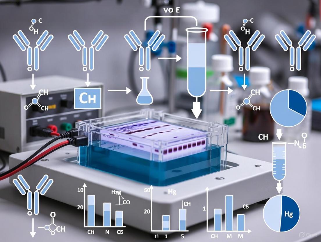
The following diagram illustrates the transformation of a native antibody into an SDS-linearized complex ready for electrophoretic separation.
The Scientist's Toolkit: Essential Reagents for SDS-PAGE
A successful SDS-PAGE experiment, especially for antibody analysis, requires a specific set of reagents, each serving a critical function.
Table 2: Key Research Reagent Solutions for SDS-PAGE
| Reagent | Composition / Example | Primary Function |
|---|---|---|
| Denaturing Agent | Sodium Dodecyl Sulfate (SDS) | Denatures proteins, masks intrinsic charge, confers negative charge [2] [1]. |
| Reducing Agent | β-mercaptoethanol (BME), Dithiothreitol (DTT) | Breaks disulfide bonds to fully dissociate antibody subunits (Heavy and Light chains) [2] [3]. |
| Sample Buffer | Laemmli Buffer (Tris-HCl, SDS, Glycerol, Bromophenol Blue, BME/DTT) | Denatures proteins, provides density for loading, and visual tracking [1]. |
| Gel Matrix | Polyacrylamide (Acrylamide + Bis-acrylamide) | Forms a porous gel mesh that acts as a molecular sieve [3]. |
| Catalyst System | Ammonium Persulfate (APS) and TEMED | Initiates and catalyzes the polymerization of acrylamide [4] [1]. |
| Electrophoresis Buffer | Tris-Glycine-SDS Buffer, pH 8.3 | Carries current and maintains pH during electrophoresis [1]. |
| Molecular Weight Marker | Prestained Protein Ladder | Allows estimation of protein molecular weights [2] [4]. |
The Discontinuous Electrophoresis System
SDS-PAGE employs a discontinuous buffer system to sharpen protein bands, resulting in higher resolution. This system uses gels with two distinct sections—a stacking gel and a separating gel—each with different pH and acrylamide concentration [1].
- Stacking Gel (pH ~6.8): The purpose of this low-concentration, low-pH gel is to concentrate all protein samples into a sharp, unified band before they enter the separating gel. The key to this process is the ionic state of glycine from the running buffer. At pH 6.8, glycine exists predominantly as a zwitterion with no net mobility. This creates a zone of low conductivity sandwiched between highly mobile chloride ions (from the gel) and the slower glycine zwitterions. The proteins, with intermediate mobility, are compressed into a thin disk within this zone [1].
- Separating Gel (pH ~8.8): Once the protein stack reaches the interface with the separating gel, the higher pH (8.8) causes glycine to lose a proton and become negatively charged glycinate ions. These ions now migrate faster than the proteins. The proteins are then deposited at the top of the separating gel and begin to be resolved based on their size as they migrate through the higher-concentration acrylamide mesh, which acts as a molecular sieve [2] [1] [3].
The workflow and ionic dynamics of this process are summarized in the following diagram.
Detailed Protocol for Antibody Purity Analysis Under Reducing Conditions
This protocol is adapted for analyzing the purity and integrity of monoclonal antibodies, specifically under reducing conditions to separate heavy and light chains [5] [4] [3].
Sample Preparation
- Dilution: Combine the antibody sample with 5X Laemmli sample buffer to a final 1X concentration. A typical load is 1-5 µg of protein per band for Coomassie staining, and less for sensitive detection methods [4].
- Reduction: Add a reducing agent to the final sample mixture. Common choices include 5% (v/v) β-mercaptoethanol or 100mM Dithiothreitol (DTT) [2] [3].
- Denaturation: Heat the mixture at 95°C for 5 minutes (or 70°C for 10 minutes) in a heat block or boiling water bath to fully denature the protein [2] [3].
- Cooling: Briefly centrifuge the tubes after heating to collect condensation and cool to room temperature before loading.
Gel Electrophoresis
- Assembly: Set up a vertical gel electrophoresis unit. Use a precast or freshly cast polyacrylamide gel with a suitable percentage (e.g., 4-20% gradient or 10-12% uniform gel) for resolving antibody fragments [4].
- Loading: Fill the buffer chambers with Tris-Glycine-SDS running buffer. Carefully load the denatured samples and a prestained protein molecular weight marker into the wells [3].
- Electrophoresis Run: Apply a constant voltage of 80-150V. The tracking dye (bromophenol blue) will migrate through the stacking and separating gels. Stop the run when the dye front is about 1 cm from the bottom of the gel [4].
Post-Electrophoresis Analysis
- Staining: Carefully open the cassette and transfer the gel to a container. Submerge the gel in Coomassie Brilliant Blue staining solution for 2-4 hours (or overnight for maximum sensitivity) with gentle agitation [4].
- Destaining: Replace the stain with an appropriate destaining solution (e.g., 10% acetic acid, 40% methanol). Change the solution several times until the background is clear and protein bands are sharply visible [4].
- Analysis: Document the gel using a gel imaging system. Under reducing conditions, a pure, intact monoclonal antibody should show two dominant bands: the Heavy Chain (~50 kDa) and the Light Chain (~25 kDa). The presence of additional lower molecular weight bands may indicate fragmentation or degradation, which requires further characterization [5].
Considerations and Artifacts in Antibody Analysis
While SDS-PAGE is a powerful tool, researchers must be aware of potential artifacts. A key consideration is that some lower molecular weight (LMW) bands observed on non-reducing SDS-PAGE of antibodies may be artifacts formed during sample preparation rather than true product-related impurities. These can be generated via disulfide bond scrambling or beta-elimination [5]. Mass spectrometry analysis has shown that modifying free sulfhydryl groups through alkylation can prevent disulfide scrambling and reduce such artifacts [5]. Furthermore, orthogonal techniques like Hydrophilic Interaction Chromatography (HILIC) coupled with mass spectrometry are emerging as powerful methods to characterize LMW impurities with less likelihood of generating artifacts compared to CE-SDS or SDS-PAGE methods [6].
The Critical Role of Reducing Agents (DTT, β-Mercaptoethanol) in Breaking Disulfide Bonds
In the realm of protein biochemistry, particularly in the analysis of therapeutic antibodies, disulfide bonds serve as critical structural elements that stabilize protein three-dimensional architecture. These covalent linkages between cysteine residues exist in two primary forms: intrachain bonds that stabilize the folded structure within a single polypeptide chain, and interchain bonds that create covalent links between separate protein subunits [7]. For complex molecules like antibodies, interchain disulfide bonds are particularly important as they connect heavy and light chains into the functional quaternary structure essential for antigen recognition and binding.
The analysis of antibody purity and subunit composition requires the disruption of these structural elements to obtain accurate molecular weight information and assess sample homogeneity. This is where reducing agents play an indispensable role in sample preparation for SDS-PAGE (Sodium Dodecyl Sulfate-Polyacrylamide Gel Electrophoresis). The fundamental purpose of compounds like dithiothreitol (DTT) and β-mercaptoethanol (BME) is to cleave disulfide bonds through a reduction reaction, thereby converting multimeric protein structures into their individual polypeptide components [8] [9]. This process is essential for accurate molecular weight determination during electrophoretic separation, as it ensures proteins migrate based solely on polypeptide chain length rather than complex quaternary structure.
Table 1: Common Reducing Agents in Protein Biochemistry
| Reducing Agent | Chemical Properties | Working Concentration | Mechanism of Action |
|---|---|---|---|
| β-Mercaptoethanol (BME) | Volatile thiol compound with distinct odor | 0.55M - 1.0% (v/v) [10] [9] | Cleaves disulfide bonds through thiol-disulfide exchange |
| Dithiothreitol (DTT) | Less volatile, more potent reducing agent | 10-100 mM [11] [9] | Reduces disulfides via intramolecular disulfide formation |
| Tris(2-carboxyethyl)phosphine (TCEP) | Odorless, air-stable phosphine derivative | 5-50 mM (not covered in detail in search results) | Reduces disulfides through phosphine oxidation |
Fundamental Mechanisms of Reducing Agents
Biochemical Principles of Disulfide Bond Reduction
The mechanism by which reducing agents break disulfide bonds revolves around the chemistry of thiol-disulfide exchange. In this redox reaction, the reducing agent contributes electrons to reduce the disulfide bond (S-S) into two free thiol groups (-SH). DTT and β-mercaptoethanol achieve this through distinct molecular pathways, though both rely on the nucleophilic properties of their sulfur atoms to attack the disulfide bond [9]. β-Mercaptoethanol functions as a small monothiol compound that interacts directly with protein disulfide bonds, forming a mixed disulfide intermediate before releasing the reduced protein thiols. The reduction potential of β-mercaptoethanol allows it to effectively disrupt most biological disulfide linkages when used at appropriate concentrations, typically 0.55M as specified in standard SDS-PAGE protocols [10].
DTT operates through a more sophisticated mechanism involving an intramolecular disulfide formation. This compound contains two thiol groups positioned to form a stable six-membered ring structure (a cyclic disulfide) upon oxidation. This intramolecular reaction is thermodynamically favorable, making DTT a stronger reducing agent than β-mercaptoethanol at equivalent concentrations [9]. The ring formation drives the reduction reaction toward completion, ensuring thorough cleavage of protein disulfide bonds. This efficiency makes DTT particularly valuable for analyzing complex antibodies with multiple interchain disulfide bonds that might resist reduction by weaker agents.
Visualization of the Reduction Mechanism
The following diagram illustrates the biochemical mechanism through which DTT reduces protein disulfide bonds:
Experimental Protocols for Antibody Analysis Under Reducing Conditions
Comprehensive Sample Preparation Protocol
The preparation of antibody samples for reducing SDS-PAGE requires precise execution of multiple critical steps to ensure complete disulfide bond reduction and protein denaturation. Begin by transferring your protein sample to a clean microcentrifuge tube. For pre-prepared lysates already containing sample buffer, add β-mercaptoethanol to a final concentration of 0.55M, which translates to approximately 1μL of stock BME per 25μL of lysate [10]. For other protein samples, mix with an equal volume of 2X Sample Buffer containing 0.55M BME [10]. Ensure that your final protein concentration is sufficiently high for detection, typically ranging from 1μg to 500μg depending on your detection method and protein characteristics [10].
Following the addition of reducing agent, thoroughly mix the samples by pipetting to ensure homogeneous distribution of all components. The subsequent heat denaturation step is crucial for complete protein unfolding. Place all microcentrifuge tubes containing samples in a heating block or water bath set to 95°C for 5 minutes [10] [11]. This heating step serves dual purposes: it facilitates thorough detergent binding to the protein backbone and enhances the efficacy of the reducing agent by providing the kinetic energy needed to break disulfide bonds. After heating, centrifuge the aliquots for 3 minutes using a microcentrifuge to pellet any insoluble debris that might interfere with electrophoresis [10]. The samples are now ready for loading into the gel, typically using volumes ranging from 5μL to 35μL per lane depending on gel thickness and well size [10].
Critical Controls and Molecular Weight Standards
The inclusion of appropriate controls is essential for accurate interpretation of reducing SDS-PAGE results. Always include molecular weight standards (protein ladders) on each gel to enable estimation of protein molecular weights [10] [11]. For SDS-PAGE followed by western blotting, use pre-stained MW markers, while for analytical gels that will be directly stained, unstained standards are preferable [10]. To specifically assess the impact of reduction on your antibody samples, include non-reduced controls prepared without DTT or β-mercaptoethanol in parallel with your reduced samples [7] [12]. This side-by-side comparison allows direct visualization of how disulfide bond cleavage affects electrophoretic mobility.
Maintain detailed records of all samples, including lane number, sample description, protein concentration, loading volume, loading amount, and the addition of reducing agent [10]. This documentation is critical for troubleshooting and ensuring experimental reproducibility. When analyzing antibodies under reducing conditions, expect to see the dissociation of the native multimeric structure into its constituent polypeptide chains. For a typical IgG antibody, reduction should yield two distinct bands corresponding to the heavy chain (~50 kDa) and light chain (~25 kDa) [13], a dramatic shift from the non-reduced form which migrates at approximately 150 kDa.
Workflow for Reduced versus Non-Reduced SDS-PAGE Analysis
The complete experimental workflow for comparing antibody samples under reducing and non-reducing conditions is summarized below:
Applications in Antibody Purity Analysis and Quality Control
Assessment of Antibody Subunit Composition and Structural Integrity
The application of reducing SDS-PAGE in antibody characterization provides critical information about subunit composition and structural integrity. Under reducing conditions, therapeutic antibodies should dissociate into predictable polypeptide patterns that confirm proper assembly and purity. For instance, a standard IgG antibody should yield two distinct bands corresponding to heavy and light chains after reduction, with minimal additional bands indicating high purity [8] [13]. The presence of unexpected bands or deviations from the expected molecular weights can indicate issues such as proteolytic degradation, incomplete synthesis, or non-covalent aggregation that might affect antibody function and safety.
The migration patterns observed in reducing SDS-PAGE also provide insights into post-translational modifications that affect molecular weight. Glycosylation, for example, adds significant mass to the Fc region of antibody heavy chains, causing them to migrate slightly higher than their predicted molecular weight based on amino acid sequence alone [8]. When combined with non-reduced analysis, reducing SDS-PAGE can reveal whether disulfide bonds are properly formed between chains, essential for maintaining therapeutic antibody stability and efficacy. This comprehensive assessment is particularly valuable during clone selection, process development, and lot-release testing in biopharmaceutical manufacturing.
Comparative Analysis of Electrophoretic Migration Patterns
The table below summarizes the key differences in antibody migration under reduced versus non-reduced conditions:
Table 2: Antibody Migration Patterns in Reduced vs. Non-Reduced SDS-PAGE
| Condition | Expected Band Pattern | Molecular Weight Range | Structural Information Obtained |
|---|---|---|---|
| Non-Reduced | Single band for intact antibody [13] | ~150 kDa for IgG [13] | Quaternary structure integrity, disulfide-mediated oligomerization |
| Reduced | Two primary bands (heavy & light chains) [13] | Heavy: ~50 kDa, Light: ~25 kDa [13] | Subunit composition, heavy chain glycosylation, proteolytic cleavage |
| Partially Reduced | Multiple intermediate bands | Variable | Incomplete disulfide reduction, structural heterogeneity |
Troubleshooting and Method Optimization
Addressing Common Experimental Challenges
Even with proper technique, researchers may encounter issues when performing reducing SDS-PAGE for antibody analysis. Poor band resolution frequently stems from incorrect acrylamide concentration for the target protein size, running the gel at excessively high voltage causing heat generation, or buffer depletion during extended runs [13]. For antibody analysis, gradient gels (e.g., 4-20%) often provide optimal resolution across the relevant molecular weight range. Smeared or distorted bands may indicate sample overloading, presence of nucleic acids or lipids in samples, incomplete protein solubilization, or high salt concentration interfering with migration [13].
When reduction-specific issues occur, such as incomplete disulfide bond cleavage, consider increasing the concentration of reducing agent, extending the heating time during sample denaturation, or switching to a stronger reducing agent like DTT. Conversely, if excessive reduction occurs (evidenced by breakdown products), reduce the concentration of DTT or BME or shorten the heating duration. Vertical streaking often results from protein precipitation in sample wells due to insufficient SDS or high salt content, while uneven migration across the gel may indicate uneven polymerization or temperature gradients during electrophoresis [13]. Methodical troubleshooting of these issues ensures reliable results in antibody purity assessment.
Essential Reagents for Disulfide Bond Reduction Studies
Table 3: Research Reagent Solutions for Reducing SDS-PAGE
| Reagent/Chemical | Function in Experiment | Key Considerations |
|---|---|---|
| Dithiothreitol (DTT) | Reduces disulfide bonds [11] [9] | More stable and efficient than BME; use fresh solutions |
| β-Mercaptoethanol (BME) | Alternative reducing agent [10] [9] | Volatile with distinctive odor; use in fume hood |
| SDS (Sodium Dodecyl Sulfate) | Denatures proteins and confers negative charge [8] | Critical for charge-to-mass uniformity; purity affects results |
| Acrylamide/Bis-acrylamide | Forms porous gel matrix for separation [8] | Concentration determines pore size and resolution range |
| APS and TEMED | Catalyzes acrylamide polymerization [8] | Freshness critical for consistent gel formation |
| Tris-Glycine-SDS Buffer | Running buffer for electrophoresis [11] | Maintains pH and conductivity during separation |
| Coomassie Brilliant Blue | Protein stain for visualization [11] [14] | Standard for general protein detection; moderate sensitivity |
The critical role of reducing agents like DTT and β-mercaptoethanol in breaking disulfide bonds extends far beyond a simple sample preparation step in SDS-PAGE. These reagents enable the fundamental characterization of antibody structure, purity, and integrity that forms the foundation of biopharmaceutical quality control. Through their specific action on disulfide bonds, these reducing agents transform complex multimeric proteins into their constituent polypeptides, allowing researchers to verify subunit composition, detect impurities, and ensure product consistency. The experimental protocols outlined in this document provide a robust framework for implementing reducing SDS-PAGE in antibody analysis, while the troubleshooting guidelines address common challenges encountered in practice. As therapeutic antibodies continue to dominate the biopharmaceutical landscape, the precise and reproducible analysis enabled by proper disulfide bond reduction remains an essential capability in modern biologics research and development.
Within the rigorous framework of therapeutic antibody development, sodium dodecyl sulfate-polyacrylamide gel electrophoresis (SDS-PAGE) under reducing conditions remains a foundational technique for assessing protein purity and integrity. This application note, situated within a broader thesis on SDS-PAGE for antibody purity analysis, provides a detailed protocol and interpretive benchmark for researchers, scientists, and drug development professionals. The core principle of reducing SDS-PAGE involves the disruption of non-covalent interactions and cleavage of disulfide bonds by agents such as dithiothreitol (DTT) or β-mercaptoethanol. This process denatures the antibody into its constituent polypeptide chains, which bind the anionic detergent SDS and acquire a uniform negative charge. Subsequently, separation is based primarily on molecular weight as the polypeptides migrate through a polyacrylamide gel matrix [8] [15]. For a standard immunoglobulin G (IgG), this yields two definitive bands: the glycosylated heavy chain at approximately 50-55 kDa and the light chain at approximately 25 kDa [16]. A single, sharp band at each expected molecular weight is the hallmark of a pure, homogenous sample, while the presence of additional bands or smearing indicates potential impurities, degradation, or the presence of fragments [8].
Theoretical Foundation and Key Band Patterns
The interpretation of SDS-PAGE results for antibodies under reducing conditions relies on a clear understanding of the expected outcomes and common anomalies. The following diagram illustrates the core workflow and the transformation of an intact antibody into its constituent chains.
Expected Band Pattern for a Purity Benchmark
A well-characterized, pure monoclonal antibody under reducing conditions should display a characteristic two-band pattern, corresponding to the individual heavy and light chains, when visualized with a sensitive stain like Coomassie Blue [16] [8]. The precise molecular weight of the heavy chain can vary slightly due to factors such as glycosylation. The light chain, typically lacking glycosylation, migrates at a consistent molecular weight. The following table summarizes the benchmark expectations for a standard IgG antibody.
Table 1: Expected Band Pattern for a Pure IgG Under Reducing SDS-PAGE
| Component | Expected Molecular Weight | Key Characteristics |
|---|---|---|
| Heavy Chain | ~50-55 kDa | Appears as a broader band due to glycosylation heterogeneity. |
| Light Chain | ~25 kDa | Appears as a sharp, defined band. |
Interpreting Deviations from the Benchmark
Deviations from the clean two-band pattern provide critical diagnostic information about product-related impurities. These anomalies often manifest as additional bands at specific molecular weights, which can be quantified using densitometry software to determine percent purity [17]. The table below catalogs common low molecular weight (LMW) impurities and their signatures on a gel.
Table 2: Common Low Molecular Weight (LMW) Impurities and Their Signatures
| Impurity Band | Apparent Molecular Weight | Potential Identity & Cause |
|---|---|---|
| High Molecular Weight Smear | >150 kDa | Protein aggregation, often induced by stress (e.g., heat) [15]. |
| Half-antibody | ~75 kDa | An antibody species lacking one heavy-light chain pair, detectable when analyzing non-reduced samples [6]. |
| Non-glycosylated Heavy Chain | ~50 kDa | A heavy chain lacking its glycan moiety, may co-migrate with the standard heavy chain but can be resolved by CE-SDS [15]. |
| Low Molecular Weight Bands | <25 kDa | Protein fragments resulting from backbone cleavage or enzymatic degradation [6] [15]. |
It is crucial to distinguish true product-related impurities from method-induced artifacts. Artifact bands can arise from incomplete denaturation of the antibody or disulfide bond scrambling [18]. These can be minimized by optimizing sample preparation protocols, including heating conditions and the use of alkylating agents like iodoacetamide (IAM) [18].
Experimental Protocol for Reducing SDS-PAGE
Research Reagent Solutions and Materials
The following table lists essential materials and reagents required for performing reducing SDS-PAGE for antibody analysis.
Table 3: Key Research Reagent Solutions for SDS-PAGE
| Reagent / Material | Function / Explanation |
|---|---|
| SDS (Sodium Dodecyl Sulfate) | Anionic detergent that denatures proteins and confers a uniform negative charge, masking intrinsic charge differences [8] [15]. |
| DTT (Dithiothreitol) or β-mercaptoethanol | Reducing agents that break inter- and intra-chain disulfide bonds, linearizing the protein for accurate molecular weight separation [8]. |
| Polyacrylamide Gel (e.g., 4-12% Bis-Tris) | Acts as a molecular sieve. Gradient gels (e.g., 4-12%) provide a broad separation range for resolving fragments and aggregates [19] [15]. |
| LDS Sample Buffer | Contains lithium dodecyl sulfate, a detergent similar to SDS, and a buffer to prepare the sample for loading [19]. |
| Pre-stained Protein Ladder | A set of proteins of known molecular weight used to estimate the size of unknown protein bands in the sample [16]. |
| Coomassie Blue Stain | A dye that binds to proteins, allowing for visualization of separated bands on the gel [17] [16]. |
Step-by-Step Workflow
The procedural workflow for sample preparation, gel electrophoresis, and analysis is outlined below.
Detailed Methodology
Sample Preparation:
- Dilute the purified antibody to a final concentration of 0.15-0.2 mg/mL using ultrapure water [15].
- Combine the diluted antibody with 4X LDS (Lithium Dodecyl Sulfate) sample buffer. Include a reducing agent, such as 1X Bolt Sample Reducing Agent (containing DTT) [19].
- Heat the mixture at 70°C for 10 minutes or 95°C for 5 minutes to ensure complete denaturation and reduction [18]. This step is critical for minimizing artifact bands caused by incomplete denaturation [18].
Gel Electrophoresis:
- Use a pre-cast polyacrylamide gradient gel (e.g., 4-12% Bis-Tris) [19] [15].
- Load 10-20 µL of the prepared sample (equivalent to 2-4 µg of protein) and a pre-stained protein molecular weight ladder into separate wells.
- Perform electrophoresis using an appropriate buffer system (e.g., MES or MOPS) at a constant voltage of 80-200V until the dye front reaches the bottom of the gel [19].
Visualization and Analysis:
- After electrophoresis, carefully disassemble the gel cassette and stain the gel with Coomassie Blue for 30-45 minutes [16].
- Destain the gel with an appropriate solution (e.g., 10% acetic acid) until the background is clear and protein bands are distinctly visible.
- Capture a digital image of the gel using a calibrated densitometer [17].
- Use quantitative image analysis software (e.g., Image Lab, Alpha View) to determine the molecular weight of observed bands and calculate the percent purity based on band intensity [17] [15].
Troubleshooting and Orthogonal Methods
Addressing Common Artifacts
A frequent challenge in SDS-PAGE analysis is the appearance of artifact bands that do not represent true product-related impurities. A major cause of these artifacts is incomplete denaturation of the antibody sample [18]. If multiple bands are observed in the high molecular weight region of a reduced gel, consider optimizing the denaturation conditions. As an alternative to heating, treating the sample with 8 M urea can also promote complete denaturation and minimize these artifacts [18]. Furthermore, the inclusion of an alkylating agent like iodoacetamide (IAM) after reduction can prevent disulfide bond scrambling, which is another potential source of artifactual bands [18].
Orthogonal Techniques for Purity Assessment
While SDS-PAGE is a powerful qualitative and semi-quantitative tool, modern antibody development requires more quantitative and high-resolution techniques. Capillary Electrophoresis SDS (CE-SDS) has emerged as a superior, automated technology for antibody purity analysis [15]. CE-SDS offers higher resolution, superior signal-to-noise ratio, and better quantitation of low-abundance impurities compared to traditional gel-based methods [15]. A key advantage is its ability to resolve and detect nonglycosylated heavy chains, which often co-migrate with glycosylated heavy chains in SDS-PAGE, leading to an overestimation of purity [15]. For unambiguous identification and characterization of LMW impurities, hydrophilic interaction chromatography coupled with mass spectrometry (HILIC-MS) provides an orthogonal method. HILIC-MS can directly identify species such as free light chains, half-antibodies, and truncated fragments, including their modification sites, within a single analysis [6]. A comprehensive characterization strategy should therefore integrate SDS-PAGE with these orthogonal methods to ensure a robust evaluation of antibody purity, identity, and stability [19].
Advantages of Reducing Conditions for Assessing Primary Structure and Subunit Integrity
This application note delineates the critical advantages of employing reducing conditions in SDS-PAGE for the precise assessment of protein primary structure and subunit integrity, with a specific focus on antibody characterization in pharmaceutical development. Reduction with agents such as dithiothreitol (DTT) or β-mercaptoethanol is a prerequisite for accurate molecular weight determination, purity assessment, and structural analysis. By cleaving disulfide bonds, these conditions ensure complete protein denaturation into constituent polypeptides, thereby preventing aberrant migration and enabling reliable data interpretation crucial for therapeutic antibody development.
Sodium dodecyl sulfate-polyacrylamide gel electrophoresis (SDS-PAGE) is a foundational technique in biochemistry for separating proteins based on molecular weight [20]. The analysis of antibodies, complex multidomain proteins stabilized by intra- and inter-chain disulfide bonds, presents particular challenges. Under non-reducing conditions, these disulfide bonds persist, leading to incomplete denaturation and anomalous electrophoretic migration [21]. The implementation of reducing conditions is therefore indispensable for researchers and drug development professionals who require accurate insights into primary structure and subunit composition to ensure antibody purity, stability, and batch-to-batch consistency.
Fundamental Principles of Reduction in SDS-PAGE
The Role of SDS and Reducing Agents
SDS-PAGE relies on the anionic detergent sodium dodecyl sulfate (SDS) to denature proteins and confer a uniform negative charge, effectively masking the protein's intrinsic charge [20] [8]. However, SDS alone cannot break covalent disulfide bonds. Reducing agents such as dithiothreitol (DTT) or β-mercaptoethanol are essential to reduce these disulfide linkages, linearizing the protein into its constituent polypeptides [8]. This combination ensures that separation is based solely on polypeptide chain length, not on native shape or charge.
Electrophoretic Migration Differences
The conformational differences between reduced and non-reduced proteins have a direct and measurable impact on gel migration:
- Reduced Proteins: Migrate strictly according to their molecular weight. For an antibody, this results in distinct bands for the heavy chain (~50-70 kDa) and light chain (~25 kDa) [22].
- Non-Reduced Proteins: Can exhibit faster or slower migration than their reduced counterparts. Polypeptides with intact intrachain disulfide bonds often migrate more rapidly due to a more compact structure. In contrast, multimers or proteins with complex disulfide networks may not enter the gel effectively [21].
Table 1: Impact of Reduction on Protein Migration in SDS-PAGE
| Condition | Protein State | Migration Behavior | Band Appearance |
|---|---|---|---|
| Reducing | Fully denatured linear polypeptides | Proportional to molecular weight | Sharp, distinct bands |
| Non-Reducing | Partially folded; disulfide bonds intact | Anomalous; influenced by structure | Diffuse or multiple bands |
Experimental Protocols
Standard Sample Preparation Protocol Under Reducing Conditions
This protocol is optimized for the analysis of monoclonal antibodies, such as the 6E10 antibody [22].
Materials:
- Protein sample (e.g., 1 µg/µL antibody in PBS)
- 5X Reducing Sample Buffer: 62 mM Tris-HCl (pH 6.8), 2% SDS (w/v), 25% glycerol, 0.01% bromophenol blue, 100 mM DTT [21] [22]. Note: DTT is added fresh; do not use β-mercaptoethanol if subsequent alkylation is planned.
- Heating block
Procedure:
- Dilution: Mix 40 µL of protein sample with 20 µL of 5X Reducing Sample Buffer [22].
- Denaturation and Reduction: Incubate the mixture for one hour at 90°C [22]. Critical: Ensure complete heating to fully denature the protein and reduce disulfide bonds.
- Cooling: Briefly centrifuge tubes to collect condensation.
- Loading: Load 10-20 µL (typically 10 µg of total protein) into the well of a polyacrylamide gel [22].
Optional Alkylation Step
Following reduction, free cysteines can reoxidize. To prevent this, an alkylation step can be introduced.
- Cooling: After the reduction step, cool the sample to room temperature.
- Alkylation: Add iodoacetamide (IAA) to a final concentration of 50-100 mM. A molar ratio of DTT to IAA of 1:3 is effective [22].
- Incubation: Incubate in the dark at room temperature for 30-60 minutes [22].
- The sample is now ready for gel loading.
The Scientist's Toolkit: Essential Reagents and Materials
The following reagents are critical for successful SDS-PAGE under reducing conditions.
Table 2: Key Research Reagent Solutions for Reducing SDS-PAGE
| Reagent/Material | Function & Importance |
|---|---|
| SDS (Sodium Dodecyl Sulfate) | Anionic detergent that denatures proteins and confers a uniform negative charge, masking intrinsic charge [20] [8]. |
| DTT (Dithiothreitol) | Reducing agent that cleaves disulfide bonds, linearizing proteins for accurate molecular weight analysis [8]. |
| β-Mercaptoethanol | Alternative reducing agent to DTT; often used at a concentration of 5% in sample buffer. |
| Iodoacetamide | Alkylating agent that caps free thiols post-reduction to prevent reformation of disulfide bonds [22]. |
| Polyacrylamide Gel (4-12% Bis-Tris) | Acts as a molecular sieve. Gradient gels (e.g., 4-12%) provide superior resolution for complex mixtures like antibody fragments [22]. |
| Molecular Weight Markers | Pre-stained or unstained protein ladders essential for calibrating the gel and estimating sample protein sizes [8]. |
Data Presentation and Analysis
Quantitative Analysis of Migration
The effect of reduction is quantifiable by comparing the apparent molecular weight of a protein under reducing versus non-reducing conditions.
Table 3: Comparative Migration Analysis of a Model IgG Antibody
| Analysis Parameter | Non-Reducing Conditions | Reducing Conditions |
|---|---|---|
| Number of Major Bands | 1 (intact IgG) | 2 (Heavy & Light chains) |
| Apparent MW of Intact IgG | ~150 kDa (may be diffuse) | Not applicable (dissociated) |
| Apparent MW - Heavy Chain | Not visible | ~50-70 kDa |
| Apparent MW - Light Chain | Not visible | ~25 kDa |
| Band Sharpness | Often diffuse | Sharp, well-defined |
Application in Purity and Integrity Assessment
- Purity: A single, sharp band for each subunit under reducing conditions indicates a homogeneous sample. Multiple bands suggest proteolytic degradation or non-uniform glycosylation [20] [8].
- Subunit Integrity: The clean separation of heavy and light chains confirms the integrity of each polypeptide. Additional bands may indicate fragments, as seen in studies of the 6E10 antibody where N-terminal heavy chain truncations were identified [22].
- Post-Translational Modifications (PTMs): Shifts in the migration of a subunit can indicate PTMs. For example, glycosylation can be detected by a downward band shift after enzymatic deglycosylation [8].
Experimental Workflow
The following diagram illustrates the logical workflow for preparing and analyzing a protein sample under reducing conditions for SDS-PAGE.
Workflow for Reducing SDS-PAGE Analysis
The use of reducing conditions in SDS-PAGE is a non-negotiable practice for the accurate assessment of protein primary structure and subunit integrity, particularly for complex proteins like antibodies. By ensuring complete linearization of polypeptides, this method provides unambiguous data on molecular weight, purity, and composition. The protocols and analyses detailed herein provide a robust framework for researchers in drug development to characterize therapeutic antibodies with the precision required for regulatory compliance and successful product development.
Optimized Protocols: A Step-by-Step Guide to Sample Prep and Electrophoresis
Within the critical field of biotherapeutic development, the analysis of antibody purity is a non-negotiable requirement for ensuring drug safety and efficacy. Sodium dodecyl sulfate-polyacrylamide gel electrophoresis (SDS-PAGE) under reducing conditions remains a foundational technique for this purpose, providing a rapid assessment of protein purity, integrity, and molecular weight. The reliability of this analysis, however, is profoundly dependent on the initial sample preparation. Inadequate denaturation can lead to misleading artifact bands, which may be mistakenly interpreted as product-related impurities, thereby compromising data integrity and decision-making [18]. This application note details a optimized and robust protocol for sample preparation, focusing on the critical interplay between heating conditions and buffer composition to ensure complete denaturation of antibody samples for accurate purity analysis.
Principles of Sample Denaturation for SDS-PAGE
The goal of sample preparation for SDS-PAGE is to dismantle the native structure of the protein and impart a uniform negative charge, allowing separation to be based solely on polypeptide chain length. This is achieved through a combination of chemical and physical treatments:
- SDS Binding: The anionic detergent Sodium Dodecyl Sulfate (SDS) disrupts hydrophobic interactions and hydrogen bonds, unfolding the protein. It binds to the polypeptide backbone at a relatively constant ratio of about 1.4 g SDS per 1.0 g of protein, conferring a net negative charge that masks the protein's intrinsic charge [23].
- Reduction of Disulfide Bonds: Antibodies contain intra- and inter-chain disulfide bonds that stabilize their structure. Reducing agents, such as Dithiothreitol (DTT) or β-mercaptoethanol (β-Me), break these covalent disulfide linkages [24] [25]. For a standard IgG, this reduces the molecule into two Heavy Chains (HC) and two Light Chains (LC).
- Heat Denaturation: Heating provides the thermal energy required to overcome kinetic barriers and facilitate the complete unfolding of the protein and the penetration of SDS and reducing agents. Incomplete denaturation is a major cause of artifact bands on SDS-PAGE, as antibodies with varying degrees of unfolding can exhibit different migration rates [18].
The following workflow outlines the core logical relationships and decision points in the sample preparation process:
Optimized Heating Conditions & Buffer Composition
Critical Parameters for Complete Denaturation
The search results consistently identify incomplete denaturation as the primary source of artifacts in SDS-PAGE analysis of antibodies [18]. A systematic approach to heating and buffer composition is required to mitigate this.
Heating Conditions: The standard protocol of heating at 95–100°C for 5 minutes is widely recommended for achieving complete denaturation [24] [26] [23]. One study investigating artifact bands on non-reducing SDS-PAGE found that heating at 75°C for 5–10 minutes significantly minimized these artifacts, but prolonged heating could generate extra bands [18]. This underscores the importance of optimizing both temperature and duration. After heating, samples should be briefly centrifuged to pellet any insoluble aggregates formed during the process [24].
Role of Reducing Agents: The choice of reducing agent impacts the procedure. DTT is effective and has less odor than β-Me, but it degrades faster in solution. β-Me is more stable and can withstand multiple freeze-thaw cycles when prepared in sample buffer [24]. For reduced SDS-PAGE, the inclusion of a fresh reducing agent is mandatory to ensure complete breakdown of the antibody into its constituent chains.
Alkylating Agents: In non-reducing SDS-PAGE, where disulfide bonds are to be preserved, the use of an alkylating agent like Iodoacetamide (IAM) is recommended. IAM blocks free sulfhydryl groups, preventing disulfide bond scrambling, which is another potential cause of artifact bands [18]. Combining heating with IAM treatment can yield slightly better results than heating alone for non-reduced samples [18].
Buffer Composition and Recipe
A well-formulated sample buffer is essential for successful denaturation. The table below details the components and a standard recipe for a 5X reducing SDS-PAGE sample buffer.
Table 1: Composition of 5X Reducing SDS-PAGE Sample Buffer
| Component | Final Concentration (in 1X) | Function |
|---|---|---|
| Tris-HCl (pH 6.8) | 62.5 mM | Provides buffering capacity at the stacking gel pH. |
| SDS | 2% (w/v) | Denatures proteins and confers uniform negative charge. |
| Glycerol | 10% (v/v) | Increases density for easy gel loading. |
| Bromophenol Blue | 0.02% (w/v) | Tracking dye to monitor electrophoresis progress. |
| DTT or β-Mercaptoethanol | 100 mM or 5% (v/v) | Reducing agent to break disulfide bonds. |
Preparation of 5X Sample Buffer:
- Combine 2.5 mL of 1 M Tris-HCl (pH 6.8), 2.0 g of SDS, 5.0 mL of glycerol (100%), and 2.0 mg of Bromophenol Blue.
- Bring the volume to 9.5 mL with distilled water.
- Just before use, add 0.5 mL of β-mercaptoethanol (for a final 5% v/v in 5X buffer) or 77 mg of DTT (for a final 100 mM in 5X buffer).
- Mix thoroughly and aliquot for single-use to prevent oxidation of the reducing agent [23].
Experimental Data and Comparative Analysis
Impact of Denaturation on Banding Patterns
Research has quantitatively demonstrated the effect of sample preparation on the resulting electrophoregram. One study using two purified monoclonal antibodies (mAb A and mAb B) showed that unheated samples on 8% Tris-glycine gels displayed multiple artifact bands [18]. However, heating at 75°C for an appropriate duration (5-10 minutes) significantly minimized these artifacts. The data further revealed that alternative denaturation methods, such as treatment with 8 M urea without heating, also promoted complete denaturation and minimized artifact bands.
Table 2: Comparative Analysis of Denaturation Methods for SDS-PAGE of mAbs
| Denaturation Method | Conditions | Impact on Artifact Bands | Key Observations | Source |
|---|---|---|---|---|
| No Heat / Incomplete Denaturation | Sample mixed with SDS buffer, no heating step | High - Multiple artifact bands present | Major cause of misleading bands on non-reducing SDS-PAGE; proteins with different folding states migrate differently. | [18] |
| Heat Denaturation | 75°C for 5-10 min | Significantly Minimized | Promotes complete denaturation; prolonged heating can generate extra bands. | [18] |
| Heat + Alkylation | Heating (e.g., 75°C) with Iodoacetamide (IAM) | Minimized (slightly better than heat alone) | Alkylation prevents disulfide scrambling; ideal for non-reducing conditions. | [18] |
| Chemical Denaturation | Treatment with 8 M Urea | Minimized (close to complete denaturation) | Serves as an effective alternative to heating for promoting denaturation. | [18] |
| Standard Protocol | 95-100°C for 5 min | Effective Denaturation | Widely recommended and used protocol for ensuring complete protein unfolding. | [24] [26] [23] |
Detailed Protocol for Sample Preparation
Materials and Reagents
- Protein Sample: Purified antibody or complex mixture.
- 5X Reducing SDS-PAGE Sample Buffer (see Table 1 for composition).
- Thermal block or water bath, capable of maintaining 95-100°C.
- Microcentrifuge tubes.
- Pipettes and tips.
Step-by-Step Procedure
- Dilute and Mix: Combine the protein sample with the 5X reducing SDS-PAGE sample buffer. A typical ratio is 4 volumes of sample to 1 volume of 5X buffer [23]. Vortex briefly to mix.
- Denature: Secure the cap of the microcentrifuge tube and heat the mixture at 95–100°C for 5 minutes [26] [23].
- Troubleshooting Tip: To prevent pressure build-up and the tubes from opening, briefly open the lids after a few seconds of heating or pierce the lid with a needle before heating.
- Centrifuge: After heating, briefly centrifuge the samples at maximum speed (e.g., 10,000-14,000 x g) for 2-3 minutes to pellet any precipitated protein or aggregates [24].
- Load and Run: Carefully load the supernatant into the wells of the prepared SDS-polyacrylamide gel. Avoid loading any pelleted material. Proceed with electrophoresis according to your standard protocol.
The Scientist's Toolkit: Research Reagent Solutions
The following table lists essential materials and reagents required for the sample preparation protocol described in this application note.
Table 3: Essential Research Reagents for SDS-PAGE Sample Preparation
| Item | Function / Application | Notes |
|---|---|---|
| Dithiothreitol (DTT) | Reducing agent for breaking disulfide bonds. | Preferred for lower odor; prepare fresh solutions as it degrades. [24] |
| β-Mercaptoethanol (β-Me) | Reducing agent for breaking disulfide bonds. | Strong odor; more stable in solution over time compared to DTT. [24] |
| Iodoacetamide (IAM) | Alkylating agent for blocking free thiols. | Critical for non-reducing SDS-PAGE to prevent disulfide scrambling. [18] |
| High-Purity SDS | Ionic detergent for protein denaturation and charge conferment. | Ensure it is of high quality to avoid interference. [23] |
| Tris-HCl Buffer | Standard buffering agent for gel and sample buffers. | Required at different pH levels for stacking (pH 6.8) and resolving (pH 8.8) gels. [23] |
| Glycerol | Density agent for sample loading. | Provides weight to sink the sample into the well. [23] |
| Bromophenol Blue | Tracking dye for monitoring electrophoresis progress. | Migrates at the dye front. [23] |
Robust sample preparation is the cornerstone of reliable antibody purity analysis by SDS-PAGE. The consistent application of optimized heating conditions (95-100°C for 5 minutes) in conjunction with a properly formulated reducing sample buffer is critical to achieving complete protein denaturation. As demonstrated, failure to do so directly leads to method-induced artifact bands that compromise data interpretation [18]. By adhering to the detailed protocols and guidelines outlined in this application note, researchers and drug development professionals can ensure the generation of high-quality, reproducible data, thereby de-risking the early stages of therapeutic antibody development and advancing candidates toward clinical application with greater confidence.
Selecting the Correct Gel Percentage (e.g., 4-20% Gradient) for Optimal Resolution of Antibody Chains
Sodium dodecyl sulfate-polyacrylamide gel electrophoresis (SDS-PAGE) remains a foundational technique for analyzing antibody purity and integrity, particularly under reducing conditions critical for therapeutic antibody development. When monoclonal antibodies are analyzed under reducing conditions, disulfide bonds are broken, separating the antibody into its constituent heavy chains (HC) at approximately 50 kDa and light chains (LC) at approximately 25 kDa [27] [28]. The principle of SDS-PAGE relies on the detergent SDS denaturing proteins and conferring a uniform negative charge, allowing separation primarily based on molecular weight as protein-SDS complexes migrate through a polyacrylamide gel matrix [27]. The selection of an appropriate gel percentage is paramount for achieving optimal resolution of these chains, with 4-20% gradient gels emerging as the superior choice for most antibody applications, providing exceptional separation across a broad molecular weight range that encompasses both HC and LC fragments [29] [30].
Gel Selection and System Configuration
Rationale for Gradient Gel Selection
The separation of antibody chains under reducing conditions requires a gel matrix capable of resolving proteins across a significant molecular weight range. Fixed-percentage gels often provide insufficient resolution for the simultaneous analysis of heavy chains, light chains, and potential fragments or aggregates. Gradient gels (4-20%) overcome this limitation by creating a pore size continuum that automatically optimizes resolution throughout the separation path [29] [30]. As proteins migrate, they encounter progressively smaller pores, resulting in sharp, well-defined bands ideal for both qualitative assessment and quantitative analysis. The Bis-Tris buffer system employed in many commercial precast gels maintains a neutral pH (approximately 6.5-7.0), minimizing gel hydrolysis and providing superior band sharpness compared to traditional Tris-glycine systems, especially for complex samples like antibody-oligonucleotide conjugates [29] [27].
Table 1: Commercially Available Precast Gels for Antibody Chain Separation
| Product Name | Gel Concentration | Well Format | Separation Range | Key Features | Optimal Sample Volume |
|---|---|---|---|---|---|
| ExpressPlus PAGE Gel [29] | 4-20% gradient | 18 wells | 180 - 20 kDa | High-performance for large loading volumes; weak acidic pH for extra stability | 5-20 µL (max 30 µL) |
| SimplePAGE Plus [30] | 4-20% gradient | 12 wells | Not specified | PMMA plastic cassette; reduced protein adsorption; MOPS/MES buffer system | 25 µL (recommended) |
| Precast Gel Plus Tris-Gly [31] | 4-20% gradient | 10 wells | Not specified | Resilient to mechanical stretching; 20-minute runtime with compatible buffer | Up to 50 µL |
Buffer System Compatibility
The choice of running buffer directly impacts electrophoresis efficiency and band resolution. MOPS (3-(N-morpholino)propanesulfonic acid) and MES (2-(N-morpholino)ethanesulfonic acid) buffer systems are specifically designed for Bis-Tris gels and provide optimal results for antibody separation [28] [30]. MOPS-SDS running buffer is particularly well-suited for resolving proteins in the 10-200 kDa range, making it ideal for simultaneously visualizing antibody heavy and light chains with high resolution [28]. These formulated running buffers enable rapid protein separation, typically completing in 50-60 minutes at constant voltage (e.g., 150-200V), though specific run times may vary based on gel dimensions and apparatus configuration [28].
Experimental Protocol for Reduced Antibody Analysis
Sample Preparation Workflow
Proper sample preparation is critical for accurate representation of antibody composition and minimizing analytical artifacts. The following protocol, adapted from established methodologies [28] [18], ensures complete denaturation and reduction:
Dilution: Adjust antibody concentration to 0.5-1.0 mg/mL using phosphate-buffered saline (PBS) or appropriate buffer. For concentration determination, use UV absorbance at 280 nm with extinction coefficient calculated from amino acid sequence.
Denaturation and Reduction:
Heat Denaturation: Incubate samples at 70-100°C for 5-10 minutes in a heat block [28] [18]. Heating is essential for complete denaturation and minimizing artifact bands caused by incomplete unfolding [18].
Cooling and Loading: Briefly centrifuge heated samples and load 10-20 µL per well alongside appropriate molecular weight markers.
Electrophoresis and Staining Procedure
Following sample preparation, execute the separation and detection phases:
Gel Assembly: Remove precast gel from packaging, rinse with deionized water, and place in electrophoresis chamber. Use 1X MOPS or MES SDS running buffer prepared from 20X stock [28].
Loading and Separation:
- Load molecular weight markers (e.g., 5 µL) in flanking wells.
- Load prepared samples (10-20 µL) in remaining wells.
- Run gel at constant voltage (150-200V) for approximately 50-60 minutes, or until dye front reaches bottom.
Post-Electrophoresis Staining:
- Transfer gel to plastic tray, rinse with deionized water for 5 minutes with gentle agitation.
- Add bio-safe Coomassie G-250 stain (e.g., SimplyBlue SafeStain) and incubate with agitation for 1 hour [28].
- Destain with multiple changes of deionized water (1 hour each) until background is clear and bands are sharply defined [28].
- Image gel using brightfield-capable system for documentation and analysis.
Essential Reagents and Research Solutions
Table 2: Key Research Reagent Solutions for Antibody SDS-PAGE
| Reagent/Category | Specific Examples | Function in Protocol |
|---|---|---|
| Precast Gels | ExpressPlus PAGE 4-20% [29], NuPAGE 4-12% Bis-Tris [28], SimplePAGE Plus 4-20% [30] | Polyacrylamide matrix for size-based separation of antibody chains |
| Running Buffers | 20X MOPS-SDS [28], MES-SDS [30] | Conducts current and maintains pH during electrophoresis |
| Reducing Agents | 10X DTT (500 mM) [28], β-mercaptoethanol | Breaks disulfide bonds to separate heavy and light chains |
| Denaturing Buffers | 4X LDS Sample Buffer [28], 4X SDS Sample Buffer | Denatures proteins and provides density for gel loading |
| Staining Reagents | SimplyBlue SafeStain [28], Coomassie R-250 | Visualizes separated protein bands after electrophoresis |
| Molecular Markers | PageRuler Plus Prestained [28], Precision Plus Protein | Provides molecular weight standards for size estimation |
Data Interpretation and Troubleshooting
Expected Results and Band Patterns
Under ideal reducing conditions, a purified monoclonal antibody sample should display two predominant bands: heavy chain at approximately 50 kDa and light chain at approximately 25 kDa [28]. The relative intensity of these bands should be approximately 2:1 (HC:LC) due to the greater mass proportion of heavy chains in intact antibodies. Higher molecular weight bands may indicate incomplete reduction, antibody aggregates, or conjugated antibodies, while lower molecular weight bands suggest proteolytic degradation or fragment formation [27] [15]. For recombinant antibodies expressed in systems like CHO cells, additional bands might indicate improper processing, such as failure to cleave signal peptides, which appears as a large shift in electrophoretic mobility [28].
Artifact Minimization and Method Optimization
A primary challenge in non-reducing and reducing SDS-PAGE is minimizing method-induced artifacts. Research indicates that incomplete denaturation represents the major cause of artifact bands [18]. Several strategies can address this:
Optimized Denaturation: Ensure adequate heating (70-100°C for 5-10 minutes) in the presence of SDS and reducing agent [18].
Alternative Denaturation Methods: For heat-sensitive antibodies, treatment with 8 M urea can promote complete denaturation without thermal stress [18].
Alkylating Agents: Combining reduction with iodoacetamide (IAM) treatment can prevent disulfide bond scrambling and further minimize artifacts [18].
Gel System Consistency: Maintain consistent gel composition and buffer systems between experiments, as variations in Bis-Tris gradient gels and running buffers (MES vs. MOPS) can produce different banding patterns [18].
Complementary Methodologies and Advanced Applications
While SDS-PAGE provides essential information about antibody chain integrity, researchers often complement it with other analytical techniques for comprehensive characterization. Capillary electrophoresis SDS (CE-SDS) offers automated, quantitative analysis with superior resolution and signal-to-noise ratio compared to traditional SDS-PAGE, enabling detection of variants like nonglycosylated IgG that may be challenging to resolve by gel electrophoresis [15]. For specialized applications such as analyzing antibody-oligonucleotide conjugates (AOCs), SDS-PAGE mobility shifts provide evidence of successful conjugation, with reducing gels offering better resolution for short oligos attached to individual chains [27]. Additionally, SYBR Gold staining can specifically detect conjugated oligonucleotides when performed prior to protein staining, providing orthogonal confirmation of conjugation [27].
The selection of 4-20% gradient gels for antibody chain separation under reducing conditions represents an optimal balance between resolution range, band sharpness, and practical convenience. When implemented with appropriate sample preparation protocols—including thorough reduction and denaturation—this approach delivers reliable, reproducible results essential for antibody quality assessment during research, development, and manufacturing processes. As therapeutic antibodies continue to grow in complexity, with an increasing prevalence of conjugates and engineered formats, proper implementation of SDS-PAGE remains a cornerstone analytical methodology for biopharmaceutical characterization.
Within the framework of analytical techniques for biotherapeutic development, sodium dodecyl sulfate-polyacrylamide gel electrophoresis (SDS-PAGE) remains a cornerstone method for assessing the purity and integrity of monoclonal antibodies (mAbs) and other recombinant protein therapeutics [32]. Under reducing conditions, which cleave inter-chain disulfide bonds, this technique provides critical information on heavy and light chain composition, reveals fragmentation, and helps monitor product-related impurities during purification processes [18]. For researchers and drug development professionals, a robust, detailed SDS-PAGE protocol is indispensable for early-stage candidate screening, quality control, and ensuring experimental reproducibility [32] [20]. This application note provides a comprehensive electrophoresis workflow, from gel loading through to staining and documentation, specifically contextualized for the analysis of antibody purity under reducing conditions.
Principles of SDS-PAGE for Antibody Analysis
In SDS-PAGE, the anionic detergent SDS denatures proteins by binding to them in a constant weight ratio, masking their intrinsic charge and conferring a uniform negative charge [20] [33]. When combined with reducing agents like dithiothreitol (DTT) or β-mercaptoethanol, disulfide bonds are broken, fully dissociating antibodies into their constituent polypeptide subunits [2]. Subsequent application of an electric field causes these SDS-polypeptide complexes to migrate through a polyacrylamide gel matrix, which acts as a molecular sieve, separating the proteins based almost exclusively on molecular mass [34] [2]. This allows researchers to separate and visualize the heavy (~50 kDa) and light chains (~25 kDa) of antibodies, identify non-glycosylated heavy chains, and detect fragments or other impurities that could impact therapeutic efficacy and safety [32] [35].
SDS-PAGE Workflow for Antibody Analysis
Materials and Reagents
Research Reagent Solutions
The following table details essential materials and their functions for SDS-PAGE analysis of antibodies.
Table 1: Key Reagents for Reducing SDS-PAGE of Antibodies
| Item | Function/Description | Key Considerations |
|---|---|---|
| Acrylamide/Bis-Acrylamide | Forms the cross-linked polyacrylamide gel matrix that acts as a molecular sieve [34]. | Concentration determines pore size (e.g., 10-12% for antibody chains) [33]. |
| SDS (Sodium Dodecyl Sulfate) | Anionic detergent that denatures proteins and confers uniform negative charge [20]. | Ensures separation is based primarily on molecular weight [2]. |
| Reducing Agent (DTT or β-ME) | Cleaves inter- and intra-chain disulfide bonds [2]. | Essential for dissociating antibody heavy and light chains [18]. |
| Tris-Based Buffers | Provides the appropriate pH environment for electrophoresis and stacking [2]. | Discontinuous system (stacking gel pH 6.8, resolving gel pH 8.8) is standard [33]. |
| Ammonium Persulfate (APS) & TEMED | Catalyzes the free-radical polymerization of acrylamide [34]. | Fresh solutions are critical for consistent gel polymerization [33]. |
| Coomassie Brilliant Blue | Anionic dye that binds nonspecifically to proteins for visualization [36]. | Standard sensitivity (nanogram range); compatible with mass spectrometry [36]. |
| Protein Molecular Weight Marker | A set of pre-stained or unstained proteins of known sizes for calibration [34]. | Allows estimation of the molecular weights of unknown protein bands [20]. |
Detailed Experimental Protocol
Sample Preparation
Proper sample preparation is the most critical step for obtaining reliable and interpretable results, especially for antibodies.
- Dilution: Dilute the purified antibody sample in a compatible buffer such as phosphate-buffered saline (PBS). A concentration of 1-2 mg/mL is often suitable.
- Denaturation and Reduction: Mix the protein sample with an equal volume of 2x Laemmli sample buffer, which typically contains 4% SDS, 10% glycerol, 0.004% bromophenol blue, 100 mM Tris-HCl (pH 6.8), and a reducing agent [33]. The critical component for antibody analysis is the reducing agent: use 10-100 mM dithiothreitol (DTT) or 5% (v/v) β-mercaptoethanol [2].
- Heating: Heat the mixture at 95°C for 5 minutes (or 70°C for 10 minutes) in a heat block or boiling water bath [2]. This step is essential for complete denaturation and disruption of the antibody's secondary and tertiary structure, minimizing the formation of artifact bands caused by incomplete unfolding [18].
- Cooling and Centrifugation: Briefly centrifuge the samples at >10,000 x g for 30 seconds to collect condensation and ensure the entire sample is at the bottom of the tube.
Gel Electrophoresis
This protocol assumes the use of a standard mini-gel vertical electrophoresis system.
Gel Preparation: While pre-cast gels are commercially available, gels can be cast in-house.
Resolving Gel: Prepare the separating gel solution at an appropriate percentage (e.g., 10-12% for resolving antibody heavy and light chains). The following table provides a sample recipe for a 10% gel [33]. Add TEMED last to initiate polymerization, pour between glass plates, and overlay with isopropanol or water for a level surface. Polymerization takes ~20-30 minutes.
Stacking Gel: After the resolving gel has polymerized, pour off the overlay. Prepare a 4-5% stacking gel solution [33]. Insert a comb into the top of the cassette and pour the stacking gel solution. Polymerization takes ~15-20 minutes.
Table 2: Example Gel Formulations for a Mini-Gel System
Component 10% Resolving Gel (10 mL) 5% Stacking Gel (5 mL) 30% Acrylamide/Bis Mix 3.3 mL 0.83 mL 1.5 M Tris-HCl (pH 8.8) 2.5 mL - 1.0 M Tris-HCl (pH 6.8) - 0.63 mL 10% (w/v) SDS 100 µL 50 µL Deionized Water 3.9 mL 3.4 mL 10% (w/v) Ammonium Persulfate (APS) 50 µL 25 µL TEMED 5-10 µL 5 µL Gel Loading:
- Once polymerized, carefully remove the comb and place the gel cassette into the electrophoresis chamber.
- Fill the inner and outer chambers with running buffer (e.g., 25 mM Tris, 192 mM glycine, 0.1% SDS, pH 8.3 [33]).
- Using a gel-loading pipette tip, load the prepared samples and a protein molecular weight marker into the wells. A typical load for a Coomassie-stained analysis of an antibody is 20-50 µg of total protein per well [33] or 1-5 µL of a pre-stained protein ladder.
Electrophoresis Run:
- Connect the apparatus to a power supply and run the gel under constant voltage.
- Stacking Phase: Run at 80 V until the dye front has condensed and entered the resolving gel.
- Separating Phase: Increase the voltage to 120 V and continue running until the bromophenol blue dye front reaches the bottom of the gel (typically 60-90 minutes total) [33].
- To prevent overheating and "smiling" bands, electrophoresis can be performed in a cold room or with a cooling unit [33].
Protein Staining and Visualization
After electrophoresis, proteins must be stained to be visualized. Coomassie Brilliant Blue staining offers an optimal balance of simplicity, cost, and sensitivity for routine analysis of antibodies.
Table 3: Post-Electrophoresis Staining and Destaining Protocol (Coomassie Blue)
| Step | Solution | Duration | Purpose |
|---|---|---|---|
| Fixing | 40% Ethanol, 10% Acetic Acid [36] | 30-60 minutes | Precipitates and immobilizes proteins in the gel; removes SDS. |
| Staining | 0.1% Coomassie R-250 in 40% Ethanol, 10% Acetic Acid [36] | 2-4 hours (with gentle shaking) | Dye binds non-specifically to proteins. |
| Destaining | 10% Ethanol, 7% Acetic Acid [33] | Several hours (with multiple changes) | Removes excess dye from the gel background, revealing clear blue protein bands. |
For samples with low protein abundance (e.g., low ng range), silver staining provides a highly sensitive alternative, though it is more complex and less compatible with downstream mass spectrometry analysis [36].
Gel Documentation and Analysis
- Imaging: Once destained, place the gel on a white-light transilluminator or scanner and capture a high-resolution digital image. Ensure the image is in a standard format (e.g., TIFF) for analysis.
- Band Analysis:
- Molecular Weight Estimation: Compare the migration distance of the sample protein bands (e.g., heavy chain, light chain) to the migration distances of the known standards in the protein ladder. Plot the log of the molecular weight of the standards against their relative front (Rf) to create a standard curve, and interpolate the molecular weight of the unknown bands [20].
- Purity Assessment: Under ideal reducing conditions, a pure, intact IgG antibody should show two dominant bands corresponding to the heavy chain (~50-55 kDa) and light chain (~25 kDa). The presence of additional bands may indicate fragments (e.g., half-antibodies), aggregates, or non-glycosylated heavy chain (NGHC) [32] [18].
- Quantification: Densitometry analysis software can be used to quantify the relative abundance or purity of specific bands by measuring their optical density.
Critical Factors for Success and Troubleshooting
The following workflow diagram and table address key considerations for ensuring high-quality results in antibody analysis.
Troubleshooting Artifact Bands
Table 4: Troubleshooting Common Issues in SDS-PAGE of Antibodies
| Issue | Potential Cause | Recommended Solution |
|---|---|---|
| Artifact Bands on Non-Reducing Gels | Incomplete denaturation of the antibody structure [18]. | Ensure sample is heated sufficiently (95°C for 5 min). As an alternative, treat with 8 M urea to promote complete unfolding [18]. |
| Smiling or Frowning Bands | Uneven heat distribution across the gel during the run. | Run the gel at a lower voltage or use a cooling apparatus to ensure even temperature [33]. |
| Smearing/Streaking | Protein aggregation or degradation; incomplete denaturation. | Extend boiling time; use fresh reducing agent; add protease inhibitors to samples [33]. |
| Poor Resolution | Incorrect acrylamide percentage; insufficient run time. | Use a gradient gel (e.g., 4-20%) for a broader size range. Adjust gel percentage to target the size of antibody chains (10-12%). Ensure sufficient run time [20]. |
| No Bands or Faint Bands | Insufficient protein load; inefficient staining. | For purity analysis, load 20-50 µg of antibody. For low-abundance impurities, use a more sensitive stain like silver stain [36]. |
A meticulously executed SDS-PAGE workflow under reducing conditions is a fundamental and powerful tool for the analytical characterization of therapeutic antibodies. By adhering to the detailed protocols for sample preparation, gel electrophoresis, and staining outlined in this application note, researchers can obtain reliable data on antibody purity, identity, and integrity. This methodology is essential for supporting downstream development, enhancing experimental reproducibility, and mitigating risks in early-stage biotherapeutic research [32]. Mastery of this technique allows for the critical assessment of product-related variants and impurities, providing a foundation for ensuring the quality, safety, and efficacy of antibody-based therapeutics.
Sodium Dodecyl Sulfate-Polyacrylamide Gel Electrophoresis (SDS-PAGE) is a foundational analytical technique in biochemistry and biopharmaceutical development for characterizing therapeutic proteins. By combining the denaturing power of SDS with the sieving effect of a polyacrylamide gel matrix, this method separates protein mixtures based on molecular weight, enabling critical assessments of protein purity, identity, and stability [37] [8]. For researchers focusing on antibody therapeutics, SDS-PAGE under reducing conditions provides an indispensable tool for verifying structural integrity, monitoring degradation, and ensuring product quality throughout development and manufacturing processes. The technique's versatility, reproducibility, and relatively simple implementation have secured its position as a standard methodology in quality control and research laboratories worldwide [37].
Core Principles and Methodology
Fundamental Mechanisms
The resolving power of SDS-PAGE stems from two complementary mechanisms that standardize protein behavior during electrophoresis. First, the anionic detergent SDS binds to proteins at a consistent ratio of approximately 1.4g SDS per 1g of protein, masking intrinsic charge differences and conferring a uniform negative charge density [8]. This charge normalization ensures protein migration through the gel depends primarily on molecular size rather than native charge. Second, the polyacrylamide gel matrix acts as a molecular sieve, with pore sizes determined by the concentrations of acrylamide and the crosslinker N,N'-methylenebisacrylamide (Bis) [8]. Under an applied electric field, smaller proteins navigate these pores more readily than larger counterparts, resulting in size-based separation.
The discontinuous buffer system developed by Laemmli further enhances resolution by incorporating two distinct gel layers with different pore sizes and pH values [37] [8]. The stacking gel (pH 6.8) with larger pores concentrates protein samples into narrow bands before they enter the separating gel (pH 8.8) where size-based separation occurs. For antibody analysis under reducing conditions, additives such as dithiothreitol (DTT) or β-mercaptoethanol are incorporated to break disulfide bonds, dissociating multi-subunit proteins into their constituent polypeptide chains for detailed characterization [37] [8].
Critical Experimental Parameters
Several technical factors significantly impact the accuracy and reliability of SDS-PAGE analysis for antibodies. Gel composition must be optimized for the target protein size range, with 7.5-12% acrylamide concentrations typically appropriate for resolving antibody heavy and light chains [38]. Sample preparation protocols must maintain consistency in heating duration and temperature during the denaturation step, as excessive heat can artificially induce degradation patterns [38]. Buffer systems must maintain optimal pH throughout electrophoresis to ensure proper protein migration and band sharpness. For quantitative applications, standardization of protein loading amounts and staining protocols is essential for generating reproducible, comparable data across experiments [39].
Figure 1: SDS-PAGE Workflow for Antibody Analysis Under Reducing Conditions. This diagram illustrates the key steps in preparing and analyzing antibodies using reducing SDS-PAGE, highlighting critical reagents and conditions that impact results.
Key Application 1: Purity Assessment
Principles and Interpretation
SDS-PAGE serves as a powerful qualitative and semi-quantitative tool for assessing antibody purity by visualizing protein composition after separation. Under ideal conditions, a pure antibody sample subjected to reducing conditions should resolve into two distinct bands corresponding to heavy chains (~50-55 kDa) and light chains (~25 kDa) without additional bands [8]. The presence of extra bands or smearing indicates the presence of impurities, protein fragments, or molecular variants that may impact therapeutic efficacy and safety. In biopharmaceutical development, this application is particularly valuable for monitoring purification processes, validating manufacturing consistency, and detecting product-related impurities [39].
Experimental Protocol for Purity Assessment
Materials and Reagents:
- Purified antibody sample
- 4X Laemmli sample buffer (containing 2% SDS)
- Reducing agent (100 mM DTT or 5% β-mercaptoethanol)
- Precast polyacrylamide gel (4-20% gradient or 12% constant)
- Electrophoresis running buffer (25 mM Tris, 192 mM glycine, 0.1% SDS, pH 8.3)
- Protein molecular weight standards
- Coomassie Blue or SYPRO Ruby staining solution
- Destaining solution (40% methanol, 10% acetic acid)
Procedure:
- Dilute antibody sample to 1 mg/mL in appropriate buffer.
- Mix antibody solution with 4X Laemmli sample buffer at 3:1 ratio.
- Add reducing agent to final concentration of 20 mM DTT or 1% β-mercaptoethanol.
- Heat mixture at 70°C for 10 minutes (or 95°C for 5 minutes for complete denaturation).
- Centrifuge briefly to collect condensate and load 10-20 μL (10-20 μg protein) per well.
- Include prestained molecular weight standards in at least one well.
- Perform electrophoresis at constant voltage (100-150V) until dye front reaches bottom.
- Carefully disassemble gel apparatus and transfer gel to staining solution.
- Incubate with gentle agitation for 1 hour (Coomassie) or overnight (SYPRO Ruby).
- Destain (Coomassie) or wash (SYPRO Ruby) according to protocol.
- Image gel using appropriate documentation system.
Interpretation and Troubleshooting: A single band for heavy chain and single band for light chain indicates high purity, while additional bands suggest impurities or fragments. Smearing may indicate protein degradation or aggregation. For quantitative purity assessment, digital image analysis software can calculate the percentage of total lane density represented by target bands [39]. To optimize results, ensure sample is fully reduced by verifying DTT freshness, avoid overloading wells which causes band distortion, and use appropriate gel percentage for optimal resolution of target size ranges.
Key Application 2: Molecular Weight Verification
Principles and Methodological Considerations
SDS-PAGE enables estimation of protein molecular weights by comparing electrophoretic mobility against standardized protein ladders with known molecular weights [8]. When proteins are uniformly coated with SDS and reduced to their constituent polypeptides, a semi-logarithmic plot of migration distance versus molecular weight typically produces a linear relationship that serves as a calibration curve for unknown proteins [8]. This application is particularly valuable for confirming the identity of recombinant antibodies, detecting unexpected cleavage products, and verifying successful engineering of antibody fragments during therapeutic development.
Experimental Protocol for Molecular Weight Verification
Materials and Reagents:
- Antibody samples and reference standards
- Precision Plus Protein Kaleidoscope Standards or equivalent
- 4-20% Tris-glycine gradient polyacrylamide gel
- Electrophoresis system with power supply
- Coomassie Blue staining reagents
- Gel documentation system with analysis software
Procedure:
- Prepare samples and standards according to purity assessment protocol.
- Load molecular weight standards in first and/or last well of gel.
- Load antibody samples in adjacent wells.
- Perform electrophoresis as described in Section 3.2.
- Stain and destain gel following standard protocols.
- Capture high-resolution digital image of stained gel.
- Using analysis software, identify standard band locations and create standard curve.
- Measure migration distance of unknown antibody heavy and light chains.
- Interpolate molecular weights from standard curve.
Data Analysis and Validation: The table below summarizes expected molecular weight ranges for different antibody forms under reducing conditions:
Table 1: Molecular Weight Standards for Antibody Verification Under Reducing Conditions
| Antibody Component | Expected Molecular Weight Range | Notes and Common Variants |
|---|---|---|
| IgG Heavy Chain | 50-55 kDa | Glycosylation can increase apparent molecular weight by 2-3 kDa |
| IgG Light Chain | 23-25 kDa | κ and λ light chains may show slight mobility differences |
| IgA Heavy Chain | 55-60 kDa | Higher molecular weight due to additional constant domains |
| IgM Heavy Chain | 70-75 kDa | Typically resolves as diffuse band due to glycosylation heterogeneity |
| Fab Fragments | 45-50 kDa | Under non-reducing conditions |
| Fc Fragments | 25-30 kDa | Result from enzymatic cleavage |
For accurate molecular weight determination, run standards and unknowns on the same gel, use fresh running buffer to maintain consistent pH and conductivity, and ensure samples are completely reduced to prevent aberrant migration of disulfide-linked complexes [37]. Note that post-translational modifications, particularly glycosylation, can cause deviations from theoretical molecular weights based solely on amino acid sequence [8].
Key Application 3: Degradation Monitoring
Principles and Quantitative Approaches
SDS-PAGE provides a sensitive method for monitoring antibody degradation under various stress conditions, including thermal exposure, oxidation, and enzymatic cleavage. By visualizing changes in banding patterns over time or under different stress conditions, researchers can identify degradation products, quantify degradation extent, and determine degradation pathways [38]. This application is particularly valuable for formulation development, stability studies, and validating cleaning processes in manufacturing equipment where residual therapeutic proteins must be effectively degraded [38]. Modified SDS-PAGE protocols that eliminate the heating step can provide more accurate assessment of degradation induced by specific stressors rather than sample preparation artifacts [38].
Experimental Protocol for Degradation Monitoring
Materials and Reagents:
- Antibody sample (1-2 mg/mL)
- Stress conditions (elevated temperature, oxidants, proteases)
- Sampling tubes and quench solutions
- Modified Laemmli buffer without heating step
- 4-20% gradient polyacrylamide gels
- Sensitive stain (Sypro Ruby or silver stain)
- Densitometry analysis software (e.g., GelAnalyzer)
Procedure:
- Subject antibody samples to controlled stress conditions (e.g., 40°C for 2 weeks, 0.1% H₂O₂ for 24h, or proteolytic enzymes).
- Withdraw aliquots at predetermined time points and immediately quench reactions.
- Prepare samples using modified SDS-PAGE protocol that excludes heating step to avoid artifactual degradation [38].
- Load equal protein amounts from each time point alongside molecular weight standards.
- Perform electrophoresis under standard conditions.
- Stain with sensitive detection method (Sypro Ruby for linear quantitation).
- Document gels and analyze using densitometry software.
- Quantify intact antibody bands and degradation products.
Quantitative Analysis Method: Digital image analysis enables quantification of degradation by measuring band intensity changes. The following calculations provide key degradation metrics:
- % Intact Protein = (Intensity of intact band / Total lane intensity) × 100
- % High Molecular Weight Aggregates = (Intensity of high MW region / Total lane intensity) × 100
- % Low Molecular Weight Fragments = (Intensity of low MW bands / Total lane intensity) × 100
- Degradation Rate Constant can be determined from slope of ln(% intact) vs. time
Table 2: SDS-PAGE Analysis of Antibody Degradation Under Stress Conditions
| Stress Condition | Observed Degradation Products | Quantitative Assessment | Preventive Strategies |
|---|---|---|---|
| Thermal Stress | High molecular weight aggregates, non-reducible fragments | Time-dependent decrease in intact heavy and light chains | Optimize formulation buffers, add stabilizers, control storage temperature |
| Oxidative Stress | Additional bands at ~35-45 kDa (fragmented heavy chain) | Specific cleavage patterns under non-reducing conditions | Include antioxidants, use inert headspace, avoid photosensitive compounds |
| Proteolytic Cleavage | Discrete lower molecular weight bands, loss of intact chains | Appearance of new bands over time, quantifiable by densitometry | Include protease inhibitors, optimize purification to remove proteases |
| Acidic Conditions | Increased fragmentation, particularly in complementarity-determining regions | pH-dependent degradation profile | Formulate at optimal pH, use appropriate buffering systems |
Figure 2: Antibody Degradation Monitoring Workflow Using SDS-PAGE. This diagram illustrates the process for monitoring antibody degradation under various stress conditions, highlighting key stress types and the resulting degradation products detectable by SDS-PAGE analysis.
Advanced Applications and Integration with Complementary Techniques
Quantitative Analysis in Pharmaceutical Development
SDS-PAGE combined with densitometry has emerged as a valuable quantitative tool for residual protein analysis in active pharmaceutical ingredients (APIs). This application is particularly relevant for biocatalytic processes where enzyme residues must be monitored in final drug products [39]. Research demonstrates that SDS-PAGE with imaging analysis can reliably quantitate both protein standards and total residual protein present within a final API, showing good agreement with traditional ELISA results while providing additional speciation information [39]. This methodology offers an orthogonal approach to traditional immunoassays, enabling detection of unknown proteins and molecular weight-based speciation that guides process optimization for improved residual protein rejection and control [39].
Methodological Modifications for Enhanced Accuracy
Standard SDS-PAGE protocols can be modified to address specific analytical challenges in degradation monitoring. The conventional heating step (90-95°C) may confound degradation studies by artificially generating fragments, particularly when samples contain cleaning agents or other stressors [38]. Eliminating the heating step during sample preparation improves linearity and provides a more accurate representation of stress-induced degradation rather than preparation artifacts [38]. This modified approach demonstrates excellent linearity across a wide concentration range (from 5X to 1/80X working concentration), enabling reliable quantification of degradation products [38].
Integration with Orthogonal Analytical Methods
While SDS-PAGE provides valuable information about protein size, purity, and integrity, its combination with complementary techniques creates a powerful analytical platform for comprehensive antibody characterization:
- Western Blotting: Following SDS-PAGE, proteins transferred to membranes can be probed with specific antibodies to confirm identity of bands and degradation products [40].
- Mass Spectrometry: Bands excised from SDS-PAGE gels can be identified using MS analysis, providing definitive identification of unexpected bands or degradation products.
- ELISA Correlation: SDS-PAGE results can be correlated with ELISA data to validate assay performance and investigate discrepancies [39].
- Size Exclusion Chromatography: SEC coupled with SDS-PAGE provides orthogonal size-based separation under non-denaturing versus denaturing conditions.
Essential Research Reagent Solutions
Table 3: Key Reagents and Materials for SDS-PAGE Analysis of Antibodies
| Reagent/Material | Function and Purpose | Selection Criteria and Considerations |
|---|---|---|
| Acrylamide/Bis Solution | Forms porous gel matrix for size-based separation | Ratio of acrylamide to bis-acrylamide determines crosslinking density; 29.2:0.8 standard |
| SDS (Sodium Dodecyl Sulfate) | Anionic detergent that denatures proteins and confers negative charge | Ultra-pure grade recommended; critical micelle concentration ~0.23% in water |
| DTT or β-mercaptoethanol | Reducing agents that break disulfide bonds | DTT preferred for stronger reducing power; fresh preparation essential for complete reduction |
| TEMED and Ammonium Persulfate | Catalyze acrylamide polymerization | TEMED concentration affects polymerization rate; APS should be freshly prepared |
| Precast Gels | Ready-to-use polyacrylamide gels | Gradient gels (4-20%) ideal for resolving antibody fragments; 12% gels for heavy/light chains |
| Protein Standards | Molecular weight calibration | Prestained standards allow tracking; broad range (10-250 kDa) recommended for antibodies |
| Coomassie/SYPRO Stains | Visualize separated protein bands | Coomassie for general use; SYPRO Ruby for higher sensitivity and linear quantitation |
| Transfer Buffers | Facilitate protein blotting to membranes | Methanol-containing buffers improve protein retention on nitrocellulose/PVDF |
SDS-PAGE remains an indispensable analytical technique for comprehensive characterization of therapeutic antibodies, providing critical information about purity, molecular weight, and degradation profiles under reducing conditions. The methodologies outlined in this application note provide researchers with robust protocols for implementing these analyses in drug development settings. As the biopharmaceutical landscape continues to evolve with increasingly complex antibody formats and biosimilar development, the fundamental principles of SDS-PAGE maintain their relevance while being enhanced through integration with complementary analytical techniques and advanced quantification methods.
Solving Common Problems: Artifacts, Smearing, and Unexpected Band Patterns
Sodium dodecyl sulfate-polyacrylamide gel electrophoresis (SDS-PAGE) remains a cornerstone technique for analyzing antibody purity, especially under reducing conditions which break disulfide bonds to separate heavy and light chains. Achieving sharp, well-resolved bands is critical for accurate interpretation in biopharmaceutical development. However, researchers frequently encounter artifacts such as smearing, vertical streaking, and poor band resolution which can compromise data reliability. This application note details the primary causes of these issues and provides validated protocols to mitigate them, with a specific focus on monoclonal antibody analysis.
Troubleshooting Common SDS-PAGE Artifacts
The following table systematizes the common problems, their causes, and specific solutions for SDS-PAGE, particularly in the context of antibody analysis.
Table 1: Troubleshooting Guide for SDS-PAGE Artifacts in Antibody Analysis
| Problem Observed | Primary Causes | Recommended Solutions |
|---|---|---|
| Smearing Bands | Incomplete protein denaturation [41]; Excess protein load [42] [43]; High salt concentration in sample [42]; Voltage too high during run [42] | Ensure full denaturation (95°C, 5 min) and immediate cooling on ice [43]; Load ≤ 10 µg of protein per well [44]; Dialyze sample or use desalting columns to reduce salt [42]; Decrease voltage by 25-50% [42] |
| Vertical Streaks | Protein aggregation or precipitation [44] [42]; Presence of air bubbles within the gel matrix [33]; Sample overload [42] | Add reducing agents (DTT, BME) to lysis buffer [44]; For hydrophobic proteins, add 4-8 M urea to sample [44]; Degas gel solutions before polymerization to remove air bubbles [33]; Centrifuge samples before loading to remove precipitates [42] |
| Poor Band Resolution | Incorrect gel percentage for protein size [43]; Incomplete gel polymerization [43]; Old or overused running buffer [43]; Insufficient electrophoresis time [42] | Use lower % gels for high MW proteins, higher % for low MW proteins [43]; Verify freshness of TEMED/APS and allow full polymerization time [33]; Prepare fresh running buffer for each experiment [43]; Prolong electrophoresis run time to ensure full separation [42] |
| Artifact Bands (Non-reducing) | Incomplete denaturation leading to disulfide bond scrambling [45] | For non-reducing SDS-PAGE, ensure complete denaturation by heating or using 8 M urea [45]; Treat samples with iodoacetamide (IAM) to alkylate free sulfhydryl groups [45] |
The Critical Role of Sample Preparation
Proper sample preparation is the most critical factor in obtaining high-quality SDS-PAGE results. For antibody purity analysis under reducing conditions, particular attention must be paid to denaturation. Incomplete denaturation is a major cause of both smearing and artifact bands, as native protein structures or scrambled disulfide bonds can form [45]. Ensure the Laemmli sample buffer contains an adequate concentration of SDS (~1-2%) and a fresh reducing agent, such as DTT (50-100 mM) or β-mercaptoethanol (1-5%) [33]. The standard denaturation protocol of heating at 95°C for 5 minutes must be followed precisely, after which samples should be immediately placed on ice to prevent gradual cooling and re-folding [43]. For some sensitive antibodies, a lower heating temperature (e.g., 60°C) may be required to prevent aggregation [42].
Figure 1: Optimal Sample Preparation Workflow. The heat denaturation and immediate cooling steps are critical for preventing protein re-folding and aggregation, which are common causes of smearing and artifacts.
Recommended Protocols for Optimal Results
Protocol 1: Standard SDS-PAGE for Antibody Purity Analysis Under Reducing Conditions
This protocol is optimized for the separation of antibody heavy and light chains.
I. Gel Preparation (12% Separating Gel, 5% Stacking Gel) Table 2: Gel Compositions for Antibody Chain Separation
| Component | 12% Separating Gel (10 mL, pH 8.8) | 5% Stacking Gel (5 mL, pH 6.8) |
|---|---|---|
| 30% Acrylamide/Bis Mix | 4.0 mL | 0.83 mL |
| Tris-HCl Buffer | 2.5 mL (1.5 M, pH 8.8) | 0.63 mL (1.0 M, pH 6.8) |
| 10% SDS | 100 µL | 50 µL |
| Deionized Water | 3.3 mL | 3.4 mL |
| 10% Ammonium Persulfate (APS) | 50 µL | 25 µL |
| TEMED | 10 µL | 10 µL |
Procedure:
- Casting Separating Gel: Combine acrylamide, Tris-HCl (pH 8.8), SDS, and water. Mix gently. Immediately before pouring, add TEMED and APS. Swirl gently to mix and pour between glass plates. Overlay with isopropanol or water for a flat interface. Polymerize for 20-30 minutes [33].
- Casting Stacking Gel: Pour off the overlay. Combine stacking gel components (excluding APS and TEMED), then add APS and TEMED. Pour over the separating gel and insert a comb immediately. Polymerize for 15-20 minutes [33].
- Safety Note: Acrylamide is a neurotoxin. Always wear gloves and work in a fume hood when handling the liquid monomer [33].
II. Sample Preparation
- Dilute your antibody sample in a suitable buffer.
- Mix the protein sample 1:1 (v/v) with 2X Laemmli buffer (containing 100 mM DTT or 5% β-mercaptoethanol).
- Denature at 95°C for 5 minutes in a heat block.
- Centrifuge at >12,000 x g for 1 minute to pellet any insoluble material.
- Load 10-20 µL (containing 1-10 µg of protein) per well [33].
III. Electrophoresis
- Fill the electrophoresis tank with 1X running buffer (25 mM Tris, 192 mM glycine, 0.1% SDS, pH 8.3) [33].
- Load samples and protein molecular weight marker into wells.
- Run at a constant voltage: 80 V through the stacking gel, then 120 V through the separating gel until the dye front reaches the bottom. Running the gel in a cold room or with a cooling unit can prevent heat-induced band distortion [43] [33].
Protocol 2: Improved Colloidal Coomassie Staining for Superior Band Visualization
This modified staining protocol enhances band sharpness and resolution by preventing protein diffusion, which is crucial for documenting antibody purity [46].
Solutions Required:
- Fixation Solution: 40% (v/v) methanol, 10% (v/v) acetic acid in water.
- Staining Solution: 0.02% (w/v) CBB G-250, 5% (w/v) aluminium sulfate, 10% (v/v) ethanol, 2% (v/v) orthophosphoric acid.
- Destaining Solution: 10% (v/v) ethanol, 2% (v/v) orthophosphoric acid.
Procedure:
- Post-Electrophoresis Fixation: After SDS-PAGE, transfer the gel to a container and incubate with Fixation Solution for 30 minutes with gentle shaking. This step precipitates and immobilizes proteins within the gel matrix [46].
- Rinse: Decant the fixation solution and rinse the gel briefly with ultrapure water.
- Staining: Incubate the gel with Staining Solution for 2 hours to overnight with gentle shaking.
- Destaining: Rinse the gel briefly with water, then destain in Destaining Solution for 3-5 minutes with shaking. Finally, wash the gel in ultrapure water for 10 minutes to remove residual colloidal particles [46].
- Storage: Store the gel in ultrapure water at 4°C and image promptly.
Figure 2: Improved Coomassie Staining Protocol. The initial fixation step is key to preventing protein diffusion, resulting in sharper, higher-resolution bands.
The Scientist's Toolkit: Essential Reagents and Materials
Table 3: Key Research Reagent Solutions for SDS-PAGE
| Reagent/Material | Function and Importance |
|---|---|
| SDS (Sodium Dodecyl Sulfate) | Anionic detergent that denatures proteins and confers a uniform negative charge, allowing separation primarily by molecular weight [33]. |
| DTT (Dithiothreitol) or BME (β-mercaptoethanol) | Reducing agents that break disulfide bonds, crucial for analyzing antibody subunits (heavy and light chains) under reducing conditions [44] [33]. |
| Acrylamide/Bis-acrylamide | Monomer and cross-linker that form the porous polyacrylamide gel matrix, which acts as a molecular sieve [33]. |
| TEMED & APS (Ammonium Persulfate) | Catalyze the free-radical polymerization of acrylamide gels. Must be fresh for complete and consistent gel polymerization [42] [33]. |
| Coomassie Brilliant Blue G-250 | Triphenylmethane dye for staining proteins; the colloidal form provides high sensitivity and low background [46]. |
| Iodoacetamide (IAM) | Alkylating agent used in non-reducing SDS-PAGE to cap free thiols and prevent disulfide bond scrambling, thereby minimizing artifact bands [45]. |
| Urea (4-8 M) | A chaotrope added to lysis or sample buffers to aid in the solubilization and denaturation of hydrophobic or aggregation-prone proteins [44] [45]. |
High-quality SDS-PAGE analysis is fundamental to accurate antibody characterization in biopharmaceutical development. The issues of smearing, vertical streaking, and poor resolution are frequently traceable to a few key steps: incomplete sample denaturation, improper gel handling, or suboptimal running conditions. By adhering to the detailed troubleshooting guidelines and optimized protocols provided herein—particularly the emphasis on complete denaturation and the improved staining method—researchers can significantly enhance the reliability and clarity of their protein analysis, ensuring robust data for critical decision-making in drug development.
Identifying and Eliminating Non-Reducing Artifacts and Incomplete Denaturation
Within the context of a broader thesis on SDS-PAGE for antibody purity analysis, understanding and mitigating artifacts under non-reducing conditions is paramount for accurate therapeutic antibody characterization. For researchers and drug development professionals, non-reducing SDS-PAGE provides richer information about product-related impurities during antibody purification, enabling detection of species such as half-antibody and three-quarter antibody that would be reduced to individual chains under reducing conditions [18]. However, method-induced artifacts—bands that do not represent true sample impurities—are a frequent phenomenon that can mislead analytical interpretation and compromise purity assessments [45] [18]. This application note systematically addresses the major causes of these artifacts, particularly incomplete denaturation, and provides detailed protocols for their elimination, framed within rigorous analytical science principles essential for biopharmaceutical development.
The Major Causes of Artifact Bands
Primary Mechanism: Incomplete Denaturation
Extensive investigation has identified incomplete denaturation as the predominant cause of artifact bands on non-reducing SDS-PAGE. When monoclonal antibodies are not fully denatured, they can adopt various conformational states with different migration rates through the gel matrix, resulting in multiple bands that do not correspond to actual molecular weight variants [45] [18]. This phenomenon occurs because the structural domains of antibodies—Fab, CH2, and CH3—exhibit differential unfolding properties in SDS sample buffer [16]. The resulting electrophoretic heterogeneity presents a significant challenge for accurate purity analysis, as these artifactual bands can be mistakenly quantified as product-related impurities.
Secondary Mechanism: Disulfide Bond Scrambling
A secondary but significant cause of artifacts involves disulfide bond scrambling, where free sulfhydryl groups in the antibody catalyze rearrangement of disulfide bonds during sample preparation [18] [5]. This process can generate fragments that appear as lower molecular weight bands, including antibody lacking one light chain, two heavy chains, one light chain and one heavy chain, free heavy chain, and free light chain [5]. Mass spectrometry analysis has confirmed that disulfide bond scrambling can be prevented by specifically modifying free sulfhydryl groups using alkylating agents [5].
Table 1: Characteristics of Major Artifact Types in Non-Reducing SDS-PAGE
| Artifact Type | Primary Cause | Appearance on Gel | Influencing Factors |
|---|---|---|---|
| Incomplete Denaturation Bands | Partial unfolding of antibody domains | Multiple bands at unexpected molecular weights | Heating conditions, gel composition, buffer systems |
| Disulfide Scrambling Fragments | Thiol-catalyzed bond rearrangement | Lower molecular weight bands | Sample buffer pH, incubation time, presence of free thiols |
| Beta-Elimination Products | Breakage of disulfide bonds | Lower molecular weight bands | High temperature, alkaline conditions |
| Non-covalent Aggregates | Reversible self-association after sample prep | High molecular weight bands | Protein concentration, surfactant type |
Quantitative Analysis of Artifact Mitigation Strategies
Research by Zhang et al. (2019) provides systematic evaluation of methods to minimize artifacts, demonstrating that approaches promoting complete denaturation effectively reduce artifactual bands [45]. Their findings, summarized in Table 2 below, offer researchers evidence-based strategies for method optimization.
Table 2: Efficacy of Different Artifact Reduction Strategies Based on Experimental Data
| Method | Protocol | Effect on Artifact Bands | Advantages | Limitations |
|---|---|---|---|---|
| Heating Alone | 75°C for 5-10 min | Significantly minimizes artifacts | Simple, rapid | Prolonged heating generates extra bands |
| Heating + IAM | 75°C for 5-10 min with 10-15 mM iodoacetamide | Slightly better than heating alone | Blocks free sulfhydryls, prevents scrambling | Additional step required |
| 8 M Urea Treatment | Room temperature incubation with 8 M urea | Close to complete denaturation, minimizes artifacts | Avoids heat-induced artifacts | May require desalting for downstream analysis |
| Alcohol Additives | 5% 1-propanol or 1-butanol in sample buffer | Reduces apparent size heterogeneity | Modulates domain unfolding | Concentration-dependent effects |
The data reveal important nuances in artifact management. While heating effectively promotes denaturation, prolonged heating can itself generate extra bands, indicating the need for precise optimization of temperature and duration [45]. The combination of heating with alkylating agents like iodoacetamide provides slightly superior results to heating alone by addressing both denaturation and disulfide scrambling [45] [18]. Notably, treating samples with 8 M urea without heating achieves near-complete denaturation, offering an alternative approach that avoids potential heat-induced artifacts [45].
Detailed Experimental Protocols
Protocol 1: Standard Non-Reducing SDS-PAGE with Artifact Minimization
Purpose: To separate monoclonal antibodies under non-reducing conditions while minimizing method-induced artifacts through optimized sample preparation.
Materials:
- Purified monoclonal antibody sample
- Iodoacetamide (IAM)
- SDS-PAGE sample buffer (5x): 250 mM Tris-HCl (pH 6.8), 10% SDS, 40% glycerol, 0.0025% bromophenol blue [23]
- 8 M urea solution (optional)
- Pre-cast or handcast 8% Tris-glycine gels or Bis-Tris gels
- Electrophoresis buffer: 0.5 M Tris base, 1.92 M glycine, 0.5% SDS [23]
- Coomassie staining solution: 0.05% Coomassie Brilliant Blue R-250, 40% ethanol, 10% glacial acetic acid [23]
- Destaining solution: 40% ethanol, 10% glacial acetic acid [23]
Procedure:
- Sample Preparation:
- Dilute antibody to 0.2-1.0 mg/mL in appropriate buffer [16].
- Add 1 volume of 5x SDS-PAGE sample buffer to 4 volumes of protein sample.
- For optimal artifact reduction, add iodoacetamide to final concentration of 10-15 mM to alkylate free sulfhydryl groups [45] [18].
- Heat at 75°C for 5-10 minutes [45].
- Briefly centrifuge to collect condensation.
Alternative Mild Denaturation:
- For heat-sensitive antibodies, treat with 8 M urea at room temperature for 15-30 minutes instead of heating [45].
- Add iodoacetamide (10-15 mM) concurrently to prevent disulfide scrambling.
Gel Electrophoresis:
Detection:
- Incubate gel in Coomassie staining solution for 30 min to 2 h with gentle shaking.
- Destain until background is clear (1-2 h), changing destaining solution as needed [23].
- For higher sensitivity, use silver staining or western blotting.
Troubleshooting:
- If artifacts persist, optimize heating duration and temperature.
- For smeared bands, reduce protein load or increase SDS concentration [42].
- If bands are poorly resolved, ensure gel is fully polymerized and use fresh buffers [43].
Purpose: To systematically identify the primary cause of artifacts in non-reducing SDS-PAGE.
Materials:
- Additional reagents: N-ethylmaleimide (NEM), β-mercaptoethanol, 1-propanol, 1-butanol
Procedure:
- Prepare multiple samples of the same antibody aliquot:
- Sample A: No heating, no additives (control)
- Sample B: Heated at 75°C for 5 min
- Sample C: Heated at 75°C for 5 min with 15 mM IAM
- Sample D: Treated with 8 M urea, no heat
- Sample E: Heated at 75°C for 5 min with 5% 1-propanol
- Sample F: Heated at 100°C for 5 min
Run all samples on the same gel to enable direct comparison.
Analyze band patterns:
- If artifacts diminish in B, C, D: Incomplete denaturation is primary cause.
- If artifacts diminish specifically in C: Disulfide scrambling contributes significantly.
- If artifacts change with E: Domain unfolding issues may be predominant.
- If artifacts increase in F: Heat-induced degradation occurring.
The Scientist's Toolkit: Essential Research Reagents
Table 3: Key Reagents for Artifact Prevention in Non-Reducing SDS-PAGE
| Reagent | Function | Optimal Concentration | Mechanism of Action |
|---|---|---|---|
| Iodoacetamide (IAM) | Alkylating agent | 10-15 mM | Blocks free sulfhydryl groups to prevent disulfide bond scrambling [45] [18] |
| N-Ethylmaleimide (NEM) | Alkylating agent | 10-20 mM | Alternative sulfhydryl blocker; faster reaction than IAM |
| 8 M Urea | Denaturant | 8 M | Promotes complete protein unfolding without heating [45] |
| 1-Propanol/1-Butanol | Alcohol additives | 5% (v/v) | Modulates domain unfolding; reduces apparent heterogeneity [16] |
| Sodium Hexadecyl Sulfate (SHS) | Surfactant | 0.1-0.5% | Competes with SDS; prevents non-covalent aggregation [47] |
Advanced Technical Considerations
Gel System Selection and Optimization
The choice of gel system significantly impacts artifact manifestation. Studies demonstrate that untreated samples may appear different on Bis-Tris gels compared to Tris-glycine gels, depending on gel composition (non-gradient vs. gradient) and running buffer (MES vs. MOPS) [45] [18]. In some cases, the apparent lack of artifact bands on gradient Bis-Tris gels may result from insufficient resolution rather than true absence of artifacts [45]. Researchers should validate their analytical methods across multiple gel systems to ensure results reflect true sample characteristics rather than methodological artifacts.
Method Transfer to Capillary Electrophoresis
As biopharmaceutical analysis increasingly adopts capillary electrophoresis-sodium dodecyl sulfate (CE-SDS), understanding parallel artifact mechanisms becomes essential. Recent research on multispecific antibodies reveals non-covalent artificial aggregates during non-reduced CE-SDS analysis that can be mitigated by adding more hydrophobic surfactants like sodium hexadecyl sulfate (SHS), reducing sample loading amount, or increasing capillary separation temperature above 40°C [47]. These findings mirror SDS-PAGE artifact mechanisms and highlight the universal challenge of incomplete denaturation across electrophoretic platforms.
Accurate identification and elimination of non-reducing artifacts is fundamental to reliable antibody purity analysis. Through systematic implementation of the protocols and principles outlined in this application note—focusing on achieving complete denaturation while preventing disulfide scrambling—researchers can significantly improve the reliability of their SDS-PAGE analyses. These methods support the broader thesis that rigorous attention to analytical artifact identification and elimination is essential for valid characterization of therapeutic antibodies throughout development and quality control processes.
Sodium dodecyl sulfate-polyacrylamide gel electrophoresis (SDS-PAGE) is a cornerstone technique for analyzing protein purity, molecular weight, and subunit composition. However, the appearance of unexpected molecular weights—a common frustration in research and development—can signal underlying issues with the protein sample itself or with the electrophoretic process. For scientists engaged in antibody purity analysis under reducing conditions, accurately interpreting these anomalies is critical, as they can indicate aggregation, proteolytic degradation, or significant post-translational modifications (PTMs) that impact therapeutic efficacy and stability. This application note provides a structured framework to diagnose and resolve the principal causes of aberrant protein migration, ensuring reliable data interpretation.
Diagnosis of Common SDS-PAGE Anomalies
Unexpected banding patterns fall into several categories, each with distinct characteristics and underlying causes. The following table summarizes the primary issues, their visual indicators, and common root causes.
Table 1: Common SDS-PAGE Anomalies and Their Diagnosis
| Anomaly Type | Visual Indicators on Gel | Potential Causes |
|---|---|---|
| Protein Aggregation | High molecular weight smears or bands at the gel top; multiple bands in non-reduced samples [16]. | Incomplete denaturation; hydrophobic interactions; disulfide bonding in non-reducing conditions; sample overheating [42]. |
| Proteolysis | Multiple lower molecular weight bands; unexpected band patterns; disappearance of the main band. | Protease contamination in the sample; excessive freeze-thaw cycles; improper sample storage [42]. |
| Post-Translational Modifications (PTMs) | Band shifts to higher (e.g., phosphorylation, glycosylation) or lower molecular weight [48]. | Glycosylation, phosphorylation, acetylation, or other covalent modifications that alter mass or SDS binding [48]. |
| Poor Band Resolution | Broad, diffuse, or smeared bands; overlapping bands [49] [42]. | Voltage too high; gel concentration inappropriate for protein size; improper buffer preparation; protein overload [49] [42]. |
| Incorrect Sample Preparation | Skewed, distorted bands or smiling bands; protein precipitation in wells. | Inadequate reduction; insufficient SDS; high salt concentration; delay between loading and running [49] [42]. |
A critical first step is to distinguish between artifacts of the electrophoresis process and true sample characteristics. Controls, including well-characterized molecular weight standards and a reference protein sample, are essential for this determination.
Experimental Protocols for Identification and Resolution
Protocol: Investigating Protein Aggregation
Unexpected high molecular weight bands often indicate protein aggregation. This protocol systematically explores common causes.
Materials:
- Sample Buffer (2X): 125 mM Tris-HCl (pH 6.8), 4% SDS, 20% Glycerol, 0.02% Bromophenol Blue.
- Reducing Agent: Fresh 1M Dithiothreitol (DTT) or 5% β-mercaptoethanol.
- Alkylating Agent: 1M Iodoacetamide (IAA) in the dark.
- Chaotrope: 8M Urea.
Method:
- Sample Preparation: Prepare four identical aliquots of your protein sample.
- Tube 1 (Reduced): Mix with an equal volume of 2X Sample Buffer containing a final concentration of 50-100 mM DTT. Heat at 95°C for 5 minutes.
- Tube 2 (Non-Reduced): Mix with an equal volume of 2X Sample Buffer without reducing agent. Heat at 95°C for 5 minutes.
- Tube 3 (Mild Heating): Mix with reducing sample buffer. Heat at 60°C for 10-15 minutes [42].
- Tube 4 (Urea Treatment): Mix with reducing sample buffer and a final concentration of 4M Urea. Do not heat above 37°C [42].
- Electrophoresis: Load all samples on the same SDS-PAGE gel, preferably a 4%-20% gradient gel for optimal resolution [42]. Run the gel at a constant voltage, ideally between 100-150V, to prevent heat-induced smiling and smearing [49].
- Analysis:
- If high molecular weight species disappear in the reduced sample (Tube 1) but not in Tube 2, the aggregation is likely due to disulfide bonding.
- If they persist in both Tubes 1 and 2 but diminish in Tube 3 (mild heat) or Tube 4 (urea), non-covalent, hydrophobic interactions are the probable cause.
- If multiple discreet bands appear in non-reducing conditions, investigate differential unfolding of protein domains, a known phenomenon for antibodies [16].
Protocol: Confirming Proteolytic Degradation
The appearance of multiple lower molecular weight bands suggests proteolysis.
Materials:
- Protease Inhibitor Cocktails (PIC): Commercial tablets or solution containing inhibitors for serine, cysteine, aspartic, and metallo-proteases.
- Pre-cast SDS-PAGE Gels.
Method:
- Sample Division: Divide the protein sample into two equal aliquots.
- Treatment: To one aliquot, add a broad-spectrum protease inhibitor cocktail according to the manufacturer's instructions. The second aliquot serves as an untreated control.
- Incubation: Incubate both aliquots on ice for 30-60 minutes. For a more rigorous test, incubate at 37°C for a shorter period (e.g., 15-30 minutes).
- Analysis: Add reducing sample buffer to both and heat at 95°C for 5 minutes. Run on an SDS-PAGE gel.
- A reduction or elimination of lower molecular weight bands in the PIC-treated sample confirms proteolysis.
- To prevent degradation in future preparations, always include PIC during cell lysis and protein purification, and avoid repeated freeze-thaw cycles [42].
Protocol: Assessing the Impact of Post-Translational Modifications
PTMs can cause specific band shifts and are a critical consideration for antibody analysis.
Materials:
- Enzymes: PNGase F (for N-linked glycosylation), Neuraminidase (for sialic acid removal), Alkaline Phosphatase (for dephosphorylation).
- Appropriate enzyme buffers.
Method:
- Enzymatic Treatment: Incubate identical aliquots of your protein sample with the appropriate enzyme (e.g., PNGase F for antibodies) or a sham buffer control, following the enzyme supplier's protocol.
- Sample Preparation: After enzymatic treatment, add reducing sample buffer and heat.
- Electrophoresis and Analysis: Run the treated and control samples on a high-resolution SDS-PAGE gel. A change in the band's migration position (e.g., a downward shift after deglycosylation) confirms the presence of that specific PTM. As PTMs like phosphorylation and acetylation can directly influence tau protein conformation and migration [48], targeted enzymatic assays are a powerful diagnostic tool.
The Scientist's Toolkit: Essential Research Reagents
Successful troubleshooting requires high-quality reagents. The following table lists key materials for SDS-PAGE analysis of antibodies.
Table 2: Key Research Reagent Solutions for SDS-PAGE Analysis
| Reagent / Material | Function / Purpose | Troubleshooting Notes |
|---|---|---|
| Dithiothreitol (DTT) | Reducing agent to break disulfide bonds. Ensures full denaturation of protein subunits. | Always use fresh solutions. Old or oxidized DTT leads to incomplete reduction and artifactual bands [42]. |
| Protease Inhibitor Cocktails (PIC) | Broad-spectrum inhibition of proteases to prevent sample degradation. | Essential for extracting labile proteins. Confirms if lower MW bands are due to degradation [42]. |
| PNGase F | Enzyme that removes N-linked glycans. | Critical for analyzing glycosylated proteins like antibodies. A change in MW post-treatment confirms N-glycosylation. |
| High-Purity SDS | Anionic detergent that denatures proteins and confers uniform charge. | Impurities can cause smearing and poor resolution. Use electrophoresis-grade SDS [50]. |
| Pre-cast Gradient Gels (e.g., 4-20%) | Polyacrylamide gels with a pore size gradient for optimal resolution of a wide MW range. | Eliminates gel-casting variability. Ideal for resolving complex mixtures and proteins of unknown size [42]. |
| Pre-stained Protein Ladder | Molecular weight standards for estimating protein size and monitoring run progress. | Essential for identifying over-running (proteins run off gel) and for MW estimation [49]. |
Visual Workflows for Problem-Solving
The following decision tree provides a logical pathway for diagnosing unexpected molecular weights based on the experimental results.
Diagram 1: SDS-PAGE Troubleshooting Workflow
The experimental approaches for investigating protein characteristics can be visualized as a multi-path workflow.
Diagram 2: Experimental Analysis Pathways
Accurate interpretation of SDS-PAGE results is fundamental to protein science, especially in therapeutic antibody development where purity and integrity are paramount. By applying the systematic diagnostic approaches and experimental protocols outlined here—targeting aggregation, proteolysis, and post-translational modifications—researchers can move from simple observation of an anomaly to a definitive understanding of its cause. This rigorous troubleshooting framework ensures that SDS-PAGE remains a powerful, reliable tool for characterizing protein therapeutics.
Within the framework of thesis research focused on antibody purity analysis under reducing conditions, the reproducibility of Sodium Dodecyl Sulfate-Polyacrylamide Gel Electrophoresis (SDS-PAGE) is paramount. This application note provides a detailed protocol for optimizing key SDS-PAGE parameters—gel percentage, voltage, and sample load—to achieve highly reproducible and reliable results for monoclonal antibody analysis. These strategies are critical for making informed decisions during biopharmaceutical development, where assessing fragments, aggregates, and degradation products like those generated by heat stress is essential [15].
Research Reagent Solutions
The following table lists essential materials and reagents required for the SDS-PAGE procedures described in this protocol.
Table 1: Key Research Reagents and Materials for SDS-PAGE
| Item | Function |
|---|---|
| Acrylamide/Bis-acrylamide | Forms the polyacrylamide gel matrix; concentration determines pore size for protein separation [51]. |
| SDS (Sodium Dodecyl Sulfate) | Anionic detergent that denatures proteins and confers a uniform negative charge, allowing separation by size rather than charge [2] [52]. |
| Tris-HCl Buffers | Used in gel preparation and running buffer; different pH levels (6.8 for stacking, 8.8 for resolving) enable the discontinuous buffer system [52]. |
| Glycine | A component of the running buffer; its charge state changes with pH, which is crucial for the stacking effect at the gel interface [52]. |
| Ammonium Persulfate (APS) & TEMED | Catalysts that initiate the polymerization reaction of acrylamide to form the gel [2] [52]. |
| β-Mercaptoethanol (BME) or DTT | Reducing agents added to sample buffer to break disulfide bonds, essential for analyzing antibody heavy and light chains under reducing conditions [2]. |
| Bromophenol Blue | Tracking dye mixed with the sample to visually monitor migration progress during electrophoresis [2] [52]. |
| Protein Molecular Weight Marker | A standard containing proteins of known sizes, run alongside samples to estimate the molecular weights of unknown proteins [2]. |
Optimization Parameters and Quantitative Data
Successful separation depends on the interplay of gel concentration, electrical settings, and sample integrity. The following tables provide optimized guidelines for each parameter.
Table 2: Optimizing Gel Percentage Based on Protein Size
| Target Protein MW Range | Recommended Gel Concentration | Application Note |
|---|---|---|
| 100 - 600 kDa | 4% | Ideal for resolving very high molecular weight proteins and complexes [51]. |
| 50 - 500 kDa | 7% | Suitable for large proteins [51]. |
| 30 - 300 kDa | 10% | A standard concentration for a broad range of proteins; suitable for intact IgG (~150 kDa) under non-reduced conditions [51]. |
| 10 - 200 kDa | 12% | Excellent for resolving antibody light chains (~25 kDa) and heavy chains (~50 kDa) under reducing conditions [51]. |
| 3 - 100 kDa | 15% | Best for low molecular weight proteins and peptides for high resolution [51]. |
Table 3: Optimizing Voltage and Electrophoresis Conditions
| Parameter | Recommended Setting | Rationale and Consideration |
|---|---|---|
| Initial Stacking Voltage | 50-60 V for ~30 minutes [53] | Low voltage allows proteins to line up sharply at the stacker-resolver interface, improving band resolution [53] [52]. |
| Resolving Voltage | 100-150 V (or 5-15 V/cm of gel length) [53] [2] | Higher voltage speeds up migration. Excessively high voltage causes overheating, leading to smiling bands or smearing [53] [54]. |
| Power Mode | Constant Voltage [53] | As resistance increases during the run, the current decreases, automatically limiting heat production and providing more consistent band geometry [53]. |
| Temperature Control | Run in a cold room or with an ice bath [53] [54] | Mitigates heat generation from electrical current, which can warp gels and distort bands [53]. |
Detailed Experimental Protocol
Sample Preparation Protocol
- Dilution: Dilute the antibody sample to a target concentration of 0.2 mg/mL with deionized water [15].
- Denaturation: Mix the protein sample with an equal volume of 2x Laemmli buffer (or a commercial equivalent like 4x LDS buffer, adjusting volumes accordingly). The buffer typically contains Tris-HCl, SDS, glycerol, bromophenol blue, and a reducing agent [15] [52].
- Reduction: Add a reducing agent, such as dithiothreitol (DTT) to a final concentration of 10-100 mM or β-mercaptoethanol to 5% (v/v), to break inter- and intra-chain disulfide bonds [2].
- Heating: Heat the mixture at 70°C for 10 minutes or 95°C for 5 minutes to fully denature the proteins [15] [2].
- Cooling: Briefly centrifuge the samples after heating to bring down condensation and cool to room temperature before loading.
Gel Electrophoresis Protocol
- Assembly: Set up the gel electrophoresis apparatus according to the manufacturer's instructions. Place the pre-cast or freshly poured polyacrylamide gel into the chamber and fill the inner and outer chambers with running buffer (e.g., Tris-Glycine-SDS buffer, pH 8.3) [52].
- Loading: Pipette the prepared samples and a protein molecular weight marker into the designated wells. To prevent edge effects, avoid leaving the outermost wells empty; load a control sample or ladder if necessary [54].
- Electrophoresis Run:
- Apply a constant voltage of 50-60 V for approximately 30 minutes as the proteins migrate through the stacking gel [53].
- Once the dye front has entered the resolving gel, increase the voltage to 100-150 V (for a mini-gel system).
- Continue the run until the bromophenol blue dye front reaches the bottom of the gel (typically 1-1.5 hours total). Do not overrun, as low molecular weight proteins (like antibody light chains) may exit the gel [54].
- Termination: Turn off the power supply once separation is complete.
The following workflow diagram summarizes the key steps and decision points in the optimized SDS-PAGE protocol:
Discussion
Advanced Considerations for Reproducibility
- Heat Management: The production of Joule heat is a major source of irreproducibility. Running gels at constant voltage helps manage heat, as current decreases over time. For critical reproducibility, especially under constant current conditions, using a cold room or an ice bath is strongly recommended to prevent "smiling" bands and gel warping [53] [54].
- Buffer Integrity: The proper ionic strength and pH of the running buffer are critical for maintaining consistent current flow and protein mobility. Improperly prepared or overly diluted buffer can lead to aberrant migration, smearing, or poor band resolution [54].
- Troubleshooting Common Issues: Smeared bands can often be resolved by lowering the running voltage, ensuring complete sample denaturation, or checking acrylamide polymerization. Distorted bands in peripheral lanes (edge effect) are avoided by loading all wells [54]. If proteins diffuse out of wells before running, minimize the time between sample loading and application of voltage [54].
Optimizing gel percentage, voltage, and sample load is fundamental to obtaining reproducible SDS-PAGE results for antibody purity analysis. By following the detailed protocols and guidelines outlined in this application note, researchers can generate high-quality, reliable data essential for characterizing monoclonal antibodies and their fragments under reducing conditions. This systematic approach to method optimization strengthens the analytical foundation for biopharmaceutical development and quality control.
Beyond the Gel: Validating Purity with CE-SDS and Orthogonal Techniques
Within the context of analytical techniques for antibody purity analysis, sodium dodecyl sulfate-polyacrylamide gel electrophoresis (SDS-PAGE) has long served as a foundational method. However, the evolution of purity analysis for biopharmaceuticals, particularly monoclonal antibodies (mAbs), has necessitated the development of more advanced techniques. Capillary electrophoresis with sodium dodecyl sulfate (CE-SDS) has emerged as a powerful alternative that addresses several limitations inherent to traditional slab gel methodologies [55]. This application note provides a detailed comparative analysis of the resolution and quantitation capabilities of SDS-PAGE versus CE-SDS, with specific focus on their application in antibody purity analysis under reducing conditions—a critical requirement for accurate assessment of heavy and light chain integrity in therapeutic antibody development.
The fundamental principle shared by both techniques involves the denaturation of proteins with SDS, which imparts a uniform negative charge-to-mass ratio, allowing separation primarily based on molecular size rather than intrinsic charge [15]. Despite this common mechanistic foundation, significant differences exist in their implementation, detection capabilities, and quantitative outputs that directly impact their suitability for various applications in biopharmaceutical characterization and quality control.
The following diagram illustrates the core workflows and fundamental differences between SDS-PAGE and CE-SDS methodologies:
Resolution and Quantitation Capabilities
Direct Performance Comparison
Table 1: Quantitative Comparison of Key Performance Parameters
| Performance Parameter | SDS-PAGE | CE-SDS |
|---|---|---|
| Resolution of Fragments | Moderate; limited by band broadening | High; superior separation of closely migrating species [15] |
| Signal-to-Noise Ratio | Low; background staining affects quantification | High; minimal background interference [15] |
| Detection of Nonglycosylated IgG | Not reliably detected [15] | Easily detected and quantified [15] |
| Assay Reproducibility (RSD) | High variability (>10% common) | Excellent precision (RSD typically 0.1-2.4%) [56] |
| Quantitation Method | Densitometry of stained bands | Direct UV absorbance at 220 nm [15] |
| Linear Dynamic Range | Limited (~10-fold) [57] | Wide (>100-fold) [56] |
| Molecular Weight Determination Trueness | 0.93-1.03 relative to reference MW [57] | 1.00-1.11 relative to reference MW [57] |
| Impact of Glycosylation on Migration | Predictable mobility shifts [58] | Substantial mobility reduction; reversed migration order observed [58] |
Analytical Performance in Antibody Analysis
Experimental data demonstrates that CE-SDS provides significantly enhanced resolution for antibody fragments compared to SDS-PAGE. In studies comparing normal and heat-stressed IgG samples, CE-SDS readily resolved and quantified multiple degradation species including light chain (LC), two heavy chains (2H), and nonglycosylated IgG, which were poorly resolved or undetectable by SDS-PAGE [15]. The quantitative nature of CE-SDS allows for precise monitoring of purity changes under stress conditions, with the technique showing a time- and temperature-dependent increase in low-molecular-weight fragments and corresponding decrease in intact antibody [56].
The impact of post-translational modifications, particularly glycosylation, differs significantly between the two techniques. Research has revealed that glycoproteins exhibit substantially reduced electrophoretic mobility in CE-SDS compared to SDS-PAGE, with reversed migration orders observed between reduced and nonreduced proteins [58]. This finding has important implications for accurate molecular weight determination and purity assessment of therapeutic antibodies, which are typically glycosylated.
Experimental Protocols
Reduced CE-SDS Protocol for Antibody Purity Analysis
Principle: Antibody samples are denatured with SDS and reduced using mercaptoethanol to separate heavy and light chains. The resulting fragments are separated by molecular weight using capillary electrophoresis with UV detection at 220 nm [56].
Materials:
- PA 800 plus capillary electrophoresis system (Beckman Coulter) or equivalent [15]
- Bare fused-silica capillary (50 μm i.d. × 30.2 cm total length)
- SDS sample buffer (pH ~9.0)
- Iodoacetamide (IAM) alkylating agent [59]
- 2-Mercaptoethanol (BME) reducing agent [56]
- Antibody samples (0.5-2.0 mg/mL concentration)
Procedure:
- Sample Preparation:
- Dilute antibody samples to 1.0 mg/mL with SDS sample buffer.
- Add 2-mercaptoethanol to a final concentration of 5% (v/v).
- Heat samples at 70°C for 10 minutes to achieve complete reduction.
- Cool samples to room temperature before analysis.
Instrument Setup:
- Install capillary according to manufacturer specifications.
- Set detection wavelength to 220 nm.
- Maintain capillary temperature at 25°C.
Separation Parameters:
- Injection: Pressure injection at 5 kV for 20 seconds.
- Separation voltage: 500 V/cm for 35 minutes.
- Use replaceable SDS-gel buffer system.
Data Analysis:
- Identify peaks based on migration time compared to reference standards.
- Quantify percent area for light chain, heavy chain, and any impurity peaks.
- Calculate purity percentage based on main peak areas.
Technical Notes:
- For artifacts inhibition, N-ethylmaleimide (NEM) at 5 mM concentration can be added to prevent disulfide bond scrambling, providing superior performance compared to iodoacetamide [59].
- Sample concentration range of 1.25-15.0 mg/mL has been validated for linear detector response [56].
- Method validation should include specificity, linearity, accuracy, precision, and robustness parameters per ICH Q2(R2) guidelines [56].
Reduced SDS-PAGE Protocol for Antibody Analysis
Principle: Antibody subunits are separated based on molecular size in a polyacrylamide gel matrix under denaturing and reducing conditions, followed by staining and densitometric analysis.
Materials:
- Invitrogen NuPAGE Mini-Gel electrophoresis system (Life Technologies) [15]
- 4-12% Bis-Tris gradient gels
- GelCode Blue stain or equivalent
- LDS sample buffer (4× concentration)
- MOPS or MES running buffer
- Molecular weight markers
- 2-Mercaptoethanol reducing agent
Procedure:
- Sample Preparation:
- Dilute antibody samples to 0.2 mg/mL with deionized water.
- Further dilute with 4× LDS sample buffer to final concentration of 0.15 mg/mL.
- Add 2-mercaptoethanol to final concentration of 5% (v/v).
- Heat samples at 70°C for 10 minutes.
Gel Electrophoresis:
- Load 10-20 μL of prepared samples into gel wells.
- Include molecular weight markers in adjacent lanes.
- Run electrophoresis at constant voltage (200 V) for approximately 35-45 minutes.
- Terminate run when dye front approaches gel bottom.
Post-Separation Processing:
- Carefully remove gel from cassette.
- Stain with GelCode Blue for 60 minutes with gentle agitation.
- Destain with deionized water until background is clear and bands are visible.
- Image gel using appropriate documentation system.
Data Analysis:
- Analyze gel images using densitometry software (e.g., Alpha View).
- Quantify band intensities for heavy chain, light chain, and impurities.
- Calculate molecular weights based on standard curve from markers.
- Determine purity percentages based on band intensity ratios.
Technical Notes:
- Gradient gels (4-12%) provide optimal resolution for antibody heavy and light chains.
- Sample heating time should be carefully controlled to prevent fragmentation artifacts.
- For accurate quantitation, ensure staining is within linear range of detection.
Research Reagent Solutions
Table 2: Essential Reagents for SDS-PAGE and CE-SDS Analysis
| Reagent/Category | Specific Examples | Function in Analysis |
|---|---|---|
| Reducing Agents | 2-Mercaptoethanol, Dithiothreitol (DTT) | Breaks disulfide bonds to separate heavy and light chains [56] |
| Alkylating Agents | N-Ethylmaleimide (NEM), Iodoacetamide (IAM) | Prevents disulfide bond scrambling and artifact formation [59] |
| Separation Matrices | Bis-Tris Polyacrylamide Gels (4-12%), Replaceable SDS-Gel Polymer | Provides molecular sieving for size-based separation [15] |
| Detection Reagents | GelCode Blue, Coomassie Stains, UV Detection at 220 nm | Enables visualization and quantitation of separated protein species [15] [56] |
| Reference Standards | Molecular Weight Markers, IgG Standards | Enables peak/band identification and molecular weight assignment [57] |
| Buffer Systems | LDS Sample Buffer, Tris-Glycine Running Buffer | Maintains optimal pH and conductivity for electrophoretic separation [15] |
The comparative analysis presented in this application note clearly demonstrates the superior resolution and quantitation capabilities of CE-SDS compared to traditional SDS-PAGE for antibody purity analysis under reducing conditions. CE-SDS provides enhanced sensitivity for detecting low-abundance impurities, superior reproducibility, and more reliable quantification of critical quality attributes such as nonglycosylated heavy chains [15] [56]. The automated nature of CE-SDS reduces operator-dependent variability and eliminates the need for post-separation staining procedures, significantly improving throughput and quantitative accuracy.
While SDS-PAGE remains a valuable technique for initial screening and educational applications due to its lower initial cost and visual appeal, CE-SDS represents the current gold standard for regulatory filing and quality control of therapeutic antibodies [55]. The migration patterns observed in CE-SDS, particularly for glycosylated proteins, require careful interpretation as they differ from traditional SDS-PAGE [58]. Nevertheless, the enhanced resolution, superior quantitation capabilities, and robust performance of CE-SDS make it the recommended technique for critical assessments of antibody purity in pharmaceutical development and manufacturing environments.
Leveraging CE-SDS for Higher-Throughput, Automated, and Quantitative Purity Assessment
Within the broader context of SDS-PAGE research for antibody purity analysis under reducing conditions, Capillary Electrophoresis with Sodium Dodecyl Sulfate (CE-SDS) has emerged as a superior analytical technology that adheres to the fundamental principles of size-based separation while offering significant enhancements in automation, quantification, and throughput. Traditional SDS-PAGE, while established, faces limitations in quantitative accuracy, resolution, and procedural efficiency [15]. CE-SDS addresses these challenges by providing a automated, quantitative, and high-resolution platform that is rapidly becoming the gold standard for monitoring critical quality attributes (CQAs) of therapeutic monoclonal antibodies (mAbs), including purity, fragmentation, and aggregation [56] [60]. This application note details protocols and data demonstrating the successful implementation of higher-throughput, automated CE-SDS workflows for quantitative purity assessment of antibodies under reducing conditions.
CE-SDS vs. SDS-PAGE: A Comparative Analysis
Direct comparison studies using the same antibody samples reveal distinct performance advantages of CE-SDS over SDS-PAGE. When analyzing heat-stressed IgG, CE-SDS demonstrates superior resolution and a significantly higher signal-to-noise ratio, enabling more accurate detection and quantitation of degradation species such as fragments and aggregates [15]. Furthermore, CE-SDS can readily separate and quantify nonglycosylated heavy chain (NG-HC) from its glycosylated counterpart, a critical separation that is challenging to achieve with SDS-PAGE due to insufficient resolution [15]. This is functionally significant because glycosylation can directly impact antibody effector functions [15].
Table 1: Comparative Analysis of SDS-PAGE and CE-SDS for Antibody Purity
| Feature | SDS-PAGE | CE-SDS |
|---|---|---|
| Quantitation | Semi-quantitative (gel staining/destaining) | Fully quantitative (on-capillary UV or LIF detection) |
| Resolution | Moderate | High |
| Signal-to-Noise Ratio | Lower | Higher [15] |
| Automation | Manual steps (gel pouring, loading, staining) | Fully automated from injection to detection |
| Throughput | Low | Medium to High (up to 96 samples in ~4 hours) [60] |
| Data Reproducibility | Moderate (RSD often >10%) | High (RSD typically <5%) [56] |
| Detection of NG-HC | Difficult or not resolved | Easily detected and quantified [15] |
| Sample Consumption | ~10-20 µg | ~1-10 µg |
Instrumentation and Reagent Solutions
The successful implementation of a CE-SDS workflow relies on specific instrumentation and reagents. Below is a toolkit of essential components.
Table 2: Research Reagent and Instrumentation Toolkit
| Item | Function/Description | Example |
|---|---|---|
| Capillary Electrophoresis System | Automated platform for sample injection, separation, and detection. | PA 800 Plus, BioPhase 8800 systems [60] [61] |
| Bare Fused Silica Capillary | The separation conduit filled with a sieving gel matrix. | BFS capillary cartridge, 30.2 cm, 50 µm ID [62] |
| SDS-MW Gel Buffer | Replaceable sieving matrix for size-based separation of SDS-protein complexes. | SCIEX CE-SDS Gel Buffer (pH 8) [62] |
| Acidic & Basic Wash Solutions | For capillary conditioning and cleaning between runs. | 0.1 N NaOH and 0.1 N HCl [62] |
| SDS Sample Buffer | Denaturing buffer for sample preparation, contains SDS. | Low pH phosphate SDS sample buffer (pH 6.5) [62] |
| Reducing Agent | Breaks disulfide bonds for analysis under reducing conditions (rCE-SDS). | β-mercaptoethanol (BME) [56] [60] |
| Alkylating Agent | Alkylates free cysteines to prevent disulfide scrambling in non-reduced (nrCE-SDS). | Iodoacetamide (IAM) [56] [62] |
| Fluorescent Dye (for LIF) | Enables highly sensitive Laser-Induced Fluorescence detection. | Chromeo P503 dye [60] |
Validated High-Throughput CE-SDS Protocol for Reducing Conditions
This detailed protocol is adapted from validated methods for determining the purity of monoclonal antibodies under reducing conditions (rCE-SDS) [56] [60] [62].
Sample Preparation
- Dilution: Dilute the antibody sample to a final concentration of 1.0 mg/mL using the recommended SDS sample buffer [15]. For lower sample consumption and higher sensitivity, concentrations as low as 10 µg/mL can be used when employing Laser-Induced Fluorescence (LIF) detection [60].
- Reduction and Denaturation: Add 5 µL of β-mercaptoethanol (BME) per 100 µL of diluted sample. Mix thoroughly and incubate at 70°C for 10 minutes to reduce disulfide bonds and denature the protein [60].
- Optional Fluorescent Labeling (for LIF detection): After reduction and cooling, add 5 µL of 0.5 mg/mL Chromeo P503 dye working solution. Mix well and incubate at 70°C for 10 minutes [60].
- Plate Loading: Transfer the prepared samples to a microtiter plate suitable for the CE instrument.
Instrument Setup and Separation
- System: This protocol is optimized for multi-capillary systems like the BioPhase 8800 for high-throughput but is also applicable to single-capillary instruments like the PA 800 Plus [61].
- Capillary: Use a bare fused-silica capillary, 30.2 cm total length (20 cm effective length to detector), 50 µm internal diameter [62].
- Separation Method:
- Capillary Conditioning: A typical conditioning protocol between runs includes rinses with deionized water, 0.1 N HCl, 0.1 N NaOH, water again, and finally the SDS-MW gel buffer. For ultra-high-throughput, conditioning steps can be minimized and performed after every 6 injections to maintain robustness without significantly compromising performance [60].
The following workflow diagram summarizes the key steps of the high-throughput protocol:
Data Analysis and Purity Determination
- Peak Identification: Identify the main peaks corresponding to the Light Chain (LC, ~25 kDa) and Glycosylated Heavy Chain (HC, ~50 kDa). The Non-Glycosylated Heavy Chain (NG-HC) will appear as a separate peak migrating slightly faster than the HC [15].
- Integration: Integrate all peaks in the electropherogram. The system software (e.g., 32 Karat, BioPhase software) automatically calculates the corrected peak areas based on migration time [56] [15].
- Purity Calculation: Under reducing conditions, the main product is the sum of the LC and HC. Purity is calculated as the percentage of the total corrected peak area represented by the (LC + HC) peaks.
- % Purity = [Area(LC) + Area(HC)] / Total Integrated Area × 100%
- Impurity Profiling: Low-molecular-weight (LMW) fragments and high-molecular-weight (HMW) aggregates are identified and quantified as product-related impurities [56].
Method Validation and Performance Data
For regulatory compliance and robust quality control, CE-SDS methods must be validated according to ICH Q2(R2) guidelines. The following table summarizes typical validation results for a properly developed rCE-SDS method, confirming its fitness for purpose [56] [62].
Table 3: Typical Validation Parameters for a rCE-SDS Purity Method
| Validation Parameter | Target | Typical Performance (rCE-SDS) |
|---|---|---|
| Specificity | No interference from blank | No interfering peaks from formulation or sample buffers [56] |
| Linearity (R²) | >0.99 | 0.99 for Light and Heavy Chains [56] |
| Accuracy | 80-120% recovery | 87-109% for Light and Heavy Chains [56] |
| Precision (Repeatability) | RSD < 5% | RSD = 2.4% for Light Chain, 2.4% for Heavy Chain [56] |
| Intermediate Precision | RSD < 5% | RSD = 1.0% for Light Chain, 0.5% for Heavy Chain [56] |
| Limit of Quantitation (LOQ) | - | ~0.6% [56] |
| Robustness | Complies when parameters are varied | Complies within defined MODR (Method Operable Design Region) [62] |
Case Study: High-Throughput Purity Analysis for Bioprocess Development
Background: Upstream bioprocessing requires the high-throughput screening of thousands of cell culture samples to select optimal clonal cell lines [60]. Traditional CE-SDS methods, while more efficient than SDS-PAGE, can become a bottleneck.
Solution: An ultra-fast CE-SDS workflow was developed using a multi-capillary BioPhase 8800 system. Key modifications to standard methods included [60]:
- Increased Electric Field and Temperature: Separation at -30 kV and 30°C.
- Streamlined Capillary Conditioning: Reduction of pre-injection conditioning steps.
- Mid-plate Conditioning: Implementation of a full conditioning cycle after every 6 injections to maintain performance.
Results: This optimized workflow reduced the analysis time per sample to just 1.8 minutes, enabling the analysis of 96 samples in approximately 4.3 hours while maintaining excellent resolution between key species like NG-HC and HC [60]. A bridging study confirmed that data generated on this high-throughput platform were highly comparable to those obtained from the established PA800+ instrument, ensuring data integrity [61]. This level of throughput is essential for making rapid, quality-driven decisions during early-stage bioprocess development.
CE-SDS represents a definitive advancement over SDS-PAGE for the quantitative assessment of antibody purity under reducing conditions. The protocols and data presented herein demonstrate that CE-SDS is not merely an automated version of gel electrophoresis but a superior analytical technology that offers enhanced resolution, sensitivity, and quantitative rigor. The development of validated, high-throughput workflows enables its application across the entire biopharmaceutical lifecycle—from rapid clone screening in upstream development to rigorous quality control (QC) of the final drug product [60]. By adopting these advanced CE-SDS methodologies, scientists and drug development professionals can significantly accelerate project timelines while ensuring a comprehensive and reliable characterization of therapeutic antibodies.
Correlating SDS-PAGE Data with SEC-HPLC and Mass Spectrometry for Comprehensive Characterization
This application note details an integrated analytical workflow for the comprehensive characterization of therapeutic antibodies, with a specific focus on correlating data from Sodium Dodecyl Sulfate-Polyacrylamide Gel Electrophoresis (SDS-PAGE) with Size Exclusion Chromatography-High Performance Liquid Chromatography (SEC-HPLC) and Mass Spectrometry (MS). Within the broader thesis of SDS-PAGE for antibody purity analysis under reducing conditions, we demonstrate how this multi-technique approach provides complementary data for assessing antibody purity, integrity, and molecular weight. The protocols and data presented herein are designed to equip researchers, scientists, and drug development professionals with a robust framework for in-depth analysis of monoclonal antibodies and related products, ensuring a more complete understanding of critical quality attributes than any single method could provide independently.
The characterization of monoclonal antibodies (mAbs) demands a multi-faceted analytical approach due to their structural complexity and the critical need to monitor product quality attributes such as size variants, purity, and fragmentation. While SDS-PAGE is a foundational technique for size-based separation of denatured proteins, its true power is unlocked when correlated with SEC-HPLC for native state size variant analysis and Mass Spectrometry for precise molecular weight determination and sequence verification [63] [64]. This synergy is essential for distinguishing between different types of impurities and degradation products, such as the hinge fragmentation commonly observed in IgG1 monoclonal antibodies [63]. Under reducing conditions, SDS-PAGE efficiently separates heavy and light chains, providing a direct assessment of subunit integrity and purity. However, the limitations of each technique—such as the apparent molecular weight anomalies sometimes observed in SDS-PAGE [16] or the suboptimal resolution of certain fragments in SEC [63]—can be overcome by integrating them into a coherent characterization strategy. This application note provides a detailed experimental framework for implementing and correlating these powerful techniques.
Comparative Technique Data and Correlation
The following table summarizes the key characteristics, strengths, and limitations of SDS-PAGE, SEC-HPLC, and Mass Spectrometry in the context of antibody characterization.
Table 1: Comparison of Key Analytical Techniques for Antibody Characterization
| Technique | Separation Principle | Key Information | Key Advantages | Key Limitations |
|---|---|---|---|---|
| SDS-PAGE | Apparent molecular weight (MW) under denaturing conditions [65] | Purity, subunit integrity (HC/LC), fragments [66] | High resolving power for fragments, simple, inexpensive [63] [64] | Apparent MW can be inaccurate [16]; semi-quantitative |
| SEC-HPLC | Hydrodynamic radius in native state [63] | Size variants (monomer, aggregates, fragments) [63] | Direct analysis of native solution state, good for aggregates | Suboptimal resolution for fragments close in size to monomer [63] |
| Mass Spectrometry | Mass-to-charge ratio (m/z) | Accurate molecular weight, post-translational modifications, sequence | High accuracy and specificity, identifies modifications | Requires sample preparation; data interpretation complexity |
Strong correlation has been demonstrated between SEC and Capillary Electrophoresis-SDS (CE-SDS, the automated counterpart to SDS-PAGE) for monitoring hinge fragmentation in IgG1 antibodies [63]. A study generating mAbs with elevated hinge fragments under low pH stress showed that the trends in fragment population over time were consistent between the two methods. This correlation is critical because CE-SDS can provide superior resolution for fragments that are poorly resolved by SEC, such as the Fc-Fab species (~100 kDa) from the intact antibody (~147 kDa) [63]. Furthermore, the integration of SDS-PAGE fractionation with MS via workflows like GeLC-MS and PEPPI-MS (Passively Eluting Proteins from Polyacrylamide Gels as Intact Species for MS) significantly enhances the depth of analysis, enabling the detection of low-abundance proteoforms that would be missed by MS alone [67].
Experimental Protocols
SDS-PAGE Under Reducing Conditions
This protocol is optimized for the analysis of antibody samples under reducing conditions to separate heavy and light chains [65] [66].
A. Sample Preparation
- Cell Lysis: Lyse cell pellets on ice for 10 minutes using an appropriate lysis buffer (e.g., 50 mM Tris, pH 7.4, 150 mM NaCl, 0.5 mM EDTA, 0.1% NP-40) containing protease inhibitors [68]. Centrifuge at 14,000-16,000 × g for 10 minutes at 4°C and collect the supernatant [65].
- Protein Quantification: Determine the protein concentration of the supernatant using a suitable assay (e.g., BCA or Bradford).
- Reduction and Denaturation: Mix the protein sample (e.g., 10-50 µg for cell lysate) with an equal volume of 2X Reducing Sample Buffer (typically containing 2% SDS, 5% 2-mercaptoethanol or 100 mM DTT, 10% glycerol, 62.5 mM Tris-HCl, pH 6.8, and bromophenol blue) [65] [66].
- Heat Denaturation: Boil the sample for 5 minutes to fully denature the proteins and reduce disulfide bonds [65]. Cool immediately on ice before loading.
B. Gel Preparation and Electrophoresis
- Gel Selection: Choose an appropriate acrylamide concentration based on the target protein size. For antibody heavy (~50 kDa) and light (~25 kDa) chains, a 12% gel is generally suitable [65] [66].
- Gel Casting: Prepare the resolving gel (e.g., for a 12% gel: 4.0 mL 30% acrylamide/bis, 2.5 mL Tris-HCl pH 8.8, 3.4 mL H₂O, 0.1 mL 10% SDS). Add 50 µL of 10% ammonium persulfate (APS) and 5 µL of TEMED to polymerize. Pour and overlay with ethanol or water. Once set, prepare and pour the stacking gel (e.g., 4.5%) and insert the comb [65].
- Electrophoresis: Assemble the gel in the running chamber filled with 1X Running Buffer (25 mM Tris, 192 mM glycine, 0.1% SDS, pH 8.3) [66]. Load the prepared samples and an appropriate molecular weight marker. Run the gel at a constant voltage (e.g., 100-150 V) until the dye front reaches the bottom of the gel [66].
Diagram 1: SDS-PAGE Experimental Workflow
SEC-HPLC for Size Variant Analysis
This protocol is used to separate and quantify antibody monomers, aggregates, and fragments under native conditions [63].
- A. Sample and Mobile Phase Preparation
- Mobile Phase: Prepare an aqueous SEC mobile phase, commonly 100-200 mM sodium phosphate or sodium sulfate, pH 6.8-7.2, to minimize non-ideal interactions with the stationary phase.
- Sample Preparation: Dialyze or dilute the antibody sample into the mobile phase. For purity analysis, a concentration of 0.5-2 mg/mL is typical. Centrifuge at >14,000 × g for 10 minutes to remove any particulate matter.
- B. Chromatographic Separation
- System Setup: Use an SEC column suitable for mAb analysis (e.g., TOSOH Biosciences TSKgel UP-SW3000). Equilibrate the column with at least 1.5 column volumes of mobile phase at a flow rate of 0.2-0.8 mL/min.
- Injection and Elution: Inject 10-100 µg of the prepared sample. Monitor the eluent by UV absorbance at 280 nm. The typical elution order is high molecular weight (HMW) aggregates, monomer, and then low molecular weight (LMW) fragments.
Mass Spectrometry Coupling
For accurate mass analysis, proteins separated by SDS-PAGE can be recovered and analyzed by MS.
- A. In-Gel Digestion (for Peptide Mapping)
- Excision and Destaining: Following SDS-PAGE, excise the protein band of interest. Destain the gel piece using 50% acetonitrile (ACN) in 25 mM ammonium bicarbonate (pH 8.4) [68].
- Trypsin Digestion: Add a solution of sequencing-grade trypsin (e.g., 15 ng/µL in 25 mM ammonium bicarbonate) to the gel piece and incubate on ice for 30 minutes. Remove excess liquid, add fresh buffer, and incubate overnight at 37°C [68].
- Peptide Extraction: Extract peptides from the gel piece using 70% ACN / 5% formic acid. Combine extracts, lyophilize, and reconstitute in 0.1% TFA for LC-MS/MS analysis [68].
- B. Passive Elution for Intact Mass Analysis (PEPPI-MS)
- Excision and Homogenization: Excise the protein band and thoroughly homogenize the gel piece in a disposable plastic homogenizer.
- Passive Extraction: Add an extraction solution of 0.05% SDS / 100 mM ammonium bicarbonate containing Coomassie Brilliant Blue (CBB) as an extraction enhancer. Shake for 10 minutes to passively elute the protein [67].
- Recovery and Analysis: Recover the eluate containing the extracted protein. The protein can then be purified and prepared for analysis by top-down or native MS [67].
The Scientist's Toolkit: Research Reagent Solutions
Table 2: Essential Reagents and Materials for Antibody Characterization
| Item | Function / Role | Key Considerations |
|---|---|---|
| SDS (Sodium Dodecyl Sulfate) | Anionic detergent that denatures proteins and confers uniform negative charge [65] | Purity is critical; it is a neurotoxin—handle with gloves [65] |
| 2-Mercaptoethanol or DTT | Reducing agent that breaks disulfide bonds for reduced SDS-PAGE [65] | β-Mercaptoethanol is toxic and has a penetrating odor [65] |
| Acrylamide/Bis-Acrylamide | Forms the cross-linked polyacrylamide gel matrix for sieving [65] | Potent neurotoxin in powder form; wear gloves and mask [65] |
| TEMED & APS | Catalyzes the polymerization of acrylamide gels [65] | TEMED is corrosive; APS solution should be prepared fresh |
| Molecular Weight Markers | Provides size standards for estimating protein molecular weight [65] [66] | Pre-stained markers are convenient for tracking gel progress |
| Iodoacetamide (IAM) | Alkylating agent used to cap free cysteine residues after reduction [64] | Prevents reformation of disulfide bonds; use fresh and protect from light |
| Trypsin, Sequencing Grade | Protease for in-gel digestion to generate peptides for LC-MS/MS [68] | High purity minimizes autolysis and non-specific cleavage |
| Anti-FLAG M2 Agarose | For affinity purification of FLAG-tagged protein complexes prior to analysis [68] | Useful for isolating specific bait protein interactomes |
Data Integration and Interpretation Workflow
The correlation of data from SDS-PAGE, SEC-HPLC, and MS is fundamental for a comprehensive characterization. The following diagram illustrates the integrative workflow and logical relationship between these techniques.
Diagram 2: Data Integration for Comprehensive Characterization
Key Correlation Points:
- A single band on reducing SDS-PAGE corresponding to the expected heavy and light chain sizes, coupled with a single dominant monomer peak in SEC-HPLC and an accurate intact mass by MS, provides strong evidence of a pure, intact antibody.
- The presence of low molecular weight bands on reducing SDS-PAGE (e.g., ~25-30 kDa or ~50 kDa) may indicate light chain or fragment presence. SEC-HPLC may show a corresponding LMW peak. MS of the excised band or SEC fraction can confirm the identity of the fragment, such as a Fab or Fc region generated by hinge fragmentation [63].
- High molecular weight bands on non-reducing SDS-PAGE and corresponding HMW peaks in SEC-HPLC suggest the presence of covalent aggregates. MS under non-denaturing conditions (native MS) can be used to investigate the stoichiometry of these aggregates.
- Discrepancies in apparent molecular weight between SDS-PAGE and MS highlight the limitations of SDS-PAGE for accurate mass determination and reinforce the need for MS verification [16].
Concluding Remarks
The orthogonal data generated by SDS-PAGE, SEC-HPLC, and Mass Spectrometry form a powerful triad for the comprehensive characterization of therapeutic antibodies. While SDS-PAGE under reducing conditions remains a cornerstone for assessing subunit purity and integrity, its correlation with SEC provides insights into the native state of the molecule, and MS delivers unequivocal confirmation of molecular identity. This multi-attribute approach is indispensable for meeting regulatory standards, ensuring product quality, and accelerating the successful development of biotherapeutics.
The development of therapeutic monoclonal antibodies (mAbs) requires rigorous analytical characterization to ensure product quality, safety, and efficacy throughout the product lifecycle. Reducing sodium dodecyl sulfate-polyacrylamide gel electrophoresis (SDS-PAGE) serves as a fundamental analytical technique for assessing antibody purity and stability by providing separation of antibody subunits based on molecular weight under denaturing conditions [15]. This case study examines the application of reducing SDS-PAGE within a comprehensive stability study framework, highlighting its capabilities, limitations, and integration with orthogonal analytical methods to characterize therapeutic antibody candidates.
The structural complexity of mAbs presents significant challenges for stability assessment. While full-length immunoglobulin G (IgG) antibodies have evolved to be structurally stable, extensive engineering—such as domain fusion or size reduction to create fragments like single-chain variable fragments (scFvs)—can compromise thermal stability, conformational integrity, and overall functional performance [19]. Reducing SDS-PAGE provides critical information on changes in antibody subunit integrity under various stress conditions, supporting formulation development and shelf-life determination.
Theoretical Principles of Reducing SDS-PAGE
Fundamental Separation Mechanism
SDS-PAGE separates proteins based on their molecular weight under denaturing conditions. The anionic detergent SDS binds to proteins at a relatively constant ratio of approximately 1.4 g SDS per 1 g of protein, conferring a uniform negative charge density that masks the protein's intrinsic charge [15] [69]. This SDS-protein complex migrates through a polyacrylamide gel matrix when an electric field is applied, with smaller polypeptides migrating faster than larger ones due to less resistance from the gel matrix [69]. The relative migration distance of a protein (Rf) is inversely proportional to the logarithm of its molecular weight, enabling molecular weight estimation when compared to appropriate standards [69].
Reduction Process in Antibody Analysis
Therapeutic antibodies are multi-chain proteins typically composed of two heavy chains (HC, ~50 kDa each) and two light chains (LC, ~25 kDa each) connected by interchain disulfide bonds. Reduction using agents such as β-mercaptoethanol (BME) or dithiothreitol (DTT) breaks these disulfide bonds, separating the antibody into its constituent polypeptide chains [35] [69]. This allows for individual assessment of heavy and light chain integrity and detection of fragmentation, truncation, or other modifications that might affect product quality.
dot code for the reducing SDS-PAGE workflow and data interpretation:
Diagram Title: Reducing SDS-PAGE Workflow
Materials and Reagents
Essential Research Reagent Solutions
Table 1: Key Reagents and Materials for Reducing SDS-PAGE
| Reagent/Material | Function | Example Specifications |
|---|---|---|
| SDS (Sodium Dodecyl Sulfate) | Denatures proteins and confers negative charge | 10% w/v solution in sample buffer |
| β-Mercaptoethanol (BME) | Reducing agent breaks disulfide bonds | 0.55M final concentration in sample [69] |
| Polyacrylamide Gel | Separation matrix with pore size gradient | 4-12% Bis-Tris gradient gel [19] [69] |
| Molecular Weight Markers | Reference standards for size determination | Pre-stained or unstained proteins of known mass [69] |
| Electrophoresis Buffer | Provides conducting medium for separation | Tris-based buffer with SDS, pH ~8.3 [69] |
| Protein Stain | Visualizes separated protein bands | Coomassie Blue, Silver Stain, or fluorescent dyes [69] |
Experimental Protocol
Sample Preparation
- Dilution: Transfer protein samples to clean microcentrifuge tubes and mix with an equal volume of 2X Sample Buffer. Final protein concentrations should be sufficiently high (typically 1-500 μg depending on detection method) [69].
- Reduction: Add β-mercaptoethanol to a final concentration of 0.55M (approximately 1 μL stock BME per 25 μL lysate) [69]. Mix well by pipetting.
- Denaturation: Heat samples at 95°C for 5 minutes in a heating block or water bath [69].
- Clarification: Centrifuge heated aliquots for 3 minutes at high speed in a microcentrifuge to pellet any debris [69].
Gel Electrophoresis
- Gel Selection: Choose appropriate gel percentage based on target protein size:
- 4-8% gels: optimal for proteins 100-500 kDa
- 4-20% gradient gels: optimal for proteins 10-200 kDa (recommended for antibody chains) [69]
- Apparatus Setup: Place gel in a clean plastic electrophoresis chamber and corresponding gel holder. Fill inner chamber with 1X SDS-PAGE Running Buffer prepared from 10X stock [69].
- Sample Loading: Load 5-35 μL prepared samples per lane, including molecular weight standards in a dedicated lane [69].
- Electrophoresis: Run at constant voltage of 150V for 45-90 minutes until the dye front migrates out from the bottom of the gel [69].
- Detection: Disassemble apparatus, remove gel from plates, and proceed with desired detection method (e.g., Coomassie Blue staining, silver staining, or western blotting) [69].
Case Study: Stability Assessment of Therapeutic Antibodies
Study Design
In this stability assessment, a therapeutic IgG antibody was subjected to accelerated stress conditions (45°C for 14 days) alongside control samples stored at recommended conditions [15]. Samples were analyzed at predetermined time points using reducing SDS-PAGE to monitor degradation patterns. Orthogonal techniques including size exclusion chromatography (SEC), capillary electrophoresis SDS (CE-SDS), and nano differential scanning fluorimetry (nanoDSF) were employed to provide comprehensive stability assessment [19] [15].
Results and Data Interpretation
Table 2: Stability Assessment of Therapeutic Antibody via Reducing SDS-PAGE
| Sample Condition | Major Bands Observed | Minor Bands/Impurities | Potential Interpretation |
|---|---|---|---|
| Control (Unstressed) | ~50 kDa (HC), ~25 kDa (LC) | Minimal impurities | Intact heavy and light chains indicate good initial purity |
| Heat-Stressed (14 days, 45°C) | ~50 kDa (HC), ~25 kDa (LC) | 300 kDa, 130 kDa, 90 kDa, 25 kDa additional bands [15] | 300 kDa: cross-linked aggregates; 130 kDa: partially reduced species; 90 kDa: heavy chain fragments; 25 kDa: light chain fragments |
Analysis of stressed samples revealed increased fragmentation and aggregation compared to controls. The additional bands observed at 300 kDa, 130 kDa, 90 kDa, and 25 kDa in heat-stressed samples indicated the formation of high molecular weight aggregates and various fragment species [15]. Quantitative densitometry showed a decrease in intact heavy and light chain content from >95% in control samples to approximately 85-90% in stressed samples, indicating degradation under accelerated conditions.
dot code for antibody degradation pathways:
Diagram Title: Antibody Degradation Pathways
Method Validation and Comparison with CE-SDS
Advantages and Limitations of Reducing SDS-PAGE
Reducing SDS-PAGE provides several benefits for stability assessment, including simplicity, low cost, and ability to analyze multiple samples simultaneously [15]. However, direct comparison with capillary electrophoresis SDS (CE-SDS) reveals several limitations:
- Resolution: CE-SDS demonstrates superior resolution and signal-to-noise ratio compared to SDS-PAGE, enabling more accurate quantification of degradation species [15].
- Sensitivity: CE-SDS can detect nonglycosylated heavy chains that are typically not resolved by SDS-PAGE, which is functionally significant as glycosylation affects antibody function [15].
- Reproducibility: CE-SDS shows excellent inter-run reproducibility, making it more suitable for quality control environments [15] [61].
- Artifact Identification: Reducing SDS-PAGE may show artificial bands due to incomplete denaturation or non-covalent interactions. For example, pertuzumab analysis revealed reversible, non-covalent light chain dimers that required special electrophoretic conditions to eliminate [35].
Interpretation Challenges
Antibody analysis using electrophoretic techniques requires careful interpretation. Studies have demonstrated that non-reducing SDS-PAGE can show unexpected larger apparent molecular weights and size heterogeneities due to differential thermal unfolding of Fab, CH2, and CH3 domains in SDS [16]. These anomalies are dependent on sample pre-electrophoretic heating temperature and duration, and can be modulated by including small concentrations of short-chain alcohols such as propanol and butanol in the sample buffer [16].
Orthogonal Method Integration
A comprehensive stability assessment requires integration of multiple analytical techniques to fully characterize therapeutic antibodies. Reducing SDS-PAGE should be complemented with:
- Size Exclusion Chromatography (SEC): Quantifies soluble aggregates and fragments under native conditions [19] [70].
- Mass Photometry: Provides accurate molecular weight determination and oligomeric state analysis [19].
- Differential Scanning Fluorimetry (nanoDSF): Assesses thermal stability and unfolding transitions [19].
- Circular Dichroism (CD): Evaluates secondary and tertiary structural integrity [19].
Studies have shown that full-length antibodies (IgG) typically exhibit high thermal and structural stability, remaining predominantly monomeric across tested conditions. In contrast, engineered fragments such as scFvs and bispecific tandem scFvs display increased aggregation propensity and reduced conformational stability [19]. These stability differences are detectable through orthogonal method integration, providing a comprehensive understanding of antibody behavior under stress conditions.
Reducing SDS-PAGE remains a valuable technique for stability assessment in therapeutic antibody development, providing critical information on subunit integrity and degradation patterns. While the method offers advantages in simplicity and accessibility, researchers should be aware of its limitations in resolution and potential artifacts. Contemporary stability studies should integrate reducing SDS-PAGE with orthogonal techniques such as CE-SDS, SEC, and biophysical methods to obtain comprehensive understanding of antibody stability profiles. This multifaceted approach supports robust formulation development, shelf-life determination, and quality assurance throughout the therapeutic antibody lifecycle.
Conclusion
Reducing SDS-PAGE remains an indispensable, cost-effective tool for the initial characterization of antibody purity and subunit integrity, providing critical insights into heavy and light chain profiles. However, for quantitative purity analysis in regulated environments, CE-SDS offers superior resolution, automation, and precision. The future of antibody analysis lies in the strategic integration of SDS-PAGE with orthogonal techniques like CE-SDS and SEC-HPLC to build a complete developability profile, essential for advancing stable, efficacious, and safe biotherapeutic products into the clinic. As novel antibody formats continue to emerge, robust and optimized electrophoretic methods will be paramount to successful development.
References
- 1. SDS-PAGE: The science behind all those bubbles [https://www.antibodiesinc.com/pages/sds-page-demystified?srsltid=AfmBOoqpWL9uH09U1gTwcaXLKG-OocIUwfjtmfhhlqNQh8O-HICnv56d]
- 2. SDS-PAGE [https://en.wikipedia.org/wiki/SDS-PAGE]
- 3. 1.15: SDS-PAGE [https://bio.libretexts.org/Bookshelves/Biotechnology/Lab_Manual%3A_Introduction_to_Biotechnology/01%3A_Techniques/1.15%3A_SDS-PAGE]
- 4. SDS-PAGE Experiment Protocol [https://microbiosci.creative-biogene.com/sds-page-experiment-protocol.html]
- 5. Characterization of lower molecular weight artifact bands ... [https://pubmed.ncbi.nlm.nih.gov/17609855/]
- 6. Characterization of product-related low molecular weight ... [https://www.sciencedirect.com/science/article/pii/S073170851830311X]
- 7. Analysis of Disulfide Bond Formation - PMC [https://pmc.ncbi.nlm.nih.gov/articles/PMC5743216/]
- 8. SDS-PAGE: A Cornerstone of Protein Analysis in Modern ... [https://www.metwarebio.com/sds-page-protein-analysis/]
- 9. Mercaptoethanol - an overview [https://www.sciencedirect.com/topics/nursing-and-health-professions/mercaptoethanol]
- 10. SDS-PAGE Protocol [https://www.rockland.com/resources/sds-page-protocol/?srsltid=AfmBOopHVAe_oVzfzjI2kTtunKjFSTb6tQm74SUIabjZfPsCWK_Luz7M]
- 11. SDS Page Protocol [https://www.neobiotechnologies.com/protocol/sds-page-protocol/?srsltid=AfmBOoqj6DPBxIX70nBl8mVSmhY9o0UkJturqO0ldCZuGkh7Dmj3GcQH]
- 12. In vitro pepsin resistance of proteins: Effect of non-reduced ... [https://www.sciencedirect.com/science/article/pii/S221475001400105X]
- 13. SDS-PAGE Protocol & Troubleshooting - Creative Biolabs [https://www.creativebiolabs.net/sds-page.htm]
- 14. Analysis of SDS-PAGE Gel Electrophoresis Results [https://www.mtoz-biolabs.com/analysis-of-sds-page-gel-electrophoresis-results.html]
- 15. Comparing SDS-PAGE and CE-SDS for Antibody Purity ... [https://www.bioprocessintl.com/qa-qc/comparing-sds-page-and-ce-sds-for-antibody-purity-analysis]
- 16. Unfolding of IgG domains detected by non-reducing SDS- ... [https://pmc.ncbi.nlm.nih.gov/articles/PMC6095737/]
- 17. Performing a Protein Purity Analysis Comparability Study [https://www.biopharminternational.com/view/performing-protein-purity-analysis-comparability-study]
- 18. Major cause of antibody artifact bands on non-reducing ... [https://www.sciencedirect.com/science/article/abs/pii/S1046592819303389]
- 19. Enhancing therapeutic antibody profiling: orthogonal ... [https://pmc.ncbi.nlm.nih.gov/articles/PMC12510943/]
- 20. SDS-PAGE guide: Sample preparation to analysis [https://www.abcam.com/en-us/knowledge-center/western-blot/sds-page-guide]
- 21. Sodium Dodecyl Sulfate Polyacrylamide Gel Electrophoresis [https://www.sciencedirect.com/topics/biochemistry-genetics-and-molecular-biology/sodium-dodecyl-sulfate-polyacrylamide-gel-electrophoresis]
- 22. tandem mass spectrometry - PMC - PubMed Central - NIH [https://pmc.ncbi.nlm.nih.gov/articles/PMC2842979/]
- 23. Protein analysis SDS PAGE [https://www.qiagen.com/us/knowledge-and-support/knowledge-hub/bench-guide/protein/protein-analysis/protein-analysis-sds-page]
- 24. Tips for Optimal SDS-PAGE Separation [https://www.rockland.com/resources/tips-for-optimal-sds-page-separation/?srsltid=AfmBOorP93Lmj_rJifsYH9bcY2x9GxQ5cvAoTuxhVQVa6tb65LbN4k1W]
- 25. Western blotting guide: Part 2, Protein separation by SDS- ... [https://www.jacksonimmuno.com/secondary-antibody-resource/technical-tips/western-blotting-guide-part-2/]
- 26. How to Prepare and Run an SDS-PAGE Gel Step by Step [https://synapse.patsnap.com/article/how-to-prepare-and-run-an-sds-page-gel-step-by-step]
- 27. A Guide to Using SDS-PAGE to Characterize Antibody- ... [https://aboligo.com/resources/analytical-characterization/sds-page-characterization]
- 28. Coomassie Purity Stain of Recombinant Antibodies [https://www.addgene.org/protocols/coomassie-purity-stain-recombinant-antibodies/]
- 29. ExpressPlus PAGE Gel, 4-20%, 18 wells [https://www.genscript.com/molecule/M42018-ExpressPlus_PAGE_Gel_4_20_18_wells.html]
- 30. SimplePAGE Plus 4~20% Bis-Tris SDS-PAGE Gel 12 Wells [https://biofargo.com/products/simplepage-plus-4-20-bis-tris-sds-page-gel-12-wells?srsltid=AfmBOoqxTctEkRDsJjU28ewaBgStmCqMpW0CPSgEbMAyrJHpH4dFGQD4]
- 31. Precast Gel Plus Tris-Gly 4-20%, 10 Wells [https://www.mpbio.com/precast-gel-plus-tris-gly-4-20-10-wells?srsltid=AfmBOorAaPsu0ViCQszCTlQ5mGSThKSJx3URr04UF64DZtHU8pdobvYq]
- 32. Enhancing therapeutic antibody profiling: orthogonal ... [https://www.frontiersin.org/journals/pharmacology/articles/10.3389/fphar.2025.1667210/full]
- 33. How Does SDS-PAGE Work? A Comprehensive Guide [https://www.metwarebio.com/how-sds-page-works-guide/]
- 34. Overview of Protein Electrophoresis [https://www.thermofisher.com/us/en/home/life-science/protein-biology/protein-biology-learning-center/protein-biology-resource-library/pierce-protein-methods/overview-electrophoresis.html]
- 35. Characterization and elimination of artificial non-covalent ... [https://www.sciencedirect.com/science/article/abs/pii/S0731708520314138]
- 36. Comprehensive Guide to Polyacrylamide Gel Staining ... [https://www.alfa-chemistry.com/resources/comprehensive-guide-to-polyacrylamide-gel-staining-techniques.html]
- 37. Deciphering food proteins: The applications of SDS-PAGE ... [https://www.sciencedirect.com/science/article/abs/pii/S2212429225003025]
- 38. Development of a Technique for Quantifying Protein ... [https://www.biopharminternational.com/view/development-technique-quantifying-protein-degradation]
- 39. Quantitation and speciation of residual protein within active ... [https://www.sciencedirect.com/science/article/abs/pii/S0731708521005045]
- 40. Multiple immunoblots by passive diffusion of proteins from ... [https://pmc.ncbi.nlm.nih.gov/articles/PMC7305815/]
- 41. SDS-PAGE Troubleshooting: Why Are My Protein Bands ... [https://synapse.patsnap.com/article/sds-page-troubleshooting-why-are-my-protein-bands-fuzzy]
- 42. Troubleshooting Sodium Dodecyl Sulfate- Polyacrylamide ... [https://www.hycultbiotech.com/sds-page/troubleshooting/]
- 43. 6 Best Tips for Troubleshooting Poor Band Separation on ... [https://azurebiosystems.com/blog/6-tips-for-troubleshooting-poor-band-separation-on-sds-page-gels/]
- 44. Troubleshooting SDS-PAGE Sample Preparation Issues [https://www.goldbio.com/blogs/articles/troubleshooting-sds-page-sample-preparation-issues?srsltid=AfmBOopa1G6gOaGozRprOCFTEYY09QAuPFd_HbN_nolO6gbTO9k1kdX8]
- 45. Major cause of antibody artifact bands on non-reducing ... [https://pubmed.ncbi.nlm.nih.gov/31344475/]
- 46. Higher resolution protein band visualisation via improvement ... [https://chembioagro.springeropen.com/articles/10.1186/s40538-022-00297-0]
- 47. Investigation and elimination of noncovalent artificial ... [https://www.sciencedirect.com/science/article/abs/pii/S0731708525000147]
- 48. Biochemical approaches to assess the impact of post ... [https://pmc.ncbi.nlm.nih.gov/articles/PMC10903483/]
- 49. Troubleshooting SDS-PAGE Gel Running Issues [https://www.goldbio.com/blogs/articles/troubleshooting-sds-page-gel-running-issues?srsltid=AfmBOooabz5FORA1oUPK5NSZtV1DrVz_fFBN2zYYUnrAS0PRG19HoJsT]
- 50. Overview of SDS-PAGE [https://www.creative-proteomics.com/resource/overview-of-sds-page.htm]
- 51. SDS-PAGE Optimization for Western Blot [https://www.bosterbio.com/protocol-and-troubleshooting/western-blotting-optimization/sds-page?srsltid=AfmBOoqcrgx5utDXO17pVwdHdt6Pz69WvGvGx5zmqESIzsWiwEUTxwn8]
- 52. SDS-PAGE: The science behind all those bubbles [https://www.antibodiesinc.com/pages/sds-page-demystified?srsltid=AfmBOoqlzd4xhrc8f96RiQb736amdpk3B4WomEvdlYhkb-Y0XMI9--qI]
- 53. How current, voltage, and power settings affect SDS-PAGE [https://www.biossusa.com/blogs/news/how-current-voltage-and-power-settings-affect-sds-page?srsltid=AfmBOoqkjZcJh31EH2EQkiua9dAv6BPkt_lPEodzawfH5JmDGGPdLs5s]
- 54. Troubleshooting SDS-PAGE Gel Running Issues [https://www.goldbio.com/blogs/articles/troubleshooting-sds-page-gel-running-issues?srsltid=AfmBOoqS8j0IjQrsvd7gSYfkQYmNA-h-soR8xkpGLx4D8EwRewlso1VX]
- 55. Advances in monoclonal antibody purity control [https://www.sciencedirect.com/science/article/abs/pii/S1046592825000671]
- 56. Assessing the Comparability of Degradation Profiles ... [https://pmc.ncbi.nlm.nih.gov/articles/PMC12472202/]
- 57. A comparative study of CE-SDS, SDS-PAGE, and Simple ... [https://pubmed.ncbi.nlm.nih.gov/33185281/]
- 58. Comparison of glycoprotein separation reveals greater ... [https://www.sciencedirect.com/science/article/abs/pii/S0731708519319922]
- 59. Investigation of monoclonal antibody fragmentation ... [https://www.sciencedirect.com/science/article/abs/pii/S0731708513001842]
- 60. Accelerating time to answer with CE-SDS and Laser ... [https://sciex.com/tech-notes/biopharma/unleashing-the-power-of-speed-accelerating-time-to-answer-with]
- 61. Increasing the Throughput of CE-SDS Experiments for ... [https://chemrxiv.org/engage/chemrxiv/article-details/68127ee6927d1c2e6672faaa]
- 62. Application of Analytical Quality by Design to the ... [https://www.sciencedirect.com/science/article/abs/pii/S0731708524004266]
- 63. Comparison of SEC and CE-SDS methods for monitoring ... [https://www.sciencedirect.com/science/article/abs/pii/S0731708517306374]
- 64. Method development and validation of capillary sodium ... [https://www.sciencedirect.com/science/article/pii/S073170851000419X]
- 65. Sodium-Dodecyl-Sulfate-polyacrylamid gel electrophoresis [https://www.hycultbiotech.com/sds-page/]
- 66. Western Blot SDS-PAGE [https://www.novusbio.com/support/support-by-application/western-blot-sds-page?srsltid=AfmBOorvQuhaH28dVRco_re6MxaDhhu2sLiKplr2dAM-Tt0Wt0RBbYAg]
- 67. In-depth structural proteomics integrating mass ... [https://www.frontiersin.org/journals/analytical-science/articles/10.3389/frans.2022.1107183/full]
- 68. Comparison of Strong Cation Exchange and SDS/PAGE ... [https://pmc.ncbi.nlm.nih.gov/articles/PMC3707127/]
- 69. SDS-PAGE Protocol [https://www.rockland.com/resources/sds-page-protocol/?srsltid=AfmBOoolYMD3hXDl-ElD4hVF-3siLvsdnbjMyQgZRR9n7eYxDjD9GhX7]
- 70. Long-term stability predictions of therapeutic monoclonal ... [https://www.nature.com/articles/s41598-021-99875-9]