A Complete Guide to Polyacrylamide Gel Preparation for Protein Electrophoresis
This article provides a comprehensive guide for researchers, scientists, and drug development professionals on preparing polyacrylamide gels for protein electrophoresis.
A Complete Guide to Polyacrylamide Gel Preparation for Protein Electrophoresis
Abstract
This article provides a comprehensive guide for researchers, scientists, and drug development professionals on preparing polyacrylamide gels for protein electrophoresis. It covers the foundational principles of SDS-PAGE and native PAGE, delivers a detailed step-by-step methodological protocol for gel casting and sample preparation, offers extensive troubleshooting for common issues like poor band separation and sample leakage, and validates the methods through comparison with advanced techniques like Blue-Native PAGE. The content is designed to equip practitioners with the knowledge to achieve reproducible, high-quality results in protein analysis for biomedical research.
Understanding Polyacrylamide Gel Electrophoresis: Principles and Core Concepts
What is Gel Electrophoresis and Why is it Indispensable for Protein Analysis?
Gel electrophoresis is a fundamental analytical technique used in molecular biology and biochemistry to separate macromolecules—including proteins, DNA, and RNA—based on their size, charge, and shape. The general principle involves applying an electric current to a gel matrix, causing charged particles to migrate towards the electrode with the opposite charge. The gel matrix acts as a molecular sieve, allowing smaller molecules to move faster and farther than larger ones, resulting in distinct bands that can be visualized and analyzed [1].
The technique's indispensability in protein analysis stems from its high resolution, sensitivity, and versatility. It enables researchers to separate complex protein mixtures, determine molecular weights, identify protein isoforms, analyze protein-protein interactions, and assess sample purity. Its role extends from basic research to clinical diagnostics and pharmaceutical development, making it a cornerstone of modern biochemical analysis [1] [2].
Fundamental Principles of Gel Electrophoresis
The rate of migration and separation efficiency in gel electrophoresis are governed by several key factors [1]:
- Charge: The net charge of a protein determines its direction and rate of migration. Positively charged proteins (cations) move toward the cathode, while negatively charged proteins (anions) move toward the anode.
- Size and Shape: Larger proteins experience greater frictional drag and migrate slower through the gel matrix. Similarly, globular proteins may migrate differently than fibrous proteins of the same molecular weight.
- Gel Matrix Composition: The porosity of the gel, determined by its concentration and cross-linking, controls size-based separation. Polyacrylamide gels offer adjustable pore sizes suitable for protein separation.
- Buffer Conditions: pH determines the charge on proteins by influencing ionization of functional groups. Ionic strength affects conductivity and electroosmotic flow.
- Temperature: Increased temperature reduces buffer viscosity, potentially increasing migration rates but may also cause protein denaturation and affect separation reproducibility.
- Electric Field Strength: Higher voltages accelerate migration but can generate excessive heat, leading to band distortion and diffusion.
For proteins, which are amphoteric molecules containing both positive and negative charges, the buffer pH relative to the protein's isoelectric point (pI) is particularly crucial. When the buffer pH is below the pI, proteins carry a net positive charge and migrate toward the cathode. When the pH is above the pI, proteins carry a net negative charge and migrate toward the anode [1].
Polyacrylamide Gel Electrophoresis (PAGE) for Protein Analysis
Polyacrylamide Gel Electrophoresis (PAGE) has become the standard method for protein separation due to its superior resolving power and chemical stability. The polyacrylamide gel matrix is formed through the copolymerization of acrylamide and bis-acrylamide (N,N'-methylenebisacrylamide), creating a cross-linked network with controllable pore sizes [2].
Key Variants of PAGE
- Native PAGE: Separates proteins under non-denaturing conditions, maintaining their native structure, biological activity, and complex formations. Separation depends on both the protein's intrinsic charge and size [2] [3].
- SDS-PAGE: Employs the anionic detergent sodium dodecyl sulfate (SDS) to denature proteins and impart a uniform negative charge. This eliminates the influence of native charge and shape, allowing separation based primarily on molecular weight [2].
- Two-Dimensional PAGE: Combines isoelectric focusing (separation by pI) in the first dimension with SDS-PAGE (separation by molecular weight) in the second dimension, providing extremely high resolution for complex protein mixtures [1].
- Blue-Native PAGE (BN-PAGE) and Clear-Native PAGE (CN-PAGE): Specialized techniques for separating intact protein complexes, particularly mitochondrial oxidative phosphorylation complexes, under non-denaturing conditions. These methods provide insights into protein complex assembly, stoichiometry, and functional relationships [4].
Comparative Analysis of Electrophoresis Techniques
Table 1: Comparison of Major Electrophoresis Techniques for Protein Analysis
| Technique | Resolution | Sensitivity | Speed | Throughput | Primary Protein Applications |
|---|---|---|---|---|---|
| Slab Gel PAGE | High | Moderate | Moderate (1-4 hours) | Low to Moderate | Protein purity assessment, molecular weight determination, western blotting |
| Capillary Electrophoresis | Very High | High | Fast (minutes) | High | Pharmaceutical protein analysis, clinical diagnostics |
| Microchip Electrophoresis | High | High | Very Fast (<5 minutes) | Very High | High-throughput screening, point-of-care testing |
| Isotachophoresis | Moderate to High | High | Moderate | Moderate | Pre-concentration of dilute samples, sample preparation |
Detailed Experimental Protocol: SDS-PAGE for Protein Analysis
Materials and Reagents
Table 2: Essential Research Reagent Solutions for SDS-PAGE
| Reagent/Solution | Function | Composition/Preparation Notes |
|---|---|---|
| Acrylamide/Bis-acrylamide Solution | Forms the cross-linked gel matrix | Typically 29:1 or 37.5:1 ratio of acrylamide to bis-acrylamide; Neurotoxin in powder form - handle with gloves and mask [2] [3] |
| Tris-HCl Buffer | Maintains stable pH during electrophoresis | Separating gel: pH 8.8; Stacking gel: pH 6.8 [2] |
| SDS (Sodium Dodecyl Sulfate) | Denatures proteins and confers uniform negative charge | Anionic detergent; typically 0.1% in gels and running buffer [2] |
| APS (Ammonium Persulfate) | Initiates polymerization reaction | Fresh aliquots recommended; store at -20°C [3] |
| TEMED | Catalyzes polymerization reaction | N,N,N',N'-Tetramethylethylenediamine; added just before casting gels [2] |
| Running Buffer | Conducts current and maintains pH | Tris-Glycine buffer with 0.1% SDS, pH ~8.3 [2] |
| Loading Buffer | Prepares samples for application | Contains SDS, glycerol, bromophenol blue tracking dye, and β-mercaptoethanol or DTT [2] |
| Staining Solution | Visualizes separated protein bands | Coomassie Blue, Silver stain, or fluorescent dyes [2] |
Step-by-Step Protocol
Gel Preparation
Assemble Gel Casting Apparatus: Clean glass plates and spacers thoroughly, then assemble according to manufacturer's instructions to create a leak-proof seal.
Prepare Separating Gel: Mix appropriate volumes of acrylamide/bis-acrylamide solution, Tris-HCl buffer (pH 8.8), SDS, TEMED, and APS according to the desired gel percentage and volume. The table below provides guidance on acrylamide concentrations for optimal separation of different protein size ranges [2] [3]:
Table 3: Polyacrylamide Gel Concentrations for Optimal Protein Separation
| % Acrylamide | Optimal Separation Range (kDa) | Applications |
|---|---|---|
| 8% | 30-200 | Large proteins |
| 10% | 20-100 | Standard separation |
| 12% | 10-70 | Small to medium proteins |
| 15% | 5-50 | Small proteins and peptides |
Pour Separating Gel: Transfer the solution into the gel cassette, leaving space for the stacking gel. Carefully overlay with isopropanol or water-saturated butanol to create a flat interface.
Polymerization: Allow the gel to polymerize completely (approximately 30 minutes). A distinct refractive interface will be visible when polymerization is complete.
Prepare Stacking Gel: Mix stacking gel solution containing acrylamide/bis-acrylamide, Tris-HCl buffer (pH 6.8), SDS, TEMED, and APS.
Pour Stacking Gel: Remove the overlay from the separating gel, rinse with deionized water, then pour the stacking gel and immediately insert a clean comb. Avoid air bubbles. Allow to polymerize for 30 minutes [2].
Sample Preparation and Loading
Prepare Protein Samples: Mix protein samples with loading buffer containing SDS and reducing agents (β-mercaptoethanol or DTT). Typical sample-to-buffer ratio is 4:1 [2].
Denature Proteins: Heat samples at 95-100°C for 5-10 minutes to ensure complete denaturation [2].
Load Samples: Place the polymerized gel into the electrophoresis chamber and fill with running buffer. Carefully remove the comb and rinse wells with running buffer. Load prepared samples into wells using a micropipette. Include appropriate molecular weight standards [2].
Electrophoresis and Visualization
Run Electrophoresis: Connect the apparatus to a power supply and run at constant voltage. Typical conditions: 80-100 V through stacking gel, 120-150 V through separating gel. Run until the tracking dye reaches the bottom of the gel [2].
Stain and Destain: After electrophoresis, carefully remove the gel from the plates and transfer to staining solution (e.g., Coomassie Blue) with gentle agitation for 30 minutes to overnight. Then destain to remove background stain and visualize protein bands [2].
Image and Analyze: Document the gel using a gel documentation system and analyze band intensities using software such as GelAnalyzer, ImageJ, or AI-powered tools like GelGenie [5] [6].
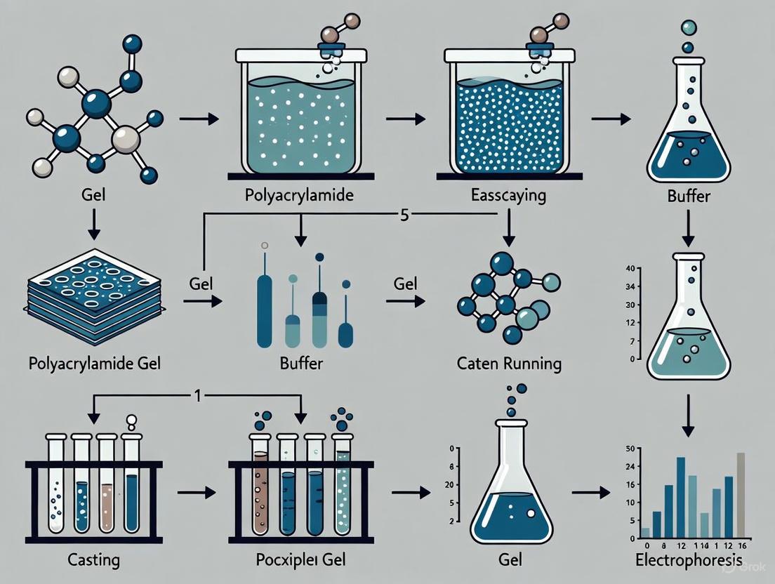
SDS-PAGE Experimental Workflow
Advanced Applications in Protein Research
Protein Quantification and Detection
Traditional protein quantification based on SDS-PAGE with staining has limitations in accuracy and dynamic range. Recent advancements have addressed these challenges through innovative approaches:
Intrinsic Fluorescence Imaging: An improved gel electrophoresis tank with online intrinsic fluorescence imaging enables real-time protein detection without staining, expanding the detection window to 10 lanes. This method demonstrates a limit of detection of 14 ng, limit of quantification of 42 ng, and a dynamic range of 50-8000 ng for bovine serum albumin (BSA) in complex samples [7].
Gaussian Fitting Arithmetic: This computational approach improves quantification accuracy from low-resolution gel images by modeling band signals as a sum of Lorentzian peaks, providing a close fit to experimental conditions. Validation studies showed recoveries of 106.37% for urine samples and 94.96% for whey samples, with intra-day and inter-day RSD values of 9.17% and 10.06% respectively [7].
Analysis of Protein Complexes
Blue-Native PAGE (BN-PAGE) and Clear-Native PAGE (CN-PAGE) have become indispensable tools for studying mitochondrial oxidative phosphorylation complexes and other membrane protein complexes. These techniques [4]:
- Resolve individual OXPHOS complexes and respiratory chain supercomplexes
- Enable analysis of assembly pathways of multi-subunit complexes
- Reveal pathologic mechanisms in patients with monogenetic OXPHOS disorders
- Allow in-gel enzyme activity staining for Complexes I, II, IV, and V
Recent protocol improvements have shortened sample extraction procedures and enhanced sensitivity of in-gel Complex V activity staining, providing robust, semi-quantitative, and reproducible results for characterizing native protein complexes [4].
Emerging Trends and Technological Advancements
The field of gel electrophoresis continues to evolve with several emerging trends enhancing its capabilities for protein analysis:
AI-Powered Image Analysis
Traditional gel image analysis methods have remained largely unchanged for decades, relying on manual processes or semi-automated algorithms that often miss bands, generate false positives, or inaccurately identify band edges. Recent breakthroughs in artificial intelligence are revolutionizing this field [5]:
GelGenie Framework: An AI-powered system using U-Net architectures trained on 500+ manually-labelled gel images can automatically identify gel bands in seconds across wide experimental conditions. The system performs pixel-level segmentation, classifying each pixel as 'band' or 'background', enabling accurate band identification even in sub-optimal conditions with warped bands, high background, gel contaminants, or diffuse bands [5].
Performance Validation: When applied to gel electrophoresis data from external laboratories, GelGenie generates results that quantitatively match those of the original authors, demonstrating its robustness and accuracy. The models are publicly available through an open-source application that requires no expert knowledge [5] [8].
Integration with Other Analytical Techniques
Modern electrophoresis increasingly combines with other powerful analytical methods:
Mass Spectrometry Coupling: Following separation by 2D-PAGE or other electrophoretic techniques, protein spots can be excised and identified by mass spectrometry, enabling comprehensive proteomic analysis [1].
Microfluidics Integration: The combination of electrophoresis with microfluidic devices enables high-throughput analysis, reduced sample requirements, and rapid results, particularly valuable in clinical diagnostics and pharmaceutical screening [1].
Modern Gel Electrophoresis Integration
Gel electrophoresis remains an indispensable technique for protein analysis due to its proven reliability, adaptability, and continuing technological evolution. From its foundational role in basic protein characterization to its advanced applications in complex system analysis and integration with cutting-edge computational methods, electrophoresis continues to provide critical insights into protein structure, function, and interactions.
The ongoing development of AI-powered analysis tools, enhanced detection methods, and integration with other analytical platforms ensures that gel electrophoresis will maintain its essential position in the researcher's toolkit. As these advancements address traditional limitations in quantification accuracy, throughput, and user accessibility, gel electrophoresis is poised to continue as a cornerstone technique supporting discoveries across biochemistry, molecular biology, clinical diagnostics, and pharmaceutical development.
Polyacrylamide gel electrophoresis (PAGE) remains a cornerstone technique in molecular biology and biochemistry laboratories worldwide, providing the foundation for protein separation and analysis. The efficacy of this technique hinges on the precise chemical structure of the polyacrylamide gel, which functions as a molecular sieve to separate biomolecules based on size and charge. This application note examines the fundamental chemistry of how acrylamide and bis-acrylamide co-polymerize to form a porous matrix, details optimized protocols for gel preparation, and provides troubleshooting guidance for common issues. Understanding these chemical principles is essential for researchers, scientists, and drug development professionals seeking to optimize protein electrophoresis for their specific research needs, from basic protein characterization to complex proteomic analyses.
The Chemical Foundation of Polyacrylamide Gels
Monomer Composition and Polymerization Chemistry
The molecular sieve properties of polyacrylamide gels originate from the specific chemical interaction between two monomers: acrylamide and N,N'-methylenebisacrylamide (bis-acrylamide). Acrylamide forms the primary backbone of the polymer matrix, while bis-acrylamide serves as the crosslinking agent that connects adjacent polyacrylamide chains [9]. This crosslinked structure creates a porous network through which proteins migrate during electrophoresis.
The polymerization reaction is catalyzed by a free-radical system involving ammonium persulfate (APS) as the initiator and tetramethylethylenediamine (TEMED) as the catalyst [10]. TEMED catalyzes the formation of free radicals from APS, which then initiate the polymerization of acrylamide monomers into long linear chains. The bis-acrylamide molecules, containing two acrylamide functional groups, incorporate into these growing chains and form cross-bridges between them, creating the characteristic three-dimensional mesh structure [10]. The resulting matrix is stable, chemically inert, and provides defined pore sizes that enable size-based separation of protein molecules.
Controlling Pore Size for Optimal Separation
The porosity of the gel matrix is precisely controlled by adjusting two key parameters: the total acrylamide concentration (%T) and the crosslinker ratio (%C). The total acrylamide concentration (%T) represents the combined mass of acrylamide and bis-acrylamide per 100 mL of solution and primarily determines the average pore size [9]. Higher %T values create denser matrices with smaller pores, which are ideal for separating lower molecular weight proteins. Conversely, lower %T values produce larger pores better suited for resolving higher molecular weight proteins [11].
The crosslinker ratio (%C) represents the proportion of bis-acrylamide relative to the total acrylamide content and affects the rigidity and porosity of the final gel structure. Standard protocols typically use a crosslinker ratio of 1-5% depending on the desired separation characteristics [12]. This precise control over matrix porosity enables researchers to tailor the gel composition to their specific protein separation needs, optimizing resolution across a wide molecular weight range.
Table 1: Optimal Acrylamide Concentrations for Separating Proteins of Different Sizes
| Protein Size (kDa) | Recommended Acrylamide Percentage |
|---|---|
| 4-40 | 20% |
| 12-45 | 15% |
| 10-70 | 12.5% |
| 15-100 | 10% |
| 25-200 | 8% |
Experimental Protocols
Reagent Preparation and Standard Formulations
Proper reagent preparation is fundamental to reproducible gel polymerization and optimal electrophoretic separation. The foundational component for most PAGE applications is a 30% acrylamide-bisacrylamide stock solution, typically prepared at a ratio of 29:1 or 37.5:1 (acrylamide:bis-acrylamide) [12]. This stock solution should be stored in dark bottles at 4°C to prevent spontaneous polymerization and decomposition.
The electrophoresis running buffer for SDS-PAGE typically consists of Tris-Glycine with 0.1% SDS, maintaining a pH of approximately 8.3-8.5 to facilitate protein migration toward the anode [10]. For specialized applications such as histidine-imidazole PAGE (HI-PAGE) for lipoprotein analysis, alternative buffer systems may be employed, such as Tris-histidine at pH 8.4 [13]. Sample loading buffer should contain SDS to denature proteins, a reducing agent (DTT or β-mercaptoethanol) to break disulfide bonds, glycerol to density-load samples into wells, and a tracking dye (bromophenol blue) to monitor migration progress [11].
Table 2: Essential Research Reagent Solutions for Polyacrylamide Gel Electrophoresis
| Reagent | Function | Standard Formulation |
|---|---|---|
| Acrylamide-Bis Solution | Forms the porous polymer matrix | 30% (w/v) in water, 29:1 or 37.5:1 ratio |
| TEMED | Catalyzes polymerization | Neat liquid, used at 0.1-0.2% final concentration |
| Ammonium Persulfate (APS) | Initiates free-radical polymerization | 10% (w/v) solution in water, freshly prepared |
| SDS (Sodium Dodecyl Sulfate) | Denatures proteins and confers uniform charge | 10% (w/v) solution in water |
| Tris Buffer | Maintains pH during electrophoresis | 1.5 M pH 8.8 (resolving gel), 0.5 M pH 6.8 (stacking gel) |
| Running Buffer | Conducts current and maintains pH | 25 mM Tris, 192 mM glycine, 0.1% SDS (w/v), pH 8.3 |
Step-by-Step Gel Casting Protocol
The following protocol details the preparation of a standard SDS-polyacrylamide gel with a stacking gel system for optimal protein separation [12] [2]:
Assemble the gel cassette: Clean glass plates with ethanol or industrial methylated spirit and assemble the casting apparatus according to manufacturer instructions, ensuring a tight seal to prevent leakage.
Prepare the resolving gel: Combine acrylamide-bis solution, Tris buffer (pH 8.8), SDS, and water in the specified volumes for the desired acrylamide percentage. Add TEMED and 10% APS last, mixing gently to avoid introducing air bubbles. Immediately pipette the solution into the gel cassette, leaving appropriate space for the stacking gel.
Overlay with isopropanol: Carefully add a layer of isopropanol or water-saturated butanol on top of the resolving gel to create a flat interface and exclude oxygen, which inhibits polymerization. Allow the gel to polymerize for 30-45 minutes at room temperature.
Prepare and add the stacking gel: After polymerization, pour off the isopropanol and rinse the gel surface with distilled water. Wick away excess liquid with lint-free tissue. Prepare the stacking gel mixture containing acrylamide, Tris buffer (pH 6.8), SDS, water, TEMED, and APS. Pour onto the resolving gel and immediately insert a clean comb, avoiding air bubbles. Allow to polymerize for 30 minutes.
Store or use the polymerized gel: Once polymerized, carefully remove the comb and rinse wells with running buffer. Gels can be used immediately or wrapped in moist tissue and plastic film for storage at 4°C for up to several weeks.
Electrophoresis Parameters and Run Conditions
For optimal protein separation, load denatured protein samples (heated at 95-100°C for 5-10 minutes in loading buffer) into the wells alongside appropriate molecular weight markers [2]. Fill the electrophoresis chamber with running buffer, ensuring the upper and lower chambers are properly connected. Apply a constant voltage of 100-150V for mini-gel systems, adjusting based on gel thickness and acrylamide percentage [14] [11]. Run the gel until the dye front reaches approximately 1 cm from the bottom, typically requiring 60-90 minutes depending on the conditions. To prevent smiling effects (curved bands) and overheating, run gels at lower voltages in a cold room or using apparatus with cooling capabilities [14].
Troubleshooting Common Issues
Despite the standardized protocols, researchers may encounter several common issues during polyacrylamide gel preparation and electrophoresis. The table below outlines these challenges, their potential causes, and recommended solutions based on established troubleshooting guidelines [14] [11]:
Table 3: Troubleshooting Common Polyacrylamide Gel Electrophoresis Issues
| Issue | Possible Cause | Solution |
|---|---|---|
| Smeared bands | Voltage too high; incomplete denaturation | Run gel at lower voltage (10-15 V/cm); ensure proper sample boiling with adequate SDS and DTT |
| Poor band separation | Gel percentage inappropriate for target protein size; insufficient run time | Use lower % gel for high MW proteins, higher % for low MW proteins; run until dye front reaches bottom |
| Smiling bands (curved bands) | Excessive heat generation during run | Run gel at lower voltage for longer time; use cooling apparatus or cold room |
| Distorted bands in peripheral lanes | Edge effect from empty wells | Load protein or ladder in all wells; avoid leaving wells empty |
| Protein samples migrating out of wells before run | Delay between loading and starting electrophoresis | Minimize time between loading and applying current; load samples quickly and consistently |
| Incomplete polymerization | Old or improperly prepared APS; insufficient TEMED | Use fresh APS prepared weekly; ensure adequate TEMED concentration; check reagent quality |
Additional issues may include vertical streaking, which often results from protein aggregation or precipitation, remedied by ensuring complete sample denaturation and reducing protein load [11]. If bands appear compressed or poorly resolved in specific regions of the gel, consider using gradient gels with increasing acrylamide concentration to improve resolution across a broader molecular weight range [10]. For persistent polymerization problems, systematically replace reagents to identify degraded components, as acrylamide solutions decompose over time, especially when improperly stored.
Advanced Applications and Methodological Variations
The versatility of polyacrylamide gel electrophoresis extends beyond standard SDS-PAGE, with several advanced applications leveraging the molecular sieve properties of the acrylamide-bis matrix. Native PAGE separates proteins in their non-denatured state, maintaining biological activity and higher-order structure by omitting SDS from the buffer system [10]. This technique separates proteins based on both size and inherent charge, enabling studies of protein complexes and functional interactions.
Two-dimensional PAGE combines isoelectric focusing (IEF) with SDS-PAGE to provide extremely high resolution separation of complex protein mixtures [10]. In this technique, proteins are first separated based on their isoelectric point in a pH gradient, then further resolved by molecular weight in the second dimension, creating a two-dimensional protein map where each spot represents an individual protein species.
Specialized applications continue to emerge, such as fluorescence-based histidine-imidazole PAGE (fHI-PAGE) for lipoprotein analysis, which provides rapid separation and quantification of lipoprotein fractions within one hour without band distortion [13]. Such methodological innovations demonstrate the continued utility and adaptability of polyacrylamide gel systems for diverse research applications in biochemistry and clinical diagnostics.
Within the broader context of preparing polyacrylamide gels for protein electrophoresis research, selecting the appropriate electrophoretic technique is a fundamental decision that directly determines the success and validity of an experiment. The choice between SDS-PAGE (Sodium Dodecyl Sulfate Polyacrylamide Gel Electrophoresis) and Native PAGE (Native Polyacrylamide Gel Electrophoresis) dictates whether proteins will be analyzed in a denatured state, separated solely by molecular mass, or in their native, functional conformation, separated by a combination of size, charge, and shape [15] [16]. This application note provides a detailed comparison of these two core techniques, including structured data tables, step-by-step protocols, and visualization tools to guide researchers and drug development professionals in making an informed choice aligned with their research objectives.
Core Principles and Comparative Analysis
SDS-PAGE is a denaturing technique where the anionic detergent SDS binds to proteins, unfolds them, and imparts a uniform negative charge. This masks the proteins' intrinsic charge and eliminates the influence of shape, resulting in separation based almost exclusively on molecular weight [15] [16]. Consequently, proteins lose their biological activity, making this method ideal for determining molecular weight, assessing purity, and analyzing subunit composition [16].
In contrast, Native PAGE is a non-denaturing technique. It is performed without SDS or reducing agents, preserving the protein's native conformation, multi-subunit structure, and biological activity [15] [17]. Separation depends on the protein's intrinsic charge, size, and three-dimensional shape, allowing researchers to study functional protein complexes, oligomerization states, and enzymatic activity [15] [16].
Table 1: Fundamental Differences Between SDS-PAGE and Native PAGE
| Criteria | SDS-PAGE | Native PAGE |
|---|---|---|
| Separation Basis | Molecular weight [15] | Size, overall charge, and shape [15] |
| Protein State | Denatured and unfolded [15] [16] | Native, folded, and functional [15] [16] |
| Key Reagents | SDS, reducing agent (e.g., DTT) [15] [17] | Non-denaturing buffers; no SDS [15] [17] |
| Sample Preparation | Heated (typically 85°C for 2 min) [15] [17] | Not heated [15] [17] |
| Protein Function Post-Run | Lost [15] | Retained [15] |
| Primary Applications | Molecular weight determination, purity check, protein expression analysis [15] | Studying protein structure, subunit composition, function, and protein-protein interactions [15] [16] |
A Decision Framework for Technique Selection
The following workflow diagram outlines the logical process for choosing between SDS-PAGE and Native PAGE based on the research goal.
Experimental Protocols
Protocol for SDS-PAGE
This protocol is adapted for a standard mini-gel format using a pre-cast Tris-Glycine gel [17].
Sample Preparation:
- Dilute your protein sample with an equal volume of 2X Tris-Glycine SDS Sample Buffer to a final 1X concentration [17].
- For reduced conditions, add a reducing agent like dithiothreitol (DTT) to a final concentration of 50 mM [17].
- Heat the samples at 85°C for 2 minutes to denature the proteins [17]. Cool briefly before loading.
Electrophoresis:
- Remove a pre-cast gel from its pouch and rinse the cassette with deionized water. Remove the comb and rinse the wells with 1X Tris-Glycine SDS Running Buffer [17].
- Assemble the gel in the electrophoresis chamber according to the manufacturer's instructions.
- Fill the inner (upper) and outer (lower) buffer chambers with 1X Tris-Glycine SDS Running Buffer [17].
- Load the prepared samples and an appropriate protein molecular weight marker into the wells.
- Run the gel at a constant voltage of 125 V until the dye front (bromophenol blue) reaches the bottom of the gel (approximately 90 minutes) [17].
Protocol for Native PAGE
This protocol describes a Native PAGE method using a Tris-Glycine system and pre-cast gels, suitable for analyzing protein oligomers [17] [18].
Sample Preparation:
- Dilute your protein sample with an equal volume of 2X Tris-Glycine Native Sample Buffer to a final 1X concentration [17].
- Do not add SDS, reducing agents, or heat the sample [15] [17]. Keep samples on ice to maintain native structure.
Electrophoresis:
- Prepare a pre-cast non-denaturing gel or cast a Tris-Glycine polyacrylamide gel (e.g., 7.5% separation gel, 4.5% stacking gel) [18].
- Assemble the gel in the electrophoresis chamber.
- Fill the buffer chambers with 1X Tris-Glycine Native Running Buffer (25 mM Tris, 192 mM glycine, pH ~8.8) [17] [18]. Note: The running buffer for native electrophoresis does not contain SDS.
- Load the prepared native samples and markers.
- Run the gel at a constant voltage of 125 V for 1-12 hours, depending on the protein system [17] [18]. The run time is typically longer than for SDS-PAGE due to the lack of a uniform charge from SDS.
The Scientist's Toolkit: Essential Research Reagents
Successful execution of protein electrophoresis relies on a set of key reagents, each with a specific function in sample preparation and separation.
Table 2: Key Research Reagent Solutions for PAGE
| Reagent | Function | SDS-PAGE | Native PAGE |
|---|---|---|---|
| SDS (Sodium Dodecyl Sulfate) | Denatures proteins and confers uniform negative charge [15] | Essential | Not Used |
| Reducing Agent (DTT/BME) | Breaks disulfide bonds for complete denaturation [15] [17] | Used (in reducing gels) | Not Used |
| Tris-Glycine SDS Buffer | Provides ionic environment for denatured protein separation [17] | Essential | Not Used |
| Tris-Glycine Native Buffer | Provides ionic environment for native protein separation [17] [18] | Not Used | Essential |
| Crosslinker (Bis-Acrylamide) | Forms the crosslinked network of the polyacrylamide gel matrix [19] [20] | Essential | Essential |
| Ammonium Persulfate (APS) & TEMED | Catalyzes the polymerization of the polyacrylamide gel [20] | Essential | Essential |
Data Interpretation and Analysis
Understanding how to interpret the results from each technique is critical. A classic example involves identifying a protein's quaternary structure.
Scenario: A protein sample runs as a 60 kDa band on non-reducing SDS-PAGE but migrates at 120 kDa on Native PAGE [21].
Inference: The protein is a dimer of 60 kDa subunits that are not linked by disulfide bonds. The non-reducing SDS-PAGE conditions would have left disulfide bonds intact; the fact that it still runs as a monomer indicates the dimer is held together by non-covalent interactions (e.g., hydrophobic, ionic) that are disrupted by SDS. In Native PAGE, the protein remains in its native, dimeric form, resulting in a larger apparent size [21].
Advanced Applications: Native PAGE Variants
For specialized analyses of complex protein assemblies, particularly mitochondrial oxidative phosphorylation (OXPHOS) complexes, advanced Native PAGE variants are used.
- Blue Native PAGE (BN-PAGE): Uses Coomassie Brilliant Blue dye, which binds to protein complexes and imparts a negative charge proportional to their mass, allowing separation in a native state. It is ideal for analyzing the size, abundance, and composition of large, multi-subunit complexes and their supercomplexes [4] [22].
- Clear Native PAGE (CN-PAGE): A related technique that separates protein complexes based on their intrinsic charge in a gradient gel without using Coomassie dye [15] [4]. These techniques can be coupled with a second-dimension SDS-PAGE to resolve the individual subunits of the complexes separated in the first dimension [4] [22].
SDS-PAGE and Native PAGE are complementary pillars of protein analysis. The decision between them rests squarely on the research question. SDS-PAGE is the tool of choice for determining molecular weight, analyzing purity, and characterizing denatured proteins. Native PAGE is indispensable for probing the functional state of proteins, revealing oligomeric structures, and studying interactions within protein complexes. By applying the guidelines, protocols, and interpretive frameworks outlined in this application note, researchers can confidently select and execute the optimal electrophoretic strategy for their work in biochemistry and drug development.
The Critical Role of SDS, Tris Buffers, APS, and TEMED in the Electrophoresis System
Application Note
In the preparation of polyacrylamide gels for protein electrophoresis, a precise understanding of the function and application of key reagents is fundamental to experimental success. This note details the critical roles of Sodium Dodecyl Sulfate (SDS), Tris buffers, Ammonium Persulfate (APS), and Tetramethylethylenediamine (TEMED) within the SDS-PAGE system. When used in concert, these reagents create the conditions necessary for the reliable separation of proteins based on their molecular weight, a cornerstone technique in biochemical research and drug development [23] [24]. The integrity of the gel matrix and the fidelity of protein migration are directly controlled by the quality, concentration, and handling of these components.
The foundation of the gel is formed by acrylamide and bisacrylamide, which copolymerize into a porous matrix. The pore size, which dictates the resolution of protein separation, is determined by the percentage of acrylamide used; higher percentages create smaller pores for better resolution of lower molecular weight proteins [24]. Within this system, Tris buffers establish and maintain the stable pH environment required for consistent protein migration and gel polymerization. APS and TEMED, the polymerization catalysts, are the engines of gel formation, initiating and propagating the free-radical reaction that solidifies the gel solution [23]. Finally, SDS is the great equalizer, binding to proteins and conferring upon them a uniform negative charge, which allows their separation to be based almost exclusively on molecular weight [24]. Any deviation in the preparation or quality of these reagents can lead to experimental artifacts, including poor band resolution, smearing, or incomplete polymerization, underscoring their non-negotiable role in the protocol [25].
Research Reagent Solutions
The following table catalogues the essential reagents for SDS-PAGE, detailing their specific functions in the electrophoresis system.
Table 1: Key Reagents for SDS-PAGE Gel Preparation and Electrophoresis
| Reagent | Function in the Electrophoresis System |
|---|---|
| SDS (Sodium Dodecyl Sulfate) | Denatures proteins and confers a uniform negative charge, allowing separation based primarily on molecular weight [24]. |
| Tris Buffers | Provides the necessary buffering capacity to maintain a stable pH during electrophoresis and gel polymerization (e.g., pH 8.8 for resolving gel, pH 6.8 for stacking gel) [23] [24]. |
| APS (Ammonium Persulfate) | Initiates the polymerization reaction of acrylamide and bisacrylamide by generating free radicals [23]. |
| TEMED | Catalyzes the polymerization reaction by accelerating the rate of free-radical formation from APS, leading to gel solidification [23]. |
| Acrylamide/Bis-Acrylamide | Forms the porous polyacrylamide gel matrix when polymerized; the ratio and concentration determine the gel's pore size and separation properties [24]. |
| Glycerol | Adds density to the sample buffer, ensuring samples sink to the bottom of the well during loading [23]. |
| Bromophenol Blue | A tracking dye that migrates ahead of the proteins, allowing visual monitoring of the electrophoresis progress [26]. |
| Coomassie Blue | A dye used for staining proteins after electrophoresis to visualize separated protein bands [26]. |
Quantitative Data Specifications
Precise formulation of gel components is critical for reproducibility. The following tables provide standard recipes for a 15% resolving gel and a 5% stacking gel, optimized for a 10 mL and 3 mL total volume, respectively [23].
Table 2: Formulation for a 15% Resolving Gel (10 mL total volume)
| Component | Volume | Final Concentration/Function |
|---|---|---|
| Deionized Water | 2.3 mL | Solvent |
| 30% Acrylamide/Bis | 5.0 mL | 15% gel matrix |
| 1.5 M Tris, pH 8.8 | 2.5 mL | ~0.375 M Tris, resolving pH |
| 10% SDS | 0.1 mL | 0.1% SDS |
| 10% Ammonium Persulfate (APS) | 0.1 mL | Polymerization initiator |
| TEMED | 0.004 mL | Polymerization catalyst |
Table 3: Formulation for a 5% Stacking Gel (3.0 mL total volume)
| Component | Volume | Final Concentration/Function |
|---|---|---|
| Deionized Water | 2.1 mL | Solvent |
| 30% Acrylamide/Bis | 0.5 mL | 5% gel matrix |
| 1.0 M Tris-HCl, pH 6.8 | 0.38 mL | ~0.127 M Tris, stacking pH |
| 10% SDS | 0.03 mL | 0.1% SDS |
| 10% Ammonium Persulfate (APS) | 0.03 mL | Polymerization initiator |
| TEMED | 0.003 mL | Polymerization catalyst |
Experimental Protocols
Detailed Methodology: SDS-PAGE Gel Preparation and Electrophoresis
I. Gel Preparation
- Assemble Casting Chamber: Clean and dry the glass plates and spacers. Assemble the casting stand according to the manufacturer's instructions to create a leak-proof chamber [24].
- Prepare Resolving Gel Solution: In a beaker or flask, combine all reagents for the resolving gel (see Table 2) except for TEMED and APS. Mix gently using a Pasteur pipette to avoid introducing air bubbles [23].
- Catalyze and Pour Resolving Gel: Add the specified volumes of 10% APS and TEMED to the solution. Swirl gently to mix. Immediately transfer the solution between the glass plates in the casting chamber, filling to about ¾ of the total height.
- Overlay and Polymerize: Carefully add a small layer of absolute ethanol or isobutyl alcohol on top of the gel solution to exclude oxygen and create a flat interface. Allow the gel to polymerize completely, which should occur within 5-30 minutes. Once polymerized, pour off the overlay, rinse with water, and absorb residual liquid with a Kimwipe or filter paper [23] [26].
- Prepare and Pour Stacking Gel: In a new container, combine all reagents for the stacking gel (see Table 3) except APS and TEMED. Add APS and TEMED, mix, and pour the solution directly onto the polymerized resolving gel. Immediately insert a clean comb into the stacking gel, avoiding air bubbles. Allow to polymerize for 20-30 minutes [23] [24].
II. Sample Preparation
- Denature Protein Samples: Mix the protein sample with an appropriate volume of Laemmli sample buffer (e.g., a 4x concentration). A typical ratio is 3 parts protein solution to 1 part 4x sample buffer [23].
- Heat Denaturation: Cap the tubes and boil the samples at 95-100°C for 5-10 minutes to fully denature the proteins [24].
- Centrifuge: Briefly centrifuge the denatured samples to collect condensation and bring the entire volume to the bottom of the tube.
III. Electrophoresis
- Set Up Apparatus: Remove the comb gently and rinse the wells with 1X electrophoresis buffer. Place the gel cassette into the electrode assembly and slide the assembly into the tank. Fill the inner and outer chambers with 1X electrophoresis buffer [23].
- Load Samples: Using a gel-loading micropipette, slowly load 20 µL of the denatured protein samples or molecular weight marker into the wells [23].
- Run Gel: Place the lid on the tank, aligning the electrodes correctly. Connect to a power supply and run the gel at a constant voltage of 80V until the dye front enters the resolving gel. Then, increase the voltage to 120V until the dye front reaches the bottom of the gel. This process typically takes 1-2 hours [23].
- Stain and Destain: After electrophoresis, carefully disassemble the apparatus and remove the gel. Visualize proteins by staining with Coomassie Blue staining solution for several hours or overnight with gentle shaking. Destain with a destaining solution (e.g., 10% acetic acid, 10% methanol) until the background is clear and protein bands are visible [26].
Troubleshooting Common Issues
The following table outlines common problems, their causes, and solutions related to key reagents and the electrophoresis process [25].
Table 4: Troubleshooting Guide for SDS-PAGE
| Problem | Possible Cause | Suggested Solution |
|---|---|---|
| Poor Band Resolution | Gel concentration is incorrect for target protein size. | Use a gradient gel (e.g., 4%-20%) if protein size is unknown, or adjust acrylamide % [25]. |
| Current/voltage is too high, causing "band smiling." | Decrease the voltage by 25-50% [25]. | |
| Band Smearing | Protein concentration is too high. | Reduce the amount of protein loaded on the gel [25]. |
| Voltage is too high. | Decrease the voltage by 25-50% [25]. | |
| Gel Does Not Polymerize | TEMED and/or APS are degraded or were forgotten. | Use fresh APS and TEMED; ensure they are added to the gel mixture [25]. |
| The temperature is too low. | Cast the gel at room temperature [25]. | |
| Samples Do Not Sink | Insufficient glycerol in sample buffer. | Ensure sample buffer contains glycerol (e.g., 2-5%) [25]. |
| Skewed/Distorted Bands | Polymerization around wells was poor or uneven. | Increase the amount of APS and TEMED slightly; ensure gel solution is mixed thoroughly and poured without bubbles [25]. |
| Protein Aggregation | Insufficient reducing agent; hydrophobic proteins. | Prepare fresh sample buffer with fresh DTT or β-mercaptoethanol; for hydrophobic proteins, add 4-8 M urea to the sample [25]. |
SDS-PAGE Workflow and Reagent Roles
The diagram below illustrates the logical workflow of an SDS-PAGE experiment, highlighting the critical points where the four key reagents (SDS, Tris, APS, TEMED) perform their essential functions.
SDS-PAGE Experimental Workflow
How Gel Percentage and Pore Size Dictate Protein Separation by Molecular Weight
Polyacrylamide gel electrophoresis (PAGE) serves as a fundamental tool in molecular biology and proteomics for separating protein mixtures based on their molecular weights. The efficacy of this separation hinges primarily on the polyacrylamide gel matrix, which functions as a molecular sieve [10]. This application note delineates the quantitative relationship between gel percentage, resultant pore size, and protein separation range, providing researchers with detailed protocols and frameworks to optimize electrophoretic conditions for specific experimental requirements. Understanding these principles is essential for preparing gels that yield high-resolution protein separation, a cornerstone technique in protein characterization, purity assessment, and proteomic analysis [10] [27].
The polyacrylamide gel matrix is formed through the copolymerization of acrylamide and a cross-linking agent, typically N,N'-methylenebisacrylamide (bis-acrylamide) [10]. The pore size of this three-dimensional network is inversely related to the total percentage of acrylamide, with higher percentages creating smaller pores that retard the migration of larger proteins [10]. This principle allows researchers to selectively tailor gel compositions to target specific molecular weight ranges of interest.
Theoretical Foundation: The Gel Matrix as a Molecular Sieve
Polyacrylamide Gel Formation and Pore Structure
The polyacrylamide gel matrix is created through a free radical polymerization reaction. Acrylamide monomers form the backbone of the polymer chains, while bis-acrylamide molecules covalently link these chains together, creating a cross-linked network [10] [28]. The polymerization is catalyzed by ammonium persulfate (APS), which provides the free radicals, and tetramethylethylenediamine (TEMED), which accelerates the radical formation [10]. The resulting gel possesses a three-dimensional mesh with pores through which proteins migrate under the influence of an electric field.
The pore size within this matrix is determined by two key factors: the total concentration of acrylamide (%T) and the degree of cross-linking (%C, representing the proportion of bis-acrylamide relative to total acrylamide) [10]. Standard protocols often use a bis-acrylamide to acrylamide ratio of 1:29, but this can be adjusted to modify the gel's mechanical properties and pore size distribution [10].
The Role of SDS in Protein Separation
In SDS-PAGE, the anionic detergent sodium dodecyl sulfate (SDS) plays a crucial role in normalizing protein charge. Proteins are denatured by heating in the presence of SDS and a reducing agent (e.g., β-mercaptoethanol or dithiothreitol), which cleaves disulfide bonds [10] [27]. SDS binds to the denatured polypeptides in a constant weight ratio (approximately 1.4 g SDS per 1 g of protein), conferring a uniform negative charge density [10] [27]. This process neutralizes the proteins' intrinsic charges and unfolds them into linear chains, transforming molecular weight separation into the primary factor governing electrophoretic mobility [29].
The following diagram illustrates the molecular separation mechanism in SDS-PAGE:
Discontinuous Buffer System
Most SDS-PAGE systems employ a discontinuous (or disc) buffer system that enhances separation resolution. This system comprises two distinct gel layers with different pore sizes and pH values: a stacking gel (typically 4-5% acrylamide, pH 6.8) and a resolving gel (varying percentages, pH 8.8) [10] [29]. The stacking gel concentrates protein samples into sharp bands before they enter the resolving gel, where actual separation occurs based on molecular size [29]. This process is facilitated by differences in ionic composition and pH between the stacking gel, resolving gel, and running buffer, creating a transient state where proteins focus into narrow zones [29].
Quantitative Relationships: Gel Percentage vs. Protein Separation
Optimal Acrylamide Percentages for Protein Separation
The appropriate acrylamide concentration depends directly on the molecular weight range of the target proteins. Lower percentage gels (e.g., 8-10%) with larger pores are optimal for resolving high molecular weight proteins, while higher percentage gels (e.g., 12-15%) with smaller pores provide better separation of lower molecular weight proteins [10]. Gradient gels, which contain a continuous increase in acrylamide concentration (e.g., 4-20%), extend the separation range across a broader spectrum of molecular weights within a single gel [10].
Table 1: Recommended Polyacrylamide Gel Percentages for Protein Separation by Molecular Weight
| Gel Percentage (%) | Effective Separation Range (kDa) | Optimal Application |
|---|---|---|
| 6-8% | 50-150 | Very high molecular weight proteins |
| 10% | 20-100 | Standard mixture of proteins |
| 12% | 10-60 | Most common range for protein analysis |
| 15% | 5-45 | Low molecular weight proteins and peptides |
Mathematical Relationship and Ferguson Analysis
The relationship between protein mobility (Rf) and gel concentration follows a logarithmic function described by the Ferguson equation: log(Rf) = log(Ro) - Kr × %T, where Rf represents the relative mobility of the protein, Ro is the free electrophoretic mobility, Kr is the retardation coefficient, and %T is the total acrylamide concentration. This mathematical relationship demonstrates that electrophoretic mobility decreases exponentially with increasing gel concentration, with larger proteins exhibiting steeper declines in mobility (higher Kr values) due to greater steric hindrance within the gel matrix.
Table 2: Separation Characteristics of Different Gel Configurations
| Gel Type | Total Acrylamide | Pore Size | Separation Principle | Applications |
|---|---|---|---|---|
| Denaturing SDS-PAGE | Variable %T | Determined by %T | Molecular weight | Most protein analyses; mass determination |
| Native PAGE | Variable %T | Determined by %T | Size, charge, shape | Active enzyme assays; protein complexes |
| Gradient Gels | Increasing %T | Decreasing pore size | Molecular weight | Broad range separation in single gel |
| Two-Dimensional PAGE | Fixed or gradient %T | Fixed or variable | pI (1D), mass (2D) | Comprehensive proteomic analysis |
Experimental Protocols
Protocol 1: Casting a Standard SDS-Polyacrylamide Gel
This protocol details the preparation of a discontinuous SDS-polyacrylamide gel with a 12% resolving gel and 5% stacking gel, suitable for separating proteins in the 10-60 kDa range [10] [27].
Research Reagent Solutions
Table 3: Essential Reagents for Polyacrylamide Gel Preparation
| Reagent | Function | Notes |
|---|---|---|
| Acrylamide/Bis-acrylamide | Gel matrix formation | Neurotoxin in monomer form; use pre-made solutions |
| Tris-HCl Buffer | pH control | Different concentrations for stacking (pH 6.8) and resolving (pH 8.8) gels |
| Sodium Dodecyl Sulfate (SDS) | Protein denaturation/detergent | Provides uniform negative charge to proteins |
| Ammonium Persulfate (APS) | Polymerization initiator | Freshly prepared solutions recommended |
| TEMED | Polymerization catalyst | Accelerates free radical formation from APS |
| Butanol/isopropanol | Gel surface dehydration | Creates flat interface for stacking gel |
Resolving Gel Preparation
- Gel Solution Composition: For a 12% resolving gel, combine the following components in a clean flask: 4.0 mL of 30% acrylamide/bis-acrylamide solution (29:1), 2.5 mL of 1.5 M Tris-HCl (pH 8.8), 3.4 mL of distilled water, 100 μL of 10% SDS, 50 μL of 10% ammonium persulfate, and 10 μL of TEMED [10].
- Casting: Mix gently and immediately pipette the solution between assembled glass plates, leaving space for the stacking gel (approximately 2 cm from the top).
- Overlay: Carefully overlay the gel solution with saturated butanol or isopropanol to exclude oxygen and create a flat meniscus.
- Polymerization: Allow the gel to polymerize completely (approximately 20-30 minutes at room temperature). A distinct refractive interface will form between the gel and overlay solution.
Stacking Gel Preparation
- Gel Solution Composition: For a 5% stacking gel, combine: 0.67 mL of 30% acrylamide/bis-acrylamide solution (29:1), 0.5 mL of 0.5 M Tris-HCl (pH 6.8), 2.8 mL of distilled water, 40 μL of 10% SDS, 30 μL of 10% ammonium persulfate, and 5 μL of TEMED [10].
- Casting: After removing the overlay from the polymerized resolving gel, pipette the stacking gel solution onto the resolving gel.
- Comb Insertion: Immediately insert a clean sample comb without introducing air bubbles.
- Polymerization: Allow complete polymerization (approximately 15-20 minutes).
The complete workflow for protein separation analysis is summarized below:
Protocol 2: Sample Preparation and Electrophoretic Conditions
Protein Sample Preparation
- Sample Buffer: Combine protein sample with 2× Laemmli buffer (typically containing: 62.5 mM Tris-HCl pH 6.8, 2% SDS, 25% glycerol, 0.01% bromophenol blue, with 5% β-mercaptoethanol or 100 mM DTT added fresh for reduction) [27] [29].
- Denaturation: Heat samples at 70-100°C for 5-10 minutes to ensure complete denaturation [27].
- Loading: Centrifuge briefly to collect condensation, then load 10-20 μL per well (typically 10-30 μg total protein for complex mixtures).
Electrophoresis Conditions
- Buffer System: Fill electrophoresis chamber with Tris-glycine running buffer (25 mM Tris, 192 mM glycine, 0.1% SDS, pH 8.3) [27].
- Voltage Parameters: Apply constant voltage: 80 V during stacking phase (approximately 20-30 minutes until dye front enters resolving gel), then increase to 120-150 V for separation (1-2 hours, until dye front reaches bottom of gel) [27] [30].
- Termination: Stop electrophoresis before the bromophenol blue tracking dye migrates off the bottom of the gel.
Protocol 3: Post-Electrophoresis Analysis
Protein Staining and Visualization
- Fixation: For Coomassie Brilliant Blue staining, immerse gel in fixative solution (40% ethanol, 10% acetic acid) for 30-60 minutes.
- Staining: Transfer to Coomassie staining solution (0.1% Coomassie R-250 in 40% ethanol, 10% acetic acid) for 1-2 hours with gentle agitation.
- Destaining: Remove background stain with destaining solution (40% ethanol, 10% acetic acid) until protein bands are clear against a transparent background.
- Documentation: Capture digital images using gel documentation systems with white light illumination [31].
Molecular Weight Determination
- Molecular Weight Markers: Include prestained or unstained protein standards in at least one lane [10].
- Standard Curve: Plot log(molecular weight) of standards versus relative mobility (Rf = migration distance of protein / migration distance of dye front).
- Interpolation: Determine unknown protein molecular weights by interpolation from the standard curve. Typical accuracy is ±10% [27].
Advanced Applications and Considerations
Gradient Gels for Enhanced Separation Range
Gradient gels provide a continuous increase in acrylamide concentration from top to bottom (e.g., 4-20%), creating a decreasing pore size gradient [10]. This configuration offers several advantages: (1) broader separation range within a single gel, (2) automatic stacking effect without need for a separate stacking gel, and (3) sharper protein bands as molecules slow down progressively in regions of appropriately sized pores [10]. Gradient gels are particularly valuable for analyzing complex protein mixtures with components spanning a wide molecular weight spectrum.
Two-Dimensional Electrophoresis
Two-dimensional PAGE (2D-PAGE) separates proteins by two distinct properties: isoelectric point (pI) in the first dimension using isoelectric focusing (IEF), followed by molecular weight separation in the second dimension using standard SDS-PAGE [10]. This technique provides the highest resolution for protein analysis, capable of resolving thousands of proteins from a single sample, making it indispensable for comprehensive proteomic studies [10] [32]. Specialized computational tools like MatGel have been developed to automate the quantification of protein spots in 2D-PAGE images, enhancing throughput and reducing manual errors [32].
Troubleshooting Common Issues
- Smearing Bands: Can result from protein overload, incomplete denaturation, too high voltage causing overheating, or gel imperfections [31] [30].
- Atypical Migration: Post-translational modifications (e.g., glycosylation, phosphorylation) can alter SDS binding and thus electrophoretic mobility [29].
- Poor Resolution: Optimize acrylamide percentage for target protein size range, ensure fresh electrophoresis buffers, and verify appropriate running conditions [31].
The precise control of polyacrylamide gel percentage represents a fundamental parameter in optimizing protein separation by molecular weight. The direct relationship between acrylamide concentration, pore size, and separation range enables researchers to strategically design electrophoretic conditions tailored to their specific protein targets. The protocols outlined in this application note provide a systematic approach to gel preparation, sample processing, and analysis that ensures reproducible, high-resolution protein separation. Mastery of these techniques forms an essential foundation for advanced proteomic investigations and biomarker discovery in both research and drug development contexts.
Step-by-Step Protocol: Casting and Running a Protein Gel for Optimal Results
For researchers in protein biochemistry and drug development, the integrity of polyacrylamide gel electrophoresis (SDS-PAGE) is foundational. The process begins at the casting station, where the quality of the gel is determined. Proper setup and reagent preparation are critical for achieving high-resolution protein separation, which underpins accurate analysis in western blotting, protein purification, and quantification. This application note provides a detailed protocol for assembling a gel casting station and preparing laboratory-grade polyacrylamide gels, ensuring reproducible and reliable results for protein electrophoresis research.
The Scientist's Toolkit: Essential Reagents and Equipment
A properly configured gel casting station requires specific reagents and equipment. The following table details the essential materials, their functions, and critical specifications for preparing polyacrylamide gels.
Table 1: Essential Reagents and Equipment for a Gel Casting Station
| Item Name | Function/Description | Key Specifications & Notes |
|---|---|---|
| Glass Plates & Spacers | Forms the mold for the gel. | Clean thoroughly before use to ensure gel polymerizes evenly and does not detach [25]. |
| Casting Frame/Stand | Holds glass plates and spacers securely to prevent leaks. | Ensure no vaseline is needed and apparatus is correctly aligned to prevent leaking [25]. |
| Acrylamide/Bis-Acrylamide | Forms the polyacrylamide matrix for size-based separation. | Typically used at a 19:1 or 29:1 ratio; potent neurotoxin—wear appropriate PPE (gloves, mask) when handling powder [3]. |
| Ammonium Persulfate (APS) | Initiates the polymerization reaction as a catalyst. | Use fresh aliquots; old or improperly stored APS will cause slow or incomplete polymerization [3] [25]. |
| TEMED | Catalyzes polymerisation by generating free radicals. | Use fresh; quantity can be adjusted to control polymerization speed [25]. |
| Tris Buffer | Provides the required pH environment for gel polymerization and electrophoresis. | Common buffers are Tris-Glycine or Tris-HCl for SDS-PAGE. |
| SDS (Sodium Dodecyl Sulfate) | Denatures proteins and confers a uniform negative charge. | Ensure SDS is added to the sample buffer; absence will prevent proteins from migrating into the gel [25]. |
| Comb | Creates wells for sample loading. | Remove carefully after polymerization to avoid damaged or distorted wells, which lead to smeared bands [33] [25]. |
| Gel Stains | For visualizing proteins post-electrophoresis. | Options include Coomassie Blue, Silver Stain, or fluorescent dyes. For DNA, SYBR-safe or GelRed are non-mutagenic alternatives to ethidium bromide [3]. |
Experimental Protocol: Assembling and Casting a Polyacrylamide Gel
Safety Precautions
- Acrylamide Handling: Acrylamide in its powdered form is a potent neurotoxin and can be easily aerosolized. Always wear a mask and gloves when weighing powdered acrylamide. Consider using pre-made liquid acrylamide solutions or pre-cast gels to minimize risk [3].
- General Practice: Wear gloves and lab coats throughout the procedure. Dispose of gel waste according to institutional safety guidelines.
Step-by-Step Methodology
Table 2: Step-by-Step Gel Casting Protocol
| Step | Procedure | Critical Parameters |
|---|---|---|
| 1. Assembly | Clean glass plates and spacers thoroughly. Assemble the plates with spacers and secure them in the casting frame. Ensure a tight seal to prevent leakage. | A leak-proof assembly is crucial. Check for gaps and ensure plates are correctly aligned [25]. |
| 2. Gel Solution Prep | Prepare the resolving gel solution according to the desired percentage (see Table 3). Mix all components except TEMED and APS. Degas the solution for 1-2 minutes to prevent bubble formation during polymerization. | Degassing improves polymerization consistency and minimizes air bubbles in the final gel. |
| 3. Polymerization Initiation | Add the required volumes of APS and TEMED to the degassed gel solution. Swirl gently to mix. Avoid introducing air bubbles. | Use fresh APS and TEMED. Incomplete polymerization can result from old reagents [3] [25]. |
| 4. Casting Resolving Gel | Pipette the resolving gel solution into the gap between the glass plates. Leave space for the stacking gel (approx. 1-2 cm below the comb teeth). | |
| 5. Overlaying | Gently overlay the resolving gel surface with water-saturated butanol or deionized water. This ensures a flat, even gel surface by excluding oxygen. | A flat interface between the stacking and resolving gels is critical for sharp band resolution [25]. |
| 6. Polymerization | Allow the gel to polymerize completely for 20-45 minutes at room temperature. Polymerization is indicated by a distinct schlieren line at the overlay-gel interface. | Do not disturb the gel during polymerization. Insufficient polymerization time can lead to poor well formation [25]. |
| 7. Casting Stacking Gel | Pour off the overlay liquid. Prepare the stacking gel solution (typically 3-5% acrylamide), add APS and TEMED, and pour it onto the polymerized resolving gel. | |
| 8. Inserting Comb | Immediately insert a clean, dry comb into the stacking gel solution. Avoid trapping air bubbles under the teeth of the comb. | Ensure the comb is level. Pushing the comb all the way to the bottom can cause sample leakage and smearing [33]. |
| 9. Final Polymerization | Allow the stacking gel to polymerize for 20-30 minutes. Once set, the gel can be used immediately or wrapped in moist paper towels, sealed in a plastic bag, and stored at 4°C for short-term use (1-2 days). | Remove the comb carefully and steadily to prevent damage to the wells [33]. |
Gel Percentage Selection Guide
The concentration of acrylamide in the resolving gel determines the pore size and thus the effective separation range for proteins of different molecular weights.
Table 3: Polyacrylamide Gel Concentrations for Optimal Protein Separation Adapted from general principles of nucleic acid and protein electrophoresis [3]
| % Acrylamide | Effective Separation Range (kDa) |
|---|---|
| 8% | 30 - 200 |
| 10% | 20 - 100 |
| 12% | 15 - 70 |
| 15% | 10 - 50 |
Note: For samples with a broad range of molecular weights or unknown sizes, a 4%-20% gradient gel is recommended for optimal resolution across sizes [25].
Workflow and Troubleshooting
The following workflow diagram outlines the entire process from setup to electrophoresis, highlighting key decision points and quality control checkpoints.
Troubleshooting Common Casting and Early Run Issues
Even with careful preparation, issues can arise. The table below lists common problems related to gel casting, their potential causes, and solutions.
Table 4: Troubleshooting Guide for Gel Casting and Initial Electrophoresis
| Problem | Possible Cause | Suggested Solution |
|---|---|---|
| Gel does not polymerize | TEMED or APS left out; reagents too old; temperature too low. | Increase APS/TEMED; use fresh aliquots; cast gel at room temperature [25]. |
| Slow or incomplete polymerization | Old APS stored above -20°C. | Use fresh APS aliquots kept frozen [3] [25]. |
| Leaking during casting | Glass plates chipped or misaligned; casting frame not sealed. | Check plate integrity; reassemble apparatus correctly; use Vaseline on spacers if needed [25]. |
| Poorly formed or damaged wells | Comb removed too early or carelessly; stacking gel too concentrated. | Allow full polymerization (30 min); remove comb steadily and carefully; use lower % acrylamide for stacking gel [33] [25]. |
| Samples do not sink into wells | Insufficient glycerol in sample buffer; comb removed too early. | Ensure sample buffer has enough glycerol; let stacking gel polymerize fully before comb removal [25]. |
| Bands are skewed or distorted | Salt concentration in sample too high; polymerization around wells is poor; gel interface uneven. | Dialyze high-salt samples; ensure well-forming gel has enough APS/TEMED; overlay resolving gel carefully [25]. |
| Smeared bands | Protein overloaded; voltage too high; well damaged during loading. | Load 0.1–0.2 µg of protein per mm well width; decrease voltage; avoid puncturing wells with pipette tip [33] [34] [25]. |
A meticulously prepared gel casting station is the first critical step in obtaining publication-quality protein separation. By adhering to the detailed protocols for reagent preparation, gel casting, and assembly outlined in this document, researchers can ensure consistency, reproducibility, and high resolution in their SDS-PAGE experiments. Attention to detail—from using fresh chemical initiators to careful handling of combs and glass plates—will minimize analytical artifacts and streamline the path to reliable protein data, thereby strengthening the foundation of any subsequent research in drug development and proteomics.
This application note provides a detailed protocol for preparing the resolving gel in polyacrylamide gel electrophoresis (PAGE), with emphasis on optimizing the critical steps of mixing, degassing, and pouring to achieve a flat polymerization interface. A flat gel interface is crucial for uniform protein migration and high-resolution separation, which are fundamental requirements for accurate analysis in protein research and drug development. The guidelines presented herein support reproducible and reliable gel preparation, enhancing data quality in electrophoretic experiments.
Polyacrylamide gel electrophoresis (PAGE) is a cornerstone technique for separating proteins based on their molecular weight. The quality of the electrophoresis results is profoundly dependent on the initial preparation of the polyacrylamide gel, particularly the resolving gel, which is responsible for separating proteins by size. The processes of mixing the gel solution, degassing to remove oxygen, and pouring the gel to achieve a flat interface are critical technical steps that directly impact polymerization quality, band sharpness, and overall separation performance [35] [36]. This protocol details these key steps within the context of preparing a standard SDS-polyacrylamide resolving gel, providing researchers with a standardized method to ensure reproducibility and optimal results.
Research Reagent Solutions
The following table lists essential materials and their specific functions in the preparation of the resolving gel.
Table 1: Essential Reagents for Resolving Gel Preparation
| Reagent | Function and Importance |
|---|---|
| Acrylamide/Bis-acrylamide | Forms the cross-linked polymer matrix that acts as a molecular sieve for separating proteins [37]. |
| Tris-HCl Buffer (e.g., 1.5 M, pH 8.8) | Provides the appropriate pH environment for efficient polymerization and protein separation in the resolving gel [36]. |
| Sodium Dodecyl Sulfate (SDS) | An ionic detergent that denatures proteins and confers a uniform negative charge, allowing separation based primarily on molecular weight [37]. |
| Ammonium Persulfate (APS) | A source of free radicals that initiates the polymerization reaction of acrylamide and bis-acrylamide [36] [38]. |
| TEMED (N,N,N',N'-Tetramethylethylenediamine) | A catalyst that accelerates the polymerization process by decomposing APS to generate free radicals [36] [38]. |
| Water-saturated n-butanol or Isopropanol | Layered on top of the poured gel solution to exclude oxygen and ensure a flat, uniform polymerization interface [36]. |
Detailed Methodology
Mixing the Gel Solution
- Recipe Formulation: Calculate the required volumes of all components based on the desired gel percentage and number of gels. A typical resolving gel solution for two gels includes water, the appropriate Tris-HCl buffer (e.g., 1.5 M, pH 8.8), acrylamide/bis-acrylamide stock solution, and a 10% SDS solution [36].
- Combining Reagents: In a clean flask, first mix the water, Tris buffer, and acrylamide solution. Swirl the flask gently to combine. It is recommended to bring the gel solution to room temperature before proceeding, as cold solutions can hold more dissolved oxygen and polymerize more slowly after degassing [35].
- Pre-degassing Consideration: Ensure the solution is well-mixed and free of large air bubbles before the degassing step.
Degassing the Gel Solution
- Purpose: Degassing is a critical step to remove dissolved oxygen from the gel solution. Oxygen inhibits the polymerization process by reacting with the free radicals generated by APS and TEMED, which can lead to slow, uneven, or incomplete polymerization, resulting in poor gel quality and irreproducible separations [35].
- Procedure: Transfer the mixed gel solution to an Ehrlenmeyer flask to prevent boil-over. Place the flask on a house vacuum or aspirator for approximately 5-10 minutes. The solution may bubble vigorously as gas is removed.
- Key Considerations:
- Duration: For polymerization systems initiated primarily by APS and TEMED, a degassing time of 5-10 minutes is typically sufficient [35]. Avoid excessive degassing (e.g., beyond 10-15 minutes), which is unnecessary and can be detrimental if riboflavin is used as a co-initiator.
- Temperature: Degassing is more effective and faster if the solution is at room temperature (23–25°C) rather than cold [35].
Initiating Polymerization and Pouring
- Adding Polymerization Initiators: After degassing, swiftly add the required volumes of 10% Ammonium Persulfate (APS) and TEMED to the flask. Swirl the flask gently but thoroughly to ensure homogeneous mixing. Note: The addition of TEMED and APS will begin the polymerization process immediately; work efficiently from this point.
- Pouring the Gel: Using a pipette or by carefully pouring, transfer the gel solution between the assembled glass plates. Pour the solution to a level approximately 1 cm below where the bottom of the comb will be seated [36].
- Achieving a Flat Interface: Immediately after pouring, gently layer a volume of water-saturated n-butanol or isopropanol on top of the gel solution. This step is crucial as it excludes ambient oxygen from the surface of the gel solution, which would otherwise inhibit polymerization and create a curved, uneven interface [36].
- Polymerization: Allow the gel to polymerize undisturbed at room temperature. Polymerization is typically complete within 20-30 minutes, indicated by a distinct refractive line forming between the polymerized gel and the overlaying alcohol solution. The gel is now ready for the next steps, including pouring the stacking gel.
Experimental Workflow
The following diagram illustrates the logical sequence of the key steps involved in preparing the resolving gel, from mixing to the completed polymerization.
Diagram 1: Workflow for preparing the resolving gel, highlighting the critical path from mixing to polymerization.
Data Presentation: Troubleshooting Common Issues
Even with a standardized protocol, issues can arise. The following table outlines common problems, their potential causes, and recommended solutions.
Table 2: Troubleshooting Resolving Gel Preparation
| Observation | Potential Cause | Troubleshooting Recommendation |
|---|---|---|
| Slow or incomplete polymerization | Insufficient degassing, leaving oxygen to inhibit the reaction [35]. | Ensure proper vacuum is applied during degassing and that the solution is at room temperature. |
| Old or inactive APS/TEMED [36]. | Prepare fresh APS solution and ensure reagents are stored properly. | |
| Curved or uneven gel interface | Failure to layer with alcohol after pouring [36]. | Always layer the gel solution with water-saturated n-butanol or isopropanol immediately after pouring. |
| Uneven sealing of the gel plates, leading to leaks. | Ensure gel plates are properly sealed with agarose or other suitable sealant before pouring [36]. | |
| Smeared protein bands | Gel polymerization at too high a temperature, causing uneven gel structure [39]. | Polymerize and run gels at a consistent, cool temperature (e.g., in a cold room or with a cooling apparatus). |
| Bubbles trapped in the polymerized gel | Failure to remove bubbles after pouring the gel solution. | Push bubbles away from the well comb or towards the edges with a pipette tip after pouring [40]. |
Discussion
The consistent preparation of a high-quality resolving gel is a foundational skill in protein biochemistry. The practice of degassing, while sometimes omitted in rushed protocols, is scientifically justified. Oxygen is a potent free radical trap that directly competes with the acrylamide polymerization reaction, leading to inconsistent pore sizes and potential gel artifacts [35]. Furthermore, achieving a flat interface via alcohol layering is a simple yet effective method to ensure that proteins in all lanes of the gel enter the resolving region simultaneously, which is a prerequisite for accurate molecular weight determination and comparative quantification.
For specialized applications, alternative polymerization methods exist. For instance, photopolymerization using riboflavin can be employed, which is compatible with a wider range of pH conditions and is non-oxidative [41]. However, it requires careful optimization of degassing time, as a small amount of oxygen is necessary for the conversion of riboflavin to its active form [35].
This application note provides a robust, step-by-step protocol for preparing a polyacrylamide resolving gel, with a focused examination of the critical steps that ensure a flat, uniform gel interface. By meticulously following the procedures for mixing, degassing, and pouring outlined herein, researchers can significantly improve the reproducibility and resolution of their protein separations, thereby enhancing the reliability of data generated in downstream analyses for research and drug development.
In the preparation of polyacrylamide gels for protein electrophoresis, achieving a uniform, flat polymerizing surface is critical for high-resolution separation. The isopropanol overlay technique is a standard laboratory practice used to create an oxygen-free and level barrier over the gel solution during the crucial polymerization period. This protocol details the application of isopropanol overlays within the context of SDS-PAGE, providing researchers with a definitive guide to improving gel quality and reproducibility for protein research and drug development.
The Principle of the Isopropanol Overlay
The Challenge of Oxygen Inhibition
The polymerization of acrylamide into a polyacrylamide gel is a free-radical chain reaction catalyzed by tetramethylethylenediamine (TEMED) and initiated by ammonium persulfate (APS) [2]. Molecular oxygen (O~2~) acts as a potent inhibitor of this process by reacting with the initiating and propagating free radicals, thereby terminating the polymerization chain reaction [42] [2]. This inhibition can lead to several artifacts:
- Uneven Polymerization: A slanted or wavy gel surface that compromises lane-to-lane running consistency.
- Poor Resolution: Incomplete polymerization results in a gel with inconsistent pore size, leading to diffuse protein bands and unreliable molecular weight determination [2].
- Extended Polymerization Time: Significant oxygen exposure can delay or entirely prevent gel formation.
The Isopropanol Solution
To mitigate the effects of oxygen, an inert, water-miscible, and denser-than-air liquid is layered on top of the freshly poured gel solution. Isopropanol (IPA) is the solvent of choice for this overlay due to its ideal physicochemical properties [42]:
- Water Miscibility: Allows for easy removal and clean interface formation.
- Inert Nature: Does not interfere with the free-radical polymerization chemistry.
- Low Density and Volatility: Ensures it sits atop the aqueous gel solution and evaporates cleanly after its function is served.
- Low Vapor Pressure: Creates a stable, sealing barrier that excludes atmospheric oxygen more effectively than more volatile solvents.
The overlay technique also serves a secondary mechanical function: the weight and surface tension of the isopropanol layer promote the formation of a perfectly flat and horizontal meniscus, which is essential for creating straight wells and uniform protein migration [42].
Experimental Protocol for Isopropanol Overlay in SDS-PAGE
The following protocol describes the specific steps for using an isopropanol overlay when casting a discontinuous SDS-polyacrylamide gel, which consists of a separating (resolving) gel and a stacking gel [42] [2].
Materials and Reagent Solutions
Research Reagent Solutions
| Reagent | Function in Gel Polymerization |
|---|---|
| Acrylamide/Bis-acrylamide | Forms the cross-linked polymer matrix backbone of the gel. |
| Tris-HCl Buffer (pH 8.8 for separating gel; pH 6.8 for stacking gel) | Provides the appropriate pH environment for polymerization and electrophoresis. |
| Sodium Dodecyl Sulfate (SDS) | Denatures proteins and confers a uniform negative charge. |
| Ammonium Persulfate (APS) | Free-radical initiator that starts the polymerization reaction. |
| TEMED | Catalyst that accelerates the decomposition of APS to generate free radicals. |
| Isopropanol (or water-saturated butanol) | Overlay solvent to exclude oxygen and ensure a flat gel surface. |
| Running Buffer (Tris/Glycine/SDS) | Provides the conductive medium and maintains pH during electrophoresis. |
Step-by-Step Procedure
Part A: Preparation of the Separating Gel
- Assemble the Gel Casting Module: Thoroughly clean and dry the glass plates and spacers. Assemble the gel cassette according to the manufacturer's instructions and ensure it is securely sealed to prevent leakage [42].
- Prepare the Separating Gel Solution: In a clean beaker or flask, mix the following components in the order listed for the desired gel concentration (e.g., 12%): appropriate volume of acrylamide/bis-acrylamide solution, Tris-HCl buffer (pH 8.8), and 10% SDS [42] [2].
- Initiate Polymerization: Immediately before pouring, add the catalysts: 10% APS and TEMED. Swirl the mixture gently to homogenize without introducing air bubbles. Note: Once APS and TEMED are added, polymerization begins rapidly; work swiftly.
- Pour the Gel and Apply the Isopropanol Overlay:
- Using a pipette, carefully transfer the separating gel solution into the gap between the glass plates, filling it to about 75% of the total height or to a point ~1 cm below the bottom of the comb teeth.
- Using a fresh pipette, slowly and carefully layer a ~1 cm depth of isopropanol (or water-saturated butanol) over the gel solution. Tilt the gel cassette at a slight angle and allow the isopropanol to run down the inner plate to minimize mixing and disturbance of the gel solution interface [42].
- Polymerize: Allow the gel to polymerize undisturbed at room temperature for 20-30 minutes. Polymerization is complete when a distinct, sharp interface is visible between the solidified gel and the isopropanol layer.
Part B: Preparation of the Stacking Gel
- Remove the Overlay and Prepare the Stacking Gel Solution:
- Once the separating gel has set, pour off the isopropanol overlay.
- Rinse the top of the gel several times with deionized water to remove any residual isopropanol, which could inhibit the polymerization of the stacking gel. Invert the gel cassette on a paper towel to remove all excess liquid [42].
- Prepare the stacking gel solution by mixing acrylamide/bis, Tris-HCl buffer (pH 6.8), 10% SDS, and water.
- Cast the Stacking Gel:
- Add APS and TEMED to the stacking gel solution and mix.
- Pour the stacking gel solution directly onto the polymerized separating gel.
- Immediately insert a clean, dry comb into the stacking gel solution, ensuring no air bubbles are trapped under the teeth.
- Complete Polymerization: Allow the stacking gel to polymerize for 20-30 minutes. After polymerization, the gel is ready for electrophoresis or can be wrapped in moist paper towels and plastic film and stored at 4°C for short-term use.
Diagram 1: SDS-PAGE Gel Casting Workflow with Isopropanol Overlay.
Performance Metrics and Troubleshooting
Quantitative Performance of Isopropanol in Polymerization
The table below summarizes key properties of isopropanol relevant to its role as an overlay and its behavior in polymerizing systems, based on data from kinetic and materials studies [43] [44].
| Parameter | Value / Observation | Context & Implication |
|---|---|---|
| Reaction Rate Constant (k~p~) | ~10x slower in IPA vs. water | For acrylic acid polymerization; highlights solvent's chain-transfer effect, aiding molecular weight control [43]. |
| Activation Energy (E~a~) | 58.6 - 88.5 kJ/mol | For acrylic acid polymerization in IPA; indicates temperature sensitivity of the reaction [43]. |
| Boiling Point | ~82°C | Ensures low volatility at room temp., creating a stable overlay seal during polymerization [44]. |
| Common Usage in SEC | 15% (v/v) in mobile phase | Used to reduce nonspecific antibody binding; demonstrates IPA's biocompatibility and utility in protein analysis [44]. |
Troubleshooting Common Issues
- Cloudy or Streaked Interface: Caused by excessive mixing of the isopropanol with the gel solution during pouring. Ensure the overlay is applied slowly and gently down the edge of the glass plate.
- Gel Fails to Polymerize: This is typically due to outdated or improperly prepared APS, contamination of reagents, or insufficient mixing of TEMED and APS. Ensure all reagents are fresh and properly mixed.
- Bubbles in the Gel Matrix: Caused by degassing of the solution after pouring. Mix and pour solutions gently to avoid introducing excess air.
- Swirled or Distorted Wells: Often a result of premature disturbance during the stacking gel polymerization. Allow the gel to polymerize completely without moving it.
The isopropanol overlay is a simple yet indispensable technique in the SDS-PAGE workflow. By effectively creating an oxygen-free environment, it ensures the reproducible production of polyacrylamide gels with uniform pore size and a flat, even surface. This reliability is foundational for obtaining high-quality, interpretable protein separation data, which is a cornerstone of modern biochemical research, quality control in the pharmaceutical industry, and diagnostic development. Mastery of this fundamental technique is essential for any researcher working with protein electrophoresis.
Within the framework of preparing polyacrylamide gels for protein electrophoresis research, the creation of the stacking gel is a critical step that directly impacts the clarity and resolution of experimental results. The stacking gel serves to concentrate protein samples into sharp, well-defined bands before they enter the resolving gel, where separation primarily occurs. This concentration effect, integral to the discontinuous buffer system, ensures that proteins enter the resolving gel as a tight, unified zone, leading to superior separation and sharper bands in the final analysis [10]. For researchers in drug development and protein science, mastering this technique is fundamental to obtaining reproducible, high-quality data from techniques such as Western blotting and protein characterization.
Scientific Principle of Stacking
The stacking process relies on the principles of a discontinuous buffer system, which encompasses differences in both pH and gel pore size between the stacking and resolving gels. The key mechanism involves the creation of a transient state where sample proteins migrate rapidly between two ion fronts, resulting in their compression into a fine band.
Diagram 1: The Protein Stacking Mechanism. This illustrates how the discontinuous buffer system uses leading and trailing ions to concentrate protein samples into a sharp band within the stacking gel.
As illustrated in Diagram 1, the system functions due to the different electrophoretic mobilities of ions in the running buffer. In the standard Tris-Glycine system used for SDS-PAGE, chloride ions (Cl⁻) from the running buffer act as leading ions due to their high mobility [10]. Glycinate ions from the running buffer act as trailing ions because at the stacking gel's pH (typically 6.8), a fraction of glycine molecules are in a neutral zwitterionic form, giving them lower mobility [10]. SDS-coated proteins, which are uniformly negatively charged, possess an intermediate mobility. When voltage is applied, this setup creates a high voltage gradient that compresses the protein samples into a very narrow zone between the leading and trailing ion fronts, resulting in the sharp, concentrated band that then enters the resolving gel [10].
Reagent Formulations and Recipes
Research Reagent Solutions
The preparation of a stacking gel requires a specific set of reagents, each serving a distinct function in forming the gel matrix and enabling the stacking phenomenon.
Table 1: Essential Reagents for Stacking Gel Preparation
| Reagent | Function | Typical Formulation |
|---|---|---|
| Acrylamide/Bis-acrylamide | Forms the porous gel matrix; lower concentration (4-5%) creates large pores for minimal sieving during stacking [10]. | 30% w/v solution, often at a 37.5:1 or 29:1 ratio of acrylamide to bis-acrylamide [45] [12]. |
| Tris-HCl Buffer | Provides the appropriate ionic strength and pH (~6.8) for the discontinuous buffer system to function [10]. | 0.5 M, pH 6.8 [45]. |
| Sodium Dodecyl Sulfate (SDS) | Anionic detergent that ensures proteins are uniformly negatively charged [10]. | 10% w/v solution [45]. |
| Ammonium Persulfate (APS) | Polymerization initiator; generates free radicals to start the cross-linking reaction [10]. | 10% w/v solution in water, prepared fresh [45]. |
| TEMED | Catalyst that accelerates the polymerization reaction by stabilizing free radicals from APS; always added last [10]. | Used as supplied [45]. |
Standardized Stacking Gel Formulation
The following table provides a standard recipe for a 5% stacking gel, which is the most commonly used concentration for protein separation. The volumes are sufficient for preparing multiple mini-gels.
Table 2: Standard 5% Stacking Gel Recipe (for ~15 mL total volume) [45] [12]
| Component | Volume for 5% Stacking Gel |
|---|---|
| Water | 5.86 mL |
| 30% Acrylamide/Bis-acrylamide Solution | 1.34 mL |
| 0.5 M Tris-HCl, pH 6.8 | 2.60 mL |
| 10% SDS | 100 µL |
| 10% APS | 100 µL |
| TEMED | 10 µL |
Step-by-Step Experimental Protocol
The process of creating the stacking gel follows the polymerization of the resolving gel. The entire workflow, from reagent preparation to final polymerization, is outlined below.
Diagram 2: Stacking Gel Preparation Workflow. This diagram outlines the key procedural steps for pouring a stacking gel that yields sharp, well-defined wells.
Detailed Methodology
Step 1: Prepare Stacking Gel Mixture Approximately 10-15 minutes before the resolving gel has fully polymerized, prepare the stacking gel solution. Combine all components listed in Table 2 except for APS and TEMED in a small beaker or tube [12]. Mix the solution gently by swirling to avoid incorporating air bubbles. Leaving out the polymerization initiators at this stage prevents premature gelation.
Step 2: Prepare the Polymerized Resolving Gel Once the resolving gel has set (typically after 20-45 minutes), pour off the isopropanol or water that was overlaid on it [12]. Use a lint-free wipe or filter paper to carefully wick away any residual liquid, and then rinse the top of the resolving gel briefly with distilled water to remove any remaining alcohol [12]. Ensure the surface of the resolving gel is level and uncontaminated.
Step 3: Initiate Polymerization and Pour Add the specified volumes of 10% APS and TEMED to the stacking gel mixture. Swirl gently but thoroughly to ensure homogenous mixing. Immediately pour the solution directly onto the surface of the polymerized resolving gel. Fill the cassette until the solution reaches the very top of the glass plates [12].
Step 4: Insert the Comb Hold the comb at a slight angle and carefully insert one edge into the gel cassette. Gently lower the rest of the comb into place, ensuring it is fully seated. This technique helps to prevent the formation of air bubbles trapped under the teeth of the comb, which would otherwise result in malformed or collapsed wells [12].
Step 5: Polymerize and Finish Allow the stacking gel to polymerize completely for 20-30 minutes at room temperature. Once polymerized, remove the comb in a slow, steady, vertical motion to prevent tearing the delicate well walls [12]. If using the gel immediately, rinse the wells gently with running buffer to remove any unpolymerized acrylamide and to condition the wells for sample loading [2].
Technical Notes and Troubleshooting
Even with careful execution, issues can arise. The table below addresses common problems and provides evidence-based solutions.
Table 3: Troubleshooting Common Issues in Stacking Gel Preparation
| Problem | Potential Cause | Solution |
|---|---|---|
| Wavy or Crooked Wells | Comb was wiggled or removed unevenly. | Remove the comb in a single, smooth, vertical motion [12]. |
| Collapsed Wells | Comb is old or damaged; gel composition is too soft. | Use a comb with sharp, undamaged teeth. Ensure the stacking gel has polymerized fully before comb removal. |
| Polymerization Too Fast/Slow | Incorrect or degraded APS/TEMED; room temperature fluctuations. | Prepare fresh 10% APS solution. TEMED concentration can be slightly adjusted to control the polymerization rate. |
| Air Bubbles Under Comb | Comb was inserted incorrectly. | Insert the comb at a 45-degree angle to allow air to escape as it is lowered [12]. |
| Diffuse Protein Bands | Improper stacking due to incorrect buffer pH or expired reagents. | Verify the pH of the stacking gel buffer (0.5 M Tris-HCl, pH 6.8) and prepare fresh running buffer [45] [10]. |
A critical consideration for all procedures involving acrylamide is safety. Acrylamide monomer is a potent neurotoxin and suspected carcinogen [45]. Handling must be conducted with strict safety precautions: always wear powder-free gloves and perform all procedures, particularly when working with unpolymerized solutions, in a fume hood or well-ventilated area to prevent inhalation of vapors or dust [45].
Proper sample preparation is the critical foundation for successful protein separation by sodium dodecyl sulfate-polyacrylamide gel electrophoresis (SDS-PAGE). This process transforms complex, three-dimensional protein structures into linear polypeptides, enabling separation based primarily on molecular weight [42]. The core principle of SDS-PAGE relies on the fact that sodium dodecyl sulfate (SDS) binds to proteins in a constant weight ratio, conferring a uniform negative charge that overwhelms the protein's intrinsic charge [27]. When combined with heat and reducing agents, this treatment denatures proteins, dismantling secondary, tertiary, and quaternary structures that would otherwise influence migration through the polyacrylamide gel matrix [46]. This application note provides detailed protocols and key considerations for mastering sample preparation to ensure reliable, reproducible, and high-resolution protein analysis for research and drug development.
Principles of Protein Denaturation
The Role of SDS, Heat, and Reducing Agents
Effective denaturation for SDS-PAGE is a multi-step process that involves the coordinated action of a detergent, heat, and often a reducing agent. Each component plays a distinct and vital role:
SDS (Sodium Dodecyl Sulfate): This anionic detergent binds to the hydrophobic regions of the protein backbone via hydrophobic interactions, with approximately 1.4 grams of SDS binding per gram of protein [27]. This binding results in a uniform negative charge per unit mass, rendering the intrinsic charge of the protein negligible and ensuring all proteins migrate toward the anode [27] [10]. Furthermore, SDS unfolds both polar and non-polar sections of the protein, disrupting hydrogen bonds and van der Waals forces that maintain secondary and tertiary structures [27].
Heat: The application of thermal energy, typically between 70°C and 100°C, accelerates the denaturing process by disrupting hydrogen bonds that stabilize the protein's native conformation [42] [27]. Heating is essential for complete denaturation and for overcoming the metastability of some SDS-resistant protein complexes [27]. Optimal heating conditions ensure thorough unfolding and SDS binding.
Reducing Agents: Many proteins possess quaternary structures stabilized by disulfide bonds (-S-S- linkages) between cysteine residues. Reducing agents, such as dithiothreitol (DTT) or β-mercaptoethanol, cleave these covalent disulfide bonds, separating polypeptide subunits and allowing for their individual analysis based on monomeric molecular weight [46].
The following diagram illustrates the sequential denaturation process of a native protein into its linear form for SDS-PAGE analysis:
Comparison of Electrophoresis Types
Understanding how SDS-PAGE compares to other electrophoresis methods highlights the critical importance of the denaturation process. The table below summarizes the key differences:
Table 1: Comparison of Polyacrylamide Gel Electrophoresis (PAGE) Types
| Method | Separation Basis | Protein State | Typical Applications | Key Reagents |
|---|---|---|---|---|
| SDS-PAGE | Molecular weight (polypeptide chain length) [46] | Denatured, linearized [10] | Molecular weight determination, purity assessment, western blotting [10] | SDS, sample buffer [42] |
| Reducing SDS-PAGE | Molecular weight of subunits [46] | Denatured, reduced (disulfide bonds broken) [46] | Analyzing oligomeric proteins, confirming subunit composition [46] | SDS, plus DTT or β-mercaptoethanol [47] |
| Native PAGE | Combined effect of size, charge, and shape [10] | Native (folded) conformation [10] | Studying native protein complexes, enzyme activity assays [10] | No denaturants or reductants [10] |
| Blue-Native (BN)-PAGE | Mass/charge ratio of native complexes [48] | Native state, functional properties retained [48] | Protein-protein interactions, analysis of multiprotein complexes [48] | Coomassie G-250 dye [48] |
Reagents and Materials
The Scientist's Toolkit: Essential Reagents for Sample Denaturation
Successful sample preparation requires a specific set of reagents, each serving a precise function in the denaturation process. The following table catalogues the essential components of the researcher's toolkit.
Table 2: Key Research Reagent Solutions for SDS-PAGE Sample Preparation
| Reagent | Function / Purpose | Typical Working Concentration / Amount |
|---|---|---|
| SDS (Sodium Dodecyl Sulfate) | Denatures proteins by binding to polypeptide backbone; confers uniform negative charge [27] [10] | 1-2% in sample buffer [27] [45] |
| Dithiothreitol (DTT) | Reducing agent that cleaves disulfide bonds; more stable and less odorous than β-ME [47] | 50 mM final concentration [47] |
| β-Mercaptoethanol (β-ME) | Reducing agent that cleaves disulfide bonds [42] [47] | 2.5% final concentration [47] |
| Tris(2-carboxyethyl)phosphine (TCEP) | Reducing agent; more stable than DTT or β-ME, less susceptible to air oxidation [47] | 50 mM final concentration [47] |
| Sample Buffer (e.g., Laemmli buffer) | Provides SDS for denaturation, reducing agent, tracking dye, glycerol for dense loading, and buffering [45] | 1X final concentration (typically 1:1 dilution of 2X buffer) [45] |
| Glycerol | Increases density of sample solution for easier loading into gel wells [45] | 5-10% in sample buffer [48] [45] |
| Bromophenol Blue | Tracking dye; migrates ahead of proteins to monitor electrophoresis progress [27] | ~0.001-0.01% in sample buffer [48] |
Detailed Experimental Protocols
Standard Protocol for Denaturing and Reducing Sample Preparation
This step-by-step protocol ensures complete denaturation and reduction of protein samples for optimal separation by SDS-PAGE.
Materials Required:
- Protein sample (cell lysate, purified protein, etc.)
- 2X or 4X Laemmli-style sample loading buffer [45]
- Reducing agent (e.g., DTT, β-Mercaptoethanol, or TCEP)
- Heating block or water bath (85-100°C)
- Microcentrifuge tubes
- Microcentrifuge
Procedure:
Dilute Sample: Mix the protein sample with an equal volume of 2X Laemmli loading buffer [45]. For a 20 μL protein sample, add 20 μL of 2X buffer. If using a concentrated stock, adjust volumes to achieve a final 1X concentration of the sample buffer.
Add Reducing Agent: Introduce the selected reducing agent to the mixture.
Heat Denaturation: Heat the samples at 85°C for 2-5 minutes [47]. Alternatively, heating at 95°C for 5 minutes or 70°C for 10 minutes is also commonly used [42] [27]. The heating step is crucial for complete denaturation but should be optimized to avoid potential proteolysis that can occur at very high temperatures [47].
Brief Centrifugation: Centrifuge the heated samples at 15,000 rpm for 1 minute (or 16,000 x g for 5 minutes) to collect condensation and pellet any insoluble debris [42] [45].
Loading: Immediately load the supernatant into the well of a polyacrylamide gel. If not used immediately, processed samples can be stored at -20°C, though re-oxidation can occur over time. For optimal results, run the samples promptly, and avoid repeated freeze-thaw cycles [47].
Alternative and Specialized Protocols
Non-Reducing SDS-PAGE: To analyze proteins without breaking disulfide bonds, simply omit the reducing agent from the sample buffer. All other steps, including SDS treatment and heating, remain the same. This is useful for assessing disulfide-mediated multimeric states [47].
Native SDS-PAGE (NSDS-PAGE): A modified SDS-PAGE method can be employed to retain some native protein features, such as bound metal ions or enzymatic activity. This involves:
- Omitting SDS and EDTA from the sample buffer.
- Omitting the heating step.
- Reducing the SDS concentration in the running buffer (e.g., to 0.0375%) [48]. This method offers a compromise between the high resolution of traditional SDS-PAGE and the functional retention of Native-PAGE [48].
Troubleshooting and Optimization
Common Pitfalls and Solutions
Even with a standardized protocol, researchers may encounter issues related to sample preparation. The following table addresses common problems and their solutions.
Table 3: Troubleshooting Guide for SDS-PAGE Sample Preparation
| Problem | Potential Cause | Solution |
|---|---|---|
| Poor Resolution or Smearing | Incomplete denaturation | Ensure correct SDS concentration; optimize heating time and temperature [27] [47] |
| Inconsistent Migration | Re-oxidation of reduced samples during storage | Add reducing agent just before heating; avoid long-term storage of reduced samples [47] |
| Gel Artifacts or Distorted Bands | High salt concentrations in sample | Dialyze sample or precipitate/resuspend in low-salt buffer prior to electrophoresis [47] |
| Viscous Samples (Cell Lysates) | Presence of genomic DNA | Shear genomic DNA by sonication or pass through a narrow-gauge needle to reduce viscosity [47] |
| Missing or Unexpected Bands | Proteolysis during heating | Consider heating at 85°C instead of 100°C to minimize protease activity [47] |
| Reduced and Non-Reduced Sample Interference | Diffusion of reducing agent between adjacent lanes | Do not run reduced and non-reduced samples in adjacent lanes on the same gel [47] |
Quantitative Data for Protocol Optimization
The table below consolidates key quantitative information from various sources to aid in precise protocol design.
Table 4: Quantitative Data for Sample Preparation Reagents and Conditions
| Parameter | Typical Range / Value | Context and Notes |
|---|---|---|
| SDS:Protein Binding Ratio | 1.4 g SDS : 1 g protein [27] | Ensures uniform charge masking |
| Protein Denaturation Onset | >0.1 mM SDS [27] | Unfolding begins |
| Complete Protein Denaturation | >1 mM SDS [27] | Most proteins are fully denatured |
| Recommended Heating Temperature | 85°C - 100°C [42] [27] [47] | 85°C for 2-5 min is recommended to avoid proteolysis [47] |
| Recommended Heating Duration | 2 minutes (at 85°C) to 10 minutes (at 70°C) [42] [27] [47] | Time and temperature are interdependent |
| DTT Final Concentration | 50 mM [47] | Effective for reducing disulfide bonds |
| β-Mercaptoethanol Final Concentration | 2.5% [47] | A common, though odorous, alternative to DTT |
Mastering sample preparation through controlled denaturation with SDS, reducing agents, and heat is a prerequisite for obtaining high-quality, reproducible results in SDS-PAGE. This application note has detailed the underlying principles, provided a standardized protocol, and offered solutions for common challenges. By understanding the role of each reagent and optimizing conditions for specific protein systems, researchers and drug development professionals can ensure that their electrophoresis data accurately reflects the protein composition of their samples, forming a reliable foundation for downstream analysis and interpretation.
In protein electrophoresis research, the electrophoretic run is a critical phase where separated protein bands are achieved. The conditions under which the gel is run—primarily the voltage applied and the careful monitoring of the dye front—profoundly influence the resolution, sharpness, and overall success of the separation [28]. This application note provides detailed methodologies for optimizing these key parameters within the context of polyacrylamide gel electrophoresis (PAGE), ensuring reproducible and high-quality results for researchers and drug development professionals.
The voltage applied during the run directly controls the speed of migration and the heat generated within the gel. Insufficient voltage leads to slow migration and band diffusion, while excessive voltage can cause overheating, resulting in gel distortion and poor resolution, known as the "smiling effect" [49]. Concurrently, tracking dyes included in the loading buffer serve as vital visual proxies for monitoring the progress of the electrophoretic run, allowing researchers to standardize run length and prevent the loss of samples [50].
Voltage Optimization for Polyacrylamide Gels
Setting the correct voltage is fundamental for achieving optimal separation of protein complexes while maintaining the integrity of the gel matrix. Polyacrylamide gels are typically run at constant voltage, with the ideal value dependent on the gel size, percentage, and buffer system.
General Principles and Calculations
A standard recommendation for setting voltage is to use 5-10 V for every centimeter of distance between the electrodes in the gel system [51]. For instance, in a mini-gel system with an electrode distance of 10 cm, a voltage range of 50-100 V would be appropriate. Lower voltage runs generate less heat, which is crucial for maintaining the stability of native protein structures and for separating large protein fragments effectively [51]. Higher voltages expedite the run but require careful temperature management to prevent heat-induced artifacts.
Practical Protocol for Voltage Settings
The following protocol outlines the steps for a standard protein separation under denaturing conditions (SDS-PAGE) on a mini-gel apparatus.
- Equipment Setup: After assembling the gel cassette and placing it in the electrophoresis chamber, fill the inner and outer chambers with the appropriate running buffer (e.g., Tris-Glycine with SDS) [18].
- Sample Loading: Mix protein samples with a reducing loading dye, denature if required, and load into the wells. Include a pre-stained protein ladder in one lane.
- Applying Voltage:
- Connect the power pack, ensuring the electrodes are correctly aligned (black to black, red to red).
- For a standard mini-gel (e.g., 8 cm x 10 cm), set the power pack to a constant voltage of 120-150 V for SDS-PAGE [18]. For native PAGE protocols, as described for detecting BiP oligomers, a voltage of 120 V for 1 hour 45 minutes to 2 hours is effective [18].
- Initiate the run. A slight effervescence may be visible at the electrodes, confirming that current is flowing.
- Completion: Terminate the run once the leading dye front has reached approximately 1 cm from the bottom of the gel.
Table 1: Recommended Voltage Guidelines for Different Gel Types and Applications
| Gel Type / Application | Recommended Voltage | Estimated Run Time | Key Rationale |
|---|---|---|---|
| SDS-PAGE (Mini-gel) | 120 - 150 V | 1 - 1.5 hours | Standard conditions for efficient separation of denatured polypeptides by molecular weight [18]. |
| Native-PAGE (Mini-gel) | 120 V | ~2 hours | Balances separation speed with heat generation to preserve protein complexes and activity [18]. |
| Large DNA Fragments (>1.5 kb) | Lower Voltage (e.g., 50-75 V) | Several hours | Better resolution of large molecules; prevents band smiling and gel melting [51]. |
| Blue-Native PAGE (BN-PAGE) | Follow specific protocol; often lower voltages are used for native complexes. | As per protocol | Maintains the integrity of delicate oxidative phosphorylation supercomplexes [4]. |
Troubleshooting Voltage-Related Issues
- "Smiling" Bands (Bands Curving Upwards): Caused by uneven heating across the gel, often from excessively high voltage [49]. Solution: Reduce the operating voltage and ensure the electrophoresis tank is placed in a cool environment or has a cooling apparatus.
- Smeared or Distorted Bands: Can result from overheating, which can begin to melt the gel or denature proteins unpredictably. Solution: Lower the voltage and check that the gel is fully submerged in an adequate volume of running buffer to dissipate heat [49].
- Slow Migration: If run times are excessively long, confirm that the power supply is delivering the correct voltage and that the buffer ionic strength is correct.
Diagram 1: Voltage Optimization and Troubleshooting Workflow. This flowchart guides the user through the process of setting and adjusting voltage during an electrophoretic run to resolve common issues.
The Role and Monitoring of the Dye Front
Tracking dyes are indispensable tools for monitoring the progress of electrophoresis. They are mixed with the protein sample prior to loading and co-migrate with the proteins through the gel.
Composition and Function of Loading Dyes
Loading dyes serve two primary functions:
- Visualization for Loading: The dye provides color, allowing the researcher to see the otherwise clear sample during pipetting, ensuring accurate and leak-free loading into the wells [50] [52].
- Density Agent: The dye contains a high percentage of glycerol or sucrose, increasing the density of the sample. This causes the sample to sink to the bottom of the well, preventing it from diffusing into the running buffer [40] [52].
Migration Properties of Common Dyes
Different dyes migrate at rates comparable to specific molecular weights, providing a rough guide to separation progress. The apparent "size" of these dyes can vary with gel concentration and buffer system [50].
Table 2: Characteristics of Common Tracking Dyes in Polyacrylamide Gels
| Tracking Dye | Color | Approximate Migration in PAGE (Bis-Tris system) | Key Considerations |
|---|---|---|---|
| Bromophenol Blue | Blue | ~5 kDa | A common choice; migrates ahead of most small proteins. Avoid if analyzing proteins close to its effective size [50]. |
| Xylene Cyanol FF | Blue-Green | ~55 kDa | Migrates with larger proteins; useful for monitoring progress of mid-to-high molecular weight separations [50]. |
| Orange G | Orange | ~1.5 kDa | Migrates very fast, near the ion front. Can run off the gel before smaller proteins are fully resolved [53] [50]. |
Protocol for Using and Monitoring the Dye Front
- Sample Preparation: Mix the protein sample with the appropriate loading buffer (e.g., 5 µl of dye per 25 µl of sample) [40]. Denature the sample if performing SDS-PAGE.
- Run Monitoring: As the electrical current is applied, the colored dye fronts will become visible migrating through the gel from the wells toward the anode (positive electrode).
- Standardizing Run Length: A common practice is to run the gel until the leading dye front (often Bromophenol Blue) has migrated to approximately 75-80% of the gel length, or about 1 cm from the bottom of the gel [40]. This ensures adequate separation without losing proteins off the end of the gel.
- Pre-stained Markers: For precise monitoring of protein migration, use a pre-stained protein ladder. These ladders contain proteins conjugated to dyes, providing colored bands that migrate according to their molecular weight, offering a real-time visual reference for protein separation [50].
The Scientist's Toolkit: Essential Research Reagents
Successful protein electrophoresis relies on a suite of specialized reagents and materials.
Table 3: Key Reagent Solutions for Polyacrylamide Gel Electrophoresis
| Reagent / Material | Function | Key Considerations |
|---|---|---|
| Polyacrylamide/Bis Solution | Forms the gel matrix; pore size determines resolution range. | Neurotoxin in monomer form; handle with gloves. Pre-mixed solutions enhance safety and reproducibility [28]. |
| SDS (Sodium Dodecyl Sulfate) | Denaturing agent that coats proteins with a uniform negative charge. | Essential for SDS-PAGE where separation is by molecular weight alone. |
| TEMED & Ammonium Persulfate (APS) | Catalyzer and initiator for acrylamide polymerization. | TEMED should be added last. Fresh APS should be prepared regularly [28]. |
| Tris-Glycine Running Buffer | Provides ions to carry current and maintains stable pH during run. | Can be used with or without SDS depending on denaturing or native conditions [18]. |
| Protein Loading Dye | Enables visualization of samples and adds density for well loading. | Often contains SDS and a reducing agent (e.g., DTT) for denaturing gels [50] [52]. |
| Pre-stained Protein Ladder | Set of protein fragments of known size for estimating molecular weight. | Provides visual feedback during the run and for post-stain orientation [49] [50]. |
| Coomassie Blue Stain | Non-covalent dye that binds proteins, creating visible blue bands. | Standard for total protein detection; less sensitive than fluorescent or silver stains [50]. |
Mastering gel running conditions is a cornerstone of reliable protein biochemistry. By systematically applying the principles and protocols outlined herein—specifically, the careful calibration of voltage based on gel geometry and the diligent use of tracking dyes to monitor run progression—researchers can achieve superior resolution and reproducibility in their polyacrylamide gel electrophoresis. This attention to detail in the run phase is critical for generating high-quality data, whether for diagnostic applications, drug development pipelines, or fundamental research into protein structure and function.
Troubleshooting Guide: Solving Common SDS-PAGE Gel Polymerization and Separation Issues
Diagnosing and Fixing Non-Parallel or Slanted Protein Bands
Within the broader context of preparing polyacrylamide gels for protein electrophoresis research, achieving straight, parallel protein bands is a fundamental prerequisite for accurate analysis. Non-parallel or slanted bands are a common issue in SDS-PAGE, indicating underlying problems with the gel matrix or electrophoretic conditions that can compromise data interpretation, molecular weight estimation, and quantitative analysis. This application note provides a systematic framework for researchers and drug development professionals to diagnose the root causes of this problem and implement effective corrective protocols. We summarize key troubleshooting data in structured tables and provide detailed methodologies to ensure reproducible, high-quality protein separation.
Diagnosis and Troubleshooting
Non-parallel bands typically arise from imperfections in the gel structure that create uneven resistance to protein migration. The table below outlines the primary causes and corresponding solutions.
Table 1: Troubleshooting Guide for Non-Parallel or Slanted Protein Bands
| Observed Problem | Primary Cause | Recommended Solution | Preventive Measure |
|---|---|---|---|
| Bands not parallel; uneven migration across lanes [54] | Uneven or slanted gel polymerization; non-uniform stacking-resolving gel interface [54] | Ensure complete gel polymerization; use a higher acrylamide concentration if needed [54] [11] | Top resolving gel with isopropanol or water to create a uniform interface [54] |
| Smiling bands (upward curved bands) [55] | Excessive heat generation during electrophoresis, causing uneven gel expansion [55] | Run gel at lower voltage for longer time; use a cold room or ice packs in the apparatus [55] | Use a cooled apparatus or ensure adequate buffer volume to act as a heat sink [56] |
| Skewed or distorted bands across the gel [25] | High salt concentration in samples; poor polymerization around wells; uneven gel interface [56] [25] | Desalt samples via dialysis, precipitation, or desalting columns [56] [25] | Filter gel reagents; mix and degas gel solution before pouring; use a spirit level to ensure even apparatus [25] |
| Edge effect (distorted bands in peripheral lanes) [55] | Empty wells at the periphery of the gel [55] | Load protein ladder or control samples in empty wells to balance current flow [55] | Always load all wells, even if with dummy samples, to ensure uniform electric field [55] |
| Crooked bands in multiple lanes [57] | Uneven top surface of the resolving gel [57] | Overlay the resolving gel carefully with water, isopropanol, or butanol during polymerization [54] [25] | Allow resolving gel to polymerize completely before pouring stacking gel [54] |
The following diagram illustrates the logical workflow for diagnosing and correcting the issue of non-parallel bands.
Diagram 1: Diagnostic workflow for non-parallel bands.
Experimental Protocols
Protocol 1: Casting a Gel with a Uniform Interface
A level interface between the stacking and resolving gel is critical for parallel band migration [54]. This protocol ensures a perfectly flat resolving gel surface.
Materials:
- Acrylamide/Bis-acrylamide stock solution
- Resolving gel buffer (e.g., 1.5 M Tris-HCl, pH 8.8)
- Stacking gel buffer (e.g., 0.5 M Tris-HCl, pH 6.8)
- 10% Sodium Dodecyl Sulfate (SDS)
- Ammonium Persulfate (APS): 10% (w/v) solution in water, prepared fresh.
- N,N,N',N'-Tetramethylethylenediamine (TEMED)
- Isopropanol (or water-saturated butanol)
- Gel cassette and casting apparatus
Method:
- Prepare Resolving Gel: Mix the resolving gel components in the following order: water, buffer, SDS, acrylamide solution, APS, and finally TEMED. Swirl gently to mix. Avoid introducing air bubbles. [25]
- Pour Gel: Immediately pipette the resolving gel mixture into the assembled gel cassette, leaving space for the stacking gel.
- Apply Overlay: Slowly pipette a layer of isopropanol (or water) on top of the resolving gel mixture to form a uniform, thin layer. This step excludes oxygen which inhibits polymerization and forces a flat, level surface [54].
- Polymerize: Allow the gel to polymerize completely for 20-30 minutes at room temperature. Polymerization is complete when a distinct schlieren line is visible at the isopropanol-gel interface.
- Prepare and Pour Stacking Gel: Pour off the isopropanol overlay. Rinse the top of the resolved gel with deionized water to remove any residual isopropanol. Invert the cassette to remove all liquid [54]. Pour the freshly prepared stacking gel mixture (containing APS and TEMED) directly onto the resolving gel and immediately insert a clean comb.
- Polymerize Stacking Gel: Allow the stacking gel to polymerize for 20-30 minutes. The gel is now ready for sample loading.
Protocol 2: Optimizing Electrophoretic Conditions to Minimize Heat
Excessive heat is a primary cause of band distortion, leading to the "smiling" effect [55]. This protocol standardizes conditions for straight bands.
Materials:
- Polymerized gel in cassette
- Running buffer (e.g., 1X Tris-Glycine-SDS)
- Electrophoresis unit with power supply
- Cooling options: Ice pack, recirculating cooler, or a cold room.
Method:
- Assemble Apparatus: Place the gel cassette into the electrophoresis chamber. Fill the inner and outer chambers with fresh running buffer, ensuring the wells are completely covered [56].
- Load Samples: Load protein samples and molecular weight standards into the wells.
- Set Electrophoretic Parameters: Connect the chamber to the power supply.
- Apply Cooling: If available, place an ice pack in the buffer chamber or run the gel in a cold room (4°C) to dissipate heat [11] [55].
- Monitor Run: Stop the run when the dye front reaches the bottom of the gel. Over-running can cause proteins to migrate off the gel, leading to incomplete band patterns [55].
The following workflow summarizes the key steps in the gel preparation and running process that are critical for preventing slanted bands.
Diagram 2: Key gel preparation protocol for straight bands.
The Scientist's Toolkit: Essential Reagents and Materials
The following table details key reagents and materials critical for successfully troubleshooting and preventing slanted bands.
Table 2: Research Reagent Solutions for Optimal Gel Polymerization and Electrophoresis
| Item | Function / Role | Troubleshooting Note |
|---|---|---|
| TEMED | Catalyst for gel polymerization; initiates the radical reaction [11]. | Use fresh TEMED; increased concentration accelerates polymerization. Incomplete polymerization leads to soft gels and aberrant migration [25]. |
| Ammonium Persulfate (APS) | Initiator for gel polymerization; generates free radicals [25]. | Prepare a 10% solution fresh weekly or store frozen aliquots; old or degraded APS leads to slow or failed polymerization [25]. |
| High-Purity Acrylamide/Bis-acrylamide | Forms the polyacrylamide gel matrix that separates proteins by size [11]. | Use high-quality reagents; poor-quality acrylamide can result in inconsistent pore sizes and skewed bands [25]. |
| Isopropanol or Butanol | Overlay solution for the resolving gel [54]. | Creates a flat, level interface by excluding oxygen and ensuring even polymerization across the entire gel surface [54]. |
| Tris-based Buffers | Provides the appropriate pH for gel polymerization and electrophoresis [56]. | Ensure correct molarity and pH; improper buffer conditions affect polymerization kinetics and protein charge, leading to poor resolution [56]. |
| SDS (Sodium Dodecyl Sulfate) | Anionic detergent that denatures proteins and confers a uniform negative charge [11]. | Check that SDS is fully dissolved in buffers and sample mix; precipitation can occur at low temperatures, leading to uneven charge and smearing [56]. |
Within the broader context of preparing polyacrylamide gels for protein electrophoresis research, preventing sample leakage represents a critical technical challenge that can compromise experimental integrity. Sample leakage and subsequent issues like distorted or smeared bands during protein electrophoresis often originate from improper comb removal and well handling practices [58]. These technical pitfalls can lead to unreliable protein separation, inaccurate molecular weight determination, and compromised quantitative analysis. This Application Note provides detailed protocols and evidence-based solutions to ensure sample integrity from gel polymerization through electrophoretic separation, specifically framed for researchers, scientists, and drug development professionals engaged in protein characterization.
Experimental Protocols for Optimal Well Preparation
Proper Comb Removal Technique
The process of comb removal requires meticulous attention to detail to prevent damage to the delicate well structures that can cause sample leakage:
- Synchronized Removal: Carefully lift the comb straight upward in a smooth, continuous motion without twisting or tilting, which can deform well partitions.
- Post-Removal Inspection: Immediately after comb removal, visually inspect each well under adequate lighting using a magnifier if necessary to identify any torn partitions or residual polyacrylamide fragments.
- Fragment Clearance: If any polymerized acrylamide fragments are observed within the wells, remove the comb and gently run distilled water over the wells in a sink before mounting the gel in the apparatus [59].
Comprehensive Well Flushing Protocol
Urea leaching from denaturing gels creates density gradients that impede sample entry, while residual fragments physically block sample migration:
- Pre-electrophoresis Flushing: Just before loading samples, thoroughly flush wells with buffer from the electrophoresis reservoir using a micropipette or syringe with an 18-gauge needle [59].
- Flushing Technique: Position the tip at the well bottom and gently dispense buffer to displace urea accumulation without damaging well walls.
- Validation Method: After flushing, confirm clear wells by visualizing against a dark background and remove any remaining debris with a fine-gauge needle.
Sample Loading Optimization
Proper loading technique prevents sample leakage and ensures even starting conditions:
- Volume Management: Load wells to a maximum of 3/4 capacity to prevent spillage into adjacent wells [58].
- Consistent Loading: Use equal volumes across all samples for uniform electrophoretic conditions.
- Bubble Prevention: Pre-rinse wells with running buffer before loading to displace air bubbles that can cause sample spillage [58].
The Scientist's Toolkit: Research Reagent Solutions
Table 1: Essential reagents and materials for preventing sample leakage in polyacrylamide gel electrophoresis
| Reagent/Material | Function in Leakage Prevention | Optimal Specifications |
|---|---|---|
| Glycerol | Increases density of loading buffer, ensuring samples sink properly into wells [58] | 5-10% in loading buffer |
| Sample Loading Buffer | Provides appropriate viscosity and density for controlled well loading | Contains glycerol, SDS, bromophenol blue [60] |
| TEMED | Catalyzes acrylamide polymerization for uniform well formation [60] | Fresh aliquots, stored airtight |
| Ammonium Persulfate (APS) | Initiates acrylamide polymerization; fresh preparation critical for consistent gels [60] [59] | 10% solution, freshly prepared or aliquoted and frozen |
| Polyacrylamide Gel System | Provides matrix for protein separation with well-defined wells | 4%-20% gradient gels for broad separation range [61] |
| Electrophoresis Buffer (TGS) | Maintains pH and conductivity during separation [62] | 25 mM Tris, 192 mM glycine, 0.1% SDS [62] |
Quantitative Data Analysis of Common Issues
Table 2: Troubleshooting guide for sample leakage and migration issues in protein electrophoresis
| Problem | Potential Causes | Recommended Solutions | Success Indicators |
|---|---|---|---|
| Sample leaking from wells | Insufficient glycerol in loading buffer [58]; Air bubbles in wells [58]; Overfilled wells [58] | Increase glycerol concentration to 10%; Pre-rinse wells with buffer; Limit volume to 3/4 well capacity [58] | Sharp, distinct bands without smearing at well bottom |
| Sample accumulation in wells | Protein aggregation [58]; Urea accumulation in wells [59]; High salt concentration | Add DTT or BME to lysis buffer; Thoroughly flush wells before loading [59]; Desalt samples | Complete sample entry with minimal well signal |
| Distorted band migration | Residual acrylamide fragments [59]; Torn well partitions | Flush wells after comb removal [59]; Practice proper comb technique | Straight, horizontal bands across all lanes |
| Vertical band streaking | Precipitated material in sample; Insufficient protein solubilization | Heat samples at 85-95°C for 5-10 minutes [59]; Add 4-8M urea for hydrophobic proteins [58] | Consistent band morphology without vertical diffusion |
Advanced Technical Considerations
Protein Solubility and Aggregation Prevention
Protein aggregation represents a significant source of well hang-up, particularly with hydrophobic or complex samples:
- Reducing Agents: Incorporate dithiothreitol (DTT) or β-mercaptoethanol at concentrations of 50-100 mM in lysis solutions to disrupt disulfide bonds and reduce protein aggregation [60] [58].
- Detergent Optimization: Ensure SDS is present at sufficient concentrations (1-2%) to fully denature proteins and provide uniform charge distribution [60].
- Solubility Enhancers: For challenging hydrophobic proteins, include 4-8M urea in the lysate solution before loading to maintain solubility [58].
- Heating Protocol: Heat samples at 95°C for 5-10 minutes to ensure complete denaturation [59] [62], followed by brief centrifugation to collect condensation.
Sample Preparation Integrity
Contaminants from tubes and reagents can significantly impact sample migration:
- Tube Selection: Occasionally, specific sample tubes may contain residues that cause samples to remain in wells; changing tube suppliers or avoiding autoclaved silanized tubes can resolve this issue [59].
- Carrier Molecules: While linear acrylamide is preferred for precipitating nucleic acids to avoid biological contaminants, it can cause well hang-up; RNA or DNA carriers may be preferable when downstream applications allow [59].
- Enzyme Contamination: Sporadic sample degradation and hang-up may indicate RNase contamination in certain tubes, requiring supplier changes [59].
Gel Polymerization Quality Control
The foundation of effective well integrity begins with consistent gel polymerization:
- Catalyst Freshness: Always use freshly prepared ammonium persulfate (10%) rather than pre-packaged capsules, which have been shown to seriously impede sample migration [59].
- Polymerization Time: Allow complete polymerization (typically 30-60 minutes) before comb removal to ensure well walls have sufficient structural integrity.
- Temperature Consistency: Maintain stable room temperature during polymerization to prevent uneven gel formation that can create irregular well structures.
Visual Guide to Proper Well Handling
The following workflow diagram summarizes the critical steps for preventing sample leakage through proper gel handling techniques:
Implementing these detailed protocols for proper comb removal and well handling techniques significantly enhances electrophoretic reproducibility by preventing sample leakage. The integrated approach addressing both gel preparation and sample optimization provides researchers with a comprehensive methodology to eliminate common electrophoretic artifacts. Consistent application of these techniques ensures reliable protein separation, accurate molecular weight determination, and valid quantitative analysis in pharmaceutical development and basic research applications.
The resolution of protein separation by sodium dodecyl sulfate–polyacrylamide gel electrophoresis (SDS-PAGE) is fundamentally governed by the porosity of the polyacrylamide gel matrix. This porosity is directly controlled by the percentage of acrylamide (and cross-linker) used to cast the gel. Selecting the correct acrylamide concentration is not a one-size-fits-all decision; it is an empirical choice that must be optimized for the specific molecular weight range of the target proteins to achieve clear, well-resolved bands. An inappropriate percentage can lead to poor separation, smearing, or proteins migrating out of the gel, compromising analytical and preparative results. This application note provides a detailed framework for researchers and drug development professionals to systematically optimize acrylamide percentage for superior band separation in protein electrophoresis.
The Principles of Polyacrylamide Gel Porosity
A polyacrylamide gel is formed through the co-polymerization of acrylamide monomers and a cross-linking agent, most commonly N,N'-methylenebisacrylamide (bis-acrylamide) [9]. The resulting structure is a three-dimensional mesh that acts as a molecular sieve.
- Pore Size Control: The pore size of this mesh is not fixed; it can be precisely tuned by varying the total concentration of acrylamide (%T) and the proportion of cross-linker (%C) [9] [63]. A higher total percentage of acrylamide results in a denser matrix with smaller pores, while a lower percentage creates a more open matrix with larger pores [64].
- Separation Mechanism: During SDS-PAGE, proteins are denatured, linearized, and coated with a uniform negative charge by the SDS detergent. When an electric field is applied, these proteins migrate through the gel matrix. Smaller proteins can navigate the pores more easily and migrate faster, while larger proteins are more hindered and migrate more slowly [64]. This size-dependent mobility is the basis for separation.
Guidelines for Acrylamide Percentage Selection
The core principle of gel selection is to choose a percentage that places the molecular weights of the proteins of interest within the linear range of the gel's separation capability. The following table provides a standard framework for selecting gel concentration based on target protein size.
Table 1: Recommended Acrylamide Percentage for Target Protein Separation
| Polyacrylamide Percentage | Optimal Linear Separation Range (kDa) | Primary Application Examples |
|---|---|---|
| 6-8% | 50 - 200 | Large proteins; antibody heavy/light chains. |
| 10% | 30 - 100 | A standard, versatile percentage for many mixtures. |
| 12% | 20 - 80 | Another common, general-purpose percentage. |
| 15% | 10 - 50 | Good for smaller proteins. |
| 4-20% Gradient | 10 - 300 | Broad-range separation of complex mixtures without prior knowledge of protein sizes. |
For example, a lower acrylamide percentage (e.g., 6-8%) creates larger pores, making it ideal for resolving high-molecular-weight proteins that would otherwise be trapped in a denser gel [65]. Conversely, a higher acrylamide percentage (e.g., 15-20%) creates a tighter mesh with smaller pores, which is necessary to resolve low-molecular-weight proteins and peptides that would otherwise migrate too quickly and run off the gel [63] [65]. For complex samples with a wide range of protein sizes, a gradient gel (e.g., 4-20%), where the acrylamide concentration increases from top to bottom, is highly recommended. This setup allows proteins to migrate freely at first and then slow down progressively as they enter tighter pores, sharpening bands and resolving a broader size range on a single gel [63].
The following decision tree provides a visual workflow for selecting and preparing the optimal gel percentage.
Experimental Protocols for Gel Preparation and Electrophoresis
Protocol 1: Preparing a Standard 12% Resolving Gel
This protocol details the preparation of a single, 1.0 mm thick, 12% polyacrylamide resolving gel, a common starting point for optimizing separation of proteins in the 20-80 kDa range.
Research Reagent Solutions: Table 2: Essential Reagents for SDS-PAGE Gel Preparation
| Reagent | Function |
|---|---|
| Acrylamide/Bis-acrylamide (30% stock, 29:1) | Forms the backbone of the polyacrylamide gel matrix. The ratio defines pore structure. |
| Tris-HCl (1.5 M, pH 8.8) | Provides the buffering environment for the resolving gel. |
| Tris-HCl (1.0 M, pH 6.8) | Provides the buffering environment for the stacking gel. |
| Sodium Dodecyl Sulfate (SDS, 10% w/v) | Denatures proteins and confers a uniform negative charge. |
| Ammonium Persulfate (APS, 10% w/v) | Free-radical initiator for the polymerization reaction. |
| N,N,N',N'-Tetramethylethylenediamine (TEMED) | Catalyst that accelerates the polymerization reaction by stabilizing free radicals. |
Procedure:
- Assemble the gel cassette according to the manufacturer's instructions for your electrophoresis system. Ensure the glass plates are clean and the cassette is properly sealed to prevent leaks.
- Prepare the resolving gel mixture by combining the following reagents in a small beaker or flask in the order listed:
- Deionized Water: 3.35 mL
- 1.5 M Tris-HCl (pH 8.8): 2.5 mL
- 10% SDS: 100 µL
- 30% Acrylamide/Bis Solution (29:1): 4.0 mL
- 10% APS: 50 µL
- TEMED: 10 µL
- Note: Add APS and TEMED last, immediately before pouring, as they will initiate polymerization rapidly.
- Mix the solution thoroughly by gently swirling. Avoid introducing air bubbles.
- Pour the gel solution directly into the assembled cassette, leaving space for the stacking gel (approximately 1-2 cm below the top of the short plate).
- Carefully overlay the gel with isopropanol or water-saturated butanol. This creates a flat, even interface by preventing oxygen (which inhibits polymerization) from contacting the gel surface.
- Allow the gel to polymerize for 20-30 minutes at room temperature. Polymerization is complete when a distinct schlieren line is visible between the set gel and the overlay liquid.
- Pour off the overlay, rinse the top of the gel with deionized water, and gently blot away excess liquid with a filter paper.
Protocol 2: Casting a 4-20% Gradient Gel for Broad-Range Separation
Gradient gels provide superior resolution across a wide mass range and are particularly valuable for analyzing complex samples, such as cell lysates in drug discovery.
Specialized Equipment: Gradient maker, peristaltic pump (optional but recommended), magnetic stirrer.
Procedure:
- Prepare two gel solutions: a low-percentage (Light) solution and a high-percentage (Heavy) solution. Keep them on ice to slow polymerization.
- Light Solution (4%): 1.65 mL 30% Acrylamide/Bis, 3.15 mL Water, 2.5 mL 1.5 M Tris-HCl (pH 8.8), 100 µL 10% SDS, 25 µL 10% APS, 5 µL TEMED.
- Heavy Solution (20%): 8.0 mL 30% Acrylamide/Bis, 1.4 mL Water, 3.75 mL 1.5 M Tris-HCl (pH 8.8), 150 µL 10% SDS, 50 µL 10% APS, 10 µL TEMED.
- Set up the gradient maker. Connect the outlet tube to a peristaltic pump, which will feed into the top of the gel cassette. Place a small stir bar in the chamber that holds the Heavy solution (the outlet chamber).
- Pour the solutions into the gradient maker. Add the Light solution to the reservoir chamber (the one not connected to the outlet). Close the interconnecting valve. Add the Heavy solution to the outlet chamber. Open the magnetic stirrer to mix the Heavy solution.
- Start the pump and open the interconnecting valve simultaneously. The Heavy solution will flow into the cassette first, followed by a continuously decreasing concentration of acrylamide as the Light solution is drawn in. A linear gradient is formed from the bottom (20%) to the top (4%) of the gel.
- Overlay and polymerize as described in Protocol 1.
- Pour a 4% stacking gel on top of the polymerized gradient resolving gel.
Protocol 3: SDS-PAGE Electrophoresis and Staining
Procedure:
- Prepare protein samples: Mix protein lysate with Laemmli buffer (containing SDS and β-mercaptoethanol) [64]. Heat at 95°C for 5 minutes to fully denature proteins.
- Load the gel: Once the stacking gel has set, remove the comb and place the cassette into the electrophoresis chamber filled with running buffer (25 mM Tris, 192 mM glycine, 0.1% SDS, pH ~8.3) [64]. Load protein samples and an appropriate molecular weight marker into the wells.
- Run electrophoresis: Apply a constant voltage. Start at 80V through the stacking gel to allow proteins to concentrate into a sharp band, then increase to 120-150V for the resolving gel until the dye front reaches the bottom.
- Visualize proteins: After electrophoresis, stain the gel using a Coomassie Brilliant Blue or a more sensitive silver stain protocol to visualize the separated protein bands [65].
Advanced Applications and Innovations
The principles of PAGE optimization extend beyond standard SDS-PAGE. Recent innovations continue to leverage the core relationship between acrylamide concentration and resolution.
- Native PAGE: For separating proteins in their native, folded state, the same principles of pore size apply, though without SDS. The choice of acrylamide percentage is equally critical for separating protein complexes based on size, charge, and shape [9].
- Clinical Diagnostics: Specialized PAGE systems, such as the recently developed Histidine-Imidazole PAGE (HI-PAGE), enable rapid lipoprotein profiling for cardiovascular risk assessment. These methods rely on optimized non-gradient polyacrylamide gels to achieve clear resolution of LDL and HDL fractions directly from human serum in under an hour [66].
- Single-Molecule Resolution: Microfluidic technology has scaled down PAGE to the chip level. These devices use in-situ polymerized polyacrylamide gels (typically 5-10%) to separate and observe the electrokinetic motion of single protein molecules, a foundational step towards advanced single-cell proteomics [67].
Within the framework of preparing polyacrylamide gels for protein electrophoresis research, complete and consistent gel polymerization is a foundational requirement for experimental success. The polymerization process, which creates the cross-linked mesh matrix essential for separating proteins by size, is critically dependent on the initiator and catalyst—ammonium persulfate (APS) and N,N,N',N'-Tetramethylethylenediamine (TEMED) [45] [68]. These reagents are the engine of the polymerization reaction, and their freshness and quality directly determine the physical properties of the gel, impacting everything from resolution and reproducibility to the very validity of the data generated. This application note details the pivotal role of fresh APS and TEMED, providing validated protocols and troubleshooting guidance to ensure robust gel polymerization for researchers and drug development professionals.
The Chemistry of Gel Polymerization
The Polymerization Reaction Cascade
Polyacrylamide gels are formed through a vinyl addition polymerization reaction, creating a three-dimensional network that serves as a molecular sieve [69]. This process consists of three key stages:
- Initiation: TEMED catalyzes the decomposition of APS, generating free sulfate radicals [69].
- Propagation: These sulfate radicals convert acrylamide monomers into free-radical states. These activated monomers then react with unactivated acrylamide and bis-acrylamide cross-linker molecules, leading to chain elongation and the formation of the characteristic porous network [69].
- Termination: The polymerization cascade concludes as the free radicals are consumed or quenched, and the supply of available monomers is depleted [69].
The entire reaction is dependent on the persistent generation of free radicals by APS and TEMED. If this initiation step is inefficient, the propagation phase will be compromised, leading to incomplete polymerization.
The Critical Role of APS and TEMED Freshness
Ammonium persulfate (APS) and TEMED are highly susceptible to degradation, which directly compromises their ability to initiate and sustain the polymerization reaction.
- APS Degradation: APS is a strong oxidizing agent that decomposes in water. Fresh APS should be prepared as a 10% (w/v) solution in deionized water [68]. However, this solution is unstable and must be stored at 4°C and used within one week [68]. Over time, or if stored improperly, APS loses its potency, resulting in a reduced yield of free radicals and failed or slow polymerization.
- TEMED Degradation: TEMED is also prone to oxidative degradation, which causes a loss of its catalytic activity [69]. It should be stored in a cool, dark place and kept tightly sealed to minimize exposure to air and moisture.
The failure modes associated with using degraded initiators are numerous, as outlined in the troubleshooting guide below. A clear sign of failed initiation is when the gel solution fails to polymerize at all, or when the gel remains soft and pliable instead of forming a firm, elastic solid [25].
Research Reagent Solutions
The following table details the key reagents required for successful polyacrylamide gel polymerization, with special emphasis on the initiator system.
Table 1: Essential Reagents for Polyacrylamide Gel Polymerization
| Reagent | Function | Key Handling & Stability Considerations |
|---|---|---|
| Acrylamide/Bis-Acrylamide | Forms the primary gel matrix; the ratio determines pore size [45]. | Neurotoxic in its monomeric form. Use powder-free gloves and handle within a fume hood [45] [68]. A pre-mixed 30% solution is recommended for safety. |
| Ammonium Persulfate (APS) | Initiator; generates free radicals to begin polymerization [69]. | Prepare a 10% (w/v) solution in water. For consistent results, store at 4°C and use within 1 week [68]. Discard if polymerization slows. |
| TEMED | Catalyst; accelerates the rate of free radical generation from APS [69]. | Store in a cool, dark place in a tightly sealed container to prevent oxidation and inactivation [69]. |
| Tris-HCl Buffer | Provides the optimal pH environment (pH ~8.8 for resolving gel) for the APS/TEMED reaction [45] [69]. | Ensure correct pH, as the free base form of TEMED is essential for initiation; low pH (protonated TEMED) slows polymerization [69]. |
Recommended Protocols
Standard Protocol for SDS-PAGE Gel Preparation
This protocol is adapted from established methods for casting discontinuous SDS-PAGE gels [12] [45] [68].
Materials:
- All reagents listed in Table 1.
- Gel casting apparatus (glass plates, spacers, comb).
- Appropriate personal protective equipment (gloves, lab coat).
Part A: Preparing the Separating Gel
- Assemble the glass plates and spacers securely.
- Prepare the separating gel solution according to the desired acrylamide percentage (e.g., Table 2). Do not add APS and TEMED at this stage.
- Add APS and TEMED last. Mix the solution gently after adding each.
- Immediately pipette the gel solution into the gap between the glass plates.
- Carefully overlay the gel solution with isopropanol or water to create a flat, even interface.
- Allow to polymerize for 20-30 minutes at room temperature. Polymerization is complete when a distinct schlieren line is visible at the gel-overlay interface.
Part B: Preparing the Stacking Gel
- Pour off the overlay liquid and wick away any residual liquid with a lint-free tissue.
- Prepare the stacking gel solution (typically 5% acrylamide) [68], again adding APS and TEMED last.
- Pour the stacking gel solution directly onto the polymerized resolving gel.
- Insert the comb immediately, ensuring no air bubbles are trapped in the wells.
- Allow to polymerize for another 15-20 minutes.
- Carefully remove the comb and rinse the wells with running buffer or deionized water to remove any unpolymerized acrylamide.
Table 2: Example Recipe for a 10% SDS-PAGE Resolving Gel (10 mL volume) [68]
| Component | Volume (mL) |
|---|---|
| Deionized Water | 3.9 |
| 30% Acrylamide/Bis Mix | 3.3 |
| 1.5 M Tris-HCl (pH 8.8) | 2.5 |
| 10% SDS | 0.1 |
| 10% APS (Fresh) | 0.05 |
| TEMED | 0.005 |
Preparation of Fresh 10% APS Solution
To ensure reliable polymerization, always prepare APS solution fresh and store it correctly.
- Weigh 0.1 g of ammonium persulfate powder.
- Dissolve in 1.0 mL of deionized water to make a 10% (w/v) solution.
- Store in a closed vial at 4°C.
- Label the vial with the preparation date and discard after one week [68].
Troubleshooting Incomplete Polymerization
Even with a standardized protocol, issues can arise. The following table focuses on problems directly linked to the initiator system and other common pitfalls.
Table 3: Troubleshooting Guide for Polymerization Problems
| Problem | Possible Cause | Recommended Solution |
|---|---|---|
| Gel does not polymerize | TEMED and/or APS were forgotten [25]. | Check your protocol and remake the gel mixture, ensuring both are added. |
| APS is degraded or inactive [25] [68]. | Prepare a fresh 10% APS solution. Store APS solution at 4°C and use within a week [68]. | |
| TEMED is oxidized and inactive [25]. | Use a fresh, properly stored bottle of TEMED. Ensure the bottle is tightly sealed after use. | |
| Polymerization is slow (>45 minutes) | Old or improperly stored APS/TEMED [25]. | Use fresh initiators. Slightly increase the amount of APS and TEMED to accelerate the reaction. |
| Temperature is too low [25]. | Perform gel casting at room temperature (23-25°C) [69]. | |
| Gel is soft or mushy | Poor quality acrylamide or bis-acrylamide [25]. | Use fresh, high-purity reagents. |
| Insufficient cross-linker (bis-acrylamide) [25]. | Recheck the recipe and ensure the correct amount of bis-acrylamide is used. | |
| White streaks or uneven polymerization | Inadequate mixing after adding APS/TEMED [25]. | Mix the gel solution gently but thoroughly after adding the initiators. |
| Air bubbles introduced during casting [68]. | Tap the glass plates gently after pouring to dislodge bubbles, or use a degassed solution. |
The preparation of high-quality polyacrylamide gels is a non-negotiable prerequisite for obtaining reliable and reproducible data in protein electrophoresis. As detailed in this application note, the cornerstone of this process is the consistent use of fresh, properly handled ammonium persulfate (APS) and TEMED. By understanding their critical role in the polymerization cascade, adhering to the prescribed protocols for solution preparation and storage, and systematically troubleshooting common issues, researchers can eliminate polymerization variability as a source of experimental error. This ensures gels with optimal physical characteristics, leading to superior band resolution and more robust downstream analyses in both academic research and drug development pipelines.
Within the framework of preparing polyacrylamide gels for protein electrophoresis research, achieving sharp, well-resolved bands is fundamental to accurate analysis. However, issues such as band smearing and poor resolution are common challenges that can compromise data integrity. These problems frequently originate from specific, and often correctable, errors in sample preparation and buffer composition. This application note provides a detailed checklist and targeted protocols to help researchers systematically address these issues, ensuring optimal gel performance and reliable results in drug development and basic research.
Troubleshooting Guide: Key Issues and Solutions
The following tables summarize the primary causes and solutions for smearing and poor resolution, categorizing them into sample-related and buffer/gel-running conditions.
Sample-Related Causes
| Problem | Possible Cause | Suggested Solution | Reference |
|---|---|---|---|
| Band Smearing | Protein concentration too high | Reduce the amount of protein loaded on the gel. Load a maximum of 10-20 µg per well for a mini-gel. | [25] [70] |
| High salt concentration in sample | Dialyze the sample, precipitate the protein with TCA, or use a desalting column. | [25] | |
| Protein aggregation | Add a reducing agent (DTT or β-mercaptoethanol) to the lysis buffer. For hydrophobic proteins, add 4-8 M urea. | [25] [70] | |
| Poor Band Resolution | Protein degradation | Ensure no protease contamination; avoid multiple freeze-thaw cycles of samples. | [25] |
| Inadequate sample denaturation | Heat samples at 95-100°C for 5-10 minutes in sample buffer to fully denature proteins. | [2] | |
| Samples Leaking from Wells | Insufficient glycerol in loading buffer | Increase the concentration of glycerol in the sample loading buffer to help samples sink properly. | [25] [70] |
| Air bubbles in wells | Rinse wells with running buffer using a pipette before loading the actual sample to displace air bubbles. | [70] |
Buffer and Gel-Running Conditions
| Problem | Possible Cause | Suggested Solution | Reference |
|---|---|---|---|
| Band Smearing | Voltage too high | Decrease the voltage by 25-50%. A standard practice is to run a mini-gel at around 150V. | [25] [71] |
| Poor Band Resolution | Incorrect gel concentration | Use a gel with a different % acrylamide or a 4%-20% gradient gel for a broader size range. | [25] [63] |
| Running buffer too diluted or improperly prepared | Remake the running buffer with the proper salt concentration to ensure correct current flow and pH. | [25] [71] | |
| Insufficient electrophoresis time | Prolong the run time, particularly for high molecular weight proteins. | [25] | |
| Old or improperly polymerized gel | Use fresh pre-cast gels or cast a fresh gel. Ensure even polymerization by filtering reagents and degassing the mixture. | [25] |
Experimental Protocols
Protocol: Sample Preparation for SDS-PAGE
This protocol ensures proteins are properly denatured, reduced, and prepared for loading onto a polyacrylamide gel.
Materials:
- Protein sample
- 2X or 5X Laemmli sample loading buffer (containing SDS, glycerol, bromophenol blue, and Tris)
- Reducing agent (e.g., DTT or β-mercaptoethanol)
- Heating block or water bath
Procedure:
- Mix Sample and Buffer: Combine an equal volume of protein sample with an equal volume of 2X sample loading buffer in a microcentrifuge tube. If using a concentrated sample, dilute it with an appropriate buffer first to avoid overloading.
- Add Reducing Agent: If the sample buffer does not contain a reducing agent, add DTT to a final concentration of 50-100 mM or β-mercaptoethanol to a final concentration of 5% (v/v). This step breaks disulfide bonds to prevent aggregation [25] [70].
- Denature Proteins: Heat the mixture at 95-100°C for 5-10 minutes in a heating block or boiling water bath to fully denature the proteins [2].
- Brief Centrifugation: After heating, briefly centrifuge the samples (10-30 seconds at maximum speed) to collect condensation and bring the entire volume to the bottom of the tube. This prevents sample loss and ensures accurate loading.
- Load Gel: The sample is now ready to be loaded onto the polyacrylamide gel. Load the well to no more than 3/4 of its capacity to prevent spillover into adjacent lanes [70].
Protocol: SDS-PAGE Gel Electrophoresis
This protocol covers the standard procedure for running a discontinuous SDS-Polyacrylamide Gel Electrophoresis (SDS-PAGE).
Materials:
- Pre-cast or freshly cast polyacrylamide gel (with stacking and resolving layers)
- Protein molecular weight standard (ladder)
- Prepared protein samples
- SDS-PAGE running buffer (e.g., Tris-Glycine-SDS)
- Vertical electrophoresis apparatus
- Power supply
Procedure:
- Assemble Gel Apparatus: Place the gel into the electrophoresis chamber according to the manufacturer's instructions.
- Fill with Buffer: Fill the inner (upper) and outer (lower) chambers with running buffer. Ensure the wells in the inner chamber are completely submerged.
- Load Samples and Ladder: Using a micropipette, load the prepared protein samples and molecular weight standard into the designated wells.
- Connect to Power Supply: Attach the lid to the electrophoresis apparatus, connecting the electrodes to the power supply correctly (black to black, red to red).
- Run Electrophoresis: Set the power supply to a constant voltage of 150V for a standard mini-gel. Run the gel until the dye front (blue line) reaches the bottom of the gel. This typically takes about 1-1.5 hours [71] [2].
- Stop Run and Analyze: Turn off the power supply, disconnect the leads, and carefully disassemble the apparatus to remove the gel for downstream staining (e.g., Coomassie) and imaging.
The Scientist's Toolkit: Research Reagent Solutions
The following table details essential materials and their functions for successful SDS-PAGE.
| Item | Function | Key Considerations | |
|---|---|---|---|
| Acrylamide/Bis-acrylamide | Forms the porous gel matrix for protein separation based on size. | Concentration determines resolution; higher % for smaller proteins. Purity is critical for consistent polymerization. | [63] [2] |
| SDS (Sodium Dodecyl Sulfate) | Anionic detergent that denatures proteins and confers a uniform negative charge. | Ensures proteins migrate proportionally to their molecular weight. Must be present in sample buffer and running buffer. | [2] |
| APS & TEMED | Catalysts for the polymerization of acrylamide (APS provides free radicals, TEMED is an accelerator). | Use fresh reagents for consistent and complete gel polymerization. Amount affects polymerization speed and gel quality. | [25] [2] |
| Tris-based Buffers | Maintains stable pH during electrophoresis (e.g., in gel and running buffer). | Correct pH and ionic strength are crucial for proper protein migration and band sharpness. | [71] [2] |
| DTT or β-mercaptoethanol | Reducing agents that break disulfide bonds within and between protein molecules. | Prevents protein aggregation and artifact bands caused by oxidation. Must be fresh for full efficacy. | [25] [70] |
| Glycerol | Component of sample loading buffer that increases sample density. | Ensures samples sink evenly to the bottom of the wells during loading, preventing spillage. | [25] [70] |
Workflow and Decision Pathways
Troubleshooting Smearing and Resolution
This diagram outlines the logical process for diagnosing and addressing common gel issues related to sample prep and buffers.
SDS-PAGE Sample Prep Workflow
This workflow details the critical steps for preparing protein samples to ensure optimal results on the gel.
In protein electrophoresis research, the integrity of experimental data is profoundly dependent on two critical and interconnected factors: optimal protein loading and the mitigation of analytical artifacts. Overloading a polyacrylamide gel leads to distorted bands, poor resolution, and inaccurate quantification, while subtle artifacts can compromise the interpretation of an otherwise perfectly executed experiment [72] [73]. This application note provides detailed methodologies and best practices, framed within the context of preparing polyacrylamide gels, to enable researchers to generate reliable and reproducible results. The following workflow outlines the core procedural pathway for successful SDS-PAGE analysis, highlighting key decision points.
Fundamental Principles of Optimal Protein Loading
The Consequences of Improper Loading
Loading an incorrect amount of protein onto a gel is a primary source of error. Overloading results in distorted, poorly resolved bands and streaking, which can spill over into adjacent lanes [72]. This saturates the detection system, making accurate quantification impossible. Conversely, under-loading leads to an inability to detect proteins of interest, particularly low-abundance proteins, resulting in faint bands that are unsuitable for analysis or publication [72]. The goal is to load a protein mass that falls within the linear dynamic range of both the gel separation matrix and the subsequent detection method.
Determining the Optimal Load
The optimal amount of protein to load is a function of the complexity of the sample and the sensitivity of the staining or detection method.
Table 1: Recommended Protein Load Based on Sample and Application
| Sample Type | Recommended Load for Coomassie | Recommended Load for Silver Stain | Recommended Load for Western Blot |
|---|---|---|---|
| Purified Protein | 0.5 - 4.0 µg [72] | 5 - 50 ng | 10 - 100 ng [74] |
| Crude Cell Lysate | 40 - 60 µg [72] | 50 - 200 ng | 10 - 50 µg [74] |
Furthermore, the sample buffer-to-protein ratio must be carefully controlled to maintain an excess of SDS. Most proteins bind SDS at a constant mass ratio of 1.4:1 (SDS:protein), but a recommended ratio of 3:1 ensures complete denaturation and charging [72]. Failure to maintain this excess can lead to incomplete denaturation and aberrant migration.
Protocol for Sample Preparation and Loading
Sample Preparation Methodology
A robust sample preparation protocol is the first defense against artifacts.
- Protein Quantification: Determine the protein concentration of all samples using a standardized protein assay (e.g., BCA or Bradford assay) [72]. This is a non-negotiable step for meaningful comparison.
- Dilution in SDS-PAGE Sample Buffer: Dilute the protein sample to the desired concentration in a high-quality SDS-PAGE sample buffer (Laemmli buffer). The buffer typically contains Tris-HCl, SDS, glycerol, bromophenol blue, and a reducing agent like β-mercaptoethanol or dithiothreitol (DTT) [75].
- Heat Denaturation: Heat the samples. To avoid cleavage of the heat-labile Asp-Pro bond, a temperature of 75°C for 5 minutes is recommended instead of the traditional 95-100°C [72]. This lower temperature is sufficient to inactivate proteases and denature most proteins without inducing acid-independent cleavage at aspartic acid-proline bonds.
- Clarification: Centrifuge heated samples at 17,000 x g for 2 minutes to remove insoluble material. Load the supernatant immediately or store it at -20°C for later use. If frozen, briefly warm the sample to 37°C and re-centrifuge before loading to re-dissolve SDS and remove any new precipitate [72].
Polyacrylamide Gel Selection
The concentration of the polyacrylamide gel must be matched to the molecular weight of the target protein(s) to achieve optimal resolution.
Table 2: Optimizing Gel Percentage for Target Protein Size
| Target Protein Size Range | Recommended Gel Percentage (%T) |
|---|---|
| >200 kDa | 4-6% [74] |
| 50-200 kDa | 8% [74] |
| 15-100 kDa | 10% [74] |
| 10-70 kDa | 12.5% [74] |
| 12-45 kDa | 15% [74] |
| 4-40 kDa | Up to 20% [74] |
For experiments probing proteins with a wide mass range or multiple isoforms, gradient gels (e.g., 4-20%) are highly recommended as they provide a broad separation window within a single gel [74].
Identifying and Mitigating Common Artifacts
Even with careful loading, artifacts can arise from subtle sources. The following diagram classifies common artifacts and their primary causes to aid in troubleshooting.
Troubleshooting and Resolution Strategies
- Protease Degradation: Manifests as a smear or multiple lower molecular weight bands below the protein of interest. To prevent this, add sample buffer to the protein and heat immediately to inactivate proteases. As little as 1 pg of protease can cause significant degradation if the heating step is delayed [72].
- Keratin Contamination: Appears as heterogeneous bands at 55-65 kDa on reducing gels. This common contaminant originates from skin, hair, and dander. To mitigate, wear gloves, use aliquoted sample buffer stored at -80°C, and run a sample buffer-only control to identify contaminated reagents [72].
- Heat-Induced Cleavage (Asp-Pro Bond): Results in specific, unexpected cleavage fragments. Avoid this by heating samples at 75°C for 5 minutes instead of higher temperatures [72].
- Protein Carbamylation: Causes charge heterogeneity and band shifting or smearing. This occurs when proteins are exposed to cyanate ions in old or impure urea solutions. Use fresh, high-purity urea, treat urea solutions with a mixed-bed resin, or include scavengers like glycylglycine. Limit the time proteins are exposed to urea solutions [72].
The Scientist's Toolkit: Essential Research Reagents
A successful electrophoresis experiment relies on high-quality, purpose-selected reagents.
Table 3: Research Reagent Solutions for Protein Electrophoresis
| Reagent / Material | Function & Description | Key Considerations |
|---|---|---|
| SDS-PAGE Sample Buffer (Laemmli Buffer) | Denatures proteins, imparts negative charge, adds density for loading, and provides a visual dye front [75]. | Must contain a reducing agent to break disulfide bonds. Aliquoting and freezing prevents keratin contamination [72]. |
| Molecular Weight Markers (Ladders) | Provide size references for estimating protein molecular weight and monitoring run progress [76]. | Choose between prestained (for run/transfer monitoring) and unstained (for accurate size determination post-staining) [76]. |
| Loading Controls (for Western Blot) | Antibodies against constitutively expressed proteins (e.g., Actin, GAPDH, Tubulin) used to normalize for protein load and transfer efficiency [77] [78]. | The loading control must be different in molecular weight from your target protein and its expression must be validated under your experimental conditions [77]. |
| Positive Control Lysate | A lysate from a cell or tissue known to express your target protein. Verifies that all reagents and procedures are working correctly [78]. | A positive result confirms the validity of a negative result in test samples. Essential for assay validation. |
| Negative Control Lysate | A lysate from a knockout cell line or tissue known not to express the target protein. Checks for non-specific antibody binding [78]. | Critical for confirming the specificity of the signal observed in your test samples. |
Advanced Applications and Method Validation: From BN-PAGE to Clinical Research
Blue-Native Polyacrylamide Gel Electrophoresis (BN-PAGE) is a powerful technique designed to separate native protein complexes under non-denaturing conditions, preserving their biological activity and subunit interactions [79] [22]. First described by Schägger and von Jagow in 1991, this method has become indispensable for studying multiprotein complexes, particularly in mitochondrial research, photosynthesis studies, and cellular signaling pathways [80] [22]. Unlike denaturing electrophoresis methods such as SDS-PAGE, which disrupt protein interactions, BN-PAGE maintains the structural integrity of protein assemblies, enabling researchers to determine the size, abundance, and subunit composition of these complexes [22] [81].
The fundamental principle behind BN-PAGE involves the use of Coomassie Brilliant Blue G-250, an anionic dye that binds to the surface of protein complexes [80] [82]. This binding imparts a negative charge shift on the proteins, facilitating their migration toward the anode during electrophoresis while preventing aggregation [80] [82]. The amount of dye bound is generally proportional to the size of the complex, allowing for separation based on molecular weight [22]. This technique provides higher resolution for analyzing multiprotein complexes than traditional methods like gel filtration or sucrose density ultracentrifugation [81].
BN-PAGE has been successfully applied to resolve various membrane protein complexes, including oligosaccharyltransferases, protein-conducting channels, oxidative phosphorylation (OXPHOS) complexes, and photosynthetic supercomplexes [83] [82]. The method is compatible with downstream applications such as second-dimension SDS-PAGE, western blot analysis, mass spectrometry, and in-gel enzyme activity staining, making it exceptionally versatile for comprehensive complexome profiling [79] [80].
Principles and Advantages of BN-PAGE
Core Principles
The BN-PAGE technique relies on several key principles that enable the separation of native protein complexes. The process begins with the gentle solubilization of biological membranes using non-ionic detergents such as n-dodecyl-β-d-maltoside or digitonin, which disrupt the lipid bilayer while preserving protein-protein interactions [80] [22]. The choice of detergent is critical, as it determines which complexes remain intact and whether supercomplexes can be preserved for analysis [83] [80].
Following solubilization, the anionic dye Coomassie Brilliant Blue G-250 is added to the protein extract [80] [82]. This dye binds to hydrophobic protein surfaces through non-covalent interactions, providing a uniform negative charge density that facilitates electrophoretic migration toward the anode at neutral pH [83] [80]. The bound dye also helps maintain the solubility of hydrophobic membrane proteins during electrophoresis by preventing aggregation [80] [82]. The inclusion of 6-aminocaproic acid in the buffers further supports protein solubility and complex stability [80] [22].
Comparative Advantages
BN-PAGE offers several significant advantages over alternative methods for analyzing protein complexes:
High Resolution: BN-PAGE can resolve protein complexes of very close molecular weights that might not be adequately separated by traditional chromatographic techniques such as gel filtration [79]. The polyacrylamide gel matrix provides superior resolution based on both size and shape of the complexes [81].
Functional Preservation: Since complexes remain in their native state, BN-PAGE allows for subsequent in-gel enzyme activity assays for various oxidative phosphorylation complexes and other enzymatic assemblies [80]. This functional analysis is impossible with denaturing methods.
Systematic Analysis: When combined with quantitative mass spectrometry and correlation profiling, BN-PAGE can unravel the multiple different assemblies a particular protein might be involved in, providing crucial topological information that is lost in conventional affinity purification approaches [79].
Technical Flexibility: The method can be adapted for various downstream applications including two-dimensional electrophoresis (BN/SDS-PAGE), western blotting, and protein complex identification via mass spectrometry [80] [22].
Table 1: Comparison of BN-PAGE with Alternative Techniques for Protein Complex Analysis
| Technique | Resolution | Preserves Native State | Throughput | Downstream Applications |
|---|---|---|---|---|
| BN-PAGE | High | Yes | Medium-High | Western blot, MS, enzyme assays |
| Gel Filtration | Low-Medium | Yes | Low | Limited by dilution effects |
| Sucrose Gradients | Medium | Yes | Low | Time-consuming fraction collection |
| Cross-linking MS | Atomic (for contacts) | Partial | Low | Complex data analysis |
| SDS-PAGE | High | No | High | Limited to denatured proteins |
BN-PAGE Protocol
This section provides a detailed step-by-step protocol for BN-PAGE, adapted from established methodologies [80] [22] [81]. The procedure covers sample preparation, gel casting, electrophoresis conditions, and downstream processing for analysis.
Sample Preparation
Proper sample preparation is critical for successful BN-PAGE analysis. The protocol below is optimized for mitochondrial protein complexes but can be adapted for other cellular fractions [22].
Mitochondrial Isolation: Resuspend 0.4 mg of sedimented mitochondria in 40 μL of buffer containing 0.75 M 6-aminocaproic acid and 50 mM Bis-Tris, pH 7.0 [22].
Solubilization: Add 7.5 μL of 10% n-dodecyl-β-D-maltopyranoside (lauryl maltoside) to the mitochondrial suspension. Mix gently and incubate on ice for 30 minutes [22].
Clarification: Centrifuge the solubilized mixture at 72,000 × g for 30 minutes at 4°C to remove insoluble material. For smaller volumes, a bench-top microcentrifuge at maximum speed (approximately 16,000 × g) can be used, though this is not ideal [22].
Supernatant Collection: Transfer the supernatant to a fresh tube, carefully avoiding the pellet.
Dye Addition: Add 2.5 μL of 5% Coomassie Blue G-250 (in 0.5 M 6-aminocaproic acid) to the supernatant [22].
Protease Inhibition: Include protease inhibitors such as 1 mM PMSF, 1 μg/mL leupeptin, and 1 μg/mL pepstatin to prevent protein degradation [22].
For whole cell lysates, an additional dialysis step is recommended to remove small molecules and ions that might interfere with electrophoresis. Dialyze against BN-Dialysis Buffer (50 mM NaCl, 50 mM Bis-Tris, 10% glycerol, 0.001% Ponceau S, pH 7.2) using a membrane with a 10 kDa molecular weight cut-off [81].
Gel Casting and Electrophoresis
BN-PAGE typically uses gradient gels to achieve optimal separation across a broad molecular weight range. The following protocol describes the preparation of a linear 6-13% polyacrylamide gradient gel using a BioRad Mini-PROTEAN II system [22].
Table 2: Gel Solutions for 6-13% Linear Gradient BN-PAGE
| Component | 6% Acrylamide Solution | 13% Acrylamide Solution |
|---|---|---|
| 30% Acrylamide/Bis (37.5:1) | 7.6 mL | 14 mL |
| ddH₂O | 9 mL | 0.2 mL |
| 1 M 6-Aminocaproic Acid, pH 7.0 | 19 mL | 16 mL |
| 1 M Bis-Tris, pH 7.0 | 1.9 mL | 1.6 mL |
| 10% Ammonium Persulfate (APS) | 200 μL | 200 μL |
| TEMED | 20 μL | 20 μL |
Gel Preparation: Mix the 6% and 13% acrylamide solutions separately in two chambers of a gradient mixer, adding APS and TEMED immediately before pouring [22].
Gel Casting: Connect the gradient mixer to a peristaltic pump set to 5 mL per minute. Open the connection between chambers and pump the gradient solution between the glass plates. The 6% solution should enter first, creating a continuous density gradient [81].
Overlay and Polymerization: Carefully overlay the gel with isopropanol to prevent oxygen inhibition and allow polymerization for at least 30 minutes at room temperature [81].
Stacking Gel: After polymerization, remove the isopropanol, rinse with water, and apply a 3.2% stacking gel solution (0.7 mL 30% acrylamide, 1.6 mL ddH₂O, 0.25 mL 1 M Bis-Tris pH 7.0, 2.5 mL 1 M 6-aminocaproic acid pH 7.0, 40 μL 10% APS, and 10 μL TEMED) [22].
Sample Loading: Load 5-20 μL of prepared sample per well. Include appropriate native molecular weight markers in one lane [22].
Electrophoresis Conditions:
- Run the gel at a constant voltage of 100-150 V in the cold (4°C) until the dye front has almost reached the bottom of the gel [22] [81].
- Use anode buffer (50 mM Bis-Tris, pH 7.0) in the lower chamber and cathode buffer (50 mM Tricine, 15 mM Bis-Tris, 0.02% Coomassie Blue G-250, pH 7.0) in the upper chamber [22].
Downstream Applications
Following BN-PAGE separation, multiple downstream applications can be employed for detailed analysis:
Second Dimension SDS-PAGE:
- Excise the lane from the BN-PAGE gel and incubate in SDS denaturing buffer (10% glycerol, 2% SDS, 50 mM Tris pH 6.8, 0.002% Bromophenol blue, 50 mM DTT) for 10 minutes at room temperature [22].
- Briefly heat the gel strip (not more than 20 seconds in a microwave) to ensure complete denaturation [81].
- Incubate the gel strip in SDS sample buffer for another 15 minutes at room temperature.
- Load the BN-PAGE gel strip horizontally onto an SDS-PAGE gel (10-20% acrylamide gradient recommended) for second dimension separation [22] [81].
Western Blotting:
- After electrophoresis, soak the gel in transfer buffer (25 mM Tris, 192 mM glycine, 10% methanol, 0.1% SDS) for 30 minutes [22].
- Use a PVDF membrane (not nitrocellulose) for blotting [82].
- Perform electroblotting at 150 mA for 1.5 hours using a fully submerged transfer system [22].
- After transfer, destain the membrane with methanol and proceed with standard immunodetection protocols [82].
In-Gel Enzyme Activity Assays: BN-PAGE separates complexes in their catalytically active forms, allowing direct assessment of enzyme function. Activity staining protocols are available for Complexes I, II, IV, and V of the mitochondrial respiratory chain [80].
Research Reagent Solutions
Successful BN-PAGE requires specific reagents optimized for native protein separation. The following table details essential materials and their functions in the BN-PAGE workflow.
Table 3: Essential Reagents for BN-PAGE Experiments
| Reagent | Function/Purpose | Example Formulation |
|---|---|---|
| n-Dodecyl-β-D-Maltoside | Non-ionic detergent for gentle membrane solubilization | 10% (w/v) solution in water [22] |
| Digitonin | Mild detergent for preserving supercomplexes | 1% (w/v) solution, often used in combination with other detergents [83] |
| Coomassie Blue G-250 | Anionic dye for charge shift and protein visualization | 5% solution in 0.5 M 6-aminocaproic acid [22] |
| 6-Aminocaproic Acid | Zwitterionic salt enhancing protein solubility | 2 M stock solution, used in buffers at 0.75 M [82] |
| Bis-Tris | Buffering agent providing stable pH at 7.0 | 1 M stock solution, pH 7.0 [22] |
| Protease Inhibitors | Prevent protein degradation during isolation | PMSF (1 mM), leupeptin (1 μg/mL), pepstatin (1 μg/mL) [22] |
| Native Markers | Molecular weight standards for native proteins | High molecular weight native marker kit [82] |
Applications and Case Studies
BN-PAGE has been successfully applied to diverse biological systems, providing insights into complex organization and function across multiple fields of research.
Mitochondrial OXPHOS Complexes
BN-PAGE has become the method of choice for investigating the assembly and organization of mitochondrial oxidative phosphorylation complexes [80]. The technique allows resolution of individual complexes (I-V) as well as higher-order supercomplexes known as respirasomes [80]. When the mild detergent digitonin is used for membrane solubilization, respiratory enzyme supercomplexes containing Complexes I, III, and IV remain intact during BN-PAGE, enabling analysis of their composition and stoichiometry [80]. This application has been particularly valuable for identifying pathological mechanisms in patients with monogenetic OXPHOS disorders, where BN-PAGE can reveal specific assembly defects [80].
Photosynthetic Complexes
In plant biology, BN-PAGE has been instrumental in characterizing the supramolecular organization of thylakoid membrane complexes [83]. Using a detergent mixture of 1% n-dodecyl-β-d-maltoside plus 1% digitonin for solubilization and 4.3-8% gel gradients for separation, researchers have resolved large photosystem I (PSI) containing megacomplexes, including PSI-NADH dehydrogenase-like complexes and PSI complexes with different light-harvesting complex complements [83]. This approach has revealed functional interactions between photosynthetic complexes and their adaptive reorganization under different environmental conditions [83].
Affinity-Purified Complexes
BN-PAGE can be combined with affinity purification techniques to resolve different functional assemblies containing a particular protein of interest [79]. In this approach, protein complexes are first isolated through affinity purification of a bait protein using an epitope tag and competitive elution, then separated through blue native electrophoresis [79]. Comparison of protein migration profiles through correlation profiling using quantitative mass spectrometry allows assignment of interacting proteins to distinct molecular entities [79]. This strategy can resolve protein complexes of close molecular weights that might not be separated by traditional chromatographic techniques such as gel filtration [79].
Troubleshooting and Technical Considerations
Successful implementation of BN-PAGE requires attention to several technical aspects that can significantly impact results.
Common Issues and Solutions
Poor Resolution of Complexes: This may result from improper solubilization. Optimize detergent type and concentration for your specific sample. For membrane proteins, n-dodecyl-β-d-maltoside at 1-2% is often effective, but digitonin or combinations may better preserve supercomplexes [83] [80].
Smearing or Streaking: Usually caused by insufficient centrifugation after solubilization or overloading of the gel. Ensure high-speed centrifugation (72,000 × g recommended) to remove insoluble material, and reduce sample load if necessary [22].
Low Protein Recovery: Hydrophobic membrane proteins may aggregate. Ensure fresh protease inhibitors are included and that 6-aminocaproic acid is present in all buffers to enhance protein solubility [80] [22].
Weak or No Enzyme Activity: For in-gel activity assays, the Coomassie dye can interfere with enzymatic reactions. Consider using Clear Native (CN)-PAGE, a variant where Coomassie is replaced by mixtures of anionic and neutral detergents in the cathode buffer, which eliminates dye interference while maintaining complex integrity [80].
Method Selection: BN-PAGE vs. CN-PAGE
While BN-PAGE is generally preferred for most applications, Clear Native PAGE (CN-PAGE) offers specific advantages in certain situations [80]. CN-PAGE replaces Coomassie blue G-250 with mixtures of anionic and neutral detergents in the cathode buffer to induce the necessary charge shift [80]. The key advantage of CN-PAGE is the absence of residual blue dye interference during downstream in-gel enzyme activity staining [80]. However, CN-PAGE may provide lower resolution for some protein complexes compared to BN-PAGE [80].
Quantitative Considerations
For quantitative analysis of complexes, several factors require attention:
Linear Range: Determine the dynamic range of detection for your specific application, particularly when using in-gel activity staining, as the relationship between band intensity and complex abundance may not be linear across all concentrations [80].
Normalization: Use appropriate loading controls and consider the use of internal standards for quantitative comparisons between samples.
Densitometry: When quantifying band intensities, ensure correct baseline determination and use evaluation methods that can resolve complexes running close together [83].
Within the framework of preparing polyacrylamide gels for protein electrophoresis research, the choice of native electrophoresis technique is critical for studying proteins in their functional, multi-subunit states. Blue-Native Polyacrylamide Gel Electrophoresis (BN-PAGE) and Clear-Native PAGE (CN-PAGE) are two premier methods that enable the separation of intact protein complexes under non-denaturing conditions. While both techniques maintain protein-protein interactions, they differ fundamentally in their mechanisms, applications, and limitations. BN-PAGE utilizes the anionic dye Coomassie Blue G to impart a negative charge to proteins, whereas CN-PAGE relies on the protein's intrinsic charge and the pore size of the gradient gel for separation [84]. This application note provides a detailed comparative analysis, including structured protocols and experimental workflows, to guide researchers in selecting and implementing the appropriate method for their specific research needs in biochemistry and drug development.
Core Principles and Comparative Analysis
The fundamental distinction between these techniques lies in their mechanisms of imparting charge to protein complexes for electrophoretic separation.
Table 1: Fundamental Characteristics of BN-PAGE and CN-PAGE
| Feature | BN-PAGE | CN-PAGE |
|---|---|---|
| Charge Source | Protein-bound Coomassie Blue G dye [84] | Protein's intrinsic charge [84] |
| Resolution | Higher resolution [84] | Lower resolution than BN-PAGE [84] |
| Mass Estimation | Accurate, dye binding proportional to mass [22] | Complicated, depends on intrinsic charge & gel pore size [84] |
| Impact on Protein Function | Can interfere with catalytic activity [84] | Milder; often retains enzymatic activity [84] |
| Typical Applications | Standard analysis of protein complexes, size/abundance determination [84] [22] | Analysis of labile assemblies, activity studies, FRET analyses [84] |
Table 2: Direct Comparison of Strengths and Limitations
| Aspect | BN-PAGE | CN-PAGE |
|---|---|---|
| Strengths | - Superior resolution & sharper bands [84]- Accurate native mass and oligomerization state determination [84]- Well-established, standard protocol [22] | - Retains labile supramolecular assemblies [84]- Compatible with catalytic activity measurements [84]- Suitable for FRET analyses [84] |
| Limitations | - Coomassie dye can disrupt weak interactions or inhibit enzyme activity [84]- May dissociate labile complexes [84] | - Lower resolution [84]- Inaccurate native mass estimation [84]- Limited to acidic proteins (pI < 7) [84] |
A key application where CN-PAGE excels is in the analysis of exceptionally labile membrane protein superstructures. Research has demonstrated that the combination of digitonin solubilization with CN-PAGE can retain labile supramolecular assemblies that dissociate under standard BN-PAGE conditions [84]. This has enabled the identification of enzymatically active oligomeric states of mitochondrial ATP synthase that were previously undetectable with BN-PAGE [84]. Conversely, for standard analyses requiring high resolution and accurate mass determination, BN-PAGE remains the preferred method [84].
Experimental Protocols
Core BN-PAGE Protocol
The following protocol, adapted from Abcam and Schägger & von Jagow, is designed for a standard Mini-PROTEAN II system [22].
Stage 1: Sample Preparation
- Solubilization: Resuspend 0.4 mg of sedimented mitochondria in 40 µL of Buffer A (0.75 M 6-aminocaproic acid, 50 mM Bis-Tris/HCl, pH 7.0, supplemented with protease inhibitors: 1 µg/mL leupeptin, 1 µg/mL pepstatin, and 1 mM PMSF) [22].
- Add 7.5 µL of 10% n-dodecyl-β-D-maltopyranoside (lauryl maltoside). Mix and incubate on ice for 30 minutes [22].
- Clarification: Centrifuge at 72,000 x g for 30 minutes. Collect the supernatant for analysis.
- Dye Addition: Add 2.5 µL of a 5% solution/suspension of Coomassie Blue G in 0.5 M aminocaproic acid to the supernatant [22].
Stage 2: Native Gel Electrophoresis (First Dimension)
- Gel Casting: It is highly recommended to use a linear gradient gel (e.g., 6-13%) for optimal separation. The following recipes are for a 10-gel multicasting chamber [22]:
- Resolving Gel (for 38 mL of 6% and 32 mL of 13%):
- 6% Acrylamide: 7.6 mL 30% Acrylamide/Bis, 19 mL 1 M aminocaproic acid (pH 7.0), 1.9 mL 1 M Bis-Tris (pH 7.0), 9 mL dd water, 200 µL 10% APS, 20 µL TEMED.
- 13% Acrylamide: 14 mL 30% Acrylamide/Bis, 16 mL 1 M aminocaproic acid (pH 7.0), 1.6 mL 1 M Bis-Tris (pH 7.0), 0.2 mL dd water, 200 µL 10% APS, 20 µL TEMED.
- Stacking Gel (5 mL): 0.7 mL 30% acrylamide, 2.5 mL 1 M aminocaproic acid (pH 7.0), 0.25 mL 1 M Bis-Tris (pH 7.0), 1.6 mL dd water, 40 µL 10% APS, 10 µL TEMED [22].
- Resolving Gel (for 38 mL of 6% and 32 mL of 13%):
- Electrophoresis:
Stage 3: Second Dimension Electrophoresis (Optional) For higher resolution of complex subunits, a denaturing second dimension can be performed.
- Cut each lane from the first-dimension BN-PAGE gel.
- Soak the gel strip in SDS-PAGE denaturing buffer (2% SDS, 50 mM DTT, 10% glycerol, 50 mM Tris, pH 6.8, 0.002% Bromophenol Blue) [22].
- Load the strip onto a standard SDS-PAGE gel (e.g., 10-20% acrylamide) and run using Tris/Glycine/SDS buffer [22].
Stage 4: Electroblotting and Immunodetection
- Soak the gel in transfer buffer (25 mM Tris, 192 mM glycine, 10% methanol) for 30 minutes.
- Transfer to a PVDF membrane using a fully submerged system at 150 mA for 1.5 hours [22].
- Proceed with standard immunodetection protocols using your primary and secondary antibodies.
Key Modifications for CN-PAGE
The protocol for CN-PAGE is similar to BN-PAGE with the following critical modifications:
- Dye Omission: The key difference is that Coomassie Blue G is omitted from the sample and the cathode buffer [84]. The cathode buffer for CN-PAGE typically contains 50 mM Tricine and 15 mM Bis-Tris (pH 7.0) without the dye.
- Solubilization Conditions: To retain labile assemblies, a combination of mild detergents is often crucial. A mixture of 1% (w/V) n-dodecyl-β-D-maltoside plus 1% (w/V) digitonin has been shown to be effective for preserving megacomplexes in thylakoid membranes [83].
The Scientist's Toolkit: Essential Reagents and Materials
Successful execution of native PAGE requires specific, high-quality reagents. The table below lists key solutions and their critical functions.
Table 3: Key Research Reagent Solutions for BN-PAGE and CN-PAGE
| Reagent / Solution | Function / Purpose | Example / Composition |
|---|---|---|
| n-Dodecyl-β-D-Maltoside (DDM) | Mild, non-ionic detergent for solubilizing membrane protein complexes while preserving native interactions [22]. | 10% (w/V) solution in water [22]. |
| Digitonin | Mild detergent used in combination with others to preserve very labile supercomplexes and megacomplexes [83]. | 1% (w/V) in a mixture with 1% DDM [83]. |
| Coomassie Blue G | Anionic dye that confers negative charge to proteins for BN-PAGE; crucial for separation and resolution [84] [22]. | 0.02% in cathode buffer; 5% for sample preparation [22]. |
| 6-Aminocaproic Acid / Bis-Tris Buffer | Provides the necessary ionic and pH environment for native electrophoresis; helps minimize protein aggregation [22]. | Buffer A: 0.75 M 6-aminocaproic acid, 50 mM Bis-Tris/HCl, pH 7.0 [22]. |
| Protease Inhibitors | Prevents proteolytic degradation of protein complexes during sample preparation. | Cocktail: 1 mM PMSF, 1 µg/mL leupeptin, 1 µg/mL pepstatin [22]. |
| Acrylamide/Bis-Acrylamide Gradient Gels | Matrix for size-based separation of complexes; gradient gels (e.g., 4.3-8% or 6-13%) improve resolution of large complexes [22] [83]. | 30% Acrylamide/Bis Solution (37.5:1) [22]. |
Application Scenarios and Decision Framework
The choice between BN-PAGE and CN-PAGE is not a matter of superiority but of strategic application. The following diagram and scenarios illustrate the decision-making process.
Scenario 1: Standard Complexome Profiling. A researcher aiming to determine the size, relative abundance, and subunit composition of mitochondrial complexes from cultured cells should select BN-PAGE. The charge-shift provided by Coomassie dye ensures accurate mass estimation and high resolution, making it ideal for this standard application [84] [22].
Scenario 2: Analysis of Labile Supramolecular Assemblies. When studying weak interactions within the photosynthetic apparatus of plants (e.g., PSI-NDH megacomplexes) or other easily disrupted supercomplexes, CN-PAGE with digitonin solubilization is the method of choice. Its milder nature helps preserve these fragile structures [84] [83].
Scenario 3: Functional Enzymatic Studies. If the goal is to correlate oligomeric state with catalytic function—for instance, to identify active states of mitochondrial ATP synthase—the use of CN-PAGE is critical. The absence of Coomassie dye prevents potential inhibition of enzyme activity, allowing for subsequent activity assays on gel slices or blots [84].
BN-PAGE and CN-PAGE are complementary techniques in the native electrophoresis toolkit. BN-PAGE stands out for its high resolution and reliability in determining the molecular mass and oligomeric state of stable protein complexes, making it the default choice for standard proteomic analyses. In contrast, CN-PAGE provides a milder alternative essential for probing labile supercomplexes and conducting functional studies that require the preservation of enzymatic activity. The strategic researcher must weigh the requirement for analytical precision against the need to preserve native integrity and function. By applying the protocols and decision framework outlined in this application note, scientists and drug development professionals can effectively leverage these powerful techniques to advance their research on protein complexes.
Within the broader context of preparing polyacrylamide gels for protein electrophoresis research, validating the performance of the gel is a critical step that ensures the reliability and interpretability of experimental data. Proper gel validation confirms that the electrophoresis run has proceeded correctly, that proteins have been separated according to their molecular weights, and that any subsequent analysis, such as western blotting or activity assays, can be performed with confidence. Two fundamental tools for this validation are protein ladders, which provide molecular weight references and visual confirmation of separation efficiency, and in-gel activity assays, which directly probe the functionality of enzymes after electrophoresis. This application note details the methodologies for employing these tools to rigorously validate gel performance, providing researchers with robust protocols to enhance the quality of their protein electrophoresis work.
The Role of Protein Ladders in Gel Validation
Protein ladders, also known as molecular weight markers, are mixtures of highly purified proteins of known molecular weights that are run alongside experimental samples on a polyacrylamide gel. They serve as essential reference points for estimating the molecular weight of unknown proteins and for monitoring the progress and quality of the electrophoretic run and subsequent transfer steps [76] [10].
During SDS-PAGE, the ionic detergent SDS denatures proteins and confers a uniform negative charge, meaning proteins separate primarily based on polypeptide size [10]. A well-resolved protein ladder, with sharp bands at their expected positions, indicates that the gel has polymerized correctly and the electrophoresis conditions were optimal. In contrast, smeared or distorted ladder bands can indicate issues such as overloading, improper gel polymerization, or suboptimal running conditions. Prestained ladders add a further layer of validation by allowing researchers to monitor the migration of proteins in real-time during electrophoresis and to confirm the efficiency of protein transfer from the gel to a membrane during western blotting [76].
Table 1: Selection Guide for Prestained Protein Ladders
| Product Name | Molecular Weight Range (kDa) | Number of Bands | Key Features | Recommended Gel Type |
|---|---|---|---|---|
| PageRuler Plus Prestained | 10–250 | 9 | Multicolor; visible & fluorescent | All SDS-PAGE gels |
| Spectra Multicolor Broad Range | 10–260 | 10 | 4-color visualization | All SDS-PAGE gels |
| HiMark Prestained | 31–460 | 9 | Optimized for high MW proteins | NuPAGE Tris-Acetate |
| iBright Prestained | 11–250 | 12 | Versatile; IgG-binding sites on 2 bands | All SDS-PAGE gels |
For precise molecular weight determination, unstained protein ladders are preferable, as the attached dyes in prestained ladders can slightly alter protein migration [76]. Unstained standards are visualized after electrophoresis by staining the gel with Coomassie Brilliant Blue, SYPRO Ruby, or other total protein stains. Some unstained ladders, such as the PageRuler Unstained series, contain proteins with a Strep-tag II sequence, enabling not only visualization by staining but also immunodetection on western blots, providing a transfer control [76].
Table 2: Selection Guide for Unstained and Specialty Protein Ladders
| Product Name / Type | Molecular Weight Range | Number of Bands | Visualization Method | Primary Application |
|---|---|---|---|---|
| PageRuler Unstained Broad Range | 5–250 kDa | 11 | Protein stain of choice | Accurate MW estimation |
| HiMark Unstained | 40–500 kDa | 9 | Protein stain of choice | High molecular weight proteins |
| NativeMark Unstained | 20–1236 kDa | 8 | Protein stain of choice | Native PAGE, size & charge |
| IEF Marker | pI 3.5–10.7 | 13 | Colorimetric | Isoelectric focusing (IEF) |
In-Gel Activity Assays as a Validation Tool
While SDS-PAGE separates denatured proteins, non-denaturing or native PAGE separates proteins based on their intrinsic charge, size, and three-dimensional shape, often preserving their biological activity [10]. In-gel activity assays leverage this principle to directly detect and quantify the activity of specific enzymes after electrophoresis, providing a powerful functional validation that complements the molecular weight information from protein ladders.
The presence of an enzymatic activity band on a gel confirms that the protein folded correctly, was purified successfully, and retained its function through the electrophoresis process. This is particularly valuable for studying metabolic enzymes, proteases, and other functional proteins. For example, an in-gel proteasome assay can quantify the activity, amount, and composition of active proteasome complexes from mammalian cell lysates, providing crucial functional data that western blotting alone cannot [85]. Similarly, an in-gel aconitase activity assay can simultaneously monitor the activity of mitochondrial (ACO2) and cytosolic (ACO1) isoforms, serving as compartment-specific surrogate markers for oxidative stress and iron status [86].
A key advantage of in-gel assays is their ability to discriminate between different isoforms or complexes of the same enzyme that may have identical molecular weights but different activities or states of post-translational modification. The following workflow generalizes the process for conducting an in-gel activity assay.
Diagram 1: In-Gel Activity Assay Workflow. The process involves non-denaturing sample preparation and electrophoresis, followed by incubation in a specific activity assay buffer for chromogenic or fluorescent detection.
Integrated Protocol: Aconitase In-Gel Activity Assay
This protocol, adapted from a peer-reviewed method, details how to simultaneously monitor the activity of mitochondrial (ACO2) and cytosolic (ACO1) aconitases in mammalian samples, validating gel performance for functional studies [86].
Materials and Reagents
- Cell or Tissue Lysates: Prepared using a non-denaturing, Triton-based or RIPA-based extraction buffer supplemented with 1-2 mM sodium citrate, 0.6 mM MnCl₂, and 1.0 mM fresh DTT to stabilize the aconitase Fe-S cluster [86].
- Specialty Reagents: Acrylamide/bis-acrylamide (37.5:1), TEMED, Ammonium Persulfate (APS), Tris base, Boric acid, Glycine, NADP-dependent Isocitrate Dehydrogenase (IDH), cis-Aconitic acid, β-NADP, Thiazolyl blue tetrazolium bromide (MTT), Phenazine methosulfate (PMS).
- Equipment: Standard vertical gel electrophoresis apparatus, power supply, and rocking platform.
Table 3: Research Reagent Solutions for Aconitase Assay
| Reagent / Solution | Function / Purpose | Critical Components |
|---|---|---|
| Triton-Citrate Extraction Buffer | Lyse cells while preserving aconitase activity | Triton X-100, Tris-Cl pH 7.5, Sodium citrate, MnCl₂, DTT |
| Non-Denaturing Gel System | Separate native aconitase isoforms | Acrylamide/bis, Tris-Borate buffer pH 8.3 (human) or 8.7 (mouse) |
| Aconitase Activity Assay Mix | Chromogenic detection of enzyme activity | cis-Aconitate, NADP, IDH, MTT, PMS |
Step-by-Step Procedure
Sample Preparation:
- Culture mammalian cells (e.g., NG108) or harvest tissues.
- Lyse samples in ice-cold 1% Triton-citrate extraction buffer (for maximal activity preservation) or RIPA-citrate buffer.
- Clarify lysates by centrifugation at 14,000 x g for 20 min at 4°C.
- Determine protein concentration of the supernatant using a compatible assay (e.g., Bio-Rad Protein Assay).
Non-Denaturing Gel Electrophoresis:
- Prepare the separating and stacking gel solutions according to the recipes below. Note: The pH of the gel and running buffers is species-specific.
- For human aconitases: Use a Tris-Borate buffer system, pH 8.3.
- For mouse aconitases: Use a Tris-Borate buffer system, pH 8.7.
- Cast the gel in a vertical gel cassette. For the separating gel (10 mL for a mini-gel), mix: 4.0 mL ProtoGel (30%), 2.5 mL Gel Buffer (Tris-Borate, pH-specific), 3.5 mL H₂O, 50 µL 10% APS, and 5 µL TEMED. Pour, overlay with isopropyl alcohol, and allow to polymerize.
- Prepare the stacking gel (4 mL): 0.53 mL ProtoGel (30%), 1.0 mL Gel Buffer (Tris-Borate, pH-specific), 2.47 mL H₂O, 30 µL 10% APS, and 3 µL TEMED. Insert the comb and polymerize.
- Dilute the clarified lysates in 4x non-denaturing loading buffer (containing glycerol and bromophenol blue, but no SDS or reducing agents).
- Load 20-50 µg of total protein per well. Run the gel in Tris-Glycine-Citrate running buffer (pH-specific) at 100-120 V constant voltage at 4°C until the dye front nears the bottom.
In-Gel Activity Detection:
- Following electrophoresis, carefully remove the gel from the cassette.
- Incubate the gel in the freshly prepared Aconitase Activity Assay Mix in the dark with gentle agitation. The assay mix typically contains 100 mM Tris-HCl (pH 8.0), 2.0 mM cis-aconitate, 2.5 mM MgCl₂, 0.4 mM β-NADP, 0.8 U/mL NADP-dependent IDH, 0.4 mg/mL MTT, and 0.1 mg/mL PMS.
- Monitor the development of dark blue formazan bands at the positions of active ACO1 and ACO2. This usually takes 10-45 minutes.
- Once bands are clearly visible, stop the reaction by rinsing the gel with distilled water.
- Capture an image of the gel for densitometric analysis.
Data Analysis and Interpretation
- Quantification: Perform densitometric analysis of the activity bands using image analysis software (e.g., ImageJ or Image Lab). Band intensity is proportional to aconitase activity.
- Validation: A successful assay will show clear, distinct bands for cytosolic ACO1 (approximately 120 kDa) and mitochondrial ACO2 (approximately 85 kDa). The absence of activity, or a significant reduction in activity, in response to experimental conditions (e.g., oxidative stress, iron chelation) serves as a functional validation of the gel system's ability to separate and preserve native enzyme function.
Advanced Validation: DNA Ladders for Reproducible Fractionation
For quantitative proteomic applications like GeLC-MS/MS (Gel-Enhanced Liquid Chromatography-Mass Spectrometry), reproducible slicing of gel lanes into fractions is critical. Traditional methods of slicing based on protein ladder bands can be inconsistent, especially with low-abundance samples where bands are faint or absent. An innovative solution to this problem is the use of DNA ladders as internal markers for precise gel cutting [87].
In this method, a commercially available DNA ladder (e.g., 1kb Plus DNA ladder) is mixed directly with the protein sample before SDS-PAGE. After electrophoresis, the gel is stained with a visible DNA stain, such as indoine blue, which is compatible with downstream mass spectrometric analysis. The sharp, well-defined DNA bands provide a reliable ruler across the entire lane, enabling highly reproducible gel slicing between different samples and experimental replicates [87]. This approach minimizes quantitative errors in label-free and peptide-labeling proteomic studies by ensuring that corresponding protein regions from different gels are excised with high precision.
Diagram 2: DNA Ladder-Assisted Gel Fractionation. Using a DNA ladder mixed with the protein sample enables precise gel slicing for reproducible quantitative proteomics.
Rigorous validation of gel performance is a cornerstone of dependable protein electrophoresis research. Protein ladders provide the fundamental framework for assessing separation efficiency and estimating molecular weight, while in-gel activity assays offer a unique window into the functional state of proteins after electrophoresis. The integration of innovative tools, such as DNA ladders for reproducible fractionation, further enhances the quantitative power of gel-based methodologies. By adopting the detailed protocols and principles outlined in this application note, researchers can ensure that their polyacrylamide gel systems are performing optimally, thereby strengthening the validity and impact of their scientific findings in basic research and drug development.
Protocol Adaptation for Mitochondrial Respiratory Complex Analysis
The analysis of mitochondrial respiratory complexes presents unique challenges that require specific adaptations to standard polyacrylamide gel electrophoresis (PAGE) techniques. These large, multi-subunit protein assemblies, which range from approximately 100 kDa to over 1000 kDa, demand specialized approaches for accurate separation, identification, and functional characterization. This protocol details the optimization of PAGE methodologies specifically for mitochondrial respiratory complexes, enabling researchers to effectively study their composition, stoichiometry, and potential modifications. The adaptations presented here are framed within the broader context of protein electrophoresis research, emphasizing how standard techniques must be modified to address the particular properties of these biologically essential membrane protein complexes.
Selection and Preparation of Polyacrylamide Gels
Gel Matrix Selection
The analysis of mitochondrial respiratory complexes necessitates careful selection of gel matrix and composition. While agarose gels are suitable for separating very large DNA fragments (0.1–25 kb) [28], they lack the resolution required for detailed protein complex analysis. Polyacrylamide gels provide superior resolution for protein separations, with pore sizes ranging between 20 and 150 nm in diameter [28]. The total concentration of acrylamide plus bisacrylamide (%T) determines the pore size, with higher percentages creating smaller pores for better separation of smaller proteins [28].
For mitochondrial respiratory complexes, which include both large assemblies and smaller subunits, gradient gels often provide optimal resolution. These gels contain a varying concentration of acrylamide, creating a gradient of pore sizes that facilitates precise separation of both high- and low-molecular-weight proteins in a single run [88].
Gel Percentage Optimization
The table below provides recommended polyacrylamide percentages for separating proteins in the size range relevant to mitochondrial respiratory complexes and their subunits:
Table 1: Recommended Polyacrylamide Gel Percentages for Protein Separation
| Polyacrylamide Gel % | Optimal Separation Range (kDa) | Application to Mitochondrial Complexes |
|---|---|---|
| 4-6% | 100-1000 | Intact Complex I, V dimers |
| 8-10% | 25-200 | Complex III, IV, and Complex I subunits |
| 12-15% | 10-100 | Core subunits of smaller complexes |
| 16-20% | 5-50 | Small subunits and assembly factors |
These recommendations are derived from standard SDS-PAGE practices where higher acrylamide concentrations create smaller pore sizes ideal for separating smaller proteins, while lower concentrations suit larger proteins [88]. For example, 10% acrylamide gels work well for proteins ranging from 15 to 100 kDa, while 8% gels accommodate larger proteins between 25 and 200 kDa [88].
Gel Casting Procedure
- Assemble gel casting apparatus: Ensure glass plates and spacers are clean and properly aligned to prevent leaks [25].
- Prepare separating gel mixture: Mix appropriate volumes of acrylamide/bis-acrylamide solution, Tris buffer, SDS (for denaturing gels), TEMED, and APS according to desired gel percentage and volume [2]. For mitochondrial complexes, a 4-20% gradient gel is often ideal.
- Pour separating gel: Use a gradient mixer for gradient gels or directly pour for single-percentage gels. Carefully overlay with isopropanol or water to create a flat interface [2].
- Polymerization: Allow the gel to polymerize for approximately 30 minutes [2]. Incomplete polymerization can lead to poor resolution and band distortion [25].
- Prepare and pour stacking gel: Use a lower concentration gel (typically 4-5%) to concentrate proteins before they enter the separating gel [88].
- Insert comb and polymerize: Remove the overlay solution, pour stacking gel, insert comb, and allow to polymerize for another 30 minutes [2].
Troubleshooting note: If gel polymerization is too slow, increase ammonium persulfate or TEMED concentrations or use fresh reagents. Poor polymerization can result in irregular bands and sample leakage [25].
Sample Preparation Techniques
Mitochondrial Isolation and Solubilization
Proper isolation and solubilization of mitochondrial complexes are critical steps that significantly impact electrophoretic results:
- Mitochondrial isolation: Prepare mitochondria from tissues or cultured cells using differential centrifugation. Maintain samples at 4°C throughout the process to preserve protein integrity.
- Membrane solubilization: Use mild detergents such as dodecyl maltoside or digitonin (1-2%) to solubilize mitochondrial membranes while preserving native protein complexes for BN-PAGE.
- Protease inhibition: Include protease inhibitors (e.g., PMSF) in all buffers to prevent protein degradation [48].
- Benzoase treatment: Add Benzonase nuclease (1000 U) to digest nucleic acids that may interfere with protein separation [48].
Denaturing vs. Native Conditions
The choice between denaturing and native conditions depends on the research objectives:
Denaturing SDS-PAGE (for subunit analysis):
- Mix protein sample with Laemmli buffer containing SDS and β-mercaptoethanol or DTT [88].
- Heat samples at 70-95°C for 5-10 minutes to denature proteins [2] [48].
- SDS binds to proteins at a ratio of approximately 1.4g SDS per 1g protein, providing uniform negative charge [88].
Native conditions (for intact complexes):
- For BN-PAGE: Use sample buffer containing 50 mM BisTris, 50 mM NaCl, 10% glycerol, and 0.001% Ponceau S, pH 7.2 [48].
- For NSDS-PAGE: Omit SDS from sample buffer and do not heat samples [48].
- Preserves enzymatic activity and metal cofactors in respiratory complexes [48].
Sample Loading Considerations
- Protein amount: Load 5-25 μg protein for analytical gels [48]. Increase to 50-100 μg for detection of low-abundance complexes.
- Loading buffer: Add glycerol to increase sample density (typically 10% final concentration) to ensure samples settle properly in wells [48].
- Volume: For standard mini-gels, limit load volume to 10-30 μL per well to prevent overflow and cross-contamination.
- 10% rule: Prepare 10% more sample volume than needed to account for pipetting losses [40].
Electrophoretic Conditions and Running Parameters
Buffer Systems
The choice of running buffer significantly impacts protein separation and complex integrity:
Table 2: Electrophoresis Buffer Systems for Mitochondrial Complex Analysis
| Method | Running Buffer Composition | Applications | Notes |
|---|---|---|---|
| SDS-PAGE | 50 mM MOPS, 50 mM Tris Base, 1 mM EDTA, 0.1% SDS, pH 7.7 [48] | Denaturing separation of complexes | Standard for subunit analysis |
| BN-PAGE | Cathode: 50 mM BisTris, 50 mM Tricine, 0.02% Coomassie G-250, pH 6.8; Anode: 50 mM BisTris, 50 mM Tricine, pH 6.8 [48] | Native separation of intact complexes | Preserves enzymatic activity |
| NSDS-PAGE | 50 mM MOPS, 50 mM Tris Base, 0.0375% SDS, pH 7.7 [48] | High-resolution native electrophoresis | Compromise between resolution and native state |
Electrophoresis Parameters
Optimal running conditions depend on gel size, percentage, and desired resolution:
- Standard SDS-PAGE: Run at 100-150V for 40-60 minutes or until dye front reaches bottom [88].
- BN-PAGE and NSDS-PAGE: Run at 150-200V for 90-95 minutes [48].
- Temperature control: Maintain temperature at 4-20°C to prevent heat-induced artifacts [89]. High temperatures can cause "smiling" bands due to uneven gel expansion [89].
- Run time adjustment: Extend run time for better separation of large complexes; shorten for small subunits.
Troubleshooting: If bands appear smeared, reduce voltage by 25-50% to improve resolution [25] [89]. Incomplete separation may require longer run times or adjusted acrylamide concentrations [25].
Detection and Visualization Methods
Protein Staining Techniques
Following electrophoresis, several staining methods can be employed to visualize mitochondrial complexes:
Coomassie Brilliant Blue:
- Standard sensitivity: Detect 50-100 ng protein per band
- Compatible with mass spectrometry
- Protocol: Stain 30-60 minutes, destain until background is clear [88]
Silver Staining:
- High sensitivity: Detect 0.1-1 ng protein per band
- More complex procedure
- Ideal for low-abundance complexes [88]
Fluorescent Stains:
- Broad dynamic range and high sensitivity
- Compatible with downstream proteomics applications [88]
- Examples: Sypro Ruby, Deep Purple
Activity Stains (for native gels):
- Preserved enzymatic activity allows specific detection of functional complexes
- Tetrazolium-based assays for dehydrogenases
- Cytochrome c oxidase activity stains [48]
Advanced Detection Methods
Recent advances in detection methodologies offer enhanced sensitivity and convenience:
Online Intrinsic Fluorescence Imaging:
- Label-free detection using intrinsic fluorescence of aromatic amino acids [90]
- Real-time monitoring of protein migration
- Avoids band broadening associated with offline staining [90]
- Limit of detection: 20 ng for BSA [90]
Western Blotting:
- Transfer proteins to membrane for immunodetection
- Use specific antibodies against complex subunits
- Enhanced sensitivity for specific proteins
Zymography:
- For activity-based detection after native PAGE
- Preserves enzymatic function during electrophoresis
Troubleshooting Common Issues
Mitochondrial respiratory complex analysis presents specific challenges that may require troubleshooting:
Table 3: Troubleshooting Common Issues in Mitochondrial Complex PAGE
| Problem | Possible Causes | Solutions |
|---|---|---|
| Smiled or frowned bands | Uneven heating across gel [89] | Run at lower voltage; use cooling apparatus [89] |
| Poor band resolution | Incorrect gel percentage; insufficient run time [25] | Adjust acrylamide concentration; extend run time [25] |
| Missing bands | Protein degradation; samples ran off gel [25] | Use protease inhibitors; adjust run time [25] |
| Vertical streaking | Sample precipitation; overloaded wells [25] | Centrifuge samples before loading; reduce load [25] |
| Artifact bands | Protein aggregation; incomplete reduction [25] | Increase reducing agent; add urea to sample [25] |
| Edge effect | Empty peripheral wells [89] | Load ladders or control samples in edge wells [89] |
Research Reagent Solutions
The following reagents are essential for successful analysis of mitochondrial respiratory complexes:
Table 4: Essential Reagents for Mitochondrial Complex PAGE
| Reagent | Function | Application Notes |
|---|---|---|
| Acrylamide/Bis-acrylamide | Forms porous gel matrix for size-based separation | 29:1 or 37.5:1 ratio of acrylamide to bis; neurotoxin - handle with care [28] |
| TEMED | Catalyzes acrylamide polymerization | Use fresh for consistent results [25] |
| Ammonium Persulfate | Initiates free radical polymerization | Prepare fresh solution for optimal polymerization [25] |
| SDS | Denatures proteins; confers uniform charge | Critical for denaturing gels; concentration affects resolution [88] |
| DTT/β-mercaptoethanol | Reduces disulfide bonds | Essential for complete denaturation; use fresh [25] |
| Coomassie G-250 | Anionic dye for protein detection in native gels | Used in BN-PAGE for charge shift and visualization [48] |
| Digitonin/DDM | Mild detergents for membrane protein solubilization | Preserve protein complexes for native electrophoresis |
| Protease Inhibitors | Prevent protein degradation during preparation | Essential for labile mitochondrial proteins [48] |
Experimental Workflow
The following diagram illustrates the complete workflow for mitochondrial respiratory complex analysis using polyacrylamide gel electrophoresis:
Workflow for Mitochondrial Complex Analysis
The adaptation of polyacrylamide gel electrophoresis for mitochondrial respiratory complex analysis requires careful consideration of gel composition, sample preparation, and electrophoretic conditions. By implementing the optimized protocols detailed in this application note, researchers can effectively separate and analyze these challenging membrane protein complexes. The methods described enable both denaturing analysis of complex subunits and native separation of intact, functional complexes, providing versatile tools for mitochondrial research. As electrophoresis technology continues to advance, particularly in the areas of detection sensitivity and resolution, these foundational protocols will support ongoing investigations into mitochondrial function and dysfunction in health and disease.
Blue Native Polyacrylamide Gel Electrophoresis (BN-PAGE) followed by denaturing SDS-PAGE represents a powerful two-dimensional (2D) technique for the comprehensive analysis of multiprotein complexes (MPCs) in their native state. First introduced by Schägger and von Jagow in 1991, BN-PAGE has become an indispensable tool in proteomics for resolving intact protein complexes based on their size, shape, and charge [22] [91]. This 2D approach combines the preservation of native protein interactions in the first dimension with the high-resolution separation of individual subunits in the second dimension, providing unique insights into complex composition, stoichiometry, and relative abundance [91] [92].
The fundamental principle underlying this technique involves the initial separation of intact protein complexes using BN-PAGE, where Coomassie dye imparts a negative charge to proteins proportional to their mass without disrupting non-covalent interactions [22]. Subsequently, these complexes are denatured and their constituent subunits are separated by SDS-PAGE based on molecular weight [91]. This orthogonal separation strategy enables researchers to study intricate protein interaction networks that play crucial roles in cell signaling, metabolism, and regulatory processes [91]. The method has proven particularly valuable for analyzing membrane protein complexes, such as those involved in oxidative phosphorylation, and for characterizing the dynamic changes in complex composition under various physiological and pathological conditions [22] [92].
Principles and Applications
Fundamental Separation Mechanisms
The resolving power of 2D BN-PAGE/SDS-PAGE stems from two complementary separation principles operating in sequential dimensions. In the first dimension (BN-PAGE), protein complexes are separated based on their hydrodynamic size and shape under native conditions [91]. The anionic dye Coomassie Blue G binds non-specifically to protein surfaces, imparting a uniform negative charge density while maintaining complex integrity [22] [92]. This charge customization allows complexes to migrate through a polyacrylamide gradient gel according to their molecular mass, with smaller complexes migrating faster than larger ones [22]. The Coomassie dye additionally serves as a stabilizer that mitigates protein aggregation during electrophoresis and enhances resolution [92].
In the second dimension (SDS-PAGE), the protein complexes separated in the first dimension are denatured using sodium dodecyl sulfate (SDS) and a reducing agent [91]. SDS binds to polypeptide chains in a consistent mass ratio, conferring a uniform negative charge while disrupting non-covalent interactions [93]. This denaturation process linearizes the proteins, ensuring that their migration through the polyacrylamide gel matrix depends solely on molecular weight rather than native charge or structure [93]. The final result is a 2D separation where monomeric proteins typically migrate along a hyperbolic diagonal, while components of stable protein complexes appear as distinct spots below this diagonal, vertically aligned according to their subunit molecular weights [91] [92].
Research Applications
The 2D BN-PAGE/SDS-PAGE technique offers diverse applications across multiple research domains, particularly in the characterization of complex protein assemblies that govern essential cellular processes. Key applications include:
Complex Structure Analysis: Determining the subunit composition, stoichiometry, and structural organization of protein complexes from various biological sources, including organelles such as mitochondria, chloroplasts, and endoplasmic reticulum [92]. This application provides crucial insights into complex assembly pathways and functional organization.
Protein-Protein Interaction Studies: Preserving transient and stable protein interactions under native conditions to identify interaction partners and investigate changes in protein networks under different cellular conditions [91] [92]. This enables the mapping of functional interactomes and their dynamic regulation.
Membrane Protein Complex Characterization: Resolving intricate membrane protein assemblies that challenge conventional electrophoretic techniques, with particular utility in studying oxidative phosphorylation complexes and transport systems [22] [92]. The method maintains complex integrity while providing high-resolution separation.
Physiological and Pathological Investigations: Examining alterations in protein complex composition, abundance, and organization under various physiological states and disease conditions [92]. This application facilitates the identification of disease-specific alterations in complexome profiles.
Comprehensive Proteomic Profiling: Integrating with mass spectrometry for system-wide identification and characterization of complex components, including post-translational modifications that regulate complex function and dynamics [92].
Table 1: Key Applications of 2D BN-PAGE/SDS-PAGE in Protein Research
| Application Domain | Specific Research Utility | Technical Advantages |
|---|---|---|
| Complexome Analysis | Analysis of protein complex assembly, stoichiometry, and organization [92] | Reveals native composition and abundance of multiprotein assemblies |
| Interaction Studies | Identification of protein-protein interactions under native conditions [91] [92] | Preserves transient and stable interactions often lost in denaturing methods |
| Membrane Proteomics | Characterization of membrane protein complexes and supercomplexes [22] [92] | Maintains integrity of hydrophobic complexes difficult to study otherwise |
| Biomarker Discovery | Detection of disease-associated alterations in complex composition [92] | Identifies pathological changes in protein networks rather than single proteins |
| Organelle Proteomics | Investigation of mitochondrial, chloroplast, and other organellar complexes [22] | Enables organelle-specific complex analysis with high resolution |
Experimental Workflow and Protocol
The following workflow diagram illustrates the complete experimental procedure for 2D BN-PAGE/SDS-PAGE analysis:
Diagram 1: Experimental workflow for 2D BN-PAGE/SDS-PAGE analysis
Sample Preparation
Proper sample preparation is critical for successful preservation of native protein complexes. The protocol begins with harvesting approximately 10×10⁶ cells or 0.4 mg of isolated mitochondria, followed by centrifugation at 350×g for 5 minutes at 4°C [91]. The pellet is washed three times with ice-cold PBS to remove contaminants, then resuspended in 250 μL of BN-Lysis Buffer containing 0.1% Triton X-100 or 0.1% n-dodecyl-β-D-maltoside to solubilize membrane proteins while maintaining complex integrity [22] [91]. Protease inhibitors (1 mM PMSF, 1 μg/mL leupeptin, and 1 μg/mL pepstatin) must be included to prevent protein degradation [22]. After 15 minutes of incubation on ice, insoluble material is removed by centrifugation at 13,000×g for 15 minutes at 4°C [91].
The supernatant undergoes dialysis against BN-Dialysis Buffer to remove salts and small metabolites that would interfere with isoelectric focusing [91]. This is accomplished using a dialysis membrane with a molecular weight cut-off of 10 kDa, submerged in a 100-fold volume of cold BN-Dialysis Buffer with continuous stirring for 6 hours or overnight at 4°C [91]. For mitochondrial samples, an alternative protocol involves resuspending 0.4 mg of sedimented mitochondria in 40 μL of 0.75 M aminocaproic acid and 50 mM Bis-Tris (pH 7.0), followed by addition of 7.5 μL of 10% n-dodecyl-β-D-maltopyranoside and incubation for 30 minutes on ice [22]. After centrifugation at 72,000×g for 30 minutes, 2.5 μL of 5% Coomassie blue G in 0.5 M aminocaproic acid is added to the supernatant [22].
BN-PAGE (First Dimension)
The first dimension separation employs a native polyacrylamide gradient gel, typically ranging from 4% to 15% acrylamide, which optimally resolves complexes between 100 kDa and 10 MDa [91] [92]. Gradient gels are prepared using a gradient mixer with two solutions: a low-concentration acrylamide solution (4%) and a high-concentration solution (15%) [91]. The gel solutions contain 0.75 M aminocaproic acid and 50 mM Bis-Tris (pH 7.0) to maintain appropriate pH and conductivity during native electrophoresis [22]. Polymerization is initiated with ammonium persulfate (APS) and TEMED immediately before pouring [22].
Table 2: BN-PAGE Gel Formulations for First Dimension Separation
| Component | 4% Separating Gel | 15% Separating Gel | Stacking Gel (3.2%) |
|---|---|---|---|
| H₂O | 9.0 mL | 0.2 mL | 1.6 mL |
| 30% Acrylamide/Bis | 7.6 mL | 14.0 mL | 0.67 mL |
| 1 M Aminocaproic Acid | 19.0 mL | 16.0 mL | 2.5 mL |
| 1 M Bis-Tris (pH 7.0) | 1.9 mL | 1.6 mL | 0.25 mL |
| 10% APS | 200 μL | 200 μL | 40 μL |
| TEMED | 20 μL | 20 μL | 10 μL |
After polymerization, 1-40 μL of dialyzed lysate and 10-20 μL of native marker proteins are loaded into the wells [91]. Electrophoresis is performed at 4°C using cathode buffer (50 mM Tricine, 15 mM Bis-Tris, 0.02% Coomassie blue G, pH 7.0) and anode buffer (50 mM Bis-Tris, pH 7.0) [22]. The gel is initially run at 100-150 V until samples enter the separating gel, then voltage is increased to 180-400 V until the dye front reaches the gel bottom (approximately 3-4 hours for minigels, 18-24 hours for large gels) [91].
SDS-PAGE (Second Dimension)
For the second dimension, the BN-PAGE gel lane is excised and incubated in SDS sample buffer (10% glycerol, 2% SDS, 50 mM Tris pH 6.8, 0.002% bromophenol blue, 50 mM dithiothreitol) for 10 minutes at room temperature to denature proteins and disrupt non-covalent interactions [22] [91]. The gel strip is then briefly heated in a microwave (not more than 20 seconds) followed by another 15-minute incubation in SDS sample buffer at room temperature to ensure complete denaturation [91].
The equilibrated BN-PAGE gel strip is positioned horizontally on top of an SDS-PAGE gel (typically 10-20% acrylamide depending on the protein size range) and sealed with agarose to ensure proper contact [22] [94]. Standard SDS-PAGE is performed using Tris-glycine running buffer (25 mM Tris, 192 mM glycine, 0.1% SDS) at constant current until the dye front reaches the gel bottom [22]. Proteins are separated based solely on molecular weight, with smaller polypeptides migrating faster through the gel matrix [93].
Detection and Analysis
Following two-dimensional separation, proteins are visualized using various detection methods. For immunoblotting, proteins are transferred to a PVDF membrane using a fully submerged electroblotting system at 150 mA for 1.5 hours with Tris-glycine transfer buffer containing 10% methanol [22]. The membrane is then blocked with PBS containing 5% non-fat milk powder before incubation with primary and secondary antibodies [22]. For comprehensive complex analysis, gels can be stained with Coomassie Brilliant Blue or silver stain, followed by spot excision and protein identification by mass spectrometry [92].
Research Reagent Solutions
Successful implementation of 2D BN-PAGE/SDS-PAGE requires specific reagents optimized for native protein separation and complex preservation. The following table details essential materials and their functions in the experimental workflow:
Table 3: Essential Research Reagents for 2D BN-PAGE/SDS-PAGE
| Reagent/Chemical | Function and Purpose | Application Notes |
|---|---|---|
| n-Dodecyl-β-D-maltoside | Mild non-ionic detergent for solubilizing membrane protein complexes while maintaining native interactions [22] | Preferred over Triton X-100 for better compatibility with downstream mass spectrometry |
| Coomassie Blue G | Anionic dye that imparts charge to proteins for electrophoretic mobility while stabilizing complex structure [22] [92] | Binds non-specifically to protein surfaces; charge proportional to protein mass |
| 6-Aminocaproic Acid | Provides essential ionic strength and minimizes protein aggregation during native electrophoresis [22] | Constituent of BN-gel buffers and sample preparation buffers |
| Bis-Tris | Buffer compound for maintaining stable pH during BN-PAGE; minimizes protein modification [22] | Used in gel buffers, anode buffer, and sample buffers at pH 7.0 |
| Protease Inhibitors (PMSF, leupeptin, pepstatin) | Prevent protein degradation during sample preparation and electrophoresis [22] | Essential for preserving labile complexes and modification states |
| Glycine | Leading ion in SDS-PAGE discontinuous buffer system for efficient protein stacking [93] | Component of SDS-PAGE running buffer and transfer buffer |
| PVDF Membrane | Hydrophobic membrane for efficient protein binding during western blotting [22] | Preferred over nitrocellulose for better protein retention and mechanical strength |
Critical Technical Considerations
Optimization Strategies
Several technical aspects require careful optimization to ensure high-quality 2D separations. For BN-PAGE, the acrylamide gradient should be tailored to the size range of complexes of interest, with 4-15% gradients suitable for most applications [22] [91]. The choice of detergent is critical for efficient solubilization while preserving complex integrity; n-dodecyl-β-D-maltoside generally provides excellent results for membrane protein complexes, while digitonin may be preferable for particularly labile interactions [22]. Sample dialysis is essential to remove interfering ions and metabolites, with extended dialysis (6 hours to overnight) recommended for whole cell lysates [91].
For the second dimension, complete equilibration of the BN-PAGE gel strip in SDS buffer is necessary for effective denaturation and transfer of proteins to the SDS-PAGE gel [91]. Brief microwave heating enhances protein elution from the BN-PAGE gel matrix but must be carefully controlled to prevent excessive heating [91]. Using PVDF membranes rather than nitrocellulose improves transfer efficiency, particularly for hydrophobic membrane proteins [22].
Troubleshooting Common Issues
Several common challenges may arise during 2D BN-PAGE/SDS-PAGE experiments:
Poor Resolution in BN-PAGE: Often results from insufficient dialysis or inappropriate detergent concentration. Ensure adequate salt removal and optimize detergent-to-protein ratio for specific sample types [91].
Horizontal Streaking: Typically caused by protein aggregation or incomplete focusing. Increase concentration of aminocaproic acid in buffers and verify proper electrophoresis conditions [22].
Vertical Streaking in Second Dimension: Usually indicates incomplete denaturation or uneven contact between gel strips. Extend equilibration time in SDS buffer and ensure proper sealing of the BN-PAGE strip to the SDS-PAGE gel [94] [91].
Low Signal in Immunodetection: May result from inefficient transfer or protein retention issues. Optimize transfer conditions and use PVDF membranes with appropriate pore size (0.2-0.45 μm) [22].
The 2D BN-PAGE/SDS-PAGE technique provides an unparalleled approach for characterizing native protein complexes, offering insights into their composition, stoichiometry, and dynamics that are difficult to obtain with other methods. When properly optimized and executed, this powerful proteomic tool can reveal fundamental aspects of cellular organization and function at the molecular level.
The Role of Gel Electrophoresis in Drug Development and Diagnostic Biomarker Discovery
Gel electrophoresis, particularly polyacrylamide gel electrophoresis (PAGE), serves as a foundational analytical technique in biochemistry and molecular biology laboratories, with profound implications for pharmaceutical research and diagnostic development [95]. This technique separates biological macromolecules—proteins and nucleic acids—according to their electrophoretic mobility, which is a function of molecular length, conformation, and charge [95]. In the context of drug discovery, which faces unsustainable program failure despite significant increases in investment, robust analytical techniques like PAGE provide critical validation at multiple stages of the development pipeline [96]. The technique's ability to provide high-resolution molecular profiles aligns well with the growing trend toward precision medicine, where treatments are tailored to individual genetic and molecular characteristics [97].
The pharmaceutical industry's demand for gel electrophoresis has been steadily increasing, with the global market for these applications estimated to exceed $1 billion [97]. This expansion is primarily attributed to rising investments in research and development activities by pharmaceutical companies, coupled with the increasing prevalence of chronic diseases that necessitate novel therapeutic interventions [97]. As dwindling discovery pipelines and rapidly expanding R&D budgets predict significant gaps in future drug markets, techniques that enhance the reliability and reproducibility of early-stage research become increasingly valuable [96].
Within this framework, PAGE serves multiple roles: it enables the analysis of purity and composition of potential drug compounds, facilitates the study of protein-drug interactions, and assesses the effects of drugs on gene expression patterns [97]. The maturation of Omics-based technologies (genomics, transcriptomics, proteomics, and metabolomics) has further expanded the applications of electrophoresis, enabling a systems biology perspective of the perturbations underlying disease processes [96]. When outputs from these different Omics streams are integrated, they offer the singular advantage of complementarity, enabling cross-corroboration and validation that is crucial for confirming drug targets and diagnostic biomarkers [96].
Principles of Polyacrylamide Gel Electrophoresis
Theoretical Foundation
Polyacrylamide gel electrophoresis separates molecules through a cross-linked polymer network created by polymerizing acrylamide monomers with bis-acrylamide (N,N'-methylenebisacrylamide) [45]. This matrix functions as a molecular sieve during electrophoresis, retarding the migration of larger molecules more than smaller ones when an electric field is applied [45]. The pore size of the gel, and thus its separation properties, can be precisely controlled by adjusting the concentrations of acrylamide and bis-acrylamide [95] [10]. The average pore diameter of polyacrylamide gels is determined by the total concentration of acrylamides (%T) and the concentration of the cross-linker bisacrylamide (%C) [95].
The electrophoretic mobility of molecules depends on several factors: field strength, net charge, molecular size and shape, ionic strength, and properties of the matrix through which migration occurs [10]. In standard PAGE procedures, the gel is cast between two glass plates in a cassette, which is then mounted vertically into an apparatus with the edges placed in contact with buffer chambers containing cathode and anode [10]. The running buffer contains ions that conduct current through the gel, enabling charged molecules to migrate toward the oppositely charged electrode [10].
Discontinuous SDS-PAGE
The most widely used electrophoresis format for protein analysis is denaturing SDS-PAGE (sodium dodecyl sulfate polyacrylamide gel electrophoresis) with a discontinuous buffer system [10]. In this method, the ionic detergent SDS denatures proteins by wrapping around the polypeptide backbone and binding to proteins in a constant weight ratio (approximately 1.4 g SDS per 1 g polypeptide) [95] [10]. When combined with a reducing agent like dithiothreitol (DTT) or 2-mercaptoethanol that cleaves disulfide bonds, proteins unfold into linear chains with negative charge proportional to their length [42] [10].
The discontinuous system employs two distinct gel regions: a stacking gel with lower acrylamide concentration (typically 4-5%) and pH (∼6.8), and a resolving gel with higher acrylamide concentration (typically 8-15%) and pH (∼8.8) [42] [45]. This configuration concentrates protein samples into tight bands before they enter the resolving gel, where separation primarily by molecular weight occurs [10]. The frictional force of the gel matrix creates a sieving effect, with smaller proteins migrating faster than larger ones [42].
Table 1: Polyacrylamide Gel Formulations for Protein Separation
| Component | 8% Resolving Gel | 10% Resolving Gel | 12% Resolving Gel | 15% Resolving Gel | 5% Stacking Gel |
|---|---|---|---|---|---|
| Water | 4.6 mL | 3.8 mL | 3.2 mL | 2.2 mL | 5.86 mL |
| 30% Acrylamide | 2.6 mL | 3.4 mL | 4.0 mL | 5.0 mL | 1.34 mL |
| 1.5M Tris-HCl, pH 8.8 | 2.6 mL | 2.6 mL | 2.6 mL | 2.6 mL | - |
| 0.5M Tris-HCl, pH 6.8 | - | - | - | - | 2.6 mL |
| 10% SDS | 100 µL | 100 µL | 100 µL | 100 µL | 100 µL |
| 10% APS | 100 µL | 100 µL | 100 µL | 100 µL | 100 µL |
| TEMED | 10 µL | 10 µL | 10 µL | 10 µL | 10 µL |
Protocol for Polyacrylamide Gel Preparation and Electrophoresis
Gel Preparation
The preparation of polyacrylamide gels requires precision and strict adherence to established procedures to ensure reliable gel quality and reproducibility [45]. What follows is a detailed protocol for standard SDS-polyacrylamide gel preparation.
Materials Required:
- Acrylamide/bis-acrylamide solution (typically 30% acrylamide with 0.8% bis-acrylamide)
- Tris buffers (1.5 M Tris-HCl, pH 8.8 for resolving gel; 0.5 M Tris-HCl, pH 6.8 for stacking gel)
- Sodium dodecyl sulfate (SDS)
- Ammonium persulfate (APS)
- TEMED (N,N,N',N'-Tetramethylethylenediamine)
- Gel casting system (glass plates, spacers, combs, casting stand)
- Protein samples and loading buffer
- Electrophoresis apparatus and power supply
Safety Considerations: Acrylamide monomer is a neurotoxin and must be handled with strict safety precautions. Procedures should be conducted with powder-free gloves under fume hoods or ventilated areas to prevent exposure to potentially harmful vapors [45].
Step-by-Step Procedure:
Glass Plate Assembly: Thoroughly clean glass plates and spacers with deionized water and ethanol to prevent contamination. Assemble the plates with spacers, ensuring the surface is stable and even for consistent gel thickness [45].
Resolving Gel Preparation: Combine components for the desired resolving gel concentration according to Table 1. Add TEMED last to initiate polymerization. Pour the solution into the assembled gel cassette, leaving space for the stacking gel. Carefully overlay with water or isopropanol to create a level surface and prevent oxygen contact, which inhibits polymerization. Allow polymerization for 20-30 minutes [42] [45].
Stacking Gel Preparation: After resolving gel polymerization, discard the overlay liquid. Prepare stacking gel solution according to Table 1. Pour the stacking gel solution onto the polymerized resolving gel and immediately insert a comb to form sample wells. Avoid introducing air bubbles. Allow polymerization for an additional 20-30 minutes [42] [45].
Sample Preparation and Electrophoresis
Protein Sample Preparation: Mix protein samples with an equal volume of Laemmli loading buffer (containing bromophenol blue, 2-mercaptoethanol, glycerol, SDS, and Tris-HCl) [45]. Heat the mixture at 95-100°C for 5-10 minutes to ensure complete protein denaturation [2] [45]. Centrifuge the sample at 16,000 ×g for 5 minutes to remove any precipitates [45].
Gel Electrophoresis Setup: Remove the comb from the polymerized stacking gel and rinse wells with running buffer. Remove the gel cassette from the casting stand and mount it in the electrophoresis chamber. Fill the upper and lower chambers with running buffer (typically containing Tris, glycine, and SDS) [42] [45].
Sample Loading and Separation: Load prepared protein samples and molecular weight markers into the wells using a micropipette [2]. Connect the electrophoresis apparatus to a power supply and apply a constant voltage (typically 100-200 V, depending on gel size) [2]. Run the gel until the tracking dye (bromophenol blue) approaches the bottom of the gel [42].
Post-Electrophoresis Analysis: Once separation is complete, disassemble the apparatus and remove the gel from the cassette using a spatula [42]. The gel can then be processed for various applications: stained with Coomassie Brilliant Blue, silver stain, or fluorescent dyes for protein visualization; used for western blotting; or prepared for mass spectrometry analysis [95] [10].
Applications in Drug Discovery and Development
Target Identification and Validation
In the early stages of drug discovery, PAGE serves as a crucial tool for target identification and validation. The process typically begins with basic research to identify novel druggable targets (proteins, DNA, RNA, or metabolites), followed by validation to confirm their therapeutic relevance [96]. SDS-PAGE is usually the first choice as an assay of purity due to its reliability and ease, allowing researchers to confirm the identity and integrity of potential protein targets [95]. The technique enables the detection of specific proteins in complex biological samples, facilitating the selection of promising targets for further investigation.
When integrated with Omics-based approaches, PAGE becomes particularly powerful for target discovery. Genomics, proteomics, and metabolomics provide comprehensive molecular profiles that can identify differentially expressed proteins in diseased versus healthy states [96]. PAGE techniques, especially two-dimensional electrophoresis (2D-PAGE), can then separate and visualize these protein expression changes, enabling researchers to pinpoint specific proteins that may serve as potential drug targets [96] [10]. This approach has been particularly valuable for diseases like malaria, tuberculosis, and HIV, where resistance acquisition has outpaced traditional drug discovery efforts [96].
Compound Screening and Validation
During the lead generation and optimization phase, PAGE provides a robust method for evaluating compound effects on protein targets. After high-throughput screening identifies initial "hits," these compounds are organized by chemical type to identify "leads" or chemical scaffolds for further refinement [96]. PAGE analysis helps verify that potential drug candidates interact with their intended targets and assesses the specificity of these interactions.
The technique is particularly valuable for studying protein-drug interactions and assessing the effects of drugs on gene expression patterns [97]. By providing a visual representation of molecular interactions and modifications, gel electrophoresis offers valuable insights into drug mechanisms and efficacy [97]. Native-PAGE, which preserves protein structure and function, can be used to study how drug candidates affect protein-protein interactions, oligomeric states, and conformational changes [10]. This information is crucial for understanding both the therapeutic potential and possible mechanisms of toxicity.
Table 2: Electrophoresis Techniques in Drug Development Pipeline
| Development Stage | Electrophoresis Method | Application | Key Information Obtained |
|---|---|---|---|
| Target Identification | 2D-PAGE | Proteomic analysis of diseased vs. healthy tissue | Differential protein expression patterns, post-translational modifications |
| Target Validation | Western Blot (after PAGE) | Confirm target presence and specificity | Protein identity, relative abundance across tissues |
| Lead Optimization | SDS-PAGE, Native-PAGE | Compound screening and characterization | Target engagement, effects on protein oligomerization |
| Pre-clinical Testing | SDS-PAGE | Assessment of drug effects in model systems | Target modulation, potential mechanism of action |
| Biomarker Discovery | Capillary Gel Electrophoresis | Analysis of nucleic acids and proteins | mRNA integrity, protein isoforms, post-translational modifications |
Biomarker Discovery for Diagnostics
The application of electrophoresis in diagnostic biomarker discovery has expanded significantly with advances in analytical techniques. Biomarkers that represent clinical endpoints and enable better clinical phenotyping and patient stratification are useful tools to validate pre-clinical assumptions more predictably [96]. PAGE methodologies, particularly when combined with mass spectrometry, provide powerful approaches for identifying and validating such biomarkers.
For nucleic acid-based biomarkers, capillary gel electrophoresis (CGE) has emerged as a particularly valuable tool. This technique uses a sieving medium made up of a crosslinked gel or an entangled polymer network within capillaries to separate macromolecules [98]. Recent research has demonstrated that capillary gel electrophoresis can effectively separate RNAs of approximately 4,000 nucleotides length and their defective RNAs differing by ≥200 nucleotides, making it suitable for analyzing mRNA integrity—a crucial factor in quality assessment of mRNA-based therapeutics and vaccines [99] [98].
The optimization of analytical parameters for CGE, including gel concentration, denaturant, preheating treatment, capillary temperature, and fluorescent dye, has significantly improved the separation of long-chain-length RNAs [99] [98]. When compared with conditions recommended in the United States Pharmacopeia (USP) draft guidelines, optimized methods demonstrated higher RNA separation for both research samples and approved mRNA vaccines [99]. This enhanced resolution is critical for accurate characterization of mRNA therapeutics and RNA-based biomarkers.
Advanced Electrophoresis Technologies
Capillary Gel Electrophoresis
Capillary gel electrophoresis (CGE) represents a significant advancement over traditional slab gel electrophoresis, particularly for analytical applications in pharmaceutical quality control [99] [98]. This technique uses fused silica capillaries filled with polymer solutions or gels as the separation matrix, allowing for automated, high-efficiency separation of macromolecules [98]. Unlike standard gel electrophoresis, CGE offers automation, high separation efficiency, on-capillary detection, anticonvective nature of the capillary, and array dimension through multicapillary systems [98].
The application of CGE in mRNA quality assessment has become particularly important with the rise of mRNA-based vaccines and therapeutics [99]. The length of mRNA is a crucial factor in quality assessment of these modalities, as it confirms the integrity of mRNA as an active component [99]. CGE must sufficiently separate full-length mRNAs from impurity RNAs (shortmers and longmers) to accurately evaluate mRNA quality [99]. Recent research has comprehensively analyzed the analytical parameters of CGE, finding that gel concentration, denaturant, preheating treatment, capillary temperature, and fluorescent dye remarkably affect the separation of long-chain-length RNAs [99].
Two-Dimensional Electrophoresis
Two-dimensional polyacrylamide gel electrophoresis (2D-PAGE) provides the highest resolution for protein analysis and remains an important technique in proteomic research [10]. This method separates proteins in two dimensions: first by their native isoelectric point using isoelectric focusing (IEF), and then by mass using standard SDS-PAGE [10]. The technique can resolve thousands of proteins on a single gel, making it invaluable for comprehensive proteomic analyses in drug discovery and biomarker identification [10].
For IEF, a pH gradient is established in a tube or strip gel using a specially formulated buffer system or ampholyte mixture [10]. Ready-made IEF strip gels (immobilized pH gradient strips or IPG strips) have standardized this process, improving reproducibility across experiments [10]. The ability of 2D-PAGE to separate protein isoforms and post-translationally modified proteins makes it particularly valuable for detecting subtle changes in protein expression and modification that may serve as disease biomarkers or drug targets [97].
Essential Research Reagent Solutions
Successful implementation of polyacrylamide gel electrophoresis in drug development requires specific reagents and materials that ensure reproducible and reliable results. The following table outlines key research reagent solutions essential for PAGE experiments in pharmaceutical research.
Table 3: Essential Reagents for Polyacrylamide Gel Electrophoresis
| Reagent/Material | Function | Application Notes |
|---|---|---|
| Acrylamide/Bis-acrylamide | Forms the cross-linked polymer matrix for molecular sieving | Neurotoxic; requires careful handling with gloves under fume hoods [45] |
| Tris-HCl Buffers | Maintains pH during electrophoresis (pH 8.8 for resolving gel, pH 6.8 for stacking gel) | Creates discontinuous buffer system for sample stacking [45] |
| SDS (Sodium Dodecyl Sulfate) | Denatures proteins and confers uniform negative charge | Critical for SDS-PAGE; binds to proteins at constant ratio (1.4g SDS:1g protein) [42] [10] |
| TEMED & APS | Polymerization initiators for polyacrylamide gel | TEMED catalyzes free radical production from APS to initiate polymerization [45] [10] |
| Protein Molecular Weight Markers | Reference standards for molecular weight determination | Essential for quantitative estimation of protein sizes [95] [10] |
| Coomassie Blue/Silver Stain | Protein visualization after electrophoresis | Coomassie for standard detection; silver stain for enhanced sensitivity [95] |
| 2-Mercaptoethanol/DTT | Reducing agents that cleave disulfide bonds | Critical for denaturing SDS-PAGE; ensures complete protein unfolding [95] |
Gel electrophoresis remains an indispensable tool in modern drug development and diagnostic biomarker discovery, despite being a decades-old technology [97]. The technique's versatility, reliability, and relatively low cost have secured its position as a fundamental method in pharmaceutical research [42]. As drug discovery pipelines face increasing challenges with attrition rates and reproducibility, PAGE provides a robust analytical approach that generates reliable, interpretable data at multiple stages of the development process [96].
The ongoing evolution of electrophoresis technologies, particularly the development of capillary-based systems and advanced detection methods, ensures that these techniques will continue to play a vital role in pharmaceutical research [99] [97]. The integration of electrophoresis with other analytical methods, such as mass spectrometry and western blotting, creates powerful workflows for comprehensive molecular characterization [97]. Furthermore, the technique's compatibility with emerging fields like personalized medicine and biologics development positions it as a key technology for future advances in drug development [97].
As the pharmaceutical industry continues to confront challenges in developing novel therapeutics, the importance of robust, reproducible analytical techniques like polyacrylamide gel electrophoresis becomes increasingly apparent. The ongoing refinement of this method, coupled with its integration into broader analytical workflows, promises to enhance our understanding of disease mechanisms and drug interactions at the molecular level, ultimately contributing to more efficient and successful drug discovery processes [97].
Conclusion
Mastering polyacrylamide gel preparation is fundamental to obtaining reliable data in protein biochemistry. This guide synthesizes the journey from core principles through practical application and troubleshooting to advanced validation. A robust SDS-PAGE protocol ensures accurate protein separation by molecular weight, while techniques like BN-PAGE open doors for studying native complexes implicated in diseases like mitochondrial disorders. The future of gel electrophoresis in biomedical research lies in its continued integration with mass spectrometry and other omics technologies, further solidifying its role in biomarker discovery, therapeutic development, and understanding the molecular basis of disease. By applying the comprehensive framework outlined here, researchers can achieve high levels of reproducibility and precision in their experiments.
References
- 1. Trends development and applications on electrophoresis ... [https://www.sciencedirect.com/science/article/abs/pii/S0039914025005193]
- 2. Protocol for Polyacrylamide Gel Electrophoresis (PAGE) [https://www.creative-proteomics.com/resource/protocol-for-polyacrylamide-gel-electrophoresis-page.htm]
- 3. Running agarose and polyacrylamide gels [https://eu.idtdna.com/page/support-and-education/decoded-plus/running-agarose-and-polyacrylamide-gels/]
- 4. Validation of blue- and clear-native polyacrylamide gel ... [https://pubmed.ncbi.nlm.nih.gov/40966204/]
- 5. GelGenie: an AI-powered framework for gel ... [https://www.nature.com/articles/s41467-025-59189-0]
- 6. Quantification of electrophoresis gel bands using QuPath ... [https://training.galaxyproject.org/training-material/topics/imaging/tutorials/electrophoresis-gel-bands-image-analysis/tutorial.html]
- 7. Protein quantification in complex samples using gel ... [https://pubs.rsc.org/en/content/articlelanding/2025/ay/d4ay02064b]
- 8. an AI-powered framework for gel electrophoresis image ... [https://ui.adsabs.harvard.edu/abs/2025NatCo..16.4087A/abstract]
- 9. Choosing Between Agarose and Polyacrylamide Gels [https://www.labx.com/resources/choosing-between-agarose-and-polyacrylamide-gels-a-comparative-guide/5537]
- 10. Overview of Protein Electrophoresis [https://www.thermofisher.com/us/en/home/life-science/protein-biology/protein-biology-learning-center/protein-biology-resource-library/pierce-protein-methods/overview-electrophoresis.html]
- 11. 6 Best Tips for Troubleshooting Poor Band Separation on ... [https://azurebiosystems.com/blog/6-tips-for-troubleshooting-poor-band-separation-on-sds-page-gels/]
- 12. An Easy SDS-PAGE Gel Recipe & 10-Step Protocol [https://bitesizebio.com/59429/sds-page-gel-recipe/]
- 13. A Fluorescence-Based Histidine-Imidazole Polyacrylamide ... [https://pmc.ncbi.nlm.nih.gov/articles/PMC12562126/]
- 14. Troubleshooting SDS-PAGE Gel Running Issues [https://www.goldbio.com/blogs/articles/troubleshooting-sds-page-gel-running-issues?srsltid=AfmBOoo8OHWFDQXP09u4yTNp_fQv8zjNpXUdNYhgttqVf4acm9pnTXrZ]
- 15. Differences between SDS PAGE and Native PAGE [https://www.geeksforgeeks.org/biology/differences-between-sds-page-and-native-page/]
- 16. How Is Native PAGE Different from SDS-PAGE? [https://synapse.patsnap.com/article/how-is-native-page-different-from-sds-page]
- 17. One-Dimensional SDS and Non-Denaturing Gel ... [https://www.thermofisher.com/us/en/home/references/protocols/proteins-expression-isolation-and-analysis/sds-page-protocol/one-dimensional-sds-and-non-denaturing-gel-electrophoresis.html]
- 18. Native polyacrylamide gel electrophoresis (native-PAGE) [https://bio-protocol.org/exchange/minidetail?id=8698058&type=30]
- 19. A Novel Method for Polyacrylamide Gel Preparation Using ... [https://www.frontiersin.org/journals/materials/articles/10.3389/fmats.2021.637278/full]
- 20. A Novel Method to Make Polyacrylamide Gels with ... [https://pmc.ncbi.nlm.nih.gov/articles/PMC8413537/]
- 21. Interpret SDS-PAGE vs Native-PAGE Results [https://www.letstalkacademy.com/sds-page-vs-native-page-protein-dimer/]
- 22. Blue native electrophoresis protocol [https://www.abcam.com/en-us/technical-resources/protocols/blue-native-electrophoresis]
- 23. SDS-PAGE Gel Electrophoresis [https://www.protocols.io/view/sds-page-gel-electrophoresis-7hnhj5e.html]
- 24. How to Prepare and Run an SDS-PAGE Gel Step by Step [https://synapse.patsnap.com/article/how-to-prepare-and-run-an-sds-page-gel-step-by-step]
- 25. Troubleshooting Sodium Dodecyl Sulfate- Polyacrylamide ... [https://www.hycultbiotech.com/sds-page/troubleshooting/]
- 26. Protocol - Tris-Tricine Peptide Separation Gels, ... [https://depts.washington.edu/bakerpg/protocols/tris_tricine_gels.html]
- 27. SDS-PAGE [https://en.wikipedia.org/wiki/SDS-PAGE]
- 28. Steps in Nucleic Acid Gel Electrophoresis [https://www.thermofisher.com/us/en/home/life-science/cloning/cloning-learning-center/invitrogen-school-of-molecular-biology/na-electrophoresis-education/na-electrophoresis-workflow.html]
- 29. SDS-PAGE: The science behind all those bubbles [https://www.antibodiesinc.com/pages/sds-page-demystified?srsltid=AfmBOoql1qQ7Xl3wZ0j5z1VVgDS3Hzfj4nwGXIvkJjnPq_KnoPLQipYB]
- 30. Gel Electrophoresis: Do's and Don'ts [https://www.amgenbiotechexperience.com/gel-electrophoresis-dos-and-donts]
- 31. Interpreting Electrophoresis Gels [https://bento.bio/resources/bento-lab-advice/interpreting-electrophoresis-gels-with-bento-lab/]
- 32. MatGel: A MATLAB program for quantitative analysis of 2D ... [https://www.sciencedirect.com/science/article/pii/S2215016122003077]
- 33. Nucleic Acid Gel Electrophoresis Troubleshooting [https://www.thermofisher.com/us/en/home/life-science/cloning/cloning-learning-center/invitrogen-school-of-molecular-biology/na-electrophoresis-education/na-electrophoresis-troubleshooting.html]
- 34. Troubleshooting SDS-PAGE Gel Running Issues [https://www.goldbio.com/blogs/articles/troubleshooting-sds-page-gel-running-issues?srsltid=AfmBOoqTTgVMioTeNgSG4UkYyRQsWqLx5bDcGVM0DYhnEVGd6Nqo1k7X]
- 35. Why should I degas my gel solution for polyacrylamide gels? [https://biology.stackexchange.com/questions/961/why-should-i-degas-my-gel-solution-for-polyacrylamide-gels]
- 36. Setting Up and Running SDS Gels [http://web.mit.edu/king-lab/www/cookbook/psdsgels.htm]
- 37. Utilization of gel electrophoreses for the quantitative ... [https://pmc.ncbi.nlm.nih.gov/articles/PMC5357110/]
- 38. Composite agarose-polyacrylamide gel electrophoresis ... [https://www.bio-protocol.org/exchange/preprintdetail?type=3&id=696]
- 39. Troubleshooting SDS-PAGE Gel Running Issues [https://www.goldbio.com/blogs/articles/troubleshooting-sds-page-gel-running-issues?srsltid=AfmBOopaWXGWwT_p9W88tba4R4FuIxETI9FWL1067_vNJr_pUcqNKAbX]
- 40. Protocols Agarose Gel Electrophoresis [https://www.addgene.org/protocols/gel-electrophoresis/]
- 41. Extended investigation of tube-gel sample preparation [https://www.nature.com/articles/s41598-018-26600-4]
- 42. The principle and method of polyacrylamide gel ... [https://www.mblbio.com/bio/g/support/method/sds-page.html]
- 43. Kinetic Study on Acrylic Acid Polymerisations in Isopropanol [https://www.mdpi.com/2076-3417/11/20/9719]
- 44. The Usage of Isopropyl Alcohol in Size-Exclusion ... [https://www.chromatographyonline.com/view/usage-isopropyl-alcohol-size-exclusion-chromatography-monoclonal-antibody-separation]
- 45. Comprehensive Guide to Polyacrylamide Gel Preparation ... [https://www.alfa-chemistry.com/resources/comprehensive-guide-to-polyacrylamide-gel-preparation-and-application.html]
- 46. Gel Electrophoresis, PAGE, SDS - Biochemistry [https://www.medschoolcoach.com/gel-electrophoresis-page-sds-page-mcat-biochemistry/]
- 47. Gel Electrophoresis Sample Preparation [https://www.thermofisher.com/us/en/home/references/protein-analysis-guide/protein-separation/gel-electrophoresis-sample-preparation.html]
- 48. Native SDS-PAGE: High Resolution Electrophoretic ... [https://pmc.ncbi.nlm.nih.gov/articles/PMC4517606/]
- 49. Eight Tips to Improve Gel Electrophoresis Results [https://www.thermofisher.com/us/en/home/brands/thermo-scientific/molecular-biology/molecular-biology-learning-center/molecular-biology-resource-library/spotlight-articles/8-DNA-ladder-tips.html]
- 50. Electrophoretic color marker [https://en.wikipedia.org/wiki/Electrophoretic_color_marker]
- 51. Choosing the right voltage for your gel electrophoresis run [https://www.minipcr.com/choosing-the-right-voltage/]
- 52. Agarose Gel Electrophoresis, How It Works and Its Uses [https://www.technologynetworks.com/analysis/articles/agarose-gel-electrophoresis-how-it-works-and-its-uses-358161]
- 53. Protein Electrophoresis Market Future Outlook 2025–2033 [https://www.linkedin.com/pulse/protein-electrophoresis-market-future-outlook-20252033-growth-xp7yf/]
- 54. Troubleshooting Gel Casting/ Polymerization Issues in ... [https://www.goldbio.com/blogs/articles/troubleshooting-gel-casting-polymerization-issues-in-sds-page?srsltid=AfmBOoqnd8hTnaKp5ajHSFOnM96_NFj_RqAlzDTjYVPJ0sUXQdIY17Bc]
- 55. Troubleshooting SDS-PAGE Gel Running Issues [https://www.goldbio.com/blogs/articles/troubleshooting-sds-page-gel-running-issues?srsltid=AfmBOophZCJ41x3oAPl8sL_LPwpuaeyfx9EgAQDzhHNNnPURdXT17R3s]
- 56. Protein Gel 1D Electrophoresis Support—Troubleshooting [https://www.thermofisher.com/us/en/home/technical-resources/technical-reference-library/protein-electrophoresis-western-blotting-support-center/protein-gel-1d-electrophoresis-support1/protein-gel-1d-electrophoresis-support-troubleshooting.html]
- 57. Failed protein gels - multiple symptoms [https://www.ruf.rice.edu/~bioslabs/studies/sds-page/gelgoofs/multi.html]
- 58. Troubleshooting SDS-PAGE Sample Preparation Issues [https://www.goldbio.com/blogs/articles/troubleshooting-sds-page-sample-preparation-issues?srsltid=AfmBOoq8qMONTVNIBxxxjBz4FFfFlHEOmZun_b8Se38IFUv97oaxhJVD]
- 59. Preventing Samples from Remaining in the Wells of Your ... [https://www.thermofisher.com/us/en/home/references/ambion-tech-support/ribonuclease-protection-assays/general-articles/preventing-samples-from-remaining-in-the-wells-of-your-gels.html]
- 60. SDS-PAGE电泳的基础原理和实验步骤 [https://www.detaibio.com/topics/sds-page-technology.html]
- 61. 聚丙烯酰胺凝胶中蛋白质的荧光银染色 [https://www.jove.com/cn/t/58669/fluorescent-silver-staining-of-proteins-in-polyacrylamide-gels]
- 62. 基于聚丙烯酰胺凝胶电泳结合在线荧光成像的蛋白质特异性 ... [https://pmc.ncbi.nlm.nih.gov/articles/PMC12412021/]
- 63. Optimization of Electrophoresis Gel Concentration [https://www.aatbio.com/resources/application-notes/optimization-of-electrophoresis-gel-concentration]
- 64. SDS-PAGE: The science behind all those bubbles [https://www.antibodiesinc.com/pages/sds-page-demystified?srsltid=AfmBOopRpBkLjlcmuCjmZlI2NIpssgZsj_PVpDp4SSkqRpFvFi312HDH]
- 65. Mastering Gel Electrophoresis: 10 Tips and Tricks for ... [https://americanlaboratorytrading.com/mastering-gel-electrophoresis-10-tips-and-tricks-for-successful-results/?srsltid=AfmBOopl2psx0OrIl1-Hxr_z_D62ypiiC97EZHBgCafWUOeVkO1J4R08]
- 66. A Fluorescence-Based Histidine-Imidazole Polyacrylamide ... [https://www.mdpi.com/2227-9059/13/10/2560]
- 67. On-chip protein separation with single-molecule resolution [https://www.nature.com/articles/s41598-020-72463-z]
- 68. How Does SDS-PAGE Work? A Comprehensive Guide [https://www.metwarebio.com/how-sds-page-works-guide/]
- 69. Titanium Dioxide Photocatalytic Polymerization of Acrylamide ... [https://pmc.ncbi.nlm.nih.gov/articles/PMC4750088/]
- 70. Troubleshooting SDS-PAGE Sample Preparation Issues [https://www.goldbio.com/blogs/articles/troubleshooting-sds-page-sample-preparation-issues?srsltid=AfmBOoqgZI_747IQIvdxPU51m3mIkoWMTo7JaajpoTXlES7_t9W6WWmN]
- 71. Troubleshooting SDS-PAGE Gel Running Issues [https://www.goldbio.com/blogs/articles/troubleshooting-sds-page-gel-running-issues?srsltid=AfmBOooAjQf0yf8snIvYeAJuMr15uIh1AMgLcP9AvJx0sgH60wAIR7Kb]
- 72. Common artifacts and mistakes made in electrophoresis [https://pmc.ncbi.nlm.nih.gov/articles/PMC7295095/]
- 73. Artifacts and Common Errors in Protein Gel Electrophoresis [https://pubmed.ncbi.nlm.nih.gov/30426446/]
- 74. Western Blot SDS-PAGE [https://www.novusbio.com/support/support-by-application/western-blot-sds-page?srsltid=AfmBOoqIa907CrrVDVdrWD6zmtwoOKIk_pzdKCbOzJ0z7dSJGZ_a9Sa-]
- 75. SDS-PAGE: The science behind all those bubbles [https://www.antibodiesinc.com/pages/sds-page-demystified?srsltid=AfmBOooPHXosYnPiz_1Ipf7QllLkZ8-uOePWfbCQmFzSNyW1ick_KwpL]
- 76. Protein Standards & Ladders [https://www.thermofisher.com/us/en/home/life-science/protein-biology/protein-gel-electrophoresis/protein-standards-ladders.html]
- 77. Loading Controls for Western Blotting [https://www.sigmaaldrich.com/US/en/technical-documents/technical-article/protein-biology/western-blotting/loading-controls-western-blotting?srsltid=AfmBOoo-tfovTxnEg-KJSc5GTppRa8JN5KPkXf8G3V-PJ1gPtY1sbhMK]
- 78. Recommended controls for western blot [https://www.abcam.com/en-us/technical-resources/guides/western-blot-guide/recommended-controls-for-western-blot]
- 79. Resolving Affinity Purified Protein Complexes by Blue ... [https://pmc.ncbi.nlm.nih.gov/articles/PMC5564443/]
- 80. Validation of blue- and clear-native polyacrylamide gel ... [https://pmc.ncbi.nlm.nih.gov/articles/PMC12445495/]
- 81. Blue Native Polyacrylamide Gel Electrophoresis (BN-PAGE ... [https://www.jove.com/t/2164/blue-native-polyacrylamide-gel-electrophoresis-bn-page-for-analysis]
- 82. Blue native polyacrylamide gel electrophoresis - NCBI - NIH [https://www.ncbi.nlm.nih.gov/books/NBK593991/]
- 83. Qualitative and quantitative evaluation of thylakoid complexes ... [https://plantmethods.biomedcentral.com/articles/10.1186/s13007-022-00858-2]
- 84. Advantages and limitations of clear-native PAGE [https://pubmed.ncbi.nlm.nih.gov/16220535/]
- 85. In-gel proteasome assay to determine the activity, amount, ... [https://pubmed.ncbi.nlm.nih.gov/34027484/]
- 86. In-Gel Activity Assay of Mammalian Mitochondrial and ... [https://en.bio-protocol.org/en/bpdetail?id=5126&type=0]
- 87. Use of DNA ladders for reproducible protein fractionation ... [https://pmc.ncbi.nlm.nih.gov/articles/PMC2667379/]
- 88. SDS-PAGE guide: Sample preparation to analysis [https://www.abcam.com/en-us/knowledge-center/western-blot/sds-page-guide]
- 89. Troubleshooting SDS-PAGE Gel Running Issues [https://www.goldbio.com/blogs/articles/troubleshooting-sds-page-gel-running-issues?srsltid=AfmBOopCai4nQjD35DdCsxNMU0PkXMRng_lELdRyOx46QR8LTkOGK8ws]
- 90. Real-time and quantitative protein detection via ... [https://www.sciencedirect.com/science/article/abs/pii/S0003267024000205]
- 91. Blue Native Polyacrylamide Gel Electrophoresis (BN-PAGE ... [https://pmc.ncbi.nlm.nih.gov/articles/PMC3339838/]
- 92. 2D Blue Native/SDS-PAGE for Complex Analysis [https://www.creative-proteomics.com/services/2d-blue-native-sds-page-for-complex-analysis.htm]
- 93. 1D SDS-PAGE and IEF Services [https://www.mtoz-biolabs.com/1d-sds-page-ief.html]
- 94. Overview of Two-Dimensional Gel Electrophoresis [https://www.creative-proteomics.com/resource/overview-of-two-dimensional-gel-electrophoresis.htm]
- 95. Polyacrylamide gel electrophoresis [https://en.wikipedia.org/wiki/Polyacrylamide_gel_electrophoresis]
- 96. “Omics”-Informed Drug and Biomarker Discovery [https://pmc.ncbi.nlm.nih.gov/articles/PMC5217350/]
- 97. Gel Electrophoresis Applications in Drug Discovery [https://eureka.patsnap.com/report-gel-electrophoresis-applications-in-drug-discovery]
- 98. Optimizing mRNA Analysis Using Capillary Electrophoresis [https://www.chromatographyonline.com/view/optimizing-mrna-analysis-using-capillary-electrophoresis]
- 99. Analytical factors for optimization of chain-length ... [https://www.sciencedirect.com/science/article/pii/S002196732500367X]