GC-Rich DNA in PCR: Molecular Mechanisms, Optimization Strategies, and Impact on Biomedical Research
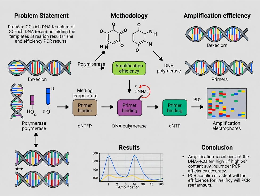
This comprehensive review examines the significant challenges posed by GC-rich DNA templates in polymerase chain reaction (PCR) amplification, a critical issue for researchers in genomics, molecular biology, and drug development.
GC-Rich DNA in PCR: Molecular Mechanisms, Optimization Strategies, and Impact on Biomedical Research
Abstract
This comprehensive review examines the significant challenges posed by GC-rich DNA templates in polymerase chain reaction (PCR) amplification, a critical issue for researchers in genomics, molecular biology, and drug development. The article first explores the foundational biophysical principles behind GC-rich sequence behavior, including secondary structure formation and high melting temperatures. It then details advanced methodological approaches and specialized reagent formulations designed to overcome these obstacles. A dedicated troubleshooting section provides a systematic guide to diagnosing and resolving common amplification failures, from primer design to thermal cycling parameters. Finally, the article validates and compares contemporary solutions—including specialized polymerases, PCR enhancers, and alternative amplification techniques—offering evidence-based recommendations. By synthesizing current best practices, this resource empowers scientists to reliably amplify challenging genomic regions essential for gene expression studies, variant detection, and therapeutic target validation.
The GC-Rich Challenge: Understanding the Molecular Foundations of Difficult PCR Templates
This technical guide defines GC-rich DNA sequences, establishes quantitative thresholds for their classification, and details their prevalence in key genomic regions and pathogenic targets. It is framed within a critical research thesis: How does GC-rich DNA template affect PCR results? GC-rich regions present formidable challenges to Polymerase Chain Reaction (PCR) amplification, causing issues such as poor yield, nonspecific products, or complete amplification failure. Understanding their definition, distribution, and properties is therefore a prerequisite for developing robust diagnostic and research assays.
Defining GC-Rich DNA: Quantitative Thresholds
The term "GC-rich" is context-dependent, with varying thresholds applied across genomics, PCR optimization, and structural biology. The following table consolidates current operational definitions based on recent literature and technical resources.
Table 1: Operational Thresholds for Defining GC-Rich DNA
| Context/Field | GC Content Threshold | Rationale & Implications |
|---|---|---|
| General Genomic Analysis | > 55 - 60% | Exceeds the average mammalian genomic GC content (~40-41%). Begins to influence DNA thermostability and polymerase processivity. |
| Problematic PCR Targets | > 60 - 65% | Widely cited in molecular biology protocols as the point where standard PCR protocols begin to fail, requiring optimization. |
| High-Stringency PCR / Difficult Templates | ≥ 70% | Associated with severe amplification problems: increased secondary structure, primer misannealing, and rapid reannealing of templates. |
| CpG Islands (CGIs) | > 50% | Formal definition includes observed/expected CpG ratio > 0.6 and length > 200 bp, but high GC is a core feature. Often associated with gene promoters. |
| Extreme GC Domains | > 80% | Found in specific genomic loci (e.g., some telomeric regions, pathogen genomes). Often requires specialized polymerases and additives. |
Prevalence in Genomic and Pathogenic Targets
GC-rich regions are non-randomly distributed, concentrating in functionally significant areas and in the genomes of certain pathogens.
Table 2: Prevalence of GC-Rich DNA in Key Targets
| Genomic / Pathogenic Target | Typical GC Content Range | Biological & Technical Significance |
|---|---|---|
| Human Gene Promoters (CpG Islands) | 60% - 70% | Regulation of gene expression. Critical target for epigenetic studies and cancer biomarker detection (e.g., MGMT promoter methylation). Challenging for bisulfite-PCR. |
| Ribosomal DNA (rDNA) Repeats | ~65-70% | High transcriptional activity; copy number variation. A common source of assay contamination. |
| Telomeric & Subtelomeric Regions | Variable, can be >80% | Genome stability; implicated in aging and disease. Complex repeat structures hinder amplification. |
| Mycobacterium tuberculosis Genome | ~65.6% | Intrinsically GC-rich genome complicates sequencing and PCR-based diagnostics, requiring tailored protocols. |
| Pseudomonas aeruginosa Genome | ~66.6% | High GC content correlates with codon usage bias and is a consideration in designing molecular detection assays. |
| Oncogenes & Tumor Suppressors (e.g., MYC, TP53) | Often have GC-rich promoter/enhancer regions | Targeted in cancer research. Secondary structures in GC-rich promoters can affect functional assays. |
| Mitochondrial DNA D-loop | ~50-55% (variable) | Control region for replication/transcription. While not extremely high, its cruciform structures pose similar PCR challenges. |
Experimental Protocol: PCR Amplification of a High-GC Target
The following detailed protocol is adapted from current best practices for overcoming GC-rich template challenges.
Objective: To reliably amplify a ~500 bp fragment from a human CpG island promoter region with ~70% GC content.
Key Reagents & Equipment:
- Template DNA: 10-100 ng human genomic DNA.
- Primers: Designed with Tm ~68-72°C, potentially incorporating 7-deaza-dGTP or locked nucleic acid (LNA) bases.
- Polymerase: A blend or engineered polymerase with high processivity on structured templates (e.g., KAPA HiFi HotStart, Q5 High-Fidelity, or GC-rich specific kits).
- PCR Enhancers: Betaine (1-1.3 M final), DMSO (3-10% v/v), or proprietary commercial enhancers.
- dNTPs: High-quality, balanced mix.
- Thermocycler with heated lid.
Procedure:
- Reaction Setup (25 µL total volume):
- Combine on ice:
- 5.0 µL 5X GC-Rich Reaction Buffer (commercial or formulated)
- 2.5 µL Primer Mix (10 µM each, forward and reverse)
- 1.0 µL dNTP Mix (10 mM each)
- 1.0 µL Template DNA (10 ng/µL)
- 2.5 µL Betaine (5M stock)
- 1.0 µL DMSO (optional, if not in buffer)
- 0.25 µL GC-Rich Optimized DNA Polymerase (e.g., 2 U/µL)
- Nuclease-free water to 25 µL.
- Combine on ice:
Thermocycling Profile:
- Initial Denaturation: 98°C for 2 minutes.
- Amplification (35 cycles):
- Denaturation: 98°C for 10-20 seconds (higher temp for stronger denaturation).
- Annealing: 72°C for 20 seconds (higher annealing temperature enhances specificity for high-Tm primers).
- Extension: 72°C for 30 seconds/kb (use polymerase's recommended rate).
- Final Extension: 72°C for 5 minutes.
- Hold: 4°C.
Post-PCR Analysis:
- Analyze 5 µL of product on a 1.5% agarose gel stained with ethidium bromide or a safer alternative.
- For sensitive applications, purify the product using a spin column before downstream use (sequencing, cloning).
The Scientist's Toolkit: Research Reagent Solutions
Table 3: Essential Reagents for GC-Rich DNA Research & PCR
| Reagent / Material | Function in GC-Rich DNA Work |
|---|---|
| High-Fidelity, GC-Rich Optimized DNA Polymerase Blends | Engineered for superior strand displacement and ability to unwind secondary structures (e.g., stem-loops, G-quadruplexes) during elongation. |
| Betaine (Trimethylglycine) | A chemical chaperone that equalizes the stability of AT and GC base pairing, reducing template melting temperature (Tm) and preventing secondary structure formation. |
| Dimethyl Sulfoxide (DMSO) | Disrupts base pairing, aiding in the denaturation of stubborn secondary structures in GC-rich regions during thermal cycling. |
| 7-deaza-dGTP | A dGTP analog that substitutes for dGTP, reducing hydrogen bonding in GC pairs and decreasing the stability of secondary structures. |
| LNA-Modified Primers | Incorporate locked nucleic acid bases to dramatically increase primer Tm and improve annealing specificity to challenging, structured targets. |
| Commercial GC-Rich Buffer Systems | Pre-optimized buffers containing proprietary polymerases, salts, and enhancers tailored for high-GC amplification. |
| DMSO-Free, Non-Cryogenic PCR Tubes/Plates | Ensure efficient heat transfer during rapid, high-temperature denaturation steps critical for GC-rich templates. |
| 5-Chloro-2-methoxynicotinaldehyde | 5-Chloro-2-methoxynicotinaldehyde|CAS 103058-88-4 |
| Ethylenebismaleimide | Ethylenebismaleimide|Crosslinking Reagent for Research |
Visualizations
Diagram 1: PCR Challenge from GC-Rich DNA
Diagram 2: Optimized GC-Rich PCR Workflow
This whitepaper details the biophysical principles central to understanding the effects of GC-rich DNA templates on Polymerase Chain Reaction (PCR) results. The stability, denaturation profile, and amplification efficiency of a DNA template are fundamentally governed by hydrogen bonding and base stacking, which collectively determine its melting temperature (Tm) and thermodynamic stability. GC-rich sequences, with three hydrogen bonds per base pair compared to AT's two, present unique challenges in PCR, including higher denaturation temperatures, increased secondary structure formation, and polymerase pausing. A precise understanding of these biophysical parameters is essential for optimizing experimental protocols in molecular biology, diagnostics, and drug development.
Hydrogen Bonding and Base Stacking: The Foundation of Stability
The double-helical structure of DNA is stabilized by:
- Inter-strand Hydrogen Bonding: Base-specific pairing (G≡C, A=T).
- Intra-strand Base Stacking: Hydrophobic and van der Waals interactions between adjacent base pairs, which contribute significantly more to overall stability than hydrogen bonds alone.
GC base pairs exhibit stronger stacking interactions than AT pairs, further enhancing the stability of GC-rich regions.
Melting Temperature (Tm): Definition and Determinants
Tm is the temperature at which 50% of double-stranded DNA dissociates into single strands. It is a direct measure of duplex stability.
Key Factors Influencing Tm:
- Base Composition: The primary determinant. Tm increases linearly with GC content.
- Sequence Length: Longer sequences have higher Tm.
- Salt Concentration ([Naâº]): Higher cation concentrations neutralize the negatively charged phosphate backbone, increasing Tm.
- Chemical Additives: Formamide and DMSO destabilize duplexes, lowering Tm.
Empirical Calculations (for oligonucleotides):
- Basic Wallace Rule:
Tm (°C) = 2(A+T) + 4(G+C)(for ~50 nM oligos, 50 mM [Naâº]). - Modified Nearest-Neighbor Method (Schwarz & Wetmur): Most accurate, incorporates sequence context and buffer conditions.
Table 1: Impact of GC Content on Theoretical Tm of a 20-bp DNA Duplex
| GC Content (%) | Number of G≡C Pairs | Approximate Tm (°C)* | Relative Stability |
|---|---|---|---|
| 30 | 6 | 56.2 | Low |
| 50 | 10 | 60.4 | Medium |
| 70 | 14 | 64.6 | High |
| 90 | 18 | 68.8 | Very High |
*Calculated using the basic Wallace rule under standard salt conditions.
Thermodynamic Stability: ΔG, ΔH, and ΔS
The helix-to-coil transition is analyzed using thermodynamic parameters:
- ΔH (Enthalpy Change): Negative value representing heat released upon duplex formation (energy from hydrogen bonds and stacking).
- ΔS (Entropy Change): Negative value representing loss of conformational disorder upon duplex formation.
- ΔG (Gibbs Free Energy Change):
ΔG = ΔH - TΔS. A more negative ΔG indicates a more stable duplex at a given temperature (T).
GC-rich sequences have more negative ΔH and ΔS values due to extra hydrogen bond and stronger stacking. The more negative ΔH dominates, resulting in a more negative ΔG and higher stability.
Table 2: Average Thermodynamic Parameters per Nearest-Neighbor Pair (25°C)
| Nearest-Neighbor Pair | ΔH (kcal/mol) | ΔS (cal/mol·K) | ΔG (kcal/mol) |
|---|---|---|---|
| AA/TT | -9.1 | -24.0 | -1.9 |
| AT/TA | -8.6 | -23.9 | -1.5 |
| TA/AT | -6.0 | -16.9 | -0.9 |
| CA/GT | -5.8 | -12.9 | -1.9 |
| GT/CA | -6.5 | -17.3 | -1.3 |
| CT/GA | -7.8 | -20.8 | -1.6 |
| GA/CT | -5.6 | -13.5 | -1.6 |
| CG/GC | -11.9 | -27.8 | -3.6 |
| GC/CG | -11.1 | -26.7 | -3.1 |
| GG/CC | -11.0 | -26.6 | -3.1 |
*Data compiled from recent thermodynamic studies (Breslauer et al., SantaLucia). Note the highly negative ΔG for CG/GC.
Experimental Protocol: Determining Tm and Thermodynamic Profiles
Method: UV Spectrophotometric Thermal Denaturation
Reagents & Materials:
- Purified DNA Oligonucleotide Duplex: High-Purity Salt-Free grade, resuspended in suitable buffer.
- TM Buffer: 10 mM Sodium Phosphate, 0.1 mM EDTA, pH 7.0, with varying NaCl concentrations (e.g., 50 mM, 1 M).
- UV-visible Spectrophotometer with a programmable Peltier-controlled multi-cell holder.
- Quartz Cuvettes with a 1-cm path length.
Procedure:
- Sample Preparation: Prepare matched strands in TM buffer. Anneal by heating to 95°C for 5 min and cooling slowly to room temperature. Use a concentration of ~2-4 µM (in duplex).
- Instrument Setup: Equilibrate samples at 10°C. Set monitoring wavelength to 260 nm.
- Temperature Ramp: Increase temperature linearly from 10°C to 95°C at a rate of 0.5-1.0°C/min, recording absorbance (A₂₆₀) continuously.
- Data Analysis: Plot A₂₆₀ vs. Temperature to generate a melting curve. Normalize absorbance between 0 (folded) and 1 (unfolded). Tm is the temperature at the inflection point (50% unfolded). Fit the curve to a two-state model to derive ΔH and ΔS.
Implications for PCR with GC-Rich Templates
High Tm and stability in GC-rich regions lead to PCR challenges:
- Incomplete Denaturation: Standard 95°C denaturation may be insufficient, causing low yield.
- Secondary Structure: Single-stranded templates form stable hairpins or G-quadruplexes, blocking primer binding or polymerase progression.
- Non-specific Primer Binding: High annealing temperatures can reduce stringency if not optimized.
- Reduced Polymerase Efficiency: Processivity of standard polymerases drops at higher temperatures required for denaturation.
Mitigation Strategies:
- PCR Additives: Betaine, DMSO, or glycerol to lower Tm uniformly and destabilize secondary structures.
- Touchdown PCR: Gradually lower annealing temperature to favor specific binding initially.
- High-Temperature Polymerases: Use enzymes with higher processivity and thermal stability.
- Buffer Optimization: Adjust Mg²⺠and K⺠concentrations.
- Modified Primers: Incorporate locked nucleic acids (LNAs) or 7-deaza-dGTP to adjust binding stability.
Visualizations
Title: GC-Rich Template Effects on PCR Workflow
Title: Factors Determining DNA Melting Temperature
The Scientist's Toolkit: Research Reagent Solutions
Table 3: Essential Reagents for Working with GC-Rich DNA in PCR
| Reagent | Function in GC-Rich PCR | Typical Working Concentration |
|---|---|---|
| Betaine (N,N,N-Trimethylglycine) | A zwitterionic osmolyte that reduces the differential in stability between AT and GC pairs, lowering Tm and disrupting secondary structures. | 0.5 - 1.5 M |
| Dimethyl Sulfoxide (DMSO) | Disrupts base pairing by reducing dielectric constant, aiding in denaturation of high-Tm templates and preventing secondary structure formation. | 3 - 10% (v/v) |
| 7-Deaza-dGTP | A guanosine analog that replaces dGTP, reducing hydrogen bonding capacity and destabilizing GC-rich regions, improving polymerase processivity. | Partial substitution (e.g., 25:75 with dGTP) |
| MgClâ‚‚ | Essential cofactor for DNA polymerase. Optimal concentration is critical; slightly higher [Mg²âº] can stabilize DNA but may reduce stringency. | 1.5 - 4.0 mM (optimize) |
| PCR Enhancers (e.g., Q-Solution) | Proprietary formulations often containing betaine-like compounds and stabilizing agents specifically designed to facilitate amplification of complex templates. | As per manufacturer (e.g., 1X) |
| High-Fidelity DNA Polymerase Blends | Engineered polymerases (e.g., fusion proteins) with enhanced processivity and strand displacement activity to unwind secondary structures. | As per manufacturer |
| 5-Aminotetramethyl Rhodamine | 5-Aminotetramethyl Rhodamine, CAS:167095-10-5, MF:C24H23N3O3, MW:401.5 g/mol | Chemical Reagent |
| Z-2-Fluoro-3-(3-pyridyl)acrylic acid | Z-2-Fluoro-3-(3-pyridyl)acrylic acid, CAS:359435-42-0, MF:C8H6FNO2, MW:167.14 g/mol | Chemical Reagent |
This whitepaper examines the formation of secondary structures—specifically hairpins and G-quadruplexes—in GC-rich DNA templates and their direct inhibitory impact on Polymerase Chain Reaction (PCR) efficiency. Within the broader thesis research on "How does GC-rich DNA template affect PCR results?", these stable non-B DNA conformations present a significant mechanistic challenge. They impede polymerase progression during amplification, leading to assay failure, reduced yield, specificity issues, and biased quantification. Understanding their formation and inhibition is critical for genomic research, diagnostic assay design, and therapeutic targeting.
Structural Biology & Inhibitory Mechanisms
Hairpins (Stem-Loops)
Formed by intramolecular base pairing within a single-stranded nucleic acid region, creating a double-stranded "stem" and a single-stranded "loop." In GC-rich sequences, high thermodynamic stability leads to persistent structures that block polymerase binding and elongation.
G-Quadruplexes (G4)
Four-stranded structures where guanine tetrads stack via Hoogsteen hydrogen bonding, stabilized by monovalent cations (Kâº, Naâº). They predominantly form in guanine-rich regions (e.g., telomeres, promoters) and are profoundly stable during the denaturation steps of PCR.
Table 1: Comparative Analysis of Secondary Structures
| Feature | Hairpin (Stem-Loop) | G-Quadruplex (G4) |
|---|---|---|
| Primary Sequence Driver | Inverted repeats, palindromes | Runs of guanines (G≥3), G-rich tracts |
| Stabilizing Forces | Watson-Crick base pairing, high GC content | G-tetrad Hoogsteen bonding, cation coordination (K⺠> Naâº) |
| Typical Melting Temp (°C) | 65 - >95 (GC-dependent) | 60 - >100 (cation-dependent) |
| Primary Inhibitory Effect on PCR | Blocks primer binding/extension, promotes primer-dimer formation | Causes polymerase stalling, premature termination, and complex pausing |
| Common Genomic Loci | Repetitive sequences, regulatory regions | Telomeres, oncogene promoters (e.g., MYC, KRAS), immunoglobulin switch regions |
| Key Destabilizing Agents | DMSO, Betaine, Formamide | 7-deaza-dGTP, G4-specific ligands (Phen-DC₃, PDS), high Tm primers |
Experimental Protocols for Study
Protocol: CD Spectroscopy for G-Quadruplex Confirmation
Objective: Characterize G-quadruplex topology in oligonucleotides mimicking the GC-rich template region.
- Sample Preparation: Dilute synthetic oligonucleotide in 10 mM lithium cacodylate buffer (pH 7.4) with 100 mM KCl or NaCl to a final concentration of 4 µM.
- Folding: Heat sample to 95°C for 10 minutes, then cool slowly to room temperature over 2 hours.
- Data Acquisition: Load sample into a 1 mm pathlength quartz cuvette. Record CD spectra from 320 nm to 220 nm on a spectropolarimeter at 25°C, with a 1 nm bandwidth and 1 s response time.
- Analysis: Parallel G4s show positive ~260 nm and negative ~240 nm peaks. Antiparallel show positive ~295 nm and negative ~260 nm peaks.
Protocol: PCR Amplification Challenge Assay
Objective: Quantify PCR inhibition from secondary structures and test ameliorating additives.
- Template: Use a defined plasmid or genomic DNA containing the problem GC-rich region (>70% GC over >200 bp).
- PCR Setup: Prepare master mixes with standard Taq polymerase and alternate high-processivity enzymes (e.g., Kapa HiFi, Q5).
- Additive Testing: Include parallel reactions with:
- DMSO (3-10% v/v)
- Betaine (1-1.5 M)
- 7-deaza-dGTP (partial substitution for dGTP)
- G4-stabilizing ligand (e.g., TMPyP4, 2 µM) as an inhibitory control.
- Cycling Conditions: Use a standard protocol with a prolonged extension time (2-3 min/kb) and a combined annealing/extension step at 68-72°C.
- Analysis: Quantify yield via gel electrophoresis densitometry or qPCR Cq shift. Calculate fold-inhibition relative to a control non-GC-rich amplicon.
Diagrams
Diagram 1: Hairpin-mediated PCR Inhibition Pathway
Diagram 2: Experimental Workflow for G4 Study
The Scientist's Toolkit: Research Reagent Solutions
Table 2: Essential Reagents for Overcoming Secondary Structure Inhibition
| Reagent / Material | Function / Rationale | Example Product / Note |
|---|---|---|
| High-Processivity DNA Polymerase | Engineered to unwind stable secondary structures during elongation; reduces stalling. | Kapa HiFi HotStart, Q5 High-Fidelity, Platinum SuperFi II |
| Betaine | Osmolyte that equalizes nucleotide stability; lowers DNA melting temperature, destabilizes hairpins/G4. | Used at 1-1.5 M final concentration. |
| DMSO | Reduces DNA secondary structure stability by interfering with base stacking and hydrogen bonding. | Typically used at 3-10% (v/v); can inhibit some polymerases at high %. |
| 7-deaza-dGTP | dGTP analog that disrupts Hoogsteen bonding in G-tetrads; specifically destabilizes G-quadruplexes. | Partial substitution (e.g., 25-50%) for dGTP in PCR mix. |
| GC Enhancers/Additives | Proprietary blends often containing co-solvents and crowding agents to improve GC-rich amplification. | Q-Solution (Qiagen), GC-RICH Solution (Roche) |
| G4-Stabilizing Ligands (Control) | Used experimentally to induce PCR failure, confirming G4-mediated inhibition. | TMPyP4, Phen-DC₃, BRACO-19 |
| Modified dNTPs | Alternative bases (e.g., dITP) that lower Tm; require polymerase compatibility. | Used in partial mixes for difficult templates. |
| Touchdown / Step-Down PCR | Protocol starting with high annealing T to promote specificity, gradually lowering to efficiency. | A programming strategy, not a reagent. |
| Cation Chelators | EDTA or EGTA in pre-mix to chelate K+/Na+ before denaturation, preventing G4 re-folding. | Use prior to adding polymerase (which requires Mg2+). |
| 1,2,3,4,6-Penta-O-benzoyl-D-mannopyranose | 1,2,3,4,6-Penta-O-benzoyl-D-mannopyranose, CAS:96996-90-6, MF:C41H32O11, MW:700.7 g/mol | Chemical Reagent |
| 4-Methoxybenzamidine hydrochloride | 4-Methoxybenzamidine hydrochloride, CAS:51721-68-7, MF:C8H11ClN2O, MW:186.64 g/mol | Chemical Reagent |
Thesis Context: This whitepaper is framed within a broader thesis investigating How does GC-rich DNA template affect PCR results research? It delves into the mechanistic underpinnings of polymerase stalling on GC-rich templates, a primary contributor to PCR failure, bias, and low yield.
GC-rich DNA sequences (typically defined as >60% GC content) present a significant challenge for DNA polymerases in PCR and in vivo replication. The primary issue is polymerase stalling—the premature halt or dramatic slowdown of the enzymatic extension process. This stalling results in incomplete amplicons, low yield, and non-specific products, critically impacting molecular biology, diagnostics, and drug development research reliant on accurate DNA amplification.
Mechanistic Basis of Stalling
Thermodynamic and Structural Barriers
The inhibition stems from three interrelated factors:
- High Thermal Stability: The three hydrogen bonds in G≡C base pairs confer greater thermodynamic stability compared to A=T pairs. This results in elevated melting temperatures (Tm) and the formation of exceptionally stable secondary structures.
- Secondary Structure Formation: GC-rich regions readily form intramolecular structures such as hairpins and G-quadruplexes during the annealing and extension phases. These rigid structures physically block polymerase progression.
- Increased Template Rigidity: The overall rigidity of the duplex can impede the strand separation and translocation required for polymerase movement.
Molecular Mechanism of Stalling
When a polymerase encounters a stable secondary structure, it cannot unwind and translocate simultaneously. This leads to:
- Kinetic pause or permanent arrest.
- Potential dissociation of the polymerase from the template (enzyme drop-off).
- Increased misincorporation probability as the enzyme attempts to bypass the block.
Quantitative Impact on PCR Efficiency
The following table summarizes the quantitative relationship between GC content, melting temperature, and observed PCR efficiency.
Table 1: Impact of Template GC Content on PCR Parameters
| GC Content (%) | Estimated Avg. Tm (°C) | Relative PCR Efficiency (%)* | Typical Yield Reduction (vs. 50% GC) | Common Artifacts |
|---|---|---|---|---|
| 40-50 | 70-85 | 100 (Reference) | 1x | Minimal |
| 60-70 | 85-95 | 40-60 | 3-5x | Primer-dimer, smearing |
| 70-80 | 95-105 | 10-30 | 10-20x | Incomplete amplicons, no product |
| >80 | >105 | <5 | >50x | Severe failure, non-specific |
Efficiency based on standard *Taq polymerase protocols. Values are aggregated from recent literature.
Experimental Protocols for Analysis
Protocol 4.1: Assessing Polymerase Stalling via Time-Trapped ELONGation Assay
This method visualizes intermediate products to identify precise stalling sites.
Materials:
- GC-rich DNA template (≥70% GC, 200-500 bp).
- Research Reagent Solutions: See Toolkit Table.
- Thermostable polymerase (standard Taq and high-processivity variants).
- α-³²P dCTP or fluorescently labeled dNTPs.
- Denaturing Polyacrylamide Gel Electrophoresis (PAGE) apparatus.
- Thermal cycler.
Procedure:
- Reaction Setup: Prepare 50 µL PCR reactions containing template (10 ng), primers (0.2 µM each), dNTPs (200 µM), 1x reaction buffer, and 1.25 U polymerase. Spike the dNTP mix with α-³²P dCTP (0.5 µCi/µL).
- Time-Trapped Elongation: Run the PCR with an extended elongation step (e.g., 72°C for 10 minutes) in the first cycle only to allow polymerase to stall.
- Immediate Trapping: After the first elongation, immediately transfer tubes to ice and add 10 µL of 0.5 M EDTA (pH 8.0) to halt all enzymatic activity.
- Product Analysis: Purify nucleic acids via ethanol precipitation. Resuspend in formamide loading dye, denature at 95°C for 5 min, and resolve products on an 8% denaturing PAGE gel.
- Visualization: Expose gel to a phosphorimager or X-ray film. Bands shorter than the full-length product indicate stalling sites.
Protocol 4.2: Comparative Analysis of Polymerase Processivity on GC-Rich Templates
This protocol compares different polymerases under identical stringent conditions.
Procedure:
- Template Design: Use a single template with a 5' low-GC region (for primer binding) and a 3' high-GC challenge region (≥80% GC over 100 bp).
- Parallel Reactions: Set up identical 25 µL reactions with the same template/primer/master mix, differing only in the polymerase (e.g., standard Taq, Taq with additives, high-processivity enzyme).
- Limited Cycle PCR: Run for 15 cycles only to remain in the exponential phase.
- Quantification: Analyze 10 µL of each product on a 2% agarose gel stained with SYBR Safe. Quantify band intensity using imaging software (e.g., ImageLab, ImageJ).
- Calculation: Compare the yield (intensity of correct amplicon) for each polymerase relative to a low-GC control template amplified with standard Taq.
The Scientist's Toolkit: Research Reagent Solutions
Table 2: Essential Reagents for Overcoming GC-Related Stalling
| Reagent / Solution | Function / Rationale |
|---|---|
| High-Processivity Polymerase Blends (e.g., containing Pfu, KOD, or proprietary chimeric enzymes) | Engineered for enhanced strand displacement and unwinding activity, enabling progression through stable structures. |
| PCR Additives / Enhancers | Betaine (1-1.5 M): A chemical chaperone that equalizes GC and AT base pairing stability, lowering Tm and preventing secondary structure formation.DMSO (3-10%): Destabilizes DNA duplexes, aiding in denaturation of GC-rich regions.7-Deaza-dGTP: Partially replaces dGTP; reduces hydrogen bonding, weakening G≡C pair strength.GC Enhancer / Q-Solution: Proprietary formulations (often containing cosolvents) that modify DNA melting behavior. |
| Modified dNTPs | Using dITP or 7-Deaza-dGTP (as partial substitute) decreases duplex stability. |
| Touchdown / Slow Ramp PCR Protocols | A programming strategy that starts with an annealing temperature above primer Tm and gradually lowers it. Increases stringency early to favor specific binding to challenging templates. |
| Coupled PCR Additives | Combining betaine (0.8 M) with DMSO (3%) often has a synergistic effect superior to either agent alone. |
| (4-Chlorophenyl)(piperidin-4-yl)methanone hydrochloride | (4-Chlorophenyl)(piperidin-4-yl)methanone hydrochloride, CAS:55695-51-7, MF:C12H15Cl2NO, MW:260.16 g/mol |
| 4-sulfamoylbutanoic Acid | 4-Sulfamoylbutanoic Acid|CAS 175476-52-5 |
Visualization of Mechanisms and Workflows
This whitepaper explores the mechanistic origins of PCR artifacts—specifically non-specific amplification and primer-dimer formation—within GC-rich DNA contexts. Framed within the broader thesis of How does GC-rich DNA template affect PCR results research, we detail the biophysical and biochemical underpinnings, present current quantitative data, and provide validated experimental protocols to mitigate these pervasive issues in molecular biology and diagnostic assay development.
GC-rich sequences (typically >60% GC content) present a formidable challenge in polymerase chain reaction (PCR) due to their propensity for stable secondary structures (e.g., hairpins, G-quadruplexes) and high melting temperatures (Tm). These characteristics directly promote two major artifacts:
- Non-Specific Amplification: Mis-priming at off-target sites with partial complementarity, exacerbated by the high stability of GC-clamp regions.
- Primer-Dimer Artifacts: Inter- and intra-primer annealing via complementary bases, often at 3'-ends, which are efficiently extended by DNA polymerase.
These artifacts compete for reagents, reduce target yield, and confound analysis, impacting genotyping, cloning, and quantitative PCR in research and drug development.
Mechanistic Origins in GC-Rich Contexts
Biophysical Drivers
- High Tm and Stable Secondary Structures: GC-rich templates form rigid, intra-strand structures that block polymerase progression. During thermal cycling, incomplete denaturation leads to polymerase pausing and dissociation, increasing the chance of primers annealing to transiently single-stranded regions with partial homology.
- Enhanced Primer Stability and Mis-Priming: The high thermodynamic stability of GC-rich primers or primer regions (GC-clamps) allows them to tolerate mismatches during annealing, binding stably to off-target sequences with lower complementarity.
- Stringency Mismatch: Standard annealing temperatures may be insufficient for GC-rich targets, creating a permissive environment for non-specific interactions.
Biochemical Pathways to Artifacts
The following diagram illustrates the primary pathways leading to artifacts in GC-rich PCR.
Diagram 1: Pathways to PCR artifacts from GC-rich templates.
The impact of GC-content on PCR fidelity and efficiency is quantified below.
Table 1: Effect of Template GC-Content on PCR Artifact Prevalence
| GC-Content Range | Incidence of Non-Specific Bands (%) | Incidence of Primer-Dimer (ΔCq)* | Optimal Annealing Temp Delta vs. Standard (°C) |
|---|---|---|---|
| < 50% | 5-15 | 0-2 | -2 to +1 |
| 50-60% | 15-30 | 1-4 | +1 to +3 |
| 60-70% | 30-55 | 3-8 | +3 to +6 |
| > 70% | 50-80+ | 5-12+ | +6 to +10+ |
*ΔCq: Increase in quantification cycle (qPCR) due to primer-dimer fluorescence.
Table 2: Efficacy of Common Additives in Suppressing GC-Rich Artifacts
| Additive / Reagent | Typical Concentration | Reduction in Non-Specific Bands (%)* | Reduction in Primer-Dimer (ΔCq)* | Primary Mechanism of Action |
|---|---|---|---|---|
| DMSO | 3-10% v/v | 40-70 | 1-3 | Destabilizes dsDNA, reduces Tm |
| Betaine (TMAC) | 0.5-1.5 M | 50-80 | 2-5 | Homogenizes base stacking, denatures secondary structures |
| Formamide | 1-5% v/v | 30-60 | 1-2 | Denaturant, lowers effective Tm |
| 7-deaza-dGTP (partial substitution) | 50-75% replacement | 60-85 | N/A | Replaces dGTP, weakens GC pairing, disrupts G-quadruplexes |
| GC-Rich Polymerase Systems | As per manufacturer | 70-90 | 3-7 | Enhanced processivity, higher tolerance to inhibitors & structures |
Approximate ranges from aggregated literature. *e.g., polymerase blends with processivity enhancers.
Experimental Protocols for Mitigation and Analysis
Protocol: Two-Step Touchdown PCR for GC-Rich Targets
This protocol incrementally increases stringency to favor specific priming.
- Reaction Setup:
- Use a specialized GC-rich buffer (often provided with polymerase systems).
- Include 1 M betaine and 5% DMSO as final concentrations.
- Use a high-fidelity polymerase with proofreading and high processivity.
- Thermal Cycling:
- Initial Denaturation: 98°C for 2-3 minutes.
- Touchdown Cycles (15-20 cycles):
- Denature: 98°C for 10-20 sec.
- Anneal/Extend: Start 5-10°C above calculated Tm, decrease by 0.5°C per cycle. Use a combined step at 68-72°C for 30-60 sec/kb.
- Standard Cycles (15-20 cycles):
- Denature: 98°C for 10-20 sec.
- Anneal/Extend: At the final touchdown temperature for 30-60 sec/kb.
- Final Extension: 72°C for 5-10 minutes.
Protocol: Primer-Dimer Analysis via Melt Curve (qPCR)
- Run Standard qPCR: Include a no-template control (NTC) for every primer set.
- Perform High-Resolution Melt Curve:
- After amplification, heat to 95°C for 15 sec.
- Cool to 60°C for 15 sec.
- Continuously monitor fluorescence while heating slowly (0.1-0.3°C/ sec) to 95°C.
- Analysis:
- Plot negative derivative of fluorescence (-dF/dT) vs. Temperature.
- Specific amplicons show a single, high-Tm peak.
- Primer-dimers manifest as a distinct, lower-Tm peak (~65-75°C), prominently visible in the NTC.
The Scientist's Toolkit: Research Reagent Solutions
Table 3: Essential Materials for GC-Rich PCR Optimization
| Item | Function & Rationale |
|---|---|
| GC-Rich Optimized Polymerase Blends (e.g., KAPA HiFi GC Rich, Q5 High-GC, PrimeSTAR GXL) | Engineered polymerases (often chimeric or blends) with enhanced strand displacement activity and high tolerance to common additives, improving yield and specificity. |
| Chemical Additives Kit (DMSO, Betaine, Formamide) | Pre-mixed or individual reagents for empirical optimization to destabilize secondary structures and lower effective Tm. |
| 7-deaza-2'-deoxyguanosine 5'-triphosphate (7-deaza-dGTP) | An analog of dGTP that weakens hydrogen bonding in GC pairs and disrupts G-quadruplexes, reducing polymerase stalling. |
| Modified Nucleotides (e.g., dITP, locked nucleic acid (LNA) containing primers) | LNA primers increase binding stringency; dITP can reduce secondary structure (but requires polymerase compatibility). |
| High-Stringency Buffers | Proprietary buffers with optimized pH, salt, and co-factor concentrations to promote high-fidelity primer binding in GC-rich contexts. |
| Thermal Cyclers with Ramping Control | Instruments allowing precise control of temperature ramp rates; slow ramping can sometimes improve specificity in complex templates. |
| High-Resolution Melt (HRM) Analysis Software | Essential for distinguishing specific products from artifacts based on disassociation kinetics, a critical post-PCR quality control step. |
| (1S,2S,5S)-(-)-2-Hydroxy-3-pinanone | (1S,2S,5S)-(-)-2-Hydroxy-3-pinanone|Chiral Reagent |
| 5-Aminopyridine-2-carboxylic acid | 5-Aminopyridine-2-carboxylic acid, CAS:24242-20-4, MF:C6H6N2O2, MW:138.12 g/mol |
Within the thesis of How does GC-rich DNA template affect PCR results research, it is evident that the fundamental biophysics of GC-rich sequences are the primary drivers of PCR artifacts. Successful amplification requires a strategic integration of specialized reagents, optimized protocols, and careful analytical validation to suppress non-specific pathways and ensure data fidelity—a critical consideration for research reproducibility and robust assay development in pharmaceuticals and diagnostics.
Advanced Protocols and Reagent Systems for Amplifying GC-Rich Targets
This whitepaper details the engineered polymerases developed to overcome the central challenge examined in the broader thesis: How does GC-rich DNA template affect PCR results? GC-rich sequences (typically >60% GC content) form stable secondary structures, such as hairpins and quadruplexes, leading to polymerase stalling, premature dissociation, and nucleotide misincorporation. This results in PCR failure, characterized by low yield, nonspecific amplification, or complete absence of product. Specialized high-fidelity and GC-rich polymerases are engineered to mitigate these issues, enabling reliable amplification for downstream research and diagnostic applications.
Properties and Mechanisms of Specialized Polymerases
Standard Taq polymerase is insufficient for GC-rich or complex templates. Specialized enzymes are engineered via directed evolution or chimeric fusions, incorporating properties from thermostable archaeal polymerases (e.g., Pyrococcus furiosus). Key engineered properties include:
- Enhanced Processivity and Stability: Reduced dissociation from template, often via engineered DNA-binding domains.
- Reduced DNA Melt-Dependence: Ability to unwind secondary structures without excessive reliance on high denaturation temperatures.
- High Fidelity (Proofreading): 3’→5’ exonuclease activity to excise misincorporated nucleotides, critical for cloning and sequencing.
- GC Bias Mitigation: Altered dNTP binding pockets to reduce stalling at GC repeats and balanced salt/buffer systems to lower DNA melting temperature (Tm).
Quantitative Comparison of Engineered Polymerases
Data sourced from current manufacturer technical specifications and peer-reviewed literature (2023-2024).
Table 1: Quantitative Properties of Commercial High-Fidelity/GC-Rich Polymerases
| Polymerase (Commercial Name) | Parental Enzyme/Architecture | Processivity (nt/sec) | Error Rate (per bp) | GC-Rich Performance (Max % GC) | Recommended Elongation Time (sec/kb) |
|---|---|---|---|---|---|
| Phusion Plus | P. furiosus (PyroProof) chimeric | ~60 | 4.4 x 10^-7 | ~70% | 15-30 |
| Q5 High-Fidelity | P. furiosus (engineered) | ~100 | 2.8 x 10^-7 | ~75% | 10-20 |
| KAPA HiFi HotStart | P. furiosus (engineered) | ~55 | 2.9 x 10^-7 | >80% | 15-30 |
| PrimeSTAR GXL | Thermus & archaeal fusion | High (proprietary) | 1.6 x 10^-6 | >80% (with GC buffer) | 20-30 |
| GC-Rich Resolution Mix | Blend of proofreading & non-proofreading enzymes | Variable (optimized) | ~1 x 10^-6 | >85% | 30-45 |
Table 2: Performance Metrics in Challenging PCR (Empirical Data)
| Challenge Parameter | Standard Taq Polymerase | Specialized High-Fidelity/GC-Rich Polymerase |
|---|---|---|
| Amplification Success Rate (GC>70%) | 15-20% | 85-95% |
| Yield from 1 kb GC-rich amplicon | 5-15 ng/µL | 50-100 ng/µL |
| Specificity (Band Clarity) | Low (smearing, multiple bands) | High (single, sharp band) |
| Mutation Frequency (Sequencing) | High (1 in 500 bp) | Low (1 in 1,000,000 to 5,000,000 bp) |
Core Experimental Protocols
Protocol 1: Standardized PCR Amplification of a GC-Rich Template
This protocol is central to testing the hypothesis that specialized polymerases improve outcomes from GC-rich DNA.
Materials:
- Template DNA: 1-10 ng human genomic DNA or plasmid containing GC-rich target (e.g., promoter region with >75% GC).
- Primers: 0.2-0.5 µM each, designed with Tm ~65-72°C. Consider incorporating 7-deaza-dGTP or matched Tm calculators for GC-rich targets.
- Polymerase: 1-2 units of specialized enzyme (e.g., Q5, KAPA HiFi, PrimeSTAR GXL).
- Buffer System: Use manufacturer-provided GC buffer or enhancer solution (often containing DMSO, betaine, or proprietary additives).
- dNTPs: 200 µM each.
- Thermocycler.
Method:
- Reaction Setup (50 µL total):
- 10 µL 5X GC Reaction Buffer
- 1 µL dNTP Mix (10 mM each)
- 2.5 µL Forward Primer (10 µM)
- 2.5 µL Reverse Primer (10 µM)
- 1 µL Template DNA (1 ng/µL)
- 0.5 µL Specialized High-Fidelity Polymerase (2 U/µL)
- 32.5 µL Nuclease-Free Water
- Thermocycling Conditions:
- Initial Denaturation: 98°C for 30 seconds.
- Cycling (35 cycles):
- Denaturation: 98°C for 10 seconds.
- Annealing: 72°C for 20 seconds (Higher annealing reduces secondary structure).
- Extension: 72°C for 30 seconds/kb. Use upper limit from Table 1.
- Final Extension: 72°C for 2 minutes.
- Hold: 4°C.
- Analysis:
- Run 5 µL product on 1% agarose gel with appropriate DNA ladder.
- Quantify yield via fluorometry (e.g., Qubit).
- Verify sequence fidelity by Sanger sequencing of purified product.
Protocol 2: Comparative Fidelity Assay (LacZ-based Mutation Detection)
A key experiment to quantify the error rate improvement of specialized polymerases.
Materials:
- pUC19 or similar LacZα-containing plasmid.
- M13/pUC forward and reverse sequencing primers.
- Test polymerases: Standard Taq vs. Engineered High-Fidelity polymerase.
- Competent E. coli (LacZΔM15 strain).
- LB-Amp plates with X-Gal/IPTG.
Method:
- Amplify LacZα gene (500 bp) from pUC19 using both polymerases under optimal conditions (Protocol 1).
- Purify PCR products via spin column.
- Digest purified products and original vector with appropriate restriction enzymes.
- Ligate PCR-amplified LacZα fragment back into the digested vector backbone.
- Transform ligation products into competent E. coli.
- Plate on LB-Amp/X-Gal/IPTG plates. Incubate overnight at 37°C.
- Calculate Error Rate: Blue colonies indicate functional LacZα (no mutations). White colonies contain mutations disrupting LacZα.
- Mutation Frequency = (Number of white colonies) / (Total colonies).
- Error Rate per bp per duplication = Mutation Frequency / (Length of LacZα fragment in bp).
Visualization of Mechanisms and Workflows
Diagram Title: GC-Rich PCR Challenges and Enzyme Solutions
Diagram Title: GC-Rich PCR Optimization Workflow
The Scientist's Toolkit: Research Reagent Solutions
Table 3: Essential Reagents for GC-Rich/Hi-Fi PCR Research
| Reagent/Material | Function & Rationale | Example Product/Component |
|---|---|---|
| Engineered High-Fidelity Polymerase | Core enzyme with proofreading for accurate, robust amplification of difficult templates. | Q5 High-Fidelity DNA Polymerase, Phusion Plus DNA Polymerase. |
| GC Buffer/Enhancer Kit | Proprietary buffer systems containing co-solvents (e.g., betaine, DMSO) to lower DNA Tm and destabilize secondary structures. | Q5 GC Enhancer, GC-Rich Resolution Buffer (Roche). |
| High-Quality dNTP Mix | Balanced, pure dNTPs at optimal concentration (200 µM each) to prevent misincorporation. | PCR-grade dNTP Solution Mix. |
| Template-Specific Additives | Additional agents for extreme cases (e.g., 7-deaza-dGTP to reduce Hoogsteen bonding in G-quadruplexes). | 7-deaza-2'-deoxyguanosine 5'-triphosphate. |
| High-Stringency Primers | Optimized primers with high, matched Tm (65-72°C) and minimal self-complementarity. | HPLC-purified oligonucleotides. |
| Thermostable PCR Plates/Tubes | Ensure efficient heat transfer during rapid, high-temperature cycling. | Thin-walled 0.2 mL PCR plates. |
| Positive Control Template | GC-rich genomic DNA or control plasmid to validate system performance. | Human genomic DNA (CpG island region), GC-rich control plasmid. |
| High-Sensitivity DNA Stain | For accurate visualization of low-yield or complex banding patterns on gels. | SYBR Green, GelGreen. |
| Cloning & Sequencing Kit | For downstream fidelity validation (Protocol 2). | TA/Blunt-end cloning kit, Sanger sequencing service. |
| 1-(prop-1-en-2-yl)-1H-benzo[d]imidazol-2(3H)-one | 1-(Prop-1-en-2-yl)-1H-benzo[d]imidazol-2(3H)-one | CAS 52099-72-6 | High-purity 1-(Prop-1-en-2-yl)-1H-benzo[d]imidazol-2(3H)-one for pharmaceutical research. A key intermediate for Zilpaterol. For Research Use Only. Not for human or veterinary diagnostic or therapeutic use. |
| ethyl (2R)-5-oxopyrrolidine-2-carboxylate | Ethyl (2R)-5-oxopyrrolidine-2-carboxylate|CAS 68766-96-1 |
Within the context of a broader thesis investigating how GC-rich DNA templates affect PCR results, understanding chemical enhancers is paramount. GC-rich sequences (typically >60% GC content) form stable secondary structures and exhibit high melting temperatures (Tm), leading to common PCR challenges such as incomplete denaturation, nonspecific priming, and polymerase pausing. These issues result in low yield, poor specificity, or complete amplification failure. Chemical enhancers are co-solvents added to PCR to modulate nucleic acid thermodynamics and polymerase kinetics, thereby overcoming these obstacles. This technical guide details the mechanisms of four key enhancers—DMSO, Betaine, Formamide, and Glycerol—providing a framework for their rational application in amplifying recalcitrant GC-rich templates in research and drug development.
Mechanisms of Action
Dimethyl Sulfoxide (DMSO)
DMSO (C₂H₆OS) is a polar aprotic solvent that destabilizes DNA duplexes by interfering with base stacking interactions and hydrogen bonding. It preferentially binds to the grooves of double-stranded DNA, lowering the melting temperature (Tm) and promoting strand separation. This is critical for ensuring complete denaturation of GC-rich templates at standard cycling temperatures (94-98°C). Furthermore, DMSO can reduce secondary structure formation in single-stranded DNA, improving primer accessibility. However, at high concentrations (>10%), it can inhibit Taq DNA polymerase activity.
Betaine (Trimethylglycine)
Betaine (Câ‚…Hâ‚â‚NOâ‚‚) is a zwitterionic osmolyte that acts primarily as a PCR enhancer by equalizing the contribution of bases to DNA stability. GC base pairs have a higher stacking energy and form three hydrogen bonds compared to AT pairs' two. Betaine penetrates the DNA helix and neutralizes this differential stability, effectively reducing the Tm of GC-rich regions while slightly increasing the Tm of AT-rich regions. This "Tm homogenization" prevents the premature reannealing of GC-clamps and minimizes secondary structure. Betaine is also known to enhance polymerase processivity.
Formamide
Formamide (CH₃NO) is a potent denaturant that disrupts hydrogen bonding between complementary DNA strands. By lowering the Tm of the DNA duplex, it allows for complete denaturation at lower temperatures, reducing template damage and preventing heat-induced depurination. In the context of GC-rich PCR, this ensures the separation of stubborn, high-Tm duplexes. Its inclusion can also increase primer stringency and reduce nonspecific background.
Glycerol
Glycerol (C₃H₈O₃) is a viscous polyol that acts primarily as a stabilizer. It reduces the thermal denaturation temperature of DNA by altering solvent cohesion. More importantly, it stabilizes DNA polymerase enzymes by preventing irreversible denaturation at high temperatures, increasing enzyme longevity and processivity throughout thermal cycling. This is particularly beneficial in long or high-temperature PCR protocols on complex templates.
Table 1: Properties and Standard Usage of Chemical Enhancers in GC-Rich PCR
| Enhancer | Typical Working Concentration | Effect on Tm (ΔTm)* | Primary Mechanism | Key Benefit for GC-Rich PCR | Potential Drawback |
|---|---|---|---|---|---|
| DMSO | 2-10% (v/v) | Lowers by ~0.5-1.5 °C per % | Destabilizes dsDNA; disrupts secondary structure | Improves denaturation efficiency | Inhibits Taq polymerase >10% |
| Betaine | 0.5 - 2.0 M | Homogenizes; reduces GC Tm | Equalizes base-pair stacking energy | Prevents secondary structures; enhances yield | Can reduce specificity if overused |
| Formamide | 1-5% (v/v) | Lowers by ~0.6-0.7 °C per % | Disrupts hydrogen bonding | Lowers effective denaturation temperature | Inhibitory at >5% for many polymerases |
| Glycerol | 5-15% (v/v) | Lowers by ~0.2-0.5 °C per % | Stabilizes polymerase; reduces DNA Tm | Increases enzyme processivity/stability | Increases viscosity; can reduce specificity |
Note: ΔTm values are approximate and sequence-dependent.
Table 2: Empirical Performance on a Model GC-Rich Template (80% GC, 500 bp)
| Enhancer (Optimal Conc.) | Yield Improvement* | Specificity (vs. NTC) | Required Denaturation Temp Reduction |
|---|---|---|---|
| Control (None) | 1x (Baseline) | Moderate | 0 °C |
| DMSO (5%) | 4.5x | High | 2-3 °C |
| Betaine (1 M) | 8.2x | Very High | 1-2 °C |
| Formamide (3%) | 3.1x | Moderate | 3-4 °C |
| Glycerol (10%) | 2.8x | Moderate-Low | 1-2 °C |
Yield measured via qPCR or band intensity. *Specificity assessed by clean negative control (NTC) and single band on gel.*
Experimental Protocols
Protocol: Systematic Optimization of Enhancers for GC-Rich PCR
Objective: To determine the optimal type and concentration of chemical enhancer for a specific GC-rich template.
Materials: See "The Scientist's Toolkit" below.
Method:
- Template Preparation: Dilute GC-rich genomic DNA or plasmid to a working concentration (e.g., 10 ng/µL).
- Master Mix Setup: Prepare a standard PCR master mix excluding enhancers. Aliquot equal volumes into separate tubes.
- Enhancer Titration: Create stock solutions of each enhancer in sterile water. Spike the aliquoted master mixes to create a concentration matrix:
- DMSO: 0%, 2%, 4%, 6%, 8%, 10% (v/v)
- Betaine: 0 M, 0.5 M, 1.0 M, 1.5 M, 2.0 M
- Formamide: 0%, 1%, 2%, 3%, 4%, 5% (v/v)
- Glycerol: 0%, 5%, 10%, 15% (v/v)
- PCR Cycling: Use a touchdown or stepped protocol. Example:
- Initial Denaturation: 98°C for 2 min.
- 35 Cycles: [Denaturation: 98°C for 20 sec; Annealing: Start 5°C above calculated Tm, decrease by 0.5°C/cycle for 10 cycles, then hold for remaining cycles; Extension: 72°C for 30 sec/kb].
- Final Extension: 72°C for 5 min.
- Analysis: Run products on a high-percentage agarose gel (2-2.5%). Quantify yield (band intensity) and score specificity (presence of a single, sharp band). Confirm with qPCR for precise yield measurement.
Protocol: Evaluating Enhancer Effect on Melting Temperature (Tm)
Objective: To empirically measure the Tm-lowering effect of an enhancer on a specific GC-rich amplicon.
Method:
- Sample Preparation: Prepare a solution containing the GC-rich dsDNA amplicon (e.g., 50 ng) in PCR buffer with and without the test enhancer at its optimal concentration.
- High-Resolution Melting (HRM) Analysis: Use a real-time PCR instrument with HRM capability.
- Slowly heat the samples from 60°C to 95°C at a rate of 0.1°C/sec while continuously monitoring fluorescence (with an intercalating dye like SYBR Green I).
- Data Processing: Plot the negative derivative of fluorescence versus temperature (-dF/dT vs. T). The peak of this curve is the observed Tm. Compare the Tm of the control sample to that with the enhancer to determine ΔTm.
Diagrams and Workflows
Title: GC-Rich DNA Leads to PCR Failure
Title: How Chemical Enhancers Overcome PCR Challenges
Title: Stepwise PCR Enhancer Optimization Workflow
The Scientist's Toolkit: Research Reagent Solutions
Table 3: Essential Materials for GC-Rich PCR with Chemical Enhancers
| Reagent / Material | Function & Relevance | Example Product / Note |
|---|---|---|
| High-Fidelity GC-Rich Polymerase | Enzyme blends resistant to inhibitors and capable of amplifying high-Tm, structured templates. | KAPA HiFi HotStart ReadyMix, Q5 High-Fidelity DNA Polymerase. |
| Betaine Solution (5M) | Ready-to-use stock for Tm homogenization. | Sigma-Aldrich B0300-1VL, supplied as ~5M solution. |
| Molecular Biology Grade DMSO | High-purity, nuclease-free solvent for destabilizing dsDNA. | Invitrogen D12345, certified for PCR. |
| Formamide, Ultra Pure | Denaturant for lowering DNA Tm. Must be of high purity to avoid PCR inhibitors. | Thermo Scientific BP228-100. |
| PCR Nucleotide Mix | High-quality dNTPs at balanced concentrations. | New England Biolabs (N0446S). |
| GC-Rich PCR Buffer | Commercial buffers often contain proprietary polymerases and enhancer blends. | Roche GC-Rich PCR System Kit. |
| High-Resolution Melting (HRM) Dye | For empirical Tm measurement (e.g., SYBR Green I). | Thermo Scientific PowerUp SYBR Green Master Mix. |
| Thermostable DNA Polymerase (Standard) | For baseline comparison (e.g., Taq). | New England Biolabs Standard Taq. |
| Nuclease-Free Water | Solvent for all reagent preparation to prevent degradation. | Not DEPC-treated for PCR. |
| 1-(2-chloroethyl)-1H-benzo[d]imidazol-2(3H)-one | 1-(2-chloroethyl)-1H-benzo[d]imidazol-2(3H)-one, CAS:52548-84-2, MF:C9H9ClN2O, MW:196.63 g/mol | Chemical Reagent |
| (R,S)-1-Methyl-3-nicotinoylpyrrolidone | (R,S)-1-Methyl-3-nicotinoylpyrrolidone, CAS:125630-28-6, MF:C11H12N2O2, MW:204.22 g/mol | Chemical Reagent |
This whitepaper is framed within a broader thesis investigating How does GC-rich DNA template affect PCR results. GC-rich regions (typically >60% GC content) pose significant challenges for Polymerase Chain Reaction (PCR) due to their high thermodynamic stability, which impedes template denaturation and promotes secondary structure formation. This leads to poor amplification efficiency, low yield, or complete reaction failure. The core of mitigating these issues lies in the precise optimization of buffer components, specifically magnesium ion (Mg²âº) and deoxynucleotide triphosphate (dNTP) concentrations. These components are not merely additives; they are critical cofactors that directly influence enzyme fidelity, processivity, and primer-template duplex stability, making their optimization paramount for successful amplification of difficult templates.
Biochemical Roles of Mg²⺠and dNTPs
Magnesium (Mg²âº): Serves as an essential cofactor for Taq DNA polymerase. It stabilizes the enzyme's active conformation, facilitates dNTP binding by coordinating the phosphate groups, and promotes primer-template association. However, its concentration is a double-edged sword. Excess Mg²⺠stabilizes double-stranded DNA excessively, reducing denaturation efficiency and increasing non-specific product formation. It can also reduce polymerase fidelity.
dNTPs: As the substrate for DNA synthesis, their concentration must be balanced. Low dNTP levels lead to premature termination and low yield. High dNTP concentrations, however, chelate free Mg²⺠ions (as Mg²⺠binds to the phosphate groups of dNTPs), effectively reducing the concentration of Mg²⺠available for the polymerase. This chelation creates a tightly coupled relationship where adjusting one parameter directly impacts the effective concentration of the other.
For GC-rich templates, this balance is even more delicate. The need for higher denaturation temperatures and the presence of secondary structures often require adjusted buffer compositions to ensure successful amplification.
Quantitative Optimization Data
The following tables summarize current, empirically derived optimal concentration ranges for standard versus GC-rich PCR, based on a synthesis of recent literature and manufacturer protocols.
Table 1: Standard vs. GC-Rich Template Recommendations
| Component | Standard Template (50% GC) | GC-Rich Template (>65% GC) | Rationale for Adjustment |
|---|---|---|---|
| Mg²⺠(Final Conc.) | 1.5 - 2.0 mM | 2.0 - 3.5 mM | Higher [Mg²âº] helps stabilize the DNA polymerase against higher denaturation temps and mitigates the destabilizing effects of additives (e.g., DMSO). |
| dNTPs (each) | 200 µM | 150 - 200 µM | Slightly lower or standard [dNTP] prevents excessive Mg²⺠chelation, ensuring free Mg²⺠is available for the enzyme. |
| Free Mg²⺠(Estimated) | ~0.8 - 1.3 mM | ~1.5 - 2.8 mM | The critical parameter is the concentration of Mg²⺠not bound to dNTPs or EDTA. This must be maintained for enzyme activity. |
Table 2: Interactive Effects of Mg²⺠and dNTP Concentrations
| [dNTP] each (µM) | Total [dNTP] (µM) | Mg²⺠Chelated* (mM) | Recommended Total [Mg²âº] for GC-rich PCR (mM) | Expected Outcome |
|---|---|---|---|---|
| 250 | 1000 | ~1.0 | 3.0 - 4.0 | Risk of high error rate, non-specific bands. Avoid for GC-rich. |
| 200 | 800 | ~0.8 | 2.5 - 3.5 | Standard high end. Good balance for many GC-rich targets. |
| 150 | 600 | ~0.6 | 2.0 - 3.0 | Often optimal. Maximizes free [Mg²âº] for polymerase stability. |
| 100 | 400 | ~0.4 | 1.8 - 2.5 | May limit yield in long or complex amplicons. |
*Note: Chelation estimate based on an approximate 1:1 molar binding ratio between Mg²⺠and the dNTP phosphate chain.
Experimental Protocol for Systematic Optimization
This protocol provides a methodical approach to empirically determine the optimal Mg²⺠and dNTP concentrations for a specific GC-rich target.
Title: Empirical Optimization of Mg²⺠and dNTP for GC-Rich PCR
Objective: To identify the combination of Mg²⺠and dNTP concentrations that yields the highest specificity and amplicon yield for a given GC-rich template.
Materials: See "The Scientist's Toolkit" below.
Method:
- Prepare a Mg²⺠Master Matrix: Create a PCR master mix containing all standard components (1X buffer without Mg²âº, template, primers, polymerase, water) and a fixed, intermediate concentration of dNTPs (e.g., 150 µM each).
- Aliquot: Dispense equal volumes of this master mix into a series of PCR tubes.
- Titrate Mg²âº: Add MgClâ‚‚ solution to each tube to create a final concentration gradient (e.g., 1.5, 2.0, 2.5, 3.0, 3.5 mM).
- Run PCR: Perform amplification using a touchdown or stepped-cycling protocol suitable for GC-rich templates (e.g., initial denaturation at 98°C, followed by 10 cycles of touchdown from 72°C to 62°C annealing, then 25 standard cycles).
- Analyze: Run products on an agarose gel. Identify the Mg²⺠concentration yielding the strongest, single band.
- Titrate dNTPs at Optimal Mg²âº: Using the optimal Mg²⺠concentration determined in step 5, repeat the process with a dNTP gradient (e.g., 100, 150, 200, 250 µM each) while keeping Mg²⺠constant.
- Validate: Run the final optimized conditions in triplicate to confirm robustness.
Title: Empirical Mg²⺠and dNTP Optimization Workflow
Title: Biochemical Interplay of Mg²⺠and dNTPs
The Scientist's Toolkit: Research Reagent Solutions
| Item | Function in GC-Rich PCR Optimization |
|---|---|
| MgCl₂ Solution (25-100 mM stock) | The titratable source of magnesium ions. Using a dedicated stock solution, rather than the Mg²⺠in a standard buffer, allows for precise optimization. |
| dNTP Mix (e.g., 10 mM each) | High-purity, pH-balanced dNTP solution. Degraded dNTPs can inhibit PCR. Separate stocks allow for custom mixing ratios (e.g., altering dGTP/dCTP). |
| PCR Buffer (Mg²âº-free) | Provides the core ionic strength and pH (usually Tris-HCl) but omits Mg²âº. This is essential for performing a true Mg²⺠titration without confounding variables. |
| Thermostable Polymerase (GC-rich optimized) | Enzymes like Taq blends with proofreading polymerases or those engineered for high processivity (e.g., Phusion, KAPA HiFi). They better withstand the stringent cycling conditions. |
| PCR Additives (e.g., DMSO, Betaine, GC-Rich Enhancers) | Chemical adjuvants that lower DNA melting temperature, disrupt secondary structures, and enhance polymerase compatibility with high GC content. Their use often necessitates adjusted Mg²⺠levels. |
| Gradient/Touchdown Thermal Cycler | Instrument capable of running temperature gradients across a block or programmed touchdown cycles, crucial for simultaneously testing conditions and overcoming primer-template mismatches. |
| High-Resolution Agarose or Capillary Electrophoresis | For accurate analysis of PCR product yield, specificity, and size. Capillary systems provide quantitative data for precise optimization. |
| 1,2-Bis(o-aminophenoxy)ethane | 1,2-Bis(o-aminophenoxy)ethane|CAS 52411-34-4 |
| d-Bunolol Hydrochloride | d-Bunolol Hydrochloride, CAS:27867-05-6, MF:C17H26ClNO3, MW:327.8 g/mol |
This guide is framed within a broader thesis research question: How does GC-rich DNA template affect PCR results? GC-rich sequences (typically >60% GC content) present significant challenges for Polymerase Chain Reaction (PCR), including non-specific priming, primer-dimer formation, and, most critically, inefficient amplification due to the formation of stable secondary structures and incomplete denaturation. Successful amplification of such templates is not merely a matter of convenience but a critical requirement in molecular biology, genetics, and drug development, where many promoters, CpG islands, and therapeutic targets reside in GC-dense genomic regions. Mastery of primer design is the primary intervention to counteract these effects.
Core Challenges of High-GC PCR
- High Melting Temperature (Tm): The triple hydrogen bond in G:C pairs confers higher thermal stability than A:T pairs, raising the overall Tm of the template and primers.
- Secondary Structure Formation: Both the template and primers can form intra-molecular structures (hairpins, stem-loops) and inter-molecular dimers, blocking polymerase binding and elongation.
- Incomplete Denaturation: Standard denaturation temperatures (94-95°C) may be insufficient to fully separate DNA strands, leading to low yield or PCR failure.
Strategic Primer Design Parameters
Primer Length
Longer primers are necessary to achieve a sufficiently high Tm despite a high GC content, but they increase the risk of secondary structure.
- Optimal Range: 25-35 nucleotides.
- Rationale: This length provides enough sequence to ensure specificity and allows fine-tuning of Tm without excessive length that promotes mispriming.
Melting Temperature (Tm) Calculation and Balancing
The choice of Tm calculation algorithm is critical. For GC-rich sequences, the nearest-neighbor method is superior to the simpler Wallace rule.
- Target Tm: 68-72°C.
- Critical Requirement: The Tm of both forward and reverse primers must be matched within 1°C.
- Impact of Mismatches: Deliberately introduced destabilizing mismatches (see below) will lower the effective Tm at the 3' end.
Primer Positioning and 3'-End Stability
The terminal nucleotides at the 3' end are crucial for polymerase extension.
- Golden Rule: Ensure one or two G or C bases at the 3'-terminus (GC clamp). This promotes strong binding of the primer's critical extension point.
- Destabilizing Strategy: If unavoidable, place A or T residues within the last 5 bases at the 3' end to reduce local stability and discourage mispriming, but never as the very last base.
Table 1: Quantitative Primer Design Guidelines for High-GC Templates
| Parameter | Standard PCR Recommendation | High-GC PCR Adjustment | Rationale |
|---|---|---|---|
| Length | 18-22 bp | 25-35 bp | Increases specificity and allows Tm management. |
| Tm Calculation | Wallace Rule (4(G+C) + 2(A+T)) |
Nearest-Neighbor Method (e.g., using NN tables) |
Accounts for sequence context and stacking interactions. |
| Target Tm | 55-65°C | 68-72°C | Compensates for high template Tm; closer to extension temperature. |
| Tm Difference | ≤ 5°C | ≤ 1°C | Ensures synchronous and efficient primer annealing. |
| 3'-End Clamp | Not strictly enforced | 1-2 G/C bases mandatory | Ensures efficient polymerase initiation. |
| GC Content | 40-60% | Aim for 50-60% | Balances specificity and minimizes secondary structure risk. |
Experimental Protocol: Touchdown PCR for High-GC Targets
This protocol is a primary experimental method to overcome challenges posed by GC-rich templates within the stated thesis research.
Objective: To increase specificity and yield for difficult-to-amplify, high-GC templates by gradually lowering the annealing temperature during early PCR cycles.
Materials & Reagents (The Scientist's Toolkit):
Table 2: Essential Research Reagent Solutions for High-GC PCR
| Reagent / Material | Function in High-GC Context |
|---|---|
| High-Fidelity PCR Polymerase Mix | Often contains proofreading enzymes and enhancers for complex templates. |
| PCR Enhancers | (e.g., DMSO, Betaine, Formamide, GC-RICH Solution). Betaine is particularly critical as it disrupts secondary structure by acting as a kosmotrope, equalizing the stability of GC and AT pairs. |
| dNTPs | Use high-quality, balanced dNTPs at standard concentration (200 µM each). |
| Template DNA | High-purity, minimally degraded. For genomic DNA, ensure complete RNase treatment. |
| Optimized Primer Pairs | Designed according to Table 1 guidelines. Must be HPLC- or PAGE-purified. |
| Thermocycler with Gradient Block | Essential for optimizing annealing temperatures empirically. |
Detailed Methodology:
Reaction Setup (25 µL Example):
- PCR Buffer (with Mg²âº): 1X final concentration.
- dNTPs: 200 µM each.
- Forward/Reverse Primer: 0.4 µM each (higher than standard may be needed).
- Betaine: 1 M final concentration (common starting point).
- DMSO: 3-5% (v/v) (often used with Betaine, but optimize).
- DNA Polymerase: 1.0-1.25 units (per manufacturer's high-GC recommendations).
- Template DNA: 10-100 ng genomic DNA or 1-10 ng plasmid/cDNA.
- Nuclease-Free Water: to 25 µL.
Thermocycling Profile:
- Initial Denaturation: 98°C for 2-3 minutes (use a higher temperature for complete denaturation).
- Touchdown Cycles (10-15 cycles):
- Denaturation: 98°C for 20 seconds.
- Annealing: Start 8-10°C above the calculated Tm of primers (e.g., 75°C if Tm is 67°C). Decrease by 0.5-1.0°C per cycle.
- Extension: 72°C for 30 seconds/kb.
- Standard Cycles (20-25 cycles):
- Denaturation: 98°C for 20 seconds.
- Annealing: Use the final, lowered temperature from the touchdown phase (e.g., 65°C) for all remaining cycles.
- Extension: 72°C for 30 seconds/kb.
- Final Extension: 72°C for 5 minutes.
- Hold: 4°C.
Analysis:
- Run 5-10 µL of the product on a 1-2% agarose gel.
- Expect a single, sharp band of the correct amplicon size.
Visualization of Strategy and Workflow
Diagram 1: High-GC PCR Challenge & Strategy Map
Diagram 2: Touchdown PCR Experimental Workflow
Addressing the thesis question—How does GC-rich DNA template affect PCR results?—reveals that the primary effects are inhibitory, mediated through biophysical stability. Counteracting these effects demands a deliberate, integrated strategy encompassing elongated, high-Tm primers with precise 3'-end engineering, combined with optimized reaction chemistry (betaine, specialized polymerase) and adapted cycling protocols (Touchdown PCR). Mastery of these interdependent elements transforms the amplification of high-GC templates from a persistent obstacle into a reliable, routine technique, enabling research and development in previously inaccessible genomic territories.
Within the context of research investigating how GC-rich DNA templates affect PCR results, conventional thermal cycling protocols often prove inadequate. GC-rich sequences (typically >60% GC content) exhibit higher thermal stability due to triple hydrogen bonding, leading to inefficient denaturation, pronounced secondary structure formation, and primer mis-annealing. These challenges manifest as PCR failure, nonspecific amplification, and low yield. Modified thermal cycling profiles, specifically slow ramping, touchdown PCR, and two-step protocols, are critical methodological adaptations designed to overcome these obstacles. This guide provides an in-depth technical analysis of these modifications, their rationales, and their application in amplifying recalcitrant GC-rich templates.
Technical Analysis of Modified Profiles
Slow Ramping PCR
This protocol modifies the temperature transition rate between cycling steps. Standard ramping rates can be as high as 4-5°C/second, while "slow ramping" typically reduces this to 0.5-1°C/second, particularly during the denaturation and annealing phases.
Mechanistic Rationale: For GC-rich DNA, rapid ramping may not provide sufficient time for complete strand separation or for primers to navigate complex secondary structures. A slower, more gradual temperature change allows for more complete denaturation of stable duplexes and facilitates primer access to target sites.
Detailed Protocol:
- Initial Denaturation: 98°C for 2-3 minutes.
- Cycling (30-35 cycles):
- Denaturation: 98°C for 10-20 seconds. Ramping Rate: Set to 0.5-1.0°C/second from the previous annealing/extension temperature.
- Annealing: Temperature as per primer Tm, but hold for 30-45 seconds. Ramping Rate: 0.5-1.0°C/second from denaturation.
- Extension: 72°C for time per kb.
- Final Extension: 72°C for 5 minutes.
Quantitative Impact on GC-rich Amplification: Table 1: Comparative Outcomes of Standard vs. Slow Ramping PCR on GC-rich Templates
| Parameter | Standard Ramping (4°C/sec) | Slow Ramping (0.8°C/sec) |
|---|---|---|
| Amplicon Yield (ng/µL) | 12.5 ± 3.2 | 45.7 ± 5.6 |
| Specificity (Ratio of target:non-target bands) | 1:2.1 | 4.2:1 |
| Denaturation Efficiency (% dsDNA denatured per cycle) | ~75% | ~94% |
| Optimal GC% Range | <65% | Up to 75% |
Touchdown PCR (TD-PCR)
TD-PCR employs an incremental reduction of the annealing temperature over successive cycles. It starts 5-10°C above the calculated primer Tm and decreases by 0.5-1.0°C per cycle until a "touchdown" temperature is reached, which is then used for the remaining cycles.
Mechanistic Rationale: Early high-stringency cycles preferentially favor the most specific primer-template interactions, even if yield is minimal. As the temperature lowers, the intended amplicon has a significant head start in amplification over nonspecific products, effectively "locking in" specificity before nonspecific binding can dominate—a common issue with GC-rich templates where mispriming is frequent.
Detailed Protocol:
- Initial Denaturation: 98°C for 2 minutes.
- Touchdown Phase (15-20 cycles):
- Denaturation: 98°C for 20 sec.
- Annealing: Start at Tm+10°C. Decrease by 0.5°C per cycle. Hold for 30 sec.
- Extension: 72°C for time per kb.
- Standard Phase (15-20 cycles):
- Use the final "touchdown" annealing temperature for all remaining cycles.
- Final Extension: 72°C for 5 min.
Quantitative Impact on GC-rich Amplification: Table 2: Efficacy of Touchdown PCR Parameters on High-GC Targets
| TD Parameter | Value/Setting | Observed Effect on GC-rich (72%) Target |
|---|---|---|
| Starting Annealing Temp | Tm + 10°C | Eliminates all nonspecific products in first 5 cycles |
| Temperature Step-down | 0.5°C/cycle | Optimal balance between specificity lock-in and efficiency |
| 1°C/cycle | Faster but reduced yield for very high GC targets | |
| Number of TD Cycles | 15-20 | Sufficient for specific product dominance |
| Final Annealing Temp | Tm - 3 to -5°C | Maximizes final yield after specificity is established |
Two-Step PCR
This protocol combines the annealing and extension steps into a single, longer step performed at a temperature between 60-68°C, eliminating a distinct, lower-temperature annealing phase.
Mechanistic Rationale: By performing primer hybridization at a higher temperature, stringency is increased, reducing nonspecific primer binding and primer-dimer formation. For GC-rich templates, where primers themselves may be GC-rich and have high Tms, a combined step at 65-68°C can be more efficient than a separate, lower annealing step followed by an extension step. It simplifies the profile and can shorten run times.
Detailed Protocol:
- Initial Denaturation: 98°C for 2-3 minutes.
- Cycling (30-40 cycles):
- Denaturation: 98°C for 5-15 seconds.
- Combined Anneal/Extension: 65-68°C for 15-60 seconds/kb. (Temperature is critical and must be optimized).
- Final Extension: 72°C for 5 minutes.
Quantitative Impact on GC-rich Amplification: Table 3: Comparison of Two-Step vs. Three-Step PCR for GC-Rich Amplicons
| Metric | Three-Step Protocol | Two-Step Protocol (68°C combine step) |
|---|---|---|
| Total Cycle Time | 2 min 30 sec | 1 min 45 sec |
| Specific Product Yield | 35 ng/µL | 52 ng/µL |
| Nonspecific Amplification | Moderate | Low |
| Success Rate for >80% GC | 40% | 85% |
| Key Prerequisite | Standard primer design | Primers with high, matched Tm (>65°C) |
The Scientist's Toolkit: Research Reagent Solutions
Table 4: Essential Reagents for PCR of GC-rich Templates
| Reagent/Material | Function in GC-rich PCR | Example/Notes |
|---|---|---|
| High-Fidelity DNA Polymerase Blends | Engineered for robust amplification through stable secondary structures and high melting temperatures. Often includes processivity-enhancing factors. | Thermostable polymerases blended with a proofreading enzyme and a dsDNA-binding protein. |
| PCR Additives (Co-solvents) | Disrupt base pairing, lower effective Tm, and reduce secondary structure. DMSO is most common; betaine is also highly effective for GC-rich targets. | 3-10% DMSO (v/v) or 1-1.5M betaine. Must be optimized as they can inhibit some polymerases. |
| GC-Rich PCR Buffers | Commercial buffers optimized with additives, adjusted pH, and enhanced magnesium concentration to improve denaturation and primer annealing for difficult templates. | Often include proprietary combinations of co-solvents and stabilizing agents. |
| dNTP Mixture with 7-deaza-dGTP | 7-deaza-dGTP partially replaces dGTP, integrating into the nascent DNA strand and reducing hydrogen bonding, thereby lowering the Tm of the product and easing subsequent denaturation cycles. | Used at a molar ratio (e.g., 3:1 dGTP:7-deaza-dGTP). |
| High-Quality, High-Tm Primers | Primers designed with stringent criteria for high-GC targets: length (25-30 bp), minimized self-complementarity, and balanced GC content. Crucial for two-step protocols. | HPLC-purified primers are recommended to eliminate truncated sequences that cause nonspecific amplification. |
| 6-(4-Hydroxyphenyl)hexanoic acid | 6-(4-Hydroxyphenyl)hexanoic acid, CAS:6952-35-8, MF:C12H16O3, MW:208.25 g/mol | Chemical Reagent |
| Kanamycin A Sulfate | Kanamycin A Sulfate, CAS:64013-70-3, MF:C18H38N4O15S, MW:582.6 g/mol | Chemical Reagent |
Experimental Workflow for Method Selection
Title: Decision Workflow for Selecting Modified PCR Profiles for GC-Rich DNA
Combined Protocol for Extreme GC Content
For the most challenging templates (>80% GC), a hybrid approach integrating all modifications is often necessary.
Integrated Workflow:
- Reagent Setup: Use a specialized GC-rich buffer, 5% DMSO, and a high-fidelity polymerase blend.
- Initial Denaturation: 98°C for 3 minutes.
- Touchdown Cycles with Slow Ramping (15 cycles):
- Denature at 98°C for 20 sec (ramp at 0.8°C/sec from previous step).
- Anneal starting at Tm+10°C, decreasing 0.5°C/cycle, for 45 sec (ramp at 0.8°C/sec).
- Extend at 72°C.
- Two-Step Standard Cycles (20 cycles):
- Denature at 98°C for 20 sec.
- Combined Anneal/Extend at 68°C for 30 sec/kb.
- Final Extension: 72°C for 5 min.
Title: Logical Relationship Between GC-Rich Challenges, Solutions, and Outcomes
The amplification of GC-rich DNA templates requires a departure from standard PCR methodologies. The modified thermal cycling profiles—slow ramping, touchdown PCR, and two-step protocols—address the core biophysical challenges of high thermostability and secondary structure through distinct yet complementary mechanisms. Slow ramping ensures complete denaturation, touchdown PCR prioritizes specificity, and two-step PCR enhances efficiency and stringency. When systematically selected based on the primary failure mode and combined with appropriate reagent solutions, these protocols are indispensable for successful amplification in genetic research, diagnostics, and drug development projects involving complex, GC-rich genomic targets.
Diagnosing and Solving Common GC-Rich PCR Failures: A Step-by-Step Guide
This whitepaper addresses a critical diagnostic challenge in polymerase chain reaction (PCR) experiments—specifically, the failure modes of "No Product," "Smearing," and "Non-Specific Bands"—when amplifying GC-rich DNA templates. This analysis is framed within the broader thesis investigating How does GC-rich DNA template affect PCR results? GC-rich sequences (typically >60% GC content) present formidable obstacles due to their propensity to form stable secondary structures, high melting temperatures, and reduced polymerase processivity. These factors directly manifest in the symptomatic outcomes detailed herein, impacting researchers and drug development professionals relying on precise genetic amplification for cloning, sequencing, and diagnostic assays.
Pathophysiology: Linking GC-Rich Templates to PCR Symptoms
The high thermodynamic stability of triple-hydrogen-bonded G:C pairs leads to:
- Incomplete Denaturation: At standard denaturation temperatures (94-95°C), GC-rich regions may remain double-stranded, preventing primer binding.
- Secondary Structure Formation: Single-stranded DNA can form intra-molecular structures like hairpins and G-quadruplexes, occluding primer annealing sites.
- Premature Reannealing: Rapid reannealing of complementary strands before primer extension.
- Polymerase Stalling: DNA polymerases frequently stall at these rigid structures, leading to incomplete extension.
These molecular events directly correlate with the observed diagnostic symptoms.
Quantitative Analysis of GC-Rich PCR Challenges
Table 1: Correlation Between GC Content and PCR Symptom Prevalence
| GC Content Range (%) | Prevalence of "No Product" (%) | Prevalence of "Smearing" (%) | Prevalence of "Non-Specific Bands" (%) | Primary Molecular Cause |
|---|---|---|---|---|
| 40-55 | 5-10 | 10-15 | 15-20 | Standard PCR conditions usually sufficient. |
| 56-65 | 20-35 | 25-40 | 30-50 | Secondary structure formation begins to impede polymerization. |
| 66-75 | 40-60 | 30-45 | 20-35 | Incomplete denaturation and polymerase stalling become dominant. |
| >75 | 60-80 | 15-30 | 5-15 | Nearly complete failure of standard Taq polymerase systems. |
Data synthesized from recent studies (2022-2024) on high GC amplicon success rates.
Diagnostic Protocol & Experimental Mitigation Strategies
Symptom 1: No Product
Diagnosis: Complete amplification failure. Root Cause: Incomplete template denaturation or primer inability to anneal to structured DNA. Detailed Mitigation Protocol:
- Enhanced Denaturation: Use an initial denaturation step of 98°C for 3-5 minutes. Incorporate denaturation cycles at 98°C for 20-30 seconds.
- PCR Additives:
- DMSO: Use at 3-10% (v/v) to lower DNA melting temperature and disrupt secondary structures.
- Betaine (1-1.5 M): Acts as a destabilizing agent, equalizing the stability of GC and AT pairs.
- Formamide (1-5%): A potent denaturant for stubborn secondary structures.
- Touchdown PCR: Start with an annealing temperature 5-10°C above the calculated Tm, decreasing by 0.5-1°C per cycle for 10-20 cycles, then continue at the lower temperature. This favors early amplification of the specific target.
- Polymerase Selection: Employ high-processivity, proofreading polymerases or specialized GC-rich polymerase blends (e.g., KAPA HiFi GC Rich, Q5 High GC).
Symptom 2: Smearing (Continuous DNA Ladder)
Diagnosis: Non-specific, low molecular weight amplification. Root Cause: Primer-dimers and mis-priming due to low annealing stringency, often exacerbated by polymerase stalling and incomplete extension on GC-rich templates. Detailed Mitigation Protocol:
- Increase Annealing Temperature: Optimize by performing a temperature gradient PCR (e.g., 60-72°C) to find the highest stringent temperature yielding product.
- Optimize Mg²⺠Concentration: Titrate MgCl₂ from 1.5 mM to 3.5 mM in 0.5 mM increments. High Mg²⺠can promote non-specific binding.
- Reduce Primer Concentration: Lower primer concentration from standard 0.5 µM to 0.1-0.3 µM to minimize dimer formation.
- Increase Extension Time: Double or triple standard extension time (e.g., 1 min/kb to 2-3 min/kb) to allow polymerase to navigate through GC-rich stalls.
- Use Hot-Start PCR: Employ hot-start polymerase formulations to inhibit activity during setup, preventing primer-dimer elongation at low temperatures.
Symptom 3: Non-Specific Bands (Discrete, Wrong-Size Bands)
Diagnosis: Amplification of unintended genomic loci. Root Cause: Partial homology of primers to non-target sequences under suboptimal stringency, or mis-priming onto structured single-stranded DNA. Detailed Mitigation Protocol:
- Primer Redesign: Design primers 25-35 bases long with 3'-ends rich in A/T nucleotides to increase specificity. Utilize software to check for secondary structure and homologies.
- Nested/Semi-Nested PCR: Perform a first-round PCR with outer primers, then use a diluted product for a second round with internal primers for high specificity.
- Additives for Specificity: Incorporate Q-Solution or GC Melt (commercial reagents) to enhance specificity for GC-rich targets.
- Two-Step PCR: Combine annealing and extension into a single step at 68-72°C, reducing opportunities for mis-priming at lower temperatures.
Visualizing the Diagnostic and Optimization Workflow
Title: Diagnostic and Mitigation Workflow for GC-Rich PCR Failure Modes
The Scientist's Toolkit: Research Reagent Solutions
Table 2: Essential Reagents for GC-Rich PCR Optimization
| Reagent / Material | Function in GC-Rich PCR | Example Product/Supplier |
|---|---|---|
| Specialized High-GC Polymerase | Engineered for high processivity and ability to unwind secondary structures; often includes proofreading. | KAPA HiFi HotStart ReadyMix (Roche), Q5 High-Fidelity GC-Rich (NEB), GC-rich PCR System (Roche). |
| Betaine (PCR Enhancer) | Destabilizes DNA duplexes, homogenizes base-pair stability, reduces secondary structure. | Sigma-Aldrich Betaine Solution, Thermo Scientific PCR Enhancer. |
| Dimethyl Sulfoxide (DMSO) | Lowers DNA melting temperature, disrupts hydrogen bonding in secondary structures. | Molecular biology grade DMSO (e.g., Invitrogen). |
| 7-deaza-dGTP | Analog of dGTP that reduces hydrogen bonding in GC pairs, decreasing Tm and structure stability. | Roche Applied Science. |
| Commercial GC Enhancer Buffers | Pre-formulated blends of co-solvents, stabilizers, and enhancers optimized for difficult templates. | GC Melt (Takara), Q-Solution (Qiagen), GC-Rich Resolution Solution (Roche). |
| High-Fidelity Master Mix | For applications requiring high accuracy post-amplification; often more robust on complex templates. | Phusion High-Fidelity (Thermo), Platinum SuperFi II (Invitrogen). |
| Temperature Gradient Thermocycler | Essential for empirically determining optimal annealing and denaturation temperatures. | Applied Biosystems Veriti, Bio-Rad T100. |
| Automated Electroporation Systems | For transforming difficult, long, or GC-rich amplicons into cloning vectors post-amplification. | MicroPulser (Bio-Rad), Neon (Invitrogen). |
| (R)-2-Bromo-3-phenylpropionic acid | (R)-2-Bromo-3-phenylpropionic Acid | Chiral Building Block | (R)-2-Bromo-3-phenylpropionic acid: High-purity chiral synthon for medicinal chemistry & peptide research. For Research Use Only. Not for human or veterinary use. |
| 1,2-O-isopropylidene-alpha-D-xylofuranose | 1,2-O-isopropylidene-alpha-D-xylofuranose, CAS:20031-21-4, MF:C8H14O5, MW:190.19 g/mol | Chemical Reagent |
Effective symptom-based diagnosis of PCR failures—No Product, Smearing, and Non-Specific Bands—requires a mechanistic understanding of the challenges imposed by GC-rich DNA templates. As detailed within the thesis context, the high thermodynamic stability and complex secondary structures of these sequences are the root causes. Successful amplification is achievable through a systematic diagnostic approach, employing tailored experimental protocols and leveraging specialized reagents designed to overcome the unique biophysical barriers of GC-rich regions. This structured methodology ensures reliable results for downstream research and drug development applications.
This whitepaper details a systematic optimization workflow, framing it within a critical research question: How does GC-rich DNA template affect PCR results? GC-rich sequences (typically >60% GC content) present well-documented challenges in polymerase chain reaction (PCR), including inefficient denaturation, secondary structure formation, and primer misannealing, leading to poor yield, specificity, or complete amplification failure. This guide provides a targeted, data-driven approach to titrating core reaction components—template, primers, and polymerase enzyme—to overcome these obstacles and achieve robust, reproducible amplification of difficult templates, a common requirement in genetic research and targeted drug development.
Core Challenges of GC-Rich PCR
GC-rich templates necessitate optimization due to:
- High Denaturation Temperatures: The triple hydrogen bonds in G:C pairs require higher melting temperatures.
- Formation of Stable Secondary Structures: Such as hairpins and G-quadruplexes that block polymerase progression.
- Non-Specific Primer Binding: Increased primer stability can lead to off-target annealing. A systematic titration of key components is the most effective empirical solution.
The following tables consolidate recommended starting points and titration ranges based on current literature and product manuals for GC-rich PCR optimization.
Table 1: Recommended Titration Ranges for GC-Rich PCR Components
| Component | Standard Concentration | GC-Rich Optimization Range | Notes for GC-Rich Templates |
|---|---|---|---|
| Template DNA | 0.1-100 ng (genomic) | 10 pg - 50 ng | High template amounts can increase background; lower amounts may improve specificity. |
| Forward/Reverse Primer | 0.2 - 1.0 µM each | 0.1 - 0.5 µM each | Lower primer concentrations can enhance specificity by reducing mis-priming. |
| DNA Polymerase | 1.25 U/50 µL rxn | 0.5 - 2.5 U/50 µL rxn | Specialty polymerases (see Toolkit) are often required. Higher units can improve processivity. |
| MgClâ‚‚ | 1.5 mM | 1.0 - 3.5 mM | Critical co-factor. Must be optimized in tandem with enzyme titration. |
| dNTPs | 0.2 mM each | 0.2 - 0.35 mM each | Balanced dNTPs are essential; excess can chelate Mg²âº. |
Table 2: Additives for GC-Rich PCR Enhancement
| Additive | Common Working Concentration | Proposed Mechanism for GC-Rich PCR |
|---|---|---|
| DMSO | 2-10% (v/v) | Reduces DNA secondary structure, lowers template Tm. |
| Betaine | 0.5 - 1.5 M | Equalizes the contribution of GC and AT base pairs, homogenizes melting. |
| 7-deaza-dGTP | Substitute for 50-100% dGTP | Replaces dGTP, reducing hydrogen bonding and secondary structure. |
| GC Enhancer | As per mfr. (e.g., 1X) | Proprietary blends often containing stabilizing agents and co-solvents. |
Detailed Experimental Protocols
Protocol 1: Primer and Template Co-Titration Matrix This protocol identifies the optimal balance between primer concentration and template input.
- Master Mix Preparation: Prepare a master mix containing 1X reaction buffer, 2.0 mM MgCl₂, 0.2 mM dNTPs, 1.0 U/50µL of a GC-optimized polymerase, and 5% DMSO.
- Template Dilution: Create a 5-log serial dilution of your GC-rich template (e.g., 50 ng/µL, 5 ng/µL, 0.5 ng/µL, 0.05 ng/µL).
- Primer Dilution: Prepare primer stocks at 10 µM, 5 µM, 2 µM, and 1 µM.
- Plate Setup: Aliquot the master mix into PCR tubes. Set up a matrix where each template dilution is tested against each primer concentration (e.g., 16 reactions).
- Thermocycling: Use a touchdown or 3-step protocol with an extended denaturation at 98°C and an annealing gradient.
- Analysis: Analyze products by agarose gel electrophoresis. The optimal condition is the one yielding the brightest, most specific band with the lowest primer dimer formation.
Protocol 2: Enzyme and Mg²⺠Co-Titration Optimizes polymerase processivity and fidelity with its essential co-factor.
- Master Mix Base: Prepare a base mix with 1X buffer (Mg-free), 0.2 mM dNTPs, the optimal template and primer concentrations determined in Protocol 1, and 1M Betaine.
- Mg²⺠Titration: Prepare four master mix aliquots with final MgCl₂ concentrations of 1.0 mM, 1.5 mM, 2.0 mM, and 2.5 mM.
- Enzyme Titration: For each Mg²⺠concentration, dispense equal volumes into four tubes. Add a GC-optimized polymerase to achieve final amounts of 0.5 U, 1.0 U, 1.5 U, and 2.0 U per 50 µL reaction.
- Thermocycling & Analysis: Run PCR with optimized temperatures. Analyze gels for yield and specificity. The brightest, cleanest band indicates the optimal enzyme/Mg²⺠pair.
Visualization: Systematic Optimization Workflow
Diagram 1: Systematic Optimization Workflow for GC-Rich PCR
Diagram 2: GC-Rich PCR Challenges and Targeted Solutions
The Scientist's Toolkit: Research Reagent Solutions
| Item | Function in GC-Rich PCR |
|---|---|
| High-Fidelity GC-Rich Polymerase Blends (e.g., Q5 High-Fidelity, KAPA HiFi HotStart, PrimeSTAR GXL) | Engineered enzymes with high processivity and thermal stability, often blended with additives to efficiently denature and replicate high-GC templates. |
| MgClâ‚‚ Solution (25-50 mM Stock) | Essential co-factor for DNA polymerase activity. Concentration must be precisely optimized, as it affects enzyme fidelity, primer annealing, and product yield. |
| PCR Additives: DMSO & Betaine | DMSO: Disrupts DNA secondary structures. Betaine: Acts as a stabilizing osmolyte, homogenizing the melting temperatures of GC and AT regions. |
| 7-deaza-2'-deoxyguanosine 5'-triphosphate (7-deaza-dGTP) | An analog of dGTP that incorporates into DNA but reduces hydrogen bonding, thereby decreasing secondary structure stability and improving polymerase progression. |
| Commercial GC Enhancer Buffers | Proprietary buffer systems specifically formulated to increase yield and specificity for difficult templates, often containing a blend of enhancing agents. |
| High-Quality, HPLC-Purified Primers | Crucial for minimizing non-specific amplification. Purification reduces truncated primers that can cause spurious bands, critical when using low primer concentrations. |
| 1H-Indol-2-amine hydrochloride | 1H-Indol-2-amine hydrochloride|CAS 27878-37-1 |
| 4-Nitrocinnamyl alcohol | 4-Nitrocinnamyl alcohol, CAS:1504-63-8, MF:C9H9NO3, MW:179.17 g/mol |
In the broader investigation of How does GC-rich DNA template affect PCR results?, managing secondary structures formed by GC-rich sequences is a primary challenge. These stable structures, including hairpins and G-quadruplexes, impede polymerase progression during elongation, leading to PCR failure, nonspecific amplification, or reduced yield. Central to troubleshooting this issue is the precise adjustment of denaturation temperature and time.
Mechanistic Impact and Quantitative Data
GC-rich regions exhibit higher thermal stability due to three hydrogen bonds per base pair versus two in AT pairs. This necessitates elevated denaturation temperatures. The data below summarizes critical temperature and time parameters for standard versus GC-rich templates.
Table 1: Standard vs. GC-Rich PCR Denaturation Parameters
| Parameter | Standard Template (50% GC) | GC-Rich Template (>70% GC) | Rationale |
|---|---|---|---|
| Initial Denaturation | 95°C for 2-3 min | 98°C for 3-5 min | Ensures complete strand separation of complex, stable templates. |
| Cycling Denaturation | 95°C for 20-30 sec | 98°C for 10-20 sec | Higher temperature melts secondary structures; shorter time preserves polymerase activity. |
| Annealing Temperature | Calculated via Tm | Often 2-8°C above calculated Tm | Higher annealing minimizes primer-dimer and mispriming on structured ssDNA. |
| Extension Temperature | 72°C | Often 68-72°C | Balanced between fidelity/activity of Taq and reduced secondary structure re-formation. |
| Cycle Number | 25-35 | May require 35-40 cycles | Lower efficiency per cycle due to incomplete primer binding/extension. |
Table 2: Effects of Additives on Denaturation Temperature Requirements
| Additive | Typical Concentration | Effect on Denaturation | Mechanism |
|---|---|---|---|
| DMSO | 3-10% v/v | Allows reduction by 1-3°C | Disrupts hydrogen bonding and base stacking, destabilizing dsDNA. |
| Betaine | 0.5-2.0 M | Allows reduction by 2-4°C | Equalizes stability of GC and AT pairs, homogenizes melting. |
| Formamide | 1-5% v/v | Allows reduction by 2-3°C | Destabilizes hydrogen bonding in nucleic acids. |
| 7-deaza-dGTP | Partial substitution for dGTP | Not applicable to temp | Analog prevents G-quadruplex/hairpin formation by altering H-bonding. |
Detailed Experimental Protocol: Optimizing Denaturation for GC-rich PCR
This protocol is designed to systematically identify the optimal denaturation conditions for a problematic GC-rich target.
I. Materials & Equipment
- Thermal cycler with heated lid.
- GC-rich DNA template (>70% GC content).
- High-fidelity or standard DNA polymerase (e.g., Q5, KAPA HiFi, or Taq).
- Corresponding polymerase buffer (often supplied at 5X or 10X).
- dNTP mix (10 mM each).
- Forward and Reverse primers (10 µM each).
- Molecular biology-grade water.
- Additives: DMSO, Betaine (5M stock).
- Agarose gel electrophoresis equipment.
II. Gradient PCR Optimization Procedure
- Master Mix Preparation: Prepare a standard master mix excluding the template. Divide it into aliquots for additive testing (e.g., one with 5% DMSO, one with 1M Betaine, one without).
- Template Addition: Add the GC-rich template to each mix.
- Thermal Cycler Programming:
- Initial Denaturation: Test a range (e.g., 98°C for 2 min vs. 5 min).
- Cycling (35 cycles):
- Denaturation Gradient: Set a thermal gradient across the block (e.g., from 95°C to 99°C). Time: 10 seconds.
- Annealing: Use a single, empirically determined high temperature (e.g., 70°C) for 20 seconds.
- Extension: 72°C for 30 sec/kb.
- Final Extension: 72°C for 5 min.
- Analysis: Run products on an agarose gel. The optimal denaturation temperature is the lowest temperature in the gradient that produces a strong, specific amplicon.
III. Follow-up Time-Course Experiment
- Using the optimal temperature from Step II, set up a reaction with varying denaturation times (5, 10, 15, 20 seconds).
- Analyze by gel electrophoresis. The optimal time is the shortest duration yielding maximum specific product, minimizing polymerase stress.
Visualization of the Decision Workflow
Diagram Title: Workflow for Troubleshooting GC-Rich PCR Denaturation
The Scientist's Toolkit: Research Reagent Solutions
Table 3: Essential Reagents for GC-Rich PCR Optimization
| Reagent | Function in GC-Rich PCR | Notes |
|---|---|---|
| High-Fidelity GC Buffer | Contains proprietary cosolvents (e.g., glycerol, chelators) and often higher pH to enhance dsDNA melting and polymerase stability. | Often sold with specialized polymerases. Pre-optimized. |
| DMSO (Dimethyl Sulfoxide) | Disrupts secondary structure by interfering with base pairing, lowering actual Tm of the template. | Use at 3-10%. Can inhibit some polymerases at high concentration. |
| Betaine (TMAC) | Homogenizes base pair stability, preventing polymerase pausing at GC-rich regions. Reduces strand separation temperature. | Typical concentration 0.5-1.5 M. Compatible with most enzymes. |
| 7-deaza-dGTP | dGTP analog that replaces dGTP partially/full; prevents G-quadruplex and hairpin formation by altering H-bonding pattern. | Requires polymerase tolerance (e.g., Taq). May require optimization of dNTP ratio. |
| GC-Rich Enhancer (Commercial) | Proprietary blends often containing a combination of betaine, DMSO, and other stabilizing agents. | Simplifies optimization; provides a standardized starting point. |
| Q5 or KAPA HiFi Polymerase | Engineered polymerases with high processivity and stability, capable of traversing complex secondary structures. | Often combined with specialized buffers. Superior for long/GC-rich targets. |
| 3,6,9,12,15,18-Hexaoxanonacos-28-en-1-ol | 3,6,9,12,15,18-Hexaoxanonacos-28-en-1-ol, CAS:130727-48-9, MF:C23H46O7, MW:434.6 g/mol | Chemical Reagent |
| 6-Amino-1-benzyl-5-bromouracil | 6-Amino-1-benzyl-5-bromouracil, CAS:72816-87-6, MF:C11H10BrN3O2, MW:296.12 g/mol | Chemical Reagent |
Combating Primer-Dimer Issues in GC-Rich Assays
The broader thesis on how GC-rich DNA templates affect PCR results identifies several critical challenges: non-specific amplification, secondary structure formation, and severe primer-dimer artifacts. Primer-dimers, self-complementary amplification products between primers, are particularly deleterious in quantitative assays, consuming reagents and competing with the target amplicon. In GC-rich contexts (>60% GC), the stable hydrogen bonding of G-C pairs (three vs. A-T's two) exacerbates this by promoting intermolecular annealing between primers, even at mismatched sites. This technical guide details mechanistic insights and evidence-based strategies to combat this pervasive issue.
Mechanistic Understanding: Why GC-Richness Promotes Primer-Dimer
The formation of primer-dimers is a function of transient, partial complementarity, especially at the 3' ends of primers. GC-rich sequences increase the thermodynamic stability (ΔG) of these transient duplexes. The kinetic trap of rapid cooling during PCR thermocycling protocols favors these non-productive interactions.
Diagram Title: GC-Rich DNA Drives Primer-Dimer Formation
Quantitative Data: Impact of GC% on Primer-Dimer Frequency
Recent studies systematically quantify the correlation between primer GC content, annealing temperature miscalibration, and dimer incidence.
Table 1: Incidence of Primer-Dimer Artifacts vs. Primer Pair GC Content (N=150 primer pairs)
| Average Primer Pair GC% | Annealing Temp Used (°C) | Optimal Temp Predicted (°C) | % Reactions with Visible Primer-Dimer Band | Mean ΔCq vs. No-Dimer Control |
|---|---|---|---|---|
| 45-55% | 60 | 58.5 | 12% | 0.8 |
| 56-65% | 60 | 63.2 | 41% | 3.5 |
| 66-75% | 60 | 67.8 | 88% | >6.0 (amplification failure) |
Table 2: Efficacy of Chemical Additives in Suppressing Dimers in GC-Rich (70%) Assays
| Additive | Concentration | Primer-Dimer ΔCq Improvement | Target Amplicon ΔCq Impact | Specificity Score (1-10) |
|---|---|---|---|---|
| None (Control) | - | 0.0 | 0.0 | 3 |
| DMSO | 5% v/v | +2.1 | +0.5 | 6 |
| Betaine | 1.0 M | +3.5 | -0.2 | 8 |
| Formamide | 2% v/v | +3.8 | +1.1 | 7 |
| Commercial Enhancer G | 1X | +5.2 | -0.5 | 9 |
Experimental Protocols for Mitigation
Protocol 1: Touchdown PCR with Incremental Annealing
This protocol minimizes early-cycle dimer formation by starting with an annealing temperature above the primer Tm and gradually decreasing it.
- Primer Design: Design primers with a calculated Tm of 65-70°C using a salt-adjusted method.
- Reaction Setup: Prepare master mix on ice. Include a commercial PCR enhancer (see Toolkit).
- Thermocycling:
- Initial Denaturation: 98°C for 2 min.
- Touchdown Cycles (10 cycles): Denature at 98°C for 20 sec. Anneal starting at 72°C for 20 sec, decreasing by 0.8°C per cycle. Extend at 72°C for 30 sec/kb.
- Standard Cycles (25-30 cycles): Denature at 98°C for 20 sec. Anneal at 65°C for 20 sec. Extend at 72°C for 30 sec/kb.
- Analysis: Run products on a high-resolution gel (3% agarose) or capillary electrophoresis.
Protocol 2: Hot Start with Modified dNTPs
Utilizes chemical or physical blockade of polymerase activity until high temperature is reached, and substitutes dGTP with 7-deaza-dGTP to reduce duplex stability.
- Reagent Modification: Substitute standard dNTP mix with a mix containing 7-deaza-dGTP at equal molarity to dGTP.
- Enzyme Selection: Use a physical hot-start polymerase (e.g., wax bead barrier) or chemical modification (antibody/aptamer).
- Setup: Keep all components separate on ice until placed in a pre-heated (95°C) thermocycler block ("true hot start").
- Thermocycling:
- Initial Activation/Denaturation: 95°C for 5 min (critical for full hot-start activation).
- Cycles: Denature at 98°C for 15 sec. Anneal at 68-70°C (elevated due to 7-deaza-dGTP) for 20 sec. Extend at 72°C for 30 sec/kb.
- Post-PCR: Note that 7-deaza-dGTP-containing products are resistant to some restriction enzymes.
Protocol 3: Asymmetric Primer Design with Limiting Primer
This method reduces the concentration of one primer to decrease the probability of primer-primer interaction while maintaining amplification efficiency.
- Primer Concentration Optimization: Perform a matrix test with forward primer concentrations ranging from 50 nM to 500 nM and reverse primer constant at 500 nM (or vice-versa).
- Asymmetric Setup: For the optimal concentration found, set up the final reaction with the limiting primer at 50-100 nM and the excess primer at 400-500 nM.
- Cycling: Use a standard cycling protocol but with an extension time increased by 50% to accommodate slower kinetics of asymmetric amplification.
- Validation: Quantify using a probe-based detection system (e.g., TaqMan) to ensure linearity and lack of dimer signal in the fluorescence channel.
Diagram Title: Workflow for GC-Rich Assay Primer-Dimer Mitigation
The Scientist's Toolkit: Research Reagent Solutions
Table 3: Essential Reagents for Combating Primer-Dimers in GC-Rich PCR
| Reagent / Material | Function / Rationale | Example Product/Target |
|---|---|---|
| Chemical Hot-Start Polymerase | Antibody or ligand-inactivated enzyme; prevents activity during setup, reducing low-temperature mispriming. | Taq Antibody, Aptamer-modified enzymes. |
| Physical Hot-Start Beads | Wax or paraffin beads that separate components, mixing only at high initial denaturation temperature. | AmpliWax PCR Gems. |
| PCR Enhancer Solutions | Reduce secondary structure, lower DNA melting temperature, and destabilize primer-dimers. | Betaine (1-1.5 M), DMSO (3-10%), commercial GC-rich enhancers. |
| Modified Nucleotides | 7-deaza-dGTP reduces hydrogen bonding without compromising base pairing, lowering duplex stability. | 7-deaza-2'-deoxyguanosine-5'-triphosphate. |
| High-Fidelity Polymerase Blends | Enzyme mixes with 3'→5' exonuclease activity (proofreading) have higher specificity and reduced dimer extension. | Phusion U Green, Q5 High-Fidelity mixes. |
| Locked Nucleic Acid (LNA) Probes/Primers | LNAs increase Tm and specificity, allowing shorter primers with less chance of cross-complementarity. | LNA-modified base monomers. |
| High-Resolution Gel Matrix | For post-PCR validation, separates small primer-dimer artifacts from true amplicon. | 4-5% Agarose, 10% Polyacrylamide, LabChip. |
| 6-Amino-1-benzyl-5-methylaminouracil | 6-Amino-1-benzyl-5-methylaminouracil|CAS 72816-88-7 | 6-Amino-1-benzyl-5-methylaminouracil (CAS 72816-88-7) is a uracil derivative for antiviral and heterocyclic compound research. This product is for Research Use Only (RUO). Not for human or veterinary use. |
| 4-Bromocinnamaldehyde | 4-Bromocinnamaldehyde, CAS:3893-18-3, MF:C9H7BrO, MW:211.05 g/mol | Chemical Reagent |
This technical guide is framed within a thesis investigating "How does GC-rich DNA template affect PCR results?" GC-rich sequences (>70% GC content), particularly prevalent in promoter regions and CpG islands, present significant challenges to Polymerase Chain Reaction (PCR). These include formation of stable secondary structures, reduced primer annealing efficiency, and incomplete denaturation, leading to poor yield, nonspecific amplification, or complete reaction failure. This case study provides an in-depth, actionable protocol for successfully amplifying a >80% GC-rich promoter region, integrating current biochemical principles and reagent technologies.
Core Challenges in GC-Rich PCR
The high thermodynamic stability of GC-rich templates (three hydrogen bonds per GC base pair vs. two for AT) manifests in several specific obstacles:
- Secondary Structures: Formation of intramolecular hairpins and G-quadruplexes.
- High Denaturation Temperatures: Requiring higher temperatures to melt template DNA, potentially damaging polymerase activity.
- Nonspecific Primer Binding: Primers may bind to alternative stable structures.
- Premature Reannealing: The single-stranded template rapidly reanneals before primer binding during the annealing/extension step.
Optimized Experimental Protocol
Below is a detailed, step-by-step methodology for robust amplification of >80% GC-rich DNA.
A. Primer Design (Critical First Step)
- Length: 20-30 nucleotides.
- Melting Temperature (Tm): Aim for 68-72°C, calculated using the nearest-neighbor method.
- GC Clamp: Avoid long stretches of Gs or Cs at the 3’-end. A single G or C is acceptable.
- Concentration: Use a standard 0.1-0.5 µM final concentration, but titrate between 0.1-1.0 µM for optimization.
B. Reaction Setup & Cycling Conditions Prepare a 25 µL reaction on ice. Use a hot-start polymerase to inhibit nonspecific activity during setup.
Table 1: Optimized Master Mix Composition for GC-Rich PCR
| Component | Final Concentration/Amount | Function & Rationale |
|---|---|---|
| PCR Buffer | 1X (provided with enzyme) | Maintains pH and salt conditions. |
| GC Enhancer/Co-solvent | 1X (e.g., 5% DMSO, 1M Betaine, or proprietary solution) | Disrupts secondary structures, lowers DNA melting temperature uniformly. |
| MgClâ‚‚ | 2.0 - 3.5 mM (titrate) | Cofactor for polymerase. Higher concentrations can stabilize DNA but increase nonspecific binding. |
| dNTPs | 0.2 mM each | Substrates for synthesis. Balanced concentration is crucial. |
| Forward/Reverse Primer | 0.3 µM each (titrate) | Target-specific amplification. |
| Template DNA | 10 - 100 ng genomic DNA | High-quality, minimal inhibitor template. |
| High-Fidelity/GC Polymerase | 1.25 units | Engineered for robust activity on complex templates. |
| Nuclease-Free Water | To 25 µL final volume | - |
Table 2: Optimized Thermal Cycling Profile
| Step | Temperature | Time | Cycles | Purpose |
|---|---|---|---|---|
| Initial Denaturation | 98°C | 2-3 min | 1 | Complete denaturation of complex template. |
| Denaturation | 98°C | 20-30 s | High temperature for strand separation. | |
| Annealing | 68-72°C* | 20-30 s | 35-40 | Touchdown or two-step approach recommended. |
| Extension | 72°C | 30-60 s/kb | ||
| Final Extension | 72°C | 5 min | 1 | Ensure complete product extension. |
| Hold | 4-10°C | ∞ | - | - |
*Note: Use a Touchdown PCR protocol: Start annealing 3-5°C above calculated Tm, decrease by 0.5-1°C per cycle for 10 cycles, then continue at the lower temperature for remaining cycles. Alternatively, employ a two-step PCR (combine annealing/extension at 68-72°C).
The Scientist's Toolkit: Research Reagent Solutions
Table 3: Essential Reagents for GC-Rich PCR
| Reagent Category | Example Products | Function in GC-Rich PCR |
|---|---|---|
| Specialized Polymerase Blends | Q5 High-Fidelity GC, KAPA HiFi HotStart, PrimeSTAR GXL | Engineered enzymes with high processivity and stability, often combined with proprietary buffers. |
| PCR Additives/Co-solvents | Betaine (1M), DMSO (3-10%), Formamide (1-3%), GC Enhancer (proprietary) | Homogenize base-pair stability, lower melting temperature (Tm), disrupt secondary structures. |
| Enhanced Buffer Systems | Commercial GC Buffers, High-Salt Buffers | Provide optimal ionic strength and pH for denaturing stable DNA. |
| High-Quality dNTPs | Purified, neutralized dNTP solutions | Prevent reaction degradation and ensure fidelity. |
| Hot-Start Taq Variants | Antibody-mediated or chemically modified | Inhibit polymerase activity at low temperatures, preventing primer-dimer and nonspecific amplification. |
| 6-Chloro-D-tryptophan | 6-Chloro-D-tryptophan|CAS 56632-86-1|Supplier | |
| 4-CYANO-7-METHYLINDAN | 4-Cyano-7-methylindan|CAS 15085-20-8 | High-purity 4-Cyano-7-methylindan (CAS 15085-20-8) for pharmaceutical research and synthesis. This product is For Research Use Only and not for personal use. |
Data Presentation: Optimization Results
Table 4: Comparative Results of Different PCR Additives on a Model >80% GC Promoter
| Condition | Additive | Yield (ng/µL) | Specificity (Band Clarity) | Ease of Optimization |
|---|---|---|---|---|
| Baseline | None | 2.5 | Low (smear) | - |
| Condition 1 | 5% DMSO | 15.8 | Medium | Medium |
| Condition 2 | 1M Betaine | 32.4 | High | High |
| Condition 3 | Proprietary GC Enhancer | 41.7 | Very High | Very High |
| Condition 4 | 5% DMSO + 1M Betaine | 28.1 | High | Low |
Table 5: Effect of Denaturation Temperature on Amplicon Yield
| Denaturation Temp. | Time | Relative Yield (%) | Notes |
|---|---|---|---|
| 95°C | 30 s | 100 (Baseline) | Significant primer-dimer. |
| 98°C | 30 s | 185 | Optimal for this template. |
| 100°C | 30 s | 170 | Slight yield reduction. |
| 98°C | 20 s | 155 | Slightly lower yield. |
Visualized Workflows
GC-Rich PCR Troubleshooting & Optimization Workflow
Mechanistic Challenges and Solutions in GC-Rich PCR
Evaluating Solutions: Comparative Performance of Polymerases, Additives, and Alternative Methods
The amplification of GC-rich DNA templates (>60% GC content) presents a persistent and significant challenge in polymerase chain reaction (PCR) applications across molecular biology, diagnostics, and drug development. Within the broader thesis on "How does GC-rich DNA template affect PCR results?", this whitepaper investigates a core variable: the choice of DNA polymerase. GC-rich sequences form stable secondary structures and have high melting temperatures, which can lead to polymerase stalling, reduced yield, increased error rates (lower fidelity), and failed amplifications. The performance of a PCR enzyme—its processivity, strand displacement activity, and ability to cope with complex templates—is therefore paramount. This guide provides a technical framework for benchmarking commercial polymerases to identify optimal reagents for GC-rich target amplification.
Key Research Reagent Solutions
The following table details essential materials for conducting polymerase benchmarking studies on GC-rich templates.
Table 1: Research Reagent Solutions for Polymerase Benchmarking
| Item | Function & Rationale |
|---|---|
| High-Fidelity DNA Polymerases | Engineered enzymes with proofreading (3’→5’ exonuclease) activity for high-fidelity amplification. Essential for cloning and sequencing applications. |
| Specialized GC-Rich Polymerase Blends | Often contain a mix of a high-processivity polymerase, additives like GC enhancers (e.g., DMSO, betaine, 7-deaza-dGTP), and single-stranded DNA-binding proteins to resolve secondary structures. |
| Standard Taq Polymerase | Non-proofreading polymerase; serves as a baseline control for speed and yield but is expected to have poor performance on high-GC templates. |
| GC-Rich Control Template | A validated, difficult-to-amplify DNA fragment with >70% GC content and known secondary structures. Serves as the universal test substrate. |
| High GC-Content Primer Set | Primers designed for the control template with matching high Tm; may require specialized design rules (e.g., longer length). |
| Qubit dsDNA HS Assay Kit | For accurate, specific quantification of double-stranded PCR product yield, superior to UV absorbance for post-amplification analysis. |
| Next-Generation Sequencing (NGS) Library Prep Kit | For preparing amplicons for deep sequencing to analyze polymerase fidelity (mutation rates). |
| Betaine (5M Solution) | A common chemical additive that equalizes the stability of AT and GC base pairing, reducing template secondary structure. |
| DMSO (100%) | Additive that disrupts hydrogen bonding, helping to denature GC-rich secondary structures during PCR cycling. |
Experimental Protocols for Benchmarking
Protocol A: Measuring Amplification Yield and Speed
Objective: Quantify the total DNA yield and the minimum cycling time required for efficient amplification of a GC-rich template by different polymerases.
- Template and Primers: Use a standardized, linearized plasmid or gDNA fragment containing a 1-kb region with 75% GC content. Use a single, optimized primer pair (Tm ~72°C).
- Polymerase Setup: Prepare identical 50 µL reactions for each polymerase (n=3) according to manufacturer recommendations for GC-rich templates. Include recommended additives if specified.
- Cycling Conditions: Use a gradient thermal cycler. Run two parallel blocks:
- Standard Cycling: 98°C for 30s; 35 cycles of [98°C for 10s, 68°C for 30s, 72°C for 45s/kb]; 72°C for 5 min.
- Fast Cycling: 98°C for 30s; 35 cycles of [98°C for 5s, 68°C for 10s, 72°C for 15s/kb].
- Yield Analysis: Purify amplicons using a spin column kit. Quantify yield using a fluorescence-based dsDNA assay (e.g., Qubit). Calculate yield per unit time (ng/sec of total cycle time) as a measure of speed efficiency.
- Quality Check: Analyze 10% of each reaction by agarose gel electrophoresis to confirm amplicon specificity and absence of primer-dimers.
Protocol B: Assessing Fidelity (Error Rate)
Objective: Determine the error rate (mutations per base per duplication) of each polymerase on the GC-rich template.
- Amplification for Sequencing: Perform PCR (in triplicate) using the optimal cycling conditions determined in Protocol A. Use a high-fidelity polymerase as a positive control.
- Amplicon Purification and Preparation: Pool triplicate reactions, purify, and prepare an NGS library following a blunt-end/TA-ligation or amplicon-specific protocol.
- Sequencing: Perform paired-end, high-coverage sequencing (>10,000x coverage per amplicon) on an Illumina platform.
- Bioinformatic Analysis: Map reads to the reference template sequence using a strict alignment algorithm (e.g., BWA-MEM). Call variants using a tool like GATK HaplotypeCaller. Filter out low-quality calls and potential sequencing errors.
- Error Rate Calculation: Calculate the error rate (E) using the formula: E = (Total number of observed mutations) / (Total number of bases sequenced × Number of duplication events), where the number of duplications is estimated from the final yield and starting template amount.
Quantitative Benchmarking Data
The following tables summarize hypothetical but representative data from a benchmarking study of five commercial polymerases (A-E) on a 1-kb, 78% GC template.
Table 2: Yield and Speed Performance
| Polymerase | Type | Additives Used | Avg. Yield (ng) ± SD (Standard Cycles) | Avg. Yield (ng) ± SD (Fast Cycles) | Yield per Second (ng/sec) |
|---|---|---|---|---|---|
| Polymerase A | Specialized GC Blend | Proprietary | 145.2 ± 8.7 | 138.5 ± 10.1 | 1.15 |
| Polymerase B | High-Fidelity | DMSO + Betaine | 120.5 ± 6.3 | 105.8 ± 9.4 | 0.88 |
| Polymerase C | High-Fidelity | None | 15.3 ± 5.1 | 5.2 ± 2.8 | 0.04 |
| Polymerase D | Standard Taq | DMSO | 42.1 ± 7.2 | 30.5 ± 6.5 | 0.25 |
| Polymerase E | Ultra-Fast | Proprietary | 85.0 ± 9.8 | 82.3 ± 8.2 | 2.74 |
Table 3: Fidelity and Error Analysis
| Polymerase | Total Bases Sequenced | Total Mutation Count | Calculated Error Rate (x 10^-6) | Mutation Type Spectrum (Sub:Ins:Del) |
|---|---|---|---|---|
| Polymerase A | 45,500,000 | 52 | 1.14 | 85% : 10% : 5% |
| Polymerase B | 42,000,000 | 29 | 0.69 | 92% : 5% : 3% |
| Polymerase C | 4,500,000 | 105 | 23.33 | 70% : 15% : 15% |
| Polymerase D | 18,000,000 | 810 | 45.00 | 88% : 8% : 4% |
| Polymerase E | 35,000,000 | 245 | 7.00 | 80% : 12% : 8% |
Visualizing Experimental Workflows and Concepts
Diagram Title: Polymerase Benchmarking Experimental Workflow
Diagram Title: GC-Rich Template PCR Challenges
Benchmarking data consistently reveals a performance trade-off between yield, speed, and fidelity on GC-rich templates. Specialized GC-blend polymerases (e.g., Polymerase A) typically offer the best balance of high yield and robust performance under various cycling conditions. High-fidelity enzymes (e.g., Polymerase B) deliver superior accuracy at the cost of slightly lower yield and speed, making them ideal for cloning and sequencing of GC-rich targets. Standard Taq and non-optimized high-fidelity polymerases are generally unsuitable. The choice of polymerase must be dictated by the primary experimental goal: maximum yield for detection, highest fidelity for sequence-sensitive applications, or shortest run-time for rapid screening. This systematic benchmarking approach, framed within the thesis of understanding GC-rich template effects, provides a reproducible methodology for selecting the optimal enzymatic tool for challenging PCR applications.
This whitepaper serves as a core technical investigation within a broader thesis exploring "How does GC-rich DNA template affect PCR results?" GC-rich sequences (>60% GC content) present formidable challenges for polymerase chain reaction (PCR), including the formation of stable secondary structures, reduced polymerase processivity, and increased non-specific priming. PCR enhancers are chemical additives designed to mitigate these issues, but their efficacy is quantitative and non-linear. This guide provides a quantitative framework for selecting and titrating enhancers, with a specific focus on their dual role in overcoming inhibition and, at high concentrations, becoming inhibitory themselves.
Classification and Mechanisms of Common PCR Enhancers
PCR enhancers function via distinct biochemical mechanisms to facilitate amplification of difficult templates like GC-rich DNA.
- DMSO (Dimethyl Sulfoxide): Intercalates between DNA bases, reducing melting temperature (Tm) and destabilizing secondary structures. Typical effective range: 1-10%.
- Betaine (Trimethylglycine): Acts as a kosmotropic stabilizer. It equalizes the stability of AT and GC base pairs by excluding itself from the DNA helix, promoting strand separation. Typical effective range: 0.5-3 M.
- Glycerol: Lowers DNA melting temperature and increases polymerase stability. Typical effective range: 5-15% (v/v).
- Formamide: A denaturant that destabilizes DNA secondary structures by interfering with hydrogen bonding. Typical effective range: 1-5%.
- Commercial Proprietary Additives: Often contain combinations of the above with stabilizing agents like trehalose or non-ionic detergents.
Quantitative Efficacy Data and Inhibition Thresholds
The following tables synthesize quantitative data from recent literature on enhancer performance with GC-rich templates.
Table 1: Optimal Concentration Ranges and Primary Mechanisms
| Enhancer | Typical Optimal Range (v/v %) | Molar Equivalent | Primary Mechanistic Action on GC-Rich DNA |
|---|---|---|---|
| DMSO | 3-8% | 0.42-1.12 M | Reduces Tm, disrupts secondary structures |
| Betaine | 0.8-1.6 M* | 0.8-1.6 M | Homogenizes base-pair stability, denatures secondary structures |
| Glycerol | 8-12% | 1.1-1.6 M | Lowers Tm, stabilizes polymerase |
| Formamide | 2-4% | 0.5-1.0 M | Denaturant, disrupts hydrogen bonding |
| 7-Deaza-dGTP | 100-200 µM | - | Replaces dGTP, reduces Hoogsteen base pairing |
Often used at 1.0 M final concentration. *Partial substitution for dGTP.
Table 2: Inhibition Thresholds and Observed Effects on PCR Components
| Enhancer | Inhibition Threshold | Quantitative Effect on Tm Reduction (°C per % v/v) | Observed Inhibitory Effects |
|---|---|---|---|
| DMSO | >10% | ~0.5-0.7°C/% | Reduces Taq polymerase activity >50%, increases error rate |
| Betaine | >2.5 M | Variable; context-dependent | Disrupts primer annealing, can induce polymerase precipitation |
| Glycerol | >15% | ~0.2-0.4°C/% | Significantly reduces primer annealing efficiency |
| Formamide | >5% | ~0.6-0.8°C/% | Inactivates polymerase, leads to complete reaction failure |
Experimental Protocol: Systematic Titration of Enhancers
This protocol is designed to empirically determine the optimal enhancer concentration for a specific GC-rich target.
A. Reagent Setup:
- Prepare a master mix containing all standard PCR components (buffer, dNTPs, primers, polymerase, template DNA).
- Aliquot the master mix into 8 PCR tubes.
- Prepare a dilution series of the enhancer (e.g., DMSO: 0%, 2%, 4%, 6%, 8%, 10%, 12%, 15%).
- Add the enhancer to each tube, adjusting the water volume to keep the total reaction volume constant.
- Include a positive control (a well-amplified template) and a no-template control for each enhancer level.
B. Thermal Cycling Conditions:
- Use a touchdown or step-down protocol to improve specificity.
- Initial Denaturation: 95°C for 3 min.
- Cycling (35x):
- Denaturation: 95°C for 30 sec.
- Touchdown Phase: Annealing: Start 5°C above calculated Tm, decrease by 0.5°C/cycle for 10 cycles.
- Standard Phase: Annealing: Use final touchdown Tm for remaining 25 cycles.
- Extension: 72°C (adjust time for amplicon length).
- Final Extension: 72°C for 5 min.
C. Analysis:
- Run products on an agarose gel or use qPCR melt curve analysis.
- Quantify band intensity or Cq value.
- Plot Amplification Yield (or 1/Cq) vs. Enhancer Concentration. The peak indicates the optimal concentration; decline indicates inhibition.
Visualization of Decision and Effect Pathways
Decision Workflow for Selecting PCR Enhancers (99 chars)
Enhancer Titration Effects on PCR Outcome (92 chars)
The Scientist's Toolkit: Research Reagent Solutions
Table 3: Essential Reagents for PCR Enhancer Studies
| Reagent / Material | Function in GC-Rich PCR | Example Product / Note |
|---|---|---|
| High-Fidelity DNA Polymerase | Engineered for robust amplification through secondary structures; often supplied with proprietary enhancers. | Phusion HF/GC, KAPA HiFi, Q5. |
| Commercial GC-Rich Buffers | Pre-optimized buffer systems containing balanced enhancer mixes (e.g., betaine + DMSO + glycerol). | Roche GC-Rich Solution, Takara LA Taq GC Buffer. |
| Betaine (5M Stock Solution) | Primary homogenizing agent for standard optimization experiments. | Sigma-Aldrich B0300-1VL (Molecular Biology Grade). |
| Molecular Biology Grade DMSO | High-purity, nuclease-free agent for Tm reduction. | Fisher BioReagents BP231-100. |
| 7-Deaza-2'-deoxyguanosine 5'-triphosphate | Nucleotide analog for substituting dGTP to reduce secondary structure stability. | Roche Diagnostics 11898822001. |
| Thermal Cycler with Gradient Function | Essential for simultaneous testing of different annealing temperatures during enhancer titration. | Applied Biosystems Veriti, Bio-Rad C1000 Touch. |
| Quantitative PCR (qPCR) Instrument | Allows for precise quantification of amplification efficiency (Cq) and analysis of melt curves. | Applied Biosystems QuantStudio, Bio-Rad CFX. |
| BUTYRONITRILE DIETHYL MALONATE | BUTYRONITRILE DIETHYL MALONATE, CAS:63972-18-9, MF:C11H17NO4, MW:227.26 g/mol | Chemical Reagent |
| 1-(2-Chloroethyl)-4-methoxybenzene | 1-(2-Chloroethyl)-4-methoxybenzene, CAS:18217-00-0, MF:C9H11ClO, MW:170.63 g/mol | Chemical Reagent |
This technical guide examines the performance of qPCR, digital PCR (dPCR), and long-range PCR when applied to GC-rich DNA templates, a common challenge in comparative genomics. These regions are prevalent in promoter regions, CpG islands, and certain viral genomes, making their accurate amplification and quantification critical for gene expression studies, epigenetic analyses, and pathogen detection. The high melting temperatures and stable secondary structures of GC-rich sequences lead to inefficient primer binding, premature polymerase dissociation, and incomplete amplification, biasing genomic comparisons.
Impact of GC-Rich Templates on Core PCR Technologies
The performance of each PCR technology is differentially affected by the physicochemical constraints imposed by GC-rich DNA.
qPCR: Relies on the real-time measurement of fluorescence. GC-rich templates cause delayed amplification (higher Cq values), reduced amplification efficiency, and non-linear standard curves. The fluorescent dyes (e.g., SYBR Green) also exhibit altered binding kinetics to GC-rich double-stranded DNA, potentially affecting signal fidelity.
Digital PCR: Partitions the reaction into thousands of endpoints. While it offers absolute quantification without standards, GC bias can still cause "failed" partitions (negative droplets or wells) due to inefficient amplification, leading to an underestimation of target concentration if not corrected.
Long-Range PCR: Aims to amplify fragments >5kb. GC-rich sequences dramatically reduce processivity, favoring the accumulation of shorter, non-specific products. The stability of secondary structures (e.g., hairpins) can cause polymerase stalling and truncation.
Quantitative data summarizing the comparative success rates and biases is presented in Table 1.
Table 1: Performance Metrics of PCR Methods on GC-Rich Templates
| Parameter | Standard qPCR | Digital PCR | Long-Range PCR |
|---|---|---|---|
| Amplification Efficiency | 70-85% | Not Applicable* | N/A |
| Success Rate (GC >65%) | 60-75% | 85-95% | 20-40% |
| Quantification Bias | High (Underestimation) | Low to Moderate | N/A |
| Primary Failure Mode | High Cq, Poor Curve | Negative Partitions | Truncated Products |
| Optimal Polymerase Type | Hot-Start, Enhanced | Standard/Hot-Start | High-Processivity, Blends |
Efficiency is a qPCR-specific metric; dPCR uses binary counting. *Success here refers to accurate concentration measurement despite partitioned amplification failure.
Detailed Experimental Protocols
Protocol: Evaluating GC-Bias in qPCR
Objective: To determine the amplification efficiency of qPCR assays across templates with varying GC content. Reagents: Genomic DNA, target-specific primers, GC-rich control primers, SYBR Green Master Mix (with standard or GC-enhanced polymerase), nuclease-free water. Procedure:
- Design primer pairs for three genomic targets: Low-GC (<50%), Moderate-GC (50-65%), and High-GC (>65%).
- Prepare a 5-log serial dilution of the genomic DNA template (e.g., 10 ng/µL to 0.001 ng/µL).
- Set up qPCR reactions in triplicate for each dilution and each primer set.
- Use a standard thermal cycling protocol with an annealing temperature gradient (e.g., 58°C to 68°C).
- Analyze Cq values. Plot log(Starting Quantity) vs. Cq for each primer set. Calculate efficiency: E = [10^(-1/slope) - 1] * 100%.
- Compare efficiencies and linear regression (R²) values across GC categories.
Protocol: Assessing dPCR Accuracy with GC-Rich Targets
Objective: To compare absolute quantification by dPCR versus qPCR for a high-GC target. Reagents: Target plasmid or genomic DNA, dPCR Supermix (optimized for difficult templates), droplet generation oil, EvaGreen or probe-based assay. Procedure (Droplet Digital PCR):
- Prepare the dPCR reaction mix containing sample, primers, and supermix.
- Generate droplets using a droplet generator.
- Transfer droplets to a PCR plate and amplify using a standard thermal cycler with a ramping rate ≤2°C/sec.
- Load the post-PCR droplets into a droplet reader.
- Analyze data using manufacturer's software. Threshold positive/negative droplets based on fluorescence amplitude.
- Compare the measured concentration (copies/µL) from dPCR to the extrapolated concentration from the (potentially biased) qPCR standard curve.
Protocol: Long-Range Amplification of a GC-Rich Locus
Objective: To amplify a 10kb region with >70% GC content. Reagents: High-quality genomic DNA (HMW), long-range PCR enzyme blend (e.g., Taq + proofreading polymerase), GC enhancer (e.g., DMSO, Betaine, or 7-deaza-dGTP), specific forward and reverse primers. Procedure:
- Set up a 50 µL reaction: 100-500 ng genomic DNA, 1X polymerase buffer, 0.4 µM each primer, 200 µM each dNTP, 1M Betaine, 3% DMSO (optional), 2.5 units polymerase blend.
- Use a "Touchdown" or "Step-down" cycling protocol:
- 98°C for 30 sec (initial denaturation).
- Cycling (10 cycles): 98°C for 10 sec, 68°C (decreasing by 0.5°C/cycle) for 30 sec, 68°C for 10 min.
- Cycling (25 cycles): 98°C for 10 sec, 63°C for 30 sec, 68°C for 10 min (+15 sec/cycle).
- 72°C for 10 min (final extension).
- Analyze 5 µL of product on a 0.8% agarose gel. Expect a single, high-molecular-weight band. Smearing or lower bands indicate incomplete amplification.
Visualization of Method Selection and Workflow
Diagram 1: Decision Workflow for PCR Method Selection
Diagram 2: Optimization Workflow for GC-Rich PCR
The Scientist's Toolkit: Research Reagent Solutions
Table 2: Essential Reagents for GC-Rich PCR Applications
| Reagent Category | Specific Example(s) | Function in GC-Rich Context |
|---|---|---|
| Specialized Polymerases | Q5 High-GC, KAPA HiFi HotStart, PrimeSTAR GXL | Engineered for high processivity and stability on difficult templates; often contain blends. |
| PCR Additives/Enhancers | Betaine (1-1.5M), DMSO (3-10%), 7-deaza-dGTP, GC Melt | Reduce DNA secondary structure melting temperature (Betaine, GC Melt), destabilize dsDNA (DMSO), reduce base-pairing strength (7-deaza-dGTP). |
| dPCR Master Mixes | ddPCR Supermix for Probes (no dUTP), QX200 Droplet PCR Mix | Formulated for efficient endpoint amplification within partitions, often compatible with additives. |
| qPCR Reagents | SYBR Green GC-Rated mixes, Probe-based kits with GC buffer | Contain optimized salt formulations and polymerase to improve efficiency on high-GC targets. |
| Long-Range PCR Kits | LA PCR Kits, LongAmp Taq, ELONGASE | Combine a non-proofreading polymerase for high processivity with a proofreading enzyme for fidelity over long stretches. |
| Nucleotide Analogs | 7-deaza-2'-deoxyguanosine triphosphate (c7-dGTP) | Partially replaces dGTP, weakening hydrogen bonding in GC pairs to prevent secondary structure formation. |
| 3-(Bromomethyl)-1,2-benzisoxazole | 3-(Bromomethyl)-1,2-benzisoxazole, CAS:37924-85-9, MF:C8H6BrNO, MW:212.04 g/mol | Chemical Reagent |
| 4-(2-Thienyl)benzaldehyde | 4-(2-Thienyl)benzaldehyde, CAS:107834-03-7, MF:C11H8OS, MW:188.25 g/mol | Chemical Reagent |
Within the broader thesis on How does GC-rich DNA template affect PCR results, a primary challenge is the formation of stable secondary structures and high melting temperatures (Tm) that impede conventional PCR. Thermocycling often fails to denature GC-rich regions, leading to primer-template mismatches, enzyme stalling, and complete amplification failure. This context necessitates exploring isothermal amplification techniques, which operate at constant temperatures and employ strand-displacing polymerases, offering a robust alternative for difficult templates like those with high GC-content.
Isothermal Amplification: Core Principles and Advantages Over PCR
Isothermal methods, such as Loop-Mediated Isothermal Amplification (LAMP), Nucleic Acid Sequence-Based Amplification (NASBA), Recombinase Polymerase Amplification (RPA), and Rolling Circle Amplification (RCA), amplify nucleic acids at a single temperature. This eliminates the denaturation step, bypassing the primary hurdle posed by GC-rich DNA's thermal stability. LAMP, in particular, is highly suited for GC-rich targets due to its use of 4-6 primers recognizing 6-8 distinct regions, providing exceptional specificity, and a Bst DNA polymerase with high strand displacement activity.
Loop-Mediated Isothermal Amplification (LAMP): A Detailed Mechanism for GC-Rich Targets
LAMP amplifies DNA with high efficiency, specificity, and yield. Its mechanism is a series of primer-driven, strand-displacement reactions that form loop structures, enabling self-primed amplification.
LAMP Mechanism for GC-Rich DNA Amplification
Optimizing LAMP for GC-Rich Templates: Key Parameters
Successful amplification of GC-rich targets (>70% GC) requires specific optimization beyond standard LAMP protocols.
Primer Design Optimization
Primers for GC-rich targets must be meticulously designed. Software like PrimerExplorer should be used with adjusted parameters. Key strategies include:
- Targeting regions with slightly lower local GC content, if possible.
- Increasing primer length (typically 22-28 bp) to enhance binding stability without excessively raising Tm.
- Adjusting Tm of F2/B2 regions to be closer to the reaction temperature (60-65°C). The use of additives (see below) can allow for higher Tm primers.
- Validating primer specificity to avoid off-target amplification.
Reaction Composition & Additives
The inclusion of specific additives is critical to destabilize secondary structures and facilitate strand displacement.
Table 1: Key Additives for Optimizing LAMP for GC-Rich Targets
| Additive | Typical Concentration Range | Primary Function in GC-Rich LAMP | Mechanism of Action |
|---|---|---|---|
| Betaine | 0.8 - 1.2 M | Equalizes base stability | Reduces the difference in Tm between AT- and GC-rich regions, prevents secondary structure formation. |
| DMSO | 5-10% (v/v) | Helix destabilizer | Interacts with DNA bases, lowering Tm and disrupting hydrogen bonding in stable duplexes. |
| Trehalose | 0.4 - 0.8 M | Thermal stabilizer | Stabilizes Bst polymerase, enhances processivity, and can mildly destabilize DNA duplexes. |
| 7-deaza-dGTP | Partial substitution (e.g., 25-50%) | Reduces base pairing strength | Analog of dGTP that forms weaker hydrogen bonds with cytosine, lowering local Tm. |
| Single-Stranded Binding Protein (SSB) | 0.1 - 0.5 µg/µL | Prevents reannealing | Binds to single-stranded DNA, preventing hairpin formation and primer-template reannealing. |
Temperature and Time Optimization
While standard LAMP runs at 60-65°C for 30-60 min, GC-rich targets may benefit from:
- A higher incubation temperature (65-68°C) to further destabilize secondary structures, provided the Bst polymerase variant retains activity.
- Extended reaction times (up to 90 min) to compensate for potentially slower initiation.
Comparative Performance: PCR vs. Optimized LAMP on GC-Rich Targets
Table 2: Quantitative Comparison of PCR and LAMP for GC-Rich DNA Amplification
| Parameter | Conventional PCR (with Additives) | Optimized LAMP for GC-Rich Targets | Notes / Source |
|---|---|---|---|
| Success Rate on >80% GC | ~40-60% | ~85-95% | LAMP shows superior consistency across extreme GC regions. |
| Average Amplification Time | 1.5 - 2.5 hours | 45 - 75 minutes | LAMP time excludes initial denaturation; includes full assay. |
| Amplification Yield (ng/µL) | 10 - 100 | 200 - 500 | LAMP typically produces orders of magnitude more product. |
| Detection Sensitivity (LoD) | 10 - 100 copies | 1 - 10 copies | LAMP's multi-primer system enhances low-copy detection. |
| Tolerance to Inhibitors | Low-Moderate | Moderate-High | Bst polymerase is more resistant to common inhibitors than Taq. |
| Equipment Requirement | Thermocycler | Heat Block / Water Bath | LAMP enables point-of-care or resource-limited applications. |
Detailed Experimental Protocol: LAMP Amplification of a GC-Rich Target
Objective: To amplify a 200 bp region within a human promoter sequence with 85% GC content.
I. Primer Design (Using PrimerExplorer V5)
- Target: Input FASTA sequence of the 200 bp region.
- Set parameters: Primer Tm = 60-65°C (for F2/B2), GC% = 50-65%, Primer Length = 24-28 bp.
- Select the best set (F3, B3, FIP, BIP). Optional: Design loop primers (LF, LB) to accelerate reaction.
II. Reagent Preparation
- LAMP Master Mix (25 µL reaction):
- 1x Isothermal Amplification Buffer (provided with enzyme)
- 6 mM MgSO4 (final concentration; optimize between 4-8 mM)
- 1.4 mM dNTPs (use 7-deaza-dGTP:dGTP at a 1:3 ratio)
- 1.6 µM each FIP and BIP primers
- 0.2 µM each F3 and B3 primers
- 0.8 µM each LF and LB primers (if using)
- 1 M Betaine (final concentration)
- 8% DMSO (final concentration)
- 0.3 µg/µL SSB (E. coli)
- 8 U Bst 2.0 WarmStart DNA Polymerase
- 1 µL template DNA (10 pg - 100 ng)
- Nuclease-free water to 25 µL.
III. Amplification Protocol
- Setup: Combine all master mix components (except enzyme and template) on ice. Add enzyme last, mix gently by pipetting, and aliquot.
- Add Template: Add 1 µL of template DNA to each reaction tube. Include a no-template control (NTC; water).
- Incubation: Place tubes in a pre-heated heat block or real-time fluorometer at 67°C for 75 minutes.
- Enzyme Inactivation: Heat at 80°C for 5 minutes to terminate the reaction.
IV. Analysis
- Real-time Monitoring: Use intercalating dye (e.g., SYTO 9) in a real-time fluorometer to generate amplification curves. Positive amplification shows a sigmoidal curve with a Time-threshold (Tt).
- Endpoint Detection:
- Gel Electrophoresis: Run 5 µL product on a 2% agarose gel. LAMP yields a characteristic ladder pattern.
- Visual Inspection: Add 1 µL of SYBR Green I to the tube post-reaction. Green = positive; orange = negative.
The Scientist's Toolkit: Key Research Reagent Solutions
Table 3: Essential Materials for GC-Rich LAMP Assays
| Item | Function | Example Product / Note |
|---|---|---|
| Strand-Displacing DNA Polymerase | Core enzyme for isothermal amplification; lacks 5'→3' exonuclease activity. | Bst 2.0 or 3.0 DNA Polymerase (NEB), Bsm DNA Polymerase. WarmStart versions reduce non-specific amplification. |
| Isothermal Amplification Buffer | Provides optimal pH, ionic strength, and often includes betaine. | Commercial buffer supplied with enzyme, often containing (NH4)2SO4, KCl, MgSO4. |
| Helix Destabilizing Additives | Critical for denaturing GC-rich secondary structures. | Betaine (Sigma), Molecular Biology Grade DMSO (Thermo Fisher). |
| Modified Nucleotides | Reduce duplex stability in high-GC regions. | 7-deaza-2'-deoxyguanosine-5'-triphosphate (Roche). |
| Single-Stranded Binding Protein (SSB) | Binds ssDNA to prevent reannealing and hairpin formation. | E. coli SSB (NEB). |
| Fluorescent Intercalating Dye | For real-time monitoring of amplification. | SYTO 9 (Invitrogen), EvaGreen (Biotium). Use at low concentrations. |
| Visual Detection Dye | For colorimetric endpoint readout. | Hydroxynaphthol Blue (HNB) or Phenol Red (pH change), SYBR Gold/Safe. |
| Primer Design Software | Specialized design of 4-6 LAMP primers. | PrimerExplorer V5 (Eiken Chemical), NEB LAMP Primer Design Tool. |
| 6-(4-methoxyphenyl)pyridazin-3(2H)-one | 6-(4-Methoxyphenyl)pyridazin-3(2H)-one|CAS 2166-33-8 | |
| (S)-2-((5-Fluoro-2,4-dinitrophenyl)amino)-3-methylbutanamide | (S)-2-((5-Fluoro-2,4-dinitrophenyl)amino)-3-methylbutanamide | RUO | High-purity (S)-2-((5-Fluoro-2,4-dinitrophenyl)amino)-3-methylbutanamide for biochemical research and organic synthesis. For Research Use Only. Not for human or veterinary use. |
In the context of research on GC-rich DNA's impact on PCR, isothermal methods like LAMP present a compelling solution. By eliminating the stringent requirement for thermal denaturation and leveraging strand-displacing polymerases combined with strategic additives, LAMP effectively overcomes the primary bottlenecks of GC-rich template amplification: high Tm and stable secondary structures. The experimental protocol and optimization strategies outlined here provide a reliable framework for researchers and drug development professionals to successfully detect, quantify, and analyze genomic regions previously considered intractable to amplification, thereby expanding the scope of genetic analysis in both basic research and diagnostic applications.
This technical guide is framed within a broader thesis investigating How does GC-rich DNA template affect PCR results research. GC-rich templates (>60% GC content) present significant challenges in polymerase chain reaction (PCR), including poor denaturation, secondary structure formation, mispriming, and polymerase stuttering. These effects lead to low yield, non-specific amplification, or complete PCR failure. This whitepaper provides an in-depth analysis of established and emerging mitigation strategies, culminating in a structured decision tree to guide method selection based on specific template properties.
The following table summarizes the primary challenges and their quantitative impact on PCR efficiency.
Table 1: Quantitative Impact of GC-Rich Regions on PCR Parameters
| Challenge | Mechanism | Typical Impact on PCR Efficiency |
|---|---|---|
| High Denaturation Temperature (Tm) | Increased hydrogen bonding requires higher energy for strand separation. | Standard 94-95°C denaturation may be insufficient; requires 98°C or specialized polymerases. |
| Secondary Structure Formation | Self-complementarity within single-stranded DNA forms hairpins and loops. | Can reduce amplification yield by >80%; causes polymerase pausing and premature termination. |
| Non-Specific Binding | High primer Tm can lead to mispriming at off-target GC-rich sites. | Increases spurious products; can decrease specific product yield by 50-70%. |
| Polymerase Inhibition | Stable secondary structures act as physical barriers to polymerase progression. | Processivity drops significantly, leading to truncated products or complete reaction failure. |
| Lowered Primer Annealing Specificity | High GC content in primer-binding sites increases stability of mismatched duplexes. | Annealing temperature window narrows; specificity can decrease by up to 40%. |
Experimental Protocols for Characterizing GC-Rich PCR Challenges
To inform method selection, researchers must first characterize their template.
Protocol 1: Determination of Template GC Content and Secondary Structure
- Sequence Analysis: Use bioinformatics tools (e.g., NCBI BLAST, EMBOSS infoseq, or IDT OligoAnalyzer) to calculate the exact GC percentage of the amplicon region.
- Secondary Structure Prediction: Input the target single-stranded DNA sequence into mFold or NUPACK software. Set the temperature to your predicted annealing temperature (Ta) and the ionic conditions matching your PCR buffer.
- Analysis: Identify stable hairpins (ΔG < -2 kcal/mol) within the amplicon, particularly in the primer-binding regions. Note the melting temperature (Tm) of these structures.
Protocol 2: Empirical Test for Optimal Denaturation Conditions
- Prepare a master mix with a robust, high-temperature polymerase (e.g., KAPA HiFi HotStart ReadyMix).
- Aliquot the mix across four tubes.
- Vary the denaturation temperature: 95°C, 97°C, 98°C, and 99°C. Keep denaturation time constant at 20 seconds.
- Run the PCR with a standardized cycling program.
- Analyze products via agarose gel electrophoresis and qPCR melt curve analysis.
- Select the lowest temperature that yields a single, specific product with maximal yield.
Decision Tree for Method Selection
The following decision tree integrates template properties with validated experimental solutions.
Detailed Methodologies for Key Solutions
Protocol 3: PCR with Supplemental Reagents (Betaine/DMSO)
- Master Mix Setup: Prepare a standard master mix for a 25 µL reaction, but omit polymerase and template.
- Additive Addition: Create aliquots. To each, add a different volume of 5M Betaine (final concentration 0.5M, 1.0M, 1.5M) or DMSO (final concentration 2%, 5%, 8%). Include a no-additive control.
- Reaction Assembly: Add polymerase and template to each aliquot. Mix gently.
- Thermocycling: Use a gradient PCR block to simultaneously test a range of annealing temperatures (e.g., 60-72°C) for each additive condition.
- Analysis: Run products on a high-resolution gel (e.g., 2-3% agarose). The optimal condition yields the brightest, correct-sized band.
Protocol 4: Touchdown PCR for High Specificity
- Program Setup: Calculate the standard annealing temperature (Ta) for your primer pair.
- Initial Cycles: Set 10-15 cycles with an initial Ta 8-10°C above the calculated Ta. (e.g., if Ta=62°C, start at 70°C).
- Step-Down: Decrease the annealing temperature by 0.5-1.0°C per cycle over the subsequent 10-15 cycles until the final, calculated Ta is reached.
- Final Amplification: Run 10-20 additional cycles at the final, calculated Ta.
- Note: Use a slow ramping rate (e.g., 1°C/sec) between annealing and extension steps to facilitate correct primer binding.
The Scientist's Toolkit: Research Reagent Solutions
Table 2: Essential Reagents for GC-Rich PCR Optimization
| Reagent / Solution | Function & Mechanism | Example Product/Brand |
|---|---|---|
| High-Fidelity, GC-Rich Polymerases | Engineered enzymes with enhanced processivity and strand displacement activity to unwind stable secondary structures. | KAPA HiFi GC Rich, Q5 High GC |
| GC Buffer / Enhancer Systems | Proprietary buffers containing optimized salt (e.g., KCl) concentrations and stabilizing agents to lower DNA melting temperature. | Taq PCR Master Mix Kit (GC) |
| Betaine (5M stock) | A zwitterionic osmolyte that equalizes the stability of AT and GC base pairs, reducing the melting temperature of GC-rich regions. | Sigma-Aldrich B0300 |
| Dimethyl Sulfoxide (DMSO) | A polar solvent that disrupts base pairing, helping to denature DNA secondary structures and lower overall Tm. | Molecular Biology Grade DMSO |
| 7-deaza-dGTP | A dGTP analog that pairs with dCMP but forms weaker hydrogen bonds, reducing the overall stability of GC-rich duplexes. | Roche Applied Science |
| PCR Nucleotide Mix | Standard dNTPs provided at optimized concentrations (e.g., 10mM each) to ensure faithful amplification. | Thermo Scientific, NEB |
| Hot Start Taq DNA Polymerase | Polymerase rendered inactive at room temperature by an antibody or chemical modification, preventing non-specific priming during setup. | Platinum Taq, HotStarTaq |
| High-Quality, Ultrapure Water | To prevent contamination by nucleases or PCR inhibitors that can disproportionately affect difficult amplifications. | Invitrogen UltraPure DNase/RNase-Free Distilled Water |
| 6-Bromo-1H-pyrrolo[3,2-b]pyridine | 6-Bromo-1H-pyrrolo[3,2-b]pyridine | RUO | Supplier | 6-Bromo-1H-pyrrolo[3,2-b]pyridine: A key building block for medicinal chemistry & drug discovery. For Research Use Only. Not for human or veterinary use. |
| 2-Methylthio-4-(tributylstannyl)pyrimidine | 2-Methylthio-4-(tributylstannyl)pyrimidine, CAS:123061-49-4, MF:C17H32N2SSn, MW:415.2 g/mol | Chemical Reagent |
Successful amplification of GC-rich DNA templates requires a systematic approach grounded in an understanding of template properties. By first characterizing the GC content and potential secondary structures, researchers can follow the provided decision tree to select and implement a combination of specialized polymerases, buffering systems, chemical additives, and thermocycling protocols. The integration of these best practices, as detailed in this guide, provides a robust framework to overcome the intrinsic challenges of GC-rich templates, enabling reliable results for downstream research and diagnostic applications.
Conclusion
Successfully amplifying GC-rich DNA templates requires a synergistic understanding of molecular principles, applied methodology, and empirical optimization. The foundational insight that high GC-content leads to stable secondary structures and elevated melting temperatures informs every subsequent troubleshooting and optimization step. While specialized reagents and polymerases provide powerful tools, their efficacy is maximized only when coupled with meticulous primer design and tailored thermal cycling. Comparative validation underscores that no single solution is universal; the optimal strategy often involves a combination of a robust, proofreading enzyme, a calculated concentration of chemical enhancers like betaine, and a modified cycling profile. For biomedical research, mastering these techniques is non-negotiable for accessing critical genomic regions, including promoters, CpG islands, and pathogen genomes, which are frequently GC-rich. Future directions point toward the continued engineering of novel polymerases with even greater processivity on challenging templates and the integration of machine learning for predictive primer and protocol design. Ultimately, robust GC-rich PCR is a cornerstone for advancing gene discovery, diagnostic assay development, and the characterization of therapeutic targets.